hcle/cgi99, a human protein that interacts with the influenza virus polymerase, is a mrna...
TRANSCRIPT
doi:10.1016/j.jmb.2006.07.085 J. Mol. Biol. (2006) 362, 887–900
hCLE/CGI-99, a Human Protein that Interacts with theInfluenza Virus Polymerase, Is a mRNA TranscriptionModulator
Alicia Pérez-González, Ariel Rodriguez, Maite HuarteIñigo J. Salanueva and Amelia Nieto⁎
Centro Nacional deBiotecnología, Cantoblanco,28049 Madrid, Spain
Present addresses: M. Huarte, HarPathology Department, New Resear854, 77 Ave Louis Pasteur, Boston 0Salanueva, Centro Nacional de InveCardiovasculares, Madrid.Abbreviations used: RNAP, RNA
ribonucleoprotein; CTD, C-terminalhuman placental ribonuclease inhibE-mail address of the correspondi
0022-2836/$ - see front matter © 2006 E
The human protein hCLE was previously identified by its interaction withthe PA subunit of influenza virus polymerase. It exhibits a sequencesimilarity of 38% with the yeast Spt16 component of the FACT complex,which is involved in transcriptional regulation. Therefore, we studied thepossible relationship of hCLE with the transcription machinery. Here weshow that hCLE and different phosphorylated forms of the RNApolymerase II (RNAP II) largest subunit, co-immunoprecipitate andcolocalize by confocal microscopy analysis. Furthermore, hCLE wasfound in nuclear sites of active mRNA synthesis, as demonstrated by itscolocalization with spots of in situ Br-UTP incorporation. Silencing of hCLEexpression by RNA interference inhibited the synthesis of RNAP IItranscripts around 50%. Accordingly, the expression profiling in hCLE-silenced cells studied by microarray analysis showed that, among the genesthat exhibited a differential expression under hCLE silencing, more than90% were down-regulated. Collectively these results indicate that hCLEworks as a positive modulator of the RNA polymerase II activity.
© 2006 Elsevier Ltd. All rights reserved.
Keywords: hCLE/CGI-99; transcription; RNAP II; influenza virus; CTD-phosphorylation
*Corresponding authorIntroduction
Cellular RNA metabolism is accomplished by awide network of multiprotein complexes that coupleRNA transcription, processing, transport and trans-lation leading to an accurate pattern of proteinexpression necessary for a proper cellular develop-ment. Many RNA viruses interfere with this net-work and take over the cellular gene expression andtranslation machinery for their own benefit. Lookingfor cellular factors that could be necessary for geneexpression of influenza virus we isolated a protein,
vard Medical School,ch Building, Room2115 MA, USA; I.stigaciones
polymerase; RNP,domain; HPRI,itor.ng author:
lsevier Ltd. All rights reserve
hCLE/CGI-99, that interacts with the PA subunit ofthe viral polymerase.1 This host factor is a 32 kDaprotein of nuclear and cytoplasmic distribution thatis expressed ubiquitously and associates to active,purified viral ribonucleoproteins (RNPs) reconsti-tuted in vivo.1 The viral RNA polymerase iscomposed of three subunits, PB1, PB2 and PA thatare part of the viral RNPs2–4 and carry out viraltranscription and replication in the nucleus of theinfected cell.5 The transcription process involves acap-stealing mechanism by which 5′-capped oligo-nucleotides derived from newly synthesized RNApolymerase (RNAP) II transcripts are used asprimers and elongated by the viral polymerase.6,7
In line with this transcription strategy, parentalvirion RNPs colocalize with active RNAP II in theinfected cell nucleus (unpublished results) and invivo reconstituted viral polymerase co-immunopre-cipitates with the RNAP II.8
Databases searches have shown that hCLE has asequence similarity of 38% to the central region of theyeast protein Cdc68 (also named ySpt16). YeastSpt16 is an essential nuclear protein that associateswith Pob3 to form the CP or yFACT complex.9
d.
888 hCLE/CGI-99 Modulates mRNA Transcription
Analogues to this complex are found in differentspecies, such as DUF from Xenopus laevis,10 DREfrom Drosophila melanogaster,11 hFACT fromhuman12 or the corresponding complex from Arabi-dopsis thaliana.13 The FACT complexes are compo-nents of transcriptional regulator complexes andbiochemical and genetic studies of transcriptionhave identified them as chromatin-specific factorsrequired for transcription elongation.14,15 YeastSpt16 was shown to copurify with factors engagedin the elongation phase of the transcriptionprocess15,16 and hFACT seems to function in vivo asa factor required for transcription elongation.17–19
Recently hCLE/CGI-99 has been found in den-drites in kinesin-associated granules that play a rolein local protein synthesis.20 During the characteriza-tion of kinesin-containing RNA-transporting gran-ules in neurons, 42 associated proteins were found,among them Staufen-1 and the hCLE/CGI-99proteins.20 Staufen-1 is involved in mRNA localiza-tion and translation and has been found associatedto the NS1 protein of influenza virus in HEK293cells.21,22 Co-immunoprecipitation studies indicatedthe association of hCLE/CGI-99 with the translationelongation factor EF-1α and DDX1 proteins in thekinesin granules.20 DDX1 protein is an RNA andDNA binding protein that binds poly(A), hashelicase and ATPase activities23,24 and has beenimplicated in the 3′ end processing of pre-mRNA.25
Taking these observations into account, we stu-died the possibility that hCLE could have a functionon cellular transcription and might represent a linkbetween the sites of capped RNA synthesis and theviral polymerase. Here we show that hCLE isphysically and functionally associated to RNAP II.The silencing of the hCLE gene caused a remarkabledecrease of cellular RNAP II-directed transcription.Moreover, transcription profiling upon hCLE silen-cing showed that among the genes that areregulated by hCLE, the majority were down-regulated, suggesting that hCLE is a modulator ofthe RNAP II activity.
Results
hCLE/CGI-99 is associated to RNA polymerase IIand is present in nuclear sites of active mRNAsynthesis
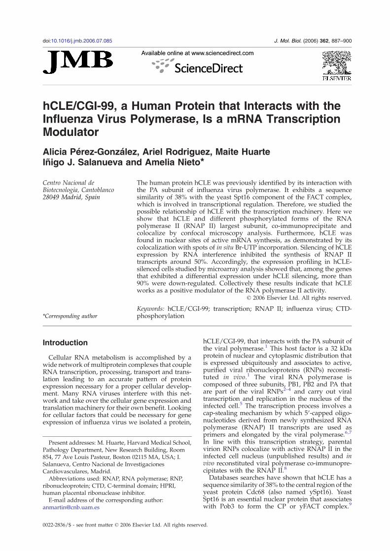
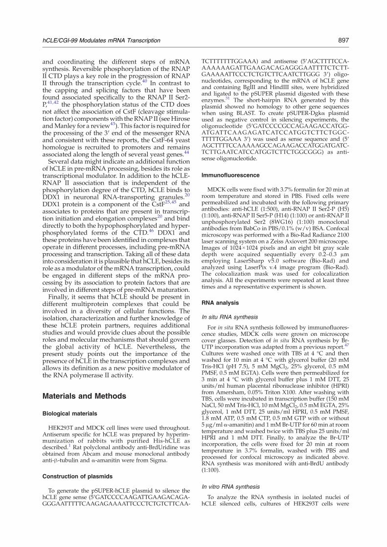
Since hCLE protein shows similarity to the yeastSpt16 factor, a component of the FACT complex, westudied the possible association of hCLE proteinwith the cellular mRNA synthesizing apparatus. Toinvestigate this hypothesis, we used confocal immu-nofluorescence microscopy in MDCK cells to simul-taneously study the localization of hCLE and RNAPII. To this aim, specific antibodies for hCLE andantibodies that recognize different phosphorylatedforms of the RNAP II largest subunit, such asunphosphorylated at serine 2 (8WG16) and phos-phorylated at serine 5 (H14) or at serine 2 (H5) of its
carboxy-terminal domain (CTD), were used.26 Agood colocalization between hCLE and the differentRNAP II forms detected with the correspondingantibodies was observed, as can be seen in thecolocalization panels (Figure 1(a)). These colocaliza-tion data have been obtained using the colocaliza-tion mask, that eliminates signals that are notcommon to both antibodies. To further characterizethe hCLE-RNAP II association, co-immunoprecipi-tation assays were carried out using nuclear extractsfrom HEK293T cells. The data are presented inFigure 1(b). The presence of hCLE and the differentRNAP II forms was verified by Western blot withthe corresponding antibodies. The hCLE proteincould be immunoprecipitated from the nuclearextracts with the specific antibody but not with anunrelated antibody. As can be seen, the hypo- andhyperphosphorylated RNAP II at serine 2 (RNAP IISer2-P) were co-immunoprecipitated with the anti-hCLE antibodies, but not with the control antibody.These results indicate that both the hypophosphory-lated and the RNAP II Ser2-P are physicallyassociated to the hCLE protein. We also wanted totest the association between hCLE and the RNAP IIphosphorylated at serine 5 (RNAP II Ser5-P).Although this form is very clearly visible in totalextracts of HEK293T cells, it was undetectable innuclear extracts. It is possible that phosphorylationat serine 5 was more transient or unstable than thatof serine 2 of the CTD, impeding its examinationunder these conditions.To ascertain whether the sites of hCLE-RNAP II
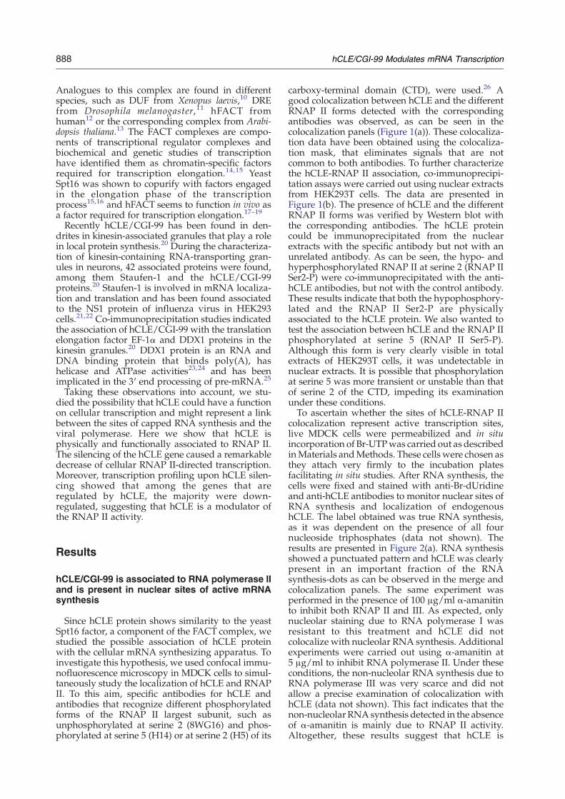
colocalization represent active transcription sites,live MDCK cells were permeabilized and in situincorporation of Br-UTPwas carried out as describedinMaterials andMethods. These cells were chosen asthey attach very firmly to the incubation platesfacilitating in situ studies. After RNA synthesis, thecells were fixed and stained with anti-Br-dUridineand anti-hCLE antibodies to monitor nuclear sites ofRNA synthesis and localization of endogenoushCLE. The label obtained was true RNA synthesis,as it was dependent on the presence of all fournucleoside triphosphates (data not shown). Theresults are presented in Figure 2(a). RNA synthesisshowed a punctuated pattern and hCLE was clearlypresent in an important fraction of the RNAsynthesis-dots as can be observed in the merge andcolocalization panels. The same experiment wasperformed in the presence of 100 μg/ml α-amanitinto inhibit both RNAP II and III. As expected, onlynucleolar staining due to RNA polymerase I wasresistant to this treatment and hCLE did notcolocalize with nucleolar RNA synthesis. Additionalexperiments were carried out using α-amanitin at5 μg/ml to inhibit RNA polymerase II. Under theseconditions, the non-nucleolar RNA synthesis due toRNA polymerase III was very scarce and did notallow a precise examination of colocalization withhCLE (data not shown). This fact indicates that thenon-nucleolar RNAsynthesis detected in the absenceof α-amanitin is mainly due to RNAP II activity.Altogether, these results suggest that hCLE is
Figure 1. hCLE associates to RNA polymerase II complexes. (a) MDCK cells were fixed, processed forimmunofluorescence and analyzed by confocal microscopy. Confocal sections were acquired sequentially every0.2–0.3 μm. An anti-hCLE antibody was used to detect hCLE protein (hCLE) and antibodies 8WG16 (top), H14 (middle),or H5 (bottom) for RNA polymerase II (RNAP II), respectively. Colocalization panel shows signals common to bothantibodies obtained with the colocalization mask. (b) HEK293T nuclear extracts were used for co-immunoprecipitationassays to study the hCLE-RNA polymerase II association. The presence of hCLE and RNA polymerase II was monitoredby Western blot with the α-hCLE or α-RNAP II (8WG16), (H5) and (H14) antibodies, respectively. (Input), represent theHEK293T nuclear extracts and (Ipp) the immunoprecipitates using anti-hCLE (α-hCLE) or a control antibody (control).The percentages of RNAP II co-immunoprecipitated with hCLE are 5.7% and 6.5% of the input, using antibodies 8WG16or H5 for Western blot detection, respectively. Bars represent 10 μm.
889hCLE/CGI-99 Modulates mRNA Transcription
physically associated to intranuclear sites of RNAP IItranscription and propose a role for hCLE in thisprocess.Next we examined if hCLE association to RNAP II
was dependent on active transcription. To answerthis question we used confocal microscopy inMDCK cells treated during 1 h with α-amanitin at100 μg/ml, to simultaneously study the pattern ofnuclear distribution of the different RNAP II formsand hCLE, in conditions of transcription inhibition.Incorporation of Br-UTP was carried out as a controlof the effectiveness of the treatment and the resultsare presented in Figure 3(a). Treatment with α-amanitin clearly inhibited the transcriptional activ-ity of the RNAP II as observed in the Br-UTP panels,where exclusively nucleolar-RNA synthesis was
detected. According to the previous results (Figure2), hCLE distribution did not show variations afterthe drug treatment. The distribution of the hypo-phosphorylated RNAP II and the RNAP II Ser2-Pdid not change upon 1 h of α-amanitin treatment,whereas the RNAP II Ser5-P, which is involved intranscription initiation,27 showed a partial disrup-tion with the drug treatment. On the other hand,1 h treatment with α-amanitin did not change theaccumulation levels of the different RNAP II forms,as detected with Western blot assays using anti-bodies aginst this protein (Figure 3(b)). Althoughchanges in the nuclear distribution of RNAP IISer2-P have been reported with α-amanitin treat-ment in HeLa cells,28,29 no major changes wereobserved for total RNAP II in this situation. The
Figure 2. hCLE is present in sites of RNA synthesis. (a) PermeabilizedMDCK cells were used to detect RNA synthesisby Br-UTP incorporation. After a RNA synthesis pulse of 1 h, cells were fixed, processed for immunofluorescence andanalyzed using confocal microscopy. Confocal sections were acquired sequentially every 0.2–0.3 μm. hCLE detection(hCLE) was achieved with anti-hCLE antibody and RNA synthesis (Br-UTP) with anti-BrdUridine antibody.Colocalization panel shows signals common to both antibodies obtained with the colocalization mask. (b) The sameexperiment was done in the presence of 100 μg/ml of α-amanitin to inhibit the RNAP II RNA synthesis. Bars represent10 μm.
890 hCLE/CGI-99 Modulates mRNA Transcription
transcriptional process is arrested at the initiationstep by high concentrations of α-amanitin,30 whichis in agreement with the observed disturbance in thenuclear localization of the RNAP II Ser5-P in thetreated cells. Alternatively, changes in the proteincomposition of the transcription initiation complexcan occur upon α-amanitin treatment resulting in arestricted reactivity of the H14 antibody in theseconditions. As can be observed, the level of coloca-lization between hCLE and the RNAP II Ser5-Pseems to decrease under conditions of inhibition ofmRNA synthesis, although the nuclear disturbanceand decreased accumulation of RNAP II Ser5-P, donot allow an unambiguous conclusion. On the otherhand, under conditions where RNAP II-dependenttranscription was inhibited, the hCLE proteinremained associated to the hypophosphorylatedRNAP II or RNAP II Ser2-P. Although it is plausiblethat hCLE-RNAP II association occurs only in thecontext of active transcription but once formed thecomplex is very stable, the presented data supportthe statement of a transcription-independent hCLE-RNAP II association.
hCLE/CGI-99 modulates the RNA polymerase IIactivity
To explore the possible role of endogenous hCLEon mRNA synthesis we performed gene-silencingexperiments using RNA interference. To study thephenotype of hCLE loss-of-function, we generated aconstruct of plasmid pSUPER31 expressing under theRNAP III H1 promoter a small hairpin transcriptcontaining a 23 nt sequence derived from the hCLEmRNA (pSUPER-hCLE). As negative control weused a construct of pSUPER expressing a 21 ntRNA derived from the transcript of the Dgkα gene(pSUPER-Dgkα).32 This gene is not expressed in thecell line used, HEK293T.32 To analyze the effec-
tiveness of the construct to down-regulate the hCLEgene, HEK293T cells were transfected with plasmidpSUPER-hCLE, or pSUPER-Dgkα and 48 h post-transfection, nuclei were isolated as described inMaterials andMethods andWestern blot assayswerecarried out using an anti-hCLE antibody. As negativecontrol the same membrane was used to performWestern blot analysis, using antibodies against β-tubulin. As can be seen in Figure 4(a), the amount ofhCLE protein was severely decreased in pSUPERhCLE transfected cells (30% of hCLE proteinremains after treatment), indicating that this treat-ment efficiently down-regulates the hCLE gene. Wealso assayed the effect of pSUPER hCLE transfec-tion in the endogenous levels of hCLE transcript inthe same conditions. Then, total RNA was isolatedand expression of hCLE gene was determined byNorthern blot assays as described in Materials andMethods. Two different species of hCLE RNAwhose accumulation decreases in pSUPER-hCLEtransfected cells were detected (Figure 4(b)).Subsequently we assayed the effect of hCLE silen-
cing on mRNA synthesis. Cultures of HEK293T cellswere transfected with pSUPER-Dgkα or pSUPER-hCLE plasmids and at 48 h post-transfection nucleiof these cells were isolated and frozen. These nucleiwere afterwards used to perform in vitro RNAsynthesis with or without α-amanitin (5 μg/ml) toevaluate the RNAP II-directed RNA synthesis asdescribed in Materials and Methods. In parallel thetransfected HEK293T cells were used to determinethe endogenous hCLE levels, to assure the effective-ness of the hCLE silencing. Five different experi-ments were carried out with triplicate independentsamples and the results obtained are shown inFigure 4(c), as average values and standard devia-tions. The RNA synthesis due to RNA polymerase IIactivity is calculated as the difference between thevalues obtained without the drug (total synthesis
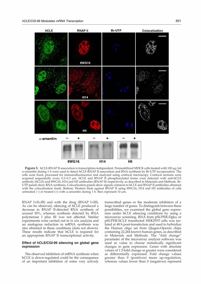
Figure 3. hCLE-RNAP II association is transcription-independent. PermeabilizedMDCK cells treated with 100 μg/mlα-amanitin during 1 h were used to detect hCLE-RNAP II association and RNA synthesis by Br-UTP incorporation. Thecells were fixed, processed for immunofluorescence and analyzed using confocal microscopy. Confocal sections wereacquired sequentially every 0.2–0.3 μm. hCLE and RNAP II phosphorylated forms were detected with anti-hCLEantibody (hCLE) and 8WG16, H14 and H5 antibodies (RNAP II) respectively, as described in Materials and Methods. Br-UTP panels show RNA synthesis. Colocalization panels show signals common to hCLE and RNAP II antibodies obtainedwith the colocalization mask. Bottom; Western blots against RNAP II using 8WG16, H14 and H5 antibodies of cellsuntreated (−) or treated (+) with α-amanitin during 1 h. Bars represent 10 μm.
891hCLE/CGI-99 Modulates mRNA Transcription
RNAP I+II+III) and with the drug (RNAP I+III).As can be observed, silencing of hCLE produced adecrease in RNAP II-directed RNA synthesis ofaround 50%, whereas synthesis directed by RNApolymerase I plus III was not affected. Similarexperiments were carried out in in situ analysis andan analogous reduction in mRNA synthesis wasalso obtained in these conditions (data not shown).These results indicate that hCLE is required foran appropriate RNAP II transcriptional activity.
Effect of hCLE/CGI-99 silencing on global geneexpression
The observed inhibition of mRNA synthesis whenhCLE is down-regulated could be the consequenceof an important inhibition of some very actively
transcribed genes or the moderate inhibition of alarge number of genes. To distinguish between thesepossibilities, we examined the global gene expres-sion under hCLE silencing conditions by using amicroarray screening. RNA from pSUPER-Dgkα orpSUPER-hCLE transfected HEK293T cells was iso-lated at 48 h post-transfection and used to hybridizethe Human oligo set from Qiagen-Operon chipscontaining 22,264 known human genes, as describedin Materials and Methods. The “fold change”parameter of the microarray analysis software wasused as value to choose statistically significantchanges in gene expression. Genes with absolutevalues of 1.5-fold change or greater were consideredas differentially expressed. Fold change valuesgreater than 0 (positives) mean up-regulation,whereas values lower than 0 (negatives) represent
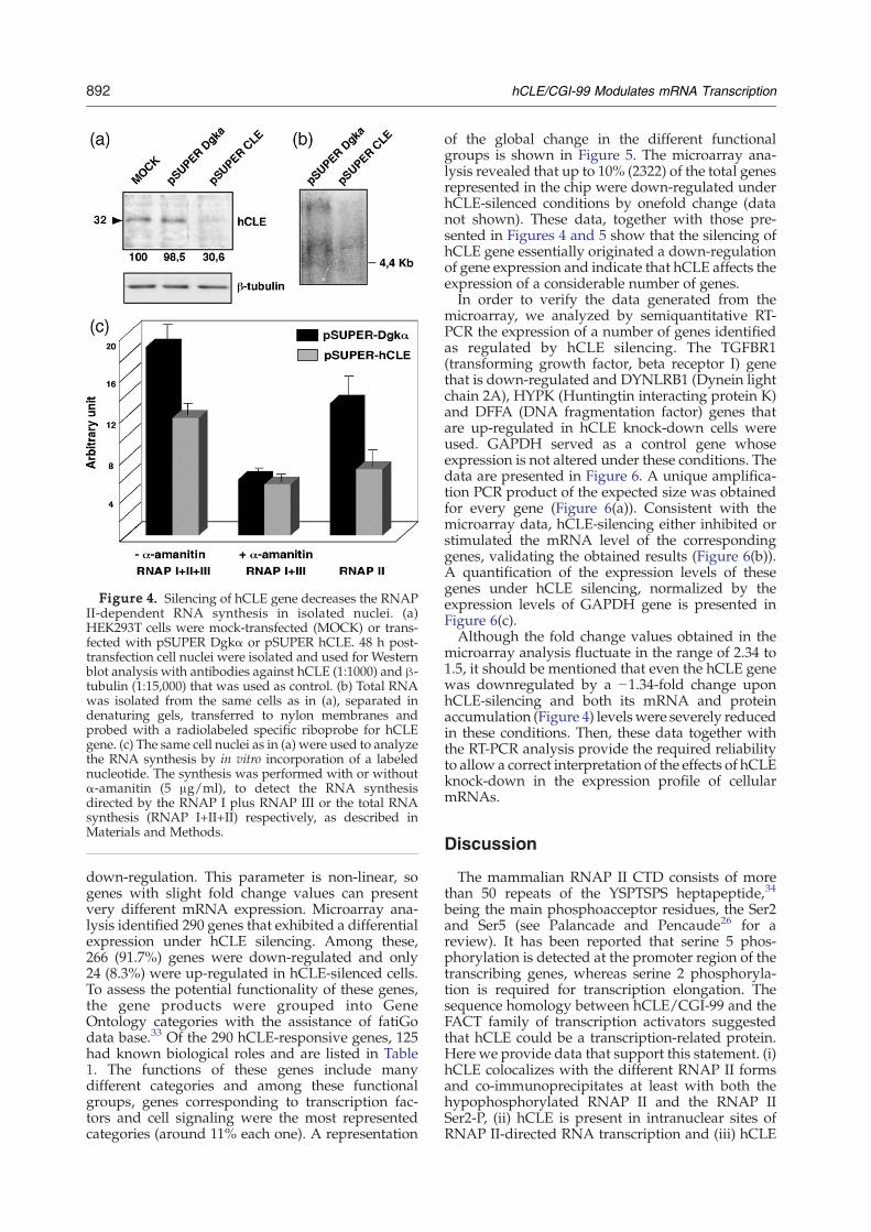
Figure 4. Silencing of hCLE gene decreases the RNAPII-dependent RNA synthesis in isolated nuclei. (a)HEK293T cells were mock-transfected (MOCK) or trans-fected with pSUPER Dgkα or pSUPER hCLE. 48 h post-transfection cell nuclei were isolated and used for Westernblot analysis with antibodies against hCLE (1:1000) and β-tubulin (1:15,000) that was used as control. (b) Total RNAwas isolated from the same cells as in (a), separated indenaturing gels, transferred to nylon membranes andprobed with a radiolabeled specific riboprobe for hCLEgene. (c) The same cell nuclei as in (a) were used to analyzethe RNA synthesis by in vitro incorporation of a labelednucleotide. The synthesis was performed with or withoutα-amanitin (5 μg/ml), to detect the RNA synthesisdirected by the RNAP I plus RNAP III or the total RNAsynthesis (RNAP I+II+II) respectively, as described inMaterials and Methods.
892 hCLE/CGI-99 Modulates mRNA Transcription
down-regulation. This parameter is non-linear, sogenes with slight fold change values can presentvery different mRNA expression. Microarray ana-lysis identified 290 genes that exhibited a differentialexpression under hCLE silencing. Among these,266 (91.7%) genes were down-regulated and only24 (8.3%) were up-regulated in hCLE-silenced cells.To assess the potential functionality of these genes,the gene products were grouped into GeneOntology categories with the assistance of fatiGodata base.33 Of the 290 hCLE-responsive genes, 125had known biological roles and are listed in Table1. The functions of these genes include manydifferent categories and among these functionalgroups, genes corresponding to transcription fac-tors and cell signaling were the most representedcategories (around 11% each one). A representation
of the global change in the different functionalgroups is shown in Figure 5. The microarray ana-lysis revealed that up to 10% (2322) of the total genesrepresented in the chip were down-regulated underhCLE-silenced conditions by onefold change (datanot shown). These data, together with those pre-sented in Figures 4 and 5 show that the silencing ofhCLE gene essentially originated a down-regulationof gene expression and indicate that hCLE affects theexpression of a considerable number of genes.In order to verify the data generated from the
microarray, we analyzed by semiquantitative RT-PCR the expression of a number of genes identifiedas regulated by hCLE silencing. The TGFBR1(transforming growth factor, beta receptor I) genethat is down-regulated and DYNLRB1 (Dynein lightchain 2A), HYPK (Huntingtin interacting protein K)and DFFA (DNA fragmentation factor) genes thatare up-regulated in hCLE knock-down cells wereused. GAPDH served as a control gene whoseexpression is not altered under these conditions. Thedata are presented in Figure 6. A unique amplifica-tion PCR product of the expected size was obtainedfor every gene (Figure 6(a)). Consistent with themicroarray data, hCLE-silencing either inhibited orstimulated the mRNA level of the correspondinggenes, validating the obtained results (Figure 6(b)).A quantification of the expression levels of thesegenes under hCLE silencing, normalized by theexpression levels of GAPDH gene is presented inFigure 6(c).Although the fold change values obtained in the
microarray analysis fluctuate in the range of 2.34 to1.5, it should be mentioned that even the hCLE genewas downregulated by a −1.34-fold change uponhCLE-silencing and both its mRNA and proteinaccumulation (Figure 4) levelswere severely reducedin these conditions. Then, these data together withthe RT-PCR analysis provide the required reliabilityto allow a correct interpretation of the effects of hCLEknock-down in the expression profile of cellularmRNAs.
Discussion
The mammalian RNAP II CTD consists of morethan 50 repeats of the YSPTSPS heptapeptide,34
being the main phosphoacceptor residues, the Ser2and Ser5 (see Palancade and Pencaude26 for areview). It has been reported that serine 5 phos-phorylation is detected at the promoter region of thetranscribing genes, whereas serine 2 phosphoryla-tion is required for transcription elongation. Thesequence homology between hCLE/CGI-99 and theFACT family of transcription activators suggestedthat hCLE could be a transcription-related protein.Here we provide data that support this statement. (i)hCLE colocalizes with the different RNAP II formsand co-immunoprecipitates at least with both thehypophosphorylated RNAP II and the RNAP IISer2-P, (ii) hCLE is present in intranuclear sites ofRNAP II-directed RNA transcription and (iii) hCLE
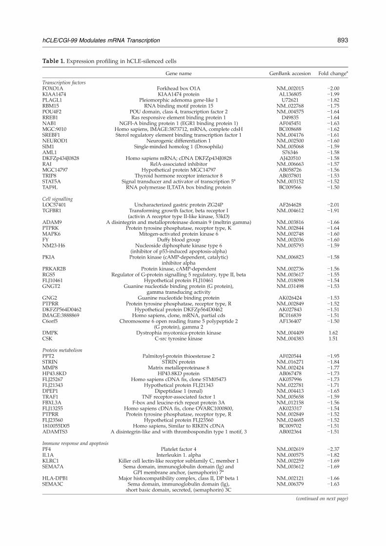
Table 1. Expression profiling in hCLE-silenced cells
Gene name GenBank accesion Fold changea
Transcription factorsFOXO1A Forkhead box O1A NM_002015 −2.00KIAA1474 KIAA1474 protein AL136805 −1.99PLAGL1 Pleiomorphic adenoma gene-like 1 U72621 −1.82RBM15 RNA binding motif protein 15 NM_022768 −1.75POU4F2 POU domain, class 4, transcription factor 2 NM_004575 −1.64RREB1 Ras responsive element binding protein 1 D49835 −1.64NAB1 NGFI-A binding protein 1 (EGR1 binding protein 1) AF045451 −1.63MGC:9010 Homo sapiens, IMAGE:3873712, mRNA, complete cdsH BC008688 −1.62SREBF1 Sterol regulatory element binding transcription factor 1 NM_004176 −1.61NEUROD1 Neurogenic differentiation 1 NM_002500 −1.60SIM1 Single-minded homolog 1 (Drosophila) NM_005068 −1.59AML1 S76346 −1.58DKFZp434J0828 Homo sapiens mRNA; cDNA DKFZp434J0828 AJ420510 −1.58RAI RelA-associated inhibitor NM_006663 −1.57MGC14797 Hypothetical protein MGC14797 AB058726 −1.56TRIP8 Thyroid hormone receptor interactor 8 AB037801 −1.53STAT5A Signal transducer and activator of transcription 5a NM_003152 −1.52TAF9L RNA polymerase II,TATA box binding protein BC009566 −1.50
Cell signallingLOC57401 Uncharacterized gastric protein ZG24P AF264628 −2.01TGFBR1 Transforming growth factor, beta receptor I
(activin A receptor type II-like kinase, 53kD)NM_004612 −1.91
ADAM9 A disintegrin and metalloproteinase domain 9 (meltrin gamma) NM_003816 −1.66PTPRK Protein tyrosine phosphatase, receptor type, K NM_002844 −1.64MAPK6 Mitogen-activated protein kinase 6 NM_002748 −1.60FY Duffy blood group NM_002036 −1.60NM23-H6 Nucleoside diphosphate kinase type 6
(inhibitor of p53-induced apoptosis-alpha)NM_005793 −1.59
PKIA Protein kinase (cAMP-dependent, catalytic)inhibitor alpha
NM_006823 −1.58
PRKAR2B Protein kinase, cAMP-dependent NM_002736 −1.56RGS5 Regulator of G-protein signalling 5 regulatory, type II, beta NM_003617 −1.55FLJ10461 Hypothetical protein FLJ10461 NM_018098 −1.54GNGT2 Guanine nucleotide binding protein (G protein),
gamma transducing activityNM_031498 −1.53
GNG2 Guanine nucleotide binding protein AK026424 −1.53PTPRR Protein tyrosine phosphatase, receptor type, R NM_002849 −1.52DKFZP564D0462 Hypothetical protein DKFZp564D0462 AK027843 −1.51IMAGE:3888869 Homo sapiens, clone, mRNA, partial cds BC016839 −1.51C6orf5 Chromosome 6 open reading frame 5 polypeptide 2
(G protein), gamma 2AF136407 −1.50
DMPK Dystrophia myotonica-protein kinase NM_004409 1.62CSK C-src tyrosine kinase NM_004383 1.51
Protein metabolismPPT2 Palmitoyl-protein thioesterase 2 AF020544 −1.95STRIN STRIN protein NM_016271 −1.84MMP8 Matrix metalloproteinase 8 NM_002424 −1.77HP43.8KD HP43.8KD protein AB067478 −1.73FLJ25267 Homo sapiens cDNA fis, clone STM05473 AK057996 −1.73FLJ21343 Hypothetical protein FLJ21343 NM_022781 −1.71DPEP1 Dipeptidase 1 (renal) NM_004413 −1.65TRAF1 TNF receptor-associated factor 1 NM_005658 −1.59FBXL3A F-box and leucine-rich repeat protein 3A NM_012158 −1.56FLJ13255 Homo sapiens cDNA fis, clone OVARC1000800, AK023317 −1.54PTPRR Protein tyrosine phosphatase, receptor type, R NM_002849 −1.52FLJ23560 Hypothetical protein FLJ23560 NM_024685 −1.521810055D05 Homo sapiens, Similar to RIKEN cDNA BC009702 −1.51ADAMTS3 A disintegrin-like and with thrombospondin type 1 motif, 3 AB002364 −1.51
Immune response and apoptosisPF4 Platelet factor 4 NM_002619 −2.37IL1A Interleukin 1. alpha NM_000575 −1.82KLRC1 Killer cell lectin-like receptor subfamily C, member 1 NM_002259 −1.69SEMA7A Sema domain, immunoglobulin domain (lg) and
GPI membrane anchor, (semaphorin) 7aNM_003612 −1.69
HLA-DPB1 Major histocompatibility complex, class II, DP beta 1 NM_002121 −1.66SEMA3C Sema domain, immunoglobulin domain (lg),
short basic domain, secreted, (semaphorin) 3CNM_006379 −1.63
(continued on next page)
893hCLE/CGI-99 Modulates mRNA Transcription
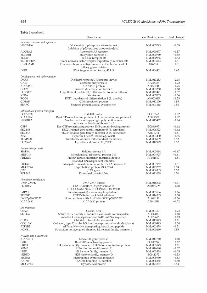
Table 1 (continued)
Gene name GenBank accesion Fold changea
Immune response and apoptosisNM23-H6 Nucleoside diphosphate kinase type 6
(inhibitor of p53-induced apoptosis-alpha)NM_005793 −1.59
ADORA3 Adenosine A3 receptor NM_000677 −1.57BDKRB1 Bradykinin receptor B1 NM_000710 −1.55TLR10 Toll-like receptor 10 NM_030956 −1.53TNFRSF10A Tumor necrosis factor receptor superfamily, member 10a NM_003844 −1.51CEACAM1 Carcinoembryonic antigen-related cell adhesion mole 1
(biliary glycoprotein)X16354 −1.51
DFFA DNA fragmentation factor, 45 kD, NM_004401 1.62
Development and differentationDKK3 Dickkopf homolog 3 (Xenopus laevis) NM_013253 −2.18CA10 Carbonic anhydrase X AF288385 −1.72KIAA1813 KIAA1813 protein AB058716 −1.75GDF9 Growth differentiation factor 9 NM_005260 −1.60FLJ13057 Hypothetical protein FLJ13057 similar to germ cell-less NM_022471 −1.57KERA Keratocan NM_007035 −1.56ROD1 ROD1 regulator of differentiation 1 (S. pombe) AK001685 −1.53CD2AP CD2-associated protein NM_012120 1.53SPARC Secreted protein, acidic, cysteine-rich NM_003118 1.51
Intracellular protein transportLOC50999 CGI-100 protein BC016556 −1.82KIAA0660 Ras-GTPase activating protein SH3 domain-binding protein 2 AB014560 −1.82NFKBIL2 Nuclear factor of kappa light polypeptide gene
enhancer in B-cells inhibitor-like 2NM_013432 −1.64
G3BP Ras-GTPase-activating protein SH3-domain-binding protein BC006997 −1.62SEC24B SEC24 related gene family, member B (S. cerevisiae) NM_006323 −1.62SEC24A SEC24 related gene family, member A (S. cerevisiae) AJ131244 −1.61XPO1 Exportin 1 (CRM1 homolog, yeast) NM_003400 −1.55TOMM34 Translocase of outer mitochondrial membrane NM_006809 1.52FLJ20695 Hypothetical protein FLJ20695 NM_017929 1.52
Protein biosynthesisSIAT8A Sialyltransferase 8A NM_003034 −1.67MRPL49 Mitochondrial ribosomal protein L49 NM_004927 −1.61PRKRIR Protein-kinase, interferon-inducible double
stranded RNAdependent inhibitor,AF081567 −1.51
EIF4A2 Eukaryotic translation initiation factor 4A, isoform 2 NM_001967 −1.51MGC2744 Hypothetical protein MGC2744 NM_025267 1.54JTV1 JTV1 gene NM_006303 1.52RPL36A Ribosomal protein L36a NM_021029 1.51
Phosphate metabolismUMP-CMPK UMP-CMP kinase NM_016308 −1.91FLJ31077 HSYRA2001574, highly similar to
GLUCOSAMINE-6-PHOSPHATE ISOMERAK055639 −1.68
IMPA1 Inositol(myo)-1(or 4)-monophosphatase 1 NM_005536 −1.66TDPGD DTDP-D-glucose 4,6-dehydratase NM_014305 −1.58DKFZp586G2222 Homo sapiens mRNA; cDNA DKFZp586G2222 AL080111 −1.56KIAA0630 KIAA0630 protein AB014530 −1.52
Ion transportCSN2 Casein, beta NM_001891 −1.73SLC4A7 Solute carrier family 4, sodium bicarbonate cotransporter,
member Homo sapiens clone 24411 mRNA sequenceAF047033AF070606
−1.68−1.63
CLIC4 Chloride intracellular channel 4 NM_013943 −1.61COL10A1 Collagen, type X, alpha 1(Schmid metaphyseal chondrodysplasia) NM_000493 −1.54ATP1B2 ATPase, Na+/K+ transporting, beta 2 polypeptide NM_001678 −1.51KCNE1 Potassium voltage-gated channel, lsk-related family, member 1 NM_000219 1.53
Nucleic acid metabolismKIAA0111 KIAA0111 gene product NM_014740 −1.68G3BP Ras-GTPase-activating protein BC006997 −1.62H4FH H4 histone family, member H SH3-domain-binding protein NM_003543 −1.61RBM7 RNA binding motif protein 7 NM_016090 −1.57H4FA H4 histone family, member A NM_003538 −1.55H2BFD H2B histone family, member D BC011372 −1.53MGEA6 Meningioma expressed antigen 6 NM_005930 −1.53RAD21 RAD21 homolog (S. pombe) NM_006265 −1.50MGC2744 Hypothetical protein NM_025267 1.54
894 hCLE/CGI-99 Modulates mRNA Transcription
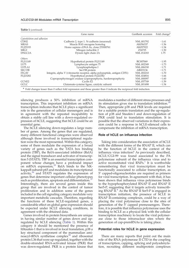
Table 1 (continued)
Gene name GenBank accesion Fold changea
Cytoskeleton and adhesionCDH2 Cadherin 2, type 1. N-cadherin (neuronal) NM_001792 −1.61MRAS Muscle RAS oncogene homolog AK056337 −1.56FLJ25193 Homo sapiens cDNA fis, clone JTH00761 AK057922 −1.54MIG2 Mitogen inducible 2 Z24725 −1.50DNLC2A Dynein light chain 2A NM_014183 1.75
OthersFLJ11149 Hypothetical protein FLJ11149 BC007069 −1.95LY75 Lymphocyte antigen 75 NM_002349 −1.73LYPLA1 Lysophospholipas I NM_006330 −1.71SEC15B Sec15B protein AB023136 −1.71ITGAV Integrin, alpha V (vitronectin receptor, alpha polypeptide, antigen CD51) NM_002210 −1.70FLJ23282 Hypothetical protein FLJ22382 NM_024816 −1.66
Coproporphyrinogen oxidase (coproporphyria, harderroporphyria NM_000097 −1.60CCNE2 Cyclin E2 NM_057749 −1.59GCLC Glutamate-cysteine ligase, catalytic subunit NM_001498 −1.54
a Fold changes lesser than 0 reflect fold repression and those greater than 0 indicate the reciprocal fold induction.
895hCLE/CGI-99 Modulates mRNA Transcription
silencing produces a 50% inhibition of mRNAtranscription. This important inhibition on mRNAtranscription indicates that hCLE plays a significantrole in the generation of cellular messengers and isin agreement with the repeated incapability toobtain a stable cell line with a down-regulated ex-pression of hCLE, suggesting that hCLE could be anessential gene.The hCLE silencing down-regulates a large num-
ber of genes. Among the genes that are regulated,many different functional categories were observedalthough those involved in transcriptional regula-tion were themost represented.Within this category,some of them modulate the expression of a broadvariety of genes such as the TATA box bindingprotein (TBP), the RelA-associated inhibitor (RelA)and the signal transducer and activator of transcrip-tion 5 (STAT5). TBP is an essential transcription com-ponent whose changes have a profound impacton mRNA expression,35 RelA binds to the NK-kappaB subunit p65 andmodulates its trancriptionalactivity,36 and STAT5 regulates the expression ofgenes that determine important cellular phenotypessuch as proliferation, apoptosis and differentiation.37
Interestingly, there are several genes inside thisgroup that are involved in the control of tumorproliferation and in addition some of the genesincluded in the cell signaling categorymodulate veryimportant metabolic cascades. Therefore, regardingthe functions of these hCLE-regulated genes, aconsiderable effect on global gene expression shouldbe expected under hCLE silencing conditions, inagreement with the obtained data.Genes involved in protein biosynthesis are unique
in having similar number of genes down and up-regulated by hCLE silencing (Table 1). Within thiscategory it should be mentioned the presence ofhStaufen-1 that is involved in local translation, p38 akey structural component of the mammalian ami-noacyl-tRNA synthetase complex38 and ribosomalprotein L36 thatwere up-regulated, togetherwith thedouble-stranded RNA-activated kinase (PKR) thatwas down-regulated. PKR is a protein kinase that
modulates a number of different stress processes andits stimulation gives rise to translation inhibition.39
Then, appropriate p38 and PKR levels are requiredfor a suitable protein translation and the up-regula-tion of p38 and Staufen-1 and down-regulation ofPKR could lead to translation stimulation. It ispossible that the observed variations in their expres-sion could be a response in hCLE-silenced cells tocompensate the inhibition of mRNA transcription.
Role of hCLE on influenza infection
Taking into consideration the association of hCLEwith the different forms of the RNAP II, which canbe the function of hCLE in the context of theinfluenza virus infection? As previously described,hCLE/CGI-99 was found associated to the PApolymerase subunit of the influenza virus and toactive reconstituted viral RNPs.1 It is worthwhileremembering that viral transcription must befunctionally associated to cellular transcription, as5′ capped-oligonucleotides are required as primersfor viral transcription. In agreement with that, it hasbeen shown that influenza virus polymerase bindsto the hypophosphorylated RNAP II and RNAP IISer5-P, suggesting that it targets actively transcrib-ing RNAP II.8 As the RNAP II Ser5-P is engaged intranscription initiation, the presence of hCLE inRNAP II-containing complexes, could be a way ofplacing the viral polymerase close to the sites ofgeneration of the 5′ capped premessengers. There-fore, it is possible that influenza polymerase uses thebinding to hCLE as a physical link with the cellulartranscription machinery to locate the viral polymer-ase close to those intranuclear sites where thegeneration of cap-premRNAs is taking place.
Potential roles for hCLE in gene expression
There are many reports that point out the mainrole of the RNA polymerase II CTD in the couplingof transcription, capping, splicing and polyadenyla-tion, recruiting different multiprotein complexes
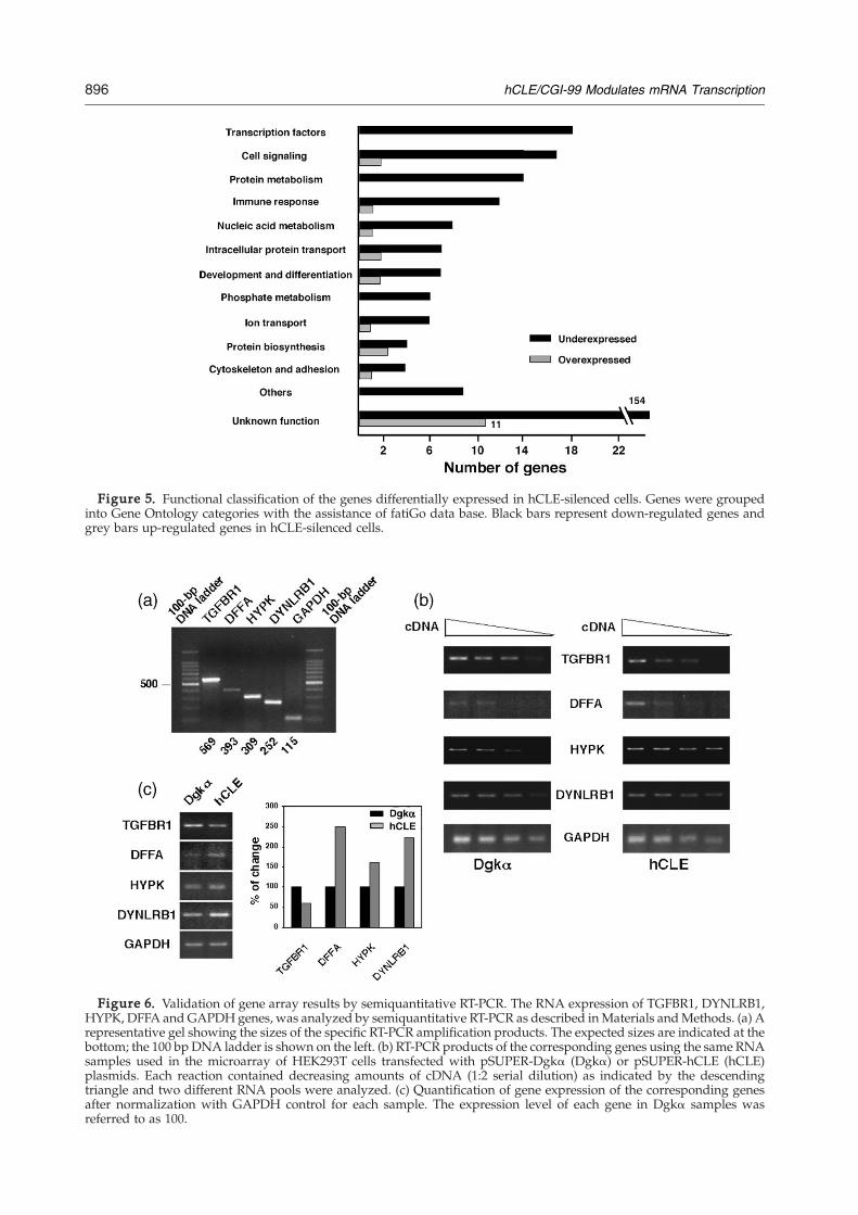
Figure 5. Functional classification of the genes differentially expressed in hCLE-silenced cells. Genes were groupedinto Gene Ontology categories with the assistance of fatiGo data base. Black bars represent down-regulated genes andgrey bars up-regulated genes in hCLE-silenced cells.
Figure 6. Validation of gene array results by semiquantitative RT-PCR. The RNA expression of TGFBR1, DYNLRB1,HYPK, DFFA and GAPDH genes, was analyzed by semiquantitative RT-PCR as described inMaterials andMethods. (a) Arepresentative gel showing the sizes of the specific RT-PCR amplification products. The expected sizes are indicated at thebottom; the 100 bp DNA ladder is shown on the left. (b) RT-PCR products of the corresponding genes using the same RNAsamples used in the microarray of HEK293T cells transfected with pSUPER-Dgkα (Dgkα) or pSUPER-hCLE (hCLE)plasmids. Each reaction contained decreasing amounts of cDNA (1:2 serial dilution) as indicated by the descendingtriangle and two different RNA pools were analyzed. (c) Quantification of gene expression of the corresponding genesafter normalization with GAPDH control for each sample. The expression level of each gene in Dgkα samples wasreferred to as 100.
896 hCLE/CGI-99 Modulates mRNA Transcription
897hCLE/CGI-99 Modulates mRNA Transcription
and coordinating the different steps of mRNAsynthesis. Reversible phosphorylation of the RNAPII CTD plays a key role in the progression of RNAPII through the transcription cycle.40 In contrast tothe capping and splicing factors that have beenfound associated specifically to the RNAP II Ser2-P,41,42 the phosphorylation status of the CTD doesnot affect the association of CstF (cleavage stimula-tion factor) componentswith the RNAP II (seeHiroseandManley for a review43). This factor is required forthe processing of the 3′ end of the messenger RNAand consistent with these reports, the CstF-64 yeasthomologue is recruited to promoters and remainsassociated along the length of several yeast genes.44
Several data might indicate an additional functionof hCLE in pre-mRNA processing, besides its role astranscriptional modulator. In addition to the hCLE-RNAP II association that is independent of thephosphorylation degree of the CTD, hCLE binds toDDX1 in neuronal RNA-transporting granules.20
DDX1 protein is a component of the CstF25,45 andassociates to proteins that are present in transcrip-tion initiation and elongation complexes20 and binddirectly to both the hypophosphorylated and hyper-phosphorylated forms of the CTD.46 DDX1 andthese proteins have been identified in complexes thatoperate in different processes, including pre-mRNAprocessing and transcription. Taking all of these datainto consideration it is plausible that hCLE, besides itsrole as amodulator of themRNA transcription, couldbe engaged in different steps of the mRNA pro-cessing by its association to protein factors that areinvolved in different steps of pre-mRNAmaturation.Finally, it seems that hCLE should be present in
different multiprotein complexes that could beinvolved in a diversity of cellular functions. Theisolation, characterization and further knowledge ofthese hCLE protein partners, requires additionalstudies and would provide clues about the possibleroles and molecular mechanisms that should governthe global activity of hCLE. Nevertheless, thepresent study points out the importance of thepresence of hCLE in the transcription complexes andallows its definition as a new positive modulator ofthe RNA polymerase II activity.
Materials and Methods
Biological materials
HEK293T and MDCK cell lines were used throughout.Antiserum specific for hCLE was prepared by hyperim-munization of rabbits with purified His-hCLE asdescribed.1 Rat polyclonal antibody anti-BrdUridine wasobtained from Abcam and mouse monoclonal antibodyanti-β-tubulin and α-amanitin were from Sigma.
Construction of plasmids
To generate the pSUPER-hCLE plasmid to silence thehCLE gene sense (5′GATCCCCAAGATTGAAGACAGA-GGGAATTTTTCAAGAGAAAATTCCCTCTGTCTTCAA-
TCTTTTTTTGGAAA) and antisense (5′AGCTTTTCCA-AAAAAAGATTGAAGACAGAGGGAATTTTCTCTT-GAAAAATTCCCTCTGTCTTCAATCTTGGG 3′) oligo-nucleotides, corresponding to the mRNA of hCLE geneand containing BglII and HindIII sites, were hybridizedand ligated to the pSUPER plasmid digested with theseenzymes.31 The short-hairpin RNA generated by thisplasmid showed no homology to other gene sequenceswhen using BLAST. To create pSUPER-Dgkα plasmidused as negative control in silencing experiments, theoligonucleotide (5′GATCCCCGCCAGAAGACCATGG-ATGATTCAAGAGATCATCCATGGTCTTCTGGC-TTTTTGGAAA 3′) was used as sense sequence and (5′AGCTTTTCCAAAAAGCCAGAAGACCATGGATGATC-TCTTGAATCATCCATGGTCTTCTGGCGGG) as anti-sense oligonucleotide.
Immunofluorescence
MDCK cells were fixed with 3.7% formalin for 20 min atroom temperature and stored in PBS. Fixed cells werepermeabilized and incubated with the following primaryantibodies: anti-hCLE (1:500), anti-RNAP II Ser2-P (H5)(1:100), anti-RNAP II Ser5-P (H14) (1:100) or anti-RNAP IIunphosphorylated Ser2 (8WG16) (1:100) monoclonalantibodies from BabCo in PBS/0.1% (w/v) BSA. Confocalmicroscopy was performed with a Bio-Rad Radiance 2100laser scanning system on a Zeiss Axiovert 200 microscope.Images of 1024×1024 pixels and an eight bit gray scaledepth were acquired sequentially every 0.2–0.3 μmemploying LaserSharp v5.0 software (Bio-Rad) andanalyzed using LaserPix v.4 image program (Bio-Rad).The colocalization mask was used for colocalizationanalysis. All the experiments were repeated at least threetimes and a representative experiment is shown.
RNA analysis
In situ RNA synthesis
For in situ RNA synthesis followed by immunofluores-cence studies, MDCK cells were grown on microscopecover glasses. Detection of in situ RNA synthesis by Br-UTP incorporation was adapted from a previous report.47
Cultures were washed once with TBS at 4 °C and thenwashed for 10 min at 4 °C with glycerol buffer (20 mMTris-HCl (pH 7.5), 5 mM MgCl2, 25% glycerol, 0.5 mMPMSF, 0.5 mM EGTA). Cells were then permeabilized for3 min at 4 °C with glycerol buffer plus 1 mM DTT, 25units/ml human placental ribonuclease inhibitor (HPRI)from Amersham, 0.05% Triton X100. After washing withTBS, cells were incubated in transcription buffer (150 mMNaCl, 50 mM Tris-HCl, 10 mMMgCl2, 0.5 mM EGTA, 25%glycerol, 1 mM DTT, 25 units/ml HPRI, 0.5 mM PMSF,1.8 mM ATP, 0.5 mM CTP, 0.5 mM GTP with or without5 μg/ml α-amanitin) and 1 mMBr-UTP for 60 min at roomtemperature and washed twice with TBS plus 25 units/mlHPRI and 1 mM DTT. Finally, to analyze the Br-UTPincorporation, the cells were fixed for 20 min at roomtemperature in 3.7% formalin, washed with PBS andprocessed for confocal microscopy as indicated above.RNA synthesis was monitored with anti-BrdU antibody(1:100).
In vitro RNA synthesis
To analyze the RNA synthesis in isolated nuclei ofhCLE silenced cells, cultures of HEK293T cells were
898 hCLE/CGI-99 Modulates mRNA Transcription
transfected with the indicated pSUPER plasmids by thecalcium phosphate method,48 the nuclei were isolated48 h post-transfection as described49 and frozen in liquidnitrogen. These nuclei were used to perform in vitroRNA synthesis by incorporation of a labeled ribonucleo-tide during a 10 min pulse, with or without α-amanintin(5 μg/ml) to evaluate the RNAP II-directed RNAsynthesis, as described.49 The RNA was isolated byphenol-extraction and ethanol-precipitation. Finally, totalRNAs were applied to denaturing gels and afterquantification by ethidium bromide staining in a Bio-Rad Chemi Doc equipment, transferred to nylonmembranes and the radioactivity was quantified in aphosphorimager.
Northern blot
Total RNA was isolated using the Ultraspec RNAIsolation Reagent from Biotex and treated with 40 μg/mlof RNase-free DNase I and 25 units/ml HPRI for 15 min at30 °C. Northern blots were performed using standardconditions49 and the filters were hybridized with anegative-polarity riboprobe to detect RNA levels ofhCLE gene.
Co-immunoprecipitation
For co-immunoprecipitation studies, nuclei of HEK293Tcells were prepared as described above and transcription-ally active nuclear extracts obtained as reported.50 Theextracts were incubated with the corresponding antibo-dies as reported51 and the co-immunoprecipitated pro-teins were analyzed by Western blotting that was done asdescribed.1 The primary antibodies used were a rabbitanti-hCLE serum (1:1000) for hCLE protein and mono-clonal antibodies 8WG16, H14 and H5 (1:500) each forRNA polymerase II.
RNA isolation and DNA microarray
Cultures of HEK293T cells were transfected withpSUPER-Dgkα or pSUPER-hCLE plasmids and at 48 hpost-transfection total RNA was extracted using Trizolreagent (Invitrogen) according to the manufacturer'sinstructions. Further purification was carried out usingthe RNAeasy Clean-Up protocol (Quiagen, Valencia, CA).Quantification and integrity of the RNAwas assessed in aBionalyzer 2100 (Agilent) by A260/280 absorption. Threedifferent RNA pools containing 10 μg of total RNAderived from three independent experiments performedin triplicate were used.Aminosilane-coated slides containing 32,256 spots
(corresponding to 22,264 unigenes) representing theHuman oligo set from Qiagen-Operon, obtained fromthe University of Cincinnati†, were hybridized by con-ventional methods with RNA probes labelled with eitherCy3 or Cy5 Mono NHS Esters (Cy™Dye Post-labellingReactive Dye Pack, Amersham). “Dye-swap” labellingwas used in half of the replicated hybridizations in orderto correct for gene-specific dye bias. Images from Cy3 andCy5 channels were equilibrated and captured with aGenePix 4000B (Axon) and spots quantified using GenPixsoftware (Axon).
†http://www.microarray.uc.edu/Resources/genelist.htm
Microarray data analysis
Normalization and statistical analysis of the expressiondata were performed by using the LIMMA softwarepackage.52 For local background correction we used the“normexp” method in LIMMA to adjust the local medianbackground estimates. The resulting log-ratios wereprint-tip loess normalized for each array. To have similardistribution across arrays and to achieve consistencyamong arrays, log-ratio values were scaled using as scaleestimator the median-absolute-value.52 To assess thedifferential expression in conditions of hCLE silencing alinear model was used. Each probe was tested forchanges in expression over replicates by using amoderated t statistic.52 To control the false discoveryrate (FDR), p-values were corrected by using the methodof Benjamini and Hochberg.53 To select differentiallyexpressed genes in conditions of hCLE knock-down twocriteria were used: FDR <10% and a fold change greaterthan 1.0. Values were considered to be significant if thecorrected p-values were <0.1 and 2,322 differentiallyexpressed genes emerged. Finally, establishing the criter-ion of a fold change greater than 1.5 at three replicas atotal of 290 genes differentially expressed in conditions ofhCLE knock-down appeared.
Semiquantitative RT-PCR and primers
The genes TGFBR1 (Transforming growth factor, betareceptor I), DYNLRB1 (Dynein light chain 2A), HYPK(Huntingtin interacting protein K) and DFFA (DNAfragmentation factor) identified among the regulatedgenes by hCLE knock-down were analyzed using semi-quantitative RT-PCR to validate the array data. RNAtemplates were reversed-transcribed into cDNA for 1 h at45 °C by using AMV reverse transcriptase (Promega)according to the manufacter's instructions. The enzymewas inactivated by heating 10 min at 75 °C; and thedenatured cDNA templates were amplified using Taqpolymerase by the following cycles: 94 °C for 10 s, 54 °Cfor 30 s and 68 °C for 60 s. A final extension was performedfor 2 min at 68 °C. To ensure that amplification remainedwithin the linear range, 1:2 serial dilutions of cDNAweremade. RT-PCR for GAPDH (25 cycles) was used as acontrol for mRNA abundance. For the other genes, thenumber of cycles ranged from 25 to 35. RT-PCR for eachgene was performed several times using different batchesof cDNA.The oligonucleotide primer pairs used for each of the
genes in this study corresponded to the following nucleo-tides: TGFBR1, 737–751 and 1288–1304 NM_004612;DYNLRB1, 63–78 and 297–315 NM_014183; HYPK, 221–237 and 513–530NM_016400 andDFFA, 695–711 and 1071–1088 NM_004401. Amplified PCR products were visualizedon a 2% (w/v) agarose gel. Amplification yielded thepredicted size of the respective amplified fragments.
Acknowledgements
We are indebted to the transcriptomics facility ofthe Centro Nacional de Biotecnología, C.S.I.C.,Madrid, Spain, that performed the microarraystudies. We thank C. Suñe for his help during thecourse of this work. We also thank N. Brewster and
899hCLE/CGI-99 Modulates mRNA Transcription
R. Singer for performing studies of hCLE in yeast.We are indebted to J. Ortin and T. Lutz for criticalreview of the manuscript. The technical assistance ofS. Gutierrez with the confocal miscroscopy studies isgratefully acknowledged. We also thank Y. Fernan-dez, J. Fernandez and C. Enriquez for their technicalsupport.A. P.-G. was a fellow from the Fondo de
Investigaciones Sanitarias and A. R. from ProgramaNacional de Formación de Personal Universitario.This work was supported by Programa Sectorial dePromoción General del Conocimiento (grantsBMC2002-01141 and BFU2005-02834).
References
1. Huarte, M., Sanz-Ezquerro, J. J., Roncal, F., Ortín, J. &Nieto, A. (2001). PA subunit from influenza viruspolymerase complex interacts with a cellular proteinwith homology to a family of transcriptional activa-tors. J. Virol. 75, 8597–8604.
2. Horisberger, M. A. (1980). The large P proteins ofinfluenza A viruses are composed of one acidic andtwo basic polypeptides. Virology, 107, 302–305.
3. Detjen, B. M., St. Angelo, C., Katze, M. G. & Krug,R. M. (1987). The three influenza virus polymerase(P) proteins not associated with viral nucleocapsidsin the infected cell are in the form of a complex. J.Virol. 61, 16–22.
4. Honda, A., Mukaigawa, J., Yokoiyama, A., Kato, A.,Ueda, S., Nagata, K. et al. (1990). Purification andmolecular structure of RNA polymerase from influ-enza virus A/PR8. J. Biochem. Tokyo, 107, 624–628.
5. Elton, D., Digard, P., Tiley, L. & Ortín, J. (2005).Structure and function of the influenza virus RNP. InContemporary Topics in Influenza Virology (Kawaoka, Y.,ed.), Horizon Scientific Press, Norfolk, VA.
6. Bouloy, M., Plotch, S. J. & Krug, R. M. (1978). GlobinmRNAs are primers for the transcription of influenzaviral RNA in vitro. Proc. Natl Acad. Sci. USA, 75,4886–4890.
7. Plotch, S. J., Bouloy, M., Ulmanen, I. & Krug, R. M.(1981). A unique cap(m7GpppXm)-dependent influ-enza virion endonuclease cleaves capped RNAs togenerate the primers that initiate viral RNA transcrip-tion. Cell, 23, 847–858.
8. Engelhardt, O. G., Smith, M. & Fodor, E. (2005).Association of the influenza A virus RNA-depen-dent RNA polymerase with cellular RNA polymer-ase II. J. Virol. 79, 5812–5818.
9. Brewster, N. K., Johnston, G. C. & Singer, R. A.(1998). Characterization of the CP complex, anabundant dimer of Cdc68 and Pob3 proteins thatregulates yeast transcriptional activation and chro-matin repression. J. Biol. Chem. 273, 21972–21979.
10. Okuhara, K., Ohta, K., Seo, H., Shioda, M., Yamada, T.,Tanaka, Y. et al. (1999). A DNA unwinding factorinvolved in DNA replication in cell-free extracts ofXenopus eggs. Curr. Biol. 9, 341–350.
11. Sliter, T. J. & Gilbert, L. I. (1992). Developmental arrestand ecdysteroid deficiency resulting from mutationsat the dre4 locus of Drosophila. Genetics, 130, 555–568.
12. Orphanides, G., Wu, W.-H., Lane, W. S., Hampsey, M.& Reinberg, D. (1999). The chromatin-specific tran-scription elongation factor FACT comprises humanSPT16 and SSRP1 proteins. Nature, 400, 284–288.
13. Duroux, M., Houben, A., Ruzicka, K., Frimi, J. &Grasser, D. (2004). The chromatin remodelling com-plex FACTassociates with actively transcribed regionsof the Arabidopsis genome. Plant J. 40, 660–671.
14. Orphanides, G., LeRoy, G., Chang, C. H., Luse, D. S. &Reinberg, D. (1998). FACT, a factor that facilitatestranscript elongation through nucleosomes. Cell, 92,105–116.
15. Krogan, N. J., Kim, M., Ahn, S. H., Zhong, G., Kobor,M. S., Cagney, G. et al. (2002). RNA polymerase IIelongation factors of Saccharomyces cerevisiae: atargeted proteomics approach. Mol. Cell. Biol. 22,6979–6992.
16. Squazzo, S. L., Costa, P. J., Lindstrom, D. L., Kumer,K. E., Simic, R., Jennings, J. L. et al. (2002). The Paf1complex physically and functionally associates withtranscription elongation factors. EMBO J. 21,1764–1774.
17. Saunders, A., Werner, J., Andrulis, E. D., Nakayama,T., Hirose, S., Reinberg, D. & Lis, J. T. (2003). TraclingFACTand the RNA polymerase II elongation complexthrough chromatin in vivo. Science, 301, 1094–1096.
18. Belotserkovskaya, R., Saunders, A., Lis, J. T. &Reinberg, D. (2004). Transcription through chromatin:understanding a complex FACT. Biochim. Biophys.Acta, 1677, 87–99.
19. Belotserkovskaya, R., Oh, S., Bondarenko, V. A.,Orphanides, G., Studitsky, V. M. & Reinberg, D.(2003). FACT facilitates transcription-dependentnucleosome alteration. Science, 301, 1090–1093.
20. Kanai, Y., Dohmae, N. & Hirokawa, N. (2004). Kinesintransport RNA: isolation and characterization of anRNA-transporting granule. Neuron, 43, 513–525.
21. Marión, R. M., Fortes, P., Beloso, A., Dotti, C. & Ortín,J. (1999). A human sequence homologue of Staufen isan RNA binding protein that is associated topolysomes and localizes to the rough endoplasmicreticulum. Moll. Cell. Biol. 19, 2212–2219.
22. Falcón, A., Fortes, P., Marión, R. M., Beloso, A. &Ortín, J. (1999). Interaction of influenza virus NS1protein and the human homologue of Staufen in vivoand in vitro. Nucl. Acids Res. 27, 2241–2247.
23. Rocak, S. & Linder, P. (2004). DEAD-box proteins: thedriving forces behind RNA metabolism. Nature Rev.Mol. Cell. Biol. 5, 232–241.
24. Chen, H.-C., Lin, W.-C., Tsay, Y.-G., Lee, S.-C. &Chang, C.-J. (2002). An RNA helicase, DDX1, inter-acting poly(A) RNA and heterogeneous nuclearribonucleoprotein K. J. Biol. Chem. 277, 40403–40409.
25. Bleoo, S., Sun, X., Hendzel, M. J., Rowe, J. M., Packer,M. & Godboult, R. (2001). Association of humanDEAD box protein DDX1 with a cleavage stimulationfactor involved in 3′-end processing of pre-mRNA.Mol. Biol. Cell, 12, 3046–3059.
26. Palancade, B. & Bensaude, O. (2003). InvestigatingRNA polymerase II carboxyl-terminal domain (CTD)phoshporylation. Eur. J. Biochem. 270, 3859–3870.
27. Kobor, M. S. & Greenblatt, J. (2002). Regulation oftranscription elongation by phosphorylation. Biochim.Biophys. Acta, 1577, 261–275.
28. Kumaran, R. I., Muralikrishna, B. & Parnaik, V. K.(2002). Lamin A/C speckles mediate spatial organi-zation of splicing factor compartments and RNApolymerase II transcription. J. Cell. Biol. 159,783–793.
29. Zhu, X., Zeng, X., Huang, B. & Hao, S. (2004). Actin isclosely associated with RNA polymerase II andinvolved in activation of gene transcription. Biochem.Biophys. Res. Commun. 321, 623–630.
900 hCLE/CGI-99 Modulates mRNA Transcription
30. Vaisius, A. C. & Wieland, T. (1982). Formation of asingle phosphodiester bond by RNA polymerase Bfrom calf thymus is not inhibited by alpha-amanitin.Biochemistry, 21, 3097–3101.
31. Brummelkamp, T. R., Bernards, R. & Agami, R. (2002).A system for stable expression of short interferingRNAs in mammalian cells. Science, 296, 550–552.
32. Avila-Flores, A., Santos, T., Rincon, E. & Merida, I.(2005). Modulation of the mTOR pathway by diacyl-glycerol kinase-produced phosphatidic acid. J. Biol.Chem. 280, 10091–10099.
33. Al-Shahrour, F., Diaz-Uriarte, R. & Dopazo, J. (2004).FatiGO: a web tool for finding significant associationsof Gene Ontology terms with groups of genes.Bioinformatics, 20, 578–580.
34. Corden, J. L., Cadena, J. M., Ahearn, J. M. & Dahmus,M. E. (1985). A unique structure at the carboxyterminus of the largest subunit of eukaryotic RNApolymerase II. Proc. Natl Acad. Sci. USA, 82, 7934–7938.
35. Hernandez, N. (1993). TBP, a universal eukaryotictranscription factor? Genes Dev. 7, 1291–1308.
36. Yang, J. P., Hori, M., Sanda, T. & Okamoto, T. (1999).Identification of a novel inhibitor of nuclear factor-kappaB, RelA-associated inhibitor. J. Biol. Chem. 274,15662–15670.
37. Wittig, I. & Groner, B. (2005). Signal transducer andactivator of transcription 5 (STAT5), a crucial regulatorof immune and cancer cells. Curr. Drug Targets ImmuneEndocr. Metabol. Disord. 5, 449–463.
38. Lee, S. W., Cho, B. H., Park, S. G. & Kim, S. (2004).Aminoacyl-tRNA synthetase complexes: beyondtranslation. J. Cell Sci. 117, 3725–3734.
39. Taylor, S. S., Haste, N. M. & Ghosh, G. (2005). PKRand eIF2alpha: integration of kinase dimerization,activation, and substrate docking. Cell, 122, 823–825.
40. Dahmus, M. E. (1996). Reversible phosphorylation ofthe C-terminal domain of RNA polymerase II. J. Biol.Chem. 271, 19009–19012.
41. Cho, E. J., Rodriguez, C. R., Takagi, T. & Buratowski,S. (1998). Allosteric interactions between cappingenzyme subunits and the RNA polymerase IIcarboxy-terminal domain. Genes Dev. 12, 3482–3487.
42. Yue, Z., Maldonado, E., Pillutla, R., Cho, H., Reinberg,D. & Shatkin, A. J. (1997). Mammalian cappingenzyme complements mutant Saccharomyces cerevisiaelacking mRNA guanylyltransferase and selectively
binds the elongating form of RNA polymerase II. Proc.Natl Acad. Sci. USA, 94, 12898–12903.
43. Hirose, Y. & Manley, J. L. (2000). RNA polymerase IIand the integration of nuclear events. Genes Dev. 14,1415–1429.
44. Calvo, O. & Manley, J. L. (2005). The transcriptionalcoactivator PC4/Sub1 has multiple functions in RNApolymerase II transcription. EMBO J. 24, 1009–1020.
45. Li, L., Roy, K., Katyal, S., Sun, X., Bleoo, S. & Godbout,R. (2006). Dynamic nature of cleavage bodies and theirspatial relationship to DDX1 bodies, Cajal bodies, andgems. Mol. Biol. Cell, 17, 1126–1140.
46. Emili, A., Shales, M., McCracken, S., Xie, W., Tucker,P. W., Kobayashi, R., Blencowe, B. J. & Ingles, C. J.(2002). Splicing and transcription-associated proteinPSF and p54nrb/nonO bind to the RNA polymeraseII CTD. RNA, 8, 1102–1111.
47. Wansink, D. G., Schul, W., van der Kraan, I., vanSteensel, B., van Driel, R. & de Jong, L. (1993).Fluorescence labeling of nascent RNA reveals tran-scription by RNA polymerase II in domains scatteredthroughout the nucleus. J. Cell Biol. 122, 283–293.
48. Wigler, M., Pellicer, A., Silverstein, S., Axel, R.,Urlaub, G. & Chasin, L. (1979). DNA-mediatedtransfer of the adenine phosphoribosyltranferaselocus into mammalian cells. Proc. Natl Acad. Sci.USA, 76, 1373–1376.
49. Sambrook, J., Fritsch, E. F. & Maniatis, T. (1989).Molecular Cloning. A Laboratory Manual. 2nd edit, ColdSpring Harbor Laboratory Press, 2nd edit Cold SpringHarbor, New York.
50. Dignam, J. D., Levovitz, R. M. & Roeder, R. G. (1983).Accurate transcrisption initiation by RNA polymeraseII in a soluble extract from isolated mammalian nuclei.Nucl. Acids Res. 11, 1475–1488.
51. Sanz-Ezquerro, J. J., de la Luna, S., Ortín, J. & Nieto, A.(1995). Individual expression of influenza virus PAprotein induces degradation of coexpressed proteins.J. Virol. 69, 2420–2426.
52. Smyth, G. K., Michaud, J. & Scott, H. S. (2005). Use ofwithin-array replicate spots for assessing differentialexpression in microarray experiments. Bioinformatics,21, 2067–2075.
53. Benjamini, Y., Drai, D., Elmer, G., Kafkafi, N. & Golani,I. (2001). Controlling the false discovery rate in behaviorgenetics research. Behav. Brain Res. 125, 279–284.
Edited by J. Karn
(Received 12 May 2006; received in revised form 28 July 2006; accepted 31 July 2006)Available online 3 August 2006