the bloom's syndrome helicase (blm) interacts physically and functionally with p12, the...
TRANSCRIPT
5166–5179 Nucleic Acids Research, 2008, Vol. 36, No. 16 Published online 5 August 2008doi:10.1093/nar/gkn498
The Bloom’s syndrome helicase (BLM) interactsphysically and functionally with p12, the smallestsubunit of human DNA polymerase dNives Selak1, Csanad Z. Bachrati2, Igor Shevelev3,4, Tobias Dietschy3,4,
Barbara van Loon1, Anette Jacob5, Ulrich Hubscher1, Joerg D. Hoheisel5,
Ian D. Hickson2 and Igor Stagljar3,4,*
1Institute of Veterinary Biochemistry and Molecular Biology, University of Zurich, Winterthurerstr. 190, CH-8057Zurich, Switzerland, 2Cancer Research UK, Weatherall Institute of Molecular Medicine, University of Oxford, JohnRadcliffe Hospital, Oxford OX3 9DS, UK, 3Department of Biochemistry, 4Department of Molecular Genetics,Faculty of Medicine, Terrence Donnelly Centre for Cellular and Biomolecular Research (dCCBR), University ofToronto, 160 College Street, Toronto ON, Canada, M5S 3E1 and 5Functional Genome Analysis, DeutschesKrebsforschungszentrum, Im Neuenheimer Feld 580, D-69120 Heidelberg, Germany
Received April 22, 2008; Revised July 18, 2008; Accepted July 20, 2008
ABSTRACT
Bloom’s syndrome (BS) is a cancer predispositiondisorder caused by mutation of the BLM gene,encoding a member of the RecQ helicase family.Although the phenotype of BS cells is suggestiveof a role for BLM in repair of stalled or damagedreplication forks, thus far there has been no directevidence that BLM associates with any of the threehuman replicative DNA polymerases. Here, we showthat BLM interacts specifically in vitro and in vivowith p12, the smallest subunit of human POL d(hPOL d). The hPOL d enzyme, as well as the isolatedp12 subunit, stimulates the DNA helicase activity ofBLM. Conversely, BLM stimulates hPOL d stranddisplacement activity. Our results provide the firstfunctional link between BLM and the replicativemachinery in human cells, and suggest that BLMmight be recruited to sites of disrupted replicationthrough an interaction with hPOL d. Finally, our dataalso define a novel role for the poorly characterizedp12 subunit of hPOL d.
INTRODUCTION
The faithful completion of chromosomal DNA replicationis of crucial importance in determining the fidelity withwhich genetic information is passed from mother to
daughter cells. Incomplete replication or the erroneouscopying of a damaged DNA template can give rise togenome instability, accumulation of mutations and, inmulticellular organisms, to neoplastic transformation (1).Chromosomal DNA replication in eukaryotic cellsrequires three distinct DNA polymerases named DNApolymerase a (POL a), e (POL e) and d (POL d). POL dand POL e are required for replication of the leadingstrand and for completion of lagging strand DNA synthe-sis. Their respective roles in the replication of leading andlagging strands are still uncertain, though it has been pro-posed that POL d and POL e function specifically at thelagging and leading strands of the replication fork, respec-tively. POL d is also involved in different DNA repairpathways as a gap filling enzyme (2). The mammalianPOL d has been studied extensively as a core enzyme con-sisting of four subunits named p125, p66, p50 and p12 (3).Two of the subunits form a tightly-associated catalyticheterodimer consisting of the catalytic p125 subunit,which has both 50 to 30 DNA polymerase and 30 to 50
exonuclease activities, and p50. The role of the p66 sub-unit is to bind PCNA, the homotrimeric sliding clamp thatfunctions as a processivity factor for POL d during DNAreplication (4). A specific role for the p12 subunit has notbeen identified thus far, although it has been shown tointeract with the p125 and p50 subunits of POL d andProliferating Cell Antigen (PCNA) (5), and data fromin vitro DNA replication assays indicate that additionof p12 enhances the DNA polymerizing activity of theenzyme (6). The levels of p12 are regulated by the
*To whom correspondence should be addressed. Tel: +1 416 946 78 28; Fax: +1 416 978 82 87; Email: [email protected] address:Nives Selak, Friedrich Miescher Institute, Maulbeerstrasse 66, CH-4058 Basel, Switzerland
The authors wish it to be known that, in their opinion, the first three authors should be regarded as joint First Authors
� 2008 The Author(s)This is an Open Access article distributed under the terms of the Creative Commons Attribution Non-Commercial License (http://creativecommons.org/licenses/by-nc/2.0/uk/) which permits unrestricted non-commercial use, distribution, and reproduction in any medium, provided the original work is properly cited.
by guest on Novem
ber 17, 2015http://nar.oxfordjournals.org/
Dow
nloaded from

proteasome through the mechanism that is not dependentupon p12 ubiquitination (7). Apart from PCNA, no otherinteracting protein has been characterized that specificallyassociates with p12.
The RecQ family of DNA helicases represents a groupof evolutionarily conserved enzymes that are involved inthe maintenance of genome stability (8,9). There are fivemembers of this family known in humans called RECQL1,BLM, WRN, RECQL4 and RECQL5. Defects in three ofthese give rise to defined clinical disorders associated withcancer predisposition and various aspects of prematureaging: mutations in the WRN and RECQL4 genes resultin Werner’s syndrome (WS) and Rothmund–Thomsonsyndrome (RTS), respectively, both of which featuregenome instability, predisposition to some types ofcancer and the early onset of several aging features.Mutations in the BLM gene cause Bloom’s syndrome(BS), which is also associated with excessive chromosomalinstability and a high incidence of cancers of all types. Incontrast to WS and RTS, no obvious premature aging hasbeen observed in BS patients (10). Cells derived from BSpatients show a 10-fold higher frequency of reciprocalexchanges between sister chromatids (SCEs), as well asexcessive chromosome breakage (11). The BLM proteinis a DNA structure-specific helicase that unwindsDNA in 30 to 50 direction (12), and shows an apparentpreference for unwinding of synthetic Holliday junctions,G-quadruplex (G4) DNA and D-loop DNA substrates(13,14). These substrates represent different DNA struc-tures that can be formed in vivo during DNA replicationand homologous recombination (HR) processes. Cell bio-logical studies have shown that BLM is localized in thenucleus of human cells within discrete foci termed pro-myelocytic leukemia (PML) nuclear bodies (15). BLMalso localizes to nucleoli in S-phase cells (16), and to tel-omeres in cells lacking telomerase (17). On the basis of theaforementioned reports, it has been proposed that BLMfunctions at the interface of DNA replication and recom-bination, and facilitates the repair of damaged DNA repli-cation forks (9,18).
A large body of evidence implicates BLM in DNA repli-cation. First, DNA replication defects, such as a retardedrate of nascent DNA chain elongation (19) and accumula-tion of abnormal replication intermediates (20), have beendescribed in BS cells. Second, BLM interacts physically andfunctionally with several proteins that play important rolesduring DNA replication, such as replication protein A(RPA) (21), FEN-1 (22) and chromatin assembly factor 1(CAF-1) (23). Third, BLM is localized to replication foci,particularly during late S phase, and this co-localizationincreases in the presence of agents that inhibit DNA repli-cation (23). Fourth, BLM expression is activated at the G1/S boundary and peaks in late S-phase/G2 (15,16,24,25).Fifth, BS cells are hypersensitive to agents that perturbDNA replication, such as hydroxyurea (HU) (26).
In this work, we report the physical and functionalinteraction of BLM with p12, the smallest subunit ofhuman POL d (hPOL d). Consistent with this interactionplaying an important biological function, we show thatthe presence of the hPOL d enzyme, as well as the p12subunit alone, can specifically stimulate the DNA helicase
activity of BLM. We also find that BLM specifically pro-motes hPOL d strand displacement activity. Furthermore,we show that the co-localization of BLM and hPOL d innuclear foci is activated during replicative stress. Our dataare consistent with a role for hPOL d in the recruitment ofBLM to sites of arrested or disrupted DNA replicationforks, in order for it to effect its role in fork repair and/or stabilization.
MATERIALS AND METHODS
Purification of the hPOL d enzyme
Four-subunit hPOL d was expressed by infection of insectcells with four recombinant baculoviruses, each encodinga subunit of hPOL d. Recombinant baculoviruses encod-ing the hPOL d subunits were a kind gift from DrValdimir Podust. In order to produce an exonuclease defi-cient hPOL d mutant, a D402A substitution mutation wasintroduced into the p125/wt cDNA by PCR-based site-directed mutagenesis of the transfer vector pVL1393/p125. Primer sequences are available upon request.Baculovirus-mediated expression of p125 D402A ininsect cells using the BacPAK6 system was conducted inaccordance with the manufacturer’s instructions(Clontech Laboratories, Mountain View, California,USA). hPOL d enzymes, wt and the exonuclease deficientmutant, as well as a three-subunit exonuclease-deficientmutant lacking the p12 subunit, were purified frominsect cells as described previously (6).
Purification of 6xHis-p12 protein
The p12 cDNA was cloned into the pRSETb vector. Theresulting pRSETb-p12 construct was verified by DNAsequencing. p12 was expressed in Escherichia coliBL21(DE3) (Novagen, Merck KGaA, Darmstadt,Germany). Expression of p12 was induced by addition of1mM IPTG to cultures grown at 378C to an A600 of 0.4.After incubation at 378C for 3 h, the cells were harvested bycentrifugation. The E. coli pellet was resuspended in 30mlof buffer A (30mM phosphate buffer, 10mM Tris–HCl,pH 8.0, 500mM NaCl, 10mM imidazole, 1mM PMSF,1 mMbenzamidine, 5 mg/ml leupeptin and 2 mg/ml pepstatinA). The cells were disrupted with a French Press (twice) andthe lysate was sonicated on ice for 1min. After centrifuga-tion (20 000 r.p.m. for 30min at 48C in a SS-34 rotor), thesoluble fraction was loaded onto a 1ml HiTrap Chelating(Ni+) column pre-equilibrated with buffer A. The columnwas washed with 50ml buffer A, and then with 20ml bufferA containing 50mM imidazole. The bound protein waseluted with 300mM imidazole in buffer A. After desaltingto buffer B (40mM Tris–HCl pH 7.5, 50mM NaCl, 1mMEDTA, 1mM 2-mercaptoethanol, 15% (v/v) glycerol,1mM PMSF, 1 mM benzamidine, 5 mg/ml leupeptin and2 mg/ml pepstatin A) using a HiTrap Desalting column,the eluate was loaded onto a 1ml HiTrap Heparincolumn pre-equilibrated with buffer B. The column waswashed with 20ml buffer B, and the p12 protein waseluted with a 20ml linear NaCl gradient (50–1000mM).p12 was eluted at 300mM NaCl as tested by SDS–PAGEand western blotting using an antibody against p12.
Nucleic Acids Research, 2008, Vol. 36, No. 16 5167
by guest on Novem
ber 17, 2015http://nar.oxfordjournals.org/
Dow
nloaded from
IgG
inpu
t
IP: α
-FL
AG
(as
ynch
r)
α-BLM
α-F LAG
IP: α
-FL
AG
(S
phas
e)
α−BLM
α-FLAG
IgG
inpu
t IP: α
-BL
M (
asyn
chr)
IP: α
-BL
M (
S ph
ase)
Inpu
t BL
M−/
− + B
LM
Inpu
t BL
M−/
−
IP α
-BL
M (
BL
M−/
− )
IP α
-BL
M (
BL
M−/
− + p
BL
M)
S ph
ase
α-BLM
α-FLAG
1 2 3 4 5 6 7 8
9 10 11 12
α-p125α-hp150(CAF-1)
67 kDa
4330
20.1
14.4
α-p12α-BLMBLM
p12
p12
p12
p12
incubation with:detection with: α-BLM
- -
C
BAIT(LexA-DBD fusion)
p12
p12
hp150 (CAF-1)
p12
Lamin C
PREY(Gal4-AD fusion)
empty prey
BLM
BLM
WRN
BLM
p12 RecQ
D
7 8 9
B
170
116
76
BSA
BL
M
WR
N
B S
A
BL
M
WR
N
BSA
BL
M
WR
N
53
incubation with:detection with: α-p12
p12
α-p12
-
A
hPol
δ
hPol
δ
α-BLMBLM
hPol
δ
hPol
λ
hPol
λ
hPol
λ
incubation with:detection with: α-BLM
-
Sf9
extr
act
Sf9
extr
act
1 2 3 4 5 6
1 2 3 4 5 6
11676
53
170
66
2014
30
β-Gal units
1182 ±163
1059 ±74
22 ±8
53 ±16
68 ±12
61±17
loading controlα-PARP1
loading controlα-PARP1
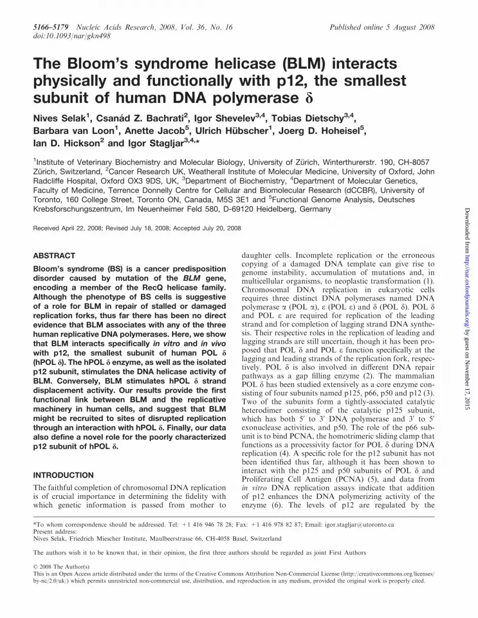
Figure 1. BLM and p12 of hPOL d interact in vitro and in vivo. (A) Left panel, far-western analysis. hPOL �, total protein extract from Sf9 insectcells, and hPOL d enzyme were subjected to SDS–PAGE, transferred to a nitrocellulose membrane, and were incubated with purified recombinantBLM. Anti-BLM antibodies were used to permit the detection of p12 as a novel BLM-interacting protein (lane 6). Molecular weight markers are alsoindicated on the left. Right panel, purified p12 subunit of hPOL d was subjected to SDS–PAGE, transferred to nitrocellulose membrane, incubatedwith BLM, and subsequently probed with the anti-BLM antibody (lane 9). (B) Reciprocal far-western analysis. BSA, WRN and BLM (left panel)were hybridized with the purified p12 and probed with the anti-p12 antibody (right panel). Anti-p12 antibodies were used to confirm that p12specifically binds to BLM (lane 6). (C) BLM and p12 of hPOL d interact in the YTH assay. The L40 yeast reporter strain was co-transformed withplasmids encoding the indicated full-length ‘bait’ (LexA-DBD) and ‘prey’ (Gal4-AD) fusions. Two independent colonies were grown on SD agarplates lacking tryptophan and leucine, but containing X-gal, prior to assessment of b-galactosidase activity. Also shown are two negative controls,p12 co-transformed with an empty prey vector and BLM co-transformed with lamin C protein. The previously described BLM/hp150 (CAF-1)
5168 Nucleic Acids Research, 2008, Vol. 36, No. 16
by guest on Novem
ber 17, 2015http://nar.oxfordjournals.org/
Dow
nloaded from
The pool of p12 protein was diluted to 50mM NaCl andfinally loaded onto a Mono S column pre-equilibratedwith buffer B. Chromatography was performed as for theHiTrap Heparin column. The yield from a 1 l culture was�0.3mg of p12 protein with a purity of over 99.5%, asjudged by Coomassie Blue staining. Protein concentrationswere determined by the Bradford method (27) by usingBovine Serum Albumin (BSA) as a calibration standard.
Production and purification of the p12 antibody
To produce the p12 antibody, the 6xHis-p12 protein wasexpressed in E. coli from the pRSETb-p12 plasmid, andpurified from E. coli using conventional chromatography(see above). Rabbits were immunized three times with300 mg of 6xHis-p12 protein being used for each immuni-zation. After the third immunization, the rabbit was sacri-ficed to obtain the serum. The p12 serum was first purifiedover a Protein G sepharose column, to isolate total IgG,and then purified over a column coupled to the p12 pro-tein, to obtain the IgGp12. For all subsequent studies, theanti-IgG p12 was used at concentration of 0.2mg/ml indilution 1:100 in TBS, 0.05% Tween-20. Control IgGwas purified from preimune serum using Protein Gsepharose column.
Far-western analysis
Far-western assays were performed as described pre-viously (28). Human WRN was a kind gift fromDr Pavel Janscak, University of Zurich. Briefly, totalextracts of Sf21 insect cells (0.825 mg), 0.8mg of hPOL d,0.3 mg of hPOL � and 0.2mg of p12 were subjected to 12%SDS–PAGE and transferred to a nitrocellulose filter.After blocking in 10% milk, 0.3% Tween-20 in TBS, for1 h at RT, the filter was incubated for 2 h at 48C with BLM(0.5 mg/ml) in TBS supplemented with 0.25% milk, 0.3%Tween-20, 1mM DTT and 1mM PMSF (hybridizationsolution). After the washing step (4� 15min, 0.25%milk, 0.3% Tween-20 in TBS), western blot was per-formed using the anti-BLM IHIC33 antibody (29) todetect the presence of BLM. For the experiment presentedin Figure 1, BLM (1.2 mg), WRN (0.8 mg) and BSA (0.4 mg)were separated on a 7.5% SDS gel and transferred to anitrocellulose membrane. The membrane was incubatedwith 6xHis-p12 in hybridization solution, with a final con-centration 6xHis-p12 of 0.1mg/ml. After washing, themembrane was probed with an anti-p12 antibody (thisstudy) to detect the presence of p12. The input sampleswere visualized with antibodies against BLM [IHIC33,
(29)] and WRN (ab-200, Abcam, Cambridge, UK). Themembrane was incubated with the BLM protein in hybri-dization solution, with a final concentration of BLM of0.5mg/ml. After washing, western blot using an anti-BLMIHIC33 antibody was performed to detect the presence ofBLM. The input samples were visualized with antibodiesagainst hPOL � (ab5954, Abcam) and p12 (this study).
Yeast two-hybrid assay
Yeast two-hybrid (YTH) assays were performed asdescribed previously (23). The activity of the reportergene (b-galactosidase) was assessed using a liquid cultureassay with O-nitrophenyl-D-galactopyranoside as a sub-strate. The constructs used to map the region of BLMthat interacts with p12 have been described previously(30). The different p12 constructs were generated byPCR using pMALc2e-p12 as a template, and werecloned into the pBTM116+2 (MBN) vector. Sequencesof all plasmids, primers used and construction schemes areavailable upon request.
Transfections and immunoprecipitation assay
The p12 cDNA was cloned into the p3xFLAG-myc-CMV-23 (Sigma-Aldrich, Buchs, SG, Switzerland) vector usingplasmid pRSETb-p12 as a template and primers 50-ggaagatctcataggggatagagatgccag -30 (BglII) and 50- cccaagcttatgggccggaagcggctc - 30 (HindIII). The resulting constructwas sequenced. 293T cells were transiently transfected withp3xFLAG-myc-CMV-23-p12 by the calcium phosphateprecipitation method. Thirty-six hours after transfection,cells were treated with 1mM HU for 24 h, and were thenharvested. Nuclear extracts were prepared from 293T cellsas described previously (31). Aliquots (500mg) of thenuclear extracts were incubated with 3 mg of anti-FLAGantibody coupled to magnetic, tosyl-activated Dynabeads(Dynal Biotech, Invitrogen, Paisley, UK, M-280) accord-ing to manufacturer’s instructions in immunoprecipitation(IP) buffer (20mM HEPES pH 7.5, 150mM NaCl, 5mMMgCl2, 0.1% Nonidet P 40, protease inhibitor) at 48C for3 h. As a control, nuclear extracts were incubated with acontrol rabbit IgG. The beads were washed three times inIP buffer, before any protein complexes bound to beadswere eluted and analysed by SDS–PAGE. A 50 mg portionof nuclear extract was used as input control. Subsequently,western blot analysis was performed using the anti-FLAG(SigmaM2) and anti-BLM ab 476 antibody (Abcam). C-18(anti-BLM, Santa Cruz) was used for the reciproical co-IPin the above-mentioned IP buffer containing 150mMKCl.
interaction (23) was used as a positive control. (D) BLM and hPOL d form a complex in human cells. 293T cells were transiently transfected withFLAG-p12, and were synchronized in S phase using 1mM HU. Nuclear extracts derived from either unsynchronized (lane 3) or S-phase synchro-nized cells (lane 4) were immunoprecipitated with the anti-FLAG antibody or control IgG, and were analysed by SDS–PAGE. One-tenth (50 mg) ofthe same nuclear extract was used as input control (lane 1). Immunoprecipitated FLAG-p12 and BLM were detected by western blotting using theanti-FLAG and anti-BLM antibody, respectively (lane 4). p125, the largest subunit of hPOL d, was also efficiently co-immunoprecipitated using thesame anti-FLAG antibody (lanes 3 and 4). Reciprocal co-IP is shown in the middle panel: lane 5, input; lane 6, IP with the control IgG; lane 7, IPwith an anti-BLM antibody (C-18) from nuclear extracts derived from unsynchronized 293T cells; lane 8, IP with an anti-BLM antibody (C-18) fromnuclear extracts derived from the S-phase synchronized 293T cells. The known BLM interacting protein, hp150 (CAF-1) was also efficientlyco-immunoprecipitated using the same anti-BLM antibody (lanes 7 and 8). As a loading control for lanes 1–8, 50 mg of the corresponding nuclearextract was probed with an anti-PARP1 antibody. Right panel shows co-IP with anti-BLM antibody (C-18) from BS cell nuclear extracts (BS) andBS cells containing the BLM cDNA (BS+pBLM). p12 could be co-immunoprecipitated in the presence of BLM from the S-phase synchronizednuclear extracts (lane 12) but not in the absence of BLM (lane 11). Lanes 9 and 10 are the inputs of the two different nuclear extracts.
Nucleic Acids Research, 2008, Vol. 36, No. 16 5169
by guest on Novem
ber 17, 2015http://nar.oxfordjournals.org/
Dow
nloaded from
Nuclear extracts from BS cells were used as a negativecontrol for the C-18 reciprocal co-IP.
DNA polymerase primer extension assay
A 18-nt primer was 50-end labeled with 32P using T4 poly-nucleotidekinase and purified on a Sephadex G25 micro-column. The X-poly template was generated by annealingan 18-nt primer to four complementary oligonucleotides.Three of these oligonucleotides are 50-nt long; the fourtholigonucleotide has an extended arm that is complemen-tary to the 18-mer primer at its 30 end. Twenty-five nucleo-tides of each four oligonucleotides are fully complementaryto two out of three other oligonucleotides, so that the resultof annealing is a cruciform structure. Annealing and puri-fication of the X-poly substrate was carried out asdescribed previously (32). Sequences of the primers usedare available upon request. Reactions (10 ml final volume)were carried out in buffer containing 40mM Tris–HClbuffer (pH 8.0), 3MmMgCl2, 1mM ATP, 50mM NaCl,2mM DTT, 0.1mg/ml BSA, 10% glycerol, 0.15 pmol of32P-18-nt-X-poly template, 100 mM each of dATP, dGTP,dCTP and dTTP, 10 ng of hPOL d exo, and the indicatedamounts of BLM, human PCNA or E. coli RecQ.Reactions were incubated at 378C for 30min, and wereterminated by rapid cooling on ice and addition of anequal volume of denaturing loading buffer. The sampleswere boiled, and 10 ml of sample were electrophoresedthrough 12.5% polyacrylamide–8M urea gel in0.5�TBE buffer, and the extension products were visua-lized by autoradiography.
DNA helicase assays. Recombinant BLM protein waspurified from yeast cells as described previously (12).The splayed arm DNA substrate that mimics a replicationfork was generated by annealing two partially complemen-tary oligonucleotides, consisting of 25 nt of fully comple-mentary and 25 nt of non-complementary sequences,and was purified as described previously (13,32). The heli-case reactions were carried out under presumed single-turnover conditions; that is with helicase concentrationin excess over substrate concentration. The 10 ml reactionscontained 1� helicase buffer [33mM Tris–acetate (pH7.8); 1mMMgCl2; 66mM sodium acetate; 0.1mg/mlBSA; 1mMDTT; 1mM ATP], 100 pM substrate, variousconcentrations of BLM and other proteins as statedin figure legends. The reaction was allowed to progressfor 15min at 378C unless otherwise stated. Analysisof reaction products was carried out as described pre-viously (32).
Indirect immunofluorescence analysis
GM00637 transformed normal human fibroblasts weregrown on coverslips and were either treated with 2.5mMHU for 18 h or cultured untreated, and were then pulselabeled with 25 mM BrdU for 5min. The coverslips werethen rinsed with ice-cold PBS. Soluble proteins wereremoved by incubating the slides in pre-extraction buffer[10mM PIPES, 300mM sucrose, 3mM MgCl2, 20mMNaCl, 0.5% Triton X-100 (pH 6.8)] for 5min on ice.The cells were then fixed in 4% paraformaldehyde for
20min on ice. The immunostaining was performed asdescribed earlier (33) using the IHIC34 rabbit polyclonalantibody (29) and the AlexaFluor 488 conjugated donkeyanti-rabbit secondary antibody (Molecular Probes,Invitrogen, Paisley, UK) to detect BLM, at 1 : 200 and1 : 800 dilutions, respectively. The A-9 mouse monoclonalantibody (Santa Cruz Biotechnology Inc., Santa Cruz,California, USA) against the catalytic subunit of hPOLd and the CY3 conjugated sheep anti-mouse secondaryantibody (Sigma-Aldrich, Gillingham, UK) were used todetect hPOL d, at 1 : 400 and 1 : 1000 dilutions, respec-tively. BrdU incorporation was detected after repeatedparaformaldehyde fixation and HCl denaturation withrat anti-BrdU primary antibody (Abcam) andAlexaFluor 350 conjugated goat anti-rat secondary anti-body (Molecular Probes), each at 1 : 300 dilution.Epifluorescence microscopy, image acquisition and analy-sis were carried out on a Nikon Eclipse 80i microscopewith the Lucia G software (Laboratory Imaging s.r.o.,Prague, Czech Republic). Grabbed images were scoredmanually using the Adobe Photoshop program. Fociobtained following staining with either antibody (greenor red) were marked and counted. Foci were counted asco-localizing if more than 50% of the green and red signalwas overlapping. Co-localization was expressed as percen-tage of the total number of BLM (green) or POL d (red)foci. The total number of cells scored in each treatmentwas 100. Two independent experiments were conductedwith nearly identical results, of which only one ispresented.
RESULTS
BLM and hPOL d interact in vitro and in vivo
To analyse the possible functional interaction of BLM andhPOL d, we first purified both BLM and the four subunithPOL d enzyme (Supplementary Figure. 1). A far-westernassay was then used to test for a specific interactionbetween BLM and one or more of the hPOL d subunits.As shown in Figure 1A, the BLM protein specificallyinteracted with a protein of apparent molecular mass of14 kDa (lane 6), which corresponds to p12, the smallestsubunit of hPOL d. In contrast, BLM did not interact withany of the other three hPOL d subunits, with an unrelatedhuman DNA polymerase, hPOL � (lane 4), or with anyprotein from the extract of Sf9 insect cells from which therecombinant hPOL d enzyme was purified (lane 5). Inorder to confirm that BLM specifically binds to p12, thefar-western analysis was repeated using full-length BLMand purified recombinant p12 immobilized on nitrocellu-lose. Clear evidence of binding was obtained (Figure 1A,lane 9). Moreover, in a reverse far-western, purified p12specifically bound to full-length BLM (Figure 1B, lane 6),but not to BSA (Figure 1B, lane 4) or to another humanRecQ helicase, WRN (Figure 1B, lane 5). No cross-reactivity of either the anti-BLM antibody with p12, orthe anti-p12 antibody with BLM was detected (Figure 1A,lanes 1–3 and 7 and Figure 1B, lanes 1–3). Taken together,these data indicate that BLM and p12 interact specificallyin vitro.
5170 Nucleic Acids Research, 2008, Vol. 36, No. 16
by guest on Novem
ber 17, 2015http://nar.oxfordjournals.org/
Dow
nloaded from
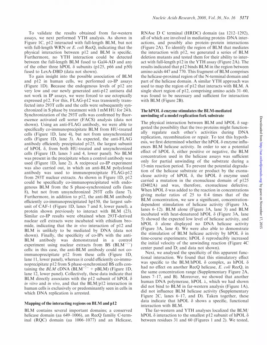
To validate the results obtained from far-westernassays, we next performed YTH analysis. As shown inFigure 1C, p12 interacted with full-length BLM, but notwith full-length WRN or E. coli RecQ, indicating that thephysical interaction between p12 and BLM is specific.Furthermore, no YTH interaction could be detectedbetween the full-length BLM fused to Gal4-AD and anyof the other three hPOL d subunits (p125, p66 and p50)fused to LexA-DBD (data not shown).
To gain insight into the possible association of BLMand p12 in human cells, we performed co-IP assays(Figure 1D). Because the endogenous levels of p12 arevery low and our newly generated anti-p12 antisera didnot work in IP assays, we were forced to use ectopicallyexpressed p12. For this, FLAG-p12 was transiently trans-fected into 293T cells and the cells were subsequently syn-chronized in S phase by treatment with 1mMHU for 24 h.Synchronization of the 293T cells was confirmed by fluor-escence activated cell sorter (FACS) analysis (data notshown). Using an anti-FLAG antibody, we were able tospecifically co-immunoprecipitate BLM from HU-treatedcells (Figure 1D, lane 4), but not from unsynchronizedcells (Figure 1D, lane 3). As expected, the anti-FLAGantibody efficiently precipitated p125, the largest subunitof hPOL d, from both HU-treated and unsynchronizedcells (Figure 1D, lanes 3 and 4, lower panel). No BLMwas present in the precipitate when a control antibody wasused (Figure 1D, lane 2). A reciprocal co-IP experimentwas also carried out, in which an anti-BLM polyclonalantibody was used to immunoprecipitate FLAG-p12from 293T nuclear extracts. As shown in Figure 1D, p12could be specifically co-immunoprecipitated with endo-genous BLM from the S phase-synchronized cells (lane8), but not from unsynchronized 293T cells (lane 7).Furthermore, in addition to p12, the anti-BLM antibodyefficiently co-immunoprecipitated hp150, the largest sub-unit of CAF-1 (Figure 1D, lanes 7 and 8, lower panel), aprotein shown previously to interact with BLM (23).Similar co-IP results were obtained when 293T-derivednuclear cell extracts were incubated with ethidium bro-mide, indicating that the in vivo interaction of p12 andBLM is unlikely to be mediated by DNA (data notshown). Finally, the specificity of co-IPs with the anti-BLM antibody was demonstrated in a controlexperiment using nuclear extracts from BS (BLM�/�)cells: in this case, the anti-BLM antibody could not co-immunoprecipitate p12 from these cells (Figure 1D,lane 11, lower panel), whereas it could efficiently co-immu-noprecipitate p12 from S phase-synchronized BS cells con-taining the BLM cDNA (BLM�/�+pBLM) (Figure 1D,lane 12, lower panel). Collectively, these data indicate thatBLM directly associates with the p12 subunit of hPOL din vitro and in vivo, and that the BLM/p12 interaction inhuman cells is exclusively or predominantly seen in cells inwhich DNA replication is arrested.
Mapping of the interacting regions on BLM and p12
BLM contains several important domains; a conservedhelicase domain (aa 649–1006), an RecQ familiy C-term-inal (RQC) domain (aa 1006–1077) and an Helicase,
RNAse D C terminal (HRDC) domain (aa 1212–1292),all of which are involved in mediating protein–DNA inter-actions, and possibly also protein–protein interactions(Figure 2A). To identify the region of BLM that mediatesthe interaction with p12, we generated a series of BLMdeletion mutants and tested them for their ability to inter-act with full-length p12 in the YTH assay (Figure 2A). Theresults indicated that p12 binds BLM in the region betweenamino acids 447 and 770. This fragment of BLM comprisesthe helicase-proximal region of the N-terminal domain andpart of the helicase domain. A similar YTH approach wasused to map the region of p12 that interacts with BLM. Asingle short region of p12, comprising amino acids 31–60,was found to be necessary and sufficient for interactionwith BLM (Figure 2B).
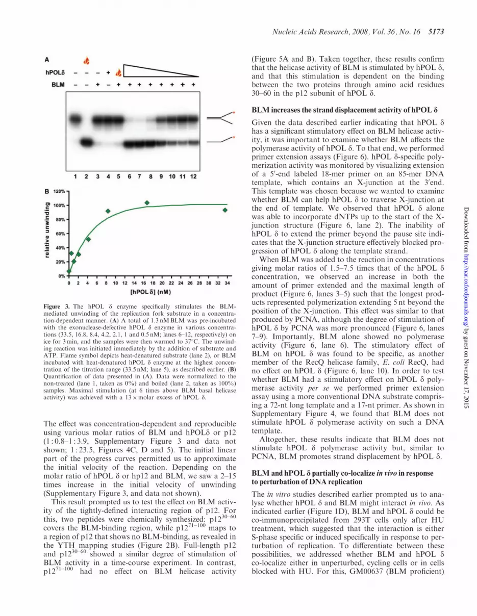
The hPOL d enzyme stimulates the BLM-mediatedunwinding of a model replication fork substrate
The physical interaction between BLM and hPOL d sug-gested the possibility that the two proteins might function-ally regulate each other’s activities during DNAreplication, recombination or repair. To test this hypoth-esis, we first determined whether the hPOL d enzyme influ-ences BLM helicase activity. In order to see a potentialeffect of hPOL d, either positive or negative, the BLMconcentration used in the helicase assays was sufficientonly for partial unwinding of the substrate during a15-min reaction period. To prevent the potential degrada-tion of the helicase substrate or product by the exonu-clease activity of hPOL d, the hPOL d enzyme usedcarried a mutation in the exonuclease domain of p125(D402A) and was, therefore, exonuclease defective.When hPOL d was added to the reaction in concentrationsgiving molar ratios of 25 to 0.4 times that of theBLM concentration, we saw a significant, concentration-dependent stimulation of helicase activity (Figure 3A,lanes 6–12). BLM alone (Figure 3A, lane 3) and BLMincubated with heat-denatured hPOL d (Figure 3A, lane5) showed the expected low level of helicase activity, andhPOL d alone displayed no DNA helicase activity(Figure 3A, lane 4). We were also able to demonstratethe stimulation of BLM helicase activity by hPOL d intime-course experiments; hPOL d reproducibly increasedthe initial velocity of the unwinding reaction (Figure 4Ccenter panel and D; and data not shown).Next, we analysed the specificity of this apparent func-
tional interaction. We found that this stimulatory effectwas specific to the BLM/hPOL d complex, as hPOL dhad no effect on another RecQ helicase, E. coli RecQ, inthe same concentration range (Supplementary Figure 2A,lanes 7–17, and B). Moreover, we showed that anotherhuman DNA polymerase, hPOL �, which we had showndid not bind to BLM in far-western analysis (Figure 1A),did not influence BLM helicase activity (SupplementaryFigure 2C, lanes 6–17, and D). Taken together, thesedata indicate that hPOL d shows a specific, functionalinteraction with BLM.The far-western and YTH analyses localized the BLM/
hPOL d interaction to the smallest p12 subunit of hPOL dbetween residues 31 and 60 (Figures 1 and 2). We tested,
Nucleic Acids Research, 2008, Vol. 36, No. 16 5171
by guest on Novem
ber 17, 2015http://nar.oxfordjournals.org/
Dow
nloaded from

therefore, the effect of recombinant p12 subunit on the heli-case activity of BLM. p12 showed a concentration-depen-dent stimulatory effect on BLM similar to that of the hPOLd enzyme; however, the effective concentration range wassuch that there was a large molar excess of p12 over BLM(Figure 4A, lanes 6–15). Interestingly, heat-denatured p12was also able to stimulate BLMhelicase activity at this highconcentration (1.609 mM; Figure 4A, lane 5), which, in thelight of the results showing that a small peptide is also able
to stimulate the helicase activity of BLM, is not inexplicable(see below). As described earlier, we saw concentration-dependent stimulation of BLM helicase activity by p12only at high p12 concentration. Therefore, we designeda different system to study this effect and monitored theprogress of the unwinding reaction over time in the pre-sence or absence of p12. This revealed that p12 and thehPOL d enzyme increased the kinetics of the BLM unwind-ing reaction to a similar extent (Figure 4C and D).
BLM
1-448
340-770
447-770
448-572
782-952
1-1417
BAIT(LexA-DBD fusion)
PREY(Gal4-AD fusion)
p12
p12
p12
p12
p12
p12
HD HRDC
X-gal test
BLM
BLM
BLM
BLM
BLM
BLM
p12
1-80
1-60
31-108
61-108
31-80
1-108
31-60
BLM1-30
BAIT(LexA-DBD fusion)
PREY(Gal4-AD fusion)
BLM
X-gal test
B
A
β-Gal units
2435±270
42.3±6
1956±138
2042±58
38±8
106±43
β-Gal units
1003±188
920±93
1299±208
1388±102
19±11
1539±110
2121±96
78±23
1
RQC
1006649 10771212 1292
1417
Figure 2. Interaction region mapping of BLM and p12. (A) Mapping of the BLM interaction region. The L40 yeast strain was co-transformed withplasmids encoding the indicated BLM fragments fused to Gal4-AD and the full-length p12 fused to LexA-DBD. Two independent colonies weregrown on SD agar plates lacking tryptophan and leucine, but containing X-gal, prior to assessment of b-galactosidase activity. Blue coloration ofcolonies is a marker of interaction. Full length BLM is also shown, with a red bar indicating its conserved helicase domain, a blue bar indicatingRQC and a black bar indicating the HRDC domain. Interactions between a given bait/prey pair were quantified by measurements of b-galactosidaseactivity. Values represent means�SD of three independent experiments. (B) Mapping of the p12 interaction region. The L40 yeast strain wasco-transformed with plasmids encoding the indicated p12 fragments fused to LexA-DBD and full length BLM fused to Gal4-AD. In both (A) and(B) the sequence boundaries of deletion mutants tested are shown with the corresponding amino acid positions indicated on the right. Valuesobtained from liquid b-galactosidase assay are shown on the right and represent means� SD of three independent experiments.
5172 Nucleic Acids Research, 2008, Vol. 36, No. 16
by guest on Novem
ber 17, 2015http://nar.oxfordjournals.org/
Dow
nloaded from
The effect was concentration-dependent and reproducibleusing various molar ratios of BLM and hPOLd or p12(1 : 0.8–1 : 3.9, Supplementary Figure 3 and data notshown; 1 : 23.5, Figures 4C, D and 5). The initial linearpart of the progress curves permitted us to approximatethe initial velocity of the reaction. Depending on themolar ratio of hPOL d or hp12 and BLM, we saw a 2–15times increase in the initial velocity of unwinding(Supplementary Figure 3, and data not shown).
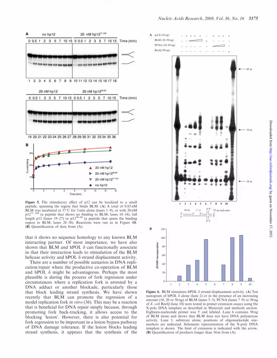
This result prompted us to test the effect on BLM activ-ity of the tightly-defined interacting region of p12. Forthis, two peptides were chemically synthesized: p1230–60
covers the BLM-binding region, while p1271–100 maps toa region of p12 that shows no BLM-binding, as revealed inthe YTH mapping studies (Figure 2B). Full-length p12and p1230–60 showed a similar degree of stimulation ofBLM activity in a time-course experiment. In contrast,p1271–100 had no effect on BLM helicase activity
(Figure 5A and B). Taken together, these results confirmthat the helicase activity of BLM is stimulated by hPOL d,and that this stimulation is dependent on the bindingbetween the two proteins through amino acid residues30–60 in the p12 subunit of hPOL d.
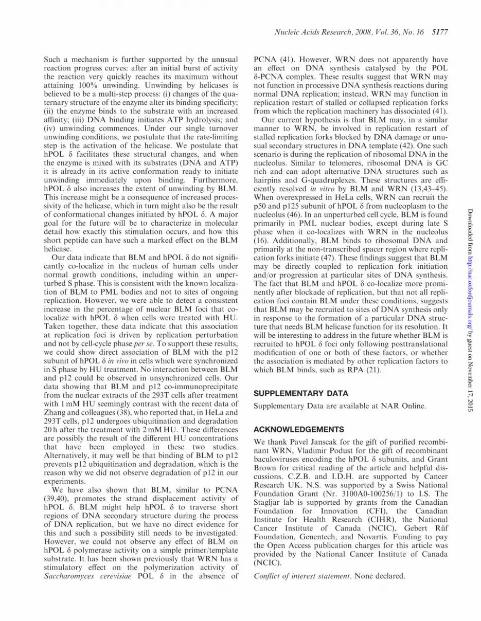
BLM increases the strand displacement activity of hPOL d
Given the data described earlier indicating that hPOL dhas a significant stimulatory effect on BLM helicase activ-ity, it was important to examine whether BLM affects thepolymerase activity of hPOL d. To that end, we performedprimer extension assays (Figure 6). hPOL d-specific poly-merization activity was monitored by visualizing extensionof a 50-end labeled 18-mer primer on an 85-mer DNAtemplate, which contains an X-junction at the 30end.This template was chosen because we wanted to examinewhether BLM can help hPOL d to traverse X-junction atthe end of template. We observed that hPOL d alonewas able to incorporate dNTPs up to the start of the X-junction structure (Figure 6, lane 2). The inability ofhPOL d to extend the primer beyond the pause site indi-cates that the X-junction structure effectively blocked pro-gression of hPOL d along the template strand.When BLM was added to the reaction in concentrations
giving molar ratios of 1.5–7.5 times that of the hPOL dconcentration, we observed an increase in both theamount of primer extended and the maximal length ofproduct (Figure 6, lanes 3–5) such that the longest prod-ucts represented polymerization extending 5 nt beyond theposition of the X-junction. This effect was similar to thatproduced by PCNA, although the degree of stimulation ofhPOL d by PCNA was more pronounced (Figure 6, lanes7–9). Importantly, BLM alone showed no polymeraseactivity (Figure 6, lane 6). The stimulatory effect ofBLM on hPOL d was found to be specific, as anothermember of the RecQ helicase family, E. coli RecQ, hadno effect on hPOL d (Figure 6, lane 10). In order to testwhether BLM had a stimulatory effect on hPOL d poly-merase activity per se we performed primer extensionassay using a more conventional DNA substrate compris-ing a 72-nt long template and a 17-nt primer. As shown inSupplementary Figure 4, we found that BLM does notstimulate hPOL d polymerase activity on such a DNAtemplate.Altogether, these results indicate that BLM does not
stimulate hPOL d polymerase activity but, similar toPCNA, BLM promotes strand displacement by hPOL d.
BLM and hPOL d partially co-localize in vivo in responseto perturbation of DNA replication
The in vitro studies described earlier prompted us to ana-lyse whether hPOL d and BLM might interact in vivo. Asindicated earlier (Figure 1D), BLM and hPOL d could beco-immunoprecipitated from 293T cells only after HUtreatment, which suggested that the interaction is eitherS-phase specific or induced specifically in response to per-turbation of replication. To differentiate between thesepossibilities, we addressed whether BLM and hPOL dco-localize either in unperturbed, cycling cells or in cellsblocked with HU. For this, GM00637 (BLM proficient)
Figure 3. The hPOL d enzyme specifically stimulates the BLM-mediated unwinding of the replication fork substrate in a concentra-tion-dependent manner. (A) A total of 1.3 nMBLM was pre-incubatedwith the exonuclease-defective hPOL d enzyme in various concentra-tions (33.5, 16.8, 8.4, 4.2, 2.1, 1 and 0.5 nM; lanes 6–12, respectively) onice for 3min, and the samples were then warmed to 378C. The unwind-ing reaction was initiated immediately by the addition of substrate andATP. Flame symbol depicts heat-denatured substrate (lane 2), or BLMincubated with heat-denatured hPOL d enzyme at the highest concen-tration of the titration range (33.5 nM; lane 5), as described earlier. (B)Quantification of data presented in (A). Data were normalized to thenon-treated (lane 1, taken as 0%) and boiled (lane 2, taken as 100%)samples. Maximal stimulation (at 6 times above BLM basal helicaseactivity) was achieved with a 13�molar excess of hPOL d.
Nucleic Acids Research, 2008, Vol. 36, No. 16 5173
by guest on Novem
ber 17, 2015http://nar.oxfordjournals.org/
Dow
nloaded from

human fibroblast cells were plated on coverslips andtreated with 2.5mM HU for 18 h, sufficient to block repli-cation, as evidenced by the depression of BrdU incorpora-tion into the nuclei (not shown). A control culture wasincubated in parallel without HU treatment. In both setsof cells, BrdU was added to a final concentration of 25 mM5min before the cells were fixed. The fixed samples werestained for the presence of hPOL d, BLM and BrdU, asdescribed in Materials and methods section. To detecthPOL d were restricted in our use of antibodies, becauseour antibody to p12 did not work in immunofluorescenceanalysis. Hence, we chose an antibody against the catalyticsubunit p125, which directly interacts with p12(Figure 1D, lanes 3 and 4). As expected from previousanalyses, BLM and hPOL d showed a punctate nuclearpattern of localization (15,25,34). Representative imagesof the staining patterns are depicted in Figure 7A and B.The degree of co-localization of nuclear foci wasthen scored. This analysis indicated a low degree of co-lo-calization in an untreated asynchronous cell popula-tion, which increased in response to HU treatment(Supplementary Figure 5). This increase in the extent ofco-localization might be restricted to S phase or might bean effect of the HU-induced replication perturbation. Toaddress this, we took advantage of the BrdU labeling ofthe actively replicating cells. When only the BrdU-positive
subpopulation was scored in the untreated cultures,the degree of co-localization was similar to that of thewhole asynchronous population. These results suggestthat BLM and hPOL d co-localize only to a limitedextent in vivo, even during an unperturbed S phase, butthat perturbation of DNA replication causes an increasein their co-localization. Nevertheless, even after treatmentof cells with HU, most BLM and hPOL d foci still did notco-localize, indicating that a significant fraction of BLMremains distant from sites of stalled replication forks.
DISCUSSION
We have shown that the BS helicase, BLM and the majorreplicative DNA polymerase, hPOL d, interact specificallyin vitro and in vivo. This interaction is direct and ismediated via the thus far poorly characterized p12 subunitof hPOL d. We mapped the site of interaction of BLMwith p12 to a region representing amino acids 447–770.This fragment includes the N-terminal part of the BLMhelicase domain and it has been shown previously that thisis involved in interaction with the WRN helicase (35). Inaddition, we mapped the region of p12 that interacts withBLM to a short fragment comprising amino acids 30–60.Database searches revealed that this p12 fragment doesnot represent any known conserved protein domain and
Figure 4. The small subunit of the hPOL d enzyme, p12, is sufficient to stimulate BLM helicase activity. (A) A total of 1.3 nMBLM was pre-incubated with various concentrations of p12 (1609, 804.5, 402.25, 201.13, 100.56, 50.28, 25.14, 12.57, 6.29 and 3.14 nM; lanes 6–15, respectively) onice for 3min, and the samples were then warmed to 378C. The unwinding reaction was initiated immediately by the addition of substrate and ATP.Controls and symbols are as in Figure 3. (B) Quantification of data from (A). (C) A total of 0.85 nM BLM was incubated at 378C for 5min alone(lanes 1–9), or with 20 nMhPOL d (lanes 10–18), or 20 nM p12 (lanes 19–27). The unwinding reaction was then initiated by the addition of ATP andsubstrate. Samples were withdrawn at the time points indicated above the lanes. (D) Quantification of data from (C).
5174 Nucleic Acids Research, 2008, Vol. 36, No. 16
by guest on Novem
ber 17, 2015http://nar.oxfordjournals.org/
Dow
nloaded from
that it shows no sequence homology to any known BLMinteracting partner. Of most importance, we have alsoshown that BLM and hPOL d can functionally associatein that their interaction leads to stimulation of the BLMhelicase activity and hPOL d strand displacement activity.
There are a number of possible scenarios in DNA repli-cation/repair where the productive co-operation of BLMand hPOL d might be advantageous. Perhaps the mostplausible is during the process of fork regression undercircumstances where a replication fork is arrested by aDNA adduct or another blockade, particularly thosethat block leading strand synthesis. We have shownrecently that BLM can promote the regression of amodel replication fork in vitro (36). This may be a reactionthat is beneficial for DNA repair simply because, throughpromoting fork back-tracking, it allows access to theblocking ‘lesion’. However, there is also potential forfork regression to be important in a lesion bypass pathwayof DNA damage tolerance. If the lesion blocks leadingstrand synthesis, it appears that the synthesis of the
Figure 5. The stimulatory effect of p12 can be localized to a smallpeptide, spanning the region that binds BLM. (A) A total of 0.85 nMBLM was incubated at 378C for 5min alone (lanes 1–9), or with 20 nMp1271–100 (a peptide that shows no binding to BLM; lanes 10–18), fulllength p12 (lanes 19–27) or p1230–60 (a peptide that spans the bindingregion to BLM; lanes 28–36). Reactions were run as in Figure 4B.(B) Quantification of data from (A).
85 nt
34 nt
18 nt
pol δ (10 ng)
BLM (10–50 ng)
PCNA (10–50 ng)
RecQ (50 ng)
+−
−
−
−
+
−
−
−
+ + −
+
−
−
−
−
−
−
−
−
+
−
−
+
−
−
+
−
−
+
−
−
+
1 2 3 4 5 6 7 8 9 10
18 nt 17 nt 25 nt each arm5′3′
3′
5′
5′
5′3′
3′
3′
5′
0
20
40
60
80
100
120
1 2 3 4 5 6 7 8 9 10
dNM
Ps
inco
rpor
atio
n
A
B
Figure 6. BLM stimulates hPOL d strand displacement activity. (A) Tennanogram of hPOL d alone (lane 2) or in the presence of an increasingamount (10, 20 or 50 ng) of BLM (lanes 3–5), PCNA (lanes 7–9) or 50 ngof E. coli RecQ (lane 10) were tested in primer extension assays using theX-poly DNA template as described in Materials and methods section.Eighteen-nucleotide primer was 50 end labeled. Lane 6 contains 50 ngof BLM alone and shows that BLM does not have DNA polymeraseactivity. Lane 1: substrate alone; positions of oligonucleotide size-markers are indicated. Schematic representation of the X-poly DNAtemplate is shown. The limit of extension is indicated with the arrow.(B) Quantification of products longer than 34 nt from (A).
Nucleic Acids Research, 2008, Vol. 36, No. 16 5175
by guest on Novem
ber 17, 2015http://nar.oxfordjournals.org/
Dow
nloaded from

lagging strand may continue for some distance ahead ofthe site of the blocked leading strand. This apparentlyfutile uncoupling of leading and lagging strand synthesishas the potential, after fork regression, to permit templateswitching with the shorter leading strand being extendedby copying of the longer lagging strand template. In thisway, once the regressed fork is reset, the leading strandwould be extended beyond the site of the lesion andnormal DNA replication could commence. hPOL dmight both recruit BLM to stalled forks and then stimu-late its catalytic activity once there. Conversely, BLMmight play a role in assisting hPOL d to access theregressed ‘4th arm’ and catalyse extension of the leadingstrand.It has been suggested that RecQ helicases together with
the type 1A topoisomerase, Top3, act at damaged replica-tion forks to resolve recombination structures likely
resulting from template switching (37). We suggest thatBLM plays a dual role in rescuing arrested replicationforks. One function is in assisting POL d to bypassDNA lesions via fork regression and template switching,and the second function of BLM is to resolve the resultingpseudo double Holliday junction with a help of Top3.These two functions of BLM may be independent ofeach other.
A striking feature of the ability of hPOL d to stimulatethe helicase activity of BLM was our finding that a shortpeptide (aa 30–60) representing the minimal bindingregion of p12 is as efficient in this stimulatory role as isthe hPOL d enzyme. This would seem to rule out manypossible mechanisms for the stimulation, including recruit-ment of BLM to the DNA substrate. Instead, these resultsargue for hPOL d acting to alter the conformation ofBLM in such a way as to enhance its helicase function.
Figure 7. Dual staining for BLM and hPOL d suggests co-localization in vivo, which is stimulated during replicative stress. (A) GM00637 cells werearrested with 2.5mMHU, and were stained as described in Materials and methods section. A representative image showing punctate BLM (green,left) and hPOL d (red, second from left) staining. Coincidence of the green and red signals (yellow signal) suggests co-localization of the two proteins.The right panel shows the nucleus of the same cell stained with the DNA dye Hoechst 33258. (B) GM00637 cells grown on coverslips without HUtreatment and were pulse-labeled with 25 mM BrdU for 5min, and then stained as described in Materials and methods section. Representative imagesshow staining of the same nucleus for BLM in green (top left), for hPOL d in red (top, second from left), for BrdU in blue (top, second from right).Phase contrast image of the same nucleus is depicted on the top right. In the bottom row, combined images of the individual stainings from the toprow are shown. As expected, the BrdU signal correlates well with the signal for hPOLd (magenta-color pattern second panel from bottom right). Thecoincidence of BLM signal with either BrdU (cyan signal in bottom left) or hPOLd (yellow signal in second panel from bottom left) is lesspronounced.
5176 Nucleic Acids Research, 2008, Vol. 36, No. 16
by guest on Novem
ber 17, 2015http://nar.oxfordjournals.org/
Dow
nloaded from
Such a mechanism is further supported by the unusualreaction progress curves: after an initial burst of activitythe reaction very quickly reaches its maximum withoutattaining 100% unwinding. Unwinding by helicases isbelieved to be a multi-step process: (i) changes of the qua-ternary structure of the enzyme alter its binding specificity;(ii) the enzyme binds to the substrate with an increasedaffinity; (iii) DNA binding initiates ATP hydrolysis; and(iv) unwinding commences. Under our single turnoverunwinding conditions, we postulate that the rate-limitingstep is the activation of the helicase. We postulate thathPOL d facilitates these structural changes, and whenthe enzyme is mixed with its substrates (DNA and ATP)it is already in its active conformation ready to initiateunwinding immediately upon binding. Furthermore,hPOL d also increases the extent of unwinding by BLM.This increase might be a consequence of increased proces-sivity of the helicase, which in turn might also be the resultof conformational changes initiated by hPOL d. A majorgoal for the future will be to characterize in moleculardetail how exactly this stimulation occurs, and how thisshort peptide can have such a marked effect on the BLMhelicase.
Our data indicate that BLM and hPOL d do not signifi-cantly co-localize in the nucleus of human cells undernormal growth conditions, including within an unper-turbed S phase. This is consistent with the known localiza-tion of BLM to PML bodies and not to sites of ongoingreplication. However, we were able to detect a consistentincrease in the percentage of nuclear BLM foci that co-localize with hPOL d when cells were treated with HU.Taken together, these data indicate that this associationat replication foci is driven by replication perturbationand not by cell-cycle phase per se. To support these results,we could show direct association of BLM with the p12subunit of hPOL d in vivo in cells which were synchronizedin S phase by HU treatment. No interaction between BLMand p12 could be observed in unsynchronized cells. Ourdata showing that BLM and p12 co-immunoprecipitatefrom the nuclear extracts of the 293T cells after treatmentwith 1mM HU seemingly contrast with the recent data ofZhang and colleagues (38), who reported that, in HeLa and293T cells, p12 undergoes ubiquitination and degradation20 h after the treatment with 2mMHU. These differencesare possibly the result of the different HU concentrationsthat have been employed in these two studies.Alternatively, it may well be that binding of BLM to p12prevents p12 ubiquitination and degradation, which is thereason why we did not observe degradation of p12 in ourexperiments.
We have also shown that BLM, similar to PCNA(39,40), promotes the strand displacement activity ofhPOL d. BLM might help hPOL d to traverse shortregions of DNA secondary structure during the processof DNA replication, but we have no direct evidence forthis and such a possibility still needs to be investigated.However, we could not observe any effect of BLM onhPOL d polymerase activity on a simple primer/templatesubstrate. It has been shown previously that WRN has astimulatory effect on the polymerization activity ofSaccharomyces cerevisiae POL d in the absence of
PCNA (41). However, WRN does not apparently havean effect on DNA synthesis catalysed by the POLd-PCNA complex. These results suggest that WRN maynot function in processive DNA synthesis reactions duringnormal DNA replication; instead, WRN may function inreplication restart of stalled or collapsed replication forksfrom which the replication machinery has dissociated (41).Our current hypothesis is that BLM may, in a similar
manner to WRN, be involved in replication restart ofstalled replication forks blocked by DNA damage or unu-sual secondary structures in DNA template (42). One suchscenario is during the replication of ribosomal DNA in thenucleolus. Similar to telomeres, ribosomal DNA is GCrich and can adopt alternative DNA structures such ashairpins and G-quadruplexes. These structures are effi-ciently resolved in vitro by BLM and WRN (13,43–45).When overexpressed in HeLa cells, WRN can recruit thep50 and p125 subunit of hPOL d from nucleoplasm to thenucleolus (46). In an unperturbed cell cycle, BLM is foundprimarily in PML nuclear bodies, except during late Sphase when it co-localizes with WRN in the nucleolus(16). Additionally, BLM binds to ribosomal DNA andprimarily at the non-transcribed spacer region where repli-cation forks initiate (47). These findings suggest that BLMmay be directly coupled to replication fork initiationand/or progression at particular sites of DNA synthesis.The fact that BLM and hPOL d co-localize more promi-nently after blockade of replication, but that not all repli-cation foci contain BLM under these conditions, suggeststhat BLM may be recruited to sites of DNA synthesis onlyin response to the formation of a particular DNA struc-ture that needs BLM helicase function for its resolution. Itwill be interesting to address in the future whether BLM isrecruited to hPOL d foci only following posttranslationalmodification of one or both of these factors, or whetherthe association is mediated by other replication factors towhich BLM binds, such as RPA (21).
SUPPLEMENTARY DATA
Supplementary Data are available at NAR Online.
ACKNOWLEDGEMENTS
We thank Pavel Janscak for the gift of purified recombi-nant WRN, Vladimir Podust for the gift of recombinantbaculoviruses encoding the hPOL d subunits, and GrantBrown for critical reading of the article and helpful dis-cussions. C.Z.B. and I.D.H. are supported by CancerResearch UK. N.S. was supported by a Swiss NationalFoundation Grant (Nr. 3100A0-100256/1) to I.S. TheStagljar lab is supported by grants from the CanadianFoundation for Innovation (CFI), the CanadianInstitute for Health Research (CIHR), the NationalCancer Institute of Canada (NCIC), Gebert RufFoundation, Genentech, and Novartis. Funding to paythe Open Access publication charges for this article wasprovided by the National Cancer Institute of Canada(NCIC).
Conflict of interest statement. None declared.
Nucleic Acids Research, 2008, Vol. 36, No. 16 5177
by guest on Novem
ber 17, 2015http://nar.oxfordjournals.org/
Dow
nloaded from

REFERENCES
1. Bell,S.P. and Dutta,A. (2002) DNA replication in eukaryotic cells.Annu. Rev. Biochem., 71, 333–374.
2. Hubscher,U., Maga,G. and Spadari,S. (2002) Eukaryotic DNApolymerases. Annu. Rev. Biochem., 71, 133–163.
3. Liu,L., Mo,J., Rodriguez-Belmonte,E.M. and Lee,M.Y. (2000)Identification of a fourth subunit of mammalian DNA polymerasedelta. J. Biol. Chem., 275, 18739–18744.
4. Maga,G. and Hubscher,U. (2003) Proliferating cell nuclearantigen (PCNA): a dancer with many partners. J. Cell Sci., 116,3051–3060.
5. Li,H., Xie,B., Zhou,Y., Rahmeh,A., Trusa,S., Zhang,S., Gao,Y.,Lee,E.Y. and Lee,M.Y. (2006) Functional roles of p12, the fourthsubunit of human DNA polymerase delta. J. Biol. Chem., 281,14748–14755.
6. Podust,V.N., Chang,L.S., Ott,R., Dianov,G.L. and Fanning,E.(2002) Reconstitution of human DNA polymerase delta usingrecombinant baculoviruses: the p12 subunit potentiates DNApolymerizing activity of the four-subunit enzyme. J. Biol. Chem.,277, 3894–3901.
7. Liu,G. and Warbrick,E. (2006) The p66 and p12 subunits of DNApolymerase delta are modified by ubiquitin and ubiquitin-like pro-teins. Biochem. Biophys. Res. Commun., 349, 360–366.
8. Bachrati,C.Z. and Hickson,I.D. (2003) RecQ helicases:suppressors of tumorigenesis and premature aging. Biochem. J., 374,577–606.
9. Opresko,P.L., Cheng,W.H. and Bohr,V.A. (2004) At the junction ofRecQ Helicase biochemistry and human disease. J. Biol. Chem.,279, 18099–18102.
10. Hickson,I.D. (2003) RecQ helicases: caretakers of the genome. Nat.Rev. Cancer, 3, 169–178.
11. Ray,J.H. and German,J. (1983) The cytogenetics of the chromosome-breakage syndromes. In German,J. (ed.), Chromosome Mutation andNeoplasia, Alan R. Liss, Inc, New York, pp. 135–168.
12. Karow,J.K., Chakraverty,R.K. and Hickson,I.D. (1997) TheBloom’s syndrome gene product is a 30-50 DNA helicase. J. Biol.Chem., 272, 30611–30614.
13. Mohaghegh,P., Karow,J.K., Brosh,R.M. Jr, Bohr,V.A. andHickson,I.D. (2001) The Bloom’s and Werner’s syndrome proteinsare DNA structure-specific helicases. Nucleic Acids Res., 29,2843–2849.
14. Bachrati,C.Z., Borts,R.H. and Hickson,I.D. (2006) Mobile D-loopsare a preferred substrate for the Bloom’s syndrome helicase. NucleicAcids Res., 34, 2269–2279.
15. Bischof,O., Kim,S.H., Irving,J., Beresten,S., Ellis,N.A. andCampisi,J. (2001) Regulation and localization of the Bloom syn-drome protein in response to DNA damage. J. Cell Biol., 153,367–380.
16. Yankiwski,V., Marciniak,R.A., Guarente,L. and Neff,N.F. (2000)Nuclear structure in normal and Bloom syndrome cells. Proc. NatlAcad. Sci. USA, 97, 5214–5219.
17. Lillard-Wetherell,K., Machwe,A., Langland,G.T., Combs,K.A.,Behbehani,G.K., Schonberg,S.A., German,J., Turchi,J.J.,Orren,D.K. and Groden,J. (2004) Association and regulation of theBLM helicase by the telomere proteins TRF1 and TRF2. Hum.Mol. Genet., 13, 1919–1932.
18. Wu,L. and Hickson,I.D. (2006) DNA helicases required forhomologous recombination and repair of damaged replicationforks. Annu. Rev. Genet., 40, 279–306.
19. Hand,R. and German,J. (1975) A retarded rate of DNA chaingrowth in Bloom’s syndrome. Proc. Natl Acad. Sci. USA, 72,758–762.
20. Lonn,U., Lonn,S., Nylen,U., Winblad,G. and German,J. (1990) Anabnormal profile of DNA replication intermediates in Bloom’ssyndrome. Cancer Res., 50, 3141–3145.
21. Brosh,R.M. Jr, Li,J.L., Kenny,M.K., Karow,J.K., Cooper,M.P.,Kureekattil,R.P., Hickson,I.D. and Bohr,V.A. (2000) Replicationprotein A physically interacts with the Bloom’s syndrome proteinand stimulates its helicase activity. J. Biol. Chem., 275,23500–23508.
22. Sharma,S., Sommers,J.A., Wu,L., Bohr,V.A., Hickson,I.D. andBrosh,R.M. Jr. (2004) Stimulation of flap endonuclease-1 by theBloom’s syndrome protein. J. Biol. Chem., 279, 9847–9856.
23. Jiao,R., Bachrati,C.Z., Pedrazzi,G., Kuster,P., Petkovic,M., Li,J.L.,Egli,D., Hickson,I.D. and Stagljar,I. (2004) Physical and functionalinteraction between the Bloom’s syndrome gene product and thelargest subunit of chromatin assembly factor 1. Mol. Cell Biol., 24,4710–4719.
24. Sanz,M.M., Proytcheva,M., Ellis,N.A., Holloman,W.K. andGerman,J. (2000) BLM, the Bloom’s syndrome protein, variesduring the cell cycle in its amount, distribution, and co-localizationwith other nuclear proteins. Cytogenet. Cell Genet., 91, 217–223.
25. Dutertre,S., Ababou,M., Onclercq,R., Delic,J., Chatton,B.,Jaulin,C. and Amor-Gueret,M. (2000) Cell cycle regulation of theendogenous wild type Bloom’s syndrome DNA helicase. Oncogene,19, 2731–2738.
26. Davies,S.L., North,P.S., Dart,A., Lakin,N.D. and Hickson,I.D.(2004) Phosphorylation of the Bloom’s syndrome helicase and itsrole in recovery from S-phase arrest. Mol. Cell Biol., 24, 1279–1291.
27. Bradford,M.M. (1976) A rapid and sensitive method for the quan-titation of microgram quantities of protein utilizing the principleof protein-dye binding. Anal. Biochem., 72, 248–254.
28. Jiao,R., Harrigan,J.A., Shevelev,I., Dietschy,T., Selak,N.,Indig,F.E., Piotrowski,J., Janscak,P., Bohr,V.A. and Stagljar,I.(2007) The Werner syndrome protein is required for recruitment ofchromatin assembly factor 1 following DNA damage. Oncogene, 26,3811–3822.
29. Wu,L., Davies,S.L., North,P.S., Goulaouic,H., Riou,J.F.,Turley,H., Gatter,K.C. and Hickson,I.D. (2000) The Bloom’s syn-drome gene product interacts with topoisomerase III. J. Biol.Chem., 275, 9636–9644.
30. Pedrazzi,G., Perrera,C., Blaser,H., Kuster,P., Marra,G.,Davies,S.L., Ryu,G.H., Freire,R., Hickson,I.D., Jiricny,J. et al.(2001) Direct association of Bloom’s syndrome gene product withthe human mismatch repair protein MLH1. Nucleic Acids Res., 29,4378–4386.
31. Petkovic,M., Dietschy,T., Freire,R., Jiao,R. and Stagljar,I. (2005)The human Rothmund-Thomson syndrome gene product,RECQL4, localizes to distinct nuclear foci that coincide with pro-teins involved in the maintenance of genome stability. J. Cell Sci.,118, 4261–4269.
32. Bachrati,C.Z. and Hickson,I.D. (2006) Analysis of the DNAunwinding activity of RecQ family helicases. Methods Enzymol.,409, 86–100.
33. Pedrazzi,G., Bachrati,C.Z., Selak,N., Studer,I., Petkovic,M.,Hickson,I.D. and Stagljar,I. (2003) The Bloom’s syndrome helicasedirectly interacts with the human mismatch repair protein MSH6.Biol. Chem., 384, 1155–1164.
34. Ababou,M., Dutertre,S., Lecluse,Y., Onclercq,R., Chatton,B. andAmor-Gueret,M. (2000) ATM-dependent phosphorylation andaccumulation of endogenous BLM protein in response to ionizingradiation. Oncogene, 19, 5955–5963.
35. von Kobbe,C., Karmakar,P., Dawut,L., Opresko,P., Zeng,X.M.,Brosh,R.M. Jr, Hickson,I.D. and Bohr,V.A. (2002) Colocalization,physical, and functional interaction between Werner and Bloomsyndrome proteins. J. Biol. Chem., 277, 22035–22044.
36. Ralf,C., Hickson,I.D. and Wu,L. (2006) The Bloom’s syndromehelicase can promote the regression of a model replication fork.J. Biol. Chem., 281, 22839–22846.
37. Liberi,G., Maffioletti,G., Lucca,C., Chiolo,I., Baryshnikova,A.,Cotta-Ramusino,C., Lopes,M., Pellicioli,A., Haber,J.E. andFoiani,M. (2005) Rad51-dependent DNA structures accumulateat damaged replication forks in sgs1 mutants defective in theyeast ortholog of BLM RecQ helicase. Genes Dev., 19, 339–350.
38. Zhang,S., Zhou,Y., Trusa,S., Meng,X., Lee,E.Y. and Lee,M.Y.(2007) A novel DNA damage response: rapid degradation of thep12 subunit of DNA polymerase d. J. Biol. Chem., 282,15330–15340.
39. Podust,V.N. and Hubscher,U. (1993) Lagging strand DNA synthesisby calf thymus DNA polymerases alpha, beta, delta and epsilon inthe presence of auxiliary proteins. Nucleic Acids Res., 21, 841–846.
40. Maga,G., Villani,G., Tillement,V., Stucki,M., Locatelli,G.A.,Frouin,I., Spadari,S. and Hubscher,U. (2001) Okazaki fragmentprocessing: modulation of the strand displacement activity of DNApolymerase delta by the concerted action of replication protein A,proliferating cell nuclear antigen, and flap endonuclease-1. Proc.Natl Acad. Sci. USA, 98, 14298–14303.
5178 Nucleic Acids Research, 2008, Vol. 36, No. 16
by guest on Novem
ber 17, 2015http://nar.oxfordjournals.org/
Dow
nloaded from
41. Kamath-Loeb,A.S., Johansson,E., Burgers,P.M.J. and Loeb,L.A.(2000) Functional interaction between the Werner syndromeprotein and DNA polymerase d. Proc. Natl Acad. Sci. USA, 97,4603–4608.
42. Kamath-Loeb,A.S., Loeb,L.A., Johansson,E., Burgers,P.M.J. andFry,M. (2001) Interactions between the Werner syndrome helicaseand DNA polymerase d specifically facilitate copying of tetraplexand hairpin structures of the D(CGG)n trinucleotide repeatsequence. J. Biol. Chem., 276, 16439–16446.
43. Fry,M. and Loeb,L.A. (1999) Human Werner syndrome DNAhelicase unwinds tetrahelical structures of the fragile X syndromerepeat sequence d(CGG)n. J. Biol. Chem., 274, 12797–12802.
44. Huber,M.D., Lee,D.C. and Maizels,N. (2002) G4 DNA unwindingby BLM and Sgs1p: substrate specificity and substrate-specificinhibition. Nucleic Acids Res., 30, 3954–3961.
45. Sun,H., Karow,J.K., Hickson,I.D. and Maizels,N. (1998) TheBloom’s syndrome helicase unwinds G4 DNA. J. Biol. Chem., 273,27587–27592.
46. Szekely,A.M., Chen,Y.H., Zhang,C., Oshima,J. and Weissman,S.M.(2000) Werner protein recruits DNA polymerase d to the nucleolus.Proc. Natl Acad. Sci. USA, 97, 11365–11370.
47. Schawalder,J., Paric,E. and Neff,N.F. (2003) Telomere and riboso-mal DNA repeats are chromosomal targets of the bloom syndromeDNA helicase. BMC Cell Biol., 4, 15.
Nucleic Acids Research, 2008, Vol. 36, No. 16 5179
by guest on Novem
ber 17, 2015http://nar.oxfordjournals.org/
Dow
nloaded from