genetic determinants of shigella pathogenicity
TRANSCRIPT
Ann. Rev. MicrobioL 1988. 42.’127-50
GENETICSHIGELLA
DETERMINANTS OFPATHOGENICITY
Anthony T. Maurelli2
Department of Microbiology, Uniformed Services University of the Health Sciences,Bethesda, Maryland 20814-4799
Philippe J. Sansonetti
Service des Ent6robact6ries, Unit6 199, Institut National de la Sant6 et de la RechercheM6dicale, Institut Pasteur, 75724 Paris Cedex 15, France
CONTENTS
INTRODUCTION ..................................................................................... 128OVERVIEW OF EARLY WORK ................................................................. 129INTERACTION WITH INDIVIDUAL EPITHELIAL CELLS ............................... 130
Role of the Virulence Plasmid ................................................................. 130Regulation of the h~vasive Phenotype by Growth Temperature .......................... | 31Strategies for Studying the 220-kb Virulence Plasmid ..................................... 132
ANALYSIS OF INTERACTION OF BACTERIA WITH CELLS .......................... 136Entry ................................................................................................ 136lntracellular Multiplication ..................................................................... 137Early Killing of Host Cells ..................................................................... 137Continuous Reinfection of Adjacent Cells .................................................... 138
INTERACTION OF BACTERIA WITH TISSUES ............................................ 138Shiga and Shiga-like Toxins .................................................................... 138Role of L~popolysaccharide in Shigella Virulence .......................................... 140kcpA Locus ........................................................................................ 142Miscellaneous Phenotypes Associated With Virulence ..................................... 142
CONCLUSIONS AND PROSPECTS ............................................................. 144
JThe US Government has the right to retain a nonexclusive, royalty-free license in and to anycopyright covering this paper.
2The opinions or assertions of ATM contained herein are the private ones of the author and arenot to be construed as official or reflecting the views of the Department of Defense or theUniformed Services University of the Health Sciences.
127
www.annualreviews.org/aronlineAnnual Reviews
Ann
u. R
ev. M
icro
biol
. 198
8.42
:127
-150
. Dow
nloa
ded
from
arj
ourn
als.
annu
alre
view
s.or
gby
Uni
form
ed S
ervi
ces
Uni
vers
ity -
HSC
EB
VC
Acc
ount
on
04/0
8/08
. For
per
sona
l use
onl
y.
128 MAURELLI & SANSONETTI
INTRODUCTION
Although endemic throughout the world, shigellosis or bacillary dysentery isof special concern in developing areas, where poor sanitation and low hygienestandards account for a large number of cases. Children are the major target ofthis enteric disease. Prevalent serotypes in these areas belong to the Shigellaflexneri species, while Shigella sonnei, Shigella boydii, and enteroinvasiveEscherichia coli (EIEC) are occasionally isolated. Shigella dysenteriae 1(Shiga bacillus) is rarely encountered but may cause devastating e.pidemics. sonnei is prevalent in western countries. The disease ranges from a milddiarrhea to a severe dysenteric syndrome with blood, mucus, and pus instools.
The essential step in the pathogenesis of shigellosis is invasion of thehuman colonic mucosa (49). The invasive process encompasses complexfeatures that include penetration into epithelial cells, intracellular multiplica-tion, and spreading to adjacent cells and to the connective tissue of intestinalvilli. These events lead to a strong inflammatory reaction, which causesabscesses and ulcerations of the colon. Bacillary dysentery does not usuallyaffect the small intestine (15). Although severe, the infection process limited to the mucosal surface and does not spread significantly from thelamina propria to the submucosa (75, 76, 98). The intensity of the in-flammatory reaction may prevent systemic dissemination of the pathogen,thus accounting for the low frequency of bacteremia. Therefore, the overallinvasive process can be considered as the integration of two steps, invasion ofthe individual epithelial cells and invasion of connective tissue. In the tissue,the infection process may encompass both an extracellular stage and anintracellular stage within phagocytes.
Although dysentery is particularly characteristic of shigellosis, waterydiarrhea is also common and often precedes dysentery. Studies of intestinalperfusions performed on monkeys infected with shigellae have shown thattransport abnormalities occur within the colon and correlate with the degree ofbacterial invasion (75). However, fluid secretion is also observed at the levelof the jejunum without evidence of invasion, and monkeys challenged in-tracecally develop only dysentery (46). Such data indicate that dysentery purely colonic, whereas diarrhea is the result of a jejunal secretion that cannotbe reabsorbed owing to colonic abnormalities. Shiga toxin, which is producedat high levels by S. dysenteriae 1, is both a potent cytotoxin and an enterotox-in (16) and is thus a likely candidate for enterotoxicity. Isolates of S. sonneiand S. flexneri produce low levels of a Shiga-like toxin (SLT) (45, 65, 66),which may also account for diarrhea. Shiga toxin and SLT are the subject of arecent review (63).
Several laboratory models are used to assess different aspects of Shigella
www.annualreviews.org/aronlineAnnual Reviews
Ann
u. R
ev. M
icro
biol
. 198
8.42
:127
-150
. Dow
nloa
ded
from
arj
ourn
als.
annu
alre
view
s.or
gby
Uni
form
ed S
ervi
ces
Uni
vers
ity -
HSC
EB
VC
Acc
ount
on
04/0
8/08
. For
per
sona
l use
onl
y.

SH1GELLA VIRULENCE 129
virulence (119). Virulent microorganisms invade cultured mammalian cellmonolayers by a process that appears similar to invasion of intestinal epithe-lial cells in vivo (49). This is the simplest system for the study of hostcell-bacteria relationships and the least stringent, requiring only that thebacteria be capable of invasion (33, 37). Epithelial and nonepithelial cell lines(e.g. HeL~. cells) are commonly used. A modification of the standard tissueculture assay allows quantitation of the microorganisms’ capacity to invadecells, mult:iply intracellularly, and spread to contiguous cells (62). Under theassay conditions shigellae cause a cytopathic effect on confluent monolayersand form clear plaques in the monolayer. The Serrny test (92) assesses thevirulence of Shigella by measuring the ability of the bacteria to cause akeratoconjunctivitis in guinea pigs or rabbits. The efficient invasion andmultiplication of shigellae within the comeal epithelium (72) mimics theirbehavior within intestinal epithelium.
The ligated rabbit ileal loop is a more definitive assay for intestinalvirulence in which invasive organisms are introduced into the lumen (23). this model, qualitative and quantitative evaluation of the invasive process canbe performed by examination of fluid accumulation, the nature of the fluid(presence of blood, pus, and mucus), and the appearance of mucosal surfaces.Microscopic examination can also be performed. Invasive microorganismsand their effects can be visualized by standard biological staining, im-munofluorescence, and electron microscopy. In contrast with tissue cultureassays, animal models also permit exploration of the capacity of invasivebacteria to survive host-cell defenses such as attack by phagocytic cells andthe bactericidal activity of serum.
This review emphasizes the genetic and molecular basis of invasiveness.Interactions of shigellae with individual eukaryotic cells and tissue infectionare particularly considered.
OVERVIEW OF EARLY WORK
Studies urldertaken at the Walter Reed Army Institute of Research in the1960s took advantage of the close genetic relatedness between E. coli K-12and Shiget’la to provide our first insights into the genes involved in Shigellavirulence. Genetic material from an E. coli K-12 Hfr donor was transferredinto S. flexneri 2a via conjugation, and the effect on virulence properties wasdetermined. Transfer of approximately 50% of the E. coil chromosome intoShigella had no effect on virulence. A transconjugant that had inherited thexyl-rha region of the E. coli chromosome expressed a reduced ability tomultiply intracellularly (18). This transconjugant proved to be Serrny testpositive and was capable of penetration of intestinal epithelium. However, theorganism :failed to cause a fatal infection in starved, opiated guinea pigs (24)
www.annualreviews.org/aronlineAnnual Reviews
Ann
u. R
ev. M
icro
biol
. 198
8.42
:127
-150
. Dow
nloa
ded
from
arj
ourn
als.
annu
alre
view
s.or
gby
Uni
form
ed S
ervi
ces
Uni
vers
ity -
HSC
EB
VC
Acc
ount
on
04/0
8/08
. For
per
sona
l use
onl
y.

130 MAURELLI & SANSONETTI
and did not cause disease when fed to rhesus monkeys (25). Thus the ability multiply intracellularly after penetration was established as an essential step inShigella pathogenicity, and the xyl-rha region was implicated in expression ofthis phenotype.
Hfr matings using E. coli as a donor revealed a second chromosomal locusthat is essential for Shigella virulence. S. flexneri 2a recipients that hadinherited the lac-gal region from the E. coli donor lost the ability to provokekeratoconjunctivitis in guinea pigs. Transductional mapping showed the ge-netic locus, termed kcpA for keratoconjunctivitis provocation, to becotransducible with pure (21).
A third chromosomal region implicated in Shigella virulence was similarlyidentified by screening His+ hybrids following a mating between an E. coliK-12 Hfr donor and an S. flexneri 2a recipient. The recombinants lacked thegroup- and type-specific S. flexneri somatic antigens and could not invade theintestinal mucosa of experimental animals or produce a positive Serrny test(22). Thus the genes controlling Shigella O-antigen biosynthesis map close tohis, a chromosomal region analogous to the site of lipopolysaccharide (LPS)biosynthetic genes (rfb) in E. coli and Salmonella. A gene(s) mapping nearpro is responsible for the type specificity of the O-antigen of S. flexneri (20).As we discuss later, some species of Shigella also employ plasmid-encodedgenes for the synthesis of their particular O-antigens.
An S. flexneri Hfr donor was used to demonstrate that Shigella genes couldbe transferred and stably maintained.and expressed in E. coli hybrids (89). addition to confirming the similar gene order of the E. coli and Shigellachromosome maps, these experiments made possible the transfer of genes inboth directions between the two organisms. Matings using an S. flexneri Hfrdonor to transfer virulence determinants into E. coli K-12 were undertaken aspart of a program to develop live oral vaccines against shigellosis. Un-fortunately, none of the recombinants had any protective value as vaccines.Even more interesting was the fact that all attempts to confer Shigellavirulence properties on a laboratory strain of E. coli K-12 by classical genetransfer techniques failed (22, 26). This result underscored the fact that thepathogenicity of Shigella involves a number of different genes dispersedaround the chromosome. What was not known at the time was that a plasmidhas a key role in Shigella virulence.
INTERACTION WITH INDIVIDUAL EPITHELIAL CELLS
Role of the Virulence PlasmidThe irreversible generation of noninvasive variants of Shigella isolates uponsubculture led to the idea that plasmid-borne genes were responsible for theinvasive phenotype. Initial observations were made in S. sonnei. A 180-
www.annualreviews.org/aronlineAnnual Reviews
Ann
u. R
ev. M
icro
biol
. 198
8.42
:127
-150
. Dow
nloa
ded
from
arj
ourn
als.
annu
alre
view
s.or
gby
Uni
form
ed S
ervi
ces
Uni
vers
ity -
HSC
EB
VC
Acc
ount
on
04/0
8/08
. For
per
sona
l use
onl
y.
SHIGELLA VIRULENCE 131
kilobase (kb) plasmid present in invasive Form I strains was absent fromnoninvasixre Form II strains (47, 79). This plasmid encodes the Form somatic antigen and when reintroduced into Form II bacteria restores both theinvasive phenotype and the somatic specificity (47, 83). A 220-kb plasmidwas then shown to be involved in the invasive ability of S. flexneri (84). Thisobservation was extended to other species of Shigella (80) as well as to EIECstrains (40, 81). The large virulence plasmids are functionally interchangeableamong these different species (81). Although their restriction endonucleasepatterns wary considerably from one serotype to another, hybridization ex-periments indicate a high level of homology (39, 80). Therefore, thesevirulence plasmids are probably descended from a common ancestor. Thisview has recently been reinforced by replicon typing studies in whichhybridization with specific probes demonstrated that all virulence plasmidstested belonged to incompatibility group FII (R. M. Silva, S. Saudi, W. K.Maas, submitted for publication). The major role of the virulence plasmid incontrolling invasion of eukaryotic cells by shigellae is emphasized by theresults of experiments in which the TnS-labeled virulence plasmid pWR110 ofS. flexneri serotype 5 was mobilized into E. coli K-12. Transconjugantscarrying pWR110 expressed high invasive potential for HeLa cells (82).
Regulation of the Invasive Phenotype by Growth Temperature
Enteric pa.thogens such as Shigella undergo a dramatic shift in life-style inpassing fr,am the outside environment to their mammalian host. To survivepassage through the gastrointestinal tract to the colon, to invade epithelialceils, and to kill these cells, Shigella most likely needs specialized functionsthat it does not require when it is outside the host. It would be prudent for abacterium to economize its protein-synthesizing energies and regulate produc-tion of gene products essential for virulence until they are required by thebacterium. In addition, the bacterium should have a system to coordinateexpression of the widely scattered genes essential for the virulent phenotype.Shigella seems to have solved these two regulatory problems by using tem-perature as a signal for controlling expression of its virulence genes. Thevirulent phenotype of Shigella spp. is temperature regulated such that strainsgrown at 37°C are fully invasive and penetrate mammalian cells, while thesame strains when grown at 30°C fail to invade the target cells (55). This lossof invasive capacity is reversible. Full virulence is restored after the growthtemperature is shifted up to 37~C and the bacteria are permitted to synthesizeproteins and to continue to grow. Therefore one or more virulence genes ofShigella are subject to regulation by growth temperature. The temperatureresponse is regulated at the level of transcription, as Maurelli & Curtiss (57)demonstrated by fusing a plasmid-encoded virulence gene with the gene for/3-galactosidase, creating a vir: :lacZ operon fusion. In this fusion, expres-
www.annualreviews.org/aronlineAnnual Reviews
Ann
u. R
ev. M
icro
biol
. 198
8.42
:127
-150
. Dow
nloa
ded
from
arj
ourn
als.
annu
alre
view
s.or
gby
Uni
form
ed S
ervi
ces
Uni
vers
ity -
HSC
EB
VC
Acc
ount
on
04/0
8/08
. For
per
sona
l use
onl
y.
132 MAURELLI & SANSONETTI
sion of the lacZ gene was driven by the promoter of the virulence gene. Thusfl-galactosidase levels reflected the temperature regulation seen for the viru-lent phenotype, i.e. they were high after growth at 37°C and greatly reducedafter growth at 30°C. This fusion was exploited to isolate mutants defective intemperature regulation of virulence-gene expression. One such regulatorymutant, derived by TnlO insertion mutagenesis, had no temperature controland constitutively expressed the virulent phenotype at both 30 and 37°C (58).The mutation was not on the 220-kb plasmid, but mapped to the chromosomeclose to galU. The inactivated gene, called virR, is postulated to encode arepressor of virulence-gene expression. The wild-type gene has been clonedand was shown to complement the mutant phenotype in trans (58).
The regulation of Shigella virulence by growth temperature has enabled usto define a virulence regulon--a network of diverse, unlinked genes that sharea common regulatory signal. Among the phenotypes associated with viru-lence, the ability to invade HeLa cells, the production of a positive Ser6nytest, the binding of Congo red (55), the production of contact-mediatedhemolysin (86), and the expression of several invasive plasmid antigens[ipaA, B, C, and D (38, 54)] are all regulated by growth temperature andcontrolled by virR. Further discussion of these individual virulence-associatedproperties is found in subsequent sections of this review.
Strategies for Studying the 220-kb Virulence Plasmid
After the discovery of virulence-associated plasmids in Shigella spp. andEIEC, concerted efforts in several laboratories were directed at identifying theplasmid genes responsible for virulence. These efforts followed two geneticapproaches: cloning and transposon mutagenesis. It was hoped that bygenerating transposon insertions that abolished the invasive capacity of theorganism, plasmid genes required for penetration could be identified and thecorresponding sequences in the wild-type strain could be cloned. Unofortunately, this tactic did not yield cloned sequences that could restoreinvasiveness to a Shigella strain that had been cured of the virulence plasmid,because multiple, unlinked genes were involved in the penetration process(54). To maximize the likelihood of cloning all of the required genes on single large fragment of virulence-plasmid DNA, a cosmid cloning strategywas employed. This resulted in the cloning of a plasmid sequence of about 37kb (Figure 1), which was sufficient to enable a plasmidless mutant of flexneri to invade HeLa cells (54). The cosmid clone, pHS4108, also ex-pressed the invasive phenotype in a temperature-regulated manner. However,for reasons to be discussed later, this clone did not produce a positive Ser6nytest or create plaques in confluent monolayers of HeLa cells. Maurelli et al(54), concluded that pHS4108 carded the genes essential for penetration mammalian cells but lacked a function(s) required for efficient intracellularmultiplication.
www.annualreviews.org/aronlineAnnual Reviews
Ann
u. R
ev. M
icro
biol
. 198
8.42
:127
-150
. Dow
nloa
ded
from
arj
ourn
als.
annu
alre
view
s.or
gby
Uni
form
ed S
ervi
ces
Uni
vers
ity -
HSC
EB
VC
Acc
ount
on
04/0
8/08
. For
per
sona
l use
onl
y.
SHIGELLA VIRULENCE 133
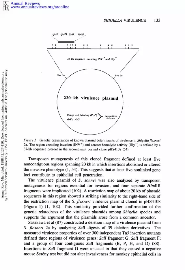
. ipaA ipaD ipaC ipaB.
,~ 37 kb sequence encodmg 1NV and Hly ,,.
Figure 1 Genetic organization of known plasmid determinants of virulence in Shigellaflexneri2a. The region encoding invasion (INV+) and contact hemolytic activity (ttly÷) is defined by a37-kb sequence present in the recombinant cosmid clone pHS4108 (54).
Transpo:~on mutagenesis of this cloned fragment defined at least fivenoncontiguous regions spanning 20 kb in which insertions abolished or alteredthe invasive phenotype (1, 54). This suggests that at least five nonlinked geneloci contribute to epithelial cell penetration.
The virulence plasmid of S. sonnei was also analyzed by transposonmutagenesis for regions essential for invasion, and four separate HindlIIfragments were implicated (102). A restriction map of about 20 kb of plasmidsequences in this region showed a striking similarity to the right-hand side ofthe restriction map of the S. flexneri virulence plasmid cloned in pHS4108(Figure 1) (1, 102). This similarity provided further confirmation of genetic relatedness of the virulence plasmids among Shigella species andsupports the argument that the plasmids arose from a common ancestor.
Sasakawa et al (87) constructed a deletion map of a virulence plasmid fromS. flexneri 2a by analyzing SalI digests of 39 deletion derivatives. Themeasured virulence properties of over 300 independent Tn5 insertion mutantsdefined three regions of virulence genes: SalI fragment G; SalI fragment F;and a group of four contiguous SalI fragments (B, P, H, and D) (88).Insertions in SalI fragment G were unusual in that they caused a negativemouse Ser~,ny test but did not alter invasiveness for monkey epithelial cells in
www.annualreviews.org/aronlineAnnual Reviews
Ann
u. R
ev. M
icro
biol
. 198
8.42
:127
-150
. Dow
nloa
ded
from
arj
ourn
als.
annu
alre
view
s.or
gby
Uni
form
ed S
ervi
ces
Uni
vers
ity -
HSC
EB
VC
Acc
ount
on
04/0
8/08
. For
per
sona
l use
onl
y.

134 MAURELLI & SANSONETrI
culture or Congo red binding. Insertions in fragments B, P, H, D, a~nd Fresulted in a pleiotropic loss of Congo red binding ability and invasiveness forboth the mouse SerEny test and cells in tissue culture. Preliminary resultsindicated that the insertions in fragments B, P, D, and H lay within a regioncovering 33 kb of the four contiguous fragments. Yet this region is separatedby more than 30 kb from fragment F, which is also required for invasion.Thus one would assume that a minimum contiguous region of 63 kb ofplasmid DNA is necessary for invasion in tissue culture cells. However, as wehave already indicated, a 37-kb fragment of DNA from the virulence plasmidof S. flexneri 5 was sufficient to encode the invasive phenotype (54). Thereason for this discrepancy is unknown, but it may be related to sequencedivergence among strains of S. flexneri.
Identification of specific virulence plasmid-encoded peptides was firstundertaken by examining minicell-producing derivatives of S. flexneri 2a.These anucleate bacterial ceils, containing only plasmid DNA, retained thecapability of penetrating HeLa cells (39). Sodium dodecyl sulfate-polyacrylamide gel electrophoresis (SDS-PAGE) of the polypeptides ex-pressed in outer membranes of minicells isolated from invasive strains of S.flexneri revealed a complement of 9-16 polypeptides ranging in size from 12to 64 kd (39). Seven polypeptides, designated a-g, were identified by two-dimensional gel electrophoresis as unique to virulence plasmids of S. flexneri2a and 5 and EIEC serogroup O143 (38). Expression of these virulenceplasmid-specific polypeptides was also partially repressed at 30°C, the non-permissive temperature for the invasive phenotype. Polypeptides a and b werenot detected at all after growth at 30°C, while c-g were weakly expressed.Polypeptides a-d were also expressed in a temperature-regulated manner bystrains containing the cosmid clone pHS4108 (54). Their sizes were estimatedto be 78, 62, 43, and 38 kd, respectively. Several of the virulence plasmid-specific polypeptides displayed extremely basic isoelectric points, whichsuggests that they may act as regulatory proteins capable of binding to DNAor as anchorage or transport proteins in the outer membrane. Some of theplasmid-encoded polypeptides necessary for the invasive phenotype may notbe detectable in such minicell experiments. Conversely, not all of thepolypeptides detected are necessarily involved in virulence, as later studiesrevealed.
Western blot analysis using rabbit antisera raised against individual plas-mid-encoded polypeptides or serum from a monkey that had been infectedwith shigellae established the immunological similarity of polypeptides b-damong strains of Shigella and EIEC (38). Although a discussion of the serumimmune response to Shigella infection is beyond the scope of this review (fora review of attempts at developing anti-Shigella vaccines, see Reference 26),we note that polypeptides a-d, as well as an additional plasmid-encoded
www.annualreviews.org/aronlineAnnual Reviews
Ann
u. R
ev. M
icro
biol
. 198
8.42
:127
-150
. Dow
nloa
ded
from
arj
ourn
als.
annu
alre
view
s.or
gby
Uni
form
ed S
ervi
ces
Uni
vers
ity -
HSC
EB
VC
Acc
ount
on
04/0
8/08
. For
per
sona
l use
onl
y.
SHIGELLA VIRULENCE 135
polypeptide of 140 kd, are predominant Shigella antigens recognized inimmunoblots by most convalescent human and monkey sera (61). That theseplasmid-encoded polypeptides are the principal antigens that induce a serumimmune response during Shigella infection does not necessarily implicatethem as e.ssential virulence antigens, but this property has been useful instudies of these polypeptides.
The genetic organization of the immunogenic peptides a-d has recentlybeen determined by two groups. Buysse et al (5) constructed a Agtl 1 expres-sion library from S. flexneri 5 strain M90T carrying the Tn5-tagged virulenceplasmid pWRll0. They used rabbit antisera specific for the immunogenicpolypeptide antigens b, c, and d to screen for recombinant phages expressingthese antigens. In this way they cloned invasion plasmid antigen (ipa) genesipaB, C, and D and identified a fourth gene, ipaH. The protein product of thislast gene had a molecular mass similar to that of IpaB (58 kd) but wasimmunologically distinct. Southern blot hybridization of various recom-binants revealed that three ipa genes mapped to contiguous HindlII fragmentsin the order ipaBCD. The restriction map. of this region of the virulenceplasmid was identical to the left-hand end of the cosmid clone pHS4108(Figure 1), with the exception of a single missing BamHI site. The location ofipaH was not determined. Buysse et al (5) did not clone the gene encodingpolypeptide a because they lacked specific antiserum for screening for such aclone. However, strain M90T carrying pWR110 was both invasive in HeLacells and positive in the Ser6ny test (82), yet it did not synthesize detectablelevels of this 78-kd polypeptide. This suggested that polypeptide a may not beessential for invasion.
Baudry et al (1) generated Tn5 insertions in the cosmid clone pHS4108 andanalyzed rnutants that were altered or blocked in invasion of HeLa cells forexpression of immunogenic peptides a-d. The results of immunoblots usingconvalescent monkey antiserum showed that insertions that reduced the ex-pression of polypeptides a, b, and d greatly decreased the invasive ability ofthe organi:~m. The role of polypeptide c in invasion could not be determinedbecause no Trl5 insertions that abolished expression ’of c were obtained. Twoinsertions that altered expression of polypeptide a had no effect on invasivepotential, which confirmed that polypeptide a was not essential for invasion ofHeLa cells.. Two-dimensional gel electrophoresis revealed that one of the Tn5mutants with reduced invasive ability no longer expressed a 21-kd polypep-tide, which was tentatively identified as the polypeptide g described earlier(38). Subcloning of fragments that expressed pol,ypeptides a-d and mappingof the Tn5 insertions within pHS4108 permitted Baudry et al (1) to deduce gene order of ipaBCDA from right to left on the map of pHS4108 (Figure 1).The gene order and location within pHS4108 are in agreement with theconclusions of Buysse et al (5).
www.annualreviews.org/aronlineAnnual Reviews
Ann
u. R
ev. M
icro
biol
. 198
8.42
:127
-150
. Dow
nloa
ded
from
arj
ourn
als.
annu
alre
view
s.or
gby
Uni
form
ed S
ervi
ces
Uni
vers
ity -
HSC
EB
VC
Acc
ount
on
04/0
8/08
. For
per
sona
l use
onl
y.

136 MAURELLI & SANSONETTI
Based on expression data from the Agtl 1 clones, Buysse et al (5) postulateddiscrete transcriptional units for ipaB, C, and D. However, Baudry et al (1)interpreted the results of the Trd insertion mutations to indicate the existenceof an operon encompassing, in order, the 21-kd peptide gene, ipaB, C, D, andA. Both groups are currently sequencing this region of plasmid DNA, and thecontrasting models of transcription should be resolved in the near future.
The virulence plasmid of S. sonnei also encodes genes for expression ofpolypeptides that are similar in size and immunological cross-reactivity to theimmunogenic peptides a-d described above in S. flexneri (38). Subclones two contiguous HindIII fragments expressed at least four polypeptides of 80,47, 41, and 38 kd. There is no evidence to date that these peptides are the onesthat are similar to the peptides a-d expressed in S. flexnerio They are likely tobe new, unrelated peptides, since the region to which these HindIII fragmentsmap lies at the opposite end of pHS4108 from the sequences known to encodefor polypeptides a~l of S. flexneri (l, 102).
ANALYSIS OF INTERACTION OF BACTERIAWITH CELLS
EntryExperiments with M90T, an invasive isolate of S. flexneri serotype 5, haveindicated that its virulence plasmid pWR100 participates at every essentialstep of the invasive process in the HeLa cell model, including entry into cells,rapid intracellular multiplication, and early killing of host cells. Preliminaryexperiments indicated that shigellae entered mammalian cells through en-docytosis. No leakage of macromolecules from recipient cells could beobserved during entry (34). Cytochalasin D, which inhibits microfilamentfunctions, blocked the entry process (37). Recent evidence indicates that flexneri enters HeLa cells via directed phagocytosis (10). Use of an anti-myosin monoclonal antibody and of 7-nitrobenz-2-oxa-l,3-diazole phallaci-din, a fluorescent dye that stains polymerized F-actin, demonstratedaccumulation of myosin and F-actin, two major components of the cellcytoskeleton, underneath the cytoplasmic membrane at the site of bacterialentry. The nature of the plasmid-encoded bacterial product, as well as that ofthe transmembrane signaling system that triggers the phagocytic process,remains unknown. As mentioned earlier, Tn5 mutagenesis of recombinantplasmid pHS4108 revealed at least five regions within a 20-kb portion of theinsert that are implicated in this phenotype (1). The observation that flexneri requires expression from several different gene loci to penetrate cellsemphasizes the complexity of the strategy that has evolved in this microorgan-ism. It differs from that of Yersinia pseudotuberculosis, in which only onegene appears necessary for promoting entry into cells (41).
www.annualreviews.org/aronlineAnnual Reviews
Ann
u. R
ev. M
icro
biol
. 198
8.42
:127
-150
. Dow
nloa
ded
from
arj
ourn
als.
annu
alre
view
s.or
gby
Uni
form
ed S
ervi
ces
Uni
vers
ity -
HSC
EB
VC
Acc
ount
on
04/0
8/08
. For
per
sona
l use
onl
y.

SHIGELLA VIRULENCE 137
lntracellular Multiplication
Plasmid genes are also needed for efficient intracellular multiplication (86).Plasmid pWR100 endows E. coli K-12 with the same replication potential aswild-type S. flexneri. A sequential electron-microscopic study of infectedHeLa cells demonstrated that invasive S. flexneri induced lysis of thephagocytic membrane shortly after penetration into cells. By 30 min aftercentrifugation-induced penetration, all bacteria were lying free within thecytoplasm of host cells (86). Similar invasiveness was observed with E. coliK-12 carr]jing pWR100. On the other hand, Salmonella typhimurium, whoselysis of the phagocytic membrane is late and inefficient, grows poorly in-tracellularly. A plasmid-mediated contact-hemolytic activity demonstrated invirulent shigellae provides a likely mechanism for lysis of the phagosome (8,86). Molecular characterization of this contact hemolysin is not yet available,although preliminary evidence indicates that the gene product(s) that triggersentry also accounts for hemolysis. This proposal is based on the observationthat all Tn5 mutations in the virulence plasmid that eliminate the entryphenotype also eliminate contact-hemolytic activity (1). By contrast, correlation has been observed between rapid intracellular growth of shigellaeand the level of Shiga toxin or SLT production (9, 86). During the infectiousprocess both S. dysenteriae 1 and S. flexneri 2a block host-cell proteinsynthesis; the block leads to preferential incorporation of labeled amino acidsinto bactel.-ial proteins (35). Although secretion of Shiga toxin or SLT shouldgive these invasive pathogens an additional advantage by precipitating cellkilling, it does not seem to account for rapid intracellular growth. Anotherfactor, the iron chelator aerobactin, may also be critical for bacterial replica-tion within cells in which iron is immobilized by ferritin. Independent studieshave recently shown that mutants of S. flexneri that no longer produceaerobactin demonstrate no significant alteration in their capacity to multiplyintracellularly (50, 60). Therefore, only early lysis of the phagocytic vacuoleso far appears to be a major factor for the rapid intracellular growth that ischaracteri~;tic of Shigella. Metabolic pathways that lead to products such asaromatic components that are not available within eukaryotic cells will alsoprobably turn out to be major factors for intracellular growth.
Early Kiilling of Host Cells
Plasmid genes are also involved in early killing of host cells. In a study of theintracellular fate of both an invasive strain and a noninvasive, plasmidlessderivative of S. flexneri, plasmid pWR100 appeared to mediate killing of hostcells (the continuous macrophage cell line J774) within 4 hr (9). For expres-sion of this activity bacteria had to be intracellular, since macrophages wereprotected by cytochalasin D. Although both strains produced equivalent levelsof SLT and inhibited protein synthesis of macrophages within 2 hr,
www.annualreviews.org/aronlineAnnual Reviews
Ann
u. R
ev. M
icro
biol
. 198
8.42
:127
-150
. Dow
nloa
ded
from
arj
ourn
als.
annu
alre
view
s.or
gby
Uni
form
ed S
ervi
ces
Uni
vers
ity -
HSC
EB
VC
Acc
ount
on
04/0
8/08
. For
per
sona
l use
onl
y.
138 MAURELLI & SANSONETTI
only invasive bacteria were able to kill host cells. Damage to macrophagescorrelated with the ability of invasive bacteria to rapidly and efficiently lysethe membrane of the phagocytic vacuole (9). Metabolic events that mediateearly killing have recently been demonstrated to include a rapid drop in theintracellular concentration of ATP, an increase in pyruvate concentration, andarrest of lactate production (85). The molecular basis of such inhibition host-cell respiration and fermentation is unknown. It is not yet clear whetherplasmid genes are directly responsible for this effect or whether plasmid-mediated lysis of the phagocytic vacuole allows diffusion of toxic productsencoded by the chromosome into the cytosol.
Continuous Reinfection of Adjacent Cells
A region (virG) of the S. flexneri virulence plasmid is considered to benecessary for continuous reinfection of adjacent cells (53). virG mutants caninvade cells and multiply intracellularly but do not spread to adjacent ceils.Within epithelia, bacteria tend to localize within the cytoplasm and convert toa spherical morphology before being eliminated. The precise alteration of theinvasive phenotype of the virG mutants has not been characterized. Wespeculate that they are either altered in their capacity to destroy the membraneof the phagocytic vacuole or unable to resist lysosomal killing. The locationof virG with respect to the essential invasion genes contained on pHS4108(Figure 1) is not known.
To summarize, the identification of the four major phenotypes (entry intocells, intracellular multiplication, early killing of host cells, and continuousreinfection of adjacent cells) represents a solid basis for an analyticalapproach to the invasive process of individual epithelial cells.
INTERACTION OF BACTERIA WITH TISSUES
Shiga and Shiga-like Toxins
Although Shiga toxin is one of the most potent bacterial toxins, its actual rolein the pathogenesis of shigellosis is still unclear (for a review on Shiga toxinand SLT, see Reference 63). S. dysenteriae 1, which produces the mostsevere shigellosis, also produces much more Shiga toxin than other species ofShigella (44, 45, 64--66). This toxin is composed of two subunits. Subunit (32-kd) possesses the biological activities. It is combined with five moleculesof the B subunit (7.7 kd), which are responsible for binding to cell-surfacereceptors (14, 90). Some strains of S. flexneri and S. sonnei produce lowlevels of Shiga-like toxin, which is neutralizable by anti-Shiga toxin sera (45,66). This toxin has not yet been purified.
Shiga toxin has three main biological activities: enterotoxicity, neurotoxic-ity, and cytotoxicity (16). The toxin inhibits protein synthesis in eukaryotic
www.annualreviews.org/aronlineAnnual Reviews
Ann
u. R
ev. M
icro
biol
. 198
8.42
:127
-150
. Dow
nloa
ded
from
arj
ourn
als.
annu
alre
view
s.or
gby
Uni
form
ed S
ervi
ces
Uni
vers
ity -
HSC
EB
VC
Acc
ount
on
04/0
8/08
. For
per
sona
l use
onl
y.
SHIGELLA VIRULENCE 139
cells (4, 99) by acting on the 60S ribosomal subunit (3, 67, 74). molecular basis of Shiga toxin cytotoxicity has recently been clearly es-tablished (17). Like the plant lectin ricin, Shiga toxin depurinates adenine4324 near the 3 ’ end of 28S RNA. Therefore, the A catalytic chain of Shigatoxin is a ihighly specific N-glycosidase.
The conlribution of Shiga toxin to virulence is unclear. It may contribute toor even be responsible for the diarrheal component of shigellosis (75).However, how Shiga toxin acts as an enterotoxin is unknown as yet. The roleof Shiga toxin in the pathogenesis of dysentery is not clear either. Volunteersfed a low toxin-producing mutant of S. dysenteriae 1 (29) showed mildersymptoms than those fed the parental strain (52). This would indicate that thetoxin has a role at some stage of the infection process. However, Shiga toxinand SLT do n0t,,appear to be involved in penetration into cells, intracellularmultiplicat!ion, or early cell killing based on in vitro models (9, 86). This viewis confirmed by recent experiments in which a Tox- mutant of S. dysenteriae1 still inw~ded HeLa cells and caused a positive Serrny test (91). Threepossibilities may be viewed at present: (a) Free toxin within the intestinallumen [which can be detected during S. dysenteriae 1 infection (13)] maybind to specific receptors on the surface of enterocytes and kill those cells byinhibition of protein synthesis; Shiga toxin has been shown to be cytotoxic forprimary cultures of human colonic epithelial cells (59). (b) Toxin releasedwithin infected enterocytes or within connective tissue of the lamina propriamay diffuse and alter adjacent cells, including phagocytic cells (if sensitive toShiga toxin), thus increasing the severity of the lesions. (c) The third possibil-ity, which we favor at present, is derived from recent evidence that thehemolytic uremic syndrome (HUS) observed after S. dysenteriae 1 or E. coliO157:H7 infection (30, 42, 48, 70, 73) may in part be due to systemicdissemination of the toxin. Shiga toxin appears to mediate vascular damage,since vascular injury observed in HUS (48) resembles that observed cerebral w~ssels of animals inoculated with Shiga toxin (2, 6). We thusspeculate that Shiga toxin produced during colonic infection may increase theseverity of the disease by causing ischemia of the intestinal tissue by eitherlocal or systemic diffusion of the toxin. Alteration of vasa nervosum may alsoaccount for more severe symptoms, including toxic megacolon. The recentproduction of a transposon mutant of S. dysenteriae 1 that does not produceShiga toxin should allow precise evaluation of the role of this toxin in theinfection process (91).
Information on the genetics of Shiga toxin production has been scarce andcontradictory. Initial reports localized a region of the S. flexneri chromosomenecessary for fluid production in rabbit ileal loops to the rha-ratl region, nearthe lysine decarboxylase-negative locus (82). However, no evidence wasprovided that the ability to cause fluid accumulation was due to the Shiga-like
www.annualreviews.org/aronlineAnnual Reviews
Ann
u. R
ev. M
icro
biol
. 198
8.42
:127
-150
. Dow
nloa
ded
from
arj
ourn
als.
annu
alre
view
s.or
gby
Uni
form
ed S
ervi
ces
Uni
vers
ity -
HSC
EB
VC
Acc
ount
on
04/0
8/08
. For
per
sona
l use
onl
y.

140 MAURELLI & SANSONETTI
toxin of S. flexneri. In another study, Timmis et al (100) constructed R-prime plasmid that contained the argE region of the chromosome of S.dysenteriae 1 and encoded production of Shiga toxin. The discrepancy be-tween the location of the toxin genes in the two species may be explained by adifferent chromosome map or the presence of several copies of the gene in S.dysenteriae 1. However, Sekizaki et al (91) recently reported that E. coliK-12 was able to produce high levels of Shiga toxin after conjugation with S.dysenteriae 1 Hfr derivatives. P1 transduction analysis allowed mapping ofthe gene encoding Shiga toxin (stx) nearpyrF at 30 min on the linkage map ofE. coli K-12 (91). This series of experiments seems to solve the problem the location of the Shiga toxin gene on the chromosome of S. dysenteriae. Asimilar approach did not reveal the location of the Shiga-like toxin gene of S.flexneri (T. Sekizaki, personal communication). The structural gene for Shigatoxin has recently been cloned. The sequence differs from that of the SLT-Itoxin gene of E. coli by three base pairs with a single amino acid change (94).
Role of Lipopolysaccharide in Shigella Virulence
The lipopolysaccharide (LPS) layer of gram-negative bacteria is an importantcomponent of the cell surface and contributes to the virulence of manypathogens by providing resistance to certain host defenses such as serumkilling and phagocytosis. Its importance has been demonstrated by theobservation that smooth-colony clinical isolates readily segregate from rough-colony variants upon passage in the laboratory. These rough mutants arealtered in their LPS structure and have lost their virulence in animal models.Rough mutants of S. flexneri 2a are still capable of invading HeLa cells but donot produce keratoconjunctivitis in guinea pigs (69). Mutants representing range of rough chemotypes from Ra to Re (no O-antigen but complete toincomplete cores) also retained the ability to invade HeLa cells, which ruledout a role for Shigella O-antigens in the invasion step in vitro (68). Thesemutants were uniformly negative in the Serrny test, which suggests that acomplete LPS structure is essential in either bacterial multiplication afterpenetration of the epithelial cell or bacterial survival in an animal host beforeinvasion.
The importance of a specific O-antigen structure for Shigella virulence wasshown ifl early experiments involving construction of intergeneric hybrids. S.flexneri hybrids expressing the E. coli 0-25 antigen retained the ability topenetrate epithelial cells and produce keratoconjunctivitis in guinea pigs (28).On the other hand, hybrids expressing E. coli 0-8 somatic antigen lost theirinvasive capacity. The avirulence of the 0-8 hybrids was attributed to thechemical structure of the 0-8 antigen, which was less similar than that of the0-25 antigen to the structure of Shigella somatic antigen (for a detailed reviewof the chemistry of S. flexneri O-antigens, see Reference 93). Thus possessionof a smooth LPS alone would not appear to be sufficient for virulence, and a
www.annualreviews.org/aronlineAnnual Reviews
Ann
u. R
ev. M
icro
biol
. 198
8.42
:127
-150
. Dow
nloa
ded
from
arj
ourn
als.
annu
alre
view
s.or
gby
Uni
form
ed S
ervi
ces
Uni
vers
ity -
HSC
EB
VC
Acc
ount
on
04/0
8/08
. For
per
sona
l use
onl
y.

SHIGELLA VIRULENCE 141
possible role for the chemical composition of the particular Shigella somaticantigen is implicated. Further evidence of the need for a specific type ofO-antigen for virulence is the strong immunological relatedness of the major-ity of the ()-antigen serotypes expressed by EIEC to the O-antigens of variousShigella spp. (7). The role of the O-antigen was further clarified reconstruc~tion experiments involving sequential transfer of S. flexneri chro-mosomal DNA into an E. coli K-12 recipient carrying the 220-kb virulenceplasmid (82). Expression of the Shigella group antigen in conjunction with thevirulence plasmid and the kcp locus was important in producing a positiveSerrny test and inflammation in rabbit ileal loops (82). However, constructionof the intergeneric hybrids involved gene transfer by conjugation, and onecannot rule out the possibility that other virulence-associated genes may beclosely linked to his and may have contributed to the observed behavior ofthese hybrids.
The genetic information required for O-antigen biosynthesis in S. flexneri ischromosomally located, and the virulence plasmid does not appear to encodeany genes for production of the LPS (84). However, S. sonnei and S.dysenteriae are different. Virulent smooth (Form I) isolates of S. sonneiirreversibly dissociate to rough (Form II) variants at a high frequency, andthese variants are unable to invade epithelial cells in the Serrny test (47). Theirreversible nature of the transition from Form I to Form II suggested aplasmid role in O-antigen biosynthesis. A 180-kb plasmid was subsequentlyidentified i.n Form I isolates of S. sonnei from diverse geographical sources.Form II is,alates had lost the plasmid. When the 180-kb plasmid was mobi-lized back into Form II S. sonnei, the resulting transconjugants again ex-pressed the characteristic S. sonnei O-antigen. Mobilization of the 180-kbplasmid into S. flexneri and Salmonella typhi also resulted in expression of S.sonnei O-~tntigen by the transconjugants, which confirmed that the plasmidcarded all of the genes necessary for the biosynthesis of the S. sonneiO-antigen. As noted earlier, this 180-kb plasmid of S. sonnei is also func-tionally and genetically analogous to the virulence plasmid of S. flexneri andis required for expression of the invasive phenotype (83, 101).
A small 9-kb plasmid in strains of S. dysenteriae 1 was found to beassociated with production of O-antigen. Mutants that had lost the plasmidexpressed a rough phenotype and reduced virulence in HeLa cells and theSerrny test. Reconstruction experiments introducing the 9-kb plasmid backinto the plasmid-cured rough mutant restored both S. dysenteriae O-antigenproduction and virulence (104). Cloning and transposon mutagenesis local-ized the plasmid-bome determinant involved in O-antigen synthesis, andminicell e~:periments demonstrated that the gene, designated rfp, produced a41-kd protein (103). The cloned rfp gene, when introduced into a S. dysenter-iae 1 strain cured of the native 9-kb plasmid, restored normal O-antigenproduction and virulence. When introduced into E. coli K-12, the cloned rfp
www.annualreviews.org/aronlineAnnual Reviews
Ann
u. R
ev. M
icro
biol
. 198
8.42
:127
-150
. Dow
nloa
ded
from
arj
ourn
als.
annu
alre
view
s.or
gby
Uni
form
ed S
ervi
ces
Uni
vers
ity -
HSC
EB
VC
Acc
ount
on
04/0
8/08
. For
per
sona
l use
onl
y.

142 MAURELLI & SANSONEI~FI
gene modified the E. coli LPS core by the addition of a galactose residue, thefirst sugar of the S. dysenteriae 1 O-antigen repeat unit (95). However,synthesis of a complete S. dysenteriae 1 O-antigen in E. coli K-12 requires thechromosomal rfb gene cluster as well (36). This region, spanning 6.4-7.5 near his, specifies at least six determinants for O-antigen production, includ-ing the synthetases and transferases required for adding two rhamnose resi-dues to the galactose to complete the O-antigen side chain (96).
kcpA Locus
The kcpA locus, located between the lac and the gal genes on the Shigellachromosome, is necessary for production of keratoconjunctivitis in guineapigs (21). Transduction with phage P1 demonstrated that this locus cotrans-duced with purE. Although the function of the kcpA locus in disease isunclear, E. coli K-12-S. flexneri hybrid strains that had received the kcpAlocus appeared more invasive in the rabbit ligated ileal loop assay, whichsuggests that this locus encodes a function necessary for tissue invasiveness(82).
Miscellaneous PhenOtypes Associated With Virulence
AEROBACTIN Microorganisms have evolved efficient high-affinity iron up-take systems in order to grow in iron-limited environments. The ability of amicroorganism to compete for iron in an infected host is considered a viru-lence factor for many pathogens (105). Certain E. coli ColV strains producethe hydroxamate siderophore aerobactin, and this iron uptake system isassociated with the strain’s ability to cause generalized extraintestinal in-fections (106). Most strains of S. flexneri and S. boydii also utilize theaerobactin system for chelating iron, while many S. sonnei and S. dysenteriaestrains employ only the enterobactin system (51). The chromosomal regionarg-mtl, which was shown by conjugational gene transfer experiments to beassociated with S. flexneri virulence (82), was subsequently found to containthe gene for aerobactin and a 76-kd iron-regulated outer-membrane protein,which is probably the aerobactin receptor protein (32). A formal test of thehypothesis that aerobactin is a virulence factor for Shigella was undertakenwhen defined transposon mutants of the aerobactin gene (iuc) was isolated.Lawlor et al (50) assayed an iuc: :Tn5 mutant constructed in vitro forinvasiveness in HeLa cells and the SerEny test. They found the mutantunaffected in invasive capacity and ability to multiply intracellularly. Theiuc:: Tn5 mutant was also screened for virulence in the chicken embryomodel (71). It was found to be lethal for chicken embryos, but at LDsovalues 10-100 times higher than those of the wild-type parent. This higherLDso was attributed to a slower growth rate for the iuc:: Tn5 mutant inthe allantoic fluid.
A TnlO insertion in the aerobactin gene of a S. flexneri 5 strain was
www.annualreviews.org/aronlineAnnual Reviews
Ann
u. R
ev. M
icro
biol
. 198
8.42
:127
-150
. Dow
nloa
ded
from
arj
ourn
als.
annu
alre
view
s.or
gby
Uni
form
ed S
ervi
ces
Uni
vers
ity -
HSC
EB
VC
Acc
ount
on
04/0
8/08
. For
per
sona
l use
onl
y.
SH1GELLA VIRULENCE 143
similarly tested in a separate study (60). Like the iuc: :Tn5 mutant, theiuc: :Tnl0 mutant was unaltered in its ability to invade and multiply withinHeLa cell:~ and killed the host cells with the same kinetics as the parent strain.However, an inoculum-dependent difference was observed in the Ser6ny test;the wild-type strain produced a positive reaction with 106 bacteria, while themutant required a 10-fold higher inoculum to produce a similar reaction.Virulence was also tested by infecting ligated rabbit ileal loops with theaerobactin mutant and evaluating fluid production and histopathology. Aninoculum-dependent effect was again observed with the iuc::TnlO mutant.When an inoculum of 107 bacteria was used, ileal loops infected with themutant displayed markedly fewer mucosal lesions and lower fluid productionthan those infected with the ~parent strain. Taken together, these resultssuggest that aerobactin production may have a role in bacterial growth in theextracellular compartment within host tissues. It should also be noted thataerobactin does not appear to be preferred over enterobactin as a siderophorefor Shigella, since S. dysenteriae 1, which causes the most severe form ofdysentery among Shigella spp,, has no aerobactin genes and apparentlyproduces only enterochelin (51).
CONGO RED BINDING ABILITY The ability to bind the dye Congo red(referred to as Pcr+ or Crb+ in the literature) was originally recognized as phenotype that differentiated between virulent and avirulent derivatives ofYersinia pestis. The observation was subsequently extended to includeShigella and several other gram-negative pathogens (71). Spontaneousmutants of S. flexneri that are no longer capable of binding Congo red (Pcr-)arise at a frequency of --10-4, are noninvasive, and frequently have eitherlost the virulence plasmid or suffered deletions in the plasmid (56). Con-versely, screening of noninvasive mutants of S. flexneri revealed that they hadalso lost the ability to bind Congo red. This observation demonstrates a closeassociation of the Per+ phenotype with invasive ability and the presence of thevirulence plasmid, which suggests Congo red binding as a possible virulencefactor. In addition, as mentioned earlier, the Pcr+ phenotype is expressed in atemperature-regulated fashion, as are the virulence genes of Shigella (55).Confirmation of the role of the plasmid in Congo red binding came with thecloning of a gene(s) for this phenotype from the virulence plasmid (11, 77).According to one report (11), a cloned 9.0-kb BarnHI fragment from thevirulence plasmid of S. flexneri lb conferred Congo red binding ability on E.coli and restored the Per+ phenotype to a plasmidless S. flexneri lb. Congored binding by the recombinant was not fully temperature regulated in S.flexneri lb, possibly because of the high copy number of the cloning vector.The 9.0-kb fragment alone was not sufficient, however, to restore virulence toa plasmidless S. flexneri lb.
Another study found that a l-kb sequence of DNA (the virF region) from
www.annualreviews.org/aronlineAnnual Reviews
Ann
u. R
ev. M
icro
biol
. 198
8.42
:127
-150
. Dow
nloa
ded
from
arj
ourn
als.
annu
alre
view
s.or
gby
Uni
form
ed S
ervi
ces
Uni
vers
ity -
HSC
EB
VC
Acc
ount
on
04/0
8/08
. For
per
sona
l use
onl
y.

144 MAURELLI & SANSONETTI
the virulence plasmid of S. flexneri 2a was sufficient for expression of thePcr+ phenotype in E. coli K-12 (77). However, unlike the previous clone, plasmidless S. flexneri 2a containing the virF clone did not become Pcr÷. It isnot clear why this clone expressed Pcr+ in E. coli but not in S. flexneri. Theauthors reported finding several other regions of the virulence plasmid thatexpressed the Pcr+ phenotype in E. coli, yet these clones were not tested in S.flexneri. It is difficult to evaluate the relevance of these plasmid regions toCongo red binding because the proper phenotype could not be restored in theparent. The virF region has been sequenced and was determined to specifythree peptides of 30, 27, and 21 kd in minicells (78). A computer-generatedsecondary structure prediction of the product of an open reading frame in thissequence indicated a protein rich in/~-sheets. This may represent a structurecapable of direct interaction with Congo red, which is known to bind to such astructure (31).
The function of the Congo red binding gene(s) in Shigella is unknown.Congo red binding in other bacterial systems has been correlated with absorp-tion of hemin [Y. pestis (97)] and protoporphyrin IX [Aeromonas salrnonicida(43)]. Daskaleros & Payne (12) observed this correlation Shigella as welland found that absorption of these iron-containing compounds was growth-temperature dependent (12). They also found that bacteria that had preboundhemin showed increased invasiveness in HeLa cells, which suggested apossible involvement of the Congo red binding gene(s) in bacterial attachmentto mammalian cells.
CONCLUSIONS AND PROSPECTS
During the six years since the first report of the involvement of a plasmid ininvasion of mammalian cells by Shigella, research in defining the plasmidvirulence determinants has moved rapidly. Application of new techniques forcreating protein fusions using phoA and lacZ will further help define plasmidvirulence genes and their role in invasion. Study of the immunogenic peptidesencoded by the plasmid should yield information that will be useful for thedesign of effective vaccines against shigellosis. The DNA sequence of severalipa genes will soon be available (B. Baudry, M. Kaczorek, P. Sansonetti,manuscript in preparation), and this will certainly give us clues as to thepossible roles of their gene products in virulence.
Genetic studies will need to focus again on the chromosomal determinantsof Shigella virulence. The gene product and the specific function of the kcpAlocus, which was first described in 1971, still remain to be uncovered. Thegenetic basis for the colony-morphology transition from transparent toopaque, which leads to loss of virulence and a host of pleiotropic changes (27,49), is unknown. The interaction between chromosomal and plasmid
www.annualreviews.org/aronlineAnnual Reviews
Ann
u. R
ev. M
icro
biol
. 198
8.42
:127
-150
. Dow
nloa
ded
from
arj
ourn
als.
annu
alre
view
s.or
gby
Uni
form
ed S
ervi
ces
Uni
vers
ity -
HSC
EB
VC
Acc
ount
on
04/0
8/08
. For
per
sona
l use
onl
y.

SHIGELLA VIRULENCE 145
genes should also be considered, as the pathogenicity of Shigella is dependenton the coordinate expression of genes on both genetic elements. Research onthe molecular mechanisms underlying the temperature regulation of Shigella
virulence will be important in understanding these interactions.Future research will probably examine the intracellular life-style of the
bacteria, specifically intracellular growth and host-cell killing. For example,the availability of defined Tox- mutants of S. dysenteriae 1 will make itpossible to examine definitively the role of toxin in pathogenesis at the
cellular and tissue levels. However, many of the genetic studies currentlyunder way to detemline the genes and their products that trigger entry mayprovide only partial answers. These studies should be complemented byexperiments that approach the question from the point of view of membranebiochemistry and cell biology. This approach is already beginning to revealthe biochemical events involved in the host-parasite interactions in Shigellainfection. Future studies will surely benefit from this multidisciplinaryapproach.
ACKNOWLEDGMENTS
The authors wish to acknowledge the outstanding contributions of SamFormal to our understanding of the genetics of Shigella pathogenicity and citehis considerable influence in our own research. We also wish to thank T. L.Hale, P. Clerc, B. Baudry, and A. D. O’Brien for helpful discussions.Research on the genetics of Shigella virulence in the laboratory of ATM iscurrently supported by USUHS protocol RO-7385.
Literature Cited
1. Baudry, B., Maurelli, A. T., Clerc, P.,Sadoff. J. C., Sansonetti, P. J. 1987.Localization of plasmid loci necessaryfor Shigella flexneri entry into HeLacells, and genetic organization of one lo-cus encoding four immunogenic polypep-tides. ,)’. Gen. Microbiol. 133:3403-13
2. Bridgewater, F. A. J., Morgan, R. S.,Rowson, K. E. K., Wright, G. P. 1955.The neutrotoxin of Shigella shigae.Morphological and functional lesionsproduced in the central nervous systemof rabbits. Br. J. Exp. Pathol. 36:447-53 /
3. Brown, J. E., Obrig, T. G., Ussery, M.A., Moran, T. P. 1986. Shiga toxinfrom Shigella dysenteriae 1 inhibits pro-tein synthesis in reticulocyte lysates byinactivation of aminoacyl-tRNA bindingMicrob. Pathog. 1:325-34
4. Brown, J. E., Rothman, S. W., Doctor,B. P. 1980. Inhibition of protein synthe-sis in intact HeLa cells by Shigella
dysenteriae 1 toxin. Infect. lmmun.29:98-107
5. Buysse, J. M., Stover, C. K., Oaks, E.V., Venkatesan, M., Kopecko, D. J.1987. Molecular cloning of invasionplasmid antigen (ipa) genes fromShigella flexneri: analysis of ipa geneproducts and genetic mapping. J. Bac-teriol. 169:2561-69
6. Cavanagh, J. B., Howard, J. G., Whit-by, J. L. 1956. The neurotoxin ofShigella shigae. A comparative study ofthe effects produced in various labora-tory animals. Br. J. Exp. Med. 37:272-78
7. Cheasty, T., Rowe, B. 1983. Antigenicrelationships between the enteroinvasiveEscherichia coli 0 antigens O28ac,Oll2ac, O124, O136, O143, O144,O152, and O164 and Shigella 0 anti-gens. J. Clin. Microbiol. 17:681-84
8. Clerc, P., Baudry, B., Sansonetti, P. J.1986. Plasmid-mediated contact haemo-
www.annualreviews.org/aronlineAnnual Reviews
Ann
u. R
ev. M
icro
biol
. 198
8.42
:127
-150
. Dow
nloa
ded
from
arj
ourn
als.
annu
alre
view
s.or
gby
Uni
form
ed S
ervi
ces
Uni
vers
ity -
HSC
EB
VC
Acc
ount
on
04/0
8/08
. For
per
sona
l use
onl
y.
146 MAURELLI & SANSONETT1
lytic activity in Shigella species: correla-tion with penetration into HeLa cells.Ann. Inst. Pasteur Microbiol.137A:267-78
9. Clerc, P., Ryter, A., Mounier, J., San-sonetti, P. J. 1987. Plasmid mediatedearly killing of eukaryotic cells byShigella flexneri as studied by infectionof J774 macrophages. Infect. Immun.55:521-27
10. Clerc, P., Sansonetti, P. J. 1987. Entryof Shigellaflexneri into HeLa cells: evi-dence for directed phagocytosis involv-ing actin polymerization and myosinaccumulation. Infect. lmmun. 55:2681-88
11. Daskaleros, P. A., Payne, S. M. 1985.Cloning the gene for Congo red bindingin Shigella flexneri. Infect. lmmun.48:165-68
12. Daskaleros, P. A., Payne, S. M. 1987.Congo red binding phenotype is associ-ated with hemin binding and increasedinfectivity of Shigella flexneri in theHeLa cell model. Infect. lmmun.55:1393-98
13. Donahue-Rolfe, A., Kelley, N. A.,Bennish, M., Keusch, G. T. 1986. En-zyme-linked immunosorbant assay forShigella toxin. J. Clin. Microbiol.24:65-68
14. Donohue-Rolfe, A., Keusch, G. T.,Edson, C., Thorley-Lawson, D., Jace-wicz, M. 1984. Pathogenesis of Shigelladiarrhea. IX. Simplified high yieldpurification of Shigella toxin andcharacterization of subunit compositionand function by the use of subunit-specific monoclonal and polyclonal anti-bodies. J. Exp. Med. 160:1767-81
15. Dupont, H. L., Formal, S. B., Homick,R. B., Snyder, M., Libonati, J. P., et al.1971. Pathogenesis of Escherichia colidiarrhea. N. Engl. J. Med. 285:1-9
16. Eiklid, K., Olsnes, S. 1983. Animaltoxicity of Shigelladysenteriae cytotox-in: evidence that the neurotoxic, entero-toxic and cytotoxic activities are due toone toxin. J. Immunol. 130:380-84
17. Endo, Y., Tsurugi, K. 1987. RNA N-glycosidase activity of ricin A-chain. J.Biol. Chem. 262:8128-30
18. Falkow, S., Schneider, H., Baron, L.S., Formal, S. B. 1963. Virulence ofEscherichia-Shigella genetic hybrids forthe guinea pig. J. Bacteriol. 86:1251-58
19. Formal, S. B., Dupont, H. L., Hornick,R., Snyder, M. J., Libonati, J., LaBrec,E, H, 1971. Experimental models in theinvestigation of the virulence of dysen-tery bacilli and Escherichia coli. Ann.NY Acad. Sci. 176:190-96
20. Formal, S. B., Gemski, P. Jr., Baron,
L. S., LaBrec, E. H. 1970. Genetictransfer of Shigella flexneri antigens toEscherichia coli K-12. Infect. lmmun.1:279-87
21. Formal, S. B., Gemski, P. Jr., Baron,L. S., LaBrec, E. H. 1971. A chromo-somal locus which controls the ability ofShigella flexneri to evoke keratoconjuc-tivitis. Infect. lmmun. 3:73-79
22. Formal, S. B., Hornick, R. B. 1978.Invasive Escherichia coli. J. Infect. Dis.137:641~-4
23. Formal, S. B., Kundel, D., Schneider,H., Kuneu, N., Spunz, H. 1961. Studieswith Vibrio cholerae in the ligated loopof the rabbit intestine. Br. J. Exp. Path-ol. 42:504-10
24. Formal, S. B., LaBrec, E. H., Kent, T.H., Falkow, S. 1965. Abortive intestinalinfection with an Escherichia coli-Shigella flexneri hybrid strain. J. Bac-teriol. 89:1374-82
25. Formal, S. B., LaBrec, E. H., Palmer,A., Falkow, S. 1965. Protection ofmonkeys against experimental shigello-sis with attenuated vaccines. J. Bacteri-ol. 90:63-68
26. Formal, S. B., Levine, M. M. 1983.Shigellosis. In Bacterial Vaccines, ed.R. Germanier, pp. 167-86. New York:Academic
27. Gemski, P. Jr., Formal, S. B. 1975.Shigellosis: an invasive infection of thegastrointestinal tract. In Microbiology1975, ed. D. Schlessinger, pp. 165-69.Washington DC: Am. Soc. Microbiol.
28. Gemski, P. Jr., Sheahan, D. G., Wash-ington, O., Formal, S. B. 1972. Viru-lence of Shigella flexneri hybrids ex-pressing Escherichia coli somatic anti-gens. Infect. Irnrnun. 6:104-11
29. Gemski, P. Jr., Takeuchi, A., Washing-ton, O., Formal, S. B. 1972. Shigellosisdue to Shigella dysenteriae 1: relativeimportance of mucosal invasion versustoxin production in pathogenesis. J. In-fect. Dis. 126:523-30
30. Gianantonio, C., Vitacco, M., Mendila-harzu, F., Rutty, A., Mendilaharzu, J.1964. The hemolytic-urcmic syndrome.J. Pediatr. 64:478-91
31. Glenner, G. G. 1980. Amyloid depositsand amyloidosis. N. Engl. J. Med.302:1283-92
32. Griffiths, E., Stevenson, P., Hale, T.L., Formal, S. B. 1985. Synthesis ofaerobactin and a 76,000-dalton iron-regulated outer membrane protein byEscherichia coli K-12-Shigella flexnerihybrids and by enteroinvasive strains ofEscherichia coli. Infect. Immun. 49:67-71
33. Hale, T. L., Bonventre, P. F. 1979.
www.annualreviews.org/aronlineAnnual Reviews
Ann
u. R
ev. M
icro
biol
. 198
8.42
:127
-150
. Dow
nloa
ded
from
arj
ourn
als.
annu
alre
view
s.or
gby
Uni
form
ed S
ervi
ces
Uni
vers
ity -
HSC
EB
VC
Acc
ount
on
04/0
8/08
. For
per
sona
l use
onl
y.
SHIGELLA VIRULENCE 147
Shigelh~ infection of Henle intestinalepithelial cells: role of’ the bacteria. In-fect. lmmun. 24:879-86
34. Hale, T. L., Formal, S. B. 1980.Cytoto~icity of Shigella dysenteriae 1for cultured mammalian cells. Am. J.Clin. A’utr. 33:2485 90
35. Hale, T. L., Formal, S. B. 1981. Proteinsynthesis in HeLa or Henle 407 cellsinfected with Shigella dysenteriae 1,Shigelh~ flexneri 2a, or Salmonellatyphimurium Wl18. Infect. lmmun.32:137--44
36. Hale, T. L., Guerry, P., Seid, R. C.,Kapfer, C., Wingfield, M. E., et al.1984. Expression of lipopolysaccharideO antigen in Escherichia coli K-12 hy-brids containing plasmid and chromo-somal genes from Shigella dystenteriae1. Infect. Irnmun. 46:470--75
37. Hale, T. L., Morris, R. E., Bonventre,P. F. 1979. Shigella infection of Henleintestinal epithelial cells: role of the hostcell. ln~fect. Immun. 24:887-94
38. Hale, T. L., Oaks, E. V., Formal, S. B.1985. Identification and antigeniccharacterization of virulence-associated,plasmid-coded proteins of Shigella spp.and enteroinvasive Escherichia coli. In-fect. lmmun. 50:620-29
39. Hale, T. L., Sansonetti, P. J., Schad, P.A., Austin, S., Formal, S. B. 1983.Characterization of virulence plasmidsand plasmid-associated outer membraneprotein~; in Shigella flexneri, Shigellasonnei, and Escherichia coli. Infect. Im-mun. 40:340-50
40. Harris, J. R., Waehsmuth, I. K., Davis,B. R., Cohen, M. L. 1982. Highmolecular weight plasmid correlateswith Escherichia coli enteroinvasive-ness. Infect. lmmun. 37:1295 98
41. Isberg, R. R., Falkow, S. 1985. A sin-gle genetic locus encoded by Yersiniapseudo~uberculosis permits invasion ofcultured animal cells by Escherichia coliK-12. Nature 317:262-64
42. Karmali, M. A., Petric, M., Lim, C.,Fleming, P. C., Arbus, G. S., Lior,H. 1985. The association between idio-pathic hemolytic uremic syndrome andinfection by Verotoxin-producing Es-cherichia coli. J. Infect. Dis. 151:775-82
43. Kay, W. W., Phipps, B. M., Ishiguro,E. E., Trust, T. J. 1985. Porphyrin bind-ing by the surface array virulence proteinof Aeromonas salrnonicida. J. Bacteriol.1 64:1332-36
44. Keusch, G. T., Donohue-Rolfe, A.,Jacewicz, M. 1982. Shigella toxin(s):description and role in diarrhea anddysentery. Pharmacol Ther. 15:403-38
45. Keusch, G. T., Jacewicz, M. 1977. Thepathogenesis of Shigella diarrhea. VI.Toxin and antitoxin in S. flexneri and S.sonnei infections in humans. J. Infect.Dis. 135:552-56
46. Kinsey, M. D., Formal, S. B., Dam-min, G. J., Giannella, R. A. 1976. Fluidand electrolyte transport in rhesus mon-keys challenged intracecally withShigella flexneri 2a. Infect. Immun.14:368-71
47. Kopecko, D. J., Washington, O., For-mal, S. B. 1980. Genetic and physicalevidence for plasmid control of Shigellasonnei Form I cell surface antigen. In-fect. Immun. 29:207-14
48. Koster, F., Levin, J., Walker, L., Tung,K. S. K., Gilman, R. H., et al. 1977.Hemolytic-uremic syndrome after shig-
¯ ellosis. Relation to endotoxemia andcirculating immune complexes. N. Engl.J. Med. 298:927-33
49. LaBrec, E. H., Schneider, H., Magnani,T. J., Formal, S. B. 1964. Epithelial cellpenetration as an essential step in thepathogenesis of bacillary dysentery. J.Bacteriol. 88:1503-18
50. Lawlor, K. M., Daskaleros, P. A.,Robinson, R. E., Payne, S. M. 1987.Virulence of iron transport mutants ofShigella flexneri and utilization of hostiron compounds. Infect. lmmun. 55:594-99
51. Lawlor, K. M., Payne, S. M. 1984.Aerobactin genes in Shigella spp. J.Bacteriol. 160:266-72
52. Levine, M. M., Dupont, H. L., Formal,S. B., I-lornick, R. B., Takeuchi, A., etal. 1973. Pathogenesis of Shigelladysenteriae 1 (Shiga) dysentery. J. In-fect. Dis. t27:261-70
53. Makino, S., Sasakawa, C., Kamata, K.,Kurata, T., Yoshikawa, M. 1986. A ge-netic determinant required for con-tinuous reinfection of adjacent cells onlarge plasmid in Shigella flexneri 2a.Cell 46:551-55
54. Maurelli, A. T., Baudry, B., d’Haute-ville, H., Hale, T. L., Sansonetti, P. J.1985. Cloning of virulence plasmidDNA sequences involved in invasion ofHeLa cells by Shigella flexneri. Infect.Immun. 49:164-71
55. Maurelli, A. T., Blackmon, B., Curtiss,R. III. 1984. Temperature-dependentexpression of virulence genes in Shigellaspecies. Infect. lmmun. 43:195-201
56. Maurelli, A. T., Blackmon, B., Curtiss,R. III. 1984. Loss of pigmentation inShigella flexneri 2a is correlated withloss of virulence and virulence-associated plasmid. Infect. lmmun. 43:397-401
www.annualreviews.org/aronlineAnnual Reviews
Ann
u. R
ev. M
icro
biol
. 198
8.42
:127
-150
. Dow
nloa
ded
from
arj
ourn
als.
annu
alre
view
s.or
gby
Uni
form
ed S
ervi
ces
Uni
vers
ity -
HSC
EB
VC
Acc
ount
on
04/0
8/08
. For
per
sona
l use
onl
y.

148 MAURELLI & SANSONETTI
57. Maurelli, A. T., Curtiss, R. III. 1984.Bacteriophage Mu dl (Apr lac) gener-ates vir-lac operon fusions in Shigellaflexneri 2a. Infect. lmmun. 45:64248
58. Maurelli, A. T., Sansonetti, P. J. 1988.Identification of a chromosomal genecontrolling temperature regulated ex-pression of Shigella virulence. Proc.Natl. Acad. Sci. USA In press
59. Moyer, M. P., Dixon, P. S., Rothman,S. W., Brown, J. E. 1987. Cytotoxicityof Shiga toxin for primary cultures ofhuman colonic and ileal epithelial cells.Infect. lmmun. 55:1533-35
60. Nassif, X., Mazert, M. C., Mounier, J.,Sansonetti, P. J. 1987. Evaluation withan iuc : : TnlO mutant of the role of aero-bactin production in the virulence ofShigella flexneri. Infect. Immun. 55:1963-69
61. Oaks, E. V., Hale, T. L., Formal, S. B.1986. Serum immune response toShigella protein antigens in rhesusmonkeys and humans infected withShigella spp. Infect. Immun. 53:57-63
62. Oaks, E. V., Wingfield, M. E., Formal,S. B. 1985. Plaque formation by virulentShigella flexneri. Infect. lmmun. 48:124-29
63. O’Brien, A. D., Holmes, R. K. 1987.Shiga and Shiga-like toxins. Microbiol.Rev. 51:206-20
64. O’Brien, A. D., LaVeck, G. D. 1982.Immunochemical and cytotoxic activi-ties of Shigella dysenteriae 1 (Shiga) andShiga-like .toxins. Infect. lmmun. 35:1151-54
65. O’Brien, A. D., LaVeck, G. D.,Thompson, M. R., Formal, S. B. 1982.Production of Shigella dysenteriae typel-like cytotoxin by Escherichia coli. J.Infect. Dis. 146:763-69
66. O’Brien, A. D., Thompson, M. R.,Gemski, P., Doctor, B. P., Formal, S.B. 1977. Biological properties ofShigellaflexneri 2a toxin and its serolog-ical relationship to Shigella dysenteriae1 toxin. Infect. Immun. 15:796-98
67. Obrig, T. G., Moran, T. P., Colinas, R.J. 1985. Ribonuclease activity associ-ated with the 60S ribosome-inactivatingproteins ricin A, phytolaecin and Shigatoxin. Biochem. Biophys. Res. Com-mun. 130:879-84
68. Okamura, N., Nagai, T., Nakaya, R.,Kondo, S., Murakami, M., Hisatsune,K. 1983. HeLa cell invasiveness and O-antigen of Shigella flexneri as separateand prerequisite attributes of virulence toevoke keratoconjunctivitis in guineapigs. Infect. lmmun. 39:505-13
69. Okamura, N., Nakaya, R. 1977. Roughmutant of Shigella flexneri 2a that pene-
trates tissue culture cells but does notevoke keratoconjunctivitis in guineapigs. Infect. lmmun. 17:4-8
70. Pai, C. H., Gordon, R., Sims, H. V.,Bryan, L. E. 1984. Sporadic cases ofhemolytic colitis associated with Es-cherichia coli 0157:H7. Ann. Intern.Med. 101:738-42
71. Payne, S. M., Finkelstein, R. A. 1977.Detection and differentiation of iron-responsive mutants on Congo red agar.Infect. Immun. 18:94-98
72. Pi6chaud, M., Szturm-Rubinstein, S.,Pi6chaud, D. 1958. Evolution histologi-que de la k6ratoconjonctivite h bacillesdysent6riques du cobaye. Ann. Inst.Pasteur 94:298-309
73. Raghupathy, P., Date, A., Shastry, J.C. M., Sudarsanam, A., Jadhav, M.1978. Haemolytic-uraemic syndromecomplicating shigella dysentery in SouthIndian children. Br. Med. J. 1:1518--21
74. Reisbig, R., Olsnes, S., Eiklid, K.1981. The cytotoxic activity of Shigellatoxin. Evidence for catalytic inactivationof the 60S ribosomal subunit. J. Biol.Chem. 256:8739-44
75. Rout, W. R., Formal, S. B., Gianella,R. A., Dammin, G. J. 1975. Thepathophysiology of Shigella diarrhea inthe rhesus monkey; intestinal transport,morphology and bacteriological studies.Gastroenterology 68:270-78
76. Rutgeerts, P., Geboes, K., Porelte, E.,Caremaus, G., Vautroffer, G. 1982.Acute infective colitis caused by endem-ic pathogens in Western Europe.Endoscopic features. Endoscopy 141:212-19
77. Sakai, T., Sasakawa, C., Makino, S.,Kamata, K., Yoshikawa, M. 1986.Molecular cloning of a genetic determi-nant from Congo red binding abilitywhich is essential for the virulence ofShigella flexneri. Infect. lmmun. 51:476-82
78. Sakai, T., Sasakawa, C., Makino, S.,Yoshikawa, M. 1986. DNA sequenceand product analysis of the virF locusresponsible for Congo red binding andcell invasion in Shigella flexneri 2a. In-fect. lmmun. 54:395-402
79. Sansonetti, P. J., David, M., Toucas,M. 1980. Corr61ation entre la perted’ADN plasmidique et le passage de laphase I virulente a la phase II avirulentechez Shigella sonnei. C. R. Acad. Sci.Ser. D. 290:879-82
80. Sansonetti, P. J., d’Hauteville, H., Eco-bichon, C., Pourcel, C. 1983. Molecu-lar comparison of virulence plasmidsin Shigella and enteroinvasive Es-
www.annualreviews.org/aronlineAnnual Reviews
Ann
u. R
ev. M
icro
biol
. 198
8.42
:127
-150
. Dow
nloa
ded
from
arj
ourn
als.
annu
alre
view
s.or
gby
Uni
form
ed S
ervi
ces
Uni
vers
ity -
HSC
EB
VC
Acc
ount
on
04/0
8/08
. For
per
sona
l use
onl
y.

SH1GELLA VIRULENCE 149
cherichia coil Ann. Microbiol. (Inst.Pasteur~ 134A:295-318
81. Sanson~:tti, P. J.’, d’Hauteville, H., For-mal, S. B., Toucas, M. 1982. Plasmid-mediated invasiveness of "’Shigella-like"Escherichia coll. Ann. Inst. PasteurMicrobiol. 132A:351-55
82. Sansonetti, P. J., Hale, T. L., Dammin,G. J., l~apfer, C., Collins, H. H. Jr.,Formal, S. B. 1983. Alterations in thepathogenicity of Escherichia coil K-12after transfer of plasmid and chromo-somal genes from Shigella flexneri. In-fect. lmmun. 39:1392-1402
83. Sansonetti, P. J., Kopecko, D. J., For-mal, S. B. 1981. Shigella sonnei plas-raids: evidence that a large plasmid isnecessary for virulence. Infect. Immun.34:75-83
84. Sansonetti, P. J., Kopecko, D. J., For-mal, S. B. 1982. Involvement of a plas-mid in the invasive ability of ShigellaflexnerL Infect. lm~nun. 35:852-60
85. Sansonetti, P. J., Mounier, J. 1987.Metabolic events mediating early killingof host cells infected by Shigella flex-neri. Microb. Pathog. 3:53-61
86. Sansonetti, P. J., Ryter, A., Clerc, P.,Maurelli, A. T., Mounier, J. 1986. Mul-tiplication of Shigella flexneri withinHeLa cells: lysis of the phagocyticvacuole and plasmid-mediated contacthemoly!;is. Infect. lmmun. 51:461-69
87. Sasakawa, C., Kamata, K., Sakai, T.,Murayama, S. Y., Makino, S., Yoshi-kawa, M. 1986. Molecular alteration ofthe 140-megadalton plasmid associatedwith lo:~s of virulence and Congo redbinding activity in Shigella flexneri. In-fect. lmmun. 51:470-75
88. Sasakawa, C., Makino, S., Kamata, K.,Yoshikawa, M. 1986. Isolation,characterization, and mapping of Tn5 in-sertions into the 140-megadalton inva-sion plasmid defective in the mouseSer6ny test in Shigella flexneri 2a. In-fect. Immun. 54:32-36
89. Schneider, H., Falkow, S. 1964.Charact,~rization of an Hfr strain ofShigella flexneri. J. Bacteriol. 88:682-89
90. Seidah, N. G., Donohue-Rolfe, A., La-zure, C., Auclair, F., Keusch, G. T.,Chr6tien, M. 1986. Complete aminoacid sequence of Shigella toxin B-chain.A novel polypeptide containing 69 ami-no acids and one disulfide bridge. J.Biol. Chem. 261:13928-31
91. Sekizaki, T., Harayama, S., Brazil, G.M., Tiramis, K. N. 1987. Localizationof stx, a determinant essential for highlevel production of Shiga toxin byShigella dysenteriae serotype 1, near
pyrF and generation of stx transposonmutants. Infect. lmmun. 55:2208-14
92. Ser6ny, B. 1957. Experimental kerato-conjuctivitis shigellosa. Acta Microbiol.Acad. Sci. Hung. 4:367-76
93. Simmons, D. A. R. 1971. Im-munochemistry of Shigella flexneri O-antigens: a study of structural and genet-ic aspects of the biosynthesis of cell-surface antigens. Bacteriol. Rev.35:117-48
94. Strockbine, N. A., Jackson, M. P.,Sung, L. M., Holmes, R. K., O’Brien,A. D. 1988. Cloning and sequencing ofthe genes for Shiga toxin from Shigelladysenteriae type 1. J. Bacteriol. In press
95. Sturm, S., Jann, B., Jann, K., Fort-nagel, P., Timmis, K. N. 1986. Geneticand biochemical analysis of Shigelladysenteriae 10 antigen polysaccharidebiosynthesis in Escherichia coil K-12:9kb plasmid of S. dysenteriae 1 de-termines addition of a galactose residueto the lipopolysaccharide core. Microb.Pathog. 1:299-306
96. Sturm, S., Jann, B., Jann, K., Fort-nagel, P., Timmis, K. N. 1986. Geneticand biochemical analysis of Shigelladysenteriae 1 O antigen polysaccharidebiosynthesis in Eschericia coli K-12:structure and functions of the rfb genecluster. Microb. Pathog. 1:307-20
97. Surgalla, M. J., Beesley, E. D. 1969.Congo red agar plating medium for de-tecting pigmentation of Pasteurella pes-tis. Appl. Microbiol. 18:834-37
98. Takeuchi, A., Sprinz, H., LaBrec, E.H., Formal, S. B. 1965. Experimentalacute colitis in the rhesus monkey.following peroral infection with Shigellaflexneri. Am. J. Pathol. 52:503-29
99. Thompson, M. R., Steinberg, M. S.,Gemski, P., Formal, S. B., Doctor, B.P. 1976. Inhibition of in vitro proteinsynthesis by Shigella dysenteriae 1toxin. Biochem. Biophys. Res. Com-mun. 71:783--88
100. Timmis, K. N., Clayton, C. L., Sekiza-ki, T. 1985. Localization of Shiga toxingene in the region of Shigella dysenter-iae 1 chromosome specifying virulencefunctions. FEMS Microbiol. Lett.30:301-5
101. Watanabe, H., Nakamura, A. 1985.Large plasmids associated with viru-lence in Shigella species have a commonfunction necessary for epithelial cellpenetration. Infect. Immun. 48:260-62
102. Watanabe, H., Nakamura, A. 1986.Identification of Shigella sonnei Form Iplasmid genes necessary for cell inva-sion and their conservation amongShigella species and enteroinvasive Es-
www.annualreviews.org/aronlineAnnual Reviews
Ann
u. R
ev. M
icro
biol
. 198
8.42
:127
-150
. Dow
nloa
ded
from
arj
ourn
als.
annu
alre
view
s.or
gby
Uni
form
ed S
ervi
ces
Uni
vers
ity -
HSC
EB
VC
Acc
ount
on
04/0
8/08
. For
per
sona
l use
onl
y.

150 MAURELLI & SANSONETTI
cherichia coli. Infect. Immun. 53:352-58
103. Watanabe, H., Nakamura, A., Timmis,K. N. 1984. Small virulence plasmid ofShigella dysenteriae 1 strain W30864encodes a 41,000-dalton protein in-volved in formation of specific lipopoly-saccharide side chains of serotype 1 iso-lates. Infect. lmmun. 46:55-63
104. Watanabe, H., Timmis, K. N. 1984. Asmall plasmid in Shigella dysenteriae 1
specifies one or more functions essentialfor O antigen production and bacterialvirulence. Infect. Immun. 43:391-96
105. Weinberg, E. D. 1978. Iron and infec-tion. Microbiol. Rev. 42:45-66
106. Williams, P. H. 1979. Novel iron uptakesystem specified by ColV plasmids: animportant component in the virulence ofinvasive strains of Escherichia coli. In-fect. Immun. 26:925-32
www.annualreviews.org/aronlineAnnual Reviews
Ann
u. R
ev. M
icro
biol
. 198
8.42
:127
-150
. Dow
nloa
ded
from
arj
ourn
als.
annu
alre
view
s.or
gby
Uni
form
ed S
ervi
ces
Uni
vers
ity -
HSC
EB
VC
Acc
ount
on
04/0
8/08
. For
per
sona
l use
onl
y.
Ann
u. R
ev. M
icro
biol
. 198
8.42
:127
-150
. Dow
nloa
ded
from
arj
ourn
als.
annu
alre
view
s.or
gby
Uni
form
ed S
ervi
ces
Uni
vers
ity -
HSC
EB
VC
Acc
ount
on
04/0
8/08
. For
per
sona
l use
onl
y.
Ann
u. R
ev. M
icro
biol
. 198
8.42
:127
-150
. Dow
nloa
ded
from
arj
ourn
als.
annu
alre
view
s.or
gby
Uni
form
ed S
ervi
ces
Uni
vers
ity -
HSC
EB
VC
Acc
ount
on
04/0
8/08
. For
per
sona
l use
onl
y.