rna structural features responsible for potato spindle tuber viroid pathogenicity
TRANSCRIPT

VIROLOGY 222, 144–158 (1996)ARTICLE NO. 0405
RNA Structural Features Responsible for Potato Spindle Tuber Viroid Pathogenicity
ROBERT A. OWENS,*,1 GERHARD STEGER,† YI HU,‡ ANDREAS FELS,†ROSEMARIE W. HAMMOND,* and DETLEV RIESNER†
*Molecular Plant Pathology Laboratory, Plant Sciences Institute, U.S. Department of Agriculture/Agricultural Research Service,Beltsville, Maryland 20705; †Institut fur Physikalische Biologie, Heinrich-Heine Universitat, Dusseldorf, Federal Republic
of Germany; and ‡Department of Plant Biology, University of Maryland, College Park, Maryland 20742
Received April 5, 1996; accepted June 5, 1996
The native structure of potato spindle tuber viroid (PSTVd) contains a series of short double helices and small internalloops that are organized into five structural domains. Nucleotides within the pathogenicity domain are known to play acritical role in modulating PSTVd symptom expression, and it has been suggested that disruption of a comparatively unstable‘‘premelting region’’ within the pathogenicity domain may be required for disease induction. We have used a combinationof quantitative bioassays, temperature gradient gel electrophoresis of circularized RNA transcripts, and thermodynamiccalculations to compare the biological and structural properties of 12 representative PSTVd sequence variants. Certainmutations appeared to act indirectly, downregulating pathogenicity by suppressing the rate of PSTVd replication/accumula-tion. The effects of other mutations appeared to be more direct, but there was no consistent correlation between symptomseverity and melting temperature. Taking into account the three-dimensional shape of RNA helices, comparison of theoptimal secondary structures for these variants point to major differences in the geometry of their pathogenicity domains;i.e., variants producing intermediate symptoms possess a linear arrangement of three consecutive helices, whereas forvariants producing mild or severe symptoms this domain is bent in opposing directions. Such alterations in RNA structuretogether with concomitant alterations in RNA–protein interaction(s) may be the primary cause of viroid pathogenicity.q 1996 Academic Press, Inc.
INTRODUCTION short double-helical regions and small internal loops; (2)three comparatively unstable ‘‘premelting (PM) regions’’
Viroids are the smallest known agents of infectiouspresent at conserved positions within the native struc-
disease—small (246–463 nt), single-stranded, andture; and (3) formation of a series of alternative structural
highly structured RNA molecules which lack both a pro-interactions during thermal denaturation in vitro (see Fig.tein capsid and detectable mRNA activity (see reviews1; for reviews, see Riesner, 1990, and Riesner and Steger,by Diener, 1987, and Semancik, 1987). Despite their ex-1990). Pairwise sequence comparisons suggest that theceedingly small size and lack of mRNA activity, viroidsrod-like native structure of PSTVd and several relatedare able to replicate autonomously and induce diseaseviroids contains five structural domains whose bound-in susceptible plant hosts. A variety of molecular ap-aries are defined by sharp changes in sequence similar-proaches have been used in efforts to identify structurality (Keese and Symons, 1985), i.e., a conserved centralfeatures which allow viroids to replicate, move from celldomain believed to contain the site where multimericto cell, and induce disease, but the molecular mecha-viroid RNAs are cleaved and ligated to form circular prog-nisms responsible for these processes remain largelyeny (Baumstark and Riesner, 1995), flanking pathogenic-unknown. The absence of viroid-encoded proteins im-ity and variable domains, and two terminal domains.plies that replication and pathogenicity result from direct
Structural elements within the pathogenicity domaininteractions between certain host cell constituents andappear to play an especially important role in modulatingeither the viroid RNA itself or complementary RNAs gen-PSTVd symptom expression. Only four nucleotide substi-erated during its replication.tutions are required to convert the Intermediate strain ofPhysical studies of potato spindle tuber viroid (PSTVd)PSTVd into a severe strain, and these changes are con-and related viroids have shown that these RNAs sharefined to a ‘‘virulence modulating (VM) region’’ that over-several unusual structural features in addition to theirlaps PM 1 within the pathogenicity domain. For certainsmall size and circularity. These include: (1) a rod-likenaturally occurring strains of PSTVd, there appears to benative structure composed of an alternating series ofan inverse correlation between the calculated stability ofPM 1 and virulence in tomato (Schnolzer et al., 1985);
1 To whom correspondence and reprint requests should be ad- Visvader and Symons (1985) failed to find a similar rela-dressed at Room 252, Bldg. 011A, Beltsville Agricultural Research Cen-tionship among a number of citrus exocortis viroid (CEVd)ter, Beltsville, MD 20705. Fax: 504-5449. E-mail: ROWENS@ASRR.
ARSUSDA.GOV (Internet). sequence variants, however. More recently, a combina-
1440042-6822/96 $18.00Copyright q 1996 by Academic Press, Inc.All rights of reproduction in any form reserved.
AID VY 8043 / 6a1b$$$341 07-17-96 13:21:58 viral AP: Virology

145PSTVd PATHOGENICITY
tion of chemical mapping studies and structural calcula- corresponding fragments from the mutant cDNAs. Re-combinant plasmids were propagated in Escherichia colitions has suggested that PSTVd symptom expression
may be modulated by either RNA conformation or se- strains JM83 or DH5a using 11 yeast-tryptone mediasupplemented with 50–100 mg/ml ampicillin (Sambrookquence-specific interactions with host factors rather than
structural stability per se (Hammond, 1992). et al., 1989).Previous studies of PSTVd pathogenicity have focused
Synthesis and analysis of PSTVd RNAs by TGGEon either selected natural isolates of this viroid (e.g.,Schnolzer et al., 1985; Gruner et al., 1995) or derivatives
Plasmid DNAs were linearized by digestion with Hind-of the Intermediate strain created by site-directed muta-
III before transcription with T7 RNA polymerase. Tran-genesis (Hammond, 1992; Owens et al., 1995). A number
scription reactions (20 ml) containing T7 RNA polymeraseof sequence variants differing markedly in their biological
were incubated for 3–4 hr at 377 as described by theproperties have been identified, but comparatively little
manufacturer (Promega) except for the addition of [a-is known about possible differences in their secondary/ 32P]UTP (1.5–5 mCi, 3000 Ci/mmol—Amersham) and antertiary structures. In order to identify more precisely the
increase in the MgCl2 concentration to 15 mM. Radioac-structural features within the pathogenicity domain of
tively labeled PSTVd RNAs to be used for TGGE analysisPSTVd which modulate symptom expression, we have
were purified by electrophoresis in 6% polyacrylamidecompared the biological and structural properties of a set
gels containing 8 M urea–11 TBE buffer (89 mM Tris,of 12 representative PSTVd sequence variants. Structural
89 mM boric acid, 2.5 mM EDTA) followed by elution ineffects of individual mutations were monitored by tem-
0.5 M NH4 acetate–0.1%SDS and ethanol precipitation.perature gradient gel electrophoresis (TGGE) of circular-
The purified linear RNAs were circularized by incuba-ized RNA transcripts as well as thermodynamic calcula-
tion with a cell-free wheat germ extract. Two microliterstions, and their biological effects were assessed by
of 51 ligase buffer [100 mM Tris–HCl (pH 8.0), 30 mMquantitative bioassays.
Mg acetate, 1 mM spermidine, 2 mM EDTA) and 5 ml ofwheat germ extract (Promega) were added to 3 ml of 32P-
MATERIALS AND METHODSlabeled PSTVd RNA (£400,000 cpm), and the resultingmixture was incubated at 377 for 30–90 min. BecauseViroid strains and cDNA clonesthe wheat germ extract contains a phosphocreatine/
Three naturally occurring isolates of PSTVd were in-phosphocreatine kinase energy generating system, RNA
cluded in the quantitative comparisons of pathogenicity.ligase activity is not dependent on the addition of exoge-
The nucleotide sequence of each isolate—the Interme-nous ATP. Following the addition of 90 ml of water, the
diate strain (Gross et al., 1978), a mild strain isolatedmixed population of linear and circular PSTVd was recov-
from cultivated potato (Schnolzer et al., 1985), and RG1,ered by phenol–chloroform extraction and ethanol pre-
a severe strain that spontaneously appeared in PSTVd-cipitation.
infected tomato seedlings growing in a greenhouse (Zim-TGGE analysis of 32P-labeled circular (and linear)
mat et al., 1990)—has been published. Eleven otherPSTVd RNAs was carried out as described by Owens et
PSTVd variants were produced by site-directed mutagen-al. (1995). The horizontal 5% polyacrylamide gel and
esis of the Intermediate strain (Loss et al., 1991; Ham-buffer reservoirs contained 0.21 TBE–5 mM NaCl and
mond, 1992; Owens et al., 1995).mixtures of wild-type and mutant PSTVd RNAs (ca. 40–
Construction of a full-length (359 nt) PSTVd cDNA60,000 cpm each) were applied to the single 12-cm sam-
clone whose BamHI termini are derived from the upperple slot. Following electrophoresis, the gel was fixed in
portion of the central conserved region was described10% ethanol–1% acetic acid, dried, and subjected to au-
by Cress et al. (1983). This cDNA, derived from the Inter-toradiography.
mediate strain of PSTVd and cloned in a ‘‘sense’’ orienta-tion within the BamHI site of pUC9 (Owens et al., 1986), Quantitative comparisons of PSTVd pathogenicitywas subcloned into plasmid pRZ6-2, a derivative ofpTZ18R (U.S. Biochemicals) in which a full-length PSTVd Development of disease symptoms in tomato seed-
lings (Lycopersicon esculentum L. cv ‘‘Rutgers’’) infectedcDNA is flanked by specially modified versions of ham-merhead and paperclip ribozymes derived from satellite by each of the 12 infectious PSTVd variants was quantita-
tively assessed as described elsewhere (Sano et al.,tobacco ringspot virus RNA (P. A. Feldstein, unpublisheddata). Transcription of the resulting plasmid DNA by T7 1992; Owens et al., 1995). Unfractionated linear T7 RNA
transcripts were used as inocula, and the concentrationRNA polymerase produces an RNA that spontaneouslyself-cleaves to release a precisely full-length PSTVd RNA of full-length PSTVd RNA in individual transcription reac-
tions was estimated by polyacrylamide gel electrophore-whose 5*-hydroxyl and 2*,3*-cyclic phosphate termini arederived from positions 88 and 87, respectively. A com- sis followed by analysis of the fixed and dried gel with
a Betascope image analyzer (Betagen). Aliquots (10 ml)plete set of recombinant plasmids was generated by re-placing the 294-bp EagI–Eco47III fragment of PSTVd- of appropriately diluted inocula containing approximately
0.5 ng PSTVd RNA in 20 mM Na phosphate (pH 7.0) wereIntermediate cDNA (positions 145–359/1–79) with the
AID VY 8043 / 6a1b$$$341 07-17-96 13:21:58 viral AP: Virology

146 OWENS ET AL.
rubbed on carborundum-dusted cotyledons of 1-week- of the resulting circular RNA:RNA duplexes was carriedout at 157 in 5% acrylamide–0.12% bisacrylamide gelsold tomato seedlings with a sterilized glass rod. One
group of inoculated plants (six plants/treatment) was containing 8 M urea–11 TBE buffer for 18 hr at 160V in a thermostatted apparatus (Model SE600—Hoefermaintained in growth chambers under conditions suit-
able for viroid replication and symptom development (i.e., Scientific).The consensus nucleotide sequence of selected prep-267, 18 hr mixed fluorescent/incandescent illumination).
At the end of the bioassay (48 days p.i.), the total height arations of PSTVd progeny was determined from enzy-matically amplified viroid cDNAs as previously describedof each plant as well as the lengths of its individual
internodes was measured. Between 2 and 6 weeks p.i., (Owens et al., 1990, 1995). After phenol–chloroform ex-traction and elution from Prep-A-Gene matrix (Bio-Rad),symptom severity in the glasshouse-grown plants was
evaluated at weekly intervals using a specially developed sequence analysis of double-stranded PCR products wascarried out using the fmol Sequencing System (Promega)numerical index (Sano et al., 1992).
PSTVd titers in individual leaves of infected plants and 5*-32P-labeled sequencing primers.were determined by periodic quantitative dot blot hybrid-
Thermodynamic calculationsization analysis. Eighteen, 25, 32, and 46 days p.i., a 4-mm disc was removed from each true leaf. Discs col- Calculations of secondary structure distributions werelected from equivalent leaves were combined within performed with LinAll (Steger et al., 1984; Schmitz andtreatments, homogenized in 150 ml of AMES buffer (Palu- Steger, 1992). In addition to the published version ofkaitis et al., 1985), and extracted with 150 ml of chloro- LinAll, the program used here takes into account theform. Aliquots (20 ml) of the resulting supernatants were extra stability of ‘‘tetraloop’’ hairpins (Groebe and Uhlen-added to 350 ml of 101 SSC–7% HCHO –20 mg/ml yeast beck, 1988; Antao et al., 1991; Antao and Tinoco, 1992).tRNA and denatured by incubation at 607 for 15 min. The program calculates the optimal structure plus a de-Aliquots (50 ml) of the resulting nucleic acid preparations fined number of suboptimal structures which are repre-as well as 6- and 36-fold dilutions prepared with the sentative of the thermodynamic structure distribution.denaturant solution were filtered through Nytran Plus Structure distributions are presented in the form of a dotmembranes (Schleicher & Schuell) previously equili- plot (see Fig. 8) in which the area of each dot at positionbrated with 101 SSC. After UV crosslinking, hybridization (i,j) is proportional to the base-pair probability of the cor-with a 32P-labeled, full-length RNA probe specific for responding nucleotides (i:j) (compare McCaskill, 1990).PSTVd was carried out as described (Owens and Diener, Furthermore, the program predicts optical denaturation1984). Serial dilutions of denatured nonradioactive curves (i.e., melting curves) from structure distributionsPSTVd RNA transcripts were included as standards, and calculated at different temperatures (Schmitz and Steger,radioactivities of individual spots were quantitated using 1992).a Betascope.
RESULTSCharacterization of PSTVd progeny
The biological and structural studies described belowinvolved a total of 14 PSTVd sequence variants. All vari-Progeny were examined for possible sequence hetero-
geneity using a combination of nondenaturing polyacryl- ants contain 359 nt, and Fig. 1 illustrates the potentialrange of structural variation within the pathogenicity do-amide gel electrophoresis (ndPAGE) of PSTVd/(0)PSTVd
duplexes (Zimmat et al., 1990) and PCR-mediated nucleo- main. For the Intermediate strain, the VM region containstwo short helical regions separated by a 4-nt symmetrictide sequence analysis (Owens et al., 1990). Starting ma-
terial for both types of analysis was total cellular RNA internal loop. Note the differences in both the size andnature of this internal loop for a naturally occurring mildextracted from infected leaf tissue, and concentrations
of circular PSTVd were estimated by polyacrylamide gel strain. RG1 is the most severe isolate of PSTVd yet de-scribed, and the three sequence changes present in thiselectrophoresis under denaturing conditions (Riesner et
al., 1987). variant appear to destabilize the 6-bp helix on the leftside of the VM region.PSTVd-complementary RNAs required for analysis of
RNA:RNA duplexes by ndPAGE were synthesized by tran- Table 1 describes the location of each sequencechange present in these variants with respect to the se-scription of monomeric, circularly permuted PSTVd cDNA
templates, i.e., plasmids pRH704 and 714 (Hecker, 1989; quence of PSTVd-Intermediate (Gross et al., 1978). Allbut one of these changes (i.e., an AA r U substitutionHecker et al., 1988) plus pPL7233 and 7318 (Loss, 1989;
Loss et al., 1991). Total cellular RNA containing 100 ng at positions 120–121 that is present in several naturallyoccurring mild strains of PSTVd) are located in the patho-of circular PSTVd was allowed to hybridize with 100 ng
of (0)PSTVd RNA transcripts in 50 ml hybridization buffer genicity domain. Two of the variants included in our stud-ies were noninfectious, i.e., inoculation of susceptible(100 mM NaCl, 0.1 mM EDTA, 1 mM Na-cacodylate, pH
6.8) by heating the samples to 957 and then allowing host plants with RNA transcripts containing mutations atpositions 310–311—either alone or in combination withthem to cool to 507 over 2–3 hr. Electrophoretic analysis
AID VY 8043 / 6a1b$$$341 07-17-96 13:21:58 viral AP: Virology

147PSTVd PATHOGENICITY
FIG. 1. Location and structure of the PSTVd pathogenicity domain. (Above) Schematic representation of the complete structure of PSTVd-Intermediate including the locations of left terminal (TL , 1-46/315-359), pathogenicity (47-73/286-314), central conserved (74-120/240-285), variable(121-148/212-239), and right terminal (TR , 149-211) domains proposed by Keese and Symons (1985), premelting (PM) regions 1–3 (Steger et al.,1984), and secondary hairpins I –III. (Below) Portions of the pathogenicity domains of three naturally occurring PSTVd isolates. Horizontal arrowsshow the locations of PM 1 and the ‘‘virulence modulating’’ (VM) region proposed by Schnolzer et al., 1985; asterisks indicate sequence differencesbetween PSTVd-Intermediate and other variants listed in Table 1. Sequences involved in formation of secondary hairpin II are underlined. Note thepresence of an additional U residue between positions 311 and 312 in PSTVd-Mild.
a mutation at position 43 —was not followed by systemic PSTVd symptoms. The specific infectivity of the preciselyfull-length, ribozyme-derived, linear PSTVd RNAs usedinfection. For the infectious variants, the stability of eachas inocula is equivalent to that of naturally occurringmutation in their respective progeny was monitored us-circular PSTVd and approximately 10,000-fold higher thaning a combination of ndPAGE and PCR-mediated se-conventional SP6 or T7 RNA transcripts (P. A. Feldstein,quence analysis. All changes were stably maintainedunpublished data).during the first passage in vivo. As previously reported
Figure 2 illustrates the range of symptoms observed(Owens et al., 1995), replication of variants containing ain Rutgers tomato (a traditional indicator host for PSTVd)C r U substitution at position 311 was accompanied byapproximately 7 weeks p.i. Note that the severe stuntingthe spontaneous appearance of a U r A change at posi-and epinasty (a downward curling of leaf lamina andtion 309.petioles) visible in plants inoculated with RG1 was virtu-ally absent from plants inoculated with PSTVd-Mild. LessEffects of sequence variation on PSTVd symptomdramatic differences in symptom expression were alsoexpressionapparent; these include (i) a variable reduction in stunting
Several earlier studies have addressed the relation- associated with mutations at positions 43 or 318 (Fig.ship between PSTVd structure and pathogenicity 2c) and (ii) the increasing severity of the symptoms in-(Schnolzer et al., 1985; Hammond, 1992; Owens et al., duced by variants M1, M2/M1, and M3/M1 (Fig. 2d).1995; Gruner et al., 1995). None of these studies, how- The severity of symptoms induced by each of the vari-ever, has compared the biological and structural proper- ants was also quantitatively assessed using the methodsties of a full range of PSTVd sequence variants, and the described by Sano et al. (1992), and the results of thesebioassays were carried out under environmental condi- analyses are presented in Figs. 2e and 2f. For most vari-tions that were only partially controlled. Data presented ants, there appeared to be a strong correlation betweenin Figs. 2 and 3 were collected from plants maintained the degree of stunting and the number of leaves exhib-in growth chambers under a moderate temperature and iting epinasty/rugosity. A possible exception to this gen-
eralization is 310G, a PSTVd variant which induced thelight regime favoring the expression of a full range of
AID VY 8043 / 6a1b$$$341 07-17-96 13:21:58 viral AP: Virology
148 OWENS ET AL.
TABLE 1
Mutations and Compensatory Changes in PSTVd Sequence Variants
Variant Replication rate Initial mutation(s) ‘‘Compensatory’’ mutations
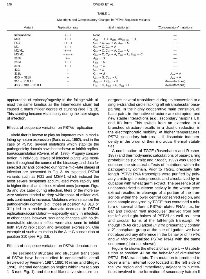
Intermediate /// None —Mild /// A310 r U, / U311a , AA120–121 r U —RG1 //// G46 r C, C47 r A, U317 r C —M1 /// G46 r C, C47 r A —M2/M1 /// G46 r C, C47 r A, C315 r U —M3/M1 //// G46 r C, C47 r A, C315 r U, U317 r C —310G // A310 r G —318A /// C318 r A —318G /// C318 r G —43G / U43 r G —311U / C311 r U U309 r A43G / 311U / U43 r G, C311 r U U309 r A310 0 311UU 0 A310 r U, C311 r U (Noninfectious)43G / 310 0 311UU 0 U43 r G, A310 r U, C311 r U (Noninfectious)
appearance of epinasty/rugosity in the foliage with al- dergoes several transitions during its conversion to asingle-stranded circle lacking all intramolecular base-most the same kinetics as the Intermediate strain but
caused a much milder degree of stunting (see Fig. 2f). pairing. In the highly cooperative main transition, allbase-pairs in the native structure are disrupted, andThis stunting became visible only during the later stages
of infection. new stable interactions (e.g., secondary hairpins I, II,and III) form. This switch from an extended to abranched structure results in a drastic reduction inEffects of sequence variation on PSTVd replicationthe electrophoretic mobility. At higher temperatures,
Viroid titer is known to play an important role in modu- PSTVd secondary hairpins I – III dissociate indepen-lating symptom expression (Sano et al., 1992), and in the dently in the order of their individual thermal stabili-case of PSTVd, several mutations which stabilize the ties.pathogenicity domain have been shown to inhibit replica- A combination of TGGE (Rosenbaum and Riesner,tion/accumulation (Owens et al., 1995). Progeny concen- 1987) and thermodynamic calculations of base-pairingtration in individual leaves of infected plants was moni- probabilities (Schmitz and Steger, 1992) was used totored throughout the course of the bioassay, and data for compare the structural effects of mutations within theselected variants collected during the mid–late stages of pathogenicity domain. Prior to TGGE, precisely full-infection are presented in Fig. 3. As expected, PSTVd length PSTVd RNA transcripts were purified by poly-variants such as RG1 and M3/M1 which induced the acrylamide gel electrophoresis and circularized by in-most severe symptoms accumulated more rapidly and
cubation with wheat germ extract. The presence of anto higher titers than the less virulent ones (compare Figs.
uncharacterized nuclease activity in the wheat germ3a and 3b). Later during infection, titers of the more se-
extract resulted in cleavage of a portion of the tran-vere variants tended to drop while those of milder vari-
scripts within the lower central conserved region, andants continued to increase. Mutations which stabilize the
each sample analyzed by TGGE thus contained a mix-pathogenicity domain (e.g., those at position 43, 318, or
ture of several different PSTVd-related RNAs, i.e., lin-311) led to a moderate–dramatic decrease in PSTVd
ear and circular ‘‘half molecules’’ derived from bothreplication/accumulation—especially early in infection.
the left and right halves of PSTVd as well as linearIn other cases, however, sequence changes with no de-and circular forms of the full-length transcript. Al-monstrable structural effect were also seen to inhibitthough RNAs circularized in vitro presumably containboth PSTVd replication and symptom expression. Onea 2*-phosphate group at the site of ligation, we haveexample of such a mutation is the A r G substitution atnot observed any difference in the behavior of in vitroposition 310 (see Fig. 3d).and in vivo circularized PSTVd RNAs with the samesequence (data not shown).Effects of sequence variation on PSTVd denaturation
Figure 4a shows the effects of a single U r G substi-tution at position 43 on the behavior of circularizedThe secondary structure and structural transitionsPSTVd RNA transcripts. This mutation is predicted toof PSTVd have been studied in considerable detailclose a small internal loop located at the left side of(reviewed by Riesner, 1987, 1990; Riesner and Steger,the VM region and immediately adjacent to nucleo-1990). Thermal denaturation begins within PM regions
1 – 3 (see Fig. 1), and the rod-like native structure un- tides involved in the formation of secondary hairpin II
AID VY 8043 / 6a1b$$$341 07-17-96 13:21:58 viral AP: Virology

149PSTVd PATHOGENICITY
FIG. 2. Symptoms of PSTVd infection in Rutgers tomato approximately 7 weeks after inoculation. Seedlings were inoculated with 0.04 ng/mlribozyme-derived PSTVd RNA, and all 12 variants were compared in the same bioassay. (a) Naturally occurring PSTVd isolates. (b–d) Variantsproduced by site-directed mutagenesis of PSTVd-Intermediate. M3/M1 is identical in sequence to the naturally occurring PSTVd isolate KF440-1(Schnolzer et al., 1985). (e–f) Quantitative assessment of stunting and epinasty/rugosity. All values are averages (six plants/treatment), and thelengths of individual segments within each bar in (e) represent the distance between successive nodes. The shaded portions of the bars shownin (f) denote leaves exhibiting epinasty/rugosity.
AID VY 8043 / 6a1b$$8043 07-17-96 13:21:58 viral AP: Virology

150 OWENS ET AL.
FIG. 3. Accumulation of PSTVd progeny in leaves of systemically infected tomatoes. Tissue samples were collected from individual leaves ofinfected plants at weekly intervals and pooled within treatments. Data from the mid–late stages of infection are presented.
(see Fig. 1). The effect of loop closure is clearly visible larized full-length transcripts by 3 – 47. Note that thisstabilizing effect increases by nearly twofold whenas an increase in the melting temperature (Tm) of the
main transition. Closing this internal loop should also half-molecules derived from the right half of PSTVdare compared.prevent disruption of base-pairing within PM 1 from
spreading leftward, thereby releasing nucleotides re- Figures 4b – 4d compare the denaturation profilesof PSTVd-Intermediate and PSTVd-Mild, two naturallyquired for the formation of secondary hairpin II. Forma-
tion of secondary hairpin II is responsible for the high occurring variants with very different biological prop-erties. Even though comparison of their lowest free-cooperativity of the PSTVd-Intermediate main transi-
tion. Indeed, the denaturation of variant 43G can be energy structures suggests that the VM region ofPSTVd-Mild should be significantly more stable thanseen to be less cooperative than that of PSTVd-Inter-
mediate — especially at higher temperatures (see that of PSTVd-Intermediate (see Fig. 1), we were un-able to distinguish between these two variants byarrow).
The structural effects of several other mutations TGGE. Pairwise comparisons with an internal stan-dard (compare Figs. 4c and 4d) showed their structuralwere also readily apparent from the behavior of the
respective full-length RNA transcripts. For example, properties to be very similar/identical, and Fig. 4bshows that TGGE analysis under the same conditionsFig. 4d compares the denaturation profile of PSTVd-
Intermediate with that of a highly stabilized mutant failed to resolve a mixture of the full-length RNAs. Thedenaturation profiles of half molecules derived fromcontaining a single A r G substitution at position 135
in the variable domain. In this case, the presence of the left half of PSTVd-Mild or -Intermediate were alsoindistinguishable (results not shown). In all, Tm valuesan additional G:C base-pair raises the Tm of the circu-
AID VY 8043 / 6a1b$$$341 07-17-96 13:21:58 viral AP: Virology

151PSTVd PATHOGENICITY
FIG. 4. Structural effects of sequence variation within the PSTVd pathogenicity domain. Samples containing approximately equal amounts of twocircularized RNA transcripts were analyzed by TGGE. The wedge at the bottom of each panel indicates the position of the 25–657 temperaturegradient, and the filled triangles denote the main transition of PSTVd-Intermediate. (a) PSTVd-Intermediate and 43G. Note the decreased cooperativityof the 43G transition, especially at higher temperatures (see arrow). (b) PSTVd-Intermediate and PSTVd-Mild. (c, d) Comparison of PSTVd-Mild (c)and PSTVd-Intermediate (d) with 135G, a highly stabilized mutant derived from PSTVd-Intermediate. As shown in (d), each sample contained sixPSTVd-related RNAs; in addition to the full-length linear (FL) and circular (FC) transcripts, cleavage of FL near position 273 by a nuclease activityin the wheat germ extract produced two half-molecules (LL and RL) that were ligated to form LC and RC. Note the increased thermal stability andcooperativity associated with transcript circularization.
AID VY 8043 / 6a1b$$8043
07-17-96 13:21:58 viral AP: Virology

152 OWENS ET AL.
for a total of 12 infectious and noninfectious PSTVdvariants were determined by TGGE.
Quantitative relationship between pathogenicitydomain structure and symptom expression
To determine how PSTVd structure might modulatesymptom expression, we sought to establish a quantita-tive relationship between these two properties. A thermo-dynamic approach to this question involved two steps,i.e., comparison of Tm values determined experimentallyby TGGE with calculated Tm values followed by compari-son of Tm values with disease severity. A second, morestructural approach compared the optimal as well assuboptimal secondary structures for the pathogenicitydomain of each variant. Schmitz and Steger (1992) havedescribed a computer algorithm which can predict theequilibrium distribution of optimal and suboptimal RNAsecondary structures at a variety of temperatures. Be-cause the temperature dependence of these equilibriumdistributions reflects denaturation behavior, it is possibleto calculate the denaturation curve of an RNA. The accu-racy of this methodology has been confirmed by compari-son of the experimental and calculated denaturationcurves for PSTVd-Intermediate (see Schmitz and Steger,1992), and the same approach was used to compare theTm values for the different transitions of our infectiousvariants. Results of these comparisons are presented inFigs. 5–8.
In Fig. 5a, the calculated Tm values for the main transi-FIG. 5. Denaturation temperatures (Tm) for the main transition oftion have been plotted against Tm values determined ex-
circularized PSTVd transcripts. Correlations between (a) Tm values de-perimentally by TGGE of circularized full-length PSTVd termined experimentally by TGGE and calculated Tm values as well astranscripts. The good correlation between experimental (b) calculated Tm values and heights of diseased plants are shown.
The continuous line is the least squares fit of all data points, and onlyand calculated values emphasizes the accuracy of thethe differences between the experimentally determined Tm values forpredicted structural distributions which provide the basisthe different variants and that of PSTVd-Intermediate are shown infor calculation of Tm values. The structures themselvesorder to avoid extrapolation of Tm values from the low ionic strength
as well as the extraordinary behavior of variants 311U conditions of TGGE (0.21 TBE–5 mM NaCl) to those used for thermody-and 310-311UU will be discussed below. In Fig. 5b, the namic calculations (1 M NaCl). For variants 318A, 43G, and 43G /
309A / 311U, the cooperative main transition is predicted to splitcalculated Tm values have been plotted against the aver-into two separate, poorly resolved transitions; the mean value of bothage height of the diseased plants, a value used as antransitions is shown. Solid circles, replication more rapid than PSTVd-inverse measure of pathogenicity. Calculated Tm valuesIntermediate; half-filled circles, replication similar to PSTVd-Intermedi-
were used for this plot in order to allow inclusion of ate; open circles, replication slower than PSTVd-Intermediate; aster-variants such as 309A / 311U for which experimentally isks, noninfectious mutants.determined Tm values were not available.
Although the relationship between thermal stabilityand pathogenicity appears to be reasonably linear, note probably fortuitous. Similar plots of Tm values versus per-
centage of leaves exhibiting epinasty or rugosity alsothat the difference between the Tm values for the least(RG1) and most stable (43G) variants is only 57 (approxi- failed to reveal a consistent correlation (results not
shown).mately). Note also that, in agreement with the results ofTGGE analysis, several pairs of variants (e.g., Intermedi- Use of the Tm value for the main transition to estimate
the stability of the pathogenicity domain is not straightfor-ate and Mild or RG1 and M1) are predicted to have nearlyidentical Tm values despite large differences in pathoge- ward because the main transition is influenced by a com-
petition between the rod-like native structure and a struc-nicity. Consequently, a simple linear relationship be-tween stability and pathogenicity seems unable to ac- ture containing secondary hairpins I to III. For example,
a C r G substitution at position 318 stabilizes the nativecount for the behavior of all variants, and the fact thatslowly replicating variants like 309A / 311U or 43G / structure by loop closure within PM 1; it also stabilizes
secondary hairpin II by addition of a G:C base-pair. Such309A / 311U appear to exhibit such a correlation is
AID VY 8043 / 6a1b$$$341 07-17-96 13:21:58 viral AP: Virology

153PSTVd PATHOGENICITY
four sequence variants (i.e., Mild, Intermediate, and M3/M1 plus PSTVd-Severe) provided the basis for an earlierproposal that PSTVd pathogenicity is inversely related tostructural stability (see Schnolzer et al., 1985). Obviously,this correlation does not hold for all variants examinedhere, because several variants possess VM regions thatare very similar in stability yet induce quite differentsymptoms (e.g., RG1 and M1 or Intermediate and 318G).The large deviations of 309A / 311U and 43G / 309A/ 311U from the interpolated line in Fig. 7 can easily beexplained by their slow replication rate.
Suboptimal structures for the pathogenicity domain
The thermodynamic distribution of optimal and subop-timal secondary structures at 257 was also calculated forthe pathogenicity domain of each variant. Selected re-sults from this analysis are presented as ‘‘dot plots’’ inFig. 8. Note that (i) these calculations were carried outon less-than-full-length RNAs in which the nucleotidescomprising the pathogenicity domain (positions 36–67and 292–325) are flanked by GC-rich ‘‘clamps’’ and (ii)dot sizes are proportional to the probability of base-pair-ing (i.e., only major species are shown). Figure 8a clearlyshows that for PSTVd-Intermediate the lowest free en-ergy structure (see Fig. 1) is the predominant species.From left to right, this structure contains a series of threeshort (i.e., 4-, 6-, and 4-bp) helices separated by internalloops containing, respectively, 2 and 4 nt. Several subop-timal structures are also visible; the most common alter-FIG. 6. Denaturation temperature (Tm) of circularized PSTVd ‘‘left-native conformation involves a rearrangement of nucleo-half’’ molecules. For a description of symbols and lines, see Fig. 5. (a)tides within the leftmost 4-bp helix (see gray arrows inCorrelation between Tm values determined experimentally by TGGE and
calculated Tm values. Only the differences between the experimentally Fig. 8b).determined Tm values for the different variants and that of PSTVd- Figures 8b and 8c illustrate how sequence variationIntermediate are shown in order to avoid extrapolation of Tm values
within the pathogenicity domain favors several additionalfrom the low ionic strength conditions of TGGE (0.21 TBE–5 mM NaCl)supoptimal structures. The secondary structure distribu-to those used for calculations (1 M NaCl). (b) Correlation betweentions for the Mild and Intermediate strains appear verycalculated Tm values and height of diseased plants.
similar. For variant RG1, note that the left side of the
difficulties were circumvented by comparing the behaviorof circularized half-molecules derived from the left sideof PSTVd (see Fig. 6). The denaturation behavior of thesetruncated molecules more directly reflects the stabilityof the pathogenicity domain than does that of the full-length circles. As shown in Fig. 6a, the correlation be-tween experimental and calculated Tm values for the cir-cularized half-molecules is quite good. Data presentedin Fig. 6b, however, show that the relationship betweenstability and pathogenicity is not monotonic. In additionto the discrepancies involving RG1 and M1 or Intermedi-ate and Mild noted above, three other variants that aremore stable than PSTVd-Mild (i.e., 43G, 318A, and 318G)produced symptoms that were more (rather than less)severe than those of the Mild strain. Once again, these
FIG. 7. Correlation between calculated denaturation temperature (Tm)discrepancies were not resolved by using alternativeof the VM region and height of diseased plants. Calculations were
measures of disease severity. performed as described by Schnolzer et al. (1985). The continuous lineFigure 7 compares the calculated stability of the VM is the least squares fit of all data points except 309A / 311U and 43G
/ 309A / 311U; the dotted line includes all data points.region with pathogenicity. A similar plot involving only
AID VY 8043 / 6a1b$$$341 07-17-96 13:21:58 viral AP: Virology

154 OWENS ET AL.
larger internal loop within PM 1, for both PSTVd-Interme-diate and the progeny of 311U (i.e., 309A/ 311U) containa 4-nt loop. The dark arrows in Figs. 8b and 8c point toan internal loop separating the middle and right helicesof PM 1; this loop may be either symmetrical (PSTVd-Intermediate) or asymmetrical (Mild and RG1).
Possible noncanonical interactions within thepathogenicity domain
Although hydrodynamic and ligand binding studiessuggest that PSTVd contains little significant tertiarystructure (reviewed by Riesner, 1990), ultraviolet cross-linking has identified a unique region of local tertiarystructure in the CCR similar to loop E in 5S ribosomalRNA (Branch et al., 1985; Wimberley et al., 1993). Thebehavior of certain PSTVd variants during TGGE as wellas comparisons of their suboptimal structures providescircumstantial evidence for the existence of additional‘‘noncanonical’’ interactions within the pathogenicity do-main. For example, TGGE analysis was unable to distin-guish the stabilizing effects of a single C r U substitutionat position 311 from those of a double AC r UU substitu-tion at positions 310–311 (see Figs. 5a and 6a). As shownin Fig. 8c, in 311U the 4-nt loop of PSTVd-Intermediateis reduced to a 2-nt loop with an A51rA310 opposition;formation of a second A:U base-pair in 310–311UU re-sults in the fusion of the two short helices, thereby creat-ing the longest uninterrupted helix in the molecule. Fail-ure of an A r G substitution at position 310 to affectTGGE behavior (see Figs. 5a and 6a) argues against theexistence of an interaction involving A51 and A310 inPSTVd-Intermediate, but reducing the loop size to 2 ntFIG. 8. Base-pair probability plots for the pathogenicity domains of
selected PSTVd variants. The structure distribution of each variant was via a C r U substitution at position 311 might facilitatecalculated from the optimal and 30 suboptimal secondary structures such an interaction. The resulting stabilization of theat 257. The probability of each possible base-pair (i:j) is proportional pathogenicity domain in variant 311U by pairing ofto the area of the dot at the corresponding position (i,j) of the matrix.
A51rA310 would explain the only deviation in the correla-Calculations were carried out for subsequences of the pathogenicitytion of experimental and calculated Tm values. If the pair-domain (positions 36–67 and 292–325) flanked by G:C-rich clamps.
These clamps simulate the stability of missing portions of the PSTVd ing does exist, this ArA interaction would appear to in-native structure (see Riesner, 1990). (a) Base-pair probability plot for the hibit PSTVd replication, for the presence of a C r Upathogenicity domain of PSTVd-Intermediate. Only the upper portion of substitution at position 311 consistently induced the ap-the matrix is shown; the lower portion is a mirror image of the upper.
pearance of a spontaneous U r A change at position(b, c) Corresponding portions near the diagonal (top left to bottom right)309. One effect of the spontaneous mutation might be tofrom probability plots of selected variants. Shaded arrows indicate
differences in the distributions of suboptimal structures; dark arrows disrupt the putative ArA interaction.point to the position of the loop separating the two helices within theVM region. Note the differences in loop type among the different PSTVd DISCUSSIONvariants.
As discussed by Diener (1987), the range of symptomsassociated with viroid infection is virtually identical to
pathogenicity domain may assume any of three different that produced by conventional plant viruses. In the appar-conformations. Figure 8c compares the structures acces- ent absence of viroid-encoded proteins to act as elicitorssible to the PSTVd variant with a C r U substitution at of the host response, the nature of the signal(s) mediatingposition 311 with those of its progeny which contain an viroid–host interaction is a matter of considerable inter-apparently compensatory U r A change at position 309. est. Variation in the severity of symptoms accompanyingComparison of these plots suggests that the additional PSTVd infection is correlated with sequence changes inmutation has little or no effect on the relative probabilities a portion of the VM region (Schnolzer et al., 1985), anof different secondary structures. Evidently, selective observation that has led to the assumption that pathoge-
nicity must be regulated by nucleotides within this region.pressures in vivo may strongly favor the presence of a
AID VY 8043 / 6a1b$$$341 07-17-96 13:21:58 viral AP: Virology

155PSTVd PATHOGENICITY
Although several mechanistic models have been pro- calculated Tm values seen in Fig. 6a provides assurancethat the thermodynamic calculations for not only the leftposed (cf. below), it is far from clear how this regulation
is accomplished on the molecular level. In the present half of PSTVd but also for the VM region are reasonablyaccurate. Figure 7b contains a plot of Tm values for thework, we have characterized 12 PSTVd sequence vari-
ants with respect to their pathogenicity and time course VM region vs plant height (the inverse of pathogenicity).Note that there is only a weak correlation between patho-of progeny accumulation as well as certain structural
features of their VM regions. All but one of the sequence genicity and thermodynamic instability, and several pairsof variants such as Intermediate and 318G or RG 1 and M1differences among these variants are located within the
pathogenicity domain. For some parameters, significant do not fit the correlation. Omitting the slowly replicatingvariants 309A/ 311U and 43G/ 309A/ 311U would notcorrelations with the pathogenicity could be obtained;
for others, correlations were either imperfect or missing improve the correlation. The strong correlation betweenexperimental and theoretical thermodynamic data seen inaltogether.Fig. 6a indicates that the problem lies not with inaccura-
Pathogenicity and the rate of PSTVd replication cies in the thermodynamic calculations but rather callsthe model itself into question.
To date, strain-dependent differences in the rate ofPSTVd replication or cell-to-cell spread have received
Sequences outside the pathogenicity domain ascomparatively little attention, and mechanistic modelssite(s) for pathogenic interactionfor PSTVd pathogenicity assume equal concentrations of
each variant. This is undoubtedly an oversimplification,By exchanging individual structural domains between
even for naturally occurring strains of PSTVd. For exam-CEVd and tomato apical stunt viroid, Sano et al. (1992)
ple, RG1 (and other severe variants) could be shown tohave shown that sequences within the TL , pathogenicity,
accumulate more rapidly than milder variants early inand variable/TR domains all contribute to viroid pathoge-
infection (see Fig. 3). For slowly replicating variants suchnicity and replication. The recent demonstration by
as 43G or 309A / 311U, the primary effect of the muta-Gruner et al. (1995) that mutations within the VM region
tion(s) may be on replication, and the effects on symptomof PSTVd can indeed affect the stability of the whole
expression are likely to be indirect. The seven variantsmolecule provides further evidence that sequences
which accumulate at relatively high rates (i.e., M1, M2/within the pathogenicity domain may not act alone to
M1, Intermediate, 310G, Mild, 318A, and 318G), producedregulate symptom expression. Their demonstration that
quite divergent disease symptoms, however.more severe strains of PSTVd are able to outcompetemilder (i.e., less easily denaturable) strains during mixedA denatured VM region as the site for pathogenicinfections suggested an alternative hypothesis for viroidinteractionpathogenicity, namely, that the VM region modulates theaccessibility of a second, probably nonoverlapping re-An apparent inverse correlation between symptom se-
verity and the calculated stability of the VM region initially gion in the molecule which acts as the primary site ofviroid–host interaction. Like the model proposed bysuggested that interaction with the host might require
the breakdown of normal secondary structure within the Schnolzer et al. (1985), this hypothesis minimizes thepossible role of the native structure in regulating viroid–pathogenicity domain (Schnolzer et al., 1985). Extensive
complementarity between nucleotides in that region and host interaction; instead, it focuses on metastable struc-ture(s) generated during synthesis and active in replica-a segment of tomato 7S RNA led to the suggestion that
viroids might inhibit the incorporation of 7S RNA into tion as well as pathogenesis. A more stable rod-like na-tive structure would depopulate the active (but metasta-signal recognition particles (Haas et al., 1988). More re-
cently, however, the same group determined the se- ble) structure more rapidly, thus explaining thecorrelation between pathogenicity and instability of thequence of 7S RNA from several different tomato cultivars
and reported that symptom severity was not correlated native structure.When Gruner et al. (1995) argued that PSTVd replica-with the degree of complementarity (Riedel et al., 1995).
Whatever the nature of the host molecule interacting with tion and pathogenicity are governed by the same orclosely related metastable structures within a replicationthe VM region in the dissociated state, we wished to test
the proposed inverse correlation with a larger series of intermediate, they predicted a faster conversion of themetastable state into the rod-like native structure forPSTVd variants. In fact, two types of correlations could
be tested. milder strains. In the absence of kinetic data, the relativestability of the native structure was used as a roughFrom a purely physical perspective, it was important to
experimentally confirm the reliability of the thermodynamic approximation of kinetic behavior. Our more completebiological data, together with additional thermodynamicpredictions. This could not be done with the isolated VM
region, but half-molecules containing the entire pathoge- experimentation and theoretical calculations, do not sup-port such a mechanistic model for PSTVd–host interac-nicity domain were accessible to TGGE analysis. The
good correlation between experimentally determined and tion. Despite a surprisingly good correlation between the
AID VY 8043 / 6a1b$$$341 07-17-96 13:21:58 viral AP: Virology

156 OWENS ET AL.
FIG. 9. Schematic representations of the PSTVd pathogenicity domain (positions 36–67 and 292–325) flanked by G:C-rich clamps. A standardA-type RNA helix is assumed, and the helix axis (indicated by dotted lines) is bent to compensate for missing nucleotides in asymmetrical loops.As described by Riesner (1990), the G:C-rich clamps simulate the stability conferred by the missing portions of the native structure. (a) Thermodynami-cally optimal secondary structures of selected PSTVd variants. (b) Two-dimensional representations of the corresponding three-dimensional struc-tures. For each variant, the vertical arrows denote corresponding positions in the two- and three-dimensional structures.
experimentally determined and calculated Tm values (cf. induced, dsRNA-activated mammalian protein kinaseFig. 5a), PSTVd pathogenicity (as measured by plant known to interact with small RNA molecules encoded byheight) was only weakly correlated with instability of the adenovirus or human immunodeficiency virus (see alsowhole molecule. Furthermore, certain pairs of variants Hiddinga et al., 1988).(e.g., Intermediate and 318G or RG1 and M1) did not Like PSTVd, adenovirus VA RNAI is a highly structured,obey the proposed inverse relationship. single-stranded RNA, and its approximately 160 nt are
arranged in two base-paired stems connected by a com-Three-dimensional structure of the pathogenicity plex stem-loop structure known as the ‘‘central domain.’’domain Although the relative contributions of the apical stem and
central domain to the ability of VA RNA to bind to theOther models attempting to explain PSTVd pathogenic-kinase and inhibit its activation are controversial, VA RNAity assume specific interactions between host compo-function both in vivo and in vitro appears to be criticallynents (most probably proteins) and certain elements ofdependent upon subtle alterations in the secondary orthe native structure. Several complexes containingtertiary structure of the central domain (e.g., Clarke et al.,PSTVd and plant nuclear proteins (MrÅ 33–92 kDa) have1994; Ghadge et al., 1994; Schmedt et al., 1995; Clarkebeen isolated from infected tomato leaf tissue (Klaff etand Matthews, 1995). The strength of the primary interac-al., 1989). These interactions appear to be primarily elec-tion between PSTVd and a host cell component mighttrostatic in nature, but no information is available aboutdepend on similar, subtle variations in the three-dimen-the site(s) of interaction. More recently, Diener et al.sional geometry of the VM region.(1993) have suggested that the triggering event in PSTVd
We compared the secondary structures of our PSTVdpathogenesis may be its interaction with a host cell pro-variants in an effort to derive general rules for the three-tein kinase. Preliminary evidence suggests that PSTVd
infection may activate a plant homolog of the interferon- dimensional structure of the VM region. Sequence differ-
AID VY 8043 / 6a1b$$$341 07-17-96 13:21:58 viral AP: Virology

157PSTVd PATHOGENICITY
ences are restricted to nucleotides 42–52 and 319–309, ciently stable to exist in their own right) often serve asrecognition sites for RNA-binding proteins (Puglisi et al.,and except for variants 43G, 318A, and 318G where the
left loop has been replaced by a base-pair, these nucleo- 1992; Battiste et al., 1994), and phylogenetic evidencepoints to an important role for these and other unusualtides are organized into a central 4- to 7-bp double-heli-
cal segment flanked by internal loops. The size and the structural elements in maintaining the overall structureof rRNA (reviewed by Gutell et al., 1994). In a similarsymmetry of these flanking loops determine the relative
orientation of the remaining portions of the rod-like native fashion, the strength of the interaction between PSTVdand a host component appears to depend on subtle vari-structure with respect to the central helix. Figure 9a con-
tains two-dimensional representations of the thermody- ations in the three-dimensional structure of the VM re-gion. Further consideration of the interactions mediatingnamically most stable structure for PSTVd-Mild, -Interme-
diate, -Severe (see Schnolzer et al., 1985), and RG1. In PSTVd pathogenicity awaits direct evidence for the inter-action of host proteins with nucleotides within the patho-comparing these structures, it is important to note that
the central helix forms about half a helical turn and, there- genicity domain.fore, the portion of the structure containing the left termi-nal loop is rotated ca. 1807 with respect to that containing ACKNOWLEDGMENTSthe central conserved region and right terminal loop. This
The first two authors contributed equally to these studies. Y.H. wasis shown schematically in Fig. 9b.supported by a grant from the USDA/NRI Competitive Grants Program
Although the absolute angles associated with different (Grant 91-373303-6649). Additional support was provided by grantsasymetrical loops are unknown, certain characteristic dif- from Deutsche Forschunggemeinschaft and Fonds der Chemischen
Industrie as well as an agreement between the USDA and the Bundes-ferences between the variants are evident. Relative tominister fur Landwirtschaft. We thank S. M. Thompson for technicalthe central helix, the right ‘‘arm’’ of all variants whoseassistance and J. N. Culver, A. Mattoo, W. O. Dawson, and P. A.symptoms are more severe than PSTVd-Intermediate canFeldstein for helpful discussions and critical review of the manuscript.
be seen to bend ‘‘downward.’’ For RG1 (the most severevariant) both the left and right arms are bent downward.
REFERENCESThe presence of symmetrical loops in the VM region ofPSTVd-Intermediate results in a coaxial arrangement of Antao, V. P., and Tinoco, I., Jr. (1992). Thermodynamic parameters for
loop formation in RNA and DNA hairpin tetraloops. Nucleic Acidshelical segments, and the right arm of PSTVd-Mild canRes. 20, 819–824.be seen to bend ‘‘upward.’’ The same tendencies were
Antao, V. P., Lai, S. Y., and Tinoco, I., Jr. (1991). A thermodynamic studyalso observed among the other variants included in ourof unusually stable RNA and DNA hairpins. Nucleic Acids Res. 19,
study as well as the six additional variants described by 5901–5905.Herold et al. (1992). This would suggest that the structure Battiste, J. L., Tan, R., Frankel, A. D., and Williamson, J. R. (1994). Binding
of an HIV Rev peptide to Rev responsive element RNA induces forma-of RG1 and other severe variants is optimal for patho-tion of purine-purine base pairs. Biochemistry 33, 2741–2747.genic interaction, possibly involving the central helix and
Baumstark, T., and Riesner, D. (1995). Only one of four possible second-both arms, and that such interaction(s) is either greatlyary structures of the central conserved region of potato spindle tuber
inhibited or completely suppressed for PSTVd-Mild. viroid is a substrate for processing in a potato nuclear extract. Nu-Of the three properties examined for their correlation cleic Acids Res. 23, 4246–4254.
Branch, A. D., Benenfeld, B. J., and Robertson, H. D. (1985). Ultravioletwith pathogenicity (i.e., instability of the VM region, insta-light-induced crosslinking reveals a unique region of local tertiarybility of the whole molecule, and three-dimensional struc-structure in potato spindle tuber viroid and HeLa 5S RNA. Proc. Natl.ture of the VM region), our results clearly point to theAcad. Sci. USA 82, 6590–6594.
three-dimensional structure of the VM region as the best Clarke, P. A., and Matthews, M. B. (1995). Interactions between thepredictor of PSTVd pathogenicity. In certain situations, double-stranded RNA binding motif and RNA: definition of the binding
site for the interferon-induced protein kinase DAI on adenovirus VAhowever, thermodynamic features of PSTVd structureRNA. RNA 1, 7–20.may also be mechanistically relevant. For example, the
Clarke, P. A., Pe’ery, T., Ma, Y., and Matthews, M. B. (1994). Structuraldecreased stability of the more pathogenic strains couldfeatures of adenovirus 2 virus-associated RNA required for binding
facilitate binding of a ‘‘bent’’ molecule to the interacting to the protein kinase DAI. Nucleic Acids Res. 22, 4364–4374.surface of a host protein. In addition, optimum replication Cress, D. E., Kiefer, M. C., and Owens, R. A. (1983). Construction of
infectious potato spindle tuber viroid cDNA clones. Nucleic Acidsof PSTVd may also require a certain degree of instabilityRes. 11, 6821–6835.in the loop immediately to the right of the central helix.
Diener, T. O. (Ed.) (1987). The Viroids. Plenum, New York.In the most slowly replicating of our variants (i.e., 311U),Diener, T. O., Hammond, R. W., Black, T., and Katze, M. G. (1993).
purine:purine interactions similar to those within the E- Mechanism of viroid pathogenesis: Differential activation of the inter-loop of 5S RNA (Wimberly et al., 1993) and involving A51 feron-induced, double-stranded RNA-activated, Mr 68,000 protein ki-
nase by viroid strains of varying pathogenicity. Biochimie 75, 533–and A310 may stabilize the structure of the VM region and538.inhibit the rate of PSTVd replication. The spontaneous
Ghadge, G., Malhotra, P., Furtado, M. R., Dhar, R., and Thimmapaya,U r A change which consistently appeared at positionB. (1994). In vitro analysis of virus-associated RNA I (VAI RNA): Inhibi-
309 may relieve this inhibition by destabilizing the neigh- tion of the double-stranded RNA-activated protein kinase PKR by VAIboring 4-bp helix. RNA mutants correlates with the in vivo phenotype and the structural
integrity of the central domain. J. Virol. 68, 4137–4151.Lone pairs (i.e., isolated interactions that are not suffi-
AID VY 8043 / 6a1b$$$341 07-17-96 13:21:58 viral AP: Virology

158 OWENS ET AL.
Groebe, D. R., and Uhlenbeck, O. C. (1988). Characterization of RNA mutations which stabilize the pathogenicity domain. Virology 208,554–564.hairpin loop stability. Nucleic Acids Res. 16, 11725–11735.
Palukaitis, P., Cotts, S., and Zaitlin, M. (1985). Detection and identifica-Gross, H. J., Domdey, H., Lossow, C., Jank, P., Raba, M., Alberty, H., andtion of viroids and viral nucleic acids by ‘‘dot-blot’’ hybridization. ActaSanger, H. L. (1978). Nucleotide sequence and secondary structure ofHorticult. 164, 109–118.potato spindle tuber viroid. Nature 273, 203–208.
Puglisi, J. D., Tan, R., Calnan, B. J., Frankel, A. D., and Williamson,Gruner, R., Fels, A., Qu, F., Zimmat, R., Steger, G., and Riesner, D.J. R. (1992). Conformation of the TAR RNA-arginine complex by NMR(1995). Interdependence of pathogenicity and replicability with potatospectroscopy. Science 257, 76–80.spindle tuber viroid. Virology 209, 60–69.
Riedel, L., Putz, A., Hauser, M. T., Luckinger, R., Wassenegger, M., andGutell, R. R., Larsen, N., and Woese, C. R. (1994). Lessons from anSanger, H. L. (1995). Characterization of the signal recognition parti-evolving RNA: 16S and 23S rRNA structures from a comparativecle (SRP) RNA population of tomato (Lycopersicon esculentum). Plantperspective. Microbiol. Rev. 58, 10–26.Mol. Biol. 27, 669–680.
Hammond, R. W. (1992). Analysis of the virulence modulating region Riesner, D. (1987). Structure formation. In ‘‘The Viroids’’ (T. O. Diener,of potato spindle tuber viroid (PSTVd) by site-directed mutagenesis. Ed.), pp. 63–98. Plenum, New York.Virology 187, 654–662. Riesner, D. (1990). Structure of viroids and their replication intermedi-
Haas, B., Klanner, A., Ramm, K., and Sanger, H. L. (1988). The 7S RNA ates. Are thermodynamic domains also functional domains? Semin.from tomato leaf tissue resembles a signal recognition particle RNA Virol. 1, 83–99.and exhibits a remarkable sequence complementarity to viroids. Riesner, D., and Steger, G. (1990). Viroid and viroid-like RNA. In ‘‘Lan-EMBO J. 7, 4063–4074. dolt-Bornstein—Group VII Biophysics’’ (W. Saenger, Ed.), Vol. 1—
Hecker, R. (1989). Thesis, Heinrich-Heine Universitat, Dusseldorf. Nucleic Acids, Subvol. d—Physical Data, II—Theoretical Investiga-Hecker, R., Wang, Z., Steger, G., and Riesener, D. (1988). Analysis of tions, pp. 194–243. Springer-Verlag, Berlin.
Riesner, D., Klaff, P., Steger, G., and Hecker, R. (1987). Viroids: Subcellu-RNA structure by temperature-gradient gel electrophoresis: Viroidlar location and structure of replication intermediates. In ‘‘Endocytobi-replication and processing. Gene 72, 59–74.ology III’’ (J. J. Lee and J. F. Fredrick, Eds.), Vol. 503, pp. 212–237.Herold, T., Haas, B., Singh, R. P., Boucher, A., and Sanger, H. L. (1992).N.Y. Acad. Sci., New York.Sequence analysis of five new field isolates demonstrates that the
Rosenbaum, V., and Riesner, D. (1987). Temperature gradient gel elec-chain length of potato spindle tuber viroid (PSTVd) is not strictlytroporesis: Thermodynamic analysis of nucleic acids and proteins inconserved but as variable as in other viroids. Plant Mol. Biol. 19,purified form and in celllular extracts. Biophys. Chem. 26, 235–246.329–333.
Sambrook, J., Fritsch, E. F., and Maniatis, T. (1989). ‘‘Molecular Clon-Hiddinga, H. J., Crum, C. J., Hu, J., and Roth, D. A. (1988). Viroid-induceding—A Laboratory Manual.’’ Cold Spring Harbor Laboratory Press,phosphorylation of a host protein related to a dsRNA-dependentCold Spring Harbor, NY.
protein kinase. Science 241, 451–453.Sano, T., Candresse, T., Hammond, R. W., Diener, T. O., and Owens,
Keese, P., and Symons, R. H. (1985). Domains in viroids: Evidence of R. A. (1992). Identification of multiple structural domains regulatingintermolecular RNA rearrangement and their contribution to viroid viroid pathogenicity. Proc. Natl. Acad. Sci. USA 89, 10104–10108.evolution. Proc. Natl. Acad. Sci. USA 82, 4582–4586. Schmedt, C., Green, S. R., Manche, L., Taylor, D. R., Ma, Y., and Mat-
Klaff, P., Gruner, R., Hecker, R., Sattler, A., Theissen, G., and Riesner, thews, M. B. (1995). Functional characterization of the RNA-bindingD. (1989). Reconstituted and cellular viroid-protein complexes. J. Gen. domain and motif of the double-stranded RNA-dependent proteinVirol. 70, 2257–2270. kinase DAI (PKR). J. Mol. Biol. 249, 29–44.
Loss, P. (1989). Thesis, Heinrich-Heine-Universitat, Dusseldorf. Schmitz, M., and Steger, G. (1992). Base-pair probability profiles of RNAsecondary structures. Comp. Appl. Biosci. 8, 389–399.Loss, P., Schmitz, M., Steger, G., and Riesner, D. (1991). Formation of
Schnolzer, M., Haas, B., Ramm, K., Hofmann, H., and Sanger, H. L.a thermodynamically metastable structure containing hairpin II is(1985). Correlation between structure and pathogenicity of potatocritical for infectivity of potato spindle tuber viroid RNA. EMBO J. 10,spindle tuber viroid (PSTV). EMBO J. 4, 2181–2190.719–727.
Semancik, J. S. (Ed.) (1987). ‘‘Viroids and Viroid-Like Pathogens.’’ CRCMcCaskill, J. S. M. (1990). The equilibrium partition function and basePress, Boca Raton.pair binding probabilities for RNA secondary structure. Biopolymers
Steger, G., Hofmann, H., Fortsch, J., Gross, H. J., Randles, J. W., Sanger,29, 1105–1119.H. L., and Riesner, D. (1984). Conformational transitions in viroidsOwens, R. A., and Diener, T. O. (1984). Spot hybridization for detectionand virusoids: Comparison of results from energy minimization algo-of viroids and viruses. In ‘‘Methods in Virology’’ (K. Maramorosch andrithm and from experimental data. J. Biomol. Struct. Dyn. 2, 543–571.
H. Koprowski, Eds.), Vol. VII, pp. 173–187. Academic Press, NewVisvader, J. A., and Symons, R. H. (1985). Eleven new sequence variants
York.of citrus exocortis viroid and correlation of sequence with pathoge-
Owens, R. A., Hammond, R. W., Gardner, R. C., Kiefer, M. C., Thompson, nicity. Nucleic Acids Res. 13, 2907–2920.S. M., and Cress, D. E. (1986). Site-specific mutagenesis of potato Wimberly, B., Varani, G., and Tinoco, I., Jr. (1993). The conformation ofspindle tuber viroid cDNA. Plant Mol. Biol. 6, 179–192. loop E of eukaryotic 5S ribosomal RNA. Biochemistry 32, 1078–1087.
Owens, R. A., Candresse, T., and Diener, T. O. (1990). Construction of Zimmat, R., Gruner, R., Hecker, R., Steger, G., and Riesner, D. (1990).novel viroid chimeras containing portions of tomato apical stunt and Analysis of mutations in viroid RNA by non-denaturing and tempera-citrus exocortis viroids. Virology 175, 238–246. ture gradient gel electrophoresis. In ‘‘Proceedings of the 6th Conver-
Owens, R. A., Chen, W., Hu, Y., and Hsu, Y-H. (1995). Suppression of sation in Biomolecular Stereodynamics’’ (R. Sarma and M. Sarma,Eds.), Vol. 3, pp. 339–357. Adenine Press, Schenectady, NY.potato spindle tuber viroid replication and symptom expression by
AID VY 8043 / 6a1b$$$341 07-17-96 13:21:58 viral AP: Virology