gallstone disease - nice guideline template
TRANSCRIPT
Internal Clinical Guidelines Team
Document information (i.e. version number etc)
Gallstone disease Diagnosis and management of cholelithiasis, cholecystitis and choledocholithiasis
Clinical Guideline <…>
Methods, evidence and recommendations
2014
Draft for Consultation
National Institute for Health and Care Excellence


Disclaimer Healthcare professionals are expected to take NICE clinical guidelines fully into account
when exercising their clinical judgement. However, the guidance does not override the responsibility of healthcare professionals to make decisions appropriate to the circumstances of each patient, in consultation with the patient and/or their guardian or carer.
Copyright National Institute for Health and Care Excellence, Date xxxx. All rights reserved. This
material may be freely reproduced for educational and not-for-profit purposes within the NHS. No reproduction by or for commercial organisations is allowed without the express
written permission of the National Institute for Health and Care Excellence.
Gallstone disease Contents
Internal Clinical Guidelines, 2014
4
Contents 1 Overview .................................................................................................................................. 7
Patient-centred care ................................................................................................................ 7
2 Summary Section .................................................................................................................. 9
Guideline development group (GDG) members.................................................................... 9
Internal clinical guidelines team.............................................................................................. 9
Centre for Clinical Practice commissioning team................................................................ 10
Key priorities for implementation .......................................................................................... 11
Algorithm................................................................................................................................. 12
List of all recommendations .................................................................................................. 13
Research recommendations ................................................................................................. 14
Strength of recommendations............................................................................................... 15
3 Methods ................................................................................................................................. 16
3.1 Additional methods used in this guideline .................................................................. 16
3.1.1 Methods for combining diagnostic evidence: ................................................ 16
3.1.2 Methods for combining direct and indirect evidence (network meta-analysis)............................................................................................................ 16
3.1.3 References ....................................................................................................... 19
4 Evidence Review and Recommendations....................................................................... 20
4.1 Signs, symptoms and risk factors for gallstone disease ........................................... 20
4.1.1 Review Question 1........................................................................................... 20
4.1.2 Evidence Review ............................................................................................. 20
4.1.3 Health economic evidence .............................................................................. 21
4.1.4 Evidence Statements ...................................................................................... 21
4.1.5 Evidence to Recommendations ...................................................................... 21
4.1.6 Recommendations ........................................................................................... 22
4.1.7 Research recommendations ........................................................................... 23
4.1.8 References ....................................................................................................... 23
4.2 Diagnosing gallstone disease ..................................................................................... 29
4.2.1 Review Question 2........................................................................................... 29
4.2.2 Evidence Review ............................................................................................. 29
4.2.3 Health economic evidence .............................................................................. 34
4.2.4 Evidence Statements ...................................................................................... 37
4.2.5 Evidence to Recommendations ...................................................................... 37
4.2.6 Recommendations ........................................................................................... 42
4.2.7 Research recommendations ........................................................................... 42
4.2.8 References ....................................................................................................... 42
4.3 Asymptomatic gallbladder stones ............................................................................... 45
4.3.1 Review Question 3........................................................................................... 45
Gallstone disease Contents
Internal Clinical Guidelines, 2014
5
4.3.2 Evidence Review ............................................................................................. 45
4.3.3 Health economic evidence .............................................................................. 46
4.3.4 Evidence Statements ...................................................................................... 46
4.3.5 Evidence to Recommendations ...................................................................... 47
4.3.6 Recommendations & Research Recommendations ..................................... 48
4.3.7 References ....................................................................................................... 48
4.4 Managing asymptomatic gallbladder stones ............................................................. 49
4.4.1 Review Question 4a ........................................................................................ 49
4.4.2 Evidence Review ............................................................................................. 49
4.4.3 Health economic evidence .............................................................................. 49
4.4.4 Evidence Statements ...................................................................................... 49
4.4.5 Evidence to Recommendations ...................................................................... 49
4.4.6 Recommendations ........................................................................................... 51
4.4.7 Research Recommendations ......................................................................... 51
4.4.8 References ....................................................................................................... 52
4.5 Managing symptomatic gallbladder stones................................................................ 53
4.5.1 Review Question 4b ........................................................................................ 53
4.5.2 Evidence Review ............................................................................................. 53
4.5.3 Health economic evidence .............................................................................. 56
4.5.4 Evidence Statements ...................................................................................... 59
4.5.5 Evidence to Recommendations ...................................................................... 59
4.5.6 Recommendations ........................................................................................... 63
4.5.7 Research recommendations ........................................................................... 64
4.5.8 References ....................................................................................................... 64
4.6 Managing common bile duct stones ........................................................................... 66
4.6.1 Review Question 4c......................................................................................... 66
4.6.2 Evidence Review ............................................................................................. 66
4.6.3 Health economic evidence .............................................................................. 74
4.6.4 Evidence Statements ...................................................................................... 77
4.6.5 Evidence to Recommendations ...................................................................... 78
4.6.6 Recommendations ........................................................................................... 82
4.6.7 Research recommendations ........................................................................... 83
4.6.8 References ....................................................................................................... 83
4.7 Timing of laparoscopic cholecystectomy ................................................................... 86
4.7.1 Review Question 5........................................................................................... 86
4.7.2 Evidence Review ............................................................................................. 86
4.7.3 Health economic evidence .............................................................................. 90
4.7.4 Evidence Statements ...................................................................................... 92
4.7.5 Evidence to Recommendations ...................................................................... 93
4.7.6 Recommendations ........................................................................................... 95
Gallstone disease Contents
Internal Clinical Guidelines, 2014
6
4.7.7 Research recommendations ........................................................................... 95
4.7.8 References ....................................................................................................... 95
4.8 Information for patients and their carers ................................................................... 96
4.8.1 Review Question 6........................................................................................... 96
4.8.2 Evidence Review ............................................................................................. 96
4.8.3 Health economic evidence .............................................................................. 97
4.8.4 Evidence Statements ...................................................................................... 97
4.8.5 Evidence to Recommendations ...................................................................... 97
4.8.6 Recommendations ........................................................................................... 99
4.8.7 Research recommendations ........................................................................... 99
4.8.8 References ....................................................................................................... 99
5 Glossary & Abbreviations ................................................................................................ 101
Overview
Gallstone disease
Internal Clinical Guidelines, 2014
7
1 Overview 1
Gallstone disease is the term used in this guideline to refer to the presence of stones in the 2 gallbladder or common bile duct and the symptoms and complications they cause. The 3
following aspects of gallstone disease are included in this guideline (full definitions of these 4 terms are provided in the glossary): 5
Asymptomatic gallbladder stones 6
Symptomatic gallbladder stones, including biliary colic, acute cholecystitis, Mirrizi 7
syndrome, and xanthogranulatomus cholecystitis. 8
Common bile duct stones, including biliary colic, cholangitis, obstructive jaundice and 9
gallstone pancreatitis. 10
Other complications of gallstones (such as gastric outlet obstruction, or gallstone ileus) and 11 other conditions related to the gallbladder (such as gallbladder cancer, or biliary dyskinesia) 12
are not included in this guideline. 13
Most people with gallstone disease have asymptomatic gallbladder stones, meaning the 14 stones are confined to the gallbladder and they do not have any symptoms. The disease is 15
identified coincidentally as a result of investigations for other conditions. People with 16 asymptomatic gallbladder stones may never go on to develop symptoms or complications, 17 but there is variation within the NHS in how people are managed once asymptomatic 18
gallbladder stones have been diagnosed. Some patients are offered treatments to prevent 19 symptoms and complications developing, and others are offered a watch and wait approach 20 so that active treatment only begins once the stones begin to cause symptoms. 21
The symptoms of gallstone disease range from mild, non-specific symptoms that can be 22 difficult to diagnose, to severe pain and/or complications which are often easily recognised 23 as gallstone disease by health professionals. People with mild, non-specific symptoms of 24
gallstone disease may attribute their symptoms to other conditions, or may be misdiagnosed 25 and undergo unnecessary investigations and treatment. This has a detrimental effect on 26 quality of life and has an impact on the use of NHS resources. Thus, there is a need to 27
identify whether there are any specific signs, symptoms or risk factors for gallstone disease 28 and to identify the best method for diagnosing the condition so that patients can be managed 29 appropriately. 30
There is uncertainty about the best way of treating gallstone disease. There are a range of 31 endoscopic, surgical and medical treatments available, but it is unclear which treatments are 32 the most appropriate for which patients. There is also uncertainty about the timing of 33 cholecystectomy, and whether it should take place during the acute presentation of the 34
disease, or if it should be delayed until after the acute symptoms have subsided. 35
This guideline addresses these uncertainties and provides recommendations on how to 36 identify, diagnose and manage gallstone disease. 37
Patient-centred care 38
This guideline offers best practice advice on the care of adults with gallstone disease. 39
Patients and healthcare professionals have rights and responsibilities as set out in the NHS 40 Constitution for England – all NICE guidance is written to reflect these. Treatment and care 41
should take into account individual needs and preferences. Patients should have the 42 opportunity to make informed decisions about their care and treatment, in partnership with 43 their healthcare professionals. Healthcare professionals should follow the Department of 44
Health’s advice on consent. If someone does not have capacity to make decisions, 45 healthcare professionals should follow the code of practice that accompanies the Mental 46
Capacity Act and the supplementary code of practice on deprivation of liberty safeguards. 47

Overview
Gallstone disease
Internal Clinical Guidelines, 2014
8
NICE has produced guidance on the components of good patient experience in adult NHS 1 services. All healthcare professionals should follow the recommendations in Patient 2 experience in adult NHS services.3
Summary section
Gallstone disease
Internal Clinical Guidelines, 2014
9
2 Summary Section 1
Guideline development group (GDG) members 2
Name Role
Gary McVeigh (GDG Chair) Professor of Cardiovascular Medicine, Queen’s University Belfast/ Consultant Physician, Belfast
Health and Social Care Trust
Elaine Dobinson Evans Patient/ carer member
Simon Dwerryhouse Consultant Upper Gastrointestinal and Bariatric Surgeon, Gloucestershire Royal Hospital
Rafik Filobbos (joined Nov 2013) Consultant Radiologist with specialist interest in Gastrointestinal/ Hepatobiliary imaging, North
Manchester General Hospital
Imran Jawaid Principal General Practitioner, Hadlow, Tonbridge
Angela Madden (co-opted expert) Professional Lead for Nutrition and Dietetics, University of Hertfordshire
Peter Morgan Consultant Anaesthetist, St James’s University Hospital
Gerri Mortimore Lead Hepatology Clinical Nurse Specialist, Derby Hospitals NHS
Foundation Trust
Kofi Oppong Consultant Gastroenterologist, Newcastle Hospitals NHS Trust
Charles Rendell Patient/ carer member
Richard Sturgess Consultant Hepatologist and Physician, University Hospital Aintree
Giles Toogood Consultant Hepatobiliary and Liver Transplant Surgeon, St James’ University Hospital
Luke Williams Consultant Gastrointestinal Radiologist, Salford Royal NHS Foundation Trust
Internal clinical guidelines team 3
Name Role
Emma Banks (until April 2013) Project Manager
Susan Ellerby Consultant Clinical Adviser
Nicole Elliott Associate Director
Michael Heath Programme Manager
Hugh McGuire (from March 2014) Technical Adviser
Stephanie Mills (from April 2013) Project Manager
Gabriel Rogers Technical Adviser (Health Economics)
Toni Tan (until March 2014) Technical Adviser
Steven Ward Technical Analyst (Health Economics)
Sheryl Warttig (until May 2014) Technical Analyst
4
Summary section
Gallstone disease
Internal Clinical Guidelines, 2014
10
Centre for Clinical Practice commissioning team 1
Name Role
Mark Baker Clinical Adviser
Joy Carvill Guideline Coordinator
Ben Doak Guideline Commissioning Manager
Jaimella Epsley (until Feb 2014) Senior Medical Editor
James Hall (from Feb 2014) Senior Medical Editor
Bhash Naidoo Senior Technical Adviser (Health Economics)
Judith Thornton Technical Lead
Sarah Willett Guideline Lead
2
3
Summary section
Gallstone disease
Internal Clinical Guidelines, 2014
11
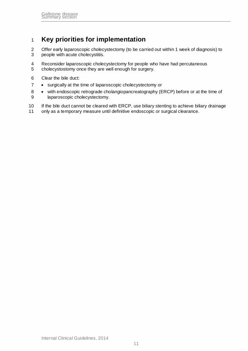
Key priorities for implementation 1
Offer early laparoscopic cholecystectomy (to be carried out within 1 week of diagnosis) to 2 people with acute cholecystitis. 3
Reconsider laparoscopic cholecystectomy for people who have had percutaneous 4 cholecystostomy once they are well enough for surgery. 5
Clear the bile duct: 6
surgically at the time of laparoscopic cholecystectomy or 7
with endoscopic retrograde cholangiopancreatography (ERCP) before or at the time of 8
laparoscopic cholecystectomy. 9
If the bile duct cannot be cleared with ERCP, use biliary stenting to achieve biliary drainage 10
only as a temporary measure until definitive endoscopic or surgical clearance. 11
Gallstone disease
Internal Clinical Guidelines, 2014
12
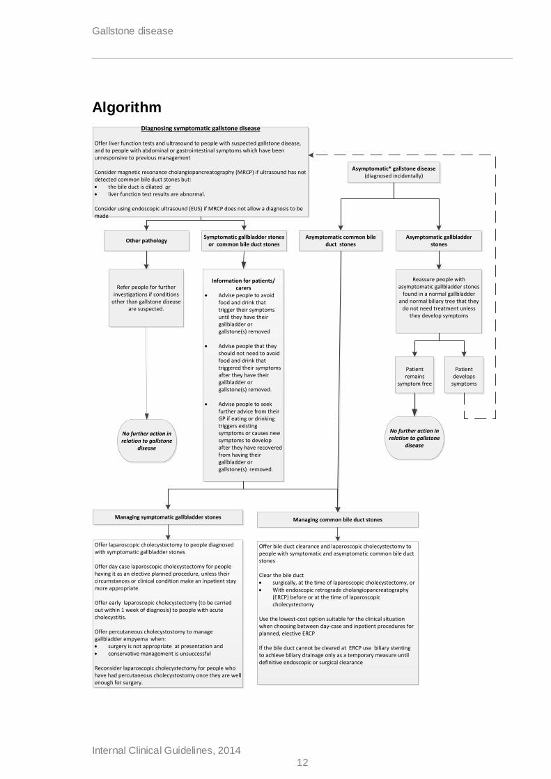
Algorithm
Reassure people with asymptomatic gallbladder stones
found in a normal gallbladder and normal biliary tree that they
do not need treatment unless they develop symptoms
Offer laparoscopic cholecystectomy to people diagnosed with symptomatic gallbladder stones
Offer day case laparoscopic cholecystectomy for people having it as an elective planned procedure, unless their circumstances or clinical condition make an inpatient stay more appropriate.
Offer early laparoscopic cholecystectomy (to be carried out within 1 week of diagnosis) to people with acute cholecystitis.
Offer percutaneous cholecystostomy to manage gallbladder empyema when: surgery is not appropriate at presentation and conservative management is unsuccessful
Reconsider laparoscopic cholecystectomy for people who have had percutaneous cholecystostomy once they are well enough for surgery.
Offer bile duct clearance and laparoscopic cholecystectomy to people with symptomatic and asymptomatic common bile duct stones
Clear the bile duct surgically, at the time of laparoscopic cholecystectomy, or With endoscopic retrograde cholangiopancreatography
(ERCP) before or at the time of laparoscopic cholecystectomy
Use the lowest-cost option suitable for the clinical situation when choosing between day-case and inpatient procedures for planned, elective ERCP
If the bile duct cannot be cleared at ERCP use biliary stenting to achieve biliary drainage only as a temporary measure until definitive endoscopic or surgical clearance
Refer people for further investigations if conditions
other than gallstone disease are suspected.
Information for patients/carers
Advise people to avoid food and drink that trigger their symptoms until they have their gallbladder or gallstone(s) removed
Advise people that they should not need to avoid food and drink that triggered their symptoms after they have their gallbladder or gallstone(s) removed.
Advise people to seek further advice from their GP if eating or drinking triggers existing symptoms or causes new symptoms to develop after they have recovered from having their gallbladder or gallstone(s) removed.
Patient develops
symptoms
Patient remains
symptom free
No further action in relation to gallstone
disease
Diagnosing symptomatic gallstone disease
Offer liver function tests and ultrasound to people with suspected gallstone disease, and to people with abdominal or gastrointestinal symptoms which have been unresponsive to previous management
Consider magnetic resonance cholangiopancreatography (MRCP) if ultrasound has not detected common bile duct stones but: the bile duct is dilated or liver function test results are abnormal.
Consider using endoscopic ultrasound (EUS) if MRCP does not allow a diagnosis to be made
No further action in relation to gallstone
disease
Symptomatic gallbladder stones or common bile duct stones
Other pathology
Asymptomatic* gallstone disease(diagnosed incidentally)
Managing symptomatic gallbladder stones Managing common bile duct stones
Asymptomatic common bile duct stones
Asymptomatic gallbladder stones
List of all recommendations
Gallstone disease
Internal Clinical Guidelines, 2014
13
List of all recommendations 1
Offer liver function tests and ultrasound to people with suspected gallstone disease, and 2 to people with abdominal or gastrointestinal symptoms which have been unresponsive to 3 previous management. (Recommendation 1) 4
5
Consider magnetic resonance cholangiopancreatography (MRCP) if ultrasound has not 6 detected common bile duct stones but the: 7
bile duct is dilated and/or 8
liver function test results are abnormal. (Reccomendation 2) 9
10
Consider endoscopic ultrasound (EUS) if MRCP does not allow a diagnosis to be made. 11
(Recommendation 3) 12
13
Refer people for further investigations if conditions other than gallstone disease are 14 suspected. (Recommendation 4) 15
16
Reassure people with asymptomatic gallbladder stones found in a normal gallbladder 17 and normal biliary tree that they do not need treatment unless they develop symptoms. 18
(Recommendation 5) 19
20
Offer laparoscopic cholecystectomy to people diagnosed with symptomatic gallbladder 21 stones. (Recommendation 6) 22
23
Offer day-case laparoscopic cholecystectomy for people having it as an elective planned 24 procedure, unless their circumstances or clinical condition make an inpatient stay more 25 appropriate. (Recommendation 7) 26
27
Offer early laparoscopic cholecystectomy (to be carried out within 1 week of diagnosis) 28 to people with acute cholecystitis. (Recommendation 14) 29
30
Offer percutaneous cholecystostomy to manage gallbladder empyema when: 31
surgery is not appropriate at presentation and 32
conservative management is unsuccessful. (Recommendation 8) 33
34
Reconsider laparoscopic cholecystectomy for people who have had percutaneous 35 cholecystostomy once they are well enough for surgery. (Recommendation 9) 36
37 Offer bile duct clearance and laparoscopic cholecystectomy to people with symptomatic 38
and/ or asymptomatic common bile duct stones. (Recommendation 10) 39
40 Clear the bile duct: 41
surgically at the time of laparoscopic cholecystectomy or 42
List of all recommendations
Gallstone disease
Internal Clinical Guidelines, 2014
14
with endoscopic retrograde cholangiopancreatography (ERCP) before 1
or at the time of laparoscopic cholecystectomy. (Recommendation 2 11) 3
4 If the bile duct cannot be cleared with ERCP, use biliary stenting to achieve biliary 5
drainage only as a temporary measure until definitive endoscopic or surgical clearance. 6 (Recommendation 12) 7
8
Use the lowest-cost option suitable for the clinical situation when choosing between day-9 case and inpatient procedures for planned, elective ERCP. (Recommendation 13) 10
11 Advise people to avoid food and drink that triggers their symptoms until they have their 12
gallbladder or gallstone(s) removed. (Recommendation 15) 13
14
Advise people that they should not need to avoid food and drink that triggered their 15
symptoms after they have their gallbladder or gallstone(s) removed. (Recommendation 16 16) 17
18 Advise people to seek further advice from their GP if eating or drinking triggers existing 19
symptoms or causes new symptoms to develop after they have recovered from having 20 their gallbladder or gallstone(s) removed. (Recommendation 17) 21
Research recommendations 22
The Guideline Development Group has made the following recommendations for research, 23
based on its review of evidence, to improve NICE guidance and patient care in the future. 24
1. What are the long-term benefits and harms of endoscopic ultrasound (EUS) compared 25
with magnetic resonance cholangiopancreatography (MRCP) in adults with suspected 26 common bile duct stones? 27
2. What are the benefits and harms of routine intraoperative cholangiography in people 28 with low to intermediate risk of common bile duct stones? 29
3. What models of service delivery enable intraoperative endoscopic retrograde 30 cholangiopancreatography (ERCP) for bile duct clearance to be delivered within the 31
NHS? What are the costs and benefits of different models of service delivery? 32
4. In adults with common bile duct stones, should laparoscopic cholecystectomy be 33
performed early (within 2 weeks of bile duct clearance), or should it be delayed (until at 34 least 4 weeks after bile duct clearance)? 35
5. What is the long-term effect of laparoscopic cholecystectomy on outcomes that are 36
important to patients? 37
List of all recommendations
Gallstone disease
Internal Clinical Guidelines, 2014
15
Strength of recommendations 1
Some recommendations can be made with more certainty than others. The Guideline 2 Development Group makes a recommendation based on the trade-off between the benefits 3
and harms of an intervention, taking into account the quality of the underpinning evidence. 4 For some interventions, the Guideline Development Group is confident that, given the 5 information it has looked at, most patients would choose the intervention. The wording used 6
in the recommendations in this guideline denotes the certainty with which the 7 recommendation is made (the strength of the recommendation). 8
For all recommendations, NICE expects that there is discussion with the patient about the 9 risks and benefits of the interventions, and their values and preferences. This discussion 10 aims to help them to reach a fully informed decision (see also ‘Patient-centred care’). 11
Interventions that must (or must not) be used 12
We usually use ‘must’ or ‘must not’ only if there is a legal duty to apply the recommendation. 13
Occasionally we use ‘must’ (or ‘must not’) if the consequences of not following the 14 recommendation could be extremely serious or potentially life threatening. 15
Interventions that should (or should not) be used – a ‘strong’ recommendation 16
We use ‘offer’ (and similar words such as ‘refer’ or ‘advise’) when we are confident that, for 17 the vast majority of patients, an intervention will do more good than harm, and be cost 18
effective. We use similar forms of words (for example, ‘Do not offer…’) when we are 19 confident that an intervention will not be of benefit for most patients. 20
Interventions that could be used 21
We use ‘consider’ when we are confident that an intervention will do more good than harm 22 for most patients, and be cost effective, but other options may be similarly cost effective. The 23 choice of intervention, and whether or not to have the intervention at all, is more likely to 24 depend on the patient’s values and preferences than for a strong recommendation, and so 25
the healthcare professional should spend more time considering and discussing the options 26 with the patient.27
Gallstone disease Methods
Internal Clinical Guidelines, 2014
16
3 Methods 1
This guideline was developed in accordance with the process set out in ‘The guidelines 2 manual (2012)’. There is more information about how NICE clinical guidelines are developed 3
on the NICE website. A booklet, ‘How NICE clinical guidelines are developed: an overview 4 for stakeholders, the public and the NHS’ is available. In instances where the guidelines 5 manual does not provide advice, additional methods are used and are described below. 6
3.1 Additional methods used in this guideline 7
3.1.1 Methods for combining diagnostic evidence: 8
Meta-analysis of diagnostic test accuracy data was conducted in accordance with the 9 process set out in the Cochrane Handbook for Systematic Reviews of Diagnostic Test 10 Accuracy (Deeks et al. 2010). 11
A Hierarchical, bivariate model was performed in R using MADA code (R Code Team 2012) 12 to generate pooled estimates of sensitivity and specificity. 13
3.1.2 Methods for combining direct and indirect evidence (network meta-analysis) 14
Conventional ‘pairwise’ meta-analysis involves the statistical combination of direct evidence 15 about pairs of interventions that originate from two or more separate studies (for example, 16 where there are two or more studies comparing A vs B). 17
In situations where there are more than two interventions, pairwise meta-analysis of the 18 direct evidence alone is of limited use. This is because multiple pairwise comparisons need 19
to be performed to analyse each pair of interventions in the evidence, and these results can 20 be difficult to interpret. Furthermore, direct evidence about interventions of interest may not 21
be available. For example studies may compare A vs B and B vs C, but there may be no 22 direct evidence comparing A vs C. Network meta-analysis overcomes these problems by 23 combining all evidence into a single, internally consistent model, synthesising data from 24
direct and indirect comparisons, and providing estimates of relative effectiveness for all 25 comparators and the ranking of different interventions. 26
The evidence in section 4.6 of this guideline was analysed using network meta-analysis, to 27 inform decisions about managing common bile duct stones. 28
Synthesis 29
Hierarchical Bayesian Network Meta-Analysis (NMA) was performed using WinBUGS 30 version 1.4.3. The models used reflected the recommendations of the NICE Decision 31 Support Unit's Technical Support Documents (TSDs) on evidence synthesis, particularly TSD 32
2 ('A generalised linear modelling framework for pairwise and network meta-analysis of 33 randomised controlled trials'; see http://www.nicedsu.org.uk). The WinBUGS code provided 34 in the appendices of TSD 2 was used without substantive alteration to specify synthesis 35
models. 36
Results were reported summarising 10,000 samples from the posterior distribution of each 37 model, having first run and discarded 50,000 ‘burn-in’ iterations. Three separate chains with 38
different initial values were used. 39
Prior distributions 40
Non-informative prior distributions were used in all models. Trial-specific baselines and 41 treatment effects were assigned N(0, 1000) priors, and the between-trial standard deviations 42
Gallstone disease Methods
Internal Clinical Guidelines, 2014
17
used in random-effects models were given U(0, 5) priors. These are consistent with the 1 recommendations in TSD 2 for dichotomous outcomes. 2
Choice of reference option 3
To undertake an NMA, one option in the network must be specified as a common ‘reference’ 4 option. The model will estimate the effects of all other options in comparison this. The choice 5 of reference option is mathematically arbitrary; however, it may have implications for the 6 computational efficiency of the network and/or the interpretability of outputs. For these 7
reasons, the option that had been compared with the highest number of the other options 8 was chosen as the reference. 9
Reported outputs 10
The NMA outputs shown in this guideline (see appendix H.7.5) are as follows: 11
Network diagram, showing the availability of evidence. In these diagrams: 12
o node size is proportional to the total number of participants across the evidence base 13 that were randomised to receive the treatment in question 14
o the width of connecting lines is proportional to the number of trial-level comparisons 15 available. 16
Table of input data, showing the evidence used in the model. 17
Relative effect matrix, showing an estimate of effect for each intervention compared with 18
each of its comparators. An estimate of effect based on direct evidence only (using 19 pairwise frequentist meta-analysis with the same fixed or random-effects models as the 20
NMA) is also presented for comparisons where data are available 21
Plot of the relative effectiveness, including the results of the NMA of each intervention 22
compared with the reference treatment (see E.2.4) and any direct estimate available for 23 the same comparison. 24
Tabulated rank probabilities, giving the probability of each treatment being best (that is, 25
ranked #1) and its median rank with 95% credible interval (CrI). In these outputs, higher 26 ranking always reflects what is best for the patient (for example, higher rates of disease 27
eradication, lower rates of adverse events, higher IQ, lower blood pressure, and so on). 28
Histograms demonstrating the probability of each treatment being at each possible rank 29
('rankograms') 30
Applying GRADE to network meta-analysis 31
The use of GRADE to assess the quality of studies addressing a particular review question 32 for pairwise comparisons of interventions is relatively established. However, the use of 33 GRADE to assess the quality of evidence across a network meta-analysis is still a 34 developing methodology. While most criteria for pairwise meta-analyses still apply, it is 35
important to adapt some of the criteria to take into consideration additional factors, such as 36 how each 'link' or pairwise comparison within the network applies to the others. As a result, 37 the following was used when modifying the GRADE framework to a network meta-analysis. 38
Risk of bias 39
In addition to the usual criteria to assess the risk of bias or 'limitations' of studies for each 40
pairwise analysis within a network, the risk of bias was assessed for each direct comparison 41 and assessed to see how it would affect the indirect comparisons. In addition, there was an 42 assessment of treatment effect modifiers to see if they differed between links in the network. 43
For network meta-analyses with a large proportion of studies that were judged to be 44 susceptible to bias, some downgrading decision rules were applied. 45
Gallstone disease Methods
Internal Clinical Guidelines, 2014
18
• If 50% or more studies in the network were inadequate or unclear for a particular 1 parameter of quality, the outcome was downgraded by 1 level. 2
• As with pairwise meta-analyses, studies with differences in concomitant treatment 3
between groups, or which did not report concomitant treatment between groups (where 4 permitted), were treated with caution. Additionally, if there were differences in concomitant 5 treatment among the studies included in different links across the network, the overall 6
outcome was downgraded. 7
Inconsistency 8
Inconsistency was assessed for the heterogeneity of individual pairwise comparisons in the 9 network, and also between direct and indirect comparisons where both were available (that 10 is, where there were ‘loops’ in the network). 11
Heterogeneity across studies for each direct pairwise meta-analysis was assessed using I2. 12 This allowed for the assessment of heterogeneity within the included studies using the 13
following decision rules: 14
• If there was considerable heterogeneity for 1 link or more in a network, the outcome 15 was downgraded 1 level. 16
• If there was more than 1 link in the network with considerable, substantial or 17 moderate heterogeneity, consideration was given to downgrading 2 levels. 18
To assess for consistency in each pairwise comparison where both direct and indirect 19 evidence are available, the values of the direct and indirect estimates were compared to see 20 if they were similar. 21
The overall value of tau was also assessed to compare heterogeneity across the network. 22
Indirectness 23
As with pairwise meta-analyses, studies included in a network were assessed for how well 24 they fit the PICO (population, intervention, comparator, outcome) specified in the review 25 protocol. 26
Imprecision 27
Imprecision was assessed for a number of variables: 28
• Sufficient head-to-head trials in the network. 29
• Sufficient number of studies to form the network (if there was a high proportion of 30 ‘links’ formed with only 1 trial, the outcome was downgraded). 31
• Overall certainty/uncertainty of the effect estimates (size of credible intervals, 32 including for each drug compared with the reference option, and size of credible intervals for 33 the overall rankings within the network). 34
• For networks, imprecision was considered around both the direct and indirect effect 35
estimates. 36
When assessing imprecision for pairwise comparisons, or for networks with only 1 tria l for all 37 ‘links’ in the network, the confidence interval around the direct estimate was used (since the 38 results were largely led by a non-informative prior).39

Gallstone disease Signs and symptoms of gallstone disease
Internal Clinical Guidelines, 2014
19
3.1.3 References 1
Deeks JJ, Boyssut PM, Gatsonis C, (eds) (2010) . Cochrane Handbook for Systematic 2 Reviews of Diagnostic Test Accuracy. Version 1.0 edition. The Cochrane Collaboration. 3
R Code Team (2012) R: A language and environment for statistical computing. Vienna: R 4 Foundation for statistical computing. 5
6
Gallstone disease Signs and symptoms of gallstone disease
Internal Clinical Guidelines, 2014
20
4 Evidence Review and Recommendations 1
2
4.1 Signs, symptoms and risk factors for gallstone disease 3
4.1.1 Review Question 1 4
What signs, symptoms, and risk factors should prompt a clinician to suspect symptomatic 5 gallstone disease in adults presenting to healthcare services? 6
4.1.2 Evidence Review 7
The aim of this question was to identify the specific signs, symptoms, and risk factors that 8 can predict gallstone disease in adults who present at healthcare services. This question did 9
not aim to identify signs, symptoms and risk factors for gallstone disease in the general 10 population. This is because the majority of people with gallstone disease in the general 11
population are asymptomatic, and the potential signs, symptoms and risk factors identified at 12 a population level may be different to the signs, symptoms and risk factors that cause people 13 to seek medical attention. 14
A systematic search was conducted (see appendix D.1), which identified 7802 references. 15 After removing duplicates the references were screened by their titles and abstracts. This led 16 to 74 references being obtained and reviewed against the inclusion and exclusion criteria as 17 described in the review protocol (appendix C.1). 18
Primary research of any study design was eligible for inclusion if it satisfied the following 19 criteria: 20
The included participants were adults presenting to healthcare services: studies were not 21 eligible if they recruited a sample of the general population. This is because the use of 22
evidence from populations in non-healthcare settings may misrepresent the type and 23 severity of the signs, symptoms and risk factors that cause people to present at 24 healthcare services. 25
Results were analysed using a multivariate method, such as multiple regression: 26
Multivariate analyses enable independent risk factors for gallstone disease to be 27 identified, as this type of analysis can account for the effects of other risk factors. For 28
example, a bivariate analysis may reveal that there are 4 risk factors for gallstone disease 29 (being over the age of 40, smoking, being obese, and having more than 1 pregnancy). 30 From this analysis it is impossible to tell if a person presenting with all 4 risk factors has a 31
different risk of gallstone disease to a person with just 2 of the risk factors. It is not known 32 if the risk factors are dependent or independent of each other. Multivariate analysis can 33 take the interrelationships between risk factors into consideration and identify independent 34
risk factors for gallstone disease. If a multivariate analysis shows that all 4 risk factors are 35 independently related to gallstone disease, then someone presenting with all 4 risk factors 36 has a different risk status to someone presenting with fewer risk factors. 37
38
Overall, 73 studies were excluded as they did not meet the eligibility criteria. A list of 39 excluded studies and reasons for their exclusion is provided in appendix F. 40
41
One study met the eligibility criteria and was included. Data were extracted into detailed 42
evidence tables (see appendix G.1) and are summarised in Table 1 below. 43
The GRADE framework was modified for this review. As prospective studies were 44 considered to be the highest quality evidence, these were rated initially as high quality while 45
Gallstone disease Signs and symptoms of gallstone disease
Internal Clinical Guidelines, 2014
21
retrospective studies were downgraded to start as low quality. The evidence for the 1 outcomes was then assessed in the normal GRADE framework by downgrading or upgrading 2 on the basis of inconsistency, imprecision and indirectness.. The modified GRADE profiles 3
are in appendix I.1.. After applying the modified GRADE framework the evidence was judged 4 to be very low in quality. Full GRADE profiles are in appendix I.1 5
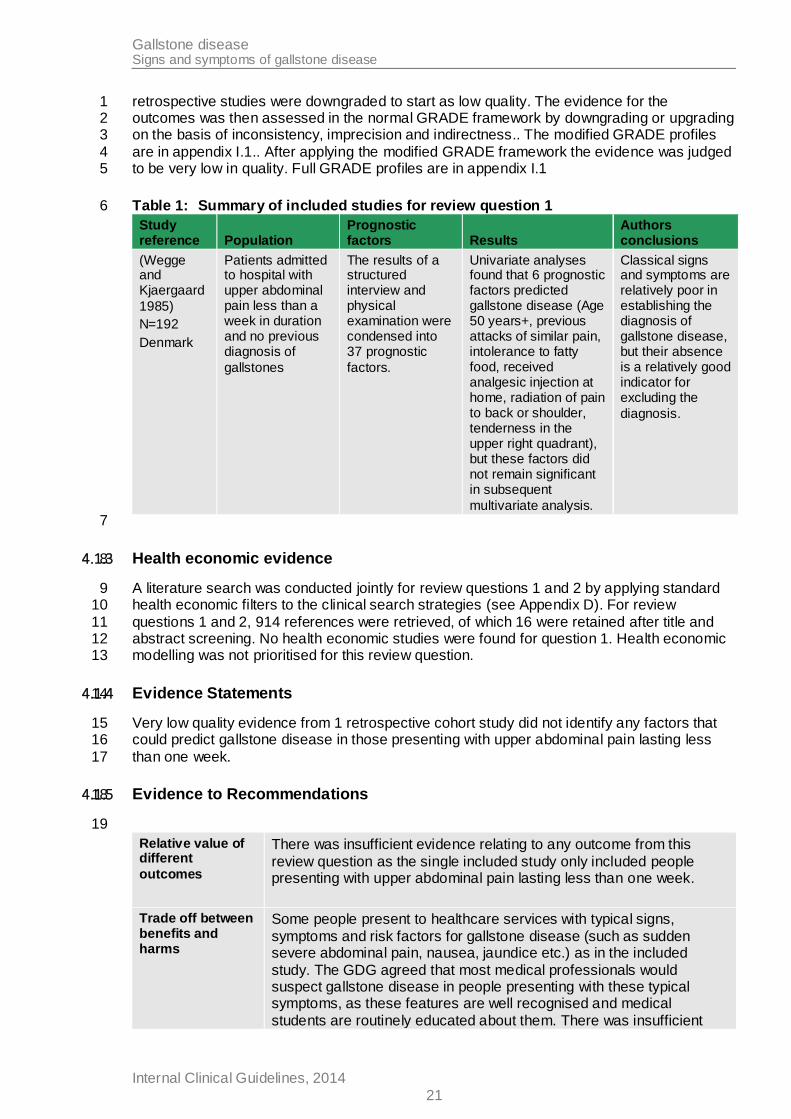
Table 1: Summary of included studies for review question 1 6
Study reference Population
Prognostic factors Results
Authors conclusions
(Wegge and Kjaergaard
1985)
N=192
Denmark
Patients admitted to hospital with upper abdominal pain less than a week in duration and no previous diagnosis of
gallstones
The results of a structured interview and physical examination were condensed into 37 prognostic
factors.
Univariate analyses found that 6 prognostic factors predicted gallstone disease (Age 50 years+, previous attacks of similar pain, intolerance to fatty food, received analgesic injection at home, radiation of pain to back or shoulder, tenderness in the upper right quadrant), but these factors did not remain significant in subsequent
multivariate analysis.
Classical signs and symptoms are relatively poor in establishing the diagnosis of gallstone disease, but their absence is a relatively good indicator for excluding the
diagnosis.
7
4.1.3 Health economic evidence 8
A literature search was conducted jointly for review questions 1 and 2 by applying standard 9 health economic filters to the clinical search strategies (see Appendix D). For review 10
questions 1 and 2, 914 references were retrieved, of which 16 were retained after title and 11 abstract screening. No health economic studies were found for question 1. Health economic 12 modelling was not prioritised for this review question. 13
4.1.4 Evidence Statements 14
Very low quality evidence from 1 retrospective cohort study did not identify any factors that 15 could predict gallstone disease in those presenting with upper abdominal pain lasting less 16
than one week. 17
4.1.5 Evidence to Recommendations 18
19
Relative value of different
outcomes
There was insufficient evidence relating to any outcome from this
review question as the single included study only included people presenting with upper abdominal pain lasting less than one week.
Trade off between benefits and harms
Some people present to healthcare services with typical signs,
symptoms and risk factors for gallstone disease (such as sudden severe abdominal pain, nausea, jaundice etc.) as in the included
study. The GDG agreed that most medical professionals would suspect gallstone disease in people presenting with these typical symptoms, as these features are well recognised and medical
students are routinely educated about them. There was insufficient
Gallstone disease Signs and symptoms of gallstone disease
Internal Clinical Guidelines, 2014
22
evidence available to support or refute these typical signs, symptoms
and risk factors as the single study identified only included people presenting with upper abdominal pain lasting less than a week. The GDG considered putting typical signs and symptoms in a
recommendation based on their combined knowledge and experience but felt that that may become a barrier to people who present with atypical signs and symptoms.
The GDG also acknowledged that some people with gallstone disease
present with symptoms that can be vague and easily misattributed to other conditions (such as indigestion or general abdominal discomfort) by both the patient and their healthcare professional. This can mean
that gallstone disease is not immediately considered, and patients may be investigated or treated for a condition that they do not have.
This ultimately affects patient quality of life, and the use of NHS resources as patients will continue to have unresolved symptoms that may get worse, resulting in inappropriate treatments and
investigations being offered to the patient. However, there was insufficient evidence available to enable this group of patients to be identified,
Thus the GDG decided not to make a recommendation based on
typical signs and symptoms or risk factors that should prompt a clinician to suspect symptomatic gallstone disease in adults presenting to healthcare services.
Consideration of Health Benefits
and Resource Use
No health economic evidence was found.
Quality of
evidence
The single study available for this review was of very low quality, and
insufficient evidence was provided to support decision making.
Other considerations
The GDG acknowledged the lack of research that was available around identifying signs, symptoms and risk factors for gallstone
disease, but felt that good quality research in this area would have limited value as the benefits would be small. The identification of specific signs, symptoms and risk factors would ultimately refine the
number of people who were offered ultrasound and liver function tests (these tests were reviewed and recommended in section 4.2 of this guideline). Since these tests are relatively low cost and easy to
perform, are low risk and minimally invasive to patients, and are widely used for a range of conditions, refining the number of these tests that are performed would not lead to major cost savings or improvements
in the quality of NHS care.
Therefore, the GDG did not feel that a research recommendation would be useful.
1
4.1.6 Recommendations 2
No recommendations were made in relation to this review question. 3
Gallstone disease Signs and symptoms of gallstone disease
Internal Clinical Guidelines, 2014
23
4.1.7 Research recommendations 1
No research recommendations were made in relation to this review question. 2
4.1.8 References 3
4
Acosta JM, Katkhouda N, Debian KA et al. (2006) Early ductal decompression versus 5 conservative management for gallstone pancreatitis with ampullary obstruction: a prospective 6
randomized clinical trial. Annals of Surgery 243: 33-40 7
Ahmed M, Diggory R (2011) The correlation between ultrasonography and histology in the 8
search for gallstones. Annals of the Royal College of Surgeons of England 93: 81-3 9
Alponat A, Kum CK, Rajnakova A et al. (1997) Predictive factors for synchronous common 10 bile duct stones in patients with cholelithiasis. Surgical Endoscopy 11: 928-32 11
Altun E, Semelka RC, Elias J, Jr. et al. (2007) Acute cholecystitis: MR findings and 12 differentiation from chronic cholecystitis. Radiology 244: 174-83 13
Amott D, Webb A, Tulloh B (2005) Prospective comparison of routine and selective operative 14
cholangiography. ANZ Journal of Surgery 75: 378-82 15
Ara R, Brazier J (2008) Deriving an algorithm to convert the eight mean SF-36 dimension 16 scores into a mean EQ-5D preference-based score from published studies (where patient 17
level data are not available). Value in Health 11: 1131-43 18
Attili AF, De SA, Capri R et al. (1995) The natural history of gallstones: the GREPCO 19 experience. The GREPCO Group. Hepatology 21: 655-60 20
Bansal VK, Misra MC, Garg P et al. (2010) A prospective randomized trial comparing two-21 stage versus single-stage management of patients with gallstone disease and common bile 22 duct stones. Surgical Endoscopy 24: 1986-9 23
Barr LL, Frame BC, Coulanjon A (1999) Proposed criteria for preoperative endoscopic 24 retrograde cholangiography in candidates for laparoscopic cholecystectomy. Surgical 25
Endoscopy 13: 778-81 26
Barthelsson C, Anderberg B, Ramel S et al. (2008) Outpatient versus inpatient laparoscopic 27 cholecystectomy: a prospective randomized study of symptom occurrence, symptom distress 28
and general state of health during the first post-operative week. Journal of Evaluation in 29 Clinical Practice 14: 577-84 30
Barthelsson C, Lutzen K, Anderberg B et al. (2003) Patients' experiences of laparoscopic 31
cholecystectomy in day surgery. Journal of Clinical Nursing 12: 253-9 32
Blay N, Donoghue J (2005) The effect of pre-admission education on domiciliary recovery 33 following laparoscopic cholecystectomy. Australian Journal of Advanced Nursing 22: 14-9 34
Blay N, Donoghue J (2006) Source and content of health information for patients undergoing 35 laparoscopic cholecystectomy. International Journal of Nursing Practice 12: 64-70 36
Boerma D, Rauws EA, Keulemans YC et al. (2002) Wait-and-see policy or laparoscopic 37
cholecystectomy after endoscopic sphincterotomy for bile-duct stones: a randomised trial. 38 Lancet 360: 761-5 39
Gallstone disease Signs and symptoms of gallstone disease
Internal Clinical Guidelines, 2014
24
Chan YL, Chan AC, Lam WW et al. (1996) Choledocholithiasis: comparison of MR 1 cholangiography and endoscopic retrograde cholangiography. Radiology 200: 85-9 2
Chopra KB, Peters RA, O'Toole PA et al. (1996) Randomised study of endoscopic biliary 3
endoprosthesis versus duct clearance for bileduct stones in high-risk patients. Lancet 348: 4 791-3 5
Cuschieri A, Lezoche E, Morino M et al. (1999) E.A.E.S. multicenter prospective randomized 6
trial comparing two-stage vs single-stage management of patients with gallstone disease and 7 ductal calculi. Surgical Endoscopy 13: 952-7 8
De Vargas MM, Lanciotti S, De Cicco ML et al. (2006) Ultrasonographic and spiral CT 9 evaluation of simple and complicated acute cholecystitis: diagnostic protocol assessment 10 based on personal experience and review of the literature. Radiologia Medica 111: 167-80 11
Deeks JJ, Bossuyt PM, Gatsonis Ce (2010) . Cochrane Handbook for Systematic Reviews of 12 Diagnostic Test Accuracy. Version 1.0 edition. The Cochrane Collaboration. 13
Department of Health (2012) National Schedule of Reference Costs 2011-2012. 14
Ding YB, Deng B, Liu XN et al. (2013) Synchronous vs sequential laparoscopic 15 cholecystectomy for cholecystocholedocholithiasis. World journal of gastroenterology : WJG 16 19: 2080-6 17
ElGeidie AA (2011a) Laparoscopic exploration versus intraoperative endoscopic 18 sphincterotomy for common bile duct stones: A prospective randomized trial. Digestive 19 Surgery 28: 424-31 20
ElGeidie AA (2011b) Preoperative versus intraoperative endoscopic sphincterotomy for 21 management of common bile duct stones. Surgical Endoscopy 25: 1230-7 22
Fan ST, Lai EC, Mok FP et al. (1993) Early treatment of acute biliary pancreatitis by 23
endoscopic papillotomy. New England Journal of Medicine 328: 228-32 24
Folsch UR, Nitsche R, Ludtke R et al. (1997) Early ERCP and papillotomy compared with 25
conservative treatment for acute biliary pancreatitis. The German Study Group on Acute 26 Biliary Pancreatitis. New England Journal of Medicine 336: 237-42 27
Griffin N, Wastle ML, Dunn WK et al. (2003) Magnetic resonance cholangiopancreatography 28
versus endoscopic retrograde cholangiopancreatography in the diagnosis of 29 choledocholithiasis. European Journal of Gastroenterology & Hepatology 15: 809-13 30
Gul R (2013) Comparison of early and delayed laparoscopic cholecystectomy for acute 31
cholecystitis: experience from a single centre. North Americal Journal of Medical Sciences 5: 32 414-8 33
Gurusamy K, Wilson E, Burroughs AK et al. (2012) Intra-operative vs pre-operative 34
endoscopic sphincterotomy in patients with gallbladder and common bile duct stones: cost-35 utility and value-of-information analysis. Applied Health Economics & Health Policy 10: 15-29 36
Hakansson K, Leander P, Ekberg O et al. (2000) MR imaging in clinically suspected acute 37
cholecystitis. A comparison with ultrasonography. Acta Radiologica 41: 322-8 38
Hayden JA, Cote P, Bombardier C (2006) Evaluation of the quality of prognosis studies in 39 systematic reviews. Annals of Internal Medicine 144: 427-37 40
Gallstone disease Signs and symptoms of gallstone disease
Internal Clinical Guidelines, 2014
25
Hollington P (1999) A prospective randomized trial of day-stay only versus overnight-stay 1 laparoscopic cholecystectomy. The Australian and New Zealand Journal of Surgery 69: 841-2 3 3
Holzknecht N, Gauger J, Sackmann M et al. (1998) Breath-hold MR cholangiography with 4 snapshot techniques: prospective comparison with endoscopic retrograde cholangiography. 5 Radiology 206: 657-64 6
Hong DF, Xin Y, Chen DW (2006) Comparison of laparoscopic cholecystectomy combined 7 with intraoperative endoscopic sphincterotomy and laparoscopic exploration of the common 8
bile duct for cholecystocholedocholithiasis. Surgical Endoscopy 20: 424-7 9
Howard K, Lord SJ, Speer A et al. (2006) Value of magnetic resonance 10 cholangiopancreatography in the diagnosis of biliary abnormalities in postcholecystectomy 11
patients: A probabilistic cost-effectiveness analysis of diagnostic strategies. International 12 Journal of Technology Assessment in Health Care 22: 109-18 13
Hui C-K, Lai K-C, Wong W-M et al. (2002) A randomised controlled trial of endoscopic 14
sphincterotomy in acute cholangitis without common bile duct stones. Gut 51: 245-7 15
Johansson M, Thune A, Blomqvist A et al. (2003) Management of acute cholecystitis in the 16 laparoscopic era: results of a prospective, randomized clinical trial. Journal of 17
Gastrointestinal Surgery 7: 642-5 18
Johansson M, Thune A, Nelvin L et al. (2006) Randomized clinical trial of day-care versus 19 overnight-stay laparoscopic cholecystectomy. British Journal of Surgery 93: 40-5 20
Jovanovic P, Salkic NN, Zerem E et al. (2011) Biochemical and ultrasound parameters may 21 help predict the need for therapeutic endoscopic retrograde cholangiopancreatography 22 (ERCP) in patients with a firm clinical and biochemical suspicion for choledocholithiasis. 23
European Journal of Internal Medicine 22: e110-e114 24
Kaltenthaler E, Vergel YB, Chilcott J et al. (2004) A systematic review and economic 25
evaluation of magnetic resonance cholangiopancreatography compared with diagnostic 26 endoscopic retrograde cholangiopancreatography. Health Technology Assessment 8: iii-89 27
Karki S (2013) Role of ultrasound as compared with ERCP in patient with obstructive 28
jaundice. Kathmandu University Medical Journal 43: 237-40 29
Keulemans Y, Eshuis J, de HH et al. (1998) Laparoscopic cholecystectomy: day-care versus 30 clinical observation. Annals of Surgery 228: 734-40 31
Khan OA, Balaji S, Branagan G et al. (2011) Randomized clinical trial of routine on-table 32 cholangiography during laparoscopic cholecystectomy. British Journal of Surgery 98: 362-7 33
Koc B, Karahan S, Adas G et al. (2013) Comparison of laparoscopic common bile duct 34
exploration and endoscopic retrograde cholangiopancreatography plus laparoscopic 35 cholecystectomy for choledocholithiasis: a prospective randomized study. American Journal 36 of Surgery 206: 457-63 37
Kolla SB, Aggarwal S, Kumar A et al. (2004) Early versus delayed laparoscopic 38 cholecystectomy for acute cholecystitis: a prospective randomized trial. Surgical Endoscopy 39 18: 1323-7 40
Kondo S, Isayama H, Akahane M et al. (2005) Detection of common bile duct stones: 41 comparison between endoscopic ultrasonography, magnetic resonance cholangiography, 42
Gallstone disease Signs and symptoms of gallstone disease
Internal Clinical Guidelines, 2014
26
and helical-computed-tomographic cholangiography. European Journal of Radiology 54: 271-1 5 2
Lai PB, Kwong KH, Leung KL et al. (1998) Randomized trial of early versus delayed 3
laparoscopic cholecystectomy for acute cholecystitis. British Journal of Surgery 85: 764-7 4
Lau JY, Leow CK, Fung TM et al. (2006) Cholecystectomy or gallbladder in situ after 5 endoscopic sphincterotomy and bile duct stone removal in Chinese patients. 6
Gastroenterology 130: 96-103 7
Lo CM (1998) Prospective randomized study of early versus delayed laparoscopic 8
cholecystectomy for acute cholecystitis. Annals of Surgery 227: 461-7 9
Macafee DA, Humes DJ, Bouliotis G et al. (2009) Prospective randomized trial using cost-10 utility analysis of early versus delayed laparoscopic cholecystectomy for acute gallbladder 11
disease. British Journal of Surgery 96: 1031-40 12
Nathanson LK, O'Rourke NA, Martin IJ et al. (2005) Postoperative ERCP versus 13 laparoscopic choledochotomy for clearance of selected bile duct calculi: a randomized trial. 14
Annals of Surgery 242: 188-92 15
National Institute for Health and Care Excellence (2013) Guide to Methods of Technology 16 Appraisal. 17
Neoptolemos JP, Carr-Locke DL, London NJ et al. (1988) Controlled trial of urgent 18 endoscopic retrograde cholangiopancreatography and endoscopic sphincterotomy versus 19 conservative treatment for acute pancreatitis due to gallstones. Lancet 2: 979-83 20
Nitsche R, Folsch UR, Ludtke R et al. (1995) Urgent ERCP in all cases of acute biliary 21 pancreatitis? A prospective randomized multicenter study. European Journal of Medical 22 Research 1: 127-31 23
Noble H, Tranter S, Chesworth T et al. (2009) A randomized, clinical trial to compare 24 endoscopic sphincterotomy and subsequent laparoscopic cholecystectomy with primary 25
laparoscopic bile duct exploration during cholecystectomy in higher risk patients with 26 choledocholithiasis. Journal of Laparoendoscopic & Advanced Surgical Techniques Part: 27 713-20 28
Oria A, Cimmino D, Ocampo C et al. (2007) Early endoscopic intervention versus early 29 conservative management in patients with acute gallstone pancreatitis and biliopancreatic 30 obstruction: a randomized clinical trial. Annals of Surgery 245: 10-7 31
Park MS, Yu JS, Kim YH et al. (1998) Acute cholecystitis: comparison of MR 32 cholangiography and US. Radiology 209: 781-5 33
Penniston KL, Nakada SY (2007) Health Related Quality of Life Differs Between Male and 34
Female Stone Formers. The Journal of Urology 178: 2435-40 35
Polkowski M, Palucki J, Regula J et al. (1999) Helical computed tomographic 36 cholangiography versus endosonography for suspected bile duct stones: a prospective 37
blinded study in non-jaundiced patients. Gut 45: 744-9 38
R Code Team (2012) . R: A language and environment for statistical computing. Vienna: R 39
Foundation for statistical computing. 40
Regan F, Fradin J, Khazan R et al. (1996) Choledocholithiasis: evaluation with MR 41 cholangiography. AJR American: 1441-5 42
Gallstone disease Signs and symptoms of gallstone disease
Internal Clinical Guidelines, 2014
27
Reinders JSK, Goud A, Timmer R et al. (2010) Early Laparoscopic Cholecystectomy 1 Improves Outcomes After Endoscopic Sphincterotomy for Choledochocystolithiasis. 2 Gastroenterology 138: 2315-20 3
Rhodes M, Sussman L, Cohen L et al. (1998) Randomised trial of laparoscopic exploration of 4 common bile duct versus postoperative endoscopic retrograde cholangiography for common 5 bile duct stones. Lancet 351: 159-61 6
Rickes S, Treiber G, Monkemuller K et al. (2006) Impact of the operator's experience on 7 value of high-resolution transabdominal ultrasound in the diagnosis of choledocholithiasis: a 8
prospective comparison using endoscopic retrograde cholangiography as the gold standard. 9 Scandinavian Journal of Gastroenterology 41: 838-43 10
Rogers SJ, Cello JP, Horn JK et al. (2010) Prospective randomized trial of LC+LCBDE vs 11
ERCP/S+LC for common bile duct stone disease. Archives of Surgery 145: 28-33 12
Schmidt M, Sondenaa K, Vetrhus M et al. (2011a) A randomized controlled study of 13 uncomplicated gallstone disease with a 14-year follow-up showed that operation was the 14
preferred treatment. Digestive Surgery 28: 270-6 15
Schmidt M, Sondenaa K, Vetrhus M et al. (2011b) Long-term follow-up of a randomized 16 controlled trial of observation versus surgery for acute cholecystitis: non-operative 17
management is an option in some patients. Scandinavian Journal of Gastroenterology 46: 18 1257-62 19
Sgourakis G, Karaliotas K (2002) Laparoscopic common bile duct exploration and 20
cholecystectomy versus endoscopic stone extraction and laparoscopic cholecystectomy for 21 choledocholithiasis. A prospective randomized study. Minerva Chirurgica 57: 467-74 22
Shiozawa S, Tsuchiya A, Kim DH et al. (2005) Useful predictive factors of common bile duct 23
stones prior to laparoscopic cholecystectomy for gallstones. Hepato-Gastroenterology 52: 24 1662-5 25
Soper NJ, Dunnegan DL (1992) Routine versus selective intra-operative cholangiography 26 during laparoscopic cholecystectomy. World Journal of Surgery 16: 1133-40 27
Soto JA, Alvarez O, Munera F et al. (2000) Diagnosing bile duct stones: comparison of 28
unenhanced helical CT, oral contrast-enhanced CT cholangiography, and MR 29 cholangiography. AJR American: 1127-34 30
Soto JA, Velez SM, Guzman J (1999) Choledocholithiasis: diagnosis with oral-contrast-31
enhanced CT cholangiography. AJR American: 943-8 32
Stiris MG (2000) MR cholangiopancreaticography and endoscopic retrograde 33 acholangiopancreaticography in patients with suspected common bile duct stones. Acta 34
Radiologica 41: 269-72 35
Sugiyama M, Atomi Y (1997) Endoscopic ultrasonography for diagnosing choledocholithiasis: 36 a prospective comparative study with ultrasonography and computed tomography. 37
Gastrointestinal Endoscopy 45: 143-6 38
Sugiyama M, Atomi Y, Hachiya J (1998) Magnetic resonance cholangiography using half-39 Fourier acquisition for diagnosing choledocholithiasis. American Journal of Gastroenterology 40
93: 1886-90 41
Gallstone disease Signs and symptoms of gallstone disease
Internal Clinical Guidelines, 2014
28
Tamhankar AP, Mazari FA, Everitt NJ et al. (2009) Use of the internet by patients undergoing 1 elective hernia repair or cholecystectomy. Annals of the Royal College of Surgeons of 2 England 91: 460-3 3
Tseng CW, Chen CC, Chen TS et al. (2008) Can computed tomography with coronal 4 reconstruction improve the diagnosis of choledocholithiasis? Journal of Gastroenterology & 5 Hepatology 23: 1586-9 6
Vetrhus M, Soreide O, Eide GE et al. (2005) Quality of life and pain in patients with acute 7 cholecystitis. Results of a randomized clinical trial. Scandinavian Journal of Surgery: SJS 94: 8
34-9 9
Vetrhus M, Soreide O, Eide GE et al. (2004) Pain and quality of life in patients with 10 symptomatic, non-complicated gallbladder stones: results of a randomized controlled trial. 11
Scandinavian Journal of Gastroenterology 39: 270-6 12
Vetrhus M, Soreide O, Nesvik I et al. (2003) Acute cholecystitis: delayed surgery or 13 observation. A randomized clinical trial. Scandinavian Journal of Gastroenterology 38: 985-14
90 15
Vetrhus M, Soreide O, Solhaug JH et al. (2002) Symptomatic, non-complicated gallbladder 16 stone disease. Operation or observation? A randomized clinical study. Scandinavian Journal 17
of Gastroenterology 37: 834-9 18
Vracko J, Markovic S, Wiechel KL (2006) Conservative treatment versus endoscopic 19 sphincterotomy in the initial management of acute cholecystitis in elderly patients at high 20
surgical risk. Endoscopy 38: 773-8 21
Wegge C, Kjaergaard J (1985) Evaluation of symptoms and signs of gallstone disease in 22 patients admitted with upper abdominal pain. Scandinavian Journal of Gastroenterology 20: 23
933-6 24
Whiting P, Rutjes AW, Dinnes J et al. (2004) Development and validation of methods for 25
assessing the quality of diagnostic accuracy studies. Health Technology Assessment 8 26
Yadav RP, Adhikary S, Agrawal CS et al. (2009) A comparative study of early vs. delayed 27 laparoscopic cholecystectomy in acute cholecystitis. Kathmandu University Medical Journal 28
7: 16-20 29
Young J, O'Connell B (2008) Recovery following laparoscopic cholecystectomy in either a 23 30 hour or an 8 hour facility. Journal of Quality in Clinical Practice 21: 2-7 31
Zhou LK, Prasoon P (2012) Mechanical and preventable factors of bile duct injuries during 32 laparoscopic cholecystectomy. [Review]. Hepato-Gastroenterology 59: 51-3 33
34
35 36
Gallstone disease Diagnosing gallstone disease
Internal Clinical Guidelines, 2014
29
4.2 Diagnosing gallstone disease 1
4.2.1 Review Question 2 2
What is the most accurate strategy for diagnosing gallstone disease in adults suspected of 3 the condition? 4
4.2.2 Evidence Review 5
The aim of this question was to assess all available methods for diagnosing gallstone 6 disease and establish which methods are the most accurate. 7
A systematic search was conducted (appendix D.2), which identified 6312 references. After 8 removing duplicates and screening the references based on their titles and abstracts, 310 9
references were obtained. These were reviewed against the inclusion and exclusion criteria 10 as detailed in the review protocols (appendix C.2). 11
Primary research utilising a randomised controlled trial, cohort, or cross sectional design was 12 eligible for inclusion. Studies also had to have utilised a valid reference standard. Currently 13 there is no accepted reference standard for confirming the presence or absence of gallstone 14 disease, so studies were only included if they met the following criteria: 15
Surgery as the reference standard for evaluating the gallbladder: this is the best available 16 method for diagnosing gallstones in the gallbladder and cholecystitis. 17
Endoscopic retrograde cholangiopancreatography (ERCP) as the reference standard for 18
evaluating the biliary tract: this is the best available method for diagnosing common bile 19
duct stones. 20
These procedures can accurately confirm the presence of gallstone disease by extracting the 21
gallstone(s). Other tests such as endoscopic ultrasound, magnetic resonance 22 cholangiopancreatographydo do not extract gallstones, so were considered unsuitable as 23
reference standards (,). 24
It is much more difficult to confirm the absence of gallstone disease. Gallstones or common 25 bile duct stones may be missed during endoscopic or surgical investigations. Patients can be 26
followed up to establish if signs and symptoms persist, which can indicate that gallstone 27 disease is present but was missed during previous investigations. However, this can be 28 misleading, as the patient’s signs and symptoms can be caused by other conditions, or new 29
gallstone disease could have developed since the original investigations. As there is no 30 alternative method for definitively confirming the absence of gallstone disease, surgery and 31 ERCP were accepted as the best available reference standards, and their potential 32
inaccuracies are acknowledged. 33
During the review, a date restriction was also imposed on all studies that utilised endoscopic, 34 surgical, or radiological methodologies. This was because technological advances have 35
made older studies of limited relevance to clinical practice today. An arbitrary publication of 36 date of 1993 or later was used as it coincides with the approximate introduction of 37 laparoscopic cholecystectomy into clinical practice in the UK. The only exceptions to this 38
date restriction were studies that focused on predicting the presence of gallstone disease 39 using clinical history taking, physical examination and simple blood tests, as these factors 40 are not as dependent on technology, and studies conducted over 20 years ago are likely to 41
still be relevant to clinical practice today. 42
Overall, 23 studies met the eligibility criteria and were included in the review. Evidence was 43 extracted into detailed evidence tables (see appendix G.2). Diagnostic test accuracy data 44 were provided by 20 studies, and where possible these data were pooled in relevant meta-45
Gallstone disease Diagnosing gallstone disease
Internal Clinical Guidelines, 2014
30
analyses. Some of these studies compared to see whether diagnostic test accuracy differed 1 depending on who interpreted the test results (for example, radiologist compared with 2 ultrasonographer, or experienced radiologist compared with inexperienced radiologist). This 3
produced different results for the same sets of patients. It would be inappropriate to use both 4 results in the meta-analysis as this would be double counting. Instead the results reported by 5 the interpreter most similar to those intended to use the test in clinical practice were included 6
in the analysis. If both interpreters were intended to use the test, their test results were 7 averaged and this was taken into the meta-analysis. 8
Data about predictive factors for gallstone disease were provided by 3 studies. It was not 9 possible to pool the data from these studies, as each study investigated different predictive 10 factors. 11
Data from the included studies were extracted into detailed evidence Table 2 and Table 3 12 below, and the GRADE framework for diagnostic evidence was used to quality assess the 13
evidence. However, for this review, the GDG took a liberal approach to set the threshold for 14 accuracy of 0.50 for both sensitivity and specificity on the basis that wanted to identify the 15 test that were better than chance and any test that did not meet this threshold were not 16
considered clinically useful.. Full GRADE profiles are presented in Appendix I.2 17
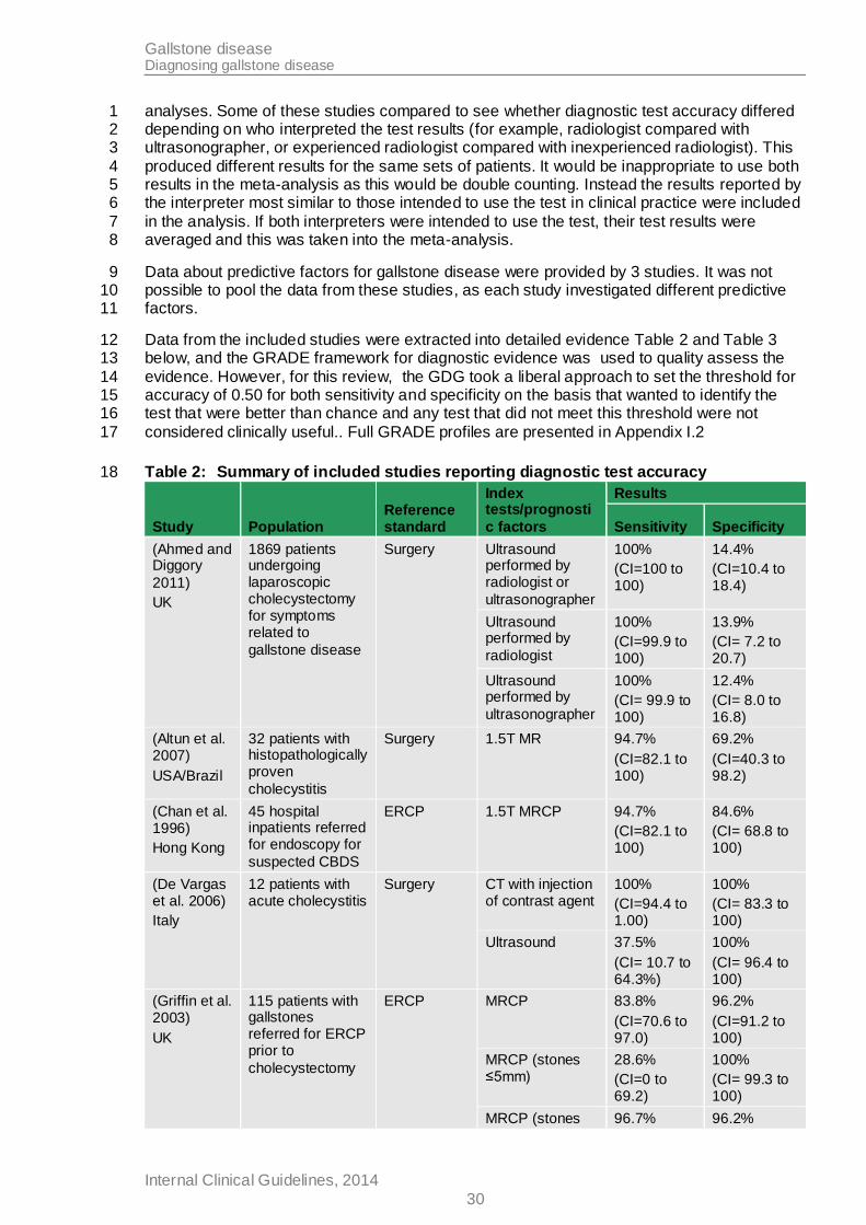
Table 2: Summary of included studies reporting diagnostic test accuracy 18
Study Population Reference standard
Index tests/prognosti
c factors
Results
Sensitivity Specificity
(Ahmed and Diggory
2011)
UK
1869 patients undergoing laparoscopic cholecystectomy for symptoms related to
gallstone disease
Surgery Ultrasound performed by radiologist or
ultrasonographer
100%
(CI=100 to 100)
14.4%
(CI=10.4 to 18.4)
Ultrasound performed by
radiologist
100%
(CI=99.9 to 100)
13.9%
(CI= 7.2 to 20.7)
Ultrasound performed by
ultrasonographer
100%
(CI= 99.9 to 100)
12.4%
(CI= 8.0 to 16.8)
(Altun et al. 2007)
USA/Brazil
32 patients with histopathologically proven
cholecystitis
Surgery 1.5T MR 94.7%
(CI=82.1 to 100)
69.2%
(CI=40.3 to 98.2)
(Chan et al. 1996)
Hong Kong
45 hospital inpatients referred for endoscopy for
suspected CBDS
ERCP 1.5T MRCP 94.7%
(CI=82.1 to 100)
84.6%
(CI= 68.8 to 100)
(De Vargas et al. 2006)
Italy
12 patients with acute cholecystitis
Surgery CT with injection of contrast agent
100%
(CI=94.4 to 1.00)
100%
(CI= 83.3 to 100)
Ultrasound 37.5%
(CI= 10.7 to 64.3%)
100%
(CI= 96.4 to 100)
(Griffin et al. 2003)
UK
115 patients with gallstones referred for ERCP prior to
cholecystectomy
ERCP MRCP 83.8%
(CI=70.6 to 97.0)
96.2%
(CI=91.2 to 100)
MRCP (stones ≤5mm)
28.6%
(CI=0 to 69.2)
100%
(CI= 99.3 to 100)
MRCP (stones 96.7% 96.2%
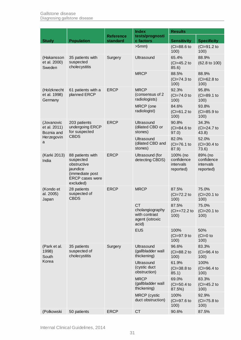
Gallstone disease Diagnosing gallstone disease
Internal Clinical Guidelines, 2014
31
Study Population Reference standard
Index tests/prognosti
c factors
Results
Sensitivity Specificity
>5mm) (CI=88.6 to 100)
(CI=91.2 to 100)
(Hakansson et al. 2000)
Sweden
35 patients with suspected
cholecystitis
Surgery Ultrasound 65.4%
(CI=45.2 to 85.6)
88.9%
(62.8 to 100)
MRCP 88.5%
(CI=74.3 to 100)
88.9%
(CI=62.8 to 100)
(Holzknecht et al. 1998)
Germany
61 patients with a planned ERCP
ERCP MRCP (consensus of 2
radiologists)
92.3%
(CI=74.0 to
100)
95.8%
(CI=89.1 to
100)
MRCP (one radiologist)
84.6%
(CI=61.2 to 100)
93.8%
(CI=85.9 to 100)
(Jovanovic et al. 2011)
Boznia and Herzegovin
a
203 patients undergoing ERCP for suspected
CBDS
ERCP Ultrasound (dilated CBD or
stones)
90.8%
(CI=84.6 to 97.0)
34.3%
(CI=24.7 to 43.8)
Ultrasound (dilated CBD and
stones)
82.0%
(CI=76.1 to 87.9)
52.0%
(CI=30.4 to 73.6)
(Karki 2013)
India
88 patients with suspected obstructive jaundice (immediate post ERCP cases were
excluded)
ERCP Ultrasound (for detecting CBDS)
100% (no confidence intervals
reported)
89% (no confidence intervals
reported)
(Kondo et al. 2005)
Japan
28 patients suspected of
CBDS
ERCP MRCP 87.5%
(CI=72.2 to 100)
75.0%
(CI=20.1 to 100)
CT cholangiography with contrast agent (iotroxic
acid)
87.5%
(CI+=72.2 to 100)
75.0%
(CI=20.1 to 100)
EUS 100%
(CI=97.9 to 100)
50%
(CI=0 to 100)
(Park et al.
1998)
South Korea
35 patients suspected of
cholecystitis
Surgery Ultrasound (gallbladder wall
thickening)
96.6%
(CI=88.2 to 100)
83.3%
(CI=96.4 to 100)
Ultrasound (cystic duct
obstruction)
61.9%
(CI=38.8 to 85.1)
100%
(CI=96.4 to 100)
MRCP (gallbladder wall
thickening)
69.0%
(CI=50.4 to 87.5%)
83.3%
(CI=45.2 to 100)
MRCP (cystic duct obstruction)
100%
(CI=97.6 to 100)
92.9%
(CI=75.8 to 100)
(Polkowski 50 patients ERCP CT 90.6% 87.5%
Gallstone disease Diagnosing gallstone disease
Internal Clinical Guidelines, 2014
32
Study Population Reference standard
Index tests/prognosti
c factors
Results
Sensitivity Specificity
et al. 1999)
Poland
referred for ERCP for suspected
CBDS
Cholangiography with contrast
infusion
(CI=79 to 100)
(CI=68.2 to 100)
EUS 91.2%
(CI=80.2 to 100)
100%
(CI=96.9 to 100)
(Regan et al. 1996)
23 patients with suspected CBDS
ERCP MR cholangiography
(no contrast
agent)
93.3%
(CI=77.4 to 100)
87.5%
(CI=53.8 to 100)
Ultrasound 60%
(CI=31.9 to 88.1)
100%
(CI=93.8 to 100)
(Rickes et al. 2006)
Germany
124 Patients suspected of
CBDS
ERCP Ultrasound (experienced
investigator)
81.5%
(CI=65 to 98)
87.5%
(CI=58.3 to 100)
Ultrasound (inexperienced
investigator)
46.3%
(CI=32.1 to 60.5)
91.4%
(CI=80.7 to 100)
(Soto et al. 2000)
Colombia
51 patients referred for ERCP for suspected
CBDS
ERCP CT 65.4%
(CI=45.2 to 85.6)
84%
(CI=67.6 to 100)
CT Cholangiography with oral contrast
(iopodic acid)
92.3%
(CI=80.1 to 100)
92%
(CI=79.4 to 100)
MRCP 96.2%
(CI=86.8 to 100)
100%
(CI= 98 to 100)
(Soto et al. 1999)
Colombia
29 patients referred for ERCP for suspected
CBDS
ERCP CT Cholangiography with oral contrast (iopanoic acid)
(radiologist 1)
92.9%
(CI=75.8 to 100)
100%
(CI=96.7 to 100)
CT Cholangiography with oral contrast (iopanoic acid)
(radiologist 2)
85.7%
(CI=63.8 to 100)
100%
(CI=96.7 to 100)
(Stiris 2000)
Norway
50 patients suspected of
CBDS
ERCP MRCP 87.5%
(CI=74.5 to 100)
94.4%
(CI=81.1 to 100)
(Sugiyama and Atomi
1997)
Japan
142 patients referred for ERCP for suspected
CBDS
ERCP EUS 96.1%
(CI=89.8 to 100)
100%
(CI=99.5 to 100)
Ultrasound 62.7%
(CI=48.5 to 77.0)
94.5%
(CI=89.3 to 99.7)
CT 70.6%
(CI=57.1 to 84.1)
96.7%
(CI=92.5 to 100)
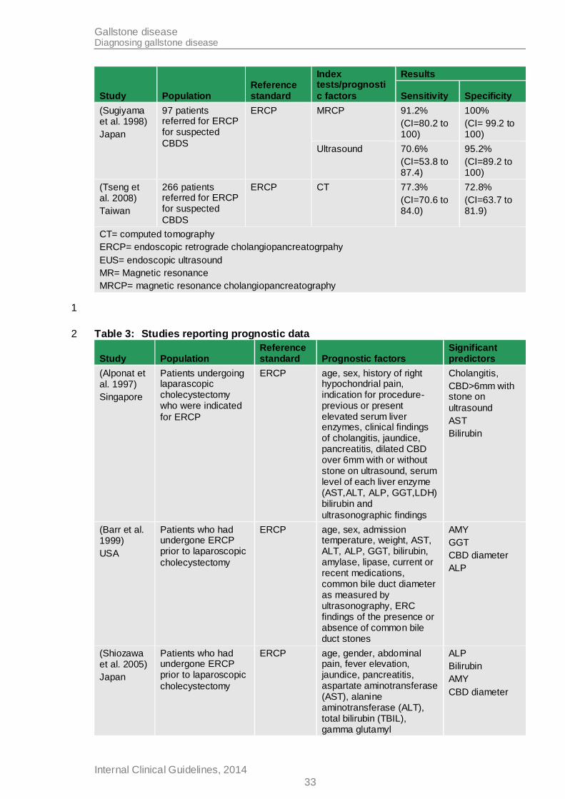
Gallstone disease Diagnosing gallstone disease
Internal Clinical Guidelines, 2014
33
Study Population Reference standard
Index tests/prognosti
c factors
Results
Sensitivity Specificity
(Sugiyama et al. 1998)
Japan
97 patients referred for ERCP for suspected
CBDS
ERCP MRCP 91.2%
(CI=80.2 to 100)
100%
(CI= 99.2 to 100)
Ultrasound 70.6%
(CI=53.8 to 87.4)
95.2%
(CI=89.2 to 100)
(Tseng et al. 2008)
Taiwan
266 patients referred for ERCP for suspected
CBDS
ERCP CT 77.3%
(CI=70.6 to 84.0)
72.8%
(CI=63.7 to 81.9)
CT= computed tomography
ERCP= endoscopic retrograde cholangiopancreatogrpahy
EUS= endoscopic ultrasound
MR= Magnetic resonance
MRCP= magnetic resonance cholangiopancreatography
1
Table 3: Studies reporting prognostic data 2
Study Population Reference standard Prognostic factors
Significant predictors
(Alponat et al. 1997)
Singapore
Patients undergoing laparascopic cholecystectomy who were indicated
for ERCP
ERCP age, sex, history of right hypochondrial pain, indication for procedure- previous or present elevated serum liver enzymes, clinical findings of cholangitis, jaundice, pancreatitis, dilated CBD over 6mm with or without stone on ultrasound, serum level of each liver enzyme (AST,ALT, ALP, GGT,LDH) bilirubin and
ultrasonographic findings
Cholangitis,
CBD>6mm with stone on
ultrasound
AST
Bilirubin
(Barr et al. 1999)
USA
Patients who had undergone ERCP prior to laparoscopic
cholecystectomy
ERCP age, sex, admission temperature, weight, AST, ALT, ALP, GGT, bilirubin, amylase, lipase, current or recent medications, common bile duct diameter as measured by ultrasonography, ERC findings of the presence or absence of common bile duct stones
AMY
GGT
CBD diameter
ALP
(Shiozawa et al. 2005)
Japan
Patients who had undergone ERCP prior to laparoscopic
cholecystectomy
ERCP age, gender, abdominal pain, fever elevation, jaundice, pancreatitis, aspartate aminotransferase (AST), alanine aminotransferase (ALT), total bilirubin (TBIL), gamma glutamyl
ALP
Bilirubin
AMY
CBD diameter
Gallstone disease Diagnosing gallstone disease
Internal Clinical Guidelines, 2014
34
Study Population Reference standard Prognostic factors
Significant predictors
transpeptidase (GGT), alkaline phosphatase (ALP), amylase (AMY), and
ultrasound findings.
AST= Aspartate aminotransferase; ALP= Alkaline phosphatase; AMY= Amylase; CBD= Common bile duct; ERCP= endoscopic retrograde cholangiopancreatography; GGT= Gamma glutamyl
transferase
4.2.3 Health economic evidence 1
A literature search was conducted jointly for review questions 1 and 2 by applying standard 2
health economic filters to the clinical search strategies (see Appendix D). Searches for 3 review questions 1 and 2 retrieved 914 studies, of which 16 were retained after title and 4 abstract screening. For review question 2, 2 health economic studies were found (Howard et 5
al. 2006; Kaltenthaler et al. 2004). An assessment of the quality of the economic evidence is 6 given in Table 4. This review question was not prioritised for original health economic 7 modelling. 8
Howard et al (2006) used a decision tree analysis to compare MRCP with ERCP for 9 diagnosing post-cholecystectomy CBDS in patients with abdominal pain and/or abnormal 10 liver function tests. Kaltenhaler et al. (2004) used a decision tree analysis to compare MRCP 11
with diagnostic ERCP for patients with suspected CBDS. The GDG felt that, as it was based 12 on UK data and covered a wider patient population, Kaltenhaler et al (2004) provided slightly 13 higher quality evidence. However, both studies were felt to be of use. Both studies found that 14
MRCP dominated ERCP, but this depended on the prior probability of CBDS. In probabilistic 15 sensitivity analysis, Howard et al. (2006) was found to be 83% cost effective (at Australian 16 $50,000/QALY threshold) and Kaltenhaler et al. (2004) was found to be 100% cost effective 17
(at UK £20,000/QALY threshold). Indeed, Kaltenhaler et al. (2004) was found to be 97% cost 18 saving. 19
MRCP appeared to be cost effective compared with ERCP for diagnosing CBDS under the 20 majority of assumptions. The GDG considered that the results from the 2 studies reflected 21
what they would expect to occur in standard clinical practice. 22
23
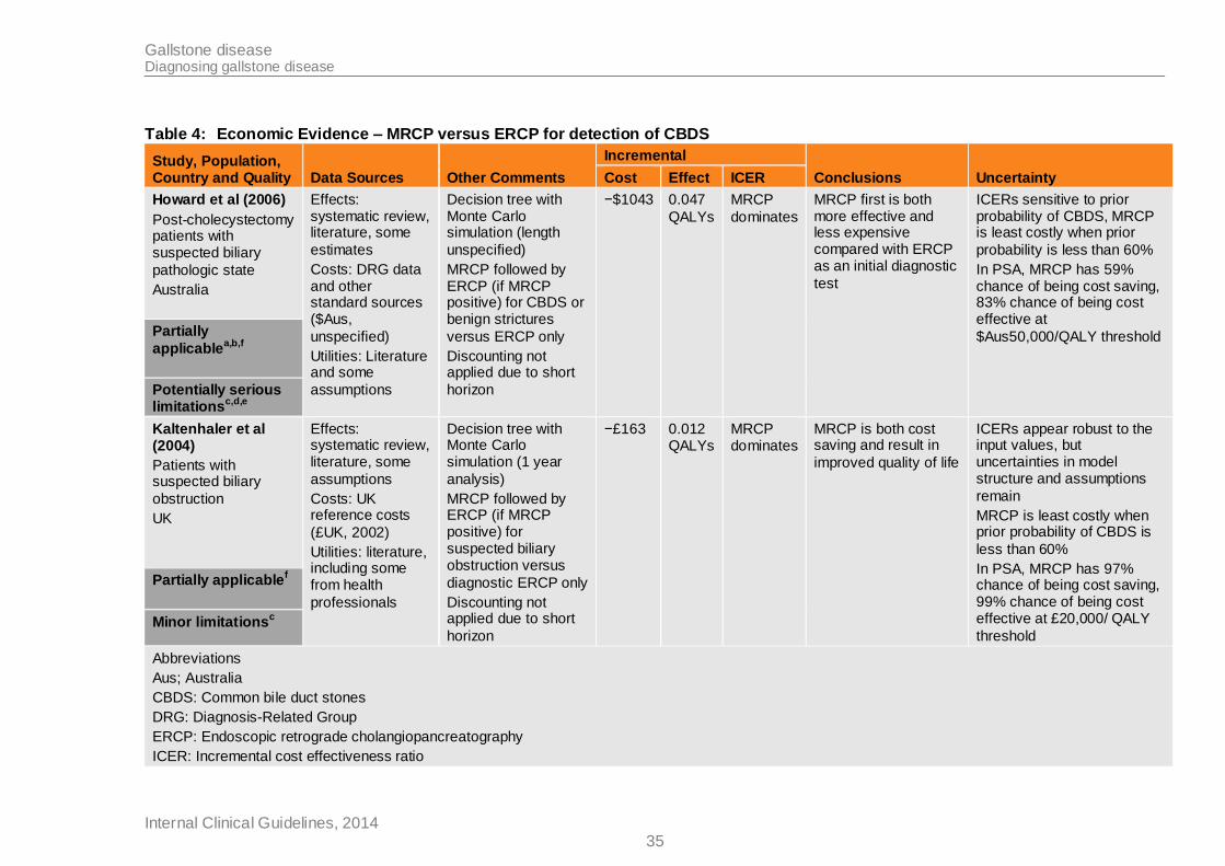
Gallstone disease Diagnosing gallstone disease
Internal Clinical Guidelines, 2014 35
Table 4: Economic Evidence – MRCP versus ERCP for detection of CBDS
Study, Population, Country and Quality Data Sources Other Comments
Incremental
Conclusions Uncertainty Cost Effect ICER
Howard et al (2006)
Post-cholecystectomy patients with suspected biliary
pathologic state
Australia
Effects: systematic review, literature, some
estimates
Costs: DRG data and other standard sources ($Aus,
unspecified)
Utilities: Literature and some
assumptions
Decision tree with Monte Carlo simulation (length
unspecified)
MRCP followed by ERCP (if MRCP positive) for CBDS or benign strictures
versus ERCP only
Discounting not applied due to short
horizon
−$1043 0.047
QALYs
MRCP
dominates
MRCP first is both more effective and less expensive compared with ERCP as an initial diagnostic
test
ICERs sensitive to prior probability of CBDS, MRCP is least costly when prior
probability is less than 60%
In PSA, MRCP has 59% chance of being cost saving, 83% chance of being cost effective at
$Aus50,000/QALY threshold Partially
applicablea,b,f
Potentially serious limitations
c,d,e
Kaltenhaler et al (2004)
Patients with suspected biliary
obstruction
UK
Effects: systematic review, literature, some
assumptions
Costs: UK reference costs
(£UK, 2002)
Utilities: literature, including some from health
professionals
Decision tree with Monte Carlo simulation (1 year
analysis)
MRCP followed by ERCP (if MRCP positive) for suspected biliary obstruction versus
diagnostic ERCP only
Discounting not applied due to short
horizon
−£163 0.012 QALYs
MRCP dominates
MRCP is both cost saving and result in
improved quality of life
ICERs appear robust to the input values, but uncertainties in model structure and assumptions
remain
MRCP is least costly when prior probability of CBDS is
less than 60%
In PSA, MRCP has 97% chance of being cost saving, 99% chance of being cost effective at £20,000/ QALY
threshold
Partially applicablef
Minor limitationsc
Abbreviations
Aus; Australia
CBDS: Common bile duct stones
DRG: Diagnosis-Related Group
ERCP: Endoscopic retrograde cholangiopancreatography
ICER: Incremental cost effectiveness ratio
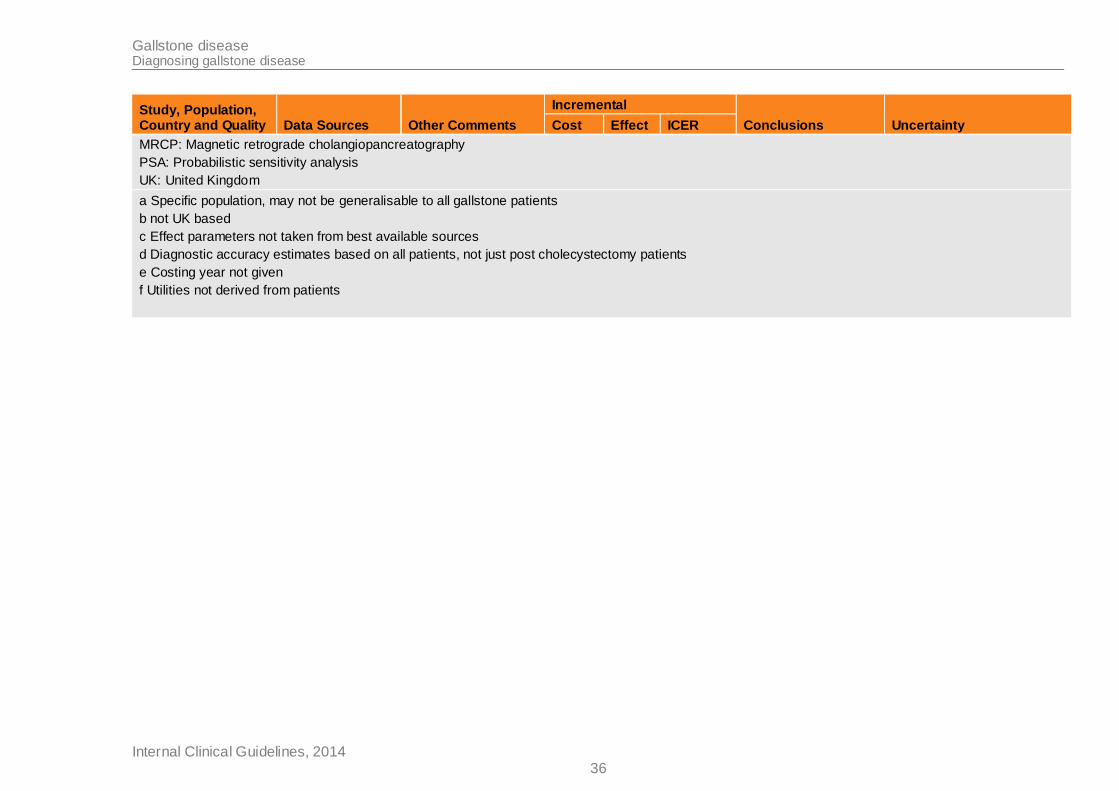
Gallstone disease Diagnosing gallstone disease
Internal Clinical Guidelines, 2014 36
Study, Population, Country and Quality Data Sources Other Comments
Incremental
Conclusions Uncertainty Cost Effect ICER
MRCP: Magnetic retrograde cholangiopancreatography
PSA: Probabilistic sensitivity analysis
UK: United Kingdom
a Specific population, may not be generalisable to all gallstone patients
b not UK based
c Effect parameters not taken from best available sources
d Diagnostic accuracy estimates based on all patients, not just post cholecystectomy patients
e Costing year not given
f Utilities not derived from patients
Gallstone disease Diagnosing gallstone disease
Internal Clinical Guidelines, 2014
37
1
4.2.4 Evidence Statements 2
While most of the diagnostic tests had sensitivities and specificities above 50%, some had 3 confidence intervals below 50% for either sensitivity or specificity. This means that there is 4
uncertainty in the evidence as some of the tests may actually perform worse than chance 5 alone. The quality of this evidence was mainly moderate to low in quality. 6
Two partially applicable health economic studies with minor limitations found that, under a 7 variety of assumptions, MRCP appeared cost effective compared with ERCP for diagnosing 8
CBDS. 9
4.2.5 Evidence to Recommendations 10
Relative value of different
outcomes
The various metrics for estimating diagnostic test accuracy were
discussed by the GDG, and they agreed to base decisions on sensitivity and specificity. This is because other diagnostic test accuracy measures are less reliable (for example, positive and
negative predictive values), or cannot be meta-analysed (for example, likelihood ratios, as robust methods for doing this have not yet been developed).
The GDG discussed patient benefits, patient harms, and clinical utilities associated with diagnostic testing, and the minimum sensitivity and specificity values that it would tolerate. It agreed that sensitivity and specificity estimates at or below 0.50 (or with confidence intervals
below 0.50) were not clinically useful as these results indicate that the test could perform the same or worse than chance alone.
The GDG agreed that sensitivity and specificity were equally as important in all diagnostic tests, except for ultrasound and liver function tests where the GDG would accept lower sensitivity values. The GDG recognised that patients with false negative results at this
early stage of the clinical pathway would be offered subsequent investigations as part of their differential diagnosis. Therefore they could tolerate lower levels of sensitivity, and so a higher number of
false negative results, as patients would present to health services with recurrent symptoms at a later time point.
The GDG felt that morbidity and mortality related to false negatives
was lower at this early stage of investigations, than for people with false positives who would undergo unnecessary treatment.
The GDG agreed that in practice other tests are only offered after ultrasound and liver function tests have been performed. Because of
this, the GDG felt that for other tests sensitivity and specificity were equally as important, as false positive and false negative results are associated with similar levels of subsequent morbidity and mortality at
this stage of the pathway.
Trade off between benefits and
harms
Abdominal ultrasound
For gallbladder stones: A single study provided very low quality evidence about ultrasound for diagnosing gallbladder stones. The
Gallstone disease Diagnosing gallstone disease
Internal Clinical Guidelines, 2014
38
study showed that ultrasound was extremely useful for identifying the
presence of gallstones (high sensitivity) but was not at all useful for ruling out gallstones in people who don’t have them (low specificity). The GDG felt that these results did not match their own experiences of
using the test to diagnose gallstones, and they felt that the test is both sensitive and specific. Since the evidence was based on a single study and contradicted clinical experience, the GDG agreed that the
results were not reliable.
For cholecystitis: Three studies provided very low quality evidence about ultrasound for diagnosing cholecystitis. Point estimates for
sensitivity and specificity were satisfactorily high, but confidence intervals fell below 0.50 for sensitivity. The GDG felt that it could accept lower sensitivity values for this test, as it is very low risk and is
used routinely in the NHS for a range of conditions. In addition, the GDG noted that the studies included in this meta-analysis had very small populations, so the wide confidence intervals may be because of
a lack of statistical power rather than a lack of sensitivity.
For common bile duct stones: Five studies provided low quality evidence about ultrasound for diagnosing common bile duct stones. Sensitivity and specificity values were satisfactorily high and
supported the GDG’s own knowledge and experience of the test. The GDG were confident that ultrasound was clinically useful in diagnosing
common bile duct stones.
Overall: the GDG felt there was sufficient evidence to recommend ultrasound for diagnosing gallstone disease since it is accurate, widely available throughout the NHS and is of very low risk to the patient.
Cholescintigraphy (HIDA scan)
No evidence was available. The GDG agreed that this test is not routinely used for diagnosing gallstone disease, and that it is usually
used for people for whom other tests are unsuitable. The GDG agreed not to make a recommendation based on the knowledge and experience of the group. The test exposes patients to ionising
radiation and the associated risks, and other tests considered in this review have evidence supporting their use for diagnosing gallbladder stones.
Computer Tomography (CT)
For cholecystitis: A single, small study provided low quality evidence
about CT for diagnosing cholecystitis. The study had unreliable estimates of specificity, as the confidence interval was below 0.50. The GDG agreed that there was insufficient evidence and decided not
to make a recommendation based on the knowledge and experience of the group. They felt that other tests considered in this review are better at diagnosing cholecystitis.
For common bile duct stones: Three studies provided moderate quality evidence about CT for diagnosing common bile duct stones. The meta-analysis demonstrated that CT had satisfactory sensitivity and specificity estimates.
Overall: CT uses ionising radiation, and the sensitivity and specificity were found to be inferior to other tests, such as MRCP. Because of
Gallstone disease Diagnosing gallstone disease
Internal Clinical Guidelines, 2014
39
this, the GDG felt that CT should not be recommended for diagnosing
gallstone disease, as other tests are more sensitive, specific, and safer.
CT cholangiography (CTC)
For common bile duct stones: Evidence about CTC for common bile
duct stones was provided by 4 studies. These showed that sensitivity and specificity were satisfactorily high, with reliable confidence intervals. However, the GDG decided not to recommend this test
because the contrast agent needed to perform CTC (iotroxic acid) is not available in the UK. This test also exposes patients to ionising radiation, and the GDG felt that there were more accurate and safer
tests for diagnosing common bile duct stones.
Endoscopic Ultrasound (EUS)
For common bile duct stones: Three studies provided low quality evidence about endoscopic ultrasound for diagnosing common bile duct stones. The meta-analysis demonstrated that endoscopic ultrasound had very high sensitivity and specificity estimates.
However, the confidence intervals for specificity fell below 0.50, indicating that there is uncertainty about whether the test is clinically useful.
The GDG felt that these results did not reflect clinical reality, as EUS is becoming widely accepted as being an accurate non-invasive test for
diagnosing common bile duct stones. However, when ERCP is used as a reference standard against which to judge the accuracy of EUS, cases that are classified as false positives on EUS might actually be revealed as false negatives on ERCP, were a perfect reference standard with which to judge both tests available. Therefore, the GDG believed that such analyses may underestimate the accuracy of EUS. .
Liver function tests
For common bile duct stones: Three studies that could not be pooled examined predictors of common bile duct stones and included liver
function tests among the variables that were examined. These studies examined different combinations of risk factors and so their results could not be meta-analysed. One study provided moderate quality
evidence, and the other 2 provided very low quality evidence. All 3 studies found that liver function tests were clinically useful in predicting common bile duct stones. The GDG agreed that there was sufficient
evidence to recommend the use of liver function tests for identifying common bile duct stones, as supportive evidence was provided by all 3 studies. The GDG based its decisions on the results of Shiozawa
(2005), based on the moderate quality rating, large sample size, narrow confidence intervals and clinically useful sensitivity and specificity estimates. Like ultrasound, the GDG felt that liver function
tests could be recommended because the test is widely available throughout the NHS and is of very low risk to the patient.
Magnetic Resonance Cholangiopancreatography (MRCP)

Gallstone disease Diagnosing gallstone disease
Internal Clinical Guidelines, 2014
40
For cholecystitis: Two studies provided low quality evidence about MRCP for diagnosing cholecystitis and demonstrated satisfactorily
high estimates of sensitivity and specificity. Only one of these studies considered the presence of gallstones when diagnosing cholecystitis. The GDG felt that healthcare professionals would consider the
presence of gallstones when making their diagnosis, and if this had been done in both studies included in the evidence, then diagnostic test accuracy would be more certain. The GDG felt that they could
explain some of the uncertainty in the sensitivity and specificity estimates for MRCP, and so could be confident that this test is
clinically useful.
For common bile duct stones: Eight studies provided moderate quality evidence about MRCP for diagnosing common bile duct stones. The meta-analysis demonstrated sufficiently robust sensitivity and
specificity values, so the GDG was confident that MRCP was also clinically useful and should be recommended.
Magnetic Resonance Imaging- contrast enhanced (MRI- contrast
enhanced)
For cholecystitis: The GDG agreed there was insufficient evidence to recommend MRI for cholecystitis, as evidence came from a single small study using contrast enhancement, which is not normally done in
clinical practice today. Point estimates were satisfactorily high, but confidence intervals for specificity fell below 0.50, so the GDG were unsure if the test was useful. The GDG agreed not to make a
recommendation about MRI based on the knowledge and experience of the group, as it felt that other tests considered in this review are better tests for diagnosing cholecystitis and conditions associated with
gallstone disease.
Summary of all diagnostic methods
After reviewing and discussing the evidence, the GDG agreed that
ultrasound, liver function tests, EUS and MRCP could be recommended for diagnosing gallstone disease. The GDG then discussed the sequencing of diagnostic tests and based these
discussions on the group’s knowledge and experience.
The evidence review in section 4.1 of this guideline discussed the 2
main ways that patients with gallstone disease present to health care services: either with typical signs, symptoms, and risk factors for gallstone disease, or with vague abdominal or gastrointestinal
symptoms that can easily be misattributed to other conditions. Because of this, the group felt that ultrasound and liver function tests should be recommended as first line investigations for patients with
suspected gallstone disease. In addition, these tests were recommended for patients with abdominal or gastrointestinal symptoms that are not responding to treatment as expected, since this
group of patients may have ‘unsuspected’ gallstone disease. This was felt to be of benefit to both patients and the NHS as these tests are
clinically useful, relatively cheap and widely available. Improving the identification of gallstone disease in people without typical symptoms would ultimately enable patients to progress to more appropriate
investigations or treatment pathways.
The group then discussed if MRCP and EUS should both be offered
Gallstone disease Diagnosing gallstone disease
Internal Clinical Guidelines, 2014
41
as second line investigations.
MRCP is widely available throughout the NHS, is relatively quick to
perform on an outpatient basis, and requires no sedation. It is less effective at diagnosing common bile duct stones than EUS and is not an appropriate test for patients with metal implants or claustrophobia.
The GDG agreed that EUS is a more accurate test than MRCP, but
EUS is an invasive test and procedural errors could result in adverse effects for the patient (such as perforation and even death). The test is normally performed with the patient under sedation, which carries its
own risks to the patient, and requires the patient to be accompanied by a responsible adult for 24 hours after sedation. People who do not
have a responsible adult to escort them will need to be admitted to hospital. Furthermore, EUS is not as widely available throughout the NHS as other tests (such as MRCP) are.
Although the GDG agreed that EUS was probably superior at
diagnosing gallstone disease than MRCP when performed by highly skilled operators, there was acknowledgement that not all operators would be highly skilled. Although skilled operators should also perform
MRCP, the GDG felt that there is less potential for harm if less skilled operators performed the test. This is because MRCP is non-invasive, and the entire diagnostic process is recorded. This means that poor
quality or equivocal tests can be reviewed by another operator, who may be able to offer a diagnosis without having to investigate the patient again. EUS only records key images from the test, so a second
reviewer would not be able to check unclear or poor quality tests and in these cases patients would need to have another test performed.
The GDG agreed that in most cases MRCP would be safer and more acceptable to patients than EUS, and recommended MRCP as a second line investigation and EUS as a third line investigation. However, the GDG wanted to stress that this should not preclude
appropriately trained and experienced operators using EUS instead of MRCP if appropriate.
Consideration of Health Benefits
and Resource Use
Limited published health economic evidence suggested that MRCP appears cost effective compared with ERCP for diagnosing CBDS. In
most situations MRCP appeared more effective and less costly than ERCP.
Despite the limitations of the evidence presented, the GDG felt that it
supported the primary use of MRCP for diagnosing CBDS.
The GDG noted that there was no health economic comparison available for many of the other diagnostic interventions under
consideration.
Quality of evidence
Overall, the evidence was moderate to very low in quality.
For gallbladder stones, only low quality evidence on ultrasound was found.
For cholecystitis, low quality evidence on MRCP and CT and very low quality evidence on ultrasound and MRI was found.
For common bile duct stones, moderate quality evidence was found
Gallstone disease Diagnosing gallstone disease
Internal Clinical Guidelines, 2014
42
on MRCP, CT, CT cholangiography, and for a model predicting
common bile duct stones using liver function tests and other predictors. Low quality evidence was found for ultrasound and endoscopic ultrasound, and very low quality evidence was found for 2
models predicting common bile duct stones using liver function tests and other predictors.
Other considerations
None
1
4.2.6 Recommendations 2
2. Offer liver function tests and ultrasound to people with suspected gallstone 3
disease, and to people with abdominal or gastrointestinal symptoms which have 4 been unresponsive to previous management 5
3. Consider magnetic resonance cholangiopancreatography (MRCP) if ultrasound 6
has not detected common bile duct stones but the: 7
bile duct is dilated and/or 8
liver function test results are abnormal. 9
4. Consider endoscopic ultrasound (EUS) if MRCP does not allow a diagnosis to be 10 made. 11
5. Refer people for further investigations if conditions other than gallstone disease 12 are suspected. 13
4.2.7 Research recommendations 14
6. What are the long-term benefits and harms of endoscopic ultrasound (EUS) 15 compared with magnetic resonance cholangiopancreatography (MRCP) in adults 16
with suspected common bile duct stones? 17
Why this is important 18
MRCP and EUS have both been found to be sufficiently accurate for diagnosing common 19 bile duct stones, with EUS regarded as the most accurate test. MRCP is non-invasive and so 20
carries negligible risks to the patient. However, EUS carries a small but significant risk of 21 patient harms, including death. There is insufficient evidence available to determine whether 22 the benefits of improved diagnosis associated with EUS outweigh its procedural risks. 23
Therefore, research is needed to compare MRCP with EUS to evaluate the subsequent 24 management of common bile duct stones. 25
26
4.2.8 References 27
Ahmed M, Diggory R (2011) The correlation between ultrasonography and histology in the 28 search for gallstones. Annals of the Royal College of Surgeons of England 93: 81-3 29
Alponat A, Kum CK, Rajnakova A et al. (1997) Predictive factors for synchronous common 30 bile duct stones in patients with cholelithiasis. Surgical Endoscopy 11: 928-32 31
Gallstone disease Diagnosing gallstone disease
Internal Clinical Guidelines, 2014
43
Altun E, Semelka RC, Elias J, Jr. et al. (2007) Acute cholecystitis: MR findings and 1 differentiation from chronic cholecystitis. Radiology 244: 174-83 2
Barr LL, Frame BC, Coulanjon A (1999) Proposed criteria for preoperative endoscopic 3
retrograde cholangiography in candidates for laparoscopic cholecystectomy. Surgical 4 Endoscopy 13: 778-81 5
Chan YL, Chan AC, Lam WW et al. (1996) Choledocholithiasis: comparison of MR 6 cholangiography and endoscopic retrograde cholangiography. Radiology 200: 85-9 7
De Vargas MM, Lanciotti S, De Cicco ML et al. (2006) Ultrasonographic and spiral CT 8 evaluation of simple and complicated acute cholecystitis: diagnostic protocol assessment 9 based on personal experience and review of the literature. Radiologia Medica 111: 167-80 10
Griffin N, Wastle ML, Dunn WK et al. (2003) Magnetic resonance cholangiopancreatography 11
versus endoscopic retrograde cholangiopancreatography in the diagnosis of 12 choledocholithiasis. European Journal of Gastroenterology & Hepatology 15: 809-13 13
Hakansson K, Leander P, Ekberg O et al. (2000) MR imaging in clinically suspected acute 14 cholecystitis. A comparison with ultrasonography. Acta Radiologica 41: 322-8 15
Holzknecht N, Gauger J, Sackmann M et al. (1998) Breath-hold MR cholangiography with 16 snapshot techniques: prospective comparison with endoscopic retrograde cholangiography. 17 Radiology 206: 657-64 18
Howard K, Lord SJ, Speer A et al. (2006) Value of magnetic resonance 19 cholangiopancreatography in the diagnosis of biliary abnormalities in postcholecystectomy 20 patients: A probabilistic cost-effectiveness analysis of diagnostic strategies. International 21 Journal of Technology Assessment in Health Care 22: 109-18 22
Jovanovic P, Salkic NN, Zerem E et al. (2011) Biochemical and ultrasound parameters may 23 help predict the need for therapeutic endoscopic retrograde cholangiopancreatography 24 (ERCP) in patients with a firm clinical and biochemical suspicion for choledocholithiasis. 25
European Journal of Internal Medicine 22: e110-e114 26
Kaltenthaler E, Vergel YB, Chilcott J et al. (2004) A systematic review and economic 27 evaluation of magnetic resonance cholangiopancreatography compared with diagnostic 28 endoscopic retrograde cholangiopancreatography. Health Technology Assessment 8: iii-89 29
Karki S (2013) Role of ultrasound as compared with ERCP in patient with obstructive 30 jaundice. Kathmandu University Medical Journal 43: 237-40 31
Kondo S, Isayama H, Akahane M et al. (2005) Detection of common bile duct stones: 32 comparison between endoscopic ultrasonography, magnetic resonance cholangiography, 33
and helical-computed-tomographic cholangiography. European Journal of Radiology 54: 271-34 5 35
Park MS, Yu JS, Kim YH et al. (1998) Acute cholecystitis: comparison of MR 36 cholangiography and US. Radiology 209: 781-5 37
Polkowski M, Palucki J, Regula J et al. (1999) Helical computed tomographic 38 cholangiography versus endosonography for suspected bile duct stones: a prospective 39 blinded study in non-jaundiced patients. Gut 45: 744-9 40
Regan F, Fradin J, Khazan R et al. (1996) Choledocholithiasis: evaluation with MR 41 cholangiography. AJR American: 1441-5 42
Rickes S, Treiber G, Monkemuller K et al. (2006) Impact of the operator's experience on 43 value of high-resolution transabdominal ultrasound in the diagnosis of choledocholithiasis: a 44
Gallstone disease Diagnosing gallstone disease
Internal Clinical Guidelines, 2014
44
prospective comparison using endoscopic retrograde cholangiography as the gold standard. 1 Scandinavian Journal of Gastroenterology 41: 838-43 2
Shiozawa S, Tsuchiya A, Kim DH et al. (2005) Useful predictive factors of common bile duct 3
stones prior to laparoscopic cholecystectomy for gallstones. Hepato-Gastroenterology 52: 4 1662-5 5
Soto JA, Alvarez O, Munera F et al. (2000) Diagnosing bile duct stones: comparison of 6 unenhanced helical CT, oral contrast-enhanced CT cholangiography, and MR 7
cholangiography. AJR American: 1127-34 8
Soto JA, Velez SM, Guzman J (1999) Choledocholithiasis: diagnosis with oral-contrast-9 enhanced CT cholangiography. AJR American: 943-8 10
Stiris MG (2000) MR cholangiopancreaticography and endoscopic retrograde 11
acholangiopancreaticography in patients with suspected common bile duct stones. Acta 12 Radiologica 41: 269-72 13
Sugiyama M, Atomi Y (1997) Endoscopic ultrasonography for diagnosing choledocholithiasis: 14 a prospective comparative study with ultrasonography and computed tomography. 15
Gastrointestinal Endoscopy 45: 143-6 16
Sugiyama M, Atomi Y, Hachiya J (1998) Magnetic resonance cholangiography using half-17 Fourier acquisition for diagnosing choledocholithiasis. American Journal of Gastroenterology 18
93: 1886-90 19
Tseng CW, Chen CC, Chen TS et al. (2008) Can computed tomography with coronal 20 reconstruction improve the diagnosis of choledocholithiasis? Journal of Gastroenterology & 21 Hepatology 23: 1586-9 22
Gallstone disease Asymptomatic gallbladder stones
Internal Clinical Guidelines, 2014
45
4.3 Asymptomatic gallbladder stones 1
2
4.3.1 Review Question 3 3
What factors predict which patients with asymptomatic gallbladder stones will develop acute 4 complications? 5
4.3.2 Evidence Review 6
The aim of this review question was to establish whether some people with asymptomatic 7 gallbladder stones are at a higher risk of developing complications than others. 8
A systematic search (appendix D.3) retrieved 12,256 references. After removing duplicates 9
and screening the references based on their titles and abstracts, 56 references were 10 obtained and reviewed against the inclusion and exclusion criteria as described in the review 11 protocol (appendix C.3). 12
Primary research using a randomised controlled trial, cohort or cross sectional design was 13 eligible for inclusion. Two main types of study were expected to be found by the search: 14 prospective studies recruiting a sample of the general population to identify and follow up 15 people with asymptomatic gallbladder stones, and retrospective reviews of people presenting 16
with symptomatic gallstone disease whose prior medical history was examined to identify 17 whether asymptomatic gallbladder stones had previously been diagnosed. Both types of 18 study were eligible for inclusion. 19
From the review, 55 studies were excluded, mainly because they provided prevalence and/or 20 incidence data only. A list of the excluded studies and reasons for their exclusion is provided 21 in appendix F. 22
One prospective cohort study met the eligibility criteria and was inc luded. Data were 23 extracted into detailed evidence tables (see appendix G.3) and are summarised in Table 5 24 below. 25
Appropriate methodology checklists were used to appraise the methodological quality of 26
individual studies, and a modified version of the GRADE framework was applied to 27 summarise the overall quality of the evidence (see appendix I.3). In this approach the 28 prospective cohort study was started with a ‘high’ quality rating and was further downgraded 29
as appropriate, according to standard GRADE framework. Overall this evidence was rated as 30 very low in quality. 31
32
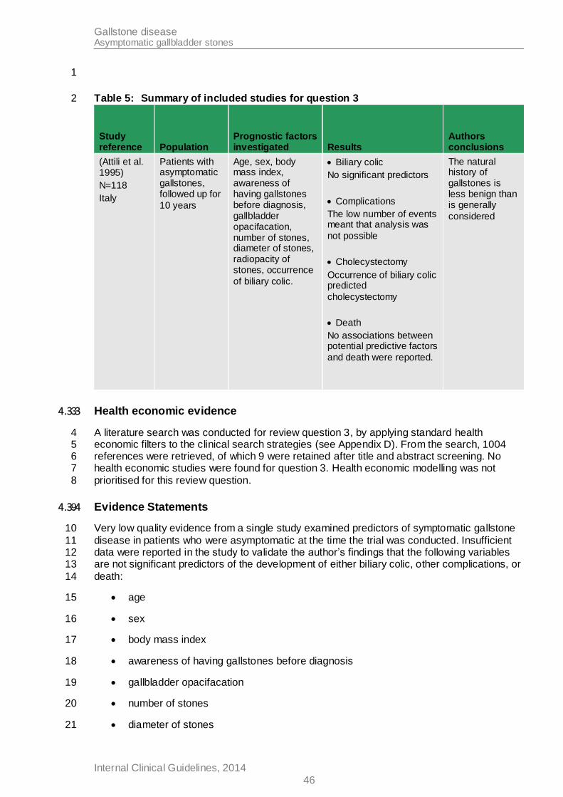
Gallstone disease Asymptomatic gallbladder stones
Internal Clinical Guidelines, 2014
46
1
Table 5: Summary of included studies for question 3 2
Study reference Population
Prognostic factors investigated Results
Authors conclusions
(Attili et al. 1995)
N=118
Italy
Patients with asymptomatic gallstones, followed up for
10 years
Age, sex, body mass index, awareness of having gallstones before diagnosis, gallbladder opacifacation, number of stones, diameter of stones, radiopacity of stones, occurrence
of biliary colic.
Biliary colic
No significant predictors
Complications
The low number of events meant that analysis was
not possible
Cholecystectomy
Occurrence of biliary colic predicted
cholecystectomy
Death
No associations between potential predictive factors
and death were reported.
The natural history of gallstones is less benign than is generally
considered
4.3.3 Health economic evidence 3
A literature search was conducted for review question 3, by applying standard health 4 economic filters to the clinical search strategies (see Appendix D). From the search, 1004 5 references were retrieved, of which 9 were retained after title and abstract screening. No 6 health economic studies were found for question 3. Health economic modelling was not 7
prioritised for this review question. 8
4.3.4 Evidence Statements 9
Very low quality evidence from a single study examined predictors of symptomatic gallstone 10
disease in patients who were asymptomatic at the time the trial was conducted. Insufficient 11 data were reported in the study to validate the author’s findings that the following variables 12 are not significant predictors of the development of either biliary colic, other complications, or 13
death: 14
age 15
sex 16
body mass index 17
awareness of having gallstones before diagnosis 18
gallbladder opacifacation 19
number of stones 20
diameter of stones 21
Gallstone disease Asymptomatic gallbladder stones
Internal Clinical Guidelines, 2014
47
radiopacity of stones. 1
Occurrence of biliary colic was not found to be a predictor of additional complications or 2 death. 3
4.3.5 Evidence to Recommendations 4
5
Relative value of different
outcomes
The evidence was based on the results of a single study. Only
descriptive data and significance levels were provided, so secondary analysis for the purposes of this review could not be performed. As no meaningful results were provided by the evidence, the GDG based its
decisions on the knowledge and experience of the group, and alongside evidence presented in section 4.4 of this guideline.
Trade off between benefits and
harms
For people with asymptomatic gallbladder stones in a normal biliary tree, the GDG agreed that there is currently no way of predicting which people will go on to develop complications. Based on their experience,
the GDG agreed that most people with asymptomatic gallbladder stones will not develop complications.
However, once a patient has been informed that they have asymptomatic gallbladder stones, they may worry about their risk of developing complications in future, and may ask about having them
treated prophylactically. Because of this, the discussions are linked to the evidence about managing asymptomatic gallbladder stones, which is reviewed and has recommendations on asymptomatic gallbladder
stones made in section 4.4. After considering the evidence (see ‘Evidence to recommendations’, section 4.4.5), the GDG felt that the prophylactic treatment of asymptomatic gallstones is likely to have a
higher risk than leaving them untreated. This is because all of the treatment options carry risks of adverse effects (such as side effects of medication and surgery risks,). Therefore the GDG recommended that
healthcare professionals should reassure their patients that asymptomatic gallstones do not need treatment unless they develop
symptoms (see section 4.4.6 of this guideline).
The GDG felt that some people with asymptomatic gallbladder stones
will have other abnormalities detected, either in their biliary tree (such as calcification of the gallbladder) or elsewhere in their body at the
time of gallbladder stone detection. For this group of people, the GDG agreed that there is a need to perform further tests and investigations to rule in or rule out other diseases and conditions, such as cancer. It
is not within the scope of this guideline to describe or review the evidence for the range of other conditions that should be investigated, so the GDG felt that they could not make recommendations about
what a healthcare professional should do when other abnormalities coexist with asymptomatic gallstones.
Consideration of Health Benefits and Resource
Use
No health economic evidence was found.
Quality of evidence
A single study providing very low quality evidence was not adequately powered to predict acute complications.
Gallstone disease Asymptomatic gallbladder stones
Internal Clinical Guidelines, 2014
48
Other
considerations
The GDG acknowledged that definitions of asymptomatic gallbladder
stones may vary and so the following definitions were developed:
Asymptomatic gallbladder stones:
– stones in the gallbladder that are found incidentally, as a result of imaging investigations unrelated to gallstone disease, in people who have been completely symptom-free
for at least 12 months before diagnosis.
Symptomatic gallbladder stones:
– gallbladder stones found on imaging of the gallbladder (the
GDG felt that a person undergoing gallbladder imaging must be experiencing symptoms to prompt the investigation)
– people with diagnosed gallbladder stones who have experienced any symptoms in the past 12 months.
4.3.6 Recommendations & Research Recommendations 1
No specific recommendations were made in relation to this question. 2
4.3.7 References 3
Attili AF, De SA, Capri R et al. (1995) The natural history of gallstones: the GREPCO 4 experience. The GREPCO Group. Hepatology 21: 655-60 5
Gallstone disease Managing asymptomatic gallbladder stones
Internal Clinical Guidelines, 2014
49
4.4 Managing asymptomatic gallbladder stones 1
2
4.4.1 Review Question 4a 3
Which strategies should be used for managing asymptomatic gallbladder stones? 4
4.4.2 Evidence Review 5
This question aimed to establish if prophylactic treatment should be offered to people with 6 asymptomatic gallbladder stones, to prevent them from developing symptoms in the future. 7
A single search was performed for questions 4a, 4b, 4c and 5, which identified 10,976 8 references. After removing duplicates and screening the references based on their titles and 9
abstracts, 210 references were obtained and reviewed against the inclusion and exclusion 10 criteria for this review question (appendix C), and 47 references met the overall inclusion 11 criteria. Details of excluded studies and reasons for their exclusion are in appendix F.4. 12
None of the 47 included references met the criteria for this specific review question (see 13 study flow chart, appendix E.4). 14
4.4.3 Health economic evidence 15
16
A literature search was conducted jointly for questions 4 and 5 by applying standard health 17
economic filters to the clinical search strategies (see Appendix D). From the literature search, 18 1396 references were retrieved for questions 4 and 5, of which none were retained for 19 question 4a. Health economic modelling was not prioritised for this review question. 20
4.4.4 Evidence Statements 21
No evidence that met the inclusion and exclusion criteria for this question was found. 22
4.4.5 Evidence to Recommendations 23
Relative value of different
outcomes
No evidence was available for this review question. The GDG based its decisions on the knowledge and experience of the group.
Trade-off between benefits and
harms
The GDG discussed the potential benefits and harms of the various treatment options that were considered, using its knowledge and experience as no evidence was available:
Dissolution therapy is used to painlessly dissolve gallbladder stones using oral tablets. This is safe, relatively cheap and patients may express a preference for this type of treatment. However,
patients have to take dissolution drugs over a long time period, and possibly for the rest of their life. Some people may find this inconvenient, particularly as they will not be experiencing any
symptoms. These drugs often have side effects such as diarrhoea, and adherence to the drugs over the long term may be affected by this. Furthermore, the GDG highlighted that dissolution therapies
only work for certain types of gallbladder stones. Since there are several types of gallbladder stones and no way of knowing which type the patient has without extracting and examining the stones,
Gallstone disease Managing asymptomatic gallbladder stones
Internal Clinical Guidelines, 2014
50
there is no way of knowing for which people dissolution therapies
will be effective.
Lithotripsy for gallbladder stones uses ultrasound waves to break
up gallstones into smaller pieces, with the idea that smaller pieces can pass naturally through the biliary system without causing any
symptoms for the patient. This treatment requires an ultrasound probe to be placed over the abdomen, and is painless and non-
invasive. However, the GDG highlighted that lithotripsy is less effective for large or multiple stones, and like dissolution therapy is less effective on certain types of stones. In addition, lithotripsy
carries a small but significant risk of pancreatitis, which can be life threatening.
Cholecystectomy is a radical treatment where the gallbladder is removed during a surgical procedure. Although this treatment is
safe and effective, there is a risk of harms (as with any surgery) such as infection, bleeding, bile leaks and injuries to the bile duct, and general risks associated with undergoing anaesthesia, all of
which are rare but can be life threatening.
Watch and wait/conservative management is when there is no
active treatment until the gallbladder stones begin to show
symptoms. In this case there is no risk to the patient until they develop symptoms and begin active treatment.
After discussing the various treatment options, the GDG agreed that
most people with asymptomatic gallbladder stones will not go on to develop complications, and so the risks of intervention outweigh the risks of leaving asymptomatic stones untreated. Furthermore, the
GDG felt that the resource implications of treating asymptomatic gallbladder stones should be considered, since the GDG felt a large proportion of the general population have gallbladder stones. Offering
unnecessary treatment to a large number of people would not be the best use of NHS resources.
The GDG also felt that it was important to discuss asymptomatic common bile duct stones, which were reviewed as part of question 4c
(see section 4.6). No evidence about asymptomatic common bile duct stones was found, but the GDG felt that they were able to make recommendations based on their knowledge and experience.
Asymptomatic common bile duct stones are different to asymptomatic
gallbladder stones.
Asymptomatic gallbladder stones are confined to the gallbladder, as are complications such as acute cholecystitis. The complications
of gallbladder stones can be life threatening but are rarely so. Deaths caused by gallbladder stone complications usually occur in people with other comorbidities.
Asymptomatic common bile duct stones are stones that have
moved out of the gallbladder and into the biliary system. Complications caused by common bile duct stones are not confined
to the gallbladder and can affect other organs in the biliary system, such as the pancreas. Complications such as pancreatitis and cholangitis are more likely to be life threatening than acute
cholecystitis, and deaths from these conditions commonly occur in
Gallstone disease Managing asymptomatic gallbladder stones
Internal Clinical Guidelines, 2014
51
people without other comorbidities.
The group agreed that the risks associated with leaving asymptomatic
common bile duct stones untreated outweighed the risks of treatment, and that common bile stones have an increased risk of serious complications than asymptomatic gallbladder stones. Therefore, the
GDG agreed that asymptomatic common bile duct stones should be managed in the same way as symptomatic common bile duct stones
(see section 4.6.5 for a discussion of this evidence). Recommendations about managing asymptomatic common bile duct stones are in section 4.6.
Consideration of
Health Benefits and Resource Use
No health economic evidence was found.
Quality of evidence
No evidence was available.
Other
considerations
The discussions around asymptomatic gallbladder stones that were linked to discussions around asymptomatic common bile duct stones
were reviewed as part of review question 4c in section 4.6.
The GDG discussed whether it would be useful to make a research recommendation about treating asymptomatic gallbladder stones, since there was no evidence to base their decisions on. The group
agreed that an appropriate trial would be one that screened a large population to identify sufficient people with asymptomatic gallbladder stones, who would then need to be followed up for many years. The
GDG felt that ideally, people included in such a trial should not be told that they have asymptomatic gallbladder stones identified, as it was felt that this knowledge could confound the findings of a trial. This is
because once people are aware they have asymptomatic gallbladder stones they may report symptoms they would not have otherwise have noticed, or seek medical attention for symptoms that they would not
have normally sought attention for. Since it would not be ethical to withhold information from patients, the GDG agreed that in reality
there is no appropriate way to conduct primary research in this area.
Therefore it was decided that a research recommendation should not be made.
1
4.4.6 Recommendations 2
6. Reassure people with asymptomatic gallbladder stones found in a normal 3
gallbladder and normal biliary tree that they do not need treatment unless they 4 develop symptoms. 5
4.4.7 Research Recommendations 6
No research recommendations were made in relation to this review question. 7

Gallstone disease Managing asymptomatic gallbladder stones
Internal Clinical Guidelines, 2014
52
4.4.8 References 1
No references2
Gallstone disease Managing symptomatic gallbladder stones
Internal Clinical Guidelines, 2014
53
4.5 Managing symptomatic gallbladder stones 1
2
4.5.1 Review Question 4b 3
Which strategies should be used for managing symptomatic gallbladder stones? 4
This question aimed to establish which management strategies offer optimal outcomes for 5 patients with symptomatic gallbladder stones, including patients with acute cholecystitis. 6
4.5.2 Evidence Review 7
A single search was performed for review questions 4a, 4b, 4c and 5, which identified 10,976 8 references. After removing duplicates and screening the references based on their titles and 9
abstracts, 210 references were obtained and reviewed against the inclusion and exclusion 10 criteria for this review question (appendix C), and 47 references met the overall inclusion 11 criteria. Details of excluded studies and reasons for their exclusion are in appendix F.4. 12
Of the 47 included references, 15 references relating to 9 randomised controlled trials met 13 the inclusion criteria for this review question (see study flow chart in appendix E.4), which 14 specifically focussed on comparisons of the following strategies for managing symptomatic 15 gallbladder stones: 16
Laparoscopic cholecystectomy alone vs laparoscopic cholecystectomy with 17 intraoperative cholangiography (3 studies) 18
Laparoscopic cholecystectomy vs cholecystostomy (no studies) 19
Laparoscopic cholecystectomy vs conservative management (1 study, 6 references) 20
Day-case laparoscopic cholecystectomy vs inpatient laparoscopic cholecystectomy for 21
acute cholecystitis (5 studies) 22
Data from the 9 included randomised controlled trials were extracted into detailed evidence 23
tables (see appendix G) and are summarised in Table 6 below. The included studies were 24
critically appraised using randomised controlled trial methodology checklists. Data were then 25 analysed using meta-analysis wherever appropriate (see appendix H.5 for all data analysis 26 outputs). 27
Outcomes were assessed using the standard GRADE approach (see appendix I.5 for full 28 GRADE profiles). In this approach, randomised controlled trials are started with a ‘high’ 29 quality rating and are further downgraded as appropriate. Overall the evidence was moderate 30 to low in quality. 31
32
33
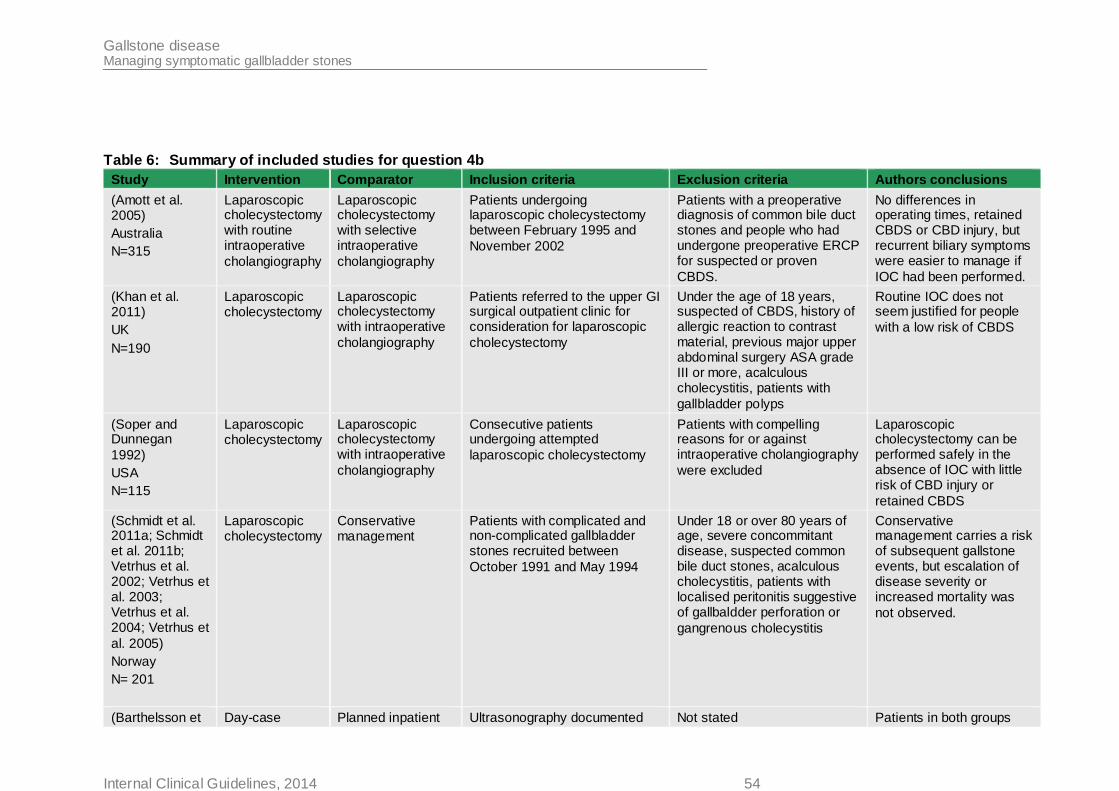
Gallstone disease Managing symptomatic gallbladder stones
Internal Clinical Guidelines, 2014 54
Table 6: Summary of included studies for question 4b
Study Intervention Comparator Inclusion criteria Exclusion criteria Authors conclusions
(Amott et al. 2005)
Australia
N=315
Laparoscopic cholecystectomy with routine intraoperative
cholangiography
Laparoscopic cholecystectomy with selective intraoperative
cholangiography
Patients undergoing laparoscopic cholecystectomy between February 1995 and
November 2002
Patients with a preoperative diagnosis of common bile duct stones and people who had undergone preoperative ERCP for suspected or proven
CBDS.
No differences in operating times, retained CBDS or CBD injury, but recurrent biliary symptoms were easier to manage if
IOC had been performed.
(Khan et al. 2011)
UK
N=190
Laparoscopic cholecystectomy
Laparoscopic cholecystectomy with intraoperative
cholangiography
Patients referred to the upper GI surgical outpatient clinic for consideration for laparoscopic
cholecystectomy
Under the age of 18 years, suspected of CBDS, history of allergic reaction to contrast material, previous major upper abdominal surgery ASA grade III or more, acalculous cholecystitis, patients with
gallbladder polyps
Routine IOC does not seem justified for people
with a low risk of CBDS
(Soper and Dunnegan
1992)
USA
N=115
Laparoscopic cholecystectomy
Laparoscopic cholecystectomy with intraoperative
cholangiography
Consecutive patients undergoing attempted
laparoscopic cholecystectomy
Patients with compelling reasons for or against intraoperative cholangiography
were excluded
Laparoscopic cholecystectomy can be performed safely in the absence of IOC with little risk of CBD injury or
retained CBDS
(Schmidt et al. 2011a; Schmidt et al. 2011b; Vetrhus et al. 2002; Vetrhus et al. 2003; Vetrhus et al. 2004; Vetrhus et
al. 2005)
Norway
N= 201
Laparoscopic cholecystectomy
Conservative management
Patients with complicated and non-complicated gallbladder stones recruited between
October 1991 and May 1994
Under 18 or over 80 years of age, severe concommitant disease, suspected common bile duct stones, acalculous cholecystitis, patients with localised peritonitis suggestive of gallbaldder perforation or
gangrenous cholecystitis
Conservative management carries a risk of subsequent gallstone events, but escalation of disease severity or increased mortality was
not observed.
(Barthelsson et Day-case Planned inpatient Ultrasonography documented Not stated Patients in both groups
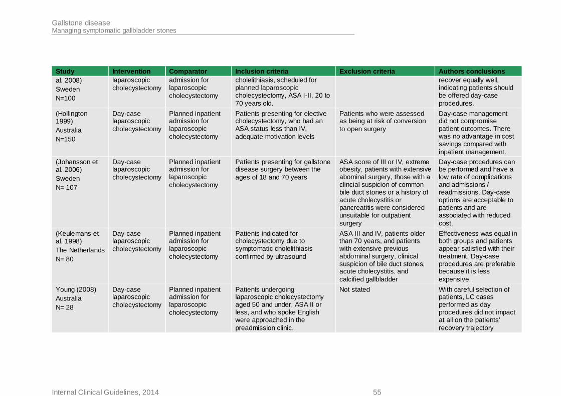
Gallstone disease Managing symptomatic gallbladder stones
Internal Clinical Guidelines, 2014 55
Study Intervention Comparator Inclusion criteria Exclusion criteria Authors conclusions
al. 2008)
Sweden
N=100
laparoscopic
cholecystectomy
admission for laparoscopic
cholecystectomy
cholelithiasis, scheduled for planned laparoscopic cholecystectomy, ASA I-II, 20 to
70 years old.
recover equally well, indicating patients should be offered day-case
procedures.
(Hollington 1999)
Australia
N=150
Day-case laparoscopic
cholecystectomy
Planned inpatient admission for laparoscopic
cholecystectomy
Patients presenting for elective cholecystectomy, who had an ASA status less than IV,
adequate motivation levels
Patients who were assessed as being at risk of conversion
to open surgery
Day-case management did not compromise patient outcomes. There was no advantage in cost savings compared with
inpatient management.
(Johansson et al. 2006)
Sweden
N= 107
Day-case laparoscopic
cholecystectomy
Planned inpatient admission for laparoscopic
cholecystectomy
Patients presenting for gallstone disease surgery between the
ages of 18 and 70 years
ASA score of III or IV, extreme obesity, patients with extensive abominal surgery, those with a clincial suspicion of common bile duct stones or a history of acute cholecystitis or pancreatitis were considered unsuitable for outpatient
surgery
Day-case procedures can be performed and have a low rate of complications and admissions / readmissions. Day-case options are acceptable to patients and are associated with reduced
cost.
(Keulemans et al. 1998)
The Netherlands
N= 80
Day-case laparoscopic
cholecystectomy
Planned inpatient admission for laparoscopic
cholecystectomy
Patients indicated for cholecystectomy due to symptomatic cholelithiasis
confirmed by ultrasound
ASA III and IV, patients older than 70 years, and patients with extensive previous abdominal surgery, clinical suspicion of bile duct stones, acute cholecystitis, and
calcified gallbladder
Effectiveness was equal in both groups and patients appear satisfied with their treatment. Day-case procedures are preferable because it is less
expensive.
Young (2008)
Australia
N= 28
Day-case laparoscopic
cholecystectomy
Planned inpatient admission for laparoscopic
cholecystectomy
Patients undergoing laparoscopic cholecystectomy aged 50 and under, ASA II or less, and who spoke English were approached in the
preadmission clinic.
Not stated With careful selection of patients, LC cases performed as day procedures did not impact at all on the patients'
recovery trajectory
Gallstone disease Managing symptomatic gallbladder stones
Internal Clinical Guidelines, 2014
56
1
4.5.3 Health economic evidence 2
A literature search was conducted jointly for questions 4 and 5 by applying standard health 3 economic filters to the clinical search strategies (see Appendix D). From the search, 1396 4
references were retrieved for questions 4 and 5, of which 1 was retained for question 4b and 5 1 was retained for question 5. As no existing health economic studies were found that 6 addressed all the comparisons in questions 4 and 5, an original economic model was 7
constructed. 8
4.5.3.1 Original health economic modelling – methods 9
A full description of the health economic model can be found in in Appendix J; a summary is 10 presented here. The model was developed in line with the NICE reference case (National 11 Institute for Health and Care Excellence 2013). A single health economic model structure 12
was developed to address all prioritised comparisons under questions 4b, 4c and 5. For 13 question 4b, no evidence was found to enable the modelling of the percutaneous 14 cholecystostomy comparison. 15
A single Markov structure with 2-week cycles was used to assess all comparisons (see 16 Figure 1). Not all states and transitions were used in each comparison (see Appendix J for 17 descriptions of the states and transitions available for each comparison). A lifetime horizon 18
was adopted to capture the long term impact of bile duct injuries and mortality differences. 19 The model is a natural history rather than a diagnosis model, so the model “knows” whether 20 a patient has CBDS, irrespective of whether the patient or clinician knows. 21
Surgical interventions (laparoscopic cholecystectomy and ERCP, or both together) are 22 represented as 2-week states, with short term surgical consequences (including mortality) 23 modelled as cost and QALY impacts within one 2-week state. Laparoscopic cholecystectomy 24 can cause bile duct injury. 25
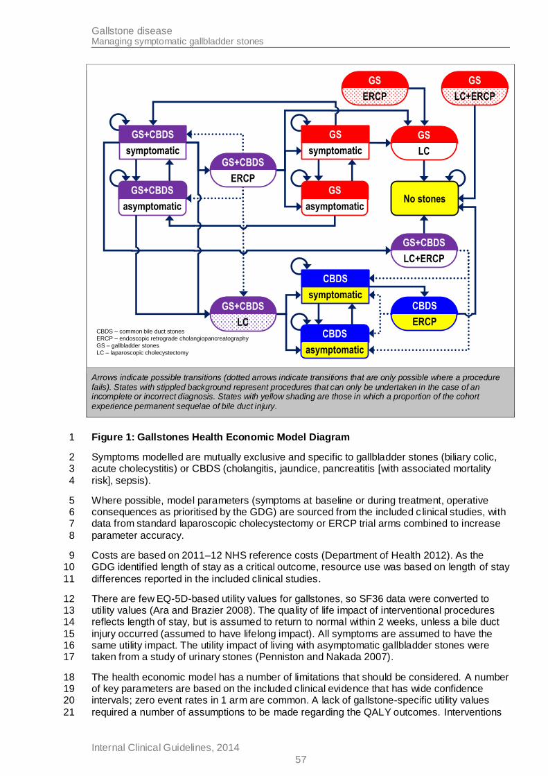
Gallstone disease Managing symptomatic gallbladder stones
Internal Clinical Guidelines, 2014
57
symptomatic
GS+CBDS
No stones
asymptomatic
CBDSERCP
CBDS
LC
GS
asymptomatic
GS
CBDS
symptomatic
LC
GS+CBDS
LC+ERCP
GS+CBDS
asymptomatic
GS+CBDS
symptomatic
GS
ERCP
GS
LC+ERCP
GS
ERCP
GS+CBDS
CBDS – common bile duct stones
ERCP – endoscopic retrograde cholangiopancreatography
GS – gallbladder stones
LC – laparoscopic cholecystectomy
Arrows indicate possible transitions (dotted arrows indicate transitions that are only possible where a procedure fails). States with stippled background represent procedures that can only be undertaken in the case of an incomplete or incorrect diagnosis. States with yellow shading are those in which a proportion of the cohort experience permanent sequelae of bile duct injury.
Figure 1: Gallstones Health Economic Model Diagram 1
Symptoms modelled are mutually exclusive and specific to gallbladder stones (biliary colic, 2 acute cholecystitis) or CBDS (cholangitis, jaundice, pancreatitis [with associated mortality 3
risk], sepsis). 4
Where possible, model parameters (symptoms at baseline or during treatment, operative 5 consequences as prioritised by the GDG) are sourced from the included c linical studies, with 6 data from standard laparoscopic cholecystectomy or ERCP trial arms combined to increase 7
parameter accuracy. 8
Costs are based on 2011–12 NHS reference costs (Department of Health 2012). As the 9 GDG identified length of stay as a critical outcome, resource use was based on length of stay 10
differences reported in the included clinical studies. 11
There are few EQ-5D-based utility values for gallstones, so SF36 data were converted to 12 utility values (Ara and Brazier 2008). The quality of life impact of interventional procedures 13 reflects length of stay, but is assumed to return to normal within 2 weeks, unless a bile duct 14
injury occurred (assumed to have lifelong impact). All symptoms are assumed to have the 15 same utility impact. The utility impact of living with asymptomatic gallbladder stones were 16 taken from a study of urinary stones (Penniston and Nakada 2007). 17
The health economic model has a number of limitations that should be considered. A number 18 of key parameters are based on the included c linical evidence that has wide confidence 19 intervals; zero event rates in 1 arm are common. A lack of gallstone-specific utility values 20
required a number of assumptions to be made regarding the QALY outcomes. Interventions 21
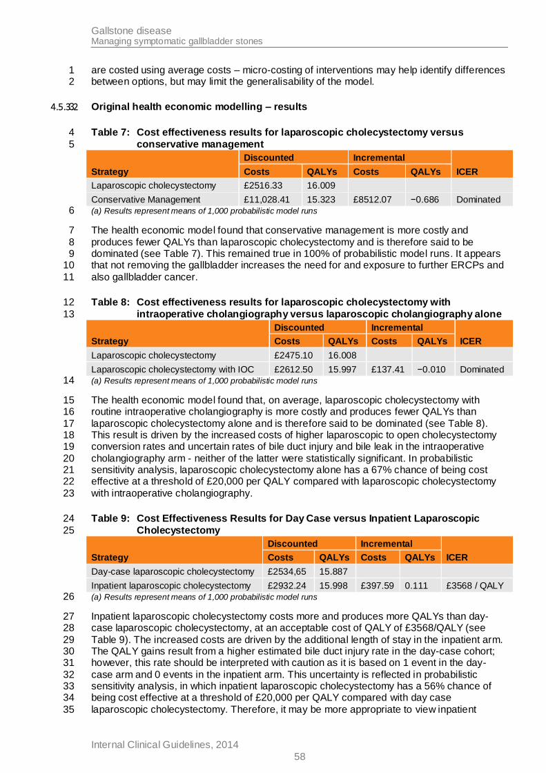
Gallstone disease Managing symptomatic gallbladder stones
Internal Clinical Guidelines, 2014
58
are costed using average costs – micro-costing of interventions may help identify differences 1 between options, but may limit the generalisability of the model. 2
4.5.3.2 Original health economic modelling – results 3
Table 7: Cost effectiveness results for laparoscopic cholecystectomy versus 4
conservative management 5
Strategy
Discounted Incremental
ICER Costs QALYs Costs QALYs
Laparoscopic cholecystectomy £2516.33 16.009
Conservative Management £11,028.41 15.323 £8512.07 −0.686 Dominated
(a) Results represent means of 1,000 probabilistic model runs 6
The health economic model found that conservative management is more costly and 7
produces fewer QALYs than laparoscopic cholecystectomy and is therefore said to be 8 dominated (see Table 7). This remained true in 100% of probabilistic model runs. It appears 9 that not removing the gallbladder increases the need for and exposure to further ERCPs and 10
also gallbladder cancer. 11
Table 8: Cost effectiveness results for laparoscopic cholecystectomy with 12
intraoperative cholangiography versus laparoscopic cholangiography alone 13
Strategy
Discounted Incremental
ICER Costs QALYs Costs QALYs
Laparoscopic cholecystectomy £2475.10 16.008
Laparoscopic cholecystectomy with IOC £2612.50 15.997 £137.41 −0.010 Dominated
(a) Results represent means of 1,000 probabilistic model runs 14
The health economic model found that, on average, laparoscopic cholecystectomy with 15 routine intraoperative cholangiography is more costly and produces fewer QALYs than 16
laparoscopic cholecystectomy alone and is therefore said to be dominated (see Table 8). 17 This result is driven by the increased costs of higher laparoscopic to open cholecystectomy 18 conversion rates and uncertain rates of bile duct injury and bile leak in the intraoperative 19
cholangiography arm - neither of the latter were statistically significant. In probabilistic 20 sensitivity analysis, laparoscopic cholecystectomy alone has a 67% chance of being cost 21 effective at a threshold of £20,000 per QALY compared with laparoscopic cholecystectomy 22
with intraoperative cholangiography. 23
Table 9: Cost Effectiveness Results for Day Case versus Inpatient Laparoscopic 24
Cholecystectomy 25
Strategy
Discounted Incremental
ICER Costs QALYs Costs QALYs
Day-case laparoscopic cholecystectomy £2534,65 15.887
Inpatient laparoscopic cholecystectomy £2932.24 15.998 £397.59 0.111 £3568 / QALY
(a) Results represent means of 1,000 probabilistic model runs 26
Inpatient laparoscopic cholecystectomy costs more and produces more QALYs than day-27 case laparoscopic cholecystectomy, at an acceptable cost of QALY of £3568/QALY (see 28
Table 9). The increased costs are driven by the additional length of stay in the inpatient arm. 29 The QALY gains result from a higher estimated bile duct injury rate in the day-case cohort; 30 however, this rate should be interpreted with caution as it is based on 1 event in the day-31
case arm and 0 events in the inpatient arm. This uncertainty is reflected in probabilistic 32 sensitivity analysis, in which inpatient laparoscopic cholecystectomy has a 56% chance of 33 being cost effective at a threshold of £20,000 per QALY compared with day case 34
laparoscopic cholecystectomy. Therefore, it may be more appropriate to view inpatient 35
Gallstone disease Managing symptomatic gallbladder stones
Internal Clinical Guidelines, 2014
59
laparoscopic cholecystectomy as more expensive than day-case laparoscopic 1 cholecystectomy with comparable QALY outcomes. 2
4.5.4 Evidence Statements 3
One study provided low quality evidence about conservative management in comparison 4 with laparoscopic cholecystectomy, and there was some evidence that laparoscopic 5 cholecystectomy was superior to conservative management. Readmission rates were lower 6
in the laparoscopic cholecystectomy group that the conservative management group, and 7 44% of people in the conservative management group also required cholecystectomy 8
Three studies provided moderate to low quality evidence about laparoscopic 9 cholecystectomy in comparison with laparoscopic cholecystectomy with intraoperative 10
cholangiography and there was uncertainty about whether the addition of intraoperative 11 cholangiography was beneficial or not since there were no significant differences between 12 the groups on any of the outcomes. 13
No evidence was available comparing cholecystostomy to laparoscopic cholecystectomy, 14 and so no conclusions can be drawn. 15
Five studies providing moderate to low quality evidence compared day-case laparoscopic 16 cholecystectomy with planned inpatient laparoscopic cholecystectomy. No differences in 17
readmission rate or quality of life were found. Unplanned inpatient admissions were required 18 in 12.1% of planned day case procedures. 19
A directly applicable original health economic model analysis with minor limitations suggests 20 laparoscopic cholecystectomy is cost effective compared with conservative management. 21
Comparisons of laparoscopic cholecystectomy with laparoscopic cholecystectomy and intra-22 operative cholangiography and day-case with inpatient laparoscopic cholecystectomy were 23
limited by the quality of the clinical evidence. 24
25
4.5.5 Evidence to Recommendations 26
Relative value of different
outcomes
In general, studies were underpowered for the outcomes that the GDG had chosen and so confidence intervals relating to effect estimates for
many outcomes were wide. This was expected, since events such as mortality and bile duct injury are relatively rare and sample sizes would need to be very large to observe sufficient rare events.
There was also a lack of randomised controlled trial (RCT) evidence for some of the comparisons that were considered as part of this
review. The GDG felt that it is generally accepted that cholecystectomy is safe and effective, and that conducting clinical trials where participants for whom cholecystectomy is appropriate are
randomised to a treatment that may be less effective may be seen as unethical, thus limiting the amount of RCT evidence that is available.
Trade off between benefits and
harms
Laparoscopic cholecystectomy compared with conservative management
The evidence comparing conservative management to laparoscopic cholecystectomy was limited. Only 1 underpowered study met the inclusion criteria. The evidence showed that disease progression was
statistically significantly lower in patients offered cholecystectomy compared with conservative management, but there were no
statistically significant differences between the groups in terms of
Gallstone disease Managing symptomatic gallbladder stones
Internal Clinical Guidelines, 2014
60
readmission, or mortality. Other outcomes that the GDG felt were
critical to their decision making (requirement for additional intervention, and length of stay) were not reported by the study. The GDG agreed that the results imply that conservative management is
only suitable as a temporary management strategy, as patients offered conservative management continued to have recurrent biliary events. This supports the GDG’s views that once a gallbladder develops
gallstones, it will continue to do so, and that the only way of stopping gallstone formation and preventing recurrent biliary events is cholecystectomy.
The GDG agreed that laparoscopic cholecystectomy has been established as a relatively safe procedure, and the risks of disease
progression outweigh the risks of this type of surgery. Of course all surgeries carry some risks, particularly for people with complex, multiple conditions and for these patients the risk of surgery may well
outweigh the risk of disease progression. Furthermore, some patients may choose not to opt for surgical treatment. Thus, the GDG agreed that conservative management should only be considered when
cholecystectomy is not appropriate, and felt that this is standard practice across the NHS already and so did not require a specific recommendation to be made.
However, the GDG did feel that sometimes older patients and those with comorbidities may be assumed to be inappropriate for
laparoscopic cholecystectomy before they have even been assessed. Therefore the group felt the need to reinforce the message that laparoscopic cholecystectomy is becoming increasingly safer and that
it is inappropriate not to offer this treatment to someone where the risks of disease progression outweigh the risks of surgery. The GDG felt justified in making a strong recommendation to offer laparoscopic
cholecystectomy to all patients with symptomatic gallbladder stones, since there is sufficient acceptance of this procedure in the clinical
community which is supported by the GDG’s experience, health economic evidence, and by a small amount of clinical evidence found in this review.
Laparoscopic cholecystectomy compared with percutaneous
cholecystostomy
No evidence that met the inclusion criteria was available about comparing percutaneous cholecystostomy to laparoscopic
cholecystectomy and so the GDG discussed this treatment option by reflecting on its knowledge and experience.
Percutaneous cholecystostomy is widely accepted to be a safe and effective treatment for patients with a gallbladder obstruction. The GDG felt that percutaneous cholecystostomy is only used when
cholecystectomy is not an appropriate treatment option (either because of patient preferences or clinical judgement), and that this is standard practice across the NHS. However, the GDG agreed that
there is inappropriate variation in the way that patients are managed once a percutaneous cholecystostomy has been placed. This was
attributed to the fact that patients presenting with symptomatic gallbladder stones will usually be admitted to a surgeon, and when surgery is identified as not being an appropriate treatment option, the
patient is then considered for percutaneous cholecystostomy. The GDG felt that some patients are never reassessed for cholecystectomy, even when their clinical condition has improved
Gallstone disease Managing symptomatic gallbladder stones
Internal Clinical Guidelines, 2014
61
making surgery an appropriate option for them. The GDG agreed that
there is a high incidence of recurrent gallbladder sepsis in this group of patients which has a high risk of mortality associated with it. Thus, the GDG agreed to recommend that patients with percutaneous
cholecystostomy should be reviewed for reconsideration for cholecystectomy once adequate drainage has been established and the acute episode has resolved in order to minimise the risk of
recurrent sepsis and mortality.
Laparoscopic cholecystectomy compared with laparoscopic
cholecystectomy with intraoperative cholangiography (IOC)
Intraoperative cholangiography is performed during laparoscopic cholecystectomy to improve the safety of the procedure, by improving visualisation of the biliary anatomy and therefore reducing bile duct
injuries, and it can also identify common bile duct stones that weren’t suspected at earlier diagnostic work up. The evidence review
considered the use of routine IOC, selective IOC, and not using IOC at all, but there were no statistically significant differences for any of the outcomes that the GDG had chosen to base its decisions on. The
GDG felt that this was because the studies were underpowered, and so were not confident in these findings. Thus, the discussions about IOC were based on the knowledge and experience of the GDG.
The GDG acknowledged that the individual studies included in the review drew mixed conclusions about whether IOC was useful or not, and this reflected current differences in clinical opinions about the usefulness of the procedure. The GDG reflected on its knowledge and
experience and agreed that using IOC to improve visualisation of biliary anatomy offers clear benefits during laparoscopic
cholecystectomy. This is because improved visualisation improves the surgeon’s ability to plan and progress with laparoscopic cholecystectomy, making it safer for the patient. Because of this the
GDG agreed that it is desirable for all surgeons performing laparoscopic cholecystectomy to be competent in IOC.
However, there was further debate in relation to the benefits and harms of IOC for detecting common bile duct stones, and these
discussions linked to evidence about managing common bile duct stones which is presented in section 4.6 of this guideline, where it was agreed that all common bile duct stones need to be removed,
regardless of whether they are causing symptoms or not.
Common bile duct stones found during IOC can be removed laparoscopically by a surgeon who has undergone specialist training.
Most surgeons trained to do laparoscopic cholecystectomy do not have this specialist training and will not remove bile duct stones laparoscopically. Alternatively, common bile duct stones can be
removed endoscopically. This can be done pre-, intra-, or post- operatively by someone who has undergone specific endoscopy training. Although some surgeons are appropriately trained for
performing endoscopies, the majority of surgeons trained to perform laparoscopic cholecystectomy are not, and surgical lists are not usually planned with both a surgeon and an endoscopist in
attendance. Thus the GDG agreed that few teams would be equipped to deal with common bile duct stones that might be identified. The GDG felt that once common bile duct stones are discovered, they
Gallstone disease Managing symptomatic gallbladder stones
Internal Clinical Guidelines, 2014
62
need removing either at the time of discovery or within 2 or 3 days.
Thus, if an unequipped team discovered stones the team would need to make an unplanned and urgent call for an appropriately trained person to remove the stones during the operation. Alternatively stones
would need to be left in situ and the patient rescheduled for another intervention, thus exposing the patient to the risk of the stone developing serious complications during the wait, and the risks
associated with undergoing a second intervention (such as risks of anaesthesia and procedural errors). Thus the group agreed that there was insufficient evidence of benefit to support a recommendation
about using IOC for diagnosing common bile duct stones, or when inadequate preoperative diagnostic tests have been performed. Instead, the GDG felt that good preoperative examinations using
diagnostic methods outlined in section 4.2 would improve the chances of common bile duct stones being identified preoperatively so that
subsequent cholecystectomy can be planned to deal with common bile duct stones by appropriate surgeons and/or endoscopists.
Overall, the GDG felt that IOC is beneficial for visualising the biliary anatomy and making laparoscopic cholecystectomy safer, and
because of this surgeons performing laparoscopic cholecystectomy should be competent to use the procedure. However, the use of IOC varies in current clinical practice as there are uncertainties about its
potential harms when unexpected common bile duct stones are discovered by surgeons who do not have specialist training to remove the stones laparoscopically. Thus the GDG decided not to make a
recommendation about IOC. Instead a research recommendation was made to support the development of more research to explore the
potential benefits and harms of this procedure.
The GDG also wanted to stress that the lack of a recommendation should not preclude suitably trained individuals from using IOC at their discretion in the appropriate clinical situations.
Day-case laparoscopic cholecystectomy compared with inpatient stay laparoscopic cholecystectomy
The choice about whether to perform laparoscopic cholecystectomy as a day-case procedure or as planned inpatient admission was also
reviewed. The evidence revealed that there were no statistically significant differences for any of the outcomes that the GDG had chosen to base their decisions on. Furthermore, the GDG
acknowledged that some of the evidence included in this comparison came from older studies that might not accurately reflect continuing improvements in surgical outcomes that have occurred over time.
Thus the discussions about day-case or planned inpatient admission were based on the knowledge and experience of the GDG.
The GDG agreed that there are no differences in important outcomes between day-case and inpatient procedures. The GDG discussed
whether day-case surgery should always be offered to patients, since it offers the benefits of enabling the patient to return home sooner, reducing the risk of hospital acquired infections, and reducing the use
of NHS resources.
However, there are various patient and clinical factors that need to be taken into consideration. For example, patients require adult
Gallstone disease Managing symptomatic gallbladder stones
Internal Clinical Guidelines, 2014
63
supervision for 24hours after cholecystectomy and people who do not
have someone at home to supervise them will require an inpatient stay. Similarly, some patients with comorbidities may need admitting to hospital before or after the operation to manage their other
conditions. Thus the GDG felt that decisions about whether to plan day-case or inpatient cholecystectomy should be decided at a local level on a case by case basis to take the various patient and clinical
factors into account.
Consideration of Health Benefits
and Resource Use
For laparoscopic cholecystectomy compared with conservative management, the economic model suggests the costs and health benefits of surgery outweigh the health detriment of leaving the gallbladder in situ and having recurrent biliary events. The GDG noted
the economic model considered this question as a binary choice – either removing the gallbladder or leaving it in situ – that is unlikely to
occur in clinical practice. In sensitivity analyses, the economic model almost always produces higher costs and lower QALYs, but did not specifically consider older patients with comorbidities.
For laparoscopic cholecystectomy versus laparoscopic cholecystectomy with intra-operative cholangiography, the economic model suggests laparoscopic cholecystectomy is a more cost-effective option. However, this reflects clinical evidence of small non-statistically
significant differences in rates of operative consequences, in which the GDG were not confident. Probabilistic analysis of the model suggested laparoscopic cholecystectomy was the cost-effective option in around
2/3 of cases over a wide range of assumed values of 1 QALY, so intra-operative cholangiography may be cost effective in some situations and when done by appropriately trained surgeons
Whilst the comparison of day case to inpatient laparoscopic cholecystectomy suggests inpatient laparoscopic cholecystectomy is more cost effective, this again is based on clinical evidence of small
non-statistically significant differences (in bile duct injury rates) in which the GDG were not confident. Increased costs of inpatient laparoscopic cholecystectomy were driven by higher lengths of stay. It
may be more appropriate to view inpatient laparoscopic cholecystectomy as more expensive than day-case laparoscopic cholecystectomy with comparable QALY outcomes.
Quality of
evidence
Evidence was of moderate to low quality, and some comparisons had
no evidence at all.
Other considerations
None
1
4.5.6 Recommendations 2
7. Offer laparoscopic cholecystectomy to people diagnosed with symptomatic 3
gallbladder stones. 4
8. Offer day-case laparoscopic cholecystectomy for people having it as an elective 5 planned procedure, unless their circumstances or clinical condition make an 6
inpatient stay more appropriate. 7
Gallstone disease Managing symptomatic gallbladder stones
Internal Clinical Guidelines, 2014
64
9. Offer percutaneous cholecystostomy to manage gallbladder empyema when: 1
surgery is not appropriate at presentation and 2
conservative management is unsuccessful. 3
10. Reconsider laparoscopic cholecystectomy for people who have had percutaneous 4 cholecystostomy once they are well enough for surgery. 5
4.5.7 Research recommendations 6
7. What are the benefits and harms of routine intraoperative cholangiography in 7 people with low to intermediate risk of common bile duct stones? 8
Why this is important 9
In the evidence reviewed for this guideline, there was a lack of randomised controlled trials of 10 intraoperative cholangiography. Therefore, there is a need for large, high-quality trials to 11 address clinical questions about the benefits and harms of intraoperative cholangiography. 12
4.5.8 References 13
Amott D, Webb A, Tulloh B (2005) Prospective comparison of routine and selective operative 14 cholangiography. ANZ Journal of Surgery 75: 378-82 15
Ara R, Brazier J (2008) Deriving an algorithm to convert the eight mean SF-36 dimension 16 scores into a mean EQ-5D preference-based score from published studies (where patient 17
level data are not available). Value in Health 11: 1131-43 18
Barthelsson C, Anderberg B, Ramel S et al. (2008) Outpatient versus inpatient laparoscopic 19 cholecystectomy: a prospective randomized study of symptom occurrence, symptom distress 20
and general state of health during the first post-operative week. Journal of Evaluation in 21 Clinical Practice 14: 577-84 22
Department of Health (2012) National Schedule of Reference Costs 2011-2012. 23
Hollington P (1999) A prospective randomized trial of day-stay only versus overnight-stay 24 laparoscopic cholecystectomy. The Australian and New Zealand Journal of Surgery 69: 841-25
3 26
Johansson M, Thune A, Nelvin L et al. (2006) Randomized clinical trial of day-care versus 27 overnight-stay laparoscopic cholecystectomy. British Journal of Surgery 93: 40-5 28
Keulemans Y, Eshuis J, de HH et al. (1998) Laparoscopic cholecystectomy: day-care versus 29 clinical observation. Annals of Surgery 228: 734-40 30
Khan OA, Balaji S, Branagan G et al. (2011) Randomized clinical trial of routine on-table 31 cholangiography during laparoscopic cholecystectomy. British Journal of Surgery 98: 362-7 32
National Institute for Health and Care Excellence (2013) Guide to Methods of Technology 33 Appraisal. 34
Penniston KL, Nakada SY (2007) Health Related Quality of Life Differs Between Male and 35 Female Stone Formers. The Journal of Urology 178: 2435-40 36
Schmidt M, Sondenaa K, Vetrhus M et al. (2011a) A randomized controlled study of 37
uncomplicated gallstone disease with a 14-year follow-up showed that operation was the 38 preferred treatment. Digestive Surgery 28: 270-6 39
Gallstone disease Managing symptomatic gallbladder stones
Internal Clinical Guidelines, 2014
65
Schmidt M, Sondenaa K, Vetrhus M et al. (2011b) Long-term follow-up of a randomized 1 controlled trial of observation versus surgery for acute cholecystitis: non-operative 2 management is an option in some patients. Scandinavian Journal of Gastroenterology 46: 3
1257-62 4
Soper NJ, Dunnegan DL (1992) Routine versus selective intra-operative cholangiography 5 during laparoscopic cholecystectomy. World Journal of Surgery 16: 1133-40 6
Vetrhus M, Soreide O, Eide GE et al. (2005) Quality of life and pain in patients with acute 7 cholecystitis. Results of a randomized clinical trial. Scandinavian Journal of Surgery: SJS 94: 8 34-9 9
Vetrhus M, Soreide O, Eide GE et al. (2004) Pain and quality of life in patients with 10 symptomatic, non-complicated gallbladder stones: results of a randomized controlled trial. 11
Scandinavian Journal of Gastroenterology 39: 270-6 12
Vetrhus M, Soreide O, Nesvik I et al. (2003) Acute cholecystitis: delayed surgery or 13 observation. A randomized clinical trial. Scandinavian Journal of Gastroenterology 38: 985-14
90 15
Vetrhus M, Soreide O, Solhaug JH et al. (2002) Symptomatic, non-complicated gallbladder 16 stone disease. Operation or observation? A randomized clinical study. Scandinavian Journal 17 of Gastroenterology 37: 834-918
Gallstone disease Managing common bile duct stones
Internal Clinical Guidelines, 2014
66
4.6 Managing common bile duct stones 1
4.6.1 Review Question 4c 2
Which strategies should be used for managing common bile duct stones? 3
4.6.2 Evidence Review 4
A single search was performed for questions 4a, 4b, 4c and 5 which identified 10,976 5 references. After removing duplicates and screening the references based on their titles and 6 abstracts, 210 references were obtained and reviewed against the inclusion and exclusion 7 criteria for this review question (appendix C), and 47 references met the inclusion criteria 8
overall. Details of excluded studies and reasons for their exclusion are in appendix F.4 . 9
Of the 47 included references, 24 references relating to 24 randomised controlled trials were 10 included in this review question (see study flow chart, appendix E.4) which specifically 11
addressed the following comparisons for managing common bile duct stones: 12
ERCP vs conservative management (9 studies) 13
ERCP and laparoscopic cholecystectomy vs ERCP alone (2 studies) 14
(pre-, intra-, post- operative) ERCP clearance of the bile duct plus laparoscopic 15
cholecystectomy compared with surgical clearance of the bile duct plus laparoscopic 16
cholecystectomy (12 studies) 17
Uncleared duct with biliary stent vs cleared duct (1 study) 18
Day-case ERCP vs planned inpatient ERCP (no studies) 19
Data were extracted into detailed evidence tables (see appendix G.4) and are summarised in 20 Table 10 below. Each study was assessed for methodological quality using randomised 21
controlled trial checklists, and some studies had methodological flaws which impacted on the 22 overall quality of the evidence. For example, some studies didn’t report randomisation 23 procedures and those that did sometimes used inappropriate methods (patient identifying 24
numbers – Hong, 2006), or had methods that led to differences in baseline characteristics of 25 the groups (for example, Hui, 2002) which could bias results. 26
Data from multiple studies were available for most of the comparisons; therefore, meta-27 analysis was performed wherever possible (see appendix H.7). In addition, network meta-28 analysis was possible to compare the different methods for ERCP clearance of the duct (pre-29 , intra-, or post- operative) in comparison with surgical clearance. Standard GRADE 30
approaches were used to assess pairwise comparisons, and a modified version of the 31 GRADE framework was used to assess the evidence analysed using network meta analysis 32 (see appendix I.6 for full GRADE profiles). In the modified approach, the standard GRADE 33
criteria still apply but additional factors are also considered such as how each 'link' or 34 pairwise comparison within the network applies to the others (see NICE clinical guideline 35 CG173, appendix D for details of the additional factors that may be considered in a network 36
meta analysis). However, since the network of evidence for this review question was 37 relatively small, very few of the additional criteria actually applied. Details of the specific 38
reasons for downgrading are stated in the footnotes of the relevant GRADE profiles in 39 appendix I.6. 40
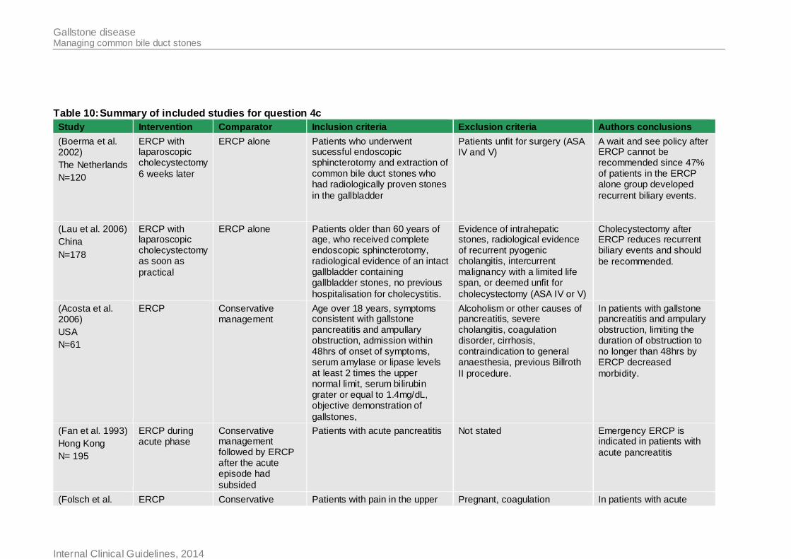
Gallstone disease Managing common bile duct stones
Internal Clinical Guidelines, 2014
Table 10: Summary of included studies for question 4c
Study Intervention Comparator Inclusion criteria Exclusion criteria Authors conclusions
(Boerma et al. 2002)
The Netherlands
N=120
ERCP with laparoscopic cholecystectomy
6 weeks later
ERCP alone Patients who underwent sucessful endoscopic sphincterotomy and extraction of common bile duct stones who had radiologically proven stones
in the gallbladder
Patients unfit for surgery (ASA IV and V)
A wait and see policy after ERCP cannot be recommended since 47% of patients in the ERCP alone group developed
recurrent biliary events.
(Lau et al. 2006)
China
N=178
ERCP with laparoscopic cholecystectomy as soon as
practical
ERCP alone Patients older than 60 years of age, who received complete endoscopic sphincterotomy, radiological evidence of an intact gallbladder containing gallbladder stones, no previous
hospitalisation for cholecystitis.
Evidence of intrahepatic stones, radiological evidence of recurrent pyogenic cholangitis, intercurrent malignancy with a limited life span, or deemed unfit for
cholecystectomy (ASA IV or V)
Cholecystectomy after ERCP reduces recurrent biliary events and should
be recommended.
(Acosta et al. 2006)
USA
N=61
ERCP Conservative management
Age over 18 years, symptoms consistent with gallstone pancreatitis and ampullary obstruction, admission within 48hrs of onset of symptoms, serum amylase or lipase levels at least 2 times the upper normal limit, serum bilirubin grater or equal to 1.4mg/dL, objective demonstration of
gallstones,
Alcoholism or other causes of pancreatitis, severe cholangitis, coagulation disorder, cirrhosis, contraindication to general anaesthesia, previous Billroth
II procedure.
In patients with gallstone pancreatitis and ampulary obstruction, limiting the duration of obstruction to no longer than 48hrs by ERCP decreased
morbidity.
(Fan et al. 1993)
Hong Kong
N= 195
ERCP during acute phase
Conservative management followed by ERCP after the acute episode had
subsided
Patients with acute pancreatitis Not stated Emergency ERCP is indicated in patients with
acute pancreatitis
(Folsch et al. ERCP Conservative Patients with pain in the upper Pregnant, coagulation In patients with acute
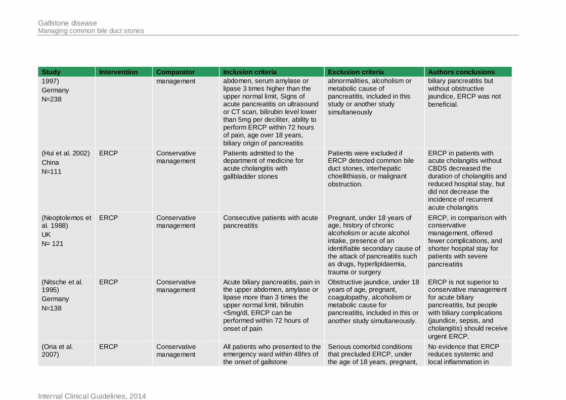
Gallstone disease Managing common bile duct stones
Internal Clinical Guidelines, 2014
Study Intervention Comparator Inclusion criteria Exclusion criteria Authors conclusions
1997)
Germany
N=238
management abdomen, serum amylase or lipase 3 times higher than the upper normal limit, Signs of acute pancreatitis on ultrasound or CT scan, bilirubin level lower than 5mg per deciliter, ability to perform ERCP within 72 hours of pain, age over 18 years,
biliary origin of pancreatitis
abnormalities, alcoholism or metabolic cause of pancreatitis, included in this study or another study
simultaneously
biliary pancreatitis but without obstructive jaundice, ERCP was not
beneficial.
(Hui et al. 2002)
China
N=111
ERCP Conservative management
Patients admitted to the department of medicine for acute cholangitis with
gallbladder stones
Patients were excluded if ERCP detected common bile duct stones, interhepatic choellithiasis, or malignant
obstruction.
ERCP in patients with acute cholangitis without CBDS decreased the duration of cholangitis and reduced hospital stay, but did not decrease the incidence of recurrent
acute cholangitis
(Neoptolemos et al. 1988)
UK
N= 121
ERCP Conservative management
Consecutive patients with acute pancreatitis
Pregnant, under 18 years of age, history of chronic alcoholism or acute alcohol intake, presence of an identifiable secondary cause of the attack of pancreatitis such as drugs, hyperlipidaemia,
trauma or surgery
ERCP, in comparison with conservative management, offered fewer complications, and shorter hospital stay for patients with severe
pancreatitis
(Nitsche et al. 1995)
Germany
N=138
ERCP Conservative management
Acute biliary pancreatitis, pain in the upper abdomen, amylase or lipase more than 3 times the upper normal limit, bilirubin <5mg/dl, ERCP can be performed within 72 hours of
onset of pain
Obstructive jaundice, under 18 years of age, pregnant, coagulopathy, alcoholism or metabolic cause for pancreatitis, included in this or
another study simultaneously.
ERCP is not superior to conservative management for acute biliary pancreatitis, but people with biliary complications (jaundice, sepsis, and cholangitis) should receive
urgent ERCP.
(Oria et al. 2007)
ERCP Conservative management
All patients who presented to the emergency ward within 48hrs of the onset of gallstone
Serious comorbid conditions that precluded ERCP, under the age of 18 years, pregnant,
No evidence that ERCP reduces systemic and local inflammation in
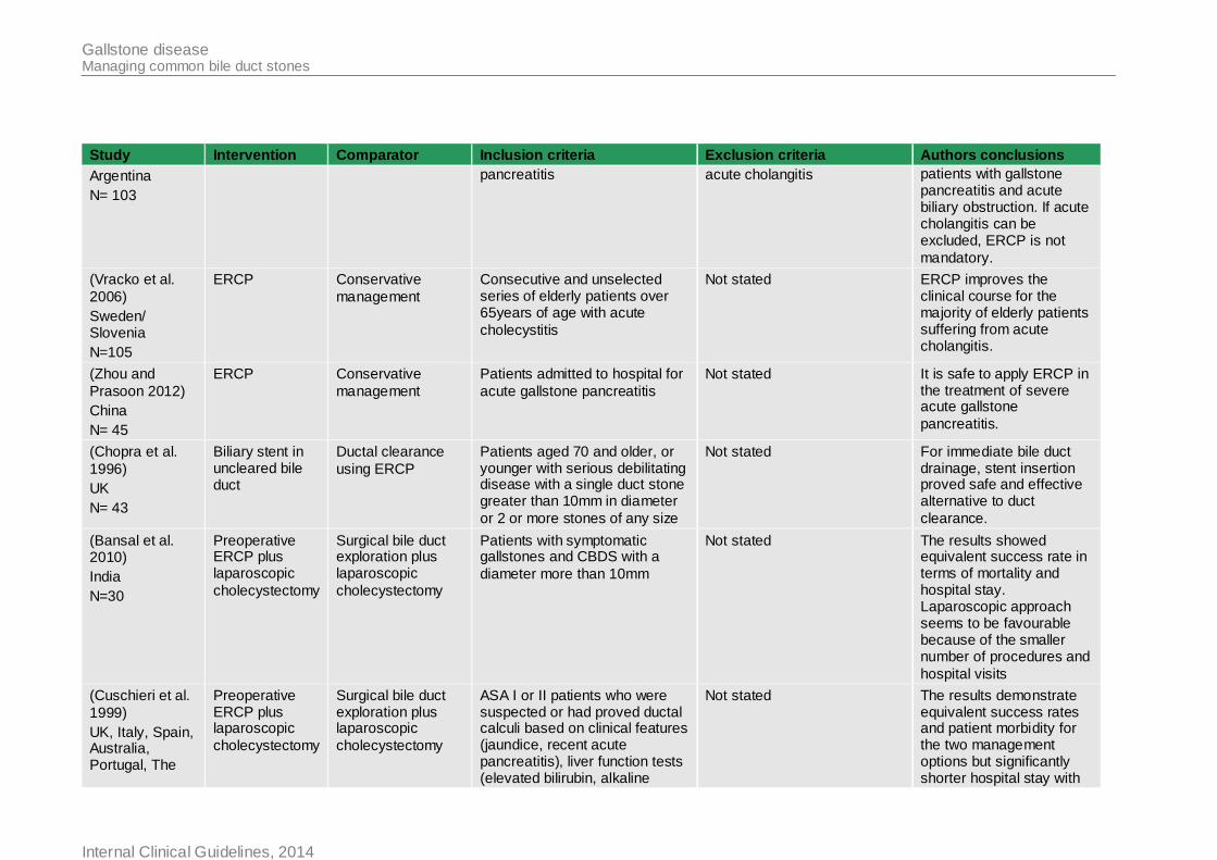
Gallstone disease Managing common bile duct stones
Internal Clinical Guidelines, 2014
Study Intervention Comparator Inclusion criteria Exclusion criteria Authors conclusions
Argentina
N= 103
pancreatitis acute cholangitis patients with gallstone pancreatitis and acute biliary obstruction. If acute cholangitis can be excluded, ERCP is not
mandatory.
(Vracko et al.
2006)
Sweden/ Slovenia
N=105
ERCP Conservative
management
Consecutive and unselected series of elderly patients over 65years of age with acute
cholecystitis
Not stated ERCP improves the clinical course for the majority of elderly patients suffering from acute cholangitis.
(Zhou and
Prasoon 2012)
China
N= 45
ERCP Conservative
management
Patients admitted to hospital for
acute gallstone pancreatitis
Not stated It is safe to apply ERCP in the treatment of severe acute gallstone
pancreatitis.
(Chopra et al.
1996)
UK
N= 43
Biliary stent in uncleared bile duct
Ductal clearance
using ERCP
Patients aged 70 and older, or younger with serious debilitating disease with a single duct stone greater than 10mm in diameter
or 2 or more stones of any size
Not stated For immediate bile duct drainage, stent insertion proved safe and effective alternative to duct
clearance.
(Bansal et al. 2010)
India
N=30
Preoperative ERCP plus laparoscopic
cholecystectomy
Surgical bile duct exploration plus laparoscopic
cholecystectomy
Patients with symptomatic gallstones and CBDS with a
diameter more than 10mm
Not stated The results showed equivalent success rate in terms of mortality and hospital stay. Laparoscopic approach seems to be favourable because of the smaller number of procedures and
hospital visits
(Cuschieri et al.
1999)
UK, Italy, Spain, Australia, Portugal, The
Preoperative ERCP plus laparoscopic
cholecystectomy
Surgical bile duct exploration plus laparoscopic
cholecystectomy
ASA I or II patients who were suspected or had proved ductal calculi based on clinical features (jaundice, recent acute pancreatitis), liver function tests (elevated bilirubin, alkaline
Not stated The results demonstrate equivalent success rates and patient morbidity for the two management options but significantly shorter hospital stay with
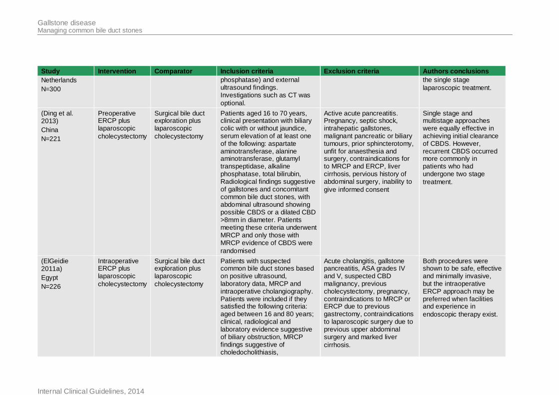
Gallstone disease Managing common bile duct stones
Internal Clinical Guidelines, 2014
Study Intervention Comparator Inclusion criteria Exclusion criteria Authors conclusions
Netherlands
N=300
phosphatase) and external ultrasound findings. Investigations such as CT was
optional.
the single stage
laparoscopic treatment.
(Ding et al. 2013)
China
N=221
Preoperative ERCP plus laparoscopic
cholecystectomy
Surgical bile duct exploration plus laparoscopic
cholecystectomy
Patients aged 16 to 70 years, clinical presentation with biliary colic with or without jaundice, serum elevation of at least one of the following: aspartate aminotransferase, alanine aminotransferase, glutamyl transpeptidase, alkaline phosphatase, total bilirubin, Radiological findings suggestive of gallstones and concomitant common bile duct stones, with abdominal ultrasound showing possible CBDS or a dilated CBD >8mm in diameter. Patients meeting these criteria underwent MRCP and only those with MRCP evidence of CBDS were
randomised
Active acute pancreatitis. Pregnancy, septic shock, intrahepatic gallstones, malignant pancreatic or biliary tumours, prior sphincterotomy, unfit for anaesthesia and surgery, contraindications for to MRCP and ERCP, liver cirrhosis, pervious history of abdominal surgery, inability to
give informed consent
Single stage and multistage approaches were equally effective in achieving initial clearance of CBDS. However, recurrent CBDS occurred more commonly in patients who had undergone two stage
treatment.
(ElGeidie 2011a)
Egypt
N=226
Intraoperative ERCP plus laparoscopic
cholecystectomy
Surgical bile duct exploration plus laparoscopic
cholecystectomy
Patients with suspected common bile duct stones based on positive ultrasound, laboratory data, MRCP and intraoperative cholangiography. Patients were included if they satisfied the following criteria: aged between 16 and 80 years; clinical, radiological and laboratory evidence suggestive of biliary obstruction, MRCP findings suggestive of choledocholithiasis,
Acute cholangitis, gallstone pancreatitis, ASA grades IV and V, suspected CBD malignancy, previous cholecystectomy, pregnancy, contraindications to MRCP or ERCP due to previous gastrectomy, contraindications to laparoscopic surgery due to previous upper abdominal surgery and marked liver
cirrhosis.
Both procedures were shown to be safe, effective and minimally invasive, but the intraoperative ERCP approach may be preferred when facilities and experience in
endoscopic therapy exist.
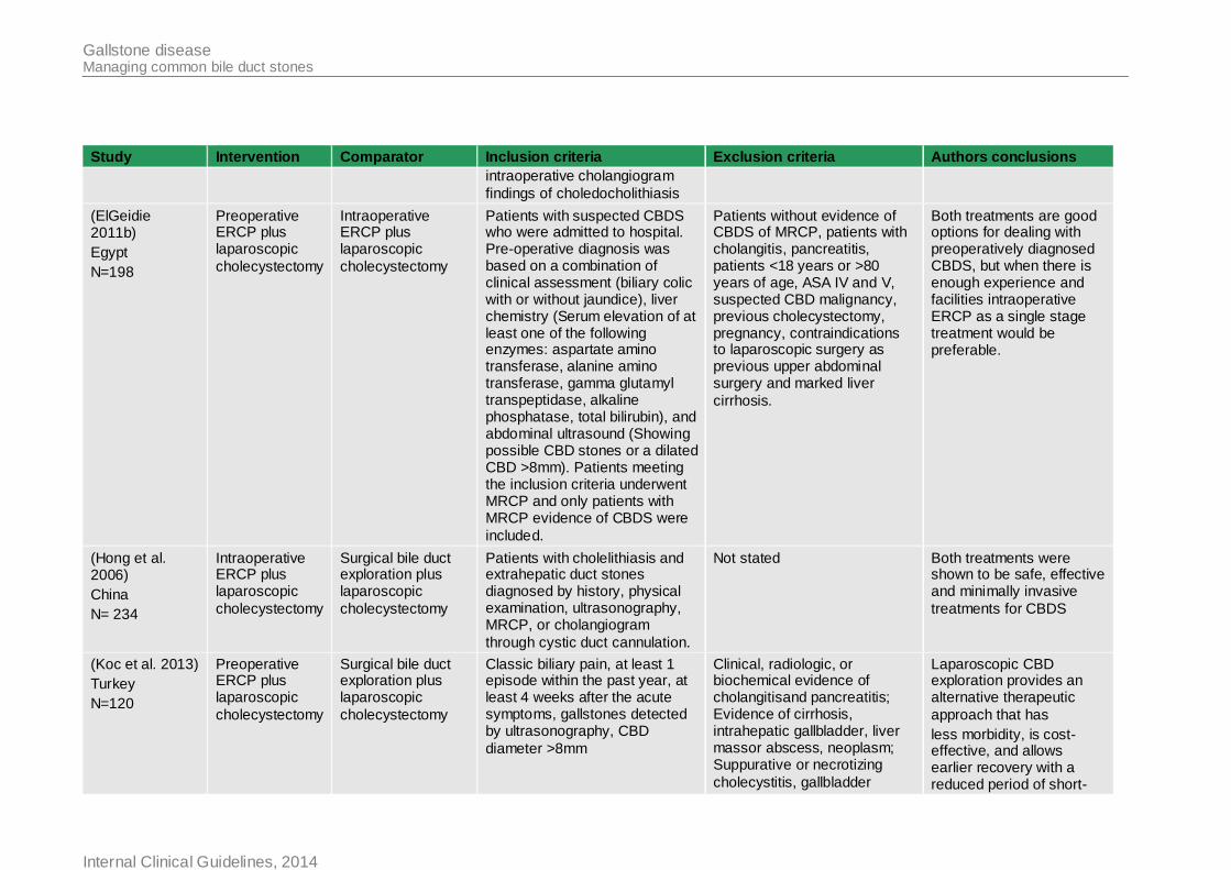
Gallstone disease Managing common bile duct stones
Internal Clinical Guidelines, 2014
Study Intervention Comparator Inclusion criteria Exclusion criteria Authors conclusions
intraoperative cholangiogram
findings of choledocholithiasis
(ElGeidie 2011b)
Egypt
N=198
Preoperative ERCP plus laparoscopic
cholecystectomy
Intraoperative ERCP plus laparoscopic
cholecystectomy
Patients with suspected CBDS who were admitted to hospital. Pre-operative diagnosis was based on a combination of clinical assessment (biliary colic with or without jaundice), liver chemistry (Serum elevation of at least one of the following enzymes: aspartate amino transferase, alanine amino transferase, gamma glutamyl transpeptidase, alkaline phosphatase, total bilirubin), and abdominal ultrasound (Showing possible CBD stones or a dilated CBD >8mm). Patients meeting the inclusion criteria underwent MRCP and only patients with MRCP evidence of CBDS were
included.
Patients without evidence of CBDS of MRCP, patients with cholangitis, pancreatitis, patients <18 years or >80 years of age, ASA IV and V, suspected CBD malignancy, previous cholecystectomy, pregnancy, contraindications to laparoscopic surgery as previous upper abdominal surgery and marked liver
cirrhosis.
Both treatments are good options for dealing with preoperatively diagnosed CBDS, but when there is enough experience and facilities intraoperative ERCP as a single stage treatment would be preferable.
(Hong et al. 2006)
China
N= 234
Intraoperative ERCP plus laparoscopic
cholecystectomy
Surgical bile duct exploration plus laparoscopic
cholecystectomy
Patients with cholelithiasis and extrahepatic duct stones diagnosed by history, physical examination, ultrasonography, MRCP, or cholangiogram
through cystic duct cannulation.
Not stated Both treatments were shown to be safe, effective and minimally invasive
treatments for CBDS
(Koc et al. 2013)
Turkey
N=120
Preoperative ERCP plus laparoscopic
cholecystectomy
Surgical bile duct exploration plus laparoscopic
cholecystectomy
Classic biliary pain, at least 1 episode within the past year, at least 4 weeks after the acute symptoms, gallstones detected by ultrasonography, CBD
diameter >8mm
Clinical, radiologic, or biochemical evidence of cholangitisand pancreatitis; � Evidence of cirrhosis, intrahepatic gallbladder, liver massor abscess, neoplasm; � Suppurative or necrotizing
cholecystitis, gallbladder
Laparoscopic CBD exploration provides an alternative therapeutic
approach that has
less morbidity, is cost-effective, and allows earlier recovery with a reduced period of short-
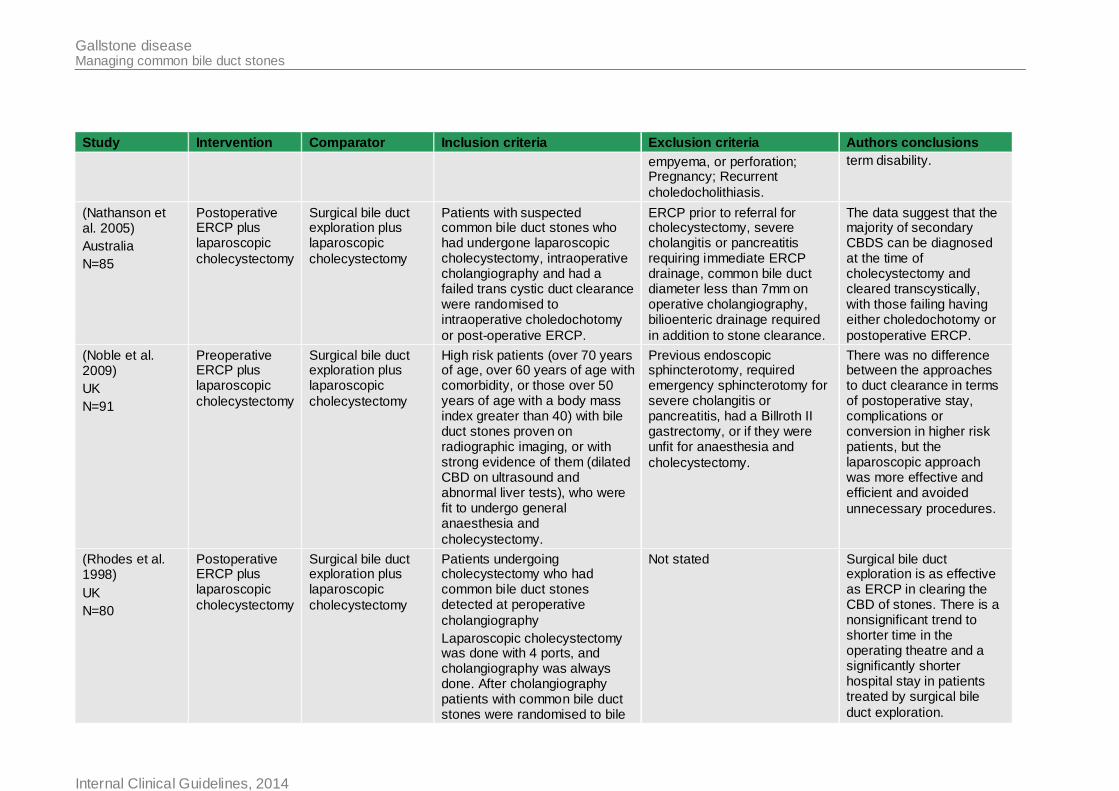
Gallstone disease Managing common bile duct stones
Internal Clinical Guidelines, 2014
Study Intervention Comparator Inclusion criteria Exclusion criteria Authors conclusions
empyema, or perforation; Pregnancy; Recurrent
choledocholithiasis.
term disability.
(Nathanson et al. 2005)
Australia
N=85
Postoperative ERCP plus laparoscopic
cholecystectomy
Surgical bile duct exploration plus laparoscopic
cholecystectomy
Patients with suspected common bile duct stones who had undergone laparoscopic cholecystectomy, intraoperative cholangiography and had a failed trans cystic duct clearance were randomised to intraoperative choledochotomy
or post-operative ERCP.
ERCP prior to referral for cholecystectomy, severe cholangitis or pancreatitis requiring immediate ERCP drainage, common bile duct diameter less than 7mm on operative cholangiography, bilioenteric drainage required
in addition to stone clearance.
The data suggest that the majority of secondary CBDS can be diagnosed at the time of cholecystectomy and cleared transcystically, with those failing having either choledochotomy or
postoperative ERCP.
(Noble et al. 2009)
UK
N=91
Preoperative ERCP plus laparoscopic
cholecystectomy
Surgical bile duct exploration plus laparoscopic
cholecystectomy
High risk patients (over 70 years of age, over 60 years of age with comorbidity, or those over 50 years of age with a body mass index greater than 40) with bile duct stones proven on radiographic imaging, or with strong evidence of them (dilated CBD on ultrasound and abnormal liver tests), who were fit to undergo general anaesthesia and
cholecystectomy.
Previous endoscopic sphincterotomy, required emergency sphincterotomy for severe cholangitis or pancreatitis, had a Billroth II gastrectomy, or if they were unfit for anaesthesia and
cholecystectomy.
There was no difference between the approaches to duct clearance in terms of postoperative stay, complications or conversion in higher risk patients, but the laparoscopic approach was more effective and efficient and avoided
unnecessary procedures.
(Rhodes et al. 1998)
UK
N=80
Postoperative ERCP plus laparoscopic
cholecystectomy
Surgical bile duct exploration plus laparoscopic
cholecystectomy
Patients undergoing cholecystectomy who had common bile duct stones detected at peroperative
cholangiography
Laparoscopic cholecystectomy was done with 4 ports, and cholangiography was always done. After cholangiography patients with common bile duct stones were randomised to bile
Not stated Surgical bile duct exploration is as effective as ERCP in clearing the CBD of stones. There is a nonsignificant trend to shorter time in the operating theatre and a significantly shorter hospital stay in patients treated by surgical bile
duct exploration.
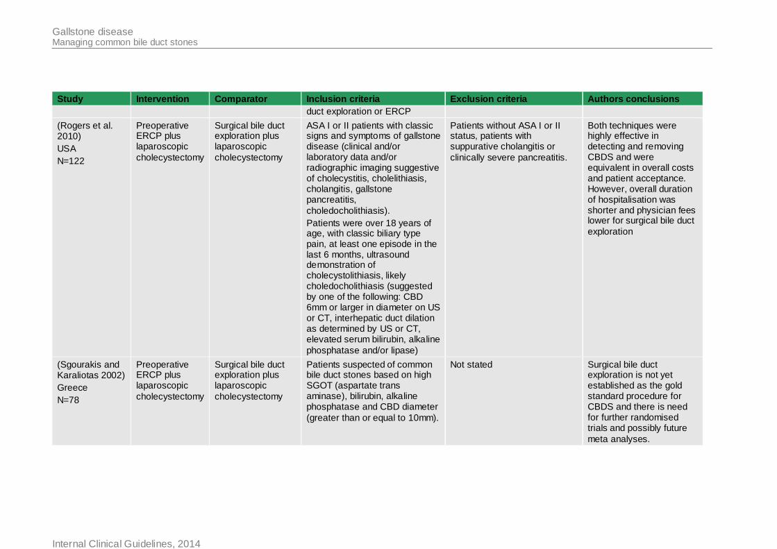
Gallstone disease Managing common bile duct stones
Internal Clinical Guidelines, 2014
Study Intervention Comparator Inclusion criteria Exclusion criteria Authors conclusions
duct exploration or ERCP
(Rogers et al. 2010)
USA
N=122
Preoperative ERCP plus laparoscopic
cholecystectomy
Surgical bile duct exploration plus laparoscopic
cholecystectomy
ASA I or II patients with classic signs and symptoms of gallstone disease (clinical and/or laboratory data and/or radiographic imaging suggestive of cholecystitis, cholelithiasis, cholangitis, gallstone pancreatitis,
choledocholithiasis).
Patients were over 18 years of age, with classic biliary type pain, at least one episode in the last 6 months, ultrasound demonstration of cholecystolithiasis, likely choledocholithiasis (suggested by one of the following: CBD 6mm or larger in diameter on US or CT, interhepatic duct dilation as determined by US or CT, elevated serum bilirubin, alkaline
phosphatase and/or lipase)
Patients without ASA I or II status, patients with suppurative cholangitis or
clinically severe pancreatitis.
Both techniques were highly effective in detecting and removing CBDS and were equivalent in overall costs and patient acceptance. However, overall duration of hospitalisation was shorter and physician fees lower for surgical bile duct
exploration
(Sgourakis and Karaliotas 2002)
Greece
N=78
Preoperative ERCP plus laparoscopic
cholecystectomy
Surgical bile duct exploration plus laparoscopic
cholecystectomy
Patients suspected of common bile duct stones based on high SGOT (aspartate trans aminase), bilirubin, alkaline phosphatase and CBD diameter
(greater than or equal to 10mm).
Not stated Surgical bile duct exploration is not yet established as the gold standard procedure for CBDS and there is need for further randomised trials and possibly future
meta analyses.
Gallstone disease Managing common bile duct stones
Internal Clinical Guidelines, 2014
74
4.6.3 Health economic evidence 1
A literature search was conducted jointly for questions 4 and 5, by applying standard health 2 economic filters to the clinical search strategies (see Appendix D). For questions 4 and 5, 3
1396 references were retrieved, of which 1was retained for question 4b and 1 was retained 4 for question 5. 5
Gurusamy et al. (2012) (see Table 11) used a decision tree to compare intraoperative and 6 preoperative ERCP for patients with gallbladder stones and CBDS (Gurusamy et al. 2012). 7
They modelled the success or failure (and subsequent repeat ERCP) of CBDS extraction, 8 complications of ERCP, consequent cholecystectomy (laparoscopic or open) and mortality 9 from symptoms and open operations. Whilst this analysis was directly relevant to the UK 10
NHS, the model did not compare all the interventions included in this guideline question (as 11 postoperative ERCP and intraoperative bile duct exploration were not included). 12
13
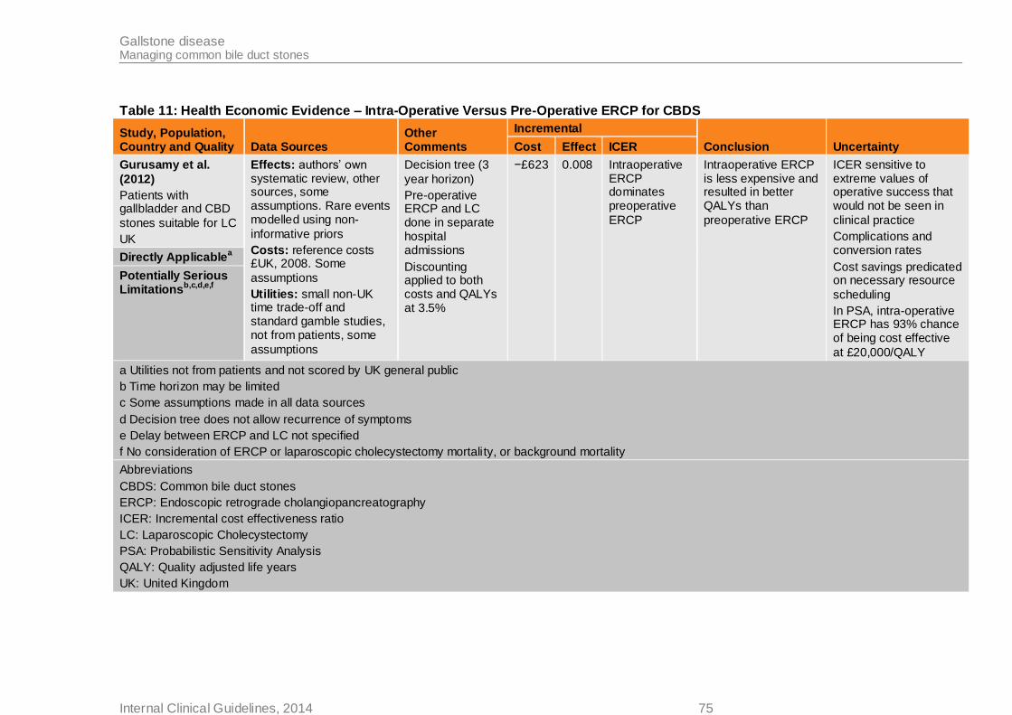
Gallstone disease Managing common bile duct stones
Internal Clinical Guidelines, 2014 75
Table 11: Health Economic Evidence – Intra-Operative Versus Pre-Operative ERCP for CBDS
Study, Population, Country and Quality Data Sources
Other Comments
Incremental
Conclusion Uncertainty Cost Effect ICER
Gurusamy et al.
(2012)
Patients with gallbladder and CBD
stones suitable for LC
UK
Effects: authors’ own systematic review, other sources, some assumptions. Rare events modelled using non-
informative priors
Costs: reference costs £UK, 2008. Some
assumptions
Utilities: small non-UK time trade-off and standard gamble studies, not from patients, some
assumptions
Decision tree (3
year horizon)
Pre-operative ERCP and LC done in separate hospital admissions
Discounting applied to both costs and QALYs at 3.5%
−£623 0.008 Intraoperative ERCP dominates preoperative
ERCP
Intraoperative ERCP is less expensive and resulted in better QALYs than
preoperative ERCP
ICER sensitive to extreme values of operative success that would not be seen in
clinical practice
Complications and conversion rates
Cost savings predicated on necessary resource
scheduling
In PSA, intra-operative ERCP has 93% chance of being cost effective
at £20,000/QALY
Directly Applicablea
Potentially Serious Limitations
b,c,d,e,f
a Utilities not from patients and not scored by UK general public
b Time horizon may be limited
c Some assumptions made in all data sources
d Decision tree does not allow recurrence of symptoms
e Delay between ERCP and LC not specified
f No consideration of ERCP or laparoscopic cholecystectomy mortality, or background mortality
Abbreviations
CBDS: Common bile duct stones
ERCP: Endoscopic retrograde cholangiopancreatography
ICER: Incremental cost effectiveness ratio
LC: Laparoscopic Cholecystectomy
PSA: Probabilistic Sensitivity Analysis
QALY: Quality adjusted life years
UK: United Kingdom
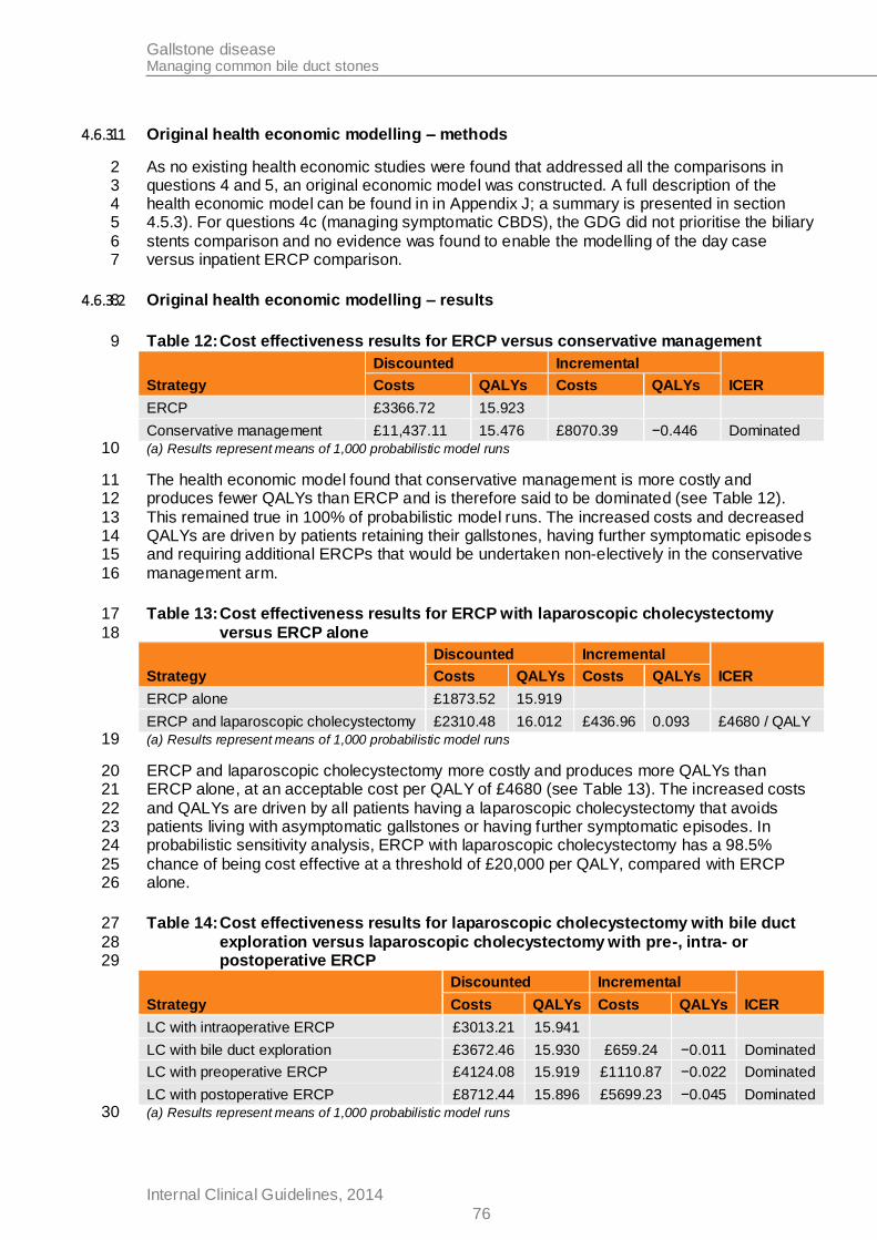
Gallstone disease Managing common bile duct stones
Internal Clinical Guidelines, 2014
76
4.6.3.1 Original health economic modelling – methods 1
As no existing health economic studies were found that addressed all the comparisons in 2 questions 4 and 5, an original economic model was constructed. A full description of the 3 health economic model can be found in in Appendix J; a summary is presented in section 4 4.5.3). For questions 4c (managing symptomatic CBDS), the GDG did not prioritise the biliary 5
stents comparison and no evidence was found to enable the modelling of the day case 6 versus inpatient ERCP comparison. 7
4.6.3.2 Original health economic modelling – results 8
Table 12: Cost effectiveness results for ERCP versus conservative management 9
Strategy
Discounted Incremental
ICER Costs QALYs Costs QALYs
ERCP £3366.72 15.923
Conservative management £11,437.11 15.476 £8070.39 −0.446 Dominated
(a) Results represent means of 1,000 probabilistic model runs 10
The health economic model found that conservative management is more costly and 11 produces fewer QALYs than ERCP and is therefore said to be dominated (see Table 12). 12
This remained true in 100% of probabilistic model runs. The increased costs and decreased 13 QALYs are driven by patients retaining their gallstones, having further symptomatic episodes 14 and requiring additional ERCPs that would be undertaken non-electively in the conservative 15
management arm. 16
Table 13: Cost effectiveness results for ERCP with laparoscopic cholecystectomy 17
versus ERCP alone 18
Strategy
Discounted Incremental
ICER Costs QALYs Costs QALYs
ERCP alone £1873.52 15.919
ERCP and laparoscopic cholecystectomy £2310.48 16.012 £436.96 0.093 £4680 / QALY
(a) Results represent means of 1,000 probabilistic model runs 19
ERCP and laparoscopic cholecystectomy more costly and produces more QALYs than 20 ERCP alone, at an acceptable cost per QALY of £4680 (see Table 13). The increased costs 21
and QALYs are driven by all patients having a laparoscopic cholecystectomy that avoids 22 patients living with asymptomatic gallstones or having further symptomatic episodes. In 23 probabilistic sensitivity analysis, ERCP with laparoscopic cholecystectomy has a 98.5% 24
chance of being cost effective at a threshold of £20,000 per QALY, compared with ERCP 25 alone. 26
Table 14: Cost effectiveness results for laparoscopic cholecystectomy with bile duct 27
exploration versus laparoscopic cholecystectomy with pre-, intra- or 28 postoperative ERCP 29
Strategy
Discounted Incremental
ICER Costs QALYs Costs QALYs
LC with intraoperative ERCP £3013.21 15.941
LC with bile duct exploration £3672.46 15.930 £659.24 −0.011 Dominated
LC with preoperative ERCP £4124.08 15.919 £1110.87 −0.022 Dominated
LC with postoperative ERCP £8712.44 15.896 £5699.23 −0.045 Dominated
(a) Results represent means of 1,000 probabilistic model runs 30
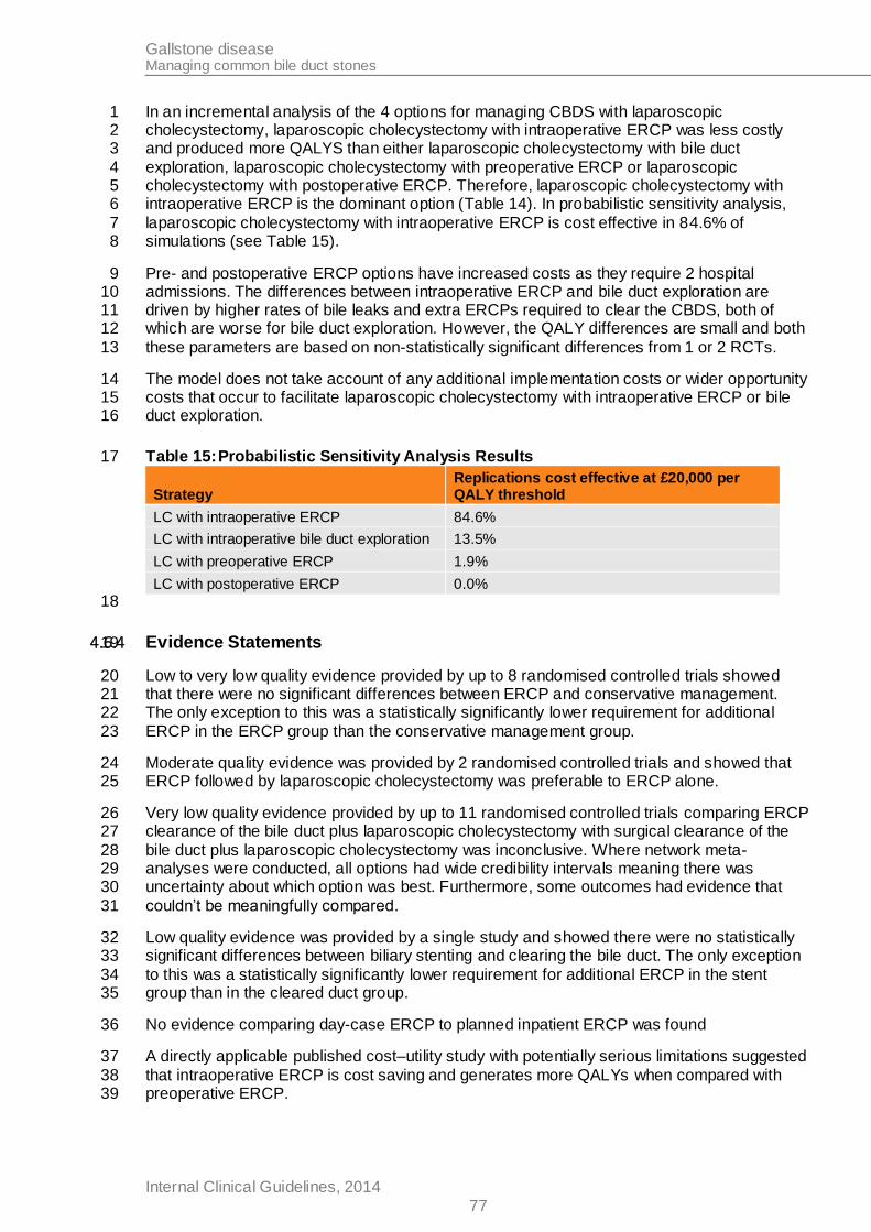
Gallstone disease Managing common bile duct stones
Internal Clinical Guidelines, 2014
77
In an incremental analysis of the 4 options for managing CBDS with laparoscopic 1 cholecystectomy, laparoscopic cholecystectomy with intraoperative ERCP was less costly 2 and produced more QALYS than either laparoscopic cholecystectomy with bile duct 3
exploration, laparoscopic cholecystectomy with preoperative ERCP or laparoscopic 4 cholecystectomy with postoperative ERCP. Therefore, laparoscopic cholecystectomy with 5 intraoperative ERCP is the dominant option (Table 14). In probabilistic sensitivity analysis, 6
laparoscopic cholecystectomy with intraoperative ERCP is cost effective in 84.6% of 7 simulations (see Table 15). 8
Pre- and postoperative ERCP options have increased costs as they require 2 hospital 9 admissions. The differences between intraoperative ERCP and bile duct exploration are 10 driven by higher rates of bile leaks and extra ERCPs required to clear the CBDS, both of 11 which are worse for bile duct exploration. However, the QALY differences are small and both 12
these parameters are based on non-statistically significant differences from 1 or 2 RCTs. 13
The model does not take account of any additional implementation costs or wider opportunity 14 costs that occur to facilitate laparoscopic cholecystectomy with intraoperative ERCP or bile 15 duct exploration. 16
Table 15: Probabilistic Sensitivity Analysis Results 17
Strategy Replications cost effective at £20,000 per QALY threshold
LC with intraoperative ERCP 84.6%
LC with intraoperative bile duct exploration 13.5%
LC with preoperative ERCP 1.9%
LC with postoperative ERCP 0.0%
18
4.6.4 Evidence Statements 19
Low to very low quality evidence provided by up to 8 randomised controlled trials showed 20 that there were no significant differences between ERCP and conservative management. 21 The only exception to this was a statistically significantly lower requirement for additional 22
ERCP in the ERCP group than the conservative management group. 23
Moderate quality evidence was provided by 2 randomised controlled trials and showed that 24 ERCP followed by laparoscopic cholecystectomy was preferable to ERCP alone. 25
Very low quality evidence provided by up to 11 randomised controlled trials comparing ERCP 26 clearance of the bile duct plus laparoscopic cholecystectomy with surgical clearance of the 27
bile duct plus laparoscopic cholecystectomy was inconclusive. Where network meta-28 analyses were conducted, all options had wide credibility intervals meaning there was 29 uncertainty about which option was best. Furthermore, some outcomes had evidence that 30
couldn’t be meaningfully compared. 31
Low quality evidence was provided by a single study and showed there were no statistically 32 significant differences between biliary stenting and clearing the bile duct. The only exception 33
to this was a statistically significantly lower requirement for additional ERCP in the stent 34 group than in the cleared duct group. 35
No evidence comparing day-case ERCP to planned inpatient ERCP was found 36
A directly applicable published cost–utility study with potentially serious limitations suggested 37
that intraoperative ERCP is cost saving and generates more QALYs when compared with 38 preoperative ERCP. 39
Gallstone disease Managing common bile duct stones
Internal Clinical Guidelines, 2014
78
A directly applicable original health economic model analysis with minor limitations suggests 1 ERCP is cost effective compared with conservative management and ERCP with 2 laparoscopic cholecystectomy is cost effective compared with ERCP alone. Based on limited 3
and inconclusive clinical evidence, laparoscopic cholecystectomy with intra-operative ERCP 4 dominates other options but without taking account of any additional implementation costs. 5
4.6.5 Evidence to Recommendations 6
Relative value of different outcomes
In general, studies were underpowered for the outcomes that the GDG
had chosen and so confidence intervals relating to effect estimates for many outcomes were wide. This was expected, since events such as
mortality and bile duct injury are relatively rare and sample sizes would need to be very large to observe sufficient rare events.
There was a lack of randomised controlled trial evidence for some of
the comparisons that were considered as part of this review. The GDG felt that this is because surgical removal of the gallbladder, and
surgical and/or endoscopic stone extraction of stones in the common bile duct are accepted as best practice. Alternative management options are only considered for patients for whom surgery/endoscopy
is not appropriate, such as those who have complex, comorbid conditions, or those who make an informed choice not to opt for surgery/endoscopy. Continual improvements in clinical outcomes for
surgery and endoscopy has meant that surgery and/or endoscopy can be offered to an increasing number of people for whom traditionally these options would not be appropriate. It would be seen as highly
unethical to randomise patients for whom surgery and/or endoscopy is appropriate to inferior treatments, and thus randomised controlled trial evidence in this area is limited.
The absence of evidence and the wide confidence intervals surrounding most effect estimates meant the GDG relied on their
knowledge and experience for most of the discussions of the interventions under review.
Trade off between benefits and
harms
ERCP compared with conservative management
Evidence about conservative management in comparison with ERCP
for common bile duct stones was inconclusive. There were no significant differences in any of the critical or important outcomes and this was attributed to lack of statistical power in the evidence.
GDG experience is that ERCP is a safe procedure and is being increasingly offered to a wide range of clinically compromised and complex patients, but there was no evidence to say that ERCP should
always be performed. The GDG agreed that there will always be patients where the risks of intervening with ERCP outweigh the risks
associated with not intervening, and for these patients conservative management is an appropriate treatment option. The GDG felt that decisions about who should be offered conservative management
should be taken locally, since such cases will be complex and dependent on the skills and experience of local health care professionals. The GDG felt it was not necessary to make a
recommendation about conservative management, as doing so may make it seem that conservative management is an appropriate option for all patients, which they agreed it wasn’t.
ERCP + laparoscopic cholecystectomy compared with ERCP
alone
Gallstone disease Managing common bile duct stones
Internal Clinical Guidelines, 2014
79
The GDG discussed whether laparoscopic cholecystectomy should
always be offered to people who have ERCP, in order to prevent recurrent disease. Evidence about this came from 2 studies, where statistically significant differences on 2 of the 3 critical outcomes that
the GDG had specified were found. The evidence showed significant reductions in disease progression/recurrence, and in the requirement for additional ERCP in the group who received laparoscopic
cholecystectomy after ERCP, compared with the group that had ERCP alone. Furthermore, subsequent cholecystectomy was required for
25% of patients who had ERCP alone. The evidence supported the knowledge and experience of the group, where it was agreed that disease recurrence is common, and the only way to prevent this is to
remove the gallbladder to stop new gallstones forming. Thus the GDG recommended that laparoscopic cholecystectomy should be offered to patients who have common bile duct stones since the harms of
recurrent events outweighed the harms of laparoscopic cholecystectomy.
Pre/post/intra operative ERCP + LC compared with BDE+LC
The evidence relating to these comparisons came from 9 studies that
were analysed using network meta-analysis whenever possible. In some instances there was insufficient evidence to form a network,
pairwise meta-analysis was performed instead. For some outcomes it wasn’t possible to meta analyse at all. This was usually when an outcome was possible in one arm of the comparison but not in the
other (for example, requirement for cholecystectomy isn’t possible in people who have already had cholecystectomy, but is possible in those waiting for deferring from cholecystectomy). In these cases
simple rates were presented.
Network meta-analysis was possible for one critical outcome (length of
stay) and three important outcomes (missed stones, failed procedure, conversion to open surgery). The results of the network analyses were inconclusive. All of the options had wide credibility intervals on all of
the outcomes, indicating there was uncertainty about which options offered the best outcomes and which offered the worst.
Mortality was also chosen as a critical outcome by the GDG, and
although mortality data were reported by most of the studies included in these comparisons, the event rates were often zero in both arms of
the trial. Since zero event data cannot be meaningfully analysed, only evidence from studies where deaths were observed were compared. It was only possible to compare mortality data in a pairwise comparison
between preoperative ERCP+LC versus BDE+LC. The results of this comparison were not statistically significant, and this was attributed to a lack of statistical power in the analysis. The point estimate did
indicate that deaths were lower in preoperative ERCP group than the bile duct exploration group.
Requirement for more than 1 ERCP was chosen as an important
outcome by the GDG, but this outcome was not possible to observe in the surgical bile duct exploration arm, since ERCP was not part of
treatment. Furthermore, not all studies reported this outcome so it was not possible to meta-analyse the data. Rates of multiple ERCP were provided but it was not possible to calculate confidence intervals
around the rates, and so this outcome had unknown levels of uncertainty.
Thus, the GDG agreed that the mixed and unclear evidence was a
Gallstone disease Managing common bile duct stones
Internal Clinical Guidelines, 2014
80
reflection of current clinical reality as there are lots of different options
for managing common bile duct stones that are used in different clinical scenarios. However, the GDG acknowledged that the studies used within this evidence often excluded patients who were older,
complex, and had comorbidities from their protocols. Thus, the range of clinical scenarios that the evidence covered does not reflect reality. Since there was insufficient evidence, decisions were based on
knowledge and experience of the group.
A discussion about the merits of ERCP+LC and BDE+LC took place.
ERCP+LC requires two separate procedures, ERCP to remove the stone and LC to remove the gallbladder. Usually these procedures are done days or weeks apart from each other. Thus the patient will
require a prolonged hospital stay during the wait, or will be admitted to hospital twice for each individual procedure. Furthermore, the patient would be exposed to the risks of anaesthesia/sedation twice, and
there is a risk that their clinical condition could deteriorate in the time between the two procedures. BDE+LC on the other hand is performed as a single procedure and would usually result in the patient having a
shorter total hospital stay (since multiple or prolonged admission is not necessary), is only exposed to one anaesthesia, and has their condition treated definitively in one procedure. Thus, the GDG agreed
that in principle BDE+LC is superior to ERCP+LC.
The GDG acknowledged that only a minority of surgeons can perform BDE+LC safely, as this is highly complex surgery that requires
specialist knowledge and experience. The GDG felt that surgeons not trained to do bile duct exploration would not and should not undertake this procedure. Conversely, laparoscopic cholecystectomy performed
after bile duct stones have been removed does not require a surgeon trained in bile duct exploration, and can be performed by any surgeon trained to perform laparoscopic cholecystectomy. Since ERCP is
widely available throughout the NHS, the GDG felt that it could recommend that bile duct stones should be removed by ERCP before laparoscopic cholecystectomy, or by surgical bile duct exploration
during laparoscopic cholecystectomy in order to reduce the risk of subsequent surgical complications. The GDG felt that the decision to use ERCP or bile duct exploration to clear the duct should be taken
locally depending on the skills and experience of the team.
Uncleared duct + biliary stent compared with cleared duct
Only one study was identified that met the inclusion criteria. The study
had a low rate of stone clearance which the GDG felt reflected the age of the paper as it was published in 1996, and higher rates of clearance are usually observed today. The GDG also acknowledged that the
study randomised patients to biliary stents without attempting to clear the duct first, which is not ethical and wouldn’t be done in practice today. The study was underpowered and so no clear conclusions
could be drawn about the efficacy and safety of biliary stenting in comparison with ductal clearance.
Biliary stenting is widely accepted to be an appropriate management
strategy when stones cannot be cleared from the bile duct and surgery is not an appropriate treatment option. Biliary stenting is associated with complications that can be life-threatening, and clinicians must
balance the risks of using stents with those associated with
Gallstone disease Managing common bile duct stones
Internal Clinical Guidelines, 2014
81
persevering in the attempt to clear the duct. The GDG felt that in
general, most clinicians would strive for a cleared duct and the decision to abort ductal clearance would be taken locally depending on the individual patient’s condition and the clinical experience of the
team. Thus, the GDG did not feel it would be of use to make recommendations about when biliary stenting should be considered. However, the GDG felt that there was inappropriate variation in the
management of patients once a biliary stent has been placed. Often biliary stents are used as long term treatment option with patients never getting reassessed for further surgical or endoscopic ductal
clearance, even when their clinical condition has improved. Instead patients will go on to have their stent replaced indefinitely. The GDG were concerned about this, since patients would be at increased risk
of recurrent events and would be at risk of experiencing cholangitis, one of the most serious complications associated with the use of
stents.
Although the evidence was inconclusive since the single study it was based on was underpowered, the point estimates showed that risk of
death and disease progression were higher in the stent group than in the cleared duct group. Furthermore, the study did find a statistically significant difference in risk of cholangitis, with those in the stent group
having a significantly greater risk of cholangitis than those in the cleared duct group. The GDG had not specified cholangitis as a
specific outcome for this evidence review, instead it chose a composite outcome that included all types of progressive disease including cholangitis, pancreatitis and jaundice. So although the
individual study observed a statistical difference in cholangitis risk, this was not observed in the analysis for this review, since the use of the composite outcome ‘disease progression’ had diluted the evidence.
Since cholangitis is such a serious and life threatening complication, the GDG could not ignore the study findings and were satisfied that the evidence did support their knowledge and experience. Thus
recommendations about managing stents once they have been placed were made.
Day-case ERCP vs planned inpatient ERCP
There was no evidence in relation to this comparison and so the GDG based its discussions on the knowledge and experience of the group, and also extrapolated from evidence presented in chapter 4.6 relating
to day-case versus inpatient laparoscopic cholecystectomy.
The GDG discussed whether day-case surgery should always be
offered to patients, since it offers the benefits of enabling the patient to return home sooner, reducing the risk of hospital acquired infections, and reducing the use of NHS resources.
However, there are various patient and clinical factors that need to be taken into consideration. For example, patients require adult supervision for 24hours after sedation and people who do not have
someone at home to supervise them will require an inpatient stay. Similarly, some patients with comorbidities may need admitting to
hospital before or after the operation to manage their other conditions. The GDG also acknowledged that definitions of inpatient admission and day-cases may vary, and that considerations of other aspects of
hospital performance may influence decisions around length of stay.
Thus, the GDG felt that decisions about whether to plan day-case or inpatient ERCP should be decided at a local level on a case by case
Gallstone disease Managing common bile duct stones
Internal Clinical Guidelines, 2014
82
basis to take the various patient and clinical factors into account.
Consideration of health benefits
and resource use
For ERCP compared with conservative management, the economic model suggests that leaving patients with common bile duct stones
results in net health losses (due to symptoms) and cost increases (due to extra non-elective interventional procedures being required). The GDG noted that the economic model considered a binary choice
between ERCP and no intervention that is unlikely to occur in clinical practice.
The economic model suggests that ERCP with laparoscopic cholecystectomy is cost effective compared with ERCP alone with an ICER of around £4,700 / QALY. The GDG agreed that the extra cost
of laparoscopic cholecystectomy would be an appropriate trade off against the extra QALYs gained by relief of recurrent biliary symptoms.
The economic analysis of laparoscopic cholecystectomy with laparoscopic bile duct exploration compared with laparoscopic
cholecystectomy with pre-, intra- or postoperative ERCP suggests that intraoperative ERCP dominates other options. However, the GDG interpreted this result cautiously. They noted the underlying network
meta-analysis was inconclusive. They also noted options modelled with 1 (intraoperative ERCP, laparoscopic bile duct exploration) rather than 2 (pre- and post-operative ERCP) hospital admissions are
cheaper and also limited the time patients could become symptomatic and lose QALYs. However, the GDG were concerned that the economic model did not reflect implementation costs that would be
associated with recommending intra-operative ERCP or laparoscopic bile duct exploration. The GDG felt that the economic analysis supported their view that laparoscopic cholecystectomy with bile duct
exploration is superior to laparoscopic cholecystectomy with ERCP, but the decision on which method should be used to clear the bile duct should be taken locally based on the skills and experience of staff.
The economic model did not consider biliary stents or day-case versus inpatient ERCP treatment.
Quality of
evidence
The overall quality of the evidence was mixed, with the majority of
evidence being low in quality.
Other considerations
None
1
4.6.6 Recommendations 2
11. Offer bile duct clearance and laparoscopic cholecystectomy to people with 3
symptomatic and asymptomatic common bile duct stones. 4
12. Clear the bile duct: 5
surgically at the time of laparoscopic cholecystectomy or 6
with endoscopic retrograde cholangiopancreatography (ERCP) before 7
or at the time of laparoscopic cholecystectomy. 8
Gallstone disease Managing common bile duct stones
Internal Clinical Guidelines, 2014
83
13. If the bile duct cannot be cleared with ERCP, use biliary stenting to achieve biliary 1 drainage only as a temporary measure until definitive endoscopic or surgical 2 clearance. 3
14. Use the lowest-cost option suitable for the clinical situation when choosing 4 between day-case and inpatient procedures for planned, elective ERCP. 5
4.6.7 Research recommendations 6
8. What models of service delivery enable intraoperative endoscopic retrograde 7
cholangiopancreatography (ERCP) for bile duct clearance to be delivered within 8 the NHS? What are the costs and benefits of different models of service delivery? 9
Why this is important 10
Evidence reviewed for this guideline identified that intraoperative ERCP is both clinically and 11
cost effective, but it is unclear whether delivery of this intervention is feasible in the NHS 12 because of the way current services are organised. It is also unclear whether intraoperative 13 ERCP will remain cost effective if services are reorganised. 14
4.6.8 References 15
Acosta JM, Katkhouda N, Debian KA et al. (2006) Early ductal decompression versus 16 conservative management for gallstone pancreatitis with ampullary obstruction: a prospective 17 randomized clinical trial. Annals of Surgery 243: 33-40 18
Bansal VK, Misra MC, Garg P et al. (2010) A prospective randomized tria l comparing two-19 stage versus single-stage management of patients with gallstone disease and common bile 20 duct stones. Surgical Endoscopy 24: 1986-9 21
Boerma D, Rauws EA, Keulemans YC et al. (2002) Wait-and-see policy or laparoscopic 22 cholecystectomy after endoscopic sphincterotomy for bile-duct stones: a randomised trial. 23 Lancet 360: 761-5 24
Chopra KB, Peters RA, O'Toole PA et al. (1996) Randomised study of endoscopic biliary 25
endoprosthesis versus duct clearance for bileduct stones in high-risk patients. Lancet 348: 26 791-3 27
Cuschieri A, Lezoche E, Morino M et al. (1999) E.A.E.S. multicenter prospective randomized 28 trial comparing two-stage vs single-stage management of patients with gallstone disease and 29
ductal calculi. Surgical Endoscopy 13: 952-7 30
Ding YB, Deng B, Liu XN et al. (2013) Synchronous vs sequential laparoscopic 31 cholecystectomy for cholecystocholedocholithiasis. World journal of gastroenterology : WJG 32 19: 2080-6 33
ElGeidie AA (2011a) Laparoscopic exploration versus intraoperative endoscopic 34 sphincterotomy for common bile duct stones: A prospective randomized trial. Digestive 35 Surgery 28: 424-31 36
ElGeidie AA (2011b) Preoperative versus intraoperative endoscopic sphincterotomy for 37 management of common bile duct stones. Surgical Endoscopy 25: 1230-7 38
Fan ST, Lai EC, Mok FP et al. (1993) Early treatment of acute biliary pancreatitis by 39 endoscopic papillotomy. New England Journal of Medicine 328: 228-32 40
Gallstone disease Managing common bile duct stones
Internal Clinical Guidelines, 2014
84
Folsch UR, Nitsche R, Ludtke R et al. (1997) Early ERCP and papillotomy compared with 1 conservative treatment for acute biliary pancreatitis. The German Study Group on Acute 2 Biliary Pancreatitis. New England Journal of Medicine 336: 237-42 3
Gurusamy K, Wilson E, Burroughs AK et al. (2012) Intra-operative vs pre-operative 4 endoscopic sphincterotomy in patients with gallbladder and common bile duct stones: cost-5 utility and value-of-information analysis. Applied Health Economics & Health Policy 10: 15-29 6
Hong DF, Xin Y, Chen DW (2006) Comparison of laparoscopic cholecystectomy combined 7 with intraoperative endoscopic sphincterotomy and laparoscopic exploration of the common 8 bile duct for cholecystocholedocholithiasis. Surgical Endoscopy 20: 424-7 9
Hui C-K, Lai K-C, Wong W-M et al. (2002) A randomised controlled trial of endoscopic 10 sphincterotomy in acute cholangitis without common bile duct stones. Gut 51: 245-7 11
Koc B, Karahan S, Adas G et al. (2013) Comparison of laparoscopic common bile duct 12 exploration and endoscopic retrograde cholangiopancreatography plus laparoscopic 13 cholecystectomy for choledocholithiasis: a prospective randomized study. American Journal 14
of Surgery 206: 457-63 15
Lau JY, Leow CK, Fung TM et al. (2006) Cholecystectomy or gallbladder in situ after 16 endoscopic sphincterotomy and bile duct stone removal in Chinese patients. 17 Gastroenterology 130: 96-103 18
Nathanson LK, O'Rourke NA, Martin IJ et al. (2005) Postoperative ERCP versus 19 laparoscopic choledochotomy for clearance of selected bile duct calculi: a randomized trial. 20 Annals of Surgery 242: 188-92 21
Neoptolemos JP, Carr-Locke DL, London NJ et al. (1988) Controlled trial of urgent 22 endoscopic retrograde cholangiopancreatography and endoscopic sphincterotomy versus 23 conservative treatment for acute pancreatitis due to gallstones. Lancet 2: 979-83 24
Nitsche R, Folsch UR, Ludtke R et al. (1995) Urgent ERCP in all cases of acute biliary 25
pancreatitis? A prospective randomized multicenter study. European Journal of Medical 26 Research 1: 127-31 27
Noble H, Tranter S, Chesworth T et al. (2009) A randomized, clinical trial to compare 28 endoscopic sphincterotomy and subsequent laparoscopic cholecystectomy with primary 29
laparoscopic bile duct exploration during cholecystectomy in higher risk patients with 30 choledocholithiasis. Journal of Laparoendoscopic & Advanced Surgical Techniques Part: 31 713-20 32
Oria A, Cimmino D, Ocampo C et al. (2007) Early endoscopic intervention versus early 33 conservative management in patients with acute gallstone pancreatitis and biliopancreatic 34 obstruction: a randomized clinical trial. Annals of Surgery 245: 10-7 35
Rhodes M, Sussman L, Cohen L et al. (1998) Randomised trial of laparoscopic exploration of 36 common bile duct versus postoperative endoscopic retrograde cholangiography for common 37 bile duct stones. Lancet 351: 159-61 38
Rogers SJ, Cello JP, Horn JK et al. (2010) Prospective randomized trial of LC+LCBDE vs 39 ERCP/S+LC for common bile duct stone disease. Archives of Surgery 145: 28-33 40
Sgourakis G, Karaliotas K (2002) Laparoscopic common bile duct exploration and 41 cholecystectomy versus endoscopic stone extraction and laparoscopic cholecystectomy for 42 choledocholithiasis. A prospective randomized study. Minerva Chirurgica 57: 467-74 43
Vracko J, Markovic S, Wiechel KL (2006) Conservative treatment versus endoscopic 44 sphincterotomy in the initial management of acute cholecystitis in elderly patients at high 45 surgical risk. Endoscopy 38: 773-8 46

Gallstone disease Managing common bile duct stones
Internal Clinical Guidelines, 2014
85
Zhou LK, Prasoon P (2012) Mechanical and preventable factors of bile duct injuries during 1 laparoscopic cholecystectomy. [Review]. Hepato-Gastroenterology 59: 51-3 2
Gallstone disease Timing of laparoscopic cholecystectomy
Internal Clinical Guidelines, 2014
86
4.7 Timing of laparoscopic cholecystectomy 1
2
4.7.1 Review Question 5 3
In adults with acute cholecystitis or symptomatic common bile duct stones, should 4 cholecystectomy be performed during the acute episode (early) or should intervention be 5
delayed until the acute episode has subsided (delayed)? 6
4.7.2 Evidence Review 7
A single search was performed for questions 4a, 4b, 4c and 5 which identified 10,976 8 references. After removing duplicates and screening the references based on their titles and 9
abstracts, 210 references were obtained and reviewed against the inclusion and exclusion 10 criteria for this review question (appendix C), and 47 references met the inclusion criteria 11
overall. Details of excluded studies and reasons for their exclusion are in appendix F.4 12
Of the 47 included references, 8 references relating to 8 randomised controlled trials were 13 included in this review question (see study flow chart) which specifically addressed the timing 14 of intervention for gallstone disease, and for the purposes of this review the following 15
definitions were used: 16
early laparoscopic cholecystectomy: performed within the first 7 days of the acute 17 presentation 18
delayed laparoscopic cholecystectomy: performed more than 4 weeks after presentation. 19
Studies that didn’t use these definitions were not eligible for inclusion. 20
Of the 8 included studies, 7 related to acute cholecystits (Johansson, 2003; Kolla, 2004; Lai, 21 1998; Lo, 1998; Macafee, 2009; Yadav, 2009) and one related to common bile duct stones 22
(Reinders, 2010). All studies were assessed for methodological quality using randomised 23 controlled trial checklists, and meta-analysis was performed wherever possible (see 24 appendix H.7). 25
Standard GRADE processes were applied to assess the quality of the evidence in relation to 26 the following outcomes: 27
Readmission due to symptoms 28
Readmission due to surgical complications 29
Length of stay 30
Mortality 31
Quality of life 32
Full GRADE profiles are available in appendix (I.7). 33
34
Overall, the individual studies had good methodological quality, with adequate randomisation 35
and appropriate study conduct. Evidence was available in relation to readmission and length 36 of stay, but outcomes such as mortality and quality of life were not reported by any of the 37 studies. There was a lack of evidence in relation to common bile duct stones. 38
39
40
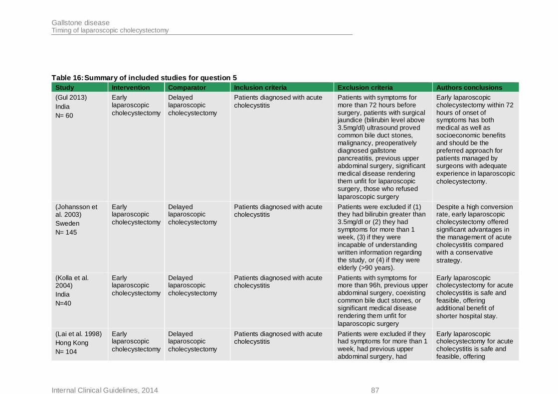
Gallstone disease Timing of laparoscopic cholecystectomy
Internal Clinical Guidelines, 2014 87
Table 16: Summary of included studies for question 5
Study Intervention Comparator Inclusion criteria Exclusion criteria Authors conclusions
(Gul 2013)
India
N= 60
Early laparoscopic
cholecystectomy
Delayed laparoscopic
cholecystectomy
Patients diagnosed with acute
cholecystitis
Patients with symptoms for more than 72 hours before surgery, patients with surgical jaundice (bilirubin level above 3.5mg/dl) ultrasound proved common bile duct stones, malignancy, preoperatively diagnosed gallstone pancreatitis, previous upper abdominal surgery, significant medical disease rendering them unfit for laparoscopic surgery, those who refused
laparoscopic surgery
Early laparoscopic cholecystectomy within 72 hours of onset of symptoms has both medical as well as socioeconomic benefits and should be the preferred approach for patients managed by surgeons with adequate experience in laparoscopic
cholecystectomy.
(Johansson et al. 2003)
Sweden
N= 145
Early laparoscopic
cholecystectomy
Delayed laparoscopic
cholecystectomy
Patients diagnosed with acute cholecystitis
Patients were excluded if (1) they had bilirubin greater than 3.5mg/dl or (2) they had symptoms for more than 1 week, (3) if they were incapable of understanding written information regarding the study, or (4) if they were elderly (>90 years).
Despite a high conversion rate, early laparoscopic cholecystectomy offered significant advantages in the management of acute cholecystitis compared with a conservative
strategy.
(Kolla et al. 2004)
India
N=40
Early laparoscopic
cholecystectomy
Delayed laparoscopic
cholecystectomy
Patients diagnosed with acute cholecystitis
Patients with symptoms for more than 96h, previous upper abdominal surgery, coexisting common bile duct stones, or significant medical disease rendering them unfit for
laparoscopic surgery
Early laparoscopic cholecystectomy for acute cholecystitis is safe and feasible, offering additional benefit of
shorter hospital stay.
(Lai et al. 1998)
Hong Kong
N= 104
Early laparoscopic
cholecystectomy
Delayed laparoscopic
cholecystectomy
Patients diagnosed with acute cholecystitis
Patients were excluded if they had symptoms for more than 1 week, had previous upper abdominal surgery, had
Early laparoscopic cholecystectomy for acute cholecystitis is safe and feasible, offering
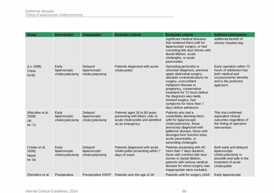
Gallstone disease Timing of laparoscopic cholecystectomy
Internal Clinical Guidelines, 2014 88
Study Intervention Comparator Inclusion criteria Exclusion criteria Authors conclusions
significant medical diseases that rendered them unfit for laparoscopic surgery, or had coexisting bile duct stones with ductal dilation, acute cholangitis, or acute
pancreatitis.
additional benefit of
shorter hospital stay
(Lo 1998)
China
N=99
Early laparoscopic
cholecystectomy
Delayed laparoscopic
cholecystectomy
Patients diagnosed with acute
cholecystitis
Spreading peritonitis or uncertain diagnosis, previous upper abdominal surgery, absolute contraindications for surgery, concomitant malignant disease or pregnancy, conservative treatment for 72 hours before the diagnosis was made, refused surgery, had symptoms for more than 7
days before admission
Early operation within 72 hours of admission has both medical and socioeconomic benefits and is the preferred
approach.
(Macafee et al. 2009)
UK
N= 72
Early laparoscopic
cholecystectomy
Delayed laparoscopic
cholecystectomy
Patients aged 18 to 80 years presenting with biliary colic or acute cholecystitis and admitted
as an emergency
Patients who had a comorbidity deeming them unfit for laparoscopic cholecystectomy, those previously diagnosed with gallstone disease, those with deranged liver function tests, acute pancreatitis, or
ascending cholangitis
This trial confirmed equivalent clinical outcomes regardless of the timing of operative
intervention.
(Yadav et al. 2009)
Nepal
N= 50
Early laparoscopic
cholecystectomy
Delayed laparoscopic
cholecystectomy
Patients diagnosed with acute cholecystitis presenting within 7
days of onset.
Patients presenting with AC more than 7 days duration, those with common bile duct stones or ductal dilation, patients with serious medical disease for whom surgery was
inappropriate were excluded.
Both early and delayed laparoscopic cholecystectomy is possible and safe in the treatment of acute
cholecystitis
(Reinders et al. Preoperative Preoperative ERCP Patients over the age of 18 Patients unfit for surgery (ASA Early laparoscopic
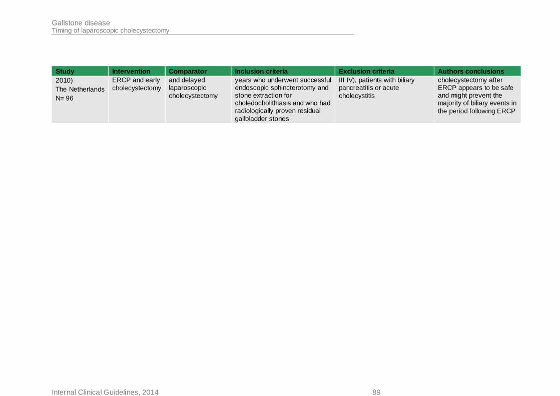
Gallstone disease Timing of laparoscopic cholecystectomy
Internal Clinical Guidelines, 2014 89
Study Intervention Comparator Inclusion criteria Exclusion criteria Authors conclusions
2010)
The Netherlands
N= 96
ERCP and early
cholecystectomy
and delayed laparoscopic
cholecystectomy
years who underwent successful endoscopic sphincterotomy and stone extraction for choledocholithiasis and who had radiologically proven residual
gallbladder stones
III IV), patients with biliary pancreatitis or acute
cholecystitis
cholecystectomy after ERCP appears to be safe and might prevent the majority of biliary events in
the period following ERCP

Gallstone disease Timing of laparoscopic cholecystectomy
Internal Clinical Guidelines, 2014 90
1
4.7.3 Health economic evidence 2
A literature search was conducted jointly for questions 4 and 5, by applying standard health 3 economic filters to the clinical search strategies (see Appendix D). 1396 references were 4 retrieved for questions 4 and 5, of which 1 was retained for question 4b and 1 was retained 5 for question 5. 6
Wilson et al. (2010) (see Table 17) used a decision tree to compare early and delayed 7 laparoscopic cholecystectomy for acute cholecystitis. The model estimated the development 8 of 4 symptoms at 9 weeks during an 18-week delay period, with a time horizon of 1 year. 9
Whilst Wilson et al. (2010) was directly relevant, it had a number of limitations, including the 10 delay length, time horizon, no consideration of CBDS and utility data. 11
4.7.3.1 Original health economic modelling – methods 12
Because Wilson et al.’s (2010) analysis had potentially serious limitations, and did not 13 address all relevant questions in this area, an original economic model was constructed. A 14 full description of the health economic model can be found in in Appendix J, a summary is 15
presented in section 4.5.3. 16
17
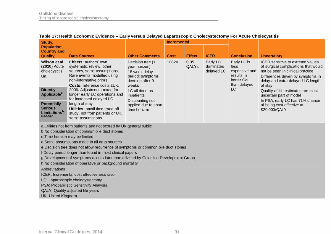
Gallstone disease Timing of laparoscopic cholecystectomy
Internal Clinical Guidelines, 2014 91
Table 17: Health Economic Evidence – Early versus Delayed Laparoscopic Cholecystectomy For Acute Cholecystitis
Study, Population, Country and
Quality Data Sources Other Comments
Incremental
Conclusion Uncertainty Cost Effect ICER
Wilson et al (2010) Acute
cholecystitis
UK
Effects: authors’ own systematic review, other sources, some assumptions. Rare events modelled using
non-informative priors
Costs: reference costs £UK, 2006. Adjustments made for longer early LC operations and for increased delayed LC
length of stay
Utilities: small time trade off study, not from patients or UK,
some assumptions
Decision tree (1 year horizon)
18 week delay period; symptoms develop after 9
weeks
LC all done as inpatients
Discounting not applied due to short
time horizon
−£820
0.05 QALYs
Early LC dominates
delayed LC
Early LC is less expensive and results in better QoL than delayed
LC
ICER sensitive to extreme values of surgical complications that would
not be seen in clinical practice
Differences driven by symptoms in delay and extra delayed LC length
of stay
Quality of life estimates are most uncertain part of model
In PSA, early LC has 71% chance of being cost effective at
£20,000/QALY
Directly Applicable
a
Potentially Serious Limitations
b,
c,d,e,f,g,h
a Utilities not from patients and not scored by UK general public
b No consideration of common bile duct stones
c Time horizon may be limited
d Some assumptions made in all data sources
e Decision tree does not allow recurrence of symptoms or common bile duct stones
f Delay period longer than found in most clinical papers
g Development of symptoms occurs later than advised by Guideline Development Group
h No consideration of operative or background mortality
Abbreviations
ICER: Incremental cost effectiveness ratio
LC: Laparoscopic cholecystectomy
PSA: Probabilistic Sensitivity Analysis
QALY: Quality adjusted life years
UK: United Kingdom
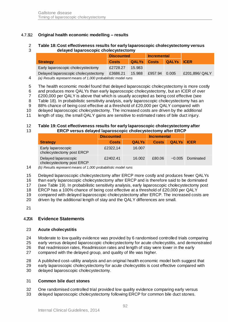
Gallstone disease Timing of laparoscopic cholecystectomy
Internal Clinical Guidelines, 2014 92
4.7.3.2 Original health economic modelling – results 1
Table 18: Cost effectiveness results for early laparoscopic cholecystectomy versus 2 delayed laparoscopic cholecystectomy 3
Strategy
Discounted Incremental
ICER Costs QALYs Costs QALYs
Early laparoscopic cholecystectomy £2728.27 15.983
Delayed laparoscopic cholecystectomy £3686.21 15.988 £957.94 0.005 £201,896/ QALY
(a) Results represent means of 1,000 probabilistic model runs 4
The health economic model found that delayed laparoscopic cholecystectomy is more costly 5 and produces more QALYs than early laparoscopic cholecystectomy, but an ICER of over 6 £200,000 per QALY is above that which is usually accepted as being cost effective (see 7
Table 18). In probabilistic sensitivity analysis, early laparoscopic cholecystectomy has an 8 88% chance of being cost effective at a threshold of £20,000 per QALY compared with 9 delayed laparoscopic cholecystectomy. The increased costs are driven by the additional 10
length of stay, the small QALY gains are sensitive to estimated rates of bile duct injury. 11
Table 19: Cost effectiveness results for early laparoscopic cholecystectomy after 12
ERCP versus delayed laparoscopic cholecystectomy after ERCP 13
Strategy
Discounted Incremental
ICER Costs QALYs Costs QALYs
Early laparoscopic cholecystectomy post ERCP
£2322,14 16.007
Delayed laparoscopic cholecystectomy post ERCP
£2402.41 16.002 £80.06 −0.005 Dominated
(b) Results represent means of 1,000 probabilistic model runs 14
Delayed laparoscopic cholecystectomy after ERCP more costly and produces fewer QALYs 15 than early laparoscopic cholecystectomy after ERCP and is therefore said to be dominated 16
(see Table 19). In probabilistic sensitivity analysis, early laparoscopic cholecystectomy post 17 ERCP has a 100% chance of being cost effective at a threshold of £20,000 per QALY 18 compared with delayed laparoscopic cholecystectomy after ERCP. The increased costs are 19
driven by the additional length of stay and the QALY differences are small. 20
21
4.7.4 Evidence Statements 22
Acute cholecystitis 23
Moderate to low quality evidence was provided by 6 randomised controlled trials comparing 24 early versus delayed laparoscopic cholecystectomy for acute cholecystitis, and demonstrated 25 that readmission rates, Readmission rates and length of stay were lower in the early 26
compared with the delayed group, and quality of life was higher. 27
A published cost–utility analysis and an original health economic model both suggest that 28 early laparoscopic cholecystectomy for acute cholecystitis is cost effective compared with 29
delayed laparoscopic cholecystectomy. 30
Common bile duct stones 31
One randomised controlled trial provided low quality evidence comparing early versus 32 delayed laparoscopic cholecystectomy following ERCP for common bile duct stones. 33
Gallstone disease Timing of laparoscopic cholecystectomy
Internal Clinical Guidelines, 2014 93
Evidence was provided for only 1 of the 5 outcomes for this comparison. This is because the 1 study either didn’t report the data, or because data were reported but zero events occurred. 2 The 1 outcome for which moderate quality evidence was available showed that there was no 3
statistically significant differences in length of stay between the 2 groups. 4
A directly applicable original health economic model analysis with minor limitations suggests 5 that early laparoscopic cholecystectomy following ERCP for common bile duct stones is cost 6
effective compared with delayed laparoscopic cholecystectomy. 7
4.7.5 Evidence to Recommendations 8
Relative value of different
outcomes
The GDG chose readmission due to symptoms and readmission due to surgical complications as outcomes that were critical to their
decision making, since it was felt that these outcomes would capture important differences between groups in relation to the complications of surgery.
Readmission due to symptoms was reported in some of the studies
relating to acute cholecystitis but was not reported in the study relating to common bile duct stones. Readmission due to surgical
complications was not reported by any of the studies included in this review question. The GDG felt that the studies that didn’t report these critical outcomes probably didn’t observe any readmissions, although
there was no way that they could be certain of this. The GDG felt that in reality, readmission rates are low and the individual studies were underpowered to observe these relatively rare events. Likewise,
mortality was reported by most of the included studies, but no events were observed since the studies were underpowered to detect this relatively rare event. The GDG felt that it is more likely for studies to
report zero mortality events than zero readmission events due to the greater importance placed on mortality than readmission.
The GDG felt that the lack of evidence supports their views that readmission and mortality are rare, and focused their attention on the
evidence relating to length of stay which was felt to be the best measure of clinically important differences between early and delayed laparoscopic cholecystectomy (LC) for the majority of patients.
Trade off between benefits and
harms
The earlier LC is performed the less potential there is for recurrent
events during the wait for surgery, therefore there are expected benefits in terms of patient quality of life and resource utilisation. However, there are concerns that early intervention could cause harm
to patients by increasing the complication rate and extending length of stay. Thus delayed surgery may have greater benefits in the longer term if surgery is safer, although patients may experience recurrent
events during the period of delay which could necessitate emergency intervention.
In the evidence relating to acute cholecystitis, the early LC group had
significantly fewer readmissions than the delayed group as well as a significantly shorter length of stay, which the GDG interpreted as
meaning that early intervention does not increase complications. The patient representatives on the group also expressed a preference for early LC due to their personal experiences of having to wait for
surgery. The patient members expressed that their quality of life was compromised during the wait by avoiding foods that triggered their
Gallstone disease Timing of laparoscopic cholecystectomy
Internal Clinical Guidelines, 2014 94
symptoms, worrying about social activities and not feeling confident to
return to work until treatment has been completed due to worries about pain and other complications, although no evidence relating to quality of life was found in this review. The GDG also felt that the
absence of evidence relating to harm, especially mortality, probably reflected the fact that these events are rare, and in the majority of cases the benefits of early intervention would far outweigh the risks.
Thus the GDG felt that there was sufficient evidence relating to the benefits of early LC to enable them to recommend that early LC should be offered to patients with acute cholecystitis.
There was limited evidence in relation to common bile duct stones.
The only evidence available for this population came from one small study for the outcome ‘length of stay’. The study did not find a
significant difference and this was attributed to a lack of statistical power. Furthermore the GDG identified that the study had excluded patients with biliary pancreatitis, which represents a large proportion of
people with common bile duct stones. Thus the paper lacks transferability to routine clinical settings. Overall, the lack of evidence meant the GDG were uncertain about whether early or delayed LC
was the optimum management strategy for common bile duct stones. The group felt that in reality there was great variation in when people were offered LC following removal of bile duct stones, and agreed that
the delay is not necessarily based on clinical decision making and is more often due to a lack of capacity.
The GDG felt that it probably wasn’t appropriate to recommend the same time frame for LC for common bile duct stones as for acute
cholecystitis. This is because common bile duct stones are more complicated and associated with higher risks than gallbladder stones that cause acute cholecystitis, particularly when severe pancreatitis
has developed. Performing early surgery may therefore increase the risk of bile duct injury and mortality. Since there was no evidence to
support or refute this notion, the GDG were not confident about the optimum time frame in which to offer LC and did not make a specific recommendation.
Consideration of
Health Benefits and Resource Use
In both health economic comparisons, the higher cost of delayed
laparoscopic cholecystectomy is driven by the longer length of stay associated with delayed laparoscopic cholecystectomy. QALY differences are small, but suggest that delaying surgery is associated
with similar or worse net quality of life than early surgery.
Quality of evidence
The GDG agreed that the evidence was of moderate to low quality.
Other
considerations
The GDG acknowledged that decisions about when to perform LC
may be based on capacity issues rather than clinical appropriateness. Thus, the GDG wanted to emphasise that the recommendations made here should improve patient flow and release capacity, rather than put
additional constrains on it. This is because the number of people requiring LC in a given year is a finite, and operating on them earlier will not increase the number of people requiring LC. Instead, offering
early cholecystectomy may actually reduce the number of readmissions, emergency operations, and length of stay.
The GDG felt the recommendations are achievable but acknowledged
Gallstone disease Timing of laparoscopic cholecystectomy
Internal Clinical Guidelines, 2014 95
that some providers may need to focus on redesigning their services
in order to implement them.
1
4.7.6 Recommendations 2
15. Offer early laparoscopic cholecystectomy (to be carried out within 1 week of 3
diagnosis) to people with acute cholecystitis. 4
4.7.7 Research recommendations 5
9. In adults with common bile duct stones, should laparoscopic cholecystectomy be 6
performed early (within 2 weeks of bile duct clearance), or should it be delayed 7 (until at least 4 weeks after bile duct clearance)? 8
Why this is important 9
There is a lack of evidence from randomised controlled trials of early compared with delayed 10 laparoscopic cholecystectomy after bile duct clearance with ERCP. It is unclear what effect 11 the timing of laparoscopic cholecystectomy has on clinical outcomes and resource use. 12
4.7.8 References 13
Gul R (2013) Comparison of early and delayed laparoscopic cholecystectomy for acute 14 cholecystitis: experience from a single centre. North Americal Journal of Medical Sciences 5: 15 414-8 16
Johansson M, Thune A, Blomqvist A et al. (2003) Management of acute cholecystitis in the 17 laparoscopic era: results of a prospective, randomized clinical trial. Journal of 18 Gastrointestinal Surgery 7: 642-5 19
Kolla SB, Aggarwal S, Kumar A et al. (2004) Early versus delayed laparoscopic 20 cholecystectomy for acute cholecystitis: a prospective randomized trial. Surgical Endoscopy 21 18: 1323-7 22
Lai PB, Kwong KH, Leung KL et al. (1998) Randomized trial of early versus delayed 23 laparoscopic cholecystectomy for acute cholecystitis. British Journal of Surgery 85: 764-7 24
Lo CM (1998) Prospective randomized study of early versus delayed laparoscopic 25 cholecystectomy for acute cholecystitis. Annals of Surgery 227: 461-7 26
Macafee DA, Humes DJ, Bouliotis G et al. (2009) Prospective randomized trial using cost-27 utility analysis of early versus delayed laparoscopic cholecystectomy for acute gallbladder 28
disease. British Journal of Surgery 96: 1031-40 29
Reinders JSK, Goud A, Timmer R et al. (2010) Early Laparoscopic Cholecystectomy 30 Improves Outcomes After Endoscopic Sphincterotomy for Choledochocystolithiasis. 31
Gastroenterology 138: 2315-20 32
Wilson E, Gurusamy K, Gluud C et al. (2010) Cost-utility and value-of-information analysis of 33 early versus delayed laparoscopic cholecystectomy for acute cholecystitis. British Journal of 34 Surgery 97: 210-9 35
Yadav RP, Adhikary S, Agrawal CS et al. (2009) A comparative study of early vs. delayed 36 laparoscopic cholecystectomy in acute cholecystitis. Kathmandu University Medical Journal 37 7: 16-2038
Gallstone disease Information for patients and their carers
Internal Clinical Guidelines, 2014 96
4.8 Information for patients and their carers 1
4.8.1 Review Question 6 2
What are the information and education needs of patients and carers of people with gallstone 3 disease? 4
4.8.2 Evidence Review 5
The aim of this review question was to identify areas for which information and education 6 should be provided. This question did not aim to compare the effectiveness of different 7 education programmes/strategies. 8
A systematic search was conducted (see appendix D) which identified 7862 references. After 9 removing duplicates the references were screened on their titles and abstracts and 61 10 references were obtained and reviewed against the inclusion and exclusion criteria as 11
described in the review protocols (appendix C). 12
Overall, 56 studies were excluded as they did not meet the eligibility criteria. A list of 13 excluded studies and reasons for their exclusion is provided in appendix F. 14
Five studies met the eligibility criteria and were included. Data were extracted into detailed 15 evidence tables (see appendix G) and are summarised in the table below. 16
Table 20: Summary of included studies for question 6 17
Study details Aims of the study
Authors conclusions
(Barthelsson et al. 2003)
Sweden
N= 12
Explore patient experiences in relation to laparoscopic
cholecystectomy
Although most patients were satisfied with day-case LC, a number of problem areas were expressed- preoperative anxiety, post-operative amnesia, experience of pain, need for additional pain medication, feelings of nausea, vomiting, bloating and swelling, information about wound care, additional telephone follow up, difficulties having small children at home.
(Blay and Donoghue
2005)
Australia
N=93
Determine if a preadmission education intervention reduced pain, increased self-care and decreased postoperative symptoms
after laparoscopic cholecystectomy
Pre admission education intervention helps reduce post-operative pain levels following LC and significantly increases patient’s knowledge of self-care and complication
management.
(Blay and Donoghue
2006)
Australia
N= 100
Determine what information pre admission nurses provided to elective laparoscopic
cholecystectomy patients.
Patients sourced information from a variety of sources, including communicating with friends and relatives for the personal perspective. Personal communication should not be replacing education by nurses. Unfortunately patients did not cite the preadmission nurse as the major source
of information in this study.
(Tamhankar
et al. 2009)
UK
N=105
Establish the proportion of people undergoing elective hernia repair or cholecystectomy who access the internet for information about their operations
A significant proportion of patients used the internet and about one third of them specifically sought information about their operation. Such information can cause worry and confusion in patients. The study highlights the need for regulated, comprehensible patient information on
Gallstone disease Information for patients and their carers
Internal Clinical Guidelines, 2014 97
Study details Aims of the study
Authors conclusions
hospital websites to which patients should
be directed.
(Young and O'Connell
2008)
Australia
N=28
A randomised controlled trial of inpatient vs day-case cholecystectomy, with a telephone survey to identify if discharge
information was sufficient.
With careful patient selection and enhanced discharge education, LC procedures performed as a day-case offer safe and
effective alternative to inpatient care.
Studies were quality assessed using methodology checklists. For qualitative studies, the 1
NICE qualitative checklist was used. The NICE guidelines manual does not provide a 2 checklist for surveys, and so a checklist originally published in the British Medical Journal 3 (see appendix K) was used to aid the quality assessment of these studies. 4
The GRADE framework for assessing quality was modified for this review in that studies, 5 rather than outcomes were assessed for quality (see appendix I.8) on the following basis. 6 Overall, the studies were of very low quality and had serious limitations; Survey studies 7 lacked detailed analysis and qualitative studies lacked rigour as they did not utilise adequate 8
research design and methodology, and they failed to provide rich data to support findings; 9 studies were limited because 4 of the 5 studies were conducted outside the UK and so lack 10
transferability, and most studies were from the perspective of elective cholecystectomy, 11 meaning that non elective surgery and other treatment options are not adequately 12 represented in the evidence base. Specific details about the reasons for this approach and 13
the judgements made are provided in the footnotes of the profiles in appendix I.8. 14
4.8.3 Health economic evidence 15
A literature search was conducted for review question 6, by applying standard health 16 economic filters to the clinical search strategies (see Appendix D). From the search, 504 17
references were retrieved, of which none were retained at title and abstract screening. Health 18 economic modelling was not prioritised for this review question. 19
4.8.4 Evidence Statements 20
Very low quality evidence from 5 studies on people waiting for or undergoing surgery for 21 gallbladder disease showed that patients requested more information on diet, wound 22 management, pain management, and resuming normal activities. Some patients had no 23
memory of the information that was provided to them, and some consulted the internet to 24 acquire additional information. Some people did not know why they had to wait for elective 25 surgery. 26
4.8.5 Evidence to Recommendations 27
28
Relative value of different
outcomes
The GDG considered all themes/outcomes that arose from the evidence, but focused its discussions on the theme/outcome of diet.
This is because most themes that arose such as wound management, resuming activity, memory and so on, were not specific to gallstone disease and the GDG agreed that other NICE products that already
exist or that are in development (such as Patient experience in adult NHS services, Hospital admission pathway, Surgery pathway) would cover these general themes more appropriately.
The group chose to focus discussions on diet because this is the area
the patient representatives felt was most important, because diet was
Gallstone disease Information for patients and their carers
Internal Clinical Guidelines, 2014 98
a key issue in the evidence, and because this issue was specific to
gallstone disease.
Previous review questions did not find sufficient evidence to support or refute a link between diet and gallstone disease, and diet as an intervention for managing gallstone disease was not evaluated in any
of the review questions. Thus, discussions about diet were based on the knowledge and experience of the GDG and a co-opted expert dietician.
Trade off between benefits and harms
Some patients with gallstone disease find that eating and/or drinking
triggers gallbladder pain and discomfort. This could be due to the mechanics of digestion because the gallbladder contracts on
consuming food and drink in order to release bile to aid digestion, and when a person has gallstone disease the gallbladder can become inflamed and infected, and this contracting process can be painful.
The GDG decided that it would not be useful to provide information to
patients about food and drink that may potentially trigger symptoms because not everyone with gallstone disease experiences pain and discomfort when eating or drinking, not all food and drink triggers
gallbladder pain, and the type of triggers vary for different individuals. Instead, the GDG felt that individual patients are best placed to identify which food and drink triggers their own pain.
The GDG agreed that once a gallbladder has been removed the
mechanism for experiencing pain is no longer there (since there is no gallbladder to contract and cause pain). The time between diagnosis
and treatment for gallbladder disease is relatively short (usually less than 3 months), so the GDG felt that there was no harm in recommending that patients should avoid food and drink that triggers
their pain between diagnosis and treatment, since harms to health or quality of life were expected to be very low. However, long term avoidance of specific foods, especially once gallstones or the
gallbladder have been removed could be detrimental and are not recommended.
The group acknowledged that some patients with gallstone disease continue to report symptoms or experience new symptoms once their
gallbladder has been removed. The GDG felt this is probably due to some underlying condition that the patient either didn’t notice before they experienced gallstone disease, or that coincidentally began at the
same time as the symptoms of gallstone disease or cholecystectomy. The GDG felt that these symptoms are often misattributed to the
absence of a gallbladder by both health professionals and patients themselves, and because of this patients may not receive appropriate investigations to identify the real cause of the symptoms. Thus, people
may continue to experience unnecessary symptoms, or restrict trigger food and drink over the long term to avoid ongoing symptoms.
Since long term dietary restriction may be harmful, and because patient quality of life is affected by on-going symptoms, the GDG felt
that people experiencing new or existing symptoms after they have recovered from cholecystectomy should be encouraged to return to their health professional, and health professionals should investigate
Gallstone disease Information for patients and their carers
Internal Clinical Guidelines, 2014 99
the patient appropriately.
Consideration of
Health Benefits and Resource Use
Health economics were not considered as a priority for this review
question. No economic studies were found.
Quality of evidence
The quality of the evidence was very low, and the studies contributing to the evidence did not provide adequate information about the particular aspects of dietary advice that should be provided. Therefore
the GDG developed the recommendations using their knowledge and experience.
Other
considerations
None.
1
4.8.6 Recommendations 2
16. Advise people to avoid food and drink that triggers their symptoms until they have 3
their gallbladder or gallstone(s) removed. 4
17. Advise people that they should not need to avoid food and drink that triggered 5
their symptoms after they have their gallbladder or gallstone(s) removed. 6
18. Advise people to seek further advice from their GP if eating or drinking triggers 7
existing symptoms or causes new symptoms to develop after they have recovered 8 from having their gallbladder or gallstone(s) removed. 9
4.8.7 Research recommendations 10
10. What is the long-term effect of laparoscopic cholecystectomy on outcomes that 11 are important to patients? 12
Why this is important 13
There is a lack of information on the long-term impact of cholecystectomy on patient 14 outcomes. Many patients report a continuation of symptoms or the onset of new symptoms 15 after laparoscopic cholecystectomy, and these affect quality of life. Research is needed to 16
establish the long-term patient benefits and harms, so that appropriate information can be 17 provided to patients to aid decision-making and long-term management of their condition. 18
4.8.8 References 19
Barthelsson C, Lutzen K, Anderberg B et al. (2003) Patients' experiences of laparoscopic 20 cholecystectomy in day surgery. Journal of Clinical Nursing 12: 253-9 21
Blay N, Donoghue J (2005) The effect of pre-admission education on domiciliary recovery 22 following laparoscopic cholecystectomy. Australian Journal of Advanced Nursing 22: 14-9 23
Blay N, Donoghue J (2006) Source and content of health information for patients undergoing 24 laparoscopic cholecystectomy. International Journal of Nursing Practice 12: 64-70 25
Tamhankar AP, Mazari FA, Everitt NJ et al. (2009) Use of the internet by patients undergoing 26 elective hernia repair or cholecystectomy. Annals of the Royal College of Surgeons of 27 England 91: 460-3 28

Gallstone disease Information for patients and their carers
Internal Clinical Guidelines, 2014 100
Young J, O'Connell B (2008) Recovery following laparoscopic cholecystectomy in either a 23 1 hour or an 8 hour facility. Journal of Quality in Clinical Practice 21: 2-72
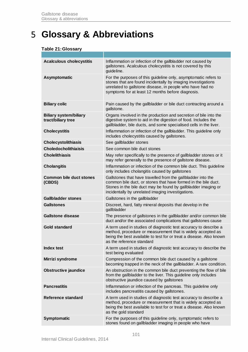
Gallstone disease Glossary & abbreviations
Internal Clinical Guidelines, 2014 101
5 Glossary & Abbreviations
Table 21: Glossary
Acalculous cholecystitis Inflammation or infection of the gallbladder not caused by gallstones. Acalculous cholecystitis is not covered by this
guideline.
Asymptomatic For the purposes of this guideline only, asymptomatic refers to stones that are found incidentally by imaging investigations unrelated to gallstone disease, in people who have had no
symptoms for at least 12 months before diagnosis.
Biliary colic Pain caused by the gallbladder or bile duct contracting around a gallstone.
Biliary system/biliary tract/biliary tree
Organs involved in the production and secretion of bile into the digestive system to aid in the digestion of food. Includes the
gallbladder, bile ducts, and some specialised cells in the liver.
Cholecystitis Inflammation or infection of the gallbladder. This guideline only
includes cholecystitis caused by gallstones.
Cholecystolithiasis See gallbladder stones
Choledocholithiaisis See common bile duct stones
Cholelithiasis May refer specifically to the presence of gallbladder stones or it may refer generally to the presence of gallstone disease.
Cholangitis Inflammation or infection of the common bile duct. This guideline
only includes cholangitis caused by gallstones
Common bile duct stones (CBDS)
Gallstones that have travelled from the gallbladder into the common bile duct, or stones that have formed in the bile duct. Stones in the bile duct may be found by gallbladder imaging or
incidentally by unrelated imaging investigations.
Gallbladder stones Gallstones in the gallbladder
Gallstones Discreet, hard, fatty mineral deposits that develop in the gallbladder
Gallstone disease The presence of gallstones in the gallbladder and/or common bile duct and/or the associated complications that gallstones cause
Gold standard A term used in studies of diagnostic test accuracy to describe a method, procedure or measurement that is widely accepted as being the best available to test for or treat a disease. Also known
as the reference standard
Index test A term used in studies of diagnostic test accuracy to describe the test being evaluated
Mirrizi syndrome Compression of the common bile duct caused by a gallstone
becoming trapped in the neck of the gallbladder. A rare condition.
Obstructive jaundice An obstruction in the common bile duct preventing the flow of bile from the gallbladder to the liver. This guideline only includes
obstructive jaundice caused by gallstones
Pancreatitis Inflammation or infection of the pancreas. This guideline only includes pancreatitis caused by gallstones.
Reference standard A term used in studies of diagnostic test accuracy to describe a method, procedure or measurement that is widely accepted as being the best available to test for or treat a disease. Also known
as the gold standard
Symptomatic For the purposes of this guideline only, symptomatic refers to stones found on gallbladder imaging in people who have
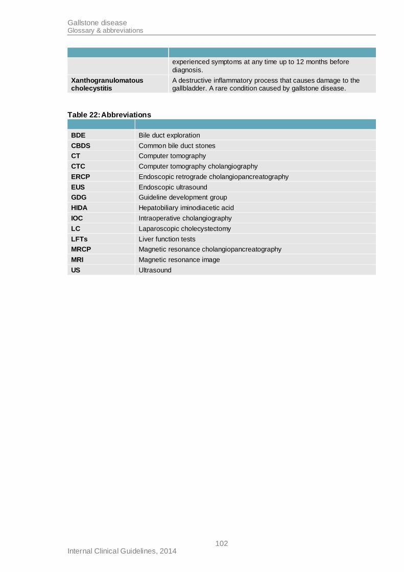
Gallstone disease Glossary & abbreviations
Internal Clinical Guidelines, 2014 102
experienced symptoms at any time up to 12 months before
diagnosis.
Xanthogranulomatous cholecystitis
A destructive inflammatory process that causes damage to the gallbladder. A rare condition caused by gallstone disease.
Table 22: Abbreviations
BDE Bile duct exploration
CBDS Common bile duct stones
CT Computer tomography
CTC Computer tomography cholangiography
ERCP Endoscopic retrograde cholangiopancreatography
EUS Endoscopic ultrasound
GDG Guideline development group
HIDA Hepatobiliary iminodiacetic acid
IOC Intraoperative cholangiography
LC Laparoscopic cholecystectomy
LFTs Liver function tests
MRCP Magnetic resonance cholangiopancreatography
MRI Magnetic resonance image
US Ultrasound