gadofluorine-enhanced magnetic resonance imaging of carotid atherosclerosis in yucatan miniswine
TRANSCRIPT
PRELIMINARY REPORT
Gadofluorine-Enhanced Magnetic Resonance Imaging ofCarotid Atherosclerosis in Yucatan Miniswine
Ioannis Koktzoglou, MS,*† Kathleen R. Harris, BA,* Richard Tang, MD,* Bonnie J. Kane, BS,‡Bernd Misselwitz, PhD,§ Hanns-Joachim Weinmann, PhD,§ Biao Lu, MD,* Ashwin Nagaraj, PhD,‡
Sanford I. Roth, MD,¶**†† Timothy J. Carroll, PhD,*† David D. McPherson, MD,‡and Debiao Li, PhD*†
Objective: The aim of this study was to determine whether gad-ofluorine, a paramagnetic magnetic resonance imaging (MRI) con-trast agent, selectively enhances carotid atherosclerotic plaques inYucatan miniswine.Methods: Atherosclerotic plaques were induced in the left carotidarteries (LCA) of Yucatan miniswine (n � 3) by balloon denudationand high cholesterol diet. T1-weighted MRI was performed beforeand 24 hours after gadofluorine injection (at a dose of 100 �mol/kg)to assess the enhancement of the balloon-injured LCA wall relativeto healthy, uninjured right carotid artery (RCA) wall. Histopathol-ogy was performed to verify the presence and composition of theatherosclerotic plaques imaged with MRI.Results: Gadofluorine was found to enhance LCA atheroscleroticlesions relative to RCA wall by 21% (P � 0.025) 24 hours aftercontrast injection. Enhancement of healthy LCA wall relative tohealthy RCA wall was not observed.Conclusion: Gadofluorine selectively enhances carotid atheroscle-rotic plaques in Yucatan miniswine. Gadofluorine appears to be apromising MR contrast agent for detection of atherosclerotic plaquesin vivo.
Key Words: magnetic resonance imaging, vessel wall imaging,atherosclerosis, contrast agents, gadofluorine
(Invest Radiol 2006;41: 299–304)
Atherosclerosis, a systemic disease associated with lipidinfiltration and fibrous proliferation in the intimal layer of
the arterial wall, accounts for nearly three-fourths of cardio-vascular disease-related deaths in the United States.1 In the
early stages of atherosclerotic disease, enlargement of theaffected artery may compensate for atherosclerotic thicken-ing, thereby avoiding luminal stenosis despite the presence ofdisease.2 This compensatory arterial process, also referred toas positive or outward arterial remodeling, may make detec-tion of atherosclerotic plaques difficult with conventionalx-ray angiography or other imaging techniques.3 Early stageplaques, however, may still be clinically important becauseacute atherothrombotic complications, including stroke andmyocardial infarction, can result.4,5
In the past decade, magnetic resonance imaging (MRI)has demonstrated the ability to directly visualize arterial walland to characterize atherosclerotic plaque composition invivo.6–9 MRI of atherosclerotic plaque, however, is techni-cally challenging because it generally requires acquisition ofhigh spatial resolution to depict arterial wall morphology,while maintaining adequate contrast-to-noise ratio (CNR)between the arterial wall and lumen. To improve arterial wallconspicuity and improve CNR, previous studies have usedcontrast-enhanced MRI with promising results.10–15 Despitethis work, it appears further research of plaque targeting MRcontrast agents is warranted to better understand the progres-sion and regression of atherosclerosis in vivo, and to improveexisting MRI techniques for the purpose of providing fast,accurate assessment of atherosclerosis within the body.
Recent studies have shown gadofluorine, a paramagneticgadolinium-based T1 contrast agent, selectively enhances aor-tic atherosclerotic plaques in rabbits up to 48 hours afterinjection.16,17 The aim of this study was to investigatewhether gadofluorine selectively enhances carotid atheroscle-rotic plaques induced by balloon injury and high cholesteroldiet in a Yucatan miniswine model. Both T1-weighted turbospin-echo (TSE) and gradient-echo sequences were used forimage acquisition.
MATERIALS AND METHODS
Animal PreparationAll experimental protocols were approved by our univer-
sity’s Animal Care and Use Committee. Yucatan miniswine(n � 3, Sinclair Research Center, Inc., Columbia, MO) wereenrolled in this study at 4 months of age (initial weight 15–20kg). Under anesthesia (ketamine 15 mg/kg IM, atropine 0.05
Received June 22, 2005, and accepted for publication, after revision, Sep-tember 13, 2005.
From the *Department of Radiology, Northwestern University, Chicago, IL;†Department of Biomedical Engineering, Northwestern University, Evan-ston, IL; ‡Department of Medicine, Northwestern University, Chicago, IL;§Schering AG, Berlin, Germany; and ¶Departments of Pathology, North-western University, Chicago, IL; **Harvard Medical School, Boston, Mas-sachusetts; and ††Massachusetts General Hospital, Boston, Massachusetts.
Reprints: Ioannis Koktzoglou, MS, Department of Radiology, NorthwesternUniversity, 448 East Ontario Street, Suite 700, Chicago, IL 60611.E-mail: [email protected].
Copyright © 2006 by Lippincott Williams & WilkinsISSN: 0020-9996/06/4103-0299
Investigative Radiology • Volume 41, Number 3, March 2006 299
mg/kg IM, and xylazine 1 mg/kg IM) percutaneous translu-minal balloon angioplasty using a 4-F embolectomy catheterwas performed on each animal to denude the left carotidartery over a �4 cm region. After a minimum of 3 months ona high cholesterol diet containing 2% cholesterol and 15%lard (Diet #TD93296; Harlan Teklad, Madison, WI), theanimals (aged 8–9 months, weight 55–70 kg) underwent MRIof the carotid arteries.
Prior to all MRI experiments, animals were anesthe-tized (ketamine 15 mg/kg IM, atropine 0.05 mg/kg IM, andxylazine 1 mg/kg IM). Nembutal (5–10 mg/kg IV) wasinjected an ear vein to facilitate intubation. During the MRIprocedure, animals were ventilated and placed on gas anes-thesia (isofluorane 1.5–3.0% with 100% oxygen at 1 L/min).A physiological measurement unit was attached to monitorrespiration, tidal volume, CO2 levels, and heart rate.
Contrast AgentGadofluorine (Schering AG, Berlin, Germany) is an am-
phiphilic, paramagnetic gadolinium (Gd)-based chelate complex(Gd-DO3A derivative) with a perfluorinated side chain. Gad-ofluorine has a molecular weight of approximately 1530 g/moland a concentration of 250 mmol Gd/L.18,19 In aqueous solution,the perfluorinated side chain allows gadofluorine to form mi-celles of �5 nm in diameter. Gadofluorine has a R1 relaxivity of17.4 L · mmol�1 · s�1 in blood at 1.5 T at 37°C.20 In rabbits,gadofluorine has a reported plasma half life of approximately11 hours16,17 and is eliminated by hepatobiliary (66%) and renal(34%) routes within 7 days.20
MRI ProtocolAll animals were imaged in the supine position in a
clinical whole body 1.5 T scanner (MAGNETOM Sonata;Siemens Medical Solutions, Erlangen, Germany) equippedwith a high-performance gradient system (maximum gradientamplitude of 40 mT/m and a maximum slew rate of 200mT/m/ms). Signal reception was performed using a 6-channelphased array coil (coil size 24 cm � 20 cm) placed anteriorto the animal, above the carotid arteries (Fig. 1A).
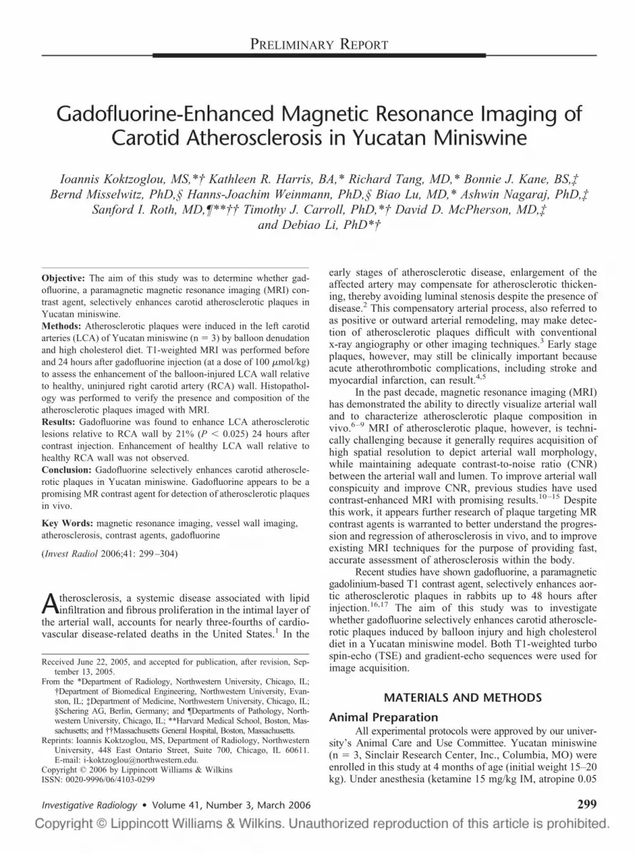
Each animal underwent MRI before and 24 hours afterintravenous injection of gadofluorine at a dose of 100 �mol/kg.We chose to perform postcontrast imaging 24 hours afterinjection of gadofluorine for 2 reasons: (1) to maximizepostcontrast enhancement of atherosclerotic plaque as previ-ously reported,17 and (2) to allow time for partial clearance ofGadofluorine from the bloodstream, thereby better facilitatingblood suppressed imaging. Axial single-shot 2D steady-statefree precession (SSFP) images were acquired in all experi-ments to locate the carotid arteries. Based on these images, a3 point tool was used to position a segmented 3D SSFPmagnetic resonance angiogram (MRA) depicting the bica-rotid trunk and the left and right common carotid arteries(LCA and RCA, respectively). Based on the MRA, 2 imagingslices were positioned perpendicular to the LCA as shown(Fig. 1B). An imaging slice was positioned through the LCAat a site of balloon denudation, hereto referred as the ‘injured’slice, whereas a second imaging slice was positioned throughthe LCA at a nondenuded region of the artery, hereto referred
as the “control” slice. Both imaging slices bisected the RCAas shown (Fig. 1B).
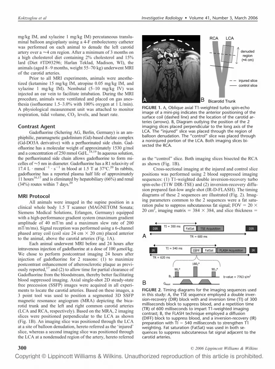
Cross-sectional imaging at the injured and control slicepositions was performed using 2 blood suppressed imagingsequences: (1) T1-weighted double inversion-recovery turbospin-echo (T1W DIR-TSE) and (2) inversion-recovery diffu-sion prepared fast-low angle shot (IR-D-FLASH). The timingdiagrams of these 2 sequences are illustrated (Fig. 2). Imag-ing parameters common to the 2 sequences were a fat satu-ration pulse to suppress subcutaneous fat signal; FOV � 20 �20 cm2, imaging matrix � 384 � 384, and slice thickness �
FIGURE 1. A, Oblique axial T1-weighted turbo spin-echoimage of a mini-pig indicates the anterior positioning of thesurface coil (dashed line) and the location of the carotid ar-teries (arrows). B, Diagram outlying the position of the 2imaging slices placed perpendicular to the long axis of theLCA. The “injured” slice was placed through the region ofballoon denudation. The “control” slice was placed througha noninjured portion of the LCA. Both imaging slices bi-sected the RCA.
FIGURE 2. Timing diagrams for the imaging sequences usedin this study: A, the TSE sequence employed a double inver-sion-recovery (DIR) block with and inversion time (TI) of 300milliseconds block to suppress blood, and a repetition time(TR) of 600 milliseconds to impart T1-weighted imagingcontrast; B, the FLASH technique employed a diffusion(DIFF) block to suppress blood, and a inversion-recovery (IR)preparation with TI � 540 milliseconds to strengthen T1weighting. Fat saturation (FatSat) was used in both se-quences to suppress subcutaneous fat signal adjacent to thecarotid arteries.
Koktzoglou et al Investigative Radiology • Volume 41, Number 3, March 2006
© 2006 Lippincott Williams & Wilkins300

3 mm, resulting in a spatial resolution � 0.5 � 0.5 � 3 mm3.The T1W DIR-TSE sequence employed a DIR preparation tosuppress signal from flowing blood and employed a repetitiontime of 600 milliseconds to achieve T1-weighted imagecontrast. Imaging parameters for the T1W DIR-TSE sequencewere: TR/TE � 600/10 milliseconds, averages (NEX) �13, imaging bandwidth (BW) � 200 Hz/pixel, flip angle �180°, echo train length � 9, echo spacing � 10 milliseconds,inversion time (TI) � 300 milliseconds, imaging time � 5minutes 35 seconds. The IR-D-FLASH sequence employed aTI of 540 milliseconds to strengthen T1-weighted imagecontrast, and a diffusion preparation to suppress signal fromflowing blood.17 Parameters for the IR-D-FLASH sequenceswere: TR/TE � 620/2.4 milliseconds, NEX � 32, BW � 345Hz/pixel, flip angle � 20°, segments � 15, imaging time �8 minutes 37 seconds. The b-value of the diffusion prepara-tion was experimentally optimized to suppress blood signal(before and 24 hours after contrast injection) while retainingadequate imaging signal-to-noise ratio (SNR). The meanb-value imparted by the diffusion preparation along the readout,phase encoding, and slice directions was 7763 seconds/m2.
Histopathologic AnalysisAll animals were euthanized after the final MRI procedure
by an intravenous injection of pentobarbital sodium and phenyt-oin sodium (Beuthanasia-D, 0.22 mL/kg). The left and rightcommon carotid arteries were excised and marked with India inkto facilitate matching of the MRI and histologic sections. Arterysamples were perfusion fixed with isopentane and frozen withliquid nitrogen. Portions of the carotid artery samples (1 cm inlength) corresponding to the positions of the MR images werecut and sent for histologic sectioning. Carotid artery specimenswere embedded in optimal cutting temperature compound, sec-tioned on a cryostat (5 �m thick sections) every 1 mm, andstained with hematoxylin-eosin and Mallory’s trichrome stains.An independent experienced pathologist blinded to the MRfindings performed the histologic analysis.
Image and Data AnalysisSNR of the LCA and RCA walls was measured both
before and 24 hours after injection of gadofluorine. SNR wasmeasured for both the control slices (slices passing through anondenuded portion of the LCA) and the injured slices (slicespassing through a balloon denuded portion of the LCA). SNRwas calculated by dividing the mean arterial wall signal bythe standard deviation of air signal located outside the animalbody. All signal measurements were performed on a work-station (Leonardo; Siemens Medical Solutions, Erlangen,Germany).
In each image, the SNR of the LCA wall was normal-ized by the SNR of the RCA wall. The relative SNR (RSNR)was defined by the relation:
RSNR � SNRLCA/SNRRCA
where SNRLCA and SNRRCA are the SNRs of the LCA andRCA, respectively. The enhancement ratio (ER) after gad-ofluorine injection was defined by the relation:
ER � RSNRPOST/RSNRPRE
where RSNRPRE and RSNRPOST are the relative SNRs beforeand 24 hours after injection of gadofluorine, respectively. Foreach slice position (control and balloon injured), the mean ERwas calculated by averaging the ER values from all imagesacquired with both IR-D-FLASH and T1W DIR-TSE se-quences. To assess whether gadofluorine selectively enhancedthe LCA wall, a one-tailed t test with statistical significancedefined at the 2.5% level was performed to determine whetherthe mean ER exceeded 1 in both the control and injured slices.
RESULTSMRI was successfully performed in all animals before
and 24 hours after gadofluorine injection. At the location ofballoon injury, mild atherosclerotic lesions were found in 2animals by histologic analysis. In the third animal, severeatherosclerotic stenosis was found over the entire length ofthe LCA.
Example T1W DIR-TSE and IR-D-FLASH imagesacquired at the control and injured slice positions in a mildlydiseased animal are shown (Figs. 3 and 4). The LCA andRCA arterial walls were isointense in the T1W DIR-TSE andIR-D-FLASH images acquired at the control slice position,both before and 24 hours after gadofluorine injection. Like-wise, the LCA and RCA walls were isointense in the imagesacquired at the injured slice position before injection ofgadofluorine.
On the other hand, balloon injured LCA wall washyperintense relative to the RCA wall in both the T1WDIR-TSE and IR-D-FLASH images 24 hours after contrastinjection, indicating selective enhancement of the injuredLCA wall by gadofluorine. Pathologic analysis at the locationof LCA wall enhancement revealed the presence of a mildatherosclerotic lesion (Fig. 4).
FIGURE 3. A, TrueFISP MRA depicting the left and rightcommon carotid arteries (LCA and RCA, respectively). In-jured (dashed line) and control (solid line) slice positions areindicated. B, T1W DIR-TSE images acquired before and 24hours after gadofluorine injection. The LCA and RCA wallsappear isointense in all images except the image acquired atthe injured slice position after gadofluorine injection. In thelatter image, the LCA wall (dashed arrow) is noticeably en-hanced relative to the RCA wall (solid arrow).
Investigative Radiology • Volume 41, Number 3, March 2006 Gadofluorine-Enhanced MRI of Carotid Atherosclerosis
© 2006 Lippincott Williams & Wilkins 301

A stenotic atherosclerotic lesion encountered in LCAof another animal is shown (Fig. 5). IR-D-FLASH imagesclearly depict enhancement of LCA wall relative to the RCAwall 24 hours after gadofluorine injection. Histopathologyrevealed the presence of a predominantly fibrofatty athero-sclerotic lesion.
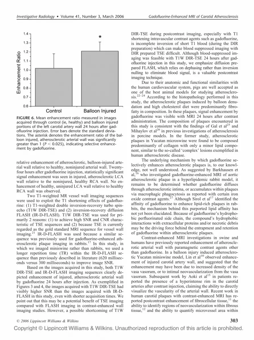
Mean ER values of the control and injured slices areplotted (Fig. 6). The mean ER of the images acquired at theinjured slice positions was 1.21 � 0.10 (P � 0.025), indicat-ing statistically significant signal enhancement of denudedLCA wall relative to healthy (ie, nondenuded) RCA wall 24hours after gadofluorine injection. The mean ER calculated fromthe images acquired at the control slice positions was 1.02 �0.16 (P � NS), suggesting no enhancement of healthy LCA wallrelative to healthy RCA wall, postcontrast injection.
The uninjured arteries were within normal limits, his-tologically. The endothelium consisted of a single layer ofcells atop the internal elastic lamina (Fig. 4D). The elasticmedia and adventitia were within normal limits. The injuredarteries had marked symmetrical and asymmetric endothelialproliferation with extracellular fibrosis (Fig. 4E). The internalelastic lamina, elastic media and adventitia were within normallimits.
DISCUSSIONThis study investigated whether gadofluorine, a paramag-
netic gadolinium-based contrast agent, enhanced atheroscle-rotic plaques induced by balloon injury and high cholesteroldiet in Yucatan miniswine. Blood suppressed cross-sectionalimaging was performed under 2 conditions—before and 24hours after contrast injection—for the purpose of locating
FIGURE 4. A, IR-D-FLASH images corresponding to the TSEimages shown in Figure 3. In the image acquired 24 hoursafter gadofluorine injection (top right), the LCA wall (dashedarrow) is enhanced relative to the RCA wall (solid arrow). Inthe remaining images, the LCA and RCA walls appear isoin-tense. B–E, Mallory’s trichrome histolopathology. B, Unin-jured, normal arterial wall. C, Injured, atherosclerotic arterialwall demonstrating a fibrocellular intimal thickening. Theblue staining indicated collagen deposition. D, Close up of Bdepicts the presence of a normal unicellular endothelial layer(arrow). E, Close-up of C depicts intimal proliferation com-posed of collagen and myofibroblasts. The clear spaces rep-resent ice crystals formed during freezing. L, lumen; I, in-tima; M, media.
FIGURE 5. A, Oblique coronal IR-D-FLASH image depictingthe LCA and RCA vessels. Note the visibly stenotic lesion inthe LCA (dashed arrow). B, Cross-sectional IR-D-FLASH im-age acquired before contrast injection at the location(dashed line) shown in A. C, Cross-sectional IR-D-FLASH im-age acquired 24 hours after gadofluorine injection at thesame location. Note the enhancement of the LCA wall post-contrast injection (dashed arrow). D, Hematoxylin and eosinsection depicting the LCA atherosclerotic lesion. E, Hematox-ylin and eosin section of the boxed region in D reveals thepresence of fibroblasts, myofibroblasts, and myoepithelialcells in the intima. Mallory’s trichrome staining depicts anintimal microvessel (inset in E). The clear spaces representice crystals formed during freezing. L: lumen, I, intima; M,media; F, fibrocellular tissue.
Koktzoglou et al Investigative Radiology • Volume 41, Number 3, March 2006
© 2006 Lippincott Williams & Wilkins302
relative enhancement of atherosclerotic, balloon-injured arte-rial wall relative to healthy, noninjured arterial wall. Twenty-four hours after gadofluorine injection, statistically significantsignal enhancement was seen in injured, atherosclerotic LCAwall relative to the noninjured, healthy RCA wall. No en-hancement of healthy, uninjured LCA wall relative to healthyRCA wall was observed.
Two T1-weighted MR vessel wall imaging sequenceswere used to exploit the T1 shortening effects of gadofluo-rine: (1) T1-weighted double inversion-recovery turbo spin-echo (T1W DIR-TSE) and (2) inversion-recovery diffusionFLASH (IR-D-FLASH). T1W DIR-TSE was used for pri-marily 2 reasons: (1) to achieve high SNR and CNR charac-teristic of TSE sequences and (2) because TSE is currentlyregarded as the gold standard MRI sequence for vessel wallimaging.21 IR-D-FLASH was used because a similar se-quence was previously used for gadofluorine-enhanced ath-erosclerotic plaque imaging in rabbits.17 In this study, inwhich we imaged miniswine rather than rabbits, we used alonger repetition time (TR) within the IR-D-FLASH se-quence than previously described in literature (620 millisec-onds versus 300 milliseconds) to improve image SNR.
Based on the images acquired in this study, both T1WDIR-TSE and IR-D-FLASH imaging sequences clearly de-picted enhancement of injured, atherosclerotic arterial wallby gadofluorine 24 hours after injection. As exemplified inFigures 3 and 4, the images acquired with T1W DIR-TSE hadvisibly higher SNR than the images acquired with IR-D-FLASH in this study, even with shorter acquisition times. Wepoint out that this may be a potential benefit of TSE imagingcompared with FLASH imaging in contrast-enhanced wallimaging studies. However, a possible shortcoming of T1W
DIR-TSE during postcontrast imaging, especially with T1shortening intravascular contrast agents such as gadofluorine,is incomplete inversion of short T1 blood (during the DIRpreparation) which can make blood suppressed imaging withDIR prepared TSE difficult. Although blood-suppressed im-aging was feasible with T1W DIR-TSE 24 hours after gad-ofluorine injection in this study, we emphasize diffusion pre-pared FLASH, which relies on dephasing rather than inversionnulling to eliminate blood signal, is a valuable postcontrastimaging technique.
Due to their anatomic and functional similarities withthe human cardiovascular system, pigs are well accepted asone of the best animal models for studying atherosclero-sis.22–24 According to the histopathology performed in thisstudy, the atherosclerotic plaques induced by balloon denu-dation and high cholesterol diet were predominantly fibro-fatty in composition. In these plaques, signal enhancement bygadofluorine was visible with MRI 24 hours after contrastadministration. The composition of plaques encountered inthis study is consistent with the findings of Gal et al25 andMihaylov et al26 in previous investigations of atherosclerosisin porcine models. In the former study, atheroscleroticplaques in Yucatan microswine were found to be composedpredominantly of collagen with only a minor lipid compo-nent, similar to the so-called ‘complex’ lesions exemplified inhuman atherosclerotic disease.
The underlying mechanism by which gadofluorine se-lectively enhances atherosclerotic plaques is, to our knowl-edge, not well understood. As suggested by Barkhausen etal,16 who investigated gadofluorine-enhanced MRI of aorticatherosclerotic plaque in a hyperlipidemic rabbit model, itremains to be determined whether gadofluorine diffusesthrough atherosclerotic intima, or accumulates within plaquesvia macrophagic phagocytosis as reported with certain ironoxide contrast agents.27 Although Sirol et al17 identified theaffinity of gadofluorine to enhance lipid-rich plaques in rab-bits, the mechanism behind this purported lipid affinity hasnot yet been elucidated. Because of gadofluorine’s hydropho-bic perfluorinated side chain, the compound’s hydrophobicinteractions with extracellular proteins and/or necrotic debrismay be the driving force behind the entrapment and retentionof gadofluorine within atherosclerotic plaques.
Contrast-enhanced MRI investigations in swine andhumans have previously reported enhancement of atheroscle-rotic arterial wall with paramagnetic contrast agents otherthan gadofluorine. In a balloon injury induced atherosclero-tic Yucatan miniswine model, Lin et al10 observed enhance-ment of injured carotid artery wall, and suggested that theenhancement may have been due to increased density of thevasa vasorum, or to intimal neovascularization from the vasavasorum. Subsequent work by Aoki et al15 in patients re-ported the presence of a hyperintense rim in the carotidarteries after contrast injection, claiming the ability to directlyvisualize the vascularity of the arterial wall. Recent work inhuman carotid plaques with contrast-enhanced MRI has re-ported postcontrast enhancement of fibrocellular tissue,11 theability to identify regions of neovascularization within fibroustissue,12 and the ability to quantify microvessel area within
FIGURE 6. Mean enhancement ratio measured in imagesacquired through control (ie, healthy) and balloon injuredportions of the left carotid artery wall 24 hours after gad-ofluorine injection. Error bars denote the standard devia-tions. The asterisk denotes the enhancement ratio of the bal-loon injured, atherosclerotic arterial wall was significantlygreater than 1 (P � 0.025), indicating selective enhance-ment by gadofluorine.
Investigative Radiology • Volume 41, Number 3, March 2006 Gadofluorine-Enhanced MRI of Carotid Atherosclerosis
© 2006 Lippincott Williams & Wilkins 303
plaque.13 Interestingly, rare intimal vessels were observed inthe histopathology performed on the stenotic, fibrocellularlesion encountered in this study (Fig. 5). Because of the longintravascular half life of gadofluorine, these vessels mayprovide a possible explanation for why this lesion enhanced24 hours after contrast administration.
A recent gadofluorine study performed in rabbits ob-served a correlation between postcontrast enhancement ofearly atherosclerotic plaques, and microvessel density withinthe adventitial vasa vasorum.28 This finding may provide apossible explanation for why enhancement of mild, nonste-notic plaques was observed in this study. However, furtherresearch is needed to clarify the mechanism(s) by whichgadofluorine selectively enhances the early atheroscleroticlesions in this animal model.
CONCLUSIONIn conclusion, gadofluorine was found to enhance non-
stenotic and stenotic carotid atherosclerotic plaques in Yuca-tan miniswine induced by balloon denudation and high cho-lesterol diet. Gadofluorine appears to be a promising contrastagent for detecting atherosclerotic plaques in vivo and mayallow for better understanding of the progression and regres-sion of atherosclerotic disease.
ACKNOWLEDGMENTSThe authors thanks Schering AG, who provided the MR
contrast agent used in this study. I. K. gratefully acknowl-edges the support of the Dr. John N. Nicholson Fellowship.
REFERENCES1. American Heart Association. Heart Disease and Stroke Statistics—2005
Update. Dallas: AHA; 2005.2. Glagov S, Weisenberg E, Zarins CK, et al. Compensatory enlargement
of human atherosclerotic coronary arteries. N Engl J Med. 1987;316:1371–1375.
3. Stiel GM, Stiel LS, Schofer J, et al. Impact of compensatory enlargementof atherosclerotic coronary arteries on angiographic assessment of cor-onary artery disease. Circulation. 1989;80:1603–1609.
4. Stary HC, Chandler AB, Dinsmore RE, et al. A definition of advancedtypes of atherosclerotic lesions and a histological classification ofatherosclerosis: a report from the Committee on Vascular Lesions of theCouncil on Arteriosclerosis, American Heart Association. Circulation.1995;92:1355–1374.
5. Falk E, Shah PK, Fuster V. Coronary Plaque Disruption. Circulation.1995;92:657–671.
6. Toussaint JF, LaMuraglia GM, Southern JF, et al. Magnetic resonanceimages lipid, fibrous, calcified, hemorrhagic, and thrombotic compo-nents of human atherosclerosis in vivo. Circulation. 1996;94:932–938.
7. Yuan C, Beach KW, Smith LH Jr, et al. Measurement of atheroscleroticcarotid plaque size in vivo using high resolution magnetic resonanceimaging. Circulation. 1998;98:2666–2671.
8. Fayad ZA, Nahar T, Fallon JT, et al. In vivo magnetic resonanceevaluation of atherosclerotic plaques in the human thoracic aorta: a
comparison with transesophageal echocardiography. Circulation. 2000;101:2503–2509.
9. Yuan C, Mitsumori LM, Beach KW, et al. Carotid atheroscleroticplaque: noninvasive MR characterization and identification of vulnera-ble lesions. Radiology. 2001;221:285–299.
10. Lin W, Abendschein DR, Haacke EM. Contrast-enhanced magneticresonance angiography of carotid arterial wall in pigs. J Magn ResonImaging. 1997;7:183–190.
11. Wasserman BA, Smith WI, Trout HH 3rd, et al. Carotid artery athero-sclerosis: in vivo morphologic characterization with gadolinium-en-hanced double-oblique MR imaging initial results. Radiology. 2002;223:566–573.
12. Yuan C, Kerwin WS, Ferguson MS, et al. Contrast-enhanced highresolution MRI for atherosclerotic carotid artery tissue characterization.J Magn Reson Imaging. 2002;15:62–67.
13. Kerwin W, Hooker A, Spilker M, et al. Quantitative magnetic resonanceimaging analysis of neovasculature volume in carotid atheroscleroticplaque. Circulation. 2003;107:851–856.
14. Wasserman BA, Casal SG, Astor BC, et al. Wash-in kinetics forgadolinium-enhanced magnetic resonance imaging of carotid atheroma.J Magn Reson Imaging. 2005;21:91–95.
15. Aoki S, Aoki K, Ohsawa S, et al. Dynamic MR imaging of the carotidwall. J Magn Reson Imaging. 1999;9:420–427.
16. Barkhausen J, Ebert W, Heyer C, et al. Detection of atherosclerotic plaquewith Gadofluorine-enhanced magnetic resonance imaging. Circulation.2003;108:605–609.
17. Sirol M, Itskovich VV, Mani V, et al. Lipid-rich atherosclerotic plaquesdetected by gadofluorine-enhanced in vivo magnetic resonance imaging.Circulation. 2004;109:2890–2896.
18. Misselwitz B, Platzek J, Raduchel B, et al. Gadofluorine 8: initialexperience with a new contrast medium for interstitial MR lymphogra-phy. Magma. 1999;8:190–195.
19. Bendszus M, Wessig C, Schutz A, et al. Assessment of nerve degener-ation by gadofluorine M-enhanced magnetic resonance imaging. AnnNeurol. 2005;57:388–395.
20. Misselwitz B, Platzek J, Weinmann H-J. Early MR lymphography withgadofluorine M in rabbits. Radiology. 2004;231:682–688.
21. Fayad ZA, Fuster V, Choudhury RP. CMR atherothrombotic plaqueimaging. In: Lardo AC, Fayad ZA, Chronos NAF, et al., eds. Cardio-vascular Magnetic Resonance. London: Taylor & Francis Group; 2004.
22. Reitman JS, Mahley RW, Fry DL. Yucatan miniature swine as a modelfor diet-induced atherosclerosis. Atherosclerosis. 1982;43:119–132.
23. Hughes HC. Swine in cardiovascular research. Lab Anim Sci. 1986;36:348–350.
24. Vesselinovitch D. Animal models and the study of atherosclerosis. ArchPathol Lab Med. 1988;112:1011–1017.
25. Gal D, Rongione AJ, Slovenkai GA, et al. Atherosclerotic Yucatanmicroswine: an animal model with high-grade, fibrocalcific, nonfattylesions suitable for testing catheter-based interventions. Am Heart J.1990;119:291–300.
26. Mihaylov D, van Luyn MJ, Rakhorst G. Development of an animalmodel of selective coronary atherosclerosis. Coron Artery Dis. 2000;11:145–149.
27. Ruehm SG, Corot C, Vogt P, et al. Magnetic resonance imaging ofatherosclerotic plaque with ultrasmall superparamagnetic particles ofiron oxide in hyperlipidemic rabbits. Circulation. 2001;103:415–422.
28. Sirol M, Purushothaman K-R, Moreno P, et al. Early versus advancedatherosclerotic plaque in vivo detection by Gadofluorine M-enhancedmagnetic resonance imaging. In: Proceedings of the 13th Annual Meet-ing of ISMRM, Miami Beach, 2005. p. 112.
Koktzoglou et al Investigative Radiology • Volume 41, Number 3, March 2006
© 2006 Lippincott Williams & Wilkins304