functional mri (fmri) in patients with spastic hemiplegia after stroke, treated with botulinum...
TRANSCRIPT
International Journal of Neurorehabilitation
Alessandro et al., Int J Neurorehabilitation Eng 2014, 1:1http://dx.doi.org/10.4172/ijn.1000104
Open AccessResearch Article
Volume 1 • Issue 1 • 1000104Int J NeurorehabilitationISSN: IJN, an open access journal
*Corresponding author: Alessandro Stecco, SCDU Radiologia, ASOU Maggiore della Carità Corso Mazzini 18, 28100, Novara, Tel: +39-03213733111; Fax : +39-03213733579; E-mail: [email protected]
Received March 12, 2014; Accepted Aprile 25, 2014; Published May 30, 2014
Citation: Alessandro S, Roberta M, Marco P, Stefano C, Lorenzo F et al. (2014) Functional MRI with Motor Imagery Task Show CNS Effects and Brain Plasticity after Botulinum Toxin Therapy in Spastic Hemiplegic Stroke Patients. Int J Neurorehabilitation 1: 104. doi:10.4172/ijn.10001104
Copyright: © 2014 Alessandro S, et al. This is an open-access article distributed under the terms of the Creative Commons Attribution License, which permits unrestricted use, distribution, and reproduction in any medium, provided the original author and source are credited.
Keywords: fMRI; Stroke; Botulinum toxin
Introduction Spasticity, an increase in velocity-dependent resistance to
stretching, is a movement disorder that manifests clinically as hyper-reactivity of the muscles [1]. It commonly occurs after ischemic lesion to the central nervous system, and 80% of stroke patients experience motor deficit due to damage to the pyramidal and/or extrapyramidal pathways [2]. These clinical features are defined as upper motor neuron syndrome, which is characterised by both positive (spasticity) and negative (hyposthenia) phenomena [3]. Spasticity in particular is a common phenomenon, and may strike roughly 38% of stroke patients a year after the ischemic event [4].
Stroke patients are spontaneously able to regain partial motor function through “brain recovery” processes linked to cortical plasticity. However, botulinum toxin A (BoNT-A) can be used to reduce involuntary muscle contraction in such patients, the rationale being that it does so by reducing cholinergic transmission at the neuromuscular junction, provoking paralysis. Local administration of BoNT-A has been shown to be more effective than systemic approaches, and is therefore a first-line treatment in muscle hyperactivity after stroke [5-7].
Although the peripheral effects of botulinum toxin are well known, only recently has evidence regarding its potential effects on the central nervous system, in particular the cerebral cortex, begun to emerge [8,9]. These effects may be explained by a reduction in the length/number/
density/excitability of type 1a sensory nerve fibres, a phenomenon that has already been described in dystonia [10,11].
Thus the aim of a peripheric botulin toxin therapy is mainly to reduce spasticity, so allowing a more regular and ordinated pattern of movement that will work on cortical function, allowing eventual a better cortical organization of neuronal activation.
This primary afferent fibre reduction and cortical plasticity are both reflected in changes in cortical activation during the actual and imagined execution and of motor tasks. So-called “motor imagery” is the dynamic state during which the representation of a specific motor action is reactivated within the working memory, without any evident motor output, and it is governed by the principles of the central motor nervous system [12,13]. Motor imagery has potential
AbstractIntroduction: Botulinum toxin is considered a first-line treatment for focal spasticity after stroke, and its peripheral
effects have been well documented. We set out to demonstrate and describe any effects it may have in the central nervous system, using fMRI to record brain activation patterns before and after its administration.
Materials and Methods: 17 subjects comprising 7 ischemic stroke patients affected by upper limb spastic hemiplegia, and 10 healthy controls were recruited and underwent three fMRI scans while performing a motor imagery task (finger tapping). Test subjects underwent fMRI before botulinum toxin therapy (T0), 4 weeks later (T1), and after 8 weeks (T2), and untreated control subjects were tested at 0, 4 and 8 weeks. The finger-tapping task was performed twice each session in both groups. Both test and control subjects performed only daily passive muscle stretching exercises between T0, T1, and T2 MR scans.
Results: fMRI confirmed the technical feasibility of the “motor imagery” paradigm in activating the motor areas in healthy subjects. While second-level analysis of the control group showed no modification in the pattern of brain activation during the finger-tapping imagery task between T0, T1, and T2, increasing focalization of mean brain activation, accompanied by a gradual reduction in secondary motor area activation after treatment (SMA and Brodmann 6) in the test group.
Conclusions: This confirms the efficacy of motor imagery as an fMRI paradigm to open a “window” into the brain that enables us to study the processes of motor function recovery after stroke in vivo. Our data show that peripheral injection of botulinum toxin alone brings about a progressive alteration in the reorganization of cortical activation after stroke, thereby confirming its therapeutic and central effects. The progressive reduction and greater focalization of activation seen continued over time, in line with the latest “small world network” theories on cortical and subcortical reorganization of cerebral functions after stroke.
Functional MRI with Motor Imagery Task Show CNS Effects and Brain Plasticity after Botulinum Toxin Therapy in Spastic Hemiplegic Stroke PatientsStecco Alessandro1*, Matheoud Roberta2, Perchinunno Marco1, Carda Stefano3, Fortunelli Lorenzo1, Marini Federica1, Cisari Carlo3 and Carriero Alessandro1
1SCDU Radiologia, ASOU Maggiore della Carità, Corso Mazzini 18, 28100, Novara, Italy2SC Fisica Sanitaria, ASOU Maggiore della Carità – Novara, Italy3SCDU Fisiatria, ASOU Maggiore della Carità – Novara, Italy
Citation: Alessandro S, Roberta M, Marco P, Stefano C, Lorenzo F et al. (2014) Functional MRI with Motor Imagery Task Show CNS Effects and Brain Plasticity after Botulinum Toxin Therapy in Spastic Hemiplegic Stroke Patients. Int J Neurorehabilitation 1: 104. doi:10.4172/ijn.10001104
Page 2 of 7
Volume 1 • Issue 1 • 1000104Int J NeurorehabilitationISSN: IJN, an open access journal
applications in motor skills training in both healthy subjects (e.g., athletes and professional musicians) and patients with neurological damage. Nevertheless, thus far few studies have focussed on motor imagery training as a means of obtaining functional recovery after stroke [14,15].
We set out to evaluate the modulatory effects of botulinum toxin therapy on the central nervous system over time on stroke-derived spastic hemiplegia patients. The primary end-point was to confirm the existence of such an effect, using fMRI to record cerebral plasticity phenomena.
Materials and Methods Patients
Ten healthy volunteers (all with left hemisphere cerebral dominance) were recruited as a control group, and were subjected to the fMRI protocol to study the brain areas activated during real and imagined motor and listening tasks. Seven patients affected by spastic hemiplegia following ischemic or haemorrhagic stroke (Table 1) were also recruited according to the following inclusion criteria [16]:
• Hemiplegia after acute ischemic or haemorrhagic stroke, as documented by CT scan;
• At least 6 months’ time elapsed after stroke;
• Upper limb spasticity of at least 1+ on the Modified Ashworth Scale (MAS) [17];
• Complete distal spastic plegia of the upper limb (residual force of finger and thumb flexors and extensors M=0 on the Medical Research Council Scale [18]
• Final BoNT-A treatment and no rehabilitation therapy in the three months before enrolment.
• The exclusion criteria were as follows [16]:
• Fixed contracture and/or bone deformity of upper limb;
• Cognitive impairment limiting comprehension of motor tasks during treatment;
• Concomitant progressive central nervous system disease;
• Peripheral nervous system disease/myopathy;
• Previous surgery to upper limb extensor or flexor muscles, or arthrodesis to the compromised side.
Study protocol
All subjects were informed as to the nature of the fMRI test before undergoing a total of three sessions, one at T0 (before commencing BoNT-A therapy), the second at T1 (4 weeks after the treatment), and the last at T2 (8 weeks after treatment). No rehabilitation therapy,
except for passive muscle stretching, was performed between sessions held at T0, T1 and T2. Healthy controls were also tested at the same three time intervals, T0, T1 and T2.
Treatment
Patients were given local injections of BoNT-A (incobotulinumtoxinA) at a dilution 100:2 cc 0.9% NaCl solution. The mean dose administered was 235±138 U (Units), range 90–400, and muscles treated were: flexor digitorum superficialis (7 patients), flexor digitorum profundus (5 patients), flexor pollicis longus (7 patients), flexor carpi ulnaris (3 patients), flexor carpi radialis (2 patients), adductor pollicis (1 patient), opponens pollicis (1 patient), flexor pollicis brevis (1 patients), and pronator teres (1 patient). The mean interval between the stroke and the treatment was well over 6 months (47.2 ± 43 months, range 12–129 months).
Experimental paradigm & fMRI technique
Before each session, subjects were asked to practice the finger-tapping task with their good hand (real finger tapping, RFT) and subsequently to imagine performing the same task with their plegic hand (imagined finger tapping, IFT), or the contralateral in the case of healthy controls. The motor imagery task consists of performing a mental simulation of the movement associated with a kinaesthetic sensation (imagining one-self performing the task).
The experimental paradigm involved the alternation of two tasks (A and B) in which the first, A, was the target (real or imagined motor task) and the second, B, involved passively listening to musical tones to clear their mind. Three of these task sequences were performed by each subject in each fMRI session, as shown in Table 2. Audio and visual stimuli for task execution were delivered to subjects by means of NordikAktiva software (NordikNeuroLab, Bergen, Norway), and the hardware setup therefore comprised MRI goggles and headphones synchronized with the scanner, and an MRI-compatible joystick for giving subjects task execution feedback. MR images were recorded using a Philips Achieva (version 2.5.3) 1.5 Tesla (Philips Healthcare, Best, The Netherlands), using an 8-channel phased-array synergy coil.
A sequence of 3D T1-weighted images (TR: 25 ms; TE 4.6 ms; acquisition matrix: 240x240; voxel size: 1x1x1 mm3) was acquired before each session and used as a template for localization of functional activations. Each of three functional runs was acquired using axial echo-planar (EPI) scans (TE=50 ms, TR=3000 ms, matrix 128x128, FOV=230x230 mm, slice thickness=4 mm, 26 slices, 1NEX) comprising 90 volumes divided into 9 blocks corresponding to alternating tasks (A-B-A-B-A-B-A-B-A), each block made up of 10 volumes of duration 30s (total run duration 270”).
The paradigm was the same for healthy controls and patients and therefore comprised
• An activation phase in which the subject was asked to press the button on the joystick repeatedly with their good hand (or to imagine doing it with their plegic/contralateral hand) upon the command “MOVE HAND” projected into the MRI goggles (task A).
wN° Patient Sex Age Side of lesion Site of Lesion Dominance 1 F 43 Right Basal Ganglia Right2 M 48 Left Fronto-parietal Right3 F 50 Right Basal Ganglia Right4 M 61 Left Fronto-parietal Right5 M 45 Right Fronto-Temporo-Parietal Right6 M 47 Left Basal Ganglia / Insula Right7 F 42 Left Basal Ganglia RightOverall 3F 4M 48 3 R and 4 L Heterogeneous 7 Right
Table 1: Patient details
TASK A1 Real finger tapping, FTR Tonal Auditory Stimulus2 Imagery finger tapping, FTI-1 Tonal Auditory Stimulus3 Imagery finger tapping, FTI-2 Tonal Auditory Stimulus
Table 2: Schematic representation of experimental paradigm followed at each session and for each subject
Citation: Alessandro S, Roberta M, Marco P, Stefano C, Lorenzo F et al. (2014) Functional MRI with Motor Imagery Task Show CNS Effects and Brain Plasticity after Botulinum Toxin Therapy in Spastic Hemiplegic Stroke Patients. Int J Neurorehabilitation 1: 104. doi:10.4172/ijn.10001104
Page 3 of 7
Volume 1 • Issue 1 • 1000104Int J NeurorehabilitationISSN: IJN, an open access journal
• A resting phase (“Rest”) in which the subject was fed musical tones comprising a kind of melody through the headphones (task B).
Statistical analysis
Functional data was analysed by means of SPM software v.8b. Functional images were corrected for movement, co-recorded with the corresponding morphological images, normalized for Talairach space, and passed through a spatial smoothing filter (FWHM 8mm). The voxels activated during each task (A or B) were detected using level 1 analysis, identifying those activated during the task (FTR or FTI1/FTI2) as ON. Group analysis was then performed (level 2 analysis, p<0.001, without correction) for each task, and the SPM tool xjView was used to determine the maximum intensity and number of voxels in each functional area (cluster), and the Brodmann area involved (quantitative analysis).
The functional map renderings were saved, and examined independently by two neuroradiologist fMRI experts, who provided semi-quantitative scores. The inter-operator agreement was calculated using Cohen’s k test.
ResultsControls
Both “first level” and “second level” group analysis (Table 3) showed that cerebral cortex in the primary and supplementary motor areas, the dorsolateral prefrontal area (DLPF), were activated during the motor imagery task. Group analysis of the three tests performed at T0, T1 and T2, showed no alteration over time.
Patients
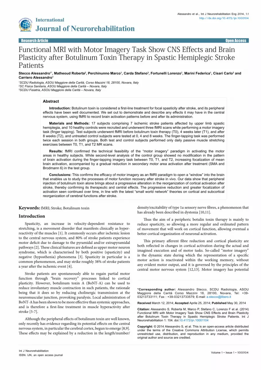
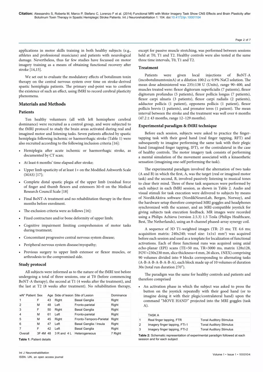
All patients managed to complete the three fMRI sessions at T0, T1 and T2, and none reported any difficulty in performing the tasks. Figure 1 show the motor areas activated in one patient who had suffered stroke to the right basal ganglia during the motor imagery tasks at T2 (8 weeks after botulinum toxin therapy). Figure 2 shows the cerebral activation upon motor imagery stimulation, subjected to level 2 analysis, at T0 (A); 4 weeks after the treatment, T1 (B); and 8 weeks after treatment, T2 (C). As we have previously noted that repeating the motor imagery task improves results and provides better statistical significance, we performed statistical analysis on the data from the second of the two tests.
Cluster 13 Sector: voxelVoxel n 127 Parietal inf R 73peak intensity 14,2253 Br 40 43Site Parietal inf R
Cluster 15 Sector: voxelVoxel n 7 Parietal inf L 6peak intensity 7,5103Site Parietal inf L
Cluster 16 Sector: voxelVoxel n 34 supp motor area L 25peak intensity 9,8075 Br 6 17Site supp motor area L supp motor area R 6
Table 3: SPM analysis of fMRI cortical activations in healthy subjects by site, voxel number, peak intensity and anatomical functional correlation Abbreviations: L=Left; R=Right; inf=inferior; supp=supplemental; Br=Broca Area
Figure 2 A-C: Level 2 analysis of Brodmann area 6 in patient group showing mean activations at T0 (A), T1 (B) and T2 (C), respectively before BoNT-A thera-py and 4 and 8 weeks after. Note the progressive reduction in size and improved definition of the activation cluster.
Figure 1: Post-processed image (SPM software) of right basal ganglia stroke patient performing motor imagery task at T2. Note the activation of areas M1, SMA, DPLF and cerebellum
A
B
C
Citation: Alessandro S, Roberta M, Marco P, Stefano C, Lorenzo F et al. (2014) Functional MRI with Motor Imagery Task Show CNS Effects and Brain Plasticity after Botulinum Toxin Therapy in Spastic Hemiplegic Stroke Patients. Int J Neurorehabilitation 1: 104. doi:10.4172/ijn.10001104
Page 4 of 7
Volume 1 • Issue 1 • 1000104Int J NeurorehabilitationISSN: IJN, an open access journal
Findings from one of the 7 stroke patients (14.3%) were not significant after SPM post-processing; the patient in question had extensive ischemic damage to the parietal lobe and the test was not found to be diagnostic. Post-processing images were assessed by two radiologists, who scored the activation of the various cortical areas as follows: 0 = no activation, 1 = possible activation, and 2 = certain activation. There was very good agreement between the two sets of qualitative scores (K=0.92)
Some of our patients were left-handed, and so in order to obtain a homogeneous group for level 2 analysis, some images were inverted (flipped) so that all lesions were pictured on the right. Level 2 analysis of the patients revealed the following (see Table 4, Charter 1, 2, 3a and 3b):
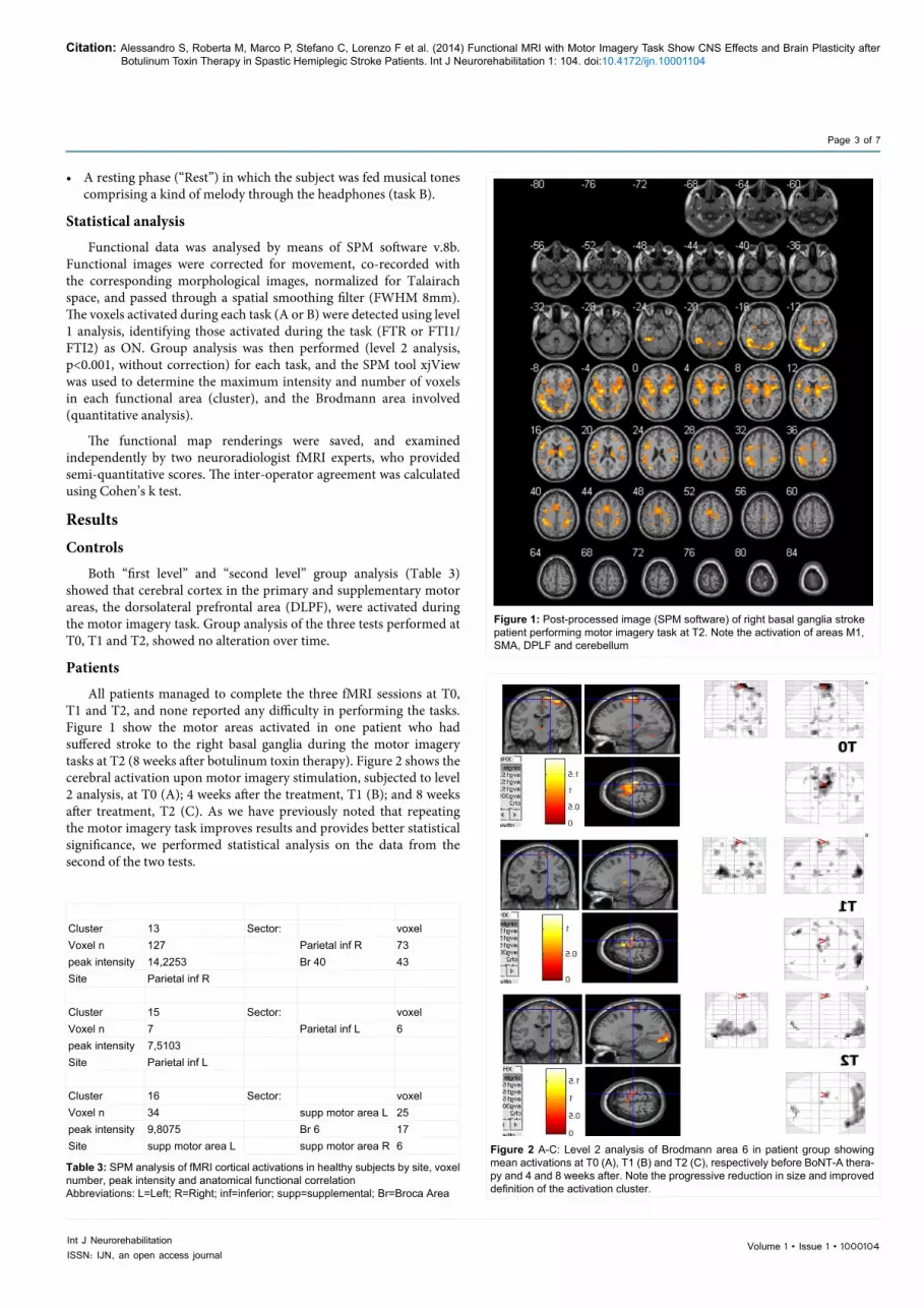
• At T0, activation was mainly seen in the secondary visual cortex (Brodmann area 18) and secondary motor areas (SMA and Brodmann 6); activation was bilateral but greater voxel numbers and intensity were seen in the hemisphere contralateral to the lesion.
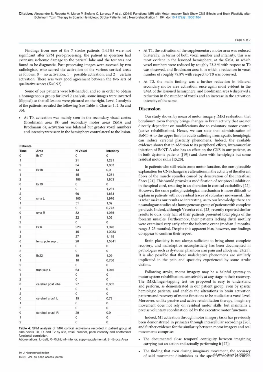
• At T1, the activation of the supplementary motor area was reduced bilaterally, in terms of both voxel number and intensity; this was most evident in the lesioned hemisphere, at the SMA, in which voxel numbers were reduced by roughly 73.2 % with respect to T0 was observed, and Brodmann area 6, in which a reduction in voxel number of roughly 79.8% with respect to T0 was observed.
• At T2, the main finding was a further reduction in bilateral secondary motor area activation, once again most evident in the SMA of the lesioned hemisphere, and Brodmann area 6 displayed a reduction in the number of voxels and an increase in the activation intensity of the same.
DiscussionOur study shows, by mean of motor imagery fMRI evaluation, that
botulinum toxin therapy brings changes in brain activity that are not directly dependent on modifications due to voluntary motor control (active rehabilitation). Hence, we can state that administration of BoNT-A to the upper limb in adults suffering from spastic hemiplegia can induce cerebral plasticity phenomena. Indeed, the available evidence shows that in addition to its peripheral effects, intramuscular injection of BoNT-A also has an effect on the CNS in our patients, as in both dystonia patients ([19]) and those with hemiplegia but some residual motor skills [15,20].
In patients who still retain some motor function, the most plausible explanation for CNS changes are alterations in the activity of the afferent fibres of the muscle spindles caused by denervation of the intrafusal fibres [21]. This would provoke a modification of reciprocal inhibition in the spinal cord, resulting in an alteration in cortical excitability [22]. However, the same pathophysiological mechanism is more difficult to explain in patients with no residual traces of voluntary movement. This is what makes our results so interesting, as to our knowledge there are no analogous studies of a homogeneous group of patients with complete paralysis. Indeed, although Veverka et al. [23] recently reported similar results to ours, only half of their patients presented total plegia of the forearm muscles. Furthermore, their patients lacking distal motility were examined very early after the ischemic event (median 5 months, range 3-23 months). Despite this apparent bias, however, our findings do appear to confirm their report.
Brain plasticity is not always sufficient to bring about complete recovery, and maladaptive neuroplasticity has been documented in pathologies such as dystonia, phantom arm pain and allodynia [24,25]. It is also possible that these maladaptive phenomena are similarly implicated in the pain and spasticity experienced by some stroke victims.
Following stroke, motor imagery may be a helpful gateway to motor system rehabilitation, conceivably at any stage in their recovery. The fMRI/finger-tapping test we proposed is easy to understand and perform, as demonstrated in our patient group, even by spastic hemiplegic patients, and enables the alterations in brain activation patterns and recovery of motor functions to be studied at a voxel level. Moreover, unlike passive and active rehabilitation therapy, imaginary movement does not rely on residual motor skills, but maintains a precise voluntary coordination led by the executive motor functions.
Indeed, M1 activation through motor imagery tasks has previously been demonstrated in primates through intracellular recordings [26], and further evidence for the similarity between motor imagery and real movements comprise:
• The documented close temporal contiguity between imagining carrying out an action and actually performing it [27];
• The finding that even during imaginary movement, the accuracy of said movement diminishes as the speed of mental execution
PatientsTime Area N Voxel Intensity0 Br17 0 01 21 1,2812 34 1,8630 Br18 13 0,91 45 1,2812 186 1,8630 Br19 0 01 9 1,2812 66 1,8630 sma L 105 1,9761 51 1,022 9 0,7730 sma R 82 1,9761 22 1,022 0 00 Br 6 223 1,9761 45 1,02532 27 1,1190 temp pole sup L 20 1,53411 0 02 0 00 Br22 19 1,091 10 0,7662 0 00 front sup L 63 1,9761 0 02 0 00 cerebell post lobe 27 0,6621 0 02 0 00 cerebell crus1 L 15 0,781 0 02 0 00 cerebell crus1 R 29 0,91 0 02 0 0
Table 4: SPM analysis of fMRI cortical activations recorded in patient group at time-points T0, T1 and T2 by site, voxel number, peak intensity and anatomical functional correlation, Abbreviations: L=Left; R=Right; inf=inferior; supp=supplemental; Br=Broca Area

Citation: Alessandro S, Roberta M, Marco P, Stefano C, Lorenzo F et al. (2014) Functional MRI with Motor Imagery Task Show CNS Effects and Brain Plasticity after Botulinum Toxin Therapy in Spastic Hemiplegic Stroke Patients. Int J Neurorehabilitation 1: 104. doi:10.4172/ijn.10001104
Page 5 of 7
Volume 1 • Issue 1 • 1000104Int J NeurorehabilitationISSN: IJN, an open access journal
increases (Fitts’ law) [28], and that the symmetry between dominant and subordinate hemisphere is maintained;
• Motor imagery can induce changes in the autonomic nervous system, increasing respiratory and heart rates.
Hence motor imagery can be likened to motor execution rather than preparation for movement, making it susceptible to functional reorganization in patients with hemiparesis following stroke, and therefore of particular interest to researchers in the field. Indeed, the generation and conservation of kinaesthetic models is mainly the province of the parietal lobe [29], and parietal lobe damage can therefore prevent formulation of motor imagery; it is interesting to note that the one patient in our study with extensive parietal damage showed no
corresponding cerebral activation during the motor imagery task. This has been noted in other studies [30], in which all but the patients with extensive parietal damage were able to complete motor imagery tasks and therefore provide useful fMRI data for analysis.
In patients without this impairment, the following brain areas are activated during a motor imagery task: the supplementary motor area (SMA), the dorsolateral prefrontal cortex (DLPFC), the primary motor area (M1), the cingulate gyrus and the cerebellum [31]. This widespread activation suggests the involvement of a complex motor circuit that extends to areas dealing with visual representation and motor imagery. Studies comparing the sites activated during real and imagined movement show that these are similar, but the extension
250
200
150
100
50
0
2,5
2
1,5
1
0,5
0T0 T1 T2 T0 T1 T2
Br17 Br18 Br19 sma L sma R Br 6 Br17
Extension of cerebral activation Intensity of cerebral activation
Br18 Br19 sma L sma R Br 6
n. v
oxel
n. v
oxel
A BCharter 1 a-b: Activation of relevant brain areas showing number and intensity of voxels
250
200
150
100
50
0
2,5
2
1,5
1
0,5
0
Extension of cerebral activation - motor areas Intensity of cerebral activation - motor areas
T0 T1 T2 T0 T1 T2
No v
oxel
Inte
nsità
sma L sma R Br 6 sma L sma R Br 6
A BCharter 2 a-b: Level 2 analysis of motor area activation showing voxel numbers and intensity
Citation: Alessandro S, Roberta M, Marco P, Stefano C, Lorenzo F et al. (2014) Functional MRI with Motor Imagery Task Show CNS Effects and Brain Plasticity after Botulinum Toxin Therapy in Spastic Hemiplegic Stroke Patients. Int J Neurorehabilitation 1: 104. doi:10.4172/ijn.10001104
Page 6 of 7
Volume 1 • Issue 1 • 1000104Int J NeurorehabilitationISSN: IJN, an open access journal
is greater in the former [31]. Indeed some report that the M1 is not activated during imaginary movement, as it is the means by which the damaged system impedes actual movement [32]. The role of the M1 remains controversial, and it is crucial that further fMRI/motor imagery studies are performed to investigate patients’ motor function in greater depth.
Likewise, although other studies [15] have demonstrated that BoNT-A reduces the extension of activation and increases the lateralization of the sensorimotor network, we were only able to confirm the former finding. Indeed, though we did observe a progressive reduction in brain area activation between 4 and 8 weeks after the BoNT-A was injected (T1 and T2, respectively), no significant lateralization of sensorimotor activation was detected.
At T0, the general hyperactive state observed, also known as chaotic motor imagery, is linked to a reactive pattern of the damaged brain, a kind of maladaption caused by the increase in the excitability of pathological proprioceptive afferent fibres (1a fibre), a spasticity-related phenomenon. This chaotic motor imagery pattern was evident in all our patients, and progressively diminished between T1 and T2. Indeed, according to Senkarova et al. [8], BoNT-A infiltration could modify the sensitive input to the CNS by peripheral neuromuscular blockade of the gamma motor neurons, which may lead to a reduction in the excitability of 1a fibres.
Although spontaneous functional recovery or that induced by rehabilitation therapy can have an influence, these mechanisms are most effective during the first three months after stroke [33], and therefore cannot wholly explain the modifications seen in our patients, who were all recruited after at least a year. We also observed that several areas of activation that appeared at 4 or 8 weeks from the BoNT-A treatment were the same as those activated during the motor imagery tasks in healthy volunteers. Group (level 2) analysis, confirming previous findings by Solodkin et al. [32], also showed how the chaotic activation pattern seen in the areas activated by motor imagery becomes progressively more defined (reduction in voxel numbers and intensity), with fewer areas being activated [34].
Recently, the fusiform gyrus, the cuneus, and the posterolateral cerebellum have been added to the list of sites typically associated with executive motor function [35], and may be involved in motor imagery [36]. It is possible that the extension and progressive intra individual increase in activation at the dentate nuclei and of the SMA may be implicated in the creation of a network with more connections and shorter communication channels, according to the definition of a “small-world network” [35].
To conclude, through fMRI we were able to confirm our initial hypothesis, namely that as well as exerting peripheral effects and therefore representing a useful physiotherapy tool, peripheral administration of the antispastic BoNT-A also affects the CNS, causing changes in cerebral activation.
In fact it provokes the same effects on cortical and subcortical reorganization described after “recovery” through physiotherapy, i.e., a loss of the initial chaotic pattern of multiple random activations, and the progressive establishment, especially between T1 and T2, of an ordered activation network including cerebellar regions, and greater activation of the SMA, as well as a greater focalization (reduction) in such activation in terms of voxel numbers and intensity.
The possible explanation of this phenomenon can be related to a central effect by reduction in the length/number/density/excitability of
type 1a sensory nerve fibres, as previously said [6,7]; this phenomenon affects the pattern of cortical activation after months, acting as a “trainer” in inducing progressively a better cortical reorganization. This is confirmed by our and also previous papers fMRI data.
In our sample this occurred in correspondence to the brain plasticity phenomena induced by the locoregional treatment, which was administered so long after the stroke that the influence of spontaneous recovery phenomena can be ruled out. As these modulation and brain plasticity changes were observed late after stroke, it would be interesting to study them in more depth.
Surely, this pilot study highlights the role of Motor Imagery as a potential interesting tool to study motor function and recovery plegic patients.
It would also be useful to repeat our investigation on a larger patient sample, as the small sample size and inclusion of left- and right-handed patients within it are its major limitations. That being said, other studies in the literature have been based on similar, or even smaller, samples [5,15,36], and, like us, other authors have made use of the image flipping technique to overcome the handedness issue in level 2 analysis [5,36].References
1. Gracies JM (2005) Pathophysiology of spastic paresis. II: Emergence of muscle overactivity. Muscle Nerve 31: 552-571.
2. Peltonen M, Stegmayr B, Asplund K (1998) Time trends in long-term survival after stroke: the Northern Sweden Multinational Monitoring of Trends and Determinants in Cardiovascular Disease (MONICA) study, 1985-1994. Stroke 29: 1358-1365.
3. Sheean G. Neurophysiology of spasticity. (2001) In: Barnes M, Johnson G (Eds.), Upper Motor Neuron Syndrome and Spasticity. Cambridge: Cambridge University Press 12-78.
4. Leathley MJ, Gregson JM, Moore AP, Smith TL, Sharma AK, et al. (2004) Predicting spasticity after stroke in those surviving to 12 months. Clin Rehabil 18: 438-443.
5. Simpson DM, Gracies JM, Yablon SA, Barbano R, Brashear A; BoNT/TZD Study Team (2009) Botulinum neurotoxin versus tizanidine in upper limb spasticity: a placebo-controlled study. J Neurol Neurosurg Psychiatry 80: 380-385.
6. Geenen C, Consky E, Ashby P (1996) Localizing muscles for botulinum toxin treatment of focal hand dystonia. Can J Neurol Sci 23: 194-197.
7. Yelnik AP, Simon O, Bensmail D, Chaleat-Valayer E, Decq P, et al. (2009) Drug treatments for spasticity. Ann Phys Rehabil Med 52: 746-756.
8. Senkárová Z, Hlustík P, Otruba P, Herzig R, Kanovský P (2010) Modulation of cortical activity in patients suffering from upper arm spasticity following stroke and treated with botulinum toxin A: an fMRI study. J Neuroimaging 20: 9-15.
9. Manganotti P, Acler M, Formaggio E, Avesani M, Milanese F, et al. (2010) Changes in cerebral activity after decreased upper-limb hypertonus: an EMG-fMRI study. Magn Reson Imaging 28: 646-652.
10. Kanovsky P, Bares M, Dufek J (2004) Spasticita: Mechanismy, Diagnostika a L´ecba. Praha:Maxdorf
11. Kanovsky P, Streitova H, Dufek J, Znojil V, Daniel P, et al. (1998) Change in lateralization of the P22/N30 cortical component of median nerve somatosensory evoked potentials in patients with cervical dystonia after successful treatment with botulinum toxin A. Mov Disord 13: 108-117.
12. Decety J, Jeannerod M, Prablanc C (1989) The timing of mentally represented actions. Behav Brain Res 34: 35-42.
13. Decety J, Grèzes J (1999) Neural mechanisms subserving the perception of human actions. Trends Cogn Sci 3: 172-178.
14. Page SJ, Levine P, Sisto S, Johnston MV (2001) A randomized efficacy and feasibility study of imagery in acute stroke. Clin Rehabil 15: 233-240.
Citation: Alessandro S, Roberta M, Marco P, Stefano C, Lorenzo F et al. (2014) Functional MRI with Motor Imagery Task Show CNS Effects and Brain Plasticity after Botulinum Toxin Therapy in Spastic Hemiplegic Stroke Patients. Int J Neurorehabilitation 1: 104. doi:10.4172/ijn.10001104
Page 7 of 7
Volume 1 • Issue 1 • 1000104Int J NeurorehabilitationISSN: IJN, an open access journal
15. Page SJ, Levine P, Leonard AC (2005) Effects of mental practice on affected limb use and function in chronic stroke. Arch Phys Med Rehabil 86: 399-402.
16. Baricich A, Carda S, Bertoni M, Maderna L, Cisari C (2008) A single-blinded, randomized pilot study of botulinum toxin type A combined with non-pharmacological treatment for spastic foot. J Rehabil Med 40: 870-872.
17. Bohannon RW, Smith MB (1987) Interrater reliability of a modified Ashworth scale of muscle spasticity. Phys Ther 67: 206-207.
18. Wade D (1992) Motorsensory impairments. In Wade DT (eds) Measurement in neurological rehabilitation. Curr Opin Neurol Neurosurg 682-686.
19. Girlanda P, Quartarone A, Sinicropi S, Nicolosi C, Roberto ML, et al. (1997) Botulinum toxin in upper limb spasticity: study of reciprocal inhibition between forearm muscles. Neuroreport 8: 3039-3044.
20. Tomasova Z, Hlustik P, Kral M, Otruba P, Herzig R, et al. (2013) Cortical activation changes in patients suffering from post-stroke arm spasticity and treated with botulinum toxin a. J Neuroimaging 23: 337-344.
21. Priori A, Berardelli A, Mercuri B, Manfredi M (1995) Physiological effects produced by botulinum toxin treatment of upper limb dystonia: changes in reciprocal inhibition between forearm muscles. Brain 118: 801–807.
22. Byrnes ML, Thickbroom GW, Wilson SA, Sacco P, Shipman JM, et al. (1998) The corticomotor representation of upper limb muscles in writer’s cramp and changes following botulinum toxin injection. Brain 121: 977-988.
23. Veverka T, Hluštìk P, Tomášová Z, Hok P, Otruba P, et al. (2012) BoNT-A related changes of cortical activity in patients suffering from severe hand paralysis with arm spasticity following ischemic stroke. J Neurol Sci 319: 89-95.
24. Pujol J, Roset-Llobet J, Rosinés-Cubells D, Deus J, Narberhaus B, et al. (2000) Brain cortical activation during guitar-induced hand dystonia studied by functional MRI. Neuroimage 12: 257-267.
25. Maihöfner C, Handwerker HO, Birklein F (2006) Functional imaging of allodynia in complex regional pain syndrome. Neurology 66: 711-717.
26. Georgopoulos AP, Lurito JT, Petrides M, Schwartz AB, Massey JT (1989) Mental rotation of the neuronal population vector. Science 243: 234-236.
27. Decety J, Jeannerod M (1995) Mentally simulated movements in virtual reality: does Fitts’s law hold in motor imagery? Behav Brain Res 72: 127-134.
28. Sirigu A, Daprati E, Pradat-Diehl P, Franck N, Jeannerod M (1999) Perception of self-generated movement following left parietal lesion. Brain 122: 1867-1874.
29. Johnson SH (2000) Imagining the impossible: intact motor representations in hemiplegics. Neuroreport 11: 729-732.
30. Hashimoto R, Rothwell JC (1999) Dynamic changes in corticospinal excitability during motor imagery. Exp Brain Res 125: 75-81.
31. Traversa R, Cicinelli P, Oliveri M, Giuseppina Palmieri M, Filippi MM, et al. (2000) Neurophysiological follow-up of motor cortical output in stroke patients. Clin Neurophysiol 111: 1695-1703.
32. Solodkin A, Hlustik P, Chen EE, Small SL (2004) Fine modulation in network activation during motor execution and motor imagery. Cereb Cortex 14: 1246-1255.
33. Wang L, Yu C, Chen H, Qin W, He Y, et al. (2010) Dynamic functional reorganization of the motor execution network after stroke. Brain 133: 1224-1238.
34. Allen G, Buxton RB, Wong EC, Courchesne E (1997) Attentional activation of the cerebellum independent of motor involvement. Science 275: 1940-1943.
35. Cramer SC, Nelles G, Schaechter JD, Kaplan JD, Finklestein SP, et al. (2001) A functional MRI study of three motor tasks in the evaluation of stroke recovery. Neurorehabil Neural Repair 15: 1-8.
36. Ward NS, Brown MM, Thompson AJ, Frackowiak RS (2003) Neural correlates of motor recovery after stroke: a longitudinal fMRI study. Brain 126: 2476-2496.
Citation: Alessandro S, Roberta M, Marco P, Stefano C, Lorenzo F et al. (2014) Functional MRI with Motor Imagery Task Show CNS Effects and Brain Plasticity after Botulinum Toxin Therapy in Spastic Hemiplegic Stroke Patients. Int J Neurorehabilitation 1: 104. doi:10.4172/ijn.10001104
Submit your next manuscript and get advantages of OMICS Group submissionsUnique features:
User friendly/feasible website-translation of your paper to 50 world’s leading languagesAudio Version of published paperDigital articles to share and explore
Special features:
350 Open Access Journals35,000 editorial team21 days rapid review processQuality and quick editorial, review and publication processingIndexing at PubMed (partial), Scopus, EBSCO, Index Copernicus and Google Scholar etcSharing Option: Social Networking EnabledAuthors, Reviewers and Editors rewarded with online Scientific CreditsBetter discount for your subsequent articles
Submit your manuscript at: http://www.omicsonline.org/submission