a physiologically based clinical measure for spastic reflexes in spinal cord injury
TRANSCRIPT

ASEB
Ss2
mt(
ves
gs
mw
muaSjpuflkEwm
swSSk
aCwB
BF
n
so
Mb
52
A
Physiologically Based Clinical Measure forpastic Reflexes in Spinal Cord Injury
la N. Benz, MSPT, T. George Hornby, PhD, Rita K. Bode, PhD, Robert A. Scheidt, PhD,
rian D. Schmit, PhDrsss
dpntmsh
b
cR
Ptbtmtpb
mtMhdtjoTjaht
hflispatrSrch
ABSTRACT. Benz EN, Hornby TG, Bode RK, Scheidt RA,chmit BD. A physiologically based clinical measure forpastic reflexes in spinal cord injury. Arch Phys Med Rehabil005;86:52-9.
Objective: To test the validity of the Spinal Cord Assess-ent Tool for Spastic reflexes (SCATS), a clinical tool in-
ended to rate spastic motor behavior after spinal cord injurySCI).
Design: By using correlational analyses, the SCATS wasalidated using concurrent measurements of kinematics andlectromyograms and traditional assessments of spasms andpastic hypertonia.
Setting: Research laboratory (kinematics and electromyo-raphy) and outpatient medical clinic (traditional measures ofpastic hypertonia).
Participants: Eleven people with SCI were used for kine-atic and electromyographic measurements. Seventeen peopleith SCI were used for comparison with other clinical scales.Interventions: Not applicable.Main Outcome Measures: Kinematic and surface electro-yographic measurements of the tested lower extremity were
sed to quantify magnitude and/or duration of motor behaviors,nd the Penn Spasm Frequency Scale (PSFS) and the Ashworthcale were used to measure spasm frequency and resistance to
oint movement for the hip flexors, knee flexors, and anklelantarflexors, respectively. Concurrently, the SCATS wassed to assess the clonus response to an imposed ankle dorsi-exion, the flexion response to a stimulus to the foot, and thenee extensor activity in response to an imposed leg extension.ach component of the SCATS was compared with the Ash-orth Scale, the PSFS, and kinematic and electromyographiceasurements by using the Spearman rank correlation test.Results: Clonus, flexor spasm, and extensor spasm re-
ponses measured by using the SCATS correlated significantlyith kinematic and electromyographic recordings (P�.01).ignificant correlations were also observed between theCATS extensor spasms and the Ashworth scores for hip andnee flexors and for ankle plantarflexors (��.98, .88, .61,
From the Sensory Motor Performance Program (Benz, Hornby, Scheidt, Schmit);nd Center for Rehabilitation Outcomes Research (Bode), Rehabilitation Institute ofhicago, Chicago, IL; Department of Physical Medicine and Rehabilitation, North-estern University, Chicago, IL (Hornby, Scheidt, Schmit); and Department ofiomedical Engineering, Marquette University, Milwaukee, WI (Scheidt, Schmit).Presented in part to the American Spinal Injury Association, May 19, 2001, Long
each, CA, and the American Physical Therapy Association’s Combined Sections,ebruary 15, 2003, Tampa, FL.Supported by the Whitaker Foundation and the National Institutes of Health (grant
o. R01-NS40901).No commercial party having a direct financial interest in the results of the research
upporting this article has or will confer a benefit upon the authors(s) or upon anyrganization with which the author(s) is/are associated.Reprint requests to Brian D. Schmit, PhD, Dept of Biomedical Engineering,arquette University, PO Box 1881, Milwaukee, WI 53201-1881, e-mail:
S0003-9993/05/8601-8743$30.00/0doi:10.1016/j.apmr.2004.01.033
rch Phys Med Rehabil Vol 86, January 2005
espectively). Also, SCATS flexor spasms and SCATS clonuscores correlated significantly with some of the Ashworthcores. Only SCATS clonus scores correlated significantly withpasm frequency measures (��.59, P�.05).
Conclusions: The SCATS produced a valid measure of 3istinct types of spastic motor behaviors in SCI and mayrovide a complementary tool for measuring spastic hyperto-ia. Such a measure is valuable because current assessmentools do not differentiate between the different types of spasticotor behaviors that manifest after SCI. Distinguishing the 3
pastic reactions using an efficient and valid clinical tool mayelp guide management of spastic hypertonia in SCI.Key Words: Muscle hypertonia; Muscle spasticity; Reha-
ilitation; Spinal cord injuries.© 2005 by the American Congress of Rehabilitation Medi-
ine and the American Academy of Physical Medicine andehabilitation
EOPLE WITH SPINAL CORD INJURY (SCI) often ex-perience exaggerated reflex responses affecting muscles
hat are deficient in voluntary control. These reflexes arise fromoth proprioceptive and exteroceptive inputs, are described ashe positive signs of the upper motoneuron syndrome, andanifest as spasticity and dystonia.1-3 The functional implica-
ions of these spastic motor behaviors are underscored byatient and clinician reports that spasticity interferes with mo-ility and activities of daily living (ADLs).3Attempts to quantify spastic hypertonia in SCI have hadixed results. Assessment tools used most often by clinicians
o measure spastic hypertonia are the Ashworth Scale4 and theodified Ashworth Scale (MAS), which measure spastic be-
aviors based on the following traditional definition: “a motorisorder characterized by a velocity-dependent increase inonic stretch reflexes (muscle tone) with exaggerated tendonerks resulting from hyperexcitability of the stretch reflex, asne component of the upper motor neuron syndrome.”5(p485)
his definition focuses on resistance to movement at a singleoint, which is primarily sensitive to stretch reflex hyperexcit-bility. The MAS is effective for globally assessing spasticypertonia in patients with tetraplegia6-8; however, limitationso the Ashworth scales have been expressed.9-11
In addition to stretch reflex excitability, spastic motor be-avior in SCI also includes hyperexcitable interneuronal re-exes involving multiple joints (ie, spasms).12-16 To address
nvoluntary motor behaviors that incorporate multijointpasms, a scale devised by Penn et al was developed by usingatient reports of spasm frequency.17 Other investigators18 haveddressed the multidimensional nature of spinal spastic hyper-onia by combining clinical and self-report measures. Despiteecognition that the manifestation of spastic motor behaviors inCI is more complex than measures of spasm frequency andesistance to single joint movement, a standardized, simplelinical measure that encompasses the primary spastic reactionsas yet to evolve.The multidimensional nature of reflex hyperexcitability in
CI is often shown by 3 distinct types of spasms that are
raiqmdvlpipat
aTtbqeifthiS
P
ccamiiaDbA
wtaAri
I
o
A es da
53SPASTIC HYPERTONIA IN SCI, Benz
eported by patients and clinicians: clonus (particularly at thenkle), flexor spasms, and extensor spasms.3 Clonus is annvoluntary rhythmic muscle contraction occurring at a fre-uency of 3 to 8Hz, and it is typically elicited by rapid passiveovement of the ankle into dorsiflexion.19 Flexor spasms are
escribed as multijoint flexion movements of the leg afterarious cutaneous stimuli,13 and extensor spasms are multijointeg extension movements often evident after changes in hiposition.15 Because each of these types of spastic responsesnvolves different reflex pathways, and therefore differentathophysiologic mechanisms, a clinical tool that is easilydministered and that is sensitive to the type of spastic hyper-onia seen in SCI would be of considerable value to clinicians.
To address the need for a clinical scale to measure spasmsnd spastic hypertonia in SCI, the Spinal Cord Assessmentool for Spastic reflexes (SCATS) was developed. The goal of
his study was 2-fold. The first goal was to validate the SCATSy using kinematic and electromyographic measurements touantify magnitude and/or duration of motor behaviors and tostablish their correlation with clinician-determined spasm rat-ngs. Second, we aimed to show that the SCATS would dif-erentiate among the 3 types of spastic reflexes, and we wishedo compare the SCATS to other clinical measures of spasticypertonia that are commonly used in the clinical setting,ncluding the Ashworth Scale and the Penn Spasm Frequency
Table 1: Subject Descriptions for Comparison of the SCATS WitSCATS With the As
SubjectNo.
ASIAClass
InjuryLevel
Age(y) Spasticity Med
Kinematic and electromyographic measures1 A T3 45 None2 D T4 40 None3 A C7 35 40mg baclofe4 A T4 35 None5 A C5 17 None6 D T9 45 20mg baclofe7 A C5 22 10mg baclofe8 A C6 47 None9 A C6 30 10mg baclofe
10 A C5 44 None11 A T4 40 None
Ashworth Scale and PSFS12 A C5 28 20mg baclofe13 A C6 52 10mg diazep14 D C5 32 10mg baclofe15 C C6 39 40mg baclofe16 A T10 48 None17 B C6 40 40mg baclofe18 A T3 51 40mg baclofe19 A C7 30 40mg baclofe20 A C45 63 40mg baclofe21 C C7 35 None22 A T4 62 None23 A T7 39 10mg baclofe24 A C5 22 None25 A T3 45 None26 A C6 45 None27 A C7 47 None
bbreviations: ASIA, American Spinal Injury Association; qid, 4 tim
cale (PSFS).17 p
METHODS
articipantsAll subjects were recruited from the inpatient and outpatient
linics of the Rehabilitation Institute of Chicago. Inclusionriteria for all components of our study included history of SCI,ge 16 to 65 years, and patient or clinician report of spasticotor behaviors. Exclusion criteria included acute orthopedic
njuries, heterotopic ossificans, decubiti, and acute urinary tractnfections. Informed consent was obtained for each subject, andll procedures were conducted in accordance with the Helsinkieclaration of 1975 and approved by the institutional reviewoards of Northwestern University and Marquette University.ll subject demographics are summarized in table 1.For kinematic and electromyographic analysis, 11 subjects
ere recruited via flyers posted in the testing facility, and aelephone interview was conducted to ensure compliance withll inclusion and exclusion criteria. For comparison with theshworth Scale and the PSFS, 17 subjects were recruited via
eferral by their primary care physician during outpatient med-cal visits.
nstrumentsThe SCATS. The development of the SCATS was based
n previous clinical measures of spastic hypertonia that use a
ematic Electromyographic Measures and for Comparison of theth Scale and PSFS
nMonths
PostinjurySCATSClonus
SCATSFlexion
SCATSExtension
48 2 1 146 2 1 030 0 2 130 3 2 23 0 0 0
360 0 1 040 0 2 224 1 1 118 0 1 124 0 3 0
360 0 1 1
48 0 1 0d 264 0 3 0
180 0 2 2192 1 0 2372 3 3 3204 0 0 024 0 3 036 1 0 0
360 0 1 030 3 1 3
180 0 0 0270 2 3 040 0 2 248 2 1 1
120 1 2 1130 2 1 1
ily; tid, 3 times daily.
h Kinhwor
icatio
n tid
n tidn tid
n qid
n qidam tin tidn qid
n qidn qidn qidn tid
n tid
hysical examination to assess involuntary motor behaviors, as
Arch Phys Med Rehabil Vol 86, January 2005
wet
fi1cratbm
0mtwomoflk
ttsori
du
ypamfspdp
cvawcsk
tflrm1i
54 SPASTIC HYPERTONIA IN SCI, Benz
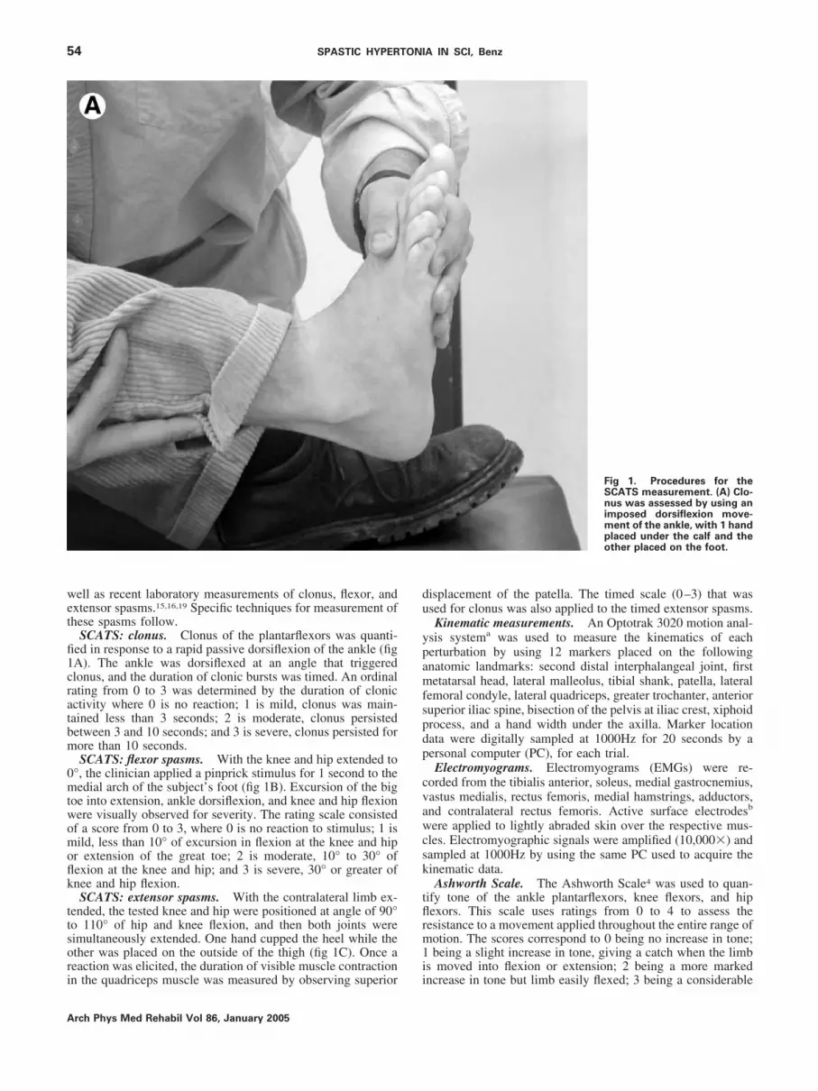
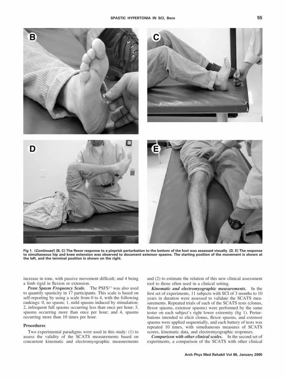
A
ell as recent laboratory measurements of clonus, flexor, andxtensor spasms.15,16,19 Specific techniques for measurement ofhese spasms follow.
SCATS: clonus. Clonus of the plantarflexors was quanti-ed in response to a rapid passive dorsiflexion of the ankle (figA). The ankle was dorsiflexed at an angle that triggeredlonus, and the duration of clonic bursts was timed. An ordinalating from 0 to 3 was determined by the duration of clonicctivity where 0 is no reaction; 1 is mild, clonus was main-ained less than 3 seconds; 2 is moderate, clonus persistedetween 3 and 10 seconds; and 3 is severe, clonus persisted forore than 10 seconds.SCATS: flexor spasms. With the knee and hip extended to
°, the clinician applied a pinprick stimulus for 1 second to theedial arch of the subject’s foot (fig 1B). Excursion of the big
oe into extension, ankle dorsiflexion, and knee and hip flexionere visually observed for severity. The rating scale consistedf a score from 0 to 3, where 0 is no reaction to stimulus; 1 isild, less than 10° of excursion in flexion at the knee and hip
r extension of the great toe; 2 is moderate, 10° to 30° ofexion at the knee and hip; and 3 is severe, 30° or greater ofnee and hip flexion.SCATS: extensor spasms. With the contralateral limb ex-
ended, the tested knee and hip were positioned at angle of 90°o 110° of hip and knee flexion, and then both joints wereimultaneously extended. One hand cupped the heel while thether was placed on the outside of the thigh (fig 1C). Once aeaction was elicited, the duration of visible muscle contraction
n the quadriceps muscle was measured by observing superior irch Phys Med Rehabil Vol 86, January 2005
isplacement of the patella. The timed scale (0–3) that wassed for clonus was also applied to the timed extensor spasms.Kinematic measurements. An Optotrak 3020 motion anal-
sis systema was used to measure the kinematics of eacherturbation by using 12 markers placed on the followingnatomic landmarks: second distal interphalangeal joint, firstetatarsal head, lateral malleolus, tibial shank, patella, lateral
emoral condyle, lateral quadriceps, greater trochanter, anterioruperior iliac spine, bisection of the pelvis at iliac crest, xiphoidrocess, and a hand width under the axilla. Marker locationata were digitally sampled at 1000Hz for 20 seconds by aersonal computer (PC), for each trial.Electromyograms. Electromyograms (EMGs) were re-
orded from the tibialis anterior, soleus, medial gastrocnemius,astus medialis, rectus femoris, medial hamstrings, adductors,nd contralateral rectus femoris. Active surface electrodesb
ere applied to lightly abraded skin over the respective mus-les. Electromyographic signals were amplified (10,000�) andampled at 1000Hz by using the same PC used to acquire theinematic data.Ashworth Scale. The Ashworth Scale4 was used to quan-
ify tone of the ankle plantarflexors, knee flexors, and hipexors. This scale uses ratings from 0 to 4 to assess theesistance to a movement applied throughout the entire range ofotion. The scores correspond to 0 being no increase in tone;being a slight increase in tone, giving a catch when the limb
s moved into flexion or extension; 2 being a more marked
Fig 1. Procedures for theSCATS measurement. (A) Clo-nus was assessed by using animposed dorsiflexion move-ment of the ankle, with 1 handplaced under the calf and theother placed on the foot.
ncrease in tone but limb easily flexed; 3 being a considerable
ia
tsr2so
P
ac
at
fiysfltbsrs
Ft t extt
55SPASTIC HYPERTONIA IN SCI, Benz
ncrease in tone, with passive movement difficult; and 4 beinglimb rigid in flexion or extension.Penn Spasm Frequency Scale. The PSFS17 was also used
o quantify spasticity in 17 participants. This scale is based onelf-reporting by using a scale from 0 to 4, with the followingankings: 0, no spasm; 1, mild spasms induced by stimulation;, infrequent full spasms occurring less than once per hour; 3,pasms occurring more than once per hour; and 4, spasmsccurring more than 10 times per hour.
roceduresTwo experimental paradigms were used in this study: (1) to
ssess the validity of the SCATS measurements based on
ig 1. (Continued ) (B, C) The flexor response to a pinprick perturbato simultaneous hip and knee extension was observed to documenhe left, and the terminal position is shown on the right.
oncurrent kinematic and electromyographic measurements e
nd (2) to estimate the relation of this new clinical assessmentool to those often used in a clinical setting.
Kinematic and electromyographic measurements. In therst set of experiments, 11 subjects with SCI of 3 months to 10ears in duration were assessed to validate the SCATS mea-urements. Repeated trials of each of the SCATS tests (clonus,exor spasms, extensor spasms) were performed by the same
ester on each subject’s right lower extremity (fig 1). Pertur-ations intended to elicit clonus, flexor spasms, and extensorpasms were applied sequentially, and each battery of tests wasepeated 10 times, with simultaneous measures of SCATScores, kinematic data, and electromyographic responses.
Comparison with other clinical scales. In the second set of
the bottom of the foot was assessed visually. (D, E) The responseensor spasms. The starting position of the movement is shown at
ion to
xperiments, a comparison of the SCATS with other clinical
Arch Phys Med Rehabil Vol 86, January 2005
sotPmsa(vt
D
iaSaocitct
aSwjbltatttuao
auot2(pjTptS
gbopvwsdTs
omsmy
deapet
mPdrica
VA
spttmwt2wrop1atajahue
rss
Va
aSkscwavtc
56 SPASTIC HYPERTONIA IN SCI, Benz
A
cales was conducted in 17 subjects with SCI during routineutpatient medical visits. A single physical therapist adminis-ered 3 measures of spastic motor behaviors, including theSFS, Ashworth Scale, and SCATS. The Ashworth Scale waseasured in the right lower extremity for knee and hip exten-
ion and ankle dorsiflexion. Subjects were between 6 monthsnd 10 years postinjury (table 1), with mixed injury levelsAmerican Spinal Injury Association scale) and were takingarious medications to control their upper motoneuron symp-oms. Each test was performed once.
ata AnalysisKinematics. Three-dimensional, lower-extremity kinemat-
cs obtained via infrared marker data were used to calculate thepproximate angles between adjacent limb segments duringCATS testing. This analysis was conducted by using anssumption of rigid body motion. A vector representing therientation of each body segment relative to the laboratoryoordinate frame was defined using markers fixed to the prox-mal and distal anatomic landmarks of each body segment:orso, pelvis, thigh, shank, and foot. The angle between adja-ent limb segments (ie, the joint angles) was estimated by usinghe vector dot product:
�joint �q�1 � q�2
�q�1��q�2�
Concurrent validity was established by using a correlationnalysis of kinematic and electromyographic signals withCATS measurements. The time trajectory of each joint angleas displayed for analysis by a trained clinician; changes in
oint angle were quantified by using an interactive, cursor-ased analysis tool developed by using custom-designed Mat-ab software.c To assess flexor spasms after pinprick stimula-ion of the medial arch, joint angle changes in the hip, knee,nd ankle were calculated by taking the difference betweenhese angles measured before and after stimulation. For quan-ification of ankle clonus after rapid, passive ankle dorsiflexion,he duration and frequency of clonic bursts were determined bysing the same graphics tool. The number of beats per secondnd the duration of the response were calculated as a measuref clonus.Electromyograms. The durations of knee (vastus medialis)
nd ankle (soleus, medial gastrocnemius) extensor EMGs weresed to provide objective verification of clinicians’ assessmentsf extensor spasm duration and clonus duration during SCATSesting. EMGs were band-pass filtered (cutoff frequencies,0Hz, 2000Hz), amplified (gain, 10,000), and low-pass filteredcutoff frequency, 500Hz), before being sampled at 1000 sam-les per second. The same cursor-based tool used to analyzeoint kinematics was applied in the analysis of EMG durations.he time trajectory of each electromyographic signal was dis-layed for every trial. Visual estimation of the onset anderminus of soleus and medial gastrocnemius EMGs was used.pasm duration was calculated directly from these measures.Correlation of SCATS and kinematic and electromyo-
raphic parameters. A correlational analysis was conductedetween the clinician-measured SCATS score and magnituder duration of the associated kinematic or electromyographicarameter. The extensor SCATS score was also correlated withastus medialis electromyographic activity; the flexor SCATSith excursion of the ankle, knee, and hip into flexion and the
um of the joint angles; and the clonus SCATS with theuration of the EMG of the medial head of the gastrocnemius.he 10 trials of data for each subject were pooled across the 11
ubjects, and a Spearman rank correlation test was used to arch Phys Med Rehabil Vol 86, January 2005
btain correlations between the SCATS and the targeted kine-atic or electromyographic parameter (��.05). The SCATS
core was then calculated in each category based on the kine-atic and electromyographic data, and the correlational anal-
sis was repeated (Spearman rank correlation test at ��.05).Repeatability of test perturbations. The mean and standard
eviation (SD) of the input perturbations were calculated forach test as follows: for clonus, we measured the input anklengular perturbation magnitude and the rate of change of thiserturbation; for the extensor spasm stimulus, we measured thextension speed at the hip and knee, as well as the position ofhe hip and the knee where spasm was elicited.
Comparison with Ashworth Scale and PSFS. To deter-ine correlations between the SCATS, Ashworth Scale, andSFS scores, a Spearman rank correlation analysis was con-ucted (��.05). Specifically, all flexor, extensor, and clonusatings were compared with Ashworth scores determined dur-ng ankle dorsiflexion and hip and knee extension. Furtheromparisons were made between individual SCATS ratingsnd the subject-reported PSFS rating.
RESULTS
alidation Using Kinematic and ElectromyographicnalysisAn example of typical input and output responses for each
pastic response is given in figure 2. Figure 2A shows the rapid,assive ankle dorsiflexion, followed by rhythmic medial gas-rocnemius-soleus electromyographic bursting and ankle plan-arflexion movements typical of clonus. Rapid dorsiflexionovements were necessary to generate clonus in all subjectsho manifested such behaviors (4/11 subjects). Mean ampli-
ude of the manual dorsiflexion perturbations � SD was1°�5.6°, generated at a mean angular velocity of 166°�61°/s,ith differences likely arising from velocity-dependent spastic
esponses typically observed with rapid stretch of plantarflex-rs in people with SCI. Similarly, figure 2C shows the rapidassive hip and knee extension, with a mean angle change of08.81°�12.3° at the hip and 106.99°�8.81° at the knee. Theverage peak speed of the perturbation was 89.15°�18.5°/s athe hip, which was followed by prolonged knee extensor andnkle plantarflexor electromyographic activity in 7 of 11 sub-ects tested. Objective measurements of flexor spasm activityfter pinprick, as shown in (fig 2B), were collected by usingip, knee, and ankle angle trajectories after medial arch stim-lation. A typical electromyographic response from the lower-xtremity musculature is also shown in this figure.
All SCATS measures correlated significantly with the cor-esponding EMG or kinematic measurement and with SCATScores identified from electromyographic or kinematic data, asummarized in table 2.
alidation of the SCATS With the Ashworth Scalend PSFSThe outcomes of statistical comparisons between the SCATS
nd the Ashworth Scale yielded variable results (table 3).pecifically, comparison of Ashworth scores from the hip,nee, and ankle all correlated significantly with SCATS exten-or spasm scores. Conversely, SCATS flexor spasm ratingsorrelated significantly only with hip flexor Ashworth scores,hile SCATS clonus scores were correlated only to knee and
nkle Ashworth measures. In consideration of the establishedalidity of SCATS (as determined earlier), the lack of consis-ent correlations between SCATS and Ashworth scores indi-ates differences in the manifestation of spastic responses
cross this subject population.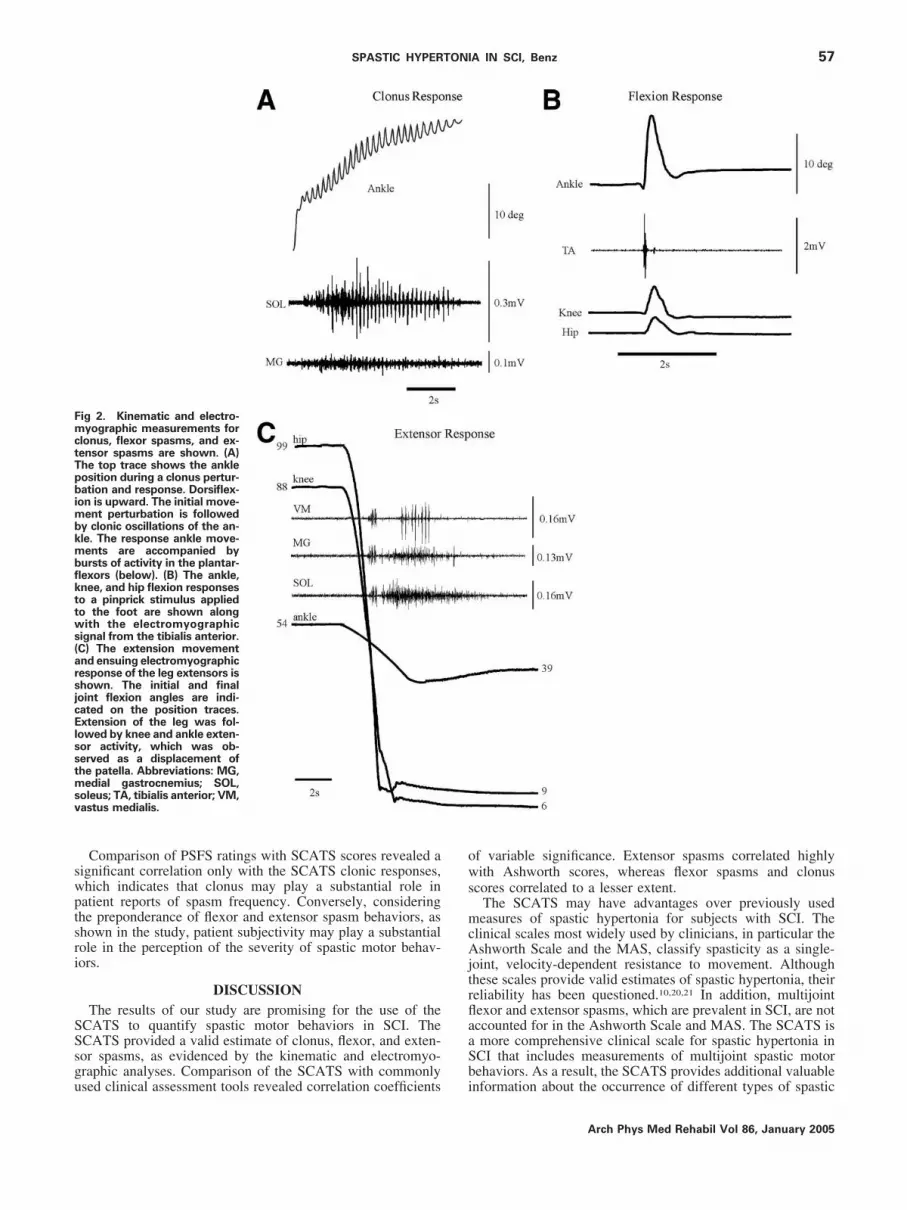
swptsri
SSsgu
ows
mcAjtrflaaSb
FmctTpbimbkmbflkttws(arsjcElsstmsv
57SPASTIC HYPERTONIA IN SCI, Benz
Comparison of PSFS ratings with SCATS scores revealed aignificant correlation only with the SCATS clonic responses,hich indicates that clonus may play a substantial role inatient reports of spasm frequency. Conversely, consideringhe preponderance of flexor and extensor spasm behaviors, ashown in the study, patient subjectivity may play a substantialole in the perception of the severity of spastic motor behav-ors.
DISCUSSIONThe results of our study are promising for the use of the
CATS to quantify spastic motor behaviors in SCI. TheCATS provided a valid estimate of clonus, flexor, and exten-or spasms, as evidenced by the kinematic and electromyo-raphic analyses. Comparison of the SCATS with commonly
ig 2. Kinematic and electro-yographic measurements for
lonus, flexor spasms, and ex-ensor spasms are shown. (A)he top trace shows the ankleosition during a clonus pertur-ation and response. Dorsiflex-
on is upward. The initial move-ent perturbation is followed
y clonic oscillations of the an-le. The response ankle move-ents are accompanied by
ursts of activity in the plantar-exors (below). (B) The ankle,nee, and hip flexion responseso a pinprick stimulus appliedo the foot are shown alongith the electromyographic
ignal from the tibialis anterior.C) The extension movementnd ensuing electromyographicesponse of the leg extensors ishown. The initial and finaloint flexion angles are indi-ated on the position traces.xtension of the leg was fol-
owed by knee and ankle exten-or activity, which was ob-erved as a displacement ofhe patella. Abbreviations: MG,edial gastrocnemius; SOL,
oleus; TA, tibialis anterior; VM,astus medialis.
sed clinical assessment tools revealed correlation coefficients i
f variable significance. Extensor spasms correlated highlyith Ashworth scores, whereas flexor spasms and clonus
cores correlated to a lesser extent.The SCATS may have advantages over previously usedeasures of spastic hypertonia for subjects with SCI. The
linical scales most widely used by clinicians, in particular theshworth Scale and the MAS, classify spasticity as a single-
oint, velocity-dependent resistance to movement. Althoughhese scales provide valid estimates of spastic hypertonia, theireliability has been questioned.10,20,21 In addition, multijointexor and extensor spasms, which are prevalent in SCI, are notccounted for in the Ashworth Scale and MAS. The SCATS ismore comprehensive clinical scale for spastic hypertonia inCI that includes measurements of multijoint spastic motorehaviors. As a result, the SCATS provides additional valuable
nformation about the occurrence of different types of spasticArch Phys Med Rehabil Vol 86, January 2005
mte
tSScraspmTfdrc
eabasmtwaqTas
druwancaahflanab
usna
mcwcwpasotwti
bamcwhipobtpIcusteimtDuc
f
*†
58 SPASTIC HYPERTONIA IN SCI, Benz
A
otor behaviors, and the incidence of different components ofhe SCATS may reflect patient-specific physiologic differ-nces.
The PSFS is a more general scale than the Ashworth, andheoretically it includes many types of spastic motor behaviors.urprisingly, the PSFS only correlated significantly withCATS clonus scores, which may indicate a substantial role oflonus in the perception of the occurrence of spasms. Theseesults suggest that the PSFS does not adequately record flexornd extensor spasms, which may only be triggered duringpecific ADLs. Flexor and extensor spasms may be used byatients to assist in functional tasks in a somewhat controlledanner and thus may not be included in the patient reports.hese observations suggest that the PSFS is useful for identi-
ying the frequency of problem-causing spasms; however, itoes not appear to distinguish between the types of spasticeflexes that are elicited. As a result, it would be beneficial forlinicians to gather more data from both measures.
Although the SCATS reliably distinguishes between differ-nt types and severity of spastic reflexes in SCI and can bedministered quickly in the clinic, it ideally should be com-ined with other measures. For example, the SCATS fails toccount for the patient’s own perspective of the severity ofpasticity. We propose that the SCATS be integrated into aultidimensional approach to the evaluation of spastic hyper-
onia by using patient and clinician reports. This approachould provide both objective measures of spastic hypertonia
nd subjective, patient-based information that could be ac-uired within a timeframe appropriate for a clinical setting.11
he SCATS augments the data provided by the Ashworth Scalend the PSFS by providing information about the type ofpastic hypertonia and the underlying physiology.
Because of the variability of SCATS scores among subjects,ifferences in manifestation of spastic motor behaviors likelyeflect differences in the pathophysiologic mechanisms thatnderlie the spasms. For example, clonus is believed to occurhen there is recurrent excitation of a hyperactive stretch reflex
nd/or activity of a central oscillator that excites the motoreurons.8,22,23 Conversely, flexor spasms are often triggered byutaneous stimuli and may be attributed to an increased excit-bility of the interneurons of the flexor reflex pathway.13,24 Thessociation of flexor spasms with the flexion withdrawal reflexas been shown by decreased thresholds for activation of theexion withdrawal reflex in SCI and by the presence of a largemplitude and duration of flexor muscle activity when a cuta-eous stimulus is applied.13 Finally, extensor spasms, whichre often triggered by extension of the hips,3 may be mediatedy increased excitability of the interneuronal circuitry normally
Table 2: Correlation of the SCATS and Kinematic andElectromyographic Measures
Measure 1(laboratory based)
Measure 2(clinical measure) � P
Vastus medialis duration Extensor SCATS .90 �.001Soleus duration Extensor SCATS .70 �.001Extensor SCATS Extensor SCATS .94 �.001Medial gastrocnemius
duration Clonus SCATS .69 .002Clonus SCATS Clonus SCATS .90 �.001Ankle excursion angle Flexor SCATS .69 �.001Knee excursion angle Flexor SCATS .81 �.001Hip excursion angle Flexor SCATS .82 �.001
pFlexor SCATS Flexor SCATS .87 �.001
rch Phys Med Rehabil Vol 86, January 2005
sed for increasing limb stiffness during standing or during thetance phase of locomotion.15 The SCATS provides a mecha-ism to accurately assess targeted spastic behaviors that aressociated with specific physiologic mechanisms.
Improved understanding of the predominant types of spasticotor behaviors is important to spasticity management, be-
ause different types of spasms have been found to interfereith functional mobility in various ways. Patients report that
lonus interferes with function primarily associated withheelchair propulsion and transfers. Flexor spasms are re-orted to interfere with function during sleep, bed positioning,nd transfers.3,6 Flexor spasms have also been clinically ob-erved to interfere with ambulation. Extensor spasms, whichccur most frequently, cause the most discomfort and producehe greatest interference with function during transfers andheelchair propulsion.3,6 However, people have also reported
hat extensor spasms can be helpful, especially when perform-ng lower-extremity dressing.
We believe that knowledge of the type of spastic motorehaviors and its severity will facilitate informed decisionsbout the administration of pharmacologic and physical treat-ent approaches. For example, clinical practice suggests that
lonus can be minimized by positioning a footplate on theheelchair that puts the ankle at an angle past that at which theyperactive reflex is elicited (�2° from maximum dorsiflex-on).25 Furthermore, evaluation of force distribution along thelantar aspect of the foot when prescribing shoes or ankle-footrthoses could minimize flexor spasms. Extensor spasms maye minimized by changes in the wheelchair seat angle (dump),o ensure that hip angle does not change during wheelchairropulsion, on uneven terrain, or with upper-extremity activity.n addition, alternative transfer techniques that do not require ahange in hip angle could be implemented. With a greaternderstanding of the physiologic mechanisms underlyingpasms and spastic hypertonia, and with clinical tools designedo assess accurately the various motor patterns seen after SCI,ffective pharmacologic interventions can be prescribed. Fornstance, knowledge of whether the spasms are related tootoneuronal or interneuronal hyperexcitability may enable
argeting of specific serotonergic or noradrenergic pathways.24
uring pharmacologic treatment, the SCATS could also besed, as clinicians use the Ashworth Scale, to assess the effi-acy of the drug.26-28
It should also be noted that the SCATS is intended primarilyor people with SCI and may not be suitable for use with other
Table 3: Spearman Rank-Order Correlation of the SCATS,Ashworth Scale, and PSFS
AshworthHip
AshworthKnee
AshworthAnkle
SCATSClonus
SCATSFlexion
SCATSExtension
PSFS .43 .43 .51 .59* .41 .40Ashworth
hip .90† .67* .56 .55* .98†
Ashworthknee .77† .65* .47 .88†
Ashworthankle .60* .40 .61*
SCATSclonus .35 .59*
SCATSflexion .56*
Significant at P�.05.Significant at P�.01.
opulations that exhibit spastic hypertonia such as those with

tai
mdmsl
1
1
1
1
1
1
1
1
1
1
2
2
2
2
2
2
2
2
2
a
bc
59SPASTIC HYPERTONIA IN SCI, Benz
raumatic brain injury, stroke, or cerebral palsy. The extensornd flexor reflexes that are common in SCI are less prominentn people with more cephelad injuries.
CONCLUSIONSThe SCATS provides a valid method for measuring spasticotor behaviors in the clinical setting and distinguishes among
ifferent types and severity of spastic reflexes. The SCATSay be a useful adjunct to self-report scales and may have a
ignificant impact on guiding both functional and pharmaco-ogic management of spasticity in SCI.
References1. Young RR, Wiegner AW. Spasticity. Clin Orthop 1987;Jun(219):
50-62.2. Meythaler JM. Physiology of spastic hypertonia. N Am Clin Phys
Med Rehabil 2001;32:2099-109.3. Little JW, Micklessen P, Umlauf R, Brittel C. Lower extremity
manifestations of spasticity in chronic spinal cord injury. Am JPhys Med Rehabil 1989;68:32-6.
4. Ashworth B. Preliminary trial of carisoprodol in multiple sclero-sis. Practitioner 1964;192:540-2.
5. Lance JW. Symposium synopsis. In: Feldman RG, Young RR,Koella WP, editors. Spasticity: disordered motor control. Chicago:Yearbook Medical Publishers; 1980. p 485-94.
6. Skold C, Levi R, Seiger A. Spasticity after traumatic spinal cordinjury: nature, severity and location. Arch Phys Med Rehabil1999;80:1548-57.
7. Meythaler JM. Spastic hypertonia. Appendix. Phys Med RehabilClin North Am 2001;12(4):953-6.
8. Hinderer SR, Dixon K. Physiologic and clinical monitoring ofspastic hypertonia. Phys Med Rehabil Clin North Am 2001;12(4):733-45.
9. Skold C, Levi R, Seiger A. Spasticity after traumatic spinal cordinjury: nature, severity and location. Arch Phys Med Rehabil1999;80:1548-57.
0. Hass BM, Crow JL. Towards a clinical measurement of spasticity?Physiotherapy 1995;81:474-9.
1. Blackburn M, van Vliet P, Mockett SP. Reliability of measure-ments obtained with the modified Ashworth scale in the lowerextremities of people with stroke. Phys Ther 2002;82:25-34.
2. Kuhn RA. Functional capacity of the isolated human spinal cord.Brain 1950;73:1-51.
3. Shahani BT, Young RR. The flexor reflex in spasticity. In: Feld-man RG, Young RR, Koella WP, editors. Spasticity: disorderedmotor control. Chicago: Yearbook Medical Publishers; 1980. p
287-95.4. Meythaler JM, Guin-Renfroe S, Brunner RC, Johnson A, HadleyMN. Intrathecal baclofen for spastic hypertonia from stroke.Stroke 2001;32:2099-109.
5. Schmit BD, Benz EN. Extensor reflexes in human spinal cordinjury: activation by hip proprioceptors. Exp Brain Res 2002;145:520-7.
6. Schmit BD, McKenna-Cole A, Rymer WZ. Flexor reflexes inchronic spinal cord injury triggered by imposed ankle rotation.Muscle Nerve 2000;23:793-803.
7. Penn RD. Intrathecal baclofen for severe spasticity. Ann N YAcad Sci 1988;153:157-66.
8. Priebe MM, Sherwood AM, Thornby JI, Kharas NF, MarkowskiJ. Clinical assessment of spasticity in spinal cord injury: a multi-dimensional problem. Arch Phys Med Rehabil 1996;77:713-6.
9. Hidler J, Rymer WZ. A simulation study of reflex instability inspasticity: origins of clonus. IEEE Trans Rehabil Eng 1999;7:327-40.
0. Bohannon RW, Smith MB. Interrater reliability of a modifiedAshworth Scale of muscle spasticity. Phys Ther 1987;67:206-7.
1. Pandyan AD, Johnson GR, Price CI, Curless RH, Barnes MP,Rodgers H. A review of the properties and limitations of theAshworth and modified Ashworth Scales as measures of spastic-ity. Clin Rehabil 1999;13:373-83.
2. Gottlieb GL, Agarwal GC. Physiological clonus in man. ExpNeurol 1977;54:616-21.
3. Rossi A, Mazzocchio R, Scarpini C. Clonus in man: a rhythmicoscillation maintained by reflex mechanisms. ElectroencephalogrClin Neurophysiol 1990;75:56-63.
4. Heckman CJ. Alterations in synaptic input to motoneurons duringpartial spinal cord injury. Med Sci Sports Exerc 1994;26:1480-90.
5. Meinders M, Price R, Lehmann JF, Questad KA. The stretchreflex response in the normal and spastic ankle: effect of ankleposition. Arch Phys Med Rehabil 1996;77:487-92.
6. Katz RT, Rovai GP, Brait C, Rymer WZ. Objective quantificationof spastic hypertonia: correlation with clinical finding. Arch PhysMed Rehabil 1992;73:339-47.
7. Sampaio C, Ferreira JJ, Pinto AA, Crespo M, Ferro JM, Castro-Caldas A. Botulinum toxin type A for the treatment of arm andhand spasticity in stroke patients. Clin Rehabil 1997;11:3-7.
8. Meythaler JM, Guin-Renfroe SG, Hadley MN. Continuously in-fused intrathecal baclofen for spastic/dystonic hemiplegia: a pre-liminary report. Am J Phys Med Rehabil 1999;78:247-54.
Suppliers. Northern Digital Inc, 103 Randall Dr, Waterloo, ON N2V 1C5,
Canada.. Model 2.1; DelSys Inc, 650 Beacon St, 6th Fl, Boston, MA 02215.. Version 6.1; The MathWorks Inc, 3 Apple Hill Dr, Natick, MA
01760-2098.
Arch Phys Med Rehabil Vol 86, January 2005