fractures of both bones forearm
TRANSCRIPT

1
FRACTURES OF BOTH BONES FOREARM – A
COMPARATIVE STUDY ON FIXATION TECHNIQUES
AND FUNCTIONAL OUTCOME BETWEEN
intramedullary nailing AND plate
osteosynthesis
DISSERTATION SUBMITTED FOR
MASTER OF SURGERY DEGREE EXAMINATION
BRANCH II ( ORTHOPAEDIC SURGERY)
April 2015
THE TAMIL NADU
DR. MGR MEDICAL UNIVERSITY
CHENNAI, TAMIL NADU

2
CERTIFICATE
This is to certify that the work entitled FRACTURES OF BOTH
BONES FOREARM – A COMPARATIVE STUDY ON FIXATION
TECHNIQUES AND FUNCTIONAL OUTCOME BETWEEN
INTRAMEDULLARY NAILING AND PLATE
OSTEOSYNTHESIS which is being submitted for M.S. Orthopaedics,
is a bonafide work of Dr. S. NITHYANANTH, Post Graduate Student
at Department of Orthopaedics, Madurai Medical College, Madurai.
DEAN
Madurai Medical College
Madurai

3
CERTIFICATE
This is to certify that the work entitled FRACTURES OF BOTH
BONES FOREARM – A COMPARATIVE STUDY ON FIXATION
TECHNIQUES AND FUNCTIONAL OUTCOME BETWEEN
INTRAMEDULLARY NAILING AND PLATE OSTEOSYNTHESIS
which is being submitted for M.S. Orthopaedics, is a bonafide work of Dr. S.
Nithyananth, Post Graduate Student at Department of Orthopaedics, Madurai
Medical College,Madurai.
He has completed the necessary period of stay in the Department and has
fulfilled the conditions required for the submission of this thesis according to
the University regulations. The study was undertaken by the candidate himself
and the observations recorded have been periodically checked by us.
Recommended and forwarded
Prof. P.V. PUGALENTHI
Prof. & HOD, Dept. Of Orthopaedics,
Madurai Medical College
Madurai

4
ACKNOWLEDGEMENT
The most pleasant part of writing a thesis is acknowledging once
gratitude to all those who have helped in its completion.
I take this opportunity to express my deep sense of gratitude although I
find words inadequate to express the greatness of Prof. P.V. PUGALENTHI,
Prof. and Head Department of Orthopaedics, Madurai Medical College who has
been a pillar of discipline, courage and immense kindness and who was
instrumental in guiding me throughout the course of this thesis. I consider
myself fortunate and privileged to work under his affectionate guidance, superb
supervision and sustained support.
I am immensely thankful to Prof.S.Shanmuganathan, Prof.
L.D.Thulasiram, Prof.R.Sivakumar & Prof.Arivasan Prof. of Orthopaedics
for their guidance and ingenious suggestions and ever available help. But for
their co-operation, this study would not have been possible.
I am extremely thankful to Dr. J. Maheshwaran, Assistant. Prof. of
Orthopaedics, who had been a constant source of inspiration to me and whose
excellent guidance, day to day help and dedication paved the way for successful
completion of this study.

5
I am extremely thankful to all my Assistant Professors for their constant
help, guidance and expert advice towards the successful completion of this
study.
Last, but not the least, I extend my thankfulness to all the patients who
have participated in this study. But for their co-operation this exercise would
have been futile.

Abstract
Background and Objectives
In the current era of industrialization, and with mechanized farming in
India, fractures of forearm bones have become more common. The forearm
serves an important role in the functioning of the upper extremity. Hence
aggressive management becomes essential .The purpose of this study was to
evaluate subjective and functional outcome after osteosynthesis of the forearm
fractures with plates and screws ( ORIF ) or Elastic nailing (CRIF).
Methods
We evaluated 20 patients who underwent internal fixation of forearm
fractures with CRIF( 10 patients) or ORIF ( 10 Patients), concerning the Range
of motion of forearm , elbow and a validated outcome measure.( Modified
Grace Eversmann scoring system) and standardized radiographs of the forearm
were evaluated. We used CHI square test to evaluate the results.
Results
There was a statistically significant difference in the final outcome in patients
undergoing ORIF with Plate and CRIF with Titanium elastic nail, with better
functional outcome in the latter. However the risk of non union and
reintervention was not different between the groups.
Conclusion
Forearm bones fractures are associated with high rates of consolidation and
satisfactory mobility of the forearm since we obtain an anatomic reduction of
the fracture, as is most easily achieved by plate fixation. However Elastic
nailing is a less invasive technique that allows restoring function more quickly
with less pain and no increased risk of complications.

6
PART A
CONTENTS Page No.
Introduction 1
History 3
Anatomy 5
Biomechanics 23
Classification 25
Mechanism of Injury 27
Investigations 28
Principles of Management 30
Methods of Management 31
Conservative Management 32
Surgical Management 34
Implant removal 42
Complications 43
Implant Profile 48
Evaluation of outcome 53

7
PART B
CONTENTS Page No.
Preamble 55
Aim of Study 56
Materials and Methods 56
Procedure and Post Operative Protocol 69
Pitfalls and their Management 73
Case Illustrations 75
Results 86
Analysis of functional outcome 88
Discussion 91
Conclusion 96
Annexure I – Proforma 98
Annexure II 103
Annexure III Ethical committee certificate 104
Master chart 106
Bibliography 108
Annexure IV – Abbrevations 111
Annexure V – Turn it in certificate 112

8
PART A
INTRODUCTION
Increasing incidence of road traffic accidents, natural disasters and
industrial accidents together with assault leads to multiple fractures and higher
incidence of morbidity. They form the major epidemic of modern era. 0f these,
the fractures involving both the bones of forearm form an important part. Even
though these fractures can be treated successfully by surgical methods, the
anatomical reduction of fracture fragments becomes absolutely essential for
effective postoperative function. Delayed hospitalization, use of indigenous
bandages and associated vascular and nerve injures contribute to increased
incidence of morbidity.
Traditionally majority of adult forearm fractures are treated by traditional
bone setters leading to various complications. Awareness about the role of
various types of surgical fixation and their role in successful management of
forearm fractures is absolutely essential for preventing this practice.
Fracture of the forearm bones may result in severe loss of function
unless adequately treated. Hence good anatomical reduction and internal
fixation of these fractures is necessary to restore function.1

9
For effective pronation and supination to occur, the maintenance of
interosseous space becomes mandatory while fixing the fractures involving
radius and ulna. Presence of comminution, the anatomy of fracture pattern and
presence of rotatory malalignment significantly contribute to the postoperative
morbidity in these fractures.
Better understanding of the injury patterns, availability of better implants,
the concept of early surgical fixation and exact post operative protocol all have
convincingly improved the functional outcome of the patient to a larger extent.
The successful management of these fractures demands familiarity with
the character of fracture, technical aspects of fracture fixation, the varieties of
implants available for fixation and the art of postoperative management.

10
HISTORICAL REVIEW
Fractures are known to occur since evolution of mankind. However
Prehistoric man must have had his troubles with broken bones. According to
Sudhoff, the bones of Neolithic man showed traces of attempts at corrections of
deformities. Apparently enough specimens of fractured bones from that age
have been found to justify statistical statements.2
Recorded descriptions of the methods of fracture treatment dates back to
Egyptian times, which has been clearly mentioned in Edwin Smith Surgical
Papyrus.Egyptians used palm bark and linen bandages for management of
fractures. Clay and lime mixed with egg white were used, but the material most
commonly used has been, the wood.3
Till the end of 19th
century, the fractures of both bones of forearm were
managed conservatively with POP cast immobilization. In the early 1900s, Lane
in London and Lambotte in Belgium reported use of plates for treating
diaphyseal fractures. However, metal reaction led to frequent failures until
modern metals were introduced in 1937 by Veriable and associates. Campbell
and Boyd used autologous tibial grafts fixed to the radius and ulna with bone
pegs or screws.

11
Even after better metals became available, many early plates were poorly
designed which led to failures. Slotted plates were introduced by Eggers and
associates by late 1960s. The idea of using plates through which active
compression could be applied began with Danis of Belgium. In 1958, Muller,
Allgower and Willenegges developed what is now known as AO compression
plates. The technique of using these plates was published in 1965, and these
became the standard mode of fixation since then.
With the advent of intramedullary nails for fractures of shaft of femur,
various devices for intramedullary fixation of radius and ulna was introduced
in 1957 by Smith and Sage. They used Krischner wires, rush nails, small ‘V’
nails and Steinmann pins for fixation. The results were discouraging. In1959,
Sage introduced triangular forearm intramedullary nails. In 1986, Street
introduced the concept of reamed forearm nails.
A study showed that certain long oblique fractures could be fixed with two
screws. Also satisfactory intramedullary fixation could be achieved by using
prebent diamond shaped nails by SMITH.4
Recently interlocking nails for both radius and ulna were introduced. Titanium
elastic nails which were developed for fractures of shaft of long bones in
pediatric and adolescent age group are being used now in adult diaphyseal
forearm fractures.

12
ANATOMY5,6
Fractures of forearm bones may result in severe loss of function unless
adequately treated. The relationship of the radiohumeral, proximal radioulnar,
ulnohumeral, radiocarpal and distal radioulnar joints and the interosseous space
must be anatomical or else some functional impairment will result, due to the
involvement of these various joints.
Embryology5.
Development of the limb buds. Limb development may be conceptualized as
the result of a series of ectodermal-mesenchymal interactions.
• The upperlimb bud appears on 26th day (end of 4th week) as small bulges on
the lateral body wall at about the level of C5 – C8.
• By 4th week they have grown to form noticeable, coronally oriented ridges.
• Limb morphogenesis takes place from 4th to 8th week.
• By 33 days the hand plate is visible.
• Digital rays appear on hand during 6th week. By 6th week end segments of
upper limb can be distinguished.
• By the 50th day or so (8th week) the elbows and shoulder are established,
and the fingers are free.
• Each limb consists of a mesenchymal core of mesoderm, covered by
ectodermal cap.

13
• Skeletal elements of limbs develop from a column like mesodermal
condensation that appears along the long axis of the limb during 5th wk and full
differentiation by 12th wk.
• Ossification begins in these cartilaginous precursors in 8th to 12th wk.
• Rotation of limbs occurs during 6th to 8th week
SKELETAL ANATOMY 6
Forearm consists of skeletal structures; interosseous membrane; stable
proximal and distal radio-ulnar joints; and soft-tissue structures, including the
muscles, nerves, and vessels that are in the forearm and that traverse it.
RADIUS6
The radius is the lateral bone of the forearm. Its proximal end articulates
with the trochlea of the humerus at the elbow joint and with the ulna at the
proximal radioulnar joint. Its distal end articulates with the scaphoid and lunate
bones at the distal radioulnar joint.
At the proximal end of the radius is the small circular, head. The upper
surface of the head is concave and articulates with the convex capitellum of the
humerus. The circumference of the head articulates with the radial notch of the
ulna. Below the head is the neck, below which there is the bicipital tuberosity
for the insertion of biceps muscle. The shaft of the radius is wider below. It has

14
a sharp interosseous border medially for the attachment of interosseous
membrane. The pronator tuberosity for the insertion of the pronator teres
muscle, lies half way down on its lateral side.
At the distal end of the radius is the styloid process; this projects distally
from its lateral margin. On the medial surface is the ulnar notch, which
articulates with the distal ulna. The distal articular surface articulates with the
scaphoid and lunate bones. On the posterior aspect of the distal end is a small
tubercle, the dorsal tubercle of Lister, which is grooved on its medial side by
the tendon of extensor pollicis longus.
Radius ossification
The radius ossifies from three centres (one primary centre and two secondary
centres). One appears centrally in the shaft in the eighth week of foetal life, and
the others appear in each end. Ossification begins in the distal epiphysis
towards the end of the first postnatal year, and in the proximal epiphysis during
the fourth year in 28 females, and fifth in males. The proximal epiphysis fuses
in the fourteenth year in females, seventeenth in males, and the distal in the
seventeenth and nineteenth years respectively. A fourth centre sometimes
appears in the tuberosity at about the fourteenth or fifteenth year.

15
Technique of Schemitsch and Richards7
To measure the radial bow, a line is drawn from the bicipital tuberosity to the
most ulnar aspect of the radius at the wrist. A perpendicular line is then drawn
from the point of the maximum radial bow to this line. The height of the
perpendicular line (the maximum radial bow) is measured in millimetres. The
distance from the bicipital tuberosity to the previously measured perpendicular
line at the point of the maximum radial bow is then measured and is recorded
as a percentage of the length of the entire bow (the distance from the mid-point
of the bicipital tuberosity to the most ulnar aspect of the subchondral bone of
the distal part of the radius). This measurement is termed the location of the
maximum radial bow. The value is expressed as a percentage.

16
ULNA6
The ulna is not a straight bone. It has a dorso medial bowing. The
proximal end articulates with the humerus at the elbow joint and with the head
of the radius at the proximal radioulnar joint. Its distal end articulates with the
radius at the distal radioulnar joint, but it is excluded from the wrist joint by the
articular disc.
The proximal end of the ulna forms the olecranon process. It has a notch
on its anterior surface, the trochlear notch, which articulates with the trochlea of
the humerus. Below the trochlear notch is the triangular coronoid process,
which has on its lateral surface the radial notch for articulation with the head of
radius.

17
The shaft of ulna tapers from above down. It has a sharp interosseous
border laterally for the attachment of the interosseous membrane. The posterior
border is rounded and subcutaneous. Below the radial notch is a depression, the
supinator fossa, which gives clearance for the movement of the bicipital
tuberosity of the radius. The posterior border of the fossa is sharp and it is
known as the supinator crest: it gives origin to the supinator muscle.
At the distal end of ulna is the small rounded head, which has projecting
from its medial aspect, the styloid process.
Ulna ossification
The ulna ossifies from four main centres (one primary center for the shaft and
secondary centers, one for distal end and two for olecranon). Ossification begins
in the midshaft about the 8th fetal week, and extends rapidly. In the 5th
(females) and 6th (males) years, a centre appears in the distal end, and extends
into the styloid process. The distal olecranon is ossified as an extension from the
shaft, the remainder from two centres, one for the proximal trochlear surface,
and the other for a thin scale-like proximal epiphysis on its summit. The latter
appears in the 9th year in females, 11th in males. The whole proximal epiphysis

18
has joined the shaft by the 14th year in females, 16th in males. The distal
epiphysis unites with the shaft in the 17th year in females, 18th in males.
INTEROSSEOUS MEMBRANE6
This connects the borders of two bones. The interosseous membrane is a
broad, thin, collagenous sheet. Its fibres slant distomedially between the radial
and ulnar interosseous borders, and its distal part is attached to the posterior
division of the radial border. The membrane is deficient proximally, starting 2
or 3 cm distal to the radial tuberosity, and broader at midlevel. An oval aperture
near its distal margin conducts the anterior interosseous vessels to the back of
the forearm, and the posterior interosseous vessels pass through a gap between
its proximal border and the oblique cord. The membrane provides attachments
for the deep forearm muscles. Its fibres appear to transmit forces which act
proximally from the hand to the radius, thence to the ulna and humerus.
19
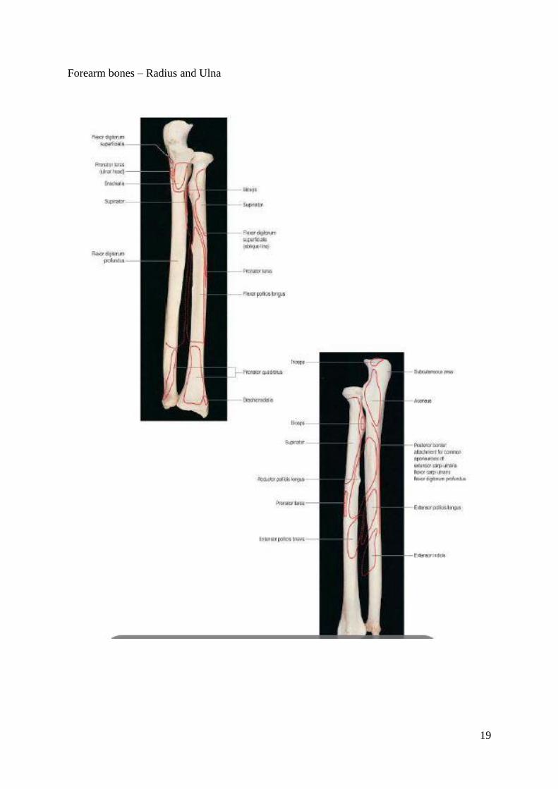
Forearm bones – Radius and Ulna

20
The radioulnar articulations:6
The radius and ulna are joined to each other at the superior and inferior
radioulnar joints. The two bones are also connected by the interosseous
membrane; which is sometimes said to constitute a middle radioulnar joint.
a. Superior radioulnar joint:
The proximal radioulnar joint is a uniaxial pivot joint. The articulating
surfaces are between the circumference of the radial head and the fibro-osseous
ring made by the ulnar radial notch and annular ligament
The essential structure is the annular ligament which holds the head
of radius in place. The annular ligament is attached to the anterior and
posterior margins of radial notch of ulna and has no attachment to radius.
Superiorly it blends with the capsule at the lower margin of the cylindrical
articular surface.
Movement-pronation and supination of forearm
b. Inferior radioulnar joint:
The distal radio-ulnar joint is a uniaxial pivot joint. The articulating surfaces
are between the convex distal head of the ulna and the concave ulnar notch of
the radius. These surfaces are connected by an articular disc.
It is closed distally by a triangular fibrocartilage which is attached to its

21
base to the ulnar notch of radius and by its apex to a fossa at the base of
ulnar styloid.
Movement-pronation and supination of forearm
The various muscles attached to radius are 6
Proximal third
(1) Biceps brachiaii (insertion)
(2) Supinator (insertion)
(3) Pronator teres (insertion)
(4) Flexor digitorum superficialis (origin)
Middle third
(1) Flexor pollicis longus (origin)
(2) Abductor pollicis longus (origin)
Distal third
(1) Pronator quadratus (insertion)
(2) Branchioradialis (insertion)
(3) Extensor pollicis brevis (origin)

22
The various muscles attached to ulna are
Proximal third:
1) Brachialis (insertion)
2) Pronator teres (origin)
3) Flexor pollicis longus (origin)
4) Triceps (insertion)
5) Anconeus (insertion)
6) Supinator (origin)
7) Abductor pollicis longus (origin)
Middle third
1) Flexor digitorum profundus (origin)
2) Flexor carpi ulnaris (origin)
3) Extensor carpi ulnaris(origin)
4) Extensor pollicis longus (origin)
Distal third
1) Extensor indices (origin)
2) Pronator quadratus (origin)

23
Superficial and Deep muscles of forearm

24
VASCULAR SUPPLY OF FOREARM6
Radial artery
Originates from brachial artery at about 1 cm distal to the flexion crease
of the elbow.
It descends along the lateral side of the forearm, The artery is medial to
the radial shaft proximally, and anterior to it distally. Runs inferolaterally under
cover of brachioradialis and distally lateral to flexor carpi radialis tendon; Its
posterior relations in the forearm are successively the tendon of biceps,
supinator, the distal attachment of pronator teres, the radial head of flexor
digitorum superficialis, flexor pollicis longus, pronator quadratus and the lower
end of the radius (where its pulsation is most accessible).
Winds around lateral aspect of radius and crosses floor of anatomical
snuff box to pierce fascia; ends by forming deep palmar arch with deep branch
of ulnar artery.
Branches in the forearm
• Radial recurrent artery
• Cutaneous branches & Muscular branches.
Ulnar artery
The ulnar artery is the larger terminal branch of the brachial artery. It
starts 1cm distal to the flexion crease of the elbow and reaches the medial side
of the forearm midway between elbow and wrist.

25
Passes inferomedially and then directly, deep to pronator teres, palmaris
longus, and flexor digitorum superficialis to reach medial side of forearm. The
ulnar artery crosses the flexor retinaculum lateral to the ulnar nerve and
pisiform bone to enter the hand and gives a deep palmar branch to deep arch
and continues as superficial palmar arch. The ulnar nerve lies medial to the
distal two-thirds of the artery.
Branches in the forearm
• Anterior and posterior ulnar recurrent arteries
• Common interosseous artery
• Anterior interosseous artery
• Muscular and nutrient branches
Radial recurrent artery
Originates on the lateral side of radial artery, just distal to its origin.
Ascends on supinator and then passes between brachioradialis and
brachialis. It supplies these muscles and the elbow joint, anastomosing with the
radial collateral branch of the profunda brachii.

26
Anterior ulnar recurrent and posterior ulnar recurrent artery.
Originates from ulnar artery, just distal to elbow joint.
AUR artery ascends between brachialis and pronator teres, supplies them
and anastomoses with the inferior ulnar collateral artery anterior to the medial
epicondyle.
PUR artery passes dorsomedially between flexores digitorum profundus and
superficialis, ascending behind the medial epicondyle; it supplies adjacent
muscles, nerve, bone and elbow joint, and anastomoses with the ulnar collateral
and interosseous recurrent arteries.
Common interosseous artery
The common interosseous artery is a short branch of the ulnar artery.
After a short course, terminates by dividing into anterior and posterior
intersosseous artery.
Anterior interosseous artery
The anterior interosseous artery descends on the anterior aspect of the
interosseous membrane with the anterior interosseous branch of the median
nerve.
The anterior interosseous artery proper leaves the anterior compartment by
piercing the interosseous membrane proximal to pronator quadratus. It
anastomoses with the posterior interosseous artery in the posterior compartment
of the forearm.

27
Posterior interosseous artery
It passes dorsally between the oblique cord and proximal border of the
interosseous membrane. It descends deep in the groove between extensor carpi
ulnaris and the extensor digiti minimi part of extensor digitorum. While in the
groove it gives rise to multiple muscular branches. The posterior interosseous
artery accompanies the deep branch of the radial nerve (posterior interosseous
nerve) on abductor pollicis longus. Distally it anastomoses with the terminal
part of the anterior interosseous artery and the dorsal carpal arch.
NERVES OF FLEXOR COMPARTMENT 6
The lateral cutaneous nerve of the forearm, the cutaneous continuation of
the musculocutaneous nerve, pierces the deep fascia above the elbow lateral to
the tendon of biceps and supplies the anterolateral surface of the forearm.
The medial cutaneous nerve of the forearm supplies front and back of the
medial part of the forearm.
The posterior cutaneous nerve of the forearm passes along the dorsum of the
forearm to the wrist. It supplies the skin along its course
Median Nerve6
Enters the forearm between the heads of pronator teres. It passes behind a
tendinous bridge between the humero-ulnar and radial heads of the flexor
digitorum superficialis, and descends through the forearm posterior and
adherent to flexor digitorum superficialis and anterior to flexor digitorum

28
profundus. About 5 cm proximal to the wrist it becomes superficial. It then
passes deep to the flexor retinaculum into the palm.
Branches in the forearm
• Anterior interosseous nerve
• Muscular branches to pronator teres, flexor carpi radialis, palmaris longus and
flexor digitorum superficialis.
• Articular branches.
Ulnar Nerve6
The ulnar nerve enters the forearm from the extensor compartment of arm
by passing between the two heads of flexor carpi ulnaris.The ulnar nerve
descends on the medial side of the forearm, lying on flexor digitorum
profundus with the ulnar artery to its radial side.
It supplies flexor carpi ulnaris and ulnar half of flexor digitorum
profundus also gives palmar cutaneous branch
Superficial terminal branch
The superficial terminal branch of the radial nerve, the cutaneous
continuation of the main nerve, runs from the cubital fossa on the surface of
supinator, pronator teres tendon and flexor digitorum superficialis, on the lateral
side of forearm under cover of brachioradialis. In the middle third of the
forearm it lies beside and lateral to radial artery. It then leaves the flexor

29
compartment of the forearm by passing backwards deep to the tendon of
brachioradialis and breaks into two or three branches.
Nerve of extensor compartment:
Posterior interosseous nerve:
The posterior interosseous nerve is the deep terminal branch of the radial
nerve. It reaches the back of the forearm by passing round the lateral aspect of
the radius between the two heads of supinator. It passes downwards over the
abductor pollicis longus origin and dips down to reach the interosseous
membrane were it passes between the muscles as far as the wrist joint. Here it
ends in a small nodule from which branches supply the wrist joint.
Branches in the forearm
Muscular branches to all muscles which arise from the common extensor origin
and deep muscles of the extensor compartment.

30
BIOMECHANICS9
The longitudinal axis of rotation of the forearm passes through the
articular surface of the radial head, the interosseous membrane, and the
articular surface of the ulna at the distal radio-ulnar joint.8
Diaphyseal fractures of the radius and ulna present specific problems in
addition to those common to all fractures of the shafts of long bones. In addition
to regaining length, apposition and axial alignment, achieving normal rotational
alignment is necessary if a good range of pronation and supination are to be
restored.
The movements of supination and pronation of the forearm involve a
rotatory movement around a vertical axis at the proximal & distal radioulnar
joints. The axis passes through the head of radius above and the attachment of
apex of the triangular articular disc below. During pronation, the entire radius
moves around the ulna through the longitudinal axis of forearm.
Pronation is performed by pronator teres and pronator quadratus.
Supination is performed by biceps brachiaii and supinator. Supination is the
powerful of the two movements, because of the strength of biceps muscle.
Maintenance of the interosseous space is essential for pronation and supination.
The biceps and the supinator exert rotational forces on fractures of the proximal
third of radius. Distally, the pronator teres at the level of mid shaft and the

31
pronator quadratus on the distal fourth of shaft of radius exert both rotational
and angulatory forces. Fractures of distal radius tend to angulate toward the
ulna by the action of the pronator quadratus and the pull of long forearm
muscles.
Rotational deformity will limit radioulnar movement. The supinator
muscles are inserted proximally and the pronators distally. Consequently in a
fracture of mid shaft of radius the proximal fragment supinates and the distal
fragment pronates, resulting in 90˚ of rotational displacement. Shortening of the
two bones following overriding may also occur. Both angular and rotational
deformities are compounded by the presence of comminution. Hence, in
addition to regaining length, bony apposition, axial alignment and achieving
normal rotational alignment is necessary, if a good range of pronation and
supination are to be restored.

32
CLASSIFICATION
Fractures of forearm are classified according to the level of fracture, the
pattern of fracture, the degree of displacement, the presence or absence of
comminution or segmental bone loss and whether they are open or closed. Each
of these factors may have some bearing on the type of treatment to be selected
and the ultimate prognosis. For descriptive purposes, it is useful to divide the
forearm into thirds, based on the linear dimensions of radius and ulna.
Disruption of proximal or distal radioulnar joints is of great signifance to the
treatment and prognosis. It is imperative to determine whether the fracture is
associated with joint injury because effective treatment demands that both the
fracture and joint injuries are treated in an integrated fashion.
AO CLASSIFICATION10
Type 22 A Simple fractures of one or both bones
A1 Simple fracture of ulna
A2 Simple fracture of radius
A3 Simple fracture of both radius & ulna
Type 22 B Wedge fractures of one or both bones
B1 Wedge fracture of ulna
B2 Wedge fracture of radius

33
B3 Wedge fracture of both radius & ulna
Type 22 C Complex fracture of one or both bones
C1 Grossly comminuted fracture of ulna with
simple fracture of radius
C2 Grossly comminuted/segmental fracture of
Radius with simple fracture of ulna
C3 Grossly comminuted fractures of both bones

34
MECHANISM OF INJURY11
The mechanisms of injury that causes fractures of the radius and ulna are
myriad. By far the most common is some form of vehicular accident, especially
automobile and motor cycle accidents. Most of these vehicular accidents result
in some type of direct blow to the forearm. Other causes of direct blow injuries
include fights in which one of the adversaries is struck in the forearm with a
stick or rod. The person throws the forearm upto protect his or her head, and the
forearm is the recipient of the violence. Following violent twisting of forearm,
rotational deforming forces act leading to fracture of forearm bones.
Gunshot wounds can cause fractures of both bones of forearm. Such
injuries are commonly associated with nerve or soft tissue defects and
frequently have significant bone loss. The other common mechanism is due to
some type of fall. The force generated is usually much greater than that required
to cause Colle’s fracture. Most forearm shaft fractures resulting from fall occur
in the athletes or in fall from heights.
The last and least common cause of diaphyseal fractures of both bone
forearm is due to pathological fractures.

35
INVESTIGATIONS18
A minimum of two views – anteroposterior and lateral – are mandatory in
all suspected forearm fractures. Additional oblique views may be required. The
following features are noted in the radiographs.
1) Degree of offset
2) Degree of angulation
3) Amount of shortening
4) Presence of comminution
Additional visualization is needed to rule out involvement of wrist, elbow
and both radioulnar joints. A line drawn through the radial shaft, neck and head
should pass through the center of the capitellum on any projection.
The rotational alignment of the forearm is difficult to determine in routine
antero posterior and lateral views. The bicipital tuberosity view recommended
by Evans is helpful in those instances. Since the proximal radial fragment could
not be controlled with closed methods, the distal radial fragment must be
brought into correct relationship with the proximal fragment. Ascertaining the
rotation of the proximal fragment from the tuberosity view before reduction,
gives some idea of how much pronation or supination of distal fragment is
needed. The tuberosity view is made with the x-ray tube tilted 20˚ towards the
olecranon, with the subcutaneous border of ulna flat on the cassette. The x-ray

36
can then be compared with the diagram showing the prominence of the tubercle
in various degree of pronation and supination. As an alternative, a film of the
opposite elbow can be made in a given degree of rotation for comparison.
Xray Images of Radial tuberosity at different levels of fracture.

37
PRINCIPLES OF MANAGEMENT
There are a number of factors which play a dynamic role in determining
the type of management, thereby influencing the prognosis. They include:
1) Amount of overriding of fracture fragments
2) Degree of Comminution
3) Extent of soft tissue injuries
4) Associated neuro vascular injuries
5) Magnitude of joint involvement
6) Presence of multiple trauma
7) The width of medullary canal
8) Degree of osteoporosis
9) Complex ipsilateral injuries (side swipe injury)
So the objectives of treatment of diaphyseal fractures of both bones in
adults are:
1) To obtain and maintain satisfactory reduction and rigid fixation.
2) To regain functional range of movement of elbow joint.
3) To regain adequate pronation and supination
4) To treat associated injuries.

38
The absence of pronation and supination is a permanent handicap since they
cannot be regained by physiotherapy or rehabilitation.
METHODS OF TREATMENT10
There are a variety of options for treating an adult with a fracture of both
bones of forearm. It is fair to say that the vast majority of fractures of both
bones of the forearm can be most effectively treated by accurate anatomical
reduction, rigid plate fixation, and early mobilization. The various modalities of
treatment available for treating adult diaphyseal fractures of both bones of
forearm are:
1) Conservative Management:
a) Cast Immobilization \
b) Closed reduction and cast immobilization
2) Surgical Management:
a) Open reduction and internal fixation with plate osteosynthesis
b) Closed reduction and Intramedullary fixation
c) External fixator application

39
CONSERVATIVE MANAGEMENT10
a) Cast Immobilization:
The rare non displaced fracture of both bones of the forearm in adults can
usually be treated by immobilization in above elbow cast with elbow in 90˚
flexion and forearm in midprone position. Angulation can be prevented by
incorporating a plaster loop on the radial side of the cast proximal to the level of
fractures. Despite good technique, an initially non displaced fracture can
become displaced while being immobilized in plaster.
b) Closed reduction & Cast immobilization:
It is difficult to reduce and maintain satisfactory position of the fragments
by closed methods due the various deforming forces acting on the fragments
and due to the role of supinators and pronators leading to rotatory instability.
Closed reduction is most successful for fractures of both radius and ulna when
the fractures are located in distal third. Functional cast bracing of forearm
fractures following 6 weeks of immobilization in arm cast helps in starting early
elbow mobilization exercises leading to lesser incidence of elbow stiffness.
Before closed reduction is undertaken, the patient must be advised that, surgical
fixation may be necessary at any time to ensure solid union in acceptable
position.

40
Technique of closed reduction14
Relaxation of muscles is mandatory for closed reduction and general
anesthesia is preferred. Tuberosity view is taken with image intensifier to
identify the degree of rotation. Traction and counter traction are applied and
ulna is reduced under direct palpation. The radius could not be palpated in the
proximal half. The forearm is placed under appropriate supination as determined
by the tuberosity view. When the fractures seem reduced and the alignment of
forearm appears satisfactory, an above elbow plaster slab is applied and check
X-rays are taken. Above elbow cast conversion is done after 1 week and
radiographs in two planes are taken at weekly intervals through the cast for the
first month and every two weeks thereafter until solid union is obtained.
There are only a few indications available for conservative treatment in
adult forearm fractures. These include
1) Undisplaced/ incomplete fractures
2) Associated life threatening trauma like head injury, chest injury etc.

41
SURGICAL MANAGEMENT
INTRODUCTION:
During the last century, surgical management of diaphyseal fractures of
both bones forearm in adults has gained widespread acceptance as operative
techniques and the quality of implants have improved. The combination of
properly designed implants, better understanding of the personality of the
fracture, minimal soft tissue handling techniques, preoperative antibiotics have
made surgical fixation safe and practical while treating these fractures.
The goals of operative treatment for diaphyseal forearm bones fractures
in adults include
a) Anatomical alignment
b) Stable fixation
c) Early mobilization
d) Early functional rehabilitation of upper limb.
Indications for operative management include virtually all diaphyseal fractures
of both bones of forearm in adults.

42
Open Reduction and Internal fixation: 10
An AO dynamic compression plate (Asian) with 3.5 mm screw system
provides for secure fixation without cast protection. In an adult, fixation by
semitubular plate does not provide with a rigid fixation. Plates are especially
useful in fixation of fractures of distal 3rd
or proximal 4th of both bones of
forearm.
a) Principles:
- Plate osteosynthesis provides for static compression at the fracture site.
- Plates should be applied on the tension side of bones whenever possible.
- For both radius and ulna, dorsal side is the tension side.
- Minimal stripping of periosteurn from ends of fracture fragments.
- Both radius and ulna has to be fixed with similar type of implant.
- Autologous bone graft is added whenever there is comminution involving
more than 1/3rd
of circumference of bone.
- Before plate application, larger comminuted fragments should be secured
to the main fragments to produce interfragmentary compression.
- Fractures of both radius and ulna should be exposed and reduced
temporarily before a plate is applied to either.

43
- Plates must be accurately centered over the fracture site and there must be
a minimal of six cortical purchases with screws on either side of fracture.
- If autologous bone graft is added, they should not be placed in the
interosseous border, else cross union may occur.
b)Technique of fixation:
RADIUS
Approach:12
for proximal half, dorsal Thompson’s approach
for distal half, anterior Henry’s approach
for mid 3rd
, either of the approaches may be used.
minimal stripping of periosteum is done to preserve blood supply
Clear away all the clotted blood from the ends of the bone
All soft tissue attachments of the comminuted fragments should be
retained, if possible
Reduce the fracture as anatomically as possible fitting any butterfly
fragments into position
Larger butterfly fragments should be fixed to the main fragment by lag
screw principle

44
An Asian DCP, usually a 6 or 7 holed plate is selected in accordance
with the fracture pattern and applied over radius
A 2.7 mm drill bit is used to drill hole in the radius, and then tapped.
An appropriate sized 3.5 mm cortical screw is measured with depth
gauge and used to fix the plate to the bone
Always drill one hole at a time and insert screw before drilling the
next screw
Similarly all the 6 or 7 holes of the plate are drilled and fixed with
screws.
Autologous bone graft is added if the comminution involves more than
one third of the circumference of radius.
ULNA12
Approach
- Incision along subcutaneous border of ulna.
- Plate is fixed on either anterior or posterior surface on which it fits best.
- Posterior surface is better since it is the tension side of ulna
- If there is comminution, place the plate on the side of comminution since
it stabilizes the loose fragments.
- Add autologous bone grafts if needed.

45
Volar Henry Approach

46
Posterior Thompson Approach to Radius

47
-
Closed intramedullary nail fixation13
While selecting an intramedullary device, it is mandatory to select a nail
of appropriate diameter for fixation. If the size of the nail is small, there is side
to side and rotatory movement leading to instability. If the size of the nail is
large, further comminution or additional fracture may occur.
Principle:13
Since the fractures of both radius and ulna are fixed in closed manner,
fracture hematoma is preserved leading to early union and
consolidation. Moreover, the chance of infection is minimized.
The ulna is fixed first
An appropriate sized nail is selected, so that the nail fits snuggly
inside the medullary canal.
Titanium elastic nail offers three point fixation thereby stabilizing the
fracture fragments.
Technique of fixation13
- C arm is mandatory

48
- Closed reduction of the bones is achieved with traction, counter traction
and manipulation
- The reduction is checked with C arm.
- For the ulna, entry point in made over the olecranon with an awl and the
position is confirmed
- A nail is introduced through the olecranon and passed across the fracture
site under image control
- For the radius – the entry point is from distal aspect and
three entry points are described
(a) just medial to Lister tubercle
(b) just lateral to Lister tubercle
(c) from radial styloid
- All 3 entry points are made 5 mm proximal to wrist joint.
- The entry point that is just medial to Lister’s tubercle is the most
preferred.
- The nail is passed, across the fracture site under C arm control.
Titanium elastic nail13
- Both the radius and ulnar nails are cut at their ends and buried

49
IMPLANT REMOVAL
Plates & screws and intramedullary nails placed on forearm bones are not
removed routinely unless they cause symptoms. In any case they should not be
removed before 2 years, even though the fracture will have appeared solid on
radiographs much earlier. The limb has to be protected in above elbow slab for
minimum 6 weeks after removal, if there is local pain or tenderness .

50
COMPLICATIONS11
The complications following operative treatment for diaphyseal
fractures of both bones forearm in adults are relatively less common because of
better surgical techniques and improved implants.
Complication of fractures:11
(a) Infection
(b) Malunion
(c) Non union
(d) Cross union
(e) Associated vascular and nerve injuries
(f) Post traumatic Stiffness
Complication of operative treatment
(a) Incomplete reduction
(b) Incongruous reduction
(c) Unstable fixation
(d) Inadequate implant
(e) Infection.

51
The use of state of the art implants and instrumentation for diaphyseal
fractures of both bones forearm does not always guarantee a favourable
outcome. The surgeon must have a thorough understanding of local anatomy,
mechanics of fracture fixation and patterns of fracture healing after internal
fixation if consistently good results are to be achieved.
Infection11
The major drawback of operative fixation is infection. It is less common
with closed intramedullary fixation than with open reduction techniques. If post
operative infection develops, appropriate antibiotics are given for 3 to 6 weeks
intravenously. Even in the presence of infection, every effort should be made
to retain the implants since stable infected fractures are easy to manage than
unstable infected fractures. However if the infection is severe, the implant has
to be removed.11
Malunion11
This is relatively more common in conservatively treated cases than in
surgically treated cases, since it is difficult to maintain the fracture fragments in
alignment when treated conservatively. The varying pull of supinators and
pronators on the fracture fragments lead to malunion.

52
Non union11
The varying causes of nonunion are inadequate immobilization, improper
fixation, implant failure and the presence of underlying infection. Gross
osteoporosis of the bones is also an important cause for nonunion. Inadequate
internal fixation, with plates which are too small, nails which are of
inappropriate size is a potent cause of nonunion. Loss of substance of radius or
ulna following gun shot injuries also lead to nonunion. Repeated manipulation
by traditional bone setters may also lead to nonunion. In a case of non union,
open reduction and internal fixation with autologous bone grafting is the
treatment of choice.
Cross union11
Cross union of the radius and ulna results from a continuous hematoma
between the two fractures. The important cause of cross union following
conservative treatment is improper reduction with bony fragments encroaching
the interosseus space. Cross union may also occur if the fractures are stabilized
by open methods and bone grafting with bone grafts kept in the interosseous
border of either bones. If cross union occurs there is loss of pronation and
supination due to a bridge of bone between radius and ulna. This bridge of bone
has to be excised for pronation and supination to occur.

53
Synostosis is relatively uncommon. Seen frequently in patients with
either a crushing injury of forearm or a head injury. The highest risk for
synostosis is in proximal fractures treated through single incision. If synostosis
develops and position of forearm is relatively functional,it is best to do nothing.
if rotational alignment of forearm is poor, an osteotomy to position the hand in
more functional position can be considered.
Post traumatic stiffness
This is more common is patients managed conservatively than by surgical
fixation. Elbow joint is notorious for developing stiffness if it is immobilized
too long. The main advantage of surgical fixation is that, since the fracture
fragments are stable after fixation, active mobilization exercises of wrist, elbow
and hand can be started early.
Nerve injuries11
Injury to posterior interosseous nerve can occur in Henry’s approach
during plating of radius. Also, there are chances of injury to recurrent radial
artery and superficial branch of radial nerve through this approach. These can
be prevented by knowing the proper anatomy of forearm and gentle handling of
soft tissues.

54
Compartment syndrome11
This can occur either after trauma or after surgery on the forearm bones.
They are usually due to faulty hemostasis or closure of the deep fascia. They
can usually be avoided by releasing the tourniquet before wound closure to
make sure hemostasis is adequate, by closing only the subcutaneous tissue and
skin.
Complications of intramedullary nail fixation:
Most complications result from improper selection of nail size. A nail
that is too long may be driven through the bone end. One that is too short may
not adequately stabilize the fracture. A nail with too large a diameter may split
the cortex and one with a smaller diameter may not adequately control
rotational alignment resulting in non-union.

55
IMPLANT PROFILE
DYNAMIC COMPRESSION PLATE 3.5 MM15,16
DCP is now the workhouse of AO system. When introduced in 1965 it
was made of Titanium, but it is now fabricated from 316 stainless steel
(Zimmer) and of Vitallium (Howmedica).15
The DCP of AO.ASIF consists of a plate with obliquity of cylindrical
screw holes for compression which is produced as the screws are driven home.
Due to this mechanism, use of a tension device is not required. This has made
the plate more adaptable to different situation of internal fixation and can be
used as a static compression plate, a buttress plate, a neutralization plate or as
DCP.16
These plates are used with cortical screws of size 3.5mm, hence the
name. The holes allow for 1mm displacement if a load screw is used, thereby
producing compression. The plate can be used with an articulated tension
device. The oval holes permit eccentric position of screws which can be used
for axial compression.
Important dimensions:
a) Thickness 3 mm

56
b) Width 10 mm
c) Hole spacing 12mm and 16 mm
d) Hole length 6.5mm
e) Length 25mm to 145 mm
f) Holes 2 to 12
AO stainless steel implants are produced from implant quality 316L stainless
steel which typically contains iron 62.5%, chromium 14.5%, nickel 2.8%,
molybdenum and minor alloy elements.
ONE THIRD TUBULAR PLATES
These plates have the form of one third of the circumference of a
cylinder. They have low rigidity since they are only 1 mm thick. The plate is
fixed with 3.5 mm cortical screw. They do not produce compression at fracture
site
Important dimensions
Thickness 1 mm
Width 9 mm
Hole spacing 12 mm and 16 mm
Length 25 mm to 145 mm
Holes 2 to 12

57
3.5 mm CORTICAL SCREW:
The holding power of the cortical screw on dense cortical bone is due to
its 1.75mm pitch and the asymmetrical buttress threads.
Important Dimensions:
Head diameter 6mm
Hexagonal socket width 2.5 mm
Core diameter 2.4 mm
Thread diameter 3.5 mm
Pitch 1.75 mm
Length 10 mm – 110 mm
TITANIUM ELASTIC NAIL
These nails are made of alloys such as Ti-6Al-7Nb. They offer
outstanding corrosion resistance, excellent biocompatibility and higher strength.
Titanium alloy implants may be ceramic shot peened and either chemically
panivated in nitric acid or anodized as a final surface treatment.

58
Implant profile
Length : 44 cm
Width : 2 mm to 5 mm
Color coded for different sizes
End : Beak shaped for easy insertion and may
be used as a reduction tool.

59

60
EVALUATION OF OUTCOME17
For evaluating the functional outcome of fracture fixation, we used the
MODIFIED GRACE AND EVERSMANN SCORING SYSTEM. This system
takes into account the following parameters:
1. SUPINATION AND PRONATION17
( Normal – pronation & supination 80 degrees each)
RATING RANGE OF MOVEMENT SCORE
EXCELLENT > 80 4
GOOD 60 TO 80 3
FAIR 40 TO 60 2
POOR < 40 1
2. RADIOLOGICAL UNION (End of 6th
week)
RADIOLOGICAL UNION SCORE
UNION PRESENT
(good callus)
2
NON UNION 1

61
3. RANGE OF MOVEMENT – ELBOW17
Range Result Score
Flexion > 120 Excellent 4
Flexion 100 to 120 Good 3
Flexion 80 to 100 Fair 2
Flexion < 80 Poor 1
Final Analysis
RESULT SCORE
EXCELLENT 10 and above
GOOD 8 to 9
FAIR 6 to 7
POOR Less than 5

62
PART- B
PREAMBLE
The diaphyseal fractures of both bone fractures of forearm is one of the
most common fracture pattern occurring in adults. These fractures are routinely
fixed by plate osteosynthesis with 3.5 mm Asian DCP efficiently and
successfully. Since this system is of load bearing type which necessitates
distruption of fracture hematoma during fixation, the choice of intramedullary
nail fixation for forearm fractures comes into play.
This series includes 20 cases (10 cases of plate osteosynthesis, 10 cases
of titanium elastic nails), all of whom were adults. The diaphyseal fractures of
both radius and ulna were selected. The outcome was analysed with special
emphasis on rotatory stability at the fracture site and time taken for full range of
motion to occur.
Based on our findings we hereby submit “Fractures of both bones
forearm – A comparative study on fixation techniques and functional
outcome between Intramedullary Nailing and Plate osteosynthesis”.

63
AIM OF STUDY
Even though fractures of both bones of forearm is one of the most
common fractures occurring in adults, they are also one of the most common
fractures to be mismanaged. Even today most of these fractures are treated by
traditional bone setters leading to increased morbidity and infection.
Traditionally, these fractures are treated by plate osteosynthesis using AO
Dynamic compression plate (Asian) very efficiently. The aim of our study is to
compare the functional outcome of fixation of both bones of forearm using plate
osteosynthesis with that of Titanium elastic nail.
This study aims to stress the need for rigid fixation of forearm fractures
and to evaluate the early restoration of movements of wrist, elbow and forearm.
MATERIALS AND METHODS
Design of the study :Prospective study
Period of study : September 2012 – September2014

64
This is a prospective study of 20 cases of diaphyseal fractures of both
bone of forearm in adults treated by surgical fixation with Plate and titanium
elstic nail.
The period of surgery and follow up extends from September 2012 to
September 2014. It includes all diaphyseal fractures of both bones of forearm in
adults. Comminuted, segmental fractures are included in this study. All
compound fractures, malunited fractures, bones with medullary canal diameter
of less than 2mm and fractures in children are excluded from this study.
Inclusion Criteria :
1. Diaphyseal fractures of both bones of forearm in adults >18 years
2. Comminuted and segmental fractures of forearm
3. Patients fit for surgery
Exclusion Criteria :
1. Compound fractures
2. Malunited fractures
3. Bones with narrow medullary canal < 2 mm in diameter
4. Patients unfit and not willing for surgery

65
The cases were analysed as per the following criteria
1) Age distribution
2) Sex distribution
3) Side of injury
4) Mode of injury
5) Classification of fracture
6) Time interval between injury and surgery
7) Associated injures
8) Complications
9) Additional procedures for complications
10) Duration between injury and hospitalization
Imaging18
The clinical signs and symptoms are usually obvious in shaft fractures of
both bones of the forearm, so are the radiologic signs. The configuration of
midshaft fractures of the radius and ulna varies depending on the mechanism of
injury and the degree of violence involved. Low-energy fractures tend to be
transverse or short oblique, whereas high-energy injuries are frequently
extensively comminuted or segmented, often with extensive soft tissue injuries.
Radiographs of the radius and ulna i. e., anteroposterior and lateral views,
were obtained. The elbow and wrist joints were included in each view.

66
I. AGE DISTRIBUTION:
The age group varied from 20 years to 70 years with the mean age of 45
years. Incidence of fracture was observed maximum between 30-50 years of
age.
Age Group Number of cases Percentage
20 – 30 years 6 30
30 – 40 7 35
40 – 50 4 20
50 – 60 2 10
60 – 70 1 5
0
1
2
3
4
5
6
7
8
20 - 30 yrs 30-40 yrs 40-50 yrs 50-60 yrs 60- 70 yrs
Age Distribution

67
II. SEX DISTRIBUTION:
Among the 20 cases, males were predominant
Sex Number of cases Percentage
Male 12 60
Female 8 40
Sex Distribution
Males
Females

68
III. Side of Injury:
Right side was common in our series
Sex Right Left Bilateral Total
Male 9 3 - 12
Female 3 5 - 8
Percentage 60 40 - -
Side distribution
Right forearm
left forearm
0
2
4
6
8
10
Males
Females

69
IV. Mode of Injury
Commonest mode of injury had been road traffic accident.
Mode of Injury No. of cases percentage
RTA 11 55%
Fall 3 15%
Assault 6 30%
0
2
4
6
8
10
12
RTA Accidental fall Assault
Mode of injury
70
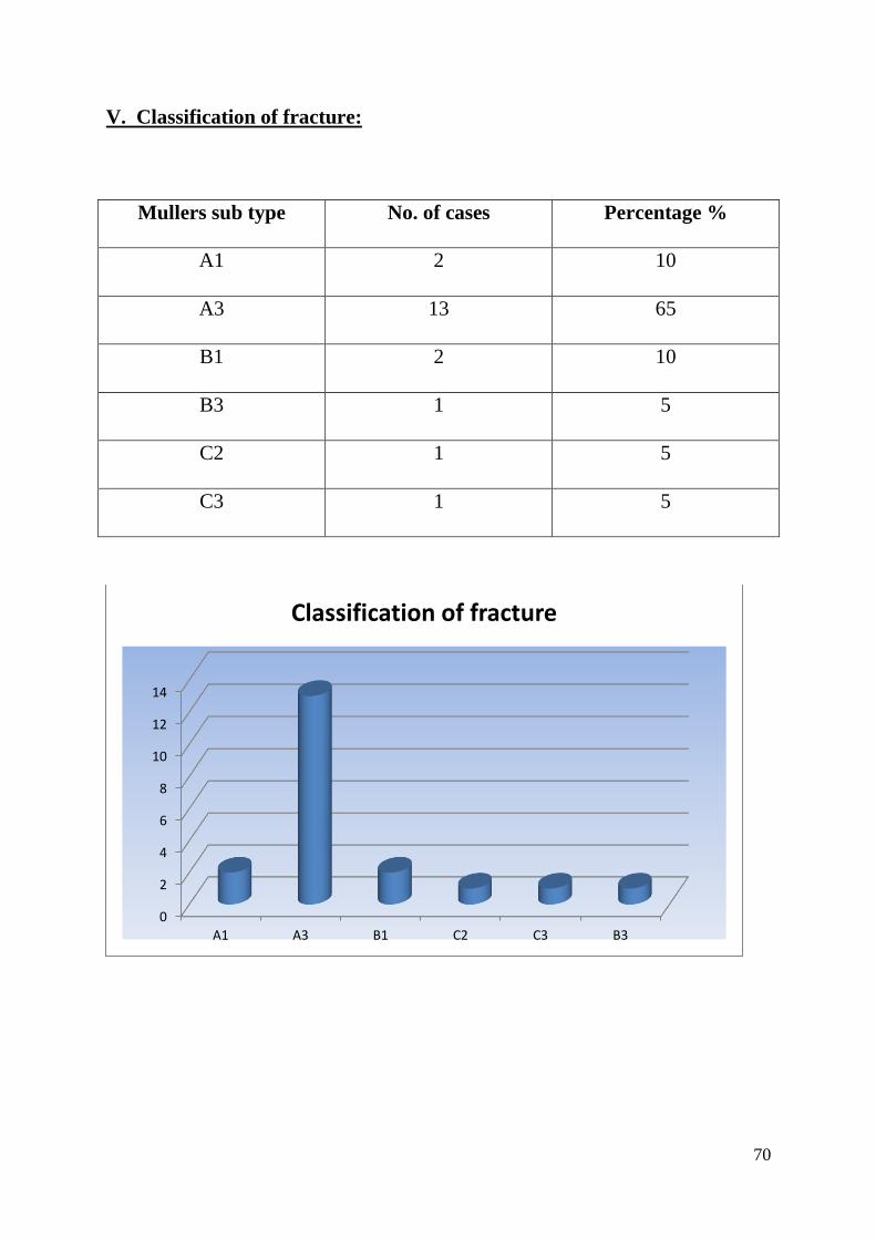
V. Classification of fracture:
Mullers sub type No. of cases Percentage %
A1 2 10
A3 13 65
B1 2 10
B3 1 5
C2 1 5
C3 1 5
0
2
4
6
8
10
12
14
A1 A3 B1 C2 C3 B3
Classification of fracture

71
VI. Time interval between injury and surgery
Time interval No. of cases Percentage %
<2 days 2 10
2 to 5 days 7 35
5 to 7 days 11 55
0
2
4
6
8
10
12
< 2 days 2 to 5 days 5 to 7 days
Time of surgery

72
VII. Associated Injuries:
Fracture of both bones leg 1
Humerus shaft fracture 1
Femur fracture 1
Chest injury 1
Head injury 4
0
0.5
1
1.5
2
2.5
3
3.5
4
Both bone legfracture
Femurfracture
Humerusfractue
Chest injury Head injury
Associated injuries

73
VIII. Complications
Tourniquet palsy 1 case (recovered)
Infection 4 cases
EPL Tendon injury 1 case
Radial styloid fracture 1 case
0
0.5
1
1.5
2
2.5
3
3.5
4
Tourniquet palsy infection Tendon rupture Radial styloidfracture
complications

74
IX. Duration between injury and hospitalization
Most of injuries were hospitalized within 12 hours.
Time interval No. of cases
0 – 3 hrs. 2
3 – 6 hrs. 12
6 – 12 hrs 6
8 -12 hrs 10
0
2
4
6
8
10
12
0-3 hrs 3-6 hrs 6-12 hrs 8-12 hrs
Time interval for hospitalization

75
X. Duration of hospital stay post operatively
Procedure Duration of stay
Plate osteosynthesis 12 days
Intramedullary Nail 5 days
0
2
4
6
8
10
12
Plateosteosynthesis Intramedullary
Nail
Duration of Hospitalization in Days
Duration of Hospitalization inDays

76
PROCEDURE AND POST OPERATIVE PROTOCOL
All the patients were received in the casualty department and were
resuscitated. If there were any other major associated injuries, they were treated
accordingly at first. After the general condition of the patient improved,
radiographs (AP View and lateral view) were taken. The fractures were reduced
in closed manner at first under sedation and an above elbow slab was applied.
Most of the cases were taken for elective fixation before 5th day. The
patients who had associated major injuries were taken up for surgery between
5th
and 7h day.

77
Open reduction and internal fixation with Dynamic Compression plate:
We routinely used tourniquet during surgery.
The radius was opened first. We always used Henry’s approach for
exposing the radius. The cleavage between flexor carpi radialis and
brachioradialis was developed. The FCR was retracted medially along with
radial artery and vein. The branchioradialis was retracted laterally along with
the sensory branch of radial nerve. The fractured ends were identified and with
minimal periosteal stripping, they were mobilized. The medullary cavity was
cleared of any hematoma and the fractured fragments were reduced by carefully
matching the interdigitations using bone holding forceps. An Asian DCP of
appropriate length was selected and applied to the radius on the volar side and
fixed with 3.5mm cortical screws. All the fractures were fixed such that there
were at least six cortical purchases on either side of the bony fragment. Then the
ulna was opened on its subcutaneous border, centering over the underlying
fracture. The interval between flexor carpi ulnaris and extensor carpi ulnaris
was identified and developed. The periosteum over the ulna was incised, the
fracture fragments were reduced and fixed with an Asian DCP similar to that of
radius. Thorough wash of both wounds done. The deep fascia was not sutured;
skin closure was done. Compression bandage was applied. Tourniquet was
released and an above elbow slab was applied.

78
POST OPERATIVE PROTOCOL:
In the immediate post operative period the upper limb was immobilized
in an above elbow slab, and kept elevated till the edema of fingers subsided.
The wound was inspected on the II POD and then suture removal was done on
Xth POD. The upper limb was immobilized depending upon the rigidity of
fixation. At the end of 4th and 6
th weeks check X rays were taken to visualize
callus response.. The pronation and supination movements were started by the
end of 6th week.
II. Closed Reduction and Fixation with Intra meduallary nailing:
Most of the fractures of Muller type A were fixed with this implant.
(A)Titanium elastic Nail fixation:
The patient is placed supine and the forearm is kept in a hand table
compatible with C arm. Tourniquet was not used. The width of the medullary
canal of radius was measured and an appropriate sized nail was selected such
that, the nail should occupy at least 60% of the medullary space. The entry was
made on the distal radius just medial to Lister tubercle, beneath the extensor
pollicis longus tendon about 5 mm proximal to wrist joint, with a 3.2 mm drill
bit. The medullary canal was entered with a curved awl and the position was

79
confirmed with C arm. The selected titanium elastic nail was introduced and
passed into the medullary canal of radius and gently pushed till it reaches the
fracture site. The fracture fragments were reduced by gentle manipulation and
the nail was entered into the distal fragment by gently rotating the tip. The
position of the nail was continuously confirmed with C arm. The nail was
passed till it reached the radial neck. The nail was then slightly withdrawn and
cut. The cut end of the nail was gently hammered so that the tip lies flush with
the bone.
The ulna was entered from the olecranon and an appropriate nail was
inserted, fracture fragments reduced and the nail gently manipulated into distal
fragment. The tip of the nail was cut and buried. The wounds were sutured.
Post operative protocol:
The upper limb was kept elevated. Wound inspection was done on II
POD. Suture removal was done on Xth POD, and above elbow cast was applied.
After 3 weeks the cast was removed and a below elbow cast was applied, after
obtaining check X rays. Active elbow mobilization exercises were started at the
end of 3rd week. By the end of 6 weeks, the cast was discontinued and active
pronation and supination exercises were started.

80
PITFALLS AND THEIR MANAGEMENT
1. Infection:
Four cases developed wound infection, 4 of them treated with plate
osteosynthesis – 3 of which are superficial and one deep. Pus culture for
sensitivity was sent in all the four cases and treated with appropriate antibiotics.
The three superficial infections subsided with treatment for 3 weeks, but the one
with deep treatment subsequently went for plate removal for ulna alone. Wound
debridement of the ulnar wound was done and the fracture was stabilized with a
3mm K wire.
2. Delayed union:
Delayed union developed in one case treated with titanium elastic nail.
The patient had segmental fracture of radius fixed with 3 mm nail. At the end of
6th
week, there was tenderness at the proximal segmental fracture site.
Radiologically there was no callus. The fracture was immobilized with above
elbow cast for another 4 weeks. Eventually there was adequate callus response
and the fracture went on to unite well.

81
3. Elbow stiffness:
3 Patients who were treated with plate osteosynthesis and one patient
treated with Titanium elastic nail developed elbow stiffness at the end of 6th
week while removing above elbow cast. The patients were put on strict regimen
involving active mobilization exercises of elbow. Eventually all 7 patients had
good range of motion of elbow.
4. Tendon injury:
One case treated with titanium elastic nail developed rupture of tendon of
extensor pollicis longus at the wrist. It occurred during the drilling of outer
cortex of distal radius just medial to Lister tubercle. The tendon of EPL was
caught by the drill bit while drilling. However there was not gross restriction in
range of movements
5. Technical complications:
a) Fracture of Radial styloid:
This complication occurred in a patient treated with titanium elastic nail.
The entry point was made more laterally over the radial styloid. During
manipulation, the tip of radial styloid fractured. This was visualized on the
immediate post operative radiograph. The patient developed wrist stiffness
which was treated with intense mobilization exercises.

82
Case Illustrations : Case No 1

83
Case No 2

84
Post op follow up at 6 months
Case No 3

85
Post op follow up at 6 months
Case No :4

86
Case no 5
Case no 6

87
Case no 7:

88
Pre op and post op x rays followed by x ray at 6 Weeks
Case no : 8

89
Case No: 9

90
Case No : 10

91
Case No :11

92
Complications
CROSS UNION

93
RESULTS
Average time of fracture healing in our study was 8 weeks. In patients
who had undergone plate osteosynthesis, it was 9 weeks whereas in patients
who had undergone nail fixation it was 6 weeks. Muller Type 22 A3 fracture
united by 11 weeks. Other fracture patterns healed between 6 and 9 weeks.
Chapman in a study had 98% union with range of 6 to 14 weeks union the
average union time was 12 weeks.19
Mc Knee study had average union time of
10.7 weeks with range of 5 to 18 weeks. He had 97.3% union rate.
4 patients had restricted pronation & supination. Three patients
were treated with plate osteosynthesis and one patient with intramedullary
nailng had restricted supination pronation due to cross union. 4 patients treated
with plate osteosynthesis gave excellent results with regard to pronation &
supination.
4 patients developed post operative stiffness of elbow joint. 3 patients
were treated with plate osteosynthesis and one patient with Titanium elastic nail
However, all these patients eventually had fair range of motion by the end of 12
weeks following intense physiotherapy.
Chapman et al reported 36 (86%) cases as excellent, 3 (7%) Good,
1 (2%) Fair and 2 (5%) Poor19
results in his study.

94
Our series had 90% (18 cases) of excellent /satisfactory results and 10%(2
cases) Poor results which is comparable to the previous studies.
The patient who had sustained fracture of radial styloid process during
titanium nail fixation following far lateral entry point developed stiffness of
wrist joint. With active exercises, the ROM was increased.
Restoration of pronation & supination activities were possible by the end
of 6th
week using intramedullary nailing whereas they were possible by the end
of 9th week using plate osteosynthesis
However in a study by Sara et al, 20
there was no significant differences
between the groups undergoing plate osteosynthesis or Elastic nailing. The risk
of non union and reintervention was not different between the groups.

95
ANALYSIS OF FUNCTIONAL OUTCOME17
The Analysis was done using modified GRACE AND EVERSMANN
RATING SYSTEM and the following results were obtained.
I. OVERALL RESULTS
Grading Number of Cases Percentage
Excellent 7 35
Good 3 15
Fair 8 40
Poor 2 10
0
1
2
3
4
5
6
7
8
Excellent good Fair poor
Overall results

96
II. RESULTS ACCORDING TO IMPLANT USED
Implant Number of cases
Grading Percentage
Plate
Osteosynthesis
1 Excellent 10
1 Good 10
6 Fair 60
1 Poor 10
Number of cases
Grading Percentage
Titanium Elastic
nail
6 Excellent 60
2 Good 20
1 Fair 10
1 Poor 10
Overall results by Implants used
Titanium elastic nail
Plate osteosynthesis0
1
2
3
4
5
6
Excellent GoodFair
Poor
Titanium elastic nail
Plate osteosynthesis

97
Statistical Analysis
The mean value of Modified Grace and eversmann score in Patients who
underwent nailing is 9.6 and in plating is 6.2 . Standard deviation being 2.17
and 2.15 for patients who underwent nailing and plating respectively.
The P value was found to be 0.002 hence the study supports that Titanium
elastic nailing is superior to Plate osteosynthesis.
0
5
10
Ten's nailing Plating
9.6
6.2
COMPARISON OF SCORE

98
DISCUSSION
The aim of this study is to compare the results of treating diaphyseal
fractures of both bones in adult forearm using plate osteosynthesis with that of
titanium elastic nail fixation.
We selected 20 cases of diaphyseal fractures involving both the bones in
the forearm in adults. The period of study was between Sep 2012 and Sep 2014.
Most of these patients fell into middle age, group with majority of them being
males. The mode of violence is either due to RTA, assault or due to accidental
fall. The patients who had simple Muller’s A3 and segmental fracture pattern
were fixed with intramedullary nail fixation and the fractures with comminution
were fixed with Intramedullary nailing. Compound fractures were excluded
from our study.
A satisfactory device for internal fixation must hold the fracture rigidly,
eliminating as completely as possible angular and rotatory motion. This can be
accomplished by either a strong intramedullary nail or AO dynamic
compression plate.

99
During plate osteosynthesis, to minimize further injury to blood supply of
the bone, the periosteum was stripped sparingly with a periosteal elevator and
only sufficiently for applying a plate. The fragments were carefully reduced
with interdigitating bone spicules being fitted properly. Comminuted fragments
were fitted accurately in place. The plates were selected such that at least there
were six cortical purchases on either side of fracture fragments. The plates were
contoured before they were applied to the bone. Our study has showed good
fracture union occurred in 80% of cases.
Earlier studies have reported an alarming refracture rate of 40% when the
plates were removed before 1 year. It is well established that the cortex beneath
a rigid plate weakens because of stress shielding, becoming thin, atrophic and
almost cancellous in nature. If soft tissue stripping has been extensive,
osteonecrosis and revascularization weakens the cortex further. In our series
involving 10 cases treated with plate osteosynthesis, we did not have refracture
in any of our patients.
While using intramedullary device for fixing the adult forearm fractures
involving both bones, rotational control in fractures near the metaphyseo-
diaphyseal junction was difficult because of wide medullary canal. Interference
fit nails do not maintain bone length if associated with bone loss. When an

100
intramedullary fixation is used, errors in selecting the proper diameter or length
of the nail and operative technique contributed to poor results. In case of the
titanium elastic nail, the distal end of nail must abut subchondral bone to
prevent shortening. The lower modulus of elasticity of titanium nails allow
easier insertion and provide more load sharing with the bone. Titanium elastic
nails produced interference fit which was responsible for the return of forearm
rotation and grip strength.
Our study had showed that good to excellent union occurred with 90% of
fractures fixed with titanium elastic nail.
We compared the results of plate fixation with that of intramedullary
fixation. Apart from the incidence of infection we did not have any
complications while treating forearm fractures with plate osteosynthesis. All the
cases healed well on controlling the infection

101
We had technical difficulties while using both titanium elastic nail. While
fixing fractures of radius involving distal 3rd
shaft, the titanium elastic nail did
not provide with adequate stability of fracture fragments because of wide
medullary canal. While using titanium elastic nail we had entry point fracture at
radius, since the entry point was shifted far laterally. That led to the fracture of
styloid process of radius which was treated conservatively. In another case,
there was avulsion of tendon of extensor pollicis longus by a drill bit. This
occurred following failure of separation of soft tissue upto the bone with a
curved artery forceps after skin incision was made.
Earlier, intramedullary devices like K wires, square nails and Rush
nails were used for fixing radius and ulna. These implants did not provide with
rotational stability at the fracture site. This lead to higher incidence of non
union. But titanium elastic nail, provided with excellent rotational stability of
fracture fragments.
We used tourniquet in fractures fixed with plate osteosynthesis. One case
of tourniquet palsy occurred but recovered eventually. Since tourniquet was not
used during intramedullary fixation, the chance for occurrence of this
neurological complication was totally eliminated.

102
Closed Intramedullary fixation offers the following advantages when
compared with plate osteosynthesis.
a) No periosteal stripping is required
b) Smaller operative wound
c) Bone grafting not necessary
d) No potential for diaphyseal refracture after implant exit.
In our study, the rehabilitation time was much shorter for fractures fixed
with intramedullary nail when compared with that of plate osteosynthesis. The
average time required for functional recovery is more than 9 weeks when plates
are used, and about 6 weeks when intramedullary nails are used. The duration of
hospital stay post operatively was also less (on an average of 5 days for
intramedullary devices and 12 days for plate osteosynthesis).
Intramedullary fixation provides for short operating time, short hospital
stay and early rehabilitation .Intramedullary fixation excels better than plate
osteosynthesis especially in cases of segmental fractures and Comminuted
fractures if closed reduction is possible.

103
CONCLUSION
The conclusion of this study are
- Diaphyseal fractures of both bones of forearm in adults are one of the
commonest fractures being reported to orthopaedic emergency
- Early fixation of fracture followed by intense physiotherapy produced
excellent results.
- Fixation with plate osteosynthesis has stood the test of time and provides
excellent fixation.
- The advantages of intramedullary fixation are
o Preservation of fracture hematoma
o Early mobilization.
o Can be done as a day care procedure
o Less post operative morbidity.
o Smaller incision – hence better cosmesis
o Last, but not the least; since there is no axial loading (like weight
bearing) after intramedullary fixation, the chances of implant failure is
very less.

104
- Titanium elastic nail fixation is particularly useful in fractures involving
middle third of radius and ulna. Providing for 3 point fixation leads to
stable fixation and proper alignment of fracture fragments.
- Being newer techniques, these intramedullary devices require further
evaluation and there is a steep learning curve.
- The presence of image control (C arm) helps in easy reduction of
fractures fragments thereby shortening intraoperative duration.
To conclude:
Forearm bones fractures are associated with high rates of consolidation
and satisfactory mobility of the forearm since we obtain an anatomic reduction
of the fracture, as is most easily achieved by plate fixation. However Elastic
nailing is a less invasive technique that allows restoring function more quickly
with less pain and less risk of complications.

105
ANEXURE - I
P R O F O R M A
Case No :
NAME : I.P. No :
AGE/SEX
D. O. A :
EDUCATION: D.O.S :
OCCUPATION: D. O. D :
ADDRESS :
CONSULTANT:
1. PRESENTING COMPLAINTS
2. HISTORY OF PRESENT ILLNESS
Mode of injury-Direct / Indirect – RTA
- Assault
- Fall on outstretched hand
- Others
Duration / side affected
3. First Aid Measures immediate
4. Past History
5. Family history
6. Personal History

106
Occupation: Nature of work
Diet : vegetarian / non vegetarian / mixed
Habits : Smoker / Alcoholic / none
EXAMINATION:
1. General physical examination:
Built:
Vitals - Pulse_____ Beats/min Temp______ °C
B. P;______ mm of Hg R.R--Cycles/min
2. Systemic examination
CVS
RS
PA
CNS
3. Local Examination:
Inspection:
Side
Attitude
Swelling
Deformity
Wounds
Others

107
Palpation:
Tenderness
Abnormal mobility
Crepitus
Distal pulses Radial artery Ulnar artery
Neurological examination of Peripheral Nerves-Basic
Motor
Sensory
Radial Nerve
Ulnar Nerve
Median Nerve
4. Associated injuries:
5. Complications:
INVESTIGATIONS:
Blood routine- Hemoglobin, Total WBC count
Differential count, ESR
BT, CT, prothrombin time
RBS, B. urea, S.creatinine
HIV 1 and 2, HbsAg
Urine routine
X-ray Forearm – AP,Lateral

108
Level of fracture
Displacement
Type of fracture
X-ray chest PA View and ECG – if patient is more than 40 years
TREATMENT:
1. Pre-operative treatment:
Above elbow POP slab with sling
Antibiotics, tetanus toxoid
Analgesics
Preoperative evaluation
2. Surgical procedure:
Type of anesthesia - GA/brachial block
Duration of surgery
Approach - Thompson/Henry
Operative findings
Operative Complications
Difficult reduction Stable/Unstable
Placement of Plate and Screws
Insertion of nails

109
3. Post operative:
Post operative Immobilisation Type and Duration
Antibiotics
Suture removal
Complications
4. Advice on Discharge
5. Duration of Hospital stay
6. Follow up
Parameters 6th
week 3 months 6 months
Pain
Radiograph
(union)
Supination &
Pronation
Elbow
Flexion,Extension
7. Complications:
Infection
Delayed union,
Non union, Malunion
Nerve injury

110
ANNEXURE – II
CONSENT FORM
FOR OPERATION/ANAESTHESIA
I_________ Hosp. No.______ in my full senses hereby give my full
consent for ______ or any other procedure deemed fit which is a diagnostic
procedure / biopsy / transfusion / operation to be performed on me / my son /
my
daughter / my ward_____age under any anaesthesia deemed fit. The nature,risks
and
complications involved in the procedure have been explained to me in my own
language and to my satisfaction. For academic and scientific purpose the
operation/procedure may be photographed or televised.
Date:
Signature/Thumb Impression
of Patient/Guardian
Name:
Designation Guardian Relation ship
Full address

111

112
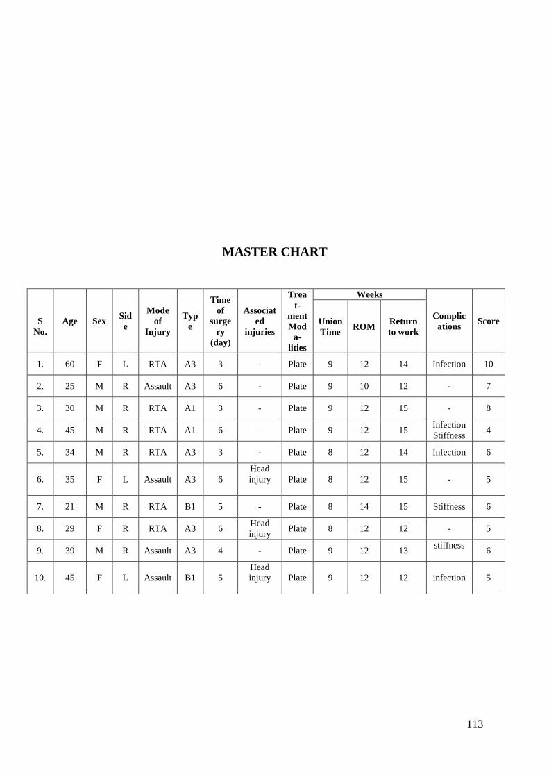
113
MASTER CHART
S
No.
Age Sex Sid
e
Mode
of
Injury
Typ
e
Time
of
surge
ry
(day)
Associat
ed
injuries
Trea
t-
ment
Mod
a-
lities
Weeks
Complic
ations Score Union
Time ROM
Return
to work
1. 60 F L RTA A3 3 - Plate 9 12 14 Infection 10
2. 25 M R Assault A3 6 - Plate 9 10 12 - 7
3. 30 M R RTA A1 3 - Plate 9 12 15 - 8
4. 45 M R RTA A1 6 - Plate 9 12 15 Infection
Stiffness 4
5. 34 M R RTA A3 3 - Plate 8 12 14 Infection 6
6. 35 F L Assault A3 6
Head
injury
Plate 8 12 15 - 5
7. 21 M R RTA B1 5 - Plate 8 14 15 Stiffness 6
8. 29 F R RTA A3 6 Head
injury Plate 8 12 12 - 5
9. 39 M R Assault A3 4 - Plate 9 12 13 stiffness
6
10. 45 F L Assault B1 5
Head
injury
Plate 9 12 12 infection 5

114
S No. Age Sex Side
Mode
of
Injury
Type
Time
of
surger
y (day)
Associated
injuries
Treat-
ment
Moda-
lities
Weeks
Complications Score Union
Time
RO
M
Return
to
work
11. 40 M R Fall A3 2 - TEN 6 9 12 - 12
12. 65 F R RTA A3 7 Fracture both
bone leg TEN 7 9 12
EPL injury
10
13. 47 F L RTA A3 5 Head injury TEN 6 9 12 -
12
14. 50 M L Fall C2 3 - TEN 6 10 13 Delayed union 9
15. 29 M R RTA A3 3
Supracondyla
r fracture
femur
TEN 7 9 12 Cross union
7
16. 50 F L Assault A3 5 -
TEN 7 9 14 - 11
17. 35 M R RTA B3 7
Shaft of
humerus with
intercondylar
humerus #
TEN 8 14 20 Elbow
stiffness 4
18. 20 F R Assault A3 2 Chest injury TEN 6 8 12 -
11
19. 52 M L Fall C3 7 - TEN 6 9 12 - 11
20. 40 M L RTA A3 3 - TEN 7 10 13
Radial styloid
fracture
9

115
BIBILIOGRAPHY
1. Richards RR. Fractures of the shafts of the radius and ulna. In. Bucholz
RW, Heckman JD, editors. Rockwood and Green’s fractures in adults. 5th
ed.(p1)
2. William a Clark. History of fracture treatment. J.bone Joint Surg.
Am.,1937;19:47-63.(p3)
3. Colton C.: History of Osteosynthesis. Chapter-2, in AO/ASIF Instruments
and implants 2nd edn, Texhammer R and C. Colton, Berlin, Springer Verlag,
1994:3pp.(p3)
4. Smith. J. E. M : Internal Fixation in the Treatment of Fractures of the
shaft of Radius and Ulna in Adults , J. Bone & Joint Surg, 1959, 41 (B) No. l,
122-31(p4)
5. William j. Larsen; human embryology 1 edition; page 281-286.(p5)
6. Gray. Henry: Osteology, forearm muscles in Gray's Anatomy Edt.
Williams, Peter L et al., Norwich, Churchill Livingstone, 1989, 410-
415pp.(p6,9,11,13,14,17,20,21)
7.. EH Schemitsch and RR Richards .The Effect of Malunion on Functional
Outcome after Plate Fixation of Fractures of Both Bones of the Forearm in
Adults. J Bone Joint Surg Am. 1992; 74:1068-1078.(p8)

116
8. Mori.k; experimental study on rotation of the fore arm-functional
anatomy of interosseous membrane. J.Japanese orthop assn. 1985:59:611-
622(p23)
9. Richards, Robin R., Chronic Disorders of the Forearm-Current Concepts
Review June 1996, Volume 78-A, Number 6 ;916-30(p23)
10. Richards RR. Fractures of the shafts of the radius and ulna. In. Bucholz
RW,Heckman JD, editors. Rockwood and Green’s fractures in adults. 5th ed.
Philadelphia, USA: Lippoincott Williams and Wilkins; 2001; p.869-
917.(p25,31,32,35)
11. Richards, R.R: Fractures of the shafts of the radius and ulna. Chapter-13
in Rockwood and Green's fractures in adults, Bucholz.R.W. And Heckman
J.D, Charles M.C. Philadelphia-Lippincott Williams & Wilkins, 2001:869-
915.(p27,43,44-47)
12. Hoppenfeld: The forearm. Chapter-4 in Surgical exposures in
orthopaedics, 2nd Edn, Philadelphia; Lippincott Raven Publishers,1999:117-
133.(p36,37)
13. Screw elastic intramedullary nail for the management of adult forearm
fractures Wasudeo Gadegone, Yogesh S Salphale, Vijayanand
Lokhande(p40,41)
14. FRACTURES OF THE RADIUS AND ULNA*

117
E. MERVYN EVANS, SWANSEA, WALES Formerly of the Birmingham
accident Hospital(p33)
15. GW Bagby J Bone Joint Surg Am. 1977;59:625-631.Compression
boneplating: historical considerations(p48)
16.. Texhammer R.: AO/ASIF Instrumentation. Chapter-6 in AO/ASIF
Instruments and implants 2nd edn, Texhammer R and C. Colton, Berlin,
Springer Verlag, 1994: 84-86(p48)
17. Grace TG, Eversmann WW Jr. Forearm fractures: treatment by rigid
fixation with early motion. J Bone Joint Surg Am. 1980;62:433-8.(p53,54)
18. Sarmiento, Augusto, Cooper. S .J. and Sinclair F. W. Forearm fractures. J
Bone & Joint Surg. 1975:57-A (3): 297-304
Mclaren AC, Hedley A, Magee F: The effect of intramedullary rod stiffness
in fracture healing. Paper presented in OTA, Toronto, 1990.p(58)
19. Chapman MW, Gordon JE, and Zissimos AO Compression-plate fixation of
acute fractures of the diaphyses of the radius and ulna. J. Bone Joint Surg.
Am., Feb 1989; 71: 159 – 169(p86)
20.sara lima diogo robles, Moura martins,Functional outcome following fixation
of fractures of both bones of forearm with plate or elastic nailing JBJS Br 2012
vol 94-B SUP XXXVII 359(p87)

118
LIST OF ABBREVATIONS
AO Arbeitsgemeinschaft fur osteosynthesefragen
A-P Antero posterior
ASIF Association for the Study of Internal Fixation
D C P Dynamic compression plate
POP Plaster of paris
AUR Anterior ulnar recurrent
PUR Posterior ulnar recurrent
ORIF Open reduction and internal fixation
R T A Road traffic accident
# Fracture
FCR Flexor Carpi radialis
EPL Extensor Pollicis longus
POD Post operative day
TEN Titanium Elastic Nail
M Male
F Female
S no Serial number
L Left
R Right

119
Turn it In Certificate