forearm fractures in children
TRANSCRIPT

Forearm Fractures in Children
Fractura antebrachii op kinderleeftijd
Joost Colaris

Colofon:
Print:
Schrijen-Lippertz
Design and layout:
Topic-art - Eric Lemmens
D&L graphics - www.dlgraphics.nl
ISBN/EAN: 978-90-8590-059-7
Copyright: No part of this book may be reproduced, stored in a retrieval system or
transmitted in any form or by any means, without written permission of the author
or, when appropriate, of the scientific journal in which parts of this book have been
published.
Forearm Fractures in Children
Fractura antebrachii op kinderleeftijd
Proefschrift
ter verkrijging van de graad van doctor aan deErasmus Universiteit Rotterdam
op gezag van derector magnificus
Prof. dr. H.A.P. Pols
en volgens besluit van het College voor Promoties.
De openbare verdediging zal plaatsvinden op
vrijdag 10 oktober 2014 om 13.30 uur
door
Joost Willem Colaris
geboren te Heerde
Promotiecommissie
Promotor:
Prof. dr. J.A.N. Verhaar
Overige leden:
Prof. dr. S.E.R. Hovius
Prof. dr. R.G.H.H. Nelissen
Prof. dr. M.H.J. Verhofstad
Copromotor:
Dr. J.H. Allema


PART I GENERAL INTRODUCTION
Chapter 1 11
Introduction and outline of the thesis
PART II METAPHYSEAL BOTH-BONE FRACTURES OF THE DISTAL FOREARM
Chapter 2 27
Below-elbow cast for metaphyseal both-bone fractures of the
distal forearm in children: a randomised multicentre study
Injury. 2012 Jul;43(7):1107-11
Chapter 3 43
Re-displacement of stable distal both-bone forearm fractures
in children: a randomised controlled multicentre trial
Injury. 2013 Apr;44(4):498-503
PART III DIAPHYSEAL BOTH-BONE FOREARM FRACTURES
Chapter 4 63
Early conversion to below-elbow cast for non-reduced diaphyseal
both-bone forearm fractures in children is safe: preliminary results
of a multicentre randomised controlled trial
Arch Orthop Trauma Surg. 2013 Oct;133(10):1407-14
Chapter 5 81
Conversion to below-elbow cast after 3 weeks is safe
for diaphyseal both-bone forearm fractures in children:
a multicentre randomized controlled trial involving 127 children
Acta Orthop. 2013 Oct;84(5):489-94
Content
7
Chapter 6 101
Single-bone intramedullary fixation of unstable both-bone
diaphyseal forearm fractures in children leads to increased
re-displacement: a multicentre randomised controlled trial
Arch Orthop Trauma Surg. 2013 Aug;133(8):1079-87
PART IV FRACTURE DISPLACEMENT
Chapter 7 121
Risk factors for the displacement of fractures of both bones
of the forearm in children
Bone Joint J. 2013 May;95-B(5):689-93
PART V LIMITATION OF FOREARM ROTATION
Chapter 8 135
Pronation and supination after forearm fractures in children:
reliability of visual estimation and conventional goniometry measurement
Injury. 2010 Jun;41(6):643-6.
Chapter 9 149
Which factors affect limitation of pronation/supination after
forearm fractures in children? A prospective multicentre study
Injury. 2014 Apr;45(4):696-700.
Chapter 10 165
Angular malalignment as cause of limitation of forearm rotation:
an analysis of prospectively collected data of both-bone
forearm fractures in children
Injury. 2014 Jun;45(6):955-9.
Content
8
Chapter 11 179
Three-dimensional imaging of children with severe limitation
of pronation/supination after a both-bone forearm fracture
Arch Orthop Trauma Surg. 2014 Mar;134(3):333-41
PART VI GENERAL DISCUSSION AND SUMMARY
Chapter 12 201
General discussion
Chapter 13 219
Recommendations for future research
Chapter 14 223
General summary
Chapter 15 229
General summary in Dutch / Samenvatting in het Nederlands
PART VII APPENDICES
PhD Portfolio 235
List of publications 241
Curriculum Vitae 247
Content
9
1010
PART I
GENERAL INTRODUCTION

CHAPTER 1
Introduction and outline of the thesis
11

Chapter 1
12
INTRODUCTION AND OUTLINE OF THE THESIS
The forearm consists of the radius and ulna which are connected by the proximal
and distal radioulnar joints, the interosseous membrane and several muscles.
Forearm rotation, consisting of pronation and supination, is a rotatory motion of the
radius around the ulna in combination with subtle translation1. The longitudinal axis
of the forearm is considered to pass through the centre of the radial head proximally
and through the ulnar fovea at the base of the ulnar styloid distally2.
Forearm rotation is commonly used in daily life; whereas pronation is used for
writing and typing, movements such as perineal hygiene and accepting monetary
change require supination. In children up to the age of 15 years, pronation of 50-80
degrees and supination of 80-120 degrees are considered normal3. Furthermore a
limitation of forearm rotation only affects daily activities if pronation or supination
is less than 50 degrees4, because the ipsilateral shoulder can compensate mild
limitation of pronation by abduction and internal rotation, and mild limitation of
supination by adduction and external rotation.
In a number of pathologies forearm rotation can be limited, such as following
a forearm fracture. Forearm fractures represent one of the most common fractures in
children; a distinction is made between fractures of the radius or the ulna only, and
fractures of both the radius and ulna. Furthermore, a differentiation is made between
incomplete fractures typical for children (torus, greenstick and bowing) and complete
fractures that occur in children as well as in adults. The treatment of these both-bone
forearm fractures depends on anatomical location (proximal metaphysis, distal
metaphysis or diaphysis) and fracture displacement (minimally displaced or severely
displaced).
Metaphyseal both-bone fractures of the distal forearm
Minimally-displaced metaphyseal both-bone fractures of the distal forearm are
divided in torus fractures, and the more severe greenstick and complete fractures.
The first group is usually treated with a below-elbow cast (BEC) for pain reduction.
The second group is generally treated with an above-elbow cast (AEC) to prevent
fracture displacement5-8. Two studies that randomised between AEC and BEC for
metaphyseal fractures of the distal forearm in children, concluded that both casts
Introduction
13
are effective to maintain reduction of the fracture, while BEC interfered less with
daily activities9-10. One of these latter studies reported a significant difference in the
arc of elbow motion in favour of the BEC group at time of cast removal and at final
examination10. However, none of these studies included only both-bone forearm
fractures, even though these fractures are notorious for instability and dislocation9,10.
Therefore, we designed the first randomised study to compare BEC and AEC for the
treatment of exclusively minimally-displaced metaphyseal both-bone fractures of
the distal forearm in children. (Chapter 2)
Severely displaced metaphyseal both-bone fractures of the distal forearm need
to be reduced and stabilised. After reduction, the method of stabilisation ranges from
BEC only, to percutaneous pinning with Kirschner wires in combination with an
AEC5,9,10,12-19. Re-displacement of the fractures treated without percutaneous pinning
has been described in 7-91% of children5,13,17-23 and such re-displacement can lead
to malunion with reduced functional and cosmetic results24,25. In case of re-
displacement the clinician is often confronted with a treatment dilemma, i.e. whether
to accept some degree of re-displacement or to perform a secondary reduction of
the fracture. Several randomised studies compared treatment modalities for a
combination of single-bone and both-bone forearm fractures in children. Two studies
randomised between AEC and BEC and found the highest percentage of re-
displacement in the AEC group, which could be explained by poor cast moulding9,10.
Two other studies randomised between AEC with or without percutaneous pinning;
re-displacement appeared only in the group without percutaneous pinning and was
21% in one study and 39% in the other15,16. Although these studies were well
performed, none of them distinguished between fractures of the radius or ulna only,
and fractures of both the radius and ulna, whereas the latter are often highly
instable9,11,23. Therefore, we performed a multicentre study designed for only
displaced metaphyseal both-bone fractures of the distal forearm in children, which
were apparently stable after reduction in the operation room. After randomisation
the fractures were treated with AEC alone or by a combination of percutaneous
pinning with AEC. (Chapter 3)
Chapter 1
14
Diaphyseal both-bone forearm fractures
Treatment modalities for diaphyseal both-bone forearm fractures in children are
casting without reduction, closed reduction with casting, closed reduction with
intramedullary nailing, and open reduction with intramedullary nailing or plate
fixation.
Most minimally-displaced fractures and displaced fractures that are stable after
reduction can be treated successfully with an AEC for 6-9 weeks. This AEC is often
converted to a BEC in the last weeks of treatment3,8,26-34. Although many of these
fractures heal without complications, limitation of pronation and supination can
occur. An average limitation of 20 degrees in about 15% of children has been
reported30,35,36. This limitation of pronation/supination after both-bone diaphyseal
forearm fractures in children seems to be caused by both malunion of the radius
and/or the ulna, and a contracture of the surrounding soft tissue36-41. Early conversion
to BEC in the treatment of these fractures could potentially influence the limitation
of pronation and supination in two opposing ways. On the one hand, BEC does not
immobilise the elbow and could therefore result in displacement of the fracture,
malunion and consequently more limitation. On the other hand, BEC allows free
movement of the elbow that could result in fewer contractures of soft tissue and
consequently less limitation. Nevertheless, the influence of AEC and BEC on
limitation of pronation/supination remains unclear. Therefore, we aimed to clarify
the role of AEC and BEC on limitation of pronation and supination in both-bone
diaphyseal forearm fractures that were minimally-displaced or stable after reduction.
(Chapters 4 and 5)
Displaced diaphyseal both-bone forearm fractures that are unstable after
reduction are generally stabilised by two intramedullary nails. As an alternative, the
possibility of single-bone fixation in both-bone forearm fractures combined with a
complementary cast has been studied34,42-50. Single-bone intramedullary fixation in
both-bone forearm fractures offers both advantages and disadvantages. Although
insertion and removal of only one nail can reduce operation time and complications,
the bone without fixation has a tendency to displace necessitating immobilisation
in a cast. To date, only one study (including 20 children, no controls) has
prospectively investigated single-bone fixation in both-bone forearm fractures45,
whereas others used a retrospective design34,42-46,49-52. Most studies concluded that
Introduction
15
single-bone intramedullary fixation of both-bone diaphyseal forearm fractures in
children is a safe and efficacious option with good functional and radiological
results34,42-46,49-52. However, these results were based on a selection of children who
sustained a both-bone forearm fracture that remained aligned and stable after single-
bone fixation34,42-46,49-52. Therefore, to systematically compare the value of the two
types of fixation, we set up a randomised study that compared single-bone with
both-bone intramedullary fixation. (Chapter 6)
Fracture displacement
While many children who sustained a both-bone forearm fracture are treated with
cast, fracture displacement may occur and has been reported in 7-91% of the
patients5,13,17-23. This displacement might lead to malunion with reduced functional
and cosmetic results24,25. Despite the high frequency of fracture displacement in cast,
the risk factors have not yet been analysed in large prospective studies10,17,19,23,53-55.
Therefore, we conducted a study to identify risk factors of fracture displacement in
cast for both-bone forearm fractures in children. (Chapter 7)
Limitation of forearm rotation
As mentioned above, both-bone forearm fractures in children can be complicated
by a limitation of forearm rotation. Therefore, measurement of pronation and
supination is important for clinical decision-making and outcome evaluation for
these fractures. Many devices have been developed to measure pronation and
supination of the forearm; these include gravity-dependent methods, tubular handles,
voltage gated sensors, and measurements of digital photographs29,56-61. For many of
these devices, reliability is fair to excellent, as expressed by intraclass correlation
coefficients62 ranging from 0.78 to 0.99 for intrarater reliability56,58,61 and from 0.72
to 0.98 for interrater reliability56,57,59,61. However, the above-mentioned devices are
complex, difficult to use in standard clinical care, and may intimidate young
children. Therefore, forearm rotation in children is still generally evaluated by visual
estimation or conventional goniometry. The reliability of these two methods was
studied in paediatric patients that had previously sustained a forearm fracture.
(Chapter 8)
Chapter 1
16
Limitation of forearm rotation can be caused by pathology of either the bones or soft
tissue in the forearm. Although a malunion is frequently mentioned as the main
cause of limitation of forearm rotation25,37,63-68 there is no consensus on this.
Whereas several studies described limitation without a malunion35,36,67, others
mentioned malunions without limitation69 or persistent limitation after a corrective
osteotomy with restoration of anatomical alignment70-72. Other bone-related causes
of limitation are a synostosis, or an impingement between radius and ulna66,67,73.
Although the exact role of soft tissue involvement in limitation of rotation is still
unclear41,74-78, contractures of the interosseous membrane might play a more
significant role than contractures of capsules of wrist and elbow69,79. Thus, many
other clinical factors associated with limitation of forearm rotation have not yet been
explored. However, knowledge on these factors can contribute to better clinical care
by change of treatment (when required), as well as more detailed information
provided to parents and children. Therefore, we analysed the relation between
several clinical factors and limitation of forearm rotation in children who sustained
a both-bone forearm fracture. (Chapter 9)
Malunion, as cause of limitation of forearm rotation, was previously studied in
human upper-extremity cadaver specimens. Artificially created angular deformities
of the forearm of 10 degrees or less resulted in minimum limitation of pronation and
supination while 20 degrees of angulation caused an important loss66,67. Others
compared these experimental results with data obtained from clinical and
radiographic examination and found that the experimental results predicted the
clinical loss of pronation/supination relatively well80. Because these findings are
highly relevant for clinicians’ decision-making regarding whether to accept some
degree of re-displacement or to perform a secondary reduction of the fracture, we
compared pronation/supination and angular malalignment in a prospective clinical
study. (Chapter 10)
Unravelling the exact cause of limitation after a both-bone forearm fracture
remains a diagnostic challenge. Although conventional radiographs can be used to
detect angular and translational malunions or synostosis, it is difficult to reveal subtle
rotational malunions and bony impingement with this method. Evidently,
contractures of the interosseous membrane are not detectable on conventional
radiographs. To clarify the causes of limitation of pronation/supination after both-
Introduction
17
bone forearm fractures in children, we performed a three-dimensional imaging study
with computed tomography (CT) and magnetic resonance imaging (MRI) to evaluate
the role of rotational malunion, bony impingement and contractures of the
interosseous membrane. (Chapter 11)
Chapter 1
18
REFERENCES
1. Nakamura T, Yabe Y, Horiuchi Y, Yamazaki N. In vivo motion analysis of forearm rotation
utilizing magnetic resonance imaging. Clin Biomech (Bristol, Avon). 1999;14(5):315-20.
2. Means KRJ, Graham TJ. chapter 23 – Disorders of the Forearm Axis. Green’s operative hand
surgery2011.
3. Daruwalla JS. A study of radioulnar movements following fractures of the forearm in
children. Clin Orthop Relat Res. 1979(139):114-20.
4. Morrey BF, Askew LJ, Chao EY. A biomechanical study of normal functional elbow motion.
The Journal of bone and joint surgery American volume. 1981;63(6):872-7.
5. Gibbons CL, Woods DA, Pailthorpe C, Carr AJ, Worlock P. The management of isolated
distal radius fractures in children. J Pediatr Orthop. 1994;14(2):207-10.
6. Jones K, Weiner DS. The management of forearm fractures in children: a plea for
conservatism. J Pediatr Orthop. 1999;19(6):811-5.
7. Noonan KJ, Price CT. Forearm and distal radius fractures in children. J Am Acad Orthop
Surg. 1998;6(3):146-56.
8. Younger AS, Tredwell SJ, Mackenzie WG. Factors affecting fracture position at cast removal
after pediatric forearm fracture. J Pediatr Orthop. 1997;17(3):332-6.
9. Bohm ER, Bubbar V, Yong Hing K, Dzus A. Above and below-the-elbow plaster casts for
distal forearm fractures in children. A randomized controlled trial. J Bone Joint Surg Am.
2006;88(1):1-8.
10. Webb GR, Galpin RD, Armstrong DG. Comparison of short and long arm plaster casts for
displaced fractures in the distal third of the forearm in children. J Bone Joint Surg Am.
2006;88(1):9-17.
11. Green JS, Williams SC, Finlay D, Harper WM. Distal forearm fractures in children:the role
of radiographs during follow up. Injury. 1998;29(4):309-12.
12. Choi KY, Chan WS, Lam TP, Cheng JC. Percutaneous Kirschner-wire pinning for severely
displaced distal radial fractures in children. A report of 157 cases. J Bone Joint Surg Br.
1995;77(5):797-801.
13. Kiely PD, Kiely PJ, Stephens MM, Dowling FE. Atypical distal radial fractures in children. J
Pediatr Orthop B. 2004;13(3):202-5.
14. Mani GV, Hui PW, Cheng JC. Translation of the radius as a predictor of outcome in distal
radial fractures of children. J Bone Joint Surg Br. 1993;75(5):808-11.
Introduction
19
15. McLauchlan GJ, Cowan B, Annan IH, Robb JE. Management of completely displaced
metaphyseal fractures of the distal radius in children. A prospective, randomised controlled
trial. J Bone Joint Surg Br. 2002;84(3):413-7.
16. Miller BS, Taylor B, Widmann RF, Bae DS, Snyder BD, Waters PM. Cast immobilization
versus percutaneous pin fixation of displaced distal radius fractures in children: a
prospective, randomized study. J Pediatr Orthop. 2005;25(4):490-4.
17. Proctor MT, Moore DJ, Paterson JM. Redisplacement after manipulation of distal radial
fractures in children. J Bone Joint Surg Br. 1993;75(3):453-4.
18. Van Leemput W, De Ridder K. Distal metaphyseal radius fractures in children: reduction
with or without pinning. Acta Orthop Belg. 2009;75(3):306-9.
19. Voto SJ, Weiner DS, Leighley B. Redisplacement after closed reduction of forearm fractures
in children. J Pediatr Orthop. 1990;10(1):79-84.
20. Blount WP. Forearm fractures in children. 1967. Clin Orthop Relat Res. 2005(432):4-7.
21. Friberg KS. Remodelling after distal forearm fractures in children. I. The effect of residual
angulation on the spatial orientation of the epiphyseal plates. Acta Orthop Scand.
1979;50(5):537-46.
22. Haddad FS, Williams RL. Forearm fractures in children: avoiding redisplacement. Injury.
1995;26(10):691-2.
23. Zamzam MM, Khoshhal KI. Displaced fracture of the distal radius in children: factors
responsible for redisplacement after closed reduction. J Bone Joint Surg Br. 2005;87(6):841-
3.
24. Prommersberger KJ, Froehner SC, Schmitt RR, Lanz UB. Rotational deformity in malunited
fractures of the distal radius. J Hand Surg [Am]. 2004;29(1):110-5.
25. Roberts JA. Angulation of the radius in children’s fractures. J Bone Joint Surg Br.
1986;68(5):751-4.
26. Adamczyk MJ, Riley PM. Delayed union and nonunion following closed treatment of
diaphyseal pediatric forearm fractures. J Pediatr Orthop. 2005;25(1):51-5.
27. Bochang C, Jie Y, Zhigang W, Weigl D, Bar-On E, Katz K. Immobilisation of forearm
fractures in children: extended versus flexed elbow. J Bone Joint Surg Br. 2005;87(7):994-6.
28. Greenbaum B, Zionts LE, Ebramzadeh E. Open fractures of the forearm in children. J
Orthop Trauma. 2001;15(2):111-8.
29. Holdsworth BJ, Sloan JP. Proximal forearm fractures in children: residual disability. Injury.
1982;14(2):174-9.
Chapter 1
20
30. Kay S, Smith C, Oppenheim WL. Both-bone midshaft forearm fractures in children. J Pediatr
Orthop. 1986;6(3):306-10.
31. Luhmann SJ, Gordon JE, Schoenecker PL. Intramedullary fixation of unstable both-bone
forearm fractures in children. J Pediatr Orthop. 1998;18(4):451-6.
32. Ono M, Bechtold JE, Merkow RL, Sherman RE, Gustilo RB. Rotational stability of diaphyseal
fractures of the radius and ulna fixed with Rush pins and/or fracture bracing. Clin Orthop
Relat Res. 1989(240):236-43.
33. Schwarz N, Pienaar S, Schwarz AF, Jelen M, Styhler W, Mayr J. Refracture of the forearm in
children. J Bone Joint Surg Br. 1996;78(5):740-4.
34. Shoemaker SD, Comstock CP, Mubarak SJ, Wenger DR, Chambers HG. Intramedullary
Kirschner wire fixation of open or unstable forearm fractures in children. J Pediatr Orthop.
1999;19(3):329-37.
35. Hogstrom H, Nilsson BE, Willner S. Correction with growth following diaphyseal forearm
fracture. Acta Orthop Scand. 1976;47(3):299-303.
36. Nilsson BE, Obrant K. The range of motion following fracture of the shaft of the forearm in
children. Acta Orthop Scand. 1977;48(6):600-2.
37. Dumont CE, Thalmann R, Macy JC. The effect of rotational malunion of the radius and the
ulna on supination and pronation. J Bone Joint Surg Br. 2002;84(7):1070-4.
38. Tynan MC, Fornalski S, McMahon PJ, Utkan A, Green SA, Lee TQ. The effects of ulnar axial
malalignment on supination and pronation. J Bone Joint Surg Am. 2000;82-A(12):1726-31.
39. van Geenen RC, Besselaar PP. Outcome after corrective osteotomy for malunited fractures
of the forearm sustained in childhood. J Bone Joint Surg Br. 2007;89(2):236-9.
40. Weinberg AM, Pietsch IT, Krefft M, Pape HC, van Griensven M, Helm MB, et al. [Pronation
and supination of the forearm. With special reference to the humero-ulnar articulation].
Unfallchirurg. 2001;104(5):404-9.
41. Yasutomi T, Nakatsuchi Y, Koike H, Uchiyama S. Mechanism of limitation of
pronation/supination of the forearm in geometric models of deformities of the forearm
bones. Clin Biomech (Bristol, Avon). 2002;17(6):456-63.
42. Cullen MC, Roy DR, Giza E, Crawford AH. Complications of intramedullary fixation of
pediatric forearm fractures. J Pediatr Orthop. 1998;18(1):14-21.
43. Dietz JF, Bae DS, Reiff E, Zurakowski D, Waters PM. Single bone intramedullary fixation of
the ulna in pediatric both bone forearm fractures: analysis of short-term clinical and
radiographic results. J Pediatr Orthop.30(5):420-4.
Introduction
21
44. Flynn JM, Waters PM. Single-bone fixation of both-bone forearm fractures. J Pediatr Orthop.
1996;16(5):655-9.
45. Houshian S, Bajaj SK. Forearm fractures in children. Single bone fixation with elastic stable
intramedullary nailing in 20 cases. Injury. 2005;36(12):1421-6.
46. Lee S, Nicol RO, Stott NS. Intramedullary fixation for pediatric unstable forearm fractures.
Clin Orthop Relat Res. 2002(402):245-50.
47. Myers GJ, Gibbons PJ, Glithero PR. Nancy nailing of diaphyseal forearm fractures. Single
bone fixation for fractures of both bones. J Bone Joint Surg Br. 2004;86(4):581-4.
48. Sharieff GQ, Kanegaye J, Wallace CD, McCaslin RI, Harley JR. Can portable bedside
fluoroscopy replace standard, postreduction radiographs in the management of pediatric
fractures? Pediatr Emerg Care. 1999;15(4):249-51.
49. Yung PS, Lam CY, Ng BK, Lam TP, Cheng JC. Percutaneous transphyseal intramedullary
Kirschner wire pinning: a safe and effective procedure for treatment of displaced diaphyseal
forearm fracture in children. J Pediatr Orthop. 2004;24(1):7-12.
50. Alnaib M, Taranu R, Lakkol S, Aldlyami E, Alcelik I, Tulloch C. Radius-only intramedullary
nailing for both-bones diaphyseal forearm fractures in children. Acta orthopaedica Belgica.
2011;77(4):458-63.
51. Kirkos JM, Beslikas T, Kapras EA, Papavasiliou VA. Surgical treatment of unstable diaphyseal
both-bone forearm fractures in children with single fixation of the radius. Injury.
2000;31(8):591-6.
52. Meier R, Prommersberger KJ, van Griensven M, Lanz U. Surgical correction of deformities
of the distal radius due to fractures in pediatric patients. Arch Orthop Trauma Surg.
2004;124(1):1-9.
53. Alemdaroglu KB, Iltar S, Cimen O, Uysal M, Alagoz E, Atlihan D. Risk factors in
redisplacement of distal radial fractures in children. J Bone Joint Surg Am. 2008;90(6):1224-
30.
54. Pretell Mazzini J, Rodriguez Martin J. Paediatric forearm and distal radius fractures: risk
factors and re-displacement—role of casting indices. Int Orthop.34(3):407-12.
55. Younger AS, Tredwell SJ, Mackenzie WG, Orr JD, King PM, Tennant W. Accurate prediction
of outcome after pediatric forearm fracture. J Pediatr Orthop. 1994;14(2):200-6.
56. Armstrong AD, MacDermid JC, Chinchalkar S, Stevens RS, King GJ. Reliability of range-of-
motion measurement in the elbow and forearm. J Shoulder Elbow Surg. 1998;7(6):573-80.
57. Flowers KR, Stephens-Chisar J, LaStayo P, Galante BL. Intrarater reliability of a new method
Chapter 1
22
and instrumentation for measuring passive supination and pronation: a preliminary study. J
Hand Ther. 2001;14(1):30-5.
58. Greene BL, Wolf SL. Upper extremity joint movement: comparison of two measurement
devices. Arch Phys Med Rehabil. 1989;70(4):288-90.
59. Karagiannopoulos C, Sitler M, Michlovitz S. Reliability of 2 functional goniometric methods
for measuring forearm pronation and supination active range of motion. J Orthop Sports
Phys Ther. 2003;33(9):523-31.
60. Patrick J. A study of supination and pronation, with especial reference to the treatment of
forearm fractures. J Bone Joint Surg Br. 1946;28:737-48.
61. Urban V, Kalberer F, Roos M, Dumont CE. [Reliability of active range-of-motion
measurement of the rotation in the forearm: comparison of three measurement devices]. Z
Orthop Ihre Grenzgeb. 2002;140(1):72-6.
62. Weir JP. Quantifying test-retest reliability using the intraclass correlation coefficient and the
SEM. J Strength Cond Res. 2005;19(1):231-40.
63. Bronstein AJ, Trumble TE, Tencer AF. The effects of distal radius fracture malalignment on
forearm rotation: a cadaveric study. J Hand Surg [Am]. 1997;22(2):258-62.
64. Kasten P, Krefft M, Hesselbach J, Weinberg AM. How does torsional deformity of the radial
shaft influence the rotation of the forearm? A biomechanical study. J Orthop Trauma.
2003;17(1):57-60.
65. Kuderna H. [Connection between deviated axis and impaired function after fractures of the
forearm (author’s transl)]. Unfallchirurgie. 1980;6(1):7-13.
66. Matthews LS, Kaufer H, Garver DF, Sonstegard DA. The effect on supination-pronation of
angular malalignment of fractures of both bones of the forearm. J Bone Joint Surg Am.
1982;64(1):14-7.
67. Tarr RR, Garfinkel AI, Sarmiento A. The effects of angular and rotational deformities of both
bones of the forearm. An in vitro study. J Bone Joint Surg Am. 1984;66(1):65-70.
68. Trousdale RT, Linscheid RL. Operative treatment of malunited fractures of the forearm. J
Bone Joint Surg Am. 1995;77(6):894-902.
69. Nenopoulos SP, Beslikas TA, Gigis JP. Long-term follow-up of combined fractures of the
proximal radius and ulna during childhood. J Pediatr Orthop B. 2009;18(5):252-60.
70. Krukhaug Y, Hove LM. Corrective osteotomy for malunited extra-articular fractures of the
distal radius: a follow-up study of 33 patients. Scand J Plast Reconstr Surg Hand Surg.
2007;41(6):303-9.
Introduction
23
71. Nagy L, Jankauskas L, Dumont CE. Correction of forearm malunion guided by the
preoperative complaint. Clin Orthop Relat Res. 2008;466(6):1419-28.
72. Price CT, Knapp DR. Osteotomy for malunited forearm shaft fractures in children. J Pediatr
Orthop. 2006;26(2):193-6.
73. Graham TJ, Fischer TJ, Hotchkiss RN, Kleinman WB. Disorders of the forearm axis. Hand
clinics. 1998;14(2):305-16.
74. Beyer W, Stolzenburg T, Paris S. [Functional limitation of the forearm after shaft fracture in
childhood. Possible role of the antebrachial interosseous membrane: MRI and ultrasound
studies]. Unfallchirurgie. 1995;21(6):275-84.
75. Kihara H, Palmer AK, Werner FW, Short WH, Fortino MD. The effect of dorsally angulated
distal radius fractures on distal radioulnar joint congruency and forearm rotation. J Hand
Surg [Am]. 1996;21(1):40-7.
76. Nakamura T, Yabe Y, Horiuchi Y. [A biomechanical analysis of pronation-supination of the
forearm using magnetic resonance imaging: dynamic changes of the interosseous
membrane of the forearm during pronation-supination]. Nippon Seikeigeka Gakkai Zasshi.
1994;68(1):14-25.
77. Nakamura T, Yabe Y, Horiuchi Y, Seki T, Yamazaki N. Normal kinematics of the interosseous
membrane during forearm pronation-supination—a three-dimensional MRI study. Hand
Surg. 2000;5(1):1-10.
78. Price CT, Scott DS, Kurzner ME, Flynn JC. Malunited forearm fractures in children. J Pediatr
Orthop. 1990;10(6):705-12.
79. Colaris JW, Biter LU, Allema JH, Bloem RM, van de Ven CP, de Vries MR, et al. Below-
elbow cast for metaphyseal both-bone fractures of the distal forearm in children: A
randomised multicentre study. Injury. 2012.
80. Sarmiento A, Ebramzadeh E, Brys D, Tarr R. Angular deformities and forearm function.
Journal of orthopaedic research : official publication of the Orthopaedic Research Society.
1992;10(1):121-33.
Chapter 1
24

Introduction
25
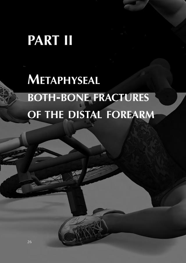
PART II
METAPHYSEAL
BOTH-BONE FRACTURES
OF THE DISTAL FOREARM
26

CHAPTER 2
Below-elbow cast formetaphyseal both-bone fracturesof the distal forearm in children:a randomised multicentre study
Injury. 2012 Jul;43(7):1107-11
Colaris JW, Biter LU, Allema JH, Bloem RM, van de Ven CP,
de Vries MR, Kerver AJ, Reijman M, Verhaar JA.
27

ABSTRACT
Introduction: Minimally displaced metaphyseal both-bone fractures of the distal
forearm in children are often treated with an above-elbow cast (AEC). Treatment with
a below-elbow cast (BEC) could give more comfort, but might lead to fracture
displacement reducing pronation and supination. Because this has not been
systematically investigated, we set up a randomised multicentre study. The purpose
of this study was to find out whether BEC causes equal limitation of pronation and
supination but with higher comfort level, compared with AEC.
Patients and methods: In four hospitals, consecutive children aged < 16 (mean 7.1)
years with a minimally displaced metaphyseal both-bone fracture of the distal
forearm were randomised to 4 weeks BEC (n=35) or 4 weeks AEC (n=31). Primary
outcome was limitation of pronation and supination 6 months after initial trauma.
The secondary outcomes were cast comfort, limitation of flexion/extension of
wrist/elbow, complications, cosmetics, complaints, and radiological assessment.
Results: A group of 35 children received BEC and 31 children received AEC.
All children attended for the final examination at a mean follow-up of 7.0 months
(range 5.0-11.6 months). Limitation of pronation and supination 6 months after initial
trauma showed no significant difference between the two groups [4.4° (± 5.8) for
BEC and 5.8° (± 9.8) for AEC]. Children treated with BEC had significantly higher
cast comfort on a visual analogue scale [5.6 (± 2.7) vs. 8.4 (± 1.4)] and needed
significantly less help with dressing (8.2 vs. 15.1 days). Six complications occurred
in the BEC group and 14 in the AEC group. Other secondary outcomes were similar
between the two groups.
Conclusions: Children with minimally displaced metaphyseal both-bone fractures
of the distal forearm should be treated with a below-elbow cast.
Chapter 2
28
INTRODUCTION
Metaphyseal both-bone fractures of the distal forearm are among the most common
injuries in childhood. The minimally displaced fractures are divided in torus fractures
and the more severe greenstick and complete fractures. The first group is usually
treated with a below-elbow cast (BEC) for comfort. The second group is usually
treated with an above-elbow cast (AEC) to prevent fracture displacement1-4.
An AEC immobilizes both wrist and elbow that potentially stabilizes the fracture
more than immobilization of the wrist only using a BEC. Less fracture stabilization
can result in displacement of the fracture leading to malunion, and limitation of
pronation and supination. However, AEC can cause more interference with daily
activities and give more discomfort than BEC.
Two studies that randomised between AEC and BEC for metaphyseal fractures
of the distal forearm in children, concluded that both casts are effective to maintain
reduction of the fracture, while BEC interfered less with daily activities5,6. One of
these studies reported a significant difference in the arc of elbow motion in favour
of the BEC group at time of cast removal and at final examination6. None of these
studies included only both-bone forearm fractures, although these fractures are
notorious for instability and dislocation5,7. Therefore, we designed the first
randomised multicentre study to compare BEC and AEC for the treatment of
exclusively minimally displaced metaphyseal both-bone fractures of the distal
forearm in children. The purpose of this study was to find out whether BEC causes
equal limitation of pronation and supination but with higher comfort level, compared
with AEC.
PATIENTS AND METHODS
Trial design and participants
A multicentre randomised trial was performed on consecutive children with a
minimally displaced metaphyseal fracture of the radius and ulna, who visited the
emergency department of one of four participating hospitals. The participating
hospitals were: Erasmus Medical Center (Rotterdam), HAGA Hospital (The Hague),
Reinier de Graaf Hospital (Delft) and Sint Franciscus Hospital (Rotterdam). The
Minimally displaced metaphyseal fractures
29
regional medical ethics committee approved the study and it was registered in
Clinical Trials.gov (NCT00397995). Informed consent was obtained for participation
from all the parents and from children aged ≥ 12 years.
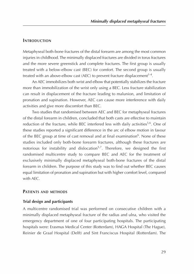
Children aged < 16 years with a metaphyseal both-bone fracture of the distal
forearm were invited to participate (Figure 1).
Figure 1. Posteroanterior and lateral radiographs of a metaphyseal both-bone fracture of the distal
forearm in a child.
The exclusion criteria were: 1) buckle fractures of both the ulna and radius, 2)
fracture sustained longer than one week, 3) a severe open fracture (Gustilo II and
III) (8), 4) a relapse fracture in the same location, and 5) need for reduction according
to a priori defined criteria (Table 1).
The primary outcome measure was limitation of pronation and supination 6
months after the fracture. The secondary outcome measures were cast comfort,
limitation of flexion and extension of wrist and elbow, complications, cosmetics,
complaints and radiological assessment.
Chapter 2
30
Procedures
The included fractures were randomised between 4 weeks of BEC or 4 weeks of
AEC. All casts were applied in neutral position and non-circumferential. All children
received a sling for at least 1 week. The children were clinically and radiologically
evaluated at 1 and 4 weeks after initial trauma by a resident, supervised by an
attending orthopaedic or trauma surgeon. A specialist of plaster revised the cast after
1 week. Where necessary, the fractures were reduced during the period of casting
according to the initial reduction criteria (Table 1). Finally, the cast was removed 4
weeks after initial treatment.
One independent orthopaedic surgeon examined all children 2 and 6 months
after initial trauma, and measured flexion and extension of wrist and elbow in
combination with pronation and supination of both arms. This technique was
standardised with the use of a 180° goniometer constructed of clear, flexible plastic
with 2 movable arms of 30 cm. Pronation and supination were scaled with a
previously used grading system9.
Patients with at least 30° of functional impairment at the 2-month examination
received a referral for physiotherapy. To evaluate the comfort of the cast a visual
analogue scale (VAS) was used; the highest score indicating maximal comfort. In
addition, a questionnaire previously used in a similar group of children, evaluated
difficulties with 7 daily activities during the period of casting6.
Six months after the initial trauma, final radiographs were taken. Both the parents
Minimally displaced metaphyseal fractures
31
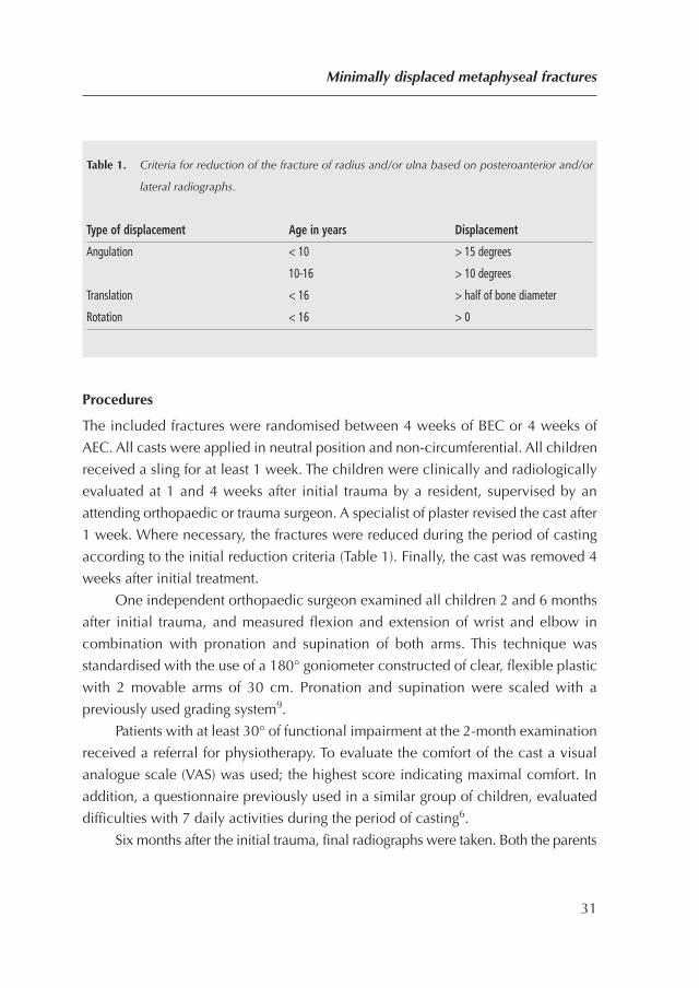
Table 1. Criteria for reduction of the fracture of radius and/or ulna based on posteroanterior and/or
lateral radiographs.
Type of displacement Age in years Displacement
Angulation < 10 > 15 degrees
10-16 > 10 degrees
Translation < 16 > half of bone diameter
Rotation < 16 > 0
and the orthopaedic surgeon completed a VAS cosmetics (maximum score indicating
best cosmetics) of the fractured arm. In the same consultation, complaints of the
fractured arm were documented using a modified grading system. This grading system
combines limitation of pronation and supination with complaints in daily life or during
strenuous activities. This system was previously used in a similar group of children10.
The parents completed the ABILHAND-Kids questionnaire11, which is a
measure of manual ability for children with upper-limb impairments. The scale is
validated for cerebral palsy children and measures a child’s ability to manage daily
activities that require the use of the upper limbs; the maximum score is 42.
The independent orthopaedic surgeon measured the sagittal and radioulnar
angulation of the fracture on the radiographs at three moments, i.e. directly after trauma,
after removal of the cast, and 6 months post trauma. The mean and standard deviation
(SD) of the sagittal and radioulnar angulation were calculated to determine the primary
displacement, re-displacement during the period of casting, and the final displacement
at 6 months. To assess the interrater reproducibility an independent trauma surgeon re-
measured the sagittal and radioulnar angulation of the fracture of 45 children.
Randomisation and masking
An independent physician randomised the children by sealed envelopes with varied
block sizes. A surgeon treated the children during the first 4 weeks without masking.
One independent orthopaedic surgeon examined all children 2 and 6 months after
the initial trauma without masking. Both the independent orthopaedic surgeon and
independent trauma surgeon measured the radiographs with masking.
Statistical Methods
To assess the required number of children, an equivalence test was used to
demonstrate the similarity of pronation and supination in both groups. Equivalence
between the two groups was defined as a maximum of 10° difference in pronation
or supination. Using an a priori calculation it was determined that, with a power of
82%, a significance of 0.05 and a SD of 15°, the two groups should consist of ± 30
children each. First it was established whether the variables had a normal distribution
using the normality Shapiro-Wilk test. Based on these analyses, the results are
presented as means (SD).
Chapter 2
32
Data were analysed using linear regression analysis. If necessary, adjustments were
made for unbalanced covariates. Differences between the two groups (BEC vs. AEC)
for the secondary outcome measures were analysed using one-way ANOVA to
correct for multiple comparisons (Bonferroni). Statistical analyses were performed
with SPSS version 17.0 (SPSS Inc., Chicago, USA).
RESULTS
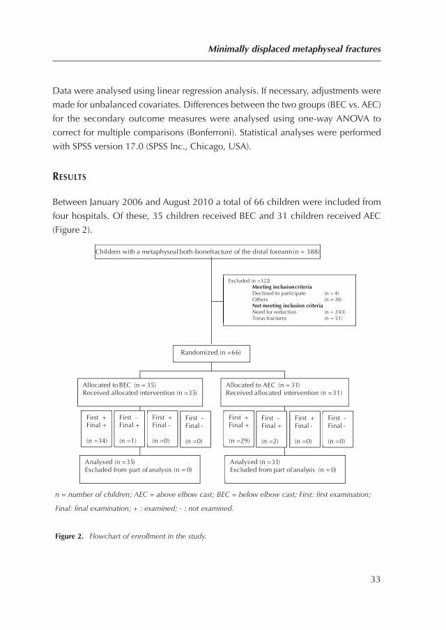
Between January 2006 and August 2010 a total of 66 children were included from
four hospitals. Of these, 35 children received BEC and 31 children received AEC
(Figure 2).
n = number of children; AEC = above elbow cast; BEC = below elbow cast; First: first examination;
Final: final examination; + : examined; - : not examined.
Figure 2. Flowchart of enrollment in the study.
Minimally displaced metaphyseal fractures
33
Children with a metaphyseal both-bone fracture of the distal forearm (n = 388)
Randomized (n = 66)
First + Final + (n =34)
First + Final - (n =0)
First - Final - (n =0)
First + Final + (n =29)
First + Final - (n =0)
First - Final - (n =0)
Excluded (n = 322) Meeting inclusion criteria Declined to participate (n = 4) Others (n = 24) Not meeting inclusion criteria Need for reduction (n = 243) Torus fractures (n = 51)
Allocated to BEC (n = 35)Received allocated intervention (n = 35)
Allocated to AEC (n = 31) Received allocated intervention (n =31)
First - Final + (n =2)
Analysed (n = 31) Excluded from part of analysis (n = 0)
First - Final + (n =1)
Analysed (n = 35) Excluded from part of analysis (n = 0)
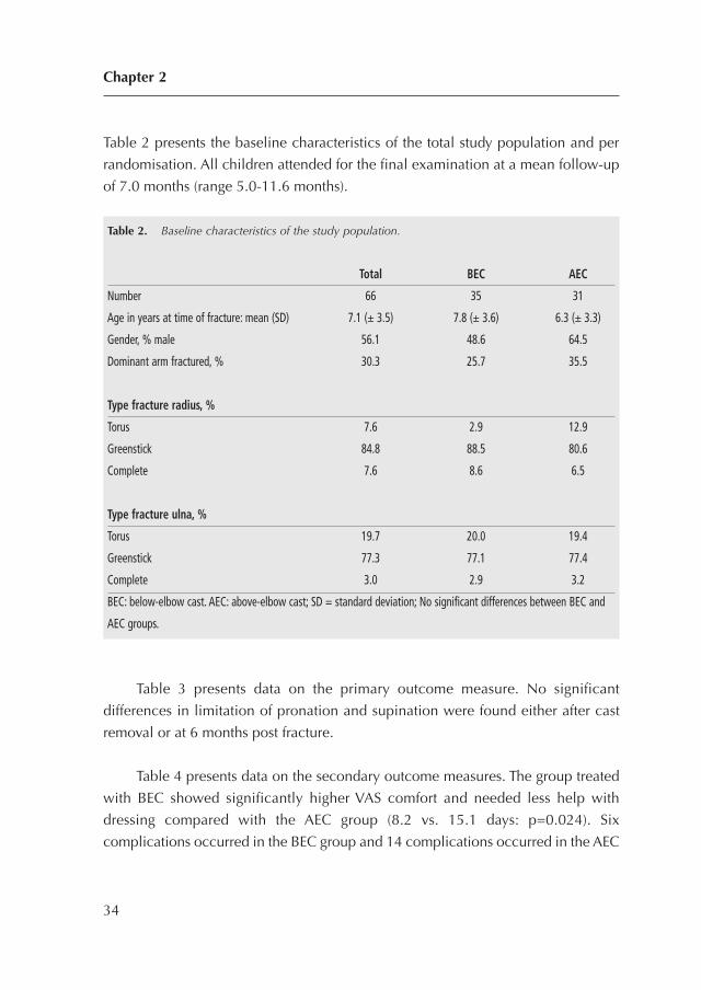
Table 2 presents the baseline characteristics of the total study population and per
randomisation. All children attended for the final examination at a mean follow-up
of 7.0 months (range 5.0-11.6 months).
Table 3 presents data on the primary outcome measure. No significant
differences in limitation of pronation and supination were found either after cast
removal or at 6 months post fracture.
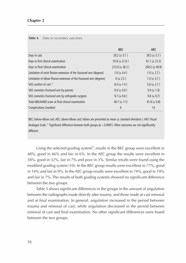
Table 4 presents data on the secondary outcome measures. The group treated
with BEC showed significantly higher VAS comfort and needed less help with
dressing compared with the AEC group (8.2 vs. 15.1 days: p=0.024). Six
complications occurred in the BEC group and 14 complications occurred in the AEC
Chapter 2
34
Table 2. Baseline characteristics of the study population.
Total BEC AEC
Number 66 35 31
Age in years at time of fracture: mean (SD) 7.1 (± 3.5) 7.8 (± 3.6) 6.3 (± 3.3)
Gender, % male 56.1 48.6 64.5
Dominant arm fractured, % 30.3 25.7 35.5
Type fracture radius, %
Torus 7.6 2.9 12.9
Greenstick 84.8 88.5 80.6
Complete 7.6 8.6 6.5
Type fracture ulna, %
Torus 19.7 20.0 19.4
Greenstick 77.3 77.1 77.4
Complete 3.0 2.9 3.2
BEC: below-elbow cast. AEC: above-elbow cast; SD = standard deviation; No significant differences between BEC and
AEC groups.
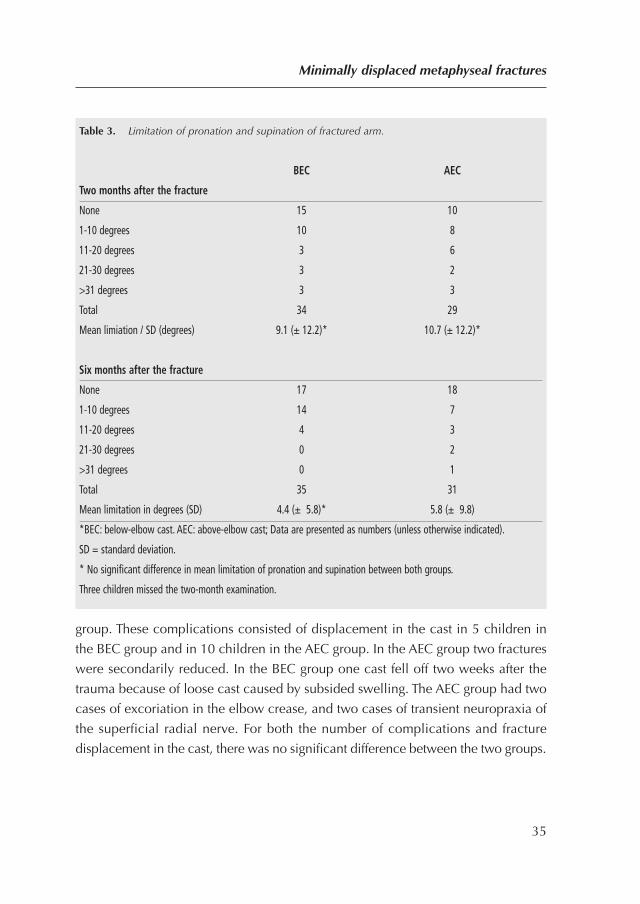
group. These complications consisted of displacement in the cast in 5 children in
the BEC group and in 10 children in the AEC group. In the AEC group two fractures
were secondarily reduced. In the BEC group one cast fell off two weeks after the
trauma because of loose cast caused by subsided swelling. The AEC group had two
cases of excoriation in the elbow crease, and two cases of transient neuropraxia of
the superficial radial nerve. For both the number of complications and fracture
displacement in the cast, there was no significant difference between the two groups.
Minimally displaced metaphyseal fractures
35
Table 3. Limitation of pronation and supination of fractured arm.
BEC AEC
Two months after the fracture
None 15 10
1-10 degrees 10 8
11-20 degrees 3 6
21-30 degrees 3 2
>31 degrees 3 3
Total 34 29
Mean limiation / SD (degrees) 9.1 (± 12.2)* 10.7 (± 12.2)*
Six months after the fracture
None 17 18
1-10 degrees 14 7
11-20 degrees 4 3
21-30 degrees 0 2
>31 degrees 0 1
Total 35 31
Mean limitation in degrees (SD) 4.4 (± 5.8)* 5.8 (± 9.8)
*BEC: below-elbow cast. AEC: above-elbow cast; Data are presented as numbers (unless otherwise indicated).
SD = standard deviation.
* No significant difference in mean limitation of pronation and supination between both groups.
Three children missed the two-month examination.
Using the selected grading system9, results in the BEC group were excellent in
48%, good in 46% and fair in 6%. In the AEC group the results were excellent in
58%, good in 32%, fair in 7% and poor in 3%. Similar results were found using the
modified grading system (10). In the BEC group results were excellent in 77%, good
in 14% and fair in 9%. In the AEC group results were excellent in 74%, good in 19%
and fair in 7%. The results of both grading systems showed no significant difference
between the two groups.
Table 5 shows significant differences in the groups in the amount of angulation
between the radiographs made directly after trauma, and those made at cast removal
and at final examination. In general, angulation increased in the period between
trauma and removal of cast, while angulation decreased in the period between
removal of cast and final examination. No other significant differences were found
between the two groups.
Chapter 2
36
Table 4. Data on secondary outcomes.
BEC AEC
Days in cast 28.2 (± 5.1 ) 28.5 (± 5.7 )
Days to first clinical examination 55.8 (± 21.6 ) 61.1 (± 21.2)
Days to final clinical examination 213.9 (± 36.2 ) 209.2 (± 40.9)
Limitation of wrist flexion-extension of the fractured arm (degrees) 1.9 (± 4.4 ) 1.0 (± 2.7 )
Limitation of elbow flexion-extension of the fractured arm (degrees) 0 (± 2.5 ) 1.0 (± 3.7 )
VAS comfort of cast * 8.4 (± 1.4 ) 5.6 (± 2.7 )
VAS cosmetics fractured arm by parents 9.4 (± 0.9 ) 9.4 (± 1.0)
VAS cosmetics fractured arm by orthopedic surgeon 9.7 (± 0.6 ) 9.6 (± 0.7)
Total ABILHAND score at final clinical examination 40.7 (± 7.1) 41.8 (± 0.8)
Complications (number) 6 14
BEC: below-elbow cast. AEC: above-elbow cast; Values are presented as mean (± standard deviation ). VAS: Visual
Analogue Scale. * Significant difference between both groups (p < 0.0001). Other outcomes are not significantly
different.

The interrater reproducibility of the radiological assessment showed an intra-
class correlation of 0.81 and 0.89 for the radioulnar angulation of the ulna and
radius, respectively. The intraclass correlation of the sagittal angulation was 0.92 for
the ulna and 0.87 for the radius.
DISCUSSION
This randomised multicentre trial shows that BEC is as effective as AEC in the
treatment of minimally displaced metaphyseal both-bone fractures of the distal
forearm in children. The children treated with BEC had similar limitation of pronation
and supination, significantly higher cast comfort, needed significantly less help with
dressing, and showed a tendency towards fewer complications.
Minimally displaced metaphyseal fractures
37
Table 5. Radiological outcomes.
BEC
Trauma Removal of cast Final clinical examination
AP ulna 6.5 (±4.1)² 5.5 (±4.2) 3.3 (±2.6)²
AP radius 3.5 (±2.8) 4.0 (±3.8) 2.3 (±2.7)
Lateral ulna 6.9 (±4.8)² 4.3 (±3.3) 3.3 (±2.6)²
Lateral radius 9.0 (±4.9)¹² 11.4 (±6.9)¹ 4.8 (±3.4)²
AEC
Trauma Removal of cast Final clinical examination
PA ulna 6.7 (±4.3)² 6.6 (±4.0) 4.4 (±3.1)²
PA radius 4.2 (±3.3)² 4.3 (±4.2) 2.8 (±2.6)²
Lateral ulna 7.9 (±5.4)¹² 5.8 (±4.7)¹ 4.0 (±3.2)²
Lateral radius 8.7 (±5.0)¹² 12.2 (±5.8)¹ 6.3 (±5.0)
²Radioulnar angulations (PA ulna, PA radius) and sagittal angulations (Lateral ulna, Lateral radius) presented in degrees
as mean (± standard deviation ).
No significant differences between BEC and AEC groups.
Significant differences (p-value < 0.05) were found between trauma radiographs and respectively radiographs removal
of cast (¹) and radiographs final examination (²).
Previous studies
In contrast with our results, an earlier study (6) found more limitation of pronation
and supination in the AEC group than in the BEC group. Our mean limitation of
pronation and supination was higher compared to these previous studies. This can
be explained by our inclusion of patients with only both-bone fractures.
In a retrospective study of almost exclusively displaced both-bone forearm
fractures treated with AEC after reduction, Daruwalla et al. found excellent results
in 50%, good results in 33%, fair results in 17%, and no poor results9. Using the
same grading system, our results seemed to be better, possibly caused by the absence
of displaced fractures in our study group.
Using the modified grading system, in a selected group of malunited diaphyseal
both-bone forearm fractures others reported 82% excellent results, 10% good results
and 8% fair results10. These better results, despite the severity of malunited
diaphyseal fractures, might be explained by the retrospective analysis and longer
follow-up period.
We found twice as many fracture displacement in the group treated with AEC
than in those treated with BEC. Similar findings have been reported by others5,6.
Immobilization of the elbow does not seem to prevent fracture displacement of a
metaphyseal fracture of the distal forearm; this might be due to less moulding around
the lower arm caused by the more difficult to apply AEC.
Radiographic assessment
Radiographic assessment showed that angulation increased in the period between
trauma and removal of the cast, which might be explained by the secondary
dislocation of the fracture during cast treatment. Decreased angulation in the period
between removal of cast and final examination might be explained by remodelling
by growth.
Limitations
This study has several limitations. Primarily, neither the children nor the independent
orthopaedic surgeon were blinded during clinical follow-up, which might have
influenced the clinical measurements. Obviously, blinding of the children was not
possible because different types of cast were used. However, both the radiographic
Chapter 2
38
assessors were blinded for the type of cast. The second limitation is our period of
follow-up in this group of children with capacity for remodelling by growth.
Although a follow-up of almost 7 months seems to be short, others found no change
in clinical or radiographic status from 6 months after treatment to the final follow-
up at an average of 2.8 years in children treated for displaced forearm fractures12.
Thereby Nilsson et al. described a persistent limitation during long term follow-up
despite correction by growth and others advised an osteotomy to correct malunion
as early as 6 months after trauma13,14. Nevertheless, most children in our study
regained a full range of pronation and supination after seven months.
Future research and study
Future studies could try to answer the following question: Why do fractures displace
more in AEC than in BEC?
Conclusions
This randomised multicentre trial shows that there is similar limitation of pronation
and supination after treatment of minimally displaced metaphyseal both-bone
fractures of the distal forearm in children with either BEC or AEC. Children treated
with BEC experienced significantly more cast comfort and significantly less
interference with daily activities during the period of casting. To conclude, children
with minimally displaced metaphyseal both-bone fractures of the distal forearm
should be treated with a properly applied below-elbow cast.
Minimally displaced metaphyseal fractures
39

Chapter 2
40
REFERENCES
1. Gibbons CL, Woods DA, Pailthorpe C, Carr AJ, Worlock P. The management of isolated
distal radius fractures in children. J Pediatr Orthop. 1994;14(2):207-10.
2. Jones K, Weiner DS. The management of forearm fractures in children: a plea for
conservatism. J Pediatr Orthop. 1999;19(6):811-5.
3. Noonan KJ, Price CT. Forearm and distal radius fractures in children. J Am Acad Orthop
Surg. 1998;6(3):146-56.
4. Younger AS, Tredwell SJ, Mackenzie WG. Factors affecting fracture position at cast removal
after pediatric forearm fracture. J Pediatr Orthop. 1997;17(3):332-6.
5. Bohm ER, Bubbar V, Yong Hing K, Dzus A. Above and below-the-elbow plaster casts for
distal forearm fractures in children. A randomized controlled trial. J Bone Joint Surg Am.
2006;88(1):1-8.
6. Webb GR, Galpin RD, Armstrong DG. Comparison of short and long arm plaster casts for
displaced fractures in the distal third of the forearm in children. J Bone Joint Surg Am.
2006;88(1):9-17.
7. Green JS, Williams SC, Finlay D, Harper WM. Distal forearm fractures in children:the role
of radiographs during follow up. Injury. 1998;29(4):309-12.
8. Gustilo RB, Simpson L, Nixon R, Ruiz A, Indeck W. Analysis of 511 open fractures. Clin
Orthop Relat Res. 1969;66:148-54.
9. Daruwalla JS. A study of radioulnar movements following fractures of the forearm in
children. Clin Orthop Relat Res. 1979(139):114-20.
10. Price CT, Scott DS, Kurzner ME, Flynn JC. Malunited forearm fractures in children. J Pediatr
Orthop. 1990;10(6):705-12.
11. Arnould C, Penta M, Renders A, Thonnard JL. ABILHAND-Kids: a measure of manual ability
in children with cerebral palsy. Neurology. 2004;63(6):1045-52.
12. Miller BS, Taylor B, Widmann RF, Bae DS, Snyder BD, Waters PM. Cast immobilization
versus percutaneous pin fixation of displaced distal radius fractures in children: a
prospective, randomized study. J Pediatr Orthop. 2005;25(4):490-4.
13. Nilsson BE, Obrant K. The range of motion following fracture of the shaft of the forearm in
children. Acta Orthop Scand. 1977;48(6):600-2.
14. Price CT, Knapp DR. Osteotomy for malunited forearm shaft fractures in children. J Pediatr
Orthop. 2006;26(2):193-6.
Minimally displaced metaphyseal fractures
41

42

CHAPTER 3
Re-displacement of stable distalboth-bone forearm fractures inchildren: a randomisedcontrolled multicentre trial
Injury. 2013 Apr;44(4):498-503
Colaris JW, Allema JH, Biter LU, de Vries MR, van de Ven CP,
Bloem RM, Kerver AJ, Reijman M, Verhaar JA.
43
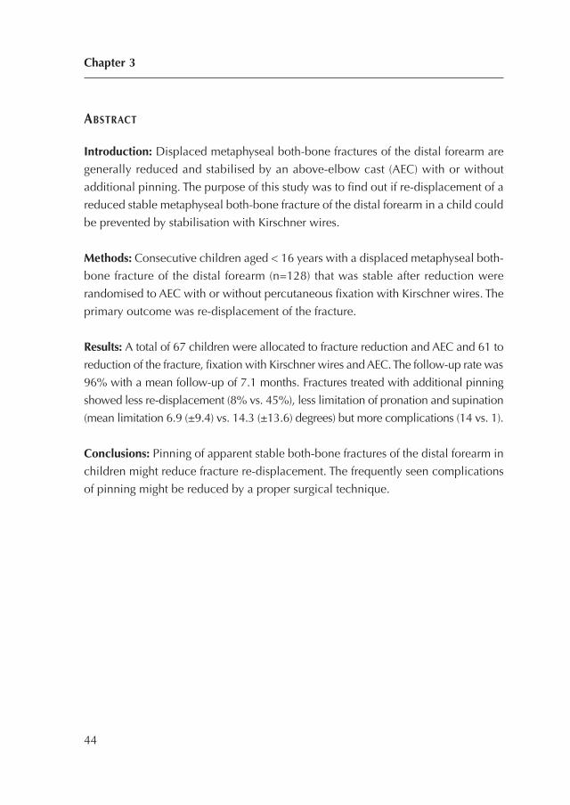
ABSTRACT
Introduction: Displaced metaphyseal both-bone fractures of the distal forearm are
generally reduced and stabilised by an above-elbow cast (AEC) with or without
additional pinning. The purpose of this study was to find out if re-displacement of a
reduced stable metaphyseal both-bone fracture of the distal forearm in a child could
be prevented by stabilisation with Kirschner wires.
Methods: Consecutive children aged < 16 years with a displaced metaphyseal both-
bone fracture of the distal forearm (n=128) that was stable after reduction were
randomised to AEC with or without percutaneous fixation with Kirschner wires. The
primary outcome was re-displacement of the fracture.
Results: A total of 67 children were allocated to fracture reduction and AEC and 61 to
reduction of the fracture, fixation with Kirschner wires and AEC. The follow-up rate was
96% with a mean follow-up of 7.1 months. Fractures treated with additional pinning
showed less re-displacement (8% vs. 45%), less limitation of pronation and supination
(mean limitation 6.9 (±9.4) vs. 14.3 (±13.6) degrees) but more complications (14 vs. 1).
Conclusions: Pinning of apparent stable both-bone fractures of the distal forearm in
children might reduce fracture re-displacement. The frequently seen complications
of pinning might be reduced by a proper surgical technique.
Chapter 3
44
INTRODUCTION
Metaphyseal fractures of the distal forearm are common in childhood (1-3). Non-
displaced fractures are generally treated with cast while displaced fractures need to
be reduced and stabilised. After reduction, the method of stabilisation ranges from
below-elbow cast only to percutaneous pinning with Kirschner wires (K wires) in
combination with an above-elbow cast (AEC)4-14. Re-displacement of the fractures
treated without percutaneous pinning has been described in 7-91% of patients6, 7,11-
13,15-18 and such re-displacement can lead to malunion with reduced functional and
cosmetic results19,20. In case of re-displacement the clinician is often confronted
with a treatment dilemma, i.e. whether to accept some degree of re-displacement
or to perform a secondary reduction of the fracture.
Several randomised studies compared treatment modalities for a combination of
single-bone and both-bone forearm fractures in children. Two studies randomised
between AEC and below-elbow cast and found the highest percentage of re-
displacement in the AEC group, which could be explained by poor cast moulding4,14.
Two other studies randomised between AEC with or without percutaneous pinning; re-
displacement appeared only in the group without percutaneous pinning and was 21%
in one and 39% in the other study9,10. Although these studies were well performed,
none distinguished between fractures of the radius or ulna only, and fractures of both
the radius and ulna, whereas the latter are notorious for their instability4,18,21.
Therefore, we performed a multicentre study designed for only displaced
metaphyseal both-bone fractures of the distal forearm in children, which were apparent
stable after reduction in the operation room. After randomisation the fractures were
treated with AEC alone or a combination of percutaneous pinning with AEC.
The purpose of this study was to find out if re-displacement of a reduced stable
metaphyseal both-bone fracture of the distal forearm in a child could be prevented
by stabilisation with K wires.
Displaced metaphyseal fractures
45
MATERIALS AND METHODS
Trial design and participants
This trial was part of a prospective multicentre study that followed several types of
both-bone forearm fractures in children. For the present study consecutive children
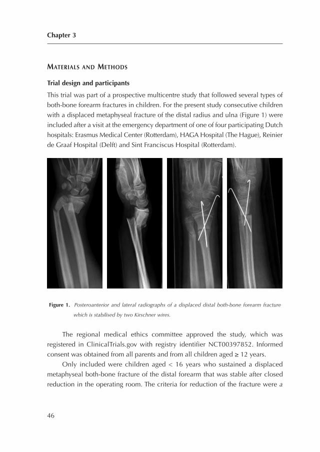
with a displaced metaphyseal fracture of the distal radius and ulna (Figure 1) were
included after a visit at the emergency department of one of four participating Dutch
hospitals: Erasmus Medical Center (Rotterdam), HAGA Hospital (The Hague), Reinier
de Graaf Hospital (Delft) and Sint Franciscus Hospital (Rotterdam).
Figure 1. Posteroanterior and lateral radiographs of a displaced distal both-bone forearm fracture
which is stabilised by two Kirschner wires.
The regional medical ethics committee approved the study, which was
registered in ClinicalTrials.gov with registry identifier NCT00397852. Informed
consent was obtained from all parents and from all children aged ≥ 12 years.
Only included were children aged < 16 years who sustained a displaced
metaphyseal both-bone fracture of the distal forearm that was stable after closed
reduction in the operating room. The criteria for reduction of the fracture were a
Chapter 3
46
priori defined and based on previous studies (5, 6, 8, 22-28). A fracture was reduced
if radius and/or ulna showed displacement on a posteroanterior and/or lateral
radiograph. Displacement of the fracture was based on angulation (> 15 degrees for
children aged less than 10 years old and >10 degrees for children between 10 and
16 years old) and/or translation (> half of the bone diameter) and/or any rotation of
radius and/or ulna.
Exclusion criteria were fractures older than 1 week, severe open fractures
(Gustilo II and III) (29), and re-fractures. The primary outcome was re-displacement
of the fracture. Secondary outcomes were limitation of pronation and supination,
limitation of flexion and extension of wrist and elbow, complication rate, complaints
in daily life, radiological assessment and cosmetics of the fractured arm.
Procedures
A surgeon reduced the fracture in the operating room under general anaesthesia
with fluoroscopic guidance. After optimal reduction by closed means, the fracture
was tested for stability. A fracture was defined as unstable if full range of pronation
and supination of the proximal forearm caused re-displacement of the fracture. This
test for stability was used before in a group of children with forearm fracturest.
Unstable fractures were excluded and treated with percutaneous pinning and AEC
for 4 weeks. The remaining fractures were defined as stable and were randomised
between AEC alone and percutaneous pinning with AEC, both for 4 weeks.
The surgeon performed the percutaneous pinning according to a standard
technique10. A small skin incision was made over the radial styloid and blunt
dissection of soft tissue was carried down to the bone. A K wire was directed
proximally and ulnarly across the fracture site, engaging the opposite cortex. A
second K wire was inserted from dorsal to volar across the fracture site through a
small incision over the interval between the fourth and fifth dorsal compartments
after blunt dissection down to the bone. Injury to the sensory branch of the radial
nerve and the extensor tendons was avoided. The K wires were bent, cut and left
transcutaneous. Additionally, the surgeon applied an AEC in the operation room.
Primarily a stockinet and layer of wool were applied to protect the skin, bony
prominences and K wires. Secondarily a well-fitted plaster slab was applied which
covered approximately 2/3 of the circumference of the arm. Finally, a bandage was
Displaced metaphyseal fractures
47
wrapped around the arm. The elbow was set in 90 degrees of flexion and the wrist
in neutral position. All children received a sling for at least 1 week.
The children underwent clinical and radiological evaluation at 1, 2 and 4
weeks after initial trauma. During these visits, a cast technician revised and renewed
the casts where necessary. Re-displacement of the fracture was defined by the loss
of reduction (angulation and/or translation) according to the primary reduction
criteria. These re-displaced fractures required a second reduction with percutaneous
pinning in the operating room. Finally, both the cast and the K wires were removed
in the outpatient clinic after 4 weeks.
One orthopaedic surgeon (who was not involved in treatment and was not
blinded) examined all children at 2 and 6 months after initial trauma. Flexion and
extension of wrist and elbow in combination with pronation and supination of both
arms were evaluated by a standardised procedure (Table 2)31.
The pronation and supination were scaled using the grading system as described
by Daruwalla with excellent, good, fair and poor results for respectively 0-10, 11-20,
21-30 and ≥ 31 degrees of limitation (Table 3)23. Children with at least 30 degrees of
functional impairment at 2 months examination received a referral for physiotherapy.
At the 6-month examination, parents and the orthopaedic surgeon completed
a visual analogue scale (VAS) cosmetics, with maximum score for similar appearance
of the fractured and non-fractures arm, to evaluate forearm alignment and scars.
During the same examination, the parents filled in the ABILHAND-Kids
questionnaire, that is a measure of manual ability for children with upper limb
impairments (Table 2). The scale is validated for cerebral palsy children and measures
a child’s ability to manage daily activities that require the use of the upper limbs,
with a maximum score of 4232.
Antero-posterior and lateral radiographs were taken at scheduled times (Table
4). The first radiographs were made in the emergency room followed by radiographs
after reduction and during follow-up at 1, 2 and 4 weeks. The final radiographs were
made at 6 months after the trauma. Angulation, translation, shortening and rotation
of fractures of all available radiographs were measured by the orthopaedic surgeon
to determine primary displacement, displacement during the first 4 weeks, and final
displacement at 6 months. The presence of rotation was evaluated by differences in
width of ulna and radius on lateral and posteroanterior radiographs (33).
Chapter 3
48
Randomisation and masking
An independent physician randomised the children by sealed envelopes with varied
block sizes. The children, parents and clinicians were not blinded for randomisation.
Statistical methods
To assess the number of children required for this trial, a superiority test was used to
demonstrate a difference in re-displacement of 17% between both groups: 3% re-
displacement in the group treated with percutaneous pinning with AEC, and 20%
re-displacement in the group treated with AEC alone6,8,9,11,13,16,17,24,34-39. With an a
priori calculation it was determined that, with a power of 80% and a significance of
0.05, the two groups should consist of ± 55 children each.
First, it was established whether the variables had a normal distribution using
the normality Shapiro-Wilk test. Based on these analyses, the results are presented
as means (standard deviations (SD)). The primary research question was analysed
using logistic regression analysis (re-displacement as dependent variable, and
intervention as independent variable). If necessary, adjustments for unbalanced
covariates took place. Differences between both groups (percutaneous pinning with
AEC vs. AEC alone) for the secondary outcome measures were analysed by one-way
Anova to correct for multiple comparisons (Bonferroni). Statistical analyses were
performed with SPSS 17.0 (SPSS Inc., Chicago, USA).
To assess the interrater reproducibility an independent trauma surgeon re-
measured the angulation of the fracture of 45 children.
RESULTS
Between January 2006 and August 2010 a total of 388 children with a metaphyseal
both-bone fracture of the distal forearm were treated in the four Dutch hospitals.
After exclusion for several reasons (details are given in Figure 2), 128 children were
included of which 67 were allocated to AEC and 61 to K wires with AEC.
Displaced metaphyseal fractures
49
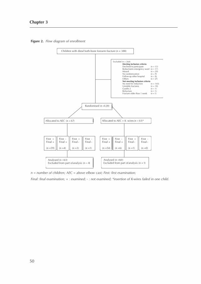
Figure 2. Flow diagram of enrollment
n = number of children; AEC = above elbow cast; First: first examination;
Final: final examination; + : examined; - : not examined; *insertion of K-wires failed in one child.
Chapter 3
50
Children with distal both-bone forearm fracture (n = 388)
Randomised (n = 128)
Excluded (n = 260) Meeting inclusion criteria Declined to participate (n = 13) Reduction in emergency ward (n = 17) Missed (n = 20) No randomization (n = 9) Follow-up other hospital (n = 8) Others (n = 27) Not meeting inclusion criteria No need for reduction (n = 145) Unstable fractures (n = 18) Gustilo 2 (n = 1) Refracture (n = 1)
Fracture older than 1 week (n = 1)
Allocated to AEC (n = 67) Allocated to AEC + K -wires (n = 61)*
Analysed (n = 63) Excluded from part of analysis (n = 4)
Analysed (n = 60)Excluded from part of analysis (n = 1)
First + Final + (n =59)
First - Final + (n =4)
First + Final - (n =3)
First - Final - (n =1)
First + Final + (n =54)
First - Final + (n =6)
First + Final - (n =1)
First - Final - (n =0)
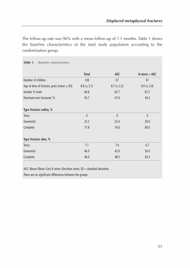
The follow-up rate was 96% with a mean follow-up of 7.1 months. Table 1 shows
the baseline characteristics of the total study population according to the
randomisation group.
Displaced metaphyseal fractures
51
Table 1. Baseline characteristics
Total AEC K-wires + AEC
Number of children 128 67 61
Age at time of fracture, years (mean ± SD) 8.8 (± 3.1) 8.7 (± 3.2) 9.0 (± 3.0)
Gender, % male 64.8 62.7 67.2
Dominant arm fractured, % 45.7 47.0 44.3
Type fracture radius, %
Torus 0 0 0
Greenstick 22.2 23.4 20.0
Complete 77.8 76.6 80.0
Type fracture ulna, %
Torus 7.1 7.6 6.7
Greenstick 46.9 43.9 50.0
Complete 46.0 48.5 43.3
AEC: Above Elbow Cast; K-wires: Kirschner wires; SD = standard deviation.
There are no significant differences between the groups.
In the AEC group 30 fractures showed re-displacement of which 17 were reduced a
second time. In the group with additional K wires, five fractures showed re-
displacement of which one fracture was reduced a second time (Table 2).
Re-displacement in the group with additional K wires was caused by
suboptimal position of the K wires in 2 children and re-displacement of the ulna in
3 children. Fracture re-displacement was first observed in 14 children at the 1-week
examination, in 19 children at the 2-week examination and in 2 children at the 4-
week examination. Thereby, we found a similar rate of re-displacement in greenstick
and complete fractures.
Pronation and supination at the final examination showed significantly less
limitation in the group initially treated with additional K wires (Table 3).
Chapter 3
52
Table 2. Outcomes.
AEC K-wires + AEC
n =67 n = 61
Re-displacement, % * 44.8 8.2
Days in cast 33.4 (±8.8) 32.2 (±6.1)
Days untill first clinical examination 68.4 (±25.4) 69.7 (±28.5)
Days untill final clinical examination 216.3 (±49.1) 215.5 (±47.2)
Referral to physiotherapy ,% 54.5 35.6
Number of visits to physiotherapy 3.9 (±7.3) 2.6 (±6.1)
Limitation of wrist flexion-extension of the fractured arm, degrees 4.4 (±6.2) 3.8 (±7.4)
Limitation of elbow flexion-extension of the fractured arm, degrees 0.5 (±2.0) 0.2 (±2.1)
VAS cosmetics fractured arm by parents 8.5 (±1.8) 8.0 (±2.2)
VAS cosmetics fractured arm by orthopedic surgeon 8.9 (±1.2) 8.4 (±1.3)
Total ABILHAND score at final clinical examination 41.5 (±1.6) 41.9 (±0.4)
Values are presented as mean (± standard deviation ) unless stated otherwise. AEC: above elbow cast; K-wires:
Kirschner wires; VAS: Visual Analogue Scale.
* Significant difference (p<0.0001).
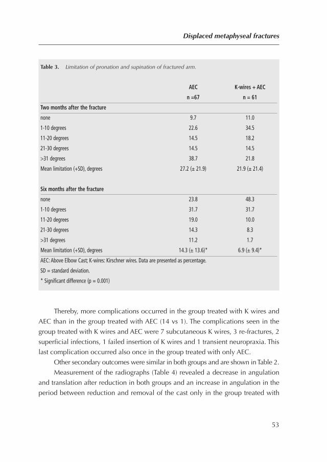
Thereby, more complications occurred in the group treated with K wires and
AEC than in the group treated with AEC (14 vs 1). The complications seen in the
group treated with K wires and AEC were 7 subcutaneous K wires, 3 re-fractures, 2
superficial infections, 1 failed insertion of K wires and 1 transient neuropraxia. This
last complication occurred also once in the group treated with only AEC.
Other secondary outcomes were similar in both groups and are shown in Table 2.
Measurement of the radiographs (Table 4) revealed a decrease in angulation
and translation after reduction in both groups and an increase in angulation in the
period between reduction and removal of the cast only in the group treated with
Displaced metaphyseal fractures
53
Table 3. Limitation of pronation and supination of fractured arm.
AEC K-wires + AEC
n =67 n = 61
Two months after the fracture
none 9.7 11.0
1-10 degrees 22.6 34.5
11-20 degrees 14.5 18.2
21-30 degrees 14.5 14.5
>31 degrees 38.7 21.8
Mean limitation (+SD), degrees 27.2 (± 21.9) 21.9 (± 21.4)
Six months after the fracture
none 23.8 48.3
1-10 degrees 31.7 31.7
11-20 degrees 19.0 10.0
21-30 degrees 14.3 8.3
>31 degrees 11.2 1.7
Mean limitation (+SD), degrees 14.3 (± 13.6)* 6.9 (± 9.4)*
AEC: Above Elbow Cast; K-wires: Kirschner wires. Data are presented as percentage.
SD = standard deviation.
* Significant difference (p = 0.001)
AEC alone. Furthermore, a decrease in angulation in the period between the removal
of the cast and the final examination was seen in both groups.
The interrater reproducibility of the radiological assessment by measurement
of angulation of the ulna and radius on both the posterioanterior and lateral
radiographs showed an intraclass correlation (ICC) range of 0.81-0.92.
Chapter 3
54
Table 4. Radiological outcomes.
AEC AEC + K-wires
Trauma After Removal Final Trauma After Removal Final
reduction of cast reduction of cast
Angulation
AP ulna 13 (±12)2 5 (±4) 2 3 6 (±4)4 4 (±3) 3 4 14 (±11) 2 5 (±4) 2 5 (±4) 4 (±3)
AP radius 12 (±9) 2 5 (±4) 2 3 8 (±7) 1 3 4 5 (± 4) 1 4 15 (±12) 2 4 (±3) 2 3 4 (±2) 1 4 3 (±2) 1 3 4
Lateral ulna 21 (±17) 2 5 (±4) 2 3 6 (±5) 4 3 (±3) 3 4 19 (±15) 2 5 (±5) 2 3 5 (±4) 4 4 (±3) 3 4
Lateral radius 21 (±12) 2 7 (±5)1 2 3 13 (±11) 1 3 4 8 (±5) 1 4 22 (±11) 2 5 (±4) 1 2 6 (±5) 1 3 4 (±2) 1 3
Translation
PA ulna 12 (±24) 2 4 (±11) 2 * * 9 (±25) 6 (±14) * *
PA radius 24 (±28) 2 7 (±9) 2 * * 29 (±36) 2 6 (±9) 2 * *
Lateral ulna 19 (±31) 2 5 (±13) 2 * * 10 (±24) 2 4 (±9) 2 * *
Lateral radius 54 (±47) 2 8 (±13) 2 * * 63 (±45) 2 5 (±9) 2 * *
Shortening of radius and/or ulna
% 68.2 67.8
Rotation of radius and/or ulna
% 15.2 21.7
Angulation: Radioulnar angulation (PA ulna, PA radius) and sagittal angulation (Lateral ulna, Lateral radius) presented in
degrees as mean (± standard deviation ).
Translation: Radioulnar translation (PA ulna, PA radius) and sagittal translation (Lateral ulna, Lateral radius) presented in
percentage of the width of ulna or radius as mean (± standard deviation ).
Shortening and rotation as percentage of appearance.
1 Significant differences between both groups.
2 3 4 Significant differences within one group
DISCUSSION
The purpose of this study was to find out if re-displacement of a reduced stable
metaphyseal both-bone fracture of the distal forearm in a child could be prevented by
stabilisation with K wires. As expected, this study shows that additional percutaneous
pinning decreased fracture re-displacement compared to AEC alone. It was also found
that children treated with additional pinning showed a small but significant decrease
of limitation of pronation and supination after 7 months. Nevertheless, the group
treated with additional pinning paid the price with a higher rate of complications.
Most of these complications consisted of subcutaneous K wires that could probably
have been avoided by not cutting the transcutaneous K wires too short.
Previous studies
Previous randomised trials reported re-displacement after reduction of distal
metaphyseal forearm fractures in children treated with an AEC in 15-42%4,9,10,14
while retrospective studies found 7-91%6,7,11-13,15-18. Our re-displacement rate of
45% in the group treated with AEC alone is relatively high and could be explained
by more instable both-both forearm fractures and the application of a plaster slab
instead of a circumferential cast directly after reduction.
Our group of children treated with K wires showed more re-displacement than
reported previously5,6,10,12. This re-displacement was caused by technical errors in
2 children and re-displacement of the non-fixated ulna in 3 children.
Similar to other studies9,11 only 51% of our re-displaced fractures were reduced
a second time, although this was suggested in our study protocol. Although not
planned, this illustrates the reluctance of the treating surgeons to choose for a second
reduction (for various reasons). On the one hand the treating surgeon may hope or
expect correction of the malunion due to growth, on the other hand the surgeon
may be reluctant to burden the child again or finds it difficult to accept failure of the
initial treatment.
Additional operations after the first reduction were mainly caused by a second
reduction and removal of subcutaneous K wires. In contrast with an earlier study9,
in the present study the K wires were generally removed in the outpatient clinic
without problems, which substantially decreases the number of re-operations.
Displaced metaphyseal fractures
55
The limitation of pronation and supination in the children treated with AEC alone
was higher in the present study than in earlier reports5,8-10,13,23,40. Our less favourable
outcomes might be explained by the inclusion of only children with both-bone
forearm fractures, the prospective nature of our study, the high rate and relatively
short time of follow-up. On closer examination, we found a limitation of pronation
and supination of ≥ 30 degrees in 11 children treated with AEC and in 3 children
treated with K wires and AEC. Only 6 of these children showed a malunion at final
radiographs. These findings support the opinion that malunion is not the only cause
of limitation of pronation and supination after both-bone forearm fractures in
children27,41-45.
Radiological assessment
As expected, measurement of the radiographs revealed a decrease in angulation and
translation after reduction in both groups, which is explained by a correct reduction.
The increase in angulation in the period between reduction and removal of the cast
only in the group treated with AEC alone was caused by re-displacement. In both
groups, a decrease in angulation in the period between the removal of the cast and
the final examination was caused by correction due to growth.
Study limitations
The first limitation was the wide age range of the included children combined with
our strict reduction criteria. This study was part of a prospective multicentre randomised
study that followed all types of both-bone forearm fractures in children and used the
same reduction criteria for metaphyseal and diaphyseal forearm fractures. Although
we adjusted the criteria for reduction to two age groups, the reduction criteria for
metaphyseal fractures in the youngest children may have been too strict.
The second limitation was our relatively short time of follow-up. A longer
period of follow-up might reduce the number of children with an impairment of
function as is described by Price et al.40.
To conclude, this is the first randomised multicentre study to evaluate displaced
metaphyseal both-bone fractures of the distal forearm in children. Pinning of
apparent stable both-bone fractures of the distal forearm in children might reduce
Chapter 3
56

fracture re-displacement and might result in less limitation of pronation and
supination. The frequently seen complications of pinning might be reduced by a
proper surgical technique.
Displaced metaphyseal fractures
57
REFERENCES
1. Jones IE, Cannan R, Goulding A. Distal forearm fractures in New Zealand children: annual
rates in a geographically defined area. N Z Med J. 2000;113(1120):443-5.
2. Reed FE, Jr., Apple DF, Jr. Ipsilateral fractures of the elbow and forearm. South Med J.
1976;69(2):149-51.
3. Worlock P, Stower M. Fracture patterns in Nottingham children. J Pediatr Orthop.
1986;6(6):656-60.
4. Bohm ER, Bubbar V, Yong Hing K, Dzus A. Above and below-the-elbow plaster casts for
distal forearm fractures in children. A randomized controlled trial. J Bone Joint Surg Am.
2006;88(1):1-8.
5. Choi KY, Chan WS, Lam TP, Cheng JC. Percutaneous Kirschner-wire pinning for severely
displaced distal radial fractures in children. A report of 157 cases. J Bone Joint Surg Br.
1995;77(5):797-801.
6. Gibbons CL, Woods DA, Pailthorpe C, Carr AJ, Worlock P. The management of isolated
distal radius fractures in children. J Pediatr Orthop. 1994;14(2):207-10.
7. Kiely PD, Kiely PJ, Stephens MM, Dowling FE. Atypical distal radial fractures in children. J
Pediatr Orthop B. 2004;13(3):202-5.
8. Mani GV, Hui PW, Cheng JC. Translation of the radius as a predictor of outcome in distal
radial fractures of children. J Bone Joint Surg Br. 1993;75(5):808-11.
9. McLauchlan GJ, Cowan B, Annan IH, Robb JE. Management of completely displaced
metaphyseal fractures of the distal radius in children. A prospective, randomised controlled
trial. J Bone Joint Surg Br. 2002;84(3):413-7.
10. Miller BS, Taylor B, Widmann RF, Bae DS, Snyder BD, Waters PM. Cast immobilization
versus percutaneous pin fixation of displaced distal radius fractures in children: a
prospective, randomized study. J Pediatr Orthop. 2005;25(4):490-4.
11. Proctor MT, Moore DJ, Paterson JM. Redisplacement after manipulation of distal radial
fractures in children. J Bone Joint Surg Br. 1993;75(3):453-4.
12. Van Leemput W, De Ridder K. Distal metaphyseal radius fractures in children: reduction
with or without pinning. Acta Orthop Belg. 2009;75(3):306-9.
13. Voto SJ, Weiner DS, Leighley B. Redisplacement after closed reduction of forearm fractures
in children. J Pediatr Orthop. 1990;10(1):79-84.
Chapter 3
58
14. Webb GR, Galpin RD, Armstrong DG. Comparison of short and long arm plaster casts for
displaced fractures in the distal third of the forearm in children. J Bone Joint Surg Am.
2006;88(1):9-17.
15. Blount WP. Forearm fractures in children. 1967. Clin Orthop Relat Res. 2005(432):4-7.
16. Friberg KS. Remodelling after distal forearm fractures in children. I. The effect of residual
angulation on the spatial orientation of the epiphyseal plates. Acta Orthop Scand.
1979;50(5):537-46.
17. Haddad FS, Williams RL. Forearm fractures in children: avoiding redisplacement. Injury.
1995;26(10):691-2.
18. Zamzam MM, Khoshhal KI. Displaced fracture of the distal radius in children: factors
responsible for redisplacement after closed reduction. J Bone Joint Surg Br. 2005;87(6):841-3.
19. Prommersberger KJ, Froehner SC, Schmitt RR, Lanz UB. Rotational deformity in malunited
fractures of the distal radius. J Hand Surg [Am]. 2004;29(1):110-5.
20. Roberts JA. Angulation of the radius in children’s fractures. J Bone Joint Surg Br.
1986;68(5):751-4.
21. Green JS, Williams SC, Finlay D, Harper WM. Distal forearm fractures in children:the role
of radiographs during follow up. Injury. 1998;29(4):309-12.
22. Boyer BA, Overton B, Schrader W, Riley P, Fleissner P. Position of immobilization for
pediatric forearm fractures. Journal of pediatric orthopedics. 2002;22(2):185-7.
23. Daruwalla JS. A study of radioulnar movements following fractures of the forearm in
children. Clin Orthop Relat Res. 1979(139):114-20.
24. Jones K, Weiner DS. The management of forearm fractures in children: a plea for
conservatism. J Pediatr Orthop. 1999;19(6):811-5.
25. Maric D, Petkovic L, Tomasevic V, Bajic L, Gajdobranski D, Marcikic A. [Osteosynthesis
with Kirschner wires in the treatment of dislocation fractures of the forearm in children].
Med Pregl. 2002;55(3-4):140-5.
26. Noonan KJ, Price CT. Forearm and distal radius fractures in children. J Am Acad Orthop
Surg. 1998;6(3):146-56.
27. Tynan MC, Fornalski S, McMahon PJ, Utkan A, Green SA, Lee TQ. The effects of ulnar axial
malalignment on supination and pronation. J Bone Joint Surg Am. 2000;82-A(12):1726-31.
28. Zimmermann R, Gabl M, Angermann P, Lutz M, Reinhart C, Kralinger F, et al. [Late
sequelae of fractures of the distal third of the forearm during the growth period]. Handchir
Mikrochir Plast Chir. 2000;32(4):242-9.
Displaced metaphyseal fractures
59
29. Gustilo RB, Simpson L, Nixon R, Ruiz A, Indeck W. Analysis of 511 open fractures. Clin
Orthop Relat Res. 1969;66:148-54.
30. Myers GJ, Gibbons PJ, Glithero PR. Nancy nailing of diaphyseal forearm fractures. Single
bone fixation for fractures of both bones. J Bone Joint Surg Br. 2004;86(4):581-4.
31. Colaris J, van der Linden M, Selles R, Coene N, Allema JH, Verhaar J. Pronation and
supination after forearm fractures in children: Reliability of visual estimation and
conventional goniometry measurement. Injury. 2010;41(6):643-6.
32. Arnould C, Penta M, Renders A, Thonnard JL. ABILHAND-Kids: a measure of manual ability
in children with cerebral palsy. Neurology. 2004;63(6):1045-52.
33. Naimark A, Kossoff J, Leach RE. The disparate diameter. A sign of rotational deformity in
fractures. J Can Assoc Radiol. 1983;34(1):8-11.
34. Chan CF, Meads BM, Nicol RO. Remanipulation of forearm fractures in children. N Z Med
J. 1997;110(1047):249-50.
35. Cullen MC, Roy DR, Giza E, Crawford AH. Complications of intramedullary fixation of
pediatric forearm fractures. J Pediatr Orthop. 1998;18(1):14-21.
36. Richter D, Ostermann PA, Ekkernkamp A, Muhr G, Hahn MP. Elastic intramedullary nailing:
a minimally invasive concept in the treatment of unstable forearm fractures in children. J
Pediatr Orthop. 1998;18(4):457-61.
37. Vainionpaa S, Bostman O, Patiala H, Rokkanen P. Internal fixation of forearm fractures in
children. Acta Orthop Scand. 1987;58(2):121-3.
38. Voto SJ, Weiner DS, Leighley B. Use of pins and plaster in the treatment of unstable
pediatric forearm fractures. J Pediatr Orthop. 1990;10(1):85-9.
39. Younger AS, Tredwell SJ, Mackenzie WG. Factors affecting fracture position at cast removal
after pediatric forearm fracture. J Pediatr Orthop. 1997;17(3):332-6.
40. Price CT, Scott DS, Kurzner ME, Flynn JC. Malunited forearm fractures in children. J Pediatr
Orthop. 1990;10(6):705-12.
41. Dumont CE, Thalmann R, Macy JC. The effect of rotational malunion of the radius and the
ulna on supination and pronation. J Bone Joint Surg Br. 2002;84(7):1070-4.
42. van Geenen RC, Besselaar PP. Outcome after corrective osteotomy for malunited fractures
of the forearm sustained in childhood. J Bone Joint Surg Br. 2007;89(2):236-9.
43. Weinberg AM, Pietsch IT, Krefft M, Pape HC, van Griensven M, Helm MB, et al. [Pronation
and supination of the forearm. With special reference to the humero-ulnar articulation].
Unfallchirurg. 2001;104(5):404-9.
Chapter 3
60

44. Yasutomi T, Nakatsuchi Y, Koike H, Uchiyama S. Mechanism of limitation of
pronation/supination of the forearm in geometric models of deformities of the forearm
bones. Clin Biomech (Bristol, Avon). 2002;17(6):456-63.
45. Nilsson BE, Obrant K. The range of motion following fracture of the shaft of the forearm in
children. Acta Orthop Scand. 1977;48(6):600-2.
Displaced metaphyseal fractures
61
PART III
DIAPHYSEAL
BOTH-BONE FOREARMFRACTURES
62
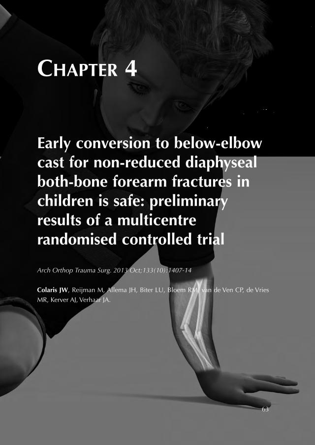
CHAPTER 4
Early conversion to below-elbowcast for non-reduced diaphysealboth-bone forearm fractures inchildren is safe: preliminaryresults of a multicentrerandomised controlled trial
Arch Orthop Trauma Surg. 2013 Oct;133(10):1407-14
Colaris JW, Reijman M, Allema JH, Biter LU, Bloem RM, van de Ven CP, de Vries
MR, Kerver AJ, Verhaar JA.
63
ABSTRACT
Introduction: This multicentre randomised controlled trial was designed to explore
whether 6 weeks above-elbow cast (AEC) or 3 weeks AEC followed by 3 weeks
below-elbow cast (BEC) cause similar limitation of pronation and supination in non-
reduced diaphyseal both-bone forearm fractures in children.
Materials and Methods: Children were randomly allocated to 6 weeks AEC or to 3
weeks AEC followed by 3 weeks BEC. The primary outcome was limitation of
pronation and supination after 6 months. The secondary outcomes were re-
displacement of the fracture, complication rate, limitation of flexion and extension
of wrist and elbow, cast comfort, cosmetics, complaints in daily life and assessment
of radiographs.
Results: A group of 23 children was treated with 6 weeks AEC and 24 children with
3 weeks AEC and 3 weeks BEC. The follow-up rate was 98% with a mean follow-up
of 7.0 months.
The mean limitation of pronation and supination was 23.3 (± 22.0) for children
treated with AEC and 18.0 (± 16.9) for children treated with AEC and BEC. The other
study outcomes were similar in both groups.
Conclusions: Early conversion to BEC is safe in the treatment of non-reduced
diaphyseal both-bone forearm fractures in children.
Chapter 4
64
INTRODUCTION
Both-bone diaphyseal forearm fractures are common in children and are generally
treated with cast, intramedullary nails or plates. The treatment with cast usually
consists of an above-elbow cast (AEC) for 6-9 weeks, although conversion to a
below-elbow cast (BEC) at the end of treatment is frequently seen1-11. Although many
of these fractures heal without complications, limitation of pronation and supination
can occur. An average limitation of 20 degrees in about 15% of children has been
reported6,12,13.
This limitation of pronation and supination after both-bone diaphyseal forearm
fractures in children seems to be caused by both malunion of the radius and/or the
ulna, and a contracture of the surrounding soft tissue13-18.
Early conversion to BEC in the treatment of these fractures could potentially
effect the limitation of pronation and supination in two opposing ways. On the one
hand, BEC does not immobilise the elbow and could therefore result in displacement
of the fracture, malunion and consequently more limitation. On the other hand, BEC
allows free movement of the elbow that could result in less contractures of soft tissue
and consequently less limitation.
Nevertheless, the influence of AEC and BEC on limitation of pronation and
supination remains unclear. Therefore, this multicentre randomised controlled trial
aimed to clarify the role of AEC and BEC on limitation of pronation and supination
in non-reduced both-bone diaphyseal forearm fractures in children. Specifically, the
following question was explored: do 6 weeks AEC as compared with 3 weeks AEC
followed by 3 weeks BEC cause similar limitation of pronation and supination?
MATERIALS AND METHODS
Trial design and participants
A multicentre randomised controlled trial was performed that included consecutive
children with a diaphyseal forearm fracture of the radius and ulna who visited the
emergency department of one of four participating hospitals. The Dutch hospitals
participating in this study were Erasmus Medical Center (Rotterdam), HAGA Hospital
(The Hague), Reinier de Graaf Hospital (Delft) and Sint Franciscus Hospital
Minimally displaced diaphyseal fractures
65
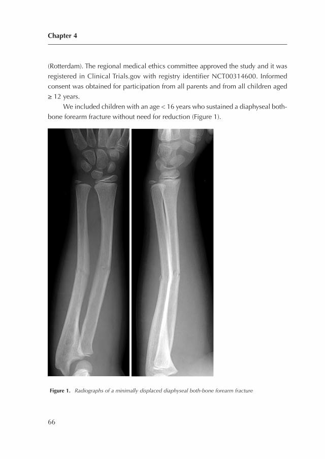
(Rotterdam). The regional medical ethics committee approved the study and it was
registered in Clinical Trials.gov with registry identifier NCT00314600. Informed
consent was obtained for participation from all parents and from all children aged
≥ 12 years.
We included children with an age < 16 years who sustained a diaphyseal both-
bone forearm fracture without need for reduction (Figure 1).
Figure 1. Radiographs of a minimally displaced diaphyseal both-bone forearm fracture
Chapter 4
66
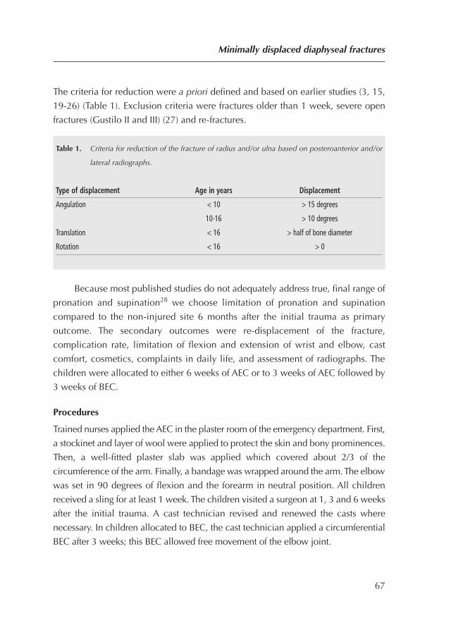
The criteria for reduction were a priori defined and based on earlier studies (3, 15,
19-26) (Table 1). Exclusion criteria were fractures older than 1 week, severe open
fractures (Gustilo II and III) (27) and re-fractures.
Because most published studies do not adequately address true, final range of
pronation and supination28 we choose limitation of pronation and supination
compared to the non-injured site 6 months after the initial trauma as primary
outcome. The secondary outcomes were re-displacement of the fracture,
complication rate, limitation of flexion and extension of wrist and elbow, cast
comfort, cosmetics, complaints in daily life, and assessment of radiographs. The
children were allocated to either 6 weeks of AEC or to 3 weeks of AEC followed by
3 weeks of BEC.
Procedures
Trained nurses applied the AEC in the plaster room of the emergency department. First,
a stockinet and layer of wool were applied to protect the skin and bony prominences.
Then, a well-fitted plaster slab was applied which covered about 2/3 of the
circumference of the arm. Finally, a bandage was wrapped around the arm. The elbow
was set in 90 degrees of flexion and the forearm in neutral position. All children
received a sling for at least 1 week. The children visited a surgeon at 1, 3 and 6 weeks
after the initial trauma. A cast technician revised and renewed the casts where
necessary. In children allocated to BEC, the cast technician applied a circumferential
BEC after 3 weeks; this BEC allowed free movement of the elbow joint.
Minimally displaced diaphyseal fractures
67
Table 1. Criteria for reduction of the fracture of radius and/or ulna based on posteroanterior and/or
lateral radiographs.
Type of displacement Age in years Displacement
Angulation < 10 > 15 degrees
10-16 > 10 degrees
Translation < 16 > half of bone diameter
Rotation < 16 > 0
Fracture displacement, as defined by the loss of reduction according to the primary
reduction criteria (Table 1), required new fracture reduction. Finally, the cast was
removed 6 weeks after initial treatment.
One orthopaedic surgeon (not involved in the primary treatment) examined all
children 2 and 6 months after the initial trauma. Flexion and extension of wrist and
elbow in combination with pronation and supination of both arms were evaluated
using a standardised procedure29. The pronation and supination were scaled using
the grading system as described by Daruwalla with excellent, good, fair and poor
results for, respectively, 0-10, 11-20, 21-30 and ≥ 31 degrees of limitation3.
Children with at least 30 degree of functional impairment at the 2-month
examination were referred to physiotherapy. Furthermore, the parents filled in a
visual analogue scale (VAS) to evaluate the comfort of the cast, with the highest score
for maximal comfort. In addition, the parents were asked to fill in a questionnaire to
evaluate difficulties with seven activities of daily living during the period of cast.
This questionnaire has previously been used in a similar group of children30.
The parents and the orthopaedic surgeon completed a VAS cosmetics of the
fractured arm, with a maximum score for similar appearance of the fractured and
non-fractured arm. In the same consultation, complaints of the fractured arm were
documented using a modified grading system. This grading system combines
limitation of pronation and supination with complaints in daily life or during
strenuous activities. It has previously been used in a similar group of children31. The
parents also filled in an ABILHAND-Kids questionnaire32, which is a measure of
manual ability for children with upper limb impairments. This scale has been
validated for children with cerebral palsy and measures a child’s ability to manage
daily activities that require use of the upper limbs, with a maximum score of 4232.
Radiographs were taken in the emergency room followed by radiographs
during follow-up at 1, 3 and 6 weeks. The final radiographs were made 6 months
after the initial trauma. The orthopaedic surgeon measured angulation, translation,
rotation and shortening of fractures to determine primary displacement,
displacement during treatment with cast and final displacement at 6 months. The
presence of rotation was evaluated by differences in the width of the ulna and radius
on lateral and posterioanterior radiographs33.
Chapter 4
68
Randomisation and masking
An independent physician randomised the children by sealed envelopes with varied
block sizes. None of the children, parents, surgeon, cast technician and independent
orthopaedic surgeon was blinded for allocation.
Statistical methods
The primary aim of the study was to determine whether 6 weeks AEC compared with
3 weeks AEC and 3 weeks BEC, cause equal limitation of pronation and supination
6 months after the initial trauma. The required number of children for this
equivalence study was calculated a priori. Equivalence between the two groups was
defined as a maximum of 15 degrees less pronation or supination in the group
treated with AEC and BEC. We chose 15 degrees of limitation in order to stay within
the safe margins of good results as graded previously3. With an a priori calculation
it was determined that with a power of 80%, an alpha of 0.05 and a standard
deviation (SD) of 15 degrees, the two groups should consist of ± 30 children each.
First, it was established whether the variables had a normal distribution using
the normality Shapiro-Wilk test. Based on these analyses, the results are presented
as means and SD. The primary research question was analysed using linear
regression analysis (limitation of pronation and supination of the fractured arm 6
months after initial trauma as dependent variable, and the intervention as
independent variable). If necessary, adjustments for unbalanced covariates were
made. Differences between the two groups for the secondary outcome measures
were analysed by one-way Anova to correct for multiple comparisons (Bonferroni).
Statistical analyses were performed with SPSS 17.0 (SPSS Inc., Chicago, USA).
RESULTS
Between January 2006 and August 2010 a total of 47 children were included in the
four Dutch hospitals. A group of 23 children was randomised to 6 weeks AEC and
24 children were randomised to 3 weeks AEC followed by 3 weeks BEC (Figure 2).
Minimally displaced diaphyseal fractures
69
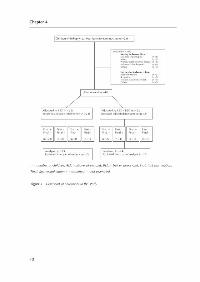
n = number of children; AEC = above elbow cast; BEC = below elbow cast; First: first examination;
Final: final examination; + : examined; - : not examined.
Figure 2. Flowchart of enrolment in the study.
Chapter 4
70
Children with diaphyseal both-bone forearm fracture (n =286)
Excluded (n = 239) Meeting inclusion criteria Declined to participate (n =5) Missed (n =2) Primary treatment other hospital (n =1) Follow-up other hospital (n =1) Others (n =7)
Not meeting inclusion criteria Reduced fracture (n =217) Re-fracture (n =2) Fracture sustained >1 week (n =1) Others (n =3)
Allocated to AEC (n = 23) Received allocated intervention (n = 23)
Allocated to AEC + BEC (n = 24) Received allocated intervention (n =24)
Analysed (n = 23) Excluded from part of analysis (n = 0)
Analysed (n = 24) Excluded from part of analysis (n = 2)
First + Final + (n =23)
First - Final + (n =0)
First + Final - (n =0)
First - Final - (n =0)
First + Final + (n =22)
First - Final + (n =1)
First + Final - (n =1)
First - Final - (n =0)
Randomised (n = 47)
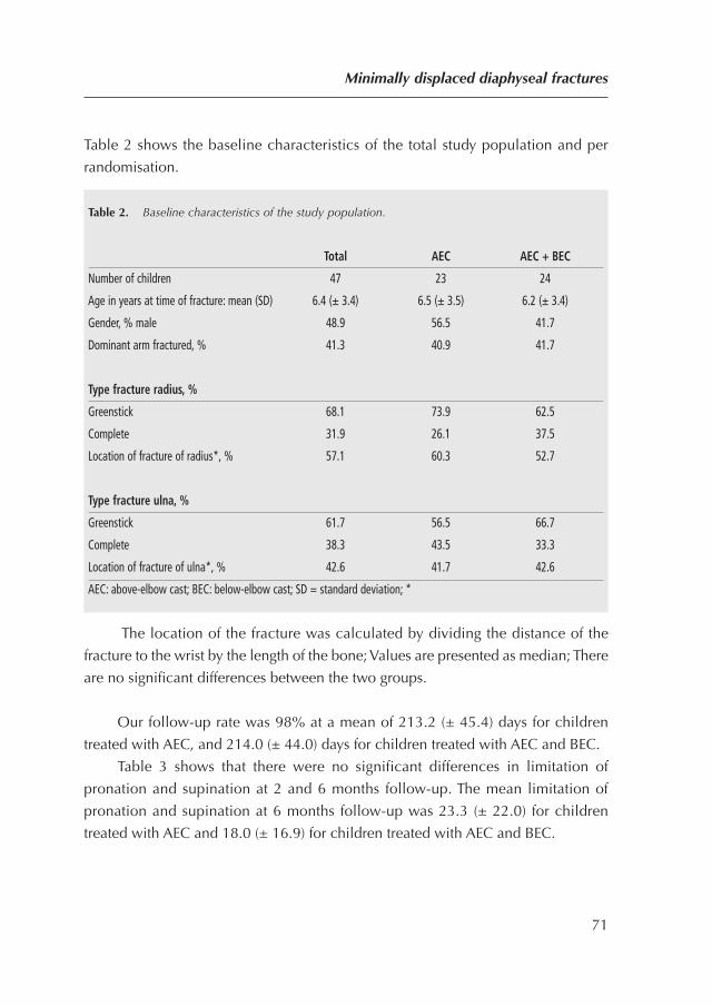
Table 2 shows the baseline characteristics of the total study population and per
randomisation.
The location of the fracture was calculated by dividing the distance of the
fracture to the wrist by the length of the bone; Values are presented as median; There
are no significant differences between the two groups.
Our follow-up rate was 98% at a mean of 213.2 (± 45.4) days for children
treated with AEC, and 214.0 (± 44.0) days for children treated with AEC and BEC.
Table 3 shows that there were no significant differences in limitation of
pronation and supination at 2 and 6 months follow-up. The mean limitation of
pronation and supination at 6 months follow-up was 23.3 (± 22.0) for children
treated with AEC and 18.0 (± 16.9) for children treated with AEC and BEC.
Minimally displaced diaphyseal fractures
71
Table 2. Baseline characteristics of the study population.
Total AEC AEC + BEC
Number of children 47 23 24
Age in years at time of fracture: mean (SD) 6.4 (± 3.4) 6.5 (± 3.5) 6.2 (± 3.4)
Gender, % male 48.9 56.5 41.7
Dominant arm fractured, % 41.3 40.9 41.7
Type fracture radius, %
Greenstick 68.1 73.9 62.5
Complete 31.9 26.1 37.5
Location of fracture of radius*, % 57.1 60.3 52.7
Type fracture ulna, %
Greenstick 61.7 56.5 66.7
Complete 38.3 43.5 33.3
Location of fracture of ulna*, % 42.6 41.7 42.6
AEC: above-elbow cast; BEC: below-elbow cast; SD = standard deviation; *
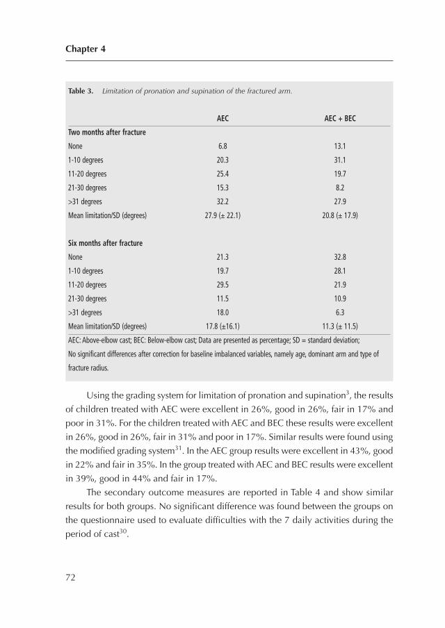
Using the grading system for limitation of pronation and supination3, the results
of children treated with AEC were excellent in 26%, good in 26%, fair in 17% and
poor in 31%. For the children treated with AEC and BEC these results were excellent
in 26%, good in 26%, fair in 31% and poor in 17%. Similar results were found using
the modified grading system31. In the AEC group results were excellent in 43%, good
in 22% and fair in 35%. In the group treated with AEC and BEC results were excellent
in 39%, good in 44% and fair in 17%.
The secondary outcome measures are reported in Table 4 and show similar
results for both groups. No significant difference was found between the groups on
the questionnaire used to evaluate difficulties with the 7 daily activities during the
period of cast30.
Chapter 4
72
Table 3. Limitation of pronation and supination of the fractured arm.
AEC AEC + BEC
Two months after fracture
None 6.8 13.1
1-10 degrees 20.3 31.1
11-20 degrees 25.4 19.7
21-30 degrees 15.3 8.2
>31 degrees 32.2 27.9
Mean limitation/SD (degrees) 27.9 (± 22.1) 20.8 (± 17.9)
Six months after fracture
None 21.3 32.8
1-10 degrees 19.7 28.1
11-20 degrees 29.5 21.9
21-30 degrees 11.5 10.9
>31 degrees 18.0 6.3
Mean limitation/SD (degrees) 17.8 (±16.1) 11.3 (± 11.5)
AEC: Above-elbow cast; BEC: Below-elbow cast; Data are presented as percentage; SD = standard deviation;
No significant differences after correction for baseline imbalanced variables, namely age, dominant arm and type of
fracture radius.
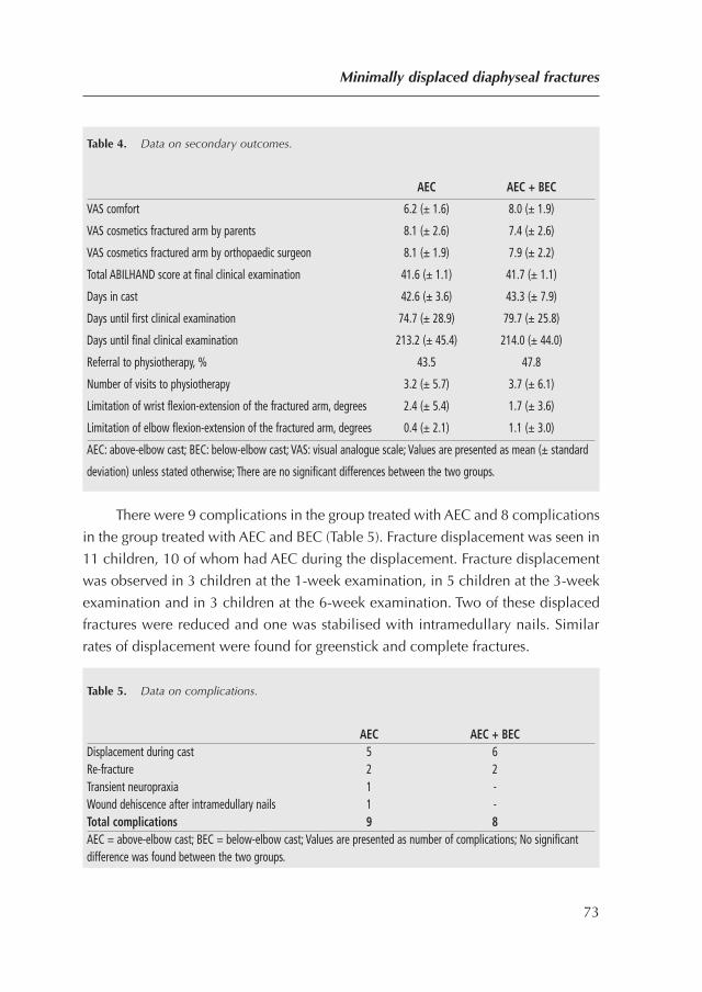
There were 9 complications in the group treated with AEC and 8 complications
in the group treated with AEC and BEC (Table 5). Fracture displacement was seen in
11 children, 10 of whom had AEC during the displacement. Fracture displacement
was observed in 3 children at the 1-week examination, in 5 children at the 3-week
examination and in 3 children at the 6-week examination. Two of these displaced
fractures were reduced and one was stabilised with intramedullary nails. Similar
rates of displacement were found for greenstick and complete fractures.
Minimally displaced diaphyseal fractures
73
Table 4. Data on secondary outcomes.
AEC AEC + BEC
VAS comfort 6.2 (± 1.6) 8.0 (± 1.9)
VAS cosmetics fractured arm by parents 8.1 (± 2.6) 7.4 (± 2.6)
VAS cosmetics fractured arm by orthopaedic surgeon 8.1 (± 1.9) 7.9 (± 2.2)
Total ABILHAND score at final clinical examination 41.6 (± 1.1) 41.7 (± 1.1)
Days in cast 42.6 (± 3.6) 43.3 (± 7.9)
Days until first clinical examination 74.7 (± 28.9) 79.7 (± 25.8)
Days until final clinical examination 213.2 (± 45.4) 214.0 (± 44.0)
Referral to physiotherapy, % 43.5 47.8
Number of visits to physiotherapy 3.2 (± 5.7) 3.7 (± 6.1)
Limitation of wrist flexion-extension of the fractured arm, degrees 2.4 (± 5.4) 1.7 (± 3.6)
Limitation of elbow flexion-extension of the fractured arm, degrees 0.4 (± 2.1) 1.1 (± 3.0)
AEC: above-elbow cast; BEC: below-elbow cast; VAS: visual analogue scale; Values are presented as mean (± standard
deviation) unless stated otherwise; There are no significant differences between the two groups.
Table 5. Data on complications.
AEC AEC + BECDisplacement during cast 5 6Re-fracture 2 2Transient neuropraxia 1 -Wound dehiscence after intramedullary nails 1 -Total complications 9 8AEC = above-elbow cast; BEC = below-elbow cast; Values are presented as number of complications; No significantdifference was found between the two groups.
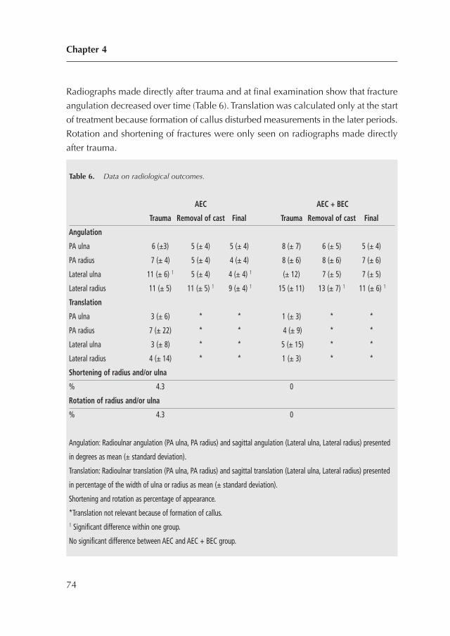
Radiographs made directly after trauma and at final examination show that fracture
angulation decreased over time (Table 6). Translation was calculated only at the start
of treatment because formation of callus disturbed measurements in the later periods.
Rotation and shortening of fractures were only seen on radiographs made directly
after trauma.
Chapter 4
74
Table 6. Data on radiological outcomes.
AEC AEC + BEC
Trauma Removal of cast Final Trauma Removal of cast Final
Angulation
PA ulna 6 (±3) 5 (± 4) 5 (± 4) 8 (± 7) 6 (± 5) 5 (± 4)
PA radius 7 (± 4) 5 (± 4) 4 (± 4) 8 (± 6) 8 (± 6) 7 (± 6)
Lateral ulna 11 (± 6) 1 5 (± 4) 4 (± 4) 1 (± 12) 7 (± 5) 7 (± 5)
Lateral radius 11 (± 5) 11 (± 5) 1 9 (± 4) 1 15 (± 11) 13 (± 7) 1 11 (± 6) 1
Translation
PA ulna 3 (± 6) * * 1 (± 3) * *
PA radius 7 (± 22) * * 4 (± 9) * *
Lateral ulna 3 (± 8) * * 5 (± 15) * *
Lateral radius 4 (± 14) * * 1 (± 3) * *
Shortening of radius and/or ulna
% 4.3 0
Rotation of radius and/or ulna
% 4.3 0
Angulation: Radioulnar angulation (PA ulna, PA radius) and sagittal angulation (Lateral ulna, Lateral radius) presented
in degrees as mean (± standard deviation).
Translation: Radioulnar translation (PA ulna, PA radius) and sagittal translation (Lateral ulna, Lateral radius) presented
in percentage of the width of ulna or radius as mean (± standard deviation).
Shortening and rotation as percentage of appearance.
*Translation not relevant because of formation of callus.1 Significant difference within one group.
No significant difference between AEC and AEC + BEC group.
None of the included fractures was primarily reduced, although 19 fractures should
have been directly reduced according to the reduction criteria. Five of these fractures
were reduced during the treatment with cast, and one of these fractures was
stabilised with intramedullary nails. We found a malunion on final radiographs in 4
children treated with AEC with, respectively, 0, 35, 50 and 75 degrees of limitation
of pronation and supination. Seven children treated with AEC and BEC suffered from
a malunion with, respectively, 0, 10, 10, 20, 25, 40 and 55 degrees of limitation.
DISCUSSION
This randomised controlled trial evaluated the influence of early conversion from
AEC to BEC in the treatment of non-reduced diaphyseal both-bone forearm fractures
in children. No significant difference was found on several clinical and radiographic
outcomes. At 6 months follow-up, both groups showed a similar but high limitation
of pronation and supination. Thereby, early conversion did not result in more re-
displacement of the fracture.
A limitation of pronation and supination of > 30 degrees at final follow-up was
found in 8 children treated with AEC (3 complete fractures and 5 greenstick fractures)
and in 4 children treated with AEC and BEC (3 complete fractures and 1 greenstick
fractures). Malunions were seen in 3 of these 8 children treated with AEC and in 2
of these 4 children treated with AEC and BEC. Consequently, malunion does not
seem to be the only cause of limitation of pronation and supination in this group of
children. Furthermore, the higher incidence of limitation in the group treated with
only AEC suggests a possible role of immobilisation of the elbow. In contrast with
BEC, AEC makes movement of the elbow impossible which can result in limitation
of pronation and supination due to contracture of the joint capsules, periarticular
soft tissues and the interosseous membrane. The absence of limitation of flexion and
extension of elbow and wrist makes a contracture of joint capsules and periarticular
soft tissue less plausible. Therefore, limitation of pronation and supination may be
caused by a malunion and/or contracture of the interosseous membrane, as also
indicated by others14,15,17,18,34,35.
Although restoration of forearm rotation is essential in evaluating the success
of forearm fracture management in children, most studies do not adequately address
Minimally displaced diaphyseal fractures
75
true, final range of pronation and supinatio28. Compared with earlier studies, in the
present study limitation of pronation and supination was relatively high. Using the
grading system (3) we found excellent results in 26% of both groups, whereas others
reported excellent results in 44%3, 77%31, 83%36, 89%6 and 100%2,22,37. Our less
favourable results may be caused by our prospective study design with its high rate
of follow-up, detailed measurements, and our high percentage of malunions.
Previous studies reported fracture displacement in conservatively treated
forearm fractures in 7-27% of their patients2,6,22,38-41, whereas we found 23%. The
fracture displacement occurred in 91% during treatment with AEC and in 73% of
them during the first 3 weeks. Therefore, even minimally displaced diaphyseal both-
bone forearm fractures in children have a tendency to displace, and this is not
prevented by immobilisation of the elbow in an AEC.
Study Limitations
This study had several limitations. First, the number of children did not reach the
estimated ± 30 required in each group. The force of the trauma needed to fracture
the diaphysis of both the radius and ulna seemed more often to cause a fracture that
needed reduction. This corresponds with our finding that only 24% of diaphyseal
both-bone forearm fractures in the present study were non-reduced fractures. A
previously scheduled time frame made further inclusion of patients impossible.
Therefore, because this study was statistically underpowered, the results should be
interpreted with caution.
Second, not all fractures were reduced according to our a priori defined criteria.
Possible explanations for this could be errors in the measurement of displacement,
or expectation of correction by growth. Nevertheless only non-reduced fractures
were included.
Conclusion
Early conversion to BEC is safe in the treatment of children with non-reduced
diaphyseal both-bone forearm fractures and does not seem to influence forearm
rotation. Displacement of the fracture is often seen which requires vigilance,
especially during the first 3 weeks of treatment.
Chapter 4
76
REFERENCES
1. Adamczyk MJ, Riley PM. Delayed union and nonunion following closed treatment of
diaphyseal pediatric forearm fractures. J Pediatr Orthop. 2005;25(1):51-5.
2. Bochang C, Jie Y, Zhigang W, Weigl D, Bar-On E, Katz K. Immobilisation of forearm
fractures in children: extended versus flexed elbow. J Bone Joint Surg Br. 2005;87(7):994-6.
3. Daruwalla JS. A study of radioulnar movements following fractures of the forearm in
children. Clin Orthop Relat Res. 1979(139):114-20.
4. Greenbaum B, Zionts LE, Ebramzadeh E. Open fractures of the forearm in children. J
Orthop Trauma. 2001;15(2):111-8.
5. Holdsworth BJ, Sloan JP. Proximal forearm fractures in children: residual disability. Injury.
1982;14(2):174-9.
6. Kay S, Smith C, Oppenheim WL. Both-bone midshaft forearm fractures in children. J Pediatr
Orthop. 1986;6(3):306-10.
7. Luhmann SJ, Gordon JE, Schoenecker PL. Intramedullary fixation of unstable both-bone
forearm fractures in children. J Pediatr Orthop. 1998;18(4):451-6.
8. Ono M, Bechtold JE, Merkow RL, Sherman RE, Gustilo RB. Rotational stability of diaphyseal
fractures of the radius and ulna fixed with Rush pins and/or fracture bracing. Clin Orthop
Relat Res. 1989(240):236-43.
9. Schwarz N, Pienaar S, Schwarz AF, Jelen M, Styhler W, Mayr J. Refracture of the forearm in
children. J Bone Joint Surg Br. 1996;78(5):740-4.
10. Shoemaker SD, Comstock CP, Mubarak SJ, Wenger DR, Chambers HG. Intramedullary
Kirschner wire fixation of open or unstable forearm fractures in children. J Pediatr Orthop.
1999;19(3):329-37.
11. Younger AS, Tredwell SJ, Mackenzie WG. Factors affecting fracture position at cast removal
after pediatric forearm fracture. J Pediatr Orthop. 1997;17(3):332-6.
12. Hogstrom H, Nilsson BE, Willner S. Correction with growth following diaphyseal forearm
fracture. Acta Orthop Scand. 1976;47(3):299-303.
13. Nilsson BE, Obrant K. The range of motion following fracture of the shaft of the forearm in
children. Acta Orthop Scand. 1977;48(6):600-2.
14. Dumont CE, Thalmann R, Macy JC. The effect of rotational malunion of the radius and the
ulna on supination and pronation. J Bone Joint Surg Br. 2002;84(7):1070-4.
Minimally displaced diaphyseal fractures
77
15. Tynan MC, Fornalski S, McMahon PJ, Utkan A, Green SA, Lee TQ. The effects of ulnar axial
malalignment on supination and pronation. J Bone Joint Surg Am. 2000;82-A(12):1726-31.
16. van Geenen RC, Besselaar PP. Outcome after corrective osteotomy for malunited fractures
of the forearm sustained in childhood. J Bone Joint Surg Br. 2007;89(2):236-9.
17. Weinberg AM, Pietsch IT, Krefft M, Pape HC, van Griensven M, Helm MB, et al. [Pronation
and supination of the forearm. With special reference to the humero-ulnar articulation].
Unfallchirurg. 2001;104(5):404-9.
18. Yasutomi T, Nakatsuchi Y, Koike H, Uchiyama S. Mechanism of limitation of
pronation/supination of the forearm in geometric models of deformities of the forearm
bones. Clin Biomech (Bristol, Avon). 2002;17(6):456-63.
19. Boyer BA, Overton B, Schrader W, Riley P, Fleissner P. Position of immobilization for
pediatric forearm fractures. Journal of pediatric orthopedics. 2002;22(2):185-7.
20. Choi KY, Chan WS, Lam TP, Cheng JC. Percutaneous Kirschner-wire pinning for severely
displaced distal radial fractures in children. A report of 157 cases. J Bone Joint Surg Br.
1995;77(5):797-801.
21. Gibbons CL, Woods DA, Pailthorpe C, Carr AJ, Worlock P. The management of isolated
distal radius fractures in children. J Pediatr Orthop. 1994;14(2):207-10.
22. Jones K, Weiner DS. The management of forearm fractures in children: a plea for
conservatism. J Pediatr Orthop. 1999;19(6):811-5.
23. Mani GV, Hui PW, Cheng JC. Translation of the radius as a predictor of outcome in distal
radial fractures of children. J Bone Joint Surg Br. 1993;75(5):808-11.
24. Maric D, Petkovic L, Tomasevic V, Bajic L, Gajdobranski D, Marcikic A. [Osteosynthesis
with Kirschner wires in the treatment of dislocation fractures of the forearm in children].
Med Pregl. 2002;55(3-4):140-5.
25. Noonan KJ, Price CT. Forearm and distal radius fractures in children. J Am Acad Orthop
Surg. 1998;6(3):146-56.
26. Zimmermann R, Gabl M, Angermann P, Lutz M, Reinhart C, Kralinger F, et al. [Late
sequelae of fractures of the distal third of the forearm during the growth period]. Handchir
Mikrochir Plast Chir. 2000;32(4):242-9.
27. Gustilo RB, Simpson L, Nixon R, Ruiz A, Indeck W. Analysis of 511 open fractures. Clin
Orthop Relat Res. 1969;66:148-54.
28. Franklin CC, Robinson J, Noonan K, Flynn JM. Evidence-based medicine: management of
pediatric forearm fractures. Journal of pediatric orthopedics. 2012;32 Suppl 2:S131-4.
Chapter 4
78
29. Colaris J, van der Linden M, Selles R, Coene N, Allema JH, Verhaar J. Pronation and
supination after forearm fractures in children: Reliability of visual estimation and
conventional goniometry measurement. Injury. 2010;41(6):643-6.
30. Webb GR, Galpin RD, Armstrong DG. Comparison of short and long arm plaster casts for
displaced fractures in the distal third of the forearm in children. J Bone Joint Surg Am.
2006;88(1):9-17.
31. Price CT, Scott DS, Kurzner ME, Flynn JC. Malunited forearm fractures in children. J Pediatr
Orthop. 1990;10(6):705-12.
32. Arnould C, Penta M, Renders A, Thonnard JL. ABILHAND-Kids: a measure of manual ability
in children with cerebral palsy. Neurology. 2004;63(6):1045-52.
33. Naimark A, Kossoff J, Leach RE. The disparate diameter. A sign of rotational deformity in
fractures. J Can Assoc Radiol. 1983;34(1):8-11.
34. Jupiter JB, Fernandez DL, Levin LS, Wysocki RW. Reconstruction of posttraumatic disorders
of the forearm. J Bone Joint Surg Am. 2009;91(11):2730-9.
35. van Geenen RC, Besselaar PP. Outcome after corrective osteotomy for malunited fractures
of the forearm sustained in childhood. The Journal of bone and joint surgery British volume.
2007;89(2):236-9.
36. Shah AS, Lesniak BP, Wolter TD, Caird MS, Farley FA, Vander Have KL. Stabilization of
adolescent both-bone forearm fractures: a comparison of intramedullary nailing versus
open reduction and internal fixation. J Orthop Trauma.24(7):440-7.
37. Boero S, Michelis MB, Calevo MG, Stella M. Multiple forearm diaphyseal fracture:
reduction and plaster cast control at the end of growth. Int Orthop. 2007;31(6):807-10.
38. Chan CF, Meads BM, Nicol RO. Remanipulation of forearm fractures in children. N Z Med
J. 1997;110(1047):249-50.
39. Monga P, Raghupathy A, Courtman NH. Factors affecting remanipulation in paediatric
forearm fractures. J Pediatr Orthop B.19(2):181-7.
40. Voto SJ, Weiner DS, Leighley B. Redisplacement after closed reduction of forearm fractures
in children. J Pediatr Orthop. 1990;10(1):79-84.
41. Schmittenbecher PP. State-of-the-art treatment of forearm shaft fractures. Injury. 2005;36
Suppl 1:A25-34.
Minimally displaced diaphyseal fractures
79

80
CHAPTER 5
Conversion to below-elbow castafter 3 weeks is safe fordiaphyseal both-bone forearmfractures in children: a multicentre randomizedcontrolled trial involving 127children
Acta Orthop. 2013 Oct;84(5):489-94
Colaris JW, Allema JH, Biter LU, Reijman M, van de Ven CP, de Vries MR, Bloem
RM, Kerver AJ, Verhaar JA.
81
ABSTRACT
Background: It is unclear whether it is safe to convert above- elbow cast (AEC) to
below-elbow cast (BEC) in a child who has sustained a displaced diaphyseal both-
bone forearm fracture that is stable after reduction. In this multicentre study, we
wanted to answer the question: does early conversion to BEC cause similar forearm
rotation to that after treatment with AEC alone?
Children and methods: Children were randomly allocated to 6 weeks of AEC, or 3
weeks of AEC followed by 3 weeks of BEC. The primary outcome was limitation of
pronation/supination after 6 months. The secondary outcomes were re-displacement
of the fracture, limitation of flexion/extension of the wrist and elbow, complication
rate, cast comfort, complaints in daily life, and cosmetics of the fractured arm.
Results: 62 children were treated with 6 weeks of AEC, and 65 children were treated
with 3 weeks of AEC plus 3 weeks of BEC. The follow-up rate was 60/62 and 64/65,
respectively with a mean time of 6.9 (4.7–13) months. The limitation of pronation/
supination was similar in both groups (18 degrees for the AEC group and 11 degrees
for the AEC/BEC group). The secondary outcomes were similar in both groups, with
the exception of cast comfort, which was in favour of the AEC/BEC group.
Interpretation: Early conversion to BEC is safe and results in greater cast comfort.
Chapter 5
82
INTRODUCTION
Most displaced diaphyseal both-bone forearm fractures in children can be treated
successfully with closed reduction and above-elbow cast (AEC) for 6–9 weeks, but
AEC is often converted to below-elbow cast (BEC) in the last weeks of treatment.
Although most of these fractures heal uneventfully, limitation of pronation and
supination can give disappointing results. Previous studies have found an average
limitation of 20 degrees in about 15% of children. The cause of this limitation is
unknown, but malunion and contracture of soft tissues have been suggested in some
studies1-9. Early conversion to BEC in the treatment of these fractures could
potentially affect limitation of pronation and supination in 2 opposing ways. On the
one hand, early conversion to BEC could result in fracture displacement, leading to
malunion and resulting in limitation of pronation and supination. On the other hand,
AEC may lead to a limitation of pronation and supination because of contracture of
soft tissue by immobilisation of the elbow. We therefore set up a randomized
controlled multicentre trial to answer the following question: does early conversion
from AEC to BEC cause forearm rotation that is similar to that from AEC alone in
reduced stable diaphyseal both-bone forearm fractures in children?
CHILDREN AND METHODS
Trial design and participants
We performed a multicentre randomized trial on consecutive children with a
displaced diaphyseal both-bone forearm fracture that was stable after reduction, who
visited the emergency department of one of 4 participating Dutch hospitals: Erasmus
Medical Center (Rotterdam), HAGA Hospital (The Hague), Reinier de Graaf Hospital
(Delft), and Sint Franciscus Hospital (Rotterdam). The regional medical ethics
committee approved the study and it was registered at Clinical Trials.gov with registry
identifier NCT00398242. The trial was performed in compliance with the Helsinki
Declaration. Informed consent for participation was obtained from all parents and
from all children aged ≥�12 years.
We included all children aged < 16 years who sustained a displaced diaphyseal
both-bone forearm fracture that was stable after reduction (Figure 1). A diaphyseal
Displaced stable diaphyseal fractures
83
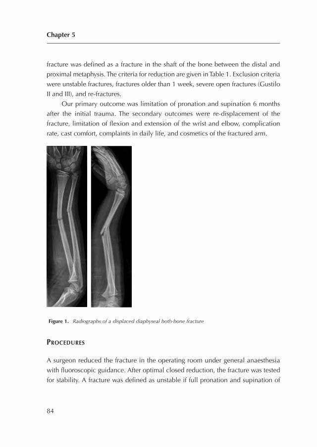
fracture was defined as a fracture in the shaft of the bone between the distal and
proximal metaphysis. The criteria for reduction are given in Table 1. Exclusion criteria
were unstable fractures, fractures older than 1 week, severe open fractures (Gustilo
II and III), and re-fractures.
Our primary outcome was limitation of pronation and supination 6 months
after the initial trauma. The secondary outcomes were re-displacement of the
fracture, limitation of flexion and extension of the wrist and elbow, complication
rate, cast comfort, complaints in daily life, and cosmetics of the fractured arm.
Figure 1. Radiographs of a displaced diaphyseal both-bone fracture
PROCEDURES
A surgeon reduced the fracture in the operating room under general anaesthesia
with fluoroscopic guidance. After optimal closed reduction, the fracture was tested
for stability. A fracture was defined as unstable if full pronation and supination of
Chapter 5
84
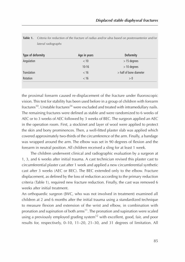
the proximal forearm caused re-displacement of the fracture under fluoroscopic
vision. This test for stability has been used before in a group of children with forearm
fractures10. Unstable fractures24 were excluded and treated with intramedullary nails.
The remaining fractures were defined as stable and were randomized to 6 weeks of
AEC or to 3 weeks of AEC followed by 3 weeks of BEC. The surgeon applied an AEC
in the operation room. First, a stockinet and layer of wool were applied to protect
the skin and bony prominences. Then, a well-fitted plaster slab was applied which
covered approximately two-thirds of the circumference of the arm. Finally, a bandage
was wrapped around the arm. The elbow was set in 90 degrees of flexion and the
forearm in neutral position. All children received a sling for at least 1 week.
The children underwent clinical and radiographic evaluation by a surgeon at
1, 3, and 6 weeks after initial trauma. A cast technician revised this plaster cast to
circumferential plaster cast after 1 week and applied a new circumferential synthetic
cast after 3 weeks (AEC or BEC). The BEC extended only to the elbow. Fracture
displacement, as defined by the loss of reduction according to the primary reduction
criteria (Table 1), required new fracture reduction. Finally, the cast was removed 6
weeks after initial treatment.
An orthopaedic surgeon (JWC, who was not involved in treatment) examined all
children at 2 and 6 months after the initial trauma using a standardized technique
to measure flexion and extension of the wrist and elbow, in combination with
pronation and supination of both arms11. The pronation and supination were scaled
using a previously employed grading system12 with excellent, good, fair, and poor
results for, respectively, 0–10, 11–20, 21–30, and 31 degrees of limitation. All
Displaced stable diaphyseal fractures
85
Table 1. Criteria for reduction of the fracture of radius and/or ulna based on posteroanterior and/or
lateral radiographs
Type of deformity Age in years Deformity
Angulation < 10 > 15 degrees
10-16 > 10 degrees
Translation < 16 > half of bone diameter
Rotation < 16 > 0
children with at least 30 degrees of functional impairment at the 2-month
examination were referred to a physiotherapist. At the 2-month examination, the
parents and children evaluated the comfort of the cast using a visual analogue scale
(VAS) with highest score for maximal comfort, and a questionnaire to evaluate
difficulties in performing 7 daily activities during the period of cast. This
questionnaire had previously been used for a similar group of children13.
6 months after the initial trauma, the parents and JWC completed a VAS
regarding cosmetics of the fractured arm, with highest score for similar cosmetics in
the injured and uninjured arm. In the same consultation, complaints concerning the
fractured arm were documented using a modified grading system. This grading system
combines limitation of pronation and supination with complaints covering daily life
or during strenuous activities. It had been used previously for a similar group of
children14. Thereafter, the parents filled in an ABILHAND-Kids questionnaire, which
is a measure of manual ability in children with upper limb impairments.
Radiographs were taken in the emergency room. These were followed by
radiographs after reduction and during follow- up at 1, 3, and 6 weeks. The final
radiographs were taken 6 months after the initial trauma. Angulation, translation,
rotation, and shortening of fractures were measured on the radiographs to determine
primary displacement, displacement during treatment with cast, and final
displacement at 6 months. Rotation of the fractured bones was analysed from
differences in the diameter of the diaphysis of the ulna and radius15. To assess the
interrater reproducibility, the angulation of the fracture was re-measured in 45
children by a trauma surgeon. The interrater reproducibility of the radiological
assessment showed an intraclass correlation (ICC) of 0.81 (95% CI: 0.68–0.89) and
0.89 (95% CI: 0.81–0.94) for the radioulnar angulation of the ulna and radius,
respectively. The ICC of sagittal angulation was 0.92 (95% CI: 0.85–0.95) for the
ulna and 0.87 (95% CI: 0.77–0.92) for the radius.
Randomization and masking
A physician who was not involved in treatment randomized the children using sealed
envelopes with varied block sizes. The randomization took place after the fracture
was reduced and defined as stable. The study was not blinded regarding the children,
parents, and clinicians. JWC collected all the data.
Chapter 5
86
Statistics
This study was an equivalency study in which we wanted to determine whether the
index treatment (a combination of AEC and BEC) was no better or worse than the
control treatment (AEC alone). The main aim of the study was to address the question:
do 6 weeks of AEC, or 3 weeks of AEC and 3 weeks of BEC, cause equal limitation
of pronation and supination 6 months after the initial trauma? We assessed the
numbers of children required with assumption of similarity of pronation and
supination in both groups. Similarity between the 2 groups was defined as a
maximum of 15 degrees less pronation or supination in the group treated with AEC
and BEC. We chose 15 degrees of limitation to stay within the safe margins of good
results as graded previously12. With an a priori calculation, it was determined that
with a power of 80%, a significance level of 0.05, and a standard deviation (SD) of
15 degrees, the groups should consist of 30 children each.
First, we assessed whether the variables had a normal distribution using the
Shapiro-Wilk test for normality. Based on these analyses, the results are presented
as mean (SD). The primary research question was examined using linear regression
analysis. If necessary, adjustments for unbalanced covariates were made. To check
for linearity, we plotted the standardized residuals of the variables used against the
standardized predicted values. If there was doubt with respect to violation of
assumptions regarding distribution of residuals, we carried out a Mann-Whitney test
comparing the 2 groups regarding the average of the measurements. Differences
between both groups (AEC vs. AEC plus BEC) for the secondary outcome measures
were analysed by one-way ANOVA to correct for multiple comparisons (Bonferroni).
Statistical analysis was performed with SPSS software version 17.0.
Displaced stable diaphyseal fractures
87
RESULTS
Between January 2006 and August 2010, 127 children were included in the study.
62 children were randomized to 6 weeks of AEC and 65 children were randomized
to 3 weeks of AEC followed by 3 weeks of BEC (Figure 2). Despite the randomization,
there was a substantial difference in age at the time of fracture, dominant arm
fractured, and fracture type of the radius (Table 2). For this reason, the statistical
analyses were corrected for these baseline variables.
Fractures were reduced in the operating room in 68% of the children in the
AEC group and in 70% of the children in the other group (AEC and BEC). The
remaining fractures were reduced at the emergency department. The follow-up rate
was 98%, with a mean follow-up time of 6.9 (4.7–12.9) months.
At 6-month follow-up, the mean limitation of pronation and supination was
18 degrees (95% CI: 0–50) for children treated with AEC, and 11 degrees (CI: 0–34)
for children treated with AEC and BEC (p = 0.05). With and without adjustment for
unbalanced covariates, no significant differences in primary outcome were found
(Table 3).
Using the grading system for limitation of pronation and supination12, the
results in the AEC-alone group were excellent in 21%, good in 38%, fair in 26%,
and poor in 15%. In the group treated with AEC and BEC, the results were excellent
in 33%, good in 37%, fair in 27%, and poor in 3%. Similar results were found using
the modified grading system14. In the AEC group, the results were excellent in 33%,
good in 42%, and fair in 25%. In the group treated with AEC and BEC, the results
were excellent in 48%, good in 30%, fair in 20%, and poor in 2%.
The secondary outcome measures showed a higher VAS for comfort in the
group of children treated with AEC and BEC (p < 0.001) (Table 4).
Chapter 5
88
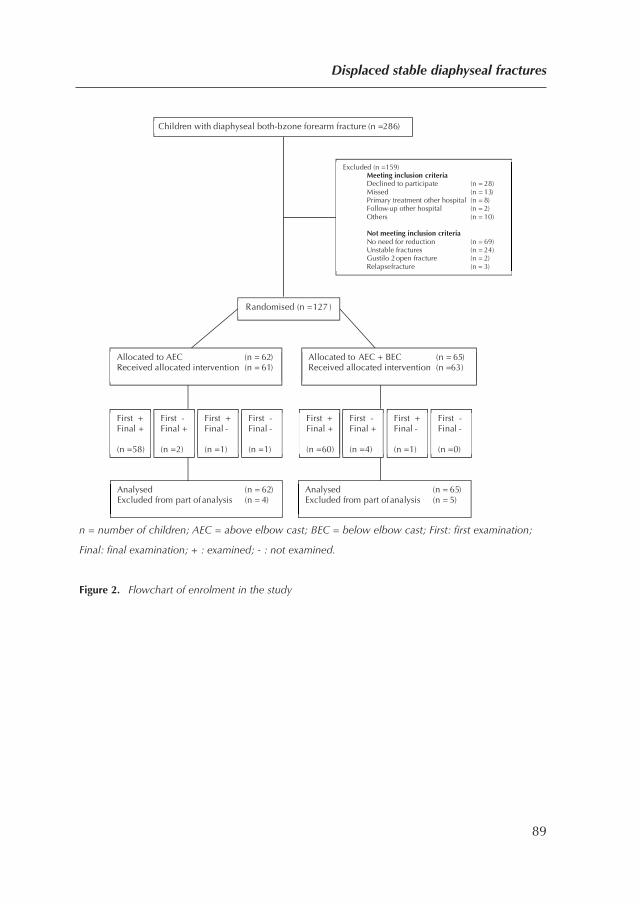
n = number of children; AEC = above elbow cast; BEC = below elbow cast; First: first examination;
Final: final examination; + : examined; - : not examined.
Figure 2. Flowchart of enrolment in the study
Displaced stable diaphyseal fractures
89
Children with diaphyseal both-bzone forearm fracture (n =286)
Randomised (n = 127 )
Excluded (n = 159) Meeting inclusion criteria Declined to participate (n = 28)Missed (n = 13)Primary treatment other hospital (n = 8) Follow-up other hospital (n = 2) Others (n = 10)
Not meeting inclusion criteria No need for reduction (n = 69)Unstable fractures (n = 24) Gustilo 2 open fracture (n = 2) Relapse fracture (n = 3)
Allocated to AEC (n = 62) Received allocated intervention (n = 61)
Allocated to AEC + BEC (n = 65) Received allocated intervention (n =63)
Analysed (n = 62) Excluded from part of analysis (n = 4)
Analysed (n = 65) Excluded from part of analysis (n = 5)
First + Final + (n =58)
First - Final + (n =2)
First + Final - (n =1)
First - Final - (n =1)
First + Final + (n =60)
First - Final + (n =4)
First + Final - (n =1)
First - Final - (n =0)
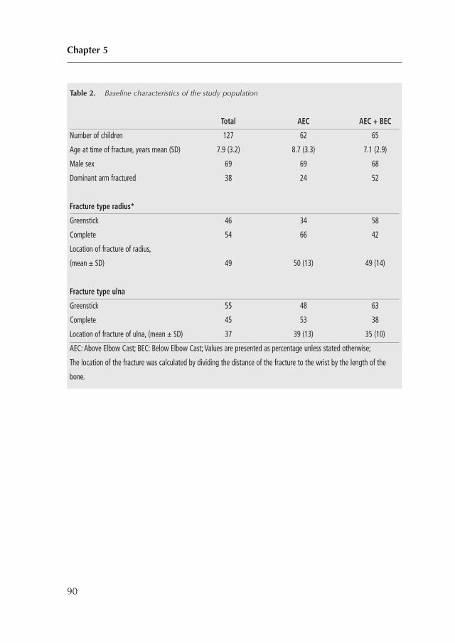
Chapter 5
90
Table 2. Baseline characteristics of the study population
Total AEC AEC + BEC
Number of children 127 62 65
Age at time of fracture, years mean (SD) 7.9 (3.2) 8.7 (3.3) 7.1 (2.9)
Male sex 69 69 68
Dominant arm fractured 38 24 52
Fracture type radius*
Greenstick 46 34 58
Complete 54 66 42
Location of fracture of radius,
(mean ± SD) 49 50 (13) 49 (14)
Fracture type ulna
Greenstick 55 48 63
Complete 45 53 38
Location of fracture of ulna, (mean ± SD) 37 39 (13) 35 (10)
AEC: Above Elbow Cast; BEC: Below Elbow Cast; Values are presented as percentage unless stated otherwise;
The location of the fracture was calculated by dividing the distance of the fracture to the wrist by the length of the
bone.
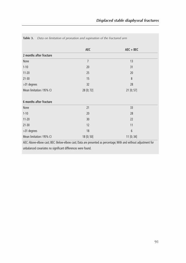
Displaced stable diaphyseal fractures
91
Table 3. Data on limitation of pronation and supination of the fractured arm
AEC AEC + BEC
2 months after fracture
None 7 13
1-10 20 31
11-20 25 20
21-30 15 8
>31 degrees 32 28
Mean limitation / 95% CI 28 [0; 72] 21 [0; 57]
6 months after fracture
None 21 33
1-10 20 28
11-20 30 22
21-30 12 11
>31 degrees 18 6
Mean limitation / 95% CI 18 [0; 50] 11 [0; 34]
AEC: Above-elbow cast; BEC: Below-elbow cast; Data are presented as percentage; With and without adjustment for
unbalanced covariates no significant differences were found.
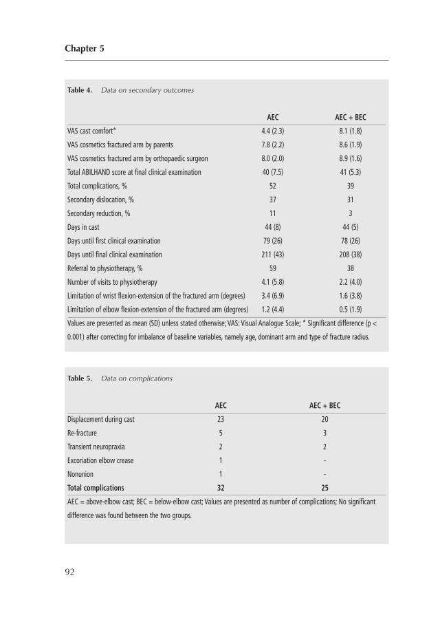
Chapter 5
92
Table 4. Data on secondary outcomes
AEC AEC + BEC
VAS cast comfort* 4.4 (2.3) 8.1 (1.8)
VAS cosmetics fractured arm by parents 7.8 (2.2) 8.6 (1.9)
VAS cosmetics fractured arm by orthopaedic surgeon 8.0 (2.0) 8.9 (1.6)
Total ABILHAND score at final clinical examination 40 (7.5) 41 (5.3)
Total complications, % 52 39
Secondary dislocation, % 37 31
Secondary reduction, % 11 3
Days in cast 44 (8) 44 (5)
Days until first clinical examination 79 (26) 78 (26)
Days until final clinical examination 211 (43) 208 (38)
Referral to physiotherapy, % 59 38
Number of visits to physiotherapy 4.1 (5.8) 2.2 (4.0)
Limitation of wrist flexion-extension of the fractured arm (degrees) 3.4 (6.9) 1.6 (3.8)
Limitation of elbow flexion-extension of the fractured arm (degrees) 1.2 (4.4) 0.5 (1.9)
Values are presented as mean (SD) unless stated otherwise; VAS: Visual Analogue Scale; * Significant difference (p <
0.001) after correcting for imbalance of baseline variables, namely age, dominant arm and type of fracture radius.
Table 5. Data on complications
AEC AEC + BEC
Displacement during cast 23 20
Re-fracture 5 3
Transient neuropraxia 2 2
Excoriation elbow crease 1 -
Nonunion 1 -
Total complications 32 25
AEC = above-elbow cast; BEC = below-elbow cast; Values are presented as number of complications; No significant
difference was found between the two groups.
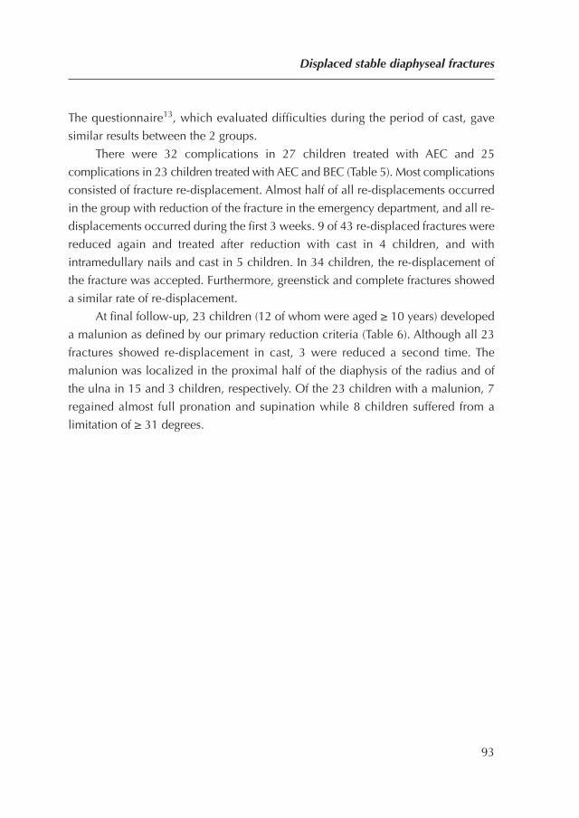
The questionnaire13, which evaluated difficulties during the period of cast, gave
similar results between the 2 groups.
There were 32 complications in 27 children treated with AEC and 25
complications in 23 children treated with AEC and BEC (Table 5). Most complications
consisted of fracture re-displacement. Almost half of all re-displacements occurred
in the group with reduction of the fracture in the emergency department, and all re-
displacements occurred during the first 3 weeks. 9 of 43 re-displaced fractures were
reduced again and treated after reduction with cast in 4 children, and with
intramedullary nails and cast in 5 children. In 34 children, the re-displacement of
the fracture was accepted. Furthermore, greenstick and complete fractures showed
a similar rate of re-displacement.
At final follow-up, 23 children (12 of whom were aged ≥ 10 years) developed
a malunion as defined by our primary reduction criteria (Table 6). Although all 23
fractures showed re-displacement in cast, 3 were reduced a second time. The
malunion was localized in the proximal half of the diaphysis of the radius and of
the ulna in 15 and 3 children, respectively. Of the 23 children with a malunion, 7
regained almost full pronation and supination while 8 children suffered from a
limitation of ≥ 31 degrees.
Displaced stable diaphyseal fractures
93
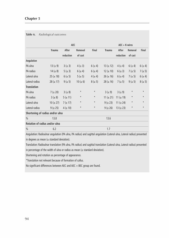
Chapter 5
94
Table 6. Radiological outcomes
AEC AEC + K-wires
Trauma After Removal Final Trauma After Removal Final
reduction of cast reduction of cast
Angulation
PA ulna 13 (± 9) 3 (± 3) 4 (± 3) 6 (± 4) 13 (± 12) 4 (± 4) 6 (± 4) 6 (± 4)
PA radius 14 (± 8) 5 (± 3) 6 (± 4) 6 (± 4) 12 (± 10) 6 (± 3) 7 (± 5) 7 (± 5)
Lateral ulna 25 (± 18) 6 (± 5) 5 (± 5) 4 (± 4) 26 (± 16) 6 (± 4) 7 (± 5) 6 (± 4)
Lateral radius 28 (± 17) 9 (± 5) 10 (± 6) 8 (± 5) 28 (± 16) 7 (± 5) 9 (± 5) 8 (± 5)
Translation
PA ulna 7 (± 20) 3 (± 8) * * 3 (± 9) 3 (± 9) * *
PA radius 3 (± 8) 5 (± 11) * * 11 (± 21) 11 (± 19) * *
Lateral ulna 10 (± 27) 7 (± 17) * * 9 (± 23) 11 (± 24) * *
Lateral radius 9 (± 25) 4 (± 10) * * 9 (± 26) 13 (± 23) * *
Shortening of radius and/or ulna
% 13.8 13.6
Rotation of radius and/or ulna
% 6.2 1.7
Angulation: Radioulnar angulation (PA ulna, PA radius) and sagittal angulation (Lateral ulna, Lateral radius) presented
in degrees as mean (± standard deviation).
Translation: Radioulnar translation (PA ulna, PA radius) and sagittal translation (Lateral ulna, Lateral radius) presented
in percentage of the width of ulna or radius as mean (± standard deviation).
Shortening and rotation as percentage of appearance.
*Translation not relevant because of formation of callus.
No significant differences between AEC and AEC + BEC group are found.
DISCUSSION
We found that in the treatment of reduced stable diaphyseal both-bone forearm
fractures in children, conversion from AEC to BEC was safe 3 weeks after the initial
trauma. Limitation in flexion and extension of the elbow did not occur in either of
the groups.
We found a limitation of pronation and supination of �30 degrees at final
follow-up in 15 of 127 children, 8 of whom also suffered a radiographic malunion.
6 children with a radio- graphic malunion at final follow-up showed no limitation
of pronation and supination. These findings demonstrate that limitation of pronation
and supination is caused by malunion and contractures, as previously stated by other
authors1,2,4,5,16,17. The idea of contractures of injured soft tissue being a cause of
limitation of pronation and supination was supported by a trend of better forearm
rotation in the BEC group, in which children could rotate the forearm early to prevent
contractures.
Compared with previous studies, limitation of pronation and supination was
relatively high in our children. Using the grading system12, we found that 21% (AEC)
and 33% (AEC and BEC) had excellent results, whereas other authors have found
excellent results in 44–100% of such children9,12,14,18-20. The modified grading
system14 resulted in 33% and 48% excellent results in our 2 groups, as compared
to 82% in another study14. Our prospective analysis, inclusion of only the more
severe both-bone forearm fractures, and the high percentage of malunions may have
been responsible for our less favourable results.
Previous studies have found a re-displacement rate of 7–27% in non-
operatively treated forearm fractures, whereas we found a rate of4%9,18,20-24. Our
higher rate of re-displacement might be explained by our strict malunion criteria,
the prospective follow-up with scheduled radiographs, our test for stability, and the
type of cast (non-circumferential cast applied directly after reduction). Although BEC
allows free movement of the elbow, in our study conversion to BEC after 3 weeks
did not cause re-displacement of fractures. In our patients, all re-displacements
occurred in the AEC group during the first 3 weeks of treatment.
In the statistical analysis, we were aware that strict correction for multiple
testing was not necessary because of the evaluation of predefined hypotheses.
Displaced stable diaphyseal fractures
95
However, we chose a conservative method to test the secondary outcome
parameters. In addition, we also tested the secondary outcome without correction
for multiple testing (using a p-value of 0.05), and even then no differences were
found between groups.
Our study had several limitations. First, several fractures were reduced at the
emergency department instead of in the operating room. The slightly higher rate of
re-displacement in fractures reduced at the emergency department (42% vs. 30%)
led us to suspect that testing of fracture stability without anaesthesia and fluoroscopy
is less optimal. Fortunately, there was an equal distribution of fractures reduced in
the emergency room in the 2 groups, and this study investigated treatment modalities
after reduction. Secondly, not all re-displaced fractures were reduced following our
study protocol. This was probably due to the expectation of correction of the
malunion by growth, and the reluctance of the surgeon to propose a second
reduction in view of the effect on the child. Thirdly, the outcome of the test for
stability after reduction was a subjective opinion of the treating physician. No tests
for stability of fractures are objective, and the test used was the only one to appear
in literature (25). The final limitation was our (short) period of follow-up in this young
population with a capacity for correction by growth. Nevertheless, these diaphyseal
fractures (especially in older children) show less correction than distal metaphyseal
fractures in younger children.
In summary, our results suggest that reduced stable diaphyseal both-bone
forearm fractures in children can be safely treated by 3 weeks of above-elbow cast
and 3 weeks of below-elbow cast. Re-displacement of the fracture might be
identified early by radiography at set times.
Chapter 5
96
REFERENCES
1. Dumont CE, Thalmann R, Macy JC. The effect of rotational malunion of the radius and the
ulna on supination and pronation. J Bone Joint Surg Br. 2002;84(7):1070-4.
2. Tynan MC, Fornalski S, McMahon PJ, Utkan A, Green SA, Lee TQ. The effects of ulnar axial
malalignment on supination and pronation. J Bone Joint Surg Am. 2000;82-A(12):1726-31.
3. van Geenen RC, Besselaar PP. Outcome after corrective osteotomy for malunited fractures
of the forearm sustained in childhood. J Bone Joint Surg Br. 2007;89(2):236-9.
4. Weinberg AM, Pietsch IT, Krefft M, Pape HC, van Griensven M, Helm MB, et al. [Pronation
and supination of the forearm. With special reference to the humero-ulnar articulation].
Unfallchirurg. 2001;104(5):404-9.
5. Yasutomi T, Nakatsuchi Y, Koike H, Uchiyama S. Mechanism of limitation of
pronation/supination of the forearm in geometric models of deformities of the forearm
bones. Clin Biomech (Bristol, Avon). 2002;17(6):456-63.
6. Nilsson BE, Obrant K. The range of motion following fracture of the shaft of the forearm in
children. Acta Orthop Scand. 1977;48(6):600-2.
7. Bhaskar AR, Roberts JA. Treatment of unstable fractures of the forearm in children. Is plating
of a single bone adequate? J Bone Joint Surg Br. 2001;83(2):253-8.
8. Hogstrom H, Nilsson BE, Willner S. Correction with growth following diaphyseal forearm
fracture. Acta Orthop Scand. 1976;47(3):299-303.
9. Kay S, Smith C, Oppenheim WL. Both-bone midshaft forearm fractures in children. J Pediatr
Orthop. 1986;6(3):306-10.
10. Myers GJ, Gibbons PJ, Glithero PR. Nancy nailing of diaphyseal forearm fractures. Single
bone fixation for fractures of both bones. J Bone Joint Surg Br. 2004;86(4):581-4.
11. Colaris J, van der Linden M, Selles R, Coene N, Allema JH, Verhaar J. Pronation and
supination after forearm fractures in children: Reliability of visual estimation and
conventional goniometry measurement. Injury. 2010;41(6):643-6.
12. Daruwalla JS. A study of radioulnar movements following fractures of the forearm in
children. Clin Orthop Relat Res. 1979(139):114-20.
13. Webb GR, Galpin RD, Armstrong DG. Comparison of short and long arm plaster casts for
displaced fractures in the distal third of the forearm in children. J Bone Joint Surg Am.
2006;88(1):9-17.
Displaced stable diaphyseal fractures
97
14. Price CT, Scott DS, Kurzner ME, Flynn JC. Malunited forearm fractures in children. J Pediatr
Orthop. 1990;10(6):705-12.
15. Naimark A, Kossoff J, Leach RE. The disparate diameter. A sign of rotational deformity in
fractures. J Can Assoc Radiol. 1983;34(1):8-11.
16. Jupiter JB, Fernandez DL, Levin LS, Wysocki RW. Reconstruction of posttraumatic disorders
of the forearm. J Bone Joint Surg Am. 2009;91(11):2730-9.
17. van Geenen RC, Besselaar PP. Outcome after corrective osteotomy for malunited fractures
of the forearm sustained in childhood. The Journal of bone and joint surgery British volume.
2007;89(2):236-9.
18. Bochang C, Jie Y, Zhigang W, Weigl D, Bar-On E, Katz K. Immobilisation of forearm
fractures in children: extended versus flexed elbow. J Bone Joint Surg Br. 2005;87(7):994-6.
19. Boero S, Michelis MB, Calevo MG, Stella M. Multiple forearm diaphyseal fracture:
reduction and plaster cast control at the end of growth. Int Orthop. 2007;31(6):807-10.
20. Jones K, Weiner DS. The management of forearm fractures in children: a plea for
conservatism. J Pediatr Orthop. 1999;19(6):811-5.
21. Chan CF, Meads BM, Nicol RO. Remanipulation of forearm fractures in children. N Z Med
J. 1997;110(1047):249-50.
22. Monga P, Raghupathy A, Courtman NH. Factors affecting remanipulation in paediatric
forearm fractures. J Pediatr Orthop B.19(2):181-7.
23. Voto SJ, Weiner DS, Leighley B. Redisplacement after closed reduction of forearm fractures
in children. J Pediatr Orthop. 1990;10(1):79-84.
24. Schmittenbecher PP. State-of-the-art treatment of forearm shaft fractures. Injury. 2005;36
Suppl 1:A25-34.
25. Meier R, Prommersberger KJ, van Griensven M, Lanz U. Surgical correction of deformities
of the distal radius due to fractures in pediatric patients. Arch Orthop Trauma Surg.
2004;124(1):1-9.
Chapter 5
98

Displaced stable diaphyseal fractures
99

100
CHAPTER 6
Single-bone intramedullaryfixation of unstable both-bonediaphyseal forearm fractures inchildren leads to increased re-displacement: a multicentre randomisedcontrolled trial
Arch Orthop Trauma Surg. 2013 Aug;133(8):1079-87
Colaris J, Reijman M, Allema JH, Kraan G, van Winterswijk P,
de Vries M, van de Ven C, Verhaar J.
101
ABSTRACT
Introduction: Both-bone diaphyseal forearm fractures in children can be stabilised
without cast by a flexible intramedullary nail in both the radius and the ulna.
Adequate results with single-bone fixation combined with a complementary cast are
also reported. However, because those results are based on a selection of children,
this trial investigates whether single-bone intramedullary fixation, compared with
both-bone intramedullary fixation, results in similar pronation and supination in
children with an unstable diaphyseal both-bone forearm fracture.
Materials and methods: In four Dutch hospitals, 24 consecutive children aged < 16
years with a displaced unstable both-bone diaphyseal forearm fracture were
randomly allocated to single-bone or both-bone intramedullary fixation. Primary
outcome was limitation of pronation and supination 9 months after initial trauma.
Secondary outcomes were limitation of flexion/extension of wrist/elbow,
complication rate, operation time, cosmetics of the fractured arm, complaints in
daily life, and assessment of radiographs.
Results: Between January 2006 and August 2010, 11 children were randomised to
single-bone fixation and 13 to both-bone fixation. In the both-bone fixation group,
two fractures were stabilized by only one nail. In both groups, median limitation of
pronation/supination at 9-months follow-up was 5-10 degrees. In both groups
operation time was similar but in the single-bone fixation group cast immobilisation
was longer (median of 37 vs. 28 days). In four children, re-displacement of the
fracture occurred in those fractures without an intramedullary nail.
Conclusions: These results caution against the use of single-bone fixation in all both-
bone forearm fractures. This method may lead to increased re-displacement and
reduced clinical results.
Chapter 6
102
INTRODUCTION
Diaphyseal both-bone forearm fractures are frequently seen in children. During
impact of trauma, the high energy level often results in a displaced fracture. The
displacement generally needs to be corrected and stabilised by a cast, intramedullary
nails and/or plates.
Fixation by intramedullary nails has gained popularity because of good clinical
and radiographic results1-6. Flexible intramedullary nails are generally used to
stabilise paediatric fractures with two nails7-9. Initially, one pre-bent nail is inserted
in the relatively straight intramedullary canal and creates a bending moment. Then,
a second pre-bent nail is inserted with the concavity of the nail opposite the first
one creating a counteracting force which stabilises the fracture. Furthermore, both-
bone forearm fractures fixated with two intramedullary nails do not need stabilisation
by a complementary cast.
In contrast to single-bone fractures, with both-bone forearm fractures the
narrow intramedullary canal of the radius and ulna makes insertion of two nails in
one bone impossible. For that reason the radius and ulna are fixated separately with
a single flexible intramedullary nail, with the concavity of the nails in the same
direction. This results in an absence of the stabilising effect of the counteracting force
of two nails with opposite concavity.
As an alternative, the possibility of single-bone fixation in both-bone forearm
fractures combined with a complementary cast has been studied2,5,10-17. Single-bone
intramedullary fixation in both-bone forearm fractures offers both advantages and
disadvantages. Although insertion and removal of only one nail can reduce operation
time and complications, the bone without fixation has a tendency to displace
necessitating immobilisation in a cast.
To date, only one study (20 children, no controls) has prospectively investigated
single-bone fixation in both-bone forearm fractures12, whereas others used a retrospective
design2,5,10-13,16-19. Most studies concluded that single-bone intramedullary fixation of
both-bone diaphyseal forearm fractures in children is a safe and efficacious option with
good functional and radiological results2,5,10-13,16-19. Nevertheless, these results were
based on a selection of children who sustained a both-bone forearm fracture that
remained aligned and stable after single-bone fixation2,5,10-13,16-19.
Displaced unstable diaphyseal fractures
103
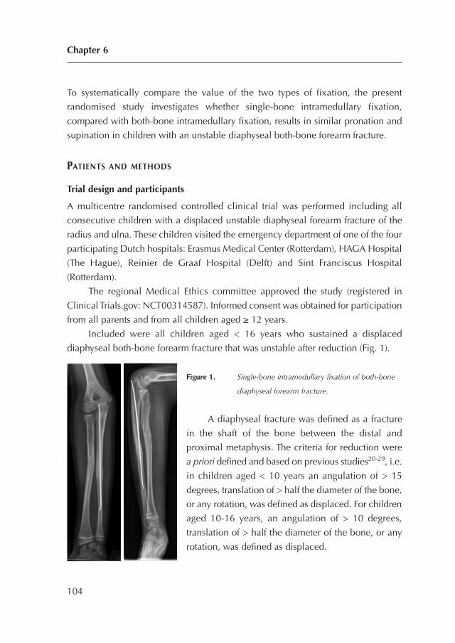
To systematically compare the value of the two types of fixation, the present
randomised study investigates whether single-bone intramedullary fixation,
compared with both-bone intramedullary fixation, results in similar pronation and
supination in children with an unstable diaphyseal both-bone forearm fracture.
PATIENTS AND METHODS
Trial design and participants
A multicentre randomised controlled clinical trial was performed including all
consecutive children with a displaced unstable diaphyseal forearm fracture of the
radius and ulna. These children visited the emergency department of one of the four
participating Dutch hospitals: Erasmus Medical Center (Rotterdam), HAGA Hospital
(The Hague), Reinier de Graaf Hospital (Delft) and Sint Franciscus Hospital
(Rotterdam).
The regional Medical Ethics committee approved the study (registered in
Clinical Trials.gov: NCT00314587). Informed consent was obtained for participation
from all parents and from all children aged ≥ 12 years.
Included were all children aged < 16 years who sustained a displaced
diaphyseal both-bone forearm fracture that was unstable after reduction (Fig. 1).
Figure 1. Single-bone intramedullary fixation of both-bone
diaphyseal forearm fracture.
A diaphyseal fracture was defined as a fracture
in the shaft of the bone between the distal and
proximal metaphysis. The criteria for reduction were
a priori defined and based on previous studies20-29, i.e.
in children aged < 10 years an angulation of > 15
degrees, translation of > half the diameter of the bone,
or any rotation, was defined as displaced. For children
aged 10-16 years, an angulation of > 10 degrees,
translation of > half the diameter of the bone, or any
rotation, was defined as displaced.
Chapter 6
104
Exclusion criteria were stable fractures, fractures older than 1 week, severe open
fractures (Gustilo II and III)30, and re-fractures.
Primary outcome was limitation of pronation and supination compared to the
uninjured arm, 9 months after the initial trauma. Secondary outcomes were
limitation of flexion/extension of wrist and elbow, complication rate, operation time,
cosmetic results, complaints in daily life, and assessment of radiographs.
Procedure
A surgeon reduced the fracture under fluoroscopic guidance (in the operating room)
with the child under general anaesthesia. After optimal reduction, the fracture was
tested for stability. A fracture was defined as unstable if maximal pronation/
supination of the proximal forearm caused re-displacement of the fracture. This test
for stability has previously been used in children with a forearm fracture (19). Stable
fractures were excluded and treated with cast immobilisation.
The unstable fractures were randomised between single-bone and both-bone
intramedullary fixation. In case of allocation to single-bone fixation, the surgeon
could stabilise the most unstable bone with an intramedullary nail. Titanium Elastic
Nails (Synthes®) were used, with a diameter of 2.0-3.0 mm depending on individual
patient anatomy. After incision of the skin and blunt dissection, the radius was pinned
retrograde via an entry hole proximal to the distal physis, while the ulna was pinned
anterograde via an entry hole distal to the olecranon physis. If reduction or insertion
of the intramedullary nail failed by closed means, the fracture was opened by small
incision. The nails were either buried or left transcutaneous, depending on the
surgeon’s choice. After stabilisation of either the ulna or radius or both bones, the
surgeon applied an above-elbow cast in the operation room. Primarily a stockinet
and layer of wool were applied to protect the skin and bony prominences.
Secondarily a well-fitted plaster slab was applied which covered approximately 2/3
of the circumference of the arm. Finally, a bandage was wrapped around the arm.
The elbow was set in 90 degrees of flexion and the wrist in neutral position. All
children received a sling for at least 1 week.
All children underwent clinical and radiological evaluation at 1, 3 and 6 weeks
after the initial trauma by a surgeon. A cast technician revised and renewed the cast
where necessary. Re-displacement, as defined by the loss of reduction according to
Displaced unstable diaphyseal fractures
105
the primary reduction criteria, required secondary reduction and intramedullary
fixation of both bones. The cast was removed 3 weeks after initial treatment. The
nails were removed in the outdoor patient clinic if left transcutaneous during the
first 2 months, or in the operation room if buried during the first 8 months.
One orthopaedic surgeon, who was not involved in the primary treatment,
examined all children 2 and 9 months after initial trauma using a standardised
technique to measure flexion/extension of wrist and elbow in combination with
pronation/supination of both arms31. The pronation/supination was scaled using the
grading system as described by Daruwalla with excellent, good, fair and poor results
for 0, 11-20, 21-30 and more than 31 degrees of limitation, respectively22. Patients
with at least 30 degree of functional impairment at the 2-months examination were
referred to a physiotherapist.
Nine months after the initial trauma final posteroanterior and lateral radiographs
were taken. In the same consultation, both the parents and the orthopaedic surgeon
completed a VAS cosmetics of the fractured arm with maximum score for similar
appearance of the fractured and non-fractured arm. Thereby, complaints of the fractured
arm were documented by a modified grading system. This grading system combines
the limitation of pronation/supination with complaints in daily life or during strenuous
activities; this system was successfully applied in a similar group of children32.
Furthermore, the parents filled in an ABILHAND-Kids questionnaire33: this is a measure
of manual ability for children with upper limb impairments. The scale is validated for
children with cerebral palsy and measures a child’s ability to manage daily activities
that require the use of the upper limbs, with a maximum score of 42 for no impairments.
The orthopaedic surgeon measured all available radiographs to determine
primary displacement, displacement during treatment with cast immobilisation, and
final displacement at 9-months follow-up.
Randomisation and masking
A physician (who was not involved in the treatment) randomised the children by
sealed envelopes with varied block sizes. The orthopaedic surgeon examined all
children 2 and 9 months after initial trauma and measured the radiographs without
masking. Obviously, the children and parents were not blinded for treatment.
Chapter 6
106
Statistical methods
The primary aim of the study was to investigate whether single-bone fixation,
compared with both-bone fixation, causes equal limitation of pronation/supination
at 9-months post-trauma. To assess the number of children required, an equivalence
test was used to demonstrate the similarity of pronation/supination in both groups.
Equivalence between the two groups was defined as a maximum of 15 degrees less
pronation or supination in the group treated with single-bone fixation after 9 months.
For this study, we decided on 15 degrees of limitation in order to remain in the safe
margin of good results as graded previously22. With an a priori calculation it was
determined that with a power of 80%, an alpha of 0.05 and a standard deviation of
15 degrees, each group should consist of 30 children.
First, it was established whether the variables had a normal distribution using
the normality Shapiro-Wilk test. Based on these analyses, the results are presented
as median and interquartile range (IQR). The primary research question was analysed
using linear regression analysis (limitation of pronation/supination of the fractured
arm 9 months after initial trauma as dependent variable, and intervention as
independent variable). If necessary, adjustments for unbalanced covariates were
made. Differences between the groups for the secondary outcome measures were
analysed by one-way ANOVA to correct for multiple comparisons (Bonferroni).
Patients were analysed according to the intention to treat principle for the
primary and secondary outcomes. Additionally, we performed a per protocol
analysis, in which the patients were analysed according to the intervention they
finally received.
Statistical analyses were performed with SPSS 17.0 (SPSS Inc., Chicago, USA).
Displaced unstable diaphyseal fractures
107
RESULTS
Between January 2006 and August 2010 a total of 24 children were included: of
these, 11 were randomised to single-bone fixation and 13 to both-bone fixation (Fig.
2). The group with single-bone fixation consisted of 7 fractures with fixation of only
the radius, and 4 with fixation of only the ulna. The group randomised to both-bone
fixation consisted of 11 fractures with fixation of both the ulna and radius, and 2
with fixation of only the ulna. For the latter 2 children, the change in treatment was
on one occasion caused by a life-threatening problem elsewhere in the hospital,
and on another occasion by an exhausting procedure of 180 min with an apparent
stable fixation of both-bones by fixation of only the ulna. Because the two children
with a change in treatment had poor results, we decided to present both the
intention-to-treat analysis and per-protocol analysis.
Table 1 presents the baseline characteristics of the total study population and
per randomisation. In the group allocated to single-bone fixation one child had a
grade 1 open fracture. Two fractures in this latter group needed an open reduction,
whereas in the group with both-bone fixation 6 fractures needed an open reduction.
Of the 2 children treated with single-bone fixation instead of the allocated both-
bone fixation, both needed an open reduction. All children in the study attended
the final examination.
Chapter 6
108
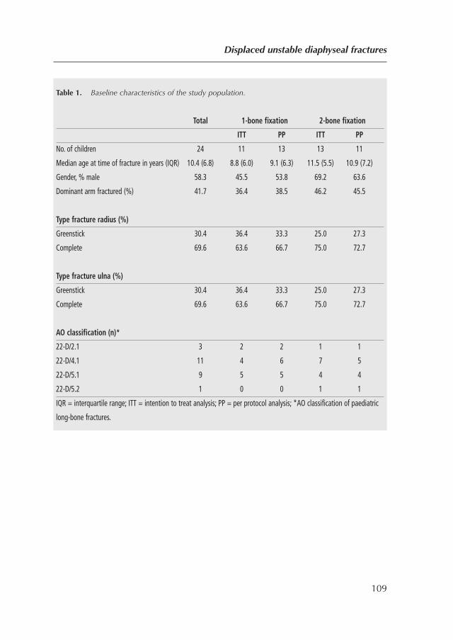
Displaced unstable diaphyseal fractures
109
Table 1. Baseline characteristics of the study population.
Total 1-bone fixation 2-bone fixation
ITT PP ITT PP
No. of children 24 11 13 13 11
Median age at time of fracture in years (IQR) 10.4 (6.8) 8.8 (6.0) 9.1 (6.3) 11.5 (5.5) 10.9 (7.2)
Gender, % male 58.3 45.5 53.8 69.2 63.6
Dominant arm fractured (%) 41.7 36.4 38.5 46.2 45.5
Type fracture radius (%)
Greenstick 30.4 36.4 33.3 25.0 27.3
Complete 69.6 63.6 66.7 75.0 72.7
Type fracture ulna (%)
Greenstick 30.4 36.4 33.3 25.0 27.3
Complete 69.6 63.6 66.7 75.0 72.7
AO classification (n)*
22-D/2.1 3 2 2 1 1
22-D/4.1 11 4 6 7 5
22-D/5.1 9 5 5 4 4
22-D/5.2 1 0 0 1 1
IQR = interquartile range; ITT = intention to treat analysis; PP = per protocol analysis; *AO classification of paediatric
long-bone fractures.
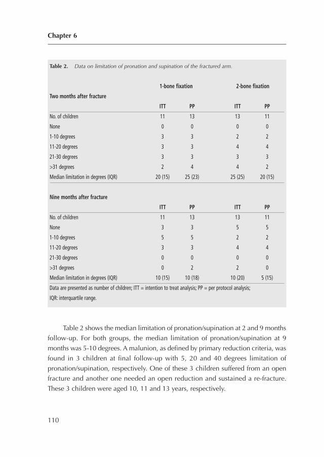
Table 2 shows the median limitation of pronation/supination at 2 and 9 months
follow-up. For both groups, the median limitation of pronation/supination at 9
months was 5-10 degrees. A malunion, as defined by primary reduction criteria, was
found in 3 children at final follow-up with 5, 20 and 40 degrees limitation of
pronation/supination, respectively. One of these 3 children suffered from an open
fracture and another one needed an open reduction and sustained a re-fracture.
These 3 children were aged 10, 11 and 13 years, respectively.
Chapter 6
110
Table 2. Data on limitation of pronation and supination of the fractured arm.
1-bone fixation 2-bone fixation
Two months after fracture
ITT PP ITT PP
No. of children 11 13 13 11
None 0 0 0 0
1-10 degrees 3 3 2 2
11-20 degrees 3 3 4 4
21-30 degrees 3 3 3 3
>31 degrees 2 4 4 2
Median limitation in degrees (IQR) 20 (15) 25 (23) 25 (25) 20 (15)
Nine months after fracture
ITT PP ITT PP
No. of children 11 13 13 11
None 3 3 5 5
1-10 degrees 5 5 2 2
11-20 degrees 3 3 4 4
21-30 degrees 0 0 0 0
>31 degrees 0 2 2 0
Median limitation in degrees (IQR) 10 (15) 10 (18) 10 (20) 5 (15)
Data are presented as number of children; ITT = intention to treat analysis; PP = per protocol analysis;
IQR: interquartile range.
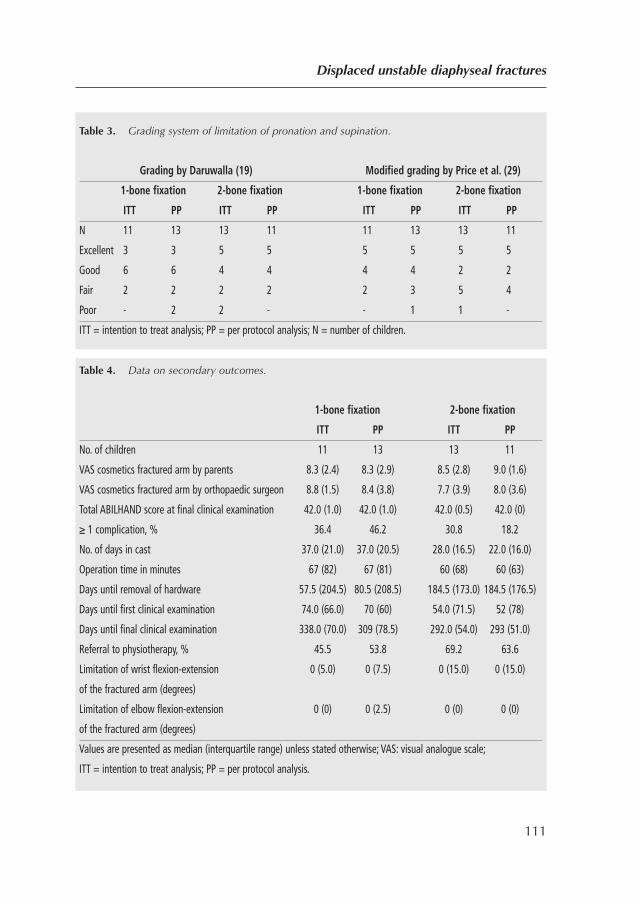
Displaced unstable diaphyseal fractures
111
Table 3. Grading system of limitation of pronation and supination.
Grading by Daruwalla (19) Modified grading by Price et al. (29)
1-bone fixation 2-bone fixation 1-bone fixation 2-bone fixation
ITT PP ITT PP ITT PP ITT PP
N 11 13 13 11 11 13 13 11
Excellent 3 3 5 5 5 5 5 5
Good 6 6 4 4 4 4 2 2
Fair 2 2 2 2 2 3 5 4
Poor - 2 2 - - 1 1 -
ITT = intention to treat analysis; PP = per protocol analysis; N = number of children.
Table 4. Data on secondary outcomes.
1-bone fixation 2-bone fixation
ITT PP ITT PP
No. of children 11 13 13 11
VAS cosmetics fractured arm by parents 8.3 (2.4) 8.3 (2.9) 8.5 (2.8) 9.0 (1.6)
VAS cosmetics fractured arm by orthopaedic surgeon 8.8 (1.5) 8.4 (3.8) 7.7 (3.9) 8.0 (3.6)
Total ABILHAND score at final clinical examination 42.0 (1.0) 42.0 (1.0) 42.0 (0.5) 42.0 (0)
≥ 1 complication, % 36.4 46.2 30.8 18.2
No. of days in cast 37.0 (21.0) 37.0 (20.5) 28.0 (16.5) 22.0 (16.0)
Operation time in minutes 67 (82) 67 (81) 60 (68) 60 (63)
Days until removal of hardware 57.5 (204.5) 80.5 (208.5) 184.5 (173.0) 184.5 (176.5)
Days until first clinical examination 74.0 (66.0) 70 (60) 54.0 (71.5) 52 (78)
Days until final clinical examination 338.0 (70.0) 309 (78.5) 292.0 (54.0) 293 (51.0)
Referral to physiotherapy, % 45.5 53.8 69.2 63.6
Limitation of wrist flexion-extension 0 (5.0) 0 (7.5) 0 (15.0) 0 (15.0)
of the fractured arm (degrees)
Limitation of elbow flexion-extension 0 (0) 0 (2.5) 0 (0) 0 (0)
of the fractured arm (degrees)
Values are presented as median (interquartile range) unless stated otherwise; VAS: visual analogue scale;
ITT = intention to treat analysis; PP = per protocol analysis.
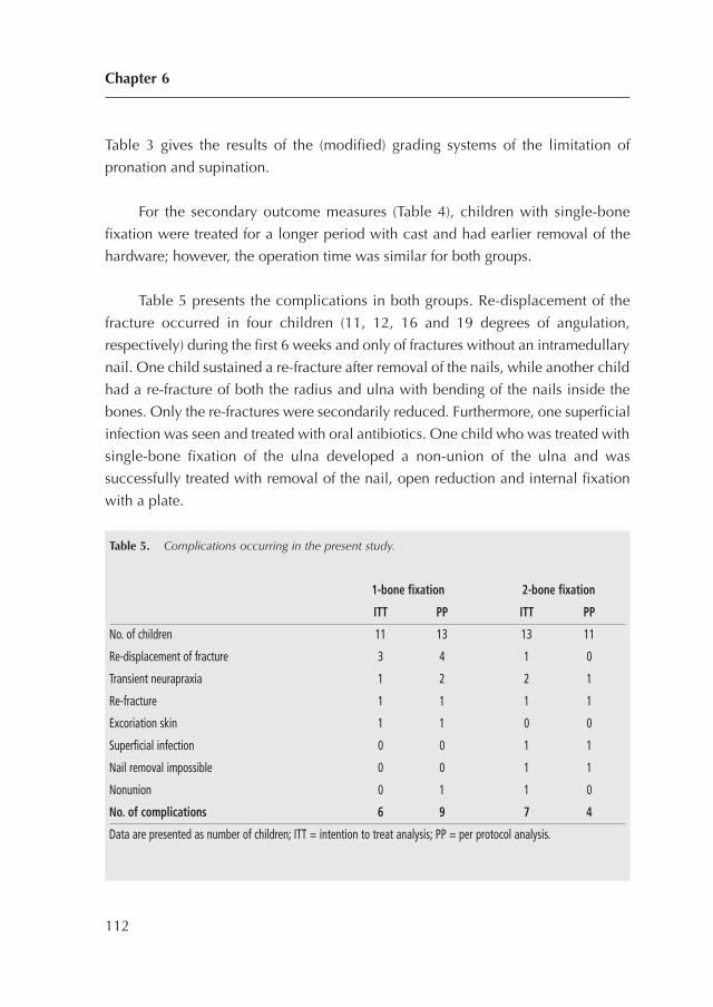
Table 3 gives the results of the (modified) grading systems of the limitation of
pronation and supination.
For the secondary outcome measures (Table 4), children with single-bone
fixation were treated for a longer period with cast and had earlier removal of the
hardware; however, the operation time was similar for both groups.
Table 5 presents the complications in both groups. Re-displacement of the
fracture occurred in four children (11, 12, 16 and 19 degrees of angulation,
respectively) during the first 6 weeks and only of fractures without an intramedullary
nail. One child sustained a re-fracture after removal of the nails, while another child
had a re-fracture of both the radius and ulna with bending of the nails inside the
bones. Only the re-fractures were secondarily reduced. Furthermore, one superficial
infection was seen and treated with oral antibiotics. One child who was treated with
single-bone fixation of the ulna developed a non-union of the ulna and was
successfully treated with removal of the nail, open reduction and internal fixation
with a plate.
Chapter 6
112
Table 5. Complications occurring in the present study.
1-bone fixation 2-bone fixation
ITT PP ITT PP
No. of children 11 13 13 11
Re-displacement of fracture 3 4 1 0
Transient neurapraxia 1 2 2 1
Re-fracture 1 1 1 1
Excoriation skin 1 1 0 0
Superficial infection 0 0 1 1
Nail removal impossible 0 0 1 1
Nonunion 0 1 1 0
No. of complications 6 9 7 4
Data are presented as number of children; ITT = intention to treat analysis; PP = per protocol analysis.
DISCUSSION
This study compared single-bone and both-bone intramedullary fixation of unstable
diaphyseal both-bone fractures in children and indicates that single-bone fixation leads
to increased re-displacement and poorer clinical outcome. Although the median
limitation of pronation/supination was similar in both groups, a higher rate of
complications occurred in the single-bone fixation group. These complications were
mainly caused by re-displacement of the fracture without a nail. Furthermore, fractures
stabilised with only one nail were immobilised in a cast for a longer period. Surprisingly,
the operation time was similar in both groups. These findings in combination with the
risks of infection and anaesthesia (for insertion and removal of the nails), makes us
believe that a child has virtually no profit from the treatment of single-bone fixation
compared with reduction and conservative treatment in cast. In our opinion, unstable
both-bone forearm fractures should be treated with two intramedullary nails with cast.
At 9-months follow-up, in both groups a relatively large number of children
had a limitation of pronation/supination of ≥ 20 degrees. This group of children was
diverse: 2 had a malunion at final examination, 1 child was operated on a second
time because of a non-union, another child re-fractured his forearm, and 2 children
were treated without complications but were immobilised in cast for 37 and 42 days,
respectively. Therefore, in our study population the cause of the limitation of
pronation/supination seems to be multifactorial.
Other studies investigating intramedullary stabilisation of both-bone forearm
fractures reported that limitation of pronation/supination occurs in 0-20% of
patients2,5,11,12,14,16-19,34-41, whereas this occurred in 67% of our group. A possible
explanation for our high rate is our prospective study design with no children lost to
follow-up, and our high rate of complications resulting in less favourable outcomes.
In other studies, the overall better results of single-bone fixation can be
explained by their selective treatment of fractures with single-bone or both-bone
fixation2,5,10-14,16-19. In those studies, the surgeon could choose per-operatively for
single-bone or both-bone fixation depending on the stability of the fracture, whereas
we used a randomisation protocol irrespective of the level of stability.
In the present study, re-displacement of the fracture occurred in 4 children and
only in those treated with single-bone fixation. Not surprisingly, all re-displacements
Displaced unstable diaphyseal fractures
113
were seen in the bone without intramedullary fixation. Such re-displacement in
single-bone intramedullary fixation of both-bone forearm fractures is not
uncommon2,5,10,13. Therefore, in children, we suggest that unstable both-bone
diaphyseal forearm fractures should be stabilised with an intramedullary nail in both
the radius and ulna to prevent re-displacement.
Study limitations
Some study limitations need to be addressed. First, the number of included children
is small: of the 249 children with a displaced diaphyseal both-bone forearm fracture
only 24 were classified as unstable. This rareness of unstable fractures is reflected in
other studies with small numbers of children10-14,17,18,42,43. Nevertheless, the
possibility exists that our test for fracture stability was inadequate or not properly
accomplished. Although we present the first randomised controlled trial of single-
versus both-bone intramedullary fixation, our study was statistically underpowered
and the results should be interpreted with caution.
Second, the follow-up period was only 9 months. Although this seems short,
in other children treated for displaced forearm fractures no change was found in
clinical or radiographic status from 6 months post-treatment until final follow-up at
(mean) 2.8 years44. Another group described a persistent limitation during long-term
follow-up despite correction by growth45, and others advised an osteotomy to correct
malunion as early as 6 months after trauma46. Although malunions in children can
correct with growth, these diaphyseal malunions are less likely to correct than distal
malunions because of the relatively long distance to the most active distal growth
plate.
Conclusion
The results of this first randomised controlled study caution against the use of single-
bone fixation in all displaced unstable both-bone diaphyseal forearm fractures in
children. The single-bone fixation method may lead to increased fracture re-
displacement and poorer clinical results.
Chapter 6
114
REFERENCES
1. Amit Y, Salai M, Chechik A, Blankstein A, Horoszowski H. Closing intramedullary nailing
for the treatment of diaphyseal forearm fractures in adolescence: a preliminary report. J
Pediatr Orthop. 1985;5(2):143-6.
2. Cullen MC, Roy DR, Giza E, Crawford AH. Complications of intramedullary fixation of
pediatric forearm fractures. J Pediatr Orthop. 1998;18(1):14-21.
3. Luhmann SJ, Gordon JE, Schoenecker PL. Intramedullary fixation of unstable both-bone
forearm fractures in children. J Pediatr Orthop. 1998;18(4):451-6.
4. Roy DR, Crawford AH. Operative management of fractures of the shaft of the radius and
ulna. Orthop Clin North Am. 1990;21(2):245-50.
5. Shoemaker SD, Comstock CP, Mubarak SJ, Wenger DR, Chambers HG. Intramedullary
Kirschner wire fixation of open or unstable forearm fractures in children. J Pediatr Orthop.
1999;19(3):329-37.
6. Verstreken L, Delronge G, Lamoureux J. Shaft forearm fractures in children: intramedullary
nailing with immediate motion: a preliminary report. J Pediatr Orthop. 1988;8(4):450-3.
7. Dietz HG, Schmittenbecher PP, Slongo T, Wilkins KE. AO manual of fracture management.
ESIN in children2006.
8. Metaizeau JP, Ligier JN. [Surgical treatment of fractures of the long bones in children.
Interference between osteosynthesis and the physiological processes of consolidation.
Therapeutic indications]. Journal de chirurgie. 1984;121(8-9):527-37.
9. Prevot J, Lascombes P, Ligier JN. [The ECMES [Centro-Medullary Elastic Stabilising Wiring)
osteosynthesis method in limb fractures in children. Principle, application on the femur.
Apropos of 250 fractures followed-up since 1979]. Chirurgie. 1993;119(9):473-6.
10. Dietz JF, Bae DS, Reiff E, Zurakowski D, Waters PM. Single bone intramedullary fixation of
the ulna in pediatric both bone forearm fractures: analysis of short-term clinical and
radiographic results. J Pediatr Orthop.30(5):420-4.
11. Flynn JM, Waters PM. Single-bone fixation of both-bone forearm fractures. J Pediatr Orthop.
1996;16(5):655-9.
12. Houshian S, Bajaj SK. Forearm fractures in children. Single bone fixation with elastic stable
intramedullary nailing in 20 cases. Injury. 2005;36(12):1421-6.
13. Lee S, Nicol RO, Stott NS. Intramedullary fixation for pediatric unstable forearm fractures.
Clin Orthop Relat Res. 2002(402):245-50.
Displaced unstable diaphyseal fractures
115
14. Myers GJ, Gibbons PJ, Glithero PR. Nancy nailing of diaphyseal forearm fractures. Single
bone fixation for fractures of both bones. J Bone Joint Surg Br. 2004;86(4):581-4.
15. Sharieff GQ, Kanegaye J, Wallace CD, McCaslin RI, Harley JR. Can portable bedside
fluoroscopy replace standard, postreduction radiographs in the management of pediatric
fractures? Pediatr Emerg Care. 1999;15(4):249-51.
16. Yung PS, Lam CY, Ng BK, Lam TP, Cheng JC. Percutaneous transphyseal intramedullary
Kirschner wire pinning: a safe and effective procedure for treatment of displaced diaphyseal
forearm fracture in children. J Pediatr Orthop. 2004;24(1):7-12.
17. Alnaib M, Taranu R, Lakkol S, Aldlyami E, Alcelik I, Tulloch C. Radius-only intramedullary
nailing for both-bones diaphyseal forearm fractures in children. Acta orthopaedica Belgica.
2011;77(4):458-63.
18. Kirkos JM, Beslikas T, Kapras EA, Papavasiliou VA. Surgical treatment of unstable diaphyseal
both-bone forearm fractures in children with single fixation of the radius. Injury.
2000;31(8):591-6.
19. Meier R, Prommersberger KJ, van Griensven M, Lanz U. Surgical correction of deformities
of the distal radius due to fractures in pediatric patients. Arch Orthop Trauma Surg.
2004;124(1):1-9.
20. Boyer BA, Overton B, Schrader W, Riley P, Fleissner P. Position of immobilization for
pediatric forearm fractures. Journal of pediatric orthopedics. 2002;22(2):185-7.
21. Choi KY, Chan WS, Lam TP, Cheng JC. Percutaneous Kirschner-wire pinning for severely
displaced distal radial fractures in children. A report of 157 cases. J Bone Joint Surg Br.
1995;77(5):797-801.
22. Daruwalla JS. A study of radioulnar movements following fractures of the forearm in
children. Clin Orthop Relat Res. 1979(139):114-20.
23. Gibbons CL, Woods DA, Pailthorpe C, Carr AJ, Worlock P. The management of isolated
distal radius fractures in children. J Pediatr Orthop. 1994;14(2):207-10.
24. Jones K, Weiner DS. The management of forearm fractures in children: a plea for
conservatism. J Pediatr Orthop. 1999;19(6):811-5.
25. Mani GV, Hui PW, Cheng JC. Translation of the radius as a predictor of outcome in distal
radial fractures of children. J Bone Joint Surg Br. 1993;75(5):808-11.
26. Maric D, Petkovic L, Tomasevic V, Bajic L, Gajdobranski D, Marcikic A. [Osteosynthesis
with Kirschner wires in the treatment of dislocation fractures of the forearm in children].
Med Pregl. 2002;55(3-4):140-5.
Chapter 6
116
27. Noonan KJ, Price CT. Forearm and distal radius fractures in children. J Am Acad Orthop
Surg. 1998;6(3):146-56.
28. Tynan MC, Fornalski S, McMahon PJ, Utkan A, Green SA, Lee TQ. The effects of ulnar axial
malalignment on supination and pronation. J Bone Joint Surg Am. 2000;82-A(12):1726-31.
29. Zimmermann R, Gabl M, Angermann P, Lutz M, Reinhart C, Kralinger F, et al. [Late
sequelae of fractures of the distal third of the forearm during the growth period]. Handchir
Mikrochir Plast Chir. 2000;32(4):242-9.
30. Gustilo RB, Simpson L, Nixon R, Ruiz A, Indeck W. Analysis of 511 open fractures. Clin
Orthop Relat Res. 1969;66:148-54.
31. Colaris J, van der Linden M, Selles R, Coene N, Allema JH, Verhaar J. Pronation and
supination after forearm fractures in children: Reliability of visual estimation and
conventional goniometry measurement. Injury. 2010;41(6):643-6.
32. Price CT, Scott DS, Kurzner ME, Flynn JC. Malunited forearm fractures in children. J Pediatr
Orthop. 1990;10(6):705-12.
33. Arnould C, Penta M, Renders A, Thonnard JL. ABILHAND-Kids: a measure of manual ability
in children with cerebral palsy. Neurology. 2004;63(6):1045-52.
34. Altay M, Aktekin CN, Ozkurt B, Birinci B, Ozturk AM, Tabak AY. Intramedullary wire
fixation for unstable forearm fractures in children. Injury. 2006;37(10):966-73.
35. Jubel A, Andermahr J, Isenberg J, Issavand A, Prokop A, Rehm KE. Outcomes and
complications of elastic stable intramedullary nailing for forearm fractures in children. J
Pediatr Orthop B. 2005;14(5):375-80.
36. Kapoor V, Theruvil B, Warwick DJ, Uglow MG. Distal radio-ulnar joint subluxation
following intramedullary nailing of the radius in a child. J Bone Joint Surg Br.
2004;86(4):592-4.
37. Kucukkaya M, Kabukcuoglu Y, Tezer M, Eren T, Kuzgun U. The application of open
intramedullary fixation in the treatment of pediatric radial and ulnar shaft fractures. J
Orthop Trauma. 2002;16(5):340-4.
38. Kumar G. Comparison of short and long arm plaster casts for displaced fractures in the distal
third of the forearm in children. J Bone Joint Surg Am. 2006;88(8):1888; author reply -9.
39. Lascombes P, Haumont T, Journeau P. Use and abuse of flexible intramedullary nailing in
children and adolescents. J Pediatr Orthop. 2006;26(6):827-34.
40. Qidwai SA. Treatment of diaphyseal forearm fractures in children by intramedullary
Kirschner wires. J Trauma. 2001;50(2):303-7.
Displaced unstable diaphyseal fractures
117
41. Richter D, Ostermann PA, Ekkernkamp A, Muhr G, Hahn MP. Elastic intramedullary nailing:
a minimally invasive concept in the treatment of unstable forearm fractures in children. J
Pediatr Orthop. 1998;18(4):457-61.
42. Calder PR, Achan P, Barry M. Diaphyseal forearm fractures in children treated with
intramedullary fixation: outcome of K-wire versus elastic stable intramedullary nail. Injury.
2003;34(4):278-82.
43. Majed A, Baco AM. Nancy nail versus intramedullary-wire fixation of paediatric forearm
fractures. J Pediatr Orthop B. 2007;16(2):129-32.
44. Miller BS, Taylor B, Widmann RF, Bae DS, Snyder BD, Waters PM. Cast immobilization
versus percutaneous pin fixation of displaced distal radius fractures in children: a
prospective, randomized study. J Pediatr Orthop. 2005;25(4):490-4.
45. Nilsson BE, Obrant K. The range of motion following fracture of the shaft of the forearm in
children. Acta Orthop Scand. 1977;48(6):600-2.
46. Price CT, Knapp DR. Osteotomy for malunited forearm shaft fractures in children. J Pediatr
Orthop. 2006;26(2):193-6.
Chapter 6
118

Displaced unstable diaphyseal fractures
119

PART IV
FRACTURE DISPLACEMENT
120
CHAPTER 7
Risk factors for the displacementof fractures of both bones of theforearm in children
Bone Joint J. 2013 May;95-B(5):689-93
Colaris JW, Allema JH, Reijman M, Biter LU, de Vries MR, van de Ven CP, Bloem
RM, Verhaar JA.
121
ABSTRACT
Forearm fractures in children have a tendency to displace in a cast leading to
malunion with reduced functional and cosmetic results. In order to identify risk
factors for displacement, a total of 247 conservatively treated fractures of the forearm
in 246 children with a mean age of 7.3 years (SD 3.2; 0.9 to 14.9) were included in
a prospective multicentre study. Multivariate logistic regression analyses were
performed to assess risk factors for displacement of reduced or non-reduced fractures
in the cast. Displacement occurred in 73 patients (29.6%), of which 65 (89.0%) were
in above-elbow casts. The mean time between the injury and displacement was 22.7
days (0 to 59). The independent factors found to significantly increase the risk of
displacement were a fracture of the non-dominant arm (p = 0.024), a complete
fracture (p = 0.040), a fracture with translation of the ulna on lateral radiographs (p
= 0.014) and shortening of the fracture (p = 0.019). Fractures of both forearm bones
in children have a strong tendency to displace even in an above-elbow cast. Severe
fractures of the non-dominant arm are at highest risk for displacement. Radiographs
at set times during treatment might identify early displacement, which should be
treated before malunion occurs, especially in older children with less potential for
remodelling.
Chapter 7
122
INTRODUCTION
Displacement of fractures of both forearm bones in children treated in a cast has been
described in between 7% and 91% of cases1-5, and can lead to malunion with reduced
functional and cosmetic results6,7. Despite the high rate of displacement, there has not
hitherto been an analysis of risk factors in large prospective studies3-5,8-11. We therefore
performed a prospective multicentre study to identify the risk factors for displacement
of reduced and unreduced fractures of both bones of the forearm in children when
treated in a cast.
Patients and Methods
Between January 2006 and August 2010 a total of 247 children aged < 16 years with
fractures of both bones of the forearm were included in a prospective study involving
four hospitals (Fig. 1, Table 1). The study had ethical approval and informed consent
was obtained from all parents, and children aged > 12 years. The criteria for reduction
were based on previous studies (Table 2)1,12-20. Exclusion criteria were: torus fractures
of both the radius and ulna; fractures sustained more than a week before presentation;
severe open fractures (Gustilo II and III)21; and re-fractures. Exclusion criteria included
those fixed with Kirschner (K-) wires or intramedullary nails, fractures without final
radiographs, initially displaced fractures without reduction and reduced fractures with
displacement on the first subsequent radiograph. The study protocol distinguished
different types of fractures. Distal metaphyseal fractures not requiring reduction were
treated with a below- or above-elbow cast for four weeks22, whereas distal
metaphyseal fractures needing reduction were treated with or without K-wires and
an above-elbow cast for four weeks23. Diaphyseal fractures not requiring reduction
and reduced stable diaphyseal fractures were treated for six weeks with an above-
elbow cast or a combination of three weeks above and three weeks below. Unstable
diaphyseal fractures were fixed with one or two intramedullary nails and three weeks
in an above-elbow cast. The cast was applied in a standard way by a trained nurse in
the emergency department or a surgeon in the operating room. Firstly, a stockinette
and layer of wool were applied to protect the skin and bony prominences. Second,
a well-fitted plaster slab was applied that covered approximately two-thirds of the
circumference of the arm. Finally a bandage was wrapped around the arm.
Risk factors for displacement of fractures
123
Radiographs were taken at presentation, after reduction and during treatment. An
orthopaedic surgeon (JWC) who was not involved in primary treatment measured
angulation, translation as a percentage of the width of radius/ulna rotation (yes/no)
and shortening (yes/no) of all fractures. Rotation was detected by differences in the
diameter of the diaphysis of radius and ulna on posteroanterior and lateral
radiographs24.
A reduced or an unreduced fracture that showed progressive displacement (Fig.
2) during follow-up was defined as displaced if further reduction was needed
according to the criteria for primary reduction (Table 2). In order to assess the inter-
rater reproducibility of the radiological assessment, a trauma surgeon (LUB) who
was not involved in treatment re-measured the angulation of the fracture in 45
children.
Chapter 7
124
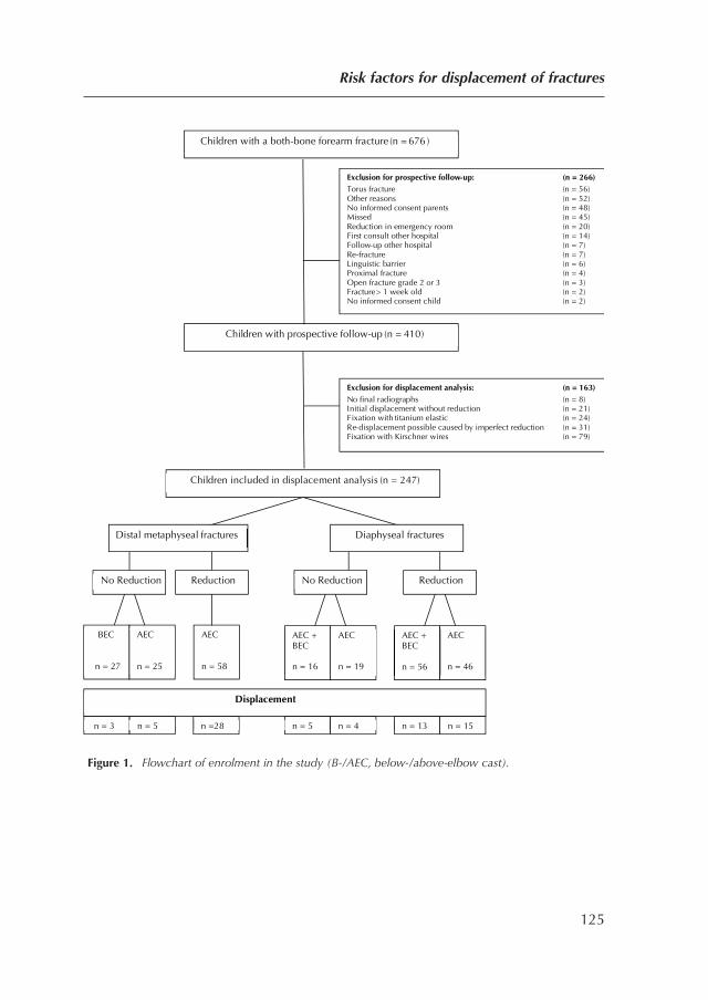
Figure 1. Flowchart of enrolment in the study (B-/AEC, below-/above-elbow cast).
Risk factors for displacement of fractures
125
Exclusion for displacement analysis: (n = 163)
No final radiographs (n = 8) Initial displacement without reduction (n = 21) Fixation with titanium elastic (n = 24) Re-displacement possible caused by imperfect reduction (n = 31) Fixation with Kirschner wires (n = 79)
Children with prospective follow-up (n = 410)
Children with a both-bone forearm fracture (n = 676 )
Exclusion for prospective follow-up: (n = 266)
Torus fracture (n = 56) Other reasons (n = 52) No informed consent parents (n = 48) Missed (n = 45) Reduction in emergency room (n = 20) First consult other hospital (n = 14) Follow-up other hospital (n = 7) Re-fracture (n = 7) Linguistic barrier (n = 6) Proximal fracture (n = 4) Open fracture grade 2 or 3 (n = 3) Fracture > 1 week old (n = 2) No informed consent child (n = 2)
Children included in displacement analysis (n = 247)
Distal metaphyseal fractures
Reduction
Displacement
Diaphyseal fractures
BEC
n = 27
AEC n = 25
AEC n = 58
AEC n = 19
AEC + BEC n = 16
n = 3 n = 5 n =28 n = 5 n = 4 n = 13 n = 15
No Reduction No Reduction Reduction
AEC + BEC n = 56
AEC n = 46
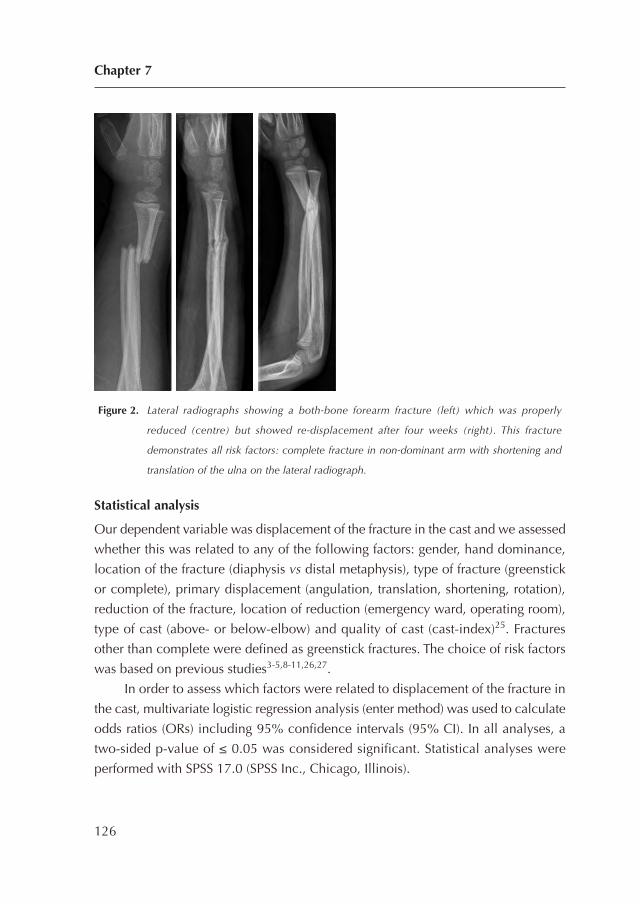
Figure 2. Lateral radiographs showing a both-bone forearm fracture (left) which was properly
reduced (centre) but showed re-displacement after four weeks (right). This fracture
demonstrates all risk factors: complete fracture in non-dominant arm with shortening and
translation of the ulna on the lateral radiograph.
Statistical analysis
Our dependent variable was displacement of the fracture in the cast and we assessed
whether this was related to any of the following factors: gender, hand dominance,
location of the fracture (diaphysis vs distal metaphysis), type of fracture (greenstick
or complete), primary displacement (angulation, translation, shortening, rotation),
reduction of the fracture, location of reduction (emergency ward, operating room),
type of cast (above- or below-elbow) and quality of cast (cast-index)25. Fractures
other than complete were defined as greenstick fractures. The choice of risk factors
was based on previous studies3-5,8-11,26,27.
In order to assess which factors were related to displacement of the fracture in
the cast, multivariate logistic regression analysis (enter method) was used to calculate
odds ratios (ORs) including 95% confidence intervals (95% CI). In all analyses, a
two-sided p-value of ≤ 0.05 was considered significant. Statistical analyses were
performed with SPSS 17.0 (SPSS Inc., Chicago, Illinois).
Chapter 7
126
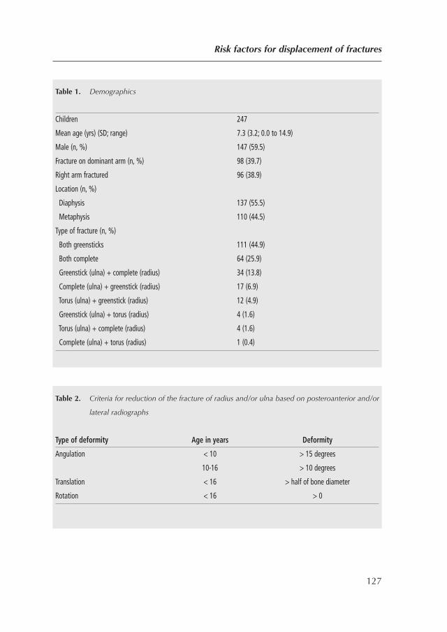
Risk factors for displacement of fractures
127
Table 1. Demographics
Children 247
Mean age (yrs) (SD; range) 7.3 (3.2; 0.0 to 14.9)
Male (n, %) 147 (59.5)
Fracture on dominant arm (n, %) 98 (39.7)
Right arm fractured 96 (38.9)
Location (n, %)
Diaphysis 137 (55.5)
Metaphysis 110 (44.5)
Type of fracture (n, %)
Both greensticks 111 (44.9)
Both complete 64 (25.9)
Greenstick (ulna) + complete (radius) 34 (13.8)
Complete (ulna) + greenstick (radius) 17 (6.9)
Torus (ulna) + greenstick (radius) 12 (4.9)
Greenstick (ulna) + torus (radius) 4 (1.6)
Torus (ulna) + complete (radius) 4 (1.6)
Complete (ulna) + torus (radius) 1 (0.4)
Table 2. Criteria for reduction of the fracture of radius and/or ulna based on posteroanterior and/or
lateral radiographs
Type of deformity Age in years Deformity
Angulation < 10 > 15 degrees
10-16 > 10 degrees
Translation < 16 > half of bone diameter
Rotation < 16 > 0
RESULTS
The fracture re-displaced in 73 children (29.6%). Re-displacement occurred more
frequently in distal metaphyseal fractures compared with diaphyseal fractures (32.7%
(36 of 110) vs 27.0% (37 of 137)). The mean time between the injury and
displacement was 22.7 days (SD 13.5; 0 to 59), and 65 re-displacements (89.0%)
were in an above-elbow cast (Fig. 2).
The factors that were significantly and independently associated with
redisplacement were hand dominance and three factors associated with the severity
of the fracture, namely complete fractures, translation of the ulna on lateral
radiographs and shortening (Table 3, Fig. 2).
DISCUSSION
We found that 29.6% of fractures of both bones of the forearm in children treated in
a cast displaced, the risk of which was increased in more severe fractures of the non-
dominant arm. Earlier authors reported displacement of between 7% and 91% of
fractures in the distal metaphysis1-5,28-31 and between 7% and 27% of fractures in
the diaphysis4,15,26,32-35 compared with 33% and 27% respectively in our study. Our
relatively high percentage of displacement can be explained by the inclusion of only
both-bone fractures, which are more unstable than single-bone fractures5,26,36.
Whereas an earlier study reported hand dominance as a risk factor for
displacement (26), others found similar numbers in dominant and non-dominant
arms (3). Our results suggest that fractures in the non-dominant arm are at increased
risk for displacement, which might be explained by less stability of the fracture by
less developed muscles in the non-dominant forearm.
As with earlier reports3-5,8,9,26 the severity of the fracture in our study seemed
highly predictive of displacement. Complete fractures significantly increased the risk
for displacement, a finding supported by some studies3-5,8,9,26 but not others4,11.
Whereas translation of the ulna on lateral radiographs has been reported as a risk
factor for displacement32, shortening has not. Complete fractures with translation
and shortening seem to be more unstable than angulated greenstick fractures with
intact periosteum on one side. Angulation of the fracture did not appear to be a risk
Chapter 7
128
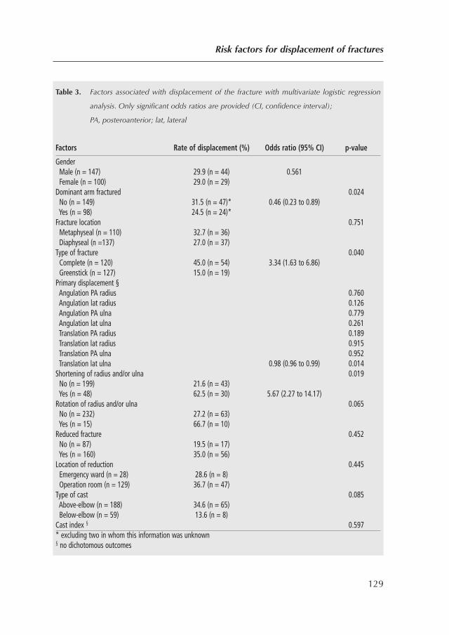
Risk factors for displacement of fractures
129
Table 3. Factors associated with displacement of the fracture with multivariate logistic regression
analysis. Only significant odds ratios are provided (CI, confidence interval);
PA, posteroanterior; lat, lateral
Factors Rate of displacement (%) Odds ratio (95% CI) p-value
GenderMale (n = 147) 29.9 (n = 44) 0.561Female (n = 100) 29.0 (n = 29)
Dominant arm fractured 0.024No (n = 149) 31.5 (n = 47)* 0.46 (0.23 to 0.89)Yes (n = 98) 24.5 (n = 24)*
Fracture location 0.751Metaphyseal (n = 110) 32.7 (n = 36)Diaphyseal (n =137) 27.0 (n = 37)
Type of fracture 0.040Complete (n = 120) 45.0 (n = 54) 3.34 (1.63 to 6.86)Greenstick (n = 127) 15.0 (n = 19)
Primary displacement §Angulation PA radius 0.760Angulation lat radius 0.126Angulation PA ulna 0.779Angulation lat ulna 0.261Translation PA radius 0.189Translation lat radius 0.915Translation PA ulna 0.952Translation lat ulna 0.98 (0.96 to 0.99) 0.014
Shortening of radius and/or ulna 0.019No (n = 199) 21.6 (n = 43)Yes (n = 48) 62.5 (n = 30) 5.67 (2.27 to 14.17)
Rotation of radius and/or ulna 0.065No (n = 232) 27.2 (n = 63)Yes (n = 15) 66.7 (n = 10)
Reduced fracture 0.452No (n = 87) 19.5 (n = 17)Yes (n = 160) 35.0 (n = 56)
Location of reduction 0.445Emergency ward (n = 28) 28.6 (n = 8)Operation room (n = 129) 36.7 (n = 47)
Type of cast 0.085Above-elbow (n = 188) 34.6 (n = 65)Below-elbow (n = 59) 13.6 (n = 8)
Cast index § 0.597* excluding two in whom this information was unknown§ no dichotomous outcomes
factor for displacement in this or previous studies3,11,37. Also, location in the
diaphysis or metaphysis had no significant influence on displacement, a finding
supported by Monga et al (26) but not Younger et al27.
Although several studies considered displacement to be related to an above-
or below-elbow cast3,10,26,27 this was not supported by our results. Also, several
studies4,8,10,11,25,32 reported quality of the cast as a risk factor, while others found no
such correlation9,26. The cast index was not considered as a risk factor in our
study25,32.
Our study has several limitations. Because it is impossible to differentiate
between imperfectly reduced and redisplaced fractures, we excluded children with
reduced fractures that showed displacement at the first available radiographs after
the reduction. Similarly, we could not analyse imperfect reduction as a risk factor
for displacement. Furthermore, we measured the cast index in non-circumferential
casts whereas this measurement is only validated for circumferential casts25,32.
Nevertheless, we assumed that the cast index is also important in non-
circumferential casts because it indicates how well the cast is moulded to the
contour of the forearm. This is supported by a study in which there were similar rates
of displacement in circumferential and non-circumferential casts3.
In summary, reduced and unreduced fractures of both bones of the forearm in
children displaced frequently, even if treated with an above-elbow cast. The most
severe fractures in the non-dominant arm had the highest risk for displacement.
Radiographs at set times during treatment might identify early displacement, which
should be treated before malunion occurs, especially in older children with less
potential for remodelling.
Chapter 7
130
REFERENCES
1. Gibbons CL, Woods DA, Pailthorpe C, Carr AJ, Worlock P. The management of isolated
distal radius fractures in children. J Pediatr Orthop. 1994;14(2):207-10.
2. Haddad FS, Williams RL. Forearm fractures in children: avoiding redisplacement. Injury.
1995;26(10):691-2.
3. Proctor MT, Moore DJ, Paterson JM. Redisplacement after manipulation of distal radial
fractures in children. J Bone Joint Surg Br. 1993;75(3):453-4.
4. Voto SJ, Weiner DS, Leighley B. Redisplacement after closed reduction of forearm fractures
in children. J Pediatr Orthop. 1990;10(1):79-84.
5. Zamzam MM, Khoshhal KI. Displaced fracture of the distal radius in children: factors
responsible for redisplacement after closed reduction. J Bone Joint Surg Br. 2005;87(6):841-3.
6. Prommersberger KJ, Froehner SC, Schmitt RR, Lanz UB. Rotational deformity in malunited
fractures of the distal radius. J Hand Surg [Am]. 2004;29(1):110-5.
7. Roberts JA. Angulation of the radius in children’s fractures. J Bone Joint Surg Br.
1986;68(5):751-4.
8. Alemdaroglu KB, Iltar S, Cimen O, Uysal M, Alagoz E, Atlihan D. Risk factors in
redisplacement of distal radial fractures in children. J Bone Joint Surg Am. 2008;90(6):1224-
30.
9. Pretell Mazzini J, Rodriguez Martin J. Paediatric forearm and distal radius fractures: risk
factors and re-displacement—role of casting indices. Int Orthop.34(3):407-12.
10. Webb GR, Galpin RD, Armstrong DG. Comparison of short and long arm plaster casts for
displaced fractures in the distal third of the forearm in children. J Bone Joint Surg Am.
2006;88(1):9-17.
11. Younger AS, Tredwell SJ, Mackenzie WG, Orr JD, King PM, Tennant W. Accurate prediction
of outcome after pediatric forearm fracture. J Pediatr Orthop. 1994;14(2):200-6.
12. Boyer BA, Overton B, Schrader W, Riley P, Fleissner P. Position of immobilization for
pediatric forearm fractures. Journal of pediatric orthopedics. 2002;22(2):185-7.
13. Choi KY, Chan WS, Lam TP, Cheng JC. Percutaneous Kirschner-wire pinning for severely
displaced distal radial fractures in children. A report of 157 cases. J Bone Joint Surg Br.
1995;77(5):797-801.
14. Daruwalla JS. A study of radioulnar movements following fractures of the forearm in
children. Clin Orthop Relat Res. 1979(139):114-20.
Risk factors for displacement of fractures
131
15. Jones K, Weiner DS. The management of forearm fractures in children: a plea for
conservatism. J Pediatr Orthop. 1999;19(6):811-5.
16. Mani GV, Hui PW, Cheng JC. Translation of the radius as a predictor of outcome in distal
radial fractures of children. J Bone Joint Surg Br. 1993;75(5):808-11.
17. Maric D, Petkovic L, Tomasevic V, Bajic L, Gajdobranski D, Marcikic A. [Osteosynthesis
with Kirschner wires in the treatment of dislocation fractures of the forearm in children].
Med Pregl. 2002;55(3-4):140-5.
18. Noonan KJ, Price CT. Forearm and distal radius fractures in children. J Am Acad Orthop
Surg. 1998;6(3):146-56.
19. Tynan MC, Fornalski S, McMahon PJ, Utkan A, Green SA, Lee TQ. The effects of ulnar axial
malalignment on supination and pronation. J Bone Joint Surg Am. 2000;82-A(12):1726-31.
20. Zimmermann R, Gabl M, Angermann P, Lutz M, Reinhart C, Kralinger F, et al. [Late
sequelae of fractures of the distal third of the forearm during the growth period]. Handchir
Mikrochir Plast Chir. 2000;32(4):242-9.
21. Gustilo RB, Simpson L, Nixon R, Ruiz A, Indeck W. Analysis of 511 open fractures. Clin
Orthop Relat Res. 1969;66:148-54.
22. Colaris JW, Biter LU, Allema JH, Bloem RM, van de Ven CP, de Vries MR, et al. Below-
elbow cast for metaphyseal both-bone fractures of the distal forearm in children: a
randomised multicentre study. Injury. 2012;43(7):1107-11.
23. Colaris JW, Allema JH, Biter LU, de Vries MR, van de Ven CP, Bloem RM, et al. Re-
displacement of stable distal both-bone forearm fractures in children: a randomised
controlled multicentre trial. Injury. 2013;44(4):498-503.
24. Naimark A, Kossoff J, Leach RE. The disparate diameter. A sign of rotational deformity in
fractures. J Can Assoc Radiol. 1983;34(1):8-11.
25. Chess DG, Hyndman JC, Leahey JL, Brown DC, Sinclair AM. Short arm plaster cast for
distal pediatric forearm fractures. J Pediatr Orthop. 1994;14(2):211-3.
26. Monga P, Raghupathy A, Courtman NH. Factors affecting remanipulation in paediatric
forearm fractures. J Pediatr Orthop B.19(2):181-7.
27. Younger AS, Tredwell SJ, Mackenzie WG. Factors affecting fracture position at cast removal
after pediatric forearm fracture. J Pediatr Orthop. 1997;17(3):332-6.
28. Blount WP. Forearm fractures in children. Clin Orthop Relat Res. 1967;51:93-107.
Chapter 7
132
29. Friberg KS. Remodelling after distal forearm fractures in children. I. The effect of residual
angulation on the spatial orientation of the epiphyseal plates. Acta Orthop Scand.
1979;50(5):537-46.
30. Kiely PD, Kiely PJ, Stephens MM, Dowling FE. Atypical distal radial fractures in children. J
Pediatr Orthop B. 2004;13(3):202-5.
31. Van Leemput W, De Ridder K. Distal metaphyseal radius fractures in children: reduction
with or without pinning. Acta Orthop Belg. 2009;75(3):306-9.
32. Bochang C, Jie Y, Zhigang W, Weigl D, Bar-On E, Katz K. Immobilisation of forearm
fractures in children: extended versus flexed elbow. J Bone Joint Surg Br. 2005;87(7):994-6.
33. Chan CF, Meads BM, Nicol RO. Remanipulation of forearm fractures in children. N Z Med
J. 1997;110(1047):249-50.
34. Kay S, Smith C, Oppenheim WL. Both-bone midshaft forearm fractures in children. J Pediatr
Orthop. 1986;6(3):306-10.
35. Schmittenbecher PP. State-of-the-art treatment of forearm shaft fractures. Injury. 2005;36
Suppl 1:A25-34.
36. Bohm ER, Bubbar V, Yong Hing K, Dzus A. Above and below-the-elbow plaster casts for
distal forearm fractures in children. A randomized controlled trial. J Bone Joint Surg Am.
2006;88(1):1-8.
Risk factors for displacement of fractures
133

PART V
LIMITATION OF
FOREARM ROTATION
134

CHAPTER 8
Pronation and supination afterforearm fractures in children:reliability of visual estimationand conventional goniometrymeasurement.
Injury. 2010 Jun;41(6):643-6.
Colaris J, van der Linden M, Selles R, Coene N, Allema JH, Verhaar J.
135
ABSTRACT
Background: Forearm fractures are common amongst children and often result in
limited rotational function. In daily practice, pronation and supination of the arm
are often visually estimated or measured using a conventional goniometer. The aim
of this study was to compare the reliability of these two methods in paediatric
patients that had previously sustained a forearm fracture.
Methods: Intra- and interrater reliability of visual estimation and conventional
goniometry were determined in 47 children who had previously sustained a forearm
fracture.
Results: Intra- and interrater reliability of visual estimation and conventional
goniometry was fair to excellent, with Intraclass Correlation Coefficients (ICC’s)
ranging between 0.75 and 0.94. In addition, the overall goniometer data consistently
showed lower Smallest Detectable Differences (SDD’s) compared to the visual
estimation data, also indicating better reliability for the goniometer method.
Conclusions: A conventional goniometer is an easy, fast and reliable method to
determine the pronation and supination in a child who had sustained a forearm
fracture. If an uncooperative child hinders the measurement, visual estimation is a
good second choice. Measurements are more reliable when repeated by the same
professional.
Chapter 8
136
INTRODUCTION
Forearm rotation, consisting of pronation and supination, is commonly used in daily
life for tasks such as eating, typing, picking up small objects and accepting change1.
In children up to the age of 15 years, pronation of 50-80 degrees and supination of
80-120 degrees is considered normal2. In a number of pathologies, pronation and
supination can be limited, such as following a forearm fracture, which is rather
common among children1,3. Measurement of pronation and supination limitation is
important for clinical decision making and outcome evaluation for these fractures.
In the past, many devices have been developed to measure pronation and
supination of the forearm. These devices include gravity-dependent methods, tubular
handles, voltage gated sensors and measurements of digital photographs1,4-9. For many
of these devices, reliability is fair to excellent, as expressed by Intraclass Correlation
Coefficients (ICCs) (10) ranging between 0.78 - 0.99 for intrarater reliability4,6,9 and
0.72 - 0.98 for interrater reliability4,5,7,9. However, the above-mentioned devices are
complex and are difficult to use in standard clinical care. In addition, they may
intimidate little children. Therefore, forearm rotation in children is still generally
evaluated by visual estimation or conventional goniometry. Although limitations of
pronation and supination are often visually estimated in clinical practice, to our
knowledge, no studies have yet investigated the reliability of visual estimation in any
age group. In addition, the reliability of conventional goniometry has been investigated
only scarcely in children1, and slightly more often in adults4-6,9,11-13.
Furthermore, most of the authors report the reliability of the goniometer
measurements only in a healthy population5,6,9,11,13, whereas only one study
evaluated the reliability of measuring rotational impairments after a trauma4. Because
visual estimation and goniometrical measurement of pronation and supination are
often used in clinical care as well as in clinical research, the aim of the current study
is to compare the reliability of these two methods in paediatric patients that had
previously sustained a forearm fracture.
Measurement of pronation and supination
137
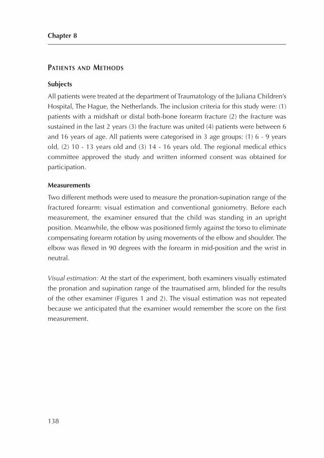
PATIENTS AND METHODS
Subjects
All patients were treated at the department of Traumatology of the Juliana Children’s
Hospital, The Hague, the Netherlands. The inclusion criteria for this study were: (1)
patients with a midshaft or distal both-bone forearm fracture (2) the fracture was
sustained in the last 2 years (3) the fracture was united (4) patients were between 6
and 16 years of age. All patients were categorised in 3 age groups: (1) 6 - 9 years
old, (2) 10 - 13 years old and (3) 14 - 16 years old. The regional medical ethics
committee approved the study and written informed consent was obtained for
participation.
Measurements
Two different methods were used to measure the pronation-supination range of the
fractured forearm: visual estimation and conventional goniometry. Before each
measurement, the examiner ensured that the child was standing in an upright
position. Meanwhile, the elbow was positioned firmly against the torso to eliminate
compensating forearm rotation by using movements of the elbow and shoulder. The
elbow was flexed in 90 degrees with the forearm in mid-position and the wrist in
neutral.
Visual estimation: At the start of the experiment, both examiners visually estimated
the pronation and supination range of the traumatised arm, blinded for the results
of the other examiner (Figures 1 and 2). The visual estimation was not repeated
because we anticipated that the examiner would remember the score on the first
measurement.
Chapter 8
138
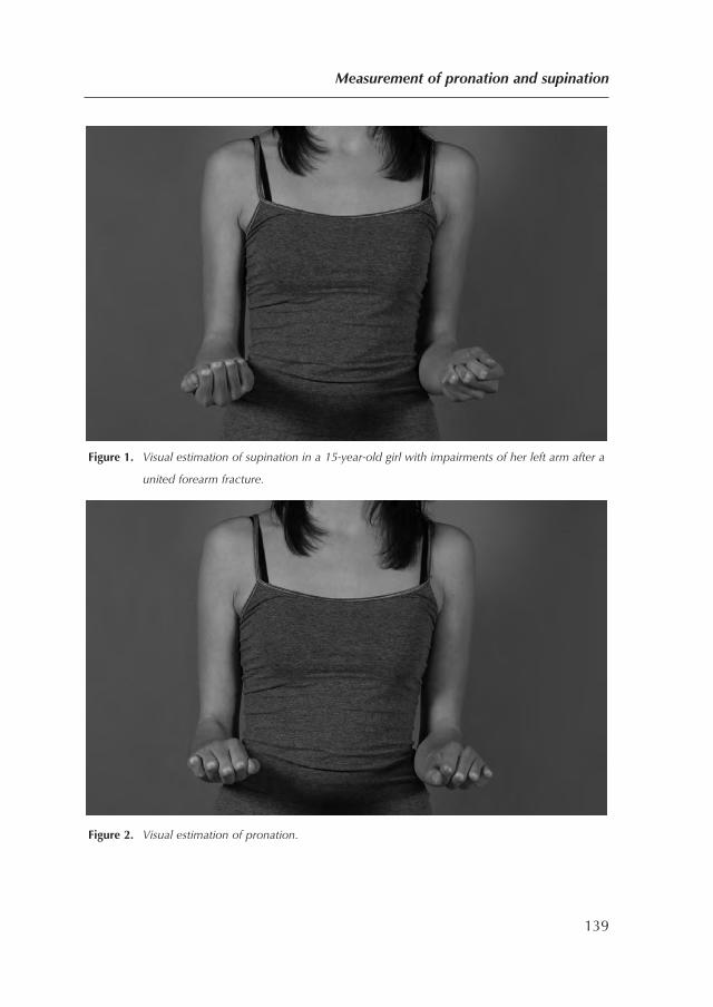
Figure 1. Visual estimation of supination in a 15-year-old girl with impairments of her left arm after a
united forearm fracture.
Figure 2. Visual estimation of pronation.
Measurement of pronation and supination
139
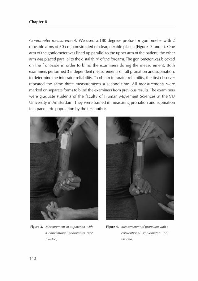
Goniometer measurement: We used a 180-degrees protractor goniometer with 2
movable arms of 30 cm, constructed of clear, flexible plastic (Figures 3 and 4). One
arm of the goniometer was lined up parallel to the upper arm of the patient, the other
arm was placed parallel to the distal third of the forearm. The goniometer was blocked
on the front-side in order to blind the examiners during the measurement. Both
examiners performed 3 independent measurements of full pronation and supination,
to determine the interrater reliability. To obtain intrarater reliability, the first observer
repeated the same three measurements a second time. All measurements were
marked on separate forms to blind the examiners from previous results. The examiners
were graduate students of the faculty of Human Movement Sciences at the VU
University in Amsterdam. They were trained in measuring pronation and supination
in a paediatric population by the first author.
Chapter 8
140
Figure 3. Measurement of supination with
a conventional goniometer (not
blinded).
Figure 4. Measurement of pronation with a
conventional goniometer (not
blinded).
Statistical analysis
First, the results of the 3 measurements of each examination were averaged. Using
SPSS 13.0, ICCs were calculated. When assessing reliability, a distinction between
intra-observer and inter-observer reliability was made. Intra-observer reliability
investigates the capability of one tester to produce consistent results. Inter-observer
reliability examines the extent of concurrence between the results of different
observers.
In addition to the ICC, the smallest detectable difference (SDD) were calculated
(10). The SDD is specifically valuable for clinical use since this index allows
distinguishing between measurement error and a real (treatment) change in
individual measurements since only a difference between consecutive measurements
that exceeds the SDD can be considered a real change. For example, a SDD of 5
degrees indicates that a follow-up measurement should differ by at least 5 degrees
from a baseline measurement to be sure that there is a real change in pronation or
supination angle of an individual subject. Hence, the SDD value reflects the smallest
clinically detectable change. Paired t-tests were used to compare the 2 experimental
methods. The effect of age on the measurement results was addressed by analysis of
variance (ANOVA) with Bonferoni post-hoc comparisons. The level of significance
was set at p<0.05.
RESULTS
Forty-seven children (34 boys and 13 girls) were included in this study. The mean
age was 11.1 ± 3.1 years (range 6-16 years). The dominant arm was fractured in 19
children. All 47 children successfully completed the study protocol. The results of
the visual estimation and goniometrical measurements are demonstrated in Table 1,
presenting the data of the whole group (6-16 years) and the data stratified according
to the 3 age groups: 6-9 years (15 patients), 10-13 years (16 patients) and 14-16
years (16 patients). Visual estimation showed higher pronation, supination and total
range of motion (ROM) angles compared to the goniometer data in all age groups.
In the overall group, this difference was statistically significant for the total ROM
only. For the 6-9 year old children, visual estimation angles were significantly larger
than goniometer angles for supination and total ROM.
Measurement of pronation and supination
141
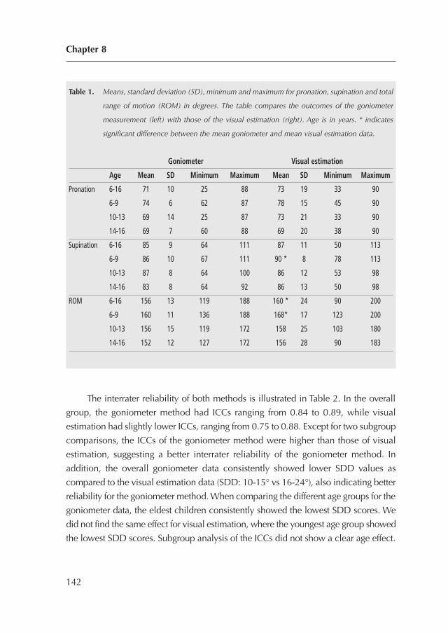
The interrater reliability of both methods is illustrated in Table 2. In the overall
group, the goniometer method had ICCs ranging from 0.84 to 0.89, while visual
estimation had slightly lower ICCs, ranging from 0.75 to 0.88. Except for two subgroup
comparisons, the ICCs of the goniometer method were higher than those of visual
estimation, suggesting a better interrater reliability of the goniometer method. In
addition, the overall goniometer data consistently showed lower SDD values as
compared to the visual estimation data (SDD: 10-15° vs 16-24°), also indicating better
reliability for the goniometer method. When comparing the different age groups for the
goniometer data, the eldest children consistently showed the lowest SDD scores. We
did not find the same effect for visual estimation, where the youngest age group showed
the lowest SDD scores. Subgroup analysis of the ICCs did not show a clear age effect.
Chapter 8
142
Table 1. Means, standard deviation (SD), minimum and maximum for pronation, supination and total
range of motion (ROM) in degrees. The table compares the outcomes of the goniometer
measurement (left) with those of the visual estimation (right). Age is in years. * indicates
significant difference between the mean goniometer and mean visual estimation data.
Goniometer Visual estimation
Age Mean SD Minimum Maximum Mean SD Minimum Maximum
Pronation 6-16 71 10 25 88 73 19 33 90
6-9 74 6 62 87 78 15 45 90
10-13 69 14 25 87 73 21 33 90
14-16 69 7 60 88 69 20 38 90
Supination 6-16 85 9 64 111 87 11 50 113
6-9 86 10 67 111 90 * 8 78 113
10-13 87 8 64 100 86 12 53 98
14-16 83 8 64 92 86 13 50 98
ROM 6-16 156 13 119 188 160 * 24 90 200
6-9 160 11 136 188 168* 17 123 200
10-13 156 15 119 172 158 25 103 180
14-16 152 12 127 172 156 28 90 183
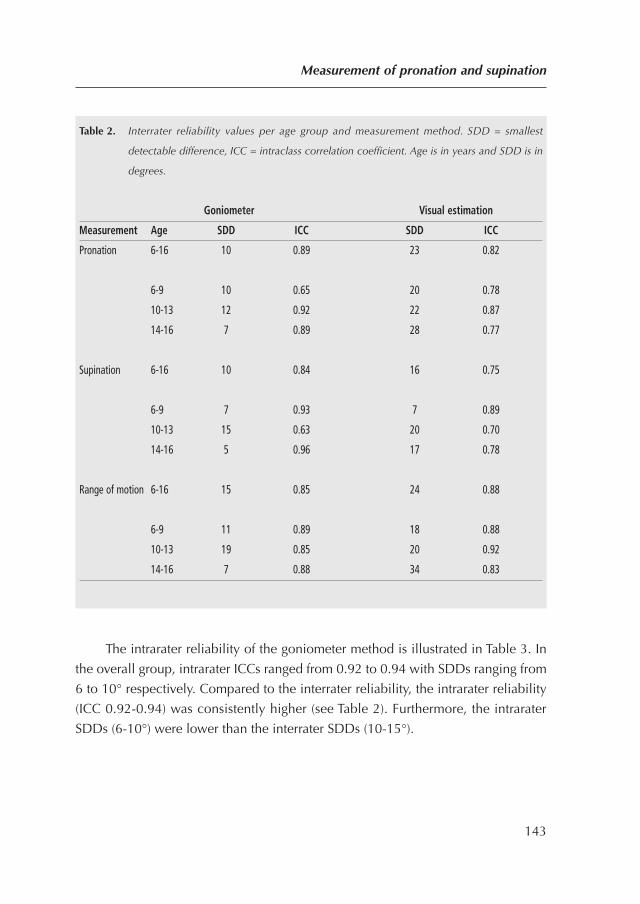
The intrarater reliability of the goniometer method is illustrated in Table 3. In
the overall group, intrarater ICCs ranged from 0.92 to 0.94 with SDDs ranging from
6 to 10° respectively. Compared to the interrater reliability, the intrarater reliability
(ICC 0.92-0.94) was consistently higher (see Table 2). Furthermore, the intrarater
SDDs (6-10°) were lower than the interrater SDDs (10-15°).
Measurement of pronation and supination
143
Table 2. Interrater reliability values per age group and measurement method. SDD = smallest
detectable difference, ICC = intraclass correlation coefficient. Age is in years and SDD is in
degrees.
Goniometer Visual estimation
Measurement Age SDD ICC SDD ICC
Pronation 6-16 10 0.89 23 0.82
6-9 10 0.65 20 0.78
10-13 12 0.92 22 0.87
14-16 7 0.89 28 0.77
Supination 6-16 10 0.84 16 0.75
6-9 7 0.93 7 0.89
10-13 15 0.63 20 0.70
14-16 5 0.96 17 0.78
Range of motion 6-16 15 0.85 24 0.88
6-9 11 0.89 18 0.88
10-13 19 0.85 20 0.92
14-16 7 0.88 34 0.83
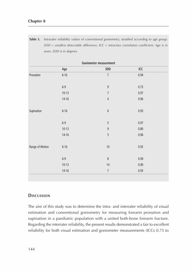
DISCUSSION
The aim of this study was to determine the intra- and interrater reliability of visual
estimation and conventional goniometry for measuring forearm pronation and
supination in a paediatric population with a united both-bone forearm fracture.
Regarding the interrater reliability, the present results demonstrated a fair to excellent
reliability for both visual estimation and goniometer measurements (ICCs 0.75 to
Chapter 8
144
Table 3. Intrarater reliability values of conventional goniometry, stratified according to age group.
SDD = smallest detectable difference, ICC = intraclass correlation coefficient. Age is in
years, SDD is in degrees.
Goniometer measurement
Age SDD ICC
Pronation 6-16 7 0.94
6-9 9 0.73
10-13 7 0.97
14-16 4 0.96
Supination 6-16 6 0.93
6-9 5 0.97
10-13 9 0.80
14-16 5 0.96
Range of Motion 6-16 10 0.92
6-9 8 0.94
10-13 14 0.90
14-16 7 0.93
0.89). These results are similar to previous studies with ICCs ranging from 0.72 to
0.98, using more complex devices to measure forearm rotation4,5,7,9. In this study,
we found that the goniometer measurements had significantly higher ICCs and lower
SDDs as compared to visual estimation, indicating better overall reliability and
higher specificity. This difference may partly be explained by the higher specificity
of the goniometer (1-degree steps) as compared to visual estimation (5-degree steps).
To our knowledge, the only study that has dealt with visual estimation dates back to
1976 and concluded that goniometer measurements were more reliable than
estimation of elbow flexion and wrist extension12. When analysing the effect of age
on the reliability of measuring forearm rotation, we found no consistent effect of age
on the ICCs. However, the SDD values of the goniometer method were consistently
lower in the eldest children. This indicates that the differences between repeated
measures are smaller in older children. For visual estimation the opposite was found:
SDD values of visual estimation were lowest in the youngest children. However,
visual estimation was still less accurate as compared to goniometer measurements
in the younger aged. The intrarater reliability of the goniometer measurements in the
present study was excellent, ranged between 0.92 to 0.94, which is comparable with
the intrarater reliability that has previously been found using more complex devices,
ranging from 0.78 to 0.994,6,9. The intrarater reliability in the present study was
consistently higher that interrater reliability, indicating that repeated examination by
the same professional should generally be preferred.
A limitation of this study is that we compared only two examiners, both of
whom were graduate students without extensive clinical experience in working with
this paediatric population. It should be noted, however, that previous literature
showed that the accuracy of measurements is independently of the type of
professional4. Another limitation was that the intrarater reliability of visual estimation
could not be reliably tested in the current study design, which consisted of a one
day visit of the children. The particular strengths of this study included the blinded
design, the measurement of a relevant group of patients and the use of conventional
goniometry as well as visual estimation as primary methods. Furthermore, the
evaluation of the intra- and inter reliability, as well as use of smallest detectable
difference scores, gives it substantial clinical relevance. In conclusion, based on the
present study, we recommend the use of the conventional goniometer for measuring
Measurement of pronation and supination
145
pronation and supination in daily practice. If an uncooperative child interferes with
the measurement, visual estimation is a good second choice. To minimise the
variability of the measurements, professionals should measure the forearm rotation
with the arms in the same documented position.
Chapter 8
146
REFERENCES
1. Holdsworth BJ, Sloan JP. Proximal forearm fractures in children: residual disability. Injury.
1982;14(2):174-9.
2. Daruwalla JS. A study of radioulnar movements following fractures of the forearm in
children. Clin Orthop Relat Res. 1979(139):114-20.
3. Kasten P, Krefft M, Hesselbach J, Weinberg AM. How does torsional deformity of the radial
shaft influence the rotation of the forearm? A biomechanical study. J Orthop Trauma.
2003;17(1):57-60.
4. Armstrong AD, MacDermid JC, Chinchalkar S, Stevens RS, King GJ. Reliability of range-of-
motion measurement in the elbow and forearm. J Shoulder Elbow Surg. 1998;7(6):573-80.
5. Flowers KR, Stephens-Chisar J, LaStayo P, Galante BL. Intrarater reliability of a new method
and instrumentation for measuring passive supination and pronation: a preliminary study. J
Hand Ther. 2001;14(1):30-5.
6. Greene BL, Wolf SL. Upper extremity joint movement: comparison of two measurement
devices. Arch Phys Med Rehabil. 1989;70(4):288-90.
7. Karagiannopoulos C, Sitler M, Michlovitz S. Reliability of 2 functional goniometric methods
for measuring forearm pronation and supination active range of motion. J Orthop Sports
Phys Ther. 2003;33(9):523-31.
8. Patrick J. A study of supination and pronation, with especial reference to the treatment of
forearm fractures. J Bone Joint Surg Br. 1946;28:737-48.
9. Urban V, Kalberer F, Roos M, Dumont CE. [Reliability of active range-of-motion
measurement of the rotation in the forearm: comparison of three measurement devices]. Z
Orthop Ihre Grenzgeb. 2002;140(1):72-6.
10. Weir JP. Quantifying test-retest reliability using the intraclass correlation coefficient and the
SEM. J Strength Cond Res. 2005;19(1):231-40.
11. Boone DC, Azen SP, Lin CM, Spence C, Baron C, Lee L. Reliability of goniometric
measurements. Phys Ther. 1978;58(11):1355-90.
12. Low JL. The reliability of joint measurement. Physiotherapy. 1976;62(7):227-9.
13. Rothstein JM, Miller PJ, Roettger RF. Goniometric reliability in a clinical setting. Elbow and
knee measurements. Phys Ther. 1983;63(10):1611-5.
Measurement of pronation and supination
147

148

CHAPTER 9
Which factors affect limitation ofpronation/supination afterforearm fractures in children? A prospective multicentre study
Injury. 2014 Apr;45(4):696-700.
Colaris JW, Allema JH, Reijman M, de Vries MR, Ulas Biter L,
Bloem RM, van de Ven CP, Verhaar JA.
149
ABSTRACT
Introduction: Both-bone forearm fractures in children frequently result in a limitation
of pronation/supination, which hinders daily activities. The purpose of this
prospective multicentre study was to investigate which clinical factors are related to
the limitation of pronation/supination in children with a both-bone forearm fracture.
Methods: In four Dutch hospitals consecutive children (<16 years) who sustained a
both-bone forearm fracture were included. Children were followed for 6-9 months
and data from questionnaires, physical examination, and X-rays were collected.
Univariate and multivariate logistic regression analyses were used to assess the
relationship between limitation of pronation/supination (≥ 20 degrees) and several
clinical factors.
Results: A group of 410 children with both-bone forearm fractures were included of
which 10 children missed the final examination (follow-up rate of 97.6%). We found
that a re-fracture (odds ratio (OR) 11.7, 95% confidence interval (CI) 1.2; 118.5), a
fracture in the diaphysis (OR 3.3, 95% CI 1.4; 7.9) and less physiotherapy during
follow-up (OR 0.9, 95% CI 0.82; 0.98) were independently associated with a
limitation of pronation/supination of 20 degrees or more.
Conclusions: These findings imply that a re-fracture and a diaphyseal located fracture
were independently of each other associated with a limitation of
pronation/supination in children with a both-bone forearm fracture. Furthermore, in
children with severe limitation extensive physiotherapy is associated with better
functional outcome.
Chapter 9
150
INTRODUCTION
Severe limitation of pronation/supination following both-bone forearm fractures in
children hinders daily activities. For example, while pronation is used for writing
and typing, movements such as perineal hygiene and accepting monetary change
require supination. Fortunately, the ipsilateral shoulder can compensate mild
limitation of pronation by abduction and internal rotation, and mild limitation of
supination by adduction and external rotation.
Pronation/supination of the forearm is not a simple rotational motion but a
complex motion with rotation and translation1. This complex motion in combination
with the complex anatomy of the forearm makes it difficult to find the exact cause
of this limitation. In general, it can be caused by pathology of the bones or soft tissue
around it. Pathology of the bones consists of a malunion of the radius and/or ulna
and is a well-known cause of limitation of pronation/supination2-9. The soft-tissue
pathology as cause of limitation is less well known. Some studies reported on
contractures of the interosseous membrane10,11 while others stressed the role of
contractures around wrist and elbow12,13.
Although previous literature has reported on pathology of bones and soft
tissue as a cause of limitation of pronation/supination, many other clinical factors
associated with this limitation have not yet been explored. However, knowledge on
these factors can contribute to better clinical care by change of treatment (when
required) and detailed information provided to parents and children.
Therefore, we designed a prospective multicentre study with the purpose of
investigating which clinical factors are related to the limitation of pronation/
supination in children with a both-bone forearm fracture.
MATERIALS AND METHODS
Study design and participants
This prospective study included consecutive children with a forearm fracture of
radius and ulna who visited the emergency department of one of four participating
Dutch hospitals: Erasmus Medical Center (Rotterdam), HAGA Hospital (The Hague),
Reinier de Graaf Hospital (Delft) and Sint Franciscus Hospital (Rotterdam). The
Which factors affect limitation of pronation/supination?
151
regional medical ethics committee approved the study. Informed consent for
participation was obtained from all parents and from all children aged ≥ 12 years.
We included children aged < 16 years who sustained a both-bone forearm
fracture in the diaphysis or distal metaphysis. The criteria for reduction were a priori
defined and based on previous studies14-23 (Table 1). Exclusion criteria were torus
fractures of both radius and ulna, fractures older than 1 week, severe open fractures
(Gustilo II and III)24 and re-fractures.
Procedures
The included children participated in several randomised controlled studies. The
study protocol distinguished different types of both-bone forearm fractures (Table 2).
Briefly, distal metaphyseal fractures without need for reduction were treated with
below-elbow cast (BEC) or above-elbow cast (AEC) for 4 weeks25, while distal
metaphyseal fractures with reduction were treated with or without Kirschner wires
(K wires) in combination with AEC for 4 weeks26. In addition, diaphyseal fractures
without reduction and reduced stable diaphyseal fractures were treated for 6 weeks
AEC, or a combination of 3 weeks AEC and 3 weeks BEC. Finally, unstable
diaphyseal fractures were fixated with 1 or 2 intramedullary nails in combination
with 3 weeks AEC. In case of an AEC, the elbow was set in 90 degrees of flexion
with the forearm in neutral position. A BEC allowed free movement of the elbow
joint.
Chapter 9
152
Table 1. Criteria for reduction of the fracture of radius and/or ulna based on posteroanterior and/or
lateral radiographs.
Type of deformity Age in years Deformity
Angulation < 10 > 15 degrees
10-16 > 10 degrees
Translation < 16 > half of bone diameter
Rotation < 16 > 0
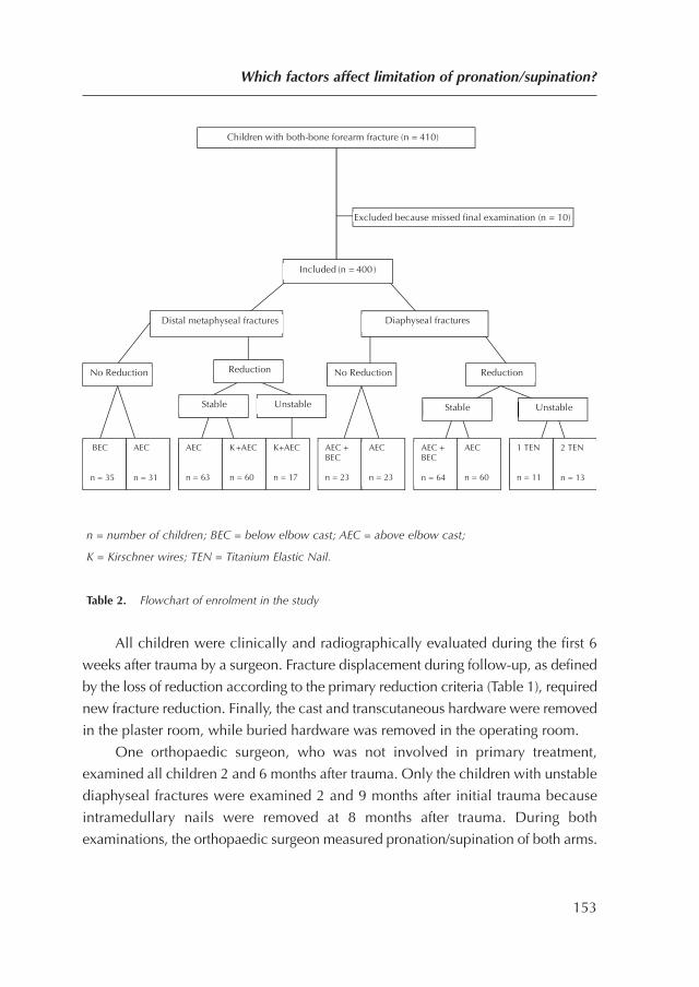
n = number of children; BEC = below elbow cast; AEC = above elbow cast;
K = Kirschner wires; TEN = Titanium Elastic Nail.
Table 2. Flowchart of enrolment in the study
All children were clinically and radiographically evaluated during the first 6
weeks after trauma by a surgeon. Fracture displacement during follow-up, as defined
by the loss of reduction according to the primary reduction criteria (Table 1), required
new fracture reduction. Finally, the cast and transcutaneous hardware were removed
in the plaster room, while buried hardware was removed in the operating room.
One orthopaedic surgeon, who was not involved in primary treatment,
examined all children 2 and 6 months after trauma. Only the children with unstable
diaphyseal fractures were examined 2 and 9 months after initial trauma because
intramedullary nails were removed at 8 months after trauma. During both
examinations, the orthopaedic surgeon measured pronation/supination of both arms.
Which factors affect limitation of pronation/supination?
153
Children with both-bone forearm fracture (n = 410)
No Reduction Reduction
BEC n = 35
AEC n = 31
AEC + BEC n = 23
AEC n = 23
K+AEC n = 17
Excluded because missed final examination (n = 10)
Included (n = 400 )
Diaphyseal fracturesDistal metaphyseal fractures
Reduction No Reduction
AEC n = 63
K+AEC n = 60
2 TEN n = 13
1 TEN n = 11
AEC n = 60
AEC + BEC n = 64
Stable Unstable Stable Unstable
This technique was standardised with the use of a 180° goniometer constructed of
clear, flexible plastic with 2 movable arms of 30 cm27. Children who suffered from
at least 30 degree of limitation at the 2-month examination were referred to a
physiotherapist.
Radiographs were taken directly after trauma, after reduction, during the
treatment in cast, and at final follow-up. The orthopaedic surgeon measured
angulation, translation, rotation and shortening of all fractures to determine primary
displacement, displacement during treatment with cast, and final displacement.
Rotation was detected by differences in diameter of diaphysis of ulna and radius on
lateral and posteroanterior radiographs28. To assess the interrater reproducibility of
the radiological assessment, a trauma surgeon who was not involved in treatment,
re-measured the angulation of the fracture in 45 children independently of the
orthopaedic surgeon.
Multivariate analysis
Our dependent variable was limitation of pronation/supination of 20 degrees or more
at the end of follow-up. For the following factors we assessed whether they were
related to the above-mentioned dependent variable: age at time of fracture, dominant
arm fractured, location of the fracture (diaphysis vs. distal metaphysis), type of
fracture (torus, greenstick, complete), primary displacement of the fracture, number
of reductions, presence of a re-fracture, duration of AEC, number of physiotherapy
visits, and final displacement of the fracture.
This choice of factors was based on the assumption that less limitation would
be found in children of younger age because of their higher repair capacity7.
Furthermore, less limitation was expected as the fracture occurred in the dominant
arm due to the need for the dominant arm in daily activities. The assumption of less
limitation in distal metaphyseal fractures, less severe type of fractures, less primary
displacement of the fracture, fewer reductions and no re-fracture, is in all these cases
based on less soft tissue damage6,7,29. Thereby, we postulated that short
immobilisation of the elbow in AEC and frequent visits to a physiotherapist might
cause less limitation30. The effect of malunion on limitation has been mentioned by
others2-9.
Chapter 9
154
Statistical Analysis
To assess which factors were related to limitation of pronation/supination of 20
degrees or more at the end of follow-up, multivariate logistic regression analysis (via
the ‘enter’ method) was used to calculate odds ratios (ORs) and 95% confidence
intervals (95% CI). In all analyses, a two-sided p-value of ≤ 0.05 was considered
significant. Statistical analyses were performed with SPSS 17.0 (SPSS Inc., Chicago,
USA).
RESULTS
Between January 2006 and August 2010 a total of 410 children with both-bone
forearm fractures were included. Ten children missed the final examination and were
therefore excluded (follow-up rate of 97.6%). The included children had several
types of both-bone forearm fractures (Table 2). The mean length of follow-up was
218 (± 49) days.
Table 3 presents the results of both groups and of the univariate and multivariate
analyses. Six of 10 factors had a significant univariate relationship with limitation of
pronation/supination. Half of these factors also showed a significant multivariate
relationship and therefore had an independent relation with limitation of
pronation/supination.
The presence of a re-fracture was strongly associated with limitation of
pronation/supination (Figure 1) with an OR of 11.7 (95% CI 1.2; 118.5). Thereby,
the location of the fracture (diaphyseal vs. distal metaphyseal) and number of
physiotherapy visits were significant in the multivariate analyses with an OR of 3.3
(95% CI 1.4; 7.9) and 0.90 (95% CI 0.82; 0.98), respectively. These findings imply
that a re-fracture, a diaphyseal located fracture and less physiotherapy were
independently of each other associated with a limitation of pronation/supination of
20 degrees or more in children with a both-bone forearm fracture. The OR of 11.7
for a re-fracture implies that a child who sustained a re-fracture had an almost 12
times increased risk to end up with a limitation of pronation/supination of 20 degrees
or more.
Which factors affect limitation of pronation/supination?
155
Chapter 9
156
Table 3. Factors associated with limitation of pronation/supination.
Logistic regression analyses
No limitation Limitation Univariate Multivariate
OR (95% CI) OR (95% CI)
Age at time of fracture, years 7.7 ± 3.4 8.9 ± 3.6 1.1 (1.0; 1.2) -Dominant arm fractured, % 39.4 40.7 - -Location of fracture, % 2.7 (1.7; 4.2) 3.3 (1.4; 7.9)
Distal metaphyseal 57.9 33.9Diaphyseal 42.1 66.1
Type of fracture radius, % - -Torus 1.4 0.9Greenstick 44.4 49.1Complete 54.2 50.0
Type of fracture ulna, % - -Torus 5.2 8.3Greenstick 54.2 59.3Complete 40.6 32.4
Primary deformity of fractureAngulation, degrees 24.2 ±14.4 24.3 ± 12.4 - -Translation, % of width 37.6 ± 44.4 27.9 ± 41.2 - -Rotation, % 9.4 9.3 - -Shortening, % 35.2 25.9 - -
Number of reductions, % - -0 27.0 20.21 67.2 67.92 5.5 11.03 0.3 0.9
Presence of a re-fracture, % 1.0 12.8 14.1 (4.0; 50.1) 11.7 (1.2; 118.5)Days in above-elbow cast 29.4 ± 8.6 32.7 ± 12.1 1.0 (1.0; 1.1) -Number of physiotherapy visits 7.5 ± 7.5 4.3 ± 4.1 0.9 (0.8; 1.0) 0.90 (0.82; 0.98)Final deformity of fracture
Angulation, degrees 9.3 ± 4.4 8.3 ± 4.1 1.0 (0.9; 1.0) -Translation, % of width 6.8 ± 13.1 4.9 ± 9.2 - -
Limitation is defined as limitation of pronation and supination of 20 degrees or more; % = percentage; Values arepresented as mean ± standard deviation unless stated otherwise; OR = Odds Ratio; CI = Confidence Interval.In the table of odds ratio only the significant results are mentioned.
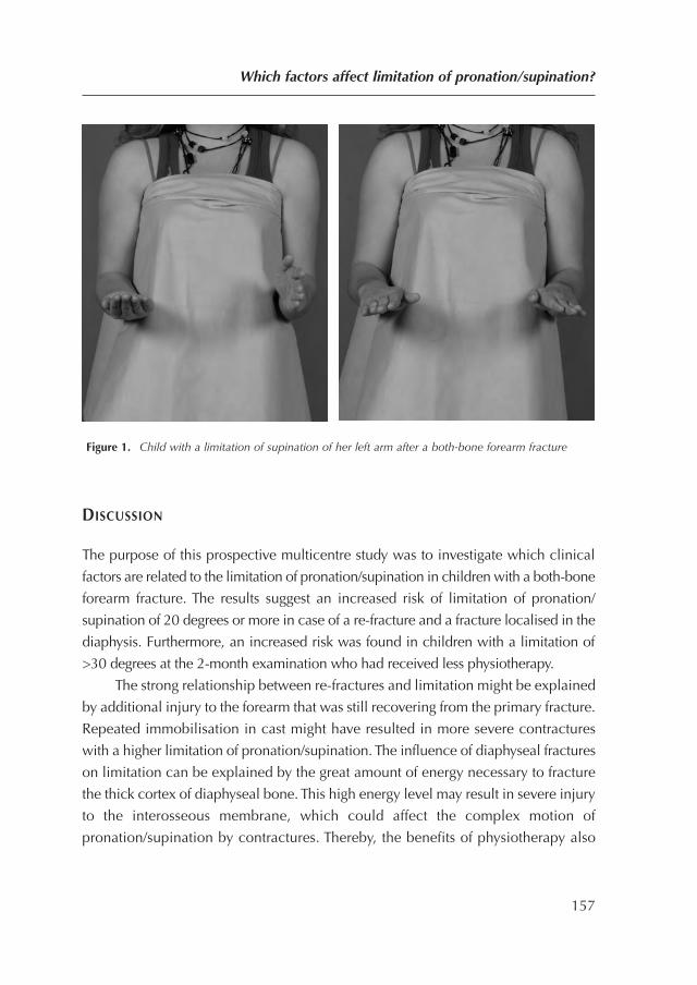
Figure 1. Child with a limitation of supination of her left arm after a both-bone forearm fracture
DISCUSSION
The purpose of this prospective multicentre study was to investigate which clinical
factors are related to the limitation of pronation/supination in children with a both-bone
forearm fracture. The results suggest an increased risk of limitation of pronation/
supination of 20 degrees or more in case of a re-fracture and a fracture localised in the
diaphysis. Furthermore, an increased risk was found in children with a limitation of
>30 degrees at the 2-month examination who had received less physiotherapy.
The strong relationship between re-fractures and limitation might be explained
by additional injury to the forearm that was still recovering from the primary fracture.
Repeated immobilisation in cast might have resulted in more severe contractures
with a higher limitation of pronation/supination. The influence of diaphyseal fractures
on limitation can be explained by the great amount of energy necessary to fracture
the thick cortex of diaphyseal bone. This high energy level may result in severe injury
to the interosseous membrane, which could affect the complex motion of
pronation/supination by contractures. Thereby, the benefits of physiotherapy also
Which factors affect limitation of pronation/supination?
157
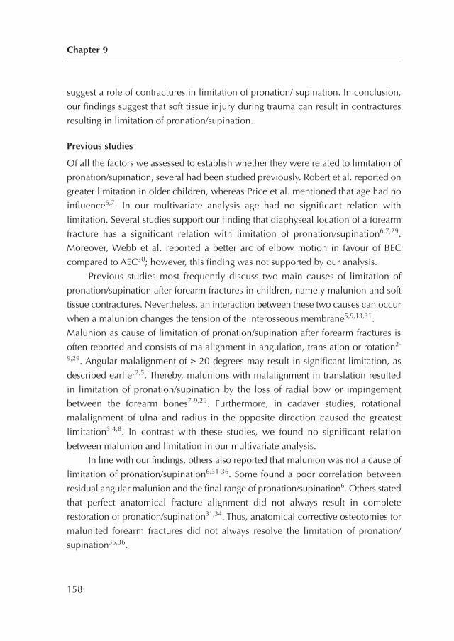
suggest a role of contractures in limitation of pronation/ supination. In conclusion,
our findings suggest that soft tissue injury during trauma can result in contractures
resulting in limitation of pronation/supination.
Previous studies
Of all the factors we assessed to establish whether they were related to limitation of
pronation/supination, several had been studied previously. Robert et al. reported on
greater limitation in older children, whereas Price et al. mentioned that age had no
influence6,7. In our multivariate analysis age had no significant relation with
limitation. Several studies support our finding that diaphyseal location of a forearm
fracture has a significant relation with limitation of pronation/supination6,7,29.
Moreover, Webb et al. reported a better arc of elbow motion in favour of BEC
compared to AEC30; however, this finding was not supported by our analysis.
Previous studies most frequently discuss two main causes of limitation of
pronation/supination after forearm fractures in children, namely malunion and soft
tissue contractures. Nevertheless, an interaction between these two causes can occur
when a malunion changes the tension of the interosseous membrane5,9,13,31.
Malunion as cause of limitation of pronation/supination after forearm fractures is
often reported and consists of malalignment in angulation, translation or rotation2-
9,29. Angular malalignment of ≥ 20 degrees may result in significant limitation, as
described earlier2,5. Thereby, malunions with malalignment in translation resulted
in limitation of pronation/supination by the loss of radial bow or impingement
between the forearm bones7-9,29. Furthermore, in cadaver studies, rotational
malalignment of ulna and radius in the opposite direction caused the greatest
limitation3,4,8. In contrast with these studies, we found no significant relation
between malunion and limitation in our multivariate analysis.
In line with our findings, others also reported that malunion was not a cause of
limitation of pronation/supination6,31-36. Some found a poor correlation between
residual angular malunion and the final range of pronation/supination6. Others stated
that perfect anatomical fracture alignment did not always result in complete
restoration of pronation/supination31,34. Thus, anatomical corrective osteotomies for
malunited forearm fractures did not always resolve the limitation of pronation/
supination35,36.
Chapter 9
158
Furthermore, several studies support the important role of contractures of soft tissue
in limitation of pronation/supination after forearm fractures7,10-13. Some reported on
contractures of the interosseous membrane10,11 while others described the role of
contractures around wrist and elbow12,13. One study described a correlation between
initial fracture displacement, MR-detected interosseous membrane alternations, and
limitation of pronation/supination37.
In summary, previous studies have conflicting opinions about the role of
malunion in the limitation of pronation/supination after forearm fractures in children.
We found a significant role of malunion based on univariate analysis. In contrast,
no significant role was found in our multivariate logistic regression analysis,
suggesting that malunion is not independently associated with a limitation of
pronation/supination of 20 degrees or more.
Our results suggest that scarring of the injured soft tissue around the fracture
can lead to contractures that probably play an important role in limitation of
pronation/supination.
Limitations
The present study has several limitations. Primarily, rotation of the fracture was
detected by differences in the diameter of diaphysis of ulna and radius on lateral
and posteroanterior radiographs28; this technique probably overlooks subtle rotation
of fractures. However, Kasten et al. found limitation of pronation/supination only in
fractures with more than 30 degrees of malrotation, which should have been
detected with the method used4.
Secondly, ideally our follow-up period should have been longer in this young
population with the capacity to correct malunion by growth. Nevertheless, malunion
was not independently associated with higher limitation in our multivariate analysis.
Thirdly, we cannot exclude injury to the distal or proximal radio-ulnar joint as
a possible cause of limitation of pronation/supination.
Conclusion
The results of this prospective multicentre study, with an analysis of 400 children,
suggest that children who suffered from a diaphyseal fracture or sustained a re-
fracture have an increased risk of limitation of pronation/supination during follow-up.
Which factors affect limitation of pronation/supination?
159
Furthermore, in children with severe limitation extensive physiotherapy is associated
with better functional outcome.
Chapter 9
160
REFERENCES
1. Nakamura T, Yabe Y, Horiuchi Y, Yamazaki N. In vivo motion analysis of forearm rotation
utilizing magnetic resonance imaging. Clin Biomech (Bristol, Avon). 1999;14(5):315-20.
2. Bronstein AJ, Trumble TE, Tencer AF. The effects of distal radius fracture malalignment on
forearm rotation: a cadaveric study. J Hand Surg [Am]. 1997;22(2):258-62.
3. Dumont CE, Thalmann R, Macy JC. The effect of rotational malunion of the radius and the
ulna on supination and pronation. J Bone Joint Surg Br. 2002;84(7):1070-4.
4. Kasten P, Krefft M, Hesselbach J, Weinberg AM. How does torsional deformity of the radial
shaft influence the rotation of the forearm? A biomechanical study. J Orthop Trauma.
2003;17(1):57-60.
5. Matthews LS, Kaufer H, Garver DF, Sonstegard DA. The effect on supination-pronation of
angular malalignment of fractures of both bones of the forearm. J Bone Joint Surg Am.
1982;64(1):14-7.
6. Price CT, Scott DS, Kurzner ME, Flynn JC. Malunited forearm fractures in children. J Pediatr
Orthop. 1990;10(6):705-12.
7. Roberts JA. Angulation of the radius in children’s fractures. J Bone Joint Surg Br.
1986;68(5):751-4.
8. Trousdale RT, Linscheid RL. Operative treatment of malunited fractures of the forearm. J
Bone Joint Surg Am. 1995;77(6):894-902.
9. Yasutomi T, Nakatsuchi Y, Koike H, Uchiyama S. Mechanism of limitation of
pronation/supination of the forearm in geometric models of deformities of the forearm
bones. Clin Biomech (Bristol, Avon). 2002;17(6):456-63.
10. Nakamura T, Yabe Y, Horiuchi Y, Seki T, Yamazaki N. Normal kinematics of the interosseous
membrane during forearm pronation-supination—a three-dimensional MRI study. Hand
Surg. 2000;5(1):1-10.
11. Nakamura T, Yabe Y, Horiuchi Y. [A biomechanical analysis of pronation-supination of the
forearm using magnetic resonance imaging: dynamic changes of the interosseous
membrane of the forearm during pronation-supination]. Nippon Seikeigeka Gakkai Zasshi.
1994;68(1):14-25.
12. Kleinman WB, Graham TJ. The distal radioulnar joint capsule: clinical anatomy and role in
posttraumatic limitation of forearm rotation. J Hand Surg Am. 1998;23(4):588-99.
Which factors affect limitation of pronation/supination?
161
13. Shirai H, Ryu J, Kish V, Abe M. The rotational effect of pronation and supination osteotomies
of the forearm in a cadaver model: a comparison of osteotomy sites on the radius and the
ulna. J Hand Surg Eur Vol. 2007;32(4):440-6.
14. Boyer BA, Overton B, Schrader W, Riley P, Fleissner P. Position of immobilization for
pediatric forearm fractures. Journal of pediatric orthopedics. 2002;22(2):185-7.
15. Choi KY, Chan WS, Lam TP, Cheng JC. Percutaneous Kirschner-wire pinning for severely
displaced distal radial fractures in children. A report of 157 cases. J Bone Joint Surg Br.
1995;77(5):797-801.
16. Daruwalla JS. A study of radioulnar movements following fractures of the forearm in
children. Clin Orthop Relat Res. 1979(139):114-20.
17. Gibbons CL, Woods DA, Pailthorpe C, Carr AJ, Worlock P. The management of isolated
distal radius fractures in children. J Pediatr Orthop. 1994;14(2):207-10.
18. Jones K, Weiner DS. The management of forearm fractures in children: a plea for
conservatism. J Pediatr Orthop. 1999;19(6):811-5.
19. Mani GV, Hui PW, Cheng JC. Translation of the radius as a predictor of outcome in distal
radial fractures of children. J Bone Joint Surg Br. 1993;75(5):808-11.
20. Maric D, Petkovic L, Tomasevic V, Bajic L, Gajdobranski D, Marcikic A. [Osteosynthesis
with Kirschner wires in the treatment of dislocation fractures of the forearm in children].
Med Pregl. 2002;55(3-4):140-5.
21. Noonan KJ, Price CT. Forearm and distal radius fractures in children. J Am Acad Orthop
Surg. 1998;6(3):146-56.
22. Tynan MC, Fornalski S, McMahon PJ, Utkan A, Green SA, Lee TQ. The effects of ulnar axial
malalignment on supination and pronation. J Bone Joint Surg Am. 2000;82-A(12):1726-31.
23. Zimmermann R, Gabl M, Angermann P, Lutz M, Reinhart C, Kralinger F, et al. [Late
sequelae of fractures of the distal third of the forearm during the growth period]. Handchir
Mikrochir Plast Chir. 2000;32(4):242-9.
24. Gustilo RB, Simpson L, Nixon R, Ruiz A, Indeck W. Analysis of 511 open fractures. Clin
Orthop Relat Res. 1969;66:148-54.
25. Colaris JW, Biter LU, Allema JH, Bloem RM, van de Ven CP, de Vries MR, et al. Below-
elbow cast for metaphyseal both-bone fractures of the distal forearm in children: a
randomised multicentre study. Injury. 2012;43(7):1107-11.
Chapter 9
162
26. Colaris JW, Allema JH, Biter LU, de Vries MR, van de Ven CP, Bloem RM, et al. Re-
displacement of stable distal both-bone forearm fractures in children: a randomised
controlled multicentre trial. Injury. 2013;44(4):498-503.
27. Colaris J, van der Linden M, Selles R, Coene N, Allema JH, Verhaar J. Pronation and
supination after forearm fractures in children: Reliability of visual estimation and
conventional goniometry measurement. Injury. 2010;41(6):643-6.
28. Naimark A, Kossoff J, Leach RE. The disparate diameter. A sign of rotational deformity in
fractures. J Can Assoc Radiol. 1983;34(1):8-11.
29. Kuderna H. [Connection between deviated axis and impaired function after fractures of the
forearm (author’s transl)]. Unfallchirurgie. 1980;6(1):7-13.
30. Webb GR, Galpin RD, Armstrong DG. Comparison of short and long arm plaster casts for
displaced fractures in the distal third of the forearm in children. J Bone Joint Surg Am.
2006;88(1):9-17.
31. Tarr RR, Garfinkel AI, Sarmiento A. The effects of angular and rotational deformities of both
bones of the forearm. An in vitro study. J Bone Joint Surg Am. 1984;66(1):65-70.
32. Hogstrom H, Nilsson BE, Willner S. Correction with growth following diaphyseal forearm
fracture. Acta Orthop Scand. 1976;47(3):299-303.
33. Nilsson BE, Obrant K. The range of motion following fracture of the shaft of the forearm in
children. Acta Orthop Scand. 1977;48(6):600-2.
34. Krukhaug Y, Hove LM. Corrective osteotomy for malunited extra-articular fractures of the
distal radius: a follow-up study of 33 patients. Scand J Plast Reconstr Surg Hand Surg.
2007;41(6):303-9.
35. Nagy L, Jankauskas L, Dumont CE. Correction of forearm malunion guided by the
preoperative complaint. Clin Orthop Relat Res. 2008;466(6):1419-28.
36. Price CT, Knapp DR. Osteotomy for malunited forearm shaft fractures in children. J Pediatr
Orthop. 2006;26(2):193-6.
37. Beyer W, Stolzenburg T, Paris S. [Functional limitation of the forearm after shaft fracture in
childhood. Possible role of the antebrachial interosseous membrane: MRI and ultrasound
studies]. Unfallchirurgie. 1995;21(6):275-84.
Which factors affect limitation of pronation/supination?
163

164
CHAPTER 10
Angular malalignment as causeof limitation of forearm rotation:an analysis of prospectivelycollected data of both-boneforearm fractures in children Injury. 2014 Jun;45(6):955-9.
Colaris JW, Reijman M, Allema JH, de Vries MR, Biter LU,
Bloem RM, van de Ven CP, Verhaar JA.
165
ABSTRACT
Introduction: Although limitation of pronation/supination following both-bone
forearm fractures in children is often attributed to an angular malunion, no clinical
study has compared pronation/supination and angular malalignment of the same
child by analysis of prospectively collected clinical data. Therefore, the purpose of
this trial was to explore whether limitation of pronation/supination can be predicted
by the degree of angular malalignment in children who sustained a both-bone
forearm fracture.
Methods: In four Dutch hospitals, consecutive children aged ≤ 16 years with a both-
bone forearm fracture were prospectively followed for 6-9 months. At final
follow-up, pronation/supination and angular malunion on radiographs were
determined.
Results: Between January 2006 and August 2010 a total of 410 children were
prospectively followed of which 393 children were included for analysis in this study.
The mean age of the children was 8.0 (± 3.5) years, 63% was male and 40%
fractured their dominant arm. The mean time to final examination was 219 (± 51)
days. Children with a metaphyseal both-bone fracture of the distal forearm with an
angular malalignment of ≤ 15 degrees had a 9-13% chance to develop a clinically
relevant limitation (i.e. < 50 degrees of pronation and/or supination), while children
with an angular malalignment of ≥ 16 degrees had a 60% chance. Children with
diaphyseal both-bone forearm fractures with ≤ 5 degrees of angular malalignment
had a 13% chance to develop a clinically relevant limitation, which showed no
significant increase with a further increase of angular malalignment.
Conclusions: Children who sustained a both-bone forearm fracture localized in the
distal metaphysis have a higher chance to develop a clinically relevant limitation of
forearm rotation in case of a more severe angular malalignment, while children with
a diaphyseal both-bone forearm fracture had a moderate chance of limitation,
irrespective of the severity of the angular malalignment.
Chapter 10
166
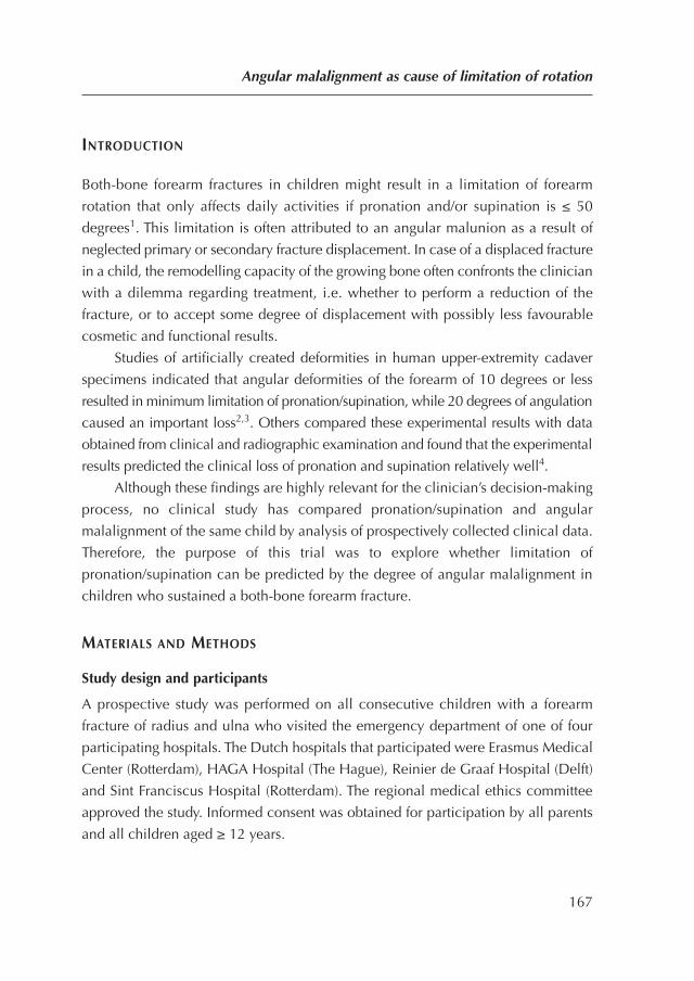
INTRODUCTION
Both-bone forearm fractures in children might result in a limitation of forearm
rotation that only affects daily activities if pronation and/or supination is ≤ 50
degrees1. This limitation is often attributed to an angular malunion as a result of
neglected primary or secondary fracture displacement. In case of a displaced fracture
in a child, the remodelling capacity of the growing bone often confronts the clinician
with a dilemma regarding treatment, i.e. whether to perform a reduction of the
fracture, or to accept some degree of displacement with possibly less favourable
cosmetic and functional results.
Studies of artificially created deformities in human upper-extremity cadaver
specimens indicated that angular deformities of the forearm of 10 degrees or less
resulted in minimum limitation of pronation/supination, while 20 degrees of angulation
caused an important loss2,3. Others compared these experimental results with data
obtained from clinical and radiographic examination and found that the experimental
results predicted the clinical loss of pronation and supination relatively well4.
Although these findings are highly relevant for the clinician’s decision-making
process, no clinical study has compared pronation/supination and angular
malalignment of the same child by analysis of prospectively collected clinical data.
Therefore, the purpose of this trial was to explore whether limitation of
pronation/supination can be predicted by the degree of angular malalignment in
children who sustained a both-bone forearm fracture.
MATERIALS AND METHODS
Study design and participants
A prospective study was performed on all consecutive children with a forearm
fracture of radius and ulna who visited the emergency department of one of four
participating hospitals. The Dutch hospitals that participated were Erasmus Medical
Center (Rotterdam), HAGA Hospital (The Hague), Reinier de Graaf Hospital (Delft)
and Sint Franciscus Hospital (Rotterdam). The regional medical ethics committee
approved the study. Informed consent was obtained for participation by all parents
and all children aged ≥ 12 years.
Angular malalignment as cause of limitation of rotation
167
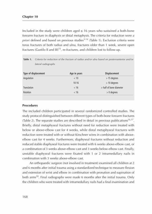
Included in the study were children aged ≤ 16 years who sustained a both-bone
forearm fracture in diaphysis or distal metaphysis. The criteria for reduction were a
priori defined and based on previous studies5-14 (Table 1). Exclusion criteria were
torus fractures of both radius and ulna, fractures older than 1 week, severe open
fractures (Gustilo II and III)15, re-fractures, and children lost to follow-up.
Procedures
The included children participated in several randomized controlled studies. The
study protocol distinguished between different types of both-bone forearm fractures
(Table 2). The separate studies are described in detail in previous publications16,17.
Briefly, distal metaphyseal fractures without need for reduction were treated with
below or above-elbow cast for 4 weeks, while distal metaphyseal fractures with
reduction were treated with or without Kirschner wires in combination with above-
elbow cast for 4 weeks. Furthermore, diaphyseal fractures without reduction and
reduced stable diaphyseal fractures were treated with 6 weeks above-elbow cast, or
a combination of 3 weeks above-elbow cast and 3 weeks below-elbow cast. Finally,
unstable diaphyseal fractures were fixated with 1 or 2 intramedullary nails in
combination with 3 weeks above-elbow cast.
An orthopaedic surgeon (not involved in treatment) examined all children at 2
and 6 months after initial trauma using a standardized technique to measure flexion
and extension of wrist and elbow in combination with pronation and supination of
both arms18. Final radiographs were made 6 months after the initial trauma. Only
the children who were treated with intramedullary nails had a final examination and
Chapter 10
168
Table 1. Criteria for reduction of the fracture of radius and/or ulna based on posteroanterior and/or
lateral radiographs
Type of displacement Age in years Displacement
Angulation < 10 > 15 degrees
10-16 > 10 degrees
Translation < 16 > half of bone diameter
Rotation < 16 > 0 degrees
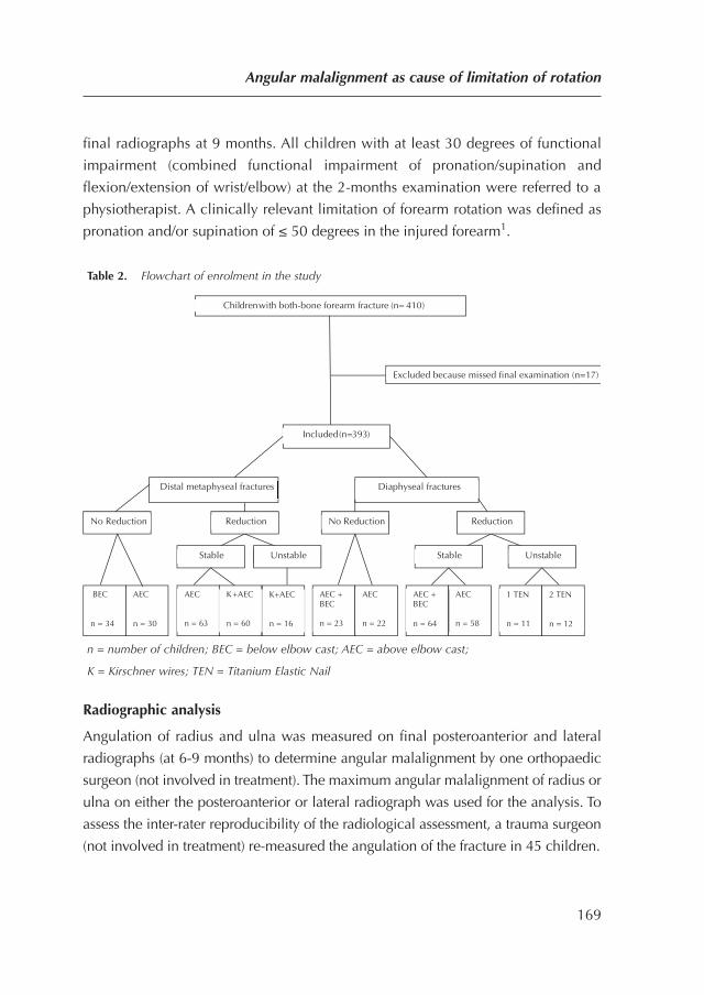
final radiographs at 9 months. All children with at least 30 degrees of functional
impairment (combined functional impairment of pronation/supination and
flexion/extension of wrist/elbow) at the 2-months examination were referred to a
physiotherapist. A clinically relevant limitation of forearm rotation was defined as
pronation and/or supination of ≤ 50 degrees in the injured forearm1.
Table 2. Flowchart of enrolment in the study
n = number of children; BEC = below elbow cast; AEC = above elbow cast;
K = Kirschner wires; TEN = Titanium Elastic Nail
Radiographic analysis
Angulation of radius and ulna was measured on final posteroanterior and lateral
radiographs (at 6-9 months) to determine angular malalignment by one orthopaedic
surgeon (not involved in treatment). The maximum angular malalignment of radius or
ulna on either the posteroanterior or lateral radiograph was used for the analysis. To
assess the inter-rater reproducibility of the radiological assessment, a trauma surgeon
(not involved in treatment) re-measured the angulation of the fracture in 45 children.
Angular malalignment as cause of limitation of rotation
169
Children with both-bone forearm fracture (n= 410)
Included (n=393)
Excluded because missed final examination (n=17)
Distal metaphyseal fractures Diaphyseal fractures
Stable Unstable
No Reduction Reduction
Unstable Stable
No Reduction Reduction
BEC n = 34
AEC n = 30
AEC n = 63
AEC + BEC n = 23
AEC n = 22
AEC + BEC n = 64
AEC n = 58
1 TEN n = 11
2 TEN n = 12
K+AEC n = 16
K+AEC n = 60
Statistical analysis
First, it was established whether the variables had a normal distribution using the
normality Shapiro-Wilk test. Based on these analyses, the results are presented as
means and range.
The main study items (limitation of pronation/supination of the fractured arm
≤ 50 degrees as dependent variable and degree of angular malalignment as
independent variable) were analysed using logistic regression analysis. If necessary,
adjustments were made for unbalanced covariates. Statistical analyses were
performed with the SPSS version 17.0 (SPSS Inc., Chicago, USA).
RESULTS
Between January 2006 and August 2010 a total of 410 children were prospectively
followed in four Dutch hospitals; of these, 393 children were included for analysis
in the present study (Table 2). The excluded children had either no clinical and/or
no radiological examination at the end of follow-up. The mean time to final
examination was 219 (99-440) days. The mean age of the children was 8.0 (0-16)
years. The children were divided in two groups in accordance with their limitation
of forearm rotation, i.e. more or less than 50 degrees of impairment. Table 3 presents
the baseline characteristics of the total study population and per group. There was a
significant difference between the two groups in location of fracture, and type of
fracture of both the radius and ulna; therefore, the statistical analyses were corrected
for these baseline variables.
Table 4 presents different categories of maximum angular malalignment on
final radiographs in relation to clinically relevant limitation of forearm rotation at
final clinical examination, i.e. < 50 degrees of pronation and/or supination. Overall
86 children had a malunion of ≤ 5 degrees of which 9.3% showed a clinically
relevant limitation of forearm rotation at final follow-up. The most severe malunion
groups (11-15 and >16 degrees of malunion) had significantly more children with a
clinically relevant limitation of forearm rotation (p < 0.0001).
Table 5 presents the limitation of forearm rotation in correlation with the severity
of malunion and location of the fracture (distal metaphyseal/diaphyseal). A significantly
higher percentage of children with a clinically relevant limitation of forearm rotation
Chapter 10
170
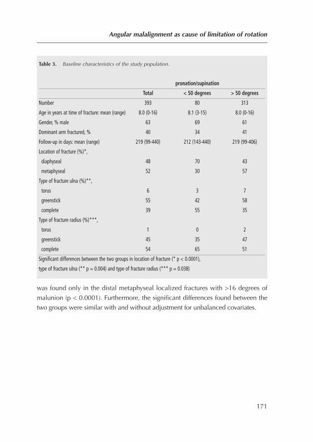
was found only in the distal metaphyseal localized fractures with >16 degrees of
malunion (p < 0.0001). Furthermore, the significant differences found between the
two groups were similar with and without adjustment for unbalanced covariates.
Angular malalignment as cause of limitation of rotation
171
Table 3. Baseline characteristics of the study population.
pronation/supination
Total < 50 degrees > 50 degrees
Number 393 80 313
Age in years at time of fracture: mean (range) 8.0 (0-16) 8.1 (3-15) 8.0 (0-16)
Gender, % male 63 69 61
Dominant arm fractured, % 40 34 41
Follow-up in days: mean (range) 219 (99-440) 212 (143-440) 219 (99-406)
Location of fracture (%)*,
diaphyseal 48 70 43
metaphyseal 52 30 57
Type of fracture ulna (%)**,
torus 6 3 7
greenstick 55 42 58
complete 39 55 35
Type of fracture radius (%)***,
torus 1 0 2
greenstick 45 35 47
complete 54 65 51
Significant differences between the two groups in location of fracture (* p < 0.0001),
type of fracture ulna (** p = 0.004) and type of fracture radius (*** p = 0.038)
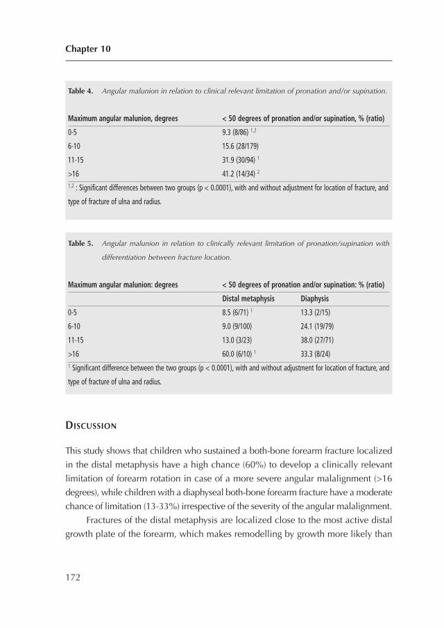
DISCUSSION
This study shows that children who sustained a both-bone forearm fracture localized
in the distal metaphysis have a high chance (60%) to develop a clinically relevant
limitation of forearm rotation in case of a more severe angular malalignment (>16
degrees), while children with a diaphyseal both-bone forearm fracture have a moderate
chance of limitation (13-33%) irrespective of the severity of the angular malalignment.
Fractures of the distal metaphysis are localized close to the most active distal
growth plate of the forearm, which makes remodelling by growth more likely than
Chapter 10
172
Table 4. Angular malunion in relation to clinical relevant limitation of pronation and/or supination.
Maximum angular malunion, degrees < 50 degrees of pronation and/or supination, % (ratio)
0-5 9.3 (8/86) 1,2
6-10 15.6 (28/179)
11-15 31.9 (30/94) 1
>16 41.2 (14/34) 2
1,2 : Significant differences between two groups (p < 0.0001), with and without adjustment for location of fracture, and
type of fracture of ulna and radius.
Table 5. Angular malunion in relation to clinically relevant limitation of pronation/supination with
differentiation between fracture location.
Maximum angular malunion: degrees < 50 degrees of pronation and/or supination: % (ratio)
Distal metaphysis Diaphysis
0-5 8.5 (6/71) 1 13.3 (2/15)
6-10 9.0 (9/100) 24.1 (19/79)
11-15 13.0 (3/23) 38.0 (27/71)
>16 60.0 (6/10) 1 33.3 (8/24)1 Significant difference between the two groups (p < 0.0001), with and without adjustment for location of fracture, and
type of fracture of ulna and radius.
fractures in the diaphysis. Therefore, a distal metaphyseal fracture with a mild to
moderate angular displacement (≤ 15 degrees) does not always need to be reduced,
especially in younger children with a high potential for remodelling by growth. In
contrast, clinicians should remain alert regarding children with diaphyseal fractures,
because even mild angular malalignment resulted in a clinically relevant limitation
of forearm rotation. Because diaphyseal fractures showed no significant increase in
limitation of pronation/supination in more severe angular malalignment fractures,
contractures of the injured interosseous membrane might also play a role.
Previous studies
Malunion as cause of limitation of pronation/supination after forearm fractures is often
reported and might consist of malalignment in angulation, translation or rotation2,19-26.
Two studies of artificially created deformities in human upper-extremity cadaver
specimens indicated that angular deformities of the forearm of 10 degrees or less
resulted in minimum limitation of pronation/supination, whereas 20 degrees of
angulation caused an important loss of forearm rotation2,3. These findings are
supported by our study only in distal metaphyseal fractures, whereas many
diaphyseal fractures even showed a limitation in minor angular malalignment.
Moreover, previous studies mentioned that angular malalignment in diaphyseal
fractures more often resulted in limitation of pronation/supination than similar
angular malalignment in metaphyseal fractures2,3. This finding was confirmed in our
study with up to 16 degrees of angular malalignment.
Malalignment in translation is reported to be a cause of limitation of pronation/
supination by the loss of radial bow or impingement between the forearm bones22,24-
26. In the present study the primary translational displacement of the fracture was
changed by appositional periosteal bone formation and at final follow-up only
minimal callus without notable translational malalignment was seen on the
radiographs. This made us believe that malalignment in translation did not
significantly contribute to limitation of forearm rotation.
One cadaveric study described that rotational malalignment might result in
limitation of pronation/supination, especially if the rotational malalignment of the
ulna and radius was in the opposite direction. In our clinical study the final
radiographs did not show any rotational malalignment.
Angular malalignment as cause of limitation of rotation
173
Study limitations
In the analysis based on differentiation of fracture location, because the number of
included children was relatively small these results should be interpreted with
caution. Furthermore, the impairment of forearm rotation might hinder a perfect
positioning of the forearm during radiography, which might influence measurement
of the angular malalignment. In this way, subtle rotational malalignment might
contribute to the limitation of forearm rotation and is difficult to measure on a
conventional radiograph. Thereby the present study focused only on limitations
related to malalignments, while contractures of traumatized soft tissue and pathology
in the distal or proximal radio ulnar joint may also have played a role. The final
limitation is the relatively short follow-up, as there is a remodelling of malunions by
growth in children which possible influences the forearm rotation.
Conclusion
This study shows that children who sustained a both-bone forearm fracture localized
in the distal metaphysis have a high chance to develop a clinically relevant limitation
of forearm rotation in the case of more severe angular malalignment, while children
with a diaphyseal both-bone forearm fracture have a moderate chance of limitation
irrespective of the severity of the angular malalignment.
Chapter 10
174
REFERENCES
1. Morrey BF, Askew LJ, Chao EY. A biomechanical study of normal functional elbow motion.
The Journal of bone and joint surgery American volume. 1981;63(6):872-7.
2. Matthews LS, Kaufer H, Garver DF, Sonstegard DA. The effect on supination-pronation of
angular malalignment of fractures of both bones of the forearm. J Bone Joint Surg Am.
1982;64(1):14-7.
3. Tarr RR, Garfinkel AI, Sarmiento A. The effects of angular and rotational deformities of both
bones of the forearm. An in vitro study. J Bone Joint Surg Am. 1984;66(1):65-70.
4. Sarmiento A, Ebramzadeh E, Brys D, Tarr R. Angular deformities and forearm function.
Journal of orthopaedic research : official publication of the Orthopaedic Research Society.
1992;10(1):121-33.
5. Boyer BA, Overton B, Schrader W, Riley P, Fleissner P. Position of immobilization for
pediatric forearm fractures. Journal of pediatric orthopedics. 2002;22(2):185-7.
6. Choi KY, Chan WS, Lam TP, Cheng JC. Percutaneous Kirschner-wire pinning for severely
displaced distal radial fractures in children. A report of 157 cases. J Bone Joint Surg Br.
1995;77(5):797-801.
7. Daruwalla JS. A study of radioulnar movements following fractures of the forearm in
children. Clin Orthop Relat Res. 1979(139):114-20.
8. Gibbons CL, Woods DA, Pailthorpe C, Carr AJ, Worlock P. The management of isolated
distal radius fractures in children. J Pediatr Orthop. 1994;14(2):207-10.
9. Jones K, Weiner DS. The management of forearm fractures in children: a plea for
conservatism. J Pediatr Orthop. 1999;19(6):811-5.
10. Mani GV, Hui PW, Cheng JC. Translation of the radius as a predictor of outcome in distal
radial fractures of children. J Bone Joint Surg Br. 1993;75(5):808-11.
11. Maric D, Petkovic L, Tomasevic V, Bajic L, Gajdobranski D, Marcikic A. [Osteosynthesis
with Kirschner wires in the treatment of dislocation fractures of the forearm in children].
Med Pregl. 2002;55(3-4):140-5.
12. Noonan KJ, Price CT. Forearm and distal radius fractures in children. J Am Acad Orthop
Surg. 1998;6(3):146-56.
13. Tynan MC, Fornalski S, McMahon PJ, Utkan A, Green SA, Lee TQ. The effects of ulnar axial
malalignment on supination and pronation. J Bone Joint Surg Am. 2000;82-A(12):1726-31.
Angular malalignment as cause of limitation of rotation
175
14. Zimmermann R, Gabl M, Angermann P, Lutz M, Reinhart C, Kralinger F, et al. [Late
sequelae of fractures of the distal third of the forearm during the growth period]. Handchir
Mikrochir Plast Chir. 2000;32(4):242-9.
15. Gustilo RB, Simpson L, Nixon R, Ruiz A, Indeck W. Analysis of 511 open fractures. Clin
Orthop Relat Res. 1969;66:148-54.
16. Colaris JW, Allema JH, Biter LU, de Vries MR, van de Ven CP, Bloem RM, et al. Re-
displacement of stable distal both-bone forearm fractures in children: a randomised
controlled multicentre trial. Injury. 2013;44(4):498-503.
17. Colaris JW, Biter LU, Allema JH, Bloem RM, van de Ven CP, de Vries MR, et al. Below-
elbow cast for metaphyseal both-bone fractures of the distal forearm in children: a
randomised multicentre study. Injury. 2012;43(7):1107-11.
18. Colaris J, van der Linden M, Selles R, Coene N, Allema JH, Verhaar J. Pronation and
supination after forearm fractures in children: Reliability of visual estimation and
conventional goniometry measurement. Injury. 2010;41(6):643-6.
19. Bronstein AJ, Trumble TE, Tencer AF. The effects of distal radius fracture malalignment on
forearm rotation: a cadaveric study. J Hand Surg [Am]. 1997;22(2):258-62.
20. Dumont CE, Thalmann R, Macy JC. The effect of rotational malunion of the radius and the
ulna on supination and pronation. J Bone Joint Surg Br. 2002;84(7):1070-4.
21. Kasten P, Krefft M, Hesselbach J, Weinberg AM. How does torsional deformity of the radial
shaft influence the rotation of the forearm? A biomechanical study. J Orthop Trauma.
2003;17(1):57-60.
22. Kuderna H. [Connection between deviated axis and impaired function after fractures of the
forearm (author’s transl)]. Unfallchirurgie. 1980;6(1):7-13.
23. Price CT, Scott DS, Kurzner ME, Flynn JC. Malunited forearm fractures in children. J Pediatr
Orthop. 1990;10(6):705-12.
24. Roberts JA. Angulation of the radius in children’s fractures. J Bone Joint Surg Br.
1986;68(5):751-4.
25. Trousdale RT, Linscheid RL. Operative treatment of malunited fractures of the forearm. J
Bone Joint Surg Am. 1995;77(6):894-902.
26. Yasutomi T, Nakatsuchi Y, Koike H, Uchiyama S. Mechanism of limitation of
pronation/supination of the forearm in geometric models of deformities of the forearm
bones. Clin Biomech (Bristol, Avon). 2002;17(6):456-63.
Chapter 10
176

Angular malalignment as cause of limitation of rotation
177
178
CHAPTER 11
Three-dimensional imaging ofchildren with severe limitation ofpronation/supination after aboth-bone forearm fracture
Arch Orthop Trauma Surg. 2014 Mar;134(3):333-41
Colaris JW, Oei S, Reijman M, Holscher H , Allema JH,Verhaar JAN.
179
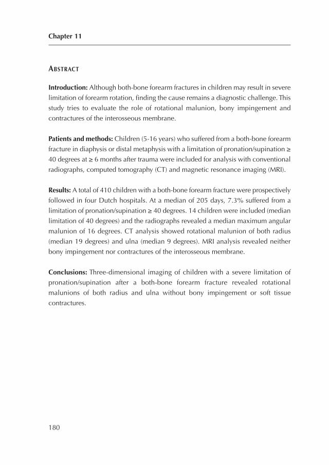
ABSTRACT
Introduction: Although both-bone forearm fractures in children may result in severe
limitation of forearm rotation, finding the cause remains a diagnostic challenge. This
study tries to evaluate the role of rotational malunion, bony impingement and
contractures of the interosseous membrane.
Patients and methods: Children (5-16 years) who suffered from a both-bone forearm
fracture in diaphysis or distal metaphysis with a limitation of pronation/supination ≥
40 degrees at ≥ 6 months after trauma were included for analysis with conventional
radiographs, computed tomography (CT) and magnetic resonance imaging (MRI).
Results: A total of 410 children with a both-bone forearm fracture were prospectively
followed in four Dutch hospitals. At a median of 205 days, 7.3% suffered from a
limitation of pronation/supination ≥ 40 degrees. 14 children were included (median
limitation of 40 degrees) and the radiographs revealed a median maximum angular
malunion of 16 degrees. CT analysis showed rotational malunion of both radius
(median 19 degrees) and ulna (median 9 degrees). MRI analysis revealed neither
bony impingement nor contractures of the interosseous membrane.
Conclusions: Three-dimensional imaging of children with a severe limitation of
pronation/supination after a both-bone forearm fracture revealed rotational
malunions of both radius and ulna without bony impingement or soft tissue
contractures.
Chapter 11
180
INTRODUCTION
Both-bone forearm fractures in children generally heal without complications. While
a limitation of pronation/supination of > 20 degrees has been reported in 3-30% of
children after a both-bone forearm fracture, especially the severe limitations of
rotation may interfere with daily activities1-4.
Although a malunion is often mentioned as main cause of limitation of forearm
rotation5-12 there is no consensus on this. Whereas several studies described
limitation without a malunion11,13,14, others mentioned malunions without
limitation15 or persistent limitation after a corrective osteotomy with restoration of
anatomical alignment16-18. Other bone-related causes of limitation are a synostosis,
or an impingement between radius and ulna9,11,19. The exact role of soft tissue
involvement in limitation of rotation remains unknown20-25. Contractures of the
interosseous membrane might play a more significant role than contractures of
capsules of wrist and elbow2,3,15.
Unravelling the exact cause of limitation after a both-bone forearm fracture in
children remains a diagnostic challenge. Therefore this study tries to evaluate the
role of rotational malunion, bony impingement and contractures of the interosseous
membrane by three-dimensional imaging.
MATERIALS AND METHODS
Study design and participants
A prospective study was performed including consecutive children with a forearm
fracture of radius and ulna who visited the emergency department of one of four
Dutch hospitals: Erasmus Medical Center (Rotterdam), HAGA Hospital (The Hague),
Reinier de Graaf Hospital (Delft) and Sint Franciscus Hospital (Rotterdam). The
regional medical ethics committee approved the study. Informed consent was
obtained from all parents and all children aged >12 years. For the present study we
included children aged 5-16 years who sustained a both-bone forearm fracture in
diaphysis or distal metaphysis with a limitation of pronation/supination ≥ 40 degrees
at final examination (≥ 6 months after trauma) (Figure 1).
3-D imaging of severe limitation of pronation/supination
181
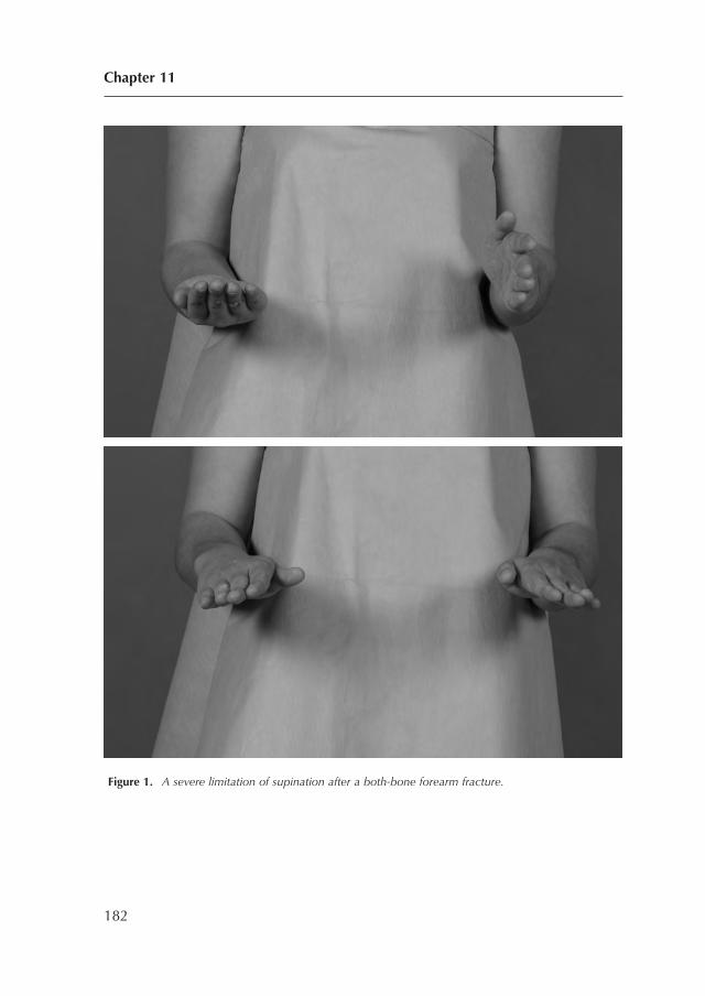
Figure 1. A severe limitation of supination after a both-bone forearm fracture.
Chapter 11
182
Clinical examination
The included children were treated based on the study protocol. This protocol
distinguished several types of fractures with several types of treatment. Briefly, distal
metaphyseal fractures without need for reduction were treated with below-elbow or
above-elbow cast for 4 weeks2, while distal metaphyseal fractures with reduction
were treated with or without Kirschner wires in combination with above-elbow cast
for 4 weeks1. Furthermore, diaphyseal fractures without reduction and reduced stable
diaphyseal fractures were treated with 6 weeks above-elbow cast, or a combination
of 3 weeks above-elbow cast and 3 weeks below-elbow cast3. Finally, unstable
diaphyseal fractures were fixated with 1 or 2 intramedullary titanium nails in
combination with 3 weeks above-elbow cast4.
One orthopaedic surgeon (who was not involved in primary treatment)
measured pronation/supination of both arms two months after initial trauma. This
technique was standardized with the use of a 180° goniometer constructed of clear,
flexible plastic with 2 movable arms of 30 cm26. Patients with at least 30 degrees of
limitation 2 months after initial trauma were referred to physiotherapy.
Six to nine months after the initial trauma the orthopaedic surgeon re-measured
the pronation/supination of the traumatised arm. Children with a limitation of
pronation/supination of ≥ 40 degrees were asked to participate in this study.
Radiographic analysis
Conventional radiography
One orthopaedic surgeon measured the fracture characteristics on conventional
posteroanterior and lateral radiographs directly after trauma and at final examination
(6-9 months after trauma) (Table 2 and 3).
Computer tomography
Torsion angle of the radius/ulna was defined as the angle between the proximal and
distal metaphysis in the transverse plane27. To compare torsion angles of left and
right radius/ulna, a computed tomography (CT) scanner was used to scan elbow and
wrist joints of both sides. We defined rotational malunion as a difference in torsion
angles of respectively radius and ulna between the injured and uninjured arm. All
images were acquired using a GE Lightspeed 4-slice CT scanner (General Electric
3-D imaging of severe limitation of pronation/supination
183
Healthcare; Milwaukee, WI, USA). Image acquisition parameters were a X-ray tube
potential of 80 kVp, a tube current of 30 mA and 2.50-mm slice thickness. Images
were reconstructed using a high spatial frequency reconstruction algorithm
(Boneplus).
With the child lying prone in the scanner, both arms were placed in overhead
position; the elbows were extended and the forearms were pronated so that the volar
aspect of the elbow was placed flat on the table. Axial images through the maximum
prominence of the bicipital tuberosity and the distal two centimetres of both radii
were made simultaneously with use of a routine CT imaging protocol. This technique
of scanning has shown good reproducibility27. The measurements of torsion angles
of radius and ulna of both arms were performed with the tools integrated in the PACS
system used in our institution, i.e. iSite Radiology 3.5 (Philips Medical Systems, Best,
the Netherlands). The difference in torsion angles of the radius of the traumatized
arm and non-traumatized arm was calculated and defined as rotational malunion
of the radius. The same procedure was repeated to determine the rotational malunion
of the ulna. The orthopaedic surgeon and a senior resident of the radiology
department (independently of each other) measured the torsion angle of the radius
and ulna of both arms using the standardized techniques described by Bindra et al.
and27 by Freitas et al.28 for the radius and by Dumont et al.29 and by Aufauvre et al.
for the ulna30 (Figure 2). All techniques were used three times to calculate the intra-
and interobserver variability (ICCs).
Chapter 11
184
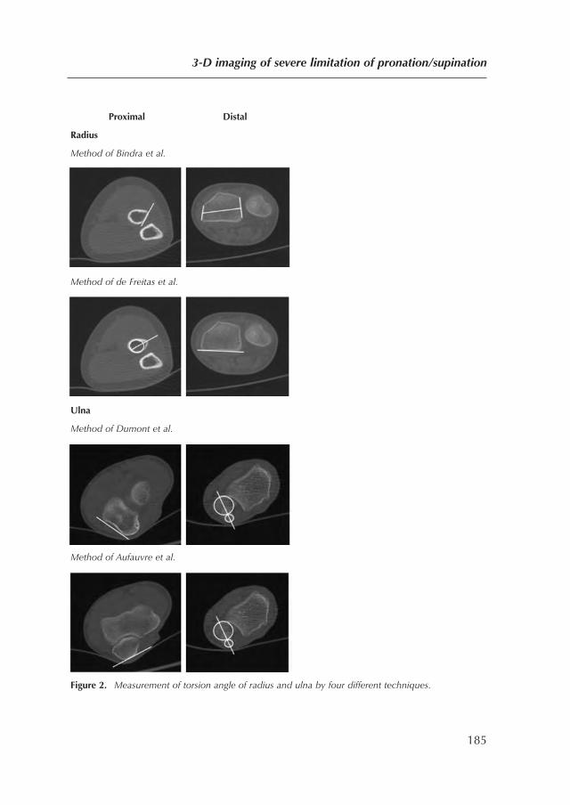
Proximal Distal
Radius
Method of Bindra et al.
Method of de Freitas et al.
Ulna
Method of Dumont et al.
Method of Aufauvre et al.
Figure 2. Measurement of torsion angle of radius and ulna by four different techniques.
3-D imaging of severe limitation of pronation/supination
185
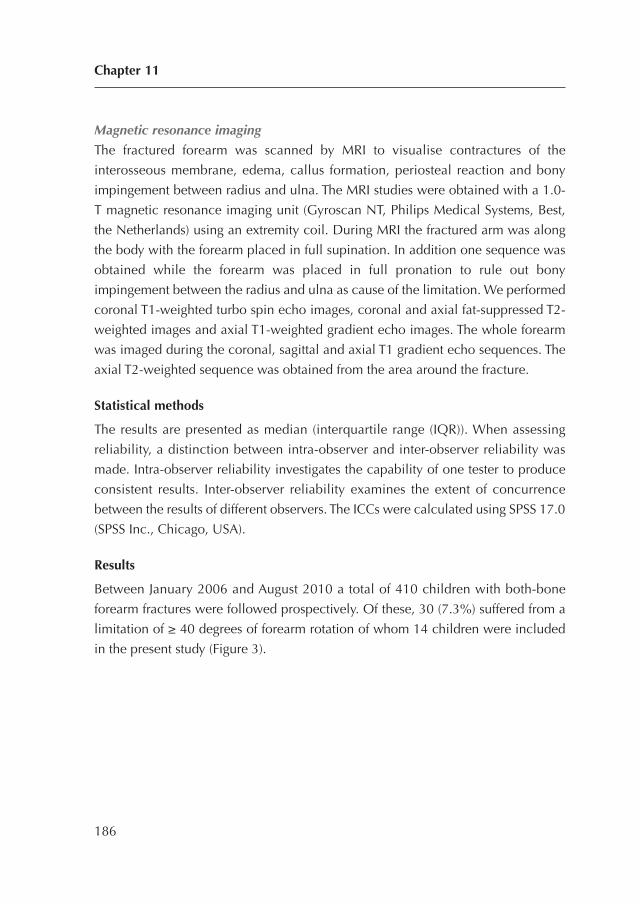
Magnetic resonance imaging
The fractured forearm was scanned by MRI to visualise contractures of the
interosseous membrane, edema, callus formation, periosteal reaction and bony
impingement between radius and ulna. The MRI studies were obtained with a 1.0-
T magnetic resonance imaging unit (Gyroscan NT, Philips Medical Systems, Best,
the Netherlands) using an extremity coil. During MRI the fractured arm was along
the body with the forearm placed in full supination. In addition one sequence was
obtained while the forearm was placed in full pronation to rule out bony
impingement between the radius and ulna as cause of the limitation. We performed
coronal T1-weighted turbo spin echo images, coronal and axial fat-suppressed T2-
weighted images and axial T1-weighted gradient echo images. The whole forearm
was imaged during the coronal, sagittal and axial T1 gradient echo sequences. The
axial T2-weighted sequence was obtained from the area around the fracture.
Statistical methods
The results are presented as median (interquartile range (IQR)). When assessing
reliability, a distinction between intra-observer and inter-observer reliability was
made. Intra-observer reliability investigates the capability of one tester to produce
consistent results. Inter-observer reliability examines the extent of concurrence
between the results of different observers. The ICCs were calculated using SPSS 17.0
(SPSS Inc., Chicago, USA).
Results
Between January 2006 and August 2010 a total of 410 children with both-bone
forearm fractures were followed prospectively. Of these, 30 (7.3%) suffered from a
limitation of ≥ 40 degrees of forearm rotation of whom 14 children were included
in the present study (Figure 3).
Chapter 11
186

The median age was 12.3 years [interquartile range IQR of 9.5; 14.7] (Table 1).
There was an equal distribution of children with a limitation of pronation or
supination. At a median of 205 days [IQR 194; 225], the median limitation of
pronation and supination was 40 degrees [IQR 40; 55] and the median maximum
angular malunion on conventional radiographs was 16 degrees [IQR 13; 19] (Table 3).
n = number of children.
Figure 3. Flowchart of enrolment in the study.
The values of rotational malunion of radius and ulna measured with CT are
reported in Table 4. CT analysis showed rotational malunion of both radius (median
19 degrees [IQR 12; 21]) and ulna (median 9 degrees [IQR 4; 12]) with no significant
difference (p=0.07). Torsion angle measurement of the radius was impossible in 5
children due to imaging problems of the radial tuberosity, i.e. only a part was
imaged, the images were oblique and not comparable with the contralateral arm,
or the radial tuberosity was not fully developed. In line with this, a not fully
developed ulnar styloid precluded measurement of the ulnar torsion angle by the
described techniques. For that reason, we used a tangential line that passes the sulcus
for the extensor carpi ulnaris as distal reference point for the measurement of torsion
(Figure 4). For the different measurement methods, the ICCs for intra-observer
reliability were 0.77-0.99 and for interobserver reliability they were 0.69-0.92.
Rotational malunion was defined as the difference in torsion angles of the
radius/ulna of the traumatized arm and the non-traumatized arm; Values are
3-D imaging of severe limitation of pronation/supination
187
Included (n = 14)
Children with both-bone forearm fracture (n = 410)
Excluded Limitation < 40 degrees (n = 370) Missed final examination (n = 10) Declined to participate (n = 9) Age < 6 years (n = 7)
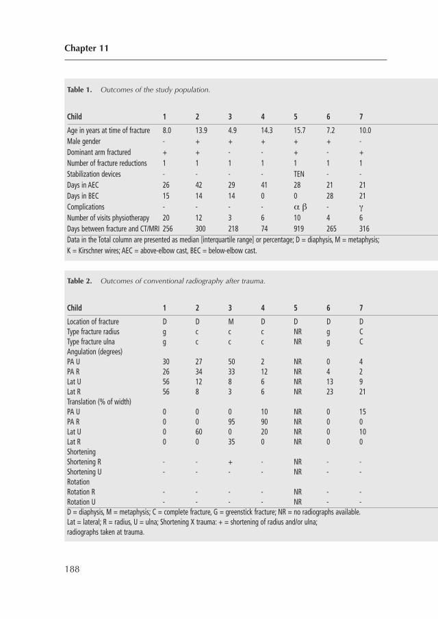
Chapter 11
188
Table 1. Outcomes of the study population.
Child 1 2 3 4 5 6 7 8 9 1
Age in years at time of fracture 8.0 13.9 4.9 14.3 15.7 7.2 10.0 Male gender - + + + + + - + + + + + + + 8Dominant arm fractured + + - - + - + - - - + - - - 3Number of fracture reductions 1 1 1 1 1 1 1 2 1 1 1 0 1 1Stabilization devices - - - - TEN - - - - K - - - -Days in AEC 26 42 29 41 28 21 21 Days in BEC 15 14 14 0 0 28 21 Complications - - - - α β - γ
Number of visits physiotherapy 20 12 3 6 10 4 6 1 Days between fracture and CT/MRI 256 300 218 74 919 265 316 Data in the Total column are presented as median [interquartile range] or percentage; D = diaphysis, M = metaphysis; K = Kirschner wires; AEC = above-elbow cast, BEC = below-elbow cast.
Table 2. Outcomes of conventional radiography after trauma.
Child 1 2 3 4 5 6 7 8 9 1
Location of fracture D D M D D D D D D M M M M DType fracture radius g c c c NR g C c c c c g c cType fracture ulna g c c c NR g C c c c c g g cAngulation (degrees)PA U 30 27 50 2 NR 0 4 2 1 4PA R 26 34 33 12 NR 4 2 5 1 3Lat U 56 12 8 6 NR 13 9Lat R 56 8 3 6 NR 23 21Translation (% of width)PA U 0 0 0 10 NR 0 15PA R 0 0 95 90 NR 0 0 2Lat U 0 60 0 20 NR 0 10Lat R 0 0 35 0 NR 0 0 0 6ShorteningShortening R - - + - NR - - - + + - - + -Shortening U - - - - NR - - - - - + - - -Rotation Rotation R - - - - NR - - - - - + - - -Rotation U - - - - NR - - - - - + - - -D = diaphysis, M = metaphysis; C = complete fracture, G = greenstick fracture; NR = no radiographs available. Lat = lateral; R = radius, U = ulna; Shortening X trauma: + = shortening of radius and/or ulna; radiographs taken at trauma.
3-D imaging of severe limitation of pronation/supination
189
8 9 10 11 12 13 14 Total
11.1 14.6 15.3 15.2 11.1 13.5 10.2 12.3 [9.5; 14.7] + + + + + + + 86%
- - - + - - - 36% 2 1 1 1 0 1 1
S - - K - - - -D 47 45 33 27 29 26 18 29 [25; 41]
0 0 0 0 0 0 25 18 [14; 26] γ δ α - - α - -
N 13 24 7 6 0 15 2 7 [4; 14] 776 346 258 217 242 806 274 270 [236; 454] C = complete fracture, G = greenstick fracture; NR = no radiographs available; TEN = titanium elastic nails.
Complications: α: neurapraxia , β : nonunion ulna, γ : re-fracture, δ: re-reduction
8 9 10 11 12 13 14
D D M M M M DT c c c c g c cT c c c c g g cA
2 1 41 40 4 10 ζ 5 1 31 45 6 14 ζ 25 13 55 31 14 32 109 18 11 43 32 7 34 106
20 5 0 100 0 0 0
P 20 95 40 50 0 45 0 10 0 0 80 0 0 100 0 65 100 90 0 100 0
- + + - - + -S - - - + - - -R
- - - + - - - - - - + - - -
D Angulation presented in degrees; Translation presented in percentage of width; PA = posteroanterior, Rotation X trauma: + = rotation of radius and/or ulna; CO = corrective osteotomy done; ζ = no PA
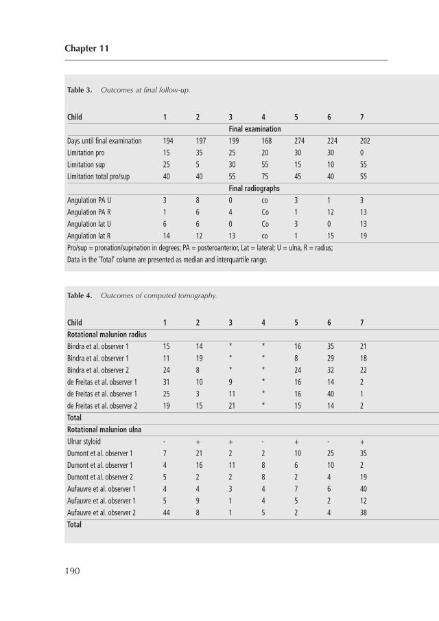
Chapter 11
190
Table 3. Outcomes at final follow-up.
Child 1 2 3 4 5 6 7 8 9 1Final examination
Days until final examination 194 197 199 168 274 224 202 Limitation pro 15 35 25 20 30 30 0 Limitation sup 25 5 30 55 15 10 55 Limitation total pro/sup 40 40 55 75 45 40 55
Final radiographs Angulation PA U 3 8 0 co 3 1 3 9 7 8 3 3 5 1 Angulation PA R 1 6 4 Co 1 12 13 Angulation lat U 6 6 0 Co 3 0 13 Angulation lat R 14 12 13 co 1 15 19 Pro/sup = pronation/supination in degrees; PA = posteroanterior, Lat = lateral; U = ulna, R = radius; Data in the ‘Total’ column are presented as median and interquartile range.
Table 4. Outcomes of computed tomography.
Child 1 2 3 4 5 6 7 8 9 1Rotational malunion radiusBindra et al. observer 1 15 14 * * 16 35 21 Bindra et al. observer 1 11 19 * * 8 29 18 Bindra et al. observer 2 24 8 * * 24 32 22 de Freitas et al. observer 1 31 10 9 * 16 14 2 de Freitas et al. observer 1 25 3 11 * 16 40 1 de Freitas et al. observer 2 19 15 21 * 15 14 2 Total Rotational malunion ulnaUlnar styloid - + + - + - + + + + + - + +Dumont et al. observer 1 7 21 2 2 10 25 35 Dumont et al. observer 1 4 16 11 8 6 10 2 Dumont et al. observer 2 5 2 2 8 2 4 19 Aufauvre et al. observer 1 4 4 3 4 7 6 40 Aufauvre et al. observer 1 5 9 1 4 5 2 12 Aufauvre et al. observer 2 44 8 1 5 2 4 38 Total
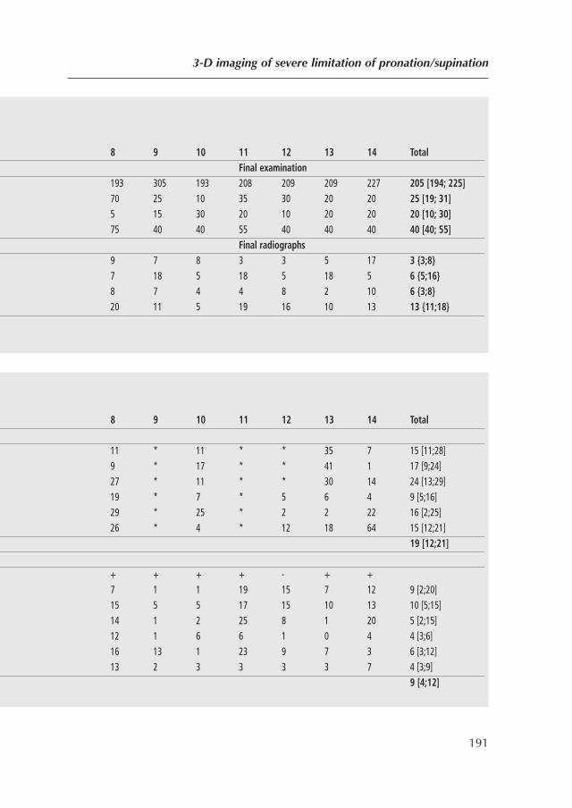
3-D imaging of severe limitation of pronation/supination
191
8 9 10 11 12 13 14 Total Final examination
193 305 193 208 209 209 227 205 [194; 225] 70 25 10 35 30 20 20 25 [19; 31] 5 15 30 20 10 20 20 20 [10; 30] 75 40 40 55 40 40 40 40 [40; 55]
Final radiographs 9 7 8 3 3 5 17 3 {3;8} 7 18 5 18 5 18 5 6 {5;16} 8 7 4 4 8 2 10 6 {3;8} 20 11 5 19 16 10 13 13 {11;18}
8 9 10 11 12 13 14 Total
11 * 11 * * 35 7 15 [11;28] 9 * 17 * * 41 1 17 [9;24] 27 * 11 * * 30 14 24 [13;29]
19 * 7 * 5 6 4 9 [5;16] 29 * 25 * 2 2 22 16 [2;25] 26 * 4 * 12 18 64 15 [12;21] 19 [12;21]
+ + + + - + +
D 7 1 1 19 15 7 12 9 [2;20] 15 5 5 17 15 10 13 10 [5;15] 14 1 2 25 8 1 20 5 [2;15] 12 1 6 6 1 0 4 4 [3;6] 16 13 1 23 9 7 3 6 [3;12] 13 2 3 3 3 3 7 4 [3;9]
9 [4;12]
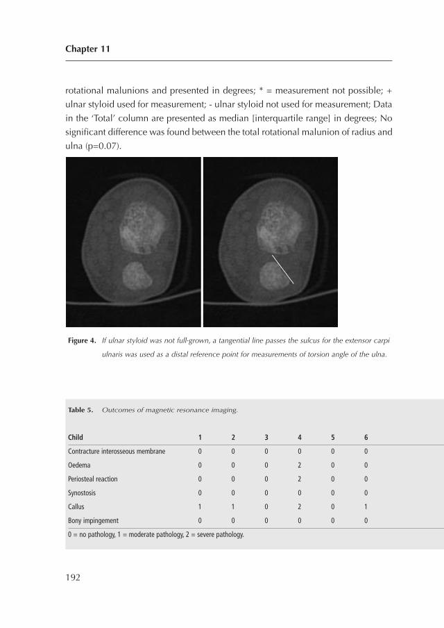
rotational malunions and presented in degrees; * = measurement not possible; +
ulnar styloid used for measurement; - ulnar styloid not used for measurement; Data
in the ‘Total’ column are presented as median [interquartile range] in degrees; No
significant difference was found between the total rotational malunion of radius and
ulna (p=0.07).
Figure 4. If ulnar styloid was not full-grown, a tangential line passes the sulcus for the extensor carpi
ulnaris was used as a distal reference point for measurements of torsion angle of the ulna.
Chapter 11
192
Table 5. Outcomes of magnetic resonance imaging.
Child 1 2 3 4 5 6 7 8 9 1
Contracture interosseous membrane 0 0 0 0 0 0 0 0 0 0 0 0 0 0
Oedema 0 0 0 2 0 0 2 0 0 1 2 0 0 0
Periosteal reaction 0 0 0 2 0 0 1 0 0 1 1 0 0 0
Synostosis 0 0 0 0 0 0 0 0 0 0 0 0 0 0
Callus 1 1 0 2 0 1 0 0 0 0 0 0 0 0
Bony impingement 0 0 0 0 0 0 0 0 0 0 0 0 0 0
0 = no pathology, 1 = moderate pathology, 2 = severe pathology.
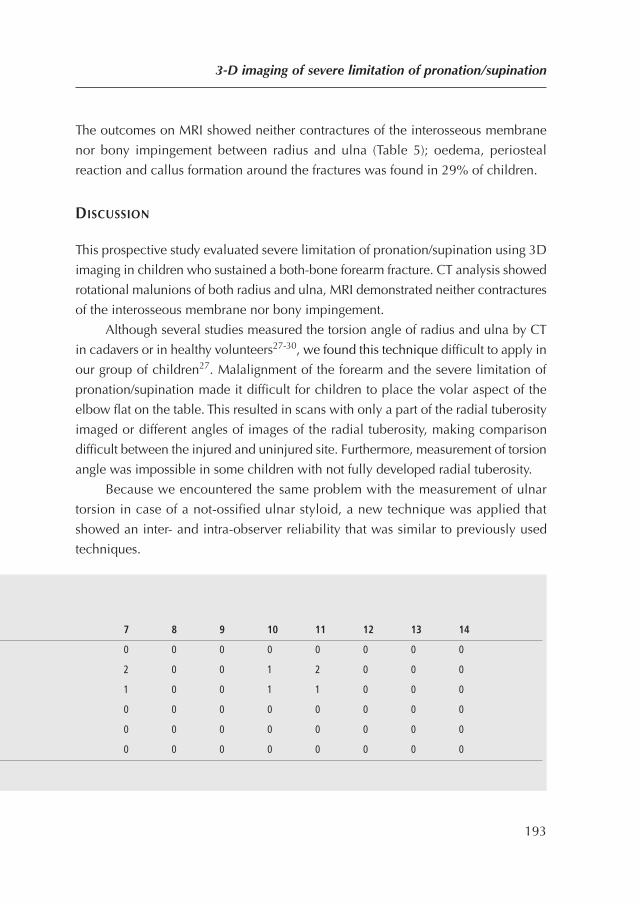
The outcomes on MRI showed neither contractures of the interosseous membrane
nor bony impingement between radius and ulna (Table 5); oedema, periosteal
reaction and callus formation around the fractures was found in 29% of children.
DISCUSSION
This prospective study evaluated severe limitation of pronation/supination using 3D
imaging in children who sustained a both-bone forearm fracture. CT analysis showed
rotational malunions of both radius and ulna, MRI demonstrated neither contractures
of the interosseous membrane nor bony impingement.
Although several studies measured the torsion angle of radius and ulna by CT
in cadavers or in healthy volunteers27-30, we found this technique difficult to apply in
our group of children27. Malalignment of the forearm and the severe limitation of
pronation/supination made it difficult for children to place the volar aspect of the
elbow flat on the table. This resulted in scans with only a part of the radial tuberosity
imaged or different angles of images of the radial tuberosity, making comparison
difficult between the injured and uninjured site. Furthermore, measurement of torsion
angle was impossible in some children with not fully developed radial tuberosity.
Because we encountered the same problem with the measurement of ulnar
torsion in case of a not-ossified ulnar styloid, a new technique was applied that
showed an inter- and intra-observer reliability that was similar to previously used
techniques.
3-D imaging of severe limitation of pronation/supination
193
7 8 9 10 11 12 13 14
0 0 0 0 0 0 0 0
O 2 0 0 1 2 0 0 0
P 1 0 0 1 1 0 0 0
S 0 0 0 0 0 0 0 0
C 0 0 0 0 0 0 0 0
0 0 0 0 0 0 0 0
0
Torsion angle of the radius
Bindra et al. measured the torsion angle of the radius with CT and Dumont et al.
repeated this measurement using MRI. The ICCs for intra-observer reliability (0.88
and 0.95) and inter-observer reliability (0.94 and 0.82) of both studies were similar
to ours (0.98 and 0.85). Freitas et al. also measured the torsion angle of the radius
and found ICCs for intra-observer reliability of 0.28-0.91 and for inter-observer
reliability of 0.75-0.90. The ICCs in the present study were relatively high with 0.92-
0.99 for intra-observer reliability and 0.69-0.78 for inter-observer reliability.
Torsion angle of the ulna
Dumont et al. measured the torsion angle of the ulna using MRI and found an ICC
for intra-observer reliability of 0.95 and an ICC for inter-observer reliability of 0.91.
The ICCs in the present study were 0.80-0.88 for intra-observer reliability and 0.85-
0.87 for inter-observer reliability. In their study, Aufauvre and colleagues did not
measure ICCs.
Magnetic Resonance Imaging
Our MRI findings showed only a few alterations, which were in line with an earlier
study (20). In our study group, oedema, callus and periosteal reaction detected on
MRI may be normal in the healing process of severe both-bone forearm fractures.
While impingement is mentioned as a potential cause of limitation of
pronation/supination after forearm fractures, we found no impingement between the
radius and ulna in maximum pronation/supination9,11,19. Although studies have
described the important role of contractures of the interosseous membrane in
limitation of pronation/supination after forearm fractures, in our study group these
were not detected on MRI9,11,22-24. However, the question remains whether the
contractures did not in fact exist, or whether they were not detectable on MRI.
Study limitations
Our CT protocol did not succeed in visualizing all radial tuberosity’s, which
precluded determination of the torsion of the radius in some children. This could
have been avoided by using a CT of both whole forearms with a bone and a soft
tissue setting. Different angles of anatomical landmarks between the two forearms
Chapter 11
194
could have been corrected using an isotropic dataset. In this way, the soft tissue
window could have visualized the insertion of the biceps tendon that might serve
as a reference for the radial tuberosity. Nevertheless, this technique would also not
visualize the tuberosity’s that were not fully developed. Furthermore, this method of
scanning employs a higher dose of radiation which is potentially hazardous for
children.
MRI of both arms with a higher resolution might have detected more soft tissue
pathology. However, the sparse availability of the MRI unit made scanning of both
arms impossible and the use of an extremity coil compensated for the relatively low
resolution.
Ideally our follow-up period should have been longer in this young population
to see if correction of malunion by growth would have influenced the limitation of
forearm rotation.
CONCLUSION
This prospective study evaluated severe limitation of pronation/supination using 3D
imaging in children who sustained a both-bone forearm fracture. The MRI
demonstrated neither contractures of the interosseous membrane nor bony
impingement. CT analysis showed rotational malunions of both radius and ulna.
Because malunion, impaired forearm rotation and a not fully developed radial
tuberosity hindered measurement of rotational malunion in several children, further
research should be done with a CT of both whole forearms with a bone and a soft
tissue setting.
3-D imaging of severe limitation of pronation/supination
195
REFERENCES
1. Colaris JW, Allema JH, Biter LU, de Vries MR, van de Ven CP, Bloem RM, et al. Re-
displacement of stable distal both-bone forearm fractures in children: a randomised
controlled multicentre trial. Injury. 2013;44(4):498-503.
2. Colaris JW, Biter LU, Allema JH, Bloem RM, van de Ven CP, de Vries MR, et al. Below-
elbow cast for metaphyseal both-bone fractures of the distal forearm in children: a
randomised multicentre study. Injury. 2012;43(7):1107-11.
3. Colaris JW, Reijman M, Allema JH, Biter LU, Bloem RM, van de Ven CP, et al. Early
conversion to below-elbow cast for non-reduced diaphyseal both-bone forearm fractures in
children is safe: preliminary results of a multicentre randomised controlled trial. Arch
Orthop Trauma Surg. 2013 Oct;133(10):1407-14.
4. Colaris J, Reijman M, Allema JH, Kraan G, van Winterswijk P, de Vries M, et al. Single-bone
intramedullary fixation of unstable both-bone diaphyseal forearm fractures in children leads
to increased re-displacement: a multicentre randomised controlled trial. Arch Orthop
Trauma Surg. 2013 Aug;133(8):1079-87
5. Bronstein AJ, Trumble TE, Tencer AF. The effects of distal radius fracture malalignment on
forearm rotation: a cadaveric study. J Hand Surg [Am]. 1997;22(2):258-62.
6. Dumont CE, Thalmann R, Macy JC. The effect of rotational malunion of the radius and the
ulna on supination and pronation. J Bone Joint Surg Br. 2002;84(7):1070-4.
7. Kasten P, Krefft M, Hesselbach J, Weinberg AM. How does torsional deformity of the radial
shaft influence the rotation of the forearm? A biomechanical study. J Orthop Trauma.
2003;17(1):57-60.
8. Kuderna H. [Connection between deviated axis and impaired function after fractures of the
forearm (author’s transl)]. Unfallchirurgie. 1980;6(1):7-13.
9. Matthews LS, Kaufer H, Garver DF, Sonstegard DA. The effect on supination-pronation of
angular malalignment of fractures of both bones of the forearm. J Bone Joint Surg Am.
1982;64(1):14-7.
10. Roberts JA. Angulation of the radius in children’s fractures. J Bone Joint Surg Br.
1986;68(5):751-4.
11. Tarr RR, Garfinkel AI, Sarmiento A. The effects of angular and rotational deformities of both
bones of the forearm. An in vitro study. J Bone Joint Surg Am. 1984;66(1):65-70.
Chapter 11
196
12. Trousdale RT, Linscheid RL. Operative treatment of malunited fractures of the forearm. J
Bone Joint Surg Am. 1995;77(6):894-902.
13. Hogstrom H, Nilsson BE, Willner S. Correction with growth following diaphyseal forearm
fracture. Acta Orthop Scand. 1976;47(3):299-303.
14. Nilsson BE, Obrant K. The range of motion following fracture of the shaft of the forearm in
children. Acta Orthop Scand. 1977;48(6):600-2.
15. Nenopoulos SP, Beslikas TA, Gigis JP. Long-term follow-up of combined fractures of the
proximal radius and ulna during childhood. J Pediatr Orthop B. 2009;18(5):252-60.
16. Krukhaug Y, Hove LM. Corrective osteotomy for malunited extra-articular fractures of the
distal radius: a follow-up study of 33 patients. Scand J Plast Reconstr Surg Hand Surg.
2007;41(6):303-9.
17. Nagy L, Jankauskas L, Dumont CE. Correction of forearm malunion guided by the
preoperative complaint. Clin Orthop Relat Res. 2008;466(6):1419-28.
18. Price CT, Knapp DR. Osteotomy for malunited forearm shaft fractures in children. J Pediatr
Orthop. 2006;26(2):193-6.
19. Graham TJ, Fischer TJ, Hotchkiss RN, Kleinman WB. Disorders of the forearm axis. Hand
clinics. 1998;14(2):305-16.
20. Beyer W, Stolzenburg T, Paris S. [Functional limitation of the forearm after shaft fracture in
childhood. Possible role of the antebrachial interosseous membrane: MRI and ultrasound
studies]. Unfallchirurgie. 1995;21(6):275-84.
21. Kihara H, Palmer AK, Werner FW, Short WH, Fortino MD. The effect of dorsally angulated
distal radius fractures on distal radioulnar joint congruency and forearm rotation. J Hand
Surg [Am]. 1996;21(1):40-7.
22. Nakamura T, Yabe Y, Horiuchi Y. [A biomechanical analysis of pronation-supination of the
forearm using magnetic resonance imaging: dynamic changes of the interosseous
membrane of the forearm during pronation-supination]. Nippon Seikeigeka Gakkai Zasshi.
1994;68(1):14-25.
23. Nakamura T, Yabe Y, Horiuchi Y, Seki T, Yamazaki N. Normal kinematics of the interosseous
membrane during forearm pronation-supination—a three-dimensional MRI study. Hand
Surg. 2000;5(1):1-10.
24. Price CT, Scott DS, Kurzner ME, Flynn JC. Malunited forearm fractures in children. J Pediatr
Orthop. 1990;10(6):705-12.
3-D imaging of severe limitation of pronation/supination
197
25. Yasutomi T, Nakatsuchi Y, Koike H, Uchiyama S. Mechanism of limitation of
pronation/supination of the forearm in geometric models of deformities of the forearm
bones. Clin Biomech (Bristol, Avon). 2002;17(6):456-63.
26. Colaris J, van der Linden M, Selles R, Coene N, Allema JH, Verhaar J. Pronation and
supination after forearm fractures in children: Reliability of visual estimation and
conventional goniometry measurement. Injury. 2010;41(6):643-6.
27. Bindra RR, Cole RJ, Yamaguchi K, Evanoff BA, Pilgram TK, Gilula LA, et al. Quantification
of the radial torsion angle with computerized tomography in cadaver specimens. The
Journal of bone and joint surgery American volume. 1997;79(6):833-7.
28. Freitas LF, Barbieri CH, Mazzer N, Zatiti SC, Bellucci AD, Nogueira-Barbosa MH.
Intraobserver and interobserver reliability of radial torsion angle measurements by a new
and alternative method with computed tomography. Clinics (Sao Paulo). 2010;65(11):1093-
7.
29. Dumont CE, Pfirrmann CW, Ziegler D, Nagy L. Assessment of radial and ulnar torsion
profiles with cross-sectional magnetic resonance imaging. A study of volunteers. J Bone
Joint Surg Am. 2006;88(7):1582-8.
30. Aufauvre B, Garcier JM, Boisgard S, Tanguy A. [Measurement of the torsion of the two
bones of the forearm: evaluation of tomodensitometric reference points]. Bull Assoc Anat
(Nancy). 1996;80(248):5-8.
Chapter 11
198

3-D imaging of severe limitation of pronation/supination
199
PART VI
GENERAL DISCUSSION
AND SUMMARY
200

CHAPTER 12
General discussion
201

Chapter 12
202
PART 1 METAPHYSEAL BOTH-BONE FRACTURES OF THE DISTAL FOREARM
Metaphyseal both-bone fractures of the distal forearm are among the most frequent
fractures in childhood. This thesis reveals that minimally displaced fractures can be
safely treated in a below-elbow cast, which is supported by previous studies1-3. In
case of a displaced fracture the clinician is often confronted with a treatment
dilemma, i.e. whether to accept some degree of displacement, or to perform a
reduction of the fracture. A perfect anatomical fracture alignment is not always
necessary in children because natural growth remodels malalignment. The highest
remodelling by growth takes place in fractures that are close to the most active distal
growth plate. Therefore, previous studies stated that metaphyseal fractures of the
distal forearm with an angulation ≤ 15 degrees can be treated without reduction in
children of all ages4. In accordance with these findings, we found that children with
an angular malalignment of ≤ 15 degrees had a less than 13% chance to develop a
clinically relevant limitation (i.e. < 50 degrees of pronation and/or supination), while
children with an angular malalignment of ≥ 16 degrees had a 60% chance.
Therefore, fractures in which the angulation exceeds 15 degrees need to be
reduced in the operating room under general anaesthesia with fluoroscopic
guidance. Because this thesis shows that secondary displacement of these reduced
fractures occurred in 45% of children treated with only an above-elbow cast, we
advise to perform stabilisation of the radius fracture by means of two crossed
percutaneous Kirschner wires (K-wires). In this case, the surgeon should also
consider stabilisation of an unstable ulnar fracture, because secondary fracture
displacement of the ulna was seen in 5% of children in which only the radius was
stabilised with K-wires. Whereas some groups buried the K-wires subcutaneously,
requiring an additional operation for removal5, in our study the K-wires were placed
transcutaneously to facilitate removal in the outpatient clinic. Nevertheless, in our
study, 11% of the K-wires became subcutaneous thereby making an additional
operation necessary. This complication might have been avoided by not cutting the
K-wires too short. Although the K-wires were placed transcutaneously, only 3% of
the children in our study developed a superficial infection; this rate is similar to that
reported by others6. In our study, no child had symptoms of neuromas of either the
radial sensory nerve, lateral cutaneous nerve of the forearm, or posterior cutaneous
General discussion
203
nerve of the forearm. This may be explained in two ways: by using a proper surgical
technique with a small skin incision and blunt dissection of soft tissue and, secondly,
due to the less vulnerable nerves in children; this is confirmed by the absence of
literature reporting on iatrogenic neuromas in childhood.
PART 2 DIAPHYSEAL BOTH-BONE FOREARM FRACTURES
Because of the relatively long distance between a diaphyseal fracture and the distal
and proximal growth plates, only minimal correction of malalignment by growth
can be expected. A previous study stated that diaphyseal fractures in children need
to be reduced in accordance with their age, i.e. in children up to 9 years of age an
angulation up to 15 degrees can be excepted, while in children 9 years of age or
older, 10 degrees of angulation is acceptable4. Nevertheless, in our opinion also
fractures with less angulation might benefit by reduction because our results indicate
that children with ≤ 5 degrees of angular malalignment already have a 13% chance
to develop a clinically relevant limitation (i.e. < 50 degrees of pronation and/or
supination).
Based on the results of our randomised controlled study, diaphyseal both-bone
forearm fractures without need for reduction could be safely be treated with an
above-elbow cast for 3 weeks, followed by a below-elbow cast. In general, among
our group of children, 6 weeks of cast was long enough. Nevertheless, painful
fractures without formation of callus on radiographs should be treated in cast for a
longer period. Because even minimally displaced fractures showed a tendency for
secondary displacement in cast, radiographs at weekly intervals until removal of the
cast should be made to avoid malalignment.
Displaced diaphyseal both-bone forearm fractures should be reduced in the
operation room under general anaesthesia with fluoroscopic guidance. If the
reduction is easy and the fracture seems to be stable, an above-elbow cast has to be
applied to stabilise the fracture. Similar to the treatment of the non-reduced fractures,
following the results of our randomised controlled study, the above-elbow cast can
be safely replaced by a below-elbow cast after 3 weeks.
Compared with previous studies, limitation of pronation/supination was
relatively high in our group of diaphyseal both-bone fractures7-13. Using the grading
Chapter 12
204
system (9) we found 21-33% excellent results, whereas others found excellent results
in 44-100%. Previous studies reported a re-displacement rate of 7-27% in
conservatively treated forearm fractures7,10,11,14-17 whereas our rate was 23-34%.
Fractures that seem to be unstable during reduction should be stabilised with
intramedullary nails. Several non-randomised and mostly retrospective studies
advocated stabilisation of both-bone forearm fractures with only one intramedullary
nail in either the radius or ulna, in combination with a supplementary cast18-28. In
those studies, the surgeon could choose per-operatively for single-bone or both-bone
fixation depending on the stability of the fracture, whereas we performed a study
with randomisation between one or two intramedullary nails irrespective of the
fracture stability. The group of children treated with one intramedullary nail showed
inferior results because of displacement of the non-fixated bone. Such re-
displacement in single-bone intramedullary fixation of both-bone forearm fractures
is not uncommon19,20,24,27. We suggest that these unstable diaphyseal fractures
should be stabilised with an intramedullary nail in both the radius and ulna to
prevent re-displacement and to enable unloaded exercises of the arm without cast
to improve final function.
PART 3 FRACTURE DISPLACEMENT
A both-bone forearm fracture destabilises the structural columns of the forearm.
Although the clinician tries to re-stabilise the fractured forearm by cast, K-wires,
intramedullary nails or plates, fracture displacement occurs frequently. Previous studies
described fracture displacement of 7-91% in the distal metaphysis17,29-36 compared
to 33% in our study, and fracture displacement in the diaphysis7,10,11,14-17,37 was found
in 7-52% compared to 27% in our study. There are several explanations for our
relatively high percentage of displacement. First, we included only both-bone
forearm fractures that are notorious for instability1,38. Second, we had a detailed
prospective follow-up with scheduled radiographs. Third, our malalignment criteria
were relatively strict, especially for young children with a high potential for
remodelling by growth. Fourth, a non-circumferential cast was applied directly after
reduction. Finally, the test used to distinguish between stable and unstable fractures
might have been influenced by the opinion of the surgeon.
General discussion
205
As expected, fractures stabilised by K-wires or intramedullary nails showed less
displacement than fractures treated with cast only. However, also in this group,
displacement was seen by improper surgical technique or displacement of the non-
fixated bone.
Our prospective multivariate regression analysis revealed fractures of the non-
dominant arm to be a risk factor for fracture displacement in cast, which was not
found by others34,39. Less stability of the fracture by less developed muscles in the
non-dominant forearm might be an explanation for this increased risk of fracture
displacement in the non-dominant forearm. Furthermore, our analysis showed that
the severity of fracture displacement directly after the trauma is highly predictive for
secondary displacement in cast. In more detail: complete fractures were at higher
risk for displacement than greenstick fractures, a finding supported by some
clinicians15,34,36,40,41 but not by others17,42. Fractures with translation of the ulna on
lateral radiographs were at risk for displacement, as reported once before15.
Shortening of fractures has not previously been reported as a risk factor. Thus, in our
study, fracture angulation did not appear to be a risk factor for displacement, as also
found in previous studies34,42. In summary, complete fractures with translation and
shortening seem to be more unstable than angulated greenstick fractures with an
intact periosteal sleeve on one side.
Several studies reported that the quality of the cast (cast index) is a risk factor
for fracture displacement2,17,40,42; however, this was not supported by our studies
and by others15,41. Although an above-elbow cast was generally considered more
likely to prevent fracture displacement than a below-elbow cast, we found a
tendency towards less fracture displacement in children treated with a below-elbow
cast. This finding has been reported before and might be explained by several
theories1-3. First, the application of an above-elbow cast might be more difficult,
resulting in an inferior cast with less three-point fixation. Second, an above-elbow
cast restricts wrist and elbow motion and, therefore, induces forced movements of
the fractured arm in daily use. Third, elbow motion in a below-elbow cast results in
micro-movements in the fracture, which facilitates secondary fracture healing.
Finally, it is possible that an above-elbow cast results in more atrophy with less
stabilising soft tissue around the fracture.
Our results suggest that a diaphyseal or metaphyseal location of the fracture
Chapter 12
206
has no significant influence on displacement, a finding supported by one group15,39
but not by another15,39. Furthermore, we found no significant effect of reduction
with or without general anaesthesia36,39. Although fracture displacement occurred
more frequently in the group of reduced fractures than in non-reduced fractures,
clinicians should also be aware of secondary displacement of minimally displaced
fractures.
Similar to other studies1,5,34 only half of our re-displaced fractures were reduced
a second time, although this was suggested in our study protocol. Although not
planned, this illustrates the restraint of treating clinicians to decide on a second
reduction. On the one hand, the surgeon may hope or expect correction of the
malalignment by growth whereas, on the other hand, the surgeon may be reluctant
to burden the child again or may find it difficult to accept failure of the initial
treatment. Nevertheless, the results of our study revealing the relation between
malunion and limitation of pronation/supination, suggest that better functional results
are achieved if clinicians follow the study protocol.
In conclusion, all both-bone forearm fractures in children need to be monitored
carefully using weekly radiographs to enable early detection of fracture displacement.
In case of displacement, the clinician should bear several factors in mind when
considering treatment, i.e. the severity of displacement, fracture in the dominant
arm, age of the child, and the distance between the fracture and the most active
distal growth plate.
PART 4 LIMITATION OF FOREARM ROTATION
Although most of the both-bone forearm fractures heal uneventfully, limitation of
pronation and supination can give disappointing results11,43-45. While movement of
the ipsilateral shoulder can compensate mild limitation of pronation/supination,
more severe limitation hinders daily activities. To function properly both pronation
and supination need to be at least 50 degrees46; in our study this was not obtained
by 20% of the children after a mean period of 218 (± 49) days. In our study the mean
limitation of forearm rotation was 12 (± 14) degrees. Despite the absence of other
prospective studies, our results seem to be slightly less successful than other series.
This might be explained in several ways. First, we only included children with
General discussion
207
relatively severe injuries that resulted in fractures of both the radius and the ulna.
Second, ours was a prospective study with a precise registration of arm function.
Third, we included many diaphyseal fractures, which are prone to impaired forearm
rotation. Fourth, a relatively large number of fractures healed in malalignment, which
increases the chance of limitation of forearm rotation. Finally, the relatively short
follow-up period, with less correction of malalignment by growth, had a negative
impact on our results.
Although not fully elucidated in literature, malunion and soft tissue contractures
are generally thought to cause impaired forearm rotation after a forearm fracture.
An interaction between these two causes may occur when a malunion changes the
tension of the interosseous membrane47-50.
Limitation of pronation/supination attributed to malunion consists of
malalignment in angulation, translation or rotation12,47,49,51-56. Whereas some
reported that angular malalignment may result in significant limitation47,48,51,57,
others found a poor correlation between residual angular malunion and the final
range of pronation/supination12,43,45,48,58-60. In our multivariate logistic regression
analysis, angular malalignment was not independently associated with a limitation
of pronation/supination. However, our clinical study relating impaired forearm
rotation with angular malalignment showed that angular malalignment in fractures
resulted in a clinically relevant limitation of pronation/supination.
Malalignment in translation is considered to cause impingement between the
forearm bones, resulting in limitation of pronation and supination49,54-56. However, in
our study, the primary translational displacement of the fracture was changed by
appositional periosteal bone formation and at final follow-up only minimal callus
without notable translational malalignment was seen on the radiographs. Furthermore,
magnetic resonance imaging of children in our study population who suffered from a
severe limitation revealed no bony impingement between the radius and ulna.
In cadaver studies rotational malalignment of the ulna and radius caused
limitation of forearm rotation52,53,55. Our clinical study with computer tomography
confirmed a rotational malalignment of both the radius and ulna in children suffering
from severe limitation of pronation/supination.
Several studies suggested that soft tissue contractures following forearm
fractures lead to limitation of pronation/supination50,54,61-63. These contractures may
Chapter 12
208
originate from muscles, capsules and the interosseous membrane. It is reported that
a volar fracture haematoma following a distal radius fracture caused a contracture
of the pronator quadratus muscle, with subsequent a limitation of supination64. In
line with this, a fracture haematoma of the pronator quadratus muscle and the
pronator teres muscle can result in contractures with limitation of supination, while
contractures of the supinator muscle and the biceps muscle can result in limitation
of pronation. Because we found more limitation of pronation than supination (29%
vs. 23%) in distal metaphyseal both-bone forearm fractures, we assume that
contractures of the pronator quadratus muscle are probably not the main cause of
limitation. Furthermore, if contracture of the biceps muscle plays a role in the
limitation of pronation, then we would also expect a limitation of elbow extension
because the biceps muscle acts as both a supinator and an elbow flexor.
Contractures of the capsule of wrist and elbow have been reported as a cause
of impaired forearm rotation50,63. However, this argument is not applicable in our
study population, because capsule contractures should have also led to impaired
flexion and extension of wrist and elbow, which was not the case in our children.
Our multivariate analysis revealed a diaphyseal fracture to be an independent
risk factor for limitation of forearm rotation. Thereby, diaphyseal fractures with
minimal angular malaligment (≤ 5 degrees) already had a 13% chance of severe
limitation of forearm rotation and this chance showed no significant increase with
more severe malalignment. Therefore, we assume that contractures of the
interosseous membrane play an important role in the limitation of pronation/
supination in diaphyseal fractures. This can be explained in several ways. First, in
our study, because about 75% of diaphyseal fractures were severe displaced, we
assume that the high force of injury that fractures the thick cortices of both the radius
and ulna is associated with significant injury to the interosseous membrane. Second,
previous studies described five ligamentous components of the interosseous
membrane, of which only the two most proximal cords showed a substantial change
in length during forearm rotation65. Immobilisation of a diaphyseal fracture in an
above-elbow cast might result in a reduced amount of lengthening of these injured
proximal cords and, consequently, impairment of forearm rotation. Other
independent risk factors revealed in our multivariate analysis were less physiotherapy
and re-fractures. Both of these suggest a substantial role of soft tissue contractures
General discussion
209
in limitation of forearm rotation: while physiotherapy decreases contractures by
stretching, a re-fracture gives rise to more contractures by an extra injury in
regenerating soft tissue. Finally, contractures of the interosseous membrane as a
cause of limitation of forearm rotation was confirmed in a study that found a
correlation between initial fracture displacement, MRI-detected interosseous
membrane alternations, and limitation of pronation/supination66.
Furthermore, pathology of the distal or proximal radio-ulnar joint may impair
forearm rotation. Although pathology in these joints seems obvious in Galeazzi or
Monteggia fracture dislocations, tightening of the triangular fibrocartilage complex
has also been described as a cause of impaired forearm rotation following radius
fractures67-69. Nevertheless, we assume that pathology of the distal or proximal radio-
ulnar joint mainly occurs if the energy of impact causes a single-bone fracture and
soft tissue injury along the other bone.
Taking all these findings into consideration, we conclude that only children
with a severe angular malalignment of a metaphyseal fracture have a high risk of
impaired forearm rotation, while children with diaphyseal fractures have a moderate
chance to develop a limitation irrespective of the severity of the angular
malalignment. In this latter group we expect a contracture of the interosseous
membrane to play a substantial role, even though we were unable to detect it on
(low resolution) MRI. Impingement between malunions of the radius and ulna did
not affect limitation, and rotational malunion seems to have some influence but is,
however, difficult to detect.
Overall, the work in this thesis has revealed more insight in the treatment and
complications of both-bone forearm fractures in children.
Chapter 12
210
REFERENCES
1. Bohm ER, Bubbar V, Yong Hing K, Dzus A. Above and below-the-elbow plaster casts for
distal forearm fractures in children. A randomized controlled trial. J Bone Joint Surg Am.
2006;88(1):1-8.
2. Webb GR, Galpin RD, Armstrong DG. Comparison of short and long arm plaster casts for
displaced fractures in the distal third of the forearm in children. J Bone Joint Surg Am.
2006;88(1):9-17.
3. Paneru SR, Rijal R, Shrestha BP, Nepal P, Khanal GP, Karn NK, et al. Randomized controlled
trial comparing above- and below-elbow plaster casts for distal forearm fractures in
children. J Child Orthop. 2010;4(3):233-7.
4. Noonan KJ, Price CT. Forearm and distal radius fractures in children. The Journal of the
American Academy of Orthopaedic Surgeons. 1998;6(3):146-56.
5. McLauchlan GJ, Cowan B, Annan IH, Robb JE. Management of completely displaced
metaphyseal fractures of the distal radius in children. A prospective, randomised controlled
trial. J Bone Joint Surg Br. 2002;84(3):413-7.
6. Subramanian P, Kantharuban S, Shilston S, Pearce OJ. Complications of Kirschner-wire
fixation in distal radius fractures. Tech Hand Up Extrem Surg. 2012;16(3):120-3.
7. Bochang C, Jie Y, Zhigang W, Weigl D, Bar-On E, Katz K. Immobilisation of forearm
fractures in children: extended versus flexed elbow. J Bone Joint Surg Br. 2005;87(7):994-6.
8. Boero S, Michelis MB, Calevo MG, Stella M. Multiple forearm diaphyseal fracture:
reduction and plaster cast control at the end of growth. Int Orthop. 2007;31(6):807-10.
9. Daruwalla JS. A study of radioulnar movements following fractures of the forearm in
children. Clin Orthop Relat Res. 1979(139):114-20.
10. Jones K, Weiner DS. The management of forearm fractures in children: a plea for
conservatism. J Pediatr Orthop. 1999;19(6):811-5.
11. Kay S, Smith C, Oppenheim WL. Both-bone midshaft forearm fractures in children. J Pediatr
Orthop. 1986;6(3):306-10.
12. Price CT, Scott DS, Kurzner ME, Flynn JC. Malunited forearm fractures in children. J Pediatr
Orthop. 1990;10(6):705-12.
13. Shah AS, Lesniak BP, Wolter TD, Caird MS, Farley FA, Vander Have KL. Stabilization of
adolescent both-bone forearm fractures: a comparison of intramedullary nailing versus
open reduction and internal fixation. J Orthop Trauma.24(7):440-7.
General discussion
211
14. Chan CF, Meads BM, Nicol RO. Remanipulation of forearm fractures in children. N Z Med
J. 1997;110(1047):249-50.
15. Monga P, Raghupathy A, Courtman NH. Factors affecting remanipulation in paediatric
forearm fractures. J Pediatr Orthop B.19(2):181-7.
16. Schmittenbecher PP. State-of-the-art treatment of forearm shaft fractures. Injury. 2005;36
Suppl 1:A25-34.
17. Voto SJ, Weiner DS, Leighley B. Redisplacement after closed reduction of forearm fractures
in children. J Pediatr Orthop. 1990;10(1):79-84.
18. Alnaib M, Taranu R, Lakkol S, Aldlyami E, Alcelik I, Tulloch C. Radius-only intramedullary
nailing for both-bones diaphyseal forearm fractures in children. Acta orthopaedica Belgica.
2011;77(4):458-63.
19. Cullen MC, Roy DR, Giza E, Crawford AH. Complications of intramedullary fixation of
pediatric forearm fractures. J Pediatr Orthop. 1998;18(1):14-21.
20. Dietz JF, Bae DS, Reiff E, Zurakowski D, Waters PM. Single bone intramedullary fixation of
the ulna in pediatric both bone forearm fractures: analysis of short-term clinical and
radiographic results. J Pediatr Orthop.30(5):420-4.
21. Flynn JM, Waters PM. Single-bone fixation of both-bone forearm fractures. J Pediatr Orthop.
1996;16(5):655-9.
22. Houshian S, Bajaj SK. Forearm fractures in children. Single bone fixation with elastic stable
intramedullary nailing in 20 cases. Injury. 2005;36(12):1421-6.
23. Kirkos JM, Beslikas T, Kapras EA, Papavasiliou VA. Surgical treatment of unstable diaphyseal
both-bone forearm fractures in children with single fixation of the radius. Injury.
2000;31(8):591-6.
24. Lee S, Nicol RO, Stott NS. Intramedullary fixation for pediatric unstable forearm fractures.
Clin Orthop Relat Res. 2002(402):245-50.
25. Meier R, Prommersberger KJ, van Griensven M, Lanz U. Surgical correction of deformities
of the distal radius due to fractures in pediatric patients. Arch Orthop Trauma Surg.
2004;124(1):1-9.
26. Myers GJ, Gibbons PJ, Glithero PR. Nancy nailing of diaphyseal forearm fractures. Single
bone fixation for fractures of both bones. J Bone Joint Surg Br. 2004;86(4):581-4.
27. Shoemaker SD, Comstock CP, Mubarak SJ, Wenger DR, Chambers HG. Intramedullary
Kirschner wire fixation of open or unstable forearm fractures in children. J Pediatr Orthop.
1999;19(3):329-37.
Chapter 12
212
28. Yung PS, Lam CY, Ng BK, Lam TP, Cheng JC. Percutaneous transphyseal intramedullary
Kirschner wire pinning: a safe and effective procedure for treatment of displaced diaphyseal
forearm fracture in children. J Pediatr Orthop. 2004;24(1):7-12.
29. Blount WP. Forearm fractures in children. 1967. Clin Orthop Relat Res. 2005(432):4-7.
30. Friberg KS. Remodelling after distal forearm fractures in children. I. The effect of residual
angulation on the spatial orientation of the epiphyseal plates. Acta Orthop Scand.
1979;50(5):537-46.
31. Gibbons CL, Woods DA, Pailthorpe C, Carr AJ, Worlock P. The management of isolated
distal radius fractures in children. J Pediatr Orthop. 1994;14(2):207-10.
32. Haddad FS, Williams RL. Forearm fractures in children: avoiding redisplacement. Injury.
1995;26(10):691-2.
33. Kiely PD, Kiely PJ, Stephens MM, Dowling FE. Atypical distal radial fractures in children. J
Pediatr Orthop B. 2004;13(3):202-5.
34. Proctor MT, Moore DJ, Paterson JM. Redisplacement after manipulation of distal radial
fractures in children. J Bone Joint Surg Br. 1993;75(3):453-4.
35. Van Leemput W, De Ridder K. Distal metaphyseal radius fractures in children: reduction
with or without pinning. Acta Orthop Belg. 2009;75(3):306-9.
36. Zamzam MM, Khoshhal KI. Displaced fracture of the distal radius in children: factors
responsible for redisplacement after closed reduction. J Bone Joint Surg Br. 2005;87(6):841-3.
37. Bowman EN, Mehlman CT, Lindsell CJ, Tamai J. Nonoperative treatment of both-bone
forearm shaft fractures in children: predictors of early radiographic failure. Journal of
pediatric orthopedics. 2011;31(1):23-32.
38. Green JS, Williams SC, Finlay D, Harper WM. Distal forearm fractures in children:the role
of radiographs during follow up. Injury. 1998;29(4):309-12.
39. Younger AS, Tredwell SJ, Mackenzie WG. Factors affecting fracture position at cast removal
after pediatric forearm fracture. J Pediatr Orthop. 1997;17(3):332-6.
40. Alemdaroglu KB, Iltar S, Cimen O, Uysal M, Alagoz E, Atlihan D. Risk factors in
redisplacement of distal radial fractures in children. J Bone Joint Surg Am. 2008;90(6):1224-30.
41. Pretell Mazzini J, Rodriguez Martin J. Paediatric forearm and distal radius fractures: risk
factors and re-displacement—role of casting indices. Int Orthop.34(3):407-12.
42. Younger AS, Tredwell SJ, Mackenzie WG, Orr JD, King PM, Tennant W. Accurate prediction
of outcome after pediatric forearm fracture. J Pediatr Orthop. 1994;14(2):200-6.
General discussion
213
43. Hogstrom H, Nilsson BE, Willner S. Correction with growth following diaphyseal forearm
fracture. Acta Orthop Scand. 1976;47(3):299-303.
44. Bhaskar AR, Roberts JA. Treatment of unstable fractures of the forearm in children. Is plating
of a single bone adequate? J Bone Joint Surg Br. 2001;83(2):253-8.
45. Nilsson BE, Obrant K. The range of motion following fracture of the shaft of the forearm in
children. Acta Orthop Scand. 1977;48(6):600-2.
46. Morrey BF, Askew LJ, Chao EY. A biomechanical study of normal functional elbow motion.
The Journal of bone and joint surgery American volume. 1981;63(6):872-7.
47. Matthews LS, Kaufer H, Garver DF, Sonstegard DA. The effect on supination-pronation of
angular malalignment of fractures of both bones of the forearm. J Bone Joint Surg Am.
1982;64(1):14-7.
48. Tarr RR, Garfinkel AI, Sarmiento A. The effects of angular and rotational deformities of both
bones of the forearm. An in vitro study. J Bone Joint Surg Am. 1984;66(1):65-70.
49. Yasutomi T, Nakatsuchi Y, Koike H, Uchiyama S. Mechanism of limitation of pronation/
supination of the forearm in geometric models of deformities of the forearm bones. Clin
Biomech (Bristol, Avon). 2002;17(6):456-63.
50. Shirai H, Ryu J, Kish V, Abe M. The rotational effect of pronation and supination osteotomies
of the forearm in a cadaver model: a comparison of osteotomy sites on the radius and the
ulna. J Hand Surg Eur Vol. 2007;32(4):440-6.
51. Bronstein AJ, Trumble TE, Tencer AF. The effects of distal radius fracture malalignment on
forearm rotation: a cadaveric study. J Hand Surg [Am]. 1997;22(2):258-62.
52. Dumont CE, Thalmann R, Macy JC. The effect of rotational malunion of the radius and the
ulna on supination and pronation. J Bone Joint Surg Br. 2002;84(7):1070-4.
53. Kasten P, Krefft M, Hesselbach J, Weinberg AM. How does torsional deformity of the radial
shaft influence the rotation of the forearm? A biomechanical study. J Orthop Trauma.
2003;17(1):57-60.
54. Roberts JA. Angulation of the radius in children’s fractures. J Bone Joint Surg Br.
1986;68(5):751-4.
55. Trousdale RT, Linscheid RL. Operative treatment of malunited fractures of the forearm. J
Bone Joint Surg Am. 1995;77(6):894-902.
56. Kuderna H. [Connection between deviated axis and impaired function after fractures of the
forearm (author’s transl)]. Unfallchirurgie. 1980;6(1):7-13.
Chapter 12
214
57. Sarmiento A, Ebramzadeh E, Brys D, Tarr R. Angular deformities and forearm function.
Journal of orthopaedic research : official publication of the Orthopaedic Research Society.
1992;10(1):121-33.
58. Krukhaug Y, Hove LM. Corrective osteotomy for malunited extra-articular fractures of the
distal radius: a follow-up study of 33 patients. Scand J Plast Reconstr Surg Hand Surg.
2007;41(6):303-9.
59. Price CT, Knapp DR. Osteotomy for malunited forearm shaft fractures in children. J Pediatr
Orthop. 2006;26(2):193-6.
60. Nagy L, Jankauskas L, Dumont CE. Correction of forearm malunion guided by the
preoperative complaint. Clin Orthop Relat Res. 2008;466(6):1419-28.
61. Nakamura T, Yabe Y, Horiuchi Y, Seki T, Yamazaki N. Normal kinematics of the interosseous
membrane during forearm pronation-supination—a three-dimensional MRI study. Hand
Surg. 2000;5(1):1-10.
62. Nakamura T, Yabe Y, Horiuchi Y. [A biomechanical analysis of pronation-supination of the
forearm using magnetic resonance imaging: dynamic changes of the interosseous
membrane of the forearm during pronation-supination]. Nippon Seikeigeka Gakkai Zasshi.
1994;68(1):14-25.
63. Kleinman WB, Graham TJ. The distal radioulnar joint capsule: clinical anatomy and role in
posttraumatic limitation of forearm rotation. J Hand Surg Am. 1998;23(4):588-99.
64. Slutsky DJ, Osterman AL. Fractures and injuries of the distal radius and carpus the cutting
edge. Philadelphia, Pa.: Saunders/Elsevier; 2009. Available from:
http://www.clinicalkey.com/dura/browse/bookChapter/3-s2.0-B9781416040835X00017.
65. Moritomo H, Noda K, Goto A, Murase T, Yoshikawa H, Sugamoto K. Interosseous
membrane of the forearm: length change of ligaments during forearm rotation. J Hand Surg
Am. 2009;34(4):685-91.
66. Beyer W, Stolzenburg T, Paris S. [Functional limitation of the forearm after shaft fracture in
childhood. Possible role of the antebrachial interosseous membrane: MRI and ultrasound
studies]. Unfallchirurgie. 1995;21(6):275-84.
67. Adams BD. Effects of radial deformity on distal radioulnar joint mechanics. J Hand Surg
Am. 1993;18(3):492-8.
General discussion
215
68. Crisco JJ, Moore DC, Marai GE, Laidlaw DH, Akelman E, Weiss AP, et al. Effects of distal
radius malunion on distal radioulnar joint mechanics—an in vivo study. Journal of
orthopaedic research : official publication of the Orthopaedic Research Society.
2007;25(4):547-55.
69. Schuind F, An KN, Berglund L, Rey R, Cooney WP, 3rd, Linscheid RL, et al. The distal
radioulnar ligaments: a biomechanical study. J Hand Surg Am. 1991;16(6):1106-14.
Chapter 12
216

General discussion
217

218
CHAPTER 13
Recommendations for future research
219

Chapter 13
220
Although this thesis aimed to further enhance the knowledge on both-bone forearm
fractures in children, several questions remain to be answered. The results of this
thesis were based on a follow-up period of approximately 7 months. Therefore, a
longer period of follow-up may provide new insights into the role of (corrected)
malunions on impaired forearm rotation.
Thereby, conservatively treated fractures might benefit from improved treatment
with cast, i.e. direct application of circumferential cast by a cast technician.
Because we allocated reduced metaphyseal both-bone fractures of the distal
forearm to treatment with an above-elbow cast with or without additional Kirschner
wires, we still do not know whether such fractures can be treated by Kirschner wires
and a below-elbow cast.
Although we considered soft tissue contractures to be an important contributor
of impaired forearm rotation, our magnetic resonance imaging (MRI) study could
not confirm this. Therefore, a MRI with a higher resolution may expose soft tissue
contractures.
Furthermore, computed tomography (CT) of both whole forearms with a bone
and a soft tissue setting may reveal the complex configuration of a malunion and
may make three-dimensional corrective osteotomy using a computer simulation
system possible.
Finally, the development of a treatment algorithm for children with impaired
forearm rotation after a both-bone forearm fracture will probably be very helpful.
Recommendations for future research
221

222

CHAPTER 14
General summary
223

Chapter 14
224
This thesis aimed to unravel several aspects of both-bone forearm fractures in
children by conducting a prospective randomised controlled study which included
410 children in four Dutch hospitals. Finally, a total of 400 children were assessed
both clinically and radiologically at a mean of 218 (± 49) days.
PART 1 METAPHYSEAL BOTH-BONE FRACTURES OF THE DISTAL FOREARM
In Chapter 2 children with minimally-displaced metaphyseal both-bone fractures of
the distal forearm were randomised to 4 weeks below-elbow cast (BEC) or 4 weeks
above-elbow cast (AEC). Children treated with BEC encountered higher cast comfort
and needed less help with dressing; furthermore, the BEC group showed fewer
complications. In conclusion, children with a minimally-displaced metaphyseal
both-bone fracture of the distal forearm should be treated with a BEC.
The children investigated in Chapter 3, who sustained a displaced metaphyseal
both-bone fracture of the distal forearm that was stable after reduction, were
randomised to AEC with or without percutaneous fixation with Kirschner wires. The
group of children treated with additional pinning showed less re-displacement, less
limitation of pronation and supination, but more complications. This study
demonstrated that pinning of apparently stable both-bone fractures of the distal
forearm in children reduces fracture re-displacement. Furthermore, the frequently
seen complications of pinning might be reduced by a proper surgical technique.
PART 2 DIAPHYSEAL BOTH-BONE FOREARM FRACTURES
In Chapters 4 and 5 we studied children with diaphyseal both-bone forearm fractures
which were either non-reduced or stable after reduction. These children were
randomly allocated to 6 weeks AEC, or to 3 weeks AEC followed by 3 weeks BEC.
The results showed that the limitation of pronation/supination was similar in both
groups. Thereby, all secondary outcomes (re-displacement of the fracture, limitation
of flexion/extension of wrist and elbow, complication rate, assessments of
radiographs, complaints in daily life and cosmetics of the fractured arm) were similar
in both groups, with the exception of cast comfort - which was in favour of the BEC
group only in the reduced fracture group. Therefore, we conclude that early
General summary
225
conversion to BEC cast in these children is safe and results in greater cast comfort.
Chapter 6 cautions against the use of single-bone fixation in all diaphyseal both-
bone forearm fractures that are unstable after reduction. Single-bone fixation leads to
increased re-displacement, (longer) cast immobilisation, and inferior clinical results.
PART 3 FRACTURE DISPLACEMENT
The aim of the study presented in Chapter 7 was to identify risk factors for fracture
displacement of both-bone forearm fractures treated with cast, by means of
multivariate logistic regression analyses. Both-bone forearm fractures in children
have a strong tendency to displace, even in an above-elbow cast. The risk factors
that, independently of each other, significantly increased the risk of fracture
displacement were severe fractures (a complete fracture, a fracture with translation
of the ulna on the lateral radiograph, and shortening of the fracture) of the non-
dominant arm.
PART 4 LIMITATION OF FOREARM ROTATION
The study presented in Chapter 8 compared the intra- and interrater reliability of
visual estimation and conventional goniometry in the determination of forearm
rotation in paediatric patients that had previously sustained a both-bone forearm
fracture. The results showed that the intra- and interrater reliability of visual
estimation and conventional goniometry was fair to excellent, with lower smallest
detectable differences in the conventional goniometry group. Thus, a conventional
goniometer is an easy, fast and reliable method to determine the pronation and
supination in a child who has sustained a forearm fracture. If an uncooperative child
hinders the measurement, visual estimation is a good second choice. Finally,
measurements proved to be more reliable when repeated by the same professional.
Chapter 9 describes which clinical factors are related to the limitation of
pronation/supination in children with a both-bone forearm fracture, based on
multivariate logistic regression analyses. The results imply that a re-fracture, a fracture
in the diaphysis, and less physiotherapy during follow-up, were independently
associated with a limitation of pronation/supination of 20 degrees or more.
Chapter 14
226
In Chapter 10 we aimed to predict the limitation of pronation/supination by the
degree of angular malalignment. The results show that fractures localised in the distal
metaphysis had a greater chance to develop a clinically relevant limitation of forearm
rotation in case of a more severe angular malalignment, while diaphyseal fractures
had a moderate chance of limitation independent of the severity of the angular
malalignment.
In Chapter 11 we used three-dimensional imaging to evaluate malalignment,
bony impingement and contractures of the interosseous membrane as a cause of
severe limitation of forearm rotation following a both-bone forearm fracture in
children. Conventional radiography, computed tomography (CT) and magnetic
resonance imaging (MRI) revealed angular and rotational malunions of both radius
and ulna without bony impingement or soft tissue contractures.
General summary
227

228
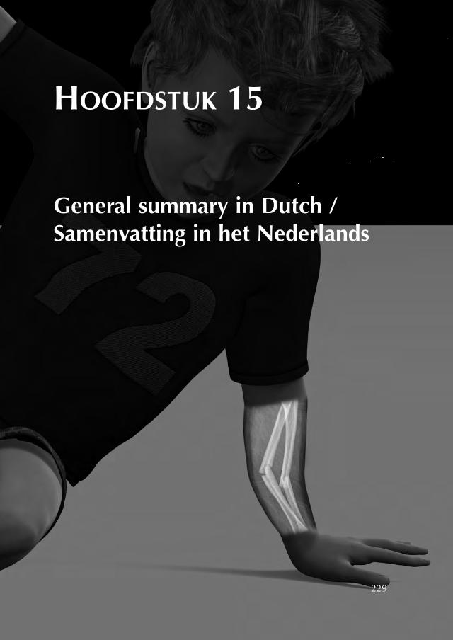
HOOFDSTUK 15
General summary in Dutch /Samenvatting in het Nederlands
229

Chapter 15
230
BREUKEN VAN ZOWEL HET SPAAKBEEN ALS DE ELLEPIJP
Dit proefschrift gaat over kinderen die een breuk hebben opgelopen van zowel het
spaakbeen als de ellepijp. In het Latijn wordt deze breuk de fractura antebrachii
genoemd. De basis van dit proefschrift betreft een onderzoek met deelname van
410 kinderen in 4 verschillende ziekenhuizen. De behandeling van deze kinderen
werd bepaald door loting tussen twee behandelingsvormen. Na gemiddeld 218
dagen werden 400 kinderen op de polikliniek teruggezien en werden röntgenfoto’s
gemaakt. Met deze gegevens werd geanalyseerd welke behandeling het beste is voor
welk type breuk. De breuken zijn in te delen naar locatie (dichtbij de pols of in het
midden van de onderarm), naar de mate van verplaatsing (weinig of fors verplaatst)
en naar de mate van stabiliteit (stabiel of instabiel). De resultaten van de
verschillende behandelingen zijn gerapporteerd in deel 1 en 2, terwijl de
verplaatsing van de breuk in gips en verminderde functie van de onderarm aan bod
komen in deel 3 en 4.
DEEL 1 BREUKEN DICHTBIJ DE POLS
In hoofdstuk 2 wordt een studie beschreven over de behandeling van breuken van
zowel het spaakbeen als de ellepijp dichtbij de pols. Deze breuken waren weinig
verplaatst en hoefden daarom niet in goede positie gezet te worden. Bij deze
kinderen werd er geloot tussen een behandeling met gips tot aan de elleboog
(onderarmgips) en gips voorbij de elleboog (bovenarmgips). De resultaten toonden
dat de groep kinderen met onderarmgips het gips comfortabeler vonden en minder
hulp nodig hadden met aankleden. Ook werden er minder complicaties gezien in
deze groep. Concluderend kunnen kinderen met een weinig verplaatste breuk
dichtbij de pols beter behandeld worden met een onderarmgips.
Hoofdstuk 3 gaat over kinderen met een fors verplaatste breuk dichtbij de pols
waarbij het nodig was de breuk in goede positie te zetten in de operatiekamer. Nadat
de breuk in goede positie was gezet, werd de stabiliteit van de breuk getest. Bij
stabiele breuken werd er geloot tussen de behandeling met alleen bovenarmgips en
een combinatie van 2 ijzeren pennetjes ter stabilisatie van de breuk en
bovenarmgips. In de groep kinderen behandeld met een combinatie van 2 ijzeren
General summary in Dutch / Samenvatting in het Nederlands
231
pennetjes en bovenarmgips werden minder verplaatsing van de breuk, een betere
functie maar meer complicaties gezien ten opzichte van de groep behandeld met
alleen bovenarmgips. Derhalve kunnen deze breuken beter gestabiliseerd worden
met 2 ijzeren pennetjes om verplaatsing van de breuk tegen te gaan. Wel is een
nauwkeurige chirurgische techniek noodzakelijk om complicaties te vermijden.
DEEL 2 BREUKEN IN HET MIDDEN VAN DE ONDERARM
Bij breuken van zowel het spaakbeen als de ellepijp in het midden van de onderarm
werd een onderscheid gemaakt tussen weinig en fors verplaatste breuken, waarbij
alleen de fors verplaatste breuken in goede positie werden gezet. Na het in goede
positie zetten van de fors verplaatste breuken werd een onderscheid gemaakt tussen
stabiele en instabiele breuken. Bij de weinig verplaatste breuken (studie beschreven
in hoofdstuk 4) en de breuken die na het in goede positie zetten stabiel waren (studie
beschreven in hoofdstuk 5), werd er geloot tussen de behandeling met
respectievelijk 6 weken bovenarmgips en een combinatie van 3 weken
bovenarmgips en 3 weken onderarmgips. In beide studies waren de resultaten tussen
de twee groepen vergelijkbaar met uitzondering van een hoger comfort in gips in
de groep behandeld met boven- en onderarmgips. Concluderend kunnen zowel
weinig verplaatste breuken als fors verplaatste breuken die na het in goede positie
zetten stabiel waren, het beste behandeld worden met een combinatie van boven-
en onderarmgips.
Hoofdstuk 6 beschrijft de fors verplaatste breuken in het midden van de
onderarm die na het in goede positie zetten instabiel waren. Deze breuken werden
volgens loting gestabiliseerd met 1 of 2 pennen in het spaakbeen en/of de ellepijp.
Breuken die werden gestabiliseerd met 1 pen vertoonden meer verplaatsing, werden
langer behandeld met gips en hadden minder goede klinische resultaten. Derhalve
kunnen deze breuken het beste worden gestabiliseerd met zowel een pen in het
spaakbeen als in de ellepijp.
Chapter 15
232
DEEL 3 VERPLAATSING VAN DE BREUK IN GIPS
Breuken van zowel het spaakbeen als de ellepijp hebben de neiging om te
verplaatsen in gips. Om te onderzoeken welke factoren van invloed zijn op het
verplaatsen van deze breuken in gips werden in hoofdstuk 7 de risicofactoren in
kaart gebracht. Hieruit bleek dat met name zeer ernstige breuken van de niet
dominante arm vaker verplaatsten in gips.
DEEL 4 VERMINDERDE ROTATIE VAN DE ONDERARM NA DE BREUK
Bij rotatie van de onderarm draait het spaakbeen om de ellepijp heen. Door een
breuk van beide botten kan een verminderde rotatie in de onderarm ontstaan. Om
de rotatie van de onderarm te meten gebruikt men vaak een gradenboog, de
goniometer. In hoofdstuk 8 wordt de rotatie van de onderarm respectievelijk geschat
en gemeten met een goniometer. Uit de resultaten blijkt dat het meten met een
goniometer nauwkeuriger is dan het schatten van de rotatie. Ook is een
vervolgmeting nauwkeuriger als dezelfde onderzoeker de meting herhaalt. In
hoofdstuk 9 blijkt dat er drie onafhankelijke risicofactoren van invloed zijn op
verminderde rotatie van de onderarm na een breuk van zowel het spaakbeen als de
ellepijp; een recidief breuk, een breuk in het midden van de onderarm en weinig
fysiotherapie. In hoofdstuk 10 werd onderzocht of het in een verkeerde positie
vastgroeien van een onderarmbreuk van invloed is op een verminderde rotatie van
de onderarm. Hieruit bleek dat er hoog risico was op het ontwikkelen van
verminderde rotatie in geval van forse scheefstand bij breuken dichtbij de pols
enerzijds, en in geval van milde scheefstand bij breuken in het midden van de
onderarm anderzijds. In hoofdstuk 11 wordt 3-dimensionaal radiologisch onderzoek
(MRI en CT) gebruikt om meer inzicht te krijgen in de oorzaak van de verminderde
rotatie na een onderarmbreuk. Hieruit kwam naar voren dat, (1) deze breuken in
een verkeerde rotatiestand kunnen vastgroeien, (2) dat er geen sprake was van botsen
tussen het spaakbeen en de ellepijp tijdens rotatie en (3) dat er geen verlittekening
was van de weke delen tussen het spaakbeen en de ellepijp.
General summary in Dutch / Samenvatting in het Nederlands
233
PART VII
APPENDICES
234
PhD Portfolio
235

PhD Portfolio
236
PHD PORTFOLIO
COURSES
Scientific writing in English for publication, Rotterdam
Developing a Cochrane systematic review, Amsterdam
Evidence based surgery in clinical practice, Zwolle
ORAL PRESENTATIONS
Which factors affect limitation of pronation/supination after forearm
fractures in children? A prospective multicentre study. Dutch
Orthopaedic Association. Rotterdam
Risk factors for the displacement of fractures of both-bones of the
forearm in children. Dutch Orthopaedic Association. Rotterdam
Risk factors for the displacement of fractures of both bones of the
forearm in children. Dutch trauma meeting, Amsterdam
Re-displacement of stable distal both-bone forearm fractures in
children: A randomised controlled multicentre study. Netherlands
Society for Hand Surgery, Amsterdam
Below-elbow cast for metaphyseal both-bone fractures of the distal
forearm in children: A randomized multicentre study. Dutch
Orthopaedic Association. Veldhoven
Re-displacement of stable distal both-bone forearm fractures in
children: A randomised controlled multicentre study. Federation of
European Societies for Surgery of the Hand, Antwerp
Re-displacement of stable distal both-bone forearm fractures in
children: A randomised controlled multicentre study. Congress of
the European Paediatric Surgeons� Association, Rome
PhD Portfolio
237
2011
2007
2005
2014
2014
2013
2012
2012
2012
2012
Both-bone forearm fractures in children. The first results. Trauma
meeting ZWOT, Rotterdam
Conversion to below-elbow cast after 3 weeks is safe for diaphyseal
both-bone forearm fractures in children. Dutch Surgery Society, Ede
Re-displacement of stable distal both-bone forearm fractures in
children: A randomised controlled multicentre study. Dutch Surgery
Society, Ede
Re-displacement of stable distal both-bone forearm fractures in
children: A randomised controlled multicentre study. Dutch trauma
meeting, Amsterdam
Both-bone forearm fractures in children. The results of a multicentre
study with inclusion of 410 children. Dutch Society for Paediatric
Orthopaedics, Utrecht
INVITED LECTURES
Below-elbow cast for metaphyseal both-bone fractures of the distal
forearm in children: A randomized multicentre study. Dutch
Orthopaedic Trauma Society, Amsterdam
Both-bone forearm fractures in children. The results of a multicentre
study with inclusion of 410 children. Dutch Society for Paediatric
Surgery, Utrecht
Both-bone forearm fractures in children. The results of a multicentre
study with inclusion of 410 children. Trauma meeting, Euregio, Hengelo
Both-bone forearm fractures in children. The results of a multicentre
study with inclusion of 410 children. Trauma meeting, TOWN, the
Hague
PhD Portfolio
238
2012
2011
2011
2011
2011
2013
2013
2013
2013
Different kind of cast for the treatment of both-bone forearm
fractures in children. Dutch trauma meeting, Amsterdam
POSTER PRESENTATIONS
Early conversion to below-elbow cast for displaced diaphyseal
both-bone forearm fractures in children: a multicenter randomized
controlled trial. Congress of the European Paediatric Surgeons
Association. Rome
Pronation and supination after forearm fractures in children:
Reliability of visual estimation and conventional goniometry
measurement. Dutch trauma meeting. Amsterdam
HONORS AND AWARDS
Biomet price
Best scientific research, Dutch Orthopaedic Association
NVOT Award
Best scientific article in Orthopaedic Trauma
Best oral presentation of Dutch trauma meeting
SUPERVISING MASTER’S THESIS
Leon Diederix, Resident Orthopaedic Surgery
Thesis: 5 years follow-up of both-bone forearm fractures in children
PhD Portfolio
239
2011
2012
2010
2014
2012
2011
2012-present

240

List of Publications
241

List of publications
242
LIST OF PUBLICATIONS
Colaris JW, Oei S, Reijman M, Holscher H, Allema JH, Verhaar JA. Three-
dimensional imaging of children with severe limitation of pronation/supination after
a both-bone forearm fracture. Arch Orthop Trauma Surg. 2014 Mar;134(3):333-41.
Colaris JW, Allema JH, Reijman M, de Vries MR, Ulas Biter L, Bloem RM, van
de Ven CP, Verhaar JA. Which factors affect limitation of pronation/supination after
forearm fractures in children? A prospective multicentre study. Injury. 2014
Apr;45(4):696-700.
Colaris JW, Allema JH, Biter LU, Reijman M, Ven CP, Vries MR, Bloem RM,
Kerver AJ, Verhaar JA. Conversion to below-elbow cast after 3 weeks is safe for
diaphyseal both-bone forearm fractures in children. Acta Orthop. 2013
Oct;84(5):489-94.
Colaris JW, Reijman M, Allema JH, Biter LU, Bloem RM, van de Ven CP, de
Vries MR, Kerver AJ, Verhaar JA. Early conversion to below-elbow cast for non-
reduced diaphyseal both-bone forearm fractures in children is safe: preliminary
results of a multicentre randomised controlled trial. Arch Orthop Trauma Surg. 2013
Oct;133(10):1407-14.
Colaris J, Reijman M, Allema JH, Kraan G, van Winterswijk P, de Vries M, van
de Ven C, Verhaar J. Single-bone intramedullary fixation of unstable both-bone
diaphyseal forearm fractures in children leads to increased re-displacement: a
multicentre randomised controlled trial. Arch Orthop Trauma Surg. 2013
Aug;133(8):1079-87.
Colaris JW, Allema JH, Reijman M, Biter LU, de Vries MR, van de Ven CP,
Bloem RM, Verhaar JA. Risk factors for the displacement of fractures of both bones
of the forearm in children. Bone Joint J. 2013 May;95-B(5):689-93.
List of publications
243
Witteveen HJ, Colaris JW. [A woman with a painful elbow]. Ned Tijdschr
Geneeskd. 2013;157(3):A4779. Dutch.
Colaris JW, Allema JH, Biter LU, de Vries MR, van de Ven CP, Bloem RM,
Kerver AJ, Reijman M, Verhaar JA. Re-displacement of stable distal both-bone
forearm fractures in children: a randomised controlled multicentre trial. Injury. 2013
Apr;44(4):498-503.
Peerbooms JC, Colaris JW, Hakkert AA, Van Appeldorn M, Bruijn DJ, Den
Oudsten BL, Gosens T. No positive bone healing after using platelet rich plasma in
a skeletal defect. An observational prospective cohort study. Int Orthop. 2012
Oct;36(10):2113-9.
Colaris JW, Biter LU, Allema JH, Bloem RM, van de Ven CP, de Vries MR,
Kerver AJ, Reijman M, Verhaar JA. Below-elbow cast for metaphyseal both-bone
fractures of the distal forearm in children: a randomised multicentre study. Injury.
2012 Jul;43(7):1107-11.
Joost W. Colaris, L. Ulas Biter, Jan Hein Allema, Rolf M. Bloem, Cees P. van de
Ven, Mark R. de Vries, Albert J.H. Kerver, Max Reijman, Jan A.N. Verhaar.
Onderarmgips bij distale fracturen van radius en ulna. Ned Tijdschr Geneeskd.
2012;156:A5332.
Zengerink I, Colaris JW. [A man without pronation and supination]. Ned
Tijdschr Geneeskd. 2011;155(42):A2723. Dutch.
Peters S, Colaris JW. Carpal coalition: symptomatic incomplete bony coalition
of the capitate and trapezoid—case report. J Hand Surg Am. 2011 Aug;36(8):1313-5.
Colaris J, van der Linden M, Selles R, Coene N, Allema JH, Verhaar J. Pronation
and supination after forearm fractures in children: Reliability of visual estimation
and conventional goniometry measurement. Injury. 2010 Jun;41(6):643-6.
List of publications
244
Peerbooms JC, de Wolf GS, Colaris JW, Bruijn DJ, Verhaar JA. No positive effect
of autologous platelet gel after total knee arthroplasty. Acta Orthop. 2009
Oct;80(5):557-62.
Colaris JW, Horn TM, van den Ende ED, Allema JH, Merkus JW. Supracondylar
fractures of the humerus in children. Comparison of results in two treatment periods.
Acta Chir Belg. 2008 Nov-Dec;108(6):715-9.
J.W. Colaris, M.R. de Vries, C.P. van de Ven, A.J.H. Kerver, L.U. Biter, J.A.N.
Verhaar. De fractura antebrachii studie. In dit Verband. 2008, nummer 3, jaargang
18, p22.
J.W. Colaris, L.N.J.E.M. Coene. Is a biopsy necessary in diagnosing Kümmell’s
disease? Journal of Orthopaedics. 2007; 4(3)e13.
Colaris JW, Reijman M, Allema JH, de Vries MR, Biter LU, Bloem RM, van de
Ven CP, Verhaar JA. Angular malalignment as cause of limitation of forearm rotation:
an analysis of prospectively collected data of both-bone forearm fractures in
children. Injury. 2014 Jun;45(6):955-9.
List of publications
245

246
Curriculum Vitae
247

Curriculum Vitae
248
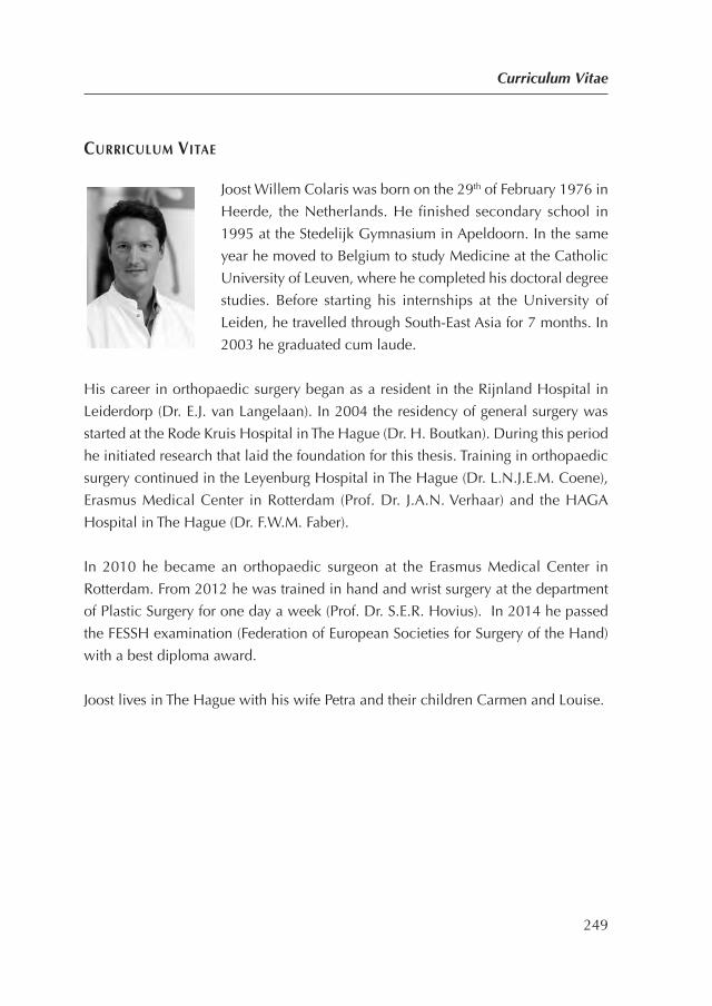
CURRICULUM VITAE
Joost Willem Colaris was born on the 29th of February 1976 in
Heerde, the Netherlands. He finished secondary school in
1995 at the Stedelijk Gymnasium in Apeldoorn. In the same
year he moved to Belgium to study Medicine at the Catholic
University of Leuven, where he completed his doctoral degree
studies. Before starting his internships at the University of
Leiden, he travelled through South-East Asia for 7 months. In
2003 he graduated cum laude.
His career in orthopaedic surgery began as a resident in the Rijnland Hospital in
Leiderdorp (Dr. E.J. van Langelaan). In 2004 the residency of general surgery was
started at the Rode Kruis Hospital in The Hague (Dr. H. Boutkan). During this period
he initiated research that laid the foundation for this thesis. Training in orthopaedic
surgery continued in the Leyenburg Hospital in The Hague (Dr. L.N.J.E.M. Coene),
Erasmus Medical Center in Rotterdam (Prof. Dr. J.A.N. Verhaar) and the HAGA
Hospital in The Hague (Dr. F.W.M. Faber).
In 2010 he became an orthopaedic surgeon at the Erasmus Medical Center in
Rotterdam. From 2012 he was trained in hand and wrist surgery at the department
of Plastic Surgery for one day a week (Prof. Dr. S.E.R. Hovius). In 2014 he passed
the FESSH examination (Federation of European Societies for Surgery of the Hand)
with a best diploma award.
Joost lives in The Hague with his wife Petra and their children Carmen and Louise.
Curriculum Vitae
249