foot-and-mouth disease virus infection in young lambs: pathogenesis and tissue tropism
TRANSCRIPT
Accepted Manuscript
Title: Foot-and-mouth disease virus infection in young lambs:Pathogenesis and tissue tropism
Authors: Eoin Ryan, Jacquelyn Horsington, StephanieDurand, Harriet Brooks, Soren Alexandersen, Joe Brownlie,Zhidong Zhang
PII: S0378-1135(07)00432-4DOI: doi:10.1016/j.vetmic.2007.08.029Reference: VETMIC 3809
To appear in: VETMIC
Received date: 24-4-2007Revised date: 23-8-2007Accepted date: 24-8-2007
Please cite this article as: Ryan, E., Horsington, J., Durand, S., Brooks, H.,Alexandersen, S., Brownlie, J., Zhang, Z., Foot-and-mouth disease virus infectionin young lambs: Pathogenesis and tissue tropism, Veterinary Microbiology (2007),doi:10.1016/j.vetmic.2007.08.029
This is a PDF file of an unedited manuscript that has been accepted for publication.As a service to our customers we are providing this early version of the manuscript.The manuscript will undergo copyediting, typesetting, and review of the resulting proofbefore it is published in its final form. Please note that during the production processerrors may be discovered which could affect the content, and all legal disclaimers thatapply to the journal pertain.
peer
-005
3231
4, v
ersi
on 1
- 4
Nov
201
0Author manuscript, published in "Veterinary Microbiology 127, 3-4 (2008) 258"
DOI : 10.1016/j.vetmic.2007.08.029
Acce
pted
Man
uscr
ipt
Foot-and-mouth disease virus infection in young lambs: pathogenesis and tissue 1
tropism2
3
Eoin Ryana,b, Jacquelyn Horsingtona, Stephanie Duranda, Harriet Brooksb, Soren 4
Alexandersenc, Joe Brownlieb, Zhidong Zhanga*.5
6
a Institute for Animal Health, Pirbright Laboratory, Ash Road, Pirbright, Woking, 7
GU24 0NF, UK.8
b Royal Veterinary College, Hawkshead Lane, North Mymms, Hatfield, AL9 7TA, 9
UK.10
c National Veterinary Institute, Danish Technical University, Lindholm, DK-4771 11
Kalvehave, Denmark12
13
*Correspondence to Zhidong Zhang, Institute for Animal Health, Pirbright 14
Laboratory, Ash Road, Pirbright, Woking, GU24 0NF, UK. 15
Email [email protected]; phone +44 (0)1483 231135; fax +44 (0)1483 16
232448.17
18
Manuscript
Page 1 of 41
peer
-005
3231
4, v
ersi
on 1
- 4
Nov
201
0
Acce
pted
Man
uscr
ipt
Abstract19
20
Foot-and-mouth disease (FMD) in adult sheep usually causes milder clinical signs 21
than in cattle or pigs, and is often subtle enough to go undiagnosed. In contrast, FMD 22
in lambs has been reported to cause high mortality during field outbreaks. In order to 23
investigate the pathogenesis of FMD in lambs, two groups, aged 10-14 days, were 24
infected with foot-and-mouth disease virus (FMDV) type O UKG. One group of 25
lambs (n=8) was inoculated with FMDV in the coronary band, while the other (n=4) 26
was infected by direct contact with FMDV-inoculated ewes. Daily serum samples and 27
temperature measurements were taken. Lambs were killed sequentially and tissue28
samples taken for analysis. Using real-time RT-PCR, viral RNA levels in tissue 29
samples and serum were measured, and a novel strand-specific real-time RT-PCR 30
assay was used to quantify viral replication levels in tissues. Tissue sections were 31
examined for histopathological lesions, and in situ hybridisation (ISH) was used to 32
localise viral RNA within histological sections. The contact-infected lambs became 33
infected approximately 24 hours after the ewes were inoculated. Vesicular lesions 34
developed on the feet of all lambs and on the caudo-lateral part of the tongues of 6 of 35
the 8 inoculated lambs and 3 of the 4 contact-infected lambs. Although no lambs 36
developed severe clinical signs, one of the contact-infected lambs died acutely at 5 37
days post-exposure. Histological examination of the heart from this lamb showed 38
multi-focal areas of lymphocytic-plasmacytic myocarditis; similar lesions were also 39
observed in the hearts of three of the inoculated lambs. Using ISH, viral RNA was 40
localised within cardiac and skeletal muscle cells from the lamb which had died, and 41
also from vesicular lesions on the coronary band and tongue of inoculated lambs. 42
Page 2 of 41
peer
-005
3231
4, v
ersi
on 1
- 4
Nov
201
0

Acce
pted
Man
uscr
ipt
These results provide a detailed description of the pathogenesis of the disease in 43
lambs.44
Keywords: foot-and-mouth disease virus; sheep; lamb; pathogenesis; in situ45
hybridisation46
47
48
Page 3 of 41
peer
-005
3231
4, v
ersi
on 1
- 4
Nov
201
0
Acce
pted
Man
uscr
ipt
INTRODUCTION49
50
Foot-and-mouth disease (FMD) is a highly contagious acute vesicular disease of 51
cloven-hoofed animals. The aetiological agent, foot-and-mouth disease virus 52
(FMDV), is classified with the Aphthovirus genus as a member of the Picornaviridae53
family (Belsham, 1993). FMD is an economically devastating disease, causing 54
significant production losses in infected domestic livestock. As a result, it is a major 55
hindrance to international trade in animals and animal products. The cost of an 56
outbreak can be enormous; the 2001 outbreak in the UK is estimated to have cost the 57
agricultural sector £3.1 billion, with similar losses to the tourism sector (Thompson et 58
al., 2002). 59
60
Clinical signs are generally milder in sheep than in cattle or pigs. Viraemia may be 61
present for up to 3 days before the appearance of vesicular lesions (Alexandersen et 62
al., 2003). During this time the sheep may be pyrexic and distressed with lameness 63
spreading through the flock. Agalactia may occur in ewes. Vesicular lesions occur in 64
the interdigital cleft, along the coronary bands and on the bulb of the heels. Oral 65
lesions are less common but can occur on the dental pad, tongue and gums (Hughes et 66
al., 2002). In pregnant sheep, transplacental infection of the foetal lamb may occur, 67
causing abortion (Ryan et al., 2007).68
69
Young lambs may die acutely following infection; associated clinical signs include 70
collapse, fever, tachycardia and marked abdominal respiration (Garcia-Mata et al.,71
1954;1955; Geering, 1967; Pay, 1988). Deaths in lambs start to occur two to three 72
days after the appearance of clinical signs in the ewes, and are usually reported to be 73
Page 4 of 41
peer
-005
3231
4, v
ersi
on 1
- 4
Nov
201
0
Acce
pted
Man
uscr
ipt
the result of heart failure or starvation; post-mortem lesions include myocarditis, 74
septicaemia, abomasitis and enteritis (Littlejohn, 1970; Salyi 1939). Reported 75
mortality rates vary widely, from 4.7% in an outbreak in India (Panisup et al., 1979)76
to 94.5% in lambs of two to 25 days old in a Russian outbreak (Khankishiev et al., 77
1958). Neonatal lamb deaths were reported in Dumfries during the 2001 outbreak in 78
the UK (Reid, 2002). A PanAsia type O virus strain was reported to have caused 79
large-scale lamb mortality in Iraq (Knowles and Samuel, 2003). 80
81
Mortality among FMD-infected calves and pigs has also been described. Death may 82
occur without vesicular lesions due to the peracute onset of FMD in these animals.83
Pale foci of myocardial necrosis are seen in the ventricular wall and papillary muscle, 84
referred to as “tiger heart” due to striping effect (Jubb, 1993). Donaldson et al. (1984)85
reported that the majority of piglets infected with an O1 Lausanne strain died or 86
became moribund and were killed without developing vesicles. The deaths of the 87
piglets started before clinical disease was evident in the sows. Macroscopic “tiger 88
heart” lesions were not observed in any piglet hearts, although in all but one, 89
myocarditis was present on histopathological examination.90
91
Despite the numerous reports of FMD-related deaths in lambs, there is little data92
available on FMDV tropism or quantitative aspects of viral RNA kinetics in neonatal 93
lambs, which would help understand the reasons for this high mortality and the 94
influence of maternal infection on young lambs. In this study, viral distribution, tissue 95
tropism and associated pathology during the acute stages of FMD were investigated in 96
neonatal lambs experimentally infected with FMDV O UKG.97
98
Page 5 of 41
peer
-005
3231
4, v
ersi
on 1
- 4
Nov
201
0
Acce
pted
Man
uscr
ipt
MATERIALS AND METHODS99
Inoculum100
The FMDV strain used was O UKG 34/2001, isolated from a pig at Cheale Meats 101
Abattoir, Essex on 20 February 2001 (Alexandersen and Donaldson, 2002). The 102
inoculum was the second pig passage of original pig epithelial tissue suspension. 103
Animal experiments104
Animals were housed in a category 4 bio-containment animal unit (Specified Animal 105
Pathogens Order, DEFRA 1998). Animal experimentation was carried out in 106
accordance with the UK Animals (Scientific Procedures) Act 1986 and associated 107
guidelines. Two separate experiments were carried out. The first involved inoculating 108
ewes and allowing their lambs to become infected by direct contact, simulating 109
natural infection in the field. In the second experiment, lambs were inoculated 110
directly.111
Experiment 1: Ten lambs, 10-14 days old, and seven dams were used in this 112
experiment, with ewes numbered VM76 to VM82. Lambs were assigned the same 113
numbers as their dams, with the prefix LA for singletons or the first of a pair of twins, 114
and LB for the second of a pair of twins, i.e. LA76 to LB82. Two lambs and a ewe 115
were killed as negative controls before the inoculations were carried out. Eight lambs 116
were inoculated with 105.9 FMDV O UKG 34/2001 in the coronary band. Two were 117
killed at 2, 4, 7, and 10 dpi, with the ewes being killed at the same timepoints as their 118
offspring.119
Experiment 2: Five ewes, each with a lamb of 10-14 days, were used in this 120
experiment, with ewes numbered from VE37 to VE41. The lambs were assigned the 121
same numbers as their ewes, with the prefix L, i.e. L37 to L41. Negative control 122
samples of serum were taken from all animals and a ewe and lamb were killed as 123
Page 6 of 41
peer
-005
3231
4, v
ersi
on 1
- 4
Nov
201
0
Acce
pted
Man
uscr
ipt
negative controls prior to inoculation. Four ewes were inoculated by coronary band 124
inoculation with 105.9 TCID50 FMDV O UKG 34/2001. The ewes were housed in pens 125
in direct contact with their lambs. One ewe and her lamb were killed at two, three and 126
four days post-inoculation (dpi). The fourth infected lamb was found dead on the 127
morning of day five post-inoculation. The mother of this lamb was killed on day 128
seven.129
130
Sample collection131
Daily serum and nasal swab samples were collected from ewes and lambs and clinical 132
signs and temperatures recorded. At post-mortem examination, various tissue samples 133
were collected, as listed in Tables 1, 2, 4 and 5. Tissues collected from ewes included 134
cervical and mandibular lymph nodes (LN), heart, liver, mammary gland, soft palate, 135
spleen, tonsil, cotyledon, muscle (taken from lumbar area), tongue epithelium, uterus 136
wall and coronary band skin (from non-inoculated coronary band). Tissues collected 137
lambs included cervical and mandibular LNs, duodenal intestine, heart, kidney, liver, 138
caudal lung lobe, muscle (from lumbar area), skin from lateral hindleg, soft palate, 139
spleen, tongue, tonsil and coronary band skin (from non-inoculated coronary band). A140
portion of each tissue was put in RNAlater (Ambion, UK) for RNA extraction, and141
10% formalin (formol-FIXX, Shandon, UK) for histological examination and in situ142
hybridisation as described below. Tissues were fixed in 10% formalin for less than 24 143
hours and then placed in 1% formalin, followed by embedding in paraffin wax.144
RNA extraction145
For automated RNA extraction from serum samples, 200l of sample was mixed with 146
300 l MagNa Pure LC total nucleic acid lysis buffer (Roche, UK). Total nucleic 147
Page 7 of 41
peer
-005
3231
4, v
ersi
on 1
- 4
Nov
201
0
Acce
pted
Man
uscr
ipt
acids were extracted and eluted in 50 l elution buffer by using the MagNa Pure LC 148
total nucleic acid isolation kit (Roche,UK) with an automated robotic workstation 149
(Roche), according to manufacturer’s instructions, as described by Quan et al. (2004). 150
For tissue samples, 20 mg of tissue were homogenised by placing them in 700 l of 151
tissue lysis buffer (Roche, UK) in Lysing Matrix D tubes (Q-Biogene, UK) and 152
shaking them at 6,500 revolutions per minute for 45 seconds three times in a FastPrep 153
FP120 homogenising machine (Q-Biogene, UK). RNA was extracted and eluted in 50 154
l elution buffer by using the MagNa Pure LC RNA extraction kit III (Roche, UK) 155
with an automated robot as described above. Extracted RNA was stored at -80oC until 156
used.157
Real-time quantitative RT-PCR assay for detection of viral RNA. 158
The level of viral RNA in tissue, serum and amniotic fluid samples was quantified by 159
real-time RT-PCR as described previously (Reid et al., 2002), using a Taqman probe 160
specific for FMDV type O strain UKG 34/2001 as described previously (Quan et al., 161
2004). PCR assays were performed on a Stratagene MX4000 machine. For the 162
generation of standard curves, a FMDV RNA standard was synthesized in vitro from 163
a plasmid containing a 500 base pair insert of the internal ribosomal entry site (IRES) 164
of FMDV O UKG 34/2001 (kindly provided by Dr G. Belsham, Institute for Animal 165
Health, UK) using a MEGAScripTM T7 kit (Ambion, UK) as described previously 166
(Quan et al., 2004).167
168
Detection of antibody in sera by ELISA169
A liquid-phase blocking ELISA was used to titrate levels of anti-FMDV antibodies in 170
serum samples, as described previously (Ferris et al., 1990). 171
Page 8 of 41
peer
-005
3231
4, v
ersi
on 1
- 4
Nov
201
0
Acce
pted
Man
uscr
ipt
172
Analysis of FMDV replication in tissues using strand specific real-time RT-PCR173
In order to analyse viral replication levels in tissue samples, a strand specific real-time 174
RT-PCR was used as previously described (Horsington and Zhang, 2007)..A ratio of 175
positive to negative strands was calculated based on the quantification of levels of 176
positive and negative sense viral RNA transcripts in tissues. This ratio gives an177
indication of the degree of viral replication in a sample, with a lower ratio indicating a 178
higher level of replication. Briefly, a tagged forward primer was designed containing 179
a 20 nucleotide ‘tag’ sequence unrelated to FMDV at the 5’ end with the rest of the 180
primer (20 bases) specific to the FMDV 3D negative strand. Similarly, a tagged 181
reverse primer was designed to target the FMDV 3D positive strand. cDNA was 182
produced using the Thermoscript (Invitrogen, UK) RT components. 6µl RNA was 183
combined with 1µl dNTPs, 0.3µl primer (Tag-3D7151F, 10µM or Tag-3D7251R, 184
10µM) and 4.7µl dH2O, and heated to 75ºC for 2 minutes. The reactions were then 185
placed on ice for 2 minutes. 4µl 5X cDNA synthesis buffer, 1µl 0.1M DTT, 1µl 186
RNaseOut, 0.5µl Thermoscript RT enzyme (all Invitrogen, UK) and 1.5 µl dH2O was 187
combined and added to the samples. Reverse transcription was performed at 62ºC for 188
35 minutes followed by 10 minutes at 95ºC. Real-time quantitative PCR was 189
performed using TAG/3D7251R primers for the negative strand assay and 190
3D7151F/TAG primers for the positive strand assay and a FAM-TAMRA probe, 191
3D7196P, on the Stratagene 3005P real-time PCR machine (Stratagene, UK). 5µl 192
cDNA was combined with 12.5µl 2x Taqman Mastermix (Applied Biosystems, UK), 193
1µl each of the forward and reverse primer (10µM), 0.5µl probe (5µM) and dH2O to 194
25µl. The program comprised of 2 minutes at 50ºC, 10 minutes at 95ºC, and 50 cycles 195
of 95ºC for 15 seconds and 58ºC for 1 minute. The standard curve was derived from 196
Page 9 of 41
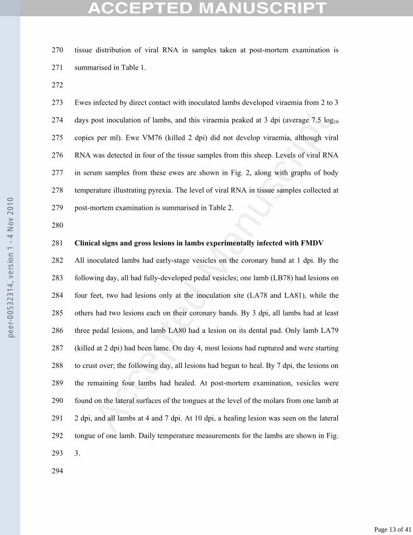
peer
-005
3231
4, v
ersi
on 1
- 4
Nov
201
0
Acce
pted
Man
uscr
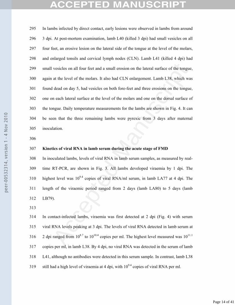
ipt
synthetic negative or positive strand FMDV transcripts from 1 x 10⁸ to 1 x 101197
copies/µl. 198
In situ hybridisation199
RNA probes and labelling 200
A plasmid containing a part of the 3D region of FMDV type O UKG 34/2001 was 201
made and cells transfected with this plasmid kindly supplied by Nicholas Juleff, 202
Institute for Animal Health. The negative-sense RNA probe, complementary to the 203
positive-sense RNA of FMDV, generated from this plasmid was used on tissues from 204
experiment 1 (lambs LA76-LB82). A plasmid containing a part of the 3D region of 205
the FMDV type O Kaufbeuren strain genome (kindly provided by Dr G. Belsham, 206
Institute for Animal Health) was used to generate probe for use on tissues collected 207
from experiment 2 (lambs L37-L41). A plasmid containing part of the SVDV genome 208
was used to generate a control ISH probe.209
The plasmids were linearised and then purified using phenol/chloroform extraction. 210
The RNA probes were synthesised from plasmid DNA and labelled with digoxenin 211
using a DIG RNA SP6/T7 labelling kit (Roche, UK) according to the manufacturer’s 212
instructions. 213
Hybridisation214
An mRNAlocatortm In situ hybridisation kit (Ambion, UK) was used to localise 215
FMDV in tissues. Sections (4 μm) of the paraffin wax-embedded samples of fixed 216
tissues were applied to superfrost microscope slides (BDH, UK). They were incubated 217
at 56oC for 20 minutes to melt the paraffin, then put in xylene (BDH, UK) for 2x15 218
minutes at room temperature to dewax. The sections were put in 100% ethanol for 219
Page 10 of 41
peer
-005
3231
4, v
ersi
on 1
- 4
Nov
201
0
Acce
pted
Man
uscr
ipt
2x10 minutes, then 5 minutes each in 90%, 70% and 50% ethanol, followed by 5 220
minutes in phosphate buffered saline (PBS). Sections were immersed in 0.05% 221
proteinase K (70 U/l) in PBS for 10 minutes at room temperature then rinsed in 222
nuclease-free water. Sections were then placed in a solution of 1.32% triethanolamine 223
and 0.5% HCl (Sigma, UK) in nuclease-free water for 3 minutes at room temperature, 224
followed by 10 minutes incubation in a solution of 1.32% triethanolamine, 0.5% HCl 225
and 0.24% acetic anhydride in nuclease-free water at room temperature. Slides were 226
washed for 5 minutes in PBS and placed in 100% ethanol for 5 minutes, then air 227
dried.228
229
Hybridisation cover chambers (Sigma, UK) were applied to slides and overlaid with 230
40 l prehybridisation buffer, followed by incubation in a humid chamber at 60oC for 231
one hour. Hybridisation solution was prepared by adding 1 l of 100 ng/l probe to 232
40 l of hybridisation buffer for each section. Each slide had two sections on it, 233
allowing one section to be overlaid with FMDV probe and the other with SVDV 234
probe as a negative control. Prehybridisation buffer was removed from the chambers, 235
then 40 l hybridisation solution added to each chamber. Slides were put on a hot 236
plate at 65oC for 5 minutes, then incubated in a humid chamber at 60oC overnight.237
238
The following morning, coverslips were removed and sections were washed in 2x 239
saline-sodium citrate (SSC) for 30 minutes at 50oC, then 1x SSC for 30 minutes at 240
50oC. The slides were treated with 0.045% RNAse A (450 U/ml) in 1x RNAse 241
digestion buffer for 30 minutes at 37oC, then washed in 2x SSC for 30 minutes at 242
37oC. Slides were incubated in a blocking solution of 0.1% Triton X-100 (Sigma, UK) 243
and 2% normal sheep serum (Vector Laboratories, UK) in 100 mM Tris HCl (pH 7.5) 244
Page 11 of 41
peer
-005
3231
4, v
ersi
on 1
- 4
Nov
201
0
Acce
pted
Man
uscr
ipt
(BDH, UK) and 150 mM NaCl (BDH, UK) for 30 minutes at room temperature in a 245
humid chamber. Following this, sections were incubated for 1 hour at room 246
temperature in a humid chamber in an antibody solution of 0.1% anti-digoxenin 247
alkaline phosphatise sheep antibody (concentration 0.75 U/l) (Roche, UK), 0.05% 248
Triton-X 100 and 1% normal sheep serum in 100 mM Tris HCl (pH 7.5) and 150 mM 249
NaCl. 250
251
Sections were then washed for 2x30 minutes in blocking solution with a gentle 252
shaker. They were incubated for 1 minute in a colouration buffer of 100mM Tris-HCl 253
(pH 9,5), 100 mM NaCl and 50 mM MgCl2. A colour solution of 0.35% 5-bromo, 4-254
chloro, 3-indolylphosphate (BCIP) (Boehringer Mannheim, Germany) and 0.45%255
nitroblue tetrazolium (NBT) (Boehringer Mannheim, Germany) was added to the 256
colouration buffer and slides were incubated in this in a humid chamber for 1 hour. A 257
water wash stopped colouration. Slides were counterstained with methyl green 258
(Vektor Laboratories, Inc., USA) for 2 minutes 30 seconds at 60oC on heat block, 259
dipped in water, then mounted with Immumount (Thermoshandon Electron 260
Corporation, UK).261
262
RESULTS263
264
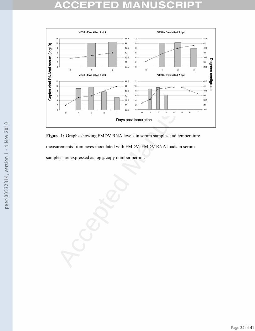
Kinetics of viral RNA in ewes experimentally infected with FMDV265
All inoculated ewes developed signs of clinical signs of FMD within 1-2 dpi. The 266
kinetics of viral RNA in serum samples are shown in Fig. 1 with daily temperature 267
data. All inoculated ewes developed viraemia at 1 dpi (average 9.6 log10 copies per 268
ml). Viral RNA levels then peaked at 2 dpi (average 10 log10 copies per ml). The 269
Page 12 of 41
peer
-005
3231
4, v
ersi
on 1
- 4
Nov
201
0
Acce
pted
Man
uscr
ipt
tissue distribution of viral RNA in samples taken at post-mortem examination is 270
summarised in Table 1.271
272
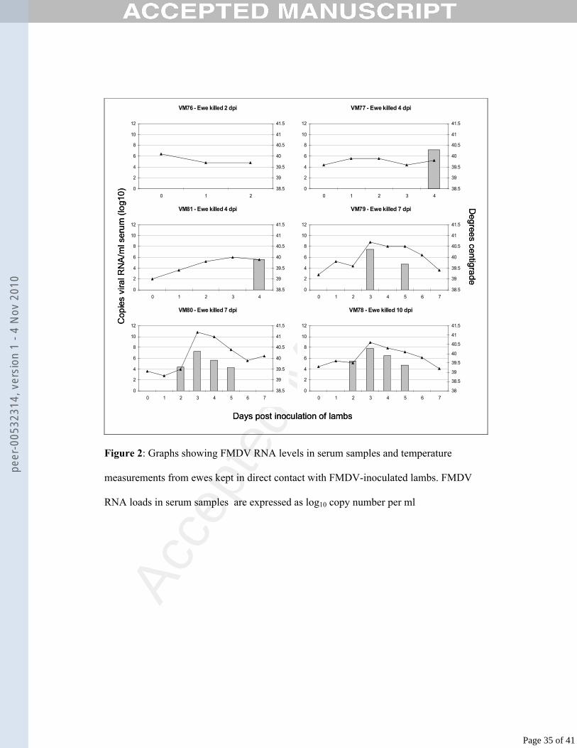
Ewes infected by direct contact with inoculated lambs developed viraemia from 2 to 3 273
days post inoculation of lambs, and this viraemia peaked at 3 dpi (average 7.5 log10274
copies per ml). Ewe VM76 (killed 2 dpi) did not develop viraemia, although viral 275
RNA was detected in four of the tissue samples from this sheep. Levels of viral RNA 276
in serum samples from these ewes are shown in Fig. 2, along with graphs of body 277
temperature illustrating pyrexia. The level of viral RNA in tissue samples collected at 278
post-mortem examination is summarised in Table 2.279
280
Clinical signs and gross lesions in lambs experimentally infected with FMDV281
All inoculated lambs had early-stage vesicles on the coronary band at 1 dpi. By the282
following day, all had fully-developed pedal vesicles; one lamb (LB78) had lesions on 283
four feet, two had lesions only at the inoculation site (LA78 and LA81), while the 284
others had two lesions each on their coronary bands. By 3 dpi, all lambs had at least 285
three pedal lesions, and lamb LA80 had a lesion on its dental pad. Only lamb LA79 286
(killed at 2 dpi) had been lame. On day 4, most lesions had ruptured and were starting 287
to crust over; the following day, all lesions had begun to heal. By 7 dpi, the lesions on 288
the remaining four lambs had healed. At post-mortem examination, vesicles were 289
found on the lateral surfaces of the tongues at the level of the molars from one lamb at 290
2 dpi, and all lambs at 4 and 7 dpi. At 10 dpi, a healing lesion was seen on the lateral 291
tongue of one lamb. Daily temperature measurements for the lambs are shown in Fig.292
3.293
294
Page 13 of 41
peer
-005
3231
4, v
ersi
on 1
- 4
Nov
201
0
Acce
pted
Man
uscr
ipt
In lambs infected by direct contact, early lesions were observed in lambs from around 295
3 dpi. At post-mortem examination, lamb L40 (killed 3 dpi) had small vesicles on all 296
four feet, an erosive lesion on the lateral side of the tongue at the level of the molars,297
and enlarged tonsils and cervical lymph nodes (CLN). Lamb L41 (killed 4 dpi) had 298
small vesicles on all four feet and a small erosion on the lateral surface of the tongue, 299
again at the level of the molars. It also had CLN enlargement. Lamb L38, which was 300
found dead on day 5, had vesicles on both fore-feet and three erosions on the tongue, 301
one on each lateral surface at the level of the molars and one on the dorsal surface of 302
the tongue. Daily temperature measurements for the lambs are shown in Fig. 4. It can 303
be seen that the three remaining lambs were pyrexic from 3 days after maternal 304
inoculation.305
306
Kinetics of viral RNA in lamb serum during the acute stage of FMD307
In inoculated lambs, levels of viral RNA in lamb serum samples, as measured by real-308
time RT-PCR, are shown in Fig. 3. All lambs developed viraemia by 1 dpi. The 309
highest level was 109.4 copies of viral RNA/ml serum, in lamb LA77 at 4 dpi. The 310
length of the viraemic period ranged from 2 days (lamb LA80) to 5 days (lamb 311
LB79). 312
313
In contact-infected lambs, viraemia was first detected at 2 dpi (Fig. 4) with serum 314
viral RNA levels peaking at 3 dpi. The levels of viral RNA detected in lamb serum at 315
2 dpi ranged from 108.7 to 1010.4 copies per ml. The highest level measured was 1011.1316
copies per ml, in lamb L38. By 4 dpi, no viral RNA was detected in the serum of lamb 317
L41, although no antibodies were detected in this serum sample. In contrast, lamb L38 318
still had a high level of viraemia at 4 dpi, with 109.4 copies of viral RNA per ml. 319
Page 14 of 41
peer
-005
3231
4, v
ersi
on 1
- 4
Nov
201
0
Acce
pted
Man
uscr
ipt
320
Antibody levels in serum from inoculated lambs321
Antibody titres detected in lamb serum from inoculated lambs are summarised in 322
Table 3. No antibodies were detected in serum from the contact-infected lambs.323
324
Distribution and quantification of viral RNA in lamb tissues325
The distribution of viral RNA in tissues from inoculated lambs is summarised in 326
Table 4. Viral RNA was detected in all tissues at 2 and 4 dpi. At 2 dpi, the highest 327
levels were found in the coronary band, tongue, skin, tonsil and CLN. At 4 dpi, there 328
was more variation between tissues, with the highest levels in the coronary band, 329
heart, CLN, mandibular lymph node (MLN), tongue, and tonsil. At 7 dpi, no viral 330
RNA was detected in the liver, and there was less in the other tissues than at earlier 331
timepoints. The tongue was the only exception to this, with an average of 1011332
copies/g. Other tissues with the most viral RNA at this timepoint were the coronary 333
band and the CLN. By 10 dpi, there was no viral RNA detectable in 7 tissue samples 334
from one lamb and 3 from the other. The tonsil, with an average of 1010 copies/g, 335
contained the highest amount of viral RNA.336
337
In contact-infected lambs, levels of viral RNA detected in lamb tissues are 338
summarised in Table 5. Viral RNA was evenly distributed throughout tissues 339
collected at 2 dpi from L39, with the highest levels in the coronary band, tongue, soft 340
palate, CLN, MLN and tonsil. At 3 dpi (L40), the highest level of viral RNA was 341
found in the skin of the foot, followed by the tongue, CLN, MLN and soft palate. The 342
lamb (L41) killed at 4 dpi had cleared the viraemia by the time it was killed although 343
antibody titration by ELISA on its serum at 4 dpi was negative. Most of the tissue 344
Page 15 of 41
peer
-005
3231
4, v
ersi
on 1
- 4
Nov
201
0
Acce
pted
Man
uscr
ipt
samples contained low levels of viral RNA, and there was none detected in the heart. 345
Only the coronary band, tongue and tonsil contained 108 copies of viral RNA or more 346
per gram. This is in marked contrast to the lamb (L38) found dead at 5 dpi, which had 347
a significant viraemia on days 2, 3 and 4 and had high levels of viral RNA in many 348
tissue samples, particularly the heart and skeletal muscle, which each contained over 349
1010 copies of viral RNA per gram.350
351
Histopathology in the heart and muscle352
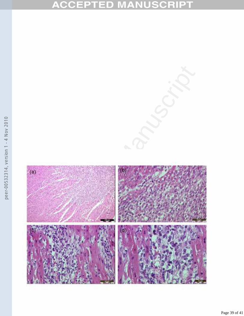
Cardiac histopathological lesions in infected lambs are shown in Fig. 5. Multiple foci353
of myocardial abnormalities were observed in sections from lamb LA81 (directly 354
inoculated with FMDV) at 4 dpi, including pale and ruptured myocardiocytes, 355
disordered myocardiocytes, aggregations of lymphocytes and oedema (Figs. 5a, 5b). 356
Similar lesions were observed in heart muscle from lambs LA80 (killed 7 dpi) and 357
LA78 (killed 10 dpi). In sections of heart from the contact-infected lamb found dead 358
on day five (L38) multi-focal myocardiocyte swelling, sometimes with hyalinisation, 359
and focally marked perivascular lymphocytic aggregation were described (Figs.5c, 360
5d). No significant histological abnormalities were recognised in sections of heart 361
from the other contact-infected lambs. In skeletal muscle sections from contact-362
infected lamb L38, disordered myocytes with pale, foamy cytoplasm were recognised 363
histologically.364
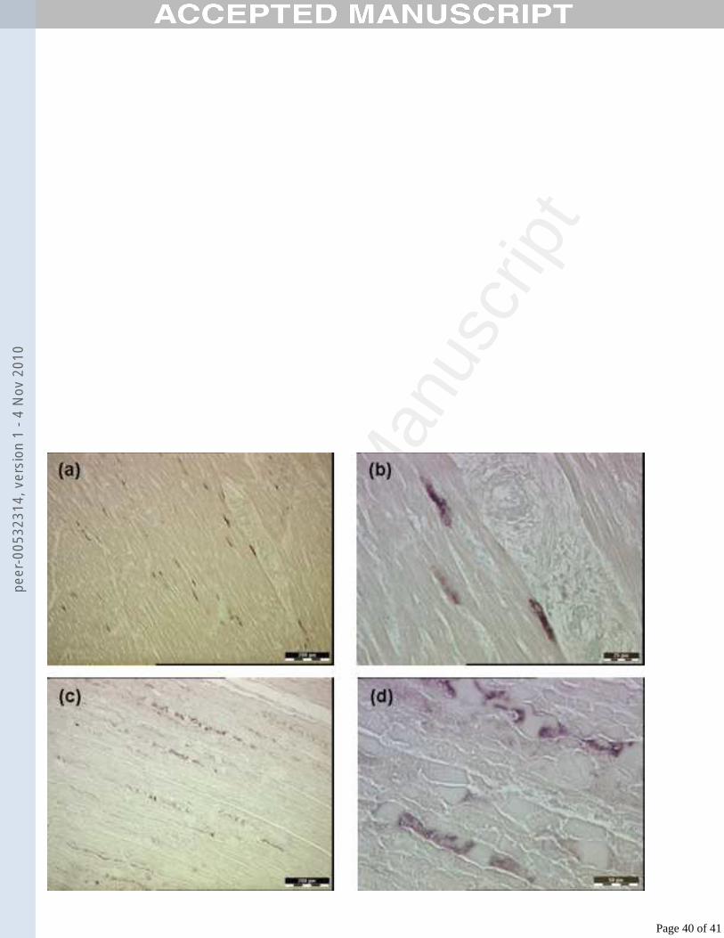
365
Localization of viral RNA in relation to histopathological lesions366
ISH was carried out on tissue sections to localise viral RNA in relation to 367
histopathological lesions. In heart sections from the contact-infected lamb found dead 368
on day five (L38), viral RNA was distributed throughout the myocardium in a diffuse 369
Page 16 of 41
peer
-005
3231
4, v
ersi
on 1
- 4
Nov
201
0
Acce
pted
Man
uscr
ipt
pattern, with positive signal found both in areas of cell swelling and in areas which 370
were unremarkable in H&E sections (Figs. 6a, 6b). The positive signal was found in 371
the cytoplasm of the cells. No positive signal was observed in heart sections from the 372
other contact-infected lambs. In addition, ISH on skeletal muscle sections from the 373
same lamb also showed viral RNA distributed throughout the sections in a multi-focal 374
array contrasting with the diffuse distribution seen in the myocardium (Figs. 6c, 6d). 375
No positive signal was found in the skeletal muscle sections from the other contact-376
infected lambs. ISH was also performed on skin, tongue, soft palate, lymph nodes and 377
lung from all contact-infected lambs, but no positive signal was detected. Viral RNA 378
was also localised within vesicles in sections of tongue and coronary band from 379
inoculated lambs (Fig. 7). Positive signal can be seen within the lesions, deep to the 380
stratum corneum and in the detached epithelium of the coronary band lesion, and 381
diffusely throughout the two microvesicles in the stratum spinosum of the tongue.382
383
Analysis of viral replication levels in tissue samples using strand-specific real-384
time RT-PCR assay 385
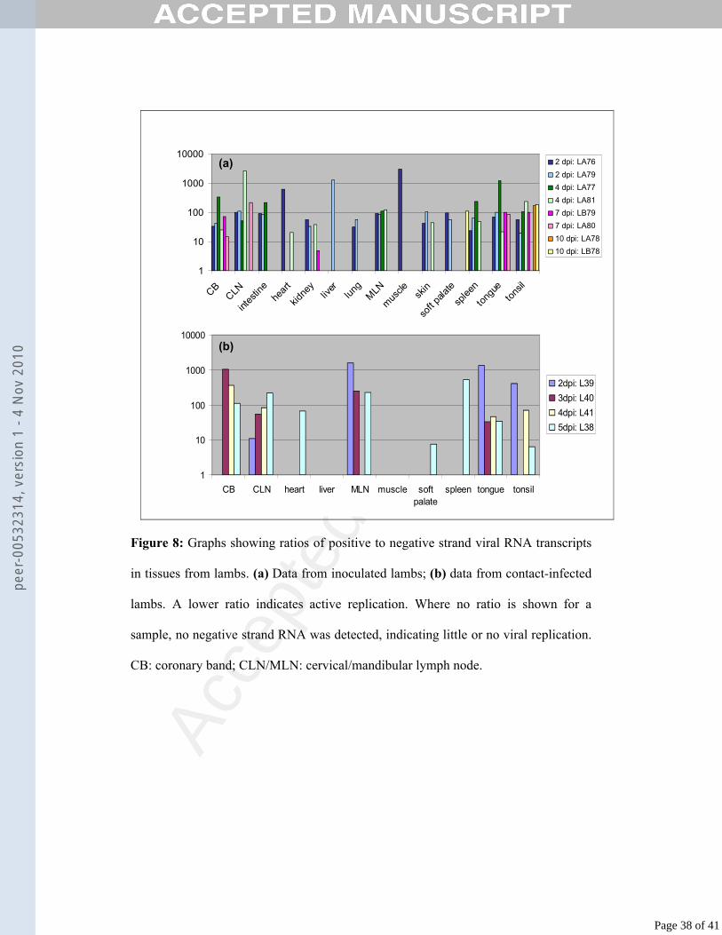
Using strand-specific real-time RT-PCR, the ratios of positive to negative viral RNA 386
strands were calculated for each tissue sample from the lambs. Fig. 8(a) shows the 387
ratios for each tissue for each inoculated lamb, while Fig. 8(b) shows the ratios for 388
each tissue in contact-infected lambs. A lower ratio indicates active replication.389
Where no ratio is shown, no negative strand viral RNA was detected, indicating little 390
or no replication was occurring in that tissue. Intermediate ratio levels are more 391
difficult to interpret, as negative viral RNA transcripts may be drained to some areas 392
via the lymphatic system or local circulation, without viral replication necessarily 393
taking place in these sites.394
Page 17 of 41
peer
-005
3231
4, v
ersi
on 1
- 4
Nov
201
0
Acce
pted
Man
uscr
ipt
395
In the inoculated lambs at 2 dpi, negative strand viral RNA transcripts were detected396
in most tissues. In the heart and muscle samples negative strand RNA was only 397
detected in samples from LA76, while negative strand RNA was only detected in the 398
liver in the sample from LA79; the ratio in these tissues was high. In all other tissues, 399
the ratio at 2 dpi was 100 or less. At 4 dpi, negative viral RNA strands were detected400
in both lambs in the CB, CLN, MLN, spleen, tongue and tonsil, while negative 401
strands in the heart, kidney and skin were detected only in LA81. Only the intestine of 402
LA77 had detectable negative strand RNA. No replication was detected in the liver, 403
lung, muscle or soft palate at 4 dpi. At 7 dpi, negative viral RNA strands were404
detected in the CB and tongue of both lambs, in the CLN of LA80 and in the kidney 405
and tonsil of LB79. Viral replication was not evident in other tissues at 7 dpi. At 10 406
dpi, negative viral RNA transcripts were detected in the tonsil of both lambs and the 407
soft palate of LB78 (Fig. 8a).408
409
In the contact-infected lambs, only the CLN, MLN, tongue and tonsil had detectable 410
negative strand viral RNA at 2 days post maternal inoculation (lamb L39). The ratio 411
in the CLN was 11, while the other three ratios ranged from 416 to 1542. At 3 dpi, the 412
CB, tongue, CLN and MLN had detectable negative strand viral RNA, with the lowest 413
ratio in the tongue (ratio 33) (lamb L40). At 4 dpi, lamb L41 had detectable negative 414
strand viral RNA in the CB, tongue, tonsil and CLN, with the lowest ratio (45) in the 415
tongue. Lamb L38, found dead at 5 dpi, had detectable negative strand viral RNA in 416
every tissue except the liver and muscle. The ratios in the soft palate and tonsil were 7 417
and 6, respectively (Fig. 8b).418
Page 18 of 41
peer
-005
3231
4, v
ersi
on 1
- 4
Nov
201
0

Acce
pted
Man
uscr
ipt
DISCUSSION419
420
The experiments described here characterised the pathogenesis and development of 421
FMD in neonatal lambs infected with FMDV. The viral RNA levels in serum and in 422
tissues samples were quantified, and viral replication levels in various tissues was 423
analysed by a novel negative strand RT-PCR assay. Importantly, viral RNA was 424
localised in cardiac and skeletal muscle cells from a lamb which died of FMD, 425
confirming the tropism of the virus for these tissues. The results provide a detailed 426
description of the pathogenesis of the disease in lambs.427
428
All four contact-infected lambs developed viraemia from 2 dpi. From this it can be 429
deduced that the lambs became infected with FMDV approximately 24 - 36 hours 430
after maternal inoculation. This allows time for the initial virus replication to occur in 431
the oropharynx, followed by dissemination to secondary sites of infection with 432
subsequent viraemia detectable at 2 dpi. Therefore, infecting lambs by contact with 433
inoculated ewes is likely to reflect the most common route of lamb infection in the 434
field during FMD outbreaks. Interestingly, the pattern of gross lesions on the tongue 435
in these lambs contrasts with that of older sheep and cattle, where lesions are more 436
common on the rostral tongue, gums and dental pad. It is thought that lesions are more 437
likely to develop at sites of trauma or intensive physical stress (Alexandersen et al., 438
2003). When young lambs suckle the ewe, the sides of the tongue rub against the 439
molars whilst the dorsum of the tongue rubs against both the lower aspect of the teat 440
and the roof of the lamb’s mouth. This may explain the difference in lesion 441
distribution. In the inoculated lambs, development of clinical signs appeared to be 442
more uniform than in the contact-infected lambs. Viraemia was present at 1 dpi in all 443
Page 19 of 41
peer
-005
3231
4, v
ersi
on 1
- 4
Nov
201
0
Acce
pted
Man
uscr
ipt
cases, with the level of viral RNA in serum ranging from 106.6 to 107.6 copies per ml. 444
The development of vesicles and onset of pyrexia was also similar in all inoculated 445
lambs. After 2 dpi, individual variation became apparent with the length of viraemia 446
varying from 2 days to 5 days. In inoculated lambs, lesions were observed on the 447
lateral aspects of the tongue at the levels of the molar teeth, as was observed in 448
contact-infected lambs. In contrast to contact infected lamb, however, no dorsal 449
tongue lesions were observed on any tongues from inoculated lambs. Furthermore, the 450
lingual lesions in contact-infected lambs appeared more severe than those in 451
inoculated lambs. This may be due to chance, but the route of infection may also have 452
influenced lesion location, as discussed above. 453
454
In investigating the kinetics of viraemia and viral RNA tissue distribution in lambs, 455
the results show that the acute-phase distribution of the virus is primarily to tissues 456
rich in epithelium such as the tongue and skin of the feet. Interestingly, the levels of 457
viral RNA found in tissue samples from inoculated lambs were generally much higher 458
than those in the contact-infected lambs. The highest level was in the coronary band at 459
2 dpi (1013.43 copies/g) compared to a peak measurement of 1010.89 copies/g at 3 dpi in 460
contact-infected lambs. These higher concentrations of viral RNA are most likely to 461
be due to the coronary band inoculation of the lambs. The initial high infectious dose 462
administered was followed by local replication and dissemination to distant secondary 463
sites of replication, with accompanying viraemia. 464
465
The histological appearance of sections of heart from the contact-infected lamb found 466
dead on day five (L38) included multiple scattered foci of myocardiocyte swelling, 467
occasionally with hyalinisation, with perivascular mononuclear aggregation (Fig. 5). 468
Page 20 of 41
peer
-005
3231
4, v
ersi
on 1
- 4
Nov
201
0
Acce
pted
Man
uscr
ipt
The heart sections of the other contact-infected lambs had no significant histological 469
abnormalities. Histological abnormalities were evident in sections of the hearts of 470
inoculated lambs at 4 dpi (Fig. 5), and also at 7 and 10 dpi. In exploring if the 471
histological abnormalities observed in the heart is related to FMDV replication, 472
abundant viral RNA was detected by ISH in the heart and muscle of contact-infected 473
lamb L38, indicating that these tissues may act as sites of FMDV tropism in young 474
lambs.This is in agreement with similar work carried out in piglets (Donaldson et al., 475
1984). It supports the theory that neonatal mortality following FMDV infection may 476
be due to viral replication in the heart, providing a plausible explanation for the 477
dramatic death rates often reported in young animals during an outbreak. In contrast 478
to the results from contact-infected lambs, no positive signal was detected in any 479
muscle or heart sections from inoculated lambs, despite histological evidence of 480
cardiac abnormalities. This may be due to the deterioration of viral RNA in tissue 481
samples during processing. Such deterioration may also be a factor in the lack of 482
positive signal in tissue sections from 7 and 10 dpi, but a more important explanatory 483
reason may be the presence of antibodies at these time-points. 484
485
The ratios of positive to negative strand viral RNA transcripts in tissue samples from 486
the lambs provided an indication of the levels of viral replication in those tissues.487
Although a low ratio is likely to indicate viral replication in a tissue and a high or zero 488
ratio indicates little or no replication, interpretation of intermediate values is more 489
difficult. Negative strand viral RNA may be drained to a tissue (e.g. lymph node) 490
from other areas without replication necessarily occurring in situ. Nevertheless, this 491
novel technique provides a degree of insight into FMDV replication which has not 492
previously been available. The results in the inoculated lambs show that over time, the 493
Page 21 of 41
peer
-005
3231
4, v
ersi
on 1
- 4
Nov
201
0
Acce
pted
Man
uscr
ipt
number of sites of replication declined. Interestingly, by 10 dpi, the only tissues where 494
negative strand viral RNA transcripts were detected were the tonsil and soft palate. 495
The ratio in these tissues at 10 dpi was still quite low at around 100, indicating a 496
degree of replication comparable to that in the epithelial tissues at 2 and 4 dpi. The 497
tonsil has previously been identified as the site of FMDV persistence in sheep 498
(Burrows, 1968), although it has also been suggested that the close association of the 499
ovine tonsil with the overlying palatine epithelium could explain this (Alexandersen et 500
al., 2003). Recent work in our laboratory has further supported this region as the site 501
of FMDV persistence in sheep (Horsington and Zhang, 2007). The low ratios found in 502
most tissues from inoculated lambs at 2 dpi show that, following inoculation with a 503
high dose of virus, negative strand viral RNA was detected at most sites in the body. 504
While replication might be expected at the recognised sites of viral replication such as 505
the coronary band, tongue, tonsil and soft palate, the low ratios in such tissues as the 506
kidney, intestine, heart, lung and spleen may indicate either viral replication at these 507
sites or negative strand viral RNA transported to these tissues via viraemic blood. In 508
the contact-infected group, viral replication occurred mainly in the epithelial tissues 509
and the CLN and MLN in the lambs killed at 2, 3 and 4 days after maternal 510
inoculation. This pattern is similar to that reported in other pathogenesis studies511
(Burrows et al., 1981), and is in contrast to the inoculated lambs, which had a wider 512
distribution of negative strand viral RNA.513
514
The results from the ewes are also significant (Tables 1 and 2). This is the first report515
we are aware of in which FMDV RNA loads in adult sheep tissues have been 516
quantified at various time-points after infection using real-time RT-PCR. Although 517
lambs are unlikely to infect ewes in the field (the reverse situation being more 518
Page 22 of 41
peer
-005
3231
4, v
ersi
on 1
- 4
Nov
201
0
Acce
pted
Man
uscr
ipt
probable), the data on transmission dynamics from inoculated lambs to in-contact 519
ewes provides a useful insight into the acute pathogenesis of FMD in sheep following 520
prolonged direct contact with infected cohorts. Viral RNA was first detected in two 521
ewes (VM78 and VM80) at 2 dpi. At 3 dpi, viraemia was detected in one more sheep, 522
while the remaining two became viraemic at 4 dpi. It is interesting that it took up to 523
four days for infection to be detected in some ewes, despite being in continuous direct 524
contact with their infected lambs. No viral RNA was detected in the serum of ewe 525
VM76, an unexpected result given the detection of viral RNA in four tissue samples. 526
It is likely that some viral RNA was present in the serum of this animal, but it may 527
have been below the limit of detection. Alternatively, the serum sample from 2 dpi 528
may have deteriorated during processing. By 10 dpi, the infection was waning and the 529
levels of viral RNA were lower, probably due to antibody clearance. In conclusion, 530
these studies of FMDV infection and replication in vivo provide a detailed description 531
of the patterns of virus load and distribution in lambs. It is clear that quantitative 532
analysis of viral load in vivo is a valuable tool in order to fully understand the 533
pathogenic steps of FMDV infection.534
535
Acknowledgements536
We would like to thank Yanmin Li, Pip Hamblin and Caroline Wright for performing 537
the antibody ELISAs and Nicholas Juleff for help with the ISH. We thank Colin 538
Randall, Bev Standish and Malcolm Turner for their assistance with and handling of539
the animals. This work was funded by Defra, UK.540
References 541
Page 23 of 41
peer
-005
3231
4, v
ersi
on 1
- 4
Nov
201
0
Acce
pted
Man
uscr
ipt
Alexandersen, S., Donaldson, A.I., 2002, Further studies to quantify the dose of 542
natural aerosols of foot-and-mouth disease virus for pigs. Epidemiol. Infect.543
128, 313-323.544
Alexandersen, S., Zhang, Z., Donaldson, A.I., Garland, A.J.M., 2003, The 545
pathogenesis and diagnosis of foot-and-mouth disease. J. Comp. Pathol. 129, 546
1-36.547
Belsham, G.J., 1993, Distinctive features of foot-and-mouth disease virus, a member 548
of the picornavirus family; aspects of virus protein synthesis, protein 549
processing and structure. Prog. Biophys. Mol. Biol. 60, 241-260.550
Burrows, R., 1968, The persistence of foot-and-mouth disease virus in sheep. Journal 551
of Hygiene, Cambridge 66, 633-640.552
Burrows, R., Mann, J.A., Garland, A.J., Greig, A., Goodridge, D., 1981, The 553
pathogenesis of natural and simulated natural foot-and-mouth disease infection 554
in cattle. J. Comp. Pathol. 91, 599-609.555
Donaldson, A.I., Ferris, N.P., Wells, G.A., 1984, Experimental foot-and-mouth 556
disease in fattening pigs, sows and piglets in relation to outbreaks in the field. 557
Vet. Rec. 115, 509-512.558
Ferris, N.P., Kitching, R.P., Oxtoby, J.M., Philpot, R.M., Rendle, R., 1990, Use of 559
inactivated foot-and-mouth disease virus antigen in liquid-phase blocking 560
ELISA. J. Virol. Methods 29, 33-41.561
Garcia-Mata,E., Federer,K.E., Pizzi,I., Aramburu,H. G., 1954, Pathogenicity of foot 562
and mouth disease virus in different species of unweaned animals. Rev-Vet-563
Milit. 2, 205-206.564
Garcia-Mata,E., Federer,K.E., Pizzi,I., Aramburu,H. G., 1955, Pathogenicity of foot 565
and mouth disease virus for new-born animals. Gac-vet. 17, 57-64.566
Page 24 of 41
peer
-005
3231
4, v
ersi
on 1
- 4
Nov
201
0
Acce
pted
Man
uscr
ipt
567
Geering, W., G., 1967, Foot and mouth disease in sheep. Aust. Vet. J.568
Horsington, J., Zhang, Z., 2007, Analysis of foot-and-mouth disease virus replication 569
using strand-specific quantitative RT-PCR. J. Virol. Methods, 144, 149-155.570
Hughes, G.J., Mioulet, V., Kitching, R.P., Woolhouse, M.E., Alexandersen, S., 571
Donaldson, A.I., 2002, Foot-and-mouth disease virus infection of sheep: 572
implications for diagnosis and control. Vet. Rec. 150, 724-727.573
Jubb, K.V.F., Kennedy, P.C., Palmer, N., 1993, Pathology of Domestic Animals, Vol 574
2, 4th Edition. Academic Press, Inc., Page 143. London, 143 p.575
Khankishiev, A., M., Gadshiev, K., S., Alekperov, Y., G., 1958, Foot and Mouth 576
Disease in New-Born Lambs. Veterinariya, Moscow 35, 59-60.577
Knowles, N.J., Samuel, A.R., 2003, Molecular epidemiology of foot-and-mouth 578
disease virus. Virus Res. 91, 65-80.579
Littlejohn, A., 1970, FMD in sheep - part 1. State Veterinary Journal 25, 3-12.580
Panisup, A., S., Kalra D, S., Chauhan H. V, S., 1979, Investigation of an outbreak of 581
FMD in sheep. Haryana Agricultural University Journal of Research 9, 111-582
114.583
Pay, T.W., F., 1988, FMD in sheep and goats: a review. FMD Bulletin 26, 2-13.584
Quan, M., Murphy, C.M., Zhang, Z., Alexandersen, S., 2004, Determinants of early 585
foot-and-mouth disease virus dynamics in pigs. J. Comp. Pathol. 131, 294-586
307.587
Reid, H.W., 2002, FMD in a parturient sheep flock. Vet. Rec. 150, 791.588
Reid, S.M., Ferris, N.P., Hutchings, G.H., Zhang, Z.D., Belsham, G.J., Alexandersen, 589
S., 2002, Detection of all seven serotypes of foot-and-mouth disease virus by 590
Page 25 of 41
peer
-005
3231
4, v
ersi
on 1
- 4
Nov
201
0
Acce
pted
Man
uscr
ipt
real-time, fluorogenic reverse transcription polymerase chain reaction assay. J. 591
Virol. Methods 105, 67-80.592
Ryan, E., Zhang, Z., Brooks, H.W., Horsington, J., Brownlie, J., 2007, Foot-and-593
Mouth Disease Virus Crosses the Placenta and Causes Death in Fetal Lambs. 594
J. Comp. Pathol. In Press.595
Salyi, G., 1939, Myocarditis in Foot and Mouth Disease of Young Lambs. Allatorv-596
Lapok. 62,159-160. 597
Thompson, D., Muriel, P., Russell, D., Osborne, P., Bromley, A., Rowland, M., 598
Creigh-Tyte, S., Brown, C., 2002, Economic costs of the foot and mouth 599
disease outbreak in the United Kingdom in 2001. Rev. Sci. Tech. Off. Int. 600
Epizoot. 21, 675-687.601
602
603
604
Page 26 of 41
peer
-005
3231
4, v
ersi
on 1
- 4
Nov
201
0
Acce
pted
Man
uscr
ipt
Table 1 Viral RNA in tissues from inoculated ewes.1
Viral RNA load †
dpi 0 2 3 4 7Ewe ID VE37* VE39 VE40 VE41 VE38
Cervical LN 0 7.6 5.41 7.44 4.67Heart 0 5.81 0 2.73 2.28Liver 0 7.09 6.77 0 0Mammary gland 0 7.33 6.61 4.76 0Mandibular LN 0 7.74 7.06 7.26 6.68Muscle 0 7.08 4.21 5.65 5.44Coronary band 0 9.05 8.7 7.02 7.83Soft palate 0 8.27 7.18 7.93 5.7
Spleen 0 7.82 6.17 6.32 4.7
Tongue 0 8.2 7.71 7.15 5.67Tonsil 0 8.68 8.0 8.65 6.69Uterus 0 7.9 7.52 5.08 4.28
2* uninfected ewe killed as a control and tissues collected for analysis.3
† viral RNA loads in tissues were quantified by real-time RT–PCR and expressed as log10 copy number 4
per g tissue5
dpi: days post inoculation at the day the ewes were killed6
LN: lymph node7
8
Table
Page 27 of 41
peer
-005
3231
4, v
ersi
on 1
- 4
Nov
201
0
Acce
pted
Man
uscr
ipt
Table 2 Viral RNA in tissues from contact-infected ewes9
Viral RNA load †
dpi 0 2 4 4 7 7 10Ewe ID VM82* VM76 VM77 VM81 VM79 VM80 VM78
Cervical LN 0 7.19 11.46 9.68 10.15 8.23 7.86Cotyledon 0 0 9.63 7.77 7.65 7.59 7.54Heart 0 7.08 9.18 8.27 7.17 0 6.88Liver 0 0 9.03 8.25 8.01 0 0Mandibular LN 0 0 9.51 10.75 8.78 9.2 8.49Muscle 0 0 8.78 7.9 8.43 7.48 6.31Coronary band 0 0 8.77 10.45 9.57 11.2 8.79Soft palate 0 0 8.39 9.67 9.53 8.15 7.25Tongue 0 7.27 8.8 8.74 0 8.31 0Tonsil 0 7.57 9.66 10.65 10.66 9.42 6.94
10* uninfected ewe killed as a control and tissues collected for analysis.11
† viral RNA loads in tissues were quantified by real-time RT–PCR and expressed as log10 copy number 12
per g tissue13
dpi: days post lamb inoculation at the day the ewes were killed14
LN: lymph node15
16
Page 28 of 41
peer
-005
3231
4, v
ersi
on 1
- 4
Nov
201
0
Acce
pted
Man
uscr
ipt
17Table 3: Serum antibody titres from inoculated lambs, measured by ELISA.18
19Antibody titreLamb
no. 0 dpi 1 dpi 2 dpi 3 dpi 4 dpi 5 dpi 6 dpi 7 dpi 8 dpi 9 dpi 10 dpi
LA76 0 0 0LA79 0 0 0LA77 0 0 0 0 64LA81 0 0 0 0 0LB79 0 0 0 0 90 1448 724 1448LA80 0 0 0 0 90 256 362 1024LA78 0 0 0 0 0 1024 1024 724 1448 2896 1448LB78 0 0 0 0 0 0 181 362 362 724 362
20
Page 29 of 41
peer
-005
3231
4, v
ersi
on 1
- 4
Nov
201
0
Acce
pted
Man
uscr
ipt
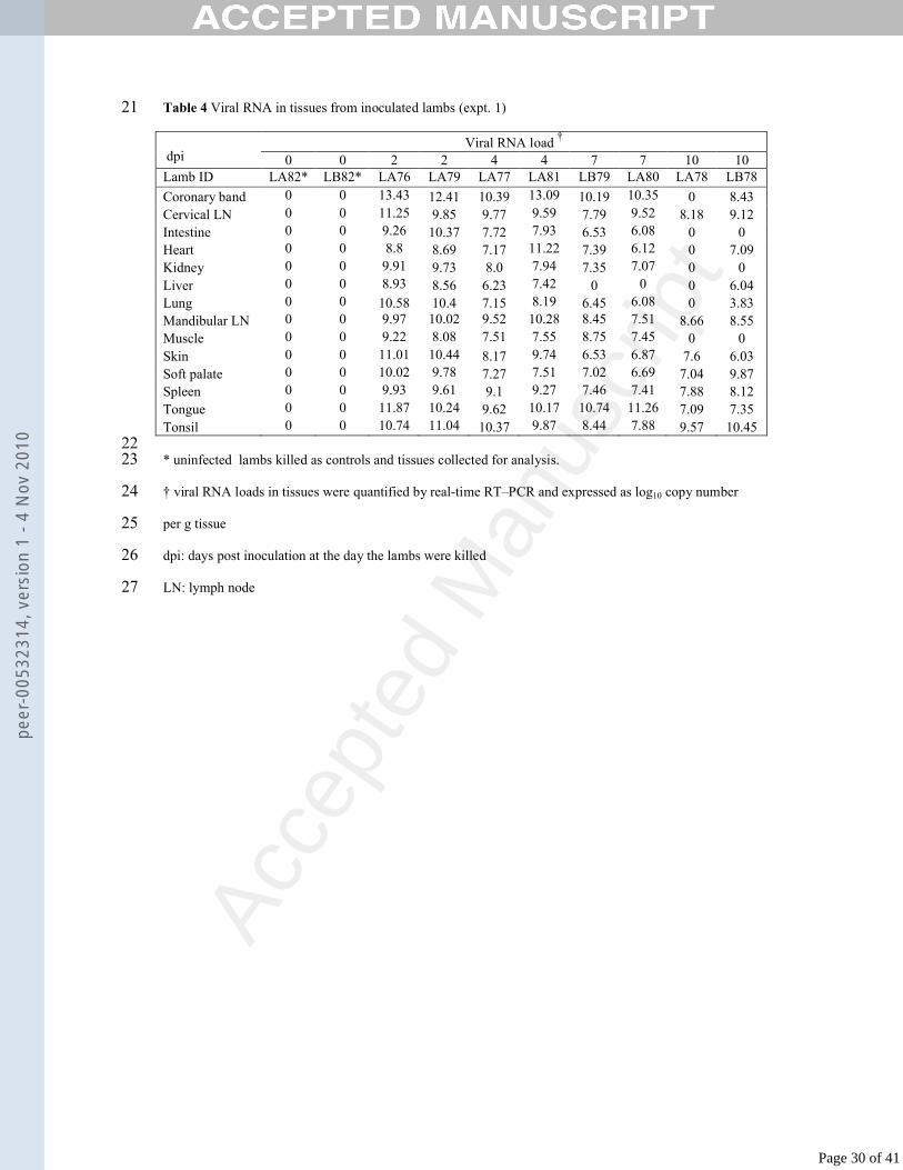
Table 4 Viral RNA in tissues from inoculated lambs (expt. 1)21
Viral RNA load †
dpi 0 0 2 2 4 4 7 7 10 10Lamb ID LA82* LB82* LA76 LA79 LA77 LA81 LB79 LA80 LA78 LB78
Coronary band 0 0 13.43 12.41 10.39 13.09 10.19 10.35 0 8.43Cervical LN 0 0 11.25 9.85 9.77 9.59 7.79 9.52 8.18 9.12Intestine 0 0 9.26 10.37 7.72 7.93 6.53 6.08 0 0Heart 0 0 8.8 8.69 7.17 11.22 7.39 6.12 0 7.09Kidney 0 0 9.91 9.73 8.0 7.94 7.35 7.07 0 0Liver 0 0 8.93 8.56 6.23 7.42 0 0 0 6.04Lung 0 0 10.58 10.4 7.15 8.19 6.45 6.08 0 3.83Mandibular LN 0 0 9.97 10.02 9.52 10.28 8.45 7.51 8.66 8.55Muscle 0 0 9.22 8.08 7.51 7.55 8.75 7.45 0 0Skin 0 0 11.01 10.44 8.17 9.74 6.53 6.87 7.6 6.03Soft palate 0 0 10.02 9.78 7.27 7.51 7.02 6.69 7.04 9.87Spleen 0 0 9.93 9.61 9.1 9.27 7.46 7.41 7.88 8.12Tongue 0 0 11.87 10.24 9.62 10.17 10.74 11.26 7.09 7.35Tonsil 0 0 10.74 11.04 10.37 9.87 8.44 7.88 9.57 10.45
22* uninfected lambs killed as controls and tissues collected for analysis.23
† viral RNA loads in tissues were quantified by real-time RT–PCR and expressed as log10 copy number 24
per g tissue25
dpi: days post inoculation at the day the lambs were killed26
LN: lymph node27
Page 30 of 41
peer
-005
3231
4, v
ersi
on 1
- 4
Nov
201
0
Acce
pted
Man
uscr
ipt
28
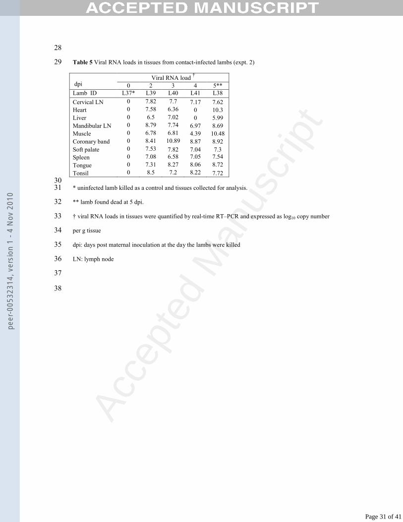
Table 5 Viral RNA loads in tissues from contact-infected lambs (expt. 2)29
Viral RNA load †
dpi 0 2 3 4 5**Lamb ID L37* L39 L40 L41 L38
Cervical LN 0 7.82 7.7 7.17 7.62Heart 0 7.58 6.36 0 10.3Liver 0 6.5 7.02 0 5.99Mandibular LN 0 8.79 7.74 6.97 8.69Muscle 0 6.78 6.81 4.39 10.48Coronary band 0 8.41 10.89 8.87 8.92Soft palate 0 7.53 7.82 7.04 7.3Spleen 0 7.08 6.58 7.05 7.54
Tongue 0 7.31 8.27 8.06 8.72
Tonsil 0 8.5 7.2 8.22 7.7230
* uninfected lamb killed as a control and tissues collected for analysis.31
** lamb found dead at 5 dpi.32
† viral RNA loads in tissues were quantified by real-time RT–PCR and expressed as log10 copy number 33
per g tissue34
dpi: days post maternal inoculation at the day the lambs were killed35
LN: lymph node36
37
38
Page 31 of 41
peer
-005
3231
4, v
ersi
on 1
- 4
Nov
201
0
Acce
pted
Man
uscr
ipt
Figure 1: Graphs showing FMDV RNA levels in serum samples and temperature 39
measurements from ewes inoculated with FMDV.40
41
Figure 2: Graphs showing FMDV RNA levels in serum samples and temperature 42
measurements from ewes kept in direct contact with FMDV-inoculated lambs.43
44
Figure 3: Graphs showing FMDV RNA levels in serum samples and temperature 45
measurements from lambs inoculated with FMDV.46
47
Figure 4: Graphs showing FMDV RNA levels in serum samples and temperature 48
measurements from lambs kept in direct contact with FMDV-inoculated ewes.49
50
Figure 5: Histological sections of heart stained with haematoxylin and eosin. (a) and 51
(b): Sections from lamb L38 (contact-infected, died 5 dpi) showing 40x and 63x 52
views of a focus of perivascular lymphocyte and plasma cell aggregation. 53
Myocardiocytes are dispersed and disrupted. (c) Section from lamb LA81 (inoculated, 54
killed 4 dpi) showing 10x view of a poorly demarcated area plasmocytic-lymphocytic 55
aggregation within myocardium; (d) 40x view of area of mononuclear inflammation 56
with myocardiocyte disruption, also from LA81. Foci dominated by myocardiocyte 57
swelling sometimes with hyalinisation, but with less marked inflammatory cell 58
aggregation were also recognised (though are not shown in these photographs). 59
60
Page 32 of 41
peer
-005
3231
4, v
ersi
on 1
- 4
Nov
201
0
Acce
pted
Man
uscr
ipt
Figure 6: ISH performed on heart (a: 10x view, b: 63x view) and skeletal muscle (c: 61
10x view, d: 40x view) sections from lamb L38, (contact-infected, died 5 dpi). 62
Positive signal is diffusely distributed throughout the myocardium, while in the 63
muscle signal can be seen at several foci along multi-nucleated myocytes.64
65
Figure 7: ISH performed on tongue and coronary band sections. (a) and (b): Tongue 66
from lamb LA77 (inoculated, killed 4 dpi), 10x and 63x views respectively. (c):67
Tongue from lamb LA76 (inoculated, killed 2 dpi), 10x view; (d) coronary band from 68
LA76, 10x view. Positive signal can be seen diffusely distributed throughout the basal 69
zone of the tongue lesions, and in the subcorneal area of the coronary band lesion. 70
71
Figure 8: Graphs showing ratios of positive to negative strand viral RNA transcripts 72
in tissues from lambs. (a) Data from inoculated lambs; (b) data from contact-infected 73
lambs. A lower ratio indicates active replication. Where no ratio is shown for a 74
sample, no negative strand RNA was detected, indicating little or no viral replication. 75
CB: coronary band; CLN/MLN: cervical/mandibular lymph node.76
777879
Page 33 of 41
peer
-005
3231
4, v
ersi
on 1
- 4
Nov
201
0
Acce
pted
Man
uscr
ipt
Figure 1: Graphs showing FMDV RNA levels in serum samples and temperature
measurements from ewes inoculated with FMDV. FMDV RNA loads in serum
samples are expressed as log10 copy number per ml.
Cop
ies
vira
l RN
A/m
l ser
um (
log1
0)
Degrees centigrade
Days post inoculation
VE39 - Ewe killed 2 dpi
0
2
4
6
8
10
12
0 1 2
38.5
39
39.5
40
40.5
41
41.5
VE40 - Ewe killed 3 dpi
0
2
4
6
8
10
12
0 1 2 3
38.5
39
39.5
40
40.5
41
41.5
VE41 - Ewe killed 4 dpi
0
2
4
6
8
10
12
0 1 2 3 4
38.5
39
39.5
40
40.5
41
41.5
VE38 - Ewe killed 7 dpi
0
2
4
6
8
10
12
0 1 2 3 4 5 6 7
38.5
39
39.5
40
40.5
41
41.5
Figure
Page 34 of 41
peer
-005
3231
4, v
ersi
on 1
- 4
Nov
201
0
Acce
pted
Man
uscr
ipt
Figure 2: Graphs showing FMDV RNA levels in serum samples and temperature
measurements from ewes kept in direct contact with FMDV-inoculated lambs. FMDV
RNA loads in serum samples are expressed as log10 copy number per ml
Cop
ies
vira
l RN
A/m
l ser
um
(lo
g10)
Deg
rees centigrade
VM76 - Ewe killed 2 dpi
0
2
4
6
8
10
12
0 1 2
38.5
39
39.5
40
40.5
41
41.5
VM77 - Ewe killed 4 dpi
0
2
4
6
8
10
12
0 1 2 3 4
38.5
39
39.5
40
40.5
41
41.5
VM81 - Ewe killed 4 dpi
0
2
4
6
8
10
12
0 1 2 3 4
38.5
39
39.5
40
40.5
41
41.5
VM79 - Ewe killed 7 dpi
0
2
4
6
8
10
12
0 1 2 3 4 5 6 7
38.5
39
39.5
40
40.5
41
41.5
VM80 - Ewe killed 7 dpi
0
2
4
6
8
10
12
0 1 2 3 4 5 6 7
38.5
39
39.5
40
40.5
41
41.5
VM78 - Ewe killed 10 dpi
0
2
4
6
8
10
12
0 1 2 3 4 5 6 7
38
38.5
39
39.5
40
40.5
41
41.5
Days post inoculation of lambs
Page 35 of 41
peer
-005
3231
4, v
ersi
on 1
- 4
Nov
201
0
Acce
pted
Man
uscr
ipt
Figure 3: Graphs showing FMDV RNA levels in serum samples and temperature
measurements from lambs inoculated with FMDV. FMDV RNA loads in serum
samples are expressed as log10 copy number per ml
LA76 - Lamb killed 2 dpi
0
2
4
6
8
10
12
0 1 2
38.5
39
39.5
40
40.5
41
41.5
LA79 - Lamb killed 2 dpi
0
2
4
6
8
10
12
0 1 2
38.5
39
39.5
40
40.5
41
41.5
LA77 - Lamb killed 4 dpi
0
2
4
6
8
10
12
0 1 2 3 4
38.5
39
39.5
40
40.5
41
41.5
LA81 - Lamb killed 4 dpi
0
2
4
6
8
10
12
0 1 2 3 4
38.5
39
39.5
40
40.5
41
41.5
LB79 - Lamb killed 7 dpi
0
2
4
6
8
10
12
0 1 2 3 4 5 6 7
38.5
39
39.5
40
40.5
41
41.5
LA80 - Lamb killed 7 dpi
0
2
4
6
8
10
12
0 1 2 3 4 5 6 7
38
38.5
39
39.5
40
40.5
41
41.5
LB78 - Lamb killed 10 dpi
0
2
4
6
8
10
12
0 1 2 3 4 5 6 7
38.5
39
39.5
40
40.5
41
41.5
LA78 - Lamb killed 10 dpi
0
2
4
6
8
10
12
0 1 2 3 4 5 6 7
38.5
39
39.5
40
40.5
41
41.5
Cop
ies
vira
l RN
A/m
l ser
um (
log1
0)D
egrees centigrade
Days post inoculation
Page 36 of 41
peer
-005
3231
4, v
ersi
on 1
- 4
Nov
201
0
Acce
pted
Man
uscr
ipt
Figure 4: Graphs showing FMDV RNA levels in serum samples and temperature
measurements from lambs kept in direct contact with FMDV-inoculated ewes. FMDV
RNA loads in serum samples are expressed as log10 copy number per ml
L39 - Lamb killed 2 dpi
0
2
4
6
8
10
12
0 1 2
38.5
39
39.5
40
40.5
41
41.5
L40 - Lamb killed 3 dpi
0
2
4
6
8
10
12
0 1 2 3
38.5
39
39.5
40
40.5
41
41.5
L41 - Lamb killed 4 dpi
0
2
4
6
8
10
12
0 1 2 3 4
38.5
39
39.5
40
40.5
41
41.5
L38 - Lamb found dead 5 dpi
0
2
4
6
8
10
12
0 1 2 3 4
38.5
39
39.5
40
40.5
41
41.5
Cop
ies
vira
l RN
A/m
l ser
um (
log1
0)
Degrees centigrade
Days post inoculation of ewes
Page 37 of 41
peer
-005
3231
4, v
ersi
on 1
- 4
Nov
201
0
Acce
pted
Man
uscr
ipt
Figure 8: Graphs showing ratios of positive to negative strand viral RNA transcripts
in tissues from lambs. (a) Data from inoculated lambs; (b) data from contact-infected
lambs. A lower ratio indicates active replication. Where no ratio is shown for a
sample, no negative strand RNA was detected, indicating little or no viral replication.
CB: coronary band; CLN/MLN: cervical/mandibular lymph node.
1
10
100
1000
10000
CB
CLN
intes
tine
hea
rt
kidn
ey liv
er lu
ng M
LN
mus
cle sk
in
soft
pala
te
sple
en
tong
ue
tons
il
2 dpi: LA76
2 dpi: LA79
4 dpi: LA77
4 dpi: LA81
7 dpi: LB79
7 dpi: LA80
10 dpi: LA78
10 dpi: LB78
1
10
100
1000
10000
CB CLN heart liver MLN muscle softpalate
spleen tongue tonsil
2dpi: L39
3dpi: L40
4dpi: L41
5dpi: L38
(a)
(b)
Page 38 of 41
peer
-005
3231
4, v
ersi
on 1
- 4
Nov
201
0
Acce
pted
Man
uscr
ipt
Figure 5
Page 39 of 41
peer
-005
3231
4, v
ersi
on 1
- 4
Nov
201
0
Acce
pted
Man
uscr
ipt
Figure 6
Page 40 of 41
peer
-005
3231
4, v
ersi
on 1
- 4
Nov
201
0
Acce
pted
Man
uscr
ipt
Figure 7
Page 41 of 41
peer
-005
3231
4, v
ersi
on 1
- 4
Nov
201
0