food intake regulating-neuropeptides are expressed and regulated through pregnancy and following...
TRANSCRIPT

BioMed Central
Reproductive Biology and Endocrinology
ss
Open AcceResearchFood intake regulating-neuropeptides are expressed and regulated through pregnancy and following food restriction in rat placentaJorge E Caminos1,2,3, Susana B Bravo1, C Ruth González1, Maria F Garcés1,2, Libia A Cepeda2, Adriana C González2, Fernando Cordido3, Miguel López1,4 and Carlos Diéguez*1,4Address: 1Department of Physiology, School of Medicine, University of Santiago de Compostela, Santiago de Compostela, Spain, 2Department of Physiology and Genetic Institute, Faculty of Medicine, National University of Colombia. Bogotá, Colombia, 3Endocrine Department, Hospital Juan Canalejo, A Coruña, Spain and 4CIBER of Obesity and Nutrition, Instituto Salud Carlos III, Santiago de Compostela, Spain
Email: Jorge E Caminos - [email protected]; Susana B Bravo - [email protected]; C Ruth González - [email protected]; Maria F Garcés - [email protected]; Libia A Cepeda - [email protected]; Adriana C González - [email protected]; Fernando Cordido - [email protected]; Miguel López - [email protected]; Carlos Diéguez* - [email protected]
* Corresponding author
AbstractBackground: Neuropeptide Y (NPY), agouti related peptide (AgRP), cocaine and amphetamine-regulated transcript (CART) and melanocortins, the products of the proopiomelanocortin(POMC), are hypothalamic peptides involved in feeding regulation and energy homeostasis. Recentevidence has demonstrated their expression in rat and human placenta.
Methods: In the current study, we have investigated the expression of those neuropeptides in therat placenta by real-time PCR using a model of maternal food restriction.
Results: Our results showed that placental-derived neuropeptides were regulated throughpregnancy and following food restriction.
Conclusion: These data could indicate that placental-derived neuropeptides represent a localregulatory circuit that may fine-tune control of energy balance during pregnancy.
BackgroundThe regulation of body weight is carried out by a complexinter-organ circuit connecting the periphery and the brain,where neurons in the hypothalamus and brainstem exertpotent effects on feeding and energy expenditure [1,2].Short-acting and long-term body-weight regulating sig-nals mainly originate from: 1) the adipose tissue, such asleptin, and interleukin 6 (IL-6); 2) the pancreas, like insu-lin or amylin; 3) the gastrointestinal tract such as ghrelin,glucagon-like peptide-1 (GLP-1), peptide YY (PYY), chole-cystokinin (CCK) and neuropeptide W (NPW); 4) the sen-
sory vagus nerve [2]. These neural and humoral signalsinteract at the brain level with widely-located target recep-tors related with the nutritional state, metabolism andreproduction [2,3].
Hypothalamic neuropeptide expression and regulationare stimulated by inputs from peripherally-derived hor-monal and nutrient-related signals essential for the centralcontrol of energy homeostasis in mammals. Neuropep-tide Y (NPY), melanocortins (POMC-derived products),agouti related peptide (AgRP), cocaine and amphetamine
Published: 2 April 2008
Reproductive Biology and Endocrinology 2008, 6:14 doi:10.1186/1477-7827-6-14
Received: 4 December 2007Accepted: 2 April 2008
This article is available from: http://www.rbej.com/content/6/1/14
© 2008 Caminos et al; licensee BioMed Central Ltd. This is an Open Access article distributed under the terms of the Creative Commons Attribution License (http://creativecommons.org/licenses/by/2.0), which permits unrestricted use, distribution, and reproduction in any medium, provided the original work is properly cited.
Page 1 of 6(page number not for citation purposes)

Reproductive Biology and Endocrinology 2008, 6:14 http://www.rbej.com/content/6/1/14
regulated transcript (CART), melanin concentrating hor-mone (MCH) and orexins, have been characterized inboth rodent models and humans, by having either director indirect effects in the feedback loop of body weight reg-ulation and reproduction [4,5].
NPY and AgRP are two anabolic neuropeptides that pro-mote weight gain by reducing energy expenditure andstimulating food intake. They are coexpressed in arcuatenucleus (ARC) neurons, which are inhibited by insulinand leptin [4,6]. In contrast, CART and POMC, also coex-pressed in the ARC neurons, are stimulated by inputs frominsulin and leptin and these catabolic neuronal pathwaysact to reduce feeding and increase energy expenditure[4,6].
Besides their role in food intake control, hypothalamicneuropeptides have also been identified in female repro-ductive tissues, such as the placenta and appear to have animportant role in the physiology of pregnancy [7-14]. It isinteresting to note that the placenta may play an impor-tant role not only in reproduction but is also an importantsite for translating inputs from diverse hormonal signalsthat powerfully influence energy balance within the fetus[15,16]. The aim of this study was to evaluate theontogenic mRNA expression pattern of NPY, AgRP,POMC, and CART throughout pregnancy in the rat pla-centa. In addition, we have determined the effects ofchronic food restriction on their expression levels.
MethodsAnimalsPregnant female rats of the Sprague Dawley strain agedbetween 10–12 weeks (bred in the Animalario GeneralUSC; Santiago de Compostela, Spain), were housed in atemperature-regulated room with a 12 h light/12 h darkcycle, with tap water and standard rat chow ad libitum or afood-restricted diet (see below). At the end of the studyperiod, animals were sacrificed, hypothalami and placen-tas were removed from each mother, and snap-frozen inliquid nitrogen for RNA extraction and real time semi-
quantitative RT-PCR. All experiments and procedures inthis study were carried out according to a protocolapproved by the Ethics Committee of the University ofSantiago de Compostela in accordance with the EuropeanUnion normative for the care and use of experimental ani-mals.
Maternal chronic food-restrictionA maternal chronic food-restriction model was usedthroughout pregnancy to study the effect of long-termundernutrition on placental neuropeptide mRNA expres-sion as previously described [17]. Briefly, virgin rats weremated on the day of proestrus and the day on which sper-matozoa were present in vaginal smear, was designatedgestational day 1. The pregnant rats were divided into twodietary groups: pregnant rats fed ad libitum and food-restricted group of pregnant rats fed 30% of ad libitumintake. Food-restricted animals were fed every day at18:00 hours. Rats were sacrificed at gestational days 12, 16and 21. We used 9 hypothalami and placentas per experi-mental group, extracted from 9 different mothers. All thesamples were analyzed individually and samples were notpooled.
RNA Isolation and real-time RT-PCRPlacental neuropeptide gene expression was analyzed byreverse transcription polymerase chain reaction (RT-PCR)as previously validated [18-20]. Total RNA was isolatedusing Trizol according to the manufacturer's protocol(Invitrogen, CA). First-strand cDNA was synthesized from2 μg of total RNA by random primer reverse transcription.A negative control without MMLV reverse transcriptasewas used to ensure specificity of the PCR amplification.The resulting cDNA was subjected to PCR amplificationusing sense and antisense primers specific for rat AgRP,CART, NPY and POMC mRNAs (Table 1). Primer pairswere designed overlapping on different exons to preventamplification from any contaminated genomic DNA(data not shown). To verify the identity of amplifiedcDNAs, PCR products were electrophoresed on a 1.5%agarose gel, which yielded DNA fragments of the expected
Table 1: Primers used for real-time RT-PCR
mRNA Primer Sequence (5'-3') Product size (bp) Accession Number
NPY Forward cgctctgcgacactacatca 157 NM_012614Reverse tttcatttcccatcaccaca
AgRP Forward tgtgggccctttattagacc 156 XM_574228Reverse ccatatggacccccaatgt
POMC Forward tccatagacgtgtggagctg 174 NM_139326Reverse gacgtacttccggggatttt
CART Forward gccctggacatctactctgc 201 NM_017110Reverse cactgcgcactgctctcc
HPRT Forward cagtcccagcgtcgtgatta 137 NM_012583Reverse agcaagtctttcagtcctgtc
Page 2 of 6(page number not for citation purposes)

Reproductive Biology and Endocrinology 2008, 6:14 http://www.rbej.com/content/6/1/14
length for all mRNAs and were confirmed by sequencing(data not shown). Rat hypoxanthine guanine phosphori-bosyl transferase (HPRT) was used as a control house-keeping gene. PCR was performed by real-time inLightCycler real-time PCR machine 2 (Roche Diagnostics,Germany) and the software version 3.5, according to themethod described elsewhere [19-21]. Briefly, the PCR pro-tocol consisted of initial denaturation at 96°C for 5 min,followed by 35 cycles of denaturation (96°C for 2 s),annealing (62°C for 15 s) and elongation (72°C for 15 s).This was followed by melting curve analysis consisting of1 cycle at 95°C for 30 s, 60°C for 15 s and a temperaturerise to 85°C at a slope of 0.2°C/s with continuous meas-urement of fluorescence. The primers specify was alsoassured by the melting curve of each gene. All sampleswere standardized against HPRT by using the 2ΔΔctmethod [19,22].
Statistical analysisStatistical analyzes were performed using GraphPad Instat(version 3.05). All group values were expressed as mean ±SEM. Differences between groups were analyzed by usingone-way analysis of variance (ANOVA) or Student's t-test.P < 0.05 was considered significant.
ResultsNPY mRNA expression is down-regulated by food restriction in the rat placentaFirstly, we studied the hypothalamic mRNA levels of NPYduring pregnancy and after food restriction. Our datashowed that the transcriptional expression of NPY isincreased at the latest pregnancy stage (21 days; P < 0.001)with food restriction inducing a further increase at both16 and 21 days of pregnancy (P < 0.001 and P < 0.05,respectively) (Figure 1A). Next, we evaluated the tran-scriptional expression of NPY in rat placenta; our resultsshowed that placental mRNA levels of NPY were signifi-cantly higher during the initial period of gestation studied(12 days), and decreased significantly (P < 0.001) withgestational age (Figure 1B). We also studied the effect offood restriction on placental mRNA expression of NPYand these data showed that the expression profile of NPYwas decreased by food restriction only at day 12 (P <0.01), and remain unchanged between 16 to 21 days,compared to the ad libitum group (Figure 1B).
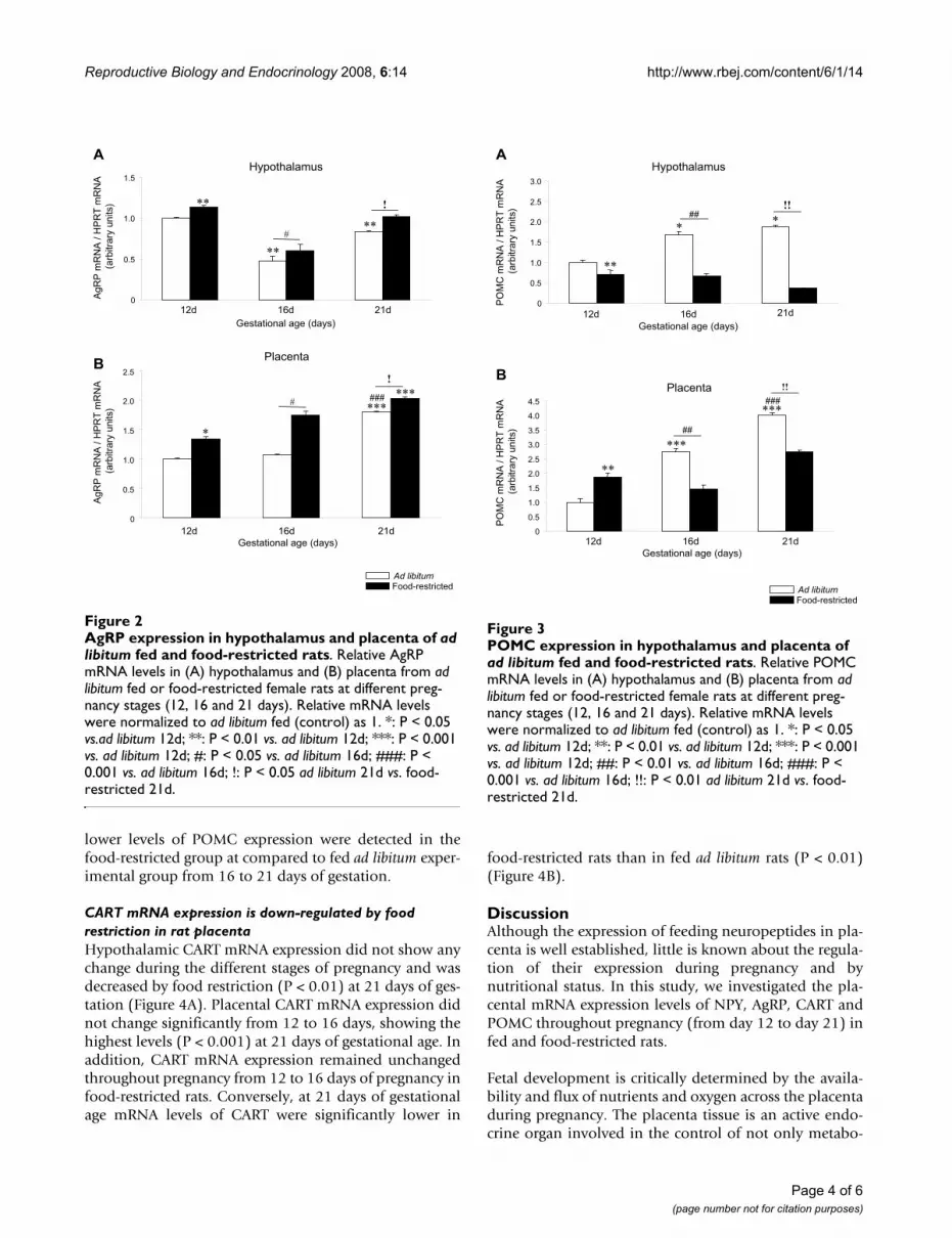
AgRP mRNA expression is up-regulated by food restriction in the rat placentaThroughout pregnancy, hypothalamic mRNA expressionlevels of AgRP were decreased (P < 0.01) at 16 and 21 daysof gestation compared to 12 days and these levels wereincreased by food restriction (P < 0.05 and P < 0.01) at alldays of gestation studied compared to the ad libitum (Fig-ure 2A). In rat placenta, throughout the same experimen-tal period, the expression mRNA profile of AgRP was
opposite to NPY (Figure 2B). Placental AgRP expressionremained unchanged between 12 and 16 days of gestationreaching the highest levels at the end of the gestationperiod (P < 0.001)(Figure 2B). Food restriction induced asignificant and progressive increase (P < 0.05) in placentaAgRP mRNA expression at all studied time points.
POMC mRNA expression shows a biphasic response to food restriction during pregnancy in rat placentaHypothalamic POMC mRNA expression was increasedduring pregnancy (P < 0.05) and decreased by foodrestriction (P < 0.01) at 12, 16 and 21 days of gestation(Figure 3A). Placental POMC mRNA expression showed asignificantly (P < 0.001) increasing trend of expressionfrom 12 to 21 days of gestation in rats fed ad libitum (Fig-ure 3B). Placental POMC expression was significantly (P <0.01) lower in fed ad libitum rats than in food-restrictedgroup at 12 days. Conversely, significantly (P < 0.01)
NPY expression in hypothalamus and placenta of ad libitum fed and food-restricted ratsFigure 1NPY expression in hypothalamus and placenta of ad libitum fed and food-restricted rats. Relative NPY mRNA levels in (A) hypothalamus and (B) placenta from ad libitum fed or food-restricted female rats at different preg-nancy stages (12, 16 and 21 days). Relative mRNA levels were normalized to ad libitum fed (control) as 1. **: P < 0.01 vs. ad libitum 12d; ***: P < 0.001 vs. ad libitum 12d; ###: P < 0.001 vs. ad libitum 16d; !: P < 0.05 ad libitum 21d vs. food-restricted 21d.
��������������������
�
�
�
��
������
�� �� � ��������� ��� �������
�
���
�
���
���
�����������
�� �� � ��������� ��� ������
�
���
��
���
��!
��
��
��
������ ������
"������
�
�
Page 3 of 6(page number not for citation purposes)
Reproductive Biology and Endocrinology 2008, 6:14 http://www.rbej.com/content/6/1/14
lower levels of POMC expression were detected in thefood-restricted group at compared to fed ad libitum exper-imental group from 16 to 21 days of gestation.
CART mRNA expression is down-regulated by food restriction in rat placentaHypothalamic CART mRNA expression did not show anychange during the different stages of pregnancy and wasdecreased by food restriction (P < 0.01) at 21 days of ges-tation (Figure 4A). Placental CART mRNA expression didnot change significantly from 12 to 16 days, showing thehighest levels (P < 0.001) at 21 days of gestational age. Inaddition, CART mRNA expression remained unchangedthroughout pregnancy from 12 to 16 days of pregnancy infood-restricted rats. Conversely, at 21 days of gestationalage mRNA levels of CART were significantly lower in
food-restricted rats than in fed ad libitum rats (P < 0.01)(Figure 4B).
DiscussionAlthough the expression of feeding neuropeptides in pla-centa is well established, little is known about the regula-tion of their expression during pregnancy and bynutritional status. In this study, we investigated the pla-cental mRNA expression levels of NPY, AgRP, CART andPOMC throughout pregnancy (from day 12 to day 21) infed and food-restricted rats.
Fetal development is critically determined by the availa-bility and flux of nutrients and oxygen across the placentaduring pregnancy. The placenta tissue is an active endo-crine organ involved in the control of not only metabo-
POMC expression in hypothalamus and placenta of ad libitum fed and food-restricted ratsFigure 3POMC expression in hypothalamus and placenta of ad libitum fed and food-restricted rats. Relative POMC mRNA levels in (A) hypothalamus and (B) placenta from ad libitum fed or food-restricted female rats at different preg-nancy stages (12, 16 and 21 days). Relative mRNA levels were normalized to ad libitum fed (control) as 1. *: P < 0.05 vs. ad libitum 12d; **: P < 0.01 vs. ad libitum 12d; ***: P < 0.001 vs. ad libitum 12d; ##: P < 0.01 vs. ad libitum 16d; ###: P < 0.001 vs. ad libitum 16d; !!: P < 0.01 ad libitum 21d vs. food-restricted 21d.
��������������������
�
���
��
��
���
���
#��
#��
��
��
�
��
��
��
���
���
�� �� � ��������� ��� ������
�
�� �� � ��������� ��� �������
�����������
"���������
�
���
��
��
���
���
#��
��
����
� �
AgRP expression in hypothalamus and placenta of ad libitum fed and food-restricted ratsFigure 2AgRP expression in hypothalamus and placenta of ad libitum fed and food-restricted rats. Relative AgRP mRNA levels in (A) hypothalamus and (B) placenta from ad libitum fed or food-restricted female rats at different preg-nancy stages (12, 16 and 21 days). Relative mRNA levels were normalized to ad libitum fed (control) as 1. *: P < 0.05 vs.ad libitum 12d; **: P < 0.01 vs. ad libitum 12d; ***: P < 0.001 vs. ad libitum 12d; #: P < 0.05 vs. ad libitum 16d; ###: P < 0.001 vs. ad libitum 16d; !: P < 0.05 ad libitum 21d vs. food-restricted 21d.
�
���
��
��
���
���
�
�
�
�
������
�� �� � ��������� ��� ������
�
�����������
"������
��������������������
���
�� �� � ��������� ��� �������
�
���
��
��
��
�
�
��
��
Page 4 of 6(page number not for citation purposes)

Reproductive Biology and Endocrinology 2008, 6:14 http://www.rbej.com/content/6/1/14
lism and energy balance, but also of other relevant bodyfunctions including reproduction [23,24]. Previous stud-ies have demonstrated the placental expression of hor-mones, such as ghrelin and leptin [25-27], as well asneuropeptides such as NPY, AgRP, CART and POMC [7-11] are involved in the regulation of energy homeostasis.However, despite these data, both the physiogical rele-vance and the regulation of these molecules in placentaltissues remain unclear.
In the hypothalamus, food restriction induces oppositeand compensatory changes in neuropeptide expression.Thus, orexigenic neuropeptides such as AgRP and NPY areup-regulated by decreased food availability whilst anorex-igenic neuropeptides such as CART and POMC are down-regulated under the same conditions. Interestingly, AgRP,CART and POMC follow the same expression pattern inplacental tissues but NPY is down-regulated in food-restricted rats. The reason for this discrepancy is unclear,but it could be related to an altered hormonal milieu after
food restriction. In this sense, recent data from our groupdemonstrates that pregnancy hormones, such as prolac-tin, play a major role on hypothalamic NPY expression[28]. Whether this interaction is present in placental tis-sues will merit further investigation. In addition, thesedata support the fact that contrary to the pattern in theARC, AgRP and NPY are probably coexpressed by differentcell populations in placental tissue and may be modu-lated by different signals.
Whatever the case, the discrepancies in NPY expressionpattern suggest that an anabolic role for placental NPY instates of increased energy demand is not clear and thispossibility merits further investigation. The data fromAgRP, CART and POMC suggest that their expression maybe under the same, or at least, a similar transcriptionalcontrol to that in the hypothalamus. Whether placentalneuropeptide protein levels correlate with mRNA expres-sion, as in the hypothalamus, will also merit further inves-tigation.
ConclusionIn summary, we demonstrate that central signals involvedin energy homeostasis in the hypothalamus are alsoexpressed and modulated by nutritional status in the ratplacenta. These neuropeptides display a specificontogenic expression pattern which change over the entiregestational range. They are also affected by chronic foodrestriction. Altogether, these data suggest that neuropep-tides could play a role in the homeostatic response toenergy availability in rat placenta.
Competing interestsWith respect to the disclosure of any financial interest, westate that none of the authors of this manuscript have anyfinancial interest that has influenced the results or inter-pretation of this manuscript.
Authors' contributionsJEC, SBB, CRG, MFG, LAC, ACG performed the experi-ments. JEC, SBB, CRG, FC, ML and CD edited andreviewed the manuscript and figures. JEC, SBB, ML andCD wrote the manuscript.
AcknowledgementsWe are very grateful to Dr. Chris Lelliott (AstraZeneca R&D; Mölndal, Sweden) for his comments and criticisms. This work has been supported by grants from Xunta de Galicia (ML: GRC2006/66), Fondo Investigationes Sanitarias (ML: PI061700), Ministerio de Educacion y Ciencia (CD: BFU2005), Mútua Madrileña (CD and ML) and European Union (CD: LSHM-CT-2003-503041).
References1. Schwartz MW, Woods SC, Seeley RJ, Barsh GS, Baskin DG, Leibel RL:
Is the energy homeostasis system inherently biased towardweight gain? Diabetes 2003, 52:232-238.
CART expression in hypothalamus and placenta of ad libitum fed and food-restricted ratsFigure 4CART expression in hypothalamus and placenta of ad libitum fed and food-restricted rats. Relative CART mRNA levels in (A) hypothalamus and (B) placenta from ad libitum fed or food-restricted female rats at different preg-nancy stages (12, 16 and 21 days). Relative mRNA levels were normalized to ad libitum fed (control) as 1. ***: P < 0.001 vs. ad libitum 12d; ###: P < 0.001 vs. ad libitum 16d; !!: P < 0.01 ad libitum 21d vs. food-restricted 21d.
�
���
��
��
���
�
���
��
��
���
���
#��
��
�� �� � ��������� ��� �������
�� �� � �
�� �� � ��������� ��� ������
��
���
�����������
"���������
��������������������
Page 5 of 6(page number not for citation purposes)

Reproductive Biology and Endocrinology 2008, 6:14 http://www.rbej.com/content/6/1/14
Publish with BioMed Central and every scientist can read your work free of charge
"BioMed Central will be the most significant development for disseminating the results of biomedical research in our lifetime."
Sir Paul Nurse, Cancer Research UK
Your research papers will be:
available free of charge to the entire biomedical community
peer reviewed and published immediately upon acceptance
cited in PubMed and archived on PubMed Central
yours — you keep the copyright
Submit your manuscript here:http://www.biomedcentral.com/info/publishing_adv.asp
BioMedcentral
2. Sandoval D, Cota D, Seeley RJ: The Integrative Role of CNSFuel-Sensing Mechanisms in Energy Balance and GlucoseRegulation. Annu Rev Physiol 2008, 70:513-535.
3. Lopez M, Tovar S, Vazquez MJ, Williams LM, Dieguez C: Peripheraltissue-brain interactions in the regulation of food intake. ProcNutr Soc 2007, 66:131-155.
4. Hillebrand JJ, de Wied D, Adan RA: Neuropeptides, food intakeand body weight regulation: a hypothalamic focus. Peptides2002, 23:2283-2306.
5. Schwartz MW, Woods SC, Porte D Jr., Seeley RJ, Baskin DG: Cen-tral nervous system control of food intake. Nature 2000,404:661-671.
6. Leibowitz SF, Wortley KE: Hypothalamic control of energy bal-ance: different peptides, different functions. Peptides 2004,25:473-504.
7. Beloosesky R, Gayle DA, Amidi F, Ahanya SN, Desai M, Ross MG:Ontogenic expression of putative feeding peptides in the ratfetal brain and placenta. Nutr Neurosci 2006, 9:33-40.
8. Grigorakis SI, Anastasiou E, Dai K, Souvatzoglou A, Alevizaki M:Three mRNA transcripts of the proopiomelanocortin genein human placenta at term. Eur J Endocrinol 2000, 142:533-536.
9. Chen CL, Chang CC, Krieger DT, Bardin CW: Expression and reg-ulation of proopiomelanocortin-like gene in the ovary andplacenta: comparison with the testis. Endocrinology 1986,118:2382-2389.
10. Dotsch J, Nusken KD, Knerr I, Kirschbaum M, Repp R, Rascher W:Leptin and neuropeptide Y gene expression in human pla-centa: ontogeny and evidence for similarities to hypotha-lamic regulation. J Clin Endocrinol Metab 1999, 84:2755-2758.
11. Singh G, Davenport AP: Neuropeptide B and W: neurotrans-mitters in an emerging G-protein-coupled receptor system.Br J Pharmacol 2006, 148:1033-1041.
12. Robidoux J, Simoneau L, St Pierre S, Masse A, Lafond J: Characteri-zation of neuropeptide Y-mediated corticotropin-releasingfactor synthesis and release from human placental trophob-lasts. Endocrinology 2000, 141:2795-2804.
13. Robidoux J, Simoneau L, St Pierre S, Ech-Chadli H, Lafond J: Humansyncytiotrophoblast NPY receptors are located on BBM andactivate PLC-to-PKC axis. Am J Physiol 1998, 274:E502-E509.
14. Graf AH, Hutter W, Hacker GW, Steiner H, Anderson V, StaudachA, Dietze O: Localization and distribution of vasoactive neu-ropeptides in the human placenta. Placenta 1996, 17:413-421.
15. Garcia MD, Casanueva FF, Dieguez C, Senaris RM: Gestational pro-file of leptin messenger ribonucleic acid (mRNA) content inthe placenta and adipose tissue in the rat, and regulation ofthe mRNA levels of the leptin receptor subtypes in thehypothalamus during pregnancy and lactation. Biol Reprod2000, 62:698-703.
16. Masuzaki H, Ogawa Y, Sagawa N, Hosoda K, Matsumoto T, Mise H,Nishimura H, Yoshimasa Y, Tanaka I, Mori T, Nakao K: Nonadiposetissue production of leptin: leptin as a novel placenta-derivedhormone in humans. Nat Med 1997, 3:1029-1033.
17. Gualillo O, Caminos JE, Nogueiras R, Seoane LM, Arvat E, Ghigo E,Casanueva FF, Dieguez C: Effect of food restriction on ghrelin innormal-cycling female rats and in pregnancy. Obes Res 2002,10:682-687.
18. Nogueiras R, Barreiro ML, Caminos JE, Gaytan F, Suominen JS, Nav-arro VM, Casanueva FF, Aguilar E, Toppari J, Dieguez C, Tena-Sem-pere M: Novel expression of resistin in rat testis: functionalrole and regulation by nutritional status and hormonal fac-tors. J Cell Sci 2004, 117:3247-3257.
19. Caminos JE, Bravo SB, Garcia-Rendueles ME, Ruth GC, Garces MF,Cepeda LA, Lage R, Suarez MA, Lopez M, Dieguez C: Expression ofneuropeptide W in rat stomach mucosa: Regulation bynutritional status, glucocorticoids and thyroid hormones.Regul Pept 2007.
20. Lage R, Dieguez C, Lopez M: Caffeine treatment regulates neu-ropeptide S system expression in the rat brain. Neurosci Lett2006, 410:47-51.
21. Heid CA, Stevens J, Livak KJ, Williams PM: Real time quantitativePCR. Genome Res 1996, 6:986-994.
22. Livak KJ, Schmittgen TD: Analysis of relative gene expressiondata using real-time quantitative PCR and the 2(-Delta DeltaC(T)) Method. Methods 2001, 25:402-408.
23. Myatt L: Placental adaptive responses and fetal programming.J Physiol 2006, 572:25-30.
24. Jansson T, Powell TL: Role of the placenta in fetal program-ming: underlying mechanisms and potential interventionalapproaches. Clin Sci (Lond) 2007, 113:1-13.
25. Gualillo O, Caminos J, Blanco M, Garcia-Caballero T, Kojima M, Kan-gawa K, Dieguez C, Casanueva F: Ghrelin, a novel placental-derived hormone. Endocrinology 2001, 142:788-794.
26. Xiao Q, Han X, Arany E, Hill D, Challis JR, McDonald TJ: Humanplacenta and fetal membranes contain peptide YY1-36 andpeptide YY3-36. J Endocrinol 1998, 156:485-492.
27. Hoggard N, Hunter L, Duncan JS, Williams LM, Trayhurn P, MercerJG: Leptin and leptin receptor mRNA and protein expressionin the murine fetus and placenta. Proc Natl Acad Sci U S A 1997,94:11073-11078.
28. Garcia MC, Lopez M, Gualillo O, Seoane LM, Dieguez C, Senaris RM:Hypothalamic levels of NPY, MCH, and prepro-orexinmRNA during pregnancy and lactation in the rat: role of pro-lactin. FASEB J 2003, 17:1392-1400.
Page 6 of 6(page number not for citation purposes)