fetal signaling through placental structure and endocrine function: illustrations and implications...
TRANSCRIPT
Fetal signaling through placental structure and endocrinefunction: illustrations and implications from a non-humanprimate model
Julienne N. Rutherford1,2
1Department of Anthropology, Northwestern University, Evanston, IL 602082Institute for Policy Research, Northwestern University, Evanston, IL 60208
AbstractThe placenta is a transmitter of fetal need and fetal quality, interfacing directly with maternalphysiology and ecology. Plasticity of placental structure and function across the developmentaltimeframe of gestation may serve as an important tool by which a fetus calibrates its growth toshifting maternal ecology and resource availability, and thereby signals its quality and adaptabilityto a changing environment. Signals of this quality may be conveyed by the size of the placentalinterface, an important marker of fetal access to maternal resources, or by production of placentalinsulin-like growth factor II, a driver of fetoplacental growth. Litter size variation in the commonmarmoset monkey offers the opportunity to explore intrauterine resource allocation and placentalplasticity in an important non-human primate model. Triplet marmosets are born at lower birthweights and have poorer postnatal outcomes and survivorship than do twins; triplet placentasdiffer in placental efficiency, microscopic morphology, and endocrine function. Through placentalplasticity, triplet fetuses are able to adjust functional access to maternal resources in a way thatallows pregnancy to proceed. However, the costs of such mechanisms may relate to reduced fetalgrowth and altered postnatal outcomes, with the potential to lead to adverse adult healthconsequences, suggesting an important link between the placenta itself and the developmentalorigins of health and disease.
Infants and juveniles of species with high degrees of parental investment have a rich vocaland behavioral vocabulary with which to communicate salient cues of need (Maestripieri2001; Parker and others 2002); in response, parents make investment decisions on the basisof these cues relative to all other offspring, current or future (Trivers 1974). Suchcommunication is a requisite component of intense infant care, and as much as it conveysneed, it conveys quality. The failure of an offspring to meet some communicatory thresholdmay be met with the failure of the parent to continue offering nourishment and protection inthe interest of funneling investment either in extant offspring or in opportunities to producefuture offspring.
This element of conflict and resolution of agendas between parent and offspring is anextension of a similar communicatory dissonance during mammalian pregnancy, where thefetus is dependent on sustained maternal support but cannot avail itself of behavioralrepertoires to convey need and quality. In this case, it is the placenta, which derives from thesame fertilized egg as does the fetus, that mediates maternal-fetal communication (Haig1996). In the anthropoid primate placenta, metabolically and hormonally active tissue of
Julienne N. Rutherford, Ph.D., Northwestern University, Department of Anthropology, 1810 Hinman Avenue, Evanston, IL 60208,Phone 847.467.1648, Fax 847.467.1778, [email protected].
NIH Public AccessAuthor ManuscriptAm J Hum Biol. Author manuscript; available in PMC 2012 April 4.
Published in final edited form as:Am J Hum Biol. 2009 ; 21(6): 745–753. doi:10.1002/ajhb.20923.
NIH
-PA Author Manuscript
NIH
-PA Author Manuscript
NIH
-PA Author Manuscript
embryonic origin, the syncytiotrophoblast, is in direct contact with the maternal circulation(Mossman 1987). Thus, the placenta is an extrasomatic fetal organ. It is a transmitter of fetalneed and fetal quality, interfacing directly with maternal physiology and ecology. Thisanatomical arrangement thus casts the fetus as having direct agency in the framing andmaintenance of the intrauterine environment, via plasticity of placental structure andfunction across the developmental timeframe of gestation.
In this paper, I will focus on the plasticity of placental function as a means ofcommunicating cues of both maternal ecology and fetal quality. To do this, I will firstreview basic placental development and function, particularly as is relevant to a signalingframework. I will then review previous research I and colleagues have conducted on thefetoplacental unit of the common marmoset monkey (Callithrix jacchus), a neotropicalprimate species with variable litter size, shared placentation, and high degrees of parentalcare. In particular, I will discuss the relations of litter size in the marmoset to maternal mass,placental size, placental microscopic architecture, and the production of placental insulin-like growth factor-II (IGF-II), a hormone responsible for autocrine and paracrine regulationof placental growth and function, and which may also play an important role in fetus-to-mother signaling. Finally, I will frame these findings in broader perspective, as they maycontribute to an understanding of the role of intrauterine endocrine processes underlyingdevelopmental origins of health, disease, and life history.
PRIMATE PLACENTATION: IMPLICATIONS FOR FETAL SIGNALINGEmbryonic origins of the placenta
The placenta develops from the junction of the outer cell mass of the developing blastocystearly in embryonic development and the uterine wall (Mossman 1987). The outer cell massforms a hollow sphere surrounding the inner cell mass, the cluster of cells that will give riseto the embryo itself (Figure 1). This outer shell is comprised of placental precursor tissue,the trophectoderm, which will ultimately give rise to the differentiated placenta. All of theanthropoid primates have hemochorial placentation, meaning that, at term, fetal andmaternal blood are separated by only three tissue layers: fetal capillary endothelium, fetalmesoderm, and syncytiotrophoblast (Luckett 1974; Mossman 1987). Hemochorialplacentation may provide the greatest opportunity for the fetus to manipulate maternalinvestment upwards because it entails the direct contact between fetal tissue and maternalcirculation (Crespi and Semeniuk 2004).The fetus employs indirect processes that releasesignaling entities into fetal circulation and then diffuse through the three layers of thehemochorial placenta into maternal circulation. Importantly, fetal tissue in the form of thesyncytiotrophoblast is also in direct contact with maternal circulation, and thus thedeveloping fetus has direct access to maternal neuroendocrine processes that regulateresource allocation because any endocrine signals produced by the placental interface canenter the maternal bloodstream directly (Figure 2). This is not to suggest that the motherdoes not have physiological tools by which she can calibrate investment in her fetus; rather,the aim here is to demonstrate that the fetus takes an active role in a dialogue regardingintrauterine resource allocation.
Insofar as fetuses pursue strategies of resource allocation that may conflict with maternalself-interest, variation in placental structure or function is the expected manifestation of suchdivergent strategies (Haig 1993). Differences in placental structure may reflect feto-maternalconflict over resources. Resource allocation conflicts can arise due to maternal disease ormalnutrition, changes in litter size above or below the norm, and genetic abnormalities;placentas from troubled pregnancies may present structural changes that enhance metabolicfunction in order to maintain fetal growth and pregnancy viability. Structural responses ofthe placenta that maintain fetal growth in the face of suboptimal gestational conditions are
Rutherford Page 2
Am J Hum Biol. Author manuscript; available in PMC 2012 April 4.
NIH
-PA Author Manuscript
NIH
-PA Author Manuscript
NIH
-PA Author Manuscript
theoretically within the domain of fetal agency because placental tissue has the samedevelopmental derivation as fetal tissue, and the same genomic identity. Haig (1993)suggests that when faced with maternal restriction of nutrients, a fetus might increase itsgross allocation of placental tissue, i.e. increase placental mass relative to fetal mass, as adirect means of altering its access to maternal resources, via hormonal signaling thatcommunicates growth state and need (Haig 1996).
Placental plasticity in the face of changing maternal ecologyThe intrauterine environment encountered by the fetus is largely a function of maternalecology: the nexus of nutritional, metabolic, endocrinological, infectious, genetic,epigenetic, and sociobehavioral inputs that coalesce into a particular pregnancy. Fetaldevelopment exhibits an exquisite plasticity in response to changes in this maternal ecologyacross gestation (e.g. variation in birth weights as a result of maternal dietary composition(Moore and others 2004). A relatively new research program, the developmental origins ofadult health and disease, places at its foundation the links between the intrauterineenvironment, birth weight, and adult outcomes (Barker 2004; Gluckman and others 2007;Godfrey and Barker 2000; Kuzawa 2005). It is critical to this paradigm to note that theplacenta mediates fetal reactions to a changing maternal ecology via functional andstructural plasticity throughout gestation (Godfrey 2002).
Amino acid metabolism by the placenta is critical for fetal growth, as amino acids arerequired for protein synthesis and accretion in the fetus (Regnault 2002). Amino acids areactively transported from maternal to fetal circulation by transporters located in thesyncytiotrophoblast (Cetin and others 2001; Jansson 2001). Amino acid transport function isreduced in intrauterine growth restriction (Cetin 2003), hypoxia (Nelson 2003), and maternalsmoking (Sastry 1991), and increased in macrosomia related to gestational diabetes (Janssonand others 2006), conditions which all have bearing on fetal development and outcome.Maternal condition thus has an impact on fetal growth through disruption of function ofamino acid transport at the maternal-fetal interface (Roos and others 2009). In fact, Janssonand Powell (2006) have suggested that placental metabolic dysfunction in response tomaternal condition may be the primary determinant, as opposed to being a consequence, offetal growth disruption. They describe the placenta as a “nutrient sensor” conveyinginformation in the form of amino acid transport rates to the fetus regarding the availability ofmaternal resources, with the consequence of appropriately calibrating fetal growth (Janssonand Powell 2006).
Maternal ecology can leave a morphological signature in the placenta as well, a signaturethat sometimes reveals temporality. For example, maternal nutrient restriction early ingestation, during peak placental growth, results in a relative placental overgrowth in rats(Doherty and others 2003; Langley-Evans and others 1996; Woodall and others 1996), sheep(Robinson and others 1994), and pigs (Pond and others 1991). Conversely, the samerestriction applied later in gestation, after maximum placental growth velocity has beenachieved yields a reduction in relative placental growth (Fowden and others 2006). Inhumans, extreme nutrient restriction due to wartime famine (i.e. Dutch Hunger Winter)yielded similar patterns of placental size relative to the timing of maternal nutritionalrestriction (Lumey 1998).
Shifting patterns of the structural correlates of placental function do not provide directmeasures of function itself but are an important means by which to understand howconstraints of the maternal ecological landscape are transmitted to the fetus and translatedinto fetal response and outcomes. Alterations in maternal energy balance and health, as wellas fetal growth and development, have a direct impact on microscopic placental structure(Mayhew and Jairam 2000; Mayhew and others 2003; Roberts and others 2001; Zamudio
Rutherford Page 3
Am J Hum Biol. Author manuscript; available in PMC 2012 April 4.
NIH
-PA Author Manuscript
NIH
-PA Author Manuscript
NIH
-PA Author Manuscript
2003). Diabetes expands the placental compartment that houses maternal blood, theintervillous space (Mayhew and Jairam 2000). High altitude hypoxia is associated with adecrease in the diffusion barrier of the placenta, a mechanism to increase oxygen diffusion(Zamudio 2003). Maternal undernutrition before and throughout pregnancy leads to areduction of the surface area of the labyrinth (the placental compartment in rodents that isanalogous to the villous tree of the human placenta and the trabeculae of the marmosetplacenta) and increases the thickness of the exchange membrane at the maternal-fetalinterface in the guinea pig placenta (Roberts and others 2001). In baboons, maternal nutrientrestriction leads to reductions in villous volume and surface area (Schlabritz-Loutsevitch andothers 2007). Changes in the size and shape of the placental interface described here haveimportant consequences in terms of surface area available to the fetus for the extraction ofmaternal resources and the production of hormonal signals.
Placental IGF-II: signal of fetal growth and developmentThe primate placenta is a critical site of hormone production during pregnancy, producingsteroid hormones such as estrogens and progesterone, and peptide hormones such aschorionic gonadotrophin (CG), corticotrophin-releasing hormone (CRH), and the insulin-like growth factors (IGF) (Murphy and others 2006; Pepe and Albrecht 1995). The IGFs areinvolved in the regulation of fetal and placental growth (Forbes and Westwood 2008).Umbilical levels of IGF-I and –II increase throughout term and are correlated with fetal andplacental weight, and low levels of both are associated with intrauterine growth restriction(Bennett and others 1983; Ong and others 2000). Both IGFs are involved in theproliferation, invasion, and differentiation of the trophoblast (Aplin and others 2000;Chakraborty and others 2002; Han and Carter 2000; Lacey and others 2002; McKinnon andothers 2001). In addition, IGF-II may play a particular role in regulating placental aminoacid metabolism. In a mouse placental-specific IGF-II knockout model, the surface area ofthe placenta is reduced at the same time that amino acid transport across the placentalinterface is increased (Constancia and others 2002). This IGF-II mediated connectionbetween morphology and function could be interpreted as a relevant signal to the mother offetal growth and demand.
Haig (1996) describes the placenta as an allocrine organ, in that it communicates not onlywith itself or with its “owner”, the fetus, but also with a genetically individuated entity, themother. Because of the correlations between placental IGF production and fetal andplacental growth and development, the IGF system can be considered a signal offetoplacental growth state, a signal that is produced by the trophoblast and delivered to themother directly via immediate adjacency to the maternal circulation. Both IGF-I and IGF-IIcross the blood-brain barrier (Reinhardt and Bondy 1994), and there are IGF-II receptors inthe adult brain (Wilczak and others 2000). While it is as yet unclear whether the maternalbrain receptors are capable of distinguishing maternal from fetal IGF-II (Heidenreich andothers 1986; Sasaki and others 1991), the ontogeny of circulating placentally-produced IGF-II suggests that increases or changes in the dynamics in circulating IGF-II over time or statecould act as a signal of appropriate fetoplacental growth, integration, and communication.
DISSECTING INTRAUTERINE DYNAMICS VIA PLACENTAL PLASTICITY INTHE COMMON MARMOSET MONKEY
The dynamism of the intrauterine environment across gestation is met with plasticity both infetal development and placental function. Birth weight is the most studied proxy measure ofintrauterine processes regulating the interaction between maternal ecology, but birth weightis an end point, not the process itself. While assaying the human placenta is not impossible,it is difficult in research settings without clinical funding or support. Most of the literature
Rutherford Page 4
Am J Hum Biol. Author manuscript; available in PMC 2012 April 4.
NIH
-PA Author Manuscript
NIH
-PA Author Manuscript
NIH
-PA Author Manuscript
on the human placenta focuses on clinical aspects of placental function, particularly inreference to pathologies of pregnancy and fetal development. Animal models have provideda rich framework for understanding placental ontogeny, function, and pathology, butdifferences in placental morphology, reproductive biology, and aspects of parentalinvestment between typical animal models such as rodents or sheep, and humans leaveunsatisfying gaps in our understanding of primate-specific fetoplacental adaptations toshifting conditions of intrauterine resource availability. Primate models of the intrauterineenvironment, with an emphasis on placental function, are needed to build bridges betweenthe clinical and lab animal research. Recently, colleagues and I have conducted a series ofstudies of the common marmoset placenta, to probe plasticity of the structural correlates ofplacental function in relation to litter size variation (Rutherford under review; Rutherfordand Tardif 2008; Rutherford and Tardif 2009). Litter size variation in the marmoset is aphenomenon with consequences for the potential availability of maternal resources and fetalgrowth, issues relevant to an understanding of the placenta as a translator of signals ofmaternal ecology and fetal need.
Maternal ecology and litter size variation in marmosetsThe marmosets and tamarins regularly produce litters up to twice per year, making them themost potentially fecund of the primates (Hearn 1985). These are dizygotic offspring(Wislocki 1939), resulting from multiple ovulations. Although twins are the normative littersize, triplets are surprisingly common in captivity. Litter sizes greater than two comprise asmuch as a third of recent births in the Southwest National Primate Research Center commonmarmoset colony (Tardif et al. 2003). Gestational length is about 143 days, regardless oflitter size. Weights for triplets are lower than for twins at birth (Chambers and Hearn 1985;Jaquish and others 1991), and as early as day 120 of a 143 day gestation (Chambers andHearn 1985) although total fetal mass produced in a triplet litter is significantly greater(Rutherford and Tardif 2008).
Litter size in captivity is related to maternal nutrition, such that larger females have largerlitters, suggesting that it is not impossible for triplet litters to occur in wild callitrichineprimate populations during seasons with abundant food supplies, particularly if augmentedby a relatively stable resource such as plant exudates (Garber 1984). In the wild, Bales et al.(2001) observed a triplet litter in a population of golden lion tamarins. DNA fingerprintingconducted by Dixson et al. (1992) indicated that three same-aged individuals were siblings,evidence of successful rearing. Though few in number, these observations of triplet litters inwild settings suggests that the ability to conceive and gestate large litters is part of anevolved suite of reproductive flexibility in the callitrichine primates.
Marmoset mothers have finite energy and time resources to devote to infant care, even in therelative abundance of captivity. Human intervention is usually necessary to rear tripletssuccessfully to weaning (Hearn and Burden 1979). Mothers devote a finite amount of timeto carrying and weaning, whether caring for a twin or a triplet litter, so that triplet infantsneed to be more vigilant in procuring and maintaining maternal investment than do twins(Tardif and others 2002b). Marmosets do not experience a postpartum anestrus and mayconceive within 14 days of delivery, and thus are often pregnant during lactation (Hearn1983; Power and others 2002). Marmoset mothers decrease caregiving efforts in the event ofconception during intense lactation (Fite and others 2005a; Fite and others 2005b).
Even when triplets do occur in captivity, triplet survival is not equivalent to that for twins. Ingeneral, marmoset infant mortality in captivity is quite high, regardless of litter size (Jaquishand others 1991). However, survivorship during the first month of life is significantly lowerfor triplets than that for twins (Jaquish et al. 1991). On average, only one infant per littersurvives the first month of life (Jaquish et al. 1991). This means that whereas one out of two
Rutherford Page 5
Am J Hum Biol. Author manuscript; available in PMC 2012 April 4.
NIH
-PA Author Manuscript
NIH
-PA Author Manuscript
NIH
-PA Author Manuscript
twins (0.50) has a chance of surviving the first month, only one of three triplets (0.33) has achance. The asymmetry in these postnatal survival probabilities suggests a powerfulselective “motive” for pursuing litter size-dependent strategies of resource solicitation.Rutherford and Tardif (2008b) have argued that “a greater number of ova increase the poolof genetic variability from which to select the best candidate for survival.This variation infetal quality and survivorship may confer a selective advantage to females who employ aflexible, resource based strategy of litter size production” (Rutherford and Tardif 2009, p.67). This also suggests the utility of flexible fetal strategies of resource solicitation andcommunication of need and quality.
Marmoset mothers: gestational energetics and investmentMaternal energy intake, mass, metabolism, and endocrine function combine to influencetotal postnatal investment in offspring. However, at no time in development is thisinteraction of factors more critical than during gestation. Murphy et al. (2006) suggest thatmaternal investment “may be supply limited, by maternal size or nutrient availability, ormay be demand driven, as in the case of multiple pregnancies (p. 142).” In marmosets,mothers do not increase energy intake during gestation compared to nonpregnant intervals,even when carrying triplets (Nievergelt and Martin 1998), suggesting that maternalinvestment in the marmoset may be both supply limited and demand driven. As aconsequence, an additional fetus may cause effective restriction of resources available forfetal development, thus creating an environment of conflict between mother and fetus, andamong littermates, over competing thresholds for maternal investment. Even if daily energyintake is not a good indicator of litter size, maternal prepregnant weight (i.e. maternal size)may set the threshold for nutrient availability to the developing fetuses.
In marmosets, heavier females tend to have larger litters (Tardif and Bales 2004; Tardif andJaquish 1994) and mothers of triplets gain more weight during pregnancy but maternal massper fetus is lower in triplet litters than in twin litters (Table 1 in Rutherford and Tardif2008b). Thus, there may still be a net decrease in resources available per fetus in largerlitters, even when a) maternal weight is absolutely greater and b) daily energy intake is notreduced.
Litter size effects on placental efficiency and functional morphology: differences in thesignaling interface
Marmosets and tamarins are unusual not only because they produce litters, but also becausedizygotic littermates share a common placental mass (Benirschke and others 1962). Thisarrangement is highly unusual among not only other primates, but among mammals (Martin1993). Typically, dizygotic primate twins have separate placentas; among other litter-bearing primates, this is also typical (Mossman 1987). In the marmoset, the placenta isbidiscoid, regardless of litter size. The two discs are highly integrated by extensive vascularconnections known as anastomoses (Wislocki 1932; Wislocki 1939). Across these vascularlines, littermates share a common circulation and access to maternal resources, raising thequestion whether twin and triplet placentas differ in the physical interface of fetal demandand maternal investment. That is, are there differences in the potential for fetal access tomaternal resources via the structural correlates of metabolic and endocrine function of theplacenta that presage the disparity in access and investment observed postnatally?
The fetal:placental weight ratio is one way to model fetal access to maternal resources. Theratio measures the amount of fetal mass that is produced by one unit of placental mass. Thisratio has also been defined as placental efficiency, and artificial selection based on placentalefficiency has been an effective tool in increasing litter size in swine (Wilson and others1999), indicating that increased efficiency may be a hallmark of litter size scaling. Haig
Rutherford Page 6
Am J Hum Biol. Author manuscript; available in PMC 2012 April 4.
NIH
-PA Author Manuscript
NIH
-PA Author Manuscript
NIH
-PA Author Manuscript
(1993) has also suggested that fetuses facing a restriction of maternal resources mightrespond by increasing their allocation to the placenta as a way of maintaining some pre-restriction threshold of investment, or in other words, increasing the relative size of theplacenta. If the triplet marmoset pregnancy is characterized, from the perspective of thefetus, as being a restricted nutrient state, then how does the placenta respond: by increasingefficiency or increasing allocation?
In a study of 26 marmoset full-term pregnancies, Rutherford and Tardif (2009) determinedthat whereas total litter mass was significantly greater in triplet litters (a difference of nearly30%), placental mass was not significantly different from that of twins. Triplet placentas arethus smaller than twin placentas, relative to fetal mass. The ratio of fetal mass to placentalmass was significantly higher in triplet pregnancies, indicating that a single gram of tripletplacenta supported 9 grams of fetal mass, compared to only 7 grams of fetal mass supportedby a gram of twin placenta (Figure 3). Whereas overall efficiency of the triplet placenta wasincreased, the allocation of placental tissue to individual fetuses is significantly decreased.Thus, whereas there is a global placental response of increased efficiency in the support oftotal mass, individual triplet fetuses may find themselves at a disadvantage, compared totheir twin counterparts, when it comes to accessing maternal resources (Rutherford andTardif 2008).
It may be this reduction of individual allocation of placental mass that is the limiter of fetalgrowth, and hence, birth weight in marmoset triplets, as suggested by the nutrient sensormodel of Jansson and Powell (2006) which casts the placenta in a causal rather thanconsequential role in the etiology of fetal growth restriction. However, it is important to notethat from a signaling/conflict viewpoint, this shift in placental efficiency, or the ability for asmaller amount of placenta to convert maternal resources into a larger amount of fetal mass,may be critical for triplet intrauterine survival, even if the consequence is lower birthweight. In marmoset neonates, performance on motor skills tests conducted within 24 hoursof birth, rather than birth weight, is the best predictor of survival (Tardif and others 2002a),indicating that low birth weight might be a reasonable tradeoff for increased placentalefficiency.
How might a relatively smaller placental mass support a larger total fetal mass? In sheeptwin litters, not only is total placental mass increased compared to that of singletons (Dwyerand others 2005), but total surface area of the interface is increased as well (Kaulfuss andothers 2000). Microscopic analysis of the marmoset placenta in relation to litter size revealsa similar pattern (Rutherford and Tardif 2009). Twin and triplet placentas did not differsignificantly in total volume or the volumes of individual placental compartments (e.g.compartments analogous to the human placental villi, maternal blood space, or fetalcapillaries, Figure 4). However, despite no significant differences in any global measure ofsize, the placental surface area is 40% larger in the triplet marmoset placenta. Thisexpansion is due to an increase of the surface area relative to its underlying volume,indicating the potential for a more topographically complex architecture, possibly due toincreased branching and/or decreased diameter of the branching structures (Rutherford andTardif 2008b).
Despite this remarkable global expansion of the interface, per fetus allotment of surface areais significantly reduced for individual triplet fetuses (Rutherford and Tardif, 2008b) (Figure5a). The biological significance of this reduction is unclear; because individual marmosetfetuses do not have individual placentas, these kinds of relations may be the best indicatorsof potential resource allocation and growth. What is clear is that the triplet marmosetplacenta functions differently than the twin placenta. Per gram it supports a larger total fetalmass (Rutherford and Tardif 2009), a functional feat probably related to an overall
Rutherford Page 7
Am J Hum Biol. Author manuscript; available in PMC 2012 April 4.
NIH
-PA Author Manuscript
NIH
-PA Author Manuscript
NIH
-PA Author Manuscript
expansion of the microscopic interface with maternal circulation (Rutherford and Tardif2008b). However, the total expansion is insufficient to yield the same ratio of surface areaper fetus as in the twin placenta, at least near or at term, a point at which fetal growthmaxima have been achieved. These relationships are suggestive of important differences inthe efficacy of the interface to convert maternal resources into fetal mass, perhaps through asyet undetermined differences in amino acid transport efficiency.
The implications for total allocrine signaling capacity of the triplet placenta, with itssignificantly expanded surface area, are important to consider. Since individual marmosetfetuses do not have individual placentas, placental interface area and function may be at bestcrude signals of total litter mass, rather than specific messengers of fetal number. However,considering the syncytiotrophoblast is the primary source of placental hormonal productionand the location of amino acid transporters, an increase in the surface area of this tissuecompartment may have attendant consequences for upregulation of maternalneuroendocrinological and metabolic pathways that impact fetal growth and development.
Determinants of placental IGF-II production in the marmosetRutherford (Rutherford under review) measured placental tissue concentrations of IGF-II in22 full-term marmoset placentas from 10 twin litters and and 12 triplet litters to determinethe presence and pattern of variation of IGF-II concentration. IGF-II was extracted fromfrozen placental tissue, and measured using enzyme-linked immunosorbent assays(Rutherford under review). Tissue concentrations are crude markers of circulating levels, butthey may indicate functional patterns relating to differences in growth and signalingpathways (Rutherford under review).
Total IGF-II tissue concentrations from placental samples were unrelated to total litter massor maternal mass (Rutherford under review) Further, just as previous studies had shown thattwin and triplet placentas do not differ significantly in weight (Rutherford and Tardif 2008),neither did they differ in terms of IGF-II (Rutherford under review) Although twin andtriplet placentas didn’t differ significantly in terms of IGF-II concentration, from theperspective of the individual fetus, differences did emerge (Figure 5b). When IGF-IIconcentration was divided by number of littermates, individual triplet fetuses wereassociated with significantly lower placental IGF-II concentrations just as they wereassociated with significantly less placental tissue, both in terms of mass (Rutherford andTardif 2008) and microscopic surface area (Rutherford and Tardif 2009).
The same study reported very significant inverse relations between interface surface areaand IGF-II concentration for a small subset of 9 placentas (r=−0.867, p=0.002; (Rutherfordunder review). IGF-II is produced by the syncytiotrophoblast, the tissue comprising theinterface so the inverse nature of the relationship is somewhat surprising. It may well be thatconcentrations at term do not reflect previous developmental processes leading to increasesin surface area in the marmoset placenta. Serial sonography is a valuable tool for trackingplacental growth processes across term (Rutherford and others 2007), and could be pairedwith assays of circulating placental hormones to hone in on temporally- and energetically-sensitive signals of fetoplacental development. It is intriguing to note that the three placentaswith both the largest interface surface areas and lowest IGF-II concentrations were all tripletplacentas (Rutherford under review).
Because the study did not examine circulating placental IGF-II it is unclear whether tissueconcentrations can be interpreted as a fetal signal that is released into maternal circulation.However, the inverse relationship between microscopic surface area and IGF-IIconcentrations indicates that such a signal is possible. Certainly there are placentalmolecular mechanisms other than IGF-II with roles in signaling fetal growth and quality
Rutherford Page 8
Am J Hum Biol. Author manuscript; available in PMC 2012 April 4.
NIH
-PA Author Manuscript
NIH
-PA Author Manuscript
NIH
-PA Author Manuscript
(e.g. leptin, insulin, placental growth factor, etc.) that could act in concert with IGF-II tocreate a signal of fetal number and quality. For example, a reliable signal of overall placentalsize that is moderated by a variable signal of surface area might be one way maternalresources could be calibrated according to litter size, in the absence of a fetoplacentalconfiguration where an individual placenta serves as a specific interlocutor for an individualfetus.
Plastic signals to navigate variable environmentsIt is clear that the triplet fetoplacental phenotype is distinct from that of twins. Triplet littersare characterized by higher total litter mass, but lower individual birthweights; the tripletplacenta is not significantly heavier, but is more efficient in its production of fetal mass. Thesurface area of the microscopic interface is significantly larger, but individual triplets areassociated with less square footage than their twin counterparts. Placental IGF-IIconcentration doesn’t differ overall, but again, individual triplets have a smaller share thando twins. The global expansion of the interface may increase amino acid transport and thepotential for endocrine signaling; this increase is probably necessary to accommodate theincreased aggregate metabolic load of a triplet litter (Rutherford and Tardif 2008). However,the per capita reductions in placental allocation, surface area representation, and IGF-IIconcentration may be important mechanisms constraining fetal growth in triplet litters.Growth constraint does not signify a failed system; rather, it may be that without suchplastic signaling pathways (morphological, hormonal) triplets would be incapable ofnavigating a restricted intrauterine environment. Unfettered fetal growth in a restrictedenvironment is not an adaptive outcome; employing a placental “nutrient sensor” thatplastically responds to a changing gestational landscape allows for appropriately calibratedgrowth (Jansson and Powell 2006).
These findings in the marmoset placenta provide a platform for targeted analysis of aminoacid metabolism across different litter sizes. Specifically, the triplet placenta, with its percapita reductions in surface area and IGF-II concentrations may upregulate amino acidtransport in order to maintain its overall higher efficiency. This prediction is consistent withintriguing work done in a mouse model demonstrating that complete knockout of placental-specific IGF-II is associated with reduced placental and fetal weights but increased activityof the System A amino acid transport system (Constancia and others 2002) that transportscritical amino acids such as leucine across the.
IMPLICATIONS OF PLACENTAL PLASTICITY FOR A COMPARATIVE LIFECOURSE BIOLOGY
I have presented a new primate model here to bolster the argument that reconstruction of thecomplexity of the intrauterine environment is incompletely done if the emphasis is simplyon maternal and fetal characteristics such as weight. Two neonates of similar birth weightscan have experienced widely different maternal ecologies with lasting consequences foradequate pre- and postnatal development and later health and disease outcomes. Thisobservation emphasizes that a variety of intrauterine ecologies can yield fetal phenotypesthat appear similar but stem from distinct etiologies and thus may have distinct outcometrajectories. In mice, removing one of the uterine horns creates a crowded intrauterineenvironment that can induce low birth weight without restricting maternal nutrition, acondition that was linked in the same study to the development of obesity in the offspring(Coe and others 2008). Considering that triplet marmosets appear to experience a similarlyrestricted intrauterine environment, without nutritional or other experimental manipulation,and that low birth weight triplets can exhibit catch-up growth and adult weights that areconsistent with a developmental programming phenotype (Tardif and Bales 2004), the
Rutherford Page 9
Am J Hum Biol. Author manuscript; available in PMC 2012 April 4.
NIH
-PA Author Manuscript
NIH
-PA Author Manuscript
NIH
-PA Author Manuscript
marmoset is an ideal candidate for a primate model of intrauterine programming of chronicconditions such as obesity (Tardif and others in press). Recent work has begun to map outlinks between adult health outcomes and placental morphology and function in humans(Fowden and others 2008; Godfrey 2002), showing, for example, a relationship betweenrelatively small placental mass and adult type-2 diabetes (Forsen et al. 2000), increasedblood pressure (Campbell et al. 1996), and coronary heart disease (Forsen et al. 1997).These links between placental morphology and postnatal outcomes may well be explainedby the plasticity of function of hormonally-mediated amino acid transport in response tomaternal ecology and the attendant variability in fetal growth patterns, but more remains tobe done to fully incorporate the placenta into the developmental origins paradigm.
An evolutionary and ecological emphasis on the entire primate fetoplacental phenotypeelucidates a dynamic link between the antecedent of the intrauterine environment, maternalecology, and the sequelae of that environment; namely, postnatal and adult outcomes.Whereas the emergence of chronic diseases in adulthood may have a more direct link to fetalorganogenesis (in that cell size and number determined during gestation may be the limiterof adult function), it is the placenta that directly informs fetal development and forms thefoundation of a fetal discourse with the external environment. This placental interrogation,via metabolic and hormonal (allocrine) pathways, of the external environment may providepredictive cues of future environments, thus forging a mechanism for phenotypic inertiaacross generations (Kuzawa 2005). In addition, through plasticity there is an obvious role forthe placenta in the embodiment of disease and disparities through developmental andintergenerational processes (Krieger 2005; Kuzawa and Sweet 2008) (Figure 6). Combiningelements of signaling and conflict theory with developmental programming theory mayinform the construction of more evolutionarily robust models of developmental processesand their consequences on a range of temporal, biological, and even sociocultural andevolutionary scales. The mechanisms underlying the lasting consequences of the intrauterineenvironment still remain much of a mystery. The placenta is the key to opening that blackbox.
AcknowledgmentsI thank Suzette Tardif for her perpetual support and collaboration. I am grateful to the Ray Ellison Grandchildren’sTrust, Friends of the Next Generation, the Ramsay Bequest at the Southwest Foundation for Biomedical Research,and the Center for Pregnancy and Newborn Research at the University of Texas Health Sciences Center at SanAntonio, whose combined support enabled the purchase of the Computer Assisted Stereoscopic Technology(CAST) system that was used for placental morphometry. Research funding was provided by NIH R01-R022022and P51-RR1396, NIDDK R01-DK776, the American Society of Primatologists, the Center for the IntegratedStudy of Animal Behavior at Indiana University. Funding by NHLBI 1-R01-HL085144 provided support while Iwrote this paper. Thoughtful comments from Pablo Nepomnaschy, Thom McDade, and an anonymous reviewercontributed to an improved paper; I take responsibility for remaining errors or lack of clarity.
LITERATURE CITEDAplin JD, Lacey H, Haigh T, Jones CJ, Chen CP, Westwood M. Growth factor-extracellular matrix
synergy in the control of trophoblast invasion. 2000; 28:199–202.Barker DJ. Developmental origins of adult health and disease. Journal of Epidemiology and
Community Health. 2004; 58:114–115. [PubMed: 14729887]Benirschke K, Anderson JM, Brownhill LE. Marrow Chimerism in Marmosets. Science. 1962;
138(3539):513–515. [PubMed: 17753948]Bennett A, Wilson DM, Liu F, Nagashima R, Rosenfeld RG, Hintz RL. Levels of insulin-like growth
factors I and II in human cord blood. Journal of Clinical Endocrinology & Metabolism. 1983;57:609–612. [PubMed: 6348065]
Rutherford Page 10
Am J Hum Biol. Author manuscript; available in PMC 2012 April 4.
NIH
-PA Author Manuscript
NIH
-PA Author Manuscript
NIH
-PA Author Manuscript
Cetin I. Placental transport of amino acids in normal and growth-restricted pregnancies. EuropeanJournal of Obstetrics & Gynecology and Reproductive Biology. 2003; 110 Supplement 1:S50–S54.[PubMed: 12965090]
Cetin I, Radaelli T, Tarrico E, Giovannini N, Alvino G, Pardi G. The endocrine and metabolic profileof the growth-restricted fetus. Journal of Pediatric Endocrinology and Metabolism. 2001; 14(S6):1497–1505. [PubMed: 11837506]
Chakraborty C, Gleeson LM, McKinnon T, Lala PK. Regulation of human trophoblast migration andinvasiveness. Canadian Journal of Physiology and Pharmacology. 2002; 80:116–124. [PubMed:11934254]
Chambers PL, Hearn JP. Embryonic, foetal, and placental development in the common marmoset(Callithrix jacchus). Journal of Zoology. 1985; 207(4):545–561.
Coe BL, Kirkpatrick JR, Taylor JA, vom Saal FS. A new 'crowded uterine horn' mouse model forexaming the relationship between foetal growth and adult obesity. Basic Clinical Pharmacology andToxicology. 2008; 102(2):162–167.
Constancia M, Hemberger M, Hughes J, Dean W, Ferguson-Smith A, Fundele R, Stewart F, Kelsey G,Fowden A, Sibley C, et al. Placental-specific IGF-II is a major modulator of placental and fetalgrowth. Nature. 2002; 417(6892):945–948. [PubMed: 12087403]
Crespi B, Semeniuk C. Parent-offspring conflict in the evolution of vertebrate reproductive mode.American Naturalist. 2004; 163(5):635–653.
Doherty CB, Lewis RM, Sharkey A, Burton GJ. Placental Composition and Surface Area but notVascularization are Altered by Maternal Protein Restriction in the Rat. Placenta. 2003; 24(1):34–38. [PubMed: 12495657]
Dwyer CM, Calvert SK, Farish M, Donbavand J, Pickup HE. Breed, litter and parity effects onplacental weight and placentome number, and consequences for the neonatal behaviour of thelamb. Theriogenology. 2005; 63(4):1092–1110. [PubMed: 15710196]
Fite JE, French JA, Patera KJ, Hopkins EC, Rukstalis M, Ross CN. Elevated urinary testosteroneexcretion and decreased maternal caregiving effort in marmosets when conception occurs duringthe period of infant dependence. Hormones and Behavior. 2005a; 47:39–48. [PubMed: 15579264]
Fite JE, Patera KJ, French JA, Rukstalis M, Hopkins EC, Ross CN. Opportunistic mothers: femalmarmosets (Callithrix kuhlii) reduce their investment in offspring when they have to, and whenthey can. Journal of Human Evolution. 2005b; 49:122–142. [PubMed: 15935439]
Forbes K, Westwood M. The IGF axis and placental function. Hormone Research. 2008; 69:129–137.[PubMed: 18219215]
Fowden A, Forhead AJ, Coan P, Burton G. The placenta and intrauterine programming. Journal ofNeuroendocrinology. 2008; 20:439–450. [PubMed: 18266944]
Fowden AL, Ward JW, Wooding FPB, Forhead AJ, Constancia M. Programming placental nutrienttransport capacity. Journal of Physiology. 2006; 572(1):5–15. [PubMed: 16439433]
Garber PA. Proposed nutritional importance of plant exudates in the diet of the Panamanian tamarin,Saguinus oedipus geoffroyi. International Journal of Primatology. 1984; 5(1):1–15.
Gluckman PD, Hanson MA, Beedle AS. Early life events and their consequences for later disease: Alife history and evolutionary perspective. American Journal of Human Biology. 2007; 19(1):1–19.[PubMed: 17160980]
Godfrey K. The role of the placenta in fetal programming - a review. Placenta. 2002; 23 Suppl.A:S20–S27. [PubMed: 11978056]
Godfrey KM, Barker DJ. Fetal nutrition and adult disease. 2000:1344S–1352S.Haig D. Genetic conflicts in human pregnancy. Quarterly Review of Biology. 1993; 68:495–532.
[PubMed: 8115596]Haig D. Placental hormones, genomic imprinting, and maternal-fetal communication. Journal of
Evolutionary Biology. 1996; 9:357–380.Han VKM, Carter AM. Spatial and temporal patterns of messenger RNA for insulin-like growth
factors and their binding proteins in the placental of man and laboratory animals. Placenta. 2000;21:280–305. [PubMed: 10736254]
Hearn, JP. The common marmoset (Callithrix jacchus). In: Hearn, JP., editor. Reproduction in NewWorld Primates: new models in medical science. Lancaster, UK: MTP Press; 1983. p. 181-215.
Rutherford Page 11
Am J Hum Biol. Author manuscript; available in PMC 2012 April 4.
NIH
-PA Author Manuscript
NIH
-PA Author Manuscript
NIH
-PA Author Manuscript
Hearn JP. The reproductive physiology of the common marmoset in captivity. International ZooYearbook. 1985; 22(1):138–143.
Hearn JP, Burden JF. 'Collaborative rearing' of marmoset triplets. Lab Animal. 1979; 13:131–133.Heidenreich KA, Freidenberg GR, Figlewicz DP, Gilmore PR. Evidence for a subtype of insulin-like
growth factor 1 receptor in brain. Regulatory Peptides. 1986; 15(4):301–310. [PubMed: 2948220]Jansson T. Amino acid transporters in the human placenta. Pediatric Research. 2001; 49(2):141–147.
[PubMed: 11158505]Jansson T, Cetin I, Powell TL, Desoye G, Radaelli T, Ericsson A, Sibley CP. Placental transport and
metabolism in fetal overgrowth - A workshop report. Placenta. 2006; 27:S109–S113. [PubMed:16542722]
Jansson T, Powell TL. Human placental transport in altered fetal growth: Does the placenta function asa nutrient sensor? A review. Placenta. 2006; 27:S91–S97. [PubMed: 16442615]
Jaquish CE, Gage TB, Tardif SD. Reproductive factors affecting survivorship in captive Callitrichidae.American Journal of Physical Anthropology. 1991; 84:291–305. [PubMed: 1902628]
Kaulfuss KH, Schramm D, Berttram M. Effect pf genotype, age of dams, litter size, birth weight andrams on morphological parameters of the placenta in sheep. Deutsche tierärztliche Wochenschrift.2000; 107(7):269–275.
Krieger N. Embodiment: a conceptual glossary for epidemiology. Journal of Epidemiology andCommunity Health. 2005; 59:350–355. [PubMed: 15831681]
Kuzawa C. Fetal origins of developmenta plasticity: are fetal cues reliable predictors of futurenutritional environments? American Journal of Human Biology. 2005; 17(1):5–21. [PubMed:15611967]
Kuzawa C, Sweet E. Epigenetics and the embodiment of race: Developmental origins of US racialdisparities in cardiovascular health. American Journal of Human Biology. 2008; 21(1):2–15.[PubMed: 18925573]
Lacey H, Haigh T, Westwood M, Aplin JD. Mesenchymally-derived insulin-like growth factor-1provides paracrine stimulus for trophoblast migration. BMC Dev Biol. 2002; 2:5. [PubMed:11972897]
Langley-Evans SC, Phillips GJ, Benediktsson R, Gardner DS, Edwards CRW, Jackson AA, Seckl JR.Protein intake in pregnancy, placental glucocorticoid metabolism and the programming ofhypertension in the rat. Placenta. 1996; 17(2–3):169–172. [PubMed: 8730887]
Luckett, WP. Comparative development and evolution of the placenta in Primates. In: Luckett, WP.,editor. Contributions to Primatology, Volume 3: Reproductive Biology of the Primates. Basel:Karger; 1974. p. 142-234.
Lumey LH. Compensatory placental growth after restricted maternal nutrition. Placenta. 1998;19:105–111. [PubMed: 9481792]
Maestripieri D. Parent-offspring conflict in primates. International Journal of Primatology. 2001;23:923–951.
Martin RD. Goeldi and the dwarfs: The evolutionary biology of the small New World Monkeys.Journal of Human Evolution. 1993; 22:367–393.
Mayhew TM, Jairam IC. Stereological comparison of 3D spatial relationships involving villi andintervillous pores in human placentas from control and diabetic pregnancies. Journal of Anatomy.2000; 197(2):263–274. [PubMed: 11005718]
Mayhew TM, Ohadike C, Baker PN, Crocker IP, Mitchell C, Ong SS. Stereological Investigation ofPlacental Morphology in Pregnancies Complicated by Pre-eclampsia with and without IntrauterineGrowth Restriction. Placenta. 2003; 24(2–3):219–226. [PubMed: 12566249]
McKinnon T, Chakraborty C, Gleeson LM, Chidac P, Lala PK. Stimulation of human extravilloustrophoblast migration by IGF-II is mediated by IGF type 2 receptor involving inhibitory G proteinsand phosphorylation of MAPK. Journal of Clinical Endocrinology & Metabolism. 2001; 86:3665–3674. [PubMed: 11502794]
Moore VM, Davies MJ, Willson KJ, Worsley A, Robinson JS. Dietary composition of pregnantwomen is related to size of the baby at birth. Journal of Nutrition. 2004; 134(7):1820–1826.[PubMed: 15226475]
Rutherford Page 12
Am J Hum Biol. Author manuscript; available in PMC 2012 April 4.
NIH
-PA Author Manuscript
NIH
-PA Author Manuscript
NIH
-PA Author Manuscript
Mossman, HW. Vertebrate Fetal Membranes: Comparative Ontogeny and Morphology, Evolution,Phylogenetic Significance, Basic Functions, Research Opportunities. New Brunswick: RutgersUniversity Press; 1987.
Murphy VE, Smith R, Giles WB, Clifton VL. Endocrine regulation of human fetal growth: the role ofthe mother, placenta, and fetus. Endocrine Reviews. 2006; 27(2):129–141.
Nelson DM. Hypoxia reduces expression and function of system A amino acid transporters in culturedhuman trophoblasts. American Journal of Physiology - Cell Physiology. 2003; 284(2):C310–C315.[PubMed: 12388062]
Nievergelt CM, Martin RD. Energy Intake During Reproduction in Captive Common Marmosets(Callithrix jacchus). Physiology & Behavior. 1998; 65(4–5):849–854. [PubMed: 10073491]
Ong K, Kratzsch J, Keiss W, Costello M, Scott C, Dunger D. Size at birth and cord blood levels ofinsulin, insulin-like growth factor-I, IGF-II, IGF-binding proteine (IGFBP-1), IGFBP-3 and thesoluble IGF-II/mannose-6-phosphate receptor in term human infants. Journal of ClinicalEndocrinology & Metabolism. 2000; 85:4266–4269. [PubMed: 11095465]
Parker GA, Royle NJ, Hartley IR. Begging scrambles with unequal chicks: interactions between needand competitive ability. Ecology Letters. 2002; 5:206–215.
Pepe GJ, Albrecht ED. Actions of placental and fetal adrenal steroid hormones in primate pregnancy.Endocrine Reviews. 1995; 16(5):608–648. [PubMed: 8529574]
Pond WG, Maurer RR, Klindt J. Fetal organ response to maternal protein deprivation duringpregnancy in swine. Journal of Nutrition. 1991; 121:504–509. [PubMed: 1706761]
Power ML, Oftedal OT, Tardif SD. Does the milk of callitrichid monkeys differ from that of largeranthropoids? American Journal of Primatology. 2002; 56:117–127. [PubMed: 11793418]
Regnault. Transport and metabolism of amino acids in placenta. Endocrine. 2002; 19(1):23–41.[PubMed: 12583600]
Reinhardt RR, Bondy CA. Insulin-like growth factors cross the blood-brain barrier. Endocrinology.1994; 135:1753–1761. [PubMed: 7525251]
Roberts CT, Sohlstrom A, Kind KL, Earl RA, Khong TY, Robinson JS, Owens PC, Owens JA.Maternal Food Restriction Reduces the Exchange Surface Area and Increases the BarrierThickness of the Placenta in the Guinea-pig. Placenta. 2001; 22(2–3):177–185. [PubMed:11170822]
Robinson, JS.; Owens, JA.; DeBarro, T.; Lok, F.; Chidzanja, S. Maternal nutrition and fetal growth. In:Ward, RHT.; Smith, SK.; Donnai, D., editors. Early Fetal Growth and Development. London:Royal College of Obstetrician and Gynaecologists Press; 1994. p. 317-328.
Roos S, Powell TL, Jansson T. Placental mTOR links maternal nutrient availability to fetal growth.Biochemical Society Transactions. 2009; 37(1):295–298. [PubMed: 19143650]
Rutherford JN. Placental insulin-like growth factor II (IGF-II) and its relation to litter size andplacental weight in captive common marmosets (Callithrix jacchus). under review.
Rutherford JN, Layne Colon DG, Tardif SD. Using ultrasound to estimate gestational age and littersize from placental measurements in marmoset monkeys (Callithrix jacchus). American Journal ofPhysical Anthropology. 2007; 128(S44):204.
Rutherford JN, Tardif SD. Placental efficiency and intrauterine resource allocation strategies in thecommon marmoset pregnancy. American Journal of Physical Anthropology. 2008; 137:60–68.[PubMed: 18470898]
Rutherford JN, Tardif SD. Developmental plasticity of the microscopic placental architecture inrelation to litter size variation in the common marmoset monkey (Callithrix jacchus). Placenta.2009; 30:105–110. [PubMed: 19038443]
Sasaki N, Nakamura K, Kubota K, Uchimura H. Characterization of receptors for insulin-like growthfactors in human brain. Gerontology. 1991; 1991 Suppl. 1(37):3–11. [PubMed: 1657729]
Sastry BV. Placental toxicology: tobacco smoke, abused drugs, multiple chemical interactions, andplacental function. Reproduction, Fertility, and Development. 1991; 3(4):355–372.
Schlabritz-Loutsevitch N, Ballesteros B, Dudley C, Jenkins S, Hubbard G, Burton GJ, Nathanielsz P.Moderate Maternal Nutrient Restriction, but not Glucocorticoid Administration, Leads to PlacentalMorphological Changes in the Baboon (Papio sp.). Placenta. 2007; 28(8–9):783–793. [PubMed:17382997]
Rutherford Page 13
Am J Hum Biol. Author manuscript; available in PMC 2012 April 4.
NIH
-PA Author Manuscript
NIH
-PA Author Manuscript
NIH
-PA Author Manuscript
Tardif SD, Bales KL. Relations among birth condition, maternal condition, and postnatal growth incaptive common marmoset monkeys (Callithrix jacchus). American Journal of Primatology. 2004;62(2):83–94. [PubMed: 14983466]
Tardif SD, Jaquish CE. The common marmoset as a model for nutritional impacts upon reproduction.Annals of the New York Academy of Sciences. 1994; 709:214–215. [PubMed: 8154709]
Tardif SD, Layne DG, Cancino L, Smucny DA. Neonatal behavioral scoring of common marmosets( Callithrix jacchus): relation to physical condition and survival. Journal of Medical Primatology.2002a; 31(3):147–151. [PubMed: 12190856]
Tardif SD, Layne DG, Smucny DA. Can marmoset mothers count to three? Ethology. 2002b;108:825–836.
Tardif SD, Power ML, Ross C, Rutherford JN, Layne Colon DG, Paulik MA. Characterization ofoverweight and obese phenotypes in a small nonhuman primate, the common marmoset (Callithrixjacchus). Obesity. in press.
Trivers RL. Parent-offspring conflict. American Zoologist. 1974; 14:249–264.Wilczak N, DeBleser P, Luiten P, Geerts A, Teelken A, DeKeyser J. Insulin-like growth factor II
receptors in human brain and their absence in astrogliotic plaques in multiple sclerosis. BrainResearch. 2000; 863(1):282–287. [PubMed: 10773220]
Wilson ME, Biensen NJ, Ford SP. Novel insight into the control of litter size in pigs, using placentalefficiency as a selection tool. Journal of Animal Science. 1999; 77(7):1654–1658. [PubMed:10438009]
Wislocki GB. Placentation in the marmoset (Oedipomidas geoffroyi) with remarks on twinning inmonkeys. Anatomical Record. 1932; 52:381–399.
Wislocki GB. Observations on twinning in marmosets. American Journal of Anatomy. 1939; 64:445–483.
Woodall SM, Breier BH, Johnston BM, Gluckman PD. A model of intrauterine growth retardationcaused by chronic maternal undernutrition in the rat: effects on the somatotrophic axis andpostnatal growth. Journal of Endocrinology. 1996; 150(2):231–242. [PubMed: 8869590]
Zamudio S. The placenta at high altitude. High Altitude Medicine and Biology. 2003; 4(2):171–191.[PubMed: 12855050]
Rutherford Page 14
Am J Hum Biol. Author manuscript; available in PMC 2012 April 4.
NIH
-PA Author Manuscript
NIH
-PA Author Manuscript
NIH
-PA Author Manuscript
Figure 1.
Rutherford Page 15
Am J Hum Biol. Author manuscript; available in PMC 2012 April 4.
NIH
-PA Author Manuscript
NIH
-PA Author Manuscript
NIH
-PA Author Manuscript
Figure 2.
Rutherford Page 16
Am J Hum Biol. Author manuscript; available in PMC 2012 April 4.
NIH
-PA Author Manuscript
NIH
-PA Author Manuscript
NIH
-PA Author Manuscript
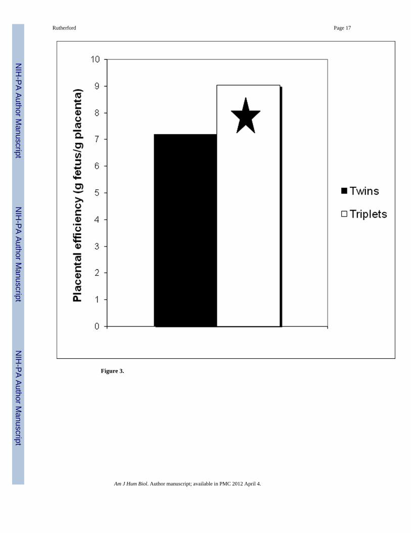
Figure 3.
Rutherford Page 17
Am J Hum Biol. Author manuscript; available in PMC 2012 April 4.
NIH
-PA Author Manuscript
NIH
-PA Author Manuscript
NIH
-PA Author Manuscript
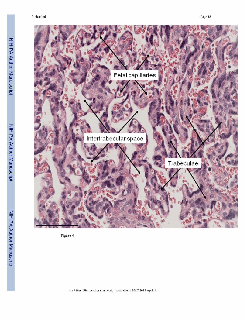
Figure 4.
Rutherford Page 18
Am J Hum Biol. Author manuscript; available in PMC 2012 April 4.
NIH
-PA Author Manuscript
NIH
-PA Author Manuscript
NIH
-PA Author Manuscript
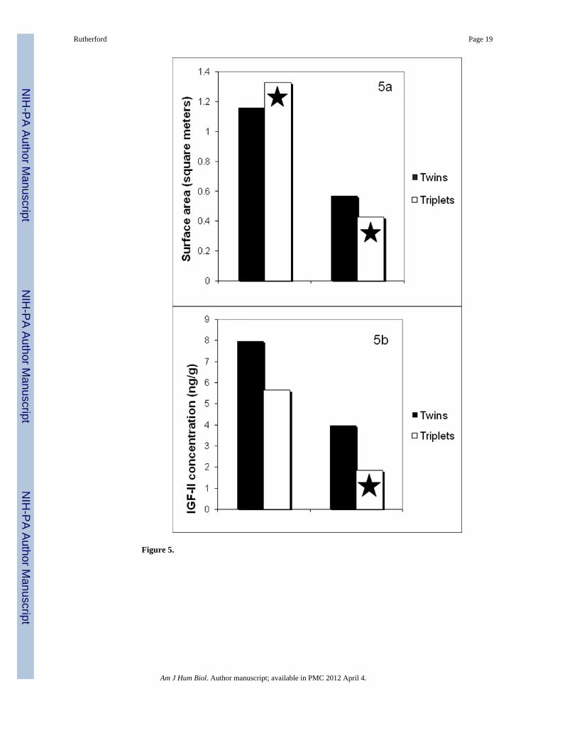
Figure 5.
Rutherford Page 19
Am J Hum Biol. Author manuscript; available in PMC 2012 April 4.
NIH
-PA Author Manuscript
NIH
-PA Author Manuscript
NIH
-PA Author Manuscript

Figure 6.
Rutherford Page 20
Am J Hum Biol. Author manuscript; available in PMC 2012 April 4.
NIH
-PA Author Manuscript
NIH
-PA Author Manuscript
NIH
-PA Author Manuscript