femtosecond diffractive imaging of biological cells
TRANSCRIPT

1
Femtosecond diffractive imaging of biological cells
Marvin M. Seibert1, Sébastien Boutet2,3,1, Martin Svenda1, Tomas Ekeberg1, FilipeR.N.C.Maia1, Michael J. Bogan2,3, Nicusor Tımneanu1, Anton Barty4,3, Stefan Hau-Riege3, CarlCaleman1, Matthias Frank3, Henry Benner3, JoannaY. Lee5, Stefano Marchesini6, JoshuaW.Shaevitz7, Daniel A. Fletcher8, SasaBajt9, Inger Andersson10, Henry N. Chapman4 and JanosHajdu1
1 Laboratory of Molecular Biophysics, Department of Cell and Molecular Biology, UppsalaUniversity, Husargatan 3, Box 596, SE-75124 Uppsala, Sweden2 SLAC National Accelerator Laboratory, 2575 Sand Hill Road, Menlo Park, California94025, USA3 Lawrence LivermoreNational Laboratory, 7000 East Avenue, Livermore, California94550,USA4 Center for Free-Electron Laser Science, University of Hamburg and DESY, Notkestrasse85,Hamburg, Germany5 Department of Biology, Stanford University, Stanford, CA 94305, USA6 Advanced Light Source, Lawrence Berkeley National Laboratory, Berkeley, California94720, USA7 Princeton University, 150 Carl Icahn Laboratory, Princeton NJ 08544, USA8 Bioengineering and Biophysics, University of California, Berkeley, California94720, USA.9 Photon Science, DESY, Notkestrasse85, 22607 Hamburg, Germany10 Swedish University of Agricultural Sciences, Department of Molecular Biology,Husargatan 3, Box 590, SE-751 24 Uppsala, Sweden
E-mail: [email protected], [email protected]
AbbreviationsCXDI: coherent X-ray diffractive imaging, ESI: electrospray ionization, FEL: free-electronlaser, FLASH: Free-electron Laser in Hamburg, LCLS: Linac Coherent Light Source, PRTF:phase retrieval transfer function, SEM: Scanning Electron Microscopy
Keywordsflash diffractive imaging, free-electron laser, Prochlorococcus marinus, Spiroplasmamelliferum, Synechococcus elongatus, FLASH, soft X-ray laser
AbstractIn a flash diffraction experiment, ashort and extremely intenseX-ray pulse illuminates thesample to obtain adiffraction pattern before theonset of significant radiation damage. Theover-sampled diffraction pattern permits phase retrieval through iterativephasing methods.Flash diffractive imaging was first demonstrated on an inorganic test object (Chapman et al.NaturePhysics 2, 839-843, 2006). Wereport here experiments on biological systems whereindividual cells were imaged, using single, 10-15 femtoseconds soft X-ray pulses at 13.5 nmwavelength from theFLASH free-electron laser in Hamburg. Simulations show thepulseheated thesample to about 160,000 K but not beforean interpretablediffraction pattern couldbeobtained. The reconstructed projection images return thestructures of the intact cells. Thesimulations suggest the averagedisplacement of ions and atoms in thehottest surface layersremained below 3 Å during thepulse.Introduction
Confidential: not for distribution. Submitted to IOP Publishing for peer review 12 August 2010pe
er-0
0569
848,
ver
sion
1 -
25 F
eb 2
011
Author manuscript, published in "Journal of Physics B: Atomic, Molecular and Optical Physics 43, 19 (2010) 194015" DOI : 10.1088/0953-4075/43/19/194015

2
Coherent X-ray diffractive imaging (CXDI) is a technique in which an object isilluminated by acoherent beam of X-rays and the exit wavefront is reconstructed from themeasurement of theamplitudes of thediffraction pattern. CXDI has been demonstrated with avariety of samples, ranging from simple test objects [1, 2], to crystals [3, 4] and cells [5-7].Due to theconsiderablepenetration depth of X-rays, CXDI permits studies on objects thatwould be too big for transmission electron microscopy.
A certain number of photons is required to impingeon thesample to achieveadesiredresolution. High doses cause thevery features of interest to bedestroyed while low doses donot produceenough scattered signal to bemeasured. This is thedilemmaof all structuralsciences (for a review see [8]). There is a timecomponent in all damageprocesses. At lowdose rates, radiation damagehas been shown to be dependent on the total doseon thesample[9]. It has been proposed that by greatly increasing thebeam intensity and reducing theexposure time, ahighly non-linear regimeof dose-dependency could be reached, whereasinglediffraction pattern may beobtained at high resolution beforedamage has time todevelop [10-13]. This requires pulses of duration shorter than the relevant dynamics involvedin radiation damage. The first machines capableof producing such ultra-short brilliant pulsesare free-electron lasers (FEL) [14-16]. Theproof of principleof flash diffractive imaging wasdemonstrated on inorganic material at awavelength of 32 nm at theVUV-FEL in Hamburg(renamed FLASH three weeks later), using asilicon nitridemembranewith an etched pattern[17].
Wereport hereuseof this technique in biology for imaging individual biological cells.Threedifferent single-celled organisms were imaged by flash diffraction, Spiroplasmamelliferum [18, 19], Prochlorococcus marinus [20] and Synechococcus elongatus [21]. S.elongatus is asmall photosynthetic cyanobacterium (1.5 µm x 0.8 µm) living in freshwaters.P. marinus is oneof thesmallest (c. 0.6 µm diameter) and most abundant species on Earth,distributed throughout theworld’s oceans between 40°N and 40°S. Spiroplasmas arepathogenic bacteria that lack cell walls and flagella; they infect plants, insects, and mammals[22, 23]. Spiroplasmas, including thespecies S. melliferum (5 µm x 0.15 µm), haveadistinctive, easily identifiableextended helical morphology. S. melliferum has been shown toswim by auniquemethod of propagation of apair of kinks along its length [18]. Thosekinksareproduced by achange in helical direction.
The threedifferent samples chosen illustratecertain aspects of thedevelopments neededin order to expand thecapabilities of flash imaging for biological cells. S. melliferum is aparticularly suitablesample, because its extreme thinness causes it to absorb littleof theincoming beam at thewavelength used, making that theBorn approximation valid. P.marinus represents amorphology that is more typical for singlecelled micro-organisms andextends this imaging method to micron-sized objects. Thecluster of S. elongatus cellsdemonstrates the imaging and reconstruction of multiple isolated objects in asparse imageframe. The S. elongatus cells in direct contact can also serveas asubstitute for illustrating theability to imagemulti-cellular organisms, or several individual cells of one speciessimultaneously.
ResultsFigure1 shows the experimental arrangement. A 45o graded multilayer mirror with a
hole in its middlewas used to let thedirect beam pass through and reflect thescattered beamonto an upward facing charge-coupled device (CCD) detector. This experimental arrangementis described in detail in [17] and [24].Figure2 shows results from an imaging experiment on Synechococcus elongatus cells. Priorto thearrival of thepulse, themembrane, spanning thewindow held three Synechococcus
peer
-005
6984
8, v
ersi
on 1
- 25
Feb
201
1

3
elongatus cells (Figure2a), which weredeposited from a2 µl droplet of 25 mM ammoniumacetate (pH 7.5) onto a20 nm thick Si3N4 membrane. The top left of Figure2b shows whathappened to thesample-holding window after being hit by asingleFLASH pulse (10 fs pulselength FWHM, 13.5 nm wavelength, focal diameter about 20-25 µm FWHM, 1014 W/cm2).Themembranewas completely destroyed (together with thecells), and parts of thesurrounding silicon wafer werealso ablated. The ablation crater in thebottom right-handcorner shows theeffect of asimilar FLASH pulse on thesilicon wafer (covered with 20 nmthick silicon nitride layer) and gives an indication of thebeam sizeand shape. Figure2cshows that despiteof the completedestruction of thecells, the imageof the threecells can beretrieved from thediffraction pattern. No prior knowledgeabout thesamplewas used. Thereconstructed imageagrees with themicroscopy image in Figure2a. Wenote that it ispractically impossible to make truly quantitativecomparisons between reconstructed imagesfrom flash diffraction patterns and prior reference photographs taken by electron- or opticalmicroscopy. The interactions aredifferent, thesensitivities aredifferent, and the responsefunctions aredifferent. Considering the resolutions available to us here, we judge agreementsqualitatively by comparing (i) theshape, (ii) dimensions, and (iii) relative positions ofparticles in the reconstructions and the reference photographs.
A single-shot diffraction pattern of an individual Prochloroccocus marinus cell (Figure3a) can beseen in Figure 3b. Thecell was deposited directly onto aSi3N4 membranebycharge-reducing nano-electrospray ionization [seeMethods]. This proceduremade it possiblefor thecells to be isolated from their medium and to beelectrostatically drawn to themembranewithout contaminants (Figure3b). The reconstructed exit wavefront is shown inFigure3c. Thediffraction pattern, re-calculated from the reconstructed image (Figure3d), isin agreement with the experimentally recorded pattern (Figure3b), supporting theaccuracy ofthe reconstruction. The reconstructed exit wavefront was obtained by iterativephase retrievalin theHAWK softwarepackage [25]. Theachieved resolution of the image was estimated bycalculating thephase retrieval transfer function (PRTF) from 30 reconstructions. ThePRTFfalls below 1/eat 83nm resolution in this case (Figure3e).
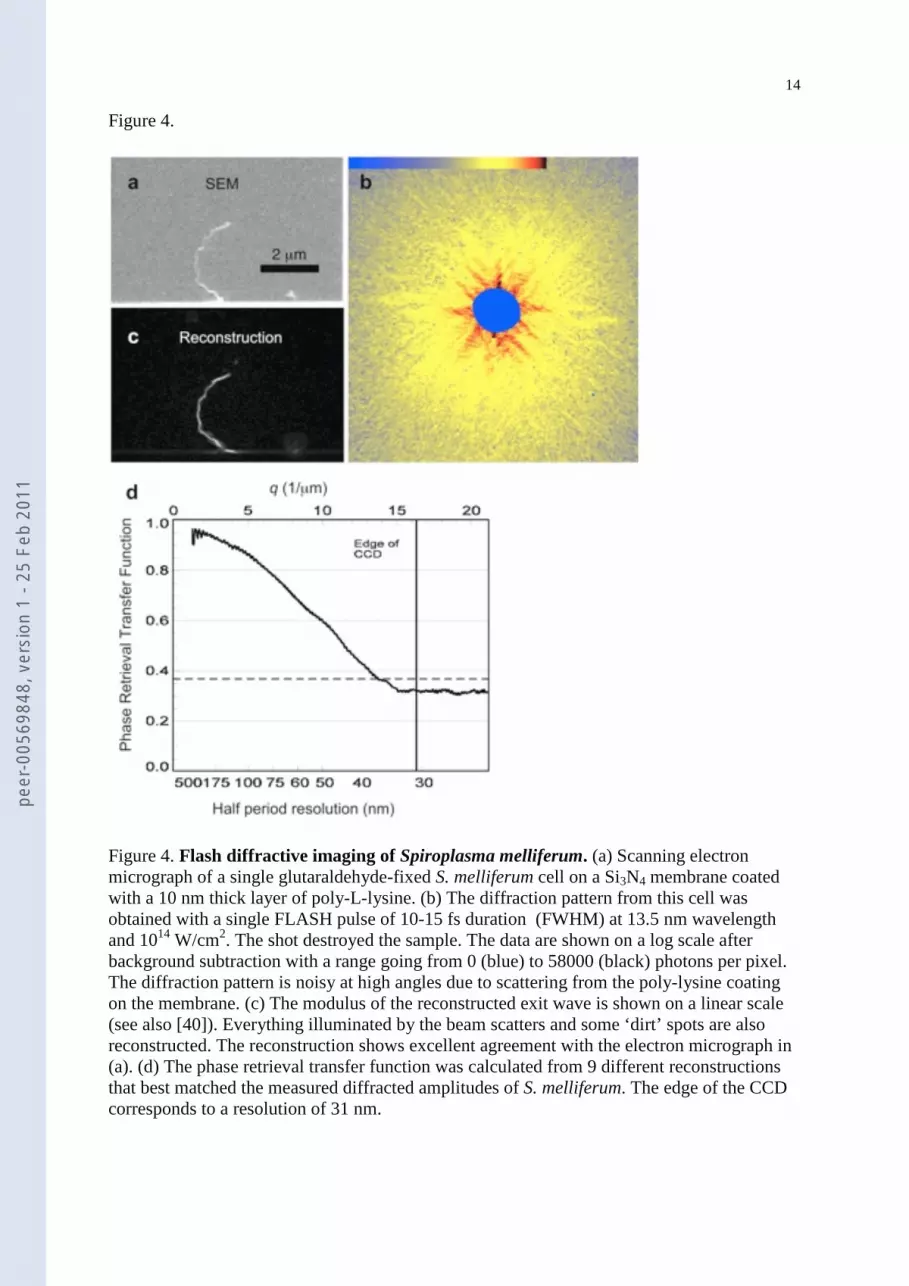
Figure4 shows imaging results with theextremely thin Spiroplasma cells (150 nmdiameter). Thesecells weredeposited onto apoly-lysinecoated silicon nitridewindow [seeMethods]. This coating was uneven and contained randomly distributed clumps of material,as can beseen on Figure 4a. As a result, thebackground in thediffraction pattern (Figure4b)was high. Thesmall fringespacings in thepattern correspond to spatial frequencies of 100 to150 nm, which is the thickness of thecell. The reconstructed exit wavefront was obtained byiterativephase retrieval with theShrinkwrap algorithm [26] [seeMethods] and is shown inFigure4c. This reconstruction agrees well with theelectron micrograph of Figure4a (takenprior to theexposure), and shows thehelical shape of thecell accurately. Theedgeof theSi3N4 membrane (directly below thecell) is also reconstructed along with lumps of materialon themembrane. Each pixel in the reconstruction corresponds to asizeof 28 nm. Thecell isabout 0.15 µm thick and it is about 5 µm long. Only the largecoherence length of the FLASHFEL beam makes it feasible to imagesuch a large cell. Theoutstanding coherencepropertiesof FLASH were exploited in an earlier experiment to image S. melliferum by massivelyparallel X-ray holography [27]. This gave a resolution of 75 nm. Our present results (Figure4d) show significantly higher resolution (38 nm) than this earlier work. This is probably dueto higher pulse intensities and to the fact that the coherence requirements of holography aresignificantly morestringent than for simplediffractive imaging. ThePRTF for thepresent S.melliferum data (Figure4d) was calculated from the9 best reconstructions. Thereconstructions selected had theclosest agreement between the reconstructed diffractedamplitudes and themeasured ones, using the lowest χ2=Σ(Ar-Am)2/ Σ(Am)2, where Ar is thescattered amplitudeof the reconstruction, Am is themeasured scattered amplitudeand thesum
peer
-005
6984
8, v
ersi
on 1
- 25
Feb
201
1

4
is over theentire array of pixels. Theχ2 valueof thebest fit was 2.3 x 10⁻⁴. Due to the largeamount of missing data from thecentral part of thepattern and thenoise from thesupportingmembrane, multiplesolutions wereobtained and only the reconstructions that represented thesamesolution wereaveraged together. The retrieved phases can beseen to become less than50% reproducibleat a resolution of 45nm, or about threequarters of theway to theedgeofthedetector. If thepoint where thePRTF falls below 1/e is used to quantify the resolution, weget a resolution of 38 nm.
Simulations on sampleheating (Figure5a) and ion movement (Figure5b) wereperformed according to [28] with theCRETIN softwarepackage [29] [seeMethods]. Theresults show theFEL pulseheated thesurfaceof thecells to about 160,000 K but noneof thereconstructed projection images show measurable damage. Due to thehigh absorption crosssection at 13.5 nm, theheating of thesample is not homogeneous. Theaverage atomicdisplacement at thesamplesurfaceduring illumination is in thesub-nanometer range (Figure5b) and thus below thediffraction limited resolution.
DiscussionRecent measurements and simulations haveshown little to no damageoccurring in
inorganic samples during pulses of 25 fs (FWHM) containing 1012 photons at 32 nmwavelength, and focused to aspot of 20-30 µm (FWHM) [13, 17, 30]. The results presentedheredemonstrate the feasibility of imaging micron-sized biological objects by flashdiffraction. TheFLASH facility at DESY in Hamburg is the first FEL operated as auser-facility [31]. Theexperiments described hereused pulses of 25±20 µJ energy with adurationof 10-15 fs at a wavelength of 13.5 nm [16]. The largevariation in the intensity of individualpulses, ranging from about 3 x 10¹¹ photons to 3 x 10¹² photons per pulse, arises from thestochastic natureof the lasing process [31]. Thecoherent beam, with an averageof about 1.6x 10¹² photons/pulse, was focused to aspot of 20±5 µm FWHM diameter at thesample.
Theproblem with imaging largebiological samples here is the lack of penetrationpower at 13.5 nm wavelength, the lack of contrast due to thevery similar scattering crosssection for thebiologically relevant elements [32] and thedifficulty of recording 3D patternsdue to the irreproducibility of biological cells. Thepenetration power and resolution willcontinue to improveas FELs areupgraded and new sources arebuilt with shorter wavelength.The Linac Coherent Light Source (LCLS) at Stanford is lasing at 1.5 Å wavelength. At longerwavelengths, high-harmonic sources driven by conventional lasers have recently reachedsufficient brilliance for single-shot flash imaging [33]. The lack of contrast can besolved bytaking advantageof large cross section differences inside thewater window between about2.3-4.4 nm wavelength [34]. The last problem is sample irreproducibility. Flash diffractionimaging offers theuniquepossibility to observea projection of an individual sample in auniqueconformation, rather than automatically producing the averagestructureof numerouscopies of theobject. This may allow theobservation of projections of rare states and fleetingprocesses that aremanifest in aclass of structures which are generally non-reproducible. Forreproduciblesamples, a recent theoretical study quantifies theeffects of sampleheterogeneityon achievable resolution [35].
Resolution could be improved with more intense FEL pulses, and also by removing thesupporting membrane, and illuminating cells free of asupport. This can be donebyaerosolizing themicroorganisms using electrospray ionisation [36, 37] or other types ofnebulizers. Droplets containing singlecells havebeen injected as a focused beam of particlesinto vacuum using aerodynamic lenses [38] and made to intersect with theFEL beam [39,40], yielding container-freedelivery of cells. Transfer of cells into theFEL beam fromsolution can bevery rapid and this raises thepossibility that the cells could still bealiveandhydrated when intersecting thebeam.
peer
-005
6984
8, v
ersi
on 1
- 25
Feb
201
1

5
MethodsSample Preparation
Synechococcus elongatus (PCC 6301) cells were grown in freshwater based Bg11media. Before the imaging experiments, thecells were centrifuged at 6000 g for 10 minutesand thepellet resuspended in sterile25 mM ammonium acetate. Cells were deposited from adroplet of 25 mM ammonium acetate (pH 7.5) onto a20 nm thick Si3N4 membrane, and leftto settle. Excess buffer was removed with amicrocapillary. The20 nm thick Si3N4
membranes havea transmission of 83% at 13.5 nm wavelength.Prochlorococcus marinus cells were cultivated in Pro99 medium, based on sterile
filtered Sargasso Seawater. Before the imaging experiments, cells werecentrifuged at 8500 gfor 10 minutes and resuspended in 25 mM ammonium acetate (viability in this medium wasvalidated for periods of several hours). From this solution, cells wereaerosolized with anano-electrospray ionization (ESI) apparatus (TSI model 3480) operated with gas flows of 1.5l/min sterile-filtered and dehumidified compressed air and 0.25 l/min of CO2 at about 2 psiand avoltageof 1.9 kV. Thehighly charged droplets wereneutralized by passing through abipolar charged gas created with aPo210 alpha-particleemitter. Theaerosol flow was directedtowards ananometer aerosol sampler (TSI model 3089) and captured on a silicon nitridemembraneplaced on top of an electrodeheld at a potential of -10 kV.
Spiroplasma melliferum cells wereattached to silicon nitridemembranes coated withpolylysine. A solution of 0.01% poly-L-lysineof molecular weight 70-150 kDa(Sigma) wasfloated over themembrane for 30 minutes and formed amonolayer of roughly 10 nm, with atransmission of 94% of thebeam at 13.5 nm. 10 µl of asolution containing glutaraldehyde-fixed S. melliferum was pipetted onto asection of thewafer containing 50 x 50 µmmembranes and air dried. The S. melliferum cell was imaged using an electron microscopeprior to illumination by theFLASH pulse and is shown in Figure4a.
FEL MeasurementsTheFEL beam with awavelength of 13.5 nm was focused using an ellipsoidal mirror
to a20 - 30 µmFWHM spot at thesample. Theellipsoidal mirror was located 2 m upstreamof thesample. Thesamplewas mounted on translation stages allowing different parts of thewafer to be illuminated. Stray light from thebeamlinewas removed with a2 mm apertureplaced 29 mm upstream of thesample. Details of theFLASH beamlineare presented in detailby [31].
Thescattered beam was reflected off a45o resonant multilayer mirror, as previouslydescribed by [17, 24]. Thedirect beam was allowed to pass through a1.5 mm hole in themiddleof themultilayer mirror, removing theneed to placeabeamstop in front of thedetector. Such abeamstop would ablatedue to the FEL beam and theemitted radiation wouldadd noise to thesignal. Themirror used had a reflectivity of 65% at 13.5 nm. Themirror actsas abandpass filter which reduces noise from plasmaglow and other sources of photons atwavelengths different from the reflection characteristics of themirror. The scattered beam,after reflection off themirror was detected using a PI-MTE direct detection back-illuminatedCCD from Princeton Instruments. Thedetector contained 1340 x 1300 pixels of 20 x 20 µm.With aCCD quantum efficiency of 42%, a total of 27% of thescattered photons weremeasured. Themirror/CCD assembly, as well as thesampleand thepinholewere all mountedin vacuo to minimizescattering of thebeam.
Thedirect beam can pass through the experiment. This is valuable for shot-to-shotbeam characterization or can even beused to providebeam to another experiment in a tandemarrangement.
peer
-005
6984
8, v
ersi
on 1
- 25
Feb
201
1

6
Simulations of Radiation DamageFor estimates on radiation damageon cells subjected to intenseand short soft X-rays,
weused theapproach from [28]. Simulation on X-ray interaction with biological materialwereperformed using thesoftwareCRETIN [29], amulti-dimensional radiation transfer codeusing non-local thermodynamic equilibrium. CRETIN provides acompleteand properphysical treatment of fundamental physical processes involving ions and electrons,ionizations, collisions, recombinations, inversebremsstrahlung, continuum lowering and canalso treat thehydrodynamic expansion of thesample. CRETIN calculates level populationsand transmission rates and produces absorption, heating rates and conduction coefficientsduring the entiresimulation. Thecomposition of thesamplewas taken to beH23C3NO10S(with adensity of 1 g/cm3), which is theaveragecomposition of awet living cell containing70% overall water. Theheating was modeled in onedimension, justified by theX-ray focalsizebeing much larger than thesample. Thesampledepth was divided into 400 zones whichexchangeenergy through radiation transport and electron thermal conduction. The timeevolution of thesample was followed with a time step of 10 as. The FEL pulsewas modeledby a top hat function with a total length of 15 fs. Previous studies haveshown that pulseshapehad little impact on the ionization rates in thesample [34].
Image ReconstructionAs aconsequenceof having ahole in themirror, thecentral part of thediffraction
pattern is lost. If themissing data corresponds to too many oscillations in thediffractionpattern, many modes are unconstrained [41]. It is thereforeessential to minimize thesizeofthehole in themirror (or detector) and recover as much of the low-resolution dataas possible.This was achieved here, and no prior knowledge was used in this work to populate themissing data.
Due to the low signal to noise ratio of thediffraction pattern at high angles in the S.melliferum diffraction pattern, the imagereconstruction was originally performed with onlythecentral half of thedata. The1340 x 1300 pixel diffracted amplitudearray was cropped andonly themiddle670 x 650 pixels wereused. Theautocorrelation function of theobject wascalculated from the Fourier transform of thediffracted intensity. Thepresenceof point-likeimpurities in thesampleproduced low-resolution copies of the Spiroplasma cell in theautocorrelation function, ahologram. From thehologram arough outlineof thecell wascreated and used as the initial support. Thediffracted wavewas retrieved by the relaxedaveraged alternating reflections (RAAR) algorithm in combination with Shrinkwrap. Theonlyconstraint used in image spacewas the finitesupport. After every 70 iterations, anew supportwas calculated from the latest iterate. The image was first convolved and then every pixelabovea threshold was used as thenew support. Thesizeof theconvolution kernel wasprogressively reduced from 3 pixels to 0.7 pixels after 7000 iterations. From thesolution, atight support was created, only slightly larger than theobject, by again thresholding theimage. This tight support was then enlarged by a factor of 2 in each direction and theentirediffraction data, not just thecentral half, was used to perform 200 separate reconstructionsstarting from different random phases. The tightness of thesupport around theobject and thehigh contrast of the image lead to rapid convergence to asolution. Having this tight supportmade it possible to let theobject be complex-valued [42, 43]. Thecurvatureof theEwaldspherewas corrected for in the reconstruction, but it is aminor effect. The best fit to thedatawas selected and is shown in Figure3a.
For the P. marinus and the S. elongatus reconstruction, the relaxed averagedalternating reflections (RAAR) algorithm in combination with Shrinkwrap was used toretrieve thephases of the diffracted wavestarting from theautocorrelation function. Theonlyconstraint used in image spacewas the finitesupport. After every 20 iterations, anew support
peer
-005
6984
8, v
ersi
on 1
- 25
Feb
201
1

7
was calculated from the latest iterate, by blurring the real-space imageand including thebrightest pixels in thesupport so that thesupport gets a certain area. Both theamount of blurand thesupport areawereprogressively decreased during reconstruction. Theblur radiusstarted at 3 pixels and was progressively decreased to 0.7 pixels at 7000 iterations. Theareastarted at 2500 pixels and decreased to 200 pixels at 10000 iterations. The reconstruction wasrepeated 30 times from different random starting phases. The resulting images wereaveragedtogether after appropriate alignment and the resulting picture is shown in Figure3c. The lackof inversion symmetry in thediffraction pattern is expected simply due to theamount ofabsorption at thewavelength used. In the P. marinus reconstructions, thesupport obtainedusing theshrinkwrap algorithm was sufficiently tight that a reconstruction of acomplex-valued object was possible. Because themaximum angleof thesignal was relatively smalland theamount of oversampling was always high, theeffect of theEwald curvaturewasdeemed sufficiently small to beneglected in this reconstruction.
AcknowledgementsWe are grateful to the scientific and technical staff of FLASH at DESY, Hamburg, in particular to R. Treusch, S.Dusterer and S. Toleikis. We also thank T. Möller, C. Bostedt and H. Thomas for use of their vacuum chamberfor these studies. We thank R. Lee and A. Szoke for discussions. This work wassupported by the followingagencies: The Swedish Research Council; The Swedish Foundation for International Cooperation in Researchand Higher Education; The Swedish Foundation for Strategic Research; The Sven and Lilly Lawski Foundation(fellowship to MMS); Natural Sciencesand Engineering Research Council of Canada through a PostdoctoralFellowship to MJB (MJB iscurrently supported through the PULSE Institute at the SLAC National AcceleratorLaboratory by the U.S. Department of Energy, Office of Basic Energy Sciences);The DFG Cluster of Excellenceat the Munich Centre for Advanced Photonics, The National Science Foundation Center for Biophotonics,University of California, Davis: The Advanced Light Source, Lawrence Berkeley Lab, under DOE contract; TheEuropean Union (TUIXS); partsof this work were performed under the auspices of the U.S. Department of Energy by Lawrence Livermore National Laboratory under Contract DE-AC52-07NA27344. Simulationswere performed on UPPMAX resourcesunder project p2009018. Access to FLASH isprovided by the EuropeanUnion contract RII3-CT-2004-506008 (IA-SFS).
peer
-005
6984
8, v
ersi
on 1
- 25
Feb
201
1

8
References
1. Miao, J.W., et al., Extending the methodology of X-ray crystallography to allowimaging of micrometre-sized non-crystalline specimens. Nature, 1999. 400(6742): p.342-344.
2. Loh, N.D., et al., Cryptotomography: reconstructing 3D Fourier intensities fromrandomly oriented single-shot diffraction patterns. . submitted, 2010.
3. Robinson, I.K., et al., Reconstruction of the shapes of gold nanocrystals usingcoherent x-ray diffraction. Physical Review Letters, 2001. 8719(19): p. 195505.
4. Pfeifer, M.A., et al., Three-dimensional mapping of a deformation field inside ananocrystal. Nature, 2006. 442(7098): p. 63-66.
5. Miao, J.W., et al., Imaging whole Escherichia coli bacteria by using single-particle x-ray diffraction. Proceedings of the National Academy of Sciences of theUnited Statesof America, 2003. 100(1): p. 110-112.
6. Shapiro, D., et al., Biological imaging by soft x-ray diffraction microscopy.Proceedings of the National Academy of Sciences of theUnited States of America,2005. 102(43): p. 15343-15346.
7. Mancuso, A.P., et al., Coherent imaging of biological samples with femtosecondpulses at the free-electron laser FLASH. New Journal of Physics, 2010. 12: p. 035003.
8. Marchesini, S., et al., Coherent X-ray diffractive imaging: applications andlimitations. Optics Express, 2003. 11(19): p. 2344-2353.
9. Garman, E.F. and S.M. McSweeney, Progress in research into radiation damage incryo-cooled macromolecular crystals. Journal of Synchrotron Radiation, 2007. 14: p.1-3.
10. Neutze, R., et al., Potential for biomolecular imaging with femtosecond X-ray pulses.Nature, 2000. 406(6797): p. 752-757.
11. Hau-Riege, S.P., R.A. London, and A. Szoke, Dynamics of biological moleculesirradiated by short x-ray pulses. Physical Review E, 2004. 69(5): p. 051906.
12. Hau-Riege, S.P., et al., Subnanometer-scale measurements of the interaction ofultrafast soft X-ray free-electron-laser pulses with matter. Physical Review Letters,2007. 98(14): p. 145502.
13. Hau-Riege, S.P., et al., Sacrificial Tamper Slows Down Sample Explosion in FLASHDiffraction Experiments. Physical Review Letters, 2010. 104(6): p. -.
14. Saldin, E.L., E.A. Schneidmiller, and M.V. Yurkov, The Physics of Free-ElectronLasers - an Introduction. Physics Reports-Review Section of Physics Letters, 1995.260(4-5): p. 187-327.
15. Ayvazyan, V., et al., First operation of a free-electron laser generating GW powerradiation at 32 nm wavelength. European Physical Journal D, 2006. 37(2): p. 297-303.
16. Ackermann, W., et al., Operation of a free-electron laser from the extreme ultravioletto the water window. NaturePhotonics, 2007. 1(6): p. 336-342.
17. Chapman, H.N., et al., Femtosecond diffractive imaging with a soft-X-ray free-electron laser. NaturePhysics, 2006. 2(12): p. 839-843.
18. Shaevitz, J.W., J.Y. Lee, and D.A. Fletcher, Spiroplasma swim by a processive changein body helicity. Cell, 2005. 122(6): p. 941-945.
19. Kurner, J., A.S. Frangakis, and W. Baumeister, Cryo-electron tomography reveals thecytoskeletal structure of Spiroplasma melliferum. Science, 2005. 307(5708): p. 436-8.
20. Chisholm, S.W., et al., A Novel Free-Living Prochlorophyte Abundant in the OceanicEuphotic Zone. Nature, 1988. 334(6180): p. 340-343.
21. Nägeli, C., Gattungen einzelliger Algen - physiologisch und systematisch bearbeitet.1849, Zürich: Frierich Schulthess.
peer
-005
6984
8, v
ersi
on 1
- 25
Feb
201
1

9
22. Whitcomb, R.F., The genus Spiroplasma. Annu Rev Microbiol, 1980. 34: p. 677-709.23. Lorenz, B., J. Schroeder, and U. Reischl, First evidence of an endogenous
Spiroplasma sp. infection in humans manifesting as unilateral cataract associatedwith anterior uveitis in a premature baby. Graefes Arch Clin Exp Ophthalmol, 2002.240(5): p. 348-53.
24. Bajt, S., et al., Camera for coherent diffractive imaging and holography with a soft-x-ray free-electron laser. Applied Optics, 2008. 47(10): p. 1673-1683.
25. Maia, F., et al., Hawk: the image reconstruction package for coherent X-raydiffractive imaging. submitted, 2010.
26. Marchesini, S., A unified evaluation of iterative projection algorithms for phaseretrieval. Review of Scientific Instruments, 2007. 78(1): p. 011301.
27. Marchesini, S., et al., Massively parallel X-ray holography. NaturePhotonics, 2008.2(9): p. 560-563.
28. Bergh, M., et al., Interaction of ultrashort x-ray pulses with B4C, SiC, and Si. PhysicalReview E, 2008. 77(2): p. 026404.
29. Scott, H.A., Cretin - a radiative transfer capability for laboratory plasmas. Journal ofQuantitativeSpectroscopy & RadiativeTransfer, 2001. 71(2-6): p. 689-701.
30. Chapman, H.N., et al., Femtosecond time-delay X-ray holography. Nature, 2007.448(7154): p. 676-679.
31. Tiedtke, K., et al., The soft x-ray free-electron laser FLASH at DESY: beamlines,diagnostics and end-stations. New Journal of Physics, 2009. 11: p. 023029.
32. Henke, B.L., E.M. Gullikson, and J.C. Davis, X-Ray Interactions - Photoabsorption,Scattering, Transmission and Reflection at E=50-30,000 Ev, Z=1-92 (Vol 54, Pg 181,1993). Atomic Dataand Nuclear DataTables, 1993. 55(2): p. 349-349.
33. Ravasio, A., et al., Single-Shot Diffractive Imaging with a Table-Top FemtosecondSoft X-Ray Laser-Harmonics Source. Physical Review Letters, 2009. 103(2): p.028104.
34. Bergh, M., et al., Feasibility of imaging living cells at subnanometer resolutions byultrafast X-ray diffraction. Quarterly Reviews of Biophysics, 2008. 41(3-4): p. 181-204.
35. Maia, F.R.N.C., et al., Structural variability and the incoherent addition of scatteredintensities in single-particle diffraction. Physical Review E, 2009. 80(3): p. 031905.
36. Bothner, B. and G. Siuzdak, Electrospray ionization of a whole virus: Analyzing mass,structure, and viability. Chembiochem, 2004. 5(3): p. 258-260.
37. Clarke, J.D. and S.N. Jayasinghe, Bio-electrosprayed multicellular zebrafish embryosare viable and develop normally. Biomed Mater, 2008. 3(1): p. 011001.
38. Liu, P., et al., Generating Particle Beams of Controlled Dimensions and Divergence.1. Theory of Particle Motion in Aerodynamic Lenses and Nozzle Expansions. AerosolScienceand Technology, 1995. 22(3): p. 293-313.
39. Bogan, M.J., et al., Single particle X-ray diffractive imaging. Nano Letters, 2008. 8(1):p. 310-316.
40. Chapman, H.N., et al., Coherent Imaging at FLASH. J Phys Conf, 2009. 186: p.012051.
41. Thibault, P., et al., Reconstruction of a yeast cell from X-ray diffraction data. ActaCrystallographicaSection A, 2006. 62: p. 248-261.
42. Fienup, J.R., Reconstruction of a Complex-Valued Object from the Modulus of ItsFourier-Transform Using a Support Constraint. Journal of theOptical Society ofAmerica a-Optics Image Scienceand Vision, 1987. 4(1): p. 118-123.
43. He, H., et al., Experimental lensless soft-X-ray imaging using iterative algorithms:phasing diffuse scattering. ActaCrystallographicaSection A, 2003. 59: p. 143-152.
peer
-005
6984
8, v
ersi
on 1
- 25
Feb
201
1

10
peer
-005
6984
8, v
ersi
on 1
- 25
Feb
201
1

11
Figures with captions
Figure1.
Figure1. The experimental setup. Thesilicon-nitridesamplewindow and theFEL pulsearesignificantly enlarged on the figure relative to the mirror and theCCD camera. Thedistancefrom thesample to thedetector was 50 mm. Themultilayer mirror has ahole in its centre tolet thedirect beam through. For details see refs [17, 24].
peer
-005
6984
8, v
ersi
on 1
- 25
Feb
201
1

12
Figure2.
Figure2. Obliteration of Synechococcus elongatus cells by a single FLASH pulse. (a)Photomicrograph of the intact cells on asilicon nitridewindow before exposure to theFLASH pulse. The edge of thewindow is visibleat thebottom. (b) Thesamewindow (at alower magnification) after being exposed to ac. 10 fs long FLASH pulse (FWHM) at 1014
W/cm2, 13.5 nm wavelength. The wafer in themicroscopewas illuminated from twodirections: with white light from above thesample planeand with a filtered "purple light"from below. This shows theblown out window as apurplesquare in (b). Thecrater at bottomright shows theprofileof theFLASH beam from a similar shot. (c) The reconstructed exitwavefront from thediffraction pattern returns the imageof theoriginal cells.
peer
-005
6984
8, v
ersi
on 1
- 25
Feb
201
1

13
Figure3.
Figure3. Flash diffractive imaging of a Prochlorocococcus marinus cell on a siliconnitride window. (a) Light microscopy imageof a single P. marinus on a20 nm thick Si3N4
membrane. (b) The experimentally recorded diffraction pattern obtained with aFLASH pulseof approximately 10 fs (FWHM) at 13.5nm and displayed on a logarithmic scale. (c) Thereconstructed exit wavefront obtained by iterative phase retrieval with the HAWK softwarepackage (Maia et al. 2010). (d) Thediffraction pattern of P. marinus, computed from thereconstruction is in good agreement with theexperimentally obtained pattern in (b). (e) Thephase retrieval transfer function for the reconstructed P. marinus cell. The optical resolutionis estimated to be83 nm.
peer
-005
6984
8, v
ersi
on 1
- 25
Feb
201
1
14
Figure4.
Figure4. Flash diffractive imaging of Spiroplasma melliferum. (a) Scanning electronmicrograph of asingle glutaraldehyde-fixed S. melliferum cell on aSi3N4 membranecoatedwith a10 nm thick layer of poly-L-lysine. (b) The diffraction pattern from this cell wasobtained with asingle FLASH pulseof 10-15 fs duration (FWHM) at 13.5 nm wavelengthand 1014 W/cm2. Theshot destroyed thesample. Thedataareshown on a log scaleafterbackground subtraction with a range going from 0 (blue) to 58000 (black) photons per pixel.Thediffraction pattern is noisy at high angles due to scattering from thepoly-lysine coatingon themembrane. (c) Themodulus of the reconstructed exit wave is shown on a linear scale(seealso [40]). Everything illuminated by thebeam scatters and some ‘dirt’ spots arealsoreconstructed. The reconstruction shows excellent agreement with theelectron micrograph in(a). (d) Thephase retrieval transfer function was calculated from 9 different reconstructionsthat best matched themeasured diffracted amplitudes of S. melliferum. The edgeof theCCDcorresponds to a resolution of 31 nm.
peer
-005
6984
8, v
ersi
on 1
- 25
Feb
201
1

15
Figure5.
Figure5. Sample heating and radiation-induced sample movement during and afterexposure to the FEL pulse. (a) Ion temperature in the P. marinus sampleduring and after a15 fs pulseas a function of sampledepth. Thepulseshapewas modeled as asimple top-hatfunction. Pulse length = 15 fs, wavelength = 13.5 nm, pulse intensity = 20 µJ, focal spot = 20µm. Thesamplebecomes hottest on theside facing theX-ray beam. (b) Averagedisplacement of ions and atoms on the illuminated surfaceduring thepulse (15 fs) for severalpulse intensities. The results show that the resolutions achieved in Figures 2-4 arenot limitedby atomic displacement or hydrodynamic expansion during the15 fs long exposure.
peer
-005
6984
8, v
ersi
on 1
- 25
Feb
201
1