femoral and vertebral strength improvements in postmenopausal women with osteoporosis treated with...
TRANSCRIPT
Femoral and Vertebral Strength Improvements inPostmenopausal Women With Osteoporosis TreatedWith DenosumabTony M Keaveny,1,2 Michael R McClung,3 Harry K Genant,4,5 Jose R Zanchetta,6 David Kendler,7
Jacques P Brown,8 Stefan Goemaere,9 Chris Recknor,10 Maria L Brandi,11 Richard Eastell,12
David L Kopperdahl,2 Klaus Engelke,13,14 Thomas Fuerst,5 Hoi‐Shen Radcliffe,15 and Cesar Libanati16
1University of California Berkeley, Berkeley, CA, USA2O.N. Diagnostics, Berkeley, CA, USA3Oregon Osteoporosis Center, Portland, OR, USA4University of California San Francisco, CA, USA5Synarc Inc., San Francisco, CA, USA6IDIM, Buenos Aires, Argentina7University of British Columbia, Vancouver, Canada8CHU de Québec Research Centre, Québec, Canada9Ghent University Hospital, Ghent, Belgium10United Osteoporosis Centers, Gainesville, GA, USA11University of Florence, Florence, Italy12University of Sheffield, Sheffield, UK13Synarc Inc., Hamburg, Germany14Institute of Medical Physics, University of Erlangen, Erlangen, Germany15Amgen Ltd., Cambridge, UK16Amgen Inc., Thousand Oaks, CA, USA
ABSTRACTIn the randomized, placebo‐controlled FREEDOM study of women aged 60 to 90 years with postmenopausal osteoporosis, treatmentwith denosumab once every 6 months for 36 months significantly reduced hip and new vertebral fracture risk by 40% and 68%,respectively. To gain further insight into this efficacy, we performed a nonlinear finite element analysis (FEA) of hip and spinequantitative computed tomography (QCT) scans to estimate hip and spine strength in a subset of FREEDOM subjects (n¼ 48 placebo;n¼ 51 denosumab) at baseline, 12, 24, and 36 months. We found that, compared with baseline, the finite element estimates of hipstrength increased from 12 months (5.3%; p< 0.0001) and through 36 months (8.6%; p< 0.0001) in the denosumab group. For theplacebo group, hip strength did not change at 12 months and decreased at 36 months (–5.6%; p< 0.0001). Similar changes wereobserved at the spine: strength increased by 18.2% at 36 months for the denosumab group (p< 0.0001) and decreased by –4.2% forthe placebo group (p¼ 0.002). At 36 months, hip and spine strength increased for the denosumab group compared with the placebogroup by 14.3% (p< 0.0001) and 22.4% (p< 0.0001), respectively. Further analysis of the finite elementmodels indicated that strengthassociated with the trabecular bone was lost at the hip and spine in the placebo group, whereas strength associated with both thetrabecular and cortical bone improved in the denosumab group. In conclusion, treatment with denosumab increased hip and spinestrength as estimated by FEA of QCT scans compared with both baseline and placebo owing to positive treatment effects in both thetrabecular and cortical bone compartments. These findings provide insight into the mechanism by which denosumab reducesfracture risk for postmenopausal women with osteoporosis. © 2014 The Authors. Journal of Bone and Mineral Research published byWiley Periodicals, Inc. on behalf of the American Society for Bone and Mineral Research. This is an open access article under the termsof the Creative Commons Attribution‐NonCommercial‐NoDerivs License, which permits use and distribution in anymedium, providedthe original work is properly cited, the use is non‐commercial and no modifications or adaptations are made.
KEY WORDS: DENOSUMAB; HIP STRENGTH; SPINE STRENGTH; FINITE ELEMENT ANALYSIS; OSTEOPOROSIS
Received in original form February 26, 2013; revised form May 30, 2013; accepted June 10, 2013. Accepted manuscript online June 21, 2013.Address correspondence to: Tony M Keaveny, PhD, Departments of Mechanical Engineering and Bioengineering, 6175 Etcheverry Hall, MC 1740, University ofCalifornia, Berkeley, CA 94720‐1740, USA. E‐mail: [email protected] work was presented in part as an abstract and oral presentation at the ASBMR 2010 Annual Meeting, October 15–19, Toronto Canada.
ORIGINAL ARTICLE JJBMR
Journal of Bone and Mineral Research, Vol. 29, No. 1, January 2014, pp 158–165DOI: 10.1002/jbmr.20242014 American Society for Bone and Mineral Research
158
Introduction
Denosumab (Prolia1, Amgen Inc., Thousand Oaks, CA, USA) isa fully human monoclonal antibody that inhibits receptor
activator of NF‐kB ligand (RANKL), a key modulator of osteoclastformation, function, and survival.(1–4) In the phase 3 FractureReduction Evaluation of Denosumab in Osteoporosis Every6 Months (FREEDOM) study of 7808 postmenopausal womenwith osteoporosis, aged between 60 and 90 years, denosumabtreatment reduced the risk of hip and new vertebral fracturesover 36 months compared with placebo by 40% and 68%,respectively.(5) The fracture efficacy in the overall FREEDOMpopulation was associated with significant differences frombaseline to month 36 between denosumab and placebo in arealbone mineral density (BMD) at the total hip and lumbar spine of6.4% and 8.8%, respectively,(6) and rapid and marked reductionsin markers of bone resorption with an attenuation of thereduction at the end of each 6‐month dosing interval.(5,7) Despitethe insight gained from the changes in BMD and bone turnovermarkers, it remains unclear to what extent denosumab alteredhip or spine strength compared with baseline and placebo, andthe extent that denosumab modified bone strength in thetrabecular and cortical compartments, both of which are relevantto understanding mechanisms of fracture protection.Because directly measuring the strength of any bone in a living
human subject is not feasible, an alternative and FDA‐approvedapproach to monitor treatment effects is to noninvasivelyestimate changes in strength using finite element analysis (FEA)of quantitative computed tomography (QCT) scans. Consistentlyvalidated for assessment of whole‐bone strength in variouscadaver studies,(8–16) FEA of hip and spine QCT scans has beenshown to predict new incident hip(17–19) and spine(16) fracturesand to differentiate those with or without prevalent spine(20–23)
and general fractures.(24) FEA has also been used to study agingeffects in hip strength,(25) estimate changes in hip(26–29) andspine(28,30–33) strength in response to various treatments as well asspace flight,(34) and estimate changes in strength that are directlyassociated with changes in the bone compartments.(35,36)
To gain further insight into the observed clinical efficacy ofdenosumab in reducing the risk of hip and spine fractures, in asubset of subjects in the FREEDOM study, we applied FEA to thehip and spine QCT scans to noninvasively assess changes in hipand spine strength associated with denosumab treatment over36 months.
Materials and Methods
Study design and participants
FREEDOM was an international, randomized, placebo‐controlledstudy in postmenopausal women with osteoporosis.(5) Partic-ipants received placebo or denosumab 60mg subcutaneouslyevery 6months for 36months, with daily supplements of calcium(�1000mg) and vitamin D (�400 IU). Postmenopausal womenaged between 60 and 90 years with a BMD T‐score of <�2.5 atthe lumbar spine and/or total hip, and not <–4.0 at either site,were included in the study. Women were excluded if they hadany severe or >2 moderate vertebral fractures, conditions thataffected bone metabolism, or had taken oral bisphosphonatesfor �36 months. Women were eligible if they had taken oralbisphosphonates for�36monthsbut noneduring the 12monthspreceding enrollment into the study. Study centers in theFREEDOM study, with expertise and access to a qualified scanner,
invited subjects to participate in a substudy of the hip andlumbar spine QCT measurements. Calibration issues andunavailability of analyzable scans at different time points limitedthe analyses to 99 subjects (n¼ 48 placebo; n¼ 51 denosumab).
The protocol was approved by an independent ethicscommittee or institutional review board at each study site.Details of the methods and main results of the FREEDOM studyhave been reported previously.(5)
Assessments
The QCT methodology used to assess BMD and geometricparameters of the total hip and lumbar spine using whole‐bodyscanners at selected study centers has been describedpreviously, as has the main BMD analysis.(37) Scans wereperformed at 120 kV with a pitch of 1 using 170 mAs in thehip and 100mAs in the spine, and reconstructed using amediumkernel and a field of view of 400mm in the hip and 360mm in thespine. The reconstructed slice thickness was �1.25mm. Scanswere obtained at baseline, 12, 24, and 36 months, and covered1 cm above the femoral head to 2 cm below the lesser trochanterfor the hip assessment, or both L1 and L2 vertebrae for the spineassessment. Scanner calibration and stability were assessed anddocumented by Synarc (Portland, OR, USA, and Hamburg,Germany) circulating a European Spine Phantom (ESP).
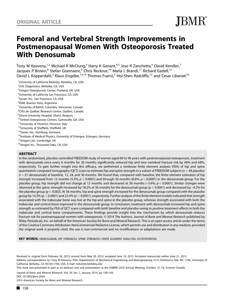
All image processing and image analysis for FEA assessmentswere performed blinded to treatment, by O.N. Diagnostics(Berkeley, CA, USA), as described elsewhere.(19,25,26,28) Briefly, theQCT images were calibrated, segmented, and converted intofinite element models (�40,000 elements per model), usingcube‐shaped, eight‐noded brick elements (1.5 mm‐sided for thehip and 1.0mm‐sided for the spine). Because delineating the truecortical envelope is difficult from clinical‐resolution CT scans,particularly where the cortex is thin, we defined a “cortical”compartment to include all obvious cortical bone (defined asbone having an apparent BMD of >1.0 g/cm3) plus any otherbone within a fixed distance of the periosteal surface (3mm forthe hip; 2mm for the spine); the trabecular compartment wasdefined as all remaining trabecular bone (Fig. 1). Element‐specific
Fig. 1. Example of finite element models for one subject at baseline. Thefull three‐dimensional model is shown, with a schematic of how the loadsare applied (through center of head at hip; distributed evenly for spine).The models for estimating strength changes associated with changes inonly the trabecular and “cortical” compartments are also shown (thin 2Dsections only). Our definition of the “cortical” compartment included allobvious cortical bone (defined as bone having an apparent BMD of>1.0 g/cm3) plus any other bone within a fixed distance of the periostealsurface (3mm for the hip; 2mm for the spine); the trabecularcompartment was defined as all of the remaining trabecular bone.
Journal of Bone and Mineral Research QCT FEA‐ASSESSED DENOSUMAB CHANGES 159
elastic properties (isotropic for the hip; anisotropic for the spine)and elastic‐plastic failure properties were all derived from thecalibrated volumetric BMD values, using validated empiricalrelations, and (for the hip) using higher strengths in compressionthan tension.(38–40) After registering each bone to its baselineconfiguration, boundary conditions were applied to simulate asevere, unprotected fall to the side of the hip, with the diaphysisangled at 15° with respect to the ground and 15° of internalrotation. Femoral strength was defined from the resultingnonlinear force‐strain curve of the whole femur as the force at4.0% strain. For the spine, a similar approachwas taken inwhich athin layer of plastic was applied over each endplate throughwhich the vertebral body was loaded to simulated failure inuniform compression. Spine strength was defined from theresulting nonlinear force‐strain curve of thewhole vertebral bodyas the force at 1.9% strain. For both hip and spine, theseimplementations have been shown to provide excellentcorrelations with measured strength from biomechanicalcadaver experiments with a Y¼ X type of absolute agreementbetween model and experiment.(16,19,41)
We also performed controlled variations of these models toprovide additional strength outcomes, including trabecular and“cortical” strengths (Fig. 1). Hip trabecular strength wascomputed by turning all the bone in the “cortical” compartmentat each time point into plastic (a stiffer plastic for the compactcortical bone and a less stiff plastic for the adjacent trabecularbone) and then virtually reloading the resulting model to failureas described above for the intact femur. Likewise, hip “cortical”strength was computed by turning all the bone in the trabecularcompartment into (a low‐stiffness) plastic and then virtuallyreloading to failure,(26) and spine trabecular strength wascomputed by virtually removing the outer 2mm of bone andthen virtually reloading to failure.(32) Because a spine modelcomprised only of a thin shell of cortical bone would be prone tobuckling if virtually loaded to failure, which would havequestionable relevance, spine “cortical” strength was calculatedas the overall spine strengthminus the spine trabecular strength.We note that these trabecular and “cortical” strengths wereadditive for the spine but not for the hip; this is because of thecomplex nonparallel load transfer paths in the hip between thetrabecular and “cortical” compartments, both of which werepresent in all hip models.
Statistical analysis
Subjects having both a baseline and�1 post‐baseline FEA resultwere included in the analysis. There was no imputation ofmissing data. The percentage and absolute changes frombaseline in hip and spine strength were analyzed using ananalysis of covariance (ANCOVA) model including treatment andadjusting for baseline value and age strata (stratification factor).Least‐squares means and 95% confidence intervals (CIs) for eachtreatment and for the treatment difference (denosumab –placebo) at each time point were generated. All analyses wereexploratory and post‐hoc. The p values and CIs were not adjustedfor multiplicity.
Results
The baseline characteristics in this subset of women from theFREEDOM study (Table 1) were similar to those reported for theoverall study population.(5) Mean age was 74.1 years in theplacebo group and 73.3 years in the denosumab group. Mean
total hip dual‐energy X‐ray absorptiometry (DXA) T‐score was–2.0 and –1.7 in the placebo and denosumab groups,respectively, and –2.8 at the lumbar spine in both treatmentgroups.
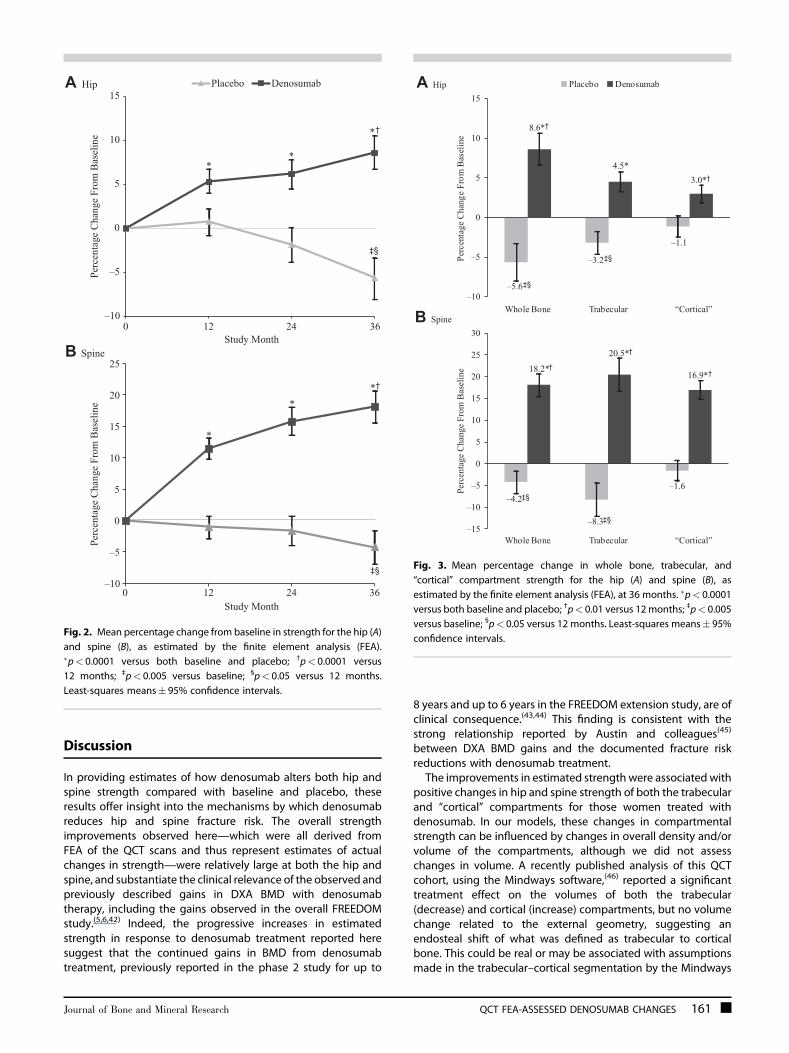
For the women treated with denosumab, hip strength asestimated by FEA increased significantly compared with baselineby 5.3% at 12 months (p< 0.0001) and progressively over time,reaching 8.6% at 36 months (p< 0.0001 compared with baselineand 12months) (Fig. 2A). By contrast, for the women treated withplacebo, hip strength did not change at 12 months anddecreased thereafter to �5.6% by 36 months compared withbaseline (p< 0.0001). At 36 months, hip strength for thedenosumab group increased by 14.3% compared with placebo(p< 0.0001). Similar changes occurred at the spine, but theincreases in strength were larger. From baseline, spine strengthincreased by 18.2% (p< 0.0001) for the denosumab group anddecreased by –4.2% (p¼ 0.002) for the placebo group at36 months (Fig. 2B), corresponding to a relative increase inspine strength for the denosumab group of 22.4% comparedwith placebo (p< 0.0001). The corresponding absolute changesin strength at the hip and spine were also significantly higher forthe denosumab group compared with baseline and the placebogroup (p� 0.0001 for all). At 36 months, mean absolute changefrombaseline in estimated hip strengthwasþ196 Newtons (N) inthe denosumab group compared with –123N in the placebogroup (p< 0.0001). At the spine, strength increased byþ513N inthe denosumab group compared with a decrease of –127N inthe placebo group.
Further analysis of the finite element models revealedincreases in strength associated with changes in both thetrabecular and “cortical” bone compartments in the denosumabgroup for both the hip and spine (Fig. 3). By contrast, in theplacebo group, the hip and spine strength associated withthe trabecular compartment preferentially decreased frombaseline through 36 months, whereas the strength associatedwith the “cortical” compartment was preserved.
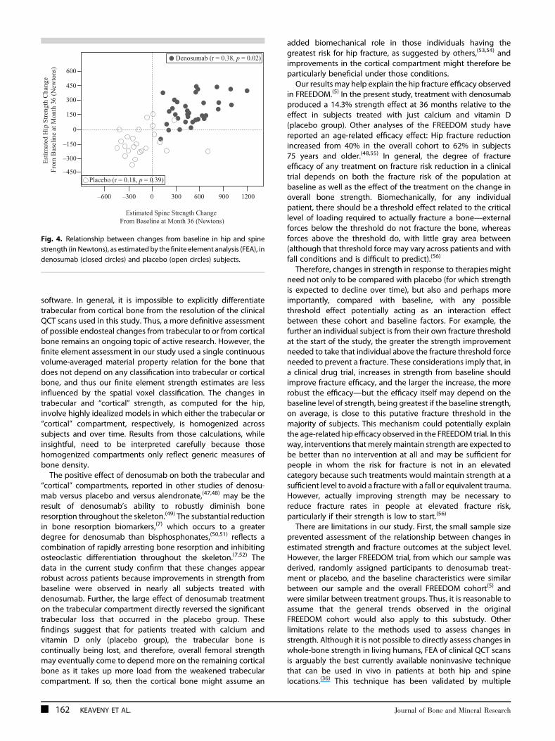
For individual subjects at 36 months, changes in hip strengthwere significantly, but weakly, correlated with changes in spinestrength for the denosumab group (r¼ 0.38; p¼ 0.02) but not forthe placebo group (r¼ 0.18; p¼ 0.39) (Fig. 4). At 36months, spinestrength increased in all denosumab‐treated subjects and hipstrength increased in all but two subjects, whereas both the hipand spine strength decreased for themajority of placebo‐treatedsubjects.
Table 1. Baseline Characteristics
Placebon¼ 48
Denosumabn¼ 51
Age (years) 74.1 (6.0) 73.3 (4.2)BMI (kg/m2) 25.7 (4.6) 26.3 (4.5)Total hip QCT BMD (mg/cm3) 217 (31) 227 (34)Lumbar spine (trabecular) QCT
BMD (mg/cm3)64.5 (17.2) 69.6 (18.8)
Total hip BMD DXA T‐score –2.0 (0.8) –1.7 (0.9)Lumbar spine BMD DXA T‐score –2.8 (0.7) –2.8 (0.5)Total hip FEA strength (Newtons) 2273 (570) 2512 (686)Total spine FEA strength (Newtons) 2841 (628) 2879 (583)
Values are means (standard deviations).BMI¼body mass index; QCT¼quantitative computed tomography;
BMD¼bone mineral density; DXA¼dual‐energy X‐ray absorptiometry;FEA¼ finite element analysis.
160 KEAVENY ET AL. Journal of Bone and Mineral Research
Discussion
In providing estimates of how denosumab alters both hip andspine strength compared with baseline and placebo, theseresults offer insight into the mechanisms by which denosumabreduces hip and spine fracture risk. The overall strengthimprovements observed here—which were all derived fromFEA of the QCT scans and thus represent estimates of actualchanges in strength—were relatively large at both the hip andspine, and substantiate the clinical relevance of the observed andpreviously described gains in DXA BMD with denosumabtherapy, including the gains observed in the overall FREEDOMstudy.(5,6,42) Indeed, the progressive increases in estimatedstrength in response to denosumab treatment reported heresuggest that the continued gains in BMD from denosumabtreatment, previously reported in the phase 2 study for up to
8 years and up to 6 years in the FREEDOM extension study, are ofclinical consequence.(43,44) This finding is consistent with thestrong relationship reported by Austin and colleagues(45)
between DXA BMD gains and the documented fracture riskreductions with denosumab treatment.
The improvements in estimated strength were associated withpositive changes in hip and spine strength of both the trabecularand “cortical” compartments for those women treated withdenosumab. In our models, these changes in compartmentalstrength can be influenced by changes in overall density and/orvolume of the compartments, although we did not assesschanges in volume. A recently published analysis of this QCTcohort, using the Mindways software,(46) reported a significanttreatment effect on the volumes of both the trabecular(decrease) and cortical (increase) compartments, but no volumechange related to the external geometry, suggesting anendosteal shift of what was defined as trabecular to corticalbone. This could be real or may be associated with assumptionsmade in the trabecular–cortical segmentation by the Mindways
*
**†
‡§
–10
–5
0
5
10
15
20
25
0 12 24 36
Perc
enta
ge C
hang
e Fr
om B
asel
ine
Study Month
‡§
–10
–5
0
5
10
15
0 12 24 36
Perc
enta
ge C
hang
e Fr
om B
asel
ine
Study Month
Placebo Denosumab
**
*†
B Spine
A Hip
Fig. 2. Mean percentage change from baseline in strength for the hip (A)and spine (B), as estimated by the finite element analysis (FEA).�p< 0.0001 versus both baseline and placebo; †p< 0.0001 versus12 months; ‡p< 0.005 versus baseline; §p< 0.05 versus 12 months.Least‐squares means� 95% confidence intervals.
–5.6‡§
–3.2‡§
–1.1
8.6*†
4.5*3.0*†
–10
–5
0
5
10
15
Whole Bone Trabecular “Cortical”
Whole Bone Trabecular “Cortical”
Perc
enta
ge C
hang
e Fr
om B
asel
ine
Placebo Denosumab
–4.2‡§
–8.3‡§
–1.6
18.2*†20.5*†
16.9*†
–15
–10
–5
0
5
10
15
20
25
30
Perc
enta
ge C
hang
e Fr
om B
asel
ine
A Hip
B Spine
Fig. 3. Mean percentage change in whole bone, trabecular, and“cortical” compartment strength for the hip (A) and spine (B), asestimated by the finite element analysis (FEA), at 36 months. �p< 0.0001versus both baseline and placebo; †p< 0.01 versus 12months; ‡p< 0.005versus baseline; §p< 0.05 versus 12 months. Least‐squares means� 95%confidence intervals.
Journal of Bone and Mineral Research QCT FEA‐ASSESSED DENOSUMAB CHANGES 161
software. In general, it is impossible to explicitly differentiatetrabecular from cortical bone from the resolution of the clinicalQCT scans used in this study. Thus, a more definitive assessmentof possible endosteal changes from trabecular to or from corticalbone remains an ongoing topic of active research. However, thefinite element assessment in our study used a single continuousvolume‐averaged material property relation for the bone thatdoes not depend on any classification into trabecular or corticalbone, and thus our finite element strength estimates are lessinfluenced by the spatial voxel classification. The changes intrabecular and “cortical” strength, as computed for the hip,involve highly idealized models in which either the trabecular or“cortical” compartment, respectively, is homogenized acrosssubjects and over time. Results from those calculations, whileinsightful, need to be interpreted carefully because thosehomogenized compartments only reflect generic measures ofbone density.
The positive effect of denosumab on both the trabecular and“cortical” compartments, reported in other studies of denosu-mab versus placebo and versus alendronate,(47,48) may be theresult of denosumab’s ability to robustly diminish boneresorption throughout the skeleton.(49) The substantial reductionin bone resorption biomarkers,(7) which occurs to a greaterdegree for denosumab than bisphosphonates,(50,51) reflects acombination of rapidly arresting bone resorption and inhibitingosteoclastic differentiation throughout the skeleton.(7,52) Thedata in the current study confirm that these changes appearrobust across patients because improvements in strength frombaseline were observed in nearly all subjects treated withdenosumab. Further, the large effect of denosumab treatmenton the trabecular compartment directly reversed the significanttrabecular loss that occurred in the placebo group. Thesefindings suggest that for patients treated with calcium andvitamin D only (placebo group), the trabecular bone iscontinually being lost, and therefore, overall femoral strengthmay eventually come to depend more on the remaining corticalbone as it takes up more load from the weakened trabecularcompartment. If so, then the cortical bone might assume an
added biomechanical role in those individuals having thegreatest risk for hip fracture, as suggested by others,(53,54) andimprovements in the cortical compartment might therefore beparticularly beneficial under those conditions.
Our results may help explain the hip fracture efficacy observedin FREEDOM.(5) In the present study, treatment with denosumabproduced a 14.3% strength effect at 36 months relative to theeffect in subjects treated with just calcium and vitamin D(placebo group). Other analyses of the FREEDOM study havereported an age‐related efficacy effect: Hip fracture reductionincreased from 40% in the overall cohort to 62% in subjects75 years and older.(48,55) In general, the degree of fractureefficacy of any treatment on fracture risk reduction in a clinicaltrial depends on both the fracture risk of the population atbaseline as well as the effect of the treatment on the change inoverall bone strength. Biomechanically, for any individualpatient, there should be a threshold effect related to the criticallevel of loading required to actually fracture a bone—externalforces below the threshold do not fracture the bone, whereasforces above the threshold do, with little gray area between(although that threshold force may vary across patients and withfall conditions and is difficult to predict).(56)
Therefore, changes in strength in response to therapies mightneed not only to be compared with placebo (for which strengthis expected to decline over time), but also and perhaps moreimportantly, compared with baseline, with any possiblethreshold effect potentially acting as an interaction effectbetween these cohort and baseline factors. For example, thefurther an individual subject is from their own fracture thresholdat the start of the study, the greater the strength improvementneeded to take that individual above the fracture threshold forceneeded to prevent a fracture. These considerations imply that, ina clinical drug trial, increases in strength from baseline shouldimprove fracture efficacy, and the larger the increase, the morerobust the efficacy—but the efficacy itself may depend on thebaseline level of strength, being greatest if the baseline strength,on average, is close to this putative fracture threshold in themajority of subjects. This mechanism could potentially explainthe age‐related hip efficacy observed in the FREEDOM trial. In thisway, interventions thatmerelymaintain strength are expected tobe better than no intervention at all and may be sufficient forpeople in whom the risk for fracture is not in an elevatedcategory because such treatments would maintain strength at asufficient level to avoid a fracture with a fall or equivalent trauma.However, actually improving strength may be necessary toreduce fracture rates in people at elevated fracture risk,particularly if their strength is low to start.(56)
There are limitations in our study. First, the small sample sizeprevented assessment of the relationship between changes inestimated strength and fracture outcomes at the subject level.However, the larger FREEDOM trial, from which our sample wasderived, randomly assigned participants to denosumab treat-ment or placebo, and the baseline characteristics were similarbetween our sample and the overall FREEDOM cohort(5) andwere similar between treatment groups. Thus, it is reasonable toassume that the general trends observed in the originalFREEDOM cohort would also apply to this substudy. Otherlimitations relate to the methods used to assess changes instrength. Although it is not possible to directly assess changes inwhole‐bone strength in living humans, FEA of clinical QCT scansis arguably the best currently available noninvasive techniquethat can be used in vivo in patients at both hip and spinelocations.(36) This technique has been validated by multiple
Estimated Spine Strength Change From Baseline at Month 36 (Newtons)
Estim
ated
Hip
Stre
ngth
Cha
nge
From
Bas
elin
e at
Mon
th 3
6 (N
ewto
ns)
–300
–150
150
450
600
–600 –300 300 900 1200
Denosumab (r = 0.38, p = 0.02)
Placebo (r = 0.18, p = 0.39)
0 600
0
300
–450
Fig. 4. Relationship between changes from baseline in hip and spinestrength (in Newtons), as estimated by the finite element analysis (FEA), indenosumab (closed circles) and placebo (open circles) subjects.
162 KEAVENY ET AL. Journal of Bone and Mineral Research
independent research groups for the hip and spine in bothcadaver bone strength(8–16) and clinical fracture outcomestudies,(16–24) and it is an FDA‐approved approach that providesan integrative outcome of whole‐bone strength that is intuitivelyassociated with the etiology of osteoporotic fractures. Themodels have limited spatial resolution (�1mm imaging voxels);therefore, they cannot capture the bone microstructure,including the trabecular microarchitecture, Haversian porosity,any microstructure of the endosteal bone at the interfacebetween well‐defined cortical and trabecular bone, or the shell‐type microstructure of the thin cortex around the femoral neck.The limited spatial resolution also precludes accounting forpotential microscale effects of denosumab. For example, it hasbeen proposed that reducing bone turnover and inhibiting“stress risers” may be an additional mechanism by whichantiresorptive therapies reduce fracture risk,(57,58) an effect thatwould serve to further increase strength for the denosumabgroup. Lastly, no adjustment was made for any possible changesin the tissue‐level material properties resulting from treatment,such as increased secondary mineralization. However, bio-mechanical studies in adult ovariectomized cynomolgus mon-keys treated with denosumab for 16 months have shown nodetectable biomechanical negative effects on tissue‐levelmaterial properties.(59) Even so, results should be interpretedwith these limitations in mind.In summary, denosumab administered subcutaneously at a
dose of 60mg every 6 months substantially improved FEAestimates of hip and spine strength, both compared withbaseline and the administration of calcium and vitamin D onlyover 36 months. These improvements were progressive overtime and were influenced by parallel positive estimated strengthchanges in both the trabecular and “cortical” compartments.These observations highlight the potential benefit of denosu-mab to improve and reverse the negative consequence ofpostmenopausal‐ and aging‐induced skeletal decay on strength,and to markedly reduce fracture incidence in subjects atincreased or high risk for hip and spine fractures.
Disclosures
TMK is a consultant for Amgen Inc. and Merck; has receivedresearch grant support from Amgen Inc., Eli Lilly, Merck, andNovartis, and is a shareholder of O.N. Diagnostics.MRM has received research grant support from Amgen Inc.
and was an advisory board member for Amgen Inc.HKG is a consultant and/or speaker for Amgen Inc., BMS,
Genentech, Eli Lilly, GSK Janssen, Merck, Novartis, Pfizer, ONO,Roche, and Servier, and is a shareholder of Synarc.JRZ has received research grant support from Amgen Inc., and
is a consultant and speaker for Amgen Inc.DK is a consultant and/or speaker for Amgen Inc., Eli Lilly,
Merck, Novartis, and Servier, and has received grant support fromAmgen Inc., Biosante, Eli Lilly, GSK, Merck, Novartis, Pfizer, Roche,and Servier.JPB is a consultant, speaker, and an advisory boardmember for
Amgen Inc., Eli Lilly, and Merck, and has received research grantsupport and honoraria from Abbott, Amgen Inc., BMS, Eli Lilly,Merck, Novartis, Pfizer, Roche, Sanofi‐Aventis, Servier, Takeda,and Warner Chilcott.SG has received research grant support fromAmgen Inc. and is
a consultant for Amgen Inc.CR has received research grant support from Medi, consulting
and advisory fees from Amgen Inc., Eli Lilly, and Novartis, lecture
fees from Novartis and Warner Chilcott, and is a shareholder inIon Med Systems.
MLB has received research grant support and honoraria fromAmgen Inc.
RE has received consulting fees from Amgen Inc., GSK,Novartis, ONO, Pfizer, Servier, andWarner Chilcott, research grantsupport from Amgen Inc., AstraZeneca, Novartis, and WarnerChilcott, and is a speaker for Eli Lilly.
DLK is an employee of and has equity interest in O.N.Diagnostics.
KE is an employee and shareholder of Synarc, and is anadvisory board member for Amgen Inc.
TF is an employee and shareholder of Synarc.HR and CL are employees of and own stock or stock options in
Amgen Inc.
Acknowledgments
Amgen Inc. (Thousand Oaks, CA, USA) sponsored this study. Theauthors thank Mandy Suggitt and Erica Rockabrand, PhD, ofAmgen Inc. for their medical writing assistance in the preparationof this manuscript.
Authors’ roles: TMK, HR, and CL contributed to the conceptionand design of the study, and analysis and interpretation of thedata. MRM, HKG, DK, JRZ, JPB, SG, CR, MLB, RE, DLK, KE, and TFcontributed to the acquisition of data and analysis andinterpretation of the data. All authors participated in draftingor revising the manuscript and approved the final version of themanuscript for submission. TK and CL take responsibility for theintegrity of the data analysis.
References
1. Lacey DL, Tan HL, Lu J, Kaufman S, Van G, Qiu W, Rattan A, Scully S,Fletcher F, Juan T, Kelley M, Burgess TL, Boyle WJ, Polverino AJ.Osteoprotegerin ligandmodulates murine osteoclast survival in vitroand in vivo. Am J Pathol. 2000;157:435–48.
2. Lacey DL, Timms E, Tan HL, Kelley MJ, Dunstan CR, Burgess T, Elliott R,Colombero A, Elliott G, Scully S, Hsu H, Sullivan J, Hawkins N, Davy E,Capparelli C, Eli A, Qian YX, Kaufman S, Sarosi I, Shalhoub V, Senaldi G,Guo J, Delaney J, Boyle WJ. Osteoprotegerin ligand is a cytokine thatregulates osteoclast differentiation and activation. Cell. 1998;93:165–76.
3. UdagawaN, Takahashi N, YasudaH,MizunoA, Itoh K, Ueno Y, Shinki T,Gillespie MT, Martin TJ, Higashio K, Suda T. Osteoprotegerinproduced by osteoblasts is an important regulator in osteoclastdevelopment and function. Endocrinology. 2000;141:3478–84.
4. Yasuda H, ShimaN, NakagawaN, Yamaguchi K, Kinosaki M,MochizukiS, Tomoyasu A, Yano K, Goto M, Murakami A, Tsuda E, Morinaga T,Higashio K, Udagawa N, Takahashi N, Suda T. Osteoclast differentia-tion factor is a ligand for osteoprotegerin/osteoclastogenesis‐inhibitory factor and is identical to TRANCE/RANKL. Proc Natl AcadSci USA. 1998;95:3597–602.
5. Cummings SR, San Martin J, McClung MR, Siris ES, Eastell R, Reid IR,Delmas P, Zoog HB, AustinM,Wang A, Kutilek S, Adami S, Zanchetta J,Libanati C, Siddhanti S, Christiansen C. Denosumab for prevention offractures in postmenopausal womenwith osteoporosis. N Engl JMed.2009;361:756–65.
6. BologneseMA, Teglbjaerg CS, Zanchetta JR, Lippuner K, McClungMR,Brandi ML, Hoiseth A, Lakatos P, Moffett AH, Lorenc RS, Wang A,Libanati C. Denosumab significantly increases DXA BMD at bothtrabecular and cortical sites: results from the FREEDOM study. J ClinDensitom. 2012;16:147–53.
7. Eastell R, Christiansen C, Grauer A, Kutilek S, Libanati C, McClung MR,Reid IR, Resch H, Siris E, Uebelhart D, Wang A, Weryha G, CummingsSR. Effects of denosumab on bone turnover markers in postmeno-pausal osteoporosis. J Bone Miner Res. 2011;26:530–7.
Journal of Bone and Mineral Research QCT FEA‐ASSESSED DENOSUMAB CHANGES 163
8. Bessho M, Ohnishi I, Matsuyama J, Matsumoto T, Imai K, Nakamura K.Prediction of strength and strain of the proximal femur by a CT‐basedfinite element method. J Biomech. 2007;40:1745–53.
9. Buckley JM, Loo K, Motherway J. Comparison of quantitativecomputed tomography‐based measures in predicting vertebralcompressive strength. Bone. 2007;40:767–74.
10. Cody DD, Gross GJ, Hou FJ, Spencer HJ, Goldstein SA, Fyhrie DP.Femoral strength is better predicted by finite element models thanQCT and DXA. J Biomech. 1999;32:1013–20.
11. Crawford RP, Cann CE, Keaveny TM. Finite element models predict invitro vertebral body compressive strength better than quantitativecomputed tomography. Bone. 2003;33:744–50.
12. Imai K, Ohnishi I, Bessho M, Nakamura K. Nonlinear finite elementmodel predicts vertebral bone strength and fracture site. Spine.2006;31:1789–94.
13. Keyak JH, Falkinstein Y. Comparison of in situ and in vitro CT scan‐based finite element model predictions of proximal femoral fractureload. Med Eng Phys. 2003;25:781–7.
14. Lengsfeld M, Schmitt J, Alter P, Kaminsky J, Leppek R. Comparison ofgeometry‐based and CT voxel‐based finite element modelling andexperimental validation. Med Eng Phys. 1998;20:515–22.
15. Martin H, Werner J, Andresen R, Schober HC, Schmitz KP. Noninvasiveassessment of stiffness and failure load of human vertebrae fromCT‐data. Biomed Tech (Berl). 1998;43:82–8.
16. Wang X, Sanyal A, Cawthon PM, Palermo L, Jekir M, Christensen J,Ensrud KE, Cummings SR, Orwoll E, Black DM, Keaveny TM.Prediction of new clinical vertebral fractures in elderly menusing finite element analysis of CT scans. J Bone Miner Res. 2012;27:808–16.
17. Keyak JH, Sigurdsson S, Karlsdottir G, Oskarsdottir D, SigmarsdottirA, Zhao S, Kornak J, Harris TB, Sigurdsson G, Jonsson BY, SiggeirsdottirK, Eiriksdottir G, Gudnason V, Lang TF. Male‐female differences inthe association between incident hip fracture and proximalfemoral strength: a finite element analysis study. Bone. 2011;48:1239–45.
18. Kopperdahl DL, Hoffmann P, Sigurdsson S, Aspelund T, SiggeirsdottirK, Eiriksdottir G, Harris T, Gudnason VG, Keaveny TM. Enhancement ofhip fracture prediction using finite element analysis of CT scans. JBone Miner Res. 2010; 25(Suppl 1):S114.
19. Orwoll ES, Marshall LM, Nielson CM, Cummings SR, Lapidus J, CauleyJA, Ensrud K, Lane N, Hoffmann PF, Kopperdahl DL, Keaveny TM.Finite element analysis of the proximal femur and hip fracture risk inolder men. J Bone Miner Res. 2009;24:475–83.
20. Faulkner KG, Cann CE, Hasegawa BH. Effect of bone distribution onvertebral strength: assessment with patient‐specific nonlinear finiteelement analysis. Radiology. 1991;179:669–74.
21. Imai K, Ohnishi I, Yamamoto S, Nakamura K. In vivo assessment oflumbar vertebral strength in elderly women using computedtomography‐based nonlinear finite element model. Spine. 2008;33:27–32.
22. Melton LJ 3rd, Riggs BL, Keaveny TM, Achenbach SJ, Hoffmann PF,Camp JJ, Rouleau PA, Bouxsein ML, Amin S, Atkinson EJ, Robb RA,Khosla S. Structural determinants of vertebral fracture risk. J BoneMiner Res. 2007;22:1885–92.
23. Melton LJ 3rd, Riggs BL, Keaveny TM, Achenbach SJ, Kopperdahl D,Camp JJ, Rouleau PA, Amin S, Atkinson EJ, Robb RA, Therneau TM,Khosla S. Relation of vertebral deformities to bone density, structure,and strength. J Bone Miner Res. 2010;25:1922–30.
24. Amin S, Kopperdhal DL. Melton LJ 3rd, Achenbach SJ, Therneau TM,Riggs BL, Keaveny TM, Khosla S. Association of hip strength estimatesby finite‐element analysis with fractures in women and men. J BoneMiner Res. 2011;26:1593–600.
25. Keaveny TM, Kopperdahl DL, Melton LJ 3rd, Hoffmann PF, Amin S,Riggs BL, Khosla S. Age‐dependence of femoral strength in whitewomen and men. J Bone Miner Res. 2010;25:994–1001.
26. Keaveny TM, Hoffmann PF, Singh M, Palermo L, Bilezikian JP,Greenspan SL, Black DM. Femoral bone strength and its relation tocortical and trabecular changes after treatment with PTH, alendro-nate, and their combination as assessed by finite element analysis ofquantitative CT scans. J Bone Miner Res. 2008;23:1974–82.
27. Keaveny TM, McClungMR,Wan X, Kopperdahl DL, Mitlak BH, Krohn K.Femoral strength in osteoporotic women treated with teriparatide oralendronate. Bone. 2012;50:165–70.
28. Lewiecki EM, Keaveny TM, Kopperdahl DL, Genant HK, Engelke K,Fuerst T, Kivitz A, Davies RY, Fitzpatrick LA. Once‐monthly oralibandronate improves biomechanical determinants of bone strengthin women with postmenopausal osteoporosis. J Clin EndocrinolMetab. 2009;94:171–80.
29. Lian KC, Lang TF, Keyak JH, Modin GW, Rehman Q, Do L, Lane NE.Differences in hip quantitative computed tomography (QCT)measurements of bone mineral density and bone strength betweenglucocorticoid‐treated and glucocorticoid‐naive postmenopausalwomen. Osteoporos Int. 2005;16:642–50.
30. Chevalier Y, Quek E, Borah B, Gross G, Stewart J, Lang T, Zysset P.Biomechanical effects of teriparatide in women with osteoporosistreated previously with alendronate and risedronate: results fromquantitative computed tomography‐based finite element analysis ofthe vertebral body. Bone. 2010;46:41–8.
31. Graeff C, Chevalier Y, Charlebois M, Varga P, Pahr D, Nickelsen TN,Morlock MM, Gluer CC, Zysset PK. Improvements in vertebral bodystrength under teriparatide treatment assessed in vivo by finiteelement analysis: results from the EUROFORS study. J BoneMiner Res.2009;24:1672–80.
32. Keaveny TM, Donley DW, Hoffmann PF, Mitlak BH, Glass EV, SanMartin JA. Effects of teriparatide and alendronate on vertebralstrength as assessed by finite element modeling of QCT scans inwomen with osteoporosis. J Bone Miner Res. 2007;22:149–57.
33. Mawatari T, Miura H, Hamai S, Shuto T, Nakashima Y, Okazaki K,Kinukawa N, Sakai S, Hoffmann PF, Iwamoto Y, Keaveny TM. Vertebralstrength changes in rheumatoid arthritis patients treated withalendronate, as assessed by finite element analysis of clinicalcomputed tomography scans: a prospective randomized clinical trial.Arthritis Rheum. 2008;58:3340–9.
34. Keyak JH, Koyama AK, LeBlanc A, Lu Y, Lang TF. Reduction in proximalfemoral strength due to long‐duration spaceflight. Bone. 2009;44:449–53.
35. Bonnick SL. Noninvasive assessments of bone strength. Curr OpinEndocrinol Diabetes Obes. 2007;14:451–7.
36. Keaveny TM. Biomechanical computed tomography‐noninvasivebone strength analysis using clinical computed tomography scans.Ann NY Acad Sci. 2010;1192:57–65.
37. McClungMR, Zanchetta JR, Høiseth A, Kendler DL, Yuen CK, Brown JP,Stonkus S, Goemaere S, Recknor C, Woodson GC, Bolognese MA,Franek E, Brandi ML, Wang A, Libanati C. Denosumab densitometricchanges assessed by quantitative computed tomgraphy at the spineand hip in postmenopausal women with osteoporosis. J ClinDensitom. 2013;16:250–6.
38. Morgan EF, Keaveny TM. Dependence of yield strain of humantrabecular bone on anatomic site. J Biomech. 2001;34:569–77.
39. Morgan EF, Bayraktar HH, Keaveny TM. Trabecular bone modulus‐density relationships depend on anatomic site. J Biomech. 2003;36:897–04.
40. Bayraktar HH, Morgan EF, Niebur GL, Morris GE, Wong EK, KeavenyTM. Comparison of the elastic and yield properties of humanfemoral trabecular and cortical bone tissue. J Biomech. 2004;37:27–35.
41. Roberts BJ, Kopperdahl DL, Thrall E, Muller JA, Keaveny TM, BouxseinML. Prediction of femoral strength in a sideways fall configurationusing QCT‐based finite element analysis. Bone. 2009;44:S72.
42. Lewiecki EM, Miller PD, McClung MR, Cohen SB, Bolognese MA, Liu Y,Wang A, Siddhanti S, Fitzpatrick LA. Two‐year treatment withdenosumab (AMG 162) in a randomized phase 2 study ofpostmenopausal women with low BMD. J Bone Miner Res. 2007;22:1832–41.
43. Bone HG, Brown JP, Chapurlat R, Franchimont N, Brandi ML,Czerwiński E, Krieg M‐A, Man Z, Mellström D, Radominski SC,Reginster JY, Resch H, Román JA, Daizadeh NS, Wang A, Geller ML,Wagman RB, Papapoulos S. The effect of six years of denosumabtreatment on new vertebral and nonvertebral fractures in postmen-opausal women with osteoporosis: results from the FREEDOMextension trial. Endocr Rev. 2012;33:S18–22.
164 KEAVENY ET AL. Journal of Bone and Mineral Research
44. McClung MR, Lewiecki EM, Geller ML, Bolognese MA, Peacock M,Weinstein RL, Ding B, Rockabrand E, Wagman RB, Miller PD. Effect ofdenosumab on bone mineral density and biochemical markers ofbone turnover: 8‐year results of a phase 2 clinical trial. Osteoporos Int.2013;24:227–35.
45. Austin M, Yang YC, Vittinghoff E, Adami S, Boonen S, Bauer DC,Bianchi G, Bolognese MA, Christiansen C, Eastell R, Grauer A, HawkinsF, Kendler DL, Oliveri B, McClung MR, Reid IR, Siris ES, Zanchetta J,Zerbini CA, Libanati C, Cummings SR. Relationship between bonemineral density changes with denosumab treatment and riskreduction for vertebral and nonvertebral fractures. J Bone MinerRes. 2012;27:687–93.
46. McClungMR, Zanchetta JR, Høiseth A, Kendler DL, Yuen CK, Brown JP,Stonkus S, Goemaere S, Recknor C, Woodson GC, Bolognese MA,Franek E, Brandi ML, Wang A, Libanati C. Denosumab densitometricchanges assessed by QCT at the spine and hip in postmenopausalwomen with osteoporosis. J Clin Densitom. 2013;16:250–6.
47. Seeman E, Delmas PD, Hanley DA, Sellmeyer D, Cheung AM, Shane E,Kearns A, Thomas T, Boyd SK, Boutroy S, Bogado C, Majumdar S, FanM, Libanati C, Zanchetta J. Microarchitectural deterioration of corticaland trabecular bone: differing effects of denosumab and alendro-nate. J Bone Miner Res. 2010;25:1886–94.
48. Simon JA, Recknor C, Moffet AH, Adachi JD, Franek E, Lewiecki EM,McClung MR, Mautalen CA, Ragi Eis S, Nicholson GC, Muschitz C, NutiR, Törring O, Wang A, Libanati C. Impact of denosumab on theperipheral skeleton of postmenopausal women with osteoporosis:bone density, mass, and strength of the radius, and wrist fracture.Menopause. 2013;20:130–7.
49. Kostenuik PJ, Smith SY, Jolette J, Schroeder J, Pyrah I, Ominsky MS.Decreased bone remodeling and porosity are associated withimproved bone strength in ovariectomized cynomolgus monkeystreated with denosumab, a fully human RANKL antibody. Bone.2011;49:151–61.
50. Brown JP, Prince RL, Deal C, Recker RR, Kiel DP, de Gregorio LH, HadjiP, Hofbauer LC, Alvaro‐Gracia JM, Wang H, Austin M, Wagman RB,Newmark R, Libanati C, San Martin J, Bone HG. Comparison of theeffect of denosumab and alendronate on BMD and biochemicalmarkers of bone turnover in postmenopausal women with low bonemass: a randomized, blinded, phase 3 trial. J Bone Miner Res. 2009;24:153–61.
51. Kendler DL, Roux C, Benhamou CL, Brown JP, Lillestol M, Siddhanti S,Man HS, San Martin J, Bone HG. Effects of denosumab on bone
mineral density and bone turnover in postmenopausal womentransitioning from alendronate therapy. J Bone Miner Res. 2010;25:72–81.
52. Kostenuik PJ, NguyenHQ, McCabe J, Warmington KS, Kurahara C, SunN, Chen C, Li L, Cattley RC, Van G, Scully S, Elliott R, Grisanti M, MoronyS, Tan HL, Asuncion F, Li X, Ominsky MS, Stolina M, Dwyer D, DougallWC, Hawkins N, Boyle WJ, Simonet WS, Sullivan JK. Denosumab, afully humanmonoclonal antibody to RANKL, inhibits bone resorptionand increases BMD in knock‐in mice that express chimeric (murine/human) RANKL. J Bone Miner Res. 2009;24:182–95.
53. Black DM, Bouxsein ML, Marshall LM, Cummings SR, Lang TF, CauleyJA, Ensrud KE, Nielson CM, Orwoll ES. Osteoporotic Fractures in MenResearch G. Proximal femoral structure and the prediction of hipfracture in men: a large prospective study using QCT. J Bone MinerRes. 2008;23:1326–33.
54. Bousson V, Le Bras A, Roqueplan F, Kang Y, Mitton D, Kolta S,Bergot C, Skalli W, Vicaut E, Kalender W, Engelke K, Laredo JD.Volumetric quantitative computed tomography of the proximalfemur: relationships linking geometric and densitometric variables tobone strength. Role for compact bone. Osteoporos Int. 2006;17:855–64.
55. Boonen S, Adachi JD, Man Z, Cummings SR, Lippuner K, Torring O,Gallagher JC, Farrerons J, Wang A, Franchimont N, San Martin J,Grauer A, McClung M. Treatment with denosumab reduces theincidence of new vertebral and hip fractures in postmeno-pausal women at high risk. J Clin Endocrinol Metab. 2011;96:1727–36.
56. Keaveny TM, Bouxsein ML. Theoretical implications of the bio-mechanical fracture threshold. J Bone Miner Res. 2008;23:1541–7.
57. Riggs BL, Melton LJ 3rd. Bone turnover matters: the raloxifenetreatment paradox of dramatic decreases in vertebral fractureswithout commensurate increases in bone density. J Bone Miner Res.2002;17:11–4.
58. Parfitt AM. High bone turnover is intrinsically harmful: two paths to asimilar conclusion. The Parfitt view. J Bone Miner Res. 2002;17:1558–9.
59. Ominsky MS, Stouch B, Schroeder J, Pyrah I, Stolina M, Smith SY,Kostenuik PJ. Denosumab, a fully human RANKL antibody, reducedbone turnover markers and increased trabecular and cortical bonemass, density, and strength in ovariectomized cynomolgus monkeys.Bone. 2011;49:162–73.
Journal of Bone and Mineral Research QCT FEA‐ASSESSED DENOSUMAB CHANGES 165