exploration génétique et moléculaire de défauts post
TRANSCRIPT

HAL Id: tel-02372023https://tel.archives-ouvertes.fr/tel-02372023
Submitted on 20 Nov 2019
HAL is a multi-disciplinary open accessarchive for the deposit and dissemination of sci-entific research documents, whether they are pub-lished or not. The documents may come fromteaching and research institutions in France orabroad, or from public or private research centers.
L’archive ouverte pluridisciplinaire HAL, estdestinée au dépôt et à la diffusion de documentsscientifiques de niveau recherche, publiés ou non,émanant des établissements d’enseignement et derecherche français ou étrangers, des laboratoirespublics ou privés.
Exploration génétique et moléculaire de défautspost-méiotiques sévères de la spermatogenèse entrainant
une infertilité masculineZine-Eddine Kherraf
To cite this version:Zine-Eddine Kherraf. Exploration génétique et moléculaire de défauts post-méiotiques sévères de laspermatogenèse entrainant une infertilité masculine. Médecine humaine et pathologie. UniversitéGrenoble Alpes, 2018. Français. �NNT : 2018GREAS010�. �tel-02372023�

THÈSE
Pour obtenir le grade de
DOCTEUR DE LA COMMUNAUTÉ UNIVERSITÉ GRENOBLE ALPES
Spécialité : BIS - Biotechnologie, instrumentation, signal et imagerie pour la biologie, la médecine et l'environnement
Arrêté ministériel : 25 mai 2016
Présentée par
ZINE-EDDINE KHERRAF
Thèse dirigée par Pierre RAY (EDISCE), UGA et CHU de Grenoble
préparée au sein de l'Institut pour l'Avancée des Biosciences dans l'École Doctorale Ingénierie pour la santé la Cognition et
l'Environnement
Exploration génétique et moléculaire de
défauts post-méiotiques sévères de la
spermatogenèse entrainant une infertilité
masculine.
Thèse soutenue publiquement le 12 juillet 2018,devant le jury composé de :
Monsieur Saadi KHOCHBINDirecteur de Recherche, CNRS Délégation Alpes, Président
Madame Catherine PATRATProfesseur des Universités - Praticien Hospitalier, Université Paris 5, Rapporteur
Monsieur Florian-Jean-Louis GUILLOUDirecteur de Recherche, INRA Centre Val de Loire, Rapporteur
Madame Sophie CHRISTIN-MAITREProfesseur des Universités - praticien hospitalier, Université Pierre et Marie Curie, Examinateur
Monsieur Pierre RAYProfesseur des universités - Praticien Hospitalier, Université Grenoble Alpes, Directeur de thèse

2

3
Résumé
Titre : Exploration génétique et moléculaire de défauts post-méiotiques sévères de la spermatogenèse entrainant une infertilité masculine.
L’infertilité est considérée actuellement par l’organisation mondiale de la santé (OMS) comme une préoccupation majeure de santé affectant plus de 50 millions de couples dans le monde. Dans les pays occidentaux, la majorité des couples infertiles ont recours aux techniques d’assistance médicale à la procréation (AMP) pour obtenir une grossesse. Malgré le succès de ces techniques, près de la moitié des couples qui ont recours à l’AMP sortent du parcours de soin sans enfant. Une partie de ces échecs est expliquée par l’altération de la gamétogenèse. Chez l’homme, la spermatogenèse fait interagir des centaines de gènes spécifiquement exprimés dans le testicule. L’abondance de ces gènes suggère que les troubles de la spermatogenèse présentent une forte composante génétique. Récemment, les avancées techniques ont favorisé l’identification de gènes responsables de ces anomalies mais la grande majorité des cas d’infertilité masculine reste classée comme idiopathique. L’objectif de la thèse est d’identifier de nouvelles causes génétiques responsables d’infertilité masculine et d’élucider les mécanismes physiopathologiques associés à ces anomalies.
Au cours de ma thèse j’ai participé avec l’équipe GETI (génétique, épigénétique et thérapies de l’infertilité) à l’exploration génétique et moléculaire de deux phénotypes distincts d’anomalies spermatiques liés à des défauts post-méiotiques de la spermatogenèse : une forme rare d’azoospermie non obstructive (ANO) et le phénotype d’anomalies morphologiques multiples du flagelle spermatique (AMMF). Enfin j’ai joué un rôle important dans la création et l’analyse de modèles murins pour caractériser la pathogénie de ces anomalies.
L’analyse génétique de deux frères infertiles nés de parents consanguins et présentant une ANO idiopathique associée à un arrêt post-méiotique de la spermatogenèse nous a permis d’identifier un variant homozygote délétère dans le gène SPINK2 qui code pour un inhibiteur de sérine-protéases. L’étude des souris KO pour ce gène nous a permis d’observer que les souris mâles adultes sont infertiles et miment parfaitement les phénotypes spermatique et testiculaire observés chez nos patients. Nous avons montré que la protéine codée par ce gène est exprimée dans l’acrosome à partir du stade de spermatide ronde. En l’absence de Spink2, l’activité protéolytique non-neutralisée des protéases cibles qui transitent par le Golgi cause sa fragmentation et bloque la spermiogénèse au stade de spermatide ronde. Nous avons également pu observer que les spermatozoïdes provenant de patients et de souris hétérozygotes présentent un taux élevé d’anomalies morphologiques et une baisse de la mobilité progressive conduisant à une hypofertilité à expressivité variable. Ces résultats montrent pour la première fois que l’oligo-tératozoospermie et l’azoospermie peuvent constituer un continuum pathologique dû à une même pathogénie.
Nous avons également réalisé le séquençage exomique complet d’une cohorte de 78 individus AMMF non apparentés et avons identifié chez 49 sujets des mutations bi-alléliques délétères dans 11 gènes candidats dont DNAH1, CFAP43, CFAP44, WDR66 et FSIP2, soit un rendement diagnostique de 63%. Ces résultats confirment l’hétérogénéité génétique du phénotype MMAF et l’efficacité diagnostique du séquençage haut débit dans son exploration. Nous avons également validé l’implication de certains gènes candidats (n=4) dans ce phénotype chez le modèle murin knock-out créé par la nouvelle technologie d’édition du génome, CRISPR/Cas9.
Dans son ensemble, ce travail montre l’intérêt et l’efficacité de la combinaison du séquençage exomique et de la technique de CRISPR/Cas9 pour étudier les troubles de la spermatogenèse et l’infertilité masculine.
Mots clés : Infertilité, spermatogenèse, azoospermie, flagelle spermatique, séquençage exomique, CRISPR/Cas9.

4

5
Abstract
Title: Genetic and molecular exploration of severe post-meiotic defects of spermatogenesis leading to male infertility.
Infertility is currently considered by the World Health Organization (WHO) as a major health concern affecting more than 50 million couples worldwide. In western countries, the majority of infertile couples seek assisted reproductive technologies (ART) to achieve a pregnancy. Despite the success of these techniques, almost half of these couples fail to obtain a child. Part of these failures are explained by the alteration of gametogenesis. In humans, spermatogenesis involves hundreds of genes specifically expressed in the testis. The abundance of these genes suggests that spermatogenic defects are associated with a strong genetic component. Recently, technical advances have led to the identification of numerous causative genes, but the vast majority of male infertility cases remain idiopathic. The aim of the present thesis is to identify new genetic causes responsible for male infertility and to elucidate the physiopathological mechanisms associated with these anomalies.
During my thesis, I participated with the team GETI (genetics, epigenetics and therapies of infertility) in the genetic exploration of two phenotypes of male infertility related to post-meiotic defects of spermatogenesis: a rare form of non-obstructive azoospermia and the phenotype of multiple morphological abnormalities of the sperm flagella (MMAF). I have also played a key role in creation and analysis of transgenic mice to better characterize the pathogeny of the identified genetic causes in Human.
Genetic analyses performed on two infertile brothers born form consanguineous parents and presenting an-idiopathic non-obstructive azoospermia associated with a post-meiotic arrest of spermatogenesis allowed us to identify a homozygous variant in the SPINK2 gene that encodes a serine-protease inhibitor. Phenotypic analysis of Spink2-/- adult male mice showed that they are infertile and perfectly mimic the sperm and testicular phenotypes observed in our patients. We showed that Spink2 protein is expressed from the round spermatid stage and localized in the acrosome, a lysosomal-like vesicle rich in proteases that play a key role during fertilization. When Spink2 is absent, the deregulated proteolytic activity of the targeted proteases such as acrosin leads to the fragmentation of the Golgi apparatus and arrest of spermiogenesis at the round spermatid stage. We also showed that sperm from heterozygous human and mice present a high level of morphological abnormalities and a decrease of progressive motility leading to a variable subfertility. These results showed for the first time that oligo-teratozoospermia and azoospermia could present a pathological continuum due to the same pathogeny.
We also performed exome sequencing in a cohort of 78 non related MMAF subjects and identified in 49 cases deleterious bi-allelic mutations in a total of 11 candidate genes including DNAH1, CFAP43, CFAP44, WDR66 and FSIP2 giving a genetic diagnosis yield of 63%. These results confirm the genetic heterogeneity of MMAF and the efficiency of high throughput sequencing in genetic exploration of this phenotype. We also demonstrated the pathogenic implication of certain candidate genes (n=4) using knock-out mice created by the new technology of genome editing, CRISPR/Cas9.
Overall, this work demonstrates the interest and effectiveness of combining exome sequencing and CRISPR/Cas9 system to study spermatogenesis disorders and male infertility.
Keywords: Infertility, spermatogenesis, azoospermia, sperm flagellum, exome sequencing, CRISPR/Cas9.

6

7
Je dédie cette thèse à mes parents Mounira et l’Hbib et à mon épouse Ismahane

8
REMERCIEMENTS
Je remercie Madame le Professeur Catherine PATRAT et Monsieur le Docteur Florian
GUILLOU pour le temps qu’ils ont accepté de me consacrer afin d’évaluer mon travail de
thèse et pour le rapport qu’ils en ont fait.
Je remercie Madame le Professeur Sophie CHRISTIN-MAITRE et Monsieur le docteur Saadi
KHOCHBIN d’avoir accepté de participer à mon jury et de juger ce travail.
Je tiens à exprimer toute ma reconnaissance envers le Professeur Pierre RAY pour m’avoir
accueilli dans son laboratoire et dirigé avec enthousiasme ces travaux, pour ses nombreux
conseils, pour sa bonne humeur et pour sa disponibilité. Merci de recevoir mes remerciements
les plus chaleureux.
Je remercie naturellement toutes les personnes de l’équipe GETI. Je remercie particulièrement
le Docteur Christophe ARNOULT, le Professeur Serge BOTTARI et le professeur Charles
COUTTON pour le cœur mis dans chacune de nos discussions et pour tout le savoir qu’ils ont
su me transmettre avec passion.
De tout mon cœur, je remercie Madame Anne GUERIN-DUGUE, la directrice de l’école
doctorale et son assistante, Madame Orélie GARZENA pour leur disponibilité,
compréhension et gentillesse.
Je tiens également à remercier la direction de l’INSERM pour m’avoir sélectionné pour un
poste d’accueil pour hospitaliers. La qualité de ce travail dépend fortement de ce poste et j’en
suis très reconnaissant.
A toutes les personnes de l’Institut pour l’avancée des biosciences et en particulier au
Professeur Pierre HAINAUT et son assistante Amélie FAUCONNET. Merci de m’avoir aidé
dans mes démarches administratives.
Au doyen de la faculté de médecine Monsieur le Professeur Patrice MORAND et tous les
membres de la commission recherche du CHUGA.
Au professeur Patrice FAURE, pour son accueil et intégration au sein de son département à
l’IBP et à toutes les personnes exerçant au sein des UM de BGM et de GI-DPI.

9
A tous mes amis qui m’ont soutenu ces dernières années, mais qui sont trop nombreux pour
les citer tous ici…
J’exprime toute ma gratitude à mes parents L’Hbib et Mounira, mes frères Mohamed, Sif et
Adam ainsi que tout le reste de ma grande famille…
Je conclurai en remerciant de tout mon cœur mon épouse Ismahane pour son aide et son
soutien inconditionnels et mes enfants Kays, Soujoud et Yassine qui illuminent ma vie chaque
jour.
Merci infiniment...

10

11
TABLE DES MATIERES
LISTE DES FIGURES ........................................................................................................................ 15
LISTE DES TABLEAUX ..................................................................................................................... 16
LISTE DES ABREVIATIONS ............................................................................................................ 17
PREFACE ............................................................................................................................................ 19
PARTIE 1: ETUDE BIBLIOGRAPHIQUE ....................................................................................... 21
Chapitre I. Aspects physiologiques et moléculaires de la fertilité masculine ......................... 23 1- Rappel : l’appareil de reproduction masculin ........................................................................ 23 2- Rôle des principales glandes annexes .................................................................................... 24
2.1- Les vésicules séminales ................................................................................................. 24 2.2- La prostate ..................................................................................................................... 24
3- Anatomie du testicule ............................................................................................................ 25 4- L’organogenèse gonadique et la différenciation testiculaire ................................................. 26
4.1- La formation des crêtes génitales .................................................................................. 26 4.2- Le développement des cellules germinales primordiales .............................................. 26 4.3- La différenciation masculine de la gonade bipotentielle ............................................... 28 4.4- La descente des testicules .............................................................................................. 30
5- Description générale de la spermatogenèse ........................................................................... 30 6- Les cellules somatiques de Sertoli ......................................................................................... 34
6.1.1- Description morphologique et fonctionnelle ......................................................... 34 6.1.2- La barrière hémato-testiculaire .............................................................................. 35
7- Maturation et différenciation des cellules germinales ........................................................... 36 7.1- Les cellules germinales souches et les spermatogonies ................................................. 36 7.2- La méiose ou division réductionnelle ............................................................................ 38
7.2.1- Les spermatocytes primaires et la méiose I ........................................................... 38 7.2.2- Les spermatocytes secondaires et la méiose II ...................................................... 40
7.3- La spermiogénèse .......................................................................................................... 40 7.3.1- Biogenèse de l’acrosome ....................................................................................... 40 7.3.2- Hypercompaction de la chromatine ....................................................................... 45 7.3.3- Formation du flagelle ............................................................................................ 47
7.3.3.1- L’axonème.......................................................................................................... 48A. Les dynéines axonémales ...................................................................................... 48 B. Les ponts radiaires ................................................................................................. 50 C. Le CSC et le N-DRC ............................................................................................. 52 D. Le complexe central .............................................................................................. 54
7.3.3.2- Les structures para-axonémales.......................................................................... 55 8- La spermiation ....................................................................................................................... 57 9- La maturation épididymaire des spermatozoïdes .................................................................. 59 10- La régulation hormonale de la spermatogenèse ................................................................ 60
Chapitre II. L’infertilité masculine ......................................................................................... 64 1- Définition et données épidémiologiques ............................................................................... 64 2- Les facteurs pré-testiculaires associés à l’infertilité masculine ............................................. 65
2.1- L’hypogonadisme hypogonadotrope ............................................................................. 65

12
2.2- L’hyperprolactinémie .................................................................................................... 65 2.3- Rôle des perturbateurs endocriniens .............................................................................. 66
3- Les facteurs testiculaires associés à l’infertilité masculine ................................................... 66 3.1- La varicocèle ................................................................................................................. 66 3.2- Le cryptorchidisme ........................................................................................................ 67 3.3- Les facteurs génétiques .................................................................................................. 67
3.3.1- L’azoospermie non-obstructive ............................................................................. 67 3.3.1.1- Le syndrome de Klinefelter ................................................................................ 68 3.3.1.2- Les microdélétions sur le bras long du chromosome Y ..................................... 69
3.3.2- Les tératozoospermies monomorphes ................................................................... 70 3.3.2.1- La macrozoospermie : les spermatozoïdes macrocéphales multi-flagellés ........ 70 3.3.2.2- La globozoospermie : les spermatozoïdes microcéphaliques ............................. 73 3.3.2.3- L’asthénozoospermie et le phénotype MMAF ................................................... 76
4- Les infertilités obstructives.................................................................................................... 78 4.1- L’absence bilatérale des canaux déférents (ABCD) ...................................................... 78
4.1.1- Le gène CFTR (cystic fibrosis transmembrane regulator) .................................... 78 4.1.2- Le gène ADGRG2 (adhesion G protein-coupled receptor G2) ............................. 80
4.2- Le syndrome de Young ................................................................................................. 81 4.3- Les obstructions post-infectieuses et iatrogènes ............................................................ 81
Chapitre III. Les investigations cliniques et paracliniques de l’infertilité masculine .......... 82 1- L’examen clinique ................................................................................................................. 82 2- L’analyse du sperme .............................................................................................................. 82
2.1- Le spermogramme ......................................................................................................... 82 2.1.1- Le spermocytogramme .......................................................................................... 84
2.2- Le dosage des biomarqueurs séminaux ......................................................................... 85 3- L’exploration hormonale de l’axe gonadotrope .................................................................... 85 4- Les investigations génétiques de routine ............................................................................... 85
4.1- Identification de nouveaux gènes candidats par le séquençage exomique .................... 86 4.1.1- Introduction ........................................................................................................... 86 4.1.2- Principes du séquençage et étapes de l’analyse bio-informatique des données .... 87
4.1.2.1- Rappel : le séquençage conventionnel de Sanger ............................................... 88 4.1.2.2- Le séquençage exomique.................................................................................... 88
E. La préparation de l’ADN ....................................................................................... 88 F. L’enrichissement ................................................................................................... 89 G. L’amplification et la réaction de séquence ............................................................ 89
4.1.2.3- L’analyse bio-informatique ................................................................................ 89 H. Objectifs et principes ............................................................................................. 89 I. Cas particulier : détection des variants de nombre de copies (CNVs, copy numbervariants) ......................................................................................................................... 91
4.2- Validation des gènes candidats chez le modèle murin CRISPR/Cas9 .......................... 92 4.2.1- Introduction ........................................................................................................... 92 4.2.2- Historique de la transgénèse murine ...................................................................... 92 4.2.3- Le système CRISPR/Cas9 ..................................................................................... 93 4.2.4- Article 1 : Creation of knock out and knock in mice by CRISPR/Cas9 to validate candidate genes for human male infertility, interest, difficulties and feasibility ................... 95
Chapitre IV. Les techniques d’Assistance Médicale à la Procréation (AMP) .................... 112 1.1- L’Insémination Artificielle (IA) .................................................................................. 113

13
1.2- La Fécondation In Vitro (FIV) .................................................................................... 113 1.2.1- La stimulation ovarienne ..................................................................................... 113 1.2.2- Le recueil des gamètes ......................................................................................... 113 1.2.3- La FIV proprement dite ....................................................................................... 114 1.2.4- Le transfert embryonnaire ................................................................................... 114
1.3- La FIV avec micro-injection intracytoplasmique du spermatozoïde ........................... 115
PARTIE 2 : ETUDE EXPERIMENTALE ........................................................................................ 118
Objectifs ......................................................................................................................................... 120
Chapitre I. L’azoospermie non-obstructive ............................................................................ 122 1- Article 2 : SPINK2 deficiency causes infertility by inducing sperm defects in heterozygotesand azoospermia in homozygotes ................................................................................................ 122 2- Discussion ........................................................................................................................... 158
Chapitre II. Le phénotype MMAF ........................................................................................ 161 1- Article 3 : A homozygous ancestral SVA-insertion mediated deletion in WDR66 inducesmultiple morphological abnormalities of the sperm flagellum and male infertility .................... 161 2- Article 4 : Mutations in CFAP43 and CFAP44 cause male infertility and flagellum defects inTrypanosoma and human ............................................................................................................ 207 3- Article 5 : Whole-exome sequencing identifies mutations in FSIP2 as a recurrent cause ofmultiple morphological abnormalities of the sperm flagella ....................................................... 256 4- Article 6 : Whole-exome sequencing of familial cases of multiple morphologicalabnormalities of the sperm flagella (MMAF) reveals new DNAH1 mutations ........................... 293 5- Discussion ........................................................................................................................... 305
5.1- Exploration génétique du phénotype MMAF .............................................................. 305 5.2- Intérêt des modèles animaux dans l’étude du phénotype MMAF ............................... 308 5.3- Conclusion ................................................................................................................... 311
PARTIE 3 : Discussion générale ....................................................................................................... 313 1- Exploration génétique de l’infertilité par séquençage haut débit ........................................ 315 2- Approches thérapeutiques ciblées dans l’infertilité masculine ............................................ 316 3- Conclusion ........................................................................................................................... 319
Références ........................................................................................................................................... 321
ANNEXES .......................................................................................................................................... 339 4- Article Annexe 1 : PATL2 is a key actor of oocyte maturation whose invalidation causesinfertility in women and mice ...................................................................................................... 341 5- Article Annexe 2 : Bases moléculaires et physiopathologiques des tératozoospermiesmonomorphes .............................................................................................................................. 387

14

15
LISTE DES FIGURES
Figure 1. Anatomie de l’appareil de reproduction masculin. ................................................................ 23 Figure 2. Anatomie descriptive du testicule. ......................................................................................... 25 Figure 3. Formation des crêtes génitales chez la souris. ....................................................................... 26 Figure 4. Les gonades à la sixième semaine du développement embryonnaire. ................................... 27 Figure 5. Régulation génétique du développement de la gonade masculine. ........................................ 29 Figure 6. Différenciation gonadique masculine chez la souris. ............................................................. 30 Figure 7. Description générale de la spermatogenèse. .......................................................................... 31 Figure 8. Le cycle de l’épithélium séminifère chez l’homme. .............................................................. 33 Figure 9. La cellule de Sertoli. .............................................................................................................. 34 Figure 10. La barrière hémato-testiculaire (blood-testis barrier)........................................................... 36 Figure 11. Développement des cellules germinales en post-natal chez l’humain. ................................ 37 Figure 12. La prophase I. ....................................................................................................................... 39 Figure 13. Assemblage et dissociation du complexe synaptonémal durant la prophase I. .................... 40 Figure 14. Schéma de l’acrosome et de ces différentes membranes avant et après la réaction acrosomique.......................................................................................................................... 41 Figure 15. Présentation schématique des principales phases de la biogenèse de l’acrosome. .............. 42 Figure 16. Organisation spatiale de la manchette dans la spermatide en élongation............................. 44 Figure 17. Remplacement des histones par les protamines et hypercompaction de la chromatine spermatique. ......................................................................................................................... 46 Figure 18. Formation du flagelle au cours de la spermiogenèse. .......................................................... 47 Figure 19. L’axonème du flagelle spermatique. .................................................................................... 48 Figure 20. Architecture d’une chaîne lourde de dynéine....................................................................... 49 Figure 21. Les bras internes et externes de dynéines. ........................................................................... 50 Figure 22. Localisation des ponts radiaires dans l’axonème de Chlamydomonas. ............................... 51 Figure 23. Impact du déficit en FAP61 et 251 sur la structure axonémale de Tétrahyména. ................ 53 Figure 24. Localisation du N-DRC. ...................................................................................................... 54 Figure 25. Organisation du complexe central. ....................................................................................... 55 Figure 26. Présentation schématique de l’ultrastructure du flagelle spermatique. ................................ 56 Figure 27. Présentation schématique du processus de spermiation. ...................................................... 58 Figure 28. Présentation schématique de l’organisation de l’épididyme chez l’homme. ....................... 59 Figure 29. Régulation hormonale de la spermatogenèse. ...................................................................... 61 Figure 30. Complexité de la régulation endocrine et paracrine de la spermatogenèse. ......................... 63 Figure 31. Anomalies chromosomiques dans le syndrome de Klinefelter . .......................................... 68 Figure 32. Microdélétions du chromosome Y. ...................................................................................... 69 Figure 33. La macrozoospermie : Rôle de l'AURKC durant la méiose et la cytokinèse. ...................... 72 Figure 34. La globozoospermie. ............................................................................................................ 73 Figure 35. Physiopathologie de la globozoospermie. ............................................................................ 75 Figure 36. Le phénotype MMAF. ......................................................................................................... 77 Figure 37. La spermatogenèse dans l’azospermie. ................................................................................ 78 Figure 38. Classification des mutations du gène CFTR. ....................................................................... 80 Figure 39. Vue d’ensemble de la stratégie d’identification d’un variant candidat par séquençage exomique. ............................................................................................................................. 87 Figure 40. Procréation naturelle versus procréation médicalement assistée. ...................................... 112 Figure 41. ICSI, intracytoplasmic sperm injection. ............................................................................. 115

16
Figure 42. Comparaison des techniques d’extraction des spermatozoïdes épididymaires et testiculaires. ............................................................................................................................................................. 116 Figure 43. Synthèse sur la localisation intra-flagellaire de certaines protéines impliquées dans le phénotype MMAF chez nos patients. .................................................................................................. 312
LISTE DES TABLEAUX
Tableau 1. Caractéristiques des protéines co-précipitées avec la calmoduline. .................................... 52 Tableau 2. Limites inférieures de référence des paramètres du sperme humain (OMS, 2010). ............ 83 Tableau 3. Comparaison des techniques d'extraction des spermatozoïdes épididymaires et testiculaires.
.......................................................................................................................................... 117Tableau 4. Liste des gènes identifiés par WES chez une cohorte de 78 sujets MMAF. ..................... 306

17
LISTE DES ABREVIATIONS
ABCD : absence bilatérale des canaux
déférents
ABP : androgen-binding protein
ADGRG2 : adhesion G protein-coupled
receptor G2
ADN : acide désoxyribonucléique
AMH : anti-Müllerian hormone
AMP : assistance médicale à la procréation
ANO : azoospermie non obstructive
AO : azoospermie obstructive
ARN : acide ribonucléique
AZF : Azoospermia factor
BHT : Barrière hémato-testiculaire
bp : base pair
Cas9 : crispr associated protein 9
CASA : Computer-Assisted Sperm Analysis
CFTR : Cystic fibrosis transmembrane
conductance regulator
CRISPR : clustred regulatory interspaced
short palindromic repeats
crRNA : crispr RNA
CSC : calmodulin and spoke associated
complex
ddNTP : didésoxyribonucléotide
dNTP : désoxyribonucléotide
FIV : fécondation in vitro
FSH : Follicle-stimulating hormone
GnRH : Gonadotropin Releasing Hormone
HCG : human Chorionic Gonadotropin
HDR : homology directed repair
HTS : high-throughput sequencing
IA : Insémination artificielle
ICSI : intra cytoplasmic sperm injection
KI : knock-in
KO : knock-out
LH : luteinising hormone
MESA : microsurgical epididymal sperm
Micro-TESE : microsurgical testicular sperm
extraction
MMAF : multiple morphological
abnormalities of the sperm flagella
N-DRC : nexin-dynein regulatory complex
NHEJ : non-homologous end-joining
NMD : nonsense-mediated decay
OATS : oligo-asthéno-térato-spermie
OMS : organisation mondiale de la santé
OS : oligozoospermie sévère
PAM : protospacer adjacent motif
pb : paire de bases
PESA : percutaneous epididymal sperm
aspiration
PLCZ : phospholipase C zeta
PRM : Protamine
sgRNA : Single guide RNA
SRY : sex-determining region of Y
chromosome
ssDNA : single-stranded DNA
TALEN : transcription activator-like effector
nuclease
TESA : percutaneous testicular sperm
aspiration
TESE : conventional testicular sperm
extraction
TNP : transition protein
tracrRNA : trans-activating crRNA
WES : Whole exome sequencing
WGS : Whole genome sequencing
ZFN : zinc finger nuclease

18

19
PREFACE
Le travail présenté dans cette thèse a été réalisé dans l’institut pour l’avancée des biosciences (IAB, INSERM 1209, CNRS UMR 5309) à Grenoble au sein de l’équipe GETI (génétique, épigénétique et thérapies de l’infertilité), sous la supervision du professeur Pierre Ray (PU-PH). La première année de ma thèse s’est déroulée en parallèle de mon activité hospitalière au sein de l’UM de génétique de l’infertilité et du diagnostic préimplantatoire moléculaire (GI-DPI) dirigée par le Pr. Ray, puis j’ai bénéficié d’un poste d’accueil INSERM pour hospitaliers qui m’a permis de me consacrer à mes travaux de recherche à plein temps pendant les deux dernières années (2016-2018).
L’objectif global de cette thèse est l’amélioration de nos connaissances sur les causes génétiques des défauts spermatogéniques sévères entrainent une infertilité masculine. Trois parties sont incluses dans ce manuscrit :
La première partie intitulée « étude bibliographique », présente les principaux aspects physiologiques et moléculaires de la fertilité masculine et décrit les principes de la prise en charge clinique et paraclinique des hommes infertiles. Elle introduit également notre stratégie d’exploration génétique des cas idiopathiques par le séquençage exomique et rapporte à travers un article de revue (Article 1), l’efficacité de la validation des variants et des gènes candidats associés par l’étude de souris transgéniques créées par la technique CRISPR/Cas9.
La seconde partie expose les études expérimentales réalisées au cours de cette thèse. Elle est organisée sous forme d’une série d’articles associés à deux chapitres : Chapitre 1 : Caractérisation moléculaire et physiopathologique d’une nouvelle cause génétique de l’azoospermie non-obstructive (Article 2). Chapitre 2 : Caractérisation génétique et moléculaire du phénotype MMAF (Articles 3, 4, 5 et 6). La dernière partie présente une discussion générale de l’intérêt du séquençage haut débit dans l’exploration génétique de l’infertilité masculine et des approches thérapeutiques ciblées heurtées à la sphère éthique et réglementaire actuelle.
Finalement, en annexe, est présenté un article original portant sur l’identification et la caractérisation moléculaire et physiopathologique d’une nouvelle cause génétique de l’infertilité féminine (article annexe 1) et un article de revue sur les bases moléculaires et physiopathologiques des tératozoospermies monomorphes (article annexe 2).

20

21
PARTIE 1: ETUDE BIBLIOGRAPHIQUE

22

23
Chapitre I. Aspects physiologiques et moléculaires de la
fertilité masculine
1- Rappel : l’appareil de reproduction masculin
L’appareil de reproduction masculin assure la production de spermatozoïdes, leur
nutrition, stockage, transport et expulsion. Il assure aussi une fonction endocrine par la
synthèse et la sécrétion de stéroïdes androgènes. Il est composé de deux testicules, de voies
spermatiques excrétrices (épididymes, canaux déférents et urètres), de glandes annexes
(vésicules séminales, prostate et glandes de cowper) et du pénis (Figure 1).
Figure 1. Anatomie de l’appareil de reproduction masculin. Depuis : https://www.ncbi.nlm.nih.gov.

24
2- Rôle des principales glandes annexes
2.1- Les vésicules séminales
Les vésicules séminales sont des glandes exocrines au nombre de 2 situées
derrière le col vésical au-dessus de la prostate. Chez l’homme, ces vésicules mesurent environ
15 cm de long et pèsent près de 5 g chacune. Elles élaborent et sécrètent un liquide de pH
légèrement alcalin riche en fructose et en prostaglandines. Le volume de ces sécrétions
constitue environ deux tiers du volume spermatique. La formation et la croissance de ces
glandes sont très dépendantes des androgènes. L’élévation des taux plasmatiques de
testostérone augmente l’activité sécrétoire de ces glandes (Gonzales, 2001). L’activité
sécrétoire des vésicules séminales est également modulée par les systèmes nerveux
sympathiques et parasympathiques via les récepteurs adrénergiques et cholinergiques
respectivement (Hsieh et al., 2014).
Les sécrétions séminales représentent les fractions tardives de l’éjaculat. Ces
sécrétions permettent de coaguler le sperme dès son éjaculation. Cette étape est importante
pour mettre les spermatozoïdes en contact avec les composants de ces secrétions et
promouvoir leur mobilité et la stabilité de leur chromatine (Du Plessis et al., 2013). La
coagulation du sperme est induite par la fixation des ions de zinc d’origine prostatique sur la
séménogéline (SgI et SgII), une glycoprotéine majeure dans les sécrétions séminales. Sa
dégradation graduelle par l’activité protéolytique de la PSA (prostate specific antigen)
contenue dans les sécrétions prostatiques permet la liquéfaction du sperme et la libération des
spermatozoïdes (Yoshida et al., 2008). Les sécrétions séminales jouent également un rôle
important dans le tractus génital féminin par la suppression de l’activité immunitaire dirigée
contre les spermatozoïdes qui pourrait conduire à leur rejet (Pakravan et al., 2015).
2.2- La prostate
Chez l’homme, la prostate est une glande de la taille d’une châtaigne, située à la
base de la vessie autour de l’urètre. La prostate est la cible directe de nombreuses pathologies
bénignes et malignes qui sont potentiellement associées à une infertilité (Verze et al., 2016).
Cette glande produit des sécrétions légèrement acides contenant de l’acide
citrique, des phosphatases, la PSA et le zinc (Mawhinney Michael and Mariotti Angelo,
2012). Ces sécrétions contiennent également des prostasomes qui sont de petites vésicules

25
d’une taille comprise entre 30 et 500 nm sécrétées par les cellules épithéliales. Ces vésicules
sont entourées d’une bicouche lipoprotéique et ont la capacité de véhiculer des molécules
comme les lipides et les protéines aux spermatozoïdes durant leur transit dans le tractus
génital féminin et pourraient jouer à ce titre un rôle important dans la fécondation (Saez and
Sullivan, 2016).
3- Anatomie du testicule
Les testicules sont deux organes symétriques de forme ovoïde. Ils sont logés dans
le scrotum et sont entourés par une enveloppe rigide nommée l’albuginée ou tunica albuginea.
Le parenchyme est divisé en 200-300 lobules séparés par des invaginations de la tunica
albuginea. La partie supérieure de cette tunique présente un épaississement triangulaire (corps
de Highmore) qui contient les canaux du rete testis. C’est au niveau de cette dernière structure
que converge l’ensemble des tubules séminifères du parenchyme testiculaire (Figure 2).
Figure 2. Anatomie descriptive du testicule. Depuis : http://cnx.org.

26
4- L’organogenèse gonadique et la différenciation testiculaire
4.1- La formation des crêtes génitales
Le développement des gonades et leur différenciation est un processus
moléculaire très complexe qui fait intervenir et interagir un grand nombre de gènes de
manière synergique ou antagoniste (Bashamboo et al., 2017).
Au cours de la quatrième semaine du développement embryonnaire, les crêtes
génitales se mettent en place sur les côtés médiaux du mésonéphros (rein embryonnaire
transitoire) et s’étendent de la région cardiaque à la région cloacale (Figure 3). Au niveau de
la région moyenne de la crête, le mésenchyme croît par prolifération et recrutement des
cellules du mésonéphros en plus de l’épaississement de l’épithélium cœlomique pour former
l’ébauche génitale qui sera envahie rapidement par les cellules germinales primordiales.
Figure 3. Formation des crêtes génitales chez la souris. (a) Embryon de 10 jours. La ligne blanche discontinue indique la localisation descrêtes génitales en développement et (b) représente une coupe transversale auniveau de ces formations (d’après Tanaka and Nishinakamura, 2014).
4.2- Le développement des cellules germinales primordiales
Les cellules germinales primordiales (CGPs) se développent à partir de l’épiblaste
postérieur proximal dès la troisième semaine de développement embryonnaire. Ce processus
est principalement contrôlé par le facteur de croissance BMP4 qui est sécrété par l’ectoderme
extra-embryonnaire (Dolci et al., 2015). Les CGPs subissent une migration extra-
embryonnaire dans la paroi postérieure du sac vitellin puis migrent de nouveau dans la paroi

27
du tube digestif pour finalement coloniser les ébauches génitales. Pendant leurs vagues de
migration entre la quatrième et sixième semaine, les CGPs continuent à proliférer par mitoses.
Jusqu’à la sixième semaine de développement, les ébauches génitales ne sont pas
différenciées chez les deux sexes (Figure 4). Plusieurs gènes ont été décrits comme étant
critiques pour le développement des gonades bipotentielles chez la souris comme Lhx9, Emx2,
Wt1, Cbx2, Nr5a1, Gata4 et Six1/4 (Lucas-Herald and Bashamboo, 2014). Chez l’Homme, en
raison des restrictions qui contraignent la recherche sur les embryons, les processus
moléculaires qui contrôlent la formation des gonades bipotentielles sont peu connus. En effet,
bien que ces processus semblent être très similaires chez les mammifères, des différences
existent. A ce titre, il a été montré que l’expression de certains facteurs de transcription est
complétement divergente chez l’Homme et la souris. Par exemple, le gène Sox2 est exprimé
préférentiellement chez la souris à l’inverse de SOX17 qui est préférentiellement exprimé
chez l’Homme (Irie et al., 2015).
Figure 4. Les gonades à la sixième semaine du développement embryonnaire. (D’après Domenice et al., 2017)

28
4.3- La différenciation masculine de la gonade bipotentielle
La différenciation sexuelle des gonades bipotentielles commence vers la septième
semaine. La différenciation des cellules de Sertoli constitue le premier évènement observé de
l’organogenèse testiculaire. L’expression du gène SRY (sex-determining region of Y
chromosome) par certaines cellules somatiques dérivées de l’épithélium cœlomique de
l’ébauche génitale est responsable de l’induction de cette différenciation.
Le gène SRY est localisé sur le bras court du chromosome Y (Yp11.31). Il a été
observé que les sujets de sexe génétique féminin (46,XX) mais portant une copie du gène SRY
développent une gonade masculine (Nakashima et al., 2014; Wu et al., 2014). Des
observations similaires ont été également observées chez la souris transgénique. Alors que les
souris XX portant le gène Sry développent une gonade masculine, les souris XY délétées pour
le gène Sry développent plutôt une gonade féminine (Gubbay et al., 1992; Hawkins et al.,
1992; Koopman et al., 1990).
L’expression du gène SRY active le gène autosomique SOX9 et induit la
différenciation des cellules de soutien de Sertoli. Plusieurs boucles de régulation se mettent
alors en place pour maintenir l’expression du gène SOX9 à un niveau élevé et contrôler le
développement testiculaire (Figure 5) (Larney et al., 2014). Actuellement, le mécanisme
moléculaire exact de la différenciation gonadique n’est pas encore connu.

29
Figure 5. Régulation génétique du développement de la gonade masculine. (D’après Eggers et al., 2014).
Une fois les cellules de Sertoli différenciées, elles établissent entre elles des
jonctions cellulaires et envoient des prolongements cytoplasmiques autour des cellules
germinales pour les englober et former des cordons sexuels, futurs tubules séminifères droits
et contournés. Des cellules somatiques d’origine mésonéphrotiques participent à la formation
externe de ces cordons et se différencient en cellules myoïdes péritubulaires. Des cellules du
mésenchyme vont se différencier en cellules de Leydig. Le mésenchyme séparant les cordons
sexuels se différencie en tissu conjonctif dense et s’organise pour subdiviser le parenchyme
testiculaire en lobules et former la tunique albuginée à la surface. L’épithélium cœlomique se
transforme en cavité séreuse autour du testicule. Finalement, le rete testis qui fait suite aux
tubules séminifères est connecté au cours du troisième mois aux canaux efférents d’origine
mésonéphrotiques qui se sont mis en place pendant la neuvième semaine (Figure 6).

30
Figure 6. Différenciation gonadique masculine chez la souris. FLCc: fetal Leydig cells. PMCs: peritubular myoid cells. PGCs: primordial germ cells (d’après Svingen and Koopman, 2013).
4.4- La descente des testicules
La descente des testicules vers la loge scrotale se fait de manière progressive
jusqu’au neuvième mois. Cette décente est soutenue par la rétraction du gubernaculum, un
ligament qui connecte la base du testicule au scrotum. Le raccourcissement de ce ligament tire
vers le bas le testicule et le fait passer par le canal inguinal pour se retrouver dans sa loge
scrotale. En passant par le canal inguinal, le testicule entraine avec lui une partie du péritoine
appelée processus vaginal.
5- Description générale de la spermatogenèse
La spermatogenèse est un processus biologique complexe évoluant dans les
tubules séminifères du testicule. Ce processus produit de manière continue les gamètes males
haploïdes (les spermatozoïdes) à partir des cellules souches germinales diploïdes (les
spermatogonies) (Griswold, 2016) à travers trois étapes successives : 1) la spermocytogenèse :
prolifération des spermatogonies et différenciation en spermatocytes, 2) la méiose: division
méiotique des spermatocytes et production des spermatides et 3) la spermiogénèse :
différenciation des spermatides ronds en spermatozoïdes (Figure 7). L’ensemble de ce
processus se déroule en 35 jours chez la souris et 64 jours chez l’homme (Schlatt and

31
Ehmcke, 2014). Ce processus permet la production massive et continue de spermatozoïdes au
cours de la vie reproductive de l’individu. On estime que chaque testicule produit environ 45
millions de spermatozoïdes par jour soit près de 1000 spermatozoïdes par seconde (Griswold,
2016).
Figure 7. Description générale de la spermatogenèse. Au sein de l’épithélium séminifère, on distingue deux types de populations cellulaires, les cellules somatiques de Sertoli et les cellules germinales en maturation depuis les spermatogonies jusqu’aux spermatozoïdes. Les cellules myoïdes sont localisées en périphérie des tubules séminifères. Entre ces tubules, est disposé l’espace interstitiel qui contient, entre autres, les cellules de Leydig, les macrophages et les vaisseaux sanguins. BTB = blood-testis barrier (d’après Rato et al., 2012).
La spermatogenèse représente un modèle d’organisation cellulaire spatio-
temporelle très complexe. Outre l’existence d’une barrière hémato-testiculaire qui
compartimentalise ces cellules selon leur mode de division (mitotique ou méiotique), la
présence de vagues de progression cellulaire le long des tubules séminifères rythme
également ce processus (Bittman, 2016). Ces vagues semblent être fortement dépendantes de
l’action de la vitamine A qui agit via son métabolite ATRA (all-trans retinoic acid) (Teletin et
al., 2017). Il a été montré que L’ATRA induit la différenciation des spermatogonies et

32
l’engagement des spermatocytes primaires en méiose de manière synchronisée. Cependant le
mécanisme moléculaire en aval de l’exposition cellulaire à ce métabolite reste inconnu
(Busada and Geyer, 2016).
L’induction de vagues spermatogéniques successives le long des tubules
séminifères explique l’observation de plusieurs associations cellulaires possibles au niveau
des sections transversales des tubules séminifères. Ces associations définissent ensemble le
cycle de l’épithélium séminifère. La classification de ces associations est généralement basée
sur l’observation de la morphologie de l’acrosome des spermatides après une coloration PAS
(periodic acid-Shiff) (Schlatt and Ehmcke, 2014). Chez l’homme, six associations ont été
définies de I à VI (Figure 8) (Amann, 2008).

33
Figure 8. Le cycle de l’épithélium séminifère chez l’homme. Les colonnes (A-E) représentent l’aspect des cellules germinales (spermatides rondes et allongées) en microscopie optique ou électronique suite à des colorations spécifiques dont la coloration PAS (en colonne A) qui permet d’observer la forme de l’acrosome au cours du cycle de l’épithélium séminifère (stades I-VI) (d’après Amann, 2008).

34
6- Les cellules somatiques de Sertoli
6.1.1- Description morphologique et fonctionnelle
Les cellules de Sertoli sont de grandes cellules reposant sur la lame basale et
dotées de plusieurs prolongements cytoplasmiques qui s’insinuent entre les cellules
germinales. Elles ont un alignement polarisé avec une base reposant sur la membrane basale
et un apex orienté vers la lumière du tubule (Figure 9) (França et al., 2016). Les cellules de
Sertoli jouent un rôle important durant la spermatogenèse. Elles assurent le support, la
protection et la nutrition des cellules germinales (Dimitriadis et al., 2015). Une seule cellule
de Sertoli supporte un nombre défini de cellules germinales à différents stades de
développement. De ce fait, le nombre total de cellules de Sertoli est en corrélation positive
avec le nombre de spermatozoïdes produits et participe à la définition du potentiel
spermatogénique du testicule. Les cellules de Sertoli sont dotées d’un pouvoir de
phagocytose. Ce processus leur permet d’éliminer les cellules germinales dégénérées et les
résidus cytoplasmiques des spermatides (Iliadou et al., 2015).
Figure 9. La cellule de Sertoli. (A) En microscopie optique après coloration H&E. (B) En microscopie confocaleaprès marquage de la tubuline (en vert) et l’actine en (rouge) (d’après França et al.,2016).

35
Au cours de la vie intra-utérine, la cellule de Sertoli primitive sécrète l’AMH qui
est responsable de la régression des conduits mülleriens (Iliadou et al., 2015).
La cellules de Sertoli exprime le récepteur de la FSH et synthétise l’inhibine B,
une glycoprotéine qui participe à la régulation de la production et la sécrétion de la FSH
(follicle-stimulating hormone) via une boucle de rétrocontrôle négatif (Namwanje and Brown,
2016). Elle produit aussi l’ABP (androgen-binding protein) qui se lie avec une forte affinité à
la testostérone et la dihydrotestostérone et permet de contrôler leur biodisponibilité dans les
tubules séminifères. Contrairement aux cellules germinales, la cellules de Sertoli exprime le
récepteur des androgènes ce qui suggère que l’effet de ces hormones au niveau des tubules
séminifères est indirectement médié par la cellule de Sertoli (Petersen and Söder, 2006).
La cellule de Sertoli produit également des facteurs de croissance qui jouent un
rôle important dans la modulation du développement des cellules germinales comme GDNF
qui inhibe l’auto-renouvellement des cellules souches spermatogoniales, BMP4 qui stimule la
prolifération et la différenciation de ces cellules et KL (KIT ligand) qui stimule un récepteur
tyrosine kinase exprimé spécifiquement par les spermatogonies en différenciation (Rossi and
Dolci, 2013).
6.1.2- La barrière hémato-testiculaire
Dans un contexte physiologique normal, le testicule assure une régulation étroite
de l’environnement immunitaire notamment grâce à l’existence d’une barrière hémato-
testiculaire (Mruk and Cheng, 2015a). En effet, Le testicule est considéré à l’instar du
cerveau, l’œil et l’utérus gestationnel comme un site remarquable de privilège immun. Cette
homéostasie immunitaire est nécessaire pour assurer une spermatogenèse fonctionnelle (Chen
et al., 2016).
La barrière hémato-testiculaire est formée par les jonctions serrées étanches
(zonula occludens) qu’établissent les cellules de Sertoli entre elles (Mruk and Cheng, 2015b).
Cette barrière subdivise l’épithélium séminifère en deux compartiments : le compartiment
basal et le compartiment ad-luminal (Figure 10). Les cellules germinales sont disposées au
sein de l’épithélium séminifère de manière très organisée avec les cellules immatures dans le
compartiment basal et les cellules de plus en plus matures dans le compartiment ad-luminal
(Gao et al., 2016).

36
Figure 10. La barrière hémato-testiculaire (blood-testis barrier). La barrière hémato-testiculaire, formée par les jonctions serrées entre les cellules de Sertoli adjacentes divise l’épithélium séminifère en deux compartiments, basal et ad-luminal. Cette compartimentalisation permet de créer un microenvironnement favorable à la méiose et la spermiogénèse au sein du compartiment ad-luminal (d’après O’Donnell et al., 2017).
7- Maturation et différenciation des cellules germinales
7.1- Les cellules germinales souches et les spermatogonies
Les cellules germinales sont des cellules capables d’entamer le processus de
méiose. Elles permettent de véhiculer l’information génétique et épigénétique aux nouvelles
générations. Au cours de l’organogenèse du testicule, les gonocytes, cellules dérivées des
cellules germinales primordiales, entrent en phase G0 et restent quiescentes jusqu’à 6-12 mois
après la naissance avant d’entamer leur différenciation en spermatogonies (Figure 11)
(Mechlin and Kogan, 2014).

37
Figure 11. Développement des cellules germinales en post-natal chez
l’humain. A) Les gonocytes migrent du centre vers la périphérie des tubules séminifèresautours du sixième mois après la naissance et se différencient en spermatogonies.Vers 3-4 ans, le centre des tubules est colonisé par les spermatocytes. UDT=Undescended testis. B) Evolution du nombre des cellules germinales dans lestubules des testicules normaux et les testicules non descendus (UDT) (d’aprèsHutson et al., 2013).
Les spermatogonies sont les cellules progénitrices de toutes les autres cellules
germinales. Elles ont une double responsabilité : 1) la production des gamètes par méiose et
2) l’auto-renouvellement par mitoses pour assurer la production continue de spermatozoïdes
tout au long de la vie reproductive de l’homme. Les spermatogonies sont localisées dans le
compartiment basal et sont en contact étroit avec les cellules de Sertoli. Durant l’avancement
des divisions mitotiques, les spermatogonies s’alignent en chaînes de différentes longueurs
grâce aux ponts cytoplasmiques qui résultent des cytokinèses incomplètes (Boitani et al.,
2016).
Ces cellules sont divisées en 3 sous-types en fonction de l’aspect de leur
hétérochromatine : les spermatogonies Ad (dark), les spermatogonies Ap (pale) et les
spermatogonies B (von Kopylow and Spiess, 2017). Les spermatogonies Ad représentent les
cellules souches quiescentes alors que les spermatogonies Ap représentent la forme cellulaire
active. Une des cellules filles Ap renouvelle le stock des spermatogonies et l’autre se
transforme en spermatogonie B. C’est la phase de multiplication cellulaire. L’étape suivante
est la phase d’accroissement durant laquelle les spermatogonies B se divisent par mitose puis
augmentent de taille pour devenir des spermatocytes primaires.

38
7.2- La méiose ou division réductionnelle
La méiose est un processus spécialisé de division cellulaire. Ce processus est
essentiel pour la reproduction sexuée chez les eucaryotes. Chez les organismes diploïdes, la
méiose permet de produire des gamètes haploïdes et générer une diversité génétique au sein
de la nouvelle génération. Cette diversité est secondaire à deux événements : la recombinaison
homologue et la ségrégation aléatoire de ces chromosomes (Ohkura, 2015). Les défauts de
ségrégation des chromosomes sont à l’origine d’aneuploïdies qui peuvent être responsables
d’infertilité, de fausses couches ou de maladies congénitales (Hassold and Hunt, 2001). La
méiose évolue en deux étapes : méiose I et méiose II. Ces deux étapes sont précédées par une
seule phase de réplication de l’ADN. La méiose I est entamée par les spermatocytes primaires
et permet la répartition des chromosomes homologues dans deux spermatocytes secondaires.
Ces cellules vont directement rentrer en méiose II qui est analogue à une division mitotique et
permet donc la ségrégation des chromatides sœurs.
7.2.1- Les spermatocytes primaires et la méiose I
Les spermatocytes primaires traversent la barrière hémato-testiculaire pour
accéder au compartiment ad-luminal. Dans ce compartiment, ces cellules deviennent
immunologiquement isolées et vont entamer la première division méiotique. Les
spermatocytes primaires sont classés selon les stades de la prophase 1 (leptotène, zygotène,
pachytène, diplotène et diacinèse) (Figure 12).

39
Figure 12. La prophase I. Le début de la méiose-I est marqué par le stade de leptotène pendant lequel, la chromatine se condense pour former les chromosomes. Les chromosomes homologues s’apparient pendant le stade de zygotène grâce à la formation de complexes synaptonémaux. Les recombinaisons homologues ont lieu pendant le stade de pachytène. Durant les phases de diplotène et de diacinèse, on observe la dissociation du complexe synaptonémal et le début de disjonction des chromosomes homologues qui ne restent appariés qu’à certains points appelés chiasma. Après la rupture de l’enveloppe nucléaire et la formation du fuseau méiotique, les chromosomes homologues s’alignent sur la plaque équatoriale de la cellule (métaphase I) (d’après Page and Hawley, 2003).
Les chromosomes homologues s’apparient lors du stade zygotène grâce à un
complexe multiprotéique : le complexe synaptonémal (Figure 13). Le processus de
recombinaison d’ADN (crossing-over) a lieu durant le stade de pachytènes (Lam and Keeney,
2014). Au stade diplotène, les complexes synaptonémaux commencent à se dissocier.
Cependant, les chromosomes homologues conservent certains points d’appariement appelés
chiasmas. La condensation maximale des chromosomes et la disparition de l’enveloppe
nucléaire caractérisent la diacinèse qui marque la fin de la prophase I.

40
Figure 13. Assemblage et dissociation du complexe synaptonémal durant la
prophase I. (d’après Burgoyne et al., 2009).
7.2.2- Les spermatocytes secondaires et la méiose II
La division réductionnelle produit des cellules haploïdes appelées les
spermatocytes secondaires. Après une très courte interphase sans réplication d’ADN, ces
cellules se divisent pour produire des spermatides rondes. Ces cellules ne se divisent plus
mais se différencient en spermatides allongées lors de la dernière étape appelée
« spermiogénèse ».
7.3- La spermiogénèse
Au cours de cette phase, les spermatides rondes subissent 3 modifications
majeures avant de se transformer en spermatides allongées : 1) la biogenèse de l’acrosome, 2)
l’hypercompaction de la chromatine et 3) la formation du flagelle (Ramalho-Santos et al.,
2002). Cette phase dure environ 23 jours chez l’homme.
7.3.1- Biogenèse de l’acrosome
L’acrosome est une vésicule apparentée à la famille des lysosomes. Il est localisé
au niveau du pôle apical de la tête du spermatozoïde et recouvre jusqu’à deux tiers de la
surface du noyau. L’acrosome a un pH acide et contient de nombreuses enzymes
protéolytiques comme la proacrosine (Mao and Yang, 2013). Au cours de la réaction
acrosomique, les enzymes acrosomales libérées permettent aux spermatozoïdes de pénétrer la

41
corona radiata et la zone pellucide entourant l’ovocyte pour permettre la fécondation (Figure
14) (Berruti, 2016).
Figure 14. Schéma de l’acrosome et de ces différentes membranes avant et
après la réaction acrosomique. (A) acrosome intact, (B) au cours de la réaction acrosomique et (C) après laréaction acrosomique. La vésicule de l’acrosome est constituée de la membraneexterne de l’acrosome (OAM) située sous la membrane plasmique (PM) et lamembrane interne de l’acrosome (IAM) collée au noyau (d’après Michaut et al.,2000).
L’acrosome est ancré au noyau par l’intermédiaire d’une structure appelée
acroplaxome et au cytosquelette par la thèque périnucléaire. Cette dernière structure contient
la phospholipase C zeta (PLCZ) qui est responsable de la cascade d’activation de l’ovocyte
(Escoffier et al., 2016). La biogenèse de l’acrosome passe par 3 phases principales : la phase
de Golgi, la phase de l’acrosome et la phase de maturation (Figure 15) (Sedó et al., 2012).

42
Figure 15. Présentation schématique des principales phases de la biogenèse de
l’acrosome. Abréviations : ag acrosomal granule, am acrosome membrane, av acrosome vesicle, ax acroplaxome, c centriole, cy cytoplasm, dp dense plaque, es apical ectoplasmic specialization, g Golgi apparatus, iam inner acrosomal membrane, if intermediate filaments, m mitochondria, mp midpiece, n spermatid nucleus, nm nuclear envelope, oam outer acrosomal membrane, pp principal piece (d’après Zakrzewski et al., 2017).

43
Durant la phase de Golgi, des vésicules proacrosomales bourgeonnent à partir
des saccules de la face trans de l’appareil de Golgi qui est orientée vers le noyau.
Durant la phase de l’acrosome, les vésicules proacrosomales fusionnent entre
elles sur l’acroplaxome pour former la vésicule acrosomale. L’acroplaxome est une structure
transitoire qui recouvre la surface apicale du noyau. Elle est formée principalement par des
microfilaments d’actine, de myosine et de kératine (Kierszenbaum et al., 2003).
Durant la phase de maturation, la manchette, une structure transitoire
également, se forme autour de la région acrosomale à partir d’un réseau de microtubules
organisés en faisceaux dirigés vers le pôle postérieur de la cellule (Pierre et al., 2012). La
vésicule acrosomale commence alors à s’étaler sur le noyau formant une coiffe céphalique.
L’autre rôle de la manchette est d’assurer une fois la vésicule acrosomale formée,
le transport actif de l’appareil de Golgi vers le flagelle en formation (Figure 16) (Zhou et al.,
2009).

44
Figure 16. Organisation spatiale de la manchette dans la spermatide en
élongation. La manchette s’étend de l’anneau périnucléaire et recouvre le pôle postérieur du noyau. Cette structure va s’étendre progressivement au cours de l’élongation de la spermatide et étirer l’acrosome et le noyau pour donner la forme finale de la tête du spermatozoïde. Elle va aussi servir de rail pour le transport de vésicules et de protéines vers le pôle postérieur de la cellules pour l’assemblage et l’élongation du flagelle (d’après Wei and Yang, 2018).

45
7.3.2- Hypercompaction de la chromatine
Au cours de sa maturation, le noyau spermatique va subir une restructuration
interne globale. Cette restructuration inclut une condensation progressive de la chromatine
avec remplacement des histones riches en lysine, par des protéines nucléaires plus basiques :
les protéines de transition, TNP1 et TNP2. Ces dernières sont par la suite remplacées par les
protamines (Figure 17) (Bao and Bedford, 2016). Chez l’homme, deux types de protamines
existent : PRM1 et PRM2 (Balhorn, 2007). Ces deux variants sont exprimés dans le
spermatozoïde en quantité équimolaire (Zhang et al., 2006). Les protamines présentent
plusieurs domaines de liaison à l’ADN appelés « enchroaching domains » riches en arginine
(acide aminé chargé positivement), qui se lie fortement au phosphosquelette de l’ADN
(chargé négativement) (Oliva, 2006). Les protamines sont aussi riches en cystéine. La
formation de ponts disulfures intra et inter-moléculaires entre ces résidus favorise la
compaction et la stabilisation de la chromatine (Ahmadi and Ng, 1999).
Ce réarrangement de la chromatine est nécessaire pour protéger le matériel
génétique du gamète male contre les agressions physiques et chimiques durant son transit
dans les voies génitales mâles et femelles. La réduction de la taille du noyau qui résulte de la
condensation de sa chromatine rend le spermatozoïde plus hydrodynamique et favorise sa
mobilité.

46
Figure 17. Remplacement des histones par les protamines et
hypercompaction de la chromatine spermatique. Avant le stade de la spermiogénèse, l’ADN génomique est enroulé autour des histones sous forme de nucléosomes puis ces derniers s’associent pour donner des structures « solénoïdes ». A cours de la spermiogénèse, les protamines remplacent les histones et forment des structures « toroïdes ». Approximativement, 85 % des histones sont remplacées par les protamines pendant la spermiogénèse chez l'Homme (d’après Schagdarsurengin et al., 2012).

47
7.3.3- Formation du flagelle
Le flagelle est responsable de la mobilité du spermatozoïde. Il commence à se
développer à un stade précoce de la spermiogénèse. Son élongation se fait à partir du centriole
distal du centrosome qui a migré vers le pôle postérieur de la cellule (Figure 18). Le flagelle
est subdivisé en trois segments : la pièce intermédiaire, la pièce principale et la pièce
terminale.
Figure 18. Formation du flagelle au cours de la spermiogenèse. Le flagelle est formé par l’assemblage de l’axonème (structure microtubulaire) et par la mise en place des structures périaxonémales (fibres denses externes, gaine fibreuse, annulus et gaine de mitochondries). Ce processus est accompagné de l’apparition de structures transitoires (en bleu) (DCC : dérivé du corps chromatoïde, CAC : corps associé au centriole, SSB : spindle shaped body). Ac : acrosome ; An : annulus ; Ax : axonème ; CR : corps résiduel ; FD : fibres denses ; GF : gaine fibreuse ; M : mitochondries ; N : noyau (d’après Escalier and Touré, 2012).

48
7.3.3.1- L’axonème
L’axonème, en position centrale, parcourt toute la longueur du flagelle (50µm
chez l’homme). L’axonème est une structure pivot du flagelle, il est composé d’une paire
centrale de microtubules entourée de 9 doublets périphériques (A+B) connectés entre eux par
des ponts de nexine. Les microtubules A portent les bras de dynéines internes et externes et
sont reliés au complexe central par des ponts radiaires (Figure 19) (Nicastro et al., 2006).
Figure 19. L’axonème du flagelle spermatique. L’axonème est composé de 9 doublets de microtubules périphériques reliés entre eux par des liens de nexine. Les doublets périphériques communiquent via des ponts radiaires avec un complexe central constitué d’une paire de microtubules entourée par une gaine périphérique. Les complexes de régulation de dynéines sont localisés entre la base des ponts radiaires et les bras internes de dynéines (d’après Z-E Kherraf, GETI).
A. Les dynéines axonémales
Les dynéines ont été initialement identifiées et purifiées à partir des axonèmes
ciliaires et flagellaires d’eucaryotes. Depuis, une autre famille de dynéines a été identifiée

49
dans le cytoplasme. Ces dynéines cytoplasmiques se fixent sur les microtubules et grâce à
leur activité ATPasique transportent les organelles, les vésicules et les particules comme les
protéines et les ARNs vers le centre de la cellule (transport rétrograde) (Sweeney and
Holzbaur, 2018). Les dynéines axonémales forment les bras internes de l’axonème. Ces bras
sont ancrés dans les microtubules A et dirigés vers les microtubules B du doublet adjacent.
Chaque bras est composé de l’association de plusieurs unités de dynéines classées en fonction
de leurs masses moléculaires : chaînes légères (≃ 30-80 kDa), chaînes intermédiaires (≃100-
160 kDa) et chaînes lourdes (≃500 kDa). Les dynéines ont été largement étudiées chez
Clamydomonas qui possède 16 gènes de chaînes lourdes (DHC, dynein heavy chain). Les
mutants de cet organisme caractérisés par une mobilité défectueuse sont typiquement nommés
pf-mutants (parlyzed flagella). Chez l’Homme, il existe également 16 gènes qui codent pour
des DHCs dont 14 sont spécifiques de l’axonème (Roberts et al., 2013). Les chaînes lourdes
sont constituées d’une tête globulaire pourvue de 6 domaines moteurs ATPases (AAA1-6,
ATPase associated with cellular activities 1-6), de deux hélices formées par des domaines
coiled-coil et d’une unité de liaison aux microtubules B (Figure 20).
Figure 20. Architecture d’une chaîne lourde de dynéine. Illustration schématique des domaines structuraux et fonctionnels d’une chaîne lourde de dynéine. CC : coiled-coil domain ; MTBD : microtubule binding domain (d’après Carter, 2013).
Les chaînes légères et intermédiaires jouent un rôle dans l’ancrage et la régulation
de l’activité motrice des bras internes et externes. Ces bras sont distribués le long de

50
l’axonème sous forme d’unités structurales répétées tous les 96 nm. Dans ces unités on
dénombre 4 bras externes identiques espacés de 24 nm et 7 bras internes différents. Les bras
externes sont composés de deux chaînes lourdes chez les vertébrés et de trois chaînes chez les
protistes (α, β et γ) (Figure 21). Ces bras jouent un rôle dans la modulation de la fréquence de
battement des cils et des flagelles. Les bras internes se présentent comme des hétérodimères
(fα et fβ) ou des monomères (a, b, c, d, e, g) de chaînes lourdes de dynéines. Chaque bras
interne exerce une activité distincte dans la modulation de l’amplitude et de la forme des
ondes générées par l’activité des bras externes.
Figure 21. Les bras internes et externes de dynéines. L’axonème est composé d’un squelette de microtubules de type 9+2 correspondant à 9 doublets périphériques et une paire centrale de microtubules singuliers. Dans les axonèmes des cils mobiles et flagelles, il existe des complexes de dynéinesorganisés en bras internes et externes. Chez Chlamydomonas, on observe 4 brasexternes formés chacun par trois chaînes lourdes de dynéine (α, β et γ) et 7 isoformes différents de bras internes formés chacun par une ou deux chaînes lourdes de dynéine (d’après Roberts et al., 2013).
B. Les ponts radiaires
Les ponts radiaires (RSs, radial spokes) sont des complexes d’au moins 23
protéines qui participent ensemble à la transduction des signaux chimio-mécaniques entre le
complexe central et les microtubules périphériques. Les RSs prennent la forme de la lettre T

51
et sont constitués d’une tige ancrée dans les microtubules A des doublets périphériques et
d’une tête orthogonale orientée vers le complexe central (Figure 22). Alors que les flagelles
de l’algue verte Chlamydomonas comptent seulement deux RSs de taille complète (RS1 et 2)
chaque 96 nm, les cils et les flagelles d’autres organismes comme le protiste Tétrahyména, les
oursins et les mammifères en contiennent 3 (RS1, 2 et 3) (Pigino et al., 2011).
Figure 22. Localisation des ponts radiaires dans l’axonème de
Chlamydomonas. (A) Présentation schématique des ponts radiaires (rouge) sur une coupetransversale de l’axonème. (B) Reconstruction tomographiques d’une unitéfondamentale de l’axonème avec les trois ponts radiaires (RS, radial spoke) : RS1,RS2 et RS3S (rouge), les bras externes de dynéines (bleu), les bras internes dedynéines (turquoise), le complexe N-DRC (vert). (C) Vue latérale du RS2 montrantsa connexion avec la queue terminale du bras interne de dynéine c (pointillésnoirs). A, microtubule A ; B, microtubule B (d’après Pigino and Ishikawa, 2012).

52
C. Le CSC et le N-DRC
Le CSC (calmoduline ans spoke-associated complex) a récemment été identifié
comme un complexe associant trois composants majeurs : la base du RS2, le N-DRC (Nexin-
dynein regulatory complex) et la base du RS3 (Heuser et al., 2012). Des études de co-
immunoprécipitation chez Chlamydomonas utilisant un anticorps spécifique de la calmoduline
(CaM) à partir d’un lysat de flagelles a permis de co-précipiter trois autres protéines (CaM-
IP2,/FAP91, CaM-IP3/FAP61 et CaM-IP4/FAP251) (Tableau 1) (Dymek and Smith, 2007).
Name Predicted
molecular mass
Apparent
molecular mass Similarities
Flagellar
proteome
CaM-IP2 ND 183kD ATT-1, AKAP-binding
protein FAP91
CaM-IP3 118kD 140kD Pyridine-disulfide
oxidoreductase domain FAP61
CaM-IP4 97kD 100kD WD repeats FAP251
Tableau 1. Caractéristiques des protéines co-précipitées avec la calmoduline. (D’après Dymek and Smith, 2007).
Le CSC intervient dans l’ancrage des RS dans les microtubules et leur
stabilisation. Il intervient également dans la régulation de la mobilité du flagelle via son
interaction, probablement calcium-dépendante, avec les dynéines. En effet, la régulation de la
mobilité ciliaire et flagellaire est régulée par des changements de concentration intracellulaire
de calcium. Cette régulation pourrait inclure le changement de fréquence, de forme et de
direction des oscillations axonémales. L’étude des knock-out de Tétrahyména protéines pour
FAP61 et FAP251 a permis de montrer l’importance de ces gènes dans la mobilité ciliaire et
de préciser la localisation des protéines associées au niveau de la base du RS3 (Figure 23)
(Urbanska et al., 2015).

53
Figure 23. Impact du déficit en FAP61 et 251 sur la structure axonémale de
Tétrahyména. (A, C, D) représentent des coupes tomographiques au niveau des unités axonémales fondamentales chez le wild-type (WT) et les knock-out (KO) pour FAP61 et 251. (B-I) représentent la structure 3D de ces unités. On observe chez le FAP61-KO la disparition de la structure en vert localisée au niveau de la base du RS3 (orange). Chez le FAP251-KO, c’est la structure sous-jacente en arc rouge qui disparaît (d’après Urbanska et al., 2015).
Le N-DRC (Nexin-Dynein Regulatory Complex) chez Chlamydomonas forme une
structure de 1.5 MDa qui s’étend de la base du RS2 jusqu’à la base du RS3 (Figure 24)
(Heuser et al., 2009). La nexine forme des ponts entre les doublets de microtubules
périphériques adjacents tous les 96 nm. Ces liens permettent de limiter le glissement excessif
des microtubules provoqué par l’activité motrice des bras internes et externes de dynéines.
Chez la plupart des espèces, le N-DRC contient au minimum 11 sous-unités. Cependant, la

54
fonction de chaque sous-unité est actuellement inconnue. Des mutations dans trois unités
(DRC1/CCDC164, DRC2/CCDC65 et DRC4/GAS8) ont été récemment associées aux
dyskinésies ciliaires primitives (Bower et al., 2018).
Figure 24. Localisation du N-DRC. Le N-DRC (vert) se prolonge entre les bases des ponts radiaires 2 et 3 (rouge) et entre les bras internes et externes de dynéines (d’après Viswanadha et al., 2017).
D. Le complexe central
Le complexe central est, comme son nom l’indique, est localisé dans le centre de
l’axonème de type « 9+2 », caractéristiques des cils mobiles et des flagelles. Il est composé de
deux microtubules singuliers (C1 et C2) chacun associé à des projections asymétriques
dirigées vers les ponts radiaires et les doublets de microtubules périphériques (Figure 25).
Alors que la perte d’une de ces projections pourrait causer une diminution significative de la

55
mobilité ciliaire ou flagellaire, la perte de l’un des microtubules centraux cause un phénotype
plus sévère de paralysie ciliaire ou flagellaire. Ceci suggère le rôle important que joue le
complexe central dans la régulation de la mobilité de ces structures. Cependant, le mécanisme
précis de son activité reste inconnu (Carbajal-González et al., 2013).
Figure 25. Organisation du complexe central. (A-B) Images en microscopie électronique. (C) Représentation schématique de la paire centrale composée des deux microtubules C1 et C2 et ses projections dirigées vers les ponts radiaires. DMT= doublets de microtubules. Barre d’échelle en B = 20 nm (d’après Carbajal-González et al., 2013).
7.3.3.2- Les structures para-axonémales
Des structures accessoires existent entre l’axonème et la membrane plasmique et
offrent un support mécanique supplémentaire pour le flagelle. La pièce intermédiaire
comporte une gaine mitochondriale entourant 9 fibres denses externes appariées aux 9
doublets de microtubules périphériques. On pense que ces fibres maintiennent la structure
élastique passive du flagelle et la protègent contre les forces de cisaillement pendant le transit
épididymaire des spermatozoïdes et leur éjaculation (Baltz et al., 1990). Les fibres denses
n’ont pas la même longueur Les fibres associées aux doublets 1, 5 et 6 sont les plus longs et
parcourent jusqu’à trois quarts de la longueur du flagelle et les fibres associées aux doublets 3
et 8 sont les plus courts et se terminent à la jonction des pièces intermédiaire et principale
(Figure 26). A partir de la pièce principale, une gaine fibreuse se met en place autour des
fibres denses et remplace la gaine mitochondriale. La gaine fibreuse va s’amincir
progressivement jusqu’à l’extrémité distale du flagelle qui représente la pièce terminale. La
gaine fibreuse et les fibres denses offrent aussi des sites de fixation pour une multitude
d’enzymes impliquées dans le métabolisme et les voies de signalisation spermatique
(Lindemann and Lesich, 2016).

56
Figure 26. Présentation schématique de l’ultrastructure du flagelle
spermatique. (D’après Z-E Kherraf, GETI).

57
8- La spermiation
La spermiation est un processus moléculaire complexe pendant lequel les
spermatides allongées achèvent leur différenciation et se détachent de l’épithélium séminifère
sous forme de spermatozoïdes. La spermiation est initiée au stade spermatogénique II chez
l’homme (VII-VIII chez la souris) par la transition des spermatides allongées depuis les
cryptes profondes de l’épithélium séminifères vers la lumière tubulaire (O’Donnell, 2014).
Cette transition est facilitée par le réseau microtubulaire du cytosquelette de la cellule de
Sertoli et les plaques jonctionnelles SEs (spécialisations ectodermiques) que cette cellule
établit avec les spermatides rondes en développement.
Une fois les spermatides allongées arrivées au contact de la lumière des tubules
séminifères, une structure transitoire appelée le complexe tubulobulbaire (TBCs, tubulobulbar
complex) apparait dans le cytoplasme de la cellule de Sertoli au contact de la spermatide
(Vogl et al., 2014). Ce complexe est formé par un réseau de filaments d’actine juxtaposés
entre la membrane cytoplasmique et le réticulum endoplasmique de la cellule de Sertoli. Ce
complexe participe au remodelage de la membrane plasmique de la tête spermatique et permet
l’internalisation des jonctions apicales entre la cellule de Sertoli et la spermatide (Kusumi et
al., 2007).
A la fin de la spermiation, l’excès cytoplasmique reste ancré dans l’épithélium
séminifère avant d’être phagocyté par la cellule de Sertoli et la spermatide se détache de
l’épithélium séminifère pour se retrouver libre dans la lumière du tubule sous forme de
spermatozoïde (Figure 27) (O’Donnell et al., 2011). Ce processus est sensible aux facteurs
hormonaux qui régulent la spermatogenèse (O’Shaughnessy, 2014). En effet, il a été observé
que l’action de la FSH et des androgènes au niveau des cellules de Sertoli module
l’expression de miARNs qui agissent de manière coordonnée pour réguler l’adhésion
cellulaire et la spermiation (Nicholls et al., 2011).

58
Figure 27. Présentation schématique du processus de spermiation. (D’après Vogl et al., 2013).

59
9- La maturation épididymaire des spermatozoïdes
Bien que les spermatozoïdes qui quittent les tubules séminifères semblent être
complétement différenciés, leur séjour dans l’épididyme est nécessaire pour leur maturation
fonctionnelle (Mawhinney Michael and Mariotti Angelo, 2012). L’épididyme est divisé
anatomiquement en trois segments successifs : un segment proximal (tête) connecté au
testicule, un segment intermédiaire (corps) et un segment distal (queue) connecté au canal
déférent (Figure 28). L’épididyme permet le transport, la concentration et
l’immunoprotection des spermatozoïdes. Il joue également un rôle de réservoir spermatique
(Belleannée et al., 2012).
Figure 28. Présentation schématique de l’organisation de l’épididyme chez
l’homme. (D’après Belleannée et al., 2012).

60
A la différence des spermatozoïdes de la queue, les spermatozoïdes testiculaires et
ceux du segment proximal de l’épididyme ne manifestent pas une mobilité progressive. Dans
ce contexte, il a été observé que la lyse des membranes spermatiques par un détergent comme
le Triton X-100, suivie d’un traitement avec l’ATP et l’AMPc rend ces spermatozoïdes
mobiles. Cependant, la mobilité acquise par ce traitement est associée à des oscillations
flagellaires différentes de celle observées chez les spermatozoïdes matures (Vadnais et al.,
2013). Ces observations montrent que la machinerie moléculaire du flagelle est complétement
fonctionnelle à la sortie du spermatozoïde du testicule et suggèrent l’implication de
l’épididyme dans l’induction de modifications biochimiques permettant de contrôler et de
moduler la mobilité spermatique. Il a été observé un changement progressif de la composition
biochimique des spermatozoïdes au cours de leur transit dans l’épididyme (Skerget et al.,
2015). Cependant, le mécanisme de ce changement n’est pas encore complétement élucidé.
Plusieurs hypothèses ont été proposées pour tenter de l’expliquer : 1) l’absorption passive des
molécules sécrétées par l’épididyme, 2) le transfert moléculaire via des exosomes d’origine
épididymaire et 3) le transfert intercellulaire direct de ces molécules entre les cellules
épithéliales de l’épididyme et les spermatozoïdes (Gervasi M. G. and Visconti P. E., 2017).
Outre, l’acquisition d’une mobilité progressive, la maturation épididymaire des
spermatozoïdes leur permet d’acquérir un potentiel fécondant (Dacheux and Dacheux, 2014).
La modification du glycocalyx et de la composition lipidique de la membrane spermatique,
les modifications post-traductionnelles des protéines acquises durant la spermatogenèse et
l’intégration de nouvelles protéines épididymaires sont responsables de l’acquisition de ce
potentiel (Sullivan and Mieusset, 2016).
10- La régulation hormonale de la spermatogenèse
Le développement et le maintien de la spermatogenèse est dépendant du contrôle
hormonal et en particulier, de l’action des gonadotrophines hypophysaires LH (luteinising
hormone) et FSH (follicle stimulating hormone). La synthèse est la sécrétion de ces hormones
est sous le contrôle direct de la gonadolibérine hypothalamique (GnRH, gonadotropin-
realising hormone). La LH (luteinising hormone) régule la production de la testostérone par
les cellules de Leydig et la FSH (follicle-stimulating hormone) agit sur les cellules de Sertoli
qui soutiennent les cellules germinales en développement. A leur tour, ces cellules sécrètent
des hormones qui participent à la régulation de l’activité sécrétoire des centres hypothalamo-
hypophysaires via des boucles de rétrocontrôle (Figure 29).

61
Figure 29. Régulation hormonale de la spermatogenèse. La production de testostérone est contrôlée par les hormones sécrétées par l’axe hypothalamo-hypophysaire. (+) représente un rétrocontrôle positif alors que (-) représente un rétrocontrôle négatif (d’après Chen et al., 2005).

62
Durant le premier cycle spermatogénique après la puberté, la FSH agit en synergie
avec les androgènes pour limiter la vague apoptotique massive qui survient à ce moment
(O’Shaughnessy, 2014). La FSH joue par la suite un rôle dans tous les cycles par
l’augmentation du nombre de spermatogonies s’engageant dans ce processus et par
conséquence le nombre de spermatocytes rentrant en méiose. Les souris et les hommes qui
présentent un déficit de la FSH ou de son récepteur sont cependant fertiles (O’Shaughnessy et
al., 2012; Tapanainen et al., 1997). Néanmoins, leurs tubules séminifères sont marqués par
une diminution du nombre des cellules germinales suggérant que la FSH n’est pas essentielle
à la spermatogenèse mais qu’elle joue un rôle important en synergie avec les androgènes dans
l’optimisation de la production des cellules germinales (Huhtaniemi, 2015). Il a été également
montré que chez les hommes infertiles, l’administration de la FSH permet d’améliorer dans
certains cas les paramètres spermatiques comme la numération et la morphologie (Valenti et
al., 2013). D’autres études chez l’homme ont également montré que la FSH stimule la méiose
II et le début de la différenciation de la spermatide ronde alors que l’action de de la
testostérone permet de potentialiser l’action de la FSH au niveau des dernières étapes de la
spermiogénèse (Dimitriadis et al., 2015).
La testostérone est synthétisée dans les cellules interstitielles de Leydig sous
l’influence de la LH et elle est fortement concentrée dans les tubules séminifères. En effet, les
taux intra-testiculaires de testostérone sont environ 100-1000 fois supérieurs aux taux sériques
(Jarow et al., 2001). La testostérone agit sur les récepteurs des androgènes qui sont présents
au niveau des cellules de Sertoli et des cellules myoïdes. Les androgènes permettent
également de réguler la prolifération des spermatogonies mais jouent surtout un rôle crucial
pour l’accomplissement de la méiose depuis les spermatocytes primaires jusqu’aux
spermatides rondes (Dimitriadis et al., 2015). Les souris qui présentent un déficit des
récepteurs d’androgènes ne parviennent pas à produire des cellules post-méiotiques et sont par
conséquence infertiles. De même, les hommes qui présentent un déficit du gène LHB
(Luteinizing Hormone Beta Polypeptide) présentent un hypogonadisme associé à une
azoospermie (Lofrano-Porto et al., 2007).
Cependant et de manière surprenante, une étude très récente a montré que chez la
souris transgénique knock-out homozygotes pour le gène Lhr, l’induction d’une
hyperactivation du récepteur de la FSH permet d’inverser le phénotype d’azoospermie et
restaurer partiellement leur fertilité (Oduwole et al., 2018). Ceci montre que cette

63
hyperactivité du récepteur de la FSH permet de compenser l’effet de la LH et suggère qu’un
traitement à fortes doses de FSH pourrait améliorer la fertilité des patients hypogonadiques.
Il existe d’autres facteurs qui agissent de manière endocrine ou paracrine pour
réguler la spermatogenèse. Cependant la liste est très longue et les mécanismes associés sont
complexes et nécessitent d’être mieux élucidés (Figure 30) (Schlatt and Ehmcke, 2014).
Figure 30. Complexité de la régulation endocrine et paracrine de la
spermatogenèse. (D’après Schlatt and Ehmcke, 2014).

64
Chapitre II. L’infertilité masculine
1- Définition et données épidémiologiques
Sur le plan nosologique, la fertilité correspond à la capacité d’obtenir un enfant.
Selon l’organisation mondiale de la santé (OMS), l’infertilité est définie par l’échec
d’obtention d’une grossesse clinique chez des couples en âge de procréer (femme âgée de 18 à
45 ans) au bout de 12 mois de rapports sexuels réguliers non protégés (Zegers-Hochschild et
al., 2017). L’infertilité est qualifiée de « primaire » si le couple ne parvient pas à concevoir un
premier enfant. Elle est dite « secondaire » en cas d’échec d’obtention de nouvelles grossesses
cliniques.
L’infertilité est actuellement considérée par l’OMS comme une préoccupation
majeure de santé. Elle affecte approximativement 9% des couples dans le monde et peut
atteindre jusqu’à 30% dans certaines régions du monde (Boivin et al., 2007; Ombelet et al.,
2008). Cette prévalence semble être constante. Cependant, en raison de la croissance de la
population mondiale, on observe une augmentation du nombre de couples infertiles passant de
42 millions (39.6 millions, 44.8 millions) en 1990 à 48.5 millions (45.0 millions, 52.6
millions) en 2010 (Mascarenhas et al., 2012). Il est important de souligner la difficulté liée à
l’estimation précise de la prévalence de l’infertilité en raison des disparités régionales dans la
définition de l’infertilité et l’absence de données suffisantes concernant le nombre d’hommes
infertiles dans certaines régions du monde (Inhorn and Patrizio, 2015).
Une étude française a évalué les étiologies d’infertilité parmi 1686 couples. Le
facteur féminin était à l’origine de l’infertilité de couple dans 33% des cas, le facteur masculin
dans 20% des cas et une origine mixte dans 39% des cas. Dans 8% des cas l’infertilité était
inexpliquée (Thonneau et al., 1991). A l’échelle mondiale, au moins 30 millions d’hommes
sont infertiles. Les régions qui présentent les taux les plus élevés de cas d’infertilité masculine
sont l’Afrique et l’Europe de l’Est (Agarwal et al., 2015). Chez l’homme les troubles de la
spermatogenèse sont les plus fréquemment impliqués dans l’infertilité. Un possible déclin de
la qualité spermatique a été rapporté dans plusieurs études (Levine et al., 2017; Vander
Borght and Wyns, 2018). Ce déclin concerne l’ensemble des paramètres spermatiques
(volume spermatique, numération, mobilité, vitalité et morphologie des spermatozoïdes).
Cependant, des biais liés à ces études pourraient être à l’origine d’une fausse interprétation

65
des données comme l’hétérogénéité des populations recrutées, la spécificité géographique, le
changement des habitudes des laboratoires au cours de l’étude et la nature des tests
statistiques employés. De plus, la durée d’abstinence sexuelle et la grande variabilité
physiologiques inter- et intra-individuelle des paramètres spermatiques pourraient également
influencer ces données (Burton, 2013). Les regards se dirigent actuellement vers la possible
implication des facteurs environnementaux et soulèvent le débat autour des perturbateurs
endocriniens (Gore et al., 2015).
2- Les facteurs pré-testiculaires associés à l’infertilité masculine
2.1- L’hypogonadisme hypogonadotrope
L’hypogonadisme hypogonadotrope traduit une insuffisance de stimulation
testiculaire par les hormones hypophysaires : LH (luteinizing hormone) et FSH (follicle
stimulating hormone). Ce déficit peut concerner le dysfonctionnement primaire de
l’hypophyse (adénome hypophysaire, craniopharyngiome, méningiome etc.) ou être
secondaire à l’insuffisance ou à l’absence de sécrétion de la GnRH (gonadotropin-realising
hormone). La cause la plus fréquente du déficit en GnRH est le syndrome de Kallmann de
Morsier (Hefner et al., 2009). Ce syndrome associe l’absence de synthèse de la GnRH, une
perte ou une baisse de l’odorat (anosmie ou hyposmie) et une infertilité. Ces symptômes sont
secondaires à un déficit de la morphogenèse des bulbes olfactifs chez le fœtus et l’arrêt de
migration des neurones hypothalamiques qui sécrètent la GnRH (Manara et al., 2014). Le
syndrome de Kallmann est lié à une forte composante génétique très hétérogène (Layman,
2013).
2.2- L’hyperprolactinémie
La prolactine est une hormone hypophysaire secrétée de façon pulsatile selon un
rythme circadien. Les concentrations plasmatiques maximales sont atteintes la nuit pendant le
sommeil et les concentrations minimales sont atteintes environ 2-3h après le réveil (Capozzi
et al., 2015). L’hyperprolactinémie (Taux de prolactine >15µg/l) chez l’homme est
caractérisée cliniquement par une gynécomastie, une galactorrhée, une baisse de la libido et
une infertilité. L’hyperprolactinémie inhibe la sécrétion pulsatile de la GnRH conduisant à
une diminution de la sécrétion des hormones hypophysaires gonadotropes. La prévalence de
ce trouble endocrinien est autour de 11% chez les hommes infertiles atteints
d’oligozoospermie (Singh et al., 2011). Le prolactinome (adénome lactotrophe) est la cause la

66
plus fréquente d’hyperprolactinémie et compte pour environ 40% des adénomes
hypophysaires (Dabbous and Atkin, 2018).
2.3- Rôle des perturbateurs endocriniens
La société humaine moderne est largement exposée à des polluants
potentiellement toxiques. Parmi ces polluants, les perturbateurs endocriniens émergent
comme un potentiel danger pour la santé reproductive. L’exposition à ces polluants
synthétiques largement diffusés dans l’environnement se fait par ingestion, inhalation et
contact dermique. Une fois dans l’organisme, ces produits interfèrent avec la régulation du
système endocrinien aux échelles génomique et physiologique (Annamalai and
Namasivayam, 2015). Il existe également une forte évidence que l’exposition prolongée à ces
produits durant la vie fœtale soit responsable de dysgénésie testiculaire ou de cryptorchidisme
conduisant à une infertilité masculine à l’âge adulte (Juul et al., 2014). Les biphényles
polychlorés (BPC), le bisphénol A (BPA), les phthalates et leurs esters, des pesticides comme
l’atrazine et les organophosphorés (OP), des insecticides comme le dichlorodiphényl-
dichloroéthylène (DDT) et certains métaux lourds tel que le cadmium sont reconnus comme
des perturbateurs endocriniens et sont suspectés d’avoir des effets délétères sur la
spermatogenèse et les fonctions spermatiques. Bien que leur utilisation dans l’industrie de
fabrication des produits courants soit strictement réglementée voire prohibée progressivement
pour certains, leur contamination environnementale persistent et il est très difficile de s’en
débarrasser rapidement (Mima et al., 2018).
3- Les facteurs testiculaires associés à l’infertilité masculine
3.1- La varicocèle
La varicocèle est une pathologie masculine fréquente dont l’incidence peut
atteindre jusqu’à 22% des hommes dans la population générale et jusqu’à 40% des hommes
infertiles (Muratorio et al., 2013). La varicocèle représente la pathologie la plus fréquemment
observée chez les hommes infertiles. Elle traduit une dilatation anormale des veines
spermatiques. Cependant, le mécanisme précis de l’altération de la spermatogenèse reste à ce
jour inconnu. Plusieurs hypothèses ont été postulées dans ce contexte comme l’élévation de la
température intra-scrotale, l’augmentation de la pression hydrostatique, l’hypoxie et le stress
oxydatif (Jensen et al., 2017). Les traitements par ligature chirurgicale ou par embolisation

67
radiologique couramment proposés à ces patients ne semblent pas améliorer leur fertilité
(Baazeem et al., 2011; Kohn et al., 2018).
3.2- Le cryptorchidisme
Le cryptorchidisme est une cause congénitale fréquente d’infertilité masculine qui
pourrait être associé à des cancers du testicule (Lee and Houk, 2013). Les enfants nés
prématurés sont les plus susceptibles de présenter un cryptorchidisme à la naissance en raison
de la descente physiologique tardive des testicules dans le scrotum pendant la vie fœtale (vers
le neuvième mois). Son incidence est d’ailleurs proche de 30% chez ces enfants. A l’âge
adulte, la fertilité est impactée chez l’ensemble des patients atteints de cryptorchidisme qu’il
soit unilatéral ou bilatéral (Goel et al., 2015). L’incidence de l’azoospermie chez les patients
présentant une forme unilatérale est de 13% et elle avoisine les 90% dans les formes
bilatérales. Une prise en charge médico-chirurgicale précoce dans les premières années de vie
permet de réduire cette incidence à l’âge adulte jusqu’à 32% (Chung and Brock, 2011). La
pathogénie liée au cryptorchidisme est complexe et très hétérogène. Elles associent des
facteurs hormonaux, génétiques, anatomiques et environnementaux (Docampo and
Hadziselimovic, 2015).
3.3- Les facteurs génétiques
La gamétogenèse masculine ou spermatogenèse est un processus physiologique
très complexe faisant interagir plusieurs centaines de gènes dans un réseau hautement
spécialisé. L’observation d’un nombre important de souris KO infertiles ainsi que l’existence
d’un nombre important de gènes spécifiquement exprimés dans le testicule (n>500) montrent
l’importance de la composante génétique dans les troubles de la spermatogenèse (Tüttelmann
et al., 2011). Compte tenu de ces arguments, on peut penser que la grande majorité des cas
d’infertilité masculine étiquetés comme idiopathiques soit d’origine monofactorielle et
monogénique. Ceci est d’autant plus vrai lorsqu’il s’agit de phénotypes sévères et/ou
homogènes comme l’azoospermie non-obstructive et les tératozoospermies monomorphes.
3.3.1- L’azoospermie non-obstructive
Les troubles quantitatifs sévères de la spermatogenèse représentés par
l’azoospermie non obstructive (ANO) et l’oligozoospermie sévère (OS) sont les plus
susceptibles d’avoir une origine génétique. Ces phénotypes sont d’ailleurs très fréquents

68
puisqu’on estime qu’ils concernent entre 20-30% des couples infertiles (Thonneau et al.,
1991). Les aneuploïdies gonosomiques et les microdélétions du chromosome Y représentent
les causes génétiques les plus fréquentes et sont retrouvés dans 2-17% des cas d’ANO et 14%
des cas d’ANO d’OS (Jungwirth et al., 2012).
3.3.1.1- Le syndrome de Klinefelter
Les insuffisances de la spermatogenèse peuvent avoir une origine
chromosomique. Le plus souvent, il s’agit d’anomalies des chromosomes sexuels parmi
lesquelles le syndrome de Klinefelter est le plus fréquent (Lanfranco et al., 2004). Le
syndrome de Klinefelter est causé par une aneuploïdie de caryotype 47,XXY (Figure 31). Ce
syndrome a été décrit pour la première fois en 1942. Il est caractérisé cliniquement par une
atrophie testiculaire associée à un trouble sévère de la spermatogenèse conduisant à une
azoospermie dans la majorité des cas. Dans 10% des cas, il existe une mosaïque germinale
avec des cellules 46,XY et d’autres 47,XXY qui conduit à un phénotype moins sévère avec
une oligozoospermie (Lejeune et al., 2014).
Figure 31. Anomalies chromosomiques dans le syndrome de Klinefelter . Résultats du caryotype conventionnel 47,XXY et démonstration du mosaïcisme par analyse FISH (fluorescent in situ hybridization). On observe deux types de populations cellulaires avec des cellules normales et d’autres avec deux chromosomes X et un chromosome Y (d’après Mehta et al., 2014).
Dans la mesure où les spermatogonies commencent à disparaitre vers la puberté
chez ces individus, un prélèvement précoce avec cryoconservation du tissus testiculaire a été
proposé pour tenter de restaurer ultérieurement la spermatogenèse (Van Saen et al., 2012).

69
3.3.1.2- Les microdélétions sur le bras long du chromosome Y
D’une taille d’environ 53 Mb, le chromosome Y contient plusieurs gènes
impliqués dans la différenciation des gonades masculines et la spermatogenèse (Skaletsky et
al., 2003). Chez près de 10% des patients souffrant d’ANO et 5% des patients avec OS, des
microdélétions du chromosome Y sont observées dans la région AZF (Azoospermia factor)
(Hotaling and Carrell, 2014). La région AZF est subdivisée en trois locus AZFa, AZFb et
AZFc (Figure 32).
Figure 32. Microdélétions du chromosome Y. Représentation schématique du chromosome Y et de la région AZF ainsi que des trois sous-régions AZF a, b, c et des principaux gènes compris dans chacune des sous-régions (d’après Tahmasbpour et al., 2014).
Les délétions du locus AZFa causent le syndrome des cellules de Sertoli seules
(Sertoli-cell-only) avec absence des cellules germinales dans l’épithélium séminifère (Nuti
and Krausz, 2008). Les délétions du locus AZFb sont associées à un blocage variable de la
spermatogenèse et donnent une azoospermie.

70
Les chances de succès d’une récupération de spermatozoïdes après TESE sont
inexistantes lorsque la délétion concerne les locus AZFa ou AZFb. Cependant, les délétions
du locus AZFc sont responsables d’azoospermie ou d’oligozoospermie du fait de la
persistance d’une spermatogenèse résiduelle. Chez les patients porteurs d’une délétion du
locus l’AZFc, les chances de succès d’une récupération de spermatozoïdes après TESE sont
de l’ordre de 50%. Ces exemples de corrélation génotype/phénotype testiculaire montrent
l’importance de développer les tests génétiques dans l’infertilité masculine en particulier pour
faire éviter aux patients des explorations invasives inutiles.
3.3.2- Les tératozoospermies monomorphes
Bien que la majorité des cas d’’infertilité masculine soit liée à un trouble
quantitatif de la spermatogenèse, beaucoup de cas sont associés à une altération qualitative de
ce processus qui aboutit à la production de spermatozoïdes anormaux et non fonctionnels. On
parle de tératozoospermie lorsque les anomalies constatées sont morphologiques. Les
tératozoospermies monomorphes sont définies par la présence dans l’éjaculat de
spermatozoïdes atypiques concernés par les mêmes anomalies morphologiques. Trois formes
rares de tératozoospermie monomorphe, représentant environ 1 % des infertilités masculines,
sont présentées ici : la macrozoospermie, la globozoospermie et le phénotype MMAF
(multiple morphological anomalies of the sperm flagellum).
3.3.2.1- La macrozoospermie : les spermatozoïdes macrocéphales multi-flagellés
La macrozoospermie est un phénotype rare caractérisé par la présence dans
l’éjaculat de 100% de spermatozoïdes présentant une tête surdimensionnée et de multiples
flagelles. Ce phénotype est souvent associé à une oligo-asthéno-zoospermie (Kahraman et al.,
1999). Depuis sa description il y a 40 ans (Ray et al., 2017), plusieurs études ont rapporté un
taux élevé d’aneuploïdie et de polyploïdie dans ces spermatozoïdes. Ces observations
suggèrent que cette anomalie soit liée à la non-disjonction des chromosomes au cours de la
méiose. En 2007, une étude réalisée par l’équipe GETI de Grenoble a permis d’identifier la
même mutation homozygote (c.144delC) dans le gène AURKC (aurora kinase C) chez
l’ensemble des patients inclus dans l’étude soit 14 patients nord-africains (Dieterich et al.,
2007). Ce gène code pour une protéine appartenant au CPC (chromosomal passenger
complex) qui régule certains événements méiotiques comme la ségrégation des chromosomes
et la cytokinèse. La mutation identifiée génère un codon stop prématuré dans le transcrit qui
est rapidement dégradé par le système NMD (nonsense-mediated decay) (Ben Khelifa et al.,

71
2011). L’étude par cytométrie en flux a montré l’existence d’une tétraploïdie homogène dans
ces spermatozoïdes expliquant que les cellules germinales ne peuvent pas se diviser après la
réplication de leur ADN (Dieterich et al., 2009). Malgré ce défaut méiotique, la
spermiogénèse n’est pas abolie chez ces patients et aboutit à la production de spermatozoïdes
macrocéphales multi-flagellés (Figure 33) (Ben Khelifa et al., 2011).
La prise en charge de ces patients devrait commencer par la recherche de cette
mutation dans le gène AURKC. La FIV-ICSI est alors formellement contre-indiquée chez les
patients présentant des mutations homozygotes en raison de la présence constante de
polyploïdie incompatible avec le développement embryonnaire. Concernant les patients qui ne
présentent pas de mutations du gène AURKC, l’ICSI pourrait être envisagée après une analyse
chromosomique par FISH (Fluorescence in situ hybridization) des spermatozoïdes afin
d’évaluer le taux d’aneuploïdie. Un suivi rapproché de la grossesse devrait alors être instauré.
Un diagnostic préimplantatoire pourrait également être envisagé dans certains cas présentant
un taux intermédiaire d’aneuploïdie.

72
Figure 33. La macrozoospermie : Rôle de l'AURKC durant la méiose et la cytokinèse. (A) Au cours de la méiose, les chromosomes homologues s’apparient et s’alignent au niveau de laplaque équatoriale grâce à l’équilibre des tensions subies par chacun des kinétochores (en jaune) d’unchromosome. A la fin de chaque division méiotique (ici la méiose II n’est pas présentée), les cellulesfilles se séparent (cytokinèse). Le gamète mâle produit est haploïde.(B) Chez les patients porteurs de mutations homozygotes du gène AURKC, on observe undéséquilibre d’alignement et une non-disjonction des chromosomes au cours de la méiose I. Onobserve aussi un échec de la cytokinèse et les spermatozoïdes produits sont tétraploïdes et pourvus deplusieurs flagelles (d’après Z-E Kherraf, GETI).

73
3.3.2.2- La globozoospermie : les spermatozoïdes microcéphaliques
La globozoospermie est aussi un phénotype rare observé chez moins de 0.1% des
hommes infertiles. Elle est caractérisée par la présence dans l’éjaculat d’une majorité de
spermatozoïdes présentant une tête globulaire dépourvue d’acrosome (Figure 34). La
présence de mariages consanguins dans les familles affectées par la globozoospermie suggère
la contribution d’une composante génétique transmise sous un mode autosomique récessif.
Deux gènes impliqués dans la biogenèse de l’acrosome et l’organisation de la tête du
spermatozoïdes ont été identifiés dans la globozoospermie (Coutton et al., 2015).
Figure 34. La globozoospermie. (A-C) Spermatozoïde normal ; (D-F) spermatozoïde globocéphalique. (A, D) Images de microscopie confocale prise après co-marquage des noyaux (en bleu) et de l’acrosome (en vert). On observe la disparition du marquage vertprécédemment localisé sur le pôle nucléaire apical du spermatozoïde globocéphalique. (B, E) Images de microscopie électronique par transmission montrant l’absence de l’acrosome (E). (C, F) images de microscopie par balayage (d’après Harbuz et al., 2011)
En 2007, Dam et al. ont identifié une mutation homozygote (c.848G>A, p.R283Q)
dans le gène SPATA16 (spermatogenesis-associated protein 16) chez trois frères d’une famille
consanguine ashkénaze (Dam et al., 2007). La protéine produite est exprimée pendant la

74
spermiogénèse. Elle est localisée dans l’appareil de Golgi et les vésicules proacrosomales qui
fusionnent sur le pôle antérieur du noyau pour former l’acrosome. Deux autres études récentes
ont permis d’identifier de nouvelles mutations du gène SPATA16 chez les patients
globocéphales (ElInati et al., 2016; Karaca et al., 2014).
En 2011, Harbuz et al. ont étudié une cohorte de 20 sujets tunisiens
globozoospermiques et ont identifié une délétion homozygote de 200 kb emportant le gène
DPY19L2 (dpy-19 like 2) dans 75% des cas (n=15) (Harbuz et al., 2011). D’autres études ont
par la suite confirmé la prévalence élevée des altérations du gène DPY19L2 dans la
globozoospermie dans d’autres régions du monde (Coutton et al., 2012; ElInati et al., 2012;
Zhu et al., 2013). La protéine DPY19L2 est localisée dans la membrane interne du noyau à
partir du stade de spermatide ronde. Elle est impliquée dans le positionnement de
l’acroplaxome, une structure dérivée du cytosquelette et localisée au niveau du pôle apical du
noyau spermatique. L’acroplaxome permet l’ancrage de l’acrosome au niveau de l’enveloppe
nucléaire. L’absence de DPY19L2 fragilise l’acroplaxome et cause le détachement et la perte
de l’acrosome au cours de la spermiogénèse (Figure 35).
L’absence de l’acrosome rend les spermatozoïdes globocéphales incompatibles
avec la fécondation in vitro (FIV) standard. Quant à la FIV-ICSI, l’échec de fécondation
observé est attribué à un déficit en facteur d’activation ovocytaire. Il s’agit d’une
phospholipase spécifique des cellules germinales testiculaires, la PLCζ qui est perdu avec
l’acrosome dans la globozoospermie. L’absence de la PLCζ peut être remplacée par
l’injection concomitante d’un ionophore calcique avec le spermatozoïde lors de l’ICSI.
Cependant, les taux de grossesses sont modérés en raison de la présence de défauts de
condensation de la chromatine et une fragmentation de l’ADN spermatique. Par ailleurs
l’utilisation d’un ionophore calcique n’est actuellement pas permise en France.

75
Figure 35. Physiopathologie de la globozoospermie. (A) Représentation schématique de la morphologie d’un spermatozoïde globocéphaledépourvu d’acrosome (à droite) comparé à un spermatozoïde normal (à gauche).(B) Biogenèse normale de l’acrosome au cours de la spermiogénèse : l’appareil de Golgiproduit des vésicules proacrosomales qui fusionnent sur l’acroplaxome positionné sur le pôleantérieur du noyau de la spermatide pour former l’acrosome.(C) Le déficit en SPATA16 chez les patients globozoospermiques empêche la fusion desvésicules proacrosomales et l’acrosome ne se forme pas.(D) En cas de déficit en DPY19L2, l’acroplaxome est fragilisé et l’acrosome en formation sedétache du pole antérieur du noyau des spermatides. Finalement, l’acrosome est perdu avecl’excès cytoplasmique du spermatozoïde au cours de sa maturation (d’après Z-E Kherraf,
GETI).

76
3.3.2.3- L’asthénozoospermie et le phénotype MMAF
L’asthénozoospermie est définie par la réduction ou l’absence de mobilité
spermatique. Selon l’OMS, le diagnostic est établi lorsqu’on observe moins de 32% de
spermatozoïdes mobiles-progressifs (Cooper et al., 2010). Ce phénotype pourrait être associé
à la nécrozoospermie ou résulter d’un défaut structurel et/ou fonctionnel du spermatozoïde.
L’asthénozoospermie est souvent inscrite dans un tableau syndromique autosomique récessif
de dyskinésie ciliaire primitive dont le principal symptôme caractéristique est l’infection
récurrente des voies respiratoires. Ces infections sont secondaires à un dysfonctionnement
ciliaire et une stase de la clairance des sécrétions muqueuses.
Parmi les gènes candidats identifiés dans l’asthénozoospermie, on peut citer
SEPT12, CATSPER1 et CATSPER2. Le gène SEPT12 code pour une GTPase impliquée dans
la formation des filaments du cytosquelette. Les mutations rapportées dans ce gène conduisent
à l’altération de l’intégrité structurelle du flagelle. Cette anomalie est secondaire à une
perturbation de la polymérisation des filaments de l’annulus, une structure en forme d’anneau
qui fait jonction entre la pièce intermédiaire et la pièce principale du flagelle (Kuo et al.,
2012). Les gènes CATSPER1 et CATSPER2 codent pour des protéines appartenant à un canal
cationique spécifiquement localisé dans la membrane cytoplasmique de la pièce principale du
flagelle. Ce canal module les flux calciques à travers la membrane flagellaire de manière pH-
dépendante et il est responsable de la capacitation et de l’hyperactivation de la mobilité du
spermatozoïde (Avenarius et al., 2009; Avidan et al., 2003).
Le phénotype MMAF (multiple morphological abnormalities of the sperm
flagella) est caractérisé par une asthénozoospermie associée à des anomalies morphologiques
multiples du flagelle incluant des flagelles courts, épais, enroulés et/ou de calibre irrégulier
(Coutton et al., 2015). En 2014, Ben Khelifa et al. ont montré que le gène DNAH1 (dynein
axonemal heavy chain 1) est muté chez approximativement un tiers des patients analysés
porteurs de ce syndrome. Lorsque la protéine DNAH1 est absente, l’architecture axonémale
est désorganisée avec souvent, une perte de la paire centrale de microtubules (Figure 36)
(Ben Khelifa et al., 2014). Plusieurs études ont montré une élévation du taux d’aneuploïdie et
une altération de la qualité nucléaire spermatique chez les patients atteints de MMAF.
Cependant, chez les patients mutés pour DNAH1, ces anomalies sont rares et la FIV-ICSI a un
bon pronostic (Wambergue et al., 2016).

77
Figure 36. Le phénotype MMAF. A gauche, un spermatozoïde normal et une coupe transversale au niveau de la pièce principale du flagelle. On note du centre vers la périphérie : un axonème avec un complexe central et 9 doublets périphériques de microtubules, sept fibres denses, une gaine fibreuse et une membrane cytoplasmique. A droite, un spermatozoïde typique d’un patient MMAF avec un flagelle de morphologie anormale et une structure interne profondément désorganisée (d’après Z-E Kherraf, GETI).

78
4- Les infertilités obstructives
Elles traduisent une anomalie obstructive des voies excrétrices des spermatozoïdes
située entre le testicule et le carrefour urogénital. Le pronostic reproductif est relativement
bon puisque la spermatogenèse est souvent normale à la différence de l’azoospermie non
obstructive où la spermatogenèse est altérée (Figure 37). Les anastomoses chirurgicales des
voies spermatiques lorsqu’elles sont possibles peuvent présenter une solution thérapeutique
efficace. Le prélèvement des spermatozoïdes en amont de l’obstruction en vue d’une
utilisation en FIV-ICSI peut être considéré comme une alternative thérapeutique palliative.
Figure 37. La spermatogenèse dans l’azospermie. Images de coupes histologiques de tubules séminifères provenant d’un patient présentant une azoospermie obstructive (a) caractérisée par une spermatogenèse normale, un patient présentant un arrêt méiotique de la spermatogenèse (b) et un patient atteint du syndrome des cellules de Sertoli seules (c) (d’après Tüttelmann et al., 2018).
4.1- L’absence bilatérale des canaux déférents (ABCD)
L’absence bilatérale des canaux déférents (ABCD) est le phénotype d’infertilité
masculine le plus fréquemment associé à des anomalies génétiques. Ce phénotype concerne 6-
25% des cas d’azoospermie obstructive (Yu et al., 2012). L’ABCD est causée par la stase et
l’épaississement des sécrétions muqueuses qui favorisent les infections puis l’obstruction
cicatricielle. C’est pour cette raison que le terme « absence » est aujourd’hui préférable au
terme « agénésie » dans l’acronyme ABCD.
4.1.1- Le gène CFTR (cystic fibrosis transmembrane regulator)
Le gène CFTR est muté dans approximativement 80% des cas (O’Flynn O’Brien
et al., 2010). L’ABCD est présente de façon constante chez les patients atteints de

79
mucoviscidose (Radpour et al., 2008). Le gène CFTR est localisé dans le chromosome 7
q31.2. Il a une taille de 190 kb et il est composé de 27 exons. Plus de 2000 mutations sont
listées dans la base de données hébergée sur le site Web :
http://www.genetsickkids.on.ca/cftr/. Les mutations les plus fréquemment observées dans les
pays européens sont : delta F308 (66.8%), G542X (2.6%), N1303K (1.6%), G551D (1.5%) et
W1282X (1.0%) (Estivill et al., 1997).
Six classes de mutations ont été décrites (Figure 38) : classe I, donnant une
protéine tronquée non fonctionnelle (G542X) ; classe II, causant un défaut d’adressage de la
protéine vers la membrane cellulaire apicale (∆F508) ; classe III, entrainant une altération de
la régulation par l’AMPc et de l’ouverture du canal Chlore (G551D) ; classe IV, causant un
déclin partiel de la conductance du Chlore (R117H) ; classe V, caractérisée par une baisse du
taux des protéines CFTR fonctionnelles (3849+10kbC-T) et classe VI causant une diminution
de la stabilité de la protéine (Q1412X). Les mutations des classes IV, V et VI sont
responsables d’un phénotype clinique moins sévère du fait de la persistance d’une activité
résiduelle des protéines CFTR (Hamada et al., 2013).

80
Figure 38. Classification des mutations du gène CFTR. (D’après Lavelle et al., 2016).
Les patients atteints d’ABCD présentent soit deux mutations majeures (classes I,
II ou III) ou la combinaison d’une mutation majeure avec une mutation mineure (classe IV ou
V) ou un polymorphisme d’épissage IVS8-5T de l’intron 8 (Dequeker et al., 2009).
La FIV-ICSI est une solution efficace pour le traitement de l’azoospermie des
patients présentant des mutations du CFTR. Cependant un conseil génétique voire un
diagnostic pré-implantatoire (DPI) doivent être conseillés aux patients dont la partenaire
présente une mutation sur ce gène pour limiter la transmission des anomalies génétiques aux
descendants.
4.1.2- Le gène ADGRG2 (adhesion G protein-coupled receptor G2)
Dans environ 20% des cas d’ABCD, on ne retrouve pas de mutation sur le gène
CFTR. En 2016, Patat et al. ont analysé une large cohorte de patients présentant une ABCD

81
confirmée non-associée à des mutations du gène CFTR (Patat et al., 2016). Avec l’utilisation
du séquençage haut débit chez 12 patients et du séquençage ciblé chez 14 autres patients, ils
ont réussi à identifier trois mutations frameshift dans le gène ADGRG2 chez 4 sujets testés.
L’ADGRG2 est un gène situé sur le chromosome X et est exprimé de manière spécifique dans
les canaux déférents. Des études antérieures avaient montré que les souris Adgrg2 knock-out
(KO) développaient elles aussi une infertilité obstructive (Davies et al., 2004). L’analyse
moléculaire du gène ADGRG2 devrait alors être envisagée chez les patients atteints d’ABCD
chez qui on n’a pas retrouvé de mutation sur le gène CFTR. Cette nouvelle analyse pourrait
réduire le nombre de cas étiquetés actuellement comme idiopathiques et améliorer le conseil
génétique chez ces patients en tenant en compte du mode de transmission lié à l’X de ces
mutations.
4.2- Le syndrome de Young
C’est une pathologie très rare dont la prévalence n’est pas connue. Elle associe
une bronchiectasie, une sinusite chronique et une azoospermie excrétoire dues à une
obstruction bilatérale des épididymes par des sécrétions épaisses. L’étiologie de ce syndrome
demeure inconnue. Il ne semble pas y avoir d’association avec des mutations du gène CFTR
(Friedman et al., 1995).
4.3- Les obstructions post-infectieuses et iatrogènes
Une mauvaise cicatrisation tissulaire à la suite d’infections génitales comme une
prostatite, une urétrite à gonocoque ou à chlamydiae ou une tuberculose urogénitale peut
aboutir à une obstruction des conduits spermatiques. Une infertilité obstructive peut
également être secondaire à des séquelles chirurgicales : c’est le cas des lésions accidentelles
du canal déférent à l’occasion d’une chirurgie herniaire, vésicale, prostatique ou pelvienne
extensive.

82
Chapitre III. Les investigations cliniques et paracliniques de
l’infertilité masculine
1- L’examen clinique
L’interrogatoire présente le temps principal d’une consultation d’andrologie. Il
s’attache à rechercher les facteurs pouvant perturber la fonction testiculaire et les antécédents
personnels et familiaux d’infertilité et de maladies génétiques. Le deuxième temps de
consultation est consacré à l’examen physique avec la recherche d’éventuels signes
d’hypoandrisme témoignant de l’altération de la fonction endocrine du testicule puis l’examen
de l’appareil génital dans sa globalité (hypo ou hypertrophie testiculaire, dilatation
épididymaire, absence des canaux déférents et/ou des vésicules séminales, varicocèle,
hypospadias etc.).
2- L’analyse du sperme
2.1- Le spermogramme
En 1951, Mac Léod étudia la relation qui pourrait exister entre la fertilité
masculine et les caractéristiques du sperme de deux groupes d’hommes, féconds et inféconds.
Depuis, l’analyse du sperme constitue la première investigation à envisager pour explorer
l’infertilité masculine. Cet examen devrait être réalisé selon les dernières recommandations de
l’OMS (World Health Organization. WHO Laboratory Manual for the Examination and
Processing of Human Semen, in 5th edn. 2010) (Esteves, 2016). Il doit être interprété en
tenant compte des données cliniques du patient (Tableau 2).

83
Paramètre Limite basse de référence
Volume du sperme (mL) 1,5 (1,4-1,7)*
Nombre total de spermatozoïdes (106/éjaculat) 39 (33-46)*
Concentration de spermatozoïdes (106/mL) 15 (12-16)*
Mobilité totale (Pr + NP, %) 40 (38-42)*
Mobilité progressive (Pr, %) 32 (31-34)*
Vitalité (spermatozoïdes vivants, %) 58 (55-63)*
Morphologie (formes normales, %) 4 (3,0-4,0)*
Tableau 2. Limites inférieures de référence des paramètres du sperme humain (OMS, 2010). *5e percentile (et intervalle de confiance à 95%). Pr = Spermatozoïdes progressifs, NP =Spermatozoïdes non progressifs.
Des facteurs pré-analytiques peuvent biaiser l’interprétation du spermogramme.
La collection doit concerner la totalité de l’éjaculat après une période d’abstinence de 2-7
jours. L’acheminement de l’échantillon au laboratoire est réalisé à température ambiante. Au
laboratoire, le sperme est liquéfié avant d’être analysé, Tout aspect anormal est noté telle
qu’une hémospermie ou une forte viscosité.
Le volume traduit essentiellement les capacités sécrétoires des glandes annexes,
le volume des spermatozoïdes dans l’éjaculat ne représente que 10-15% du volume total. Un
volume spermatique faible inférieur à 1.5 mL (hypospermie) traduit une atteinte des glandes
annexes (prostatite, vésiculite, agénésie des glandes séminales) ou un trouble éjaculatoire. Le
diagnostic d’éjaculation rétrograde est confirmé par l’absence d’éjaculat visible avec
l’observation de spermatozoïdes dans un échantillon d’urine post-éjaculatoire.
Le pH normal du sperme est compris entre 7.4 et 8. Des valeurs plus basses
orientent vers un défaut de sécrétion des vésicules séminales (normalement alcaline) alors que
des valeurs alcalines orientent plutôt vers un défaut de sécrétion de la prostate (normalement
acide).
La numération des spermatozoïdes est exprimée en concentration
(millions/mL). Une numération normale compte 15-200 millions de spermatozoïdes par
millilitre. En dessous de 15 millions par millilitre, on parlera d’oligozoospermie. Devant

84
l’absence de spermatozoïdes, il est important de rechercher leur présence dans le culot de
centrifugation (3000 g pendant 15 min) avant de conclure à une azoospermie.
La mobilité des spermatozoïdes est d’abord appréciée au microscope optique
puis peut être analysée par vidéomicrographie assistée par ordinateur (system CASA,
Computer-Assisted Sperm Analysis). Ce système permet une mesure automatisée de la
trajectoire de la tête du spermatozoïde qui est un bon reflet de l’activité du flagelle. Devant
une mobilité abaissée, un test de vitalité par coloration à l’éosine-nigrosine (test de Williams)
s’avère nécessaire pour différentier la mort cellulaire de l’asthénozoospermie. Ce test consiste
à évaluer le pourcentage des spermatozoïdes morts qui prennent la coloration rose-orangé sur
fond noir et le pourcentage des spermatozoïdes vivants incolores.
Les cellules rondes doivent être comptées et leur nature appréciée. Il peut s’agir
de cellules épithéliales provenant des voies urogénitales, de cellules germinales immatures ou
de leucocytes.
2.1.1- Le spermocytogramme
Cet examen vient compléter les données du spermogramme en quantifiant les
anomalies morphologiques des spermatozoïdes en microscopie optique après coloration de
Harris Shorr ou de Papanicolaou modifiée (les plus utilisées). L’observation est faite sur cent
spermatozoïdes. Les anomalies morphologiques sont décrites selon la classification de David
modifiée par Auger et Eustache (la plus utilisée). Cette classification répertorie sept anomalies
de la tête (allongée, amincie, microcéphale, macrocéphale, multiple, présentant un acrosome
anormal ou absent, présentant une base anormale), trois anomalies de la pièce intermédiaire
(angulée, grêle, présence de restes cytoplasmiques) et cinq anomalies de la pièce principale
(absente, écourtée, de calibre irrégulier, enroulée, multiple). En dessous de 4% de formes
typiques on parle de tératozoospermie. L’existence d’anomalies morphologiques multiples
doit faire calculer l’index d’anomalies multiples, IAM (nombre d’anomalies observées /
nombre total de spermatozoïdes atypiques) qui doit être inférieur à 1,6.
Si le spermogramme-spermocytogramme est normal, il n’est pas nécessaire de le
contrôler. Cependant si une anomalie a été observée, il faudrait le répéter sur un autre recueil
à au moins un mois d’intervalle après le précédent en raison de la grande fluctuation
physiologique des paramètres spermatiques (Dua and Vaidya, 1996).

85
2.2- Le dosage des biomarqueurs séminaux
Le liquide séminal est produit principalement par l’épididyme et les glandes
annexes. Le dosage des marqueurs biochimiques séminaux est réalisé en cas d’anomalies du
spermogramme ou d’une suspicion d’infection génitale. La L-carnitine et l’α-glucosidase sont
deux marqueurs caractéristiques de l’épididyme, le fructose est caractéristique des vésicules
séminales et l’acide citrique, les phosphatases acides et le zinc sont caractéristiques de la
prostate. Dans le cadre de l’azoospermie, les résultats de ces dosages apportent des
renseignements précieux et permettent de distinguer une azoospermie sécrétoire (testiculaire)
d’une azoospermie excrétoire (obstructive) et de préciser le niveau d’obstruction dans ce
dernier cas.
3- L’exploration hormonale de l’axe gonadotrope
Lorsqu’un bilan hormonal est indiqué, il consiste à doser en première intention la
FSH et éventuellement l’inhibine B pour évaluer la spermatogenèse en plus de la testostérone
totale pour évaluer l’androgenèse. Si la testostéronémie est basse, il convient de contrôler le
résultat sur un nouveau prélèvement et de réaliser le dosage de la LH et de la prolactine.
4- Les investigations génétiques de routine
Il est recommandé chez les patients présentant une oligozoospermie (numération
< 5 millions/mL) ou une azoospermie non obstructive de réaliser un caryotype et une
recherche de microdélétions du chromosome Y. Les aneuploïdies gonosomiques et les
microdélétions du chromosome Y sont retrouvées respectivement dans 2-17% et 14% des cas
d’azoospermie non obstructive (ANO) et d’oligozoospermie sévère (OS).
La présence d’une absence bilatérale des canaux déférents (ABCD) doit indiquer
une étude moléculaire du gène CFTR (cystic fibrosis transmembrane conductance regulator)
chez le patient. Le CFTR est trouvé muté dans 60-90% des cas d’ABCD. Suite à un diagnostic
positif, la conjointe doit être génotypée avant toute biopsie testiculaire et ICSI compte tenu du
risque de transmission de la mucoviscidose à la descendance.

86
4.1- Identification de nouveaux gènes candidats par le séquençage
exomique
4.1.1- Introduction
Beaucoup de maladies humaines résultent d’anomalies génétiques.
L’identification des facteurs génétiques sous-jacents dans une pathologie spécifique pourrait
aider dans l’établissement d’un diagnostic précis et guider le patient dans le parcours de soin
(Smith, 2017). L’achèvement du projet du génome humain (the human genome project) et
l’introduction des techniques de génotypage haut débit vers le début du 21ème siècle ont
permis le développement d’études d’association génomique (genome-wide association
studies, GWAS) qui ont facilité la détection de certains loci dans le génome humain en
association avec des maladies. Cependant, cette approche n’a pas été suffisamment puissante
pour couvrir de façon complète les causes génétiques associées aux maladies rares et/ou
complexes. Les instruments de séquençage de nouvelle génération (NGS, next-generation
sequencing) sont devenus disponible à partir de 2005 et le nombre de publications
scientifiques utilisant ces techniques a augmenté de manière exponentielle depuis 2010.
Comme il existe non seulement une mais plusieurs méthodes de séquençage de nouvelle
génération, le terme de NGS ne semble pas être adapté et le terme de séquençage haut débit
(HTS, hight throughput sequencing generation) est actuellement préféré.
Dans le domaine de la génétique humaine, le développement des méthodes
d’enrichissement ciblé comme le séquençage exomique (WES, whole exome sequencing) qui
permet le séquençage sélectif des régions génomiques codantes (exons). Ces régions
représentent approximativement 1% du génome mais sont concernées par la grande majorité
des mutations pathogéniques. L’emploi du séquençage exomique a permis de réduire la
quantité des séquences à générer et de ce fait, le rapport coût-efficacité de la technique
(Bamshad et al., 2011). L’utilisation du séquençage exomique dans la recherche
translationnelle a significativement amélioré les rendement du diagnostic génétique dans
plusieurs domaines de la médecine (Petersen et al., 2017). Le WES est alors devenu une
méthode de choix pour identifier les nouvelles causes génétiques responsables de maladies
héréditaires et il est actuellement largement utilisé dans la pratique clinique de routine dans
beaucoup de laboratoires. Avec le progrès continu apporté aux techniques de séquençage haut
débit, l’analyse bio-informatique et le stockage des données, le WES va être prochainement

87
remplacé par le séquençage du génome complet (WGS, whole genome sequencing) qui grâce à
sa plus grande couverture va permettre d’améliorer encore plus le rendement diagnostique.
4.1.2- Principes du séquençage et étapes de l’analyse bio-informatique des données
Afin d’identifier et de sélectionner un excellent gène candidat dans le phénotype
étudié, plusieurs étapes sont nécessaires. Globalement, ce processus se divise en deux étapes
principales qui sont schématisées dans la Figure 39 et qui sont détaillées ci-dessous.
Figure 39. Vue d’ensemble de la stratégie d’identification d’un variant
candidat par séquençage exomique. (D’après Goh and Choi, 2012).

88
4.1.2.1- Rappel : le séquençage conventionnel de Sanger
Le séquençage Sanger est une méthode introduite en 1977 par F. Sanger, S.
Nicklen, et A. R. Coulson pour déterminer l’ordre séquentiel des quatre nucléotides
Adénosine (A), Guanine (G), Cytosine (C) et Thymine des acides désoxyribonucléiques
(ADN) (Sanger et al., 1977). Le principe de la méthode est celui d’une synthèse enzymatique
sélective. Une étape d’amplification de l’ADN par PCR est réalisée en amont de la réaction de
séquence pendant laquelle, les amplicons sont produits. Cette réaction utilise une polymérase
spécifique dépourvue de son activité exonucléase. Le mixte est composé de 4
désoxyribonucléotides (dATP, dGTP, dCTP et dTTP) et un seul type de
didésoxyribonucléotides (ddATP, ddGTP, ddCTP ou ddTTP). Les ddNTPs rentrent en
compétition avec les dNTPs et comme ils sont dépourvus de leur groupe hydroxyle 3’-OH, ils
agissent comme des bloqueurs d’élongation et donc de terminateurs de chaine. La réaction est
réalisée 4 fois en présence d’un ddNTP différent dans chaque réaction. Il en résulte alors un
mélange de fragments de tailles croissantes chacun terminé par un ddNTP donnée
correspondant au nucléotide de la séquence cible. Pour identifier la position de chaque ddNTP
et donc les positions des nucléotides correspondants dans la séquence à déterminer, les
fragments subissent une migration différentielle selon leurs tailles en électrophorèse. Grâce
aux progrès techniques continus apportés à cette méthode pendant plusieurs décennies, le
procédé est devenu plus simple et rapide. Grâce à l’utilisation de ddNTPs marqués par des
fluorophores différents, une seule réaction de séquence est réalisée. Quant à la séparation des
fragments, c’est l’électrophorèse capillaire qui est employée actuellement en couplage avec
une lecture informatique instantanée des séquences. L’automatisation de cette technique a
permis, en 2001, de réaliser le premier séquençage du génome humain entier (Lander et al.,
2001; Venter et al., 2001). Cependant, ceci a pris 13 ans pour sa réalisation et pour un coût
total estimé à 2.7 billions de dollars américains. Les nouvelles technologies de séquençage du
génome doivent alors répondre à trois questions cruciales :1) Comment augmenter la taille des
fragments ? 2) Comment accélérer le débit des réactions ? Et 3) comment réduire le coût de la
procédure ?
4.1.2.2- Le séquençage exomique
E. La préparation de l’ADN
Pour être séquencé dans son intégralité par les méthodes haut débit, le génome
doit être digéré par des procédés enzymatiques, chimiques ou physiques, en fragments d’une

89
taille comprise entre 50 et 500 pb et ce sont ces petites séquences qui feront l’objet de lecture
par le séquenceur de manière parallèle. Et c’est d’ailleurs pour cette raison que l’on a décrit
souvent le séquençage haut débit comme un séquençage parallèle massif. Cette technologie a
comme principaux avantages par rapport au séquençage Sanger, la capacité de lecture rapide
d’un nombre conséquent de fragments en même temps et à cout réduit. Ces avantages
pratiques et économiques ont permis une diffusion rapide de cette nouvelle technologie dans
la recherche clinique.
F. L’enrichissement
Dans un souci de réduction de la masse de données à stoker et du coût de la
procédure, des méthodes d’enrichissement se sont développées par la suite permettant de
réaliser un séquençage massif mais ciblé sur certaines régions génomiques précises comme
des panels de gènes d’intérêt dans des pathologies particulières ou encore l’ensemble des
régions codantes du génome (l'exome). L’étude de l’exome est cliniquement intéressante.
Bien que l’exome ne représente que 1% du génome humain, les mutations associées
représentent près de 90% des causes responsables de maladies génétiques. Pour ces raisons, le
séquençage exomique est considéré actuellement comme le standard pratique et accessible
dans le cadre de recherche sur des pathologies génétiques et se révèle être un outil robuste
pour l’identification de variants associés aux pathologies.
G. L’amplification et la réaction de séquence
Afin d’améliorer la qualité du signal recueilli, le séquençage est précédé par une
étape d’amplification qui permet l’obtention de plusieurs milliers de clones pour chaque
fragment d’ADN capturé. Cette étape est réalisée selon les technologies : soit sur une phase
solide (Illumina) soit en phase liquide en émulsion (Ion Torrent). L’ensemble des clones
produits seront séquencés en parallèle générant des signaux protoniques ou photoniques qui
seront ensuite traduits en séquences nucléotidiques (Heather and Chain, 2016).
4.1.2.3- L’analyse bio-informatique
H. Objectifs et principes
L’objectif de l’utilisation du séquençage exomique dans cette thèse était
d’identifier de nouveaux variants et gènes candidats dans l’infertilité masculine. Un des
challenges de l’analyse des données obtenues est de sélectionner les variants responsables du
phénotype de chaque individu parmi les autres variants comme ceux non pathogéniques ou

90
résultant d’une erreur technique du séquençage. En effet, le séquençage exomique permet
d’identifier environ 25000 variants SNV (single nucleotide variant) et indels (petites
insertions et délétions) par individu. Parmi ces variants, près de 95% sont déjà connus comme
de simples polymorphismes dans la population régionale ou générale. Les autres variants
peuvent être pathogènes et altérer la fonction de la protéine. De manière synthétique, la
détection de ces variants à partir des données du séquençage exomique et la sélection du
variant causal suivent trois étapes principales :
1. L’alignement des séquences des fragments (reads) sur un génome de référence ;
2. Localisation et appel des variants dans les génomes séquencés ;
3. Filtration des variants pour éliminer ceux de faible qualité et annotation des variants
retenus avec des informations supplémentaires.
L’étape de filtrage est considérée comme une étape critique dans l’analyse des
données du séquençage exomique. Nous avons défini des paramètres de filtrage par défaut qui
peuvent être modifiés ultérieurement en fonction des besoins. Ces paramètres sont les
suivants :
L’union des variants : lorsque des individus porteurs d’un même phénotype
présentent des liens de parenté, seul les variants communs chez l’ensemble des individus
seront retenus.
Génotypes des variants : Du fait de la forte consanguinité qui caractérise nos
patients, nous postulons que les anomalies génétiques associées à leur infertilité sont
transmises selon un mode autosomique récessif. Dans ce cas, la présence de deux allèles
mutés est nécessaire. Nous éliminons donc les variants hétérozygotes et nous conservons les
variants homozygotes et hétérozygotes composites. Dans ce dernier cas, il est impossible de
déterminer le phasage des variants (en cis : sur le même allèle ou en trans : sur deux allèles
différents) et une analyse familiale s’impose. En effet, seuls les variants hétérozygotes en
trans seront retenus.
Impact du variant : afin de ne conserver que les variants les plus susceptibles
d’être délétères et d’impacter la fonction de la protéine, seuls sont retenus, les variants
présents dans les exons codants y compris les mutations faux-sens prédites comme délétères
par des outils bio-informatiques tels que PolyPhen et SIFT, et les régions d’épissage flanquant
les exons et en particulier, les sites consensus accepteurs et donneurs d’épissage.

91
Fréquence des variants : Dans les maladies autosomiques récessives, en raison
de la pression de sélection négative, les variants délétères attendus sont généralement rares.
Les variants fréquemment retrouvés dans la population générale (fréquence allélique ≥1%)
sont donc éliminés.
Présence des variants chez des sujets contrôles : Ce filtre permet de détecter les
variants faux-positifs résultant d’artéfacts liés aux différentes étapes en amont de l’analyse
bio-informatique. Il permet aussi, de mettre en évidence les variants propres à une population
donnée. En effet, les variants homozygotes communs chez deux groupes d’individus
présentant des phénotypes différents sont moins susceptibles de causer le phénotype des
individus étudiés et sont donc rejetés.
I. Cas particulier : détection des variants de nombre de copies (CNVs, copy number
variants)
Lorsque la couverture d’une région est inférieure ou supérieure à celle des régions
voisines ou de la même région provenant d’un génome de référence, cette information
pourrait être utilisée pour appeler une délétion ou une duplication. Pour les données du
séquençage exomique, cette analyse s’avère difficile en raison de la grande hétérogénéité des
couvertures relatives aux différents reads (profondeur de lecture). Parmi les logiciels
d’analyse des CNV à partir des données d’exomes, on retrouve le logiciel ExomeDepth. Ce
logiciel permet de réaliser des moyennes des profondeurs de lecture par exon cible. Après une
normalisation, il compare les moyennes obtenues pour chaque exon par rapport à la
couverture du même exon séquencé chez environ une dizaine de contrôles. Nous avons utilisé
ce logiciel et avons identifié avec succès une large délétion intragénique d’environ 8 kb qui
emporte deux exons du gène WDR66. Les résultats détaillés ainsi que leur discussion sont
présentés dans l’article 3 de ce manuscrit.

92
4.2- Validation des gènes candidats chez le modèle murin CRISPR/Cas9
4.2.1- Introduction
En utilisant le WES, l’identification de potentiels gènes candidats dans un
phénotype donné est devenue actuellement relativement facile, rapide et abordable. La
difficulté vient surtout du nombre important de variants identifiés chez chaque individu et la
sélection du variant causal. La validation des variants candidats et des gènes associés aux
phénotypes explorés représente actuellement un défi majeur. Pour cela, l’utilisation des
modèles cellulaires et animaux est critique pour confirmer le lien entre le gène muté et le
phénotype observé et caractériser la physiopathologie associée aux anomalies génétiques. En
raison de la forte homologie génétique et physiologique entre la souris et l’Homme, la souris
représente un des meilleurs modèles pour valider les gènes candidats, en particulier dans
l’infertilité masculine.
4.2.2- Historique de la transgénèse murine
Les souris transgéniques étaient traditionnellement générées par mutagenèse
insertionnelle ou par des stratégies de transgénèse ciblée (Wang et al., 2013). En utilisant la
méthode conventionnelle de transgénèse ciblée, les mutations sont introduites par intégration
d’une matrice de réparation exogène par recombinaison homologue dans les cellules souches
embryonnaires. L’injection de ces cellules modifiées dans les blastocystes permet alors la
production de souris chimériques présentant un mosaïcisme germinal. (Capecchi, 2005). Ce
processus est cependant laborieux et couteux puisque la fréquence d’évènements désirables
est extrêmement rare (<0.01%) (Capecchi, 1989) et c’est d’ailleurs la raison pour laquelle
d’autres technologies alternatives se sont développées pour accélérer ce processus.
Récemment, l’utilisation des nucléases artificielles cible-dépendantes comme les ZFNs (zinc
finger nuclease) et les TALENs (transcriptional activator-like effector nucleases) a permis la
manipulation et l’édition du génome sans faire recours aux cellules souches embryonnaires
(Barman et al., 2017). Ces nucléases sont composées de domaines spécifiques de liaison sur
l’ADN et couplées avec des nucléases de restriction non spécifiques. Il a été observé que
l’introduction de ces nucléases par une injection dans le zygote murin pouvait causer une
cassure double-brin dans un site génomique spécifique. Cependant, les difficultés pratiques
associées au design et à la synthèse de ces nucléases ont limité la diffusion de leur utilisation.

93
4.2.3- Le système CRISPR/Cas9
Bien que la technique CRISPR (clustered regularly interspersed short palindromic
repeats)/Cas9 (CRISPR-associated protein 9) soit développée en parallèle des systèmes ZFNs
et TALENs, son utilisation s’est rapidement et largement diffusée puisqu’elle ne nécessite
pas la synthèse d’une protéine spécifique pour chaque cible génomique (Sapranauskas et al.,
2011).
Le technologie CRISPR/Cas9 est dérivée d’un remarquable système immunitaire
microbien adaptatif (Barrangou et al., 2007) retrouvé dans la plupart des bactéries et des
archae. Chez ces organismes, les loci CRISPR sont composés de séquences palindromiques
répétées et séparées par de courtes séquences d’ADN dérivées d’éléments infectieux
antérieurs comme les phages et les plasmides. Ces éléments sauvegardés comme des
protospacers dans les loci CRISPR représentent la mémoire immunitaire de ces organismes
(Barrangou et al., 2007). La transcription de ces loci et le processing de l’ARN transcrit
permettent de produire deux types d’ARNs, les crRNAs (CRISPR RNAs) et les tracrRNAs
(trans-activating crRNAs). Ces deux ARNs sont hybridés pour pouvoir se lier à la Cas9 et
former un complexe effecteur Cas9-crRNA-tracrRNA (Deltcheva et al., 2011). Ce complexe
est guidé par une séquence guide de 20 nucléotides dans le crRNA et cause une cassure
double-brin dans la région génomique cible (Garneau et al., 2010). Il a été démontré que
l’hybride crRNA-tracrRNA pouvait être synthétisé sous forme d’une seule molécule d’ARN
chimérique (sgRNA, single guide RNA) capable de guider la Cas9 vers la séquence cible
(Jinek et al., 2012). En plus du sgRNA, la Cas9 nécessite, pour cliver la région cible, la
reconnaissance préalable d’un motif PAM (protospacer adjacent motif), une séquence de trois
nucléotides directement adjacente de l’extrémité 3’ du brin complémentaire de la séquence
cible. Dans le cas du Streptococcus pyogenes, la Cas9 reconnait la séquence 5’-NGG-3’(N
correspondant à n’importe quelle base) comme un motif PAM (Doudna and Charpentier,
2014).
En pratique, le sgRNA et la Cas9 sont introduits dans la cellule soit par micro-
injection soit par électroporation. Une fois dans la cellule, le sgRNA s’hybride avec la Cas9 et
la guide vers la séquence génomique cible pour induire une cassure double-brin généralement
trois nucléotides avant le motif PAM. La cassure générée peut être auto-réparée par les
mécanismes intrinsèques de la cellule manipulée soit par le système de réparation aléatoire,

94
NHEJ (nonhomologous end joining) ou le système de réparation dirigée, HDR (homology-
directed repair).
Quand la réparation se fait par le système NHEJ, des mutations de type InDels
(petites insertions et délétions) peuvent se produire. Si la séquence cible est contenue dans une
région codante et si le nombre de nucléotides concernés n’est pas un multiple de trois, on
parle alors de mutation « frameshift » qui induisent un décalage du cadre de lecture et
l’instabilité de l’ARNm. L’ARN transcrit à partir de ce gène muté peut être reconnu et
dégradé par le système NMD (non-sense mediated mRNA decay) ou être transcrit en protéine
tronquée non fonctionnelle. Ce mécanisme est intéressant pour supprimer la fonction d’un
gène donné. La réparation par le système HDR nécessite la co-injection d’une matrice de
réparation sous forme d’un simple-brin d’ADN (ssDNA single stranded DNA) d’une taille
moyenne de 100 pb (Hsu et al., 2014). Cette matrice doit présenter une homologie avec les
séquences flanquant la région cible. Ce mécanisme est intéressant pour introduire des
modifications désirées dans la région cible comme des mutations ou une séquence codant
pour un tag fluorescent ou immunogène.
Nous avons utilisé cette nouvelle technologie pour produire des souris knock-out
et knock-in pour valider les gènes candidats dans le phénotype MMAF. La méthodologie, les
résultats et leur discussion sont présentés dans l’article suivant (Article 1).

95
4.2.4- Article 1 : Creation of knock out and knock in mice by CRISPR/Cas9 to
validate candidate genes for human male infertility, interest, difficulties and
feasibility
Zine-Eddine Kherraf, Beatrice Conne, Amir Amiri-Yekta, Marie Christou-Kent,
Charles Coutton, Jessica Escoffier, Serge Nef, Christophe Arnoult & Pierre F. Ray.
Molecular and Cellular Endocrinology. Mars 2018.

96
Contexte et principaux résultats de l’étude :
Le séquençage haut débit (HTS, high throughput sequencing) et le CRISPR/Cas9
sont deux technologies récentes qui sont en train de révolutionner la recherche clinique et
biologique. L’HTS permet d’identifier de nouveaux variants génétiques dans différentes
pathologies et le CRISPR/Cas9 permet de créer des modèles cellulaires et animaux pour
valider l’impact de ces variants, caractériser la fonction des gènes candidats identifiés et tester
de nouvelles approches thérapeutiques ciblées pour corriger les anomalies moléculaires et
cellulaires associées.
Nous avons analysé une cohorte de 78 sujets présentant des anomalies
morphologiques multiples des flagelles spermatiques (MMAF), une forme sévère d’infertilité
masculine. En utilisant, le séquençage exomique (WES, whole exome sequencing), nous
avons identifié des mutations homozygotes dans des gènes candidats autosomiques chez 63%
des sujets analysés. Afin de valider ces gènes candidats dans le phénotype MMAF et
caractériser leurs fonctions dans la biogenèse du flagelle au cours de la spermiogénèse, nous
avons produit avec la technique CRISPR/Cas9 quatre lignées de souris knock-out (KO) et une
lignée knock-in (KI). Avec cette technique, nous avons pu obtenir 31% de souris nées
vivantes et présentant des mutations dans les séquences génomiques ciblées. Tous les
évènements identifiés étaient des insertions ou des délétions localisées à proximité du PAM
(protospacer adjacent motif), une séquence de trois nucléotides directement adjacente à la
région cible. De manière surprenante, nous avons observé des taux élevés de mosaïcisme
germinal avec 30% des animaux de la génération F1 portant des mutations différentes de
celles identifiées chez leurs parents à partir de tissus somatiques (queue). Ceci montre que le
système CRISPR/Cas9 reste actif au cours des divisions cellulaires successives à partir du
zygote injecté.
Au total, nous avons produit 5 lignées de souris transgéniques différentes et dans
chaque cas, un animal homozygote pouvait être obtenu dans un délai de 6 mois. Ces résultats
montrent que la combinaison du WES et du CRISPR/Cas9 est une stratégie robuste et efficace
pour identifier et valider les gènes candidats responsables d’infertilité masculine.

Creation of knock out and knock in mice by CRISPR/Cas9 to validate
candidate genes for human male infertility, interest, difficulties and
feasibility
Zine-Eddine Kherraf a, b, Beatrice Conne c, Amir Amiri-Yekta a, b, d, Marie Christou Kent a,Charles Coutton a, e, Jessica Escoffier a, Serge Nef c, Christophe Arnoult a, 1,Pierre F. Ray a, b, *, 1
a Genetic Epigenetic and Therapies of Infertility, Institute for Advanced Biosciences, Inserm U1209, CNRS UMR 5309, Universit�e Grenoble Alpes, F-38000,
Grenoble, Franceb UM GI-DPI, CHU Grenoble Alpes, Grenoble, F-38000, Francec Department of Genetic Medicine and Development University of Geneva Medical School, CH 1211, Geneva 4, Switzerlandd Reproductive Biomedicine Research Center, Royan Institute for Reproductive Biomedicine, ACECR, Tehran, PO Box 16635-148, Irane UM de G�en�etique Chromosomique, CHU de Grenoble, Grenoble, F-38000, France
a r t i c l e i n f o
Article history:
Received 12 October 2017
Received in revised form
2 March 2018
Accepted 3 March 2018
Available online 6 March 2018
Keywords:
High throughput sequencing
CRISPR/Cas9
Spermatogenesis
Sperm flagella
Knock out mice
a b s t r a c t
High throughput sequencing (HTS) and CRISPR/Cas9 are two recent technologies that are currently
revolutionizing biological and clinical research. Both techniques are complementary as HTS permits to
identify new genetic variants and genes involved in various pathologies and CRISPR/Cas9 permits to
create animals or cell models to validate the effect of the identified variants, to characterize the
pathogeny of the identified variants and the function of the genes of interest and ultimately to provide
ways of correcting the molecular defects.
We analyzed a cohort of 78 infertile men presenting with multiple morphological anomalies of the
sperm flagella (MMAF), a severe form of male infertility. Using whole exome sequencing (WES), ho-
mozygous mutations in autosomal candidate genes were identified in 63% of the tested subjects. We
decided to produce by CRISPR/cas9 four knock-out (KO) and one knock-in (KI) mouse lines to confirm
these results and to increase our understanding of the physiopathology associated with these genetic
variations. Overall 31% of the live pups obtained presented a mutational event in one of the targeted
regions. All identified events were insertions or deletions localized near the PAM sequence. Surprisingly
we observed a high rate of germline mosaicism as 30% of the F1 displayed a different mutation than the
parental event characterized on somatic tissue (tail), indicating that CRISPR/Cas9 mutational events kept
happening several cell divisions after the injection.
Overall, we created mouse models for 5 distinct loci and in each case homozygous animals could be
obtained in approximately 6 months. These results demonstrate that the combined use of WES and
CRISPR/Cas9 is an efficient and timely strategy to identify and validate mutations responsible for
infertility phenotypes in human.
© 2018 Elsevier B.V. All rights reserved.
Contents
1. Introduction . . . . . . . . . . . . . . . . . . . . . . . . . . . . . . . . . . . . . . . . . . . . . . . . . . . . . . . . . . . . . . . . . . . . . . . . . . . . . . . . . . . . . . . . . . . . . . . . . . . . . . . . . . . . . . . . . . . . . . . 71
2. Material and methods . . . . . . . . . . . . . . . . . . . . . . . . . . . . . . . . . . . . . . . . . . . . . . . . . . . . . . . . . . . . . . . . . . . . . . . . . . . . . . . . . . . . . . . . . . . . . . . . . . . . . . . . . . . . . . 73
2.1. CRISPR/Cas9-mediated mice genome edition . . . . . . . . . . . . . . . . . . . . . . . . . . . . . . . . . . . . . . . . . . . . . . . . . . . . . . . . . . . . . . . . . . . . . . . . . . . . . . . . . . . . . 73
2.2. Mice genotyping strategy . . . . . . . . . . . . . . . . . . . . . . . . . . . . . . . . . . . . . . . . . . . . . . . . . . . . . . . . . . . . . . . . . . . . . . . . . . . . . . . . . . . . . . . . . . . . . . . . . . . . . . 74
* Corresponding author. Genetics of Infertility and PGD, Institut de Biologie et
Pathologie, Rþ2, CHU Grenoble Alpes 38 043, Grenoble Cedex 9, France.
E-mail address: [email protected] (P.F. Ray).1 Shared leadership.
Contents lists available at ScienceDirect
Molecular and Cellular Endocrinology
journal homepage: www.elsevier .com/locate/mce
https://doi.org/10.1016/j.mce.2018.03.002
0303-7207/© 2018 Elsevier B.V. All rights reserved.
Molecular and Cellular Endocrinology 468 (2018) 70e80
97

3. Results . . . . . . . . . . . . . . . . . . . . . . . . . . . . . . . . . . . . . . . . . . . . . . . . . . . . . . . . . . . . . . . . . . . . . . . . . . . . . . . . . . . . . . . . . . . . . . . . . . . . . . . . . . . . . . . . . . . . . . . . . . . . 74
3.1. Genetic results . . . . . . . . . . . . . . . . . . . . . . . . . . . . . . . . . . . . . . . . . . . . . . . . . . . . . . . . . . . . . . . . . . . . . . . . . . . . . . . . . . . . . . . . . . . . . . . . . . . . . . . . . . . . . . . 74
3.2. Creation of F0 KO mice for Cfap43, Cfap44, FlagC, FlagF and of a HA tagged FlagF gene using the CRISPR/Cas9 technique . . . . . . . . . . . . . . . . . . 75
3.2.1. Creation of KO mice for Cfap43 . . . . . . . . . . . . . . . . . . . . . . . . . . . . . . . . . . . . . . . . . . . . . . . . . . . . . . . . . . . . . . . . . . . . . . . . . . . . . . . . . . . . . . . . . 75
3.2.2. Creation of KO mice for Cfap44 . . . . . . . . . . . . . . . . . . . . . . . . . . . . . . . . . . . . . . . . . . . . . . . . . . . . . . . . . . . . . . . . . . . . . . . . . . . . . . . . . . . . . . . . 75
3.2.3. Creation of KO mice for FlagC, FlagF and tagged mice FlagF-HA . . . . . . . . . . . . . . . . . . . . . . . . . . . . . . . . . . . . . . . . . . . . . . . . . . . . . . . . . . . . . . . . . . . . 75
3.3. Size and position (in regards to the PAM) of the obtained mutations . . . . . . . . . . . . . . . . . . . . . . . . . . . . . . . . . . . . . . . . . . . . . . . . . . . . . . . . . . . . . . . . 76
3.4. Overall results . . . . . . . . . . . . . . . . . . . . . . . . . . . . . . . . . . . . . . . . . . . . . . . . . . . . . . . . . . . . . . . . . . . . . . . . . . . . . . . . . . . . . . . . . . . . . . . . . . . . . . . . . . . . . . . . 76
4. Discussion . . . . . . . . . . . . . . . . . . . . . . . . . . . . . . . . . . . . . . . . . . . . . . . . . . . . . . . . . . . . . . . . . . . . . . . . . . . . . . . . . . . . . . . . . . . . . . . . . . . . . . . . . . . . . . . . . . . . . . . . . 77
4.1. Frequent occurrence of F0 mosaic . . . . . . . . . . . . . . . . . . . . . . . . . . . . . . . . . . . . . . . . . . . . . . . . . . . . . . . . . . . . . . . . . . . . . . . . . . . . . . . . . . . . . . . . . . . . . . 77
4.2. Localization of the mutational events and off target mutations . . . . . . . . . . . . . . . . . . . . . . . . . . . . . . . . . . . . . . . . . . . . . . . . . . . . . . . . . . . . . . . . . . . . 78
4.3. Technical difficulties . . . . . . . . . . . . . . . . . . . . . . . . . . . . . . . . . . . . . . . . . . . . . . . . . . . . . . . . . . . . . . . . . . . . . . . . . . . . . . . . . . . . . . . . . . . . . . . . . . . . . . . . . . 78
4.4. Timing of CRISPR/Cas9 gene editing projects . . . . . . . . . . . . . . . . . . . . . . . . . . . . . . . . . . . . . . . . . . . . . . . . . . . . . . . . . . . . . . . . . . . . . . . . . . . . . . . . . . . . . 78
5. Conclusion . . . . . . . . . . . . . . . . . . . . . . . . . . . . . . . . . . . . . . . . . . . . . . . . . . . . . . . . . . . . . . . . . . . . . . . . . . . . . . . . . . . . . . . . . . . . . . . . . . . . . . . . . . . . . . . . . . . . . . . . 78
Conflicts of interest . . . . . . . . . . . . . . . . . . . . . . . . . . . . . . . . . . . . . . . . . . . . . . . . . . . . . . . . . . . . . . . . . . . . . . . . . . . . . . . . . . . . . . . . . . . . . . . . . . . . . . . . . . . . . . . . . 79
Acknowledgments . . . . . . . . . . . . . . . . . . . . . . . . . . . . . . . . . . . . . . . . . . . . . . . . . . . . . . . . . . . . . . . . . . . . . . . . . . . . . . . . . . . . . . . . . . . . . . . . . . . . . . . . . . . . . . . . . . 79
Supplementary data . . . . . . . . . . . . . . . . . . . . . . . . . . . . . . . . . . . . . . . . . . . . . . . . . . . . . . . . . . . . . . . . . . . . . . . . . . . . . . . . . . . . . . . . . . . . . . . . . . . . . . . . . . . . . . . . 79
References . . . . . . . . . . . . . . . . . . . . . . . . . . . . . . . . . . . . . . . . . . . . . . . . . . . . . . . . . . . . . . . . . . . . . . . . . . . . . . . . . . . . . . . . . . . . . . . . . . . . . . . . . . . . . . . . . . . . . . . . . 79
1. Introduction
Fertility disorders affect more than 15% of couples worldwide
and are considered as a major health concern by the world health
organization (WHO). Approximately half of these cases are related
to male factors but despite the large number of genes specifically
expressed in testes during spermatogenesis (Soumillon et al., 2013)
and the multitude of KO infertile male mice described in the liter-
ature, only few genes have so far been associated with humanmale
infertility and its molecular basis remains largely unknown
(Coutton et al., 2015). The recent development of new high
throughput sequencing (HTS) techniques and efficient targeted
enrichment methods such as whole exome sequencing (WES) now
allow the sequencing of all the coding sequences of any human
being. The increased use of WES in human genetics allowed the
identification of a large number of new Mendelian disease genes
(Alkuraya, 2016) and the technique is now increasingly used in the
field of male infertility (Ray et al., 2017). Using WES, the identifi-
cation of plausible candidate genes for a particular phenotype is
now relatively easy, quick and affordable. The difficulty comes from
the large number of likely deleterious variants identified for each
patient which renders difficult the identification of the causal
mutation(s). For each candidate variant/gene, the main challenge is
to validate the direct link between the identified variant and the
patient's phenotype. Cellular or animal models are thus critical to
confirm the link between a gene defect and a phenotype and to
characterize the physiopathology associated with the identified
genetic anomalies. Because of the high genetic homology between
mice and men, the mouse model represents one of the best models
for confirming and characterizing gene defects identified in human
and this is particularly true for male reproductive disorders.
Mutant mice were traditionally generated using insertional
mutagenesis or gene-targeting strategies (Wang et al., 2013). Using
conventional gene targeted strategy, mutations are introduced by
integrating exogenous repair templates through homologous
recombination in embryonic stem (ES) cells. Injection of modified
ES cells into blastocysts then allows the production of chimeric
mice (Capecchi, 2005). This process is however laborious and
expensive as the frequency of desired events was extremely rare
(<0.01%) (Capecchi, 1989) and this is the reason why alternative
technologies bypassing these difficulties were needed to accelerate
this process. Recently, the use of artificial site-specific nucleases,
such as zinc finger nuclease (ZFN) and transcriptional activator-like
effector nucleases (TALENs) has enabled genome editing without
the use of ES cells (Barman et al., 2017). However, difficulties
associated with nucleases design and synthesis limited the wide-
spread adoption of these technologies. The CRISPR (clustered
regularly interspersed short palindromic repeats)/Cas9 (CRISPR-
associated protein 9) technique which does not require the syn-
thesis of target specific proteins has recently emerged as the
method of choice for producingmutations in cells or animal models
(Sapranauskas et al., 2011). The system requires the use of a single
guide RNA (sgRNA) which will define the nucleotide sequence to be
targeted and the Cas9 nuclease which will create a double strand
break (DSB) underneath the RNA/DNA hybrid complex (Fig.1A). The
DSBs are usually repaired by the error-prone non-homologous end
joining (NHEJ) mechanism that often creates small insertions and
deletions (indels) thereby initiating truncating mutations (Fig. 1B).
DSB can also be repaired by homology-directed repair (HDR) (Hsu
et al., 2014) if a DNA template with homology to the sequence
surrounding the DSB is found during the repair mechanism
(Fig. 1C). The co-injection of a single stranded DNA (ssDNA) mole-
cule homologous to the sequences flaking the targeted region will
therefore favor HDR over NHEJ and will permit the replacement of
the original sequence by the newly introduced sequence. Using a
ssDNA slightly modified from the target genomic DNA permits to
create a specific knock in (KI) or to insert a defined sequence that
will be transcribed and translated. For the creation of mutant mice,
the sgRNA and the Cas9 protein and potentially a ssDNA can be
directly microinjected in a zygote to create a mutation at a pre-
defined locus (Fig. 2). Homozygous mutant animals can be obtained
after 1 or 2 back crossing permitting to create specific KO animals in
only a few months (Fig. 2).
The recruitment of patients presenting with specific morpho-
logical anomalies of the spermatozoa enabled us to identify several
genes associated with specific infertility phenotypes. We have
demonstrated that approximately 80% of patients with large
headed multiflagelled spermatozoa have a defective AURKC gene
(Dieterich et al., 2009, 2007) and that in excess of 70% of men with
round acrosomeless spermatozoa have a homozygous deletions of
the DPY19L2 gene (Coutton et al., 2012; Harbuz et al., 2011). The
absence of spermatozoa in the ejaculate can also be caused by post
meiotic defects and WES allowed to demonstrate that mutations in
SPINK2, coding for a protease inhibitor targeting the acrosin could
in fact prevent acrosome formation and block the spermiogenesis
process (Kherraf et al., 2017). We now focus our research on
Z.-E. Kherraf et al. / Molecular and Cellular Endocrinology 468 (2018) 70e80 71
98

subjects presenting with Multiple Morphological Anomalies of the
sperm Flagella (MMAF) characterized by the presence of short,
bent, and coiled flagella and flagella of irregular width or absent
mainly due to numerous ultrastructural defects of the axoneme
leading to terato-asthenozoospermia. In 2014, we identified path-
ogenic mutations in the dynein axonemal heavy chain 1 (DNAH1)
gene in 28% of the MMAF patients analyzed (Ben Khelifa et al.,
2014). This result indicates that MMAF is genetically heteroge-
neous phenotype and that DNAH1 is likely to be only one of the
genes involved in this phenotype. Analysis of familial MMAF cases
subsequently allowed us to confirm the implication of DNAH1 in
this syndrome (Amiri-Yekta et al., 2016). We then carried out WES
analysis on 78 patients presenting with severe MMAF phenotype
and identified CFAP43 and CFAP44 as two other genes involved in
this phenotype (Coutton et al., 2018). Others have also reported
mutations in CFAP43 and CFAP44 as important causes of MMAF in
human (Tang et al., 2017; Sha et al., 2017). Further analysis of WES
data allowed us to identify 9 other candidate genes which are
currently being analyzed (data not published). To investigate the
role of all these newly identified genes we performed gene
Fig. 1. The CRISPR/Cas9 system for genome editing.
1) The Cas9 endonuclease is directed to the targeted sequence by base pairing between the specific sgRNA (orange) and its target within the genomic DNA sequence. A “NGG” PAM
sequence (Green NGG) located immediately downstream from the target sequence in the non-complementary strand is required for Cas9 recognition.
2) The Cas9 induces a DSB (red arrow-head) within the sgRNA targeted genomic DNA (orange box), normally between the third and the fourth bases from the PAM (orange box).
3) The error-prone NHEJ repair mechanism induces unpredictable insertions or deletions (InDels) represented by a red box, close to the initial DSB and usually within the sgRNA
targeted sequence (orange box). The majority of InDels (which are not a multiple of 3 nucleotides) induce a frameshift that introduces a premature stop codon (black rectangle)
which induces mRNA degradation (by non-sense mediated mRNA decay) and/or give a truncated non-functional protein. In most cases a null allele is obtained.
4) Co-injection of a ssDNA template homologous to the targeted sequence permits to repair the ds break by homology directed repair (HDR). Here the ssDNA template contains a
small sequence (27 bp) coding for an HA-tag (yellow box) inserted immediately at the end of the protein of interest just before the stop codon (black box) to add a 9 amino acid tag
to the targeted protein to facilitate its detection in-vivo. Compared to the targeted sequence, the ssDNA is also modified at the PAM sequence, the NGG (green box) is replaced by a
TTG (blue box) to prevent new targeting by the sgDNA and ds breaks of the modified allele.
Z.-E. Kherraf et al. / Molecular and Cellular Endocrinology 468 (2018) 70e8072
99

invalidation in mouse using CRISPR/Cas9 technology for Cfap43 and
Cfap44 and two of the 9 newly identified genes referred as FlagC
and FlagF. Furthermore, as we could not obtain any specific anti-
bodies for FlagF by conventional methods, we have generated a
mouse lineage containing a HA-tagged FlagF gene. We describe
here the techniques used and the results obtained to create these 5
transgenic mouse lines in a context of male infertility.
2. Material and methods
2.1. CRISPR/Cas9-mediated mice genome edition
The aim of this workwas to generate 4mouse lineages invalided
for the Cfap43, Cfap44, FlagC and FlagF genes, respectively, to vali-
date the candidate genes by assessing the spermatogenesis of the
Fig. 2. Creation of KO/KI mice using the CRISPR/Cas9 system.
1) A minimal injection will contain a eukaryotic plasmid allowing the transcription of the sgRNA and the Cas9 (box on the left). The Cas9 protein will then be produced by the oocyte
endogenous machinery. Knock in (KI) production can be achieved by the coinjection of a single stranded (ss) DNA for HDR. For insertion of a 50 Tag, the sgRNA target (red line) is
centered on the stop codon (TGA) marking the end of the coding sequence. The plasmid and, if necessary, the ssDNA are microinjected into the pronuclei of mice zygotes.
2) Obtained embryos are transferred into pseudopregnant females.
3) Genotyping of the obtained pups by Sanger sequencing allows the identification of founder mice (F0) carrying frameshift indels (or the correctly inserted sequence of choice).
Because CRISPR/Cas9 system can remain active after several mitotic divisions founder mice can be mosaics carrying different mutations.
4) Breeding selected founder mice with wild-type (WT) C57BL/6J mice allows alleles segregation and transmission to the F1 mice generation. Sanger sequencing allows the
identification of F1 mice with the same heterozygous frameshift indel.
5)Breeding these mice together leads to the production of F2 mice normally comprising 50% of heterozygotes, 25% of mutated homozygotes and 25% of WT mice. The mouse lineage
is established and can be maintained for phenotypic characterization and further functional work.
Z.-E. Kherraf et al. / Molecular and Cellular Endocrinology 468 (2018) 70e80 73
100

corresponding knock-out mice. Additionally, the generation of a
knock in (KI) was also carried out consisting of inserting 27 nu-
cleotides coding for the HA tag at the end of the FlagF gene.
Generation of CRISPR/Cas9 edited mice was done at University
of Geneva (UNIGE). All the procedures were done in Geneva until
the birth of the modified litters. Mice were housed with unlimited
access to food and water and were sacrificed by cervical dislocation
after 8 weeks old, which means that they were pubescent and that
their reproductive organs were fully established. All animal pro-
cedures were ran according to the Swiss and French guidelines on
the use of animals in scientific investigations with the approval of
the French local Ethical Committee (ComEth Grenoble N� 318,
ministry agreement number #7128 UHTA-U1209-CA) and the Di-
rection G�en�erale de la Sant�e (DGS) for the State of Geneva.
All KO mice were created using the CRISPR/Cas9 technique as
described in Figs. 1 and 2. To maximize the chances of generating
deleterious mutations, two gRNA located in two distinct coding
exons located at the beginning of the targeted genes were always
used. For each gene the two gRNAs were inserted in two distinct
plasmids, each plasmid also containing the Cas9 sequence. The
Cas9 gene was driven by a CMV promoter and the gRNA and its
RNA scaffold by an U6 promoter. Full plasmids (pSpCas9 BB-2A-
GFP (PX458)) containing the specific sgRNA were ordered from
Genescript (https://www.genscript.com/crispr-cas9-protein-
crRNA.html). Both plasmids were co-injected in the zygotes’
pronuclei as described in Fig. 2 at a concentration of 2.5 ng/mL. We
note that we directly injected the plasmids as delivered by the
supplier avoiding the in vitro production and purification of Cas9
proteins and sgRNA.
For the realization of the KI, one plasmid containing one sgRNA
and the Cas9 was injected (5 ng/mL) with the single stranded DNA
(50 ng/mL) for homology directed repair (Fig. 2). The single stranded
DNA was 187 nt long and contained two 80 nucleotide-long arms
homologous to the nucleotides located before and after FlagF stop
codon, surrounding the 27 nucleotides encoding the HA Tag ending
with a TAG stop codon (TAC CCA TAC GAT GTT CCA GAT TAC GCT
TAG). The sgRNA sequence overlapped the native Flagf stop codon
which was followed, 8 nucleotides further, by a TGG suitable as a
Protospacer Adjacent Motif (PAM) sequence, which is an essential
targeting component for efficient Cas9 binding and cleavage. PAM
sequence consists of any nucleotide followed by two G (XGG). It is
important to understand that if successful, the sgRNA will induce a
double strand break near the site where the DNA modification is
wanted (here at the end of the target gene to allow the insertion of
a fusion HA Tag) and the break will either be repaired by HDR with
the ssDNA and introduce the Tag, or, if unsuccessful by NHEJ. Due to
the persistence of Cas9 and sgRNA several dsbreak can occur suc-
cessively in the zygote or in early cleavage stage embryos. A suc-
cessfully inserted Tag by HDR can therefore undergo a newDSB and
repair by NHEJ thus altering the initial event and likely preventing
the traduction of the Tag sequence. To limit this risk, in the ssDNA
the TGG PAM sequence was changed to TTG. As the presence of a
PAM sequence is necessary for DSB, this will normally prevent the
occurrence of secondary events leading to the alteration of
correctly inserted Tag. The synthetic ssDNA was purchased from
IDT (https://www.idtdna.com/pages/products/dna-rna/ultramer-
oligos). After the injection, zygotes were left for 4e6 h before be-
ing introduced into pseudopregnant host females and carried to
term.
Edited founders were identified by Sanger sequencing from digit
biopsies. Mice carrying the desired modification events (frameshift
mutation or tag insertion) were crossed with C57BL6/J to ensure
germline transmission and eliminate any possible mosaicism.
Heterozygous animals with the same modification were then
mated to generate homozygous offspring.
2.2. Mice genotyping strategy
DNA for genotyping was isolated from tail/digit biopsies. Tissues
biopsies were digested in 200 ml of DirectPCR Lysis Reagent (Tail)
(Viagen Biotech Inc, CA, USA) and 0.2mg of proteinase K for
12e15 h at 55 �C followed by 1 h at 85 �C for proteinase K inacti-
vation. The digested tissues were directly used for PCRs. Thirty-five
cycles of PCR amplification were carried out with a hybridization
temperature of 60 �C. Sequencing reactions were performed using
BigDye Terminator v3.1 (Applied Biosystems). Sequence analyses
were carried out on ABI 3130XL (Applied Biosystems). Sequences
were analyzed using seqscape software (Applied Biosystems).
3. Results
3.1. Genetic results
WES had been performed on a total of 78 subjects with a MMAF
phenotype (Coutton et al., 2018). After applying a series of
computing filters permitting in particular to exclude all the non-
rare variants (>1% in the ExAC database), we considered a gene to
be a candidate gene if a homozygous loss of function variant was
found in at least two subjects and if the gene was described to be
highly expressed in the testis. A total of 11 genes corresponding to
these criteria were identified (Table 1). Ten patients had a homo-
zygous loss of function variant in CFAP43, six in CFAP44 and six in
DNAH1, three genes previously described as MMAF related genes
(Coutton et al., 2018). Eight additional undisclosed autosomal genes
were found to harbor homozygous mutations in at least two pa-
tients and are referred as FLAG A-H. FLAGA was mutated in 7 sub-
jects, FLAGB in 4 subjects, FLAG C and F in 6 patients and FLAG G and
H were found to be mutated in 4 patients. The variants localization
and their respective impact on the FLAGC and FLAGF proteins are
shown in Supp. Table 1. If all mutations are confirmed to be causal, a
total of 49 subjects will have been diagnosed by WES with an
overall efficiency of 63% (49/78).
To confirm the involvement of these genes in MMAF and to
further characterize their functionwe decided to create KO animals
for Cfap43, Cfap44 (Coutton et al., 2018) and FlagC and FlagF (char-
acterization still in progress). Seven subjects carried a homozygous
FlagA deleterious variant but as this gene had already been
described to be coding for an axonemal protein and the creation of
KO animals was not undertaken for this gene. Finally, KO for the
other genes (FlagB, D, E, G, H) may be done at a later stage.
Table 1
List of autosomal genes identified as candidate after exome sequencing of 78 MMAF
patients and number of patients harboring a homozygous deleterious homozygous
mutation.
Gene Number of mutated subjects % of tested subjects
CFAP43 a 10 12.8
FLAGA 7 9.0
CFAP44a 6 7.7
DNAH1 6 7.7
FLAGB 4 5.1
FLAGCa 3 3.8
FLAGD 3 3.8
FLAGE 3 3.8
FLAGFa,b 3 3.8
FLAGG 2 2.6
FLAGH 2 2.6
Total 49 62.8
a Indicates the genes for which KO mice were produced by CRISPR/Cas9.b The gene for which a HA Tag was inserted. Identity of FLAG (for Flagellar gene)
A-F is currently undisclosed.
Z.-E. Kherraf et al. / Molecular and Cellular Endocrinology 468 (2018) 70e8074
101

3.2. Creation of F0 KO mice for Cfap43, Cfap44, FlagC, FlagF and of a
HA tagged FlagF gene using the CRISPR/Cas9 technique
3.2.1. Creation of KO mice for Cfap43
Two separate series of injections were performed to generate
mutated mice carrying frameshift indels. Data related to injected/
transferred zygotes were not recorded for these first two experi-
ments. The first injection allowed the production of 12 animals but
none carried a mutation in either of the targeted exons (Supp.
Table 2). The second injection resulted in 9 births with 5 mutated
mice. Two mice had heterozygous complex indels, probably caused
by the presence of two different variants on both alleles (not
characterized) in the target region 1 (TR1), two mice had a het-
erozygous deletion (delAAGG or delTGTGAAGGGC) in the target
region 2 (TR2) and a fifth mouse had a homozygous mutation delG
in the TR2 (Supp. Tables 1 and 2). By chance, this latter mouse was a
male and enabled us to analyze the morphology of the sperm of a
KO Cfap43 animal without further crossing. All the observed sper-
matozoa were immotile with very short flagella, typical of the
MMAF phenotype (Coutton et al., 2018). Although it was impossible
to generate a lineage from this infertile male, this first result
strongly suggested that CFAP43was one of the genes responsible of
the MMAF syndrome. Because we could not rule out an off target
effect, a new colony was established by mating the F0 mouse car-
rying the delAAGG variant with a C57BL/6J WT animal for pheno-
type confirmation and further investigations. Of the 15 F1 pups
obtained in two separate litters, 5 (33%) inherited the genotyped
parental mutation (Supp. Table 3) and three additional F1 (20%)
harbored a four nucleotides insertion (insTCCA) that had not been
detected in the mutant parent (Supp. Tables 1 and 2). Homozygous
Cfap43�/� delAAGG F2 males generated from F1 animals were
infertile. All spermatozoa showed similar defects to those observed
in the initial F0 homozygous delG mouse and were short
(approximately 1/5th of the normal size) or absent and did not
show any movement (Fig. 3).
3.2.2. Creation of KO mice for Cfap44
A total of three injections were performed in order to invalidate
this gene. Despite the large number of pups obtained (n¼ 26), only
one mouse was mutated and had an insertion (insTCAGATA) in the
TR1 (Supp. Tables 1 and 2). Backcrossing of this mouse permitted to
obtain three F1 litters (Supp. Table 3). Out of the 27 newborns, 8
mice (30%) had inherited the parental mutation (insTCAGATA). Two
additional mutations (delTAC and delTATA in same target region,
TR1) were found in a total of six mice (23%). Breeding were done for
InsTCAGATA and delTATA animals and homozygous males were
generated for both these Cfap44mutations. All Cfap44�/�malemice
were infertile. Their sperm flagella were of normal size but often
presented irregular caliber of the midpiece (Fig. 3). All spermatozoa
were however immotile, often with a mild vibrating pulse.
3.2.3. Creation of KOmice for FlagC, FlagF and tagged mice FlagF-HA
A total of five injections were carried out for FlagC, FlagF and
FlagF-HA (Supp. Table 2). An average of 139 zygotes were injected
per session. Seventy percent of the zygotes survived the 1-cell stage
and could be transferred into pseudopregnant females. On average
30 embryos were transferred per female. The transfer of 479
injected embryos permitted the birth of 65 pups indicating that
only 14% of the embryos transferred led to a live birth (Supp.
Table 2). Three of the five injections realized permitted to obtain
>50% mutated live pups (Supp. Table 2). At least two mutated pups
were obtained following each injection session.
Only one FlagC targeted injection was performed which
permitted to obtain two F0 heterozygous animals. The first carried
two uncharacterized events on TR1 and the second had a hetero-
zygous delTAC on TR1 and a 12 nucleotide deletion in TR2
(delAAACCCGTTAGC). Following the backcrossing of the second
mouse, 7 out of 19 (37%) newborn inherited the parental mutated
allele (delTAC þ delAAACCCGTTAGC) and 5 (26%) presented an insT
in TR1. FlagC�/� animals have just been produced. Homozygous
male are unfertile and we observed that all spermatozoa present a
Fig. 3. Morphology of spermatozoa from Cfap43 and Cfap44 KO mice.
All spermatozoa from Cfap43�/� mice were short (approximately 1/5th of the normal size) or absent. Spermatozoa from Cfap44�/� mice were of normal size but often presented a
kink at the end of the mid-piece. They did not show any progressive movement but sometimes a mild vibration. Scale bar 20 mm.
Z.-E. Kherraf et al. / Molecular and Cellular Endocrinology 468 (2018) 70e80 75
102

short flagella, a feature typical of the MMAF phenotype and very
similar to what was observed on Cfap43�/� mice (data not shown).
Creation of KO FlagF animals was also successful. Seventeen animals
were created following two series of injections. A delG mutation
was identified in one homozygous female mouse. Two animals
carried a heterozygous event in TR1 (delTTCG, delCAGCTGGAA) and
one had a complex event (uncharacterized) with the presence of a
complex genomic rearrangement. Furthermore one F0 had a 12
nucletides insertion in TR2 (insAATGTACATAG). Generation of F1
animals carrying the delTTCG is still in progress.
Finally, we decided to insert an HA tag at the end of FlagF gene to
compensate the lack of specific antibodies. The co-injection of the
plasmid with the target RNA and the Cas9 gene along with the
single stranded DNA for HDR was very successful in creating
mutated pups. Six animals had the correct HA tag inserted by HDR,
four of which had an indel on the other allele (Supp. Tables 1 and 2).
Eight animals had a heterozygous indel created by NHEJ. In total 9
animals were compound heterozygotes (with two indels or one
indel and one TAG insertion or perhaps sometimes a third event,
possibly in mosaic. Due to the complexity of the events, we esti-
mate that at least 12 events could not be characterized.
The twomice with only the HA tag insertionwere bred withWT
C57BL/6J animals to generate FlagF tagged mice. FlagF-HA homo-
zygous males showed a normal fertility demonstrating that the tag
does not interfere with the protein function. Studies of FlagF-HA
localization and pulldown experiments are currently ongoing.
3.3. Size and position (in regards to the PAM) of the obtained
mutations
Overall, following 10 series of injection targeting a total of 9
distinct sequences, we obtained (in F0 and F1) a total of 24
characterized individual de novo mutations (with a total of 20
different mutations). All the characterized mutations were indels
and the HA tag was inserted correctly in 6 F0 animals. In addition,
there were at least 18 bi-allelic heterozygous variants, often in
mosaic that were not characterized. Deletions removed 1 to 21
nucleotides and insertions added 1 to 11 nucleotides. Approxi-
mately 80% of the indels were deletions. Half the events were small
deletion of�1 to�4 nucleotides. The longest deletion and insertion
were 15 and 11 nucleotides long respectively (Supp. Table 4 and
Fig. 4).
All observed indels were located less than 20 nucleotides away
from the PAM and the median point for indels was located 4 nu-
cleotides before the PAM's first nucleotide. Only 4 events concerned
some nucleotides located after the PAM (Supp. Table 4 and Fig. 5).
3.4. Overall results
Overall a total of 10 injection sessions were realized and all but
three permitted to obtain at least onemutated pup.We can see that
despite the three failures, we obtained an average of 3.4 mutated
pups per injection session, highlighting the fact that the injections
were very successful in producing mutated pups (Supp. Table 2).
Mutated animals were obtained for all genes investigated.
Phenotypic characterization of the mutated animals has so far only
been done for Cfap43 and Cfap44 genes. We obtained two inde-
pendent mouse strains or homozygous animals for Cfap43 and
Cfap44 with different insertions/deletions of a few nucleotides, all
inducing a translational frameshift expected to lead to complete
absence of the protein or production of a truncated protein. We
observed in each case the same reproductive phenotype, so for
Cfap43we restricted our study to a strainwith a 4 bp deletion in the
exon 21 (delAAGG) and for Cfap44 we focused on a strain with a 7
Fig. 4. Distribution of all de novo indels identified according to their size.
The graph shows the frequency of the indels according to their size in nucleotide. We counted all events observed in F0 animals as well as the new, non-parental events observed in
F1. Identical indels identified in several F0 are independent mutational events and were all taken into consideration whereas identical non-parental indels transmitted by the same
F0 parent were considered as a single event (due to parental germline mosaicism) and were counted only once.
As the HDR mechanism does not result in the production of indels, insertions of the HA tag which occurred during FlagF-HA production were not taken into consideration here. Are
presented here all the observed NHEJ events (including those which took place following injections for FlagF-HA production). All indels are described in Supp. Table 2.
Z.-E. Kherraf et al. / Molecular and Cellular Endocrinology 468 (2018) 70e8076
103

bp insertion in exon 3 (InsTCAGATA). RT-PCR was performed on
testis ARN from Cfap43�/� and Cfap44�/�micewhich confirmed the
production of abnormal transcripts in both mutants leading to a
premature stop codon.
Reproductive phenotype was studied for both KO mice models.
Homozygous KO males exhibited complete infertility when mated
with WT females. Sperm concentrations fell within the normal
values for mouse but all spermatozoawere immotile. For Cfap43�/�,
100% of sperm had a short flagella whereas Cfap44�/� had a flagella
of normal lengthwith somemoderatemorphological abnormalities
but with a complete absence of motility (Fig. 3). In both cases the
axonemal structure was altered, often with an absence of the
central microtubule pair.We note that the short flagella observed in
Cfap43�/� animals is one of the hallmark of theMMAF phenotype in
human, the KO model thus producing a faithful reproduction of the
human anomalies. Sperm from Cfap44�/� animals do not present
any obvious morphological anomalies of the flagella but are
immotile and present the same ultrastructural defects observed in
human. This confirms the human genetic results but highlights
some slight differences between human and mouse flagellar
growth (Coutton et al., 2018).
4. Discussion
Whole exome sequencing is a powerful technology that enables
discovering new candidate genes implicated in different human
disorders such as infertility. Validation of these genes through the
study of transgenic animal models is important and enables to
greatly improve our understanding of spermatogenesis and male
reproduction. We observed here that CRISPR/Cas9 delivers its
promises as mutated F0 animals were usually obtained in one
month after the first injection and stable lineages producing ho-
mozygous animal could be obtained in approximately 6 months.
Interestingly, we observed that the three KO models we have
created (i.e. the Cfap43, Cfap44 and FlagC genes) reproduced the
human phenotype. This confirms that mouse is a good model to
study human male infertility and spermatogenesis. Furthermore,
we have worked on several other models and have so far always
observed a good correlation between the mouse and the human
phenotype: we observed for instance that Dpy19l2�/� mice
perfectly mimicked the globozoospermia phenotype (Pierre et al.,
2012) and Spink2�/� mice had azoospermia, like our mutated pa-
tients (Kherraf et al., 2017).
4.1. Frequent occurrence of F0 mosaic
It has been reported that gRNA and Cas9 can be passed on
through several successive mitoses initiating double strand break
and NHEJ in different daughter cells thus producing mosaic ani-
mals carrying different mutations in different tissues (Inui et al.,
2014; Midic et al., 2017; Yen et al., 2014). Here F0 animal were
genotyped using tail/digit DNA, containing mostly bone, cartilage,
muscle and skin tissues. We observed that all bred F0 animals did
not only pass the initially identified mutation but that 23% of
their offspring carried one of two different indels not present in
the F0's tail. Most of these mutations were found in several sib-
lings thus indicating that the analyzed F0 presented a germinal
mosaic, often with three different mutations. As such variants
were not detected in the tail/digit biopsies it seems that germ cell
progenitors have been more subjected to CRISPR induced double
strand breaks than the progenitors of the tested somatic cells.
However, during the course of Sanger sequencing we observed a
few abnormal electropherograms of very low intensity over-
lapping the normal sequence that could sign of the presence of
low level mosaicism. When breeding these F0 animals we never
obtained any mutations in the F1s, thus indicating that these
abnormal sequences could have been the signs of low level so-
matic mosaicism.
Fig. 5. Localization of NHEJ events on the sequence surrounding the PAM.
As in Fig. 4, all F0 and F1 de novo indels are taken into consideration. The plot indicates the number of times (frequency) a nucleotide was concerned by an indel. For the deletions, all
deleted nucleotides were taken into account. For the insertions, both nucleotides located before and after the insertion were considered as affected by the NHEJ and counted. When
an event occurred within a single nucleotide repeat, the most 50 nucleotide was taken into consideration. All indels are described in Supp. Table 2. NGG corresponds to the PAM
sequence.
Z.-E. Kherraf et al. / Molecular and Cellular Endocrinology 468 (2018) 70e80 77
104

The high rate of mosaicism observed here could be due in part to
the fact that we chose to inject plasmids permitting the expression
of the sgRNA and the Cas9 protein. It is likely that the plasmids will
not be as quickly degraded than native RNA and protein thus
permitting to induce some mutational even after several cell di-
visions. This can be an advantage as we could obtain some new
mutations and an increased number of mutated animals. It how-
ever complicates the analysis and to limit the extent of mosaicism
we can suggest to reduce the quantity of plasmid injected or to
directly inject the protein and RNA. Interestingly, it was observed in
human embryos that the rate of mosaicism was reduced when
injecting the MI oocyte directly during the fertilization by ICSI
rather than to inject the zygote (Ma et al., 2017) as is usually done
and as was done here. Earlier injection of the CRISPR/Cas9 elements
could therefore be an interesting alternative to reduce the occur-
rence of mosaicism.
4.2. Localization of the mutational events and off target mutations
The PAM, a short sequence motif adjacent to the gRNA-targeted
sequence on the invading DNA, plays an essential role in Cas9
binding and DNA break (Shah et al., 2013). In our experience a
suitable PAM sequence can always be found, especially if the
objective is to create KO animals allowing great flexibility for
choosing suitable target regions. For the creation of KI, gRNA has to
be located near the target region leaving fewer choices and thus
restricting the number of available gRNA sequences. We only have
one experiencewith KI production but we had excellent results and
could quickly obtain animals with the desired insertion, these re-
sults confirms that CRISPR/Cas9 is an efficient tool to generate KI
animals (Seruggia and Montoliu, 2014). Double strand breaks nor-
mally occur 3 nucleotide prior the PAM sequence. As the breaks are
repairedwe cannot have a direct localization of the break.What can
be observed is the localization of the modified nucleotide during
the NHEJ repair process. We observed in Fig. 5 that the nucleotides
that are most often modified are in fact the nucleotides located in
positions 3 and 4 before the PAM. These results thus confirm that
the DSB occurs most often between these two nucleotides and
confirm that the obtained indels are located within the target
sequence.
Many studies describe a relative high level of off-target muta-
genesis induced by the CRISPR-Cas9 system when injected into
cultured human cells (Fu et al., 2013; Kuscu et al., 2014) whereas
many others showed a low incidence of this undesirable effect
when this system is injected into mouse zygotes (Hryhorowicz
et al., 2017; Iyer et al., 2015). Concerning our experience, we only
sequenced the targeted sequences and have noway of assessing the
frequency of such events. We therefore expect that Cas9 usage will
slightly increase the overall rate of de novo indels. Simple statistic
tell us that 90% of these events will not occur in coding sequence
and an even greater percentage will have no deleterious effect. To
be absolutely sure that the observed phenotype is due to the
selected mutation, we used the following two strategies: 1)
breeding with WT C57BL6/J animal and use F3-F5 animals. As off
target events are unlikely to be allelic with the targeted sequence,
the selection and backcrossing of the selected KO/KI will quickly
eliminate the unwantedmutations. 2) Analyze several animals with
different mutations. As we have seen here the CRISPR/Cas9 tech-
nique allows to obtain several animals with different indels.
Furthermore, some F0 can have a homozygous mutation. An
acceptable solution is to phenotype one or two F0 homozygous
animal and to establish a stable line with a different mutation. The
observation of the same phenotype in several animals with
different indels rules out the possibility of a non-specific effect from
an off target event.
4.3. Technical difficulties
The microinjection of Cas9-sgRNA into embryos requires a
relatively high skill level. Recently, electroporation was used as an
alternative to microinjection and demonstrated a high efficiency
for introduction of Cas9-sgRNA into zygotes (Kaneko, 2017; Wefers
et al., 2017). The injection can however be easily performed by any
staff trained for mouse ICSI. As any highly technical gesture, there is
a learning curve. We observed that our results improved with time
(we had more births and mutations in the later injections). We
believe that this might be partly related with skills improvement of
the technical staff.
In our experience the use of “all in one” vectors (with the sgRNA
and Cas9) and its direct injection into the male pronucleus saved
much time and many operator dependent operations (such as RNA
isolation and purification).
We note that some sgRNA did not permit to obtain any mutated
animals (or very few) but the co-injection of two sgRNA always
permitted to obtain several mutated pups. The use of two sgRNA
located within the same gene has been described to mediate large
deletions loosely delimited by the two sequences targeted by the
sgRNAs (Chen et al., 2014; Han et al., 2014). In our experience such
events are rare as we did evidenced them. Here, our main goal was
to produce KO animals and the occurrence of such large deletion
would be interesting as they would necessarily be deleterious and
would have the added advantage of being easy to detect by a simple
PCR. As we did not observe any disadvantage associated with the
co-injection of two sgRNA we recommend this strategy.
4.4. Timing of CRISPR/Cas9 gene editing projects
Overall a CRISPR/Cas9 project can be divided into three steps: 1.
target sequence selection, design of mutant alleles and preparation/
ordering of plasmids/reagents, 2. microinjection or electroporation
of zygotes and embryo transfer, and 3. genotyping of founder mice,
back crossing and phenotyping. The first step only take a few hours
of intellectual work and 2e3 weeks of production, usually by a
service provider. In step 2 female mice are treated with hormones
for superovulation starting three days before zygote microinjec-
tions. Upon the mating of superovulated females to males, zygotes
are collected the next day, microinjected with CRISPR/Cas9 re-
agents and transferred into foster mothers, which give rise to birth
after 3 weeks. Step 3 is the longest and usually takes approximately
6 months (Fig. 2). Biopsies can be taken from the pups derived from
microinjected zygotes at the age of 10 days and used for genotyp-
ing. Initial genotyping will have to be done by double stranded
sequencing of the target region, usually taking approximately 2
weeks. In many cases CRISPR/Cas9 microinjection permit the birth
of mutant F0 founders that can be mated toWT animals when they
reach fertility around 9 weeks. Considering that mice will reach
reproductive age at 9 weeks, the expected timeframe to obtain the
first mutated mature F1 animals is therefore of 24 weeks (2
gestation periods of 3 weeks and 2 periods of sexual maturation of
9 weeks) and mature F1 progeny will be available after 36 weeks
(See Fig. 2). Mutated F0 pups recovered from manipulated zygotes
are however not always successfully produced and a new series of
injection must then be performed. It will usually take at least a
month to reschedule the injection procedure.
5. Conclusion
Our genetic study on infertile subjects with flagellar defects
(MMAF phenotype) permitted to identify some very plausible
mutations located in 11 candidate genes in 63% of the analyzed
subjects. This clearly confirms the interest of WES for both
Z.-E. Kherraf et al. / Molecular and Cellular Endocrinology 468 (2018) 70e8078
105

pathological gene identification and diagnostic purpose in the
context of male infertility. We also confirmed that the CRISPR/cas9
technique is very efficient to create KO and KI animals and is a
promising strategy to study the function of candidate genes for
infertility while permitting to convincingly confirm their
pathogeny.
Conflicts of interest
The authors declare no conflict of interest.
Acknowledgments
This work was mainly supported by the following grants: The
“MAS-Flagella” project financed by French ANR and the DGOS for
the program PRTS 2014 to PR and CA and the Swiss National Science
Foundation (Sinergia grant N� CRSII5_171007) to SN.
Appendix A. Supplementary data
Supplementary data related to this article can be found at
https://doi.org/10.1016/j.mce.2018.03.002.
References
Alkuraya, F.S., 2016. Discovery of mutations for Mendelian disorders. Hum. Genet.135, 615e623. https://doi.org/10.1007/s00439-016-1664-8.
Amiri-Yekta, A., Coutton, C., Kherraf, Z.-E., Karaouz�ene, T., Le Tanno, P., Sanati, M.H.,
Sabbaghian, M., Almadani, N., Gilani, S., Ali, M., Hosseini, S.H., Bahrami, S.,Daneshipour, A., Bini, M., Arnoult, C., Colombo, R., Gourabi, H., Ray, P.F., 2016.
Whole-exome sequencing of familial cases of multiple morphological abnor-malities of the sperm flagella (MMAF) reveals new DNAH1 mutations. Hum.
Reprod. 31, 2872e2880. https://doi.org/10.1093/humrep/dew262.Barman, H.K., Rasal, K.D., Chakrapani, V., Ninawe, A.S., Vengayil, D.T.,
Asrafuzzaman, S., Sundaray, J.K., Jayasankar, P., October 2017. Gene editing tools:
state-of-the-art and the road ahead for the model and non-model fishes.Transgenic Res. 26 (5), 577e589. https://doi.org/10.1007/s11248-017-0030-5.
Ben Khelifa, M., Coutton, C., Zouari, R., Karaouz�ene, T., E, D., Pernet-Gallay, K.,Merdassi, G., Satre, V., Jouk, P.S., Hennebicq, S., Grunwald, D., Escalier, D., Per-
net-Gallay, K., Jouk, P.S., Thierry-Mieg, N., Tour�e, A., Arnoult, C., Ray, P.F., 2014.
Mutations in DNAH1, which encodes an inner arm heavy chain dynein, lead tomale infertility due to multiple morphological abnormalities of the sperm
flagella. Am. J. Hum. Genet. 94 (1), 94e104.Capecchi, M.R., 1989. Altering the genome by homologous recombination. Science
244, 1288e1292.
Capecchi, M.R., 2005. Gene targeting in mice: functional analysis of the mammaliangenome for the twenty-first century. Nat. Rev. Genet. 6, 507e512. https://doi.
org/10.1038/nrg1619.Chen, X., Xu, F., Zhu, C., Ji, J., Zhou, X., Feng, X., Guang, S., 2014. Dual sgRNA-directed
gene knockout using CRISPR/Cas9 technology in Caenorhabditis elegans. Sci.Rep. 4. https://doi.org/10.1038/srep07581.
Coutton, C., Zouari, R., Abada, F., Khelifa, M.B., Merdassi, G., Triki, C., Escalier, D.,
Hesters, L., Mitchell, V., Levy, R., Sermondade, N., Boitrelle, F., Vialard, F.,Satre, V., Hennebicq, S., Jouk, P.-S., Arnoult, C., Lunardi, J., Ray, P.F., 2012. MLPA
and sequence analysis of DPY19L2 reveals point mutations causing globozoo-spermia. Hum. Reprod. 27, 2549e2558. https://doi.org/10.1093/humrep/des160.
Coutton, C., Escoffier, J., Martinez, G., Arnoult, C., Ray, P.F., 2015. Teratozoospermia:
spotlight on the main genetic actors in the human. Hum. Reprod. Update 21,455e485. https://doi.org/10.1093/humupd/dmv020.
Coutton, C., Vargas, A.S., Amiri-Yekta, A., Kherraf, Z.-E., Ben Mustapha, S.F., LeTanno, P., Wambergue-Legrand, C., Karaouz�ene, T., Martinez, G., Crouzy, S.,
Daneshipour, A., Hosseini, S.H., Mitchell, V., Halouani, L., Marrakchi, O.,Makni, M., Latrous, H., Kharouf, M., Deleuze, J.-F., Boland, A., Hennebicq, S.,
Satre, V., Jouk, P.-S., Thierry-Mieg, N., Conne, B., Dacheux, D., Landrein, N.,
Schmitt, A., Stouvenel, L., Lor�es, P., El Khouri, E., Bottari, S.P., Faur�e, J., Wolf, J.-P.,Pernet-Gallay, K., Escoffier, J., Gourabi, H., Robinson, D.R., Nef, S., Dulioust, E.,
Zouari, R., Bonhivers, M., Tour�e, A., Arnoult, C., Ray, P.F., 2018. Mutations inCFAP43 and CFAP44 cause male infertility and flagellum defects in Trypano-
soma and human. Nat. Commun. 9. https://doi.org/10.1038/s41467-017-02792-
7.Dieterich, K., Soto Rifo, R., Karen Faure, A., Hennebicq, S., Amar, B.B., Zahi, M.,
Perrin, J., Martinez, D., S�ele, B., Jouk, P.-S., Ohlmann, T., Rousseaux, S., Lunardi, J.,Ray, P.F., 2007. Homozygous mutation of AURKC yields large-headed polyploid
spermatozoa and causes male infertility. Nat. Genet. 39, 661e665. https://doi.org/10.1038/ng2027.
Dieterich, K., Zouari, R., Harbuz, R., Vialard, F., Martinez, D., Bellayou, H., Prisant, N.,
Zoghmar, A., Guichaoua, M.R., Koscinski, I., Kharouf, M., Noruzinia, M., Nadifi, S.,
Sefiani, A., Lornage, J., Zahi, M., Viville, S., Sele, B., Jouk, P.-S., Jacob, M.-C.,
Escalier, D., Nikas, Y., Hennebicq, S., Lunardi, J., Ray, P.F., 2009. The Aurora KinaseC c.144delC mutation causes meiosis I arrest in men and is frequent in the North
African population. Hum. Mol. Genet. 18, 1301e1309. https://doi.org/10.1093/hmg/ddp029.
Fu, Y., Foden, J.A., Khayter, C., Maeder, M.L., Reyon, D., Joung, J.K., Sander, J.D., 2013.
High-frequency off-target mutagenesis induced by CRISPR-Cas nucleases inhuman cells. Nat. Biotechnol. 31, 822e826. https://doi.org/10.1038/nbt.2623.
Han, J., Zhang, J., Chen, L., Shen, B., Zhou, J., Hu, B., Du, Y., Tate, P.H., Huang, X.,Zhang, W., 2014. Efficient in vivo deletion of a large imprinted lncRNA by
CRISPR/Cas9. RNA Biol. 11, 829e835. https://doi.org/10.4161/rna.29624.Harbuz, R., Zouari, R., Pierre, V., Ben Khelifa, M., Kharouf, M., Coutton, C.,
Merdassi, G., Abada, F., Escoffier, J., Nikas, Y., Vialard, F., Koscinski, I., Triki, C.,
Sermondade, N., Schweitzer, T., Zhioua, A., Zhioua, F., Latrous, H., Halouani, L.,Ouafi, M., Makni, M., Jouk, P.-S., S�ele, B., Hennebicq, S., Satre, V., Viville, S.,
Arnoult, C., Lunardi, J., Ray, P.F., 2011. A recurrent deletion of DPY19L2 causesinfertility in man by blocking sperm head elongation and acrosome formation.
Am. J. Hum. Genet. 88, 351e361. https://doi.org/10.1016/j.ajhg.2011.02.007.
Hryhorowicz, M., Lipi�nski, D., Zeyland, J., Słomski, R., 2017. CRISPR/Cas9 immunesystem as a tool for genome engineering. Arch. Immunol. Ther. Exp. 65,
233e240. https://doi.org/10.1007/s00005-016-0427-5.Hsu, P.D., Lander, E.S., Zhang, F., 2014. Development and applications of CRISPR-Cas9
for genome engineering. Cell 157, 1262e1278. https://doi.org/10.1016/j.cell.
2014.05.010.Inui, M., Miyado, M., Igarashi, M., Tamano, M., Kubo, A., Yamashita, S., Asahara, H.,
Fukami, M., Takada, S., 2014. Rapid generation of mouse models with definedpoint mutations by the CRISPR/Cas9 system. Sci. Rep. 4, 5396. https://doi.org/
10.1038/srep05396.Iyer, V., Shen, B., Zhang, W., Hodgkins, A., Keane, T., Huang, X., Skarnes, W.C., 2015.
Off-target mutations are rare in Cas9-modified mice. Br. J. Pharmacol. 12, 479,
479. https://doi.org/10.1038/nmeth.3408.Kaneko, T., 2017. Genome editing in mouse and rat by electroporation.
Methods Mol. Biol. Clifton NJ. 1630, 81e89. https://doi.org/10.1007/978-1-4939-7128-2_7.
Kherraf, Z.-E., Christou-Kent, M., Karaouzene, T., Amiri-Yekta, A., Martinez, G.,
Vargas, A.S., Lambert, E., Borel, C., Dorphin, B., Aknin-Seifer, I., Mitchell, M.J.,Metzler-Guillemain, C., Escoffier, J., Nef, S., Grepillat, M., Thierry-Mieg, N.,
Satre, V., Bailly, M., Boitrelle, F., Pernet-Gallay, K., Hennebicq, S., Faur�e, J.,Bottari, S.P., Coutton, C., Ray, P.F., Arnoult, C., 2017. SPINK2 deficiency causes
infertility by inducing sperm defects in heterozygotes and azoospermia in ho-mozygotes. EMBO Mol. Med. 9, 1132e1149. https://doi.org/10.15252/emmm.
201607461.
Kuscu, C., Arslan, S., Singh, R., Thorpe, J., Adli, M., 2014. Genome-wide analysis re-veals characteristics of off-target sites bound by the Cas9 endonuclease. Nat.
Biotechnol. 32, 677e683. https://doi.org/10.1038/nbt.2916.Ma, H., Marti-Gutierrez, N., Park, S.-W., Wu, J., Lee, Y., Suzuki, K., Koski, A., Ji, D.,
Hayama, T., Ahmed, R., Darby, H., Dyken, C.V., Li, Y., Kang, E., Park, A.-R., Kim, D.,
Kim, S.-T., Gong, J., Gu, Y., Xu, X., Battaglia, D., Krieg, S.A., Lee, D.M., Wu, D.H.,Wolf, D.P., Heitner, S.B., Belmonte, J.C.I., Amato, P., Kim, J.-S., Kaul, S.,
Mitalipov, S., 2017. Correction of a pathogenic gene mutation in human em-bryos. Nature 548, 413. https://doi.org/10.1038/nature23305.
Midic, U., Hung, P.-H., Vincent, K.A., Goheen, B., Schupp, P.G., Chen, D.D., Bauer, D.E.,VandeVoort, C.A., Latham, K.E., 2017. Quantitative assessment of timing, effi-
ciency, specificity and genetic mosaicism of CRISPR/Cas9-mediated gene editing
of hemoglobin beta gene in rhesus monkey embryos. Hum. Mol. Genet. 26,2678e2689. https://doi.org/10.1093/hmg/ddx154.
Pierre, V., Martinez, G., Coutton, C., Delaroche, J., Yassine, S., Novella, C., Pernet-Gallay, K., Hennebicq, S., Ray, P.F., Arnoult, C., 2012. Absence of Dpy19l2, a new
inner nuclear membrane protein, causes globozoospermia in mice by pre-
venting the anchoring of the acrosome to the nucleus. Dev. Camb. Engl. 139,2955e2965. https://doi.org/10.1242/dev.077982.
Ray, P.F., Toure, A., Metzler-Guillemain, C., Mitchell, M.J., Arnoult, C., Coutton, C.,2017. Genetic abnormalities leading to qualitative defects of sperm morphology
or function. Clin. Genet. 91, 217e232. https://doi.org/10.1111/cge.12905.Sapranauskas, R., Gasiunas, G., Fremaux, C., Barrangou, R., Horvath, P., Siksnys, V.,
2011. The Streptococcus thermophilus CRISPR/Cas system provides immunity in
Escherichia coli. Nucleic Acids Res. 39, 9275e9282. https://doi.org/10.1093/nar/gkr606.
Seruggia, D., Montoliu, L., 2014. The new CRISPR-Cas system: RNA-guided genomeengineering to efficiently produce any desired genetic alteration in animals.
Transgenic Res. 23, 707e716. https://doi.org/10.1007/s11248-014-9823-y.
Sha, Yan-Wei, Wang, Xiong, Xu, Xiaohui, Su, Zhi-Ying, Cui, Yuanqing, Mei, Li-Bin, Huang, Xian-Jing, Chen, Jie, He, Xue-Mei, Ji, Zhi-Yong, Bao, Hongchu,
Yang, Xiaoyu, Li, Ping, Li, Lin, 2017. Novel mutations in CFAP44 andCFAP43 cause multiple morphological abnormalities of the sperm flagella
(MMAF). Reprod. Sci. 1933719117749756 https://doi.org/10.1177/
1933719117749756.Shah, S.A., Erdmann, S., Mojica, F.J.M., Garrett, R.A., 2013. Protospacer recognition
motifs: mixed identities and functional diversity. RNA Biol. 10, 891e899. https://doi.org/10.4161/rna.23764.
Soumillon, M., Necsulea, A., Weier, M., Brawand, D., Zhang, X., Gu, H., Barth�es, P.,Kokkinaki, M., Nef, S., Gnirke, A., Dym, M., de Massy, B., Mikkelsen, T.S.,
Kaessmann, H., 2013. Cellular source and mechanisms of high transcriptome
complexity in the mammalian testis. Cell Rep. 3, 2179e2190. https://doi.org/10.1016/j.celrep.2013.05.031.
Z.-E. Kherraf et al. / Molecular and Cellular Endocrinology 468 (2018) 70e80 79
106

Tang, S., Wang, X., Li, W., Yang, X., Li, Z., Liu, W., Li, C., Zhu, Z., Wang, L., Wang, J.,
Zhang, L., Sun, X., Zhi, E., Wang, H., Li, H., Jin, L., Luo, Y., Wang, J., Yang, S.,Zhang, F., 2017. Biallelic mutations in CFAP43 and CFAP44 cause male infertility
with multiple morphological abnormalities of the sperm flagella. Am. J. Hum.Genet. 100, 854e864. https://doi.org/10.1016/j.ajhg.2017.04.012.
Wang, H., Yang, H., Shivalila, C.S., Dawlaty, M.M., Cheng, A.W., Zhang, F., Jaenisch, R.,
2013. One-step generation of mice carrying mutations in multiple genes byCRISPR/Cas-mediated genome engineering. Cell 153, 910e918. https://doi.org/
10.1016/j.cell.2013.04.025.
Wefers, B., Bashir, S., Rossius, J., Wurst, W., Kühn, R., 2017. Gene editing in mousezygotes using the CRISPR/Cas9 system. Methods San Diego Calif 121e122,
55e67. https://doi.org/10.1016/j.ymeth.2017.02.008.Yen, S.-T., Zhang, M., Deng, J.M., Usman, S.J., Smith, C.N., Parker-Thornburg, J.,
Swinton, P.G., Martin, J.F., Behringer, R.R., 2014. Somatic mosaicism and allele
complexity induced by CRISPR/Cas9 RNA injections in mouse zygotes. Dev. Biol.393, 3e9. https://doi.org/10.1016/j.ydbio.2014.06.017.
Z.-E. Kherraf et al. / Molecular and Cellular Endocrinology 468 (2018) 70e8080
107

Supplementarytable1:ListofhomozygousvariantsidentifiedinFLAGCandFLAGF.
Gene Nucleotide change Amino-acid change gnomAD frequency
FLAGC
c.2353_2354delTT p.Leu785MetfsTer5 (p.L785X) 0
c.1023+1G>A Splicing variant 0
c.2279T>A p.Ile760Asn (p.I760N) 0
FLAGF
c.1084C>T p.Arg362Ter (p.R362*) 4.69x10⁵
c.2112delG p.Arg704SerfsTer7 (R704X) 0
c.2324A>T p.Asp775Val (p.D775V) 0
108

Supplementarytable2:GenerationofF0miceafterCRISPR/cas9injectionandnumberofhomozygous(hmz)orheterozygous(htz)mutated(mut)pupsintargetregion1(TR1)andtargetregion2(TR2).
Cfap43 Cfap44 FlagC FlagF FlagF-HA Total Av./Inj. Inj. 1 Inj. 2 Inj. 1 Inj. 2 Inj. 3 Inj. 1 Inj. 1 Inj. 2 Inj. 1 Inj. 2
Injected zygotes 97 130 166 135 167 695 139 Transferred embryos 66 66 118 94 135 479 95.8 Newborns 12 9 7 10 9 8 3 14 10 20 102 10.2 Pups with 1 htz TR1 mut. 0 2 0 0 1 1 1 1 4 4 15 1.5 Pups with ≥2 TR1 mut. 0 0 0 0 0 1 0 1 1 8 10 1 Pups with 1 TR1 hmz mut. 0 0 0 0 0 0 1 0 0 0 1 0.1 Pups with 1 htz TR2 mut. 0 2 0 0 0 1 0 1 / / 4 0.4 Pups with ≥ 2 TR2 mut. 0 0 0 0 0 0 0 0 / / 0 0 Pups with 1 TR2 hmz mut 0 1 0 0 0 0 0 0 / / 1 0.1 Total No of mut. pups 0 5 0 0 1 3 2 3 5 12 31 % mut. Pups per live pup 0.0 55.6 0.0 0.0 11.1 37.5 66.7 21.4 50 60 30.4 Total No of de novo indels 0 6 0 0 1 4 3 3 6 20 43
109

Supplementarytable3:GenerationofF1micefromCRISPR/cas9createdF0andnumberofpupspresentingtheparentalmutationF0oranewmutationMut.1orMut.2.Litter 1 and 2 for a same gene are generated with the same parents. The new mutations mut. 1 and mut.2 are identical from one litter to another. A maximum of two different “new” mutations (plus the parental mutation) were identified the 3 analyzed couples.
Cfap43 Cfap44 FlagC Total % mut. Litter 1 Litter 2 Litter 1 Litter 2 Litter 3 Litter 1 Litter 2
Newborn 3 12 6 11 10 9 10 61 Pups with mut. F0 2 3 1 3 4 4 3 20 % pups with F0 mut. 66,7 25,0 16,7 27,3 40,0 44,4 30,0 32,8 Pups with mut. 1 0 3 1 2 0 4 1 11 18,0 Pups with mut. 2 0 0 1 1 1 0 0 3 4,9 Total with non parental mut 0 3 2 3 1 4 1 14 % pups with non parental mut 0,0 25,0 33,3 27,3 10,0 44,4 10,0 23,0 Total Nb mut. pups 2 6 3 6 5 8 4 34 % overall mut. pups 66,7 50,0 50,0 54,5 50,0 88,9 40,0 55,7
110

Supplementarytable4:ListofallmutationsidentifiedinFOandF1animalsandpositioninregardtothePAM1stnucleotide.*indicates the mutations selected for generation of F1.†indicates the F0 homozygous mutations. Position refers to the position in regards to the PAM 1st nucleotide (CGG).ie: in “CAGCTGACTCATATACCGACGG”delTATA (italic) is in position -5 in regards to the PAM (in bold). F0, F1 indicates if the mutation was first identified in F0 or F1 animals. “Target” indicates if the indels occurred in the first or second targeted region.
Gene Targeted
Position Size F0 F1 Mutation
Cfap43 Target1 NC NC 2 Uncharacterized events
Cfap43 Target2
4 1 2 delG† 4 +4 1 insTCCA 6 4 1 delAAGG* 10 10 1 delTGTGAAGGGC
Cfap44 Target1
4 +7 1 InsTCAGATA*
6 3 1 delTAC 8 4 1 delTATA*
Cfap44 Target2 /
FlagC Target1
NC NC 2 Uncharacterized events 4 +1 1 insT* 4 3 1 delTAC
FlagC Target2 13 12 1 delAAACCCGTTAGC
FlagF Target1
2 Uncharacterized events 3 1 2 delG† 6 4 1 delTTCG
FlagF Target2 4 +11 1 insAATGTACATAG
FlagF-HA
NC NC ≥12 Uncharacterized events 6 HA inserted 3 2 1 delAG 3 3 1 delAGC 4 9 1 delCAGCTGGAA 5 3 1 delGCA 9 8 3 delTTCAGCAG 15 15 1 delAATTAGTTCAGCAGC
Median 4 3 Total ≥40 5
111

112
Chapitre IV. Les techniques d’Assistance Médicale à la
Procréation (AMP)
Les techniques d’assistance médicale à la procréation consistent à manipuler les
gamètes males et/ou femelles pour procéder à une fécondation. Elles ne permettent pas
nécessairement de corriger la ou les cause(s) à l’origine de l’infertilité du couple mais
permettent de pallier certaines difficultés à concevoir. Dans les pays occidentaux, la majorité
des couples infertiles ont recours aux techniques d’assistance médicale à la procréation
(AMP) pour obtenir une grossesse. On compte actuellement environ cinq millions de
personnes devant leur existence à ces techniques. En Europe et aux USA, les techniques
d’AMP sont responsables de 1-3% des naissances. En France en 2006, on a recensé 119 000
cas de recours aux techniques d’AMP comprenant les inséminations, les FIV (fécondation in
vitro), les ICSI (micro-injection intra-cytoplasmique de spermatozoïdes) et les transferts
d’embryons congelés (Figure 40).
Figure 40. Procréation naturelle versus procréation médicalement assistée. IUI : insémination artificielle intra-utérine ; IVF : fécondation in vitro ; Sperm retrieval ICSI : Extraction de spermatozoïdes testiculaires ou épididymaires en vue d’une ICSI (micro-injection intracytoplasmique) (adaptée de Sakkas et al., 2015).

113
1.1- L’Insémination Artificielle (IA)
L’insémination artificielle consiste à introduire des spermatozoïdes préalablement
sélectionnés dans le tractus génital féminin en utilisant un cathéter qui franchit le col utérin.
L’insémination est réalisée de façon synchrone avec l’ovulation soit le lendemain du pic
spontané de la LH soit 34-36 h après le déclenchement artificiel de l’ovulation. Cette
technique ne peut être envisagée que si les trompes utérines sont perméables et qu’un nombre
suffisant de spermatozoïdes mobiles peut être obtenu.
1.2- La Fécondation In Vitro (FIV)
La fécondation in vitro (FIV) consiste à reproduire la fécondation et une partie du
développement embryonnaire précoce au laboratoire en dehors du tractus génital féminin. Les
principales indications de cette technique sont la stérilité tubaire et les infertilités
idiopathiques. Elle se déroule en quatre étapes successives : 1) la stimulation ovarienne, 2) le
recueil des gamètes, 3) la FIV proprement dite et 4) le transfert embryonnaire dans l’utérus.
1.2.1- La stimulation ovarienne
La stimulation ovarienne a pour objectif de permettre la maturation simultanée de
plusieurs follicules afin d’augmenter les chances de fécondation et de grossesse. Les
stimulateurs de l’ovulation ont beaucoup évolué dans la pratique clinique, du citrate du
clomifène (inducteur de sécrétion de la FSH) aux gonadotrophines (urinaires humaines ou
recombinantes). Plusieurs schémas thérapeutiques existent. En vue d’une FIV, le protocole
associe généralement une gonadotrophine à un agoniste ou antagoniste de la GnRH qui en
mettant l’hypophyse au repos permettent de prévenir l’ovulation prématurée. Le monitoring
de la stimulation ovarienne est indispensable pour la surveillance du bon déroulement de la
croissance des follicules ovariens. Il est basé sur le contrôle échographique des ovaires et le
dosage plasmatique du 17-β-estradiol (E2).
1.2.2- Le recueil des gamètes
Habituellement, lorsque la taille des follicules recrutés atteint 17-18 mm avec un
taux plasmatique de 17-β-estradiol de l’ordre de 1000 pg/mL l’ovulation est déclenchée.
L’injection de l’hCG (human chorionic gonadotropin) reproduit le pic préovulatoire et induit
la maturation finale des ovocytes. Le recueil des ovocytes matures est alors réalisé par
ponction ovarienne du liquide folliculaire par voie transvaginale échoguidée juste avant

114
l’ovulation soit 34-36 h après l’injection de l’hCG. Le sperme est recueilli par masturbation
au laboratoire, le jour de la ponction des follicules, il est ensuite préparé techniquement afin
de sélectionner les meilleurs spermatozoïdes en ce qui concerne la morphologie et la mobilité.
1.2.3- La FIV proprement dite
La FIV consiste à mettre en culture les spermatozoïdes sélectionnés avec les
ovocytes recueillis. La fécondation survient entre 16h et 20h après la mise en culture,
caractérisée par l’apparition de deux pronucléi, mâle et femelle, suivie par la première
division cellulaire mitotique de l’ovocyte fécondé (zygote) aboutissant à la formation de
l’embryon.
1.2.4- Le transfert embryonnaire
Il s’agit de la dernière étape de la FIV. Les embryons obtenus sont cultivés in
vitro habituellement pendant 48 h voir plus dans certains cas, avant qu’ils ne soient transférés
dans la cavité utérine. Les bonnes pratiques recommandent le transfert d’un seul embryon afin
de limiter le risque de grossesse multiple. Les autres embryons seront cryoconservés pour une
utilisation ultérieure. La phase lutéale est souvent soutenue par l’administration de
progestérone. Le premier dosage de β-hCG plasmatique a lieu deux semaines après la
ponction folliculaire.

115
1.3- La FIV avec micro-injection intracytoplasmique du spermatozoïde
En 1992, Palermo et al., rapportèrent la première grossesse obtenue par l’injection
d’un seul spermatozoïde dans le cytoplasme d’un ovocyte mature (Palermo G et al., 1992).
Depuis, le traitement de l’infertilité masculine a connu un réel essor. Le spermatozoïde retenu
est chargé dans une micropipette puis introduit dans le cytoplasme d’un ovocyte mature (en
métaphase II) décoronisé et maintenu par une micropipette de contention (Figure 41).
Figure 41. ICSI, intracytoplasmic sperm injection. (D’après McKnight and McKenzie, 2016).
Cette technique peut être envisagée avec des spermatozoïdes épididymaires
obtenus par PESA (percutaneous epididymal sperm aspiration) ou MESA (microsurgical
epididymal sperm aspiration) en cas d’azoospermie obstructive (AO) ou avec des
spermatozoïdes testiculaires obtenus par TESA (percutaneous testicular sperm aspiration),
TESE (conventional testicular sperm extraction) ou Micro-TESE (microsurgical testicular
sperm extraction) en cas d’azoospermie non-obstructive (ANO) (Figure 42, Tableau 3).

116
Figure 42. Comparaison des techniques d’extraction des spermatozoïdes épididymaires et
testiculaires. MESA : microsurgical epididymal sperm aspiration. Micro-TESE : microsurgical testicular sperm extraction (d’après Esteves et al., 2011a).

117
Tableau 3. Comparaison des techniques d'extraction des spermatozoïdes épididymaires et
testiculaires. PESA: percutaneous epididymal sperm aspiration; MESA: microsurgical epididymal sperm; TESA: percutaneous testicular sperm aspiration; TESE: conventional testicular sperm extraction; Micro-TESE: microsurgical testicular sperm extraction (d’après Esteves et al., 2011b).
Malgré le succès des techniques d’AMP, la moitié des couples sortent du parcours de soin
sans enfant. Une partie de ces échecs est expliquée par l’altération de la gamétogenèse et la
production de gamètes incompatibles avec la fécondation et/ou le développement
embryonnaire. Bien que la gamétogenèse occupe une place centrale dans la biologie humaine,
sa physiologie n’a été que partiellement explorée et sa pathogénie moléculaire reste à ce jour
insuffisamment connue.

118
PARTIE 2 : ETUDE EXPERIMENTALE

119

120
Objectifs
L’objectif de cette thèse est l’identification et la caractérisation moléculaire et
physiopathologique de nouvelles causes génétiques entrainant des anomalies
spermatogéniques sévères et une infertilité masculine. La partie expérimentale de cette thèse
est subdivisée en deux parties.
La première partie est centrée sur une forme rare d’azoospermie non obstructive caractérisée
par un blocage spermatogénique post-méiotique. Cette partie a comme objectif principal, la
caractérisation moléculaire et physiopathologique du déficit du gène SPINK2 chez l’Homme
et la souris transgénique knock-out. Une mutation perte de fonction homozygote du gène
SPINK2 a été identifiée par séquençage exomique chez deux frères atteints nés de parents
consanguins. La compréhension de la fonction de ce gène et de la pathogénie moléculaire et
cellulaire associée pourrait nous ouvrir des voies thérapeutiques intéressantes chez les patients
porteurs de mutations dans ce gène spécifique de la spermiogénèse.
La seconde partie de la thèse a comme objectif principal l’identification de causes génétiques
responsables d’une forme rare et sévère de tératozoospermie monomorphe caractérisée par
une mosaïque d’anomalies morphologiques du flagelle spermatique (phénotype MMAF)
entrainant une asthénozoospermie et une infertilité masculine primaire isolée. Une étude
antérieure réalisée par notre équipe a permis d’identifier des mutations homozygotes délétères
dans le gène DNAH1 chez un tiers des sujets testés ce qui suggère que d’autres gènes sont
également impliqués. Grâce au séquençage exomique nous espérons identifier ces gènes et
optimiser le rendement du diagnostic génétique dans le phénotype MMAF.

121

122
Chapitre I. L’azoospermie non-obstructive
1- Article 2 : SPINK2 deficiency causes infertility by inducing sperm
defects in heterozygotes and azoospermia in homozygotes
Kherraf ZE, Christou-Kent M, Karaouzène T, Amiri-Yekta A, Martinez G, Vargas
AS, Lambert E, Borel C, Dorphin B, Aknin-Seifer I, Mitchell MJ, Metzler-Guillemain
C, Escoffier J, Nef S, Grepillat M, Thierry-Mieg N, Satre V, Bailly M, Boitrelle F,
Pernet-Gallay K, Hennebicq S, Fauré J, Bottari SP, Coutton C, Ray PF & Arnoult C.
EMBO Molecular Medicine. Mai 2017.

123
Contexte et principaux résultats de l’étude :
L’azoospermie non obstructive (ANO) traduit un trouble quantitatif sévère de la
spermatogenèse et à ce titre est suspectée d’avoir une forte composante génétique. Cependant,
jusqu’à présent, très peu de gènes ont été associés à la survenue des ANOs. Les récentes
avancées technologiques apportées par le séquençage haut débit ont stimulé la recherche dans
ce domaine. Grace à l’approche de séquençage exomique en particulier, l’identification de
nouveaux variants pathogéniques est devenue plus rapide et efficace.
Lors de mon travail de thèse j’ai analysé les résultats de séquençage exomique
réalisé pour deux frères atteints d’ANO primaire et issus de parents apparentés. Cette analyse
m’a permis d’identifier une mutation homozygote localisée à proximité d’un site accepteur
d’épissage dans le gène SPINK2 qui code pour un inhibiteur de sérine-protéases décrit comme
étant fortement exprimé dans le testicule. Afin de confirmer la responsabilité de ce gène dans
la survenue de l'infertilité masculine et de comprendre sa fonction, nous avons étudié des
souris KO pour Spink2. Nous avons observé que les souris knock-out (KO) homozygotes
miment parfaitement le phénotype d’azoospermie observé chez les patients. Nous avons
montré que la protéine codée par ce gène est exprimée dans l’acrosome à partir du stade de
spermatide ronde et que son absence induit un stress cellulaire qui perturbe l’appareil de
Golgi et empêche la biogenèse de l’acrosome et la poursuite de la spermiogénèse. Nous avons
également pu observer que l’haploinsuffisance de ce gène chez l’homme et chez la souris était
associé à un taux élevé d’anomalies morphologiques des spermatozoïdes et une baisse de la
mobilité progressive conduisant à une hypofertilité variable. Ces résultats montrent pour la
première fois que l’oligo-tératozoospermie et l’azoospermie peuvent constituer un continuum
pathologique dû à une même pathogénie moléculaire.
La caractérisation moléculaire et physiopathologique de cette cause génétique
nous ouvre plusieurs voies thérapeutiques qui pourraient permettre de rétablir une
spermatogenèse fonctionnelle chez les patients concernés et leur offrir une chance de
conception intraconjugale.

124

Research Article
SPINK2 deficiency causes infertility by inducing
sperm defects in heterozygotes and azoospermia
in homozygotes
Zine-Eddine Kherraf1,†, Marie Christou-Kent1,†, Thomas Karaouzene1, Amir Amiri-Yekta1,2,3,
Guillaume Martinez1, Alexandra S Vargas1, Emeline Lambert1, Christelle Borel4, Béatrice Dorphin5,
Isabelle Aknin-Seifer6, Michael J Mitchell7, Catherine Metzler-Guillemain7, Jessica Escoffier1,
Serge Nef4, Mariane Grepillat1, Nicolas Thierry-Mieg8, Véronique Satre1,9, Marc Bailly10,11,
Florence Boitrelle10,11, Karin Pernet-Gallay12, Sylviane Hennebicq1,13, Julien Fauré2,12,
Serge P Bottari1,14, Charles Coutton1,9, Pierre F Ray1,2,‡,* & Christophe Arnoult1,‡
Abstract
Azoospermia, characterized by the absence of spermatozoa in the
ejaculate, is a common cause of male infertility with a poorly charac-
terized etiology. Exome sequencing analysis of two azoospermic
brothers allowed the identification of a homozygous splice mutation
in SPINK2, encoding a serine protease inhibitor believed to target
acrosin, the main sperm acrosomal protease. In accord with these
findings, we observed that homozygous Spink2 KO male mice had
azoospermia. Moreover, despite normal fertility, heterozygous male
mice had a high rate of morphologically abnormal spermatozoa and
a reduced sperm motility. Further analysis demonstrated that in the
absence of Spink2, protease-induced stress initiates Golgi fragmen-
tation and prevents acrosome biogenesis leading to spermatid dif-
ferentiation arrest. We also observed a deleterious effect of acrosin
overexpression in HEK cells, effect that was alleviated by SPINK2
coexpression confirming its role as acrosin inhibitor. These results
demonstrate that SPINK2 is necessary to neutralize proteases during
their cellular transit toward the acrosome and that its deficiency
induces a pathological continuum ranging from oligoasthenoterato-
zoospermia in heterozygotes to azoospermia in homozygotes.
Keywords azoospermia; exome sequencing; genetics; infertility;
spermatogenesis
Subject Categories Genetics, Gene Therapy & Genetic Disease; Urogenital
System
DOI 10.15252/emmm.201607461 | Received 13 December 2016 | Revised 14
April 2017 | Accepted 26 April 2017 | Published online 29 May 2017
EMBO Mol Med (2017) 9: 1132–1149
Introduction
The World Health Organization estimates that 50 million couples
worldwide are confronted with infertility. Assisted reproduction
technologies (ART) initiated 35 years ago by Nobel Prize Winner
Robert Edwards have revolutionized the practice of reproductive
medicine, and it is now estimated that approximately 15% of
couples in Western countries seek assistance from reproductive clin-
ics for infertility or subfertility. Despite technological breakthroughs
and advances, approximately half of the couples concerned still fail
to achieve a successful pregnancy even after repeated treatment
cycles. Alternative treatment strategies should therefore be
1 Genetic Epigenetic and Therapies of Infertility, Institute for Advanced Biosciences, Inserm U1209, CNRS UMR 5309, Université Grenoble Alpes, Grenoble, France2 CHU de Grenoble, UF de Biochimie Génétique et Moléculaire, Grenoble, France3 Department of Genetics, Reproductive Biomedicine Research Center, Royan Institute for Reproductive Biomedicine, ACECR, Tehran, Iran4 Department of Genetic Medicine and Development, University of Geneva Medical School, Geneva 4, Switzerland5 Laboratoire d’Aide Médicale à la Procréation, Centre AMP 74, Contamine-sur-Arve, France6 Laboratoire de Biologie de la Reproduction, Hôpital Nord, Saint Etienne, France7 Aix Marseille Univ, INSERM, GMGF, Marseille, France8 Univ. Grenoble Alpes / CNRS, TIMC-IMAG, Grenoble, France9 CHU de Grenoble, UF de Génétique Chromosomique, Grenoble, France10 Department of Reproductive Biology and Gynaecology, Poissy General Hospital, Poissy, France11 EA 7404 GIG, Université de Versailles Saint Quentin, Montigny le Bretonneux, France12 Grenoble Neuroscience Institute, INSERM 1216, Grenoble, France13 CHU de Grenoble, UF de Biologie de la procréation, Grenoble, France14 CHU de Grenoble, UF de Radioanalyses, Grenoble, France
*Corresponding author. Tel: +33 4 76 76 55 73; E-mail: [email protected]†These authors contributed equally to this work‡These authors contributed equally to this work as senior authors
EMBO Molecular Medicine Vol 9 | No 8 | 2017 ª 2017 The Authors. Published under the terms of the CC BY 4.0 license1132
Published online: May 29, 2017
125

envisaged to improve ART success rate, especially for patients
impervious to usual assisted reproductive technologies. Improve-
ment in treatment efficiency essentially depends upon an accurate
diagnosis and the characterization of the molecular etiology of the
defect. These efforts to better characterize infertility subtypes should
first be concentrated on the most severe defects since they generally
have a poor prognosis and affected patients would benefit the most
from new treatments. Moreover, the most severe phenotypes are
more likely to be caused by monogenic defects which are easier to
identify. As such, the genetic exploration of non-obstructive
azoospermia (NOA), the absence of spermatozoa in the ejaculate
due to a defect in spermatogenesis, should be considered a priority.
NOA is a common cause of infertility found in approximately 10%
of the couples assessed for infertility. Although a genetic etiology is
likely to be present in most cases of azoospermia, only a few defec-
tive genes have so far been associated with this pathology account-
ing for a minority of cases. At present, only chromosomal
abnormalities (mainly 47XXY, Klinefelter syndrome identified in
14% of cases) and microdeletions of the Y chromosome are routi-
nely diagnosed, resulting in a positive genetic diagnosis in < 20% of
azoospermia cases (Tuttelmann et al, 2011). The evolution of
sequencing technologies and the use of whole-exome or whole-
genome sequence (WES/WGS) analysis paves the way to a great
improvement in our ability to characterize the causes of genetically
heterogeneous pathologies such as NOA.
Spermatogenesis can be subdivided into three main steps: (i)
multiplication of diploid germ cells; (ii) meiosis, with the shuffling
of parental genes and production of haploid cells; and (iii) spermio-
genesis, the conversion of round spermatids into one of the smallest
and most specialized cells in the body, the spermatozoa. NOA is
expected to be mainly caused by failures in steps 1 and 2, and it is
indeed what has been observed in a majority of cases so far. Very
recently, defects in six genes were linked to azoospermia in man.
Most of these genes code for meiosis-controlling proteins such as
TEX11, TEX15, SYCE1, or MCM8, and the absence of the functional
proteins induces a blockage of meiosis (Tuttelmann et al, 2011;
Maor-Sagie et al, 2015; Okutman et al, 2015; Yang et al, 2015;
Yatsenko et al, 2015). Another WES analysis of two consanguineous
families identified likely causal mutations in TAF4B and ZMYND15
(Ayhan et al, 2014). Study of Taf4b KO mice showed that homozy-
gous mutant males are subfertile with extensive pre-meiotic germ
cell loss due to altered differentiation and self-renewal of the sper-
matogonial stem cell pool, thus illustrating that pre-meiotic block
induces NOA. More surprisingly, ZMYND15 codes for a spermatid-
specific histone deacetylase-dependent transcriptional repressor and
its absence in mice induced a significant depletion of late-stage
spermatids (Yan et al, 2010) suggesting that NOA can also be
induced by post-meiotic defects.
Here, WES analysis of two brothers with NOA led to the identifi-
cation of a homozygous truncating mutation in the SPINK2 gene
coding for a Kazal family serine protease inhibitor. Studying KO
mice, we observed that homozygous KO animals also suffered from
azoospermia thus confirming the implication of SPINK2 in NOA.
Furthermore, we observed that SPINK2 is expressed from the
round-spermatid stage onwards thus confirming that post-meiotic
anomalies can result in NOA. We suggest that SPINK2 is necessary
to neutralize the action of acrosomal proteases shortly after their
synthesis and before they can be safely stored in the acrosome
where they normally remain dormant until their release during the
acrosome reaction. We also show that in the absence of SPINK2,
protease-induced stress initiates Golgi fragmentation contributing to
the arrest of spermatid differentiation and their shedding from the
seminiferous epithelium. The characterization of the molecular
pathophysiology of this defect opens several novel therapeutic
perspectives which may allow the restoration of a functional
spermatogenesis.
Results
Medical assessment of two brothers with defective
sperm production
Two French brothers (Br1 and Br2), born from second cousin
parents (Fig 1A), and their respective wives sought medical advice
from infertility clinics in France (Chatellerault, Tours, Poissy, and
Grenoble) between 2005 and 2014 after 2 years of unsuccessful
attempts to spontaneously conceive. Analyses of their ejaculates
(Fig 1B; n = 5) evidenced the absence of spermatozoa for the first
brother (Br1) and a very low concentration (0–200,000/ml, mean
126,000/ml n = 5) for the second (Br2). Moreover, all spermatozoa
were immotile and presented an abnormal morphology (pin-shaped
head devoid of acrosome; detached flagella) and were not suitable
for in vitro fertilization (IVF) with intracytoplasmic sperm injection
(ICSI). Interestingly, ejaculates of both brothers presented a signifi-
cant concentration of germ cells (8.6 × 10⁶ � 6.2 × 10⁶/ml and
9.0 × 10⁶ � 7.0 × 10⁶/ml for Br-1 and Br-2, respectively) likely
corresponding to spermatids. As Br1 and Br2 both present a severe
default of sperm production with a high number of spermatids in
the ejaculate, we believe that they present the same phenotype,
likely caused by the same genetic defect. A normal karyotype was
observed for both brothers (46,XY), and no deletions of the Y
▸Figure 1. Azoospermia in two consanguineous brothers.
A Genetic tree of the studied family showing affected brothers Br1 and Br2 illustrating the consanguinity of the parents (P1 and P2).
B Comparisons of ejaculate volume (n = 5) and spermograms (n = 5) of brothers Br1 and Br2 with those of fertile controls (n = 35) evidence the absence of mature
sperm and the presence of round cells in the ejaculates. Data represent mean � SEM. P-values are P = 4 × 10�4 (a), P = 0.6 (b, non-significant), and P = 4 × 10�5
(c); statistical differences were assessed using t-test.
C, D Testis sections from a fertile control and (D) patient Br1 stained with periodic acid–Schiff (PAS). The lumen of tubules from the control is large and mature sperm
are present (C), whereas the lumen of most of seminiferous tubules from patient Br1 is filled with non-condensed and early condensed round spermatids and no
mature sperm are observed. Scales bars, 100 lm.
E, F In the fertile control (E) seminiferous tubule cross sections, spermatogonia (Sg), spermatocytes (Sc) and spermatids (RS) are regularly layered, whereas the different
types of spermatogenic cells are disorganized in patient Br1 (F). Scales bars, 100 lm.
ª 2017 The Authors EMBO Molecular Medicine Vol 9 | No 8 | 2017
Zine-Eddine Kherraf et al Mutations in SPINK2 induce azoospermia EMBO Molecular Medicine
1133
Published online: May 29, 2017
126

A
B
C D
E F
Figure 1.
EMBO Molecular Medicine Vol 9 | No 8 | 2017 ª 2017 The Authors
EMBO Molecular Medicine Mutations in SPINK2 induce azoospermia Zine-Eddine Kherraf et al
1134
Published online: May 29, 2017
127

chromosome were observed at the AZF loci. Testis sperm extraction
was carried out twice for Br1 in 2008 and 2014. Each time the recov-
ery was unsuccessful (although a few spermatozoa were observed
in fixed dilacerated testicular tissues) suggesting a diagnosis of post-
meiotic NOA. Histological analysis of seminiferous tubules obtained
from Br1 biopsies showed: (i) a disorganization of the structure of
the tubules; (ii) that the lumen of the seminiferous tubules were
filled with immature germ cells, an indication of intense desquama-
tion of the germinal epithelium; and (iii) a reduced number of round
spermatids, with an overrepresentation of early round spermatids
(Fig 1C–F). Brother Br2 has only had spermograms for diagnostic
purposes which did not show any ICSI-compatible spermatozoa and
has not been able to attempt ART.
Whole-exome sequencing identifies a homozygous truncating
mutation in SPINK2
Since the brothers were married to unrelated women, we excluded
the possibility of a contributing female factor and focused our
research on the brothers. Given the familial history of consanguinity,
we postulated that their infertility was likely caused by a common
homozygous mutation. We proceeded with WES to identify a possi-
ble genetic defect(s) which could explain the observed azoospermia.
After exclusion of common variants, both bothers carried a total of
121 identical missense heterozygous variants (none appearing as
obvious candidate) and only five identical homozygous variants
common to both brothers (Appendix Table S1). Among these dif-
ferent genes, only the Chr4:57686748G>C SPINK2 variant was
described to be predominantly expressed in human testis
(Appendix Fig S1A) as well as in mouse testis (Appendix Fig S1B).
The mutation was validated by Sanger sequencing in both brothers
(homozygous) and their parents (heterozygous) (Fig 2A). SPINK2
thus appeared as the best candidate to explain the human condition.
The variant Chr4:57686748G>C was not present in > 121,000 alleles
analyzed in the ExAC database (http://exac.broadinstitute.org) and
could have an effect on RNA splicing. SPINK2 is located on chromo-
some 4 and contains four exons (Fig 2B). The gene codes for a Kazal
type 2 serine protease inhibitor also known as an acrosin–trypsin
inhibitor. The Ensembl expression database (www.ensembl.org)
predicts the presence of four transcripts. We studied the expression
of the different transcripts in human testis by RT–PCR, and only one
band was present corresponding to NM_021114, ENST00000248701,
which codes for a protein of 9.291 kDa consisting of 84 amino acids
(Fig 2C). All nucleotide sequences herein refer to this transcript. The
identified mutation, c.56-3C>G, is located three nucleotides before
exon 2 and may create a new splice acceptor site, leading to a frame-
shift and premature stop codon in exon 2 and the generation of an
abnormal transcript (T1) and/or to the skipping of exon 2 (44 nt)
giving rise to an early stop codon at the beginning of exon 3 and the
generation of another abnormal transcript (T2) (Fig 2B). To validate
these hypotheses, RT–PCR was performed on testicular extract from
Br1. Two bands were observed (Fig 2C) and sequenced after isola-
tion of each band following gel electrophoresis. Sequence analysis
demonstrated that the bands corresponded to T1 and T2, demon-
strating that both abnormal transcripts were present in the patient’s
testis (Fig 2D). Since the protease inhibitor and binding sites of the
protein are coded mostly by exon 3, it is expected that the truncated
proteins corresponding to T1 and T2 transcripts are not functional
(Appendix Fig S2). Sequencing of Br1’s transcripts therefore
confirms that the identified splice variant abrogates the production
of a full-length protein thereby confirming its role as a deleterious
mutation.
Importance of SPINK2 variants as a cause for human infertility:
sequence analysis of a cohort of infertile men with an
altered spermatogenesis
We sequenced SPINK2 whole coding sequences of 611 patients
affected by azoo- or oligozoospermia (210 patients with azoosper-
mia, 393 subjects with oligozoospermia and 8 with unspecified
cause). Only one variant, identified in patient 105 (P105), was not
described in ExAC and was likely deleterious (Appendix Table S2).
This variant, c.1A>T (Fig EV1A), abrogates the SPINK2 start codon
and was present heterozygously in P105, a man with oligozoosper-
mia. An alternate start site could potentially be used in the middle
of exon 3 allowing the synthesis of a truncated protein of 2 kDa
lacking the reactive site and disulfide bonds, both known to be
crucial for SPINK2 function (Fig EV1B). However, overexpression of
the mutated gene in HEK cells did not produce any portion of the
SPINK2 protein indicating that the putative alternative start site is
not functional and that the alteration of the initial start site does not
permit the synthesis of any part of the SPINK2 protein. This was
evidenced by transfecting HEK293 cells with a plasmid containing
the full human SPINK2 ORF sequence with the c.1A>T mutation and
a C-terminus DDK-tagged. Extracted proteins were loaded onto a
20% acrylamide gel and detected with anti-DDK or anti-SPINK2
antibodies (Fig EV1C). P105 and his wife, born from non-consangui-
neous parents, experienced a 5-year period of infertility before
giving birth to a healthy boy conceived spontaneously. They sought
medical advice 2 years after their son’s birth to initiate a second
pregnancy. Sperm analysis resulted in the diagnosis of oligozoosper-
mia associated with a reduced percentage of progressive motile
spermatozoa (Table EV1). The patient’s sperm morphology was
assessed with Harris–Shorr staining using the modified David’s clas-
sification and showed that 34–39% of sperm had a normal morphol-
ogy (n = 2). The main defects observed were abnormal acrosome
(34–39%) and defective neck–head junction (40–46%), defects that
are similar to those observed in patient Br2.
This analysis indicates that SPINK2 defects are extremely rare
with an allelic frequency of approximately 1/1,200 in the cohort of
infertile men analyzed. The rarity of SPINK2 variants and the fact
that P2, the father of Br1 and Br2, also harboring a heterozygous
mutation, presents in a milder phenotype than P105 could indicate
that SPINK2 haploinsufficiency induces a milder phenotype of
oligozoospermia with an incomplete penetrance on infertility.
Homozygous Spink2 KO mice have azoospermia due to a
spermiogenesis blockade at the round-spermatid stage
In order to confirm that the absence of SPINK2 leads to azoosper-
mia, homozygous Spink2 KO (�/�) mice were obtained and their
reproductive phenotype was studied. We first performed qRT–PCR
on Spink2+/+ and Spink2�/� testis mRNA extracts to validate the
absence of Spink2 mRNA and thus of protein. Contrary to what was
observed in WT littermates, we observed no Spink2 amplification in
KO males, confirming Spink2 deficiency (Appendix Fig S3). Males
ª 2017 The Authors EMBO Molecular Medicine Vol 9 | No 8 | 2017
Zine-Eddine Kherraf et al Mutations in SPINK2 induce azoospermia EMBO Molecular Medicine
1135
Published online: May 29, 2017
128

were completely infertile, whereas no reproductive defects were
observed in females (Fig 3A1). Homozygous KO mice had compara-
tively smaller sized testes and a testis/body weight ratio half that
of their wild-type (WT) littermates [3.63 � 0.21 in WT and
1.77 � 0.03 in KO (Fig 3A2)]. Furthermore, there was a complete
absence of spermatozoa in Spink2�/� caudal epididymis (Fig 3A3)
which only contained round cells likely corresponding to round
spermatids and multinucleated cells, known as symblasts. Histologi-
cal studies of KO seminiferous tubules stained with periodic acid–
Schiff (PAS) revealed the presence of germ cells up to the early
round-spermatid stage but condensed and elongated spermatids and
mature spermatozoa were completely absent, contrary to WT
(Fig 3B1 and C1). The lumen of the seminiferous tubules of
Spink2�/� males contained round cells and symblasts (Fig EV2A
and B), a result in agreement with observations of the cellular
content of the cauda epididymis, which showed the presence of
round cells only (Fig EV2C). In contrast to what was observed in
WT (Fig 3B2), sections of caudal epididymis confirmed the absence
of spermatozoa and the presence of symblasts and round cells
(Fig 3C2). Comparing PAS staining of Spink2+/+ and Spink2�/�
seminiferous tubules, we noticed that contrary to WT, Spink2�/�
round spermatids did not contain an acrosomal vesicle, suggesting
A B
C
D
Figure 2. Identification of a SPINK2 variant (c.56-3C>G) by exome sequencing and its consequences on splicing and translation.
A The identified variant, homozygous in patients 1 and 2 and heterozygous in their parents, is located three nucleotides before exon 2 and creates an AG that
immediately precedes the original AG splice acceptor site.
B If recognized during splicing, this new acceptor site is expected to add two nucleotides (AG) at the beginning of exon 2, inducing a frameshift leading to a stop codon
3 amino acids later (transcript 1). The non-recognition of the abnormal acceptor site is expected to induce the skipping of exon 2 (transcript 2). The first stop codon
can be observed 15 codons after the mis-inserted exon 3.
C RT–PCR of mRNA extracts from fertile control (Ctrl) and the brother Br1. Results show one band for Ctrl. The sequencing of this band showed that it corresponds to
transcript NM_021114. For Br1, two bands were present, named T1 and T2. Bottom gel shows T1 and T2 after gel isolation.
D Transcripts T1 and T2 were collected and sequenced: T1 showed the insertion of an additional AG (red-dashed rectangle) leading to a premature stop codon (black
box), whereas transcript T2 showed that exon 2 had been excised; these two transcripts correspond to the expected transcripts 1 and 2 from panel (B). Stop codons
are shown in black boxes.
EMBO Molecular Medicine Vol 9 | No 8 | 2017 ª 2017 The Authors
EMBO Molecular Medicine Mutations in SPINK2 induce azoospermia Zine-Eddine Kherraf et al
1136
Published online: May 29, 2017
129

A1 A2 A3
B1 B2 B3
C1 C2
D1 D2
C3
Figure 3.
ª 2017 The Authors EMBO Molecular Medicine Vol 9 | No 8 | 2017
Zine-Eddine Kherraf et al Mutations in SPINK2 induce azoospermia EMBO Molecular Medicine
1137
Published online: May 29, 2017
130

that the absence of Spink2 prevents acrosome biogenesis (Fig 3B3
and C3). This point was confirmed by immunofluorescent staining
using the Sp56 antibody, a specific marker of the acrosome (Kim
et al, 2001) (Fig 3D1 and D2). We then identified the spermatogonia
using an anti-PLZF antibody (Zhang et al, 2014) (Fig EV3A and B)
and observed no significant difference in the median number of
spermatogonia per tubule (n = 30) between Spink2+/+ and
Spink2�/� mice (Fig EV3C). These results indicate that the absence
of Spink2 does not impact spermatogonial survival but leads to an
early arrest of round-spermatid differentiation. Overall, the
Spink2�/� mouse phenotype perfectly mimics the human condition
and confirms that SPINK2 deficiency is involved in human
azoospermia.
SPINK2 is an acrosomal protein
In order to further investigate the molecular pathogeny of this
SPINK2-dependent azoospermia, we determined the localization of
SPINK2 in human and mouse testis. We first verified the specificity
of a SPINK2 antibody through Western blot (WB) and immunofluo-
rescence (IF) experiments on HEK293 cells overexpressing human
SPINK2. In Western blots, the SPINK2 antibody recognized three
bands of less than 17 kDa weight, likely corresponding to oligo-
meric complexes (Appendix Fig S4A). No bands appeared in non-
transfected cells. Moreover, the overexpressed SPINK2 featured a
DDK-tag which was recognized by an anti-DDK-tag antibody reveal-
ing three bands of identical molecular mass (Appendix Fig S4B). No
bands were observed when the primary antibody was omitted.
SPINK2 expression was also studied by IF and confocal microscopy.
Transfected cells displayed a cytoplasmic staining, whereas no
staining was observed in non-transfected cells (Appendix Fig S4C).
Taken together, these results demonstrate the specificity of this anti-
body in WB and IF experiments. Next, the localization of SPINK2
was determined by IF in human and mouse seminiferous tubule
cross sections and in mature sperm (Fig EV4). In mouse, SPINK2
was present in the acrosomal vesicle from the beginning of the acro-
some’s biogenesis at the round-spermatid stage as indicated by a
colocalization with Sp56, a marker of the acrosome (Fig EV4A and
B). In accordance with the results shown in Fig 3D2, no SPINK2
staining was observed in Spink2�/� testis cross sections (Fig EV4C).
A similar localization was observed for SPINK2 in human seminifer-
ous tubule sections (Fig EV4D). Finally, we observed that SPINK2
remains present in the acrosome of human and mouse mature sper-
matozoa (Fig EV4E and F).
Ultrastructure of Spink2�/� round spermatids shows that fusion
of proacrosomal vesicles is hampered and that the Golgi
apparatus is fragmented
We showed that SPINK2 is located in the acrosome and that its
absence prevents acrosome biogenesis. To understand the reasons
for the absence of acrosome biogenesis, we performed transmission
electronic microscopy (EM) to study the ultrastructure of round
spermatids from Spink2�/� males (Fig 4). In wild-type round sper-
matids, proacrosomal vesicles generated by the Golgi apparatus
docked in a specialized area of the nuclear envelope (NE) and fused
together to form a giant acrosomal vesicle (Fig 4A). Contrary to
WT, in Spink2�/�, the proacrosomal vesicles generated by the Golgi
apparatus of round spermatids were mostly unable to fuse (Fig 4B2,
white arrowheads), likely explaining the absence of acrosome
biogenesis. Moreover, the Golgi apparatus from Spink2�/� animals
produced abnormal proacrosomal vesicles of irregular sizes
(Fig 4B2) and showed a considerable disorganization with a
decreased proportion of flattened membrane stacks (Fig 4B2)
displaying shorter lengths (Fig 4C). Acrosome biogenesis is depen-
dent on the simultaneous synthesis of vesicles by the Golgi appara-
tus and the modification of the nuclear envelope (NE) facing the
Golgi apparatus, with tight apposition of both nuclear membranes
and aggregation of a nuclear dense lamina (NDL) on the nuclear
side of the inner nuclear membrane (Kierszenbaum et al, 2003). In
Spink2�/� round spermatids, the densification of the NE appears to
occur normally and the NDL is clearly visible in EM (Fig 4B2).
Using IF, the modification of the NE facing the Golgi apparatus was
followed with an anti-Dpy19l2 antibody. We indeed had previously
shown that Dpy19l2 participates in linking the acrosome to the
nucleus and that it is located in the nuclear membrane facing the
forming acrosome (Pierre et al, 2012) and is thus a component of
this specialized area of the nuclear envelope. In costaining experi-
ments using anti-Dpy19l2 and anti-GM130 antibodies to stain the
nuclear envelope facing the acrosomal vesicle (evidenced by the
NDL in EM) and the Golgi apparatus, respectively, we found that in
WT round spermatids, the Golgi apparatus is either located immedi-
ately in front of the NDL in the early phase of acrosome biogenesis
or, at a slightly later stage, lies adjacent to it (Fig 4D1 and D2). In
contrast to WT, the Golgi apparatus of Spink2�/� round spermatids
was positioned randomly around the nucleus, often found on the
opposite side of the NDL (Fig 4D3–D6) indicative of a disruption of
the polarity of the NDL and of the Golgi apparatus, which should
both be located at the apical face of the round spermatid.
◀Figure 3. Spink2�/� males are infertile and azoospermic, and spermatogenesis presents a post-meiotic blockade.
A1 Litter size of Spink2�/� and Spink2+/� males mated with wild-type females (n = 5).
A2 Testis/body weight ratio for WT and Spink2�/� mice (n = 6) and morphology and size of wild-type and Spink2�/� testes of male siblings. Scale bar, 5 mm.
A3 Sperm concentrations from the cauda epididymis of wild-type, Spink2+/�, and Spink2�/� male testes (n = 10).
B, C Histological comparisons of testis and epididymis from WT and Spink2�/� mice. (B1, C1) Periodic acid–Schiff (PAS) staining of seminiferous tubule cross sections
shows complete spermatogenesis in WT (B1) contrary to Spink2�/� mice (C1), where condensed, elongated spermatids and mature sperm are absent. (B2, C2)
Sections of epididymis stained with eosin/hematoxylin. In the lumen of tubules from WT mice, mature sperm are present (B2), whereas only round cells and
multinucleated symblasts occupy the lumen of tubules from Spink2�/� mice (C2). (B3, C3) Enlargement of seminiferous tubule sections stained with PAS
evidences deep pink staining in round spermatids, which corresponds to the acrosome in WT mice (B3), whereas round spermatids from Spink2�/� mice present
no deep pink staining, indicating that the acrosome is not formed (C3). Scale bars, 100 lm.
D1, D2 Immunofluorescence experiments using an anti-Sp56 antibody (red staining) confirm the presence of the acrosome in seminiferous tubule sections from WT
contrary to those from Spink2�/� mice, where no staining is observed. Scale bars, 100 lm.
Data information: Data represent mean � SEM. P-values are P = 1 × 10�5 (a) and P = 1 × 10�4 (b); statistical differences were assessed using t-test. NS, not statistically
significant.
EMBO Molecular Medicine Vol 9 | No 8 | 2017 ª 2017 The Authors
EMBO Molecular Medicine Mutations in SPINK2 induce azoospermia Zine-Eddine Kherraf et al
1138
Published online: May 29, 2017
131

A C
B1
D E
B2
Figure 4.
ª 2017 The Authors EMBO Molecular Medicine Vol 9 | No 8 | 2017
Zine-Eddine Kherraf et al Mutations in SPINK2 induce azoospermia EMBO Molecular Medicine
1139
Published online: May 29, 2017
132

Disjunction of the Golgi apparatus and of the NDL was also
observed in EM (Fig EV5A). Moreover, anti-GM130 staining in
Spink2�/� round spermatids appeared disseminated and punctu-
ated, confirming the disorganization of the Golgi apparatus and indi-
cating a fragmentation of the organelle (Fig 4D4–D6).
Interestingly, EM observations of Spink2�/� round spermatids
showed the presence of multivesicular bodies, a known biomarker
of microautophagy (Li et al, 2012) (Fig EV5B). These latter struc-
tures strongly suggest that the absence of Spink2 activates an
uncharacterized self-degradation pathway. Visual signs of the initial
events of microautophagy occurring at the Golgi apparatus level are
the engulfment of vesicles (Fig 4B2, black arrows) and the presence
of already engulfed vesicles (Fig 4B2, black arrowhead). We note
that the thorough examination of round spermatids on EM images
did not reveal any detectable signs of morphological hallmarks of
apoptosis such as chromatin condensation, fragmentation of the
plasma membrane, and the presence of apoptotic bodies. Moreover,
no differences in DNA damage were observed between WT and
Spink2�/� round spermatids when assessed by terminal deoxynu-
cleotidyl transferase (TdT)-mediated deoxyuridine triphosphate
(dUTP)-nick-end labeling (TUNEL) test (Appendix Fig S5). Alto-
gether, these results suggest that the absence of Spink2 at the
round-spermatid stage does not activate the apoptotic pathway.
Rescue of acrosin-induced cell proliferation defects by
coexpression with SPINK2
During spermatid differentiation, several enzymes, involved in
sperm penetration through the protective layers surrounding the
oocytes, accumulate in the acrosomal vesicle. Among these different
enzymes, several proteases have been described to play a key role,
including acrosin, believed to be the main acrosomal protease (Liu
& Baker, 1993). Acrosin, a trypsin-like protease, is synthesized in
the reticulum as a zymogen (proacrosin), transits through the Golgi
apparatus, and accumulates in the acrosomal vesicle. Autoactivation
of acrosin is pH-dependent and occurs at a pH > 6 (Meizel &
Deamer, 1978) leading to sequential N-ter and C-ter cleavages of the
proacrosin (46 kDa), eventually giving active forms of acrosin with
lower weights of 20–34 kDa (Baba et al, 1989; Zahn et al, 2002).
Since the pH of both the endoplasmic reticulum and the Golgi appa-
ratus is greater than 6 (Rivinoja et al, 2012), we postulated that
Spink2, as a serine peptidase inhibitor, prevents acrosin autoactiva-
tion in these cellular compartments, thus preventing cellular stress
induced by uncontrolled protease activation. Such stress would
cause cellular defects including Golgi apparatus destabilization and
defective acrosome biogenesis leading to spermatid differentiation
arrest. To test this hypothesis, heterologous expressions of human
C-terminus DDK-tagged proacrosin (ACR), SPINK2, or both were
carried out in HEK293 cells and the kinetics of cell proliferation
were followed using xCELLigence Real-Time Cell Analysis (RTCA)
technology for the different conditions. It is worth noting that no
members of the SPINK family are reported to be expressed in
HEK293 cells. Analyses of kinetics showed that proacrosin expres-
sion quickly led to cell proliferation arrest and detachments in
contrast to what was observed in the control condition (Fig 5A and
B). Interestingly, cells showed a normal proliferation when SPINK2
was coexpressed with proacrosin (Fig 5A and B), therefore demon-
strating that cell stress and damages induced by the proacrosin were
prevented by SPINK2 coexpression. The presence of the different
overexpressed proteins was verified in the different conditions by
Western blotting using the SPINK2 antibody (Fig 5C), an anti-
acrosin (Fig 5D), and the anti-DDK (Fig 5E) antibodies. In extracts
of HEK293 cells transfected with proacrosin only and revealed with
an anti-acrosin antibody (Fig 5D), two bands were present at
around 46 and 34 kDa. The latter (red arrowhead) likely corre-
sponds to the active form of acrosin resulting from the cleavage of
proacrosin upon autoactivation. This band was not present when
acrosin was coexpressed with SPINK2 or in non-transfected cells
(control). Moreover, a closer inspection of the band around 46 kDa
in the extracts of cells transfected with proacrosin only
or proacrosin + SPINK2 shows that this band is of lower MW
and was less intense in “acrosin” extract compared to
“acrosin + SPINK2” cell extract, showing the process of successive
cleavages occurring during proacrosin autoactivation (Zahn et al,
2002). Similar results were obtained with the anti-DDK antibody
(Fig 5E). It is worth noting that anti-DDK antibody immunodeco-
rates the zymogen form only and not the active form of acrosin
because the C-terminus containing the DDK-tag is cleaved upon
autoactivation. Western blot results thus demonstrate that coexpres-
sion of proacrosin with SPINK2 prevented its autoactivation. We
can thus conclude that in the absence of a serine peptidase inhibitor,
proacrosin can autoactivate and induces a cellular stress leading to
◀Figure 4. Lack of Spink2 prevents the fusion of proacrosomal vesicles and induces a disorganization of the Golgi apparatus.
A Partial section of a WT round spermatid observed by EM showing the early biogenesis of the acrosome (Acr) due to the continuous formation and aggregation of
small vesicles (white arrows) coming from the Golgi apparatus (GA). The nuclear envelope (NE) facing the acrosome has a specific organization and is associated with
the nuclear dense lamina (NDL). N, nucleus. Scale bar, 400 nm.
B Ultrastructure of the Golgi apparatus in Spink2�/� round spermatid observed by EM. (B1) Ultrastructure of a Spink2�/� round spermatid observed at low
magnification. The black box corresponds to the Golgi apparatus and is enlarged in (B2). (B2) In the absence of Spink2, vesicles do not aggregate at the nuclear
envelope although modification of the NE and formation of the NDL occur. Unfused vesicles of different sizes accumulate in the cytoplasm with very few docking on
the nuclear envelope (white arrowhead). Moreover, the GA shows disorganization with strong decrease or absence of stacks of flattened membranes. Finally,
microautophagy-like structures and vesicles with a double membrane (black arrowhead) are observed around the GA (black arrows). M, mitochondria. Scale bars,
6 lm (B1) and 2 lm (B2).
C The length of flattened saccules is statistically reduced in Spink2�/� round spermatids (WT saccules, n = 74; and KO saccules, n = 136). Data represent mean � SEM;
the statistical difference was assessed with t-test, P-value as indicated.
D Absence of Spink2 induces Golgi apparatus fragmentation and mislocalization. (D1, D2) IF experiments using an anti-Dpy19l2 antibody marking the specific NE facing
the NDL (green staining) and an anti-GM130 antibody marking the cis-Golgi (red staining) show that the Golgi apparatus (GA) is a compact structure and located
either in front of the NDL or close to it in WT round spermatids (normal). (D3–D6) In contrast, similar double staining of round spermatids from Spink2�/� mice shows
that only one-third of GA are compact and normally placed (D3) and the other GA are either displaced (D4), fragmented (D5), or both (D6). In panel (D6), white
asterisk corresponds to a GA belonging to a different cell.
E Quantification of the morphology and the relative localization of the GA and Dpy19l2 staining in WT (n = 40) and Spink2�/� (n = 39) round spermatids.
EMBO Molecular Medicine Vol 9 | No 8 | 2017 ª 2017 The Authors
EMBO Molecular Medicine Mutations in SPINK2 induce azoospermia Zine-Eddine Kherraf et al
1140
Published online: May 29, 2017
133

cell proliferation arrest and cell detachment, a phenotype similar to
that observed in round spermatids from Spink2�/� males.
SPINK2 haploinsufficiency induces sperm defects with
incomplete penetrance in man
Only one additional subject, P105, was identified with a SPINK2
heterozygous deleterious variant, and we cannot be sure that this
variant is the cause of the patient’s oligozoospermia. Two arguments
could in fact suggest that SPINK2 haploinsufficiency is not deleteri-
ous: (i) Br1 and Br2’s father is SPINK2 heterozygous and has
conceived six children spontaneously, and unfortunately, we could
not obtain sperm samples to characterize this man’s sperm parame-
ters; and (ii) because heterozygous Spink2+/� male mice are fertile,
they did not produce litters of reduced size (Fig 3A). We however
carried out a detailed characterization of Spink2+/� and Spink2+/+
A
C D E
B
Figure 5. Heterologous expression of proacrosin in HEK293 cells induces acrosin activation and cell proliferation arrest, a phenotype rescued by SPINK2
coexpression.
A Representative kinetics of HEK293 cell proliferation measured with Real-Time Cell Analysis (RTCA) technology in different conditions as indicated. Each point
corresponds to the mean of four technical replicates measured simultaneously. Black arrows indicate the time of cell plating (t = 0 h) and introduction of the
different plasmids in the cell chambers (t = 16 h).
B Scatter plots showing the mean and SD of the cell index measured at 40 h after plating (corresponding to cell proliferation and detachment) in different transfection
conditions and measured for three independent biological replicates. Statistical differences were assessed using t-test, P-values as indicated.
C Western blot using an anti-SPINK2 antibody showing the expression of SPINK2 in cell extracts of HEK293 cells transfected with different plasmids containing SPINK2
(SP) or acrosin and SPINK2.
D Representative Western blot using an anti-acrosin antibody. In extracts of HEK293 cells transfected with proacrosin only (lane “acrosin”), two bands were observed,
one at around 34 kDa and corresponding to the active form of acrosin (red arrowhead) and one at 46 kDa and corresponding to the zymogen form, whereas in
extracts of HEK293 cells transfected with proacrosin and SPINK2 (lane “Acr + SP”), only the zymogen form was observed. Equal protein loading was verified by stain-
free gel technology (Taylor & Posch, 2014) and Western blots against tubulin (Appendix Fig S6). Note that the zymogen form in lane “acrosin” has a slightly lower
mass and that the band is less intense than that in lane “Acr + SP”.
E Representative Western blot using an anti-DDK antibody showing the expression of the proacrosin zymogen form in HEK293 cells transfected with different plasmids
as indicated. Note that once more, the zymogen form in the lane “acrosin” has a slightly lower mass and that the band is less intense than that in lane “Acr + SP”.
Similarly, equal protein loading was verified by stain-free gel technology (Appendix Fig S6).
ª 2017 The Authors EMBO Molecular Medicine Vol 9 | No 8 | 2017
Zine-Eddine Kherraf et al Mutations in SPINK2 induce azoospermia EMBO Molecular Medicine
1141
Published online: May 29, 2017
134

sperm parameters to address the question of the impact of SPINK2
haploinsufficiency on mouse spermatogenesis. Heterozygous males
displayed a significant increase in teratozoospermia (Fig 6A). Abnor-
mal spermatozoa showed non-hooked heads, isolated heads, or a
malformed base of the head (Fig 6B). Moreover, sperm motility of
heterozygous males was impaired with lower total and progressive
motility (Fig 6C). We note that the observed defects are very similar
to those observed in the heterozygous patient P105 (Table EV1). We
can therefore conclude that in mice, SPINK2 haploinsufficiency
induces asthenoteratospermia with no alteration of reproductive fit-
ness, whereas in man it leads to oligoteratozoospermia with variable
expressivity and infertility with an incomplete penetrance.
Discussion
SPINK family emerges as an important family for human
genetic diseases
SPINK proteins are serine protease inhibitors containing one or
several Kazal domains which interact directly with the catalytic
domains of proteases blocking their enzymatic activity (Rawlings
et al, 2004). The Kazal domain structure contains three disulfide
bonds which are highly conserved. Different SPINK proteins are
specifically expressed in different tissues and inhibit a number of
serine proteases, such as secreted trypsin in the pancreas, acrosin in
sperm, or kallikrein in the skin. Downregulation of the activity of dif-
ferent SPINK proteins leads to severe pathologies such as chronic
pancreatitis and Netherton syndrome. In the pancreas, trypsin is
produced as an inactive zymogen to prevent cell damage, yet the
trypsinogen is occasionally able to autoactivate. This protease activ-
ity is then blocked by SPINK1. Chronic pancreatitis can be triggered
by mutations of SPINK1 that decrease or suppress its trypsin inhi-
bitor function, leading to cell distress (Chen et al, 2000; Witt et al,
2000). In the skin, kallikrein-related peptidases are controlled by
SPINK5 and unopposed kallikrein-peptidase activity due to SPINK5
deficiency leads to Netherton syndrome, a severe skin disease (Furio
& Hovnanian, 2014). SPINK6 and SPINK9 are also expressed in the
skin, and altered expression levels are associated with atopic
dermatitis or psoriasis (Redelfs et al, 2016). The other members of
the SPINK family, including SPINK2, have not yet been associated
with a human pathology. Here, we have clearly demonstrated that
the absence of SPINK2 induces azoospermia, a severe infertility
phenotype, emphasizing the importance of this family in human
pathologies.
Role of SPINK2 during spermiogenesis
We have shown that SPINK2 is located in the acrosomal vesicle in
round spermatids and remains present in mature spermatozoa,
suggesting that this protein is necessary for spermiogenesis and
sperm survival. SPINK proteins are known to control protease activ-
ities in different tissues (Witt et al, 2000; Rawlings et al, 2004;
Ohmuraya et al, 2012; Furio & Hovnanian, 2014) and since SPINK2
is located in the acrosome, it very likely neutralize acrosomal
proteases before their release prior fertilization. Several proteases
have been described to be present in the acrosome (Arboleda &
Gerton, 1987; Kohno et al, 1998; Cesari et al, 2004). Among these,
acrosin (Acr) was the first to be described and is the acrosomal
protein which has been the most studied. Acrosin is present in the
acrosome as a zymogen called proacrosin (Huang-Yang & Meizel,
1975) which is predicted to be activated during the acrosome reac-
tion (Brown & Harrison, 1978) upon a rise in acrosomal pH to 7
which induces pH-dependent proacrosin autoactivation (Baba et al,
1989). Before the acrosome reaction, at least two mechanisms
prevent autoactivation: The first is the acrosomal acidic pH which is
below 5, which blocks autoactivation of proacrosin (Meizel &
Deamer, 1978); and the second is the presence in the sperm of a
non-fully characterized proacrosin conversion inhibitor of 12 kDa
which has been purified from boar acrosome (Kennedy et al, 1982).
The presented results strongly suggest that this protein is in fact
SPINK2. Proacrosin is however produced in the endoplasmic reticu-
lum and transits through the Golgi apparatus, two cellular compart-
ments with a pH of approximately 7 and 6.5, respectively. In these
compartments, autoactivation of proacrosin is thus possible and
would result in the release of active acrosin within these appara-
tuses. We therefore believe that SPINK2, which transits through the
same cellular compartments, quenches this premature protease
activity and prevents the described cascade of events leading to
azoospermia. This hypothesis is supported by heterologous expres-
sion experiments: We have indeed demonstrated that proacrosin
expression in HEK293 cells induces (i) autoactivation of proacrosin
and (ii) cellular proliferation arrest and cell detachment. Moreover,
cellular toxicity of proacrosin expression is prevented by SPINK2
coexpression, showing the ability of SPINK2 to inhibit acrosin activ-
ity.
One of the most striking effects of SPINK2 deficiency is the frag-
mentation of the Golgi apparatus, a key organelle for protein
processing and translocation, in particular for membrane proteins.
The notable strong desquamation of the germinal epithelium may
be due to severe changes in membrane protein composition result-
ing from a defective Golgi apparatus function.
Impact of SPINK2 deficiency
We have shown that the absence of SPINK2 in round spermatids
leads to several subcellular defects targeting the process of proacro-
somal vesicle formation by the Golgi apparatus. The observed
abnormalities include the disorganization and delocalization of the
Golgi apparatus, the presence of vesicles of various sizes, and the
absence of proacrosomal vesicle fusion. The absence of SPINK2
likely allows proacrosin autoactivation within the reticulum and the
Golgi apparatus compartments, leading to the above-described
subcellular defects. It was previously shown that transgenic expres-
sion of porcine proacrosin in mice led to post-meiotic cell death and
oligozoospermia, supporting the hypothesis that unbalanced expres-
sion of acrosin/Spink2 is deleterious (O’Brien et al, 1996). Interest-
ingly, we have demonstrated that the cell responds to this stress by
activating a microautophagy-like pathway: First, we showed that
larger vacuoles engulfed small vacuoles, likely leading to the
observed heterogeneity in vacuole size in the vicinity of the Golgi
apparatus; and secondly, multivesicular bodies, a hallmark of
microautophagy (Li et al, 2012), were clearly observed within
Spink2�/� round spermatids, whereas they were never observed in
WT. Furthermore, the lack of various SPINK proteins induces autop-
hagy-induced cell death in regenerating Hydra (Chera et al, 2009)
EMBO Molecular Medicine Vol 9 | No 8 | 2017 ª 2017 The Authors
EMBO Molecular Medicine Mutations in SPINK2 induce azoospermia Zine-Eddine Kherraf et al
1142
Published online: May 29, 2017
135

A
B
C
Figure 6. Sperm from Spink2+/� heterozygous mice exhibit morphological defects and low motility.
A Light microscopy analysis of sperm from Spink2+/� heterozygous mice reveals the presence of numerous non-typical forms of sperm. Scale bars, 25 lm. Graph on the
right shows the mean � SD percentage of defective sperm in WT (n = 3) and Spink2+/� mice (n = 3).
B Anomalies were observed in the head and the mid- and principle pieces in WT and Spink2+/� mice (n = 3).
C Total and progressive sperm motility were strongly decreased in Spink2+/� heterozygous mice (n = 5) in comparison with WT sperm (n = 5).
Data information: n represents the number of biological replicates, and for each replicate, more than 100 sperm were assessed per condition. Data are presented as
mean � SD. Statistical differences were assessed using t-test, P-values as indicated.
ª 2017 The Authors EMBO Molecular Medicine Vol 9 | No 8 | 2017
Zine-Eddine Kherraf et al Mutations in SPINK2 induce azoospermia EMBO Molecular Medicine
1143
Published online: May 29, 2017
136

and was also described in newborn mice when Spink3 (orthologue
of Spink1) is mutated (Ohmuraya et al, 2005). Based on these
results, it has been postulated that SPINK1/Spink3 could have the
dual function of protease inhibitor and negative regulator of autop-
hagy (Ohmuraya et al, 2012). Our results show that the absence of
SPINK2 induces a microautophagy-like pathway in germ cells
thereby further supporting this hypothesis.
Oligozoospermia and azoospermia is a continuum correlated
with SPINK2 haploinsufficiency
We observed that in man, the presence of a homozygous SPINK2muta-
tion leads to azoospermia while a heterozygous mutation can induce
oligozoospermia suggesting that SPINK2 haploinsufficiency can result
in oligozoospermia. In mice, we showed that the complete absence of
the protein leads to azoospermia. We also showed that heterozygous
animals have terato-astheno-zoospermia but with no obvious decrease
in sperm number and no impact on fertility. A previous study carried
out in a different mouse hypomorphic mutant line showed that a
significant inactivation of Spink2 (likely in excess of 90%) led to a
reduction by half of sperm number within the epididymis and a five-
fold increase in morphologically abnormal spermatozoa. Male mice
also exhibited a reduced fertility and produced litters of reduced size
with an average of 5.19 pups by litter compared to 8.56 in controls
(Lee et al, 2011). These results and the results presented therein thus
confirm that in mice, the severity of the phenotype is dependent on
Spink2 expression levels and that there is a phenotypic continuum
ranging from (i) azoospermia in the complete absence of the protein
(ii) to teratozoospermia and oligozoospermia associated with subfertil-
ity when only a fraction of the protein is present and finally (iii) to
astheno-teratozoospermia with no impact on fertility when half of the
protein is present. These observations in mice strongly support the
notion that SPINK2 heterozygous mutations in man will impact sper-
matogenesis with a variable effect on fertility. We identified only one
heterozygous mutation out of 611 analyzed patients indicating that
SPINK2 variants are very rare, likely because heterozygous variants
underwent a strong negative selection during evolution. This hypothe-
sis is supported by data from the ExAC database which indicate that
the SPINK2 gene has a high probability of loss of function intolerance
(pLI = 0.72).
The testis is the organ which expresses the highest number of
tissue-specific transcripts (n > 500) (Feig et al, 2007; Dezso et al,
2008) and altered spermatogenesis has been observed in knockout
mouse models for more than 388 genes (Massart et al, 2012). It is
therefore expected that NOA is genetically highly heterogeneous and
that few patients carry causal defects on the same gene. Due to the
involvement of the corresponding proteins in multiple phases of
spermatogenesis, the causes of azoospermia are numerous and
involve genes controlling spermatogonial self-renewal, meiosis, and
spermiogenesis. Here, we have confirmed that alterations of
spermiogenesis do not only lead to teratozoospermia as described
several times previously (Dieterich et al, 2007; Harbuz et al, 2011;
Ben Khelifa et al, 2014) but also to azoospermia. The vast majority
of patients with an altered spermatogenesis can be treated with IVF
or by the direct injection of a sperm into the oocyte (ICSI). Most
patients with NOA however cannot benefit from ICSI-IVF treatments.
Identifying the genetic defects responsible for NOA and characteriz-
ing their molecular pathogeny will provide a basis for the
development of therapeutic solutions tailored to the patient. In this
particular case, we have shown that SPINK2 deficiency can induce
azoospermia and demonstrated that unrestricted acrosomal protease
activity induces the arrest of spermiogenesis. Moreover, we provided
evidence that this process activates a microautophagy-like pathway.
As we have shown that the pool of undifferentiated spermatogonia is
not affected, we can envisage a method of treatment targeting
protease activity using a protease inhibitor, as is done for chronic
pancreatitis caused by SPINK1 deficiency (Kambhampati et al,
2014).
Materials and Methods
Patients and biological samples
Human sperm were obtained from patients consulting for diagnosis
or assisted reproductive techniques at the fertility center of the
Grenoble University Hospital. All patients signed an informed
consent for use of part of their samples in research programs
respecting the WMA declaration of Helsinki. The samples were then
stored in the CRB Germetheque (certification under ISO-9001 and
NF-S 96-900) following a standardized procedure. Consent for CRB
storage was approved by the CPP Sud-Ouest of Toulouse (coordina-
tion of the multisite CRB Germetheque). The storage and transfer
authorization number for the CRB Germetheque is AC2009-886. The
scientific and ethical board of the CRB Germetheque approved the
transfer of the semen samples for this study. Additional DNA
samples from patients with azoospermia and oligozoospermia were
obtained from the CHU of Grenoble, Saint Etienne, and Marseille.
All patients gave their informed consent for the anonymous use of
their leftover samples. Brothers Br1 and Br2 are French citizens
from a traveling group originating from Romania but whose recent
ancestors lived in Spain and the south of France. Subject P105 is
also a French citizen with eastern ascendants (from Russia).
Exome sequencing and bioinformatic analysis
Genomic DNA was isolated from saliva using Oragene saliva DNA
collection kit (DNA Genotek Inc., Ottawa, Canada). Exome capture
was performed using NimbleGen SeqCap EZ Kit version 2 (Roche).
Sequencing was conducted on an Illumina HiSeq 2000 instrument
with paired-end 76-nt reads. Sequence reads were aligned to the
reference genome (hg19) using MAGIC (SEQC/MAQC-III Consor-
tium, 2014). Duplicate reads and reads that mapped to multiple
locations in the exome were excluded from further analysis. Posi-
tions with sequence coverage below 10 on either the forward or
reverse strand were excluded. Single nucleotide variations (SNV)
and small insertions/deletions (indels) were identified and quality-
filtered using in-house scripts. The most promising candidate
variants were identified using an in-house bioinformatics pipeline.
Variants with a minor allele frequency > 5% in the NHLBI ESP6500
or in 1000 Genomes Project phase 1 datasets, or > 1% in ExAC,
were discarded. We also compared these variants to an in-house
database of 56 control exomes. All variants present in a homozy-
gous state in this database were excluded. We used Variant Effect
Predictor (VEP) to predict the impact of the selected variants. We
only retained variants impacting splice donor/acceptor sites or
EMBO Molecular Medicine Vol 9 | No 8 | 2017 ª 2017 The Authors
EMBO Molecular Medicine Mutations in SPINK2 induce azoospermia Zine-Eddine Kherraf et al
1144
Published online: May 29, 2017
137

causing frameshift, in-frame insertions/deletions, stop gain, stop
loss, or missense variants except those scored as “tolerated” by SIFT
(sift.jcvi.org) and as “benign” by PolyPhen-2 (genetics.bwh.harva
rd.edu/pph2). All steps from sequence mapping to variant selection
were performed using the ExSQLibur pipeline (https://github.com/
tkaraouzene/ExSQLibur). Our datasets were obtained from subjects
who have consented to the use of their individual genetic data for
biomedical research, but not for unlimited public data release.
Therefore, we submitted it to the European Genome-phenome
Archive, through which researchers can apply for access of the raw
data under the accession number EGAD00001003326.
Sanger sequencing
Sanger sequencing of the four SPINK2 exons and intron borders was
carried out using the primers described in Appendix Table S3. Thirty-
five cycles of PCR amplification were carried out with a hybridization
temperature of 60°C. Sequencing reactions were performed using
BigDye Terminator v3.1 (Applied Biosystems). Sequence analyses
were carried out on ABI 3130XL (Applied Biosystems). Sequences
were analyzed using seqscape software (Applied Biosystems).
RT–PCR and quantitative real-time PCR
Total RNA from various tissues including testes from three WT and
homozygous KO mice was extracted using the mirVanaTM PARISTM
Kit (Life Technologies�) according to the manufacturer’s protocol.
Human cDNAs were obtained from Life Technologies� mRNA.
Reverse transcription was carried out with 5 ll of extracted
RNA (~500 ng). Hybridization of the oligo dT was performed by
incubating for 5 min at 65°C and quenching on ice with the
following mix: 5 ll RNA, 3 ll of poly-T-oligo primers (dT) 12–18
(10 mM; Pharmacia), 3 ll of the four dNTPs (0.5 mM, Roche
Diagnostics) and 2.2 ll of H2O. Reverse transcription then was
carried out for 30 min at 55°C after the addition of 4 ll of 5×
buffer, 0.5 ll RNase inhibitor, and 0.5 ll Transcriptor reverse tran-
scriptase (Roche Diagnostics). One microliter of the obtained cDNA
mix was used for the subsequent PCR. Primers are described in
Appendix Table S4.
A specific region of the transcript was amplified using a StepOne-
PlusTM Real-Time PCR System (Life Technologies�) with Power
SYBR� Green PCR Master Mix (Life Technologies�) according to the
manufacturer’s protocol. PCR without template was used as a nega-
tive control to verify experimental results. The sequence for oligonu-
cleotide primers used and their product sizes are summarized in
Appendix Table S5.
After amplification, the specificity of the PCR was determined by
both melt-curve analysis and gel electrophoresis to verify that only
a single product of the correct size was present. Quantification of
the fold change in gene expression was determined by the relative
quantification method (2�DDCT ) using the beta-actin gene as a refer-
ence. Data are shown as the average fold increase � standard error
of the mean.
Primary antibodies
SPINK2 rabbit polyclonal antibody was from Sigma-Aldrich
(HPA026813) and used at 1/1,000 for Western blot analysis. Sperm
protein Sp56 and Golgi matrix protein GM130 (610822) mouse
monoclonal antibodies were from QED Bioscience Inc. (used at
1/800 and 1/200, respectively). Promyelocytic leukemia zinc finger
protein PLZF rabbit polyclonal antibody (Sc-22839) was from Santa
Cruz Biotechnology. Dpy19l2 antibodies were produced in rabbit as
polyclonal antibodies raised against RSKLREGSSDRPQSSC and
CTGQARRRWSAATMEP peptides corresponding to amino acids
6–21 and 21–36 of the N-terminus of mouse Dpy19l2 (Pierre et al,
2012). DDK antibody was from OriGene (TA50011) or Sigma-
Aldrich (FLAG� M2F1804) and used at 1/10,000 for Western blot
analysis. Acrosin antibody was previously described (Gallo et al,
1991) and is a gift from Denise Escalier.
Western blot analysis
HEK293 cells were lysed in 25 mM Tris pH 7.4, 5 mM EDTA, 1%
Triton X-100, and complete protease inhibitor cocktail (Roche) and
were then centrifuged. After centrifugation at 20,000 g for 15 min at
4°C, the soluble supernatant was conserved and subjected to SDS–
PAGE. The protein concentration from supernatants was quantified
by the bicinchoninic acid assay (BCA assay) using bovine serum
albumin as a standard. Sample concentrations were adjusted and
mixed with 1× high-SDS sample buffer (4% SDS, 62 mM Tris–HCl
pH 6.8, 0.1% bromophenol blue, 15% glycerol, 5% ß-mercap-
toethanol) and separated using 4–20% SDS mini-PROTEAN� TGX
Stain-FreeTM Precast Gels (Bio-Rad) or 10 and 20% polyacrylamide–
SDS gels and transferred into PVDF membranes (Millipore, 0.2 lm)
using Trans-Blot� TurboTM Blotting System and Midi Transfer Packs
(Bio-Rad). The membranes were blocked in 5% non-fat dry milk in
PBS/0.1% Tween and incubated for 1 h at room temperature with
the primary antibody, followed by 45-min incubation with a
species-matched horseradish peroxidase-labeled secondary antibody
(1/10,000) (Jackson ImmunoResearch). Immunoreactivity was
detected using chemiluminescence detection kit reagents (Luminata;
Millipore) and a ChemiDoc Station (Bio-Rad).
Real-time cell analysis
The growth, proliferation, and adhesion kinetics of HEK293 cells
were determined using RTCA technology (ACEA Biosciences, San
Diego, CA, USA). Fifty microliters of DMEM supplemented with
10% HI-FBS and 50 lg/ml gentamicin (cell culture medium) was
loaded in each well of the E-plate 96 (gold-microelectrode array inte-
grated E-plate; ACEA Biosciences). E-plate 96 was then connected to
the system to obtain background impedance readings. Around
1.5 × 10⁴ cells in 50 ll were added to the wells containing 50 ll of
culture medium. The E-plates were placed on the RTCA SP Station
located in a 37°C, 5% CO2 tissue culture incubator for continuous
impedance recording. The cell index values measured by continuous
impedance recordings every 5 min are proportional to the number
of adherent cells. After 16–17 h, cells were transfected as described
below, and for each of the conditions, four replicates were done.
The assay was conducted for 40 h.
Mice
All animal procedures were run according to the French guidelines
on the use of animals in scientific investigations with the approval of
ª 2017 The Authors EMBO Molecular Medicine Vol 9 | No 8 | 2017
Zine-Eddine Kherraf et al Mutations in SPINK2 induce azoospermia EMBO Molecular Medicine
1145
Published online: May 29, 2017
138

the local ethical committee (Grenoble-Institut des Neurosciences—
ethical committee, study agreement number 004). Mice were eutha-
nized by cervical dislocation.
The Spink2tm1.1 (KOMP)Vlcg mouse strain used for this
research project was created from ES cell clone Spink2_AG5_M7,
generated by Regeneron Pharmaceuticals, Inc. and made into live
mice by the KOMP Repository (www.komp.org) and the Mouse
Biology Program (www.mousebiology.org) at the University of Cali-
fornia Davis. The methods used to create the VelociGene targeted
alleles have been published (Valenzuela et al, 2003). They were
then reared by the Mouse Clinical Institute—MCI—located in Stras-
bourg as part of the “knockout mouse project”. The colony used in
this study was initiated from two couples consisting of heterozygous
females and males. Mice were housed with unlimited access to food
and water and were sacrificed after 8 weeks of age (the age of
sexual maturity).
Genotyping
DNA for genotyping was isolated from tail biopsies. Tail biopsies
(2 mm in length) were digested in 200 ll of DirectPCR Lysis Reagent
(Tail) (Viagen Biotech Inc, CA, USA) and 0.2 mg of proteinase K for
12–15 h at 55°C followed by 1 h at 85°C for proteinase K inactiva-
tion. The DNA was directly used for PCRs. Multiplex PCR was done
for 35 cycles, with an annealing temperature of 58°C, and an elonga-
tion time of 60 s at 72°C. PCR products were separated by 2%
agarose gel electrophoresis. Genotypes were determined according
to the migration pattern. Primers are described in Appendix
Table S6.
Phenotypic analysis of mutant mice
To test fertility, pubescent Spink2�/� males (8-week-old) were
mated with WT females.
To determine sperm concentration, sperm samples were
collected from the cauda epididymis and vas deferens of 8-week-old
males, and sperm number was determined using a hemocytometer
under a light microscope.
Sperm motility analysis
Experiments were performed on a CASA CEROS v.12 (Hamilton
Thorne Biosciences, Beverly, MA, USA) using Leja double-chamber
slides (Leja Products B.V., the Netherlands) for standard count with
100 lm depth. After epididymal extraction, sperm cells were
allowed to swim for 10 min at 37°C and then were immediately
analyzed. At least 150 cells were analyzed per sample with the
following parameters: acquisition rate: 60 Hz; number of frames:
45; minimum contrast: 50; minimum cell size: 5; low static-size
gate: 0.3; high static-size gate: 1.95; low static-intensity gate: 0.5;
high static-intensity gate: 1.3; minimum elongation gate: 0; maxi-
mum elongation gate: 87; and magnification factor: 0.7. The motility
parameters measured were curvilinear velocity (VCL), straight-line
velocity (VSL), average path velocity (VAP), and amplitude of
lateral head displacement (ALH). Motile sperm were defined by
VAP > 1 and progressive sperm were defined by VAP > 30 and
VSL/VAP > 0.7.
Histological analysis
To analyze testicular integrity, testes from adult Spink2+/+ and
Spink2�/� mice were fixed by immersion in 4% paraformaldehyde
(PFA) for 14 h, embedded in paraffin, and sectioned (4 lm). For
histological analysis, after being deparaffinized slides were stained
with hematoxylin and eosin or by the PAS technique. The colored
sections were digitized at ×40 magnification through an Axioscope
microscope (Zeiss, Germany) equipped with a motorized X–Y-
sensitive stage. For sperm morphology analysis, sperm were washed
twice in PBS and then displayed over slides, dried at room tempera-
ture, and then fixed in 75% ethanol for Harris–Shorr staining. At
least 100 sperm cells were analyzed per sample.
Testicular germ cell dissociation
C57BL/6 male or Spink2 KO mice (8-week-old) were euthanized by
cervical dislocation. The testes were surgically removed and placed
in PBS (at room temperature). The tunica albuginea was removed
from the testes with sterile forceps and discarded. Then, the testes
were incubated in 1 mg/ml of collagenase solution in EKRB cell
buffer containing (in mM) 2 CaCl2, 12.1 glucose, 10 HEPES, 5 KCl, 1
MgCl2, 6 Na-lactate, 150 NaCl, 1 NaH2PO4, and 12 NaHCO3 pH 7, and
agitated horizontally at a maximum of 120 rpm for 30 min at 25°C.
The dispersed seminiferous tubules were then washed with PBS and
cut thinly. Cells were dissociated by gentle pipetting filtered through
a 100-lm filter and then pelleted by centrifugation at 500 g for 7 min.
Cells were resuspended in 1 ml PBS, fixed with 4% PFA solution,
washed with PBS, and finally layered onto polylysine-coated slides.
Immunohistochemistry
Mice were anesthetized by intraperitoneal injection of a ketamine/
xylazine cocktail (87.5 mg/kg ketamine and 12.5 mg/kg xylazine)
and sacrificed through intracardiac perfusion of PFA (4%). The
testes and epididymides were removed and fixed for a further 8 h
before paraffin embedding and sectioning. Mature sperm cells were
obtained for analysis through mechanical dilaceration of the epidi-
dymis. Sperm cells were fixed in 4% PFA for 1 min and washed in
PBS before being spotted onto poly-L-lysine-pre-coated slides. Sper-
matogenic cells of the round-spermatid stage were purified by unit
gravity sedimentation from a spermatogenic cell suspension
obtained from sexually mature males as described in Yassine et al
(2015).
For immunofluorescence experiments, heat-induced antigen
retrieval was performed by boiling slides immersed in either 0.01 M
sodium citrate buffer–0.05% Tween-20, pH 6.0, or 10 mM Tris
base–1 mM EDTA solution–0.05% Tween-20, pH 9.0, for 15–
25 min. Sections were blocked in 2% goat serum–0.1% Triton X-
100 for 1 h at RT and incubated with primary antibodies overnight
at 4°C. The slides were then washed and incubated with secondary
antibody (DyLight 549-conjugated goat anti-mouse IgG or DyLight
488-conjugated goat anti-rabbit IgG, Jackson ImmunoResearch) and
Hoechst 33342 for 2 h at RT, rinsed, and mounted with Dako
mounting medium (Life Technology). Images were taken by confo-
cal microscopy (Zeiss LSM 710) and processed using Zen 2009
software.
EMBO Molecular Medicine Vol 9 | No 8 | 2017 ª 2017 The Authors
EMBO Molecular Medicine Mutations in SPINK2 induce azoospermia Zine-Eddine Kherraf et al
1146
Published online: May 29, 2017
139

Electron microscopy (EM)
Adult male mice were anesthetized and fixed by intracardiac injec-
tion with 2% glutaraldehyde and 2.5% PFA in 0.1 M cacodylate, pH
7.2. For morphological analysis, samples were fixed with 2.5%
glutaraldehyde in 0.1 M cacodylate buffer pH 7.4 over 24 h at room
temperature. Samples were then washed with buffer and post-fixed
with 1% osmium tetroxide and 0.1 M cacodylate pH 7.2 for 1 h at
4°C. After extensive washing with water, cells were further stained
with 1% uranyl acetate pH 4 in water for 1 h at 4°C before being
dehydrated through graded alcohol (30%–60%–90%–100%–100%–
100%) and infiltrate with a mix of 1/1 epon/alcohol 100% for 1 h
and several baths of fresh epon (Flukka) during 3 h. Finally,
samples were embedded in a capsule full of resin that was left to
polymerize over 72 h at 60°C. Ultrathin sections of the samples
were cut with an ultramicrotome (Leica), and the sections were
post-stained with 5% uranyl acetate and 0.4% lead citrate before
being observed with an electron microscope at 80 kV (JEOL
1200EX). Images were acquired with a digital camera (Veleta; SIS,
Olympus), and morphometric analysis was performed with iTEM
software (Olympus).
Cell culture and transfection
Mycoplasma-free HEK293 cells were a gift from A. Andrieux from
Grenoble Neuroscience Institute and grown in Dulbecco’s modified
Eagle’s medium supplemented with 10% FBS (Invitrogen, France)
and 50 lg/ml gentamicin (Sigma) in a 37°C, 5% CO2 cell culture
incubator and transiently transfected with Cter-DDK-tagged human
acrosin (RC214256; OriGene, Rockville, MD, USA) and/or human
SPINK2 (RC205388; OriGene) and/or human c.1A>T mutated
SPINK2-containing pCMV6 plasmids, using JetPRIME Transfection
Reagent (Polyplus, France) according to the manufacturer’s instruc-
tions. For immunochemistry experiments, transfected cells were
fixed with 4% PFA 2 days after transfection.
DNA strand breaks
Sections were permeabilized using a 0.1% (v/v) Triton X-100 and
0.1% (w/v) sodium citrate in 1× PBS for 2 min and labeled by
terminal deoxynucleotidyl transferase-mediated deoxy-UTP nick-
end labeling (TUNEL) according to the Roche protocol of the In Situ
Cell Detection Kit (Roche Diagnostics, Mannheim, Germany). Nuclei
were counterstained in a 0.5 lg/ml Hoechst solution for 3 min,
washed in PBS for 3 min, and mounted with DAKO mounting
medium.
Statistical analyses
n represents the number of biological replicates. For sperm
analyses, for each replicate, more than 100 sperm were assessed
per condition. Statistical analyses were performed with SigmaPlot
10 and GraphPad Prism 7. t-Tests were used to compare WT and
KO samples. Data represent mean � SEM or SD, as indicated.
Statistical tests with a two-tailed P-value ≤ 0.05 were considered
significant.
Expanded View for this article is available online.
AcknowledgementsWe thank the GIN electron microscopy platform and Anne Bertrand, and the
IAB microscopy platform and Alexei Grichine and Jacques Mazzega for their
technical help. We thank Myriam Dridi for her work on HEK cells and antibody
validation, Jean Pascal Hograindleur for his help for CASA experiments and
Denise Escalier for her generous gift of human anti-acrosin antibody. This
work was mainly supported by the French research agency (ANR) within the
2009 Genopat program for the ICG2I project “Identification and characteriza-
tion of genetic causes of male infertility” to PR and CA. Support was also
obtained from the Fondation Maladies Rares (FMR) for the project R16070CC,
“Identification of genetic causes of human NOA”.
Author contributionsPFR and CA designed the study, supervised all laboratory work, and wrote the
manuscript. They have full access to all of the data in the study and take
responsibility for the integrity of the data and its accuracy. All authors read,
corrected, and made a significant contribution to the manuscript. Z-EK, TK,
AA-Y, CB, MG, NT-M, and CC produced and analyzed the genetic data, and
Z-EK, MC-K, AA-Y, and ASV performed immunohistochemistry (IF) experiments.
SPB, JE and EL performed Western blot experiments and real-time cell
The paper explained
Problem
Infertility concerns one in seven couples and is usually addressed by
performing in vitro fertilization (IVF) often by injecting spermatozoa
directly into the oocytes by intracytoplasmic sperm injection (ICSI).
Some men have a non-obstructive azoospermia (NOA), caused by a
deficient spermatogenesis, and have no spermatozoa in the ejaculate.
In some cases, a testicular biopsy can be performed in hope of finding
some mature spermatozoa that will be used for ICSI, but most men
with NOA will not be able to have biological children. It is believed
that most cases of NOA are caused by a genetic factor, but a diagno-
sis is obtained for only approximately 20% of patients.
Results
We performed exome sequencing on two brothers with NOA and
identified a homozygous mutation in the SPINK2 gene coding for a
serine protease inhibitor believed to target the acrosin, the main
protease of the acrosome, a large vesicle located to the anterior part
of the spermatozoa and containing an enzyme mix necessary to
perforate the zona pellucida of the oocyte to achieve fertilization.
Mouse study allowed to observe that homozygous KO male also had
NOA, confirming the human diagnostic. Germ cells could go through
meiosis but were blocked at the round-spermatid stage. We further
observed that in the round spermatids, in the absence of SPINK2, the
acrosin could autoactivate during its transit through the endoplasmic
reticulum and the Golgi apparatus leading to a disorganization of the
Golgi and its inability to form the acrosome and a block at the
round-spermatid stage. We further demonstrate that the presence of
a heterozygous SPINK2 mutation was also deleterious leading to the
production of sperm with variable levels of anomalies.
Impact
We identified a new gene leading to male infertility permitting to
improve the diagnostic efficiency for NOA patients. We demonstrate
that whole-exome sequencing is an efficient technique to identify
new infertility genes and to realize a diagnostic for affected men. We
showed that the control of proteases by antiproteases, and in particu-
lar by SPINK2, is critical during spermiogenesis and demonstrate that
the SPINK gene family is involved not only in pancreatitis or skin
disease but also in male infertility.
ª 2017 The Authors EMBO Molecular Medicine Vol 9 | No 8 | 2017
Zine-Eddine Kherraf et al Mutations in SPINK2 induce azoospermia EMBO Molecular Medicine
1147
Published online: May 29, 2017
140

analyses. Z-EK and GM performed sperm analyses; KP-G and ASV performed
the electron microscopy; and BD, IA-S, MM, CM-G, SN, VS, MB, FB, JF, and SH
provided clinical samples and data and supplied biological materials.
Conflict of interestThe authors declare that they have no conflict of interest.
References
Arboleda CE, Gerton GL (1987) Studies of three major proteases associated
with guinea pig sperm acrosomes. J Exp Zool 244: 277 – 287
Ayhan O, Balkan M, Guven A, Hazan R, Atar M, Tok A, Tolun A (2014)
Truncating mutations in TAF4B and ZMYND15 causing recessive
azoospermia. J Med Genet 51: 239 – 244
Baba T, Michikawa Y, Kawakura K, Arai Y (1989) Activation of boar proacrosin
is effected by processing at both N- and C-terminal portions of the
zymogen molecule. FEBS Lett 244: 132 – 136
Ben Khelifa M, Coutton C, Zouari R, Karaouzene T, Rendu J, Bidart M, Yassine
S, Pierre V, Delaroche J, Hennebicq S et al (2014) Mutations in DNAH1,
which encodes an inner arm heavy chain dynein, lead to male infertility
from multiple morphological abnormalities of the sperm flagella. Am J
Hum Genet 94: 95 – 104
Brown CR, Harrison RA (1978) The activation of proacrosin in spermatozoa
from ram bull and boar. Biochim Biophys Acta 526: 202 – 217
Cesari A, Sanchez JJ, Biancotti JC, Vazquez-Levin MH, Kaiser G, Palma GA,
Alberio R, Vincenti AE, Fornes MW (2004) Immunolocalization of bovine
sperm protease BSp120 by light and electron microscopy during
capacitation and the acrosome reaction: its role in in vitro fertilization.
Mol Reprod Dev 69: 411 – 418
Chen JM, Mercier B, Audrezet MP, Ferec C (2000) Mutational analysis of the
human pancreatic secretory trypsin inhibitor (PSTI) gene in hereditary and
sporadic chronic pancreatitis. J Med Genet 37: 67 – 69
Chera S, Buzgariu W, Ghila L, Galliot B (2009) Autophagy in Hydra: a
response to starvation and stress in early animal evolution. Biochim
Biophys Acta 1793: 1432 – 1443
Dezso Z, Nikolsky Y, Sviridov E, Shi W, Serebriyskaya T, Dosymbekov D,
Bugrim A, Rakhmatulin E, Brennan RJ, Guryanov A et al (2008) A
comprehensive functional analysis of tissue specificity of human gene
expression. BMC Biol 6: 49
Dieterich K, Soto RR, Faure AK, Hennebicq S, Ben Amar B, Zahi M, Perrin J,
Martinez D, Sele B, Jouk PS et al (2007) Homozygous mutation of AURKC
yields large-headed polyploid spermatozoa and causes male infertility. Nat
Genet 39: 661 – 665
Feig C, Kirchhoff C, Ivell R, Naether O, Schulze W, Spiess AN (2007) A new
paradigm for profiling testicular gene expression during normal and
disturbed human spermatogenesis. Mol Hum Reprod 13: 33 – 43
Furio L, Hovnanian A (2014) Netherton syndrome: defective kallikrein inhibition
in the skin leads to skin inflammation and allergy. Biol Chem 395: 945 – 958
Gallo JM, Escalier D, Grellier P, Precigout E, Albert M, David G, Schrevel J (1991)
Characterization of a monoclonal antibody to human proacrosin and its use
in acrosomal status evaluation. J Histochem Cytochem 39: 273 – 282
Harbuz R, Zouari R, Pierre V, Ben Khelifa M, Kharouf M, Coutton C, Merdassi
G, Abada F, Escoffier J, Nikas Y et al (2011) A recurrent deletion of
DPY19L2 causes infertility in man by blocking sperm head elongation and
acrosome formation. Am J Hum Genet 88: 351 – 361
Huang-Yang YH, Meizel S (1975) Purification of rabbit testis proacrosin and
studies of its active form. Biol Reprod 12: 232 – 238
Kambhampati S, Park W, Habtezion A (2014) Pharmacologic therapy for
acute pancreatitis. World J Gastroenterol 20: 16868 – 16880
Kennedy WP, Swift AM, Parrish RF, Polakoski KL (1982) Proacrosin conversion
inhibitor. Purification and initial characterization of a boar sperm protein
which prevents the conversion of proacrosin into acrosin. J Biol Chem 257:
3095 – 3099
Kierszenbaum AL, Rivkin E, Tres LL (2003) Acroplaxome, an F-actin-keratin-
containing plate, anchors the acrosome to the nucleus during shaping of
the spermatid head. Mol Biol Cell 14: 4628 – 4640
Kim KS, Cha MC, Gerton GL (2001) Mouse sperm protein sp56 is a component
of the acrosomal matrix. Biol Reprod 64: 36 – 43
Kohno N, Yamagata K, Yamada S, Kashiwabara S, Sakai Y, Baba T (1998)
Two novel testicular serine proteases, TESP1 and TESP2, are present in the
mouse sperm acrosome. Biochem Biophys Res Commun 245: 658 – 665
Lee B, Park I, Jin S, Choi H, Kwon JT, Kim J, Jeong J, Cho BN, Eddy EM, Cho C
(2011) Impaired spermatogenesis and fertility in mice carrying a mutation
in the Spink2 gene expressed predominantly in testes. J Biol Chem 286:
29108 – 29117
Li WW, Li J, Bao JK (2012) Microautophagy: lesser-known self-eating. Cell Mol
Life Sci 69: 1125 – 1136
Liu DY, Baker HW (1993) Inhibition of acrosin activity with a trypsin inhibitor
blocks human sperm penetration of the zona pellucida. Biol Reprod 48:
340 – 348
Maor-Sagie E, Cinnamon Y, Yaacov B, Shaag A, Goldsmidt H, Zenvirt S, Laufer
N, Richler C, Frumkin A (2015) Deleterious mutation in SYCE1 is associated
with non-obstructive azoospermia. J Assist Reprod Genet 32: 887 – 891
Massart A, Lissens W, Tournaye H, Stouffs K (2012) Genetic causes of
spermatogenic failure. Asian J Androl 14: 40 – 48
Meizel S, Deamer DW (1978) The pH of the hamster sperm acrosome. J
Histochem Cytochem 26: 98 – 105
O’Brien DA, Welch JE, Goulding EH, Taylor AA Jr, Baba T, Hecht NB, Eddy EM
(1996) Boar proacrosin expressed in spermatids of transgenic mice does
not reach the acrosome and disrupts spermatogenesis. Mol Reprod Dev 43:
236 – 247
Ohmuraya M, Hirota M, Araki M, Mizushima N, Matsui M, Mizumoto T,
Haruna K, Kume S, Takeya M, Ogawa M et al (2005) Autophagic cell death
of pancreatic acinar cells in serine protease inhibitor Kazal type 3-
deficient mice. Gastroenterology 129: 696 – 705
Ohmuraya M, Sugano A, Hirota M, Takaoka Y, Yamamura K (2012) Role of
intrapancreatic SPINK1/Spink3 expression in the development of
pancreatitis. Front Physiol 3: 126
Okutman O, Muller J, Baert Y, Serdarogullari M, Gultomruk M, Piton A, Rombaut
C, Benkhalifa M, Teletin M, Skory V et al (2015) Exome sequencing reveals a
nonsense mutation in TEX15 causing spermatogenic failure in a Turkish
family. Hum Mol Genet 24: 5581 – 5588
Pierre V, Martinez G, Coutton C, Delaroche J, Yassine S, Novella C, Pernet-
Gallay K, Hennebicq S, Ray PF, Arnoult C (2012) Absence of Dpy19l2, a
new inner nuclear membrane protein, causes globozoospermia in mice by
preventing the anchoring of the acrosome to the nucleus. Development
139: 2955 – 2965
Rawlings ND, Tolle DP, Barrett AJ (2004) Evolutionary families of peptidase
inhibitors. Biochem J 378: 705 – 716
Redelfs L, Fischer J, Weber C, Wu Z, Meyer-Hoffert U (2016) The serine
protease inhibitor of Kazal-type 9 (SPINK9) is expressed in lichen simplex
chronicus, actinic keratosis and squamous cell carcinoma. Arch Dermatol
Res 308: 133 – 137
Rivinoja A, Pujol FM, Hassinen A, Kellokumpu S (2012) Golgi pH, its regulation
and roles in human disease. Ann Med 44: 542 – 554
EMBO Molecular Medicine Vol 9 | No 8 | 2017 ª 2017 The Authors
EMBO Molecular Medicine Mutations in SPINK2 induce azoospermia Zine-Eddine Kherraf et al
1148
Published online: May 29, 2017
141

SEQC/MAQC-III Consortium (2014) A comprehensive assessment of RNA-seq
accuracy, reproducibility and information content by the Sequencing
Quality Control Consortium. Nat Biotechnol 32: 903 – 914
Taylor SC, Posch A (2014) The design of a quantitative western blot
experiment. Biomed Res Int 2014: 361590
Tuttelmann F, Werny F, Cooper TG, Kliesch S, Simoni M, Nieschlag E (2011)
Clinical experience with azoospermia: aetiology and chances for
spermatozoa detection upon biopsy. Int J Androl 34: 291 – 298
Valenzuela DM, Murphy AJ, Frendewey D, Gale NW, Economides AN,
Auerbach W, Poueymirou WT, Adams NC, Rojas J, Yasenchak J et al (2003)
High-throughput engineering of the mouse genome coupled with high-
resolution expression analysis. Nat Biotechnol 21: 652 – 659
Witt H, Luck W, Hennies HC, Classen M, Kage A, Lass U, Landt O, Becker M
(2000) Mutations in the gene encoding the serine protease inhibitor, Kazal
type 1 are associated with chronic pancreatitis. Nat Genet 25: 213 – 216
Yan W, Si Y, Slaymaker S, Li J, Zheng H, Young DL, Aslanian A, Saunders L,
Verdin E, Charo IF (2010) Zmynd15 encodes a histone deacetylase-
dependent transcriptional repressor essential for spermiogenesis and male
fertility. J Biol Chem 285: 31418 – 31426
Yang F, Silber S, Leu NA, Oates RD, Marszalek JD, Skaletsky H, Brown LG,
Rozen S, Page DC, Wang PJ (2015) TEX11 is mutated in infertile men with
azoospermia and regulates genome-wide recombination rates in mouse.
EMBO Mol Med 7: 1198 – 1210
Yassine S, Escoffier J, Nahed RA, Pierre V, Karaouzene T, Ray PF, Arnoult C
(2015) Dynamics of Sun5 localization during spermatogenesis in wild
type and Dpy19l2 knock-out mice indicates that Sun5 is not involved
in acrosome attachment to the nuclear envelope. PLoS One 10:
e0118698
Yatsenko AN, Georgiadis AP, Ropke A, Berman AJ, Jaffe T, Olszewska M,
Westernstroer B, Sanfilippo J, Kurpisz M, Rajkovic A et al (2015) X-linked
TEX11 mutations, meiotic arrest, and azoospermia in infertile men. N Engl
J Med 372: 2097 – 2107
Zahn A, Furlong LI, Biancotti JC, Ghiringhelli PD, Marijn-Briggiler CI, Vazquez-
Levin MH (2002) Evaluation of the proacrosin/acrosin system and its
mechanism of activation in human sperm extracts. J Reprod Immunol 54:
43 – 63
Zhang T, Murphy MW, Gearhart MD, Bardwell VJ, Zarkower D (2014) The
mammalian Doublesex homolog DMRT6 coordinates the transition
between mitotic and meiotic developmental programs during
spermatogenesis. Development 141: 3662 – 3671
License: This is an open access article under the
terms of the Creative Commons Attribution 4.0
License, which permits use, distribution and reproduc-
tion in any medium, provided the original work is
properly cited.
ª 2017 The Authors EMBO Molecular Medicine Vol 9 | No 8 | 2017
Zine-Eddine Kherraf et al Mutations in SPINK2 induce azoospermia EMBO Molecular Medicine
1149
Published online: May 29, 2017
142

Expanded View Figures
A
B
C
Figure EV1. Genetic analyses of the c.1A>T variant identified in patient P105.
A The c.1A>T variant, heterozygous in patient P105, abrogates the original start codon.
B An alternative start codon is present in the middle of exon 3 and may initiate translation to potentially produce a protein containing the last 27 amino acids of the
wild-type protein.
C Western blot of HEK293 cell extracts transfected with C-terminus DDK-tagged SPINK2 or c.1A>T SPINK2 mutant and revealed by anti-DDK or anti-SPINK2 antibodies
shows the absence of the putative truncated SPINK2 form.
ª 2017 The Authors EMBO Molecular Medicine
Zine-Eddine Kherraf et al Mutations in SPINK2 induce azoospermia EMBO Molecular Medicine
EV1143

A
C
B
Figure EV2. Lack of Spink2 leads to germ cell desquamation and the presence of round cells in the cauda epididymis.
A, B Staining with hematoxylin and eosin of seminiferous tubule sections from Spink2�/� mice showed the presence of numerous round cells or symblasts within the
lumen of tubules. Note that photograph (A) corresponds to a close-up of part of Fig 3C1 as it nicely illustrates the presence of symblasts in the tubule. Scale bar,
100 lm.
C Spermatocytograms of cauda epididymis extracts revealed the presence of only round cells at concentrations above 2 million/ml, whereas such cells were absent in
WT extracts. Data are presented as mean � SEM (n = 5).
EMBO Molecular Medicine ª 2017 The Authors
EMBO Molecular Medicine Mutations in SPINK2 induce azoospermia Zine-Eddine Kherraf et al
EV2 144

A B
C
Figure EV3. Pool of non-differentiated spermatogonia is conserved in seminiferous tubules of Spink2�/� mice.
A, B Identification of spermatogonia by IF experiments using an anti-PLZF antibody in WT and Spink2�/� seminiferous tubule sections, respectively. Scale bars as
indicated.
C Box plot showing the number of spermatogonia per seminiferous tubule section (n = 30 for each). No difference in the number of spermatogonia per seminiferous
tubule section was observed. The line within the box marks the median, whiskers (error bars) above and below the box indicate the 90th and 10th percentiles, and
black dots represent outliers.
ª 2017 The Authors EMBO Molecular Medicine
Zine-Eddine Kherraf et al Mutations in SPINK2 induce azoospermia EMBO Molecular Medicine
EV3145

A
C
B
D
E F
Figure EV4. SPINK2 is located in the acrosome of round spermatids and mature spermatozoa in mouse and human.
A Immunofluorescence experiments using an anti-SPINK2 antibody (green staining) reveals that Spink2 is located in the acrosome of WT mouse round spermatids.
Scale bar, 10 lm.
B A double staining using an anti-SPINK2 antibody (green staining) and an anti-Sp56 antibody (red staining) shows that the two signals are colocalized. Scale bar,
10 lm.
C No Spink2 staining is observed in seminiferous tubule sections from Spink2�/� mice, stained with an anti-SPINK2 antibody. Scale bar, 10 lm.
D In sections of human seminiferous tubules, a similar localization of SPINK2 is observed in round spermatids, corresponding to the acrosome. Scale bar, 10 lm.
E IF experiments using an anti-SPINK2 antibody (green staining) reveal that Spink2 is located in the acrosome of WT epididymal mouse sperm. Scale bar, 10 lm.
F SPINK2 localization within the acrosome is conserved in ejaculated human sperm. No staining is observed in the midpiece or principle piece. Scale bar, 5 lm.
EMBO Molecular Medicine ª 2017 The Authors
EMBO Molecular Medicine Mutations in SPINK2 induce azoospermia Zine-Eddine Kherraf et al
EV4 146

A
B
Figure EV5. Disjunction of the polarity of the Golgi apparatus and the nuclear dense lamina (NDL) in Spink2�/� round spermatids and presence of
multivesicular bodies in the cytoplasm of Spink2�/� round spermatids.
A Partial section of a Spink2�/� round spermatid observed by EM showing the spatial disjunction of the Golgi apparatus and the NDL. NDL is evidenced by apposition of
dense material on the nuclear side of the inner nuclear membrane. Another hallmark of this specialized area of the nuclear envelope is the tight apposition of both
inner and outer nuclear membranes along the NDL. M, mitochondria. Scale bar, 2 lm.
B Partial section of a Spink2�/� round spermatid observed by EM showing structures corresponding to multivesicular bodies (MVB). M, mitochondria; NE, nuclear
envelope; NDL, nuclear dense lamina. Scale bar, 2 lm.
ª 2017 The Authors EMBO Molecular Medicine
Zine-Eddine Kherraf et al Mutations in SPINK2 induce azoospermia EMBO Molecular Medicine
EV5147

Appendix Table of content
Appendix Table S1. List of homozygous variants present in B1 and B2.
Appendix Table S2. List of SPINK2 variants identified in azoo and oligo spermic patients.
Appendix Table S3. Sequence of the primer used for SPINK2 sanger sequencing.
Appendix Table S4. Sequence of the primer used in RTPCR and size of the amplified products
Appendix Table S5. Sequence of the primer used in real time RTPCR and size of the amplified products
Appendix Table S6. Sequence of the primer used for genotyping and size of the amplified products.
Appendix Figure S1. Expression of SPINK2mRNA in different organs in human and mouse.
Appendix Figure S2. Impact on the protein structure of the c.2063C>G SPINK2 variant
Appendix Figure S3. Absence of Spink2 expression in testicular extracts from KO mice.
Appendix Figure S4. Specificity of antibodies targeting SPINK2.
Appendix Figure S5. Absence of Spink2 does not lead to DNA fragmentation in spermatogenic cells.
Appendix Figure S6. Equal protein loading was verified by stainfree gel technology and Western blots against house keeping protein.
148

Appendix Table S1. List of homozygous variants present in B1 and B2.Coordinates of all variations are based on the UCSC GRCh37/hg19 assembly. & No RefSeq transcript accession number currently available. Nucleotidic references
Gene Variant coordinates TranscriptcDNA
Variation
Amino acid variation
Prediction Testis expression
SPINK2 Chr4:57686748G>C
NM_001271718.1
NM_001271720.1
NM_001271722.1
NM_021114.3
Splice acceptor site
DamagingHigh and
predominant
GUF1 Chr4:44683156G>T NM_021927.2 c.637G>T p.Ser148Ile Damaging Low
ZCCHC5 ChrX:77913158A>G NM_152694.2 c.1056A>G p.Phe254Leu Damaging Low
ARR3 ChrX:69498482C>GNM_004312.2
ENST00000374495&c. 947C>Gc.994C>G
p.Ser299Cys Damaging Low
FTHL17 ChrX:31089629TC>AA NM_031894.2 c.542C>A p.Glu148Leu Damaging low
149

Appendix Table S2. List of SPINK2 variants identified in azoo and oligo spermic patients (RefSeq : NM_021114).
Coordinates of all variations are based on the UCSC GRCh37/hg19 assembly. & No RefSeq transcript accession number currently available. The 5’UTR was only sequenced in 363 patients and the whole coding sequence was sequenced in all 611 patients . When the variant was not identified in ExAC the number of ExAC controls indicated corresponds to the number of subject analyzed in ExAC for the nearest described variant. We note that the beginning of SPINK2 exon1 is particularly poorly covered.
PositionLocalisation of the variation
cDNA referenceConsequences on the protein
Nb of homozygous patients (%)
Nb of ExAC homozygous controls (%)
Nb of heterozygous patients (%)
Nb of ExAC heterozygous controls (%)
4:57687851 (rs115163565) 5’ UTR c.123 A>T 2/363 (0,55) 307/4980 (6,2) 32/363 (8,8)
1816/4980 (36,5)
4:57687849 (rs114591157) 5’ UTR c.121 G>C
3/363 (0,83) 318/5065 (6,3)
30/363 (8,3)1852/5065
(36,0)
4:57687828 Exon 1 c.1 A>T p.M1L 0/611 0/6054 1/611 0/6054
150

Appendix Table S3. Sequence of the primer used for SPINK2 sanger sequencing.
Primer name Primer sequence Size of amplicon
SPINK2hSeqEx1F GAGTGGCGCAGGTAACAGAC245 bp
SPINK2hSeqEx1R CTGGGGAACCGCCAGTAAC
SPINK2hSeqEx2F TGGCTAATGCCTCAAATTCC339 bp
SPINK2hSeqEx2R ACGCAGTCCTCAATGGTTTC
SPINK2hSeqEx3F ACACGGTGAAACCCTGTCTC552 bp
SPINK2hSeqEx3R CAGAGGTTGCAGTGAACCAA
SPINK2hSeqEx4F GTGGGGACTTTCACCCTCTT423 bp
SPINK2hSeqEx4R GCAAAAGCCAAGAAACAAGG
Appendix Table S4. Sequence of the primer used in RTPCR and size of the amplified products
Appendix Table S5. Sequence of the primer used in real time RTPCR and size of the amplified productsPrimer name Primer sequence Size of amplicon
SPINK2hqRTF CACTTTAACCCTGTGTGTGG117 bp
SPINK2hqRTR TCAGCAGGGTCCATTTCGAA Spink2mqRTF CTCATGAGACTCTCGACTCTTCC
150 bpSpink2mqRTR TACATTCATTGCTGTAAGTGTTCATATC ACTBhqRTF CCAACCGCGAGAAGATGA
97 bpACTBhqRTR CCAGAGGCGTACAGGGATAG ActbmqRTF ACCAGAGGCATACAGGGACA
104 bpActbmqRTR CTAAGGCCAACCGTGAAAAG
Primer name Primer sequence Size of amplicon
Spink2WTF GCAATGGGCGTATCTCAAAT174 bp
Spink2WTR GGGACCTGATTTTCATGCAC
Spink2KOF CTCTTCCTCGCTCCCTTCTT213 bp
Spink2KOR GGGATTCTCCCAATCTCTCC
Primer name Primer sequence Size of amplicon
SPINK2hRTF CTGCTCCTGGCAGTTACCTT221 bp
SPINK2hRTR CAGGGTCCATTTCGAATGAT
Appendix Table S6. Sequence of the primer used for genotyping and size of the amplified products.
151

Appendix Figure S1. Expression of SPINK2mRNA in different organs in human and mouse. The expression pattern of SPINK2 transcript in various human (A) and mouse (B) tissues was determined by Quantitative realtime RTPCR. Values were normalized to the expression level of betaactin via the 2∆∆CT method. The 2∆∆CT value was set at 0 in heart, resulting in an arbitrary expression of 1. Data are presented as mean ± standard error mean (n=3). Statistical differences were assessed using ttest.
152

Appendix Figure S2. Impact on the protein structure of the c.2063C>G SPINK2 variant(A) The normal transcript encodes for an 84 amino acids protein predicted to contain a signal peptide at the Nterminus region (blue) and a Kazaltype serine protease inhibitor domain at the Cterminus region (orange and red). UniProtKB accession number for human
SPINK2 is P20155. (B) The c.2063C>G splice variant generates two abnormal transcripts, T1 and T2 (see figure 2). Translation of thesetranscripts is predicted to produce truncated proteins lacking the Kazaltype serine protease inhibitor domain.
A
B
153

Appendix Figure S3. Absence of Spink2 expression in testicular extracts from KO mice. Relative quantification of Spink2 transcript in testicular cells from (A)WT mice (n=3) and (B) Spink2 / mice was determined by quantitative realtime RTPCR. Values were normalized to the expression level of betaactin via the 2∆∆CT method. The 2∆∆CT value was set at 0 in liver cells from WT mice (n=3), resulting in an arbitrary expression of 1. Data are presented as mean ± standard error mean. Statistical differences were assessed using ttest.
154

Appendix Figure S4. Specificity of antibodies targeting SPINK2. (A)Western blot of protein extracts from HEK 293 cells heterologously transfected with or without a plasmid containing human isoform of SPINK2 tagged with DDK and revealed with an antiSPINK2 antibody. (B) Similar experiment but revealed with an antiDDK antibody. (C) Confocal images of HEK 293 cells transfected with human SPINK2DDK and stained with an antiSPINK2 antibodies (green) (top). All cells were counterstained with Hoechst (blue, staining) to mark the nuclei. Only transfected cells showed specific staining. Bottom, confoncalimages of control non transfected cells showing no staining.
155

Appendix Figure S5. Absence of Spink2 does not lead to DNA fragmentation in spermatogenic cells.
(A) Representative images of TUNEL staining (green) counterstained with Hoechst (blue) of WT and Spink2-/- seminiferous tubule. Scale bar
70 µm. (B) Graph showing the % of cells with non fragmented DNA in WT (99.6 +/ 0.3 mean +/ SD, number of cells assessed 230270 per
biological replicate, n=3) and in Spink2-/- (99.6 +/ 0.1 mean +/ SD, number of cells assessed 300380 per biological replicate, n=4)
seminiferous tubules. NS “not statistically significant” (ttest) (C) Representative images of TUNEL staining (green) counterstained with
Hoechst (blue) of fertile control and patient B1 seminiferous tubule. Scale bar 70 µm. (D) Graph showing the % of cells with non fragmented
DNA in fertile control (number of cells assessed 812, n=1) and patient B1 control (number of cells assessed 789, n=1) seminiferous tubules.
A) B)C) D)
Mouse Human Patient B1
156

Appendix Figure S6. Equal protein loading was verified by stainfree gel technology and Western blots against house keeping protein
(A) StainfreeTM technology was used to control protein loading levels. Left, activated gel showing loading of Westernblot of Fig 9D and
right of Fig 9E (B) Identical loading as tested in figure 9D and 9D were revealed by antitubulin antibody and confirmed equal protein
loading between different lanes.
157

158
2- Discussion
Les investigations génétiques réalisées dans le cadre de l’azoospermie non-
obstructive n’ont pas connu de changement depuis plusieurs décennies. Les tests génétiques
réalisés en routine sont le caryotype à la recherche d’anomalies chromosomiques de structure
ou de nombre et la recherche des microdélétions des régions AZFs dans le bras long du
chromosome Y. Ces deux tests permettent d’établir un diagnostic génétique dans moins de
5% des cas d’infertilité, tous phénotypes confondus, et dans 20% des cas d’azoospermie
(Tüttelmann et al., 2018). Grâce à l’avènement récent des technologies de séquençage haut
débit comme le séquençage exomique, on espère identifier de nouvelles causes et améliorer le
rendement du diagnostic génétique dans ce phénotype.
Des études antérieures de familles consanguines présentant de multiples
descendants azoospermiques ont permis d’identifier plusieurs gènes candidats dans
l’azoospermie autosomique récessive. En 2014, deux variants candidats ont été décrits dans
les gènes TAF4B (TATA-box binding protein associated factor 4b) et ZMYND15 (zinc finger
MYND-type containing 15) (Ayhan et al., 2014). Les souris KO pour les orthologues de ces
deux gènes sont infertiles et présentent une ANO. En 2015, trois autres variants ont été décrits
chez des patients consanguins azoospermiques dans les gènes TEX15 (testis expressed 15),
MCM8 (minichromosome maintenance 8 homologous recombination repair factor) et SYCE1
(synaptonemal complex central element protein 1) (Maor-Sagie et al., 2015; Okutman et al.,
2015; Tenenbaum-Rakover et al., 2015). Ces trois gènes sont fortement exprimés dans le
testicule et les souris KO pour leurs orthologues présentent un blocage spermatogénique
méiotique à l’instar des patients mutés (Bolcun-Filas et al., 2009; Lutzmann et al., 2012; Yang
et al., 2008).
En 2015, Yatsenko et al. ont étudié la souris KO pour Tex11 et ont observé que les
mâles présentent une azoospermie associée à un blocage méiotique de la spermatogenèse
(Yatsenko et al., 2015). Le séquençage ciblé de l’orthologue humain TEX11 (testis expressed
11), situé sur le chromosome X, chez deux cohortes de patients azoospermiques a permis
d’identifier six mutations associées à une perte de fonction de la protéine (Yang et al., 2015;
Yatsenko et al., 2015). L’observation histologique des coupes de biopsies testiculaires
réalisées chez trois de ces patients a montré un blocage méiotique de la spermatogenèse ce qui
est concordant avec le phénotype des souris KO. La fréquence de l’ensemble de ces mutations
pathogènes TEX11 chez les patients azoospermiques a été estimée à 1%. Actuellement,

159
TEX11 est le seul gène auquel on a associé des mutations pathogènes chez des patients non
apparentés atteints d’ANO.
Malgré ces découvertes encourageantes, la majorité des gènes candidats a été
identifiée dans une seule étude et n’a pas fait l’objet d’une validation par une nouvelle étude
indépendante. La difficulté de reproduire les mêmes résultats vient de l’absence de
caractérisation phénotypique complète chez les patients infertiles étudiés. Dans
l’azoospermie, le phénotype testiculaire est souvent inconnu au moment de l’analyse des
résultats de l’exploration génétique. L’autre limite des études réalisées est leur puissance. En
effet, le nombre de patients analysés est souvent faible et insuffisant pour détecter les variants
génétiques rares. Un autre point à critiquer est la concentration de l’analyse sur des familles
consanguines pour rechercher des causes autosomiques récessives alors que les causes
autosomiques dominantes pourraient être majoritairement impliquées. D’autres efforts doivent
donc être fournis pour mieux caractériser la génétique complexe de ce phénotype très sévère.
La majorité des patients qui présentent des troubles de la spermatogenèse
pourraient bénéficier des techniques de procréation assistée comme la fécondation in vitro
standard (FIV) ou associée à une micro-injection du spermatozoïde dans le cytoplasme de
l’ovocyte (FIV-ICSI). Cependant, la majorité des patients présentant une ANO ne peuvent pas
bénéficier de ces traitements. L’identification des causes génétiques responsables d’ANO
ainsi que la caractérisation de leur pathogénie moléculaire pourraient constituer une base pour
le développement de nouvelles stratégies thérapeutiques plus personnalisées. Dans ce cas
particulier, nous avons montré que les mutations du gène SPINK2 pouvait être associés à une
ANO par blocage post-méiotique de la spermatogenèse. Le fait que le pool des
spermatogonies présentes dans l’épithélium séminifère soit conservé, nous permet d’envisager
des thérapies ciblant l’activité protéolytique dérégulée de l’acrosine. Des traitements
similaires existent déja dans le cadre de la pancréatite chronique causée par une altération du
gène SPINK1 et une dérégulation de l’activité protéolytique de la trypsine (Kambhampati et
al., 2014). Cependant, en raison de la présence d’une barrière hémato-testiculaire très
sélective, il est difficile d’obtenir dans les tubules séminifères des concentrations
satisfaisantes du produit administré même par voie systémique. D’autres moyens doivent être
envisagés et développés pour répondre à ce challenge.

160

161
Chapitre II. Le phénotype MMAF
1- Article 3 : A homozygous ancestral SVA-insertion mediated deletion
in WDR66 induces multiple morphological abnormalities of the sperm
flagellum and male infertility
Zine-Eddine Kherraf, Amir Amiri-Yekta, Denis Dacheux, Thomas Karaouzène,
Charles Coutton, Marie Christou-Kent, Guillaume Martinez, Pauline Le Tanno,
Selima Fourati Ben Mustapha, Lazhar Halouani, Ouafi Marrakchi, Mounir Makni,
Habib Latrous, Mahmoud Kharouf, Karin Pernet Gallay, Nicolas Thierry-Mieg,
Hamid Gourabi, Derrick R Robinson, Serge Crouzy, Michael Blum, Aminata Touré,
Raoudha Zouari, Christophe Arnoult, Mélanie Bonhivers & Pierre F. Ray.
The American Journal of Human Genetics. Accepté.

162
Contexte et principaux résultats de l’étude :
La première exploration génétique du phénotype MMAF chez des cas familiaux,
d’abord par cartographie d’homozygotie puis par séquençage exomique, nous a permis
d’identifier DNAH1 comme premier gène impliqué dans ce phénotype (article 6). Nous avons
réalisé par la suite, le séquençage exomique chez 78 patients MMAF non apparentés et avons
identifié des variants bi-alléliques délétères chez 28% des patients analysés dans les gènes
DNAH1, CFAP43 et CFAP44 (article 4). Nous avons poursuivi l’analyse bio-informatique des
données de séquençage exomique de ces 78 patients en ajoutant un module (logiciel
ExomeDepth) à notre pipeline d’analyse bioinformatique. Ce module permet l’analyse des
CNVs (copy number variants) qui sont des délétions ou des duplications de séquences qui
peuvent concerner un simple exon et jusqu’à plusieurs gènes. On note que les analyses
initiales permettaient de détecter uniquement les substitutions délétères ou petites
insertions/délétions de quelques nucléotides. L’analyse CNV chez ces patients nous a permis
d’identifier une délétion homozygote de deux exons (20/22 et 21/22) du gène WDR66 chez 7
patients non-apparentés d’origine nord-africaine.
Grâce à une stratégie de « PCR walking » nous avons caractérisé l’étendue de la
délétion et séquencé les points de cassure. Vingt patients supplémentaires ont été testés et
cette même mutation a été retrouvée chez deux patients (résultats non-inclus dans l’article).
Nous avons observé que tous les patients présentaient exactement la même délétion de 7731
pb et présentaient le même haplotype au niveau de cette région indiquant un effet fondateur.
L’analyse bio-informatique des points de cassure et des séquences flanquantes montre que cet
évènement est la résultante d’une recombinaison complexe entre des rétrotransposons de type
SINE-VNTR-Alus (SVA) localisés dans WDR66 et dans le gène CLIP1 distant de 400Mb du
locus délété. Aucune autre anomalie génétique n’a été détectée dans ce locus chez les patients
de la cohorte.
Nous avons confirmé par RT-qPCR à partir d’un panel de tissus humains que
WDR66 est spécifiquement exprimé dans le testicule. Ce gène code pour une protéine
appartenant à la famille des « WD-repeat proteins » qui sont impliquées dans des processus
cellulaires variés. Il a été montré précédemment que Fap251, l’orthologue de WDR66 chez
Chlamydomonas code pour une protéine localisée dans l’axonème ciliaire au niveau du CSC
(calmodulin and spoke-associated complex) qui est situé dans la base du radial-spoke 3 (RS3).
Il a également été montré que l’inactivation de Fap251 chez Tetrahymena cause une baisse

163
significative de la mobilité ciliaire et une déstructuration voir une perte des RS3. Grâce à
l’utilisation du modèle Trypanosoma nous avons démontré que la partie délétée de la protéine
était critique pour sa fonction. En effet, nous avons montré que l’expression de l’orthologue
de WDR66 chez ce protiste (CFap251Try) tronqué pour la région C-terminale qui est délétée
chez les patients était également associée à une altération de la mobilité flagellaire. Ce résultat
permet de confirmer que chez les patients délétés, c’est bien la délétion des deux exons 20/22
et 21/22 de WDR66 qui est responsable du phénotype MMAF.
Ce travail démontre que les altérations de WDR66 sont responsables d’environ
10% du phénotype MMAF chez les patients Nord Africains et suggère que la protéine
WDR66 joue un rôle déterminant dans le maintien de l’intégrité de l’axonème et de la
mobilité du flagelle spermatique.

1
A homozygous ancestral SVA-insertion mediated deletion in WDR66 induces multiple
morphological abnormalities of the sperm flagellum and male infertility
Zine-Eddine Kherraf1,2, Amir Amiri-Yekta1,2,3, Denis Dacheux4,5, Thomas Karaouzène1,
Charles Coutton1,6, Marie Christou-Kent1, Guillaume Martinez1,6, Nicolas Landrein4, Pauline Le
Tanno1, Selima Fourati Ben Mustapha7, Lazhar Halouani7, Ouafi Marrakchi7, Mounir Makni7,
Habib Latrous7, Mahmoud Kharouf7, Karin Pernet Gallay8, Hamid Gourabi3, Derrick R
Robinson4, Serge Crouzy10, Michael Blum9, Nicolas Thierry-Mieg9, Aminata Touré11,12,13,
Raoudha Zouari7, Christophe Arnoult1, Mélanie Bonhivers4† and Pierre F. Ray1,2 ∑†
1Genetic Epigenetic and Therapies of Infertility, Institute for Advanced Biosciences, Inserm
U1209, CNRS UMR 5309, Université Grenoble Alpes, F-38000 Grenoble, France 2CHU de Grenoble, UM GI-DPI, Grenoble, F-38000, France
3 Department of Genetics, Reproductive Biomedicine Research Center, Royan Institute for
Reproductive Biomedicine, ACECR, Tehran, Iran.PO Box 16635-148
4University Bordeaux, Microbiologie Fondamentale et Pathogénicité, CNRS UMR 5234,
Bordeaux, France
5Institut Polytechnique de Bordeaux, Microbiologie Fondamentale et Pathogénicité, CNRS
UMR 5234, Bordeaux, France
6CHU de Grenoble, UM de Génétique Chromosomique, Grenoble, F-38000, France 7Polyclinique les Jasmins, Centre d'Aide Médicale à la Procréation, Centre Urbain Nord, 1003
Tunis, Tunisia
8Grenoble Neuroscience Institute, INSERM 1216, Grenoble, F38000, France
9Univ. Grenoble Alpes / CNRS, TIMC-IMAG, F-38000 Grenoble, France
10 Laboratoire de Chimie et Biologie des Métaux, Institut de Recherche en Technologie et
Sciences pour le Vivant, CEA iRTSV/LCBM/GMCT, CNRS UMR 5249, Université Grenoble
Alpes, F-38054, Grenoble Cedex 9, France.
11Institut National de la Santé et de la Recherche Médicale, INSERM U1016. Institut Cochin.
Paris 75014, France
12Centre National de la Recherche Scientifique, CNRS UMR8104. Paris 75014, France 13Université Paris Descartes, Sorbonne Paris Cité, Faculté de Médecine. Paris 75014, France
∑Corresponding author: Pierre F Ray, UM GI-DPI, IBP, 38043 Grenoble cedex 9, France.
Tel: +33-4-76-76-55-73; E-mail: [email protected]
† Shared leadership
164

2
ABSTRACT
Multiple morphological abnormalities of the sperm flagellum (MMAF) is a severe
form of male infertility defined by the presence of a mosaic of anomalies including short, bent,
curled, thick or absent flagella caused by a severe disorganization of the axoneme and of the
peri-axonemal structures. Mutations in DNAH1, CFAP43 and CFAP44, three genes encoding
axonemal related proteins, have been described to account for approximately 30% of the
MMAF cases reported so far. Here, we searched for pathological copy number variants (CNVs)
in whole exome sequencing (WES) data from a cohort of 78 MMAF subjects to identify new
MMAF associated genes.
We identified a homozygous deletion removing the two penultimate exons of WDR66
(also named CFAP251), a gene coding for an axonemal protein preferentially expressed in the
testis and described to localize to the CSC (Calmodulin and Spoke associated Complex), at the
base of radial spoke 3. Sequence analysis of the breakpoint region revealed in all deleted
subjects the presence of a single chimeric SVA (SINE-VNTR-Alu) at the breakpoint site,
suggesting that the initial deletion event was potentially mediated by an SVA insertion-
recombination mechanism.
Study of Trypanosoma WDR66’s ortholog (TbWDR66) highlighted a high sequence
and structural analogy with the human protein and confirmed axonemal localization of the
protein. Reproduction of the human deletion in TbWDR66 impaired flagellar movement thus
confirming WDR66 as a causative gene for MMAF phenotype and highlighted the importance
of the WDR66 C-terminal region.
165

3
INTRODUCTION
Male infertility is a common reproductive disorder characterized by an extremely
heterogenous pathogeny mostly due to an altered sperm production during spermatogenesis.
Spermatogenesis is a complex process involving three successive steps: the mitotic proliferation
of spermatogonial stem cells, the meiotic division of spermatocytes and finally the
spermiogenesis, a key post-meiotic event contributing to major morphological changes of the
spermatids such as chromatin compaction, acrosome biogenesis and assembly of flagellum
structures 1. Transcriptomic analyses of germ cells revealed the dynamic transcription of over
4,000 genes during human spermatogenesis 2 but the genetic basis of male infertility has so far
not been extensively studied 3. Monomorphic sperm defects such as globozoospermia, sperm
macrocephaly, headless spermatozoa and multiple morphological abnormalities of the sperm
flagella (MMAF) have been demonstrated to be caused by genetic defects affecting several
testis-specific genes 4. For affected men, intracytoplasmic sperm injection (ICSI), if indicated,
represents the unique option for conception but significant efforts should be made to
characterize the genetic causes behind their sperm defects and to better understand the nature
of their molecular alterations.
In this study, we focused on subjects presenting a typical MMAF phenotype
characterized by the presence of immotile sperm with short, bent, coiled, absent flagellum or
flagellum with an irregular width (Figure 1). In 2014, our team first identified pathogenic
mutations in the dynein axonemal heavy chain 1 (DNAH1) gene in 28% of the MMAF
individuals analyzed 5. Recently, whole exome sequencing (WES) of 78 MMAF individuals
allowed us to identify bi-allelic mutations in CFAP43 and CFAP44 (previously known as
WDR96 and WDR52 respectively) encoding tryptophan–aspartate (W-D) repeat proteins of
unknown functions highly expressed in the testis 6. Mutations in these genes have also been
reported by others 7,8 and account for 20.5% of the MMAF individuals of our cohort 3. Other
rarer homozygous missense mutations have been reported in single familial cases of MMAF in
CEP135 9 and AK7 10, encoding for a centriolar protein and an axonemal adenylate kinase,
respectively. These results highlight the genetic heterogeneity associated with this phenotype
and, importantly, they indicate that additional genes are likely to be associated with MMAF.
Copy number variations (CNVs), structural variants defined as large insertions,
deletions or duplications of genomic segments of more than 1 kb 11 are important elements of
human genetic diversity present in healthy people 12–14, but are also increasingly recognized as
166

4
an important etiology of many human diseases including neuro-developmental diseases,
chronic inflammations, and cancers 15. WES is a cost effective technique permitting to detect
pathogenic mutations. Its efficiency at identifying single nucleotide variants (SNVs) and small
InDels has been proven countless times. It also has recently been used for detecting copy
number variations CNVs 16,17 but its performance and limits for detecting CNVs are still
undefined. We reanalyzed the WES data from our cohort of 78 WES individuals described
previously 6 focusing on CNVs detection using the ExomeDepth software 18 and identified a
homozygous intragenic deletion of 8.4 kb encompassing WDR66 exons 20 and 21 (out of
twenty) in seven individuals. WDR66, also known as CFAP251, encodes a WD-repeat protein
preferentially expressed in the testis. We characterized the extent of the deletion at the
nucleotide level and conclude that the deletion was caused by a SVA-insertion mediated
deletion mechanism 19. Using the Trypanosoma model we confirmed the importance of the
WDR66 deleted domain. After CFAP43 (WDR96), and CFAP44 (WDR52), two WDR genes
associated with MMAF, we demonstrated that WDR66 is a new MMAF associated gene and
confirm the importance of WD-repeat proteins in human diseases and especially in male
infertility.
167

5
MATERIAL AND METHODS
Study Participants
We included 78 subjects consulting for isolated primary infertility between 2008 and 2016 and
presenting asthenozoospermia due to a combination of morphological defects of the sperm
flagella described as follow: absent, short, bent, coiled flagella and/or flagella of irregular
width. Forty-six individuals were of North African origin and consulted for primary infertility
at the Clinique des Jasmin in Tunis. Ten individuals originated from the Middle East (Iranians)
and were treated in Tehran at the Royan Institute (Reproductive Biomedicine Research Center)
for primary infertility and twenty-two individuals were recruited in France: 21 at the Cochin
Hospital and one in Lille’s hospital. Saliva and/or peripheral blood were obtained for all
participants. During their medical consultation, all subjects answered a health questionnaire
focused on primary ciliary dyskinesia (PCD) manifestations. All had an isolated MMAF
phenotype with no mention of any other PCD symptoms. Ciliary function could however not
be examined (in nasal or lung cilia) and we cannot exclude the possibility of a sub-clinical
alteration of the ciliary function. Informed consent was obtained from all the subjects
participating in the study according to the local protocols and the principles of the Declaration
of Helsinki. Controls from fertile individuals with normal spermograms were obtained from
CRB Germetheque. Consent for CRB storage was approved by the CPCP Sud-Ouest of
Toulouse (coordination of the multi-sites CRB Germetheque). The detailed description of the
cohort can be found in 6.
Study of sperm morphology, spermograms and transmission electron microscopy
Sperm morphology was first analyzed on slides under a microscope after modified
Papanicolaou staining. Sperm analysis was carried out in the source laboratories during the
course of the routine biological examination of the patient, according to World Health
Organization (WHO) guidelines 20. Then, transmission electron microscopy (TEM) assessment
was carried out. Sperm cells from an affected individual deleted for WDR66 were fixed in PBS
with 4% paraformaldehyde for 60 minutes. They were pelleted (1,000 x g 10 min), resuspended
in fixation buffer (2.5 % glutaraldehyde, 2% PFA, 100 mM Phosphate buffer pH 7.4, 50 mM
sucrose for 2 h). Fixed cells were washed in buffer for 10 min, post-fixed in 1% OsO4 for 1 h,
washed 3 times in water, then samples were stained in 2% uranyl acetate in water at 4°C
overnight. Samples were next washed in water, dehydrated in ethanol, embedded in Epoxy resin
and polymerized during 60 hours at 60°C. Sections were stained with 2% uranyl and lead
168

6
citrate, and visualized in a transmission electron microscope (JEOL 1200EX). Trypanosomes
were prepared for TEM as above with the exception that they were embedded in Spur resin.
Whole-Exome Sequencing and data processing
Genomic DNA was isolated from saliva using Oragen DNA extraction kit
(DNAgenotech®, Ottawa, Canada). Coding regions and intron/exon boundaries were enriched
using the "all Exon V5 kit" (Agilent Technologies, Wokingham, UK). WES and initial data
analyses were performed as described in 6. CNVs, which had not been analyzed previously,
were analyzed with the ExomeDepth software package 18. We retained only overlapping CNVs
found in at least two subjects and not described as benign in Cathagenia. All data are based on
GRCh37/hg19.
RT-PCR and qRT-PCR
qRT-PCR was performed on a cDNA Panel from testis, lung, kidney and heart tissues
(Clontech), using a StepOnePlus™ Real-Time PCR System (Life Technologies®) with Power
SYBR® Green PCR Master Mix (Life Technologies®) according to the manufacturer’s
protocol. Quantification of the fold change in gene expression was determined by the relative
quantification (RQ) method (2-∆∆CT) using the GAPDH gene as a reference. Data are shown as
the mean of RQ ± standard deviation. Primers are described in Table S2.
Immunofluorescence and confocal microscopy on human sperm cells
Immunostaining was carried out on samples from two patients P7 and P9. Sperm was
fixed in PBS, 4% paraformaldehyde for 7 min. After washing in PBS, the sperm suspension
was spotted onto 0.1% poly L-lysine pre-coated slides (Thermo Scientific). After attachment,
sperm was permeabilized with 0.1% (v/v) Triton X-100-DPBS (Triton X-100; Sigma-Aldrich)
for 15 min at room temperature. Slides were then blocked in 5% corresponding normal serum-
DPBS (normal goat or donkey serum; GIBCO, Invitrogen) and incubated overnight at 4°C with
primary antibodies. Polyclonal rabbit anti-WDR66 antibody was purchased from Sigma-
Aldrich, France and used at 1:50. Monoclonal mouse anti-acetylated α-tubulin antibody was
purchased from Sigma-Aldrich and used at 1:2,500 dilution. The slides were then washed and
incubated with secondary antibody diluted at 1:800 (Dylight 549-conjugated goat anti-mouse
IgG and Dylight 488-conjugated goat anti-rabbit IgG, Jackson ImmunoResearch) and Hoechst
169

7
33342 (Sigma-Aldrich) for 1h at RT, rinsed and mounted with Dako mounting medium (Life
Technology). Images were acquired on a confocal microscopy Zeiss LSM 710 confocal
microscope and processed using Zen 2009 software. The list of the antibodies used in the study
is provided in Table S3.
Breakpoints investigation
A walking PCR approach was conducted to get closer to the deleted fragment. A long-
range PCR was performed to amplify the CNV breakpoint junctions and the product was
analyzed by Sanger sequencing to refine the breakpoints. To investigate the potential
mechanism causing the deletion, the sequences around the breakpoints were aligned with each
other using the Basic Local Alignment Search Tool (BLAST) and in addition, were analyzed
for different repetitive elements using repeatmasker software (http://www.repeatmasker.org).
PCR primers and protocols used for each individual are listed in Table S1. Sequencing reactions
were carried out with BigDye Terminator v3.1 (Applied Biosystems). Sequence analysis was
carried out on ABI 3130XL (Applied Biosystems). Sequences were analyzed using SeqScape
software (Applied Biosystems).
Trypanosoma brucei cell lines, culture, transfection, and western blotting
The trypanosome cell lines used in this study derived from the parental (WT) procyclic
form T. brucei 427 29-13 (present in the insect host, PCF) and bloodstream form T. brucei 427
90-13 strains (present in the mammalian host, BSF), both co-expressing the T7 RNA
polymerase and tetracycline repressor 21. Cells were cultured, transfected and cloned as
described in 22,23 in medium supplemented with hygromycin (25 μg/mL for PCF, 5 μg/mL for
BSF), neomycin (10 μg/mL for PCF, 2.5 μg/mL for BSF), and blasticidin (20 μg/mL for PCF
and BSF) for constitutive expression of 10TY1-tagged proteins, and with phleomycin (5 μg/mL
for PCF, 2.5 μg/mL for BSF) for RNAi inducible cells. RNAi interference was induced with
tetracycline (10 μg/mL).
To produce transgenes at their endogenous loci encoding proteins tagged at the C-
terminus with a 10TY1 tag (TbWDR66TY1, TbWDR66-ΔCterTY1), WT cells were transfected
with tagging cassettes that were obtained by PCR using 1) a forward primer containing 80
nucleotides from the 3′-end of TbWDR66 or TbWDR66-ΔCter ORF and a sequence specific of
the pPOTv7-10TY1-neo vector 24 to amplify the TY1 tag, and 2) a reverse primer containing
170

8
80 nucleotides from the 3’-UTR of TbWDR66, and a sequence specific of the vector to amplify
a selection gene 24. For RNA interference (RNAi), a fragment of TbWDR66 ORF (bp 2719 to
3070, corresponding to the aa 903 - 1023 C-terminal domain) was cloned into p2T7tiB 25 and
transfected into the TbWDR66TY1 and TbWDR66-ΔCterTY1 background cell lines. The primer
sequences are presented in Table S4 and a schematic of the constructs in Figure S1. RNA
interference was induced by tetracycline (10 μg/mL). For western blotting, samples were
prepared and treated as in 6.
Immunofluorescence on Trypanosoma cells
Cells were collected, washed, and processed for immuno-labelling as in 3. Briefly, cells were
pelleted and washed, loaded on poly-L-lysine coated slide, and air-dried. After rehydration,
cells were fixed and permeabilized in methanol at -20°C, rehydrated in PBS, then incubated
with the primary antibodies in two steps: 1) anti-TY1 (BB2) followed by anti-mouse IgG1
Alexa594-conjugated (Thermofischer A21125), 2) anti-PFR2 (rabbit polyclonal) and anti-
tubulin TAT1 mouse IgG2a primary antibodies followed by anti-rabbit ATTO-N647-
conjugated (Sigma 40839) and anti-mouse IgG2a Alexa-conjugated (Thermofischer A21131)
secondary antibodies. Nuclei and kinetoplasts were stained with DAPI (10 μg/mL) in
epifluorescence experiments. Epifluorescence images were acquired on a Zeiss Imager Z1
microscope, using a Photometrics Coolsnap HQ2 camera, with a Zeiss 100x objective (NA 1.4)
using Metamorph software (Molecular Devices), and analysed with ImageJ. STED images were
acquired on a Leica DMI6000 TCS SP8 X-STED microscope with an HC PL APO CS2
100x/1.40 oil objective and de-convolved with Huygens Pro 16.10. A list of all the antibodies
is supplied in Table S5.
Video-microscopy on Trypanosoma cells
Video-microscopy was carried out as described in 26. Briefly, BSF cells (WT and RNAi induced
cells for 48 h) were washed and cell mobility was recorded by phase contrast on a Zeiss
AxioImager with a 40X objective (NA 1.3). Fifty seconds of digital video from separate regions
were captured and analysed using Metamorphoh software (Molecular Devices) and video edited
using ImageJ (setting frame rate were10 fps).
171

9
Electron microscopy on Trypanosoma cells
Cells were fixed in culture medium, by the addition of glutaraldehyde to a final concentration
of 2.5%, for 60 min. They were pelleted (1,000 g, 10 min), resuspended in fixation buffer (2.5
% glutaraldehyde, 2% PFA, 100 mM Phosphate buffer pH 7.4, 50 mM sucrose for 2 H). Fixed
cells were washed in water for 10 min, post-fixed in 1% OsO4 for 1 H, washed 3 times in water,
then en bloc stained in 2% uranyl acetate in water at 4°C. Samples were next washed in water
3x for 15 min, dehydrated in ethanol, embedded in Spurr resin and polymerised overnight at
60°C. Sections (50-60 nm) were stained with UranyLess 1 minute, washed in water followed
by lead citrate. Sections were visualized on a TECNAI 12 TEM.
172

10
RESULTS
Identification of the identified CNV
Using the ExomeDepth software package 18 we re-analyzed the WES data obtained from the 78
MMAF individuals described in 6. Only one homozygous deleterious deletion was identified in
several subjects and affected a testis-expressed gene. It was found in seven apparently unrelated
North-African individuals and removed exons 20 and 21 (out of 22) of the WDR66 gene. The
deletion was found in 7 out of the 46 Tunisian MMAF subjects analyzed here, indicating that
in this population, the WDR66 deletion is responsible for 15% of the cases. The presence of the
deletion was visualized in all deleted individuals using the Integrative Genomics Viewer (IGV)
software (Figure 2A). The presence of the deletion in patients was also confirmed by CGH array
(Figure S2). Consistent with a negative selection of homozygotes throughout evolution, the
identified variant was absent from public databases, including DECIPHER
(https://decipher.sanger.ac.uk/) 27, Database of Genomic Variants (DGV; http://dgv.tcag.ca/) 28,
and Copy Number Variation in Disease (CNVD; http://bioinfo.hrbmu.edu.cn/CNVD) 29 and
only found in one heterozygous Non-Finnish European (NFE) from the 60,706 unrelated
individuals aggregated in the ExAC database (http://exac.broadinstitute.org). We note however
that all deleted subjects were North African, a population which is not well represented in these
databases.
WDR66 gene description, transcripts expression and protein localization.
WDR66, also known as CFAP251 (cilia- and flagella-associated protein 251) is located on
chromosome 12 (ENSG00000158023, chr12: 122,355,768-122,441,833), approximately 11
Mb from the telomere of 12q and contains 22 exons. Transcription of WDR66 leads to two
isoforms T1 (NM_144668) encoding 1,149 amino-acids (aa) (Q8TBY9-1) and T2
(NM_001178003) encoding 941 aa (Q8TBY9-2). As the deleted exon 20 and 21 are only
present in T1 it is important to measure the respective expression of both transcripts in tissues
of interest to confirm that T1 is expressed in the testis. We therefore measured the respective
expression levels of the transcripts by performing quantitative RT-PCR (qRT-PCR) in different
human tissues using specific primers for each isoform T1 and T2. Results showed that T1 is
highly and predominantly expressed in the testis and moderately expressed in other ciliated
tissues (especially the lung) whereas T2 has a negligible expression in all tested tissues (Figure
2B). These results thus indicate that T1, the transcript affected by the identified deletion, is the
173

11
main physiological transcript and is highly expressed in the testis (Figure 2C). In this
manuscript we will therefore refer to this transcript (ENST00000288912.8) and to the protein
it encodes. Consequences of the deletion on the transcript are c.[3007_3337del].
Using the InterPro domain annotation program (http://www.ebi.ac.uk/interpro/), we found that
nine WD40 repeat domains are present in both isoforms T1 and T2, and one calcium-binding
EF-hand domain is present only in isoform T1 in the C-terminus region (Figure 2D and Table
S7) indicating that the activity of this isoform could be modulated by calcium. In NP_653269
(the protein encoded by T1) the first amino acid (aa) affected by the identified deletion is
predicted to be aa1003. Deletion of exons 20 and 21 induce a frameshift and translation of exon
22 is predicted to be incorrect for the first 25 aa before inducing a stop codon 26 nucleotides
after the first abnormally coded aa: p.[Ile1003Lysfs26].
We then wanted to determine if the deleted transcript is present in sperm from individuals
carrying the genomic deletion or subjected to mRNA decay. Unfortunately, we could not obtain
enough biological material to perform a reliable RT-PCR. mRNA decay has however been
shown to be elicited by abnormally long 3’UTRs and to be more efficient when a premature
stop codon is introduced near the 5’ end of the transcript 30. The deletion identified here removes
the two penultimate exons and creates a new stop codon in the last exon near the transcript’s
3’end. We, therefore, do not expect this mechanism to be active here. Out of the 4 commercially
available antibodies we observed that only one provided a specific staining (Table S3). This
antibody has been raised against a 101 amino acid epitope overlapping with the patient’s
deletion. We observed that, although the staining was weaker than in control sperm, the
patient’s short and abnormal flagella were immunodecorated along the flagella suggesting that
the truncated protein is present in the deleted subjects and located to the flagella (Figure 2E).
The intensity of the signal is patient’s sperm is however very weak and in the absence of a
negative control we cannot exclude the possibility that it could be due to non-specific staining
and that the protein is completely absent in sperm from deleted subjects.
174

12
Breakpoint characteristics and CNV mutational mechanisms
Then, to unequivocally confirm the exact size and position of the deletion, we
performed a walking PCR approach. We validated a PCR protocol that amplified specifically a
950 bp fragment in deleted individuals using the primers WDR66-F located 5209 bp upstream
of exon 20 and WDR66-R positioned 1597 bp downstream of exon 21 (Figure 2A). As could
be expected the 8541 bp interval in controls individuals could not be amplified and yielded no
signal whereas a band of the expected size was obtained from all deleted individuals (Figure
2B). Sanger analysis of the PCR sequence covering the breakpoint region in the deleted
individuals allowed the characterization of the breakpoint at the single-nucleotide resolution
(Figure 2C). We observed that the deletion spans 7591 bp with exons 20-21 of the WDR66
gene completely deleted (chr12:122,432,883-122,440,629). All deleted individuals carried the
exact same deletion with the same nucleic acid sequence at the breakpoint locus suggesting that
they had all inherited the same deleted allele from a common ancestor. The loss of exons 20
and 21 is predicted to cause a frameshift leading to a premature stop codon at the beginning of
the last exon 22. Therefore, the 147 amino acids encompassing the C-terminus region of the
protein is predicted to be lost (=12.8% of the full-length protein).
To estimate the allelic frequency of this variant and to confirm that it is not a frequent
CNV in the North African population we carried out a multiplex PCR allowing the co-
amplification of the deleted allele (950 bp of product size) with the normal allele (220 bp) thus
including a positive control in all reactions. A total of 100 North-African individuals were
analyzed (Figure S3). None of the tested controls amplified the deletion indicating that the
variant is also rare within the North-African population with an allelic frequency < 1/200. We
then re-analyzed the whole data from WES analysis (SNPs and indels) of the 78 individuals but
we did not identify any other WDR66 deleterious variants.
To investigate the origin of this complex rearrangement, we carried out a series of bio-
informatic analyses focusing on the genomic region surrounding the deletion. Clustering the
deleted sequence using repeatmasker software (http://www.repeatmasker.org) indicated that the
deleted region is rich in transposable elements and prone to genomic rearrangements. In
addition, the centromeric breakpoint of the identified deletion was localized at the exact ending
of an SVA_F retrotransposable element. Intriguingly, detailed sequence analysis revealed that
in the deleted subjects, the telomeric fragment of this element (122, 432,706-122,432-883) did
not perfectly match the expected SVA-F sequence (Figure S4). Alignment of this sequence
175

13
using BLAT showed 100% similarity to a distal sequence of an SVA-D element (Figure S4)
located in intron 5 of CLIP1, a gene located 400 kb telomeric from WDR66 (Figure 3A). In
non-deleted control individuals, the abnormal telomeric end of WDR66 SVA_F sequence
perfectly matched the sequence of reference (GRCh37/hg19) indicating that the short SVA_D
sequence observed only in deleted subjects was specific to the deletion event and came from
CLIP1 SVA_D retrotransposable element. We conclude that SVA-D was likely retrotransposed
in intron 21 of WDR66 and initiated a non-allelic homologous recombination (NAHR) with the
SVA-F element in intron 19, inducing the formation of a single chimeric SVA-F/SVA-D
sequence, which preceded the WDR66 deletion (Figure 3B-D). This indicates that WDR66-
SVA-F recombined at the following localization: 122,432,691-122,432,706. Such a mechanism
has already been observed and has been described as a SVA-insertion mediated deletion 19.
Dating of the identified mutation
To compute the age of the deletion, we use the size of the shared ancestral haplotypes
surrounding the WDR66 deletion locus (Table S6) that is equal to 2.60 cM when assuming a
recombination rate of 1.30 cM/Mb 31. The probability to observe an ancestral haplotype of size
c (measured in Morgans) or larger around the WDR66 deletion is equal to (1− �)� where g
is the age of the deletion measured in generations 32. The probability that the 2n haplotypes
carried by the 7 individual is larger than c is equal to (1− �)2��, which can be approximated
by . Therefore, the length of the shared ancestral haplotype can be approximated by �−2��� an
exponential distribution rate of 2��. The parameter g can be estimated by 1
2��, which provides
an estimate of g=27 generations. However, Labuda et al. have shown that dating arising from
genetic clock methods is biased downwards because they do not account for population
expansion 33. To account for an exponential expansion of rate r per generation, the
mathematical expression: 1� ln ( �.����−1) should be added to the estimates provided by genetic
clock methods. Assuming that an upper bound for the expansion rate is given by the
expansion r = 0.4, as was calculated for the Ashkenazy Jewish populations 34, we found that the
age of the mutation is in the range of 27–43 generations. Taking 25 years for the generation
time, the mutation would have then arisen 675–1075 years ago. Heterozygous individuals have
apparently not been negatively selected and the deleted allele now seems relatively frequent in
Tunisia as 7 out of the 46 Tunisian MMAF subjects analyzed here (15%) had a homozygous
WDR66 deletion.
176

14
Phenotypic analysis of MMAF individuals carrying genomic deletion in WDR66
The analysis of semen samples from the seven individuals carrying the same mutation
showed normal semen volume and concentration although with large inter-individual variations
(Table 1). A total absence of sperm motility was observed for all individuals carrying the
WDR66 deletion (Table 1). Sperm morphology appeared seriously altered under light
microscopy examination with 100% of abnormal spermatozoa as illustrated in Figure 1. The
most common morphological defects observed in the semen of all individuals concerned the
flagellum dimensions (reduced length and irregular caliber). A high rate of acrosomal
abnormalities was also observed in many deleted subjects affecting 56 to 95 % of spermatozoa.
We compared the sperm parameters of individuals with different genotypes (mutated for
DNAH1, CFAP43, CFAP44 and WDR66), however, no significant difference was observed
among the different groups (Table S8).
Due to an insufficient number of sperm cells collected from mutated individuals, only
sperm cells from one individual carrying the WDR66 deletion could be analyzed by
transmission electron microscopy (TEM) (Figure 1). Observation of longitudinal sections
showed severe axonemal and peri-axonemal defects affecting the outer dense fibers (ODF), the
fibrous sheath (FS) and the mitochondrial sheath (MS), which appeared completely
disorganized resulting in aborted flagella or their replacement by a cytoplasmic mass englobing
unassembled axonemal component (not shown). Due to the oligo-teratozoospermia observed in
this individual, only a few cross-sections (<10) presented a sufficient quality to be analyzed.
Among these sections, 100% were abnormal and the main defect constantly observed concerned
the axonemal and the peri-axonemal structures such as unassembled FS (Figure 1F).
Functional study of the Trypanosoma WDR66 ortholog
To better characterize WDR66 localization and function we decided to use a tractable
model organism, which could be used for forward and reverse genetics. We choose
Trypanosoma brucei (T. brucei) because this flagellated, pathogenic, protozoan is a common
model to study the function of axonemal proteins and has contributed to elucidating the
molecular pathogeny of human ciliopathies 35 and more recently of MMAF syndrome 6.
177

15
BLASTp analysis on T. brucei genome database 36 using the human WDR66 sequence
identified the T. brucei ortholog Tb927.3.1670 (named TbWDR66 in this study). TbWDR66 is
a 1027 amino acids long protein. The sequence identity calculated with Clustal omega 37
between the 2 proteins is 30.55 %. Furthermore, alignment of the secondary structures of the
human and the T. brucei protein shows a near perfect match (Figure S5 and Table S7)
suggesting that the different protein domains (including the C-terminal domain containing an
EF-hand motif) have preserved their functionality throughout evolution. Moreover, previous
functional genomics and proteomic studies identified TbWDR66 as a flagellar protein 38,39. In
addition, TbWDR66 is the Tetrahymena and Chlamydomonas CFAP251 ortholog that were
shown to play a role in building the radial spoke 3 in cilia 40.
Because the localization and function of WDR66 in the trypanosome flagellum is currently
unknown, we localized TbWDR66 in bloodstream form (BSF, present in the mammalian host)
and procyclic form (PCF, present in the insect host) T. brucei, using 10xTY1-tagged protein by
generating T. brucei cell lines expressing endogenous levels of C-terminal tagged TbWDR66
(TbWDR66TY1) 24,41. TbWDR66TY1 was found in the axoneme in BSF as substantiated by
double labelling with antibodies against the paraflagellar rod structure (PFR) and TY1 (Figure
5A). We also used stimulated emission depletion microscopy (STED) and triple PFR-tubulin-
TY1 labelling, which confirmed the axoneme localization in the PCF (Figure S6).
TbWDR66TY1 signal extended throughout the whole flagellum length, as demonstrated by the
TY1 labelling preceding the PFR at the proximal end of the flagellum and along the flagellum
up to its distal end (Figure 5A, white boxes). We also generated BSF and PCF cell lines
expressing TbWDR66 deleted of its C-terminal domain (deleted aa 907-1027, TbWDR66-
ΔCterTY1), corresponding to the deletion of exons 20-22 in the human protein, and showed that
the absence of the C-ter domain did not affect protein localization (Figure 5A and Figure S6)
similar to our observations in the sperm of patients with the WDR66 C-terminal deletion.
To evaluate the impact of TbWDR66 knockdown on flagellum structure and function, we
generated a BSF cell line expressing TbWDR66TY1 and inducible for TbWDR66 by RNA
interference (RNAi) targeting the sequence encoding the C-terminal domain (Figure S1).
Although RNAi induction did not fully deplete WDR66TY1 levels as shown by
immunofluorescence and western-blot detection of the tagged protein (Figure 5AB) it did lead
to a decrease in cell proliferation compared to WT or to non-induced cultures (Figure 5C). More
importantly, RNAi induction strongly affected flagellum motility as shown by video-
178

16
microscopy (Video S1 for WT to be compared to Video S2 for cells expressing TbWDR66TY1
and RNAi induced for 48 h). Anecdotally but notably, we have observed by electron
microscopy some flagella sections (2%, n>149) showing a disorganization of the axoneme with
displaced DMTs, a phenotype not observed in the WT cell control (Figure 5D). Together, these
data indicate that TbWDR66 plays an important role in flagellar motility and is also likely to
be involved in axoneme structure, two functions that are in agreement with what has been
described for CFAP251 in Tetrahymena 40 .
To assess the role of the TbWDR66 C-terminal domain in protein function, we generated a
cell line expressing TbWDR66-ΔCterTY1 and inducible for the RNAi targeting only the
endogenous TbWDR66 (Figure S1B). Cell proliferation was not affected by the expression of
TbWDR66-ΔCterTY1 in RNAi non-induced cells (Figure 5C). As expected, expression and
localization of TbWDR66-ΔCterTY1 was not affected by the RNAi as shown by
immunofluorescence and western-blot analyses (Figure 5AB). However, flagellum motility was
impaired similarly to the phenotypes induced by RNAi in the WDR66TY1 background cell line
(Video S3) demonstrating that expression of TbWDR66 deleted of its C-terminal domain
cannot complement for the down-regulation of the full-length TbWDR66 protein. Further, cell
proliferation decrease was slightly stronger than in the WDR66TY1 background cell line
suggesting that, in the context of a protein complex, presence of a non-functional protein might
be more deleterious than absence of the protein (Figure 5C). Taken together, these data
demonstrate that TbWDR66 is involved in flagellum motility and that its C-terminal domain is
required for protein function.
179

17
DISCUSSION
WDR66 is a new gene involved human male infertility accounting for 9 % of MMAF cases.
In the present study, we analyzed 78 MMAF individuals combining SNPs/indels with
CNV analysis on WES data and identified a homozygous intragenic deletion encompassing
exons 20 and 21 of the WDR66 gene, which encodes a WD-repeat protein. Interestingly, we
showed previously that two other WDR proteins (CFAP43 and CFAP44) are responsible for
MMAF phenotype and account for 20.5% of the same cohort 6. We report in this study the
implication of a new WD-repeat protein in 15% of MMAF subjects from Tunisia indicating
that WDR66 is another important gene for this phenotype. This suggests that WDR proteins are
enriched in axonemal and periaxonemal structures. Interestingly, we observed the exact same
deletion in all seven individuals, all were of North African origin indicating a founder effect.
Study of the SNPs present around the deleted regions confirmed this and showed that all deleted
subjects shared an ancestral haplotype of 2.60 cM suggesting that their common ancestor lived
675–1075 years ago.
Interest of WES to detect pathological CNVs and importance of SVA retrotransposons
elements.
Copy number variants (CNVs) are a common cause of disease but they had not been
explored from whole exome sequencing (WES) data in the field of male infertility and
particularly in MMAF phenotype. In the current study, we analyzed WES data using the
ExomeDepth software, which is based on the assessment of sequencing read depth. We
analyzed the breakpoints to elucidate the mechanisms underlying the deletion. Results showed
a high density of Alu retrotransposons in the region flanking the deletion breakpoints suggesting
that this region exhibits a high degree of genomic instability. Interestingly, we observed a single
chimeric SVA element located precisely adjacent to the centromeric breakpoint strongly
suggesting that the creation of this abnormal SVA and the deletion event are the consequence
of the same non-allelic homologous recombination (NAHR) between SVA_F in intron 19 of
WDR66 and the SVA_D retrotransposed from intron 5 of CLIP1 to intron 21 of WDR66. This
event resulted in the deletion of a portion of each SVA element and of the intervening DNA
sequences (Figure 3). This mechanism was previously described as SVA insertion-mediated
deletion (SIMD) 19. SVAs, originally named SINE-R, are composite transposons which make
180

18
up 0.2% of the human genome and represent the youngest and currently the most active
retrotransposable elements (RE) of the human genome 42. SVAs are mobilized by L1 enzymatic
machinery throughout the genome using a “copy-and-paste” mechanism 43. It has been shown
that retrotransposition of SVA elements is a source of insertions, deletions and rearrangements
within the target site leading to various pathologies 44. The spectrum of diseases that have been
reported related to SVA transposition is increasing progressively including Marfan syndrome,
hemophilia B, neurofibromatosis type 1 and 2, leukemia and congenital muscular dystrophy 45–
50. In this study, we report for the first time, a homozygous CNV generated by an SVA
transposition event causing multiple morphological abnormalities of the sperm flagellum and
leading to male infertility.
WDR66 function and effect of the identified deletion
The axoneme, the internal cytoskeleton of motile cilia and flagella, is a highly
evolutionary conserved structure which consists of nine doublet microtubules (DMTs)
circularly arranged around the central pair complex of microtubules (CPC) (‘9+ 2’ structure)
51. The beating of cilia and flagella is orchestrated by multiprotein-ATPase complexes that are
located on the peripheral doublets, which provide the sliding force for sperm motility. In
addition, the sperm flagellum harbors specific peri-axonemal structures, which are not found in
other motile cilia, a helical mitochondrial sheath (MS) in the midpiece, the fibrous sheath (FS)
in the principal piece (PP) and outer dense fibers (ODF) in the midpiece and the proximal part
of the PP 52.
Previous reports showed that WDR66 ortholog (FAP251) in Chlamydomonas localizes
to the CSC (Calmoduline and Spoke associated Complex) at the base of the radial spoke 3 (RS3)
53. The CSC was recently identified as an axonemal complex that associates the bases of RS2
and 3 and the N-DRC ( Nexin-Dynein Regulatory Complex) and is involved in the modulation
of the dynein activity and therefore in the regulation of flagellar motility53. Recent work in
Tetrahymena showed that, in the absence of CFAP251, RS3 was either missing or incomplete
and the protozoa had an altered swimming ability with abnormal ciliary waveform 40. Here we
studied the localization of WDR66 ortholog in Trypanosoma brucei and used this model to
confirm the deleterious effect of the identified deletion. Immunofluorescence and STED
microscopy confirmed that TbWDR66 is an axonemal protein and confirmed that in
Trypanosoma, knockdown by RNAi affected flagellar structure and motility. We then generated
181

19
cell lines with a deletion of TbWDR66 C-terminal domain corresponding to the deletion
identified in MMAF individuals. We observed that the absence of the C-ter domain did not
affect the protein localization (Figure 5A and Figure S6) but severely affected the cells mobility,
as was observed on the sperm of deleted individuals. This work confirmed that the WDR66
Cter domain, which contains a calcium-regulating EF-hand domain is critical to maintaining
flagella function by probably regulating flagellar beating but we also observed that WDR66
also plays a critical structural role, perhaps by consolidating the links between the RS3s and the
microtubule doublets.
Impact of the MMAF genes on flagellar structure and ultrastructure in mammals and
Trypanosoma
The 7 subjects with a partial deletion of WDR66 described here present a classical MMAF
syndrome with the typical hallmarks of the phenotype: a near absence of motility (3%), a high
proportion of short (39%), absent (27%), coiled (21%) flagella and of flagella of irregular
caliber (43%) as described in table 1 and highlighted in Figure 1 A-D). These parameters are
similar to those found previously in patients with mutations in other MMAF genes such as
DNAH1, CFAP43 and CFAP44 6, confirming the relative homogeneity of the phenotype and
underlining its genetic heterogeneity. Previous ultrastructural analyses of the sperm flagella of
subjects mutated for the three initial genes highlighted a severe disorganization of the axoneme
affecting in particular the central pair microtubules but also the other doublets and even the
extra-axonemal structures. For instance, we have shown that the absence of DNAH1, a protein
located in the inner dynein arm facing RS3 (and thus a putative partner of WDR66) induces a
strong disorganization of the axonemal structure. Similarly, the absence of CFAP43 and 44,
two proteins playing a role in the interaction between the axoneme and periaxonemal structures
leads to a strong disorganization of the axonemal structure in human and mouse. Here we
showed that the absence of a functional WDR66 protein also induces a spectacular
disorganization of the flagella ultrastructure (Figure 1 E,F). Interestingly, we observed that the
level of flagellar disorganization is much more pronounced in man and mouse sperm than in
flagellate protozoan depleted of the MMAF orthologues (as illustrated by the study of
CAFAP43, CFAP44 and here of WDR66 orthologues in Trypanosoma). In the studied
mammals, the absence of these axonemal proteins seems to have a profound destabilizing effect
during flagellar growth inducing the different morphological anomalies described above which
182
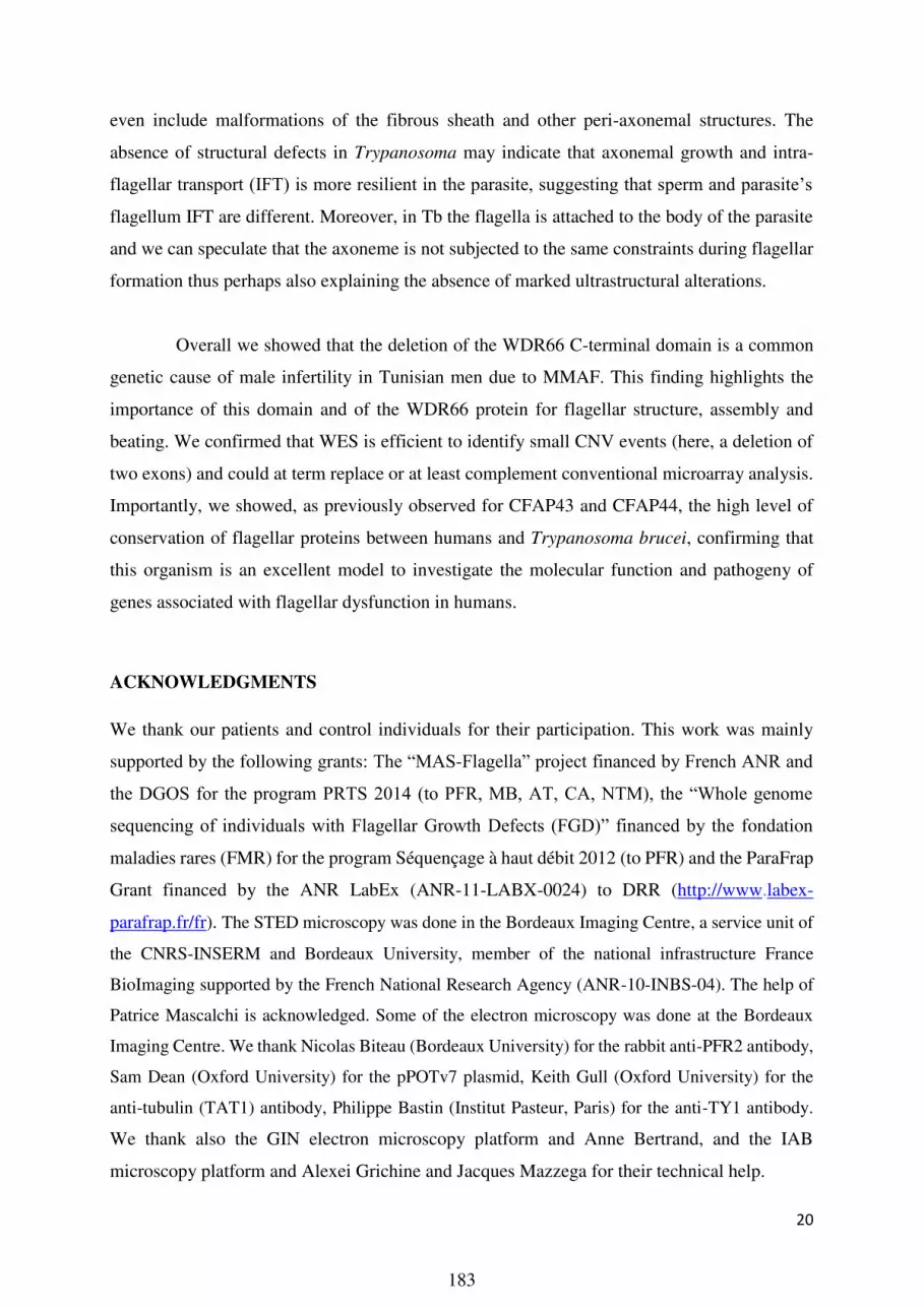
20
even include malformations of the fibrous sheath and other peri-axonemal structures. The
absence of structural defects in Trypanosoma may indicate that axonemal growth and intra-
flagellar transport (IFT) is more resilient in the parasite, suggesting that sperm and parasite’s
flagellum IFT are different. Moreover, in Tb the flagella is attached to the body of the parasite
and we can speculate that the axoneme is not subjected to the same constraints during flagellar
formation thus perhaps also explaining the absence of marked ultrastructural alterations.
Overall we showed that the deletion of the WDR66 C-terminal domain is a common
genetic cause of male infertility in Tunisian men due to MMAF. This finding highlights the
importance of this domain and of the WDR66 protein for flagellar structure, assembly and
beating. We confirmed that WES is efficient to identify small CNV events (here, a deletion of
two exons) and could at term replace or at least complement conventional microarray analysis.
Importantly, we showed, as previously observed for CFAP43 and CFAP44, the high level of
conservation of flagellar proteins between humans and Trypanosoma brucei, confirming that
this organism is an excellent model to investigate the molecular function and pathogeny of
genes associated with flagellar dysfunction in humans.
ACKNOWLEDGMENTS
We thank our patients and control individuals for their participation. This work was mainly
supported by the following grants: The “MAS-Flagella” project financed by French ANR and
the DGOS for the program PRTS 2014 (to PFR, MB, AT, CA, NTM), the “Whole genome
sequencing of individuals with Flagellar Growth Defects (FGD)” financed by the fondation
maladies rares (FMR) for the program Séquençage à haut débit 2012 (to PFR) and the ParaFrap
Grant financed by the ANR LabEx (ANR-11-LABX-0024) to DRR (http://www.labex-
parafrap.fr/fr). The STED microscopy was done in the Bordeaux Imaging Centre, a service unit of
the CNRS-INSERM and Bordeaux University, member of the national infrastructure France
BioImaging supported by the French National Research Agency (ANR-10-INBS-04). The help of
Patrice Mascalchi is acknowledged. Some of the electron microscopy was done at the Bordeaux
Imaging Centre. We thank Nicolas Biteau (Bordeaux University) for the rabbit anti-PFR2 antibody,
Sam Dean (Oxford University) for the pPOTv7 plasmid, Keith Gull (Oxford University) for the
anti-tubulin (TAT1) antibody, Philippe Bastin (Institut Pasteur, Paris) for the anti-TY1 antibody.
We thank also the GIN electron microscopy platform and Anne Bertrand, and the IAB
microscopy platform and Alexei Grichine and Jacques Mazzega for their technical help.
183

21
AUTHORS’ ROLES
Z-EK, MB, CA and PFR analyzed the data and wrote the manuscript; Z-EK, AA-Y and CC
performed molecular work; TK, NT-M, PFR analyzed the genetic data; Z-EK, MC-K, CC, CC,
GM, PLT performed IF experiments; Z-EK and SC performed protein in silico analysis, Z-EK,
GM, CC and KP-G performed EM experiments, DD, DRR, NL and MB performed
Trypanosoma work, SFBM, LH, OM, MM, HL, MK, HG, RZ provided clinical samples and
data; MB and PFR designed the study, supervised all molecular laboratory work, had full access
to all of the data in the study and take responsibility for the integrity of the data and its accuracy.
All authors contributed to the report.
CONFLICT OF INTEREST STATEMENT
The authors have declared that no conflict of interest exists.
REFERENCES
1. Neto, F.T.L., Bach, P.V., Najari, B.B., Li, P.S., and Goldstein, M. (2016). Spermatogenesis in humans and its affecting factors. Semin. Cell Dev. Biol. 59, 10–26.
2. Jan, S.Z., Vormer, T.L., Jongejan, A., Röling, M., Silber, S.J., de Rooij, D.G., Hamer, G., Repping, S., and van Pelt, A.M.M. (2017). Unraveling transcriptome dynamics in human spermatogenesis. Dev. Camb. Engl.
3. Coutton, C., Escoffier, J., Martinez, G., Arnoult, C., and Ray, P.F. (2015). Teratozoospermia: spotlight on the main genetic actors in the human. Hum. Reprod. Update 21, 455–485.
4. Ray, P.F., Toure, A., MetzlerGuillemain, C., Mitchell, M.J., Arnoult, C., and Coutton, C. (2017). Genetic abnormalities leading to qualitative defects of sperm morphology or function. Clin. Genet. 91, 217–232.
5. Ben Khelifa, M., Coutton, C., Zouari, R., Karaouzène, T., Rendu, J., Bidart, M., Yassine, S., Pierre, V., Delaroche, J., Hennebicq, S., et al. (2014). Mutations in DNAH1, which Encodes an Inner Arm Heavy Chain Dynein, Lead to Male Infertility from Multiple Morphological Abnormalities of the Sperm Flagella. Am. J. Hum. Genet. 94, 95–104.
6. Coutton, C., Vargas, A.S., AmiriYekta, A., Kherraf, Z.E., Ben Mustapha, S.F., Le Tanno, P., WambergueLegrand, C., Karaouzène, T., Martinez, G., Crouzy, S., et al. (2018). Mutations in CFAP43
184

22
and CFAP44 cause male infertility and flagellum defects in Trypanosoma and human. Nat. Commun. 9,.
7. Tang, S., Wang, X., Li, W., Yang, X., Li, Z., Liu, W., Li, C., Zhu, Z., Wang, L., Wang, J., et al. (2017). Biallelic Mutations in CFAP43 and CFAP44 Cause Male Infertility with Multiple Morphological Abnormalities of the Sperm Flagella. Am. J. Hum. Genet. 100, 854–864.
8. YanWei Sha, Xiong Wang, Xiaohui Xu, ZhiYing Su, Yuanqing Cui, LiBin Mei, XianJing Huang, Jie Chen, XueMei He, ZhiYong Ji, et al. (2017). Novel Mutations in CFAP44 and CFAP43 Cause Multiple Morphological Abnormalities of the Sperm Flagella (MMAF). Reprod. Sci. 1933719117749756.
9. Sha, Y.W., Xu, X., Mei, L.B., Li, P., Su, Z.Y., He, X.Q., and Li, L. (2017). A homozygous CEP135 mutation is associated with multiple morphological abnormalities of the sperm flagella (MMAF). Gene 633, 48–53.
10. Lorès, P., Coutton, C., El Khouri, E., Stouvenel, L., Givelet, M., Thomas, L., Rode, B., Schmitt, A., Louis, B., Sakheli, Z., et al. (2018). Homozygous missense mutation L673P in adenylate kinase 7 (AK7) leads to primary male infertility and multiple morphological anomalies of the flagella but not to primary ciliary dyskinesia. Hum. Mol. Genet.
11. Feuk, L., Carson, A.R., and Scherer, S.W. (2006). Structural variation in the human genome. Nat. Rev. Genet. 7, 85–97.
12. Iafrate, A.J., Feuk, L., Rivera, M.N., Listewnik, M.L., Donahoe, P.K., Qi, Y., Scherer, S.W., and Lee, C. (2004). Detection of largescale variation in the human genome. Nat. Genet. 36, 949–951.
13. Mills, R.E., Walter, K., Stewart, C., Handsaker, R.E., Chen, K., Alkan, C., Abyzov, A., Yoon, S.C., Ye, K., Cheetham, R.K., et al. (2011). Mapping copy number variation by populationscale genome sequencing. Nature 470, 59–65.
14. Sebat, J., Lakshmi, B., Troge, J., Alexander, J., Young, J., Lundin, P., Månér, S., Massa, H., Walker, M., Chi, M., et al. (2004). Largescale copy number polymorphism in the human genome. Science 305, 525–528.
15. Almal, S.H., and Padh, H. (2012). Implications of gene copynumber variation in health and diseases. J. Hum. Genet. 57, 6.
16. Ma, D., Yang, J., Wang, Y., Huang, X., Du, G., and Zhou, L. (2017). Whole exome sequencing identified genetic variations in Chinese hemangioblastoma patients. Am. J. Med. Genet. A. 173, 2605–2613.
17. Tsuchida, N., Nakashima, M., Kato, M., Heyman, E., Inui, T., Haginoya, K., Watanabe, S., Chiyonobu, T., Morimoto, M., Ohta, M., et al. (2017). Detection of copy number variations in epilepsy using exome data. Clin. Genet.
18. Plagnol, V., Curtis, J., Epstein, M., Mok, K.Y., Stebbings, E., Grigoriadou, S., Wood, N.W., Hambleton, S., Burns, S.O., Thrasher, A.J., et al. (2012). A robust model for read count data in exome sequencing experiments and implications for copy number variant calling. Bioinforma. Oxf. Engl. 28, 2747–2754.
185

23
19. Lee, J., Ha, J., Son, S.Y., and Han, K. (2012). Human Genomic Deletions Generated by SVAAssociated Events. Comp. Funct. Genomics 2012, 807270.
20. Jungwirth, A., Giwercman, A., Tournaye, H., Diemer, T., Kopa, Z., Dohle, G., Krausz, C., and European Association of Urology Working Group on Male Infertility (2012). European Association of Urology guidelines on Male Infertility: the 2012 update. Eur. Urol. 62, 324–332.
21. Wirtz, E., Leal, S., Ochatt, C., and Cross, G.A. (1999). A tightly regulated inducible expression system for conditional gene knockouts and dominantnegative genetics in Trypanosoma brucei. Mol. Biochem. Parasitol. 99, 89–101.
22. Brun, R., and Schönenberger, null (1979). Cultivation and in vitro cloning or procyclic culture forms of Trypanosoma brucei in a semidefined medium. Short communication. Acta Trop. 36, 289–292.
23. Schumann Burkard, G., Jutzi, P., and Roditi, I. (2011). Genomewide RNAi screens in bloodstream form trypanosomes identify drug transporters. Mol. Biochem. Parasitol. 175, 91–94.
24. Dean, S., Sunter, J., Wheeler, R.J., Hodkinson, I., Gluenz, E., and Gull, K. (2015). A toolkit enabling efficient, scalable and reproducible gene tagging in trypanosomatids. Open Biol. 5, 140197.
25. LaCount, D.J., Barrett, B., and Donelson, J.E. (2002). Trypanosoma brucei FLA1 is required for flagellum attachment and cytokinesis. J. Biol. Chem. 277, 17580–17588.
26. Oberholzer, M., Lopez, M.A., Ralston, K.S., and Hill, K.L. (2009). Approaches for functional analysis of flagellar proteins in African trypanosomes. Methods Cell Biol. 93, 21–57.
27. Bragin, E., Chatzimichali, E.A., Wright, C.F., Hurles, M.E., Firth, H.V., Bevan, A.P., and Swaminathan, G.J. (2014). DECIPHER: database for the interpretation of phenotypelinked plausibly pathogenic sequence and copynumber variation. Nucleic Acids Res. 42, D993.
28. MacDonald, J.R., Ziman, R., Yuen, R.K.C., Feuk, L., and Scherer, S.W. (2014). The Database of Genomic Variants: a curated collection of structural variation in the human genome. Nucleic Acids Res. 42, D986992.
29. Qiu, F., Xu, Y., Li, K., Li, Z., Liu, Y., DuanMu, H., Zhang, S., Li, Z., Chang, Z., Zhou, Y., et al. (2012). CNVD: text miningbased copy number variation in disease database. Hum. Mutat. 33, E23752381.
30. Schweingruber, C., Rufener, S.C., Zünd, D., Yamashita, A., and Mühlemann, O. (2013). Nonsensemediated mRNA decay — Mechanisms of substrate mRNA recognition and degradation in mammalian cells. Biochim. Biophys. Acta BBA Gene Regul. Mech. 1829, 612–623.
31. JensenSeaman, M.I., Furey, T.S., Payseur, B.A., Lu, Y., Roskin, K.M., Chen, C.F., Thomas, M.A., Haussler, D., and Jacob, H.J. (2004). Comparative recombination rates in the rat, mouse, and human genomes. Genome Res. 14, 528–538.
32. Slatkin, M., and Rannala, B. (2000). Estimating allele age. Annu. Rev. Genomics Hum. Genet. 1, 225–249.
186

24
33. Labuda, M., Labuda, D., KorabLaskowska, M., Cole, D.E., Zietkiewicz, E., Weissenbach, J., Popowska, E., Pronicka, E., Root, A.W., and Glorieux, F.H. (1996). Linkage disequilibrium analysis in young populations: pseudovitamin Ddeficiency rickets and the founder effect in French Canadians. Am. J. Hum. Genet. 59, 633–643.
34. Risch, N., de Leon, D., Ozelius, L., Kramer, P., Almasy, L., Singer, B., Fahn, S., Breakefield, X., and Bressman, S. (1995). Genetic analysis of idiopathic torsion dystonia in Ashkenazi Jews and their recent descent from a small founder population. Nat. Genet. 9, 152–159.
35. Vincensini, L., Blisnick, T., and Bastin, P. (2011). 1001 model organisms to study cilia and flagella. Biol. Cell 103, 109–130.
36. Aslett, M., Aurrecoechea, C., Berriman, M., Brestelli, J., Brunk, B.P., Carrington, M., Depledge, D.P., Fischer, S., Gajria, B., Gao, X., et al. (2010). TriTrypDB: a functional genomic resource for the Trypanosomatidae. Nucleic Acids Res. 38, D457462.
37. Sievers, F., Wilm, A., Dineen, D., Gibson, T.J., Karplus, K., Li, W., Lopez, R., McWilliam, H., Remmert, M., Söding, J., et al. (2011). Fast, scalable generation of highquality protein multiple sequence alignments using Clustal Omega. Mol. Syst. Biol. 7, 539.
38. Broadhead, R., Dawe, H.R., Farr, H., Griffiths, S., Hart, S.R., Portman, N., Shaw, M.K., Ginger, M.L., Gaskell, S.J., McKean, P.G., et al. (2006). Flagellar motility is required for the viability of the bloodstream trypanosome. Nature 440, 224–227.
39. Subota, I., Julkowska, D., Vincensini, L., Reeg, N., Buisson, J., Blisnick, T., Huet, D., Perrot, S., SantiRocca, J., Duchateau, M., et al. (2014). Proteomic analysis of intact flagella of procyclic Trypanosoma brucei cells identifies novel flagellar proteins with unique sublocalization and dynamics. Mol. Cell. Proteomics MCP 13, 1769–1786.
40. Urbanska, P., Song, K., Joachimiak, E., KrzemienOjak, L., Koprowski, P., Hennessey, T., JerkaDziadosz, M., Fabczak, H., Gaertig, J., Nicastro, D., et al. (2015). The CSC proteins FAP61 and FAP251 build the basal substructures of radial spoke 3 in cilia. Mol. Biol. Cell 26, 1463–1475.
41. Bastin, P., Bagherzadeh, Z., Matthews, K.R., and Gull, K. (1996). A novel epitope tag system to study protein targeting and organelle biogenesis in Trypanosoma brucei. Mol. Biochem. Parasitol. 77, 235–239.
42. Cordaux, R., and Batzer, M.A. (2009). The impact of retrotransposons on human genome evolution. Nat. Rev. Genet. 10, 691–703.
43. Raiz, J., Damert, A., Chira, S., Held, U., Klawitter, S., Hamdorf, M., Löwer, J., Strätling, W.H., Löwer, R., and Schumann, G.G. (2012). The nonautonomous retrotransposon SVA is transmobilized by the human LINE1 protein machinery. Nucleic Acids Res. 40, 1666–1683.
44. Hancks, D.C., and Kazazian, H.H. (2016). Roles for retrotransposon insertions in human disease. Mob. DNA 7, 9.
187

25
45. Brett, M., Korovesis, G., Lai, A.H.M., Lim, E.C.P., and Tan, E.C. (2017). Intragenic multiexon deletion in the FBN1 gene in a child with mildly dilated aortic sinus: a retrotransposal event. J. Hum. Genet. 62, 711–715.
46. Legoix, P., Sarkissian, H.D., Cazes, L., Giraud, S., Sor, F., Rouleau, G.A., Lenoir, G., Thomas, G., and ZucmanRossi, J. (2000). Molecular characterization of germline NF2 gene rearrangements. Genomics 65, 62–66.
47. Nakamura, Y., Murata, M., Takagi, Y., Kozuka, T., Nakata, Y., Hasebe, R., Takagi, A., Kitazawa, J., Shima, M., and Kojima, T. (2015). SVA retrotransposition in exon 6 of the coagulation factor IX gene causing severe hemophilia B. Int. J. Hematol. 102, 134–139.
48. Takasu, M., Hayashi, R., Maruya, E., Ota, M., Imura, K., Kougo, K., Kobayashi, C., Saji, H., Ishikawa, Y., Asai, T., et al. (2007). Deletion of entire HLAA gene accompanied by an insertion of a retrotransposon. Tissue Antigens 70, 144–150.
49. Vogt, J., Bengesser, K., Claes, K.B.M., Wimmer, K., Mautner, V.F., van Minkelen, R., Legius, E., Brems, H., Upadhyaya, M., Högel, J., et al. (2014). SVA retrotransposon insertionassociated deletion represents a novel mutational mechanism underlying large genomic copy number changes with nonrecurrent breakpoints. Genome Biol. 15, R80.
50. Xiong, H., Wang, S., Kobayashi, K., Jiang, Y., Wang, J., Chang, X., Yuan, Y., Liu, J., Toda, T., Fukuyama, Y., et al. (2009). Fukutin gene retrotransposal insertion in a nonJapanese Fukuyama congenital muscular dystrophy (FCMD) patient. Am. J. Med. Genet. A. 149A, 2403–2408.
51. Ishikawa, T. (2017). Axoneme Structure from Motile Cilia. Cold Spring Harb. Perspect. Biol. 9, a028076.
52. Lindemann, C.B., and Lesich, K.A. (2016). Functional anatomy of the mammalian sperm flagellum. Cytoskelet. Hoboken NJ 73, 652–669.
53. Heuser, T., Dymek, E.E., Lin, J., Smith, E.F., and Nicastro, D. (2012). The CSC connects three major axonemal complexes involved in dynein regulation. Mol. Biol. Cell 23, 3143–3155.
188

26
Table 1. Semen parameters of the 7 deleted MMAF subjects.
Patient Identification P1 P2 P3 P4 P5 P6 P7 Mean ± SD Lower Reference Limits (WHO 2010)
Geographical origin Tunisia Tunisia Tunisia Tunisia Tunisia Tunisia Tunisia Consanguinity Yes Yes Yes Yes No Yes Yes
Age 45 54 51 45 31 28 59 44.7 ± 10.6 (n’=7)
Sperm volume (mL) 4 1.8 1.7 2.2 3.4 3 5 3.0 ± 1.1 (n’=7) 1.5
Sperm concentration (million/mL) 11.3 6 56 56 60 7,8 15.5 30.3 ± 23.5
(n’=7) 15
Total motility 1 h (%) 2 1 5 0 5 5 4 3.1 ± 1.9 (n’=7) 40
Vitality (%) 53 80 73 79 62 NC 47 65.6 ± 12.6 (n’=6) 58
Normal spermatozoa (%) 0 0 0 0 0 0 0 0 (n’=7) 4
Abnormal acrosome 66 56 56 78 80 NC 95 71.8 ± 15.3 (n’=6)
Abnormal head morphology 42 10 20 58 8 NC 7 24.1 ± 25.1 (n’=6)
Abnormal base 36 2 32 14 2 NC 3 14.8 ± 15.6 (n’=6)
Absent flagella (%) 31 6 22 50 24 NC 30.5 27.2 ± 13.1 (n’=6)
Short flagella (%) 34 32 59 32 50 NC 25 38.6 ± 11.8 (n’=6)
Coiled flagella (%) 20 17 27 34 8 NC 27 21.1 ± 8.3 (n’=6)
Flagella of irregular caliber (%) 4 68 37 50 68 NC 33 43.3 ± 22.1 (n'=6)
Multiple anomalies index 2.5 2.3 2.7 3.4 2.5 NC 2.56 2.6 ± 0.4 (n’=6)
189

27
LEGENDS TO FIGURES
Figure 1: Defective sperm flagellar formation in WDR66 mutated individuals.
(A-D) Light microscopy analysis of control (A) and individual’s sperm (B-D) reveals multiple
morphological abnormalities of the sperm flagella in individuals. (E-F) TEM micrographs of
cross-sections through sperm cell midpieces. (E) Control sperm shows a homogeneous ring-
like mitochondrial sheath (MS) surrounding 9 outer dense fibers (ODF, arrowheads) and the
central axoneme characterized by the typical presence of 9 microtubule doublets (MD, arrows)
and the two central microtubules singlets (MS). (F) Representative sperm from an affected men
showing a partitioned MS abnormally positioned and assembled ODF and MD and the absence
of the MS. Scale bars, 10 µm (A–D) and 200 nm (E–F).
Figure 2: Confirmation of the presence of the WDR66 deletion in patients and validation
of its effect on mRNA and protein.
(A) Deleted individuals (P1 and P2) show a complete absence of sequence coverage of exons
20 and 21 as illustrated by IGV as opposed to controls (C1)
(B) In human, WDR66 encodes for two transcripts: T1 (ENST00000288912.8, NM_144668),
with 22 exons coding for a 1149 amino acid protein and T2 (ENST00000397454.2,
NM_001178003) with 18 exons coding for a 941 amino acid protein. Only T1 is affected by
the identified deletion (black double arrow).
(C) Relative expression (2-∆∆Ct) of both transcripts (T1 and T2) performed on human panel of
cDNAs from two tissues expressing 9+2 secondary motile axonemes (testis and lung) and two
tissues expressing 9+0 primary immotile axonemes (kidney and lung). GAPDH was used as
reference gene and T2 expression level in brain as reference sample. Data are expressed as
mean ± SD.
(D) Representation of WDR66 protein encoded by T1. We note the presence of nine WD40
repeat domains and one calcium-binding EF-hand (α) in the C-terminus extremity. The first
deleted amino acid (aa) is aa1003. Deletion of exons 20 and 21 induce a frameshift. Translation
of exon 22 is then incorrect for the first 25 aa (orange line) before inducing a stop codon 26
nucleotides after the first abnormal aa. The protein encoded by the deleted T1 is therefore
expected to stop at aa1028.
(E) Double immunofluorescence labelling of WDR66 and α-tubulin in human sperm cells: In
control sperm, the yellow stain resulted from merged signals indicates the co-localization of
both proteins within the flagellar axoneme. The images shown are representative of many cells
examined in two independent experiments. Scale Bar: 10 µm.
Figure 3: Fine characterization of the deletion and theoretical description of the initial
deletion event
(A) Representation in control subjects of part of the WDR66 gene (between exon 19 and 22)
and localization of the deletion-specific primers F1 and R1.
190

28
(B) Representation of the same region in deleted subjects. Detailed analysis of the deleted
region within WDR66 highlights the presence of a type F SINE-VNTR-Alu (SVA)
retrotransposon (SVA-F) positioned at the centromeric border of the identified WDR66 deletion
(orange box).
(C) PCR of the deleted region. In controls (C1-C3), F1 and F2 primers are too distant (8541
bp) and do not yield any amplification. PCR amplification with F1 and F2 of patients (P1-P7)
permits the amplification of a fragment of 950nt.
(D) Sanger sequencing through the deletion breakpoints permits to localize the exact
position of the breakpoints, indicating that a total of 7591nt were deleted. It also highlights the
presence of a 165nt inserted sequence (yellow) at the junction point.
(E) Blast analysis of the 165nt inserted sequence shows that an identical sequence (100%
identity) is present within an SVA-D sequence located 400kb telomeric of SVA-F between
exons 6 and 5 of the CLIP1 gene (yellow box).
(F) SVA-D was likely retrotransposed between WDR66 exons 21 and 22
(G) An intra-chromatidien non-allelic homologous recombination (NAHR) was then
initiated
(H) Inducing the formation of a hybrid SVA-F/SVA-D sequence preceding the WDR66
exons 20 and 21 deletions.
Figure 5: TbWDR66 is an axoneme protein and its C-terminal domain is required for
function in BSF trypanosome.
(A) Control BSF cells (WT) or expressing TbWDR66TY1 or TbWDR66-ΔCterTY1 were
immunolabelled with anti-PFR (white) and anti-TY1 (red) showing that anti-TY1 decorates the
tagged proteins throughout the full-length of the flagellum. Induction of RNAi for 48 h
(+RNAi) showed a decrease of fluorescence intensity of TbWDR66TY1 labelling but not of
TbWDR66-ΔCterTY1 labelling. Nuclei and kinetoplasts (mitochondrial genomes) were labelled
with DAPI (blue). Scale bar represents 5 μm. (B) Western-blot analysis of the expression of
TbWDR66TY1 and TbWDR66-ΔCterTY1 without (NI) and with 24 h, 48 h, 72 h of RNAi
induction shows a decrease of TbWDR66TY1 (indicated by the % of labelling relative to the
non-induced condition) expression, whilst expression of TbWDR66-ΔCterTY1 is not affected.
Tubulin labeling was used as loading control. (C) TbWDR66 RNAi affects cell proliferation.
Growth curves for WT cells, and RNAi non-induced or induced cells expressing TbWDR66TY1
or TbWDR66-ΔCterTY1. Cells were counted every 24 h. The graph represents the cumulative
number of cells per ml. Error bars represent the standard error from 3 independent experiments.
(D) Electron microscopy images of thin sections of a WT cell (a), and a cell expressing
TbWDR66TY1 (b) or TbWDR66-ΔCterTY1 (c) and RNAi-induced for 48 h show that the decrease
TbWDR66 expression induces a severe disorganization of the axoneme and of the paraxonemal
structure, which is not rescued by the expression of TbWDR66-ΔCterTY1. Scale bars represent
50 nm.
LEGENDS OF SUPPLEMENTARY MATERIAL
Figure S1. Schematic representation of the RNAi and endogenous tagging strategies.
191

29
A. One allele out of two is replaced by a PCR product allowing the insertion of the TY1 tag and a
selection gene. The RNAi will target both alleles, and upon induction, both WT and TY1-tagged protein
will be knocked down. B. One allele out of two is replaced by a PCR product allowing the shortening
of TbWDR66 ORF, the insertion of the C-terminal TY1 tag and a selection gene. The RNAi targets only
the WT allele, and as a consequence, only the WT protein will be knocked down, whilst TbWDR66-
ΔCterTY1 expression is not affected.
Figure S2: Array-CGH profile of chromosome 12 showing a large homozygous deletion of WDR66
gene in the patient P1.
(A) Each spot represents an oligonucleotide on the array. Spots are graphically represented according to
their corresponding position along the chromosome. Values indicated on the top are expressed as log2
ratios of fluorescence intensity. We observe that three spots are deviated and have ratios between -2.358
and -4.621, indicating that the patient has a homozygous deletion in the WDR66 gene.
Figure S3: gel electrophoresis illustrating the duplex PCR used to screen 100 North-African
individuals for WDR66 deletion
The primer used permit to amplify a 220 nucleotide sequence located within the WDR66 deletion hence
present only in non-deleted alleles and a 950 nucleotide sequence encompassing the deletion breakpoint
hence only present in the deleted alleles. In lane 1 (-/-) the amplified DNA was extracted from a non-
deleted control, in lane 2 (+/+) from the homozygous deleted patient 1. Lane 3 (+/-) was obtained from
mixing deleted and non-deleted DNAs. S1-S4 corresponds to the first 4 tested North African subjects.
MW= molecular weight.
Figures S4: Alignment of the SVA-F/D sequence encompassing the centromeric breakpoint in
patients with the SVA-F and SVA-D sequences in WDR66 (intron 19) and CLIP1 (intron 5)
respectively.
Alignment was performed using CLC sequence viewer. D indicates the variants identical between the
patients and the CLIP1 SVA-D sequence, whereas F indicates the variants identical between the patient
and the WDR66 SVA-F sequence. Results show that in patients, the telomeric part of the recombined
SVA is homologous to the CLIP1 SVA-D (yellow line) whereas the centromeric SVA sequence
corresponds to the originalWDR66 SVA-F intron 19 (grey line). The 15 nucleotides indicated between
the D and the F sequence (double black arrow) corresponds to the minimal region localizing the
centromeric breakpoint. The telomeric breakpoint is indicated by a black vertical arrow. In deleted men,
the sequence following the telomeric breakpoint corresponds to WDR66 intron 21.
Figure S5. Secondary structure prediction of human WDR66 and Trypanosoma TbWDR66.
Elements of the secondary structures of WDR66 and tbWDR66 were predicted by Porter 4.0. C, E and
H correspond to coils, extended conformations (β-sheets) and helices (α-helices), respectively.
Figure S6. TbWDR66TY1 and TbWDR66-ΔCterTY1 are axoneme proteins in PCF Trypanosoma. A.
Epifluorescence analysis of immunolabeled PCF cells expressing TbWDR66TY1 (a-d) and TbWDR66-
ΔCterTY1 (e-h) using anti-PFR2 (grey) that labels the paraflagellar rod and anti-TY1 (red) antibodies.
Scale bar represents 5 μm. B. Deconvolved images of STED confocal microscopy experiments on PCF
cells expressing TbWDR66TY1 (a-d) or TbWDR66-ΔCterTY1 (e-h) and immuno-labeled with anti-PFR2
(grey), anti-TY1 (red), and anti-tubulin (green) that labels the axonemal microtubules as well as the
subpellicular microtubules (delimitated by the dotted white lines). Scale bar represents 0.5 μm.
192

30
Video S1. Illustration of mobility of WT cells showing active flagellar beating.
Video S2. Illustration of mobility of cells expressing TbWDR66TY1 and RNAi-induced for 48 h.
Flagellar beating is slower compared to WT cells, and the cells remain primarily in one location
demonstrating a flagellum motility defect.
Video S3. Illustration of mobility of cells expressing TbWDR66-ΔCterTY1 and RNAi-induced for
48 h.
Flagellar beating is slower compared to WT cells, and the cells remain primarily in one location
demonstrating a flagellum motility defect demonstrating that TbWDR66-ΔCterTY1 cannot complement
for the down-regulation of the full-length TbWDR66 protein.
193

Figure 1194

B
D
Figure 2
C
P2
C1
AP1
Exon 20Exon 21
T1
T2
E
N C
Deletion
195

E
Figure 3
A
F
G
H
DC
B
196

NI 24h 48h120 -
100 -
50 -
60 -
TbWDR66TY1
100 48 28%
72h
31
TY1
tubulin
kDa
120 -100 -
NI 24h 48h
50 -
60 -
TY1
tubulin
TbWDR66-∆CterTY1
100 92 135%
72h
131
kDa
Cu
mu
lati
ve c
ell n
um
ber
(/m
L)
105
106
107
108
109
1010
Time of induction (hours)
1011
TbWDR66TY1RNAi non induced
TbWDR66TY1RNAi induced
TbWDR66-ΔCterTY1RNAi non induced
TbWDR66-ΔCterTY1RNAi induced
WT
A
B C
D
TbWDR66TY1
TbWDR66-ΔCterTY1
- RNAi
+ RNAi
WT
- RNAi
+ RNAi
α-PFR α-TY1 Merge Phase contrast
Figure 4
a b c
197

5’UTR 3’UTR TbWDR66-ΔCter
TY1 tag
RNAi target
TbWDR66 5’UTR
TY1 tag 3’UTR
TbWDR66 TY1 tag
aa 906 aa 1027
TbWDR66-ΔCter TY1 tag
aa 906
Cell line expressing TbWDR66TY1
Cell line expressing TbWDR66-ΔCterTY1
TbWDR66 5’UTR 3’UTR
TbWDR66
aa 906 aa 1027
TbWDR66
TbWDR66
aa 906 aa 1027
A�
RNAi induction
DN
A
Pro
tein
RNAi induction
DN
A
Pro
tein
B�
selection
gene
selection
gene
5’UTR 3’UTR
Figure S1198

-4 -3 -2 -1 0 1 2 3 4
122477 M
b 122450 M
b122424 M
b122 397 M
b
Figure S2199

MW del-/- del+/+ del+/- S1 S2 S3 S4 Blank
1 Kb
200 bp
Figure S3200

D D
F
DD
DD D
F FF F
F
DD
Figure S4201

Figure S5202

TY1 � PFR�
TY1 � PFR�
TbWDR66�
TbWDR66-ΔCter�
Tubulin � PFR� Tubulin� TY1� PFR�
Tubulin � PFR � Tubulin� TY1 � PFR�
a � b� c� d�
e � f � g � h�
TbWDR66TY1�
a� b� c � d�
TbWDR66-ΔCterTY1 �
e � f � g � h �
PFR �
PFR �
PFR � TbWDR66TY1�
PFR � TbWDR66-ΔCterTY1�
A�
B�
Figure S6203

Table S1. List of primers used for multiplex PCR to amplify differentially wild-type and
mutated allele and for deleted allele sequencing.
Primer name Primer sequence (5’-3’) Tm (°C) Product length (bp) WDR66-Ex20F AGCTTTGAGGTGCTCGGTTA 59 220
(in controls) WDR66-Int20R CCTCATGGCTCAAATCTGGT 58
WDR66-Int19F (F1) GAATAGAAAGGCGGGAAAGG 56 950 (in mutated patients) WDR66-Int22R (R1) AACTCTGGGTGGAAATTGCTTA 58
Table S2. List of primers used for qRT-PCR experiment on human sperm extracts.
Transcripts’ names are listed in Ensembl database (assembly GRCh37/hg19). Their respective
transcript ID is ENST00000288912.4 (T1) and ENST00000397454.2 (T2).
Primer name Primer sequence (5’-3’) Tm (°C) Product length (bp) GAPDH-F ACCCACTCCTCCACCTTTGA 60
101 GAPDH-R CTGTTGCTGTAGCCAAATTCGT 60
WDR66-T1F AGCTTTGAGGTGCTCGGTTA 60 132
WDR66-T1R CAGTGAAGCAAAGCAATCCA 60
WDR66-T2F GGTGCAGTGGAAAATCACCT 60 139
WDR66-T2R CTGTGAATCCCAGCTCCACT 60
Table S3. Antibodies used for immunofluorescence experiments on human sperm cells.
Primary antibodies Source Type Dilution Reference WDR66 Rabbit Polyclonal 1/20 Sigma-Aldrich- HPA040005
α-tubulin Mouse Monoclonal 1/200 Sigma-Aldrich-T7451
Table S4. Endogenous tagging and RNAi primer sequences for T. brucei experiments.
TbWDR66 RNAi
1036 5’-GCCGGCCGCTCTAGAtgtgatgtgagcagcattccg-3’
1037 5’-TAAGCTTGCTCTAGAtttcctcttccccaccgtca-3’
TbWDR66TY1
endogenous tagging
1038
5’-CGGAAAATCTTCTTGGATTTGAAGATTATGATGCTATGGCCCAGGGTGATGACGGTGGGGAAGAGGAAATGAGTTTGCAGggttctggtagtggttcc-3’
1039
5’-GAGAGAAAACGAGCATTCCACTGATGGTTTGGTACCTTTAGTCTCCAAATTCACCTGCCCATAACTCTCTGTTCCCTCACccaatttgagagacctgtgc-3’
TbWDR66-
ΔCterTY1
endogenous tagging
1039
5’-GAGAGAAAACGAGCATTCCACTGATGGTTTGGTACCTTTAGTCTCCAAATTCACCTGCCCATAACTCTCTGTTCCCTCACccaatttgagagacctgtgc-3’
1040
5’-CGGAAAATCTTCTTGGATTTGAAGATTATGATGCTATGGCCCAGGGTGATGACGGTGGGGAAGAGGAAATGAGTTTGCAGggttctggtagtggttcc-3’
204

Table S5. List of primary and secondary antibodies used in immunofluorescence
experiments in Trypanosoma.
Antibodies References Species Protein localization Dilution
TAT1 (tubulin) Described in (Woods et al., 1989)
Mouse IgG2a Cytoskeleton/flagellum 1/50
PFR2 Gift from Dr N. Biteau* Rabbit PFR 1/400
BB2 (TY1) Described in (Bastin et al., 1996)
Mouse IgG1 TY1-tagged protein 1/500
Anti-mouse IgG2a Alexa-488
Invitrogen A21131 Goat 1/100
Anti-mouse IgG1 Alexa-594
Invitrogen A21125 Goat 1/100
Anti-rabbit ATTO-N647
Sigma 40839 Goat 1/100
Bastin, P., Bagherzadeh, Z., Matthews, K.R., and Gull, K. (1996). A novel epitope tag system to study protein targeting and organelle biogenesis in Trypanosoma brucei. Mol. Biochem. Parasitol. 77, 235–239. Woods, A., Sherwin, T., Sasse, R., MacRae, T.H., Baines, A.J., and Gull, K. (1989). Definition of individual components within the cytoskeleton of Trypanosoma brucei by a library of monoclonal antibodies. J. Cell Sci. 93, 491–500. * MFP, University of Bordeaux, CNRS UMR 5234
Table S6. List of all chromosome 12 SNVs obtained by WES of all deleted subjects
The smallest common region of homozygosity was establish by taking into account the SNPs
common for at least 6 of the 7 analysed patients (P1-P7) that are different from the control
individual (C). The region taken into consideration is surrounded by a black box. The extend
of the WDR66 gene is highlighted in yellow.
See the excel file “Table S6”
Table S7. Protein folds found in the 2 target sequences WD40 folds and other β-domains are found in the two proteins by program Superfamily with very good confidence (e-value corresponds to the probability of finding such folds by chance in the sequence). Both proteins also share α-EF-Hand domains in their C-Terminal region.
Protein Family Domains (aa number) E-value
WDR66
Quinoprotein alcohol dehydrogenase-like (β)
249-407, 460-486, 601-774 1.26e-33
WD40 repeat-like (β) 691-922 1.83e-16
EF-hand (α) 980-1099 3.33e-7
TbWDR66
WD40 repeat-like (β) 218-372, 458-630 2.75e-40
YVTN repeat-like/Quinoprotein amine dehydrogenase (β)
20-94, 148-319 1.65e-8
WD40 repeat-like (β) 591-631, 699-796 6.59e-6
EF-hand (α) 863-981 2.24e-2
205

Table S8. Average semen parameters in the different genotype groups for the 78 MMAF
subjects.
Semen parameters
MMAF WDR66 patients n=7
MMAF CFAP43 patients n=10
MMAF CFAP44 patients n=6
MMAF DNAH1 patients n=6
MMAF all
mutations n=29
MMAF with
unknown causes n=49
Overall MMAF patients n=78
Mean age (years)
44.7 ± 10.6 (n’=7)
37.7 ± 9.6 (n’=9)
41.3 ± 4.3 (n’=6)
41.5 ± 4.4 (n’=6)
41.0 ± 8.4 (n’=28)
41.9 ± 7.3 (n’=49)
41.6 ± 7.7 (n’=77)
Sperm volume (ml)
3.0 ± 1.1 (n’=7)
3.5 ± 1.4 (n’=8)
3.2 ± 0.87 (n’=6)
3.5 ± 1.2 (n’=6)
3.3 ± 1.1 (n’=28)
3.6 ± 1.5 (n’=48)
3.5 ± 1.4 (n’=75)
Sperm concentration
(106/ml)
30.3 ± 23.5 (n’=7)
27.2 ± 23.4 (n’=8)
7.9 ± 8.4 (n’=6)
22.9 ± 15.2 (n’=6)
22.3 ± 20.4 (n’= 28)
27.2 ± 37.2 (n’=48)
25.6 ± 32.1 (n’=75)
Motility (a+b+c) 1 h
3.1 ± 1.9 (n’=7)
0 ± 0 (n’=9)
0 ± 0 (n’=6)
2.6 ± 4.2 (n’=6)
1.3 ± 2.5 (n=29)
5.3 ± 6.4 (n’=48)
3.9 ± 5.6 (n’=76)
Vitality 65.6 ± 12.6 (n’=6)
55.5 ± 24 (n’=8)
43.3 ± 22.6 (n’=6)
51.2 ± 23.1 (n’=5)
54.5 ± 21.2 (n’=26)
51.9 ± 20.2 (n’=47)
52.7 ± 20 (n’=72)
Normal spermatozoa
0 (n’=7)
1.25 ± 3.5 (n’=8)
0 ± 0 (n’=6)
0 ± 0 (n’=6)
0.4 ± 1.9 (n’=27)
2.3 ± 4.3 (n’=48)
1.6 ± 2.7 (n’=61)
Abnormal acrosome
71.8 ± 15.3 (n’=6)
41.1 ± 29.8 (n’=6)
85.2 ± 7.9 (n’=5)
47.0 ± 31.3 (n’=5)
60.8 ± 28.3 (n’=22)
63.6 ± 25.8 (n’=44)
62.6 ± 26.5 (n’=66)
Abnormal head
morphology
24.1 ± 25.1 (n’=6)
8.0 ± 3.5 (n’=5)
54.6 ± 27.8 (n’=5)
45.0 ± 55.6 (n’=4)
31.9 ± 33.7 (n’=20)
32.3 ± 21.7 (n’=43)
32.2 ± 25.8 (n’=63)
Abnormal base
14.8 ± 15.6 (n’=6)
22.2 ± 14.5 (n’=5)
54.0 ± 29.7 (n’=5)
26.5 ± 22.3 (n’=4)
28.8 ± 24.8 (n’=20)
32.4 ± 19.2 (n’=42)
31.2 ± 21.0 (n’=62)
Absent flagella
27.2 ± 13.1 (n’=6)
21.8 ± 17.6 (n’=5)
36.8 ± 4.1 (n’=5)
25.6 ± 15.9 (n’=5)
26.9 ± 14.3 (n’=22)
17.3 ± 15.5 (n’=45)
20.7 ± 15.7 (n’=66)
Short Flagella 38.6 ± 11.8 (n’=6)
65.3 ± 31.7 (n’=8)
52.2 ± 27 (n’=6)
49.8 ± 24.3 (n’=5)
53.3 ± 25.2 (n’=26)
38.6 ± 27.0 (n’=47)
43.7 ± 27.3 (n’=72)
Coiled Flagella
21.1 ± 8.3 (n’=6)
8.2 ± 6 (n’=6)
14.4 ± 7 (n’=5)
9 ± 6.3 (n’=5)
12.8 ± 9.0 (n’=23)
13.0 ± 9.7 (n’=47)
12.8 ± 9.4 (n’=69)
Flagella of irregular caliber
43.3 ± 22.1 (n'=6)
20.2 ± 19.3 (n’=5)
28.4 ± 16.9 (n’=5)
35 ± 22.7 (n’=5)
32.0 ± 21.8 (n’=22)
31.3 ± 26.6 (n’=46)
31.7 ± 25.1 (n’=67)
Multiple anomalies index
2.6 ± 0.4 (n’=6)
2.3 ± 0.2 (n’=4)
3.4 ± 0.4 (n’=5)
2.3 ± 1.4 (n’=5)
2.6 ± 0.8 (n’=20)
2.7 ± 0.6 (n’=41)
2.7 ± 0.7 (n’=61)
Values are expressed in percent, unless specified otherwise.Values are mean ± SD; n= total number of patients in each group; n’= number of patients used to calculate the average based on available data. We compared statistical differences between MAAF due to WDR66, CFAP43, CFAP44 and DNAH1 mutations versus MAAF due to uncharacterized genetic cause. No significant statistical difference was observed between the two groups.
206

207
2- Article 4 : Mutations in CFAP43 and CFAP44 cause male infertility
and flagellum defects in Trypanosoma and human
Charles Coutton, Alexandra S. Vargas 1, Amir Amiri-Yekta , Zine-Eddine Kherraf,
Selima Fourati Ben Mustapha, Pauline Le Tanno, Clémentine Wambergue-Legrand,
Thomas Karaouzène, Guillaume Martinez, Serge Crouzy, Abbas Daneshipour,
Seyedeh Hanieh Hosseini, Valérie Mitchell, Lazhar Halouani, Ouafi Marrakchi,
Mounir Makni, Habib Latrous, Mahmoud Kharouf, Jean-François Deleuze, Anne
Boland, Sylviane Hennebicq, Véronique Satre, Pierre-Simon Jouk, Nicolas Thierry-
Mieg, Beatrice Conne, Denis Dacheux, Nicolas Landrein, Alain Schmitt, Laurence
Stouvenel, Patrick Lorès, Elma El Khouri, Serge P. Bottari, Julien Fauré, Jean-
Philippe Wolf, Karin Pernet-Gallay, Jessica Escoffier, Hamid Gourabi, Derrick R.
Robinson, Serge Nef, Emmanuel Dulioust, Raoudha Zouari, Mélanie Bonhivers,
Aminata Touré, Christophe Arnoult & Pierre F. Ray.
Nature Communications. Février 2018.

208
Contexte et principaux résultats de l’étude :
La première exploration génétique du phénotype MMAF chez des cas familiaux,
d’abord par cartographie d’homozygotie puis par séquençage exomique, nous a permis
d’identifier DNAH1 comme premier gène impliqué dans ce phénotype. Cependant, les
mutations identifiées étaient variées et la majorité des individus testés par séquençage
exomique est restée sans diagnostic génétique. Ceci suggère fortement que d’autres gènes sont
impliqués dans ce phénotype. Nous avons donc réalisé une étude par séquençage exomique
chez une cohorte de 78 sujets MMAF non apparentés et ayant consulté pour infertilité
primaire isolée. Ces patients ont été recrutés à partir de centres de procréation médicalement
assistée (PMA) en France, Iran et Tunisie. A cette date, seul le gène DNAH1 était décrit
somme étant associé au phénotype MMAF. Du fait de la taille importante de DNAH1, les 78
exons de ce gène n’ont pas été séquencés par la technique de Sanger avant la réalisation du
séquençage exomique pour cette nouvelle cohorte. L’analyse des données de séquençage
exomique nous a permis d’identifier des mutations homozygotes du gène DNAH1 chez six
patients (8%). D’autres mutations homozygotes délétères ont été identifiées chez 20% des
sujets analysés dans deux gènes codant pour des protéines associées aux cils et aux flagelles :
CFAP43 (n=10) et CFAP44 (n=6). Ces protéines font partie de la famille des protéines WDR
(WD40-repeats) qui sont impliquées dans de nombreuses fonctions cellulaires mais dont le
rôle dans l’assemblage et la fonction du flagelle spermatique reste inconnu.
Nous avons pu produire des souris knock-out pour ces deux gènes en utilisant la
nouvelle technologie d’édition du génome, CRISPR/Cas9. Nous avons montré que les souris
mâles adultes sont infertiles et présentent une asthénozoospermie sévère associée à des
anomalies morphologiques flagellaires multiples. Nous avons également pu réaliser des
études fonctionnelles chez le protiste Trypanosoma brucei dont le flagelle présente une forte
homologie structurale avec le flagelle spermatique chez l’homme et nous avons observé après
atténuation de l’activité des gènes orthologues, une altération de la mobilité flagellaire. Nous
avons également déterminé grâce au STED (Immuno-electron and stimulated-emission-
depletin) la localisation flagellaire des protéines codées par ces gènes chez Trypanosoma
entre les microtubules périphériques de l’axonème et le PFR (paraflagellar rod).
Ce travail a donc permis d’identifier deux nouveaux gènes associés au phénotype
MMAF. Par ailleurs, l’étude de ces deux modèles animaux nous a permis de valider ces
résultats et de suggérer que les protéines CFAP43 et CFAP44 ont probablement une fonction

209
structurale inédite consistant à relier l’axonème aux structures péri-axonémales. Des études
plus approfondies sont nécessaires chez le mammifère afin d’identifier la localisation exacte
de ces protéines et les structures axonémales et péri-axonémales avec lesquelles elles
interagissent.

ARTICLE
Mutations in CFAP43 and CFAP44 cause maleinfertility and flagellum defects in Trypanosoma andhumanCharles Coutton et al.#
Spermatogenesis defects concern millions of men worldwide, yet the vast majority remains
undiagnosed. Here we report men with primary infertility due to multiple morphological
abnormalities of the sperm flagella with severe disorganization of the sperm axoneme, a
microtubule-based structure highly conserved throughout evolution. Whole-exome sequen-
cing was performed on 78 patients allowing the identification of 22 men with bi-alle-
lic mutations in DNAH1 (n= 6), CFAP43 (n= 10), and CFAP44 (n= 6). CRISPR/Cas9 created
homozygous CFAP43/44 male mice that were infertile and presented severe flagellar defects
confirming the human genetic results. Immunoelectron and stimulated-emission-depletion
microscopy performed on CFAP43 and CFAP44 orthologs in Trypanosoma brucei evidenced
that both proteins are located between the doublet microtubules 5 and 6 and the paraflagellar
rod. Overall, we demonstrate that CFAP43 and CFAP44 have a similar structure with a
unique axonemal localization and are necessary to produce functional flagella in species
ranging from Trypanosoma to human.
DOI: 10.1038/s41467-017-02792-7 OPEN
Correspondence and requests for materials should be addressed to P.F.R. (email: [email protected])#A full list of authors and their affliations appears at the end of the paper.
NATURE COMMUNICATIONS | �(2018)�9:686� |DOI: 10.1038/s41467-017-02792-7 |www.nature.com/naturecommunications 1
1234567890():,;
210

Medical treatment of infertility has rapidly evolved overthe past four decades, but much remains to beaccomplished1. Despite recent success in identifying
infertility genes2–4, most genetic causes of male infertility arecurrently uncharacterized and additional efforts should be pur-sued to better characterize male infertility. We demonstratedpreviously that mutations in the DNAH1 gene are responsible formultiple morphological abnormalities of the flagella (MMAF), aninfertility phenotype characterized by severe asthenozoospermiadue to a combination of flagellar defects including short, curled,abnormal width, rolled, or absent flagella5,6. DNAH1 encodes anaxonemal inner dynein arm heavy chain, the lack of which leadsto a strong disorganization of the axoneme5. DNAH1 mutationswere identified in approximately one-third of the studied patients,indicating that MMAF is genetically heterogeneous and that othergenes are likely to be involved in this syndrome2.
In the present study, we analyzed 78 MMAF patients usingwhole-exome sequencing (WES) and showed that in addition tomutations in DNAH1, mutations in CFAP43 and CFAP44, twogenes encoding for WD repeat domains (WDR) containing
proteins, are responsible for MMAF syndrome and account for20.5% of this cohort. Most importantly, we investigated the roleof these novel genes by performing gene invalidation andsilencing in two evolutionary distant models, Trypanosoma andmouse, yet sharing an extremely conserved flagellar structure.Using this original approach, we demonstrate the importance ofWDR proteins for axonemal structure of flagella and male fertilityin humans.
ResultsIdentification of CFAP43 and CFAP44 mutations in MMAFpatients. In the present study, we analyzed a cohort of 78individuals presenting with a MMAF phenotype defined by thepresence in the ejaculate of immotile spermatozoa with severalabnormalities of the sperm flagellum including short, coiled,absent, and flagella of irregular caliber5 (Fig. 1a). A majority ofpatients originated from North Africa, 46 were recruited inTunisia, 10 in Iran, and 22 in France. The average semen para-meters of all 78 MMAF patients included in the cohort are
Control
P43–8 P44–3
CFAP44 patient CFAP43 patient
10 μm
*
*&
&
@
@
c.1240_1241delGT(n=4)
p.Val414LeufsTer46
c.1040T>C(n=1)
p.Val347Ala
c.1387G>T(n=4)
p.Glu463Ter
c.1890+1G>A(n=2)
p.Pro63Ile*22
c.2818dupG(n=2)
p.Glu940GlyfsTer19
c.3175C>T(n=2)
p.Arg1059Terc.4767delT(n=2)
p.Ile1589MetfsTer6
CFAP44-hg19: ENST00000393845.2
CFAP43-hg19: ENST00000357060.3
1
1 200 400 600 800 1,000 1,200 1,400 1,600 1,665
C
C
5′
1
5′
N
1 200 400 600
n : Number of mutated allele : WD repeat domain : Coiled coil domain
800 1,000 1,200 1,400 1,600 1,800 1,854
N
3′
3′
c.1302dupT(n=1)
p.Leu435SerfsTer26c.2680C>T(n=2)
p.Arg894Ter
c.2658G>A(n=2)
p.Trp886Ter
c.3352C>T(n=2)
p.Arg1118Terc.3882delA(n=2)
p.Glu1294AspfsTer47
c.3541-2A>C(n=4)
p.Ser1181Lysfs*4c.2141+5G>A(n=2)
p.Lys714Val*11
10
10 20 30
20 30
a
b
c
Fig. 1Morphology of spermatozoa from the patients P43-8 and P44-3 and position of CFAP43 and CFAP44mutations in the intron–exon structure and in the
protein. a All spermatozoa have a much shorter flagellum than those of controls. Additional features of MMAF spermatozoa are absent (*), thick (&), and
rolled flagella (@). b Mutations identified in the CFAP43 gene. c Mutations identified in the CFAP44 gene. Blue squares stand for WD repeat domains and
green squares for coiled-coiled domains as described by Uniprot server. Mutations are annotated in accordance to the HGVS’s recommendations
ARTICLE NATURE COMMUNICATIONS | DOI: 10.1038/s41467-017-02792-7
2 NATURE COMMUNICATIONS | �(2018)�9:686� |DOI: 10.1038/s41467-017-02792-7 |www.nature.com/naturecommunications
211

described in Table 1. Nearly no spermatozoa with normal mor-phology could be observed in the ejaculate of MMAF individuals(1.6%); an average of 20.7 and 43.7% of spermatozoa had noflagella and short flagella, respectively, and 31.7% of the sper-matozoa had flagella with an irregular caliber. As a result, totalsperm motility was dramatically reduced to 3.9% (normal value>40%), which prevented natural conception for all individuals.Given the notion of consanguinity for most individuals from thecohort, we postulated that infertility was likely transmittedthrough recessive inheritance and probably often resulted fromhomozygous mutations. After exclusion of frequent variants andapplying stringent filters, a limited list of homozygous variantswas identified for each proband. First, we identified six patients(7.7%) with mutations in the DNAH1 gene (Table 2), previouslyidentified as the main cause of MMAF phenotype5,6. We
subsequently identified 10 subjects with variants in CFAP43(12.8%), eight of which had a homozygous loss-of-function var-iant and two with two likely deleterious variants (Table 2). Inaddition, six subjects (7.7%) had a homozygous loss-of-functionvariant in CFAP44 (Table 2). These two CFAP genes (for cilia andflagella associated protein) were reported in public expressiondatabases to be strongly expressed in the testis and to be con-nected with cilia and flagella structure and/or functions7. Quan-titative real-time reverse transcription PCR (RT-qPCR)experiments performed in human and mouse tissue panels con-firmed that CFAP43 and CFAP44 mRNA in testis was pre-dominant and very significantly higher than in the other testedtissues (Supplementary Fig. 1). Taking into account the highnumber of mutated patients and the specific expression pattern ofthe two genes, we focused on these two genes, which appeared as
Table 1 Average semen parameters in the different genotype groups for the 78 MMAF subjects
Semen parameters MMAF CFAP43patients, n= 10
MMAF CFAP44patients, n= 6
MMAF DNAH1patients, n= 6
MMAF all mutations,n= 22
MMAF withunknown causes,n= 56
Overall MMAF,n= 78
Mean age (years) 37.7± 9.6 (n′= 9) 41.3± 4.3 (n′= 6) 41.5± 4.4 (n′= 6) 39.8± 7 (n′ = 21) 42.3± 7.9 (n′= 56) 41.6± 7.7 (n′= 77)Sperm volume (ml) 3.5± 1.4 (n′= 8) 3.2± 0.87 (n′= 6) 3.5± 1.2 (n′= 6) 3.4 ± 1.2 (n′= 20) 3.5± 1.5 (n′= 55) 3.5± 1.4 (n′= 75)Sperm concentration(106/ml)
27.2± 23.4 (n′= 8) 7.9± 8.4 (n′= 6) 22.9 ± 15.2 (n′= 6) 20.1± 18.8 (n′= 20) 27.6± 35.7 (n′= 55) 25.6± 32.1 (n′= 75)
Motility (a + b + c) 1 h 0± 0 (n′= 9) 0± 0 (n′= 6) 2.6± 4.2 (n′= 6) 0.7 ± 2.4* (n′= 21) 5± 6.1 (n′= 55) 3.9 ± 5.6 (n′= 76)Vitality 55.5± 24 (n′= 8) 43.3± 22.6 (n′= 6) 51.2± 23.1 (n′= 5) 50.5 ± 22.7 (n′= 19) 53.4± 20 (n′= 53) 52.7 ± 20 (n′= 72)Normal spermatozoa 1.25± 3.5 (n′= 8) 0± 0 (n′= 6) 0± 0 (n′ = 6) 0.5 ± 2.3 (n′= 20) 2.1± 4.1 (n′= 54) 1.6± 2.7 (n′= 61)Absent flagella 21.8± 17.6 (n′= 5) 36.8± 4.1 (n′= 5) 25.6 ± 15.9 (n′= 5) 28.1 ± 14.4* (n′= 15) 18.5± 15.5 (n′= 51) 20.7± 15.7 (n′= 66)Short flagella 65.3± 31.7 (n′= 8) 52.2 ± 27 (n′= 6) 49.8 ± 24.3 (n′= 5) 57.1± 27.9* (n′= 19) 38.9± 25.7 (n′= 53) 43.7 ± 27.3 (n′= 72)Coiled flagella 8.2± 6 (n′= 6) 14.4 ± 7 (n′= 5) 9± 6.3 (n′= 5) 10.4 ± 6.6 (n′= 16) 13.5± 10 (n′= 53) 12.8 ± 9.4 (n′= 69)Bent flagella 10.3± 6 (n′= 3) 9 (n′= 1) 6± 8.5 (n′= 2) 8.7± 5.8 (n′= 6) 12± 9 (n′= 20) 4.2± 8.4 (n′= 26)Flagella of irregularcaliber
20.2 ± 19.3 (n′= 5) 28.4 ± 16.9 (n′= 5) 35± 22.7 (n′= 5) 27.9 ± 19.4 (n′= 15) 32.8± 26.7 (n′= 52) 31.7± 25.1 (n′= 67)
Multiple anomaliesindex
2.3± 0.2 (n′= 4) 3.4± 0.4 (n′= 5) 2.3± 1.3 (n′= 5) 2.7 ± 1 (n′= 14) 2.7± 0.6 (n′= 47) 2.7± 0.7 (n′= 61)
Values are expressed in percent, unless specified otherwise
Values are mean± SD; n= total number of patients in each group; n′= number of patients used to calculate the average based on available data
We compared statistical differences between MMAF due to CFAP43, CFAP44, and DNAH1 mutations versus MMAF due to uncharacterized genetic cause
CFAP cilia and flagella associated protein; MMAF, multiple morphological abnormalities of the flagella
*A significant difference P< 0.05
Table 2 CFAP43 (WDR96), CFAP44 (WDR52), and DNAH1 variants identified by WES for all the analyzed subjects (n= 78)
Gene Variant coordinates Transcript cDNA variation Amino-acid variation Patients Nationality Hom./Het.
CFAP43 Chr10:105,912,486 ENST00000357060 c.3541−2A>C p.Ser1181Lysfs*4 P43-1, P43-2 Tunisia Homozygous
CFAP43 Chr10:105,956,662 ENST00000357060 c.1240_1241delGT p.Val414LeufsTer46 P43-3, P43-4 Afghanistan, Iran Homozygous
CFAP43 Chr10:105,928,535 ENST00000357060 c.2658G>A p.Trp886Ter P43-5 Algeria Homozygous
CFAP43 Chr10:105,928,513 ENST00000357060 c.2680C>T p.Arg894Ter P43-6 Algeria Homozygous
CFAP43 Chr10:105,905,296 ENST00000357060 c.3882delA p.Glu1294AspfsTer47 P43-7 Tunisia Homozygous
CFAP43 Chr10:105,921,781 ENST00000357060 c.3352C>T p.Arg1118Ter P43-8 Tunisia Homozygous
CFAP43 Chr10:105,953,765 ENST00000357060 c.1302dupT p.Leu435SerfsTer26 P43-9 France Heterozygous
CFAP43 Chr10:105,963,485 ENST00000357060 c.1040T>C p.Val347Ala P43-9 France Heterozygous
CFAP43 Chr10:105,944,769 ENST00000357060 c.2141+5G>A p.Lys714Val*11 P43-10 Turkey Homozygous
CFAP44 Chr3:113,114,596 ENST00000393845 c.1890+1G>A p.Pro631Ile*22 P44-1 Tunisia Homozygous
CFAP44 Chr3:113,063,450 ENST00000393845 c.3175C>T p.Arg1059Ter P44-2 Tunisia Homozygous
CFAP44 Chr3:113,082,107_
11,3082,108
ENST00000393845 c.2818dupG p.Glu940GlyfsTer19 P44-3 Morocco Homozygous
CFAP44 Chr3:113,119,479 ENST00000393845 c.1387G>T p.Glu463Ter P44-4, P44-5 Algeria Homozygous
CFAP44 Chr3:113,023,990 ENST00000393845 c.4767delT p.Ile1589MetfsTer6 P44-6 Algeria Homozygous
DNAH1 Chr3:52,414,073 ENST00000420323 c.7531delC p.Gln2511SerfsTer27 PDNA-1 Tunisia Homozygous
DNAH1 Chr3:52,382,924 ENST00000420323 c.2127dupC p.Ile710HisfsTer4 PDNA-2 Tunisia Homozygous
DNAH1 Chr3:52,395,227 ENST00000420323 c.4744_4752
delCCAGCTGGC
p.Pro1582_Gly1584del PDNA-3 Tunisia Homozygous
DNAH1 Chr3:52,394,055 ENST00000420323 c.4531G>A p.Val1511Met PDNA-4 Tunisia Heterozygous
DNAH1 Chr3:52,394,397 ENST00000420323 c.4642C>G p.Leu1548Val PDNA-4 Iran Heterozygous
DNAH1 Chr3:52,409,423 ENST00000420323 c.7153T>A p.Trp2385Arg PDNA-5 Iran Heterozygous
DNAH1 Chr3:52,423,486 ENST00000420323 c.9505C>G p.Arg3169Gly PDNA-5 France Heterozygous
cDNA, complementary DNA; CFAP cilia and flagella associated protein; WES, whole-exome sequencing
NATURE COMMUNICATIONS | DOI: 10.1038/s41467-017-02792-7 ARTICLE
NATURE COMMUNICATIONS | �(2018)�9:686� |DOI: 10.1038/s41467-017-02792-7 |www.nature.com/naturecommunications 3
212

the best candidates to explain the primary infertility observed forthese individuals. All individuals with CFAP43 or CFAP44mutations were unrelated; to our knowledge, none carried rarevariants in genes previously reported to be associated with maleinfertility. CFAP43 and CFAP44 encoded proteins that belong tothe WDR protein family and are both composed of nine WDrepeats8.
CFAP43 (also known as WDR96, NM_025145) is localized onchromosome 10 and contains 38 exons encoding a predicted1,665-amino-acid protein (Q8NDM7). We identified 9 differentpathogenic variants in CFAP43 in 10 unrelated individuals fromthe cohort. The splicing variant c.3541−2A>C was identified intwo unrelated individuals and affects a consensus splice acceptorsite of CFAP43 intron 27 (Table 2, Fig. 1b). This variant wasabsent from the 60,706 unrelated individuals aggregated in theExAC database (http://exac.broadinstitute.org), consistent with adeleterious effect on protein function. Three other variantscorrespond to stop-gain mutations identified in three subjects:c.2658G>A and c.2680C>T are located in exon 21 andc.3352C>T in the exon 26 (Fig. 1b). These three variants werefound in the ExAC database with very low allele frequencyranging from 8.24e−06 to 9.89e−05. Two others mutations weresmall frameshift indels not listed in ExAC: c.1240_1241delGT(found in two unrelated patients) and c.3882delA (Table 2,Fig. 1b). All these mutations generate premature stop codons andare predicted to produce no protein or truncated non-functionalproteins. Apart from these obvious harmful mutations found ineight patients, we also identified two patients harboring likelydeleterious mutations in CFAP43. The first patient harbors ahomozygous splicing mutation c.2141+5G>A, not listed in ExACwhich, according to the splice site prediction algorithm HumanSplicing Finder (http://www.umd.be/HSF3), likely alters theconsensus splice donor site of CFAP43 exon 16. Unfortunately,we could not obtain any additional biological material from thispatient and could not verify the effect of this variant on mRNAexpression. The second patient is a compound heterozygouscarrying one single-nucleotide duplication, c.1320dupT, locatedin the exon 11 and an additional missense mutation, Val347Ala,located in exon 8 of the CFAP43 gene. Although predictionsoftware classified this last variant as likely “benign,” thismutation affects a conserved residue located in the N-terminalpart of the protein within a WDR known to be important forprotein/protein interactions9. Moreover, this missense variant isfound at a very low prevalence in the general populationestimated to 7.45e−5 (9/12078). Altogether the above-identifiedmutations clearly indicate that in humans, loss of function ofCFAP43 is associated with asthenozoospermia and spermflagellum defects.
CFAP44 (also known as WDR52, NM_001164496) is localizedon chromosome 3 and contains 35 exons encoding a predicted1,854-amino-acid protein (Q96MT7). Five different homozygousvariants were identified in six unrelated patients (Table 2, Fig. 1c).Stop-gain mutations c.1387G>T and c.3175C>T are located inexons 12 and 23, respectively. The frameshift mutations,c.4767delT, with a single-nucleotide deletion, are located in exon31, and the variant c.2818dupG, with a single-nucleotideduplication, is located in exon 21. The last mutation, c.1890+1C>T, affects exon 15 consensus splice donor site. All thesemutations are predicted to generate a premature stop codon.None of these variants were listed in the ExAC database.
The presence of all variants was verified by bidirectional Sangersequencing (Supplementary Fig. 2). The detailed sperm para-meters of the patients carrying DNAH1, CFAP43, or CFAP44mutations were compared with each other. There was nosignificant difference between the three groups (Table 1) asillustrated by the similar morphologies of sperm from patient
P43-8 and P44-3, mutated for CFAP43 and CFAP44, respectively(Fig. 1a). Data from all these groups were therefore pooled andcompared with data from the patients with no identifiedmutation. Patients with a mutation had a significantly higherrate of spermatozoa with short or absent flagella and presented asignificantly lower motility rate compared to patients with noidentified mutations. There was no difference in the other spermparameters (Table 1).
CFAP43 and CFAP44 mutations induce severe axonemal dis-organization. The internal skeleton of motile cilia and flagella,named the axoneme, is a highly evolutionary conserved structure,which consists of nine doublets of microtubules (DMTs) circu-larly arranged around a central pair complex (CPC) of micro-tubules (′9+2′ structure). All DMTs are not identical and areorganized in a highly reproducible fashion with substructuresspecific to certain doublets. The convention adopted in thenumbering of the DMTs was based on the relative position ofeach DMT with regard to the plane of the CPC10,11. The beatingof cilia and flagella is orchestrated by multiprotein-ATPasecomplexes, located on the peripheral doublets, which provide thesliding force for sperm motility. In addition, the sperm flagellumharbors specific peri-axonemal structures, which are not found inother motile cilia, a helical mitochondrial sheath (MS) in themidpiece, the fibrous sheath (FS) in the principal piece (PP), andouter dense fibers (ODFs) in the midpiece and the proximal partof the PP.
We studied the ultrastructure of sperm cells from mutatedpatients in CFAP43 and CFAP44 by transmission electronmicroscopy (TEM) (Fig. 2). Due to insufficient amount of spermcells collected from most CFAP44 and CFAP43 patients, only onepatient for each gene was studied by TEM. For each patient, wecould observe longitudinal sections and >20 cross-sectionspresenting a sufficient quality to observe the ultrastructure ofthe axonemal components. Longitudinal sections showed severeaxonemal and peri-axonemal defects affecting the ODF, the FS,and the MS, which appeared completely disorganized resulting inaborted flagella or their replacement by a cytoplasmic massenglobing unassembled axonemal components (Fig. 2). InCFAP43-mutated and CFAP44-mutated patients, approximately95% of the cross-sections were abnormal and the main defectobserved was the absence of the CPC (9+0 conformation)observed in 81.8, and 66.7% of the cases, respectively, comparedto 0% in control fertile subject (Supplementary Table 1). In theresidual fraction (~5%) of axoneme with normal (9+2)conformation, peri-axonemal structures abnormalities wereconstantly observed (Supplementary Table 1). The lack of centralpair defects was associated with peripheral doublets disorganiza-tion in 13.6 and 19% for CFAP43 and CFA44, respectively(Supplementary Table 1). Cross-sections with a single centralmicrotubule (9+1 conformation) were observed in about 10% ofcases, only for the CFAP44 patient. Interestingly, in CFAP43patients, the CPC, when present, was misoriented compared tocontrol sections in which the CPC is normally parallel to the axisof the two longitudinal columns of the FS (Fig. 2).
To define the ultrastructural defects evidenced by TEM, weperformed immunofluorescence (IF) experiments using antibo-dies targeting different axonemal proteins. In CFAP43-mutatedand CFAP44-mutated patients, staining of SPAG6, a proteinlocated in the CPC, was abnormal and atypical compared tocontrol staining (Fig. 3a–c). SPAG6 staining was absent inCFAP43 patient sperm cells, whereas in CFAP44 patients’ spermcells, SPAG6 immunostaining was present but displayed anabnormal and diffuse pattern concentrated in the midpiece of thespermatozoa, quickly diminishing along the flagellum (Fig. 3d–i).
ARTICLE NATURE COMMUNICATIONS | DOI: 10.1038/s41467-017-02792-7
4 NATURE COMMUNICATIONS | �(2018)�9:686� |DOI: 10.1038/s41467-017-02792-7 |www.nature.com/naturecommunications
213

Additionally, staining of the radial spoke head protein RSPH1presented significantly different patterns compared to controls. InCFAP43-mutated patients, the RSPH1 staining was completelyabnormal with a marked diffuse staining, concentrated in themidpiece, whereas the tubulin staining remained restricted to theaxoneme (Fig. 3m–o). In CFAP44-mutated patients’ sperm cells,RSPH1 staining was significantly reduced (Fig. 3p–r). ForCFAP43-mutated and CFAP44-mutated patients, immunostain-ing for AKAP4, DNALI1, DNAI2, and GAS8 were all comparablewith controls (Fig. 3j–l), suggesting that FS, outer dynein arms,inner dynein arms, and the nexin-dynein regulatory complex,respectively, were not directly affected by mutations in CFAP43or CFAP44 (Supplementary Table 2—Human). Unfortunately,we could not obtain any specific antibodies against CFAP43 andCFAP44that would allow the localization of these proteins inmouse and human flagella.
Analysis of Cfap43 and Cfap44 knockout mice. We assessed theimpact of Cfap43 and Cfap44 absence on mouse spermatogenesisby generating knockout (KO) animals using the CRISPR-Cas9technology. We obtained four independent mutational events for
Cfap43 and three for Cfap44; all were insertions/deletions of a fewnucleotides inducing a translational frameshift expected to lead tothe complete absence of the protein or the production of atruncated protein. Because all KO strains for Cfap43 presentedthe same reproductive phenotype (identical sperm morphologyassociated with complete infertility), we randomly chose one andrestricted our study to a strain with a 4 bp deletion in exon 21(delAAGG). Similarly, the Cfap44 lines presented the samereproductive phenotype, and we focused on a strain with a 7 bpinsertion in exon 3 (InsTCAGATA). RT-PCR was performed ontestis RNA from Cfap43−/− and Cfap44−/− mice, which confirmedthe production of abnormal transcripts in both mutants leadingto a premature stop codon (Supplementary Fig. 3).
The reproductive phenotype was studied for both KO micemodels. Homozygous KO females were fully fertile and gavelitters of normal size (7.8± 1.8 and 7.3± 3.5 versus 6.7± 0.5pups/litter (mean± SD, n = 7) for Cfap43−/−, Cfap44−/−, andwild-type (WT), respectively), contrary to homozygous KOmales, which exhibited complete infertility when mated withWT females (Fig. 4a). Epididymal sperm concentrations were11.4± 0.9, 11.2± 0.4, and 14.4± 3.1×10⁶ sperm/ml (mean± SD,n = 3), for Cfap43−/−, Cfap44−/−, and WT, respectively, and fall
500 nm
Control CFAP43 CFAP44
100 nm
100 nm
No CP No CP
3–6 axis
3–8 axis
Extra DMT
Rotated CP Disassembled CP
Fig. 2 Transmission electron microscopy analysis of sperm cells from CFAP43 and CFAP44 patients shows a severe axonemal disorganization. (Left panel)
Longitudinal sections (scale bar= 500 nm) and cross-sections (scale bars, 100 nm) of sperm flagellum from control. (Central panel) Longitudinal sections
(scale bars, 500 nm) and cross-sections (scale bars, 100 nm) of sperm flagellum from CFAP43-mutated patient (P43-8). We can notice a short short tail
corresponding to a cytoplasmic mass containing the different components of the flagellum, all unorganized. In CFAP43 upper cross-section, the CPC is not
aligned with DMTs 3 and 8 (red line) and is rotated by 90°. We can observe the absence of central pair of microtubules in other cross-sections. (Right
panel) Ultrastructure of CFAP44-mutated sperm (P44-3) longitudinal sections (scale bars= 500 nm) show similar ultrastructure of short tail (cytoplasmic
mass). In CFAP44 upper cross-section, the central pair is disassembled and displaced (red arrow). We can observe the absence of central pair of
microtubules in other cross-sections. Scales bars for cross-sections= 100 nm
NATURE COMMUNICATIONS | DOI: 10.1038/s41467-017-02792-7 ARTICLE
NATURE COMMUNICATIONS | �(2018)�9:686� |DOI: 10.1038/s41467-017-02792-7 |www.nature.com/naturecommunications 5
214

within the normal values for the mouse. We next studied spermmorphology and observed that for Cfap43−/− males, 100% ofsperm displayed a typical human MMAF phenotype with short,thick, and coiled flagella (Fig. 4b); although slightly deformed,sperm head exhibited an overall normal hooked form (Fig. 4e). Incontrast, sperm from Cfap44−/− males had normal flagellumlength, but most of them showed abnormal forms and irregularcaliber of the midpiece (Fig. 4c–f). In sperm from Cfap44−/− mice,staining of Mpc1l, a sperm mitochondrial protein12, wasdiscontinuous or punctiform, strongly suggesting that thedistribution of the mitochondria surrounding the axoneme wasirregular, consistent with the observed midpiece abnormalities(Fig. 4h–m). For both Cfap43−/− and Cfap44−/− sperm, theobserved structural flagellum defects were associated with severemotility deficiencies (Fig. 4g) with sperm from Cfap43−/− malesshowing complete immotility whereas those from Cfap44−/−
sometime presented some small vibrations (SupplementaryMovie 1), in contrast to the motile sperm from Cfap44+/−
heterozygotes (Supplementary Movie 2). Interestingly, weobserved that in heterozygous animals, Cfap43+/− mice had animpaired progressive sperm motility and Cfap44+/− mice had anincreased proportion or morphologically abnormal spermatozoa(Fig. 4g).
To better characterize the molecular defects induced by theabsence of Cfap43 and Cfap44 in mouse sperm, we studied by IFthe presence and localization of several proteins belonging todifferent substructures of the axoneme in sperm from both KOmouse models. The presence of the following proteins wasinvestigated: Dnah5 and Dnali1 as markers of dynein arms,Rsph1 and Rsph4a as markers of radial spokes, Gas8 as a markerof nexin links, and Spef2 as a marker of the central pair(Supplementary Table 2—Mouse). In Cfap43−/−-mutated ani-mals, Spef2, Rsph1, and Rsph4a were clearly missing (Fig. 5d–f,j–l) as compared to controls (Fig. 5a–c, g–i, SupplementaryTable 2, and Supplementary Fig. 4), indicating that the center ofthe axoneme, including the head of the radial spoke interactingwith the central pair, was absent. In contrast, no obvious defectswere observed in sperm from Cfap44−/−, suggesting that allsubstructures were present (Supplementary Table 2—Mouse).
The impact of the absence of these proteins on the flagellumultrastructure was analyzed on sperms from Cfap43−/− andCfap44−/− by TEM. In the midpiece of WT mouse flagellum, the9+2 axoneme is surrounded by nine ODFs and the mitochondriasheath13. The ODFs 1, 5, 6, and 9 are parallel and aligned with thecentral pair (Fig. 5m, see also Fig. 6a and Supplementary Fig. 5afor the DMT annotation). In addition, a transversal complex iscomposed of the central pair and DMTs 3 and 8. This complexincludes ODFs 3 and 8 in the midpiece and is closely associatedwith the FS in the PP through two stalks emerging from thelongitudinal column and linking DMTs 3 and 814. In the PP, theaxoneme is surrounded by seven ODFs (3 and 8 are missing) andby an FS containing two longitudinal columns. Contrary to whatis usually observed in human sperm, this organization is highlyreproducible and <5% of WT sections from mouse spermflagellum present structural defects (Supplementary Fig. 6a).Analyses of longitudinal sections from Cfap43−/− sperm showedthat the observed short tail actually corresponds to a largecytoplasmic mass containing unorganized structural componentsof the flagellum (Supplementary Fig. 7). When the axoneme waspresent, all transversal sections observed revealed a substantialstructural disorganization with uneven distribution of the nineDMTs and the absence of the CPC (Fig. 5n). In contrast, inCfap44−/− sperm, the 9+2 organization of the axoneme waspreserved in 70% of cases (Fig. 6b, d, e, g–i). The observed defectsincluded the absence of peripheral doublets, external shift, ormislocalization of the central pair and distorted circular
Control
RSPH1
CFAP43
patient
CFAP43
patient
CFAP44
patient
CFAP44
patient
Control
SPAG6 Tubulin
Tubulin
Overlay
Overlay
a b c
d e f
g h i
j k l
m n o
p q r
5 μm
5 μm
5 μm
5 μm
5 μm
5 μm
Fig. 3 Immunofluorescence staining in CFAP43 and CFAP44 patients reveals an
abnormal axonemal organization. a–c sperm cells from a fertile control stained
with anti SPAG6 (green), a protein located in the CPC, and anti-acetylated
tubulin (red) antibodies. DNA was counterstained with Hoechst 33342. c
Corresponds to a and b overlay and shows that in control sperm, SPAG6 and
tubulin staining superimpose. Scale bars= 5 µm. d–f SPAG6 staining is absent
in sperm from the patient P43-5 homozygous for the c.2658C>T variant in
CFAP43. d–i Similar IF experiments performed with sperm cells from the patient
P44-2 homozygous for the c.3175C>T variant in CFAP44. Scale bar= 5 µm.
Contrary to the control, the SPAG6 immunostaining (green) is abnormal with a
diffuse pattern concentrated in the midpiece of the spermatozoa and is not
detectable in the principle piece. j–l Sperm cells from a fertile control stained
with anti RSPH1 (green), a protein of the radial spoke’s head, and anti-
acetylated tubulin (red) antibodies. DNA was counterstained with Hoechst
33342. l corresponds to j and k overlay and shows that RSPH1 and tubulin
staining superimpose in control sperm. Scale bar= 5 µm. m–o In sperm from
the patient P43-5, RSPH1 staining (green) is significantly different from control
(m) with a marked diffuse staining. p–r In sperm from the patient P44-2 the
intensity of the RSPH1 staining is strongly reduced
ARTICLE NATURE COMMUNICATIONS | DOI: 10.1038/s41467-017-02792-7
6 NATURE COMMUNICATIONS | �(2018)�9:686� |DOI: 10.1038/s41467-017-02792-7 |www.nature.com/naturecommunications
215

distribution of the DMTs with CPC misorientation (Fig. 6d–f).Cfap44−/− sperm also exhibited important defects of the ODFsand of the FS (Supplementary Fig. 6b). In the midpiece, thenumber of dense fibers was significantly higher and theirlocalization and orientation were defective (Fig. 6b, c). In the
PP, ODFs 3 and 8 were abnormally retained, preventing normalanchoring of the FS’s stalks of on DMTs 3 and 8 (Fig. 6g–i). Thelongitudinal columns also presented several defects: they weremisaligned and not facing the 3-central-8 complex, leading tonotable asymmetry of the structure (Fig. 6k–n), which was
14 120p < 0.01*
p = 0.14
p < 0.01*
p < 0.01*
100
80
60
% a
bn
orm
al sp
erm
40
20
0
p < 0.01*
p < 0.01*
p < 0.01* p < 0.01*
p < 0.01*
p = 0.09 p = 0.46
p = 0.13p = 0.02*
p < 0.01*
p = 0.46p = 0.0912
10
Nu
mb
er
of
pu
ps p
er
litte
r
8
6
4
2
WT
Cfa
p4
3+
/–
Cfa
p4
3–/–
WT
70
60
50
40
% t
ota
l m
otilit
y%
pro
gre
ssiv
e m
otilit
y
30
20
10
070
60
50
40
30
20
10
0
0 0
Ove
rla
y
0 0
WT
Mp
c1
lT
ub
ulin
Cfa
p4
3–/–
Cfa
p4
4–/–
Cfap44–/–
WT
Cfa
p4
4+
/–
Cfa
p4
4–/–
WT
Cfa
p4
3+
/–
Cfa
p4
3–/–
WT
Cfa
p4
4+
/–
Cfa
p4
4–/–
WT
Cfa
p4
3+
/–
Cfa
p4
3–/–
WT
Cfa
p4
4+
/–
Cfa
p4
4–/–
0
0 0
a b c
d e f
g
h k
i l
j m
NATURE COMMUNICATIONS | DOI: 10.1038/s41467-017-02792-7 ARTICLE
NATURE COMMUNICATIONS | �(2018)�9:686� |DOI: 10.1038/s41467-017-02792-7 |www.nature.com/naturecommunications 7
216

reinforced in numerous sperm by the presence of a thirdlongitudinal column (Fig. 6m, n). Finally, the defects detected inCfap44−/− MS by optic and fluorescence microscopy werespecified by TEM analyses of sperm midpieces, evidencinguneven distribution and fragmentation of the mitochondria(Supplementary Fig. 8).
TbCFAP43 and TbCFAP44 are necessary axonemal proteins.To better characterize CFAP43 and CFAP44 localization andfunction and to overcome the absence of reliable antibodies inhuman and murine cells, we decided to use Trypanosoma brucei(T. brucei) as an additional model organism because this flagel-lated protozoan has largely contributed to elucidating the mole-cular pathogeny of human ciliopathies15. The T. brucei axonemeis similar to that of mammalian flagella, but its peri-axonemalstructure, although comparable in function, is different. Instead ofthe FS and the ODFs found in mammals’ flagella, it contains theparaflagellar rod (PFR), a complex structure connecting with theaxonemal doublets 4–716,17 which plays a role in flagellummotility18,19 and serves as a platform for metabolic and signalingenzymes20–22. In T. brucei the DMTs are numbered from thedoublet opposite the PFR (no. 1), which is normally parallel to thecentral singlets. The doublets are then numbered clockwise,doublets 5 and 6 facing the PFR and doublets 4 and 7 beingpositioned on each side of the PFR (Supplementary Fig. 5b forDMT annotation and relative PFR localization).
BLASTp analysis on T. brucei genome database23 usinghuman CFAP43 and CFAP44 sequences identified T. bruceiorthologs Tb927.7.3560 (named TbCFAP44 in this study) andTb927.4.5380 (named TbCFAP43 in this study), respectively.The sequence identity, calculated with clustal omega24, betweenthe four proteins shows that CFAP44 is closest to TbCFAP44with 25.6% sequence identity and CFAP43 closest to TbCFAP43with 20.8% sequence identity. Previous functional genomics andproteomic studies identified TbCFAP44 and TbCFAP43 asflagellar proteins25,26. In addition, TbCFAP44 is the Chlamydo-monas FAP44 ortholog, and is involved in flagellar motility (alsoreferred to as T. brucei components of motile flagella 7(TbCMF7 in ref. 27). However, the function of TbCFAP44 andTbCFAP43 are currently unknown. To confirm that bothTrypanosoma proteins are the orthologs of the mouse andhuman proteins, we compared their secondary structure. Thesecondary structure predicted by Porter 4.028 shows that thefour proteins share a common general motif with β-stranddomains in the first half of the protein and α-helical domains inthe second half (C-Ter) (Supplementary Fig. 9). We note that inCFAP43 and TbCFAP43, all residues between amino acids 719and 1,657 and 631 and 1,446, respectively, are predicted to formα-helices. Interestingly, analyses using Superfamily29,30 indi-cated that long α-helices stretches such as those identified inCFAP43 (amino acids 719–1,657) have not yet been reported inany known fold. According to SCOP31, the N-terminal part ofall four proteins (almost half of the proteins) belongs to the classof “All β-proteins” and “7-bladed β-propeller fold.” These
N-terminal domains also belong to the WD40 repeat-likesuperfamily (Supplementary Table 3). This structural compara-tive analysis indicates that TbCFAP44 and TbCFAP43 and theirhuman orthologs share unique structural similarities andtherefore demonstrates that these proteins belong to the samefamily.
We localized TbCFAP44 and TbCFAP43 in bloodstream fromT. brucei (BSF, present in the mammalian host), using 10TY1-tagged and 3myc-tagged proteins by generating T. brucei cell linesexpressing endogenous levels of N-terminal 10TY1-tagged proteins(10TY1TbCFAP44 and 10TY1TbCFAP43) and C-terminal 3myc-tagged proteins (TbCFAP44myc and TbCFAP43myc). TY1 tag(EVHTNQDPLDGS, 1.8 kDa) is used as an epitope for immuno-labeling32 used for numerous immunolocalizations in trypano-somes, either as 1xTY1, 3xTY1, or, more recently, 10xTY133. Bothproteins, whatever the tag used, were found in the axoneme assubstantiated by co-labeling with an antibody against the PFRstructure (Supplementary Figs. 10a, b). We acknowledge that thetag might interfere with protein function and localization; however,this result suggests that the 10TY1 tag did not interfere with thetrafficking of either proteins which extended throughout the wholeflagellum length, as demonstrated by the 3myc and 10TY1 labelingpreceding the PFR at the posterior end of the flagellum (next to thekinetoplast) and along the flagellum up to its anterior end. Tofurther investigate this localization we used the 10TY1 tag whichproduces a strong IF staining32 necessary for stimulated-emission-depletion (STED) confocal microscopy34 and created two double-labeled cell lines 10TY1TbCFAP44/TbCFAP43myc and 10TY1TbC-FAP43/TbCFAP44myc. Triple labeling STED confocal microscopyof 10TY1-tagged proteins, tubulin, and PFR2 on permeabilizedwhole cells demonstrated that 10TY1TbCFAP44 and 10TY1TbC-FAP43 are closely associated to the axoneme along the PFR (Fig.7a–f, respectively); similar results were obtained on detergent-extracted cytoskeletons. To confirm this remarkable localization,TEM immunogold labeling was performed using differently taggedproteins. We used the myc tag, a small tag of 10 amino acids whichis known to have minimum effect on localization. Concordant withthe STED microscopy results, all immunogold beads were locatedonly along the axoneme and facing the PFR, therefore indicatingthat TbCFAP44 and TbCFAP43 are in direct contact only with thedoublets facing the PFR, which correspond to DMTs 5 and 635
(Fig. 7g–j). The intervals between two adjacent gold particles weremeasured and the minimal distance between two particles wasaround 25 nm for both proteins (Fig. 7g, h, inset). Finally, 10TY1-tagged proteins presented an identical subcellular localization inTEM (Supplementary Fig. 11)
To evaluate the impact of the absence of TbCFAP44 andTbCFAP43 on the structure of the trypanosome flagellum and itsbeating, we knocked down protein expression by inducible RNAinterference (RNAi), either in the parental cell line (cell linesTbCFAP44RNAi, TbCFAP43RNAi) or in the cell lines expressingthe myc-tagged proteins (cell lines TbCFAP44myc
RNAi,TbCFAP43myc
RNAi) (Fig. 8). Efficiency and specificity of RNAiknockdown of TbCFAP44 and TbCFAP43 were validated by
Fig. 4 Reproductive phenotype of homozygous and heterozygous Cfap43 and Cfap44 male mice. a Fertility of Cfap43+/−, Cfap44+/− Cfap43−/−, and
Cfap44−/− males. Heterozygous and homozygous mutant males were mated with WT females and the numbers of pups per litter were measured. KO
males were completely sterile. b, c Spermatocytograms showing the number of abnormal sperm in heterozygous and homozygous mutant males. d–f
Images of typical sperm stained with Harris–Shorr from Cfap43−/− and Cfap44−/− males. Scale bars= 10 µm. g Total and progressive motilities of sperm
from Cfap43−/− and Cfap44−/− males. h–m The mitochondria sheath is fragmented in Cfap44−/− males. Staining of WT sperm with an anti-MCPl1 (h,
green), a mitochondrial transporter, and anti-acetylated tubulin (i, red) antibodies. j Overlay of MCPl1 and tubulin staining. Sperms were counterstained
with Hoechst (blue). k–m Similar experiments on sperm from Cfap44−/− males. Scale bars= 10 µm. a–c, g Data represent mean± SD; statistical differences
were assessed with t test, P value as indicated
ARTICLE NATURE COMMUNICATIONS | DOI: 10.1038/s41467-017-02792-7
8 NATURE COMMUNICATIONS | �(2018)�9:686� |DOI: 10.1038/s41467-017-02792-7 |www.nature.com/naturecommunications
217

RT-PCR (Supplementary Fig. 12) and by IF, showing a cleardecrease of myc labeling in the new flagellum of TbCFAP44mycRNAi-induced and TbCFAP43myc
RNAi-induced cells (Fig. 8e–l,respectively). Cell proliferation was assessed in parental, non-induced, and induced TbCFAP44RNAi and TbCFAP43RNAi cells(and in TbCFAP44myc
RNAi and TbCFAP43mycRNAi cell lines,
showing the same results). In all cases, both TbCFAP44RNAi-induced and TbCFAP43RNAi-induced cells showed abnormalbeating (Supplementary Movies 2–5) and stopped proliferatingafter 24 h and eventually died (Fig. 8m). These growth defectswere accompanied by a defect in cytokinesis producing multi-flagellated cells (Supplementary Fig. 13), a phenotype previouslydescribed when proteins directly or indirectly involved in flagellarmotility are knocked down in BSF36. We observed that theabsence of TbCFAP44myc and TbCFAP43myc did not impact
flagella length (Fig. 8n). Overall, these results indicate thatTbCFAP44 and TbCFAP43 are necessary for flagellar functionand thus cell proliferation in T. brucei BSF.
To assess if the observed cellular phenotypes are caused byeach protein independently or if the absence of one induces theabsence of the other, we took advantage of the double-labeled celllines 10TY1TbCFAP44/TbCFAP43myc and 10TY1TbCFAP43/TbCFAP44myc. Inactivation of TbCFAP43 by RNAi did not alterthe amount and localization of TbCFAP44; conversely, inactiva-tion of TbCFAP44 had no impact on TbCFAP43 protein(Supplementary Fig. 14).
The impact of TbCFAP44 and TbCFAP43 knockdown was alsoinvestigated by TEM. In control longitudinal sections of parentalcells, the flagellum exits the cell through the flagellar pocket(Fig. 8o), and, in cross-section, the canonical ultrastructure of the
ODF
DMTs
Mt
Mt
ODF
DMTs
a d
b
c f
e
WT
WTWT
Overlay
Overlay
Rsph4a
Spef2
Tubulin
Tubulin
g
h
i
j
k
l
Cfap43 –/– Cfap43 –/–
Cfap43 –/–
m n
Fig. 5 The sperm axoneme from Cfap43−/− males is fully disorganized. a Central pair is absent in sperm from Cfap43−/− males. Staining of WT sperm with
an anti-Spef2, a marker of the projection 1b of singlet C1 (a, green) and anti-acetylated tubulin (b, red) antibodies. c Overlay of Spef2 and tubulin staining.
Sperms were counterstained with Hoechst (blue). d–f Similar experiments on sperm from Cfap43−/− males. Scale bars= 5 µm. g–l Head of radial spoke are
absent in sperm from Cfap43−/− males. Staining of WT sperm with an anti-Rsph4a, another protein of the RS head (g, green) and anti-tubulin (h, red)
antibodies. i Overlay of Rsph4a and tubulin staining. Sperm were counterstained with Hoechst (blue). j–l Similar experiments on sperm from Cfap43−/−
males. Scale bars= 5 µm. m, n Transversal section of a sperm from WT (m) and Cfap43−/− (n) males observed by EM in the midpiece region. Note the
specific arrangement of the ODFs around the axoneme in WT sperm and the complete disorganization of the DMTs and the absence of the central pair in
Cfap43−/− sperm. Scale bars= 240 nm. Number of cross-section for WT and Cfap43−/− were 21 and 30, respectively. All cross-section were defective for
Cfap43−/−. DMTs, doublet of microtubules; ODF, outer dense fiber; Mt, mitochondria
NATURE COMMUNICATIONS | DOI: 10.1038/s41467-017-02792-7 ARTICLE
NATURE COMMUNICATIONS | �(2018)�9:686� |DOI: 10.1038/s41467-017-02792-7 |www.nature.com/naturecommunications 9
218

1
56
8
3
d e f
g h i
j
k l
m n
o
c
ba
cp
WT
1
3
CP8
1
3
CP
8
Cfap44–/–
Cfap44–/– Cfap44–/–Cfap44–/–
Cfap44–/–
Cfap44–/–
Cfap44–/–WT
Cfa
p44–/
–
WTWT
100
80
Pe
rce
nta
ge
of
ab
no
rma
l
prin
cip
al p
iece
s
60
40
20
0
Cfa
p44–/
–
20p < 0.01*
15
10
Nu
mb
er
of
OD
F
5
0
WT
Fig. 6 Electron microscopy cross-sections of sperm from Cfap44−/− males reveal multiple structural axonemal defects. a Cross-section of the midpiece of a
WT sperm, showing the arrangement of the ODFs around the axoneme. b Presence of extra ODFs in midpieces sections of sperm from Cfap44−/− males.
The orientation of ODFs is also defective, leading to an increase of the midpiece diameter. a, b Same scale bars= 250 nm. c Graph showing the increased
number of ODF in the mutant. Data represent mean± SD; the statistical difference was assessed with t test, P value as indicated. Twenty-one cross-
sections in the midpiece region were analyzed for WT and 21 for Cfap44−/−. d–f Various structural defects of the axoneme in sperm from Cfap44−/− males.
d Four DMTs (4–7) were missing. The central pair is shifted at the periphery (red arrowhead). e DMTs 5–7 were missing. Note the presence of a third
longitudinal column (LC). f Irregular distribution of the DMTs associated with a rotation of the central pair (straight red line). Scale bars= 196 nm. g In WT
sperm, the fibrous sheath is linked to the 3-central-8 complex by stalks emerging from the longitudinal columns (white arrowheads). h, i The presence of
extra ODFs facing DMTs 3 and 8 (red arrowheads) prevents a normal anchoring of the fibrous sheath’s stalks on DMTs 3 and 8. Scale bars= 270 nm. j–o
LCs are not aligned with 3–8 CPC axis. In contrast to WT, where the 3-central-8 complex is aligned with LC to form almost a straight line (j, red line), LC
are misaligned in sperm from Cfap44−/− males, leading to notable asymmetry of the structure (k, m, red lines). l, n The presence of a third LC increases
asymmetry. Scale bars= 184 nm. o Bar graph showing the % of defects observed in the principal piece as described in g–n. One hundred cross-sections in
the principal piece region were analyzed for WT and 50 for Cfap44−/−
ARTICLE NATURE COMMUNICATIONS | DOI: 10.1038/s41467-017-02792-7
10 NATURE COMMUNICATIONS | �(2018)�9:686� |DOI: 10.1038/s41467-017-02792-7 |www.nature.com/naturecommunications
219

axoneme, composed of nine DMTs and the CPC, is observed(Fig. 8r). In control cell line only a small proportion (0.6%) ofaxonemes (n = 172) were abnormal. In contrast, TbCFAP44RNAi-induced and TbCFAP43RNAi-induced cells were abnormal anddisplayed more than two flagella in one abnormally enlargedflagellar pocket (Fig. 8p, q). In cross-section, flagella exhibitednumerous abnormal axonemes with 90° rotated CPC, adefect never observed in control trypanosome cell lines (Fig.8p) and mainly displaced CPC and DMTs (Fig. 8s, t). Overall27.3% of TbCFAP44RNAi (n = 139) and 27.7% of the TbCFAP43R-NAi (n = 150) showed axonemal defects. We could notexclude some minor modifications in PFR; however, thedisruption of the axoneme was so extreme after TbCFA44 orTbCFA43 RNAi knockdown that it was difficult to assess them.Altogether, these results confirm the essential role of TbCFAP43and TbCFAP44 in the organization of the axonemeultrastructure.
DiscussionOur work illustrates the efficiency of the combination of WESwith an original workflow for the validation of the candidategenes. We identified two new genes responsible for MMAFsyndrome and several new DNAH1 mutations, reinforcing theimportance of this gene in MMAF syndrome. The prevalence ofDNAH1 mutations was lower than what was reported previously,maybe due to a wider geographic recruitment of patients in thecurrent study5,6. Altogether, DNAH1, CFAP43, and CFAP44mutations were identified in 28.2% of the analyzed subjects (n =78) originating from North Africa, Europe, and the Middle East.These results underline the importance of these three genes inMMAF syndrome and will permit to improve the diagnosisefficiency of infertile MMAF patients. To investigate the possi-bility of a potential genotype–phenotype correlation, we exam-ined the semen parameters of patients carrying mutations inDNAH1, CFAP43, and CFAP44 and MMAF patients with yet
Tubulin 10TY1TbCFAP44 PFR Tubulin 10TY1TbCFAP43 PFR
g
h
i
j
Stimulated emission depletion (STED) super-resolution microscopy
Transmission electonic microscopy (TEM)
a d
e
f
b
c
No first
PFR2
TbCFAP43myc
TbCFAP44myc
Fig. 7 Orthologs of T. brucei TbCFAP44 and TbCFAP43 are flagellar proteins apposed to DMTs 5–6. a–f STED confocal microscopy, deconvolution, and 3D
reconstruction views of the co-labeling, on permeabilized whole cells, of tubulin (green), PFR2 (gray), and 10TY1TbCFAP44 (a–c) and 10TY1TbCFAP43 (d–f)
showing that TbCFAP44 and TbCFAP43 are facing the PFR and are closely associated with the tubulin, which likely corresponds to DMTs 5–6. Scale
bars=0.5 μm. g–j Electron immunogold labeling of cytoskeleton-extracted cells expressing TbCFAP44myc (g) and TbCFAP43myc (h). Controls with no
primary antibody (i) and anti-PFR2 (j). Gold beads size was either 6 nm (g, h) or 10 nm (i, j). Scale bars=200 nm in g, h; 100 nm in i, j. Insets are
enlargement of images of flagella taken from the main panels and scale bars=10 nm
NATURE COMMUNICATIONS | DOI: 10.1038/s41467-017-02792-7 ARTICLE
NATURE COMMUNICATIONS | �(2018)�9:686� |DOI: 10.1038/s41467-017-02792-7 |www.nature.com/naturecommunications 11
220

unidentified mutations. There were not differences between thefirst three groups except for a higher rate of spermatozoa withshort and absent flagella and a lower motility rate evidenced inthe mutated subjects compared to patients with no identifiedmutation, highlighting the severity of mutations in these threegenes. The identified mutations are distributed throughout thewhole CFAP43 and 44 genes indicating that the entirety of both
proteins is necessary for preserving their functionality. Interest-ingly among CFAP43-mutated patients, only one (P43-9) carrieda missense variant, with potentially a milder effect (Val347Ala).This patient has the lowest percentage of sperm with short flagella(22%) with 10% of morphologically normal spermatozoa (versus0% for the other CFAP43-mutated subjects) and therefore pre-sents a milder phenotype compared to the others with
TbCFAP44 non-induced
TbCFAP43 non-induced
TbCFAP44 induced
TbCFAP43 induced
Parental
TbCFAP44RNAi induced TbCFAP43RNAi inducedParental
Time of induction (h)
Cumulative cell number/mlnm
0 24 48 72
FP
PFR
PFRFP
FP
FP
*
*
**
*
*
**
*
*
104
105
106
107
108
o p q
r s t
CP
PFR
PFR
PFR
Pa
ren
tal
Fla
ge
llum
le
ng
th (
μm
)
30.0
25.0
20.0
15.0
10.0
5.0
0.0Parental
n=14
RNAi
TbCFAP44
n=13
RNAi
TbCFAP43
n=14
NF
NF
NF
NF
OF
OF
OF
OF
OF
Anti-myc
TbCFAP44myc
TbCFAP43myc
Tb
WC
FA
P4
4m
ycR
NA
i
ind
uce
d
Tb
CF
AP
43
mycR
NA
i
ind
uce
d
a b c d
e f g h
i j k l
ARTICLE NATURE COMMUNICATIONS | DOI: 10.1038/s41467-017-02792-7
12 NATURE COMMUNICATIONS | �(2018)�9:686� |DOI: 10.1038/s41467-017-02792-7 |www.nature.com/naturecommunications
221

homozygous truncating mutations. These data support the exis-tence of a genotype–phenotype correlation as had been demon-strated for DNAH1-mutated subjects5,6. We have previouslydemonstrated that MMAF patients with DNAH1 mutations had agood prognosis using ICSI37, but it remains difficult to predict thesuccess rate for the other MMAF genotypes. Additional correla-tion studies should now be performed to take into account theindividual genotypes in the counseling of MMAF patients. Exomesequencing of MMAF patients permitted a diagnosis efficiency of27% for the three validated genes. Subsequent analysis of exomedata from the remaining 56 subjects permitted to identify 7additional candidate genes with bi-allelic mutations found in atleast two individuals in a total of 23 subjects. The identifiedmutations and these additional genes are currently being inves-tigated. If all these variants are confirmed to be deleterious, adiagnosis will be obtained for 45/78 patients (58%), confirmingthe interest of WES as a diagnostic tool for MMAF syndrome.
Identification of disease-causing genes has recently been cat-alyzed by high-throughput genome-wide sequencing technolo-gies. New challenges are now arising, in particular, the validationof candidate genes in the investigated phenotypes. CRISPR/Cas9is a very efficient and fast technique to create KO models38,compatible with the increased number of mutations/genes foundby high-throughput sequencing. To confirm the pathogenicity ofthe identified mutations, we produced two knockout mice linesusing CRISPR/Cas9 technology. Cfap43−/− males were infertileand 100% of their spermatozoa were immotile and morpholo-gically abnormal with short and coiled tail, a phenotype verysimilar to human MMAF (Fig. 4e). Cfap44−/− male mice werealso infertile due to flagellar immotility, yet presenting subtlerflagellar defects than those observed in patients with CFAP44mutations (Fig. 4f). Such a phenotypic discrepancy betweenmouse and human is not uncommon. For example, Dnah1 KOmice display asthenozoospermia without morphological defectsof the flagellum39, whereas in humans DNAH1 truncatingmutations induce a MMAF phenotype5. Nevertheless, TEManalysis of Cfap44−/− sperm evidenced ultrastructural defectssimilar to those found in CFAP44 patients, confirming thatCFAP44 is necessary for axonemal organization and functionboth in mouse and human. The reason why this axonemaldisorganization does not impact the overall flagellum mor-phology and length in mice is not known. It is however highlyunlikely that a functional truncated protein could persist andhave a rescue effect in KO mice since they are predicted at bestto produce a truncated peptide of 49 out of the 1,854 aminoacids of the full-length protein (Supplementary Fig. 3). Overall,the tandem use of WES and CRISPR/Cas9 technology in mouse
was a very efficient strategy to identify and validate the muta-tions responsible for infertility phenotypes.
The structure and organization of motile cilia or flagellum wasmainly deciphered from models including the green alga Chla-mydomonas reinhardtii, the protozoa T. brucei and Tetrahymena,sea urchins, zebrafish, Xenopus, and mouse15,40. Such a wideselection of distant models is possible because motile cilia orflagella are built on a canonical 9+2 axoneme which forms ahighly organized protein network remarkably conserved duringevolution41. Here we chose to use T. brucei as this model has twoadvantages, first, in contrast to Chlamydomonas and othermodels, it shares some specific structural axonemal characteristicswith mammalian flagellum such as a full RS3 and a fixed orien-tation of the central pair during flagellum beating, and second, itallows forward and reverse genetics for an easier characterizationof gene function. BlastP enabled us to identify CFAP43 andCFAP44 likely orthologs in T. brucei with good sequence identity.Interestingly, in-depth structural analyses showed that theseproteins share some unique structural similarity, in particular, acommon general motif with β-strand domains in the N-terminalregion followed by α-helical domains in the C-terminal region.These results confirm the relatedness of the identified orthologsand suggest that CFAP43 and CFAP44 proteins may have asimilar function as reported for others proteins with similarstructures42. Consistent with this, we showed that in T. brucei,inactivation of one protein by RNAi did not alter the amount andlocalization of the other protein, indicating that (i) the phenotypeobserved when invalidating each protein is independent of theother protein, (ii) the presence of one cannot compensate for theabsence of the other, and (iii) if the two proteins interact with oneanother, they do not depend on each other for adequate posi-tioning and docking.
In all studied models (i.e. humans, mouse, and T. brucei), theinvalidation of each protein induced important axonemal dis-organization. It is important to underline that the destabilizationof the axoneme can be triggered by the absence of proteinslocated either in axonemal or peri-axonemal structures43,44,making it difficult to assess the function from the observeddefects. Only the localization at the ultrastructural level would-provide a clue on protein function. For this purpose, we gener-ated several T. brucei cell lines expressing endogenously taggedproteins, and studied their localization by STED and immuno-EM. Using two different tags, we showed that both proteins havea restricted location within the flagellum, closely associated withthe part of the axoneme facing the PFR (Fig. 7) and most prob-ably corresponding to DMTs 5 and 635. This localization rules outa potential role of both proteins in the intraflagellar transport
Fig. 8 T. brucei CFAP44 and CFAP43 are necessary for cell survival and proper axonemal organization. a–d Immunofluorescence on detergent-extracted
cells of parental T. brucei stained with anti-PFR (red) and anti-myc (green) antibodies. e–h Immunofluorescence on detergent-extracted cells expressing
TbCFAP44myc and RNAi induced (24 h) for TbCFAP44 (TbCFAP44mycRNAi). Cells showed no or weak myc labeling (green) on the new flagellum (NF), while
the old flagellum (OF) remained labeled. PFR is labeled in red. Note: cells with a maximum of two flagella were chosen for clear imaging. i–l Similar RNAi
experiments as performed in e–h for TbCFAP43 RNAi in cells expressing TbCFAP43myc (TbCFAP43mycRNAi). Cells showed no or weak myc labeling (green)
on the new flagellum (NF), while the old flagellum (OF) remained labeled. PFR is labeled in red. Nuclei and kinetoplasts (mitochondrial genome) are labeled
with DAPI (blue). Scale bars=5 μm. Insets are enlargement of images of flagella taken from the main panels and display areas indicated by white arrows
(scale bar=1 µm). m Growth curves for parental cells, and TbCFAP44RNAi and TbCFAP43RNAi cells, non-induced or induced with tetracycline. The graph
represents the cumulative number of cells per ml. Error bars represent the standard error from three independent experiments. n Flagellum length
measurement of the new flagellum in cells bearing two flagella from parental cells, and from cells expressing TbCFAP44myc and TbCFAP44myc and induced
(24 h) for TbCFAP44 RNAi and TbCFAP43 RNAi, respectively. Flagellum length was measured in cells showing a clear decrease in myc labeling in the new
flagellum. o–t Electron micrographs of stained thin sections of parental cells (o, r) bearing one flagellar pocket (FP) and one flagellum (*), and of 24 h
induced TbCFAP44RNAi (p, s) and TbCFAP43RNAi (q, t) cells. s and t are enlargements of p and q, respectively. Scale bars=100 nm in o–q; 200 nm in r–t.
Note: in TbCFAP44RNAi and TbCFAP43RNAi cells, the flagellar pocket is enlarged and bears more than two flagella (p, q). Some of these flagella present
axonemal defects including displaced and rotated CPC and shifted DMTs (p, white arrows, s, t)
NATURE COMMUNICATIONS | DOI: 10.1038/s41467-017-02792-7 ARTICLE
NATURE COMMUNICATIONS | �(2018)�9:686� |DOI: 10.1038/s41467-017-02792-7 |www.nature.com/naturecommunications 13
222

(IFT) since IFT in T. brucei flagella was never described betweenthe PFR and the axoneme and is circumscribed to two sets ofdoublet microtubules 3–4 and 7–8, located on each side of thePFR45. Moreover, the absence of cytoplasmic material accumu-lated at the basis of the flagellum and in the flagellar pocket andthe normal length of the flagellum in both RNAi mutants (Fig. 8)do not support the IFT hypothesis46.
Interestingly, it has been reported that two specific structuresare present around DMTs 5 and 6: the first one is proposed toconsolidate the interaction between the axoneme and the para-axonemal components47 and the second one to specificallystrengthen DMTs 5–6 interaction, known as the 5–6 bridge11,made of inner and outer subunits. Both structures, the 5–6 bridgeand the connecting proteins linking the axoneme to the PFR, areknown to present an intervallic pattern11,47. This subcellulardistribution is in agreement with our super-resolution andimmuno-EM results which showed a minimum interval betweentwo immunogold particles of around 25 nm for both TbCFAP44and TbCFAP43, a distance similar to the interval between twoouter 5–6 bridge subunits observed in the flagellum of sea urchinsperm11. All these connecting structures, located in the same area,could share physical interactions and the absence of one of theCFAP proteins could destabilize the whole complex, leading toboth para-axonemal and axonemal defects. This echoes theanomalies evidenced in IF and TEM experiments in both mousemodels and in man. This was particularly noticeable in Cfap44−/−
sperm, where DMTs 5–6 were preferentially missing and para-axonemal structures such as dense fibers and FSs were defective(Fig. 6). Altogether, these results suggest that TbCFAP44 andTbCFAP43 may be the first two proteins specifically located nextto DMTs 5–6, and therefore opening the way for a fine char-acterization of the protein complexes located in this area.
Apart from infertility, the studied patients did not present anyobvious PCD-associated symptoms, such as cough, rhinitis, sinu-sitis, and rhinorrhea and chronic bronchitis. It is not very surprisingbecause flagellar and cilia defects are not always associated48. Wealready have shown that it is also true for DNAH1-mutatedpatients5,6,49,50. These observations suggest that mutations inMMAF genes are only responsible for primary infertility withoutother PCD features and reinforce the presumption that the spermflagellum is assembled and organized through a specific way, dif-ferent from other cilia. These differences are notably illustrated by adifferent beating pattern and the presence of specific peri-axonemalstructures, mainly the FS and the ODFs51. Importantly, wedemonstrated here that in the trypanosome, CFAP43 and CFAP44are associated to a subset of microtubule doublets facing the PFR.The PFR does not exist in mammalian sperm but its function islikely covered by the FS and the ODF. The fact that the absence ofCFAP43/44 has no visible impact on motile cilia could therefore beexplained by the functional specificity of the proteins: to interact/link the axoneme with flagella specific extra-axonemal structures,not present in motile cilia in respiratory tissues, brain, or in theoviduct. Further work should now be performed to elucidate thedifferential composition and organization of motile cilia and fla-gella. Last, analysis of KO heterozygous mice showed a smalldeterioration of sperm motility and morphology, suggesting thatheterozygous mutations in key spermatogenesis genes might, aloneor more likely through cumulative genetic and/or environmentalfactors, contribute to the less severe but much more frequentphenotype of mild to intermediate oligoasthenozoospermia.
MethodsSubjects and controls. We included 78 subjects presenting with asthenozoospermiadue to a combination of morphological defects of the sperm flagella including: absent,short, bent, coiled flagella, and of irregular width without any of the additionalsymptoms associated with primary ciliary dyskinesia (PCD). The morphology of
patients’ sperm was assessed with Papanicolaou staining. Small variations in protocolmight occur between the different laboratories. Subjects were recruited on the basis ofthe identification of >5% of at least three of the aforementioned flagellar morpho-logical abnormalities (absent, short, coiled, bent, and irregular flagella).
The global average of all semen parameters are presented in Table 1 and werecompared between the different genotype groups using a two-tailedt test. Forty-sixpatients were of North African origin and consulted for primary infertility at theClinique des Jasmin in Tunis. Ten individuals originated from the Middle East(Iranians) and were treated in Tehran at the Royan Institute (ReproductiveBiomedicine Research Center) for primary infertility and 22 patients were recruitedin France: 21 at the Cochin Institute and 1 in Lille.
All patients were recruited between 2008 and 2016. All subjects had normal somatickaryotypes. Approximately half of the patients declared to be born from relatedparents. Sperm analysis was carried out in the source laboratories during the course ofthe routine biological examination of the patient, according to World HealthOrganization (WHO) guidelines52. Saliva and/or peripheral blood was obtained for allparticipants. During their medical consultation, all subjects answered a healthquestionnaire focused on PCD manifestations for infertility. Informed consent wasobtained from all the subjects participating in the study according to the local protocolsand the principles of the Declaration of Helsinki. In addition, the study was approvedby local ethic committees. The samples were then stored in the CRB Germetheque(certification under ISO-9001 and NF-S 96-900) following a standardized procedure.Controls from fertile individuals with normal spermograms were obtained from CRBGermetheque. Consent for CRB storage was approved by the CPCP Sud-Ouest ofToulouse (coordination of the multi-sites CRB Germetheque).
WES and bioinformatics analysis. Genomic DNA was isolated from saliva usingOragen DNA Extraction Kit (DNAgenotech®, Ottawa, ON, Canada). Coding regionsand intron/exon boundaries were enriched using the all Exon V5 Kit (AgilentTechnologies, Wokingham, UK). DNA sequencing was undertaken at the Genoscope,Evry, France, on the HiSeq 2000 from Illumina®. Sequence reads were aligned to thereference genome (hg19) using MAGIC53. MAGIC produces quality-adjusted variantand reference read counts on each strand at every covered position in the genome.Duplicate reads and reads that mapped to multiple locations in the genome wereexcluded from further analysis. Positions whose sequence coverage was below 10 oneither the forward or reverse strand were marked as low confidence, and positionswhose coverage was below 10 on both strands were excluded. Single-nucleotidevariations and small insertions/deletions (indels) were identified and quality-filteredusing in-house scripts. Briefly, for each variant, independent calls are made on eachstrand, and only positions where both calls agree are retained. The most promisingcandidate variants were identified using an in-house bioinformatics pipeline, as fol-lows. Variants with a minor allele frequency >5% in the NHLBI ESP6500 (ExomeVariant Server, NHLBI GO Exome Sequencing Project (ESP), Seattle, WA) or in 1,000Genomes Project phase 1 datasets54, or >1% in ExAC55, were discarded. We alsocompared these variants to an in-house database of 94 control exomes obtained fromsubjects mainly originated from North Africa and Middle East corresponding to thegeographical origin of most patients from this study and which is under-representedin SNP public databases. All variants present in homozygous state in this databasewere excluded. We used Variant Effect Predictor (VEP version 8156) to predict theimpact of the selected variants. We only retained variants impacting splice donor/acceptor or causing frameshift, inframe insertions/deletions, stop-gain, stop loss, ormissense variants, except those scored as “tolerated” by SIFT57 (sift.jcvi.org) and as“benign” by Polyphen-258 (genetics.bwh.harvard.edu/pph2). Finally, identified muta-tions were validated by Sanger sequencing. PCR primers and protocols used for eachpatient are listed in the Supplementary Table 3. Sequencing reactions were carried outwith BigDye Terminator v3.1 (Applied Biosystems). Sequences analysis were carriedout on ABI 3130XL (Applied Biosystems). Sequences were analyzed using SeqScapesoftware (Applied Biosystems).
RT-qPCR analysis. RT-qPCR was performed with cDNAs from various tissues ofhuman and mouse including testes. A panel of eight organs was used for mouseexperiments: testis, brain, lung, kidney, liver, stomach, colon, and heart. RNAextraction were performed from three DBA-C57 WT mice with the mirVana™PARIS™ Kit (Life Technologies®). For human experiments, a panel of the threemain ciliated tissues was used: testis, brain, and lung. Human RNAs were pur-chased from Life Technologies®. Reverse transcriptions were performed using theHigh Capacity cDNA Reverse Transcription Kit (Applied Biosystem®). Eachsample was assayed in triplicate for each gene on a StepOnePlus (Life Technolo-gies®), with Power SYBR®Green PCR Master Mix (Life Technologies®). The PCRcycle was as follows: 10 min at 95 °C, 1 cycle for enzyme activation; 15 s at 95 °C,60 s at 60 °C with fluorescence acquisition, 40 cycles for the PCR. RT-qPCR datawere normalized using the reference housekeeping gene ACTB for human andmouse with the −ΔΔCt method59. The 2−ΔΔCt value was set at 0 in brain cells,resulting in an arbitrary expression of 1. Primers sequences and RT-qPCR con-ditions are indicated in the Supplementary Table 4. The efficacy of primers waschecked using a standard curve. Melting curve analysis was used to confirm thepresence of a single PCR product. Statistics were performed using a two-tailedt teston the Prism 4.0 software (GraphPad, San Diego, CA, USA) to compare the relativeexpression of CFAP43 and CFAP44 transcripts in several organs. Statistical testswith a two-tailedP values ≤0.05 were considered significant.
ARTICLE NATURE COMMUNICATIONS | DOI: 10.1038/s41467-017-02792-7
14 NATURE COMMUNICATIONS | �(2018)�9:686� |DOI: 10.1038/s41467-017-02792-7 |www.nature.com/naturecommunications
223

Immunostaining in human and mouse sperm cells. We performed IF staining onhuman and mouse spermatozoa as described by our laboratory60. In human,immunostaining could be carried out on samples from two of the patients with astop-gain mutation, one mutated in each gene. IF experiments were performedusing sperm cells from control individuals, from the patient P43-5 homozygous forthe c.2658C>T variant in CFAP43 and from the patient P44-2 homozygous for thec.3175C>T variant in CFAP44. Sperm cells were fixed in phosphate-buffered saline(PBS)/4% paraformaldehyde for 1 min at room temperature. After washing in 1 mlPBS, the sperm suspension was spotted onto 0.1% poly-L-lysine pre-coated slides(Thermo Scientific). After attachment, sperms were permeabilized with 0.1% (v/v)Triton X-100–DPBS (Triton X-100; Sigma-Aldrich) for 5 min at room tempera-ture. Slides were then blocked in 5% corresponding normal serum–DPBS (normalgoat or donkey serum; Gibco, Invitrogen) and incubated overnight at 4 °C withprimary antibodies. For human experiments, the following primary antibodies wereused: DNAI2, DNALI1, RSPH1, RPSH4A, SPAG6, GAS8, AKAP4, and anti-acetylated-α-tubulin. For mouse experiments: Rsph1, Rsph4a, Gas8, Spef2, Dnali1,Dnah5, Mpc1l, and anti-acetylated-α-tubulin. Primary antibodies references, pro-vider, species, and dilutions used are listed in the Supplementary Table 5. Washeswere performed with 0.1% (v/v) Tween-20–DPBS, followed by 1 h incubation atroom temperature with secondary antibodies. Highly cross-adsorbed secondaryantibodies (Dylight 488 and Dylight 549) were from Jackson Immunoresearch®.Appropriate controls were performed, omitting the primary antibodies. Sampleswere counterstained with 5 mg/ml Hoechst 33342 (Sigma-Aldrich) and mountedwith DAKO mounting media (Life Technology). Fluorescence images were cap-tured with a confocal microscope (Zeiss LSM 710).
Electron microscopy of human and mouse sperm cells. We performed TEMexperiments on human and mouse (Cfap43−/− and Cfap44−/− and WT) sperma-tozoa. In human, TEM experiments were performed using sperm cells from fertilecontrol individuals, from patient P43-8 homozygous for the c.3352C>T variant inCFAP43 and from patient P44-3 homozygous for the c.2818dupG variant inCFAP44. Sperm cells were fixed with 2.5% glutaraldehyde in 0.1 M sodium phos-phate (pH 7.4) during 2 h at room temperature. Cells were washed with buffer andpost-fixed with 1% osmium tetroxide in the same buffer for 1 h at 4 °C. Afterwashing with distilled water, cells were stained overnight at 4 °C with 0.5% uranylacetate (pH 4.0). Cells were dehydrated through graded alcohol (30, 60, 90, 100,100, and 100; 10 min for each bath) and infiltrated with a mix of 1:1 Epon/alcohol100% for 2 h before two baths of fresh Epon for 2 h. Finally, cells were included infresh Epon and polymerized during 2 days at 60 °C. Ultrathin sections (90 nm forhuman samples and 60 nm for mouse samples) of the cell pellet were done with anultramicrotome (Leica). Sections were post-stained 10 min with 5% uranyl acetate(pH 4.0), washed four times with distilled water (1 h), and then stained with 0.4%lead citrate before being observed in a TEM at 80 kV (JEOL 1200EX), and imageswere acquired with a digital camera (Veleta, Olympus).
Trypanosoma cultures and transfection. The trypanosome cell lines used in thisstudy derived from the bloodstream form parental T. brucei 427 90-13 strain co-expressing the T7 RNA polymerase and tetracycline repressor61. Cells were cul-tured at 37 °C and 5% CO2 and transfected and cloned as described in ref. 62 inmedium supplemented with puromycin (0.1 μg/ml) or blasticidin (10 μg/ml) forconstitutive expression of myc-tagged proteins and 10TY1-tagged proteinsrespectively, and with phleomycin (2.5 μg/ml) for RNAi-inducible cells. RNAiinterference was induced with tetracycline (10 μg/ml).
Cell lines generated for this study. For WT level of expression of 3 x myc C-terminal tagged proteins (TbCFAP44myc, TbCFAP43myc), parental cells weretransfected as in ref. 62 with a tagging cassette that was obtained by PCR using a setof long primers containing 80 nucleotides from the 5′-UTR and 3′-UTR flankingregions of the TbCFAP44 and TbCFAP43 open reading frame (ORF) and pMO-Tag23M vector as template20 (Supplementary Table 6). For RNAi, fragments ofTbCFAP44 ORF (bp 1,880–2,309) and TbCFAP43 ORF (bp 131–596) were clonedinto p2T7tiB63 and transfected into TbCFAP44myc, TbCFAP43myc cell lines gen-erating the cell lines TbCFAP44myc
RNAi and TbCFAP43mycRNAi (Supplementary
Table 6). For WT level of expression of 10 x TY1 N-terminal-tagged proteins, celllines expressing TbCFAP44myc
RNAi and TbCFAP43mycRNAi were transfected with a
tagging cassette that was obtained by PCR using a set of primers amplifying the endof the 5′-UTR and the beginning of, respectively, the TbCFAP43 and TbCFAP44ORF and the pPOTv4-10TY1-blast vector as template34 (Supplementary Table 6).
Trypanosoma IF. Cells were collected, washed, and processed for immunolabelingon cytoskeletons (from detergent-extracted cytoskeleton) as in ref. 33, except that thecytoskeletons were fixed in methanol at −20 °C for 30min. Samples were incubatedwith the primary antibodies anti-myc and rabbit anti-PFR2 (PFR2 is a protein of thepara-axonemal structure called PFR, 1:2,000 dilution in PBS) and with secondaryantibodies anti-mouse FITC conjugated (Sigma F-2012, 1:400 dilution in PBS) andanti-rabbit Alexa-594 conjugated (Thermo Fisher A11012, 1:400 dilution in PBS).Nuclei and kinetoplasts were stained with DAPI (4',6-diamidino-2-phenylindole; 10μg/ml). Images were acquired on a Zeiss Imager Z1 microscope, using a Photometrics
Coolsnap HQ2 camera, with Zeiss ×100 or ×63 objectives (NA 1.4) using theMetamorph software (Molecular Devices), and processed with ImageJ.
For STED analysis on whole cells, cells were collected, washed, and loaded onpoly-L-lysine-coated coverslips and air-dried. After a 5 min rehydration step in PBS,cells were fixed in methanol at −20 °C for at least 30min. Samples were incubatedwith antibodies in four steps: (1) with the primary antibody anti-TY1 (BB2)32, amouse IgG1 monoclonal, (2) followed by the secondary antibody Alexa-594 coupledanti-mouse IgG1, (3) anti-tubulin (TAT1)64, a mouse IgG2a monoclonal togetherwith anti-PFR2 (rabbit polyclonal), (4) Alexa-488-coupledanti-mouse IgG2a andATTO647N-coupled anti-rabbit secondary antibodies. Coverslips were mountedusing ProLong Gold antifade reagent. Images were acquired on a Leica DMI6000TCS SP8 X-STED microscope with a ×93 glycerol objective (NA 1.3), de-convolvedwith Huygens Pro 16.10, and 3D reconstructions were generated using Imaris X648.1.2. Primary antibodies' references, provider, species, and dilutions used are listedin Supplementary Table 6.
Electron microscopy of Trypanosoma cells. Cells were fixed in culture medium,by the addition of glutaraldehyde to a final concentration of 2.5%, for 60 min. Theywere pelleted (1,000 g, 10 min), resuspended in fixation buffer (2.5% glutar-aldehyde, 2% PFA, 100 mM phosphate buffer, pH 7.4, 50 mM sucrose for 2 h).Fixed cells were washed in water for 10 min, post-fixed in 1% OsO4 for 1 h, washedthree times in water, and then samples were stained in 2% uranyl acetate in water at4 °C overnight. Samples were next washed in water, dehydrated in ethanol,embedded in Spurr resin, and polymerized overnight at 60 °C. Sections (60–80 nm)were stained in 2% uranyl, then lead citrate, and visualized on a TECNAI 12 TEM.
Immunogold labeling on detergent-extracted cytoskeleton from Trypanosoma
cells. Cell cultures were pelleted (1,000 g, 10 min), washed in vPBS (PBS, sucrose15.7 g/l, glucose 1.8 g/l) and extracted with PIPES 100 mM, NP-40 (Igepal) 1%,MgCl2 1 mM, benzonase. Cytoskeletons were pelleted by centrifugation 10 min at2,500 g and washed twice in PIPES 100 mM, 1%, MgCl2 1 mM, benzonase. Twohundred and fifty microliters of droplets of cytoskeletons were placed on parafilm.Butvar covered, charged, carbon-coated nickel grids were floated on each drop andincubated for 10 min to let the cytoskeletons adhere. The grids were then movedthrough two blocking droplets for 5 min each (blocking buffer—1% Fish skingelatin, 0.01% Tween-20 in PBS) and then to droplets containing the primaryantibody in blocking buffer (anti-TY1 1:10, anti-myc, anti-PFR2 mouse mono-clonal L8C4) for 2 h at room temperature. The grids were washed four times inblocking buffer and incubated with gold-conjugated secondary antibodies—EMGAR10 1:20 (anti-mouse, 6 nm), in blocking buffer for 2 h at room temperature.After incubation, grids were washed 3× for 5 min in blocking buffer, 2× 5 min in0.1% fish skin gelatin, 0.001% Tween-20 in PBS, 2× for 5 min in PBS, and thenfixed in 2.5% glutaraldehyde in milliQ H2O for 5 min. Samples were negativelystained with 10 µl auro-thioglucose for 20 s. Samples were visualized on a FEITecnai 12 electron microscope, camera ORIUS 1000 11M Pixel (resolution 3–5nm). Images were acquired with Digitalmicrograph and processed with ImageJ.
Western blotting on detergent-extracted cytoskeleton from Trypanosoma
cells. Proteins from detergent-extracted cytoskeleton (2.107) were separated onsodium dodecyl sulfate-polyacrylamide gel electrophoresis gels (6%) and semi-drytransferred (Bio-Rad) for 45 min at 25 V on PVDF membrane. After a 1 h blockingstep in 5% milk in PBS-0.2% Tween-20, the membranes were incubated overnightat 4 °C with the anti-TY1 primary antibody (BB2, 1 :5,000) diluted in blockingbuffer. After three washes in blocking buffer and one wash in 1M NaCl, themembranes were incubated with the HRP-conjugated secondary antibody (Jack-son, 1:10,000 dilution), washed twice 10 min in blocking buffer and twice 5 min inPBS. Blots were revealed using the Clarity Western ECL Substrate Kit (Bio-Rad)with the ImageQuant LAS4000. After membrane stripping, protein loading wascontrolled by probing tubulin (TAT1, 1:500) as described above.
CRISPR/Cas9 KO mice. All animal procedures were run according to the Swissand French guidelines on the use of animals in scientific investigations with theapproval of the French local Ethical Committee (ComEth Grenoble No. 318,ministry agreement number #7128 UHTA-U1209-CA) and the Direction Généralede la Santé (DGS) for the State of Geneva. All the procedures were done in Genevauntil the birth of the modified litters. For each gene, three plasmids were injecteddirectly into the nucleus of the zygotes. One plasmid expressed the Cas9 proteinand the other two expressed two distinct RNA guides (single guide RNA, orsgRNA) targeting exons 2 and 21 of the Cfap43 gene and exons 3 and 15 of theCfap44 gene. All plasmids (pGS-U6-sgRNA expression vector) were ordered fromGeneScript (Piscataway, NJ, USA) with the different cDNA sequences alreadyinserted. sgRNA sequences are indicated in the Supplementary Table 7. The Cas9nuclease and sgRNAs were introduced into fertilized oocytes. Microinjectedoocytes were introduced into pseudopregnant host females and carried to term.Edited founders were identified by Sanger sequencing from tail biopsies. Tailbiopsies (2 mm in length) were digested in 200 μl lysis Direct PCR Lysis Reagent(Tail) (Viagen Biotech Inc., Los Angeles, CA, USA) and 0.2 mg of proteinase K for12–15 h at 55 °C and 1 h at 85 °C. The DNA was directly used for PCRs. The twotargeted exons were amplified using the following PCR protocol: 59 °C x 1 cycle,
NATURE COMMUNICATIONS | DOI: 10.1038/s41467-017-02792-7 ARTICLE
NATURE COMMUNICATIONS | �(2018)�9:686� |DOI: 10.1038/s41467-017-02792-7 |www.nature.com/naturecommunications 15
224

58 °C x 1 cycle, and 57 °C x 35 cycles with 1 min elongation. Sanger sequencing wasthen performed to identify CRISP/Cas9-induced mutations and genotypes weredetermined according to the sequence electrophoregrams. Mice carrying desiredmodification events are bred with C57BL6/J to ensure germline transmission andeliminate any possible mosaicism. From the second generation of mice, genotypingwas performed by high-resolution melting (HRM) using MeltDoctor™ HRMMaster Mix with the following parameters: 10 min at 95 °C for enzyme activation,15 s at 95 °C for denaturation, 1 min at 60 °C for annealing and extension, then forthe melting curve, 1 min at 95 °C for denaturation, 1 min at 60 °C for annealing, 15s at 95 °C for HRM and 15 s at 60 °C for annealing. List of primers used for micegenotyping with both methods is available in the Supplementary Table 8.
Heterozygous animals were mated to generate homozygous offspring.Approximately 25% of the offspring were homozygous for the mutated allele,indicating the absence of an increased embryonic or postnatal lethality inCfap43−/−and Cfap44−/− mice. Mice were housed with unlimited access to food andwater and were sacrificed by cervical dislocation after 8 weeks old, which meansthat they were pubescent and that their reproductive organs were fully established.
The edited gene expression in mutant mice was validated by RT-PCR followed bySanger sequencing of testicular transcripts. RT-PCR experiments were performed usingtestis RNA from WT, heterozygous, and homozygous animals. Reverse transcriptionwas carried out with 5 µl of extracted RNA (∼500 ng) using Macherey Nagel columns(Macherey Nagel, Hoerdt, France) according to the manufacturer’s protocol.Hybridization of the oligo(dT) was performed by incubating for 5min at 65 °C andquenching on ice with the following mix: 5 µl RNA, 3 µl of poly T oligo primers (dT)12–18 (10mM, Pharmacia), 3 µl of the four dNTPs (0.5mM, Roche Diagnostics), and2.2 µl of H2O. Reverse transcription then was carried out for 30min at 55° after theaddition of 4 µl of 5× buffer, 0.5 µl RNase inhibitor, and 0.5 µl of Transcriptor reversetranscriptase (Roche Diagnostics). Two microliters of the obtained cDNA mix wasused for the subsequent PCR and Sanger sequencing. Primers sequences and RT-PCRconditions are indicated in the Supplementary Table 9.
Phenotypic analysis of mouse mutant. To test the fertility, pubescent Cfap43−/−and Cfap44−/− males (8 weeks old) were mated with C57BL6/J females. Todetermine sperm concentration, sperm samples were collected from the cauda epi-didymis of 8-week-old Cfap43−/−and Cfap44−/− and sperm number was determinedusing a hemocytometer under a light microscope. For sperm morphology, sperm wasdisplayed over a slide, dried at room temperature, and then fixed in 75% ethanol.Harris–Schorr staining was performed according to the WHO protocol. Schorrstaining solution was obtained from Merck and at least 100 sperm per animal wereanalyzed. Mobility of sperm was assessed with computer-assisted motility analysis(CEROS I, Hamilton Thorn Research, Beverly, MA, USA) in an analysis chamber(100 μm depth, 30 μl volume, Leja Products B.V., Netherlands) at 37 °C. The settingsemployed for the analysis were as follows: acquisition rate, 60 Hz; number of frames,100; minimum contrast, 25; minimum cell size, 10; low static size gate, 2.4; highstatic size gate, 2.4; low static intensity gate, 1.02; high static intensity gate, 1.37;minimum elongation gate, 12; maximum elongation gate, 100; magnification factor,0.70. The motility parameters measured were curvilinear velocity, average pathvelocity (VAP), and straight line velocity (VSL). At least 100 motile sperm wereanalyzed for each assay. Motile sperm and progressive sperm were characterized byVAP >1 μm/s, by average path velocity >30 μm/s and straightness (VSL/VAP)>70%, respectively. For each genotype, five mice were used.
Statistical analyses were performed with the SigmaPlot software. Unpaired t testwas used to compare the different genotypes. Statistical tests with a two-tailedPvalues ≤0.05 were considered significant.
RNA expression analysis. Total RNA was isolated from 108 cells of parental, non-induced, and tetracycline-induced RNAi cells using the TRIzol reagent according tothe manufacturer’s instructions (Life Technologies). The constitutively expressedhousekeeper telomerase reverse transcriptase was used as an internal control65. RT-PCR was carried out with a SuperScript III One-step RT-PCR System with Plati-num Taqhigh-fidelity polymerase (Life Technologies Ltd, Paisley, UK) followingthe manufacturer’s protocol. Briefly, 100 ng of total RNA were mixed with primersand reverse transcriptase solution in 50 μl total volume and using the followingcycle protocol: 30 min at 55 °C (reverse transcription); 2 min at 94 °C (inactivationof reverse transcriptase and activation Taq polymerase); followed by 25 cycles ofregular PCR (denaturation: 94 °C for 15 s; annealing: 55 °C for 30 s; extension: 68 °C for 40 s); and finalized with a hold for 5 min at 68 °C. The RT-PCR productswere resolved on 1% agarose running gel with BET and visualized by UV light.Primer sequence is provided in Supplementary Table 10.
Data availability. Data on genetic variants described here are available on ClinVar(https://www-ncbi-nlm-nih-gov.gate2.inist.fr/clinvar/) – submission IDSUB2319254444. All other data that support the findings of this study are availablefrom the corresponding author upon reasonable request.
Received: 1 June 2017 Accepted: 28 December 2017
References1. Coutton, C., Fissore, R. A., Palermo, G. D., Stouffs, K. & Touré, A. Male
infertility: genetics, mechanism, and therapies. Biomed. Res. Int. 2016,7372362 (2016).
2. Coutton, C., Escoffier, J., Martinez, G., Arnoult, C. & Ray, P. F.Teratozoospermia: spotlight on the main genetic actors in the human. Hum.Reprod. Update 21, 455–485 (2015).
3. Ray, P. F. et al. Genetic abnormalities leading to qualitative defects of spermmorphology or function. Clin. Genet. 91, 217–232 (2017).
4. Kherraf, Z.-E. et al. SPINK2 deficiency causes infertility by inducing spermdefects in heterozygotes and azoospermia in homozygotes. EMBO Mol. Med.9, 1132–1149 (2017).
5. Ben Khelifa, M. et al. Mutations in DNAH1, which encodes an inner armheavy chain dynein, lead to male infertility from multiple morphologicalabnormalities of the sperm flagella. Am. J. Hum. Genet. 94, 95–104 (2014).
6. Amiri-Yekta, A. et al. Whole-exome sequencing of familial cases of multiplemorphological abnormalities of the sperm flagella (MMAF) reveals newDNAH1 mutations. Hum. Reprod. (Oxf., Engl.) 31, 2872–2880 (2016).
7. Ivliev, A. E., ’t Hoen, P. A., van Roon-Mom, W. M., Peters, D. J. & Sergeeva,M. G. Exploring the transcriptome of ciliated cells using in silico dissection ofhuman tissues. PLoS ONE 7, e35618 (2012).
8. Smith, T. F. Diversity of WD-repeat proteins. Subcell. Biochem. 48, 20–30(2008).
9. Smith, T. F. Diversity of WD-Repeat Proteins (Landes Bioscience, Austin,2013).
10. Afzelius, B. Electron microscopy of the sperm tail; results obtained with a newfixative. J. Biophys. Biochem. Cytol. 5, 269–278 (1959).
11. Lin, J., Heuser, T., Song, K., Fu, X. & Nicastro, D. One of the nine doubletmicrotubules of eukaryotic flagella exhibits unique and partially conservedstructures. PLoS ONE 7, e46494 (2012).
12. Vanderperre, B. et al. MPC1-like is a placental mammal-specificmitochondrial pyruvate carrier subunit expressed in postmeiotic male germcells. J. Biol. Chem. 291, 16448–16461 (2016).
13. Fawcett, D. W. The mammalian spermatozoon. Dev. Biol. 44, 394–436 (1975).14. Lindemann, C. B., Orlando, A. & Kanous, K. S. The flagellar beat of rat sperm
is organized by the interaction of two functionally distinct populations ofdynein bridges with a stable central axonemal partition. J. Cell. Sci. 102,249–260 (1992).
15. Vincensini, L., Blisnick, T. & Bastin, P. 1001 model organisms to study ciliaand flagella. Biol. Cell Auspices Eur. Cell Biol. Organ. 103, 109–130 (2011).
16. de Souza, W. & Souto-Padrón, T. The paraxial structure of the flagellum oftrypanosomatidae. J. Parasitol. 66, 229–236 (1980).
17. Sherwin, T. & Gull, K. The cell division cycle of Trypanosoma brucei brucei:timing of event markers and cytoskeletal modulations. Philos. Trans. R. Soc.Lond. Ser. B 323, 573–588 (1989).
18. Santrich, C. et al. A motility function for the rod of Leishmania parasitesrevealed by PFR-2 gene knockouts. Mol. Biochem. Parasitol. 90, 95–109 (1997).
19. Bastin, P., Sherwin, T. & Gull, K. Paraflagellar rod is vital for trypanosomemotility. Nature 391, 548 (1998).
20. Oberholzer, M., Morand, S., Kunz, S. & Seebeck, T. A vector series for rapidPCR-mediated C-terminal in situ tagging of Trypanosoma brucei genes. Mol.Biochem. Parasitol. 145, 117–120 (2006).
21. Pullen, T. J., Ginger, M. L., Gaskell, S. J. & Gull, K. Protein targeting of anunusual, evolutionarily conserved adenylate kinase to a eukaryotic flagellum.Mol. Biol. Cell 15, 3257–3265 (2004).
22. Ridgley, E., Webster, P., Patton, C. & Ruben, L. Calmodulin-bindingproperties of the paraflagellar rod complex from Trypanosoma brucei. Mol.Biochem. Parasitol. 109, 195–201 (2000).
23. Aslett, M. et al. TriTrypDB: a functional genomic resource for theTrypanosomatidae. Nucleic Acids Res. 38, D457–D462 (2010).
24. Sievers, F. et al. Fast, scalable generation of high-quality protein multiplesequence alignments using Clustal Omega. Mol. Syst. Biol. 7, 539 (2011).
25. Subota, I. et al. Proteomic analysis of intact flagella of procyclic Trypanosomabrucei cells identifies novel flagellar proteins with unique sub-localization anddynamics. Mol. Cell. Proteom. 13, 1769–1786 (2014).
26. Broadhead, R. et al. Flagellar motility is required for the viability of thebloodstream trypanosome. Nature 440, 224–227 (2006).
27. Baron, D. M., Ralston, K. S., Kabututu, Z. P. & Hill, K. L. Functional genomicsin Trypanosoma brucei identifies evolutionarily conserved components ofmotile flagella. J. Cell Sci. 120, 478–491 (2007).
28. Mirabello, C. & Pollastri, G. Porter, PaleAle 4.0: high-accuracy prediction ofprotein secondary structure and relative solvent accessibility. Bioinformatics(Oxf., Engl.) 29, 2056–2058 (2013).
29. Wilson, D. et al. SUPERFAMILY—sophisticated comparative genomics, datamining, visualization and phylogeny. Nucleic Acids Res. 37, D380–D386 (2009).
30. Gough, J., Karplus, K., Hughey, R. & Chothia, C. Assignment of homology togenome sequences using a library of hidden Markov models that represent allproteins of known structure. J. Mol. Biol. 313, 903–919 (2001).
ARTICLE NATURE COMMUNICATIONS | DOI: 10.1038/s41467-017-02792-7
16 NATURE COMMUNICATIONS | �(2018)�9:686� |DOI: 10.1038/s41467-017-02792-7 |www.nature.com/naturecommunications
225

31. Murzin, A. G., Brenner, S. E., Hubbard, T. & Chothia, C. SCOP: a structuralclassification of proteins database for the investigation of sequences andstructures. J. Mol. Biol. 247, 536–540 (1995).
32. Bastin, P., Bagherzadeh, Z., Matthews, K. R. & Gull, K. A novel epitope tagsystem to study protein targeting and organelle biogenesis in Trypanosomabrucei. Mol. Biochem. Parasitol. 77, 235–239 (1996).
33. Albisetti, A. et al. Interaction between the flagellar pocket collar and the hookcomplex via a novel microtubule-binding protein in Trypanosoma brucei.PLoS Pathog. 13, e1006710 (2017).
34. Dean, S. et al. A toolkit enabling efficient, scalable and reproducible genetagging in trypanosomatids. Open Biol. 5, 140197 (2015).
35. Langousis, G. & Hill, K. L. Motility and more: the flagellum of Trypanosomabrucei. Nat. Rev. Microbiol. 12, 505–518 (2014).
36. Ralston, K. S., Kisalu, N. K. & Hill, K. L. Structure–function analysis of dyneinlight chain 1 identifies viable motility mutants in bloodstream-form Trypanosoma brucei. Eukaryot. Cell 10, 884–894 (2011).
37. Wambergue, C. et al. Patients with multiple morphological abnormalities ofthe sperm flagella due to DNAH1 mutations have a good prognosis followingintracytoplasmic sperm injection. Hum. Reprod. (Oxf., Engl.) 31, 1164–1172(2016).
38. Ran, F. A. et al. Genome engineering using the CRISPR-Cas9 system. Nat.Protoc. 8, 2281–2308 (2013).
39. Neesen, J. et al. Disruption of an inner arm dynein heavy chain gene results inasthenozoospermia and reduced ciliary beat frequency. Hum. Mol. Genet. 10,1117–1128 (2001).
40. Ostrowski, L. E., Dutcher, S. K. & Lo, C. W. Cilia and models for studyingstructure and function. Proc. Am. Thorac. Soc. 8, 423–429 (2011).
41. Silflow, C. D. & Lefebvre, P. A. Assembly and motility of eukaryotic cilia andflagella. Lessons from Chlamydomonas reinhardtii. Plant Physiol. 127,1500–1507 (2001).
42. Alberts, B. et al. Molecular Biology of the Cell (Garland Science, New York, 2002).43. Coutton, C., Escoffier, J., Martinez, G., Arnoult, C. & Ray, P. F.
Teratozoospermia: spotlight on the main genetic actors in the human. Hum.Reprod. Update 21, 455–485 (2015).
44. Yan, W. Male infertility caused by spermiogenic defects: lessons from geneknockouts. Mol. Cell. Endocrinol. 306, 24–32 (2009).
45. Morga, B. & Bastin, P. Getting to the heart of intraflagellar transport usingTrypanosoma and Chlamydomonasmodels: the strength is in their differences.Cilia 2, 16 (2013).
46. Huet, D., Blisnick, T., Perrot, S. & Bastin, P. The GTPase IFT27 is involved inboth anterograde and retrograde intraflagellar transport. eLife 3, e02419 (2014).
47. Koyfman, A. Y. et al. Structure of Trypanosoma brucei flagellum accounts forits bihelical motion. Proc. Natl. Acad. Sci. USA 108, 11105–11108 (2011).
48. Praveen, K., Davis, E. E. & Katsanis, N. Unique among ciliopathies: primaryciliary dyskinesia, a motile cilia disorder. F1000prime Rep. 7, 36 (2015).
49. Wang, X. et al. Homozygous DNAH1 frameshift mutation causes multiplemorphological anomalies of the sperm flagella in Chinese. Clin. Genet. https://doi.org/10.1111/cge.12857 (2016).
50. Sha, Y. et al. DNAH1 gene mutations and their potential association withdysplasia of the sperm fibrous sheath and infertility in the Han Chinesepopulation. Fertil. Steril. 107, 1312–1318.e2 (2017).
51. Oberholzer, M. et al. Trypanosomes and mammalian sperm: one of a kind?Trends Parasitol. 23, 71–77 (2007).
52. World Health Organisation. WHO Laboratory Manual for the Examinationand Processing of Human Semen, 5th ed. (World Health Organization, 2010).
53. SEQC/MAQC-III Consortium. A comprehensive assessment of RNA-seqaccuracy, reproducibility and information content by the Sequencing QualityControl Consortium. Nat. Biotechnol. 32, 903–914 (2014).
54. 1000 Genomes Project Consortium. et al. An integrated map of geneticvariation from 1,092 human genomes. Nature 491, 56–65 (2012).
55. Lek, M. et al. Analysis of protein-coding genetic variation in 60,706 humans.Nature 536, 285–291 (2016).
56. McLaren, W. et al. The Ensembl Variant Effect Predictor. Genome Biol. 17,122 (2016).
57. Kumar, P., Henikoff, S. & Ng, P. C. Predicting the effects of coding non-synonymous variants on protein function using the SIFT algorithm. Nat.Protoc. 4, 1073–1081 (2009).
58. Adzhubei, I. A. et al. A method and server for predicting damaging missensemutations. Nat. Methods 7, 248–249 (2010).
59. Livak, K. J. & Schmittgen, T. D. Analysis of relative gene expression data usingreal-time quantitative PCR and the 2(−Delta Delta C(T)) method.Methods 25,402–408 (2001).
60. Escoffier, J. et al. Subcellular localization of phospholipase Cζ in human spermand its absence in DPY19L2-deficient sperm are consistent with its role inoocyte activation. Mol. Hum. Reprod. 21, 157–168 (2015).
61. Wirtz, E., Leal, S., Ochatt, C. & Cross, G. A. A tightly regulated inducibleexpression system for conditional gene knockouts and dominant-negativegenetics in Trypanosoma brucei. Mol. Biochem. Parasitol. 99, 89–101 (1999).
62. Dauchy, F.-A. et al. Trypanosoma brucei CYP51: essentiality and targetingtherapy in an experimental model. PLoS Negl. Trop. Dis. 10, e0005125 (2016).
63. LaCount, D. J., Barrett, B. & Donelson, J. E. Trypanosoma brucei FLA1 isrequired for flagellum attachment and cytokinesis. J. Biol. Chem. 277,17580–17588 (2002).
64. Woods, A. et al. Definition of individual components within the cytoskeletonof Trypanosoma brucei by a library of monoclonal antibodies. J. Cell Sci. 93(Part 3), 491–500 (1989).
65. Brenndörfer, M. & Boshart, M. Selection of reference genes for mRNAquantification in Trypanosoma brucei.Mol. Biochem. Parasitol. 172, 52–55 (2010).
AcknowledgementsWe thank Anne Bertrand from the GIN electron microscopy platform and the IAB micro-
scopy platform and Alexei Grichine and Jacques Mazzega for their technical help. We thank
Klaus Ersfeld (Bayreuth University) for the anti-myc antibody, Nicolas Biteau (Bordeaux
University) for the anti-PFR2 antibody, Keith Gull (Oxford University) for the TAT1 anti-
body, Philippe Bastin (Institut Pasteur, Paris) for the anti-TY1 antibody, and Vanderperre
Benoît and Martinou Jean Claude (UNIGE university, Geneva, CH) for the Mpcl1l antibody.
We thank Denise Escalier and Gérard Gascon for discussions and expertise on the topic. The
STED microscopy was done in the Bordeaux Imaging Centre a service unit of the CNRS-
INSERM and Bordeaux University, member of the national infrastructure France BioImaging
supported by the French National Research Agency (ANR-10-INBS-04). The help of Christel
Poujol and Patrice Mascalchi is acknowledged. Some of the electron microscopy was done at
the Bordeaux Imaging Centre. We thank our patients and control individuals for their par-
ticipation. This work was mainly supported by the following grants: The “MAS-Flagella”
project financed by French ANR and the DGOS for the program PRTS 2014 and the “Whole
genome sequencing of patients with Flagellar Growth Defects (FGD)” financed by the fon-
dation maladies rares (FMR) for the program Séquençage à haut débit 2012.
Author contributionsC.C., M.B., A.T., C.A., and P.F.R. analyzed the data and wrote the manuscript; Z.-E.K., A.
A.-Y., L.S., P.L., E.E.K., J.F., and C.C. performed molecular work; T.K., J.-F.D., A.B., and
N.T.-M. analyzed genetic data; C.C., A.S.V., P.L.T., C.W.-L., and S.P.B. performed IF
experiments; S.C. performed protein in silico analysis, K.P.-G., A.S.V, A.S., and A.T.
performed EM experiments, Z.-E.K., A.A.-Y., A.S.V., B.C., S.F.B.M., S.N., and J.E. per-
formed mouse work, D.D., N.L., D.R.R., and M.B. performed Trypanosoma work. N.L.
and M.B. performed STED analysis. S.F.B.M., G.M., A.D., S.H.H., V.M., L.H., O.M., M.
M., H.L., M.K., S.H., V.S., P.-S.J., J.-P.W., H.G., E.D., and R.Z. provided clinical samples
and data; M.B., A.T., C.A., and P.F.R. designed the study, supervised all molecular
laboratory work, had full access to all of the data in the study. and takes responsibility for
the integrity of the data and its accuracy. All authors contributed to the report.
Additional informationSupplementary Information accompanies this paper at https://doi.org/10.1038/s41467-
017-02792-7.
Competing interests: The authors declare no competing financial interests.
Reprints and permission information is available online at http://npg.nature.com/
reprintsandpermissions/
Publisher's note: Springer Nature remains neutral with regard to jurisdictional claims in
published maps and institutional affiliations.
Open Access This article is licensed under a Creative Commons
Attribution 4.0 International License, which permits use, sharing,
adaptation, distribution and reproduction in any medium or format, as long as you give
appropriate credit to the original author(s) and the source, provide a link to the Creative
Commons license, and indicate if changes were made. The images or other third party
material in this article are included in the article’s Creative Commons license, unless
indicated otherwise in a credit line to the material. If material is not included in the
article’s Creative Commons license and your intended use is not permitted by statutory
regulation or exceeds the permitted use, you will need to obtain permission directly from
the copyright holder. To view a copy of this license, visit http://creativecommons.org/
licenses/by/4.0/.
© The Author(s) 2018
NATURE COMMUNICATIONS | DOI: 10.1038/s41467-017-02792-7 ARTICLE
NATURE COMMUNICATIONS | �(2018)�9:686� |DOI: 10.1038/s41467-017-02792-7 |www.nature.com/naturecommunications 17
226

Charles Coutton1,2, Alexandra S. Vargas 1, Amir Amiri-Yekta 1,3,4, Zine-Eddine Kherraf1,3,
Selima Fourati Ben Mustapha5, Pauline Le Tanno1,2, Clémentine Wambergue-Legrand1,3, Thomas Karaouzène1,3,
Guillaume Martinez1,2, Serge Crouzy6, Abbas Daneshipour4, Seyedeh Hanieh Hosseini7, Valérie Mitchell8,
Lazhar Halouani5, Ouafi Marrakchi5, Mounir Makni5, Habib Latrous5, Mahmoud Kharouf5,
Jean-François Deleuze9, Anne Boland9, Sylviane Hennebicq1,10, Véronique Satre1,2, Pierre-Simon Jouk11,
Nicolas Thierry-Mieg12, Beatrice Conne13, Denis Dacheux14,15, Nicolas Landrein14, Alain Schmitt16,17,18,
Laurence Stouvenel16,17,18, Patrick Lorès16,17,18, Elma El Khouri16,17,18, Serge P. Bottari1, Julien Fauré19,20,
Jean-Philippe Wolf18,21, Karin Pernet-Gallay20, Jessica Escoffier1, Hamid Gourabi4, Derrick R. Robinson14,
Serge Nef13, Emmanuel Dulioust18,21, Raoudha Zouari5, Mélanie Bonhivers 14, Aminata Touré16,17,18,
Christophe Arnoult1 & Pierre F. Ray 1,3
1Genetic Epigenetic and Therapies of Infertility, Institute for Advanced Biosciences, Inserm U1209, CNRS UMR 5309, Université Grenoble Alpes,
38000 Grenoble, France. 2CHU de Grenoble, UM de Génétique Chromosomique, 38000 Grenoble, France. 3CHU de Grenoble, UM GI-DPI, 38000
Grenoble, France. 4Department of Genetics, Reproductive Biomedicine Research Center, Royan Institute for Reproductive Biomedicine, ACER,
Tehran, 16635-148, Iran. 5Polyclinique les Jasmins, Centre d’Aide Médicale à la Procréation, Centre Urbain Nord, 1003 Tunis, Tunisia. 6Laboratoire
de Chimie et Biologie des Métaux, Institut de Recherche en Technologie et Sciences pour le Vivant, CEA iRTSV/LCBM/GMCT, CNRS UMR 5249,
Université Grenoble Alpes, 38054 Grenoble, France. 7Department of Andrology, Reproductive Biomedicine Research Center, Royan Institute for
Reproductive Biomedicine, ACECR, Tehran, 16635-148, Iran. 8EA 4308, Department of Reproductive Biology and Spermiology-CECOS Lille,
University Medical Center, Lille, 59037, France. 9Centre National de Génotypage, Institut de Génomique, CEA, 91000Evry, France. 10CHU de
Grenoble, UF de Biologie de la procréation, 38000 Grenoble, France. 11CHU de Grenoble, UF de Génétique Médicale, 38000 Grenoble, France.12Univ. Grenoble Alpes/CNRS TIMC-IMAG, 38000 Grenoble, France. 13Department of Genetic Medicine and Development, University of Geneva
Medical School, 1211 Geneva, Switzerland. 14Microbiologie Fondamentale et Pathogénicité CNRS UMR 5234, University Bordeaux, 33000 Bordeaux,
France. 15Bordeaux-INP, Microbiologie Fondamentale et Pathogénicité, UMR-CNRS 5234, 33000 Bordeaux, France. 16Institut National de la Santé
et de la Recherche Médicale, INSERM U1016, Institut Cochin, 75014 Paris, France. 17Centre National de la Recherche Scientifique, CNRS UMR8104,
75014 Paris, France. 18Sorbonne Paris Cité, Faculté de Médecine, Université Paris Descartes, 75014 Paris, France. 19CHU de Grenoble UF de
Biochimie Génétique et Moléculaire, 38000 Grenoble, France. 20Grenoble Neuroscience Institute, INSERM 1216, 38000 Grenoble, France.21Laboratoire d’Histologie Embryologie - Biologie de la Reproduction, GH Cochin Broca Hôtel Dieu, Assistance Publique-Hôpitaux de Paris, 75014
Paris, France. Mélanie Bonhivers, Aminata Touré, Christophe Arnoult, and Pierre F. Ray contributed equally to this work.
ARTICLE NATURE COMMUNICATIONS | DOI: 10.1038/s41467-017-02792-7
18 NATURE COMMUNICATIONS | �(2018)�9:686� |DOI: 10.1038/s41467-017-02792-7 |www.nature.com/naturecommunications
227

1
SUPPLEMENTARY INFORMATION
SUPPLEMENTARY FIGURES
Supplementary Figure 1. Relative mRNA Expression of human and mouse CFAP43 and CFAP44 transcripts. CFAP43 and CFAP4 mRNA levels in a panel of human and mouse normal tissues. Results are presented as the mean of triplicates (ratio target gene/ACTB) ± Standard Deviation (SD). RT‐qPCR data were normalized using the reference gene ACTB with the ‐ ΔΔCt method. Brain expression is arbitrarily set to 1. In human and mouse, CFAP43 (blue columns) and CFAP44 (red columns) have their strongest expression in testis compared to other organs. Unpaired t‐test, ***P< 0.001.
228

2
Supplementary Figure 2. Electrophoregrams of Sanger sequencing for all CFAP43 and CFAP44 mutated patients compared to reference sequence.
229

3
Supplementary Figure 3. Identification of indels in mice generated by CRISPR/Cas9 technology. Cfap44 and Cfap43 KO mice were generated by targeting exon 3 and 21, respectively. Sequencing of transcripts obtained from the testis of mutant mice allowed us to identify a frameshift mutation InsTCAGATA in Cfap44 and delAAGG in Cfap43 (Red boxes) leading to a premature stop codon (Black box).
230

4
Supplementary Figure 4. Head of radial spoke Rsph1 are absent in sperm from Cfap43 / males. (a1,a3) Staining of WT sperm with an anti‐Rsph1 (a1, green) and anti‐tubulin (a3, red) antibodies. (a5) overlay of Rsph1 and tubulin staining. Sperm were counterstained with Hoechst (blue). (a2, a4, a6) Similar experiments on sperm from Cfap43/ males. Scale bars = 5 µm.
231

5
Supplementary Figure 5. Drawing of the ultrastructure of mouse and Trypanosoma b. axonemes, showing the annotations of the DMTs.
a. In mouse, drawing representing a section of the midpiece of the flagellum. The mitochondria and the plasma membrane are not represented. The axoneme is surrounded by 9 dense fibers. b. In Trypanosoma b., drawing representing a section of the flagellum and showing the organization of the axoneme and the PFR.
232

6
Supplementary Figure 6. Electron microscopy images of the ultrastructure of sperm from WT and Cfap44/ male at low magnification.
(a) Note the uniform structure of the WT sperm. (b) In contrast, in Cfap44‐/‐ sperm, the overall shapes of the flagella are heterogeneous. Scale bars = 402 nm.
233

7
Supplementary Figure 7. TEM of sperm from Cfap43/ exhibiting a disorganized cytoplasmic mass. Electron microscopy images of sperm from Cfap43‐/‐ males show the absence of organized flagellum in short tail sperm. The short tail rather corresponds to an unorganized cytoplasmic mass containing the different components of the flagellum. scale bar = 2 µm.
234

8
Supplementary Figure 8. Defective mitochondria organization in sperm from Cfap44/ Longitudinal sections obtained by electron microscopy of sperm from Cfap44‐/‐ males showing that the mitochondria are fragmented (a) and irregularly layered (b). Left image scale bar = 510 nm and right image scale bar = 395 nm.
235

9
Supplementary Figure 9. Secondary structure prediction. Elements of the secondary structures of the CFAP44, TbCFAP44, CFAP43, TbCFAP43 were predicted by Porter 4.0. C, E and H correspond to coils, extended conformations (β‐sheets) and helices (α‐helices), respectively. C, E, H are shown for all residues and a detail of the N‐ter β‐domain is highlighted in the inset.
236

10
237

11
Supplementary figure 10. Orthologs of T. brucei TbCFAP44 and TbCFAP43 are flagellar proteins. (a) Localization of 10TY1TbCFAP44 and 10TY1TbCFAP43 were observed by IF. (a1a4) Immunofluorescence on detergent extracted cells of parental T. brucei (non expressing myc‐tagged proteins) stained with anti‐PFR (green) and anti‐TY1 (red) antibodies. No TY1 staining was observed on parental cells, confirming the specificity of the anti‐TY1 antibody. (a5a8) Immunofluorescence on detergent extracted cells constitutively expressing 10TY1‐tagged TbCFAP44 and non‐induced for TbCFAP43RNAi. The flagella were labelled with the anti‐PFR (green), and 10TY1‐tagged protein with anti‐TY1 (green). Note the red staining of the entire flagellum showing that TbCFAP44 localisation is restricted to the flagellum. (a9a12) Similar experiments as performed in a5‐a8 for TbCFAP43. Insets are enlargement of images of flagella taken from the main panels and display areas indicated by white arrows (scale bar 1 µm). Nuclei and kinetoplasts (mitochondrial genome) are labelled with DAPI (blue). Scale bars represent 5 μm. (b). Similar experiments performed with TbCFAP44myc and TbCFAP43myc
proteins and stained with anti‐PFR (red) and anti‐myc (green) antibodies. An identical localization of both proteins is observed with both tags.
238

12
Supplementary Figure 11. Ultrastructural localization of 10TY1TbCFAP43 and 10TY1TbCFAP44. Electron immuno‐gold labelling of detergent‐extracted cytoskeleton cells expressing 10TY1TbCFAP44 (a) and of 10TY1TbCFAP43 (b). Gold beads size was 6 nm Scale bars represent 500 nm. Insets are enlargement of images of flagella taken from the main panels. Scale bars represent 10 nm.
239

13
Supplementary Figure 12. Quantification of TbCFAP43 and TbCFAP44 RNAi knockdown. Total RNA was isolated from parental and non‐induced cells, and from 6 and 12h RNAi induced cells. mRNA decrease upon RNAi induction was tested by semi‐quantitative RT‐PCR using primer sets specific to TbCFAP44 (left panel) or TbCFAP43 (right panel) and quantified (lower panel) relative to the telomerase reverse transcriptase (TERT) level.
240

14
Supplementary figure 13. Knockdown of TbCFAP43 and TbCFAP44 induces cytokinesis defects and produces multiflagellated cells. Parental cells (a), and cells induced 24h for the RNAi of TbCFPA44 (b) and TbCFAP43 (c) were detergent extracted and methanol fixed. The flagella (F, asterisk) were immuno‐labelled with mAb25, an anti‐axonemal protein (green). Parental cells have 2 flagella whereas mutant cells have 4. The nuclei (N, triangle) and the kinetoplasts (K, arrow head) were labelled with DAPI. Scale bars 5 μm. 4K4F2N means cell with 4 kinetoplasts, 4 flagella and 2 nuclei.
241

15
242

16
Supplementary Figure 14. Expression of TbCFAP43 does not rescue cell death induced by knockdown of TbCFAP44 and vice versa. (a) Western‐blot analysis of the expression of endogenously tagged 10TY1TbCFAP44 and 10TY1TbCFAP43 in the background of the cell line TbCFAP43myc/TbCFAP43 RNAi and TbCFAP44myc/TbCFAP44 RNAi respectively. RNAi was non induced (‐) or induced (+) for 24 h. Expected MM of 10TY1TbCFAP44 is 213.7 KDa, and of 10TY1TbCFAP43 is 180 KDa. Loading was controled using anti‐tubulin. Detergent‐extracted cytoskeleton (2.107) were loaded on a 6% SDS‐PAGE. (b) Growth curves for parental cells, and 10TY1TbCFAP44/TbCFAP43myc
RNAi and 10TY1TbCFAP43/TbCFAP44myc
RNAi cells, non‐induced or induced with tetracycline. Cells were counted every 24 h. The graph represents the cumulative number of cells per ml. Error bars represent the standard error from 3 independent experiments. (c) Immunofluorescence on detergent extracted cells of parental T. brucei cell, 10TY1TbCFAP44/TbCFAP43myc
RNAi and 10TY1TbCFAP43/TbCFAP44myc
RNAi cells (non‐induced or induced with tetracycline) and labelled with anti‐PFR (green) and anti‐TY1 (red) antibodies. Anti‐TY1 labelling is still present on all flagella in RNAi induced cells. Nuclei and kinetoplasts (mitochondrial genome) are labelled with DAPI (blue). Scale bars represent 5 μm.
243

17
SUPPLEMENTARY TABLES
Supplementary Table 1. Sperm axonemal abnormalities observed by transmission electron microscopy (TEM) in individuals P438 carrying mutations in CFAP43 and in individual P443 carrying mutations in CFAP44.
CPC central pair complexe; DMTs Doublets of microtubules * Peri‐axonemal structures (i.e. longitudinal columns) were mislocalized in all cases; a defect also observed in 4.3% of control sections.
Individual Normal (9+2) Abnormal
CPC defects (9+0)
CPC defects (9+1)
CPC defects (8+1)
CPC and DMTs defects
CFAP 43 (P438)
4.6* 96.4 81.8 0 0 13.6
CFAP 44 (P443 )
4.8 * 95.2 66.7 9.5 0 19
Control 100 0 0 0 0 0
Number of section analyzed: 23 sections for fertile control, 21 sections for CFAP44 mutated patient (1 patient) and 22 sections for CFAP43 patient (1 patient).
244

18
Supplementary Table 2. Absence of CFAP43 or CFAP44 modulate intensities of staining of different axonemal substructures in mouse and human flagella.
The following antibodies were used to identify sub‐components of the axoneme: α‐RSPH1 and α‐RSPH4A for radial spokes, α‐GAS8 for N‐DRC, α‐DNALI1 for IDA, α‐DNAI2 and α‐DNAH5 for ODA, α‐SPAG6 and α‐SPEF2 for CP and α‐AKAP4 for fibrous sheath. ++ intense staining, + intermediate staining, +/‐ weak
HUMAN
Antibody
Fertile
control
CFAP43 mutated
patients
CFAP44
mutated
patients
Radial spoke
α‐RSPH1 ++ atypical +/‐‐
α‐RSPH4A + atypical +/‐‐
Central pair complex
α‐SPAG6 ++ ‐
Atypical
weak
NDRC α‐GAS8 + + +
IDA α‐DNALI1 ++ ++ ++
ODA α‐DNAI2 ++ ++ ++
Fibrous sheath
α‐AKAP4 ++ ++ ++
245

19
Supplementary Table 2 (continued)
MOUSE
Antibody
WT
Cfap43/
Cfap44/
Radial spoke
α‐RSPH1 ++ ‐ ++
α‐RSPH4A ++ ‐ ++
Central pair complex
α‐SPEF2 + ‐ +
NDRC α‐GAS8 + + +
IDA α‐DNALI1 ++ ++ ++
ODA α‐DNAH5 ++ ++ ++
246

20
Supplementary Table 3. Protein folds found in the 4 target sequences. WD40 folds are found for all Nter domains of the proteins using the Superfamily program with very good confidence (evalue corresponds to the probability of finding such folds by chance in the sequence). Protein Family Domains (aa number) E‐value CFAP44 WD40 repeat‐like
WD40 repeat‐like WD40 repeat‐like
234‐285,379‐398,460‐632 565‐661,740‐890 146‐396
2.93e‐40 3.71e‐21 2.23e‐20
CFAP43 WD40 repeat‐like 110‐216,347‐503,626‐713 3.4e‐30 Prot‐TbCFAP44 WD40 repeat‐like
WD40 repeat‐like 75‐249,276‐338,392‐462 389‐567,672‐748
1.46e‐36 1.47e‐15
Prot‐TbCFAP43 WD40 repeat‐like Tricorn protease domain 2
228‐279,319‐466,528‐616 34‐281
2.0e‐12 1.8e‐08
247

21
Supplementary Table 4. Primers used for CFAP43 and CFAP44’s patients Sanger sequencing verification.
Primer name Primer sequence (5’‐3’) Amplicon Size (bp) CFAP43‐Ex8‐F AGGACAGTGCATCTATGCAAAC
516 bp CFAP43‐Ex8‐F TGCAGCATACTCTGCTCCTC CFAP43‐Ex10‐F TTCAAAAACAACTGCCACCA
409 bp CFAP43‐Ex10‐R GCACACTGGAAAGGGAGGTA CFAP43‐Ex11‐F TCCTGTGCCATCTTATGCAG
533 bp CFAP43‐Ex11‐R TGCAAGGACAGACCTGGGTA CFAP43‐Ex16‐F AAGCCGTGTGGTAGAATTGC
464 bp CFAP43‐Ex16‐R ATCCTAAACCTGCCCAATCC CFAP43‐Ex21‐F AGTGCAAGGTGGCTCAAAGT
439 bp CFAP43‐Ex21‐R GACTGAGCACAGAGGGAAGG CFAP43‐Ex26‐F TCTAATGTTGGGTGGGAAGC
386 bp CFAP43‐Ex26‐R GCCTCCTCACTTAGCCTCCT CFAP43‐ Ex28‐F CAAGTGGGAGCATCTCTGGT
434 bp CFAP43‐ Ex28‐R GTAAAGGGGAAGGTGCACAA CFAP43‐Ex31‐F AGGTTTCCAAACCAAGTTGC
382 bp CFAP43‐Ex31‐R ACCGTGCTTACACAGATCCTT CFAP44‐Ex12‐F ACTTGCGCAAAATCACACAG
421 bp CFAP44‐Ex12‐R GGAAAATCCCCAAATGCTCT CFAP44‐Ex15‐F AATGGGGAAAATTGAGCAAG
401 bp CFAP44‐Ex15‐R CTGACCCTCACTGCAGGAAC CFAP44‐Ex21‐F TTGAAACAGAGCCAATTCCA
530 bp CFAP44‐Ex21‐R GATGCCCAAAATAAATGGCTA CFAP44‐Ex23‐F CAATCCAACTTTCATGACATCC
484 bp CFAP44‐Ex23‐R AGAGGGGCAAGAGATTGAGC CFAP44‐Ex31‐F GGACATGTCTTCTGGGCTCT
443 bp CFAP44‐Ex31‐R TGAAAAGCTTGGGTGTGATG
248

22
Supplementary Table 5. Primers used for RTqPCR of CFAP43 and CFAP44 in human and mouse.
Primer names Primer Sequence (5’3’) Tm (C°) CFAP43_5F CFAP43_6R
CTGGTAGGCAAAGAGGCAGA TCACAGCCAATGTACAAGTC
60
Cfap43_2F Cfap43_3R
ATGGCAACTAATGTGCCCAGT GCAAAGTAGAGTGTAGTCCAGGA
60
CFAP44_5F CFAP44_6R
CTACCTGCGAAGTAGCAGTG CGAAGGACTCTGTATGGTCT
60
Cfap44_15F Cfap44_16R
TTGCCAGTTGATGTGGTCTC ACACGTTCGGGATTGCAAAC
60
ACTB_F ACTB_R
CCAACCGCGAGAAGATGA CCAGAGGCGTACAGGGATAG
60
Actb_F Actb_R
ACCAGAGGCATACAGGGACA CTAAGGCCAACCGTGAAAAG
60
249

23
Supplementary Table 6. List of primary antibodies used in immunofluorescence experiments in human, mouse and Trypanosoma
Primary antibodies Reference Species Protein localization Dilution Human experiments
DNAI2 Abnova H00064446‐M01 mouse ODA 1/400 DNALI1 Sigma‐Aldrich® HPA028305 rabbit IDA 1/100 RSPH1 Sigma‐Aldrich® HPA017382 rabbit RS, head 1/100 RSPH4A Sigma‐Aldrich® HPA031196 rabbit RS, head 1/100 GAS8 Sigma‐Aldrich® HPA041311 rabbit N‐DRC 1/100 SPAG6 Sigma‐Aldrich® HPA038440 rabbit CPC 1/500 AKAP4 Sigma‐Aldrich® HPA020046 rabbit FS 1/100
Acetylated tubulin Sigma‐Aldrich® T7451 mouse Microtubules 1/2000
Mouse experiments
Rsph1 Sigma‐Aldrich® HPA017382 rabbit RS, head 1/100 Rsph4a Sigma‐Aldrich® HPA031196 rabbit RS, head 1/50 Gas8 Sigma‐Aldrich® HPA041311 rabbit N‐DRC 1/100 Spef2 Sigma‐Aldrich® HPA040343 rabbit CPC 1/1000 Dnali1 Sigma‐Aldrich® HPA028305 rabbit IDA 1/100 Dnah5 Sigma‐Aldrich® HPA037470 rabbit ODA 1/100 Mpc1l Described in (1) rabbit Mitochondria 1/500
Acetylated tubulin Sigma‐Aldrich®® T7451 mouse Microtubules 1/500
T brucei experiments
myc Santa Cruz monoclonal 9E10, sc‐40 mouse Myc‐tagged proteins 1/10
PFR2 Gift of Dr N. Biteau (2) rabbit PFR / flagellum 1/2000; 1/400 (STED)
mAb25 (TbSAXO) Described in (3) mouse Microtubules/axoneme 1/5
BB2 (TY1) Described in (4) mouse TY1‐tagged proteins 1/500 (IF); 1/10 (iEM)
TAT1 (tubulin) Described in (4) mouse Cytoskeleton / flagellum 1/50 (IF); 1/10 (iEM)
L8C4 (PFR2) Described in mouse PFR / flagellum neat
Abbreviations are as follows : ODA, Outer Dynein Arms ; IDA, Inner Dynein Arms ; RS, Radial Spokes ; FS, Fibrous sheath ; CPC, Central Pair Complex;
250

24
(1) Vanderperre et al, (2016) MPC1‐like Is a Placental Mammal‐specific Mitochondrial Pyruvate Carrier Subunit Expressed in Postmeiotic Male Germ Cells.J Biol Chem. 2016 Aug 5;291(32):16448‐61. doi: 10.1074/jbc.M116.733840.
(2) Laboratoire de Microbiologie Fondamentale et Pathogénicité UMR5234‐CNRS F‐33076 BORDEAUX
(3) D. Dacheux et al., (2012) A MAP6‐Related Protein Is Present in Protozoa and Is Involved in Flagellum Motility,” PLoS One 7 (2): e31344, doi:10.1371/journal.pone.0031344.
(4) Bastin et al., “A Novel Epitope Tag System to Study Protein Targeting and Organelle Biogenesis in Trypanosoma Brucei,” Mol Biochem Parasitol 77, no. 2 (1996): 235–39.
(5) Linda Kohl, Trevor Sherwin, and Keith Gull, “Assembly of the Paraflagellar Rod and the Flagellum Attachment Zone Complex During the Trypanosoma Brucei Cell Cycle,” Journal of Eukaryotic Microbiology 46, no. 2 (March 1, 1999): 105–9, doi:10.1111/j.1550‐7408.1999.tb04592.x.
251

25
Supplementary Table 7. Endogenous tagging and RNAi primer sequences for T. brucei experiments
3xmyc Cterminal endogenous tagging primers TbCFAP44 5’‐GGAAATTGATGCACTGAGAACAGAAGTTGCACTCCTACGTACGAAGGGTGGACACGTGTATGCCGCTGCTAT
GGCAGCCGGCAGGggtaccgggccccccctcgag‐3’ 5’‐CACTCCTTCCCAAATACATACTGGTATTGAAGCGCTTCGCTGTTCGTGATGGTGCCAAGGCACAACATAGAGAG
AAACTAGCAGCtggcggccgctctagaactagtggat‐3’ TbCFAP43 5’‐ACTTGTTCGCCTTAAAAACGAGGTAGATCGCTTGCGTGAGGCCACATTTCCCTCATTTGCTGTTGTTACGAGGA
GGACCGCCAGAggtaccgggccccccctcgag‐3’ 5’‐ CTTCCTTCTGCCACTTGTCCGAATAACGCCGACTTCTGCCTCACTTGCCCCCCCTCGCCAGTGTCAGCAGGGCAC
CGCCAtggcggccgctctagaactagtggat‐3’ RNAi primers: TbCFAP44 (target bp 1880‐2309 within the ORF) 5’‐GCCGGCCGCTCTAGAgggccggtggagtagagct‐3’ 5’‐TAAGCTTGCTCTAGAggttggagttgggcaacagg‐3’ TbCFAP43 (target bp 131‐596 within ORF) 5’‐GCCGGCCGCTCTAGActtgtgaaggccgc‐3’ 5’‐TAAGCTTGCTCTAGAgaccctcagtgccccatacg‐3’ RTPCR primers TbCFAP44 5’‐GGCTATTTTCGCTTCTGGCG ‐3’ 5’‐GCTTCGTCACAGCGTTGTTT ‐3’ TbCFAP43 5’‐AGAGACTGAACAGGCGCTTC ‐3’ 5’‐TCGCTCACCATAGCGTTCTC ‐3’ TbTERT 5’‐GAGCGTGTGACTTCCGAAGG‐3’ 5’‐AGGAACTGTCACGGAGTTTGC ‐5’
252

26
Supplementary Table 8. Guide RNA sequences for CRISPR/Cas9 mice generation.
Gene Exon targeted Guide RNA sequence (5’3’)
Cfap43 exon 2
exon 21
AATGTGCCCAGTGAAGTCG
CTCCATGGCTGTGAAGGGC
Cfap44 exon 3
exon 15
CAGCTGACTCATATACCGA
AGAAAGAATACAAGCCGA
253

27
Supplementary Table 9. Primers for CRISPR/Cas9 mice genotyping.
Gene Method PCR Primer Name PCR Primer sequence Tm (°C)
Cfap43
Sanger sequencing
Wdr96m_2F
Wdr96m_2R
CAAAGATGGGTGCAAGGATT
AGCAGGGGGAAGAATGAACT
56
56
Wdr96m_21F
Wdr96m_21R
GTGCAGGGGAATCCTGTAAA
AGCAGGGGGAAGAATGAACT
58
59
HRM 96HRM_21F TGTGGAGATGCACAACTTGG 58
96HRM_21R GTCGGAGGCTGTGTGTCTCT 61
Cfap44
Sanger sequencing
Wdr52mT1_3F
Wdr52mT1_3R
TCTATACACGAAGCTTCACAAGACA
AGAAACAGAATGTAGAACCCCTGA
60
59
HRM 52HRM_3F
52RHRM_3R
CGAAGCTTCACAAGACACCA
GCAGGAGCCTGAACGTACTC
58
60
254

28
Supplementary Table 10. List of primers used for RTPCR experiments for Cfap43 and Cfap44.
Primer name Exons Primer sequence (5’3’) Tm
Cfap43_E20‐21F 20‐21 GGAGGAAGTAGCGAAGATAA 60
Cfap43_E22R 22 CAACTCCTGTAGCTCCTCTT 60
Cfap44_E2F 2 ATATGAAGGAGCCAGATGAC 60
Cfap44_E3R 3 CGTACTCTTGAGATGAATGC 60
255

256
3- Article 5 : Whole-exome sequencing identifies mutations in FSIP2 as
a recurrent cause of multiple morphological abnormalities of the
sperm flagella
Guillaume Martinez, Zine-Eddine Kherraf, Raoudha Zouari, Selima Fourati Ben
Mustapha, Antoine Saut, Karin Pernet-Gallay, Anne Bertrand, Marie Bidart, Jean
Pascal Hograindleur, Amir Amiri-Yekta, Mahmoud Kharouf, Thomas Karaouzène,
Nicolas Thierry-Mieg, Denis Dacheux-Deschamps, Véronique Satre, Mélanie
Bonhivers, Aminata Touré, Christophe Arnoult, Pierre F. Ray & Charles Coutton.
Human Reproduction. Accepté.

257
Contexte et principaux résultats de l’étude :
L’analyse des données issues du séquençage exomique de la cohorte de 78
patients MMAF nous a permis d’identifier un nouveau gène candidat. En effet, des mutations
perte de fonction homozygotes du gène FSIP2 ont été identifiées chez 4 sujets non apparentés.
Après les quatre gènes précédemment décrits (DNAH1, CFAP43, CFAP44 puis WDR66), il
s’agit du 5ème gène candidat identifié dans cette cohorte. Aucune des 4 mutations identifiées
n’est décrite dans les bases de données. Nous avons validé ces mutations par séquençage
Sanger et avons montré par qRT-PCR à partir d’un panel de tissus humains et murins que le
gène FSIP2 est spécifiquement exprimé dans le testicule. De plus, la protéine codée par ce
gène est décrite comme interagissant avec la gaine fibreuse, une structure para-axonémale
spécifique du flagelle spermatique.
Nous avons réalisé une analyse approfondie de plusieurs protéines flagellaires par
immunofluorescence et microscopie confocale sur des spermatozoïdes provenant des patients
mutés pour FSIP2. De manière intéressante, nous avons observé que la protéine AKAP4 était
absente des flagelles des patients présentant une mutation de FSIP2 alors qu’elle était présente
chez les autres patients MMAF mutés pour un autre gène (DNAH1 ou CFAP43 ou CFAP44).
AKAP4 est une protéine de la gaine fibreuse qui porte un domaine de liaison à FSIP2 et a été
précédemment décrite comme étant impliqué dans le phénotype MMAF chez la souris et
l’Homme. Ce résultat confirme donc la forte interaction entre FSIP2 et AKAP4. L’étude de
l’ultrastructure flagellaire de ces spermatozoïdes par microcopie électronique a révélé une
dysplasie de la gaine fibreuse et une désorganisation profonde de la structure axonémale ce
qui suggère le rôle important de ce nouveau gène dans l’assemblage et l’élongation du flagelle
spermatique.
Dans cet article, nous avons donc identifié un cinquième gène impliqué dans le
phénotype MMAF. Ce travail permet par ailleurs d’établir que les altérations des protéines de
la gaine fibreuse comme les altérations des protéines de l’axonème entrainent la survenue
d’un phénotype spermatique MMAF.

Whole-exome sequencing identifies mutations in FSIP2 as a recurrent cause of multiple morphological abnormalities of the sperm flagella.
Running title: Loss of function mutations in FSIP2 cause MMAF phenotype
Guillaume Martinez1,2, Zine-Eddine Kherraf1,3, Raoudha Zouari4, Selima Fourati Ben
Mustapha4, Antoine Saut1,2, Karin Pernet-Gallay5, Anne Bertrand5, Marie Bidart6, Jean Pascal
Hograindleur1, Amir Amiri-Yekta1,6,7, Mahmoud Kharouf4, Thomas Karaouzène1,8, Nicolas
Thierry-Mieg8, Denis Dacheux-Deschamps9,10, Véronique Satre1,2, Mélanie Bonhivers9,10,
Aminata Touré11,12,13, Christophe Arnoult1, Pierre F. Ray1,3,$* and Charles Coutton1,2,$
1 University Grenoble Alpes, INSERM U1209, CNRS UMR 5309, Institute for Advanced Biosciences, Team Genetics Epigenetics and Therapies of Infertility, 38000 Grenoble, France.
2 CHU Grenoble Alpes, UM de Génétique Chromosomique, Grenoble, France.
3 CHU de Grenoble, UM GI-DPI, Grenoble, F-38000, France
4 Clinique des Jasmins, 23, Av. Louis BRAILLE 1002 Belvedere, Tunis, Tunisia
5 Grenoble Neuroscience Institute, INSERM 1216, Grenoble, F38000, France
6 CHU Grenoble Alpes, UM de Biochimie Génétique et Moléculaire, Grenoble, France.
7 Department of Genetics, Reproductive Biomedicine Research Center, Royan Institute for
Reproductive Biomedicine, ACECR, Tehran, Iran.
8 Univ. Grenoble Alpes / CNRS, TIMC-IMAG, F-38000 Grenoble, France
9 Université de Bordeaux, Microbiologie Fondamentale et Pathogénicité, CNRS UMR 5234, Bordeaux, France.
10 Institut Polytechnique de Bordeaux, Microbiologie Fondamentale et Pathogénicité, UMR-CNRS 5234, F-33000 Bordeaux, France.
11 INSERM U1016, Institut Cochin, Paris 75014, France.
12 Centre National de la Recherche Scientifique UMR8104, Paris 75014, France.
13 Faculté de Médecine, Université Paris Descartes, Sorbonne Paris Cité, Paris 75014, France.
$ Equal contribution, the authors should both be regarded as last authors.
* Corresponding author: Pierre F Ray, UM GI-DPI, IBP, CHU Grenoble Alpes, 38043 Grenoble cedex 9, France. Tel: +33-4-76-76-55-73; E-mail: [email protected]
258

Abstract
STUDY QUESTION: Can whole-exome sequencing (WES) of infertile patients identify new genes responsible for multiple morphological abnormalities of the sperm flagella (MMAF)?
SUMMARY ANSWER: WES analysis of a large cohort of 78 infertile men with a MMAF phenotype permitted the identification of 4 homozygous mutations in the Fibrous Sheath Interacting Protein 2 (FSIP2) gene in 4 unrelated individuals.
WHAT IS KNOWN ALREADY: The use of high-throughput sequencing techniques revealed that mutations in DNAH1, CFAP43 and CFAP44, account for approximately one third of MMAF cases thus indicating that other relevant genes await identification.
STUDY DESIGN, SIZE, DURATION: This was a retrospective genetics study of 78 patients presenting a MMAF phenotype recruited in France, Tunisia and Iran between 2008 and 2015.
PARTICIPANTS/MATERIALS, SETTINGS, METHOD: WES was performed for all 78 subjects. Relative expression of the selected candidate gene was assessed by RT-qPCR in a panel of normal human and mouse tissues. Immunofluorescence (IF) and Transmission Electron Microscopy (TEM) analyses were performed to characterize the structural and ultrastructural anomalies present in sperm flagellum of patients. All identified variants were confirmed by Sanger sequencing.
MAIN RESULTS AND THE ROLE OF CHANCE: We identified four unrelated patients (4/78, 5.1%) with homozygous loss of function mutations in the FSIP2 gene, which encodes a protein of the sperm fibrous sheath (FS) specifically expressed in human and mouse testis. None of these mutations were reported in control sequence databases. TEM analyses showed a complete disorganization of the FS associated with axonemal defects. IF analyses confirmed that the central-pair microtubules and the inner and outer dynein arms of the axoneme were abnormal in patients carrying FSIP2 mutations. Importantly, and in contrast to what was observed in MMAF patients with mutations in other MMAF-related genes (DNAH1, CFAP43 and CFAP44), mutations in FSIP2 led to the absence of AKAP4, a protein which interacts with FSIP2 and which was previously described in mouse and in humans to be associated with a MMAF-like phenotype.
LIMITATIONS, REASONS FOR CAUTION: The low number of biological samples and the absence of a reliable anti-FSIP2 antibodies prevented to formally demonstrate that the FSIP2 protein was absent in sperm from mutated subjects.
WIDER IMPLICATIONS OF THE FINDINGS: Our findings indicate that FSIP2 is one of the main genes involved in MMAF syndrome. In humans, previous genes associated with a MMAF phenotype encode for axonemal-associated proteins (DNAH1, CFAP43 and CFAP44). We show here that FISP2, a protein of the fibrous sheath, is necessary for its own assembly but also for the overall axonemal and flagellar biogenesis in man. Last, this work reinforces the demonstration that WES sequencing is a good strategy to reach a genetic diagnosis for patients with severe male infertility phenotypes.
STUDY FUNDING/COMPETING INTEREST(S): This work was supported by the following grants: the 'MAS-Flagella' project financed by the French ANR and the DGOS for
259

the program PRTS 2014 (14-CE15) and the 'Whole genome sequencing of patients with Flagellar Growth Defects (FGD)' project financed by the Fondation Maladies Rares for the program Séquençage à haut débit 2012. The authors have no conflict of interest.
Key words: Male infertility, genetic diagnosis, gene mutation, flagellum, fibrous sheath, MMAF, FSIP2, AKAP4, whole-exome sequencing.
260

Introduction
Teratozoospermia, a condition defined by the presence of sperm with abnormal
morphology, regroups a large variety of phenotypes, many of which are among the most severe
infertility phenotypes in human (Coutton et al., 2015). Multiple morphological abnormalities
of the sperm flagella (MMAF phenotype) is a subgroup of teratozoospermia characterized by
the presence in the ejaculate of immotile spermatozoa with several abnormalities of the sperm
flagellum including short, coiled, absent and flagella of irregular caliber (for illustration see
Fig. 1) (Ray et al., 2017). Similar phenotypes were previously studied and described as
“dysplasia of the fibrous sheath (DFS),” “short tails,” or “stump tails” (Coutton et al., 2015).
Mutations in DNAH1, CFAP43, CFAP44 and CFAP69 have recently been reported to be
recurrent genetic causes of MMAF in human highlighting the genetic heterogeneity of this
phenotype (Ben Khelifa et al., 2014; Amiri-Yekta et al., 2016; Sha et al., 2017; Tang et al.,
2017, Dong et al., 2018). These genes encode different components of the axoneme, a highly
conserved central cytoskeletal structure shared by both the sperm flagellum and other motile
cilia (Inaba, 2007; Satir and Christensen, 2008). In mouse models, in addition to axonemal
genes, mutations in several genes coding for accessory structures of the flagellum have also
been associated with sperm tail defects (Lehti and Sironen, 2017). Interestingly, a first genetic
diagnosis was proposed twelve years ago for a patients described with DFS. He presented a
deletion removing part of AKAP3 and AKAP4, two proteins of the sperm fibrous sheath
(Baccetti et al., 2005), one of the main peri-axonemal structures surrounding the flagellum
axoneme (Inaba, 2003, 2011). No other mutations in AKAP genes nor in any other FS-related
genes were however reported since this initial description of an AKAP3/4 deletion (Turner et
al., 2001; Coutton et al., 2015; Ray et al., 2017). More than 20 proteins are estimated to be
located in, or closely associated to the fibrous sheath of mammalian spermatozoa (Eddy, 2007).
261

Altogether, these proteins are involved in the control of sperm flagellum elasticity, glycolytic
activities and phosphorylation signaling pathways that regulate sperm motility (Miki et al.,
2002; Eddy, 2007; Inaba, 2011).
Here we analyzed the genetic data obtained by whole-exome sequencing (WES) from a
large cohort of 78 infertile MMAF patients, and identified 4 unrelated subjects carrying bi-
allelic loss of function mutations in the Fibrous Sheath Interacting Protein 2 (FSIP2), which
encode a FS protein interacting with AKAP4. Importantly, we observed in FSIP2 mutated
individuals, that the AKAP4 protein was absent from all spermatozoa; a feature which was not
observed in spermatozoa from other MMAF individuals of different genetic etiology. Overall,
our work confirms what has been suggested long ago: that not only axonemal components, but
also peri-axonemal proteins from the FS play an important role in the assembly and/or the
stability of the axoneme (Escalier and David, 1984; Chemes et al., 1987, 1998) and that genetic
defects interfering with the competent synthesis of these proteins induce a MMAF phenotype
Figure 1. Morphology of spermatozoa from FSIP2 mutated patients. Morphology of spermatozoa from FSIP2_3 and FSIP2_4 mutated patients. All
spermatozoa from mutated patients have shorter flagella than controls. Additional
features of MMAF spermatozoa such as thick and coiled flagella are also observed.
Scale bars 10 µm.
262

Material and Methods
Full materials and methods is provided online.
Subjects and controls
WES was performed on a large cohort of 78 MMAF patients as previously described in
Coutton et al., (2018). All patients presented with a typical MMAF phenotype defined by
asthenozoospermia (total motility <40%) with >5% of at least three flagellar morphological
abnormalities (absent, short, coiled, bent and irregular flagella). The four FSIP2 patients
(FSIP2_1 to FSIP2_4) originated from North Africa (Tunisians) and were treated in Tunis at the
Clinique des Jasmins for primary infertility from 2008 to 2015.
Whole-Exome Sequencing (WES) and Sanger sequencing
WES was performed according to our protocol described in Coutton et al., (2018). All FSIP2
mutations identified by WES were validated by Sanger sequencing. PCR primers and protocols
used for each patient are listed in the Supplementary Table SI. Sequences analyses were carried
out on an ABI 3130XL (Applied Biosystems). Sequences were analysed using the SeqScape
software (Applied Biosystems).
Quantitative real-time RT-PCR analysis
RT-qPCR was performed with cDNAs from various tissues of human and mouse,
including testes (Amsbio, Abingdon, UK). Each sample was assayed in triplicate for each gene
on a StepOnePlus (LifeTechnologies®), with Power SYBR®Green PCR Master Mix (Life
Technologies®). RT-qPCR data were normalized using the reference housekeeping gene ACTB
for human with the -ΔΔCt method (Livak and Schmittgen, 2001). The 2-ΔΔCt value was set at
263

0 in brain cells, resulting in an arbitrary expression of 1. Primers sequences and RT-qPCR
conditions are indicated in the Supplementary Table SII.
Immunostaining in human sperm cells
Immunofluorescence (IF) staining was performed on sperm cells from fertile control
individuals, FSIP2_3 and FSIP2_4 patients and MMAF patients with mutation in other MMAF-
causing genes (CFAP43, CFAP44 and DNAH1).
Transmission electron microscopy of human sperm cells
Transmission electron microscopy (TEM) experiments were performed using sperm
cells from fertile control individuals and from patient FSIP2_4 carrying the FSIP2
c.16389_16392delAATA homozygous variant.
264

Results
Whole-exome sequencing (WES) identifies homozygous truncating mutations in FSIP2 in
four MMAF patients.
We previously described the genetic analysis of a cohort of 78 MMAF individuals
permitting the identification of bi-allelic mutation in DNAH1, CFAP43 or CFAP44 in a total of
28 subjects (Coutton et al., 2018). Additional work permitted the identification of homozygous
loss of function mutations in the CFAP69 gene of two unrelated patients from the cohort (Dong
et al. 2018). Here we searched for novel genes associated with MMAF by performing an
extensive reanalysis of the remaining exomes and applying stringent technical and biological
filters. We identified four patients (4/78, 5.1%) with homozygous truncating mutations in the
FSIP2 gene. The FSIP2 gene (NM_173651) encodes a 6907-amino acid protein (Q5CZC0) and
is described as one of the main gene of the flagella FS. All identified variants: c.910delC,
c.16389_16392delAATA, c.2282dupA and a complex variant c.1606_1607insTGT;
1607_1616delAAAGATTGCA are frameshift variants inserting a premature stop codon likely
abrogating the production of a functional protein. Details of the variants is provided in Fig. 2
and in Supplementary Table SIII. All the identified variants were absent from control sequence
databases (dbSNP, 1000 Genomes Project, NHLBI Exome Variant Server, gnomAD and in-
house database), which is consistent with the expected key role of FSIP2 in male reproduction
and the negative selection of the mutations.
265

Figure 2. Location of FSIP2 mutations in the intron-exon structure and in the protein representation. Electropherograms of Sanger sequencing for all FSIP2 mutated patients compared to reference sequence
are reported. Orange square stands for FSIP2_C domains (Fibrous sheath-interacting protein 2, C-terminal,
IPR031554) as predicted by InterPro server (https://www.ebi.ac.uk/interpro/). Mutations are annotated in
accordance to the HGVS’s recommendations. According to the InterPro server, three homologous domains
called FSIP2_C (C-terminus of the FSIP2 protein) are present in the C terminal part of the protein. FSIP2_C
domains are present in other proteins and have been found to be repeated up to 10 times but their function
is not yet known (Finn et al., 2017).
The FSIP2 gene was reported in public expression databases to be strongly expressed
in the testis and to be connected with the sperm flagellum (Brown et al., 2003). We performed
RT-qPCR experiments which confirmed that, in mouse and human, FSIP2 is exclusively
expressed in the testis (Fig. S1).
In conclusion, taking into account: i.) the identification of several unrelated patients
carrying homozygous FSIP2 truncating mutations absent from all control sequences databases,
ii.) the absence in these patients of other pathogenic variants in genes significantly expressed
in testis and/or related to cilia/flagellum iii.) FSIP2 specific expression pattern, and iv.) data
266

from the literature reporting the involvement of AKAP4, a protein interacting with FSIP2, in
human and mouse MMAF-like phenotype, we conclude that the identified FSIP2 mutations are
responsible for the MMAF phenotype of patients FSIP_1-4.
Genotype-phenotype correlation
As observed for the other MMAF individuals of the cohort, analysis of sperm
morphology from patients FSIP_1-4 revealed very few spermatozoa with normal flagella at the
expense of spermatozoa with short, absent or irregular flagella (Fig. 1). The semen parameters
of the overall MMAF cohort and the detailed semen parameters of FSIP2 patients (patients
FSIP_1-4) are presented in Tables I and II. Interestingly, we observed that FSIP2 patients
(FSIP_1-4) presented a significant lower percentage of spermatozoa with absent flagellum
compared to MMAF patients with mutations in axonemal genes DNAH1, CFAP43 or CFAP44
(t-test; P<0.05) but no other differences were observed in the semen parameters between the
two groups (Tables I and II).
FSIP2 mutations lead to severe disorganization of the fibrous sheath and axonemal
structure
We next studied the ultrastructure of sperm cells from patient FSIP2_4 by transmission
electron microscopy (TEM) (Fig. 3). Due to low sperm concentration, only few usable images
could be obtained. Longitudinal sections showed severe FS defects which appeared completely
disorganized and dysplastic (Fig. 3A, B). ODFs remained present whereas the mitochondrial
sheath was totally absent from the midpiece and, as a consequence, the hypertrophic FS
extended up to the sperm neck (Fig. 3B). Cross-sections permitting to observe the ultrastructure
of the axonemal components were also analyzed (n=15). In the midpiece of spermatozoa from
fertile controls, the axoneme is surrounded by 9 outer dense fibers ODFs and the mitochondrial
267

sheath (Fig. 3C); while in the principal piece, the axoneme is surrounded by 7 ODFs and
flanked by the fibrous sheath composed of two longitudinal columns connected by
circumferential ribs (ODFs 3 and 8 are replaced by the two longitudinal columns) (Fig. 3D). In
FSIP2 patients, 100% of the analyzed cross-sections showed redundant and randomly arranged
FS without the expected organization in longitudinal columns and circumferential ribs (Fig. 3E-
H). In some principal-piece cross-sections, we numbered nine ODFs indicating that the outer
dense fibers 3 and 8 were not replaced by the longitudinal columns (Fig. 3H). ODFs
organization was frequently altered with the presence of supernumerary or missing ODFs (Fig.
3E-G). Mitochondrial sheaths were not visible in any cross-sections. IF experiments with anti-
HSP60, which detects a mitochondrial protein located in the midpiece (Magnoni et al., 2014),
showed that mitochondria are still present but strongly disorganized and scattered all along the
sperm flagellum (Fig. S2). This suggests that the absence of the mitochondrial sheath is not due
to the absence of mitochondria but may be due to the impaired caudal migration of the annulus
during spermiogenesis. The axoneme also showed a constant abnormal conformation with
variable degrees of defects ranging from the absence of the central pair (CP) (9+0 conformation)
to a complete disorganization (Fig. 3E-H). In addition to these obvious flagellar defects, we
also observed some severe nuclear alterations particularly in chromatin texture (data not
shown).
268

Figure 3. Transmission Electron Microscopy (TEM) analyses of sperm cells from FSIP2 patient. Longitudinal sections of sperm flagellum from control. In the midpiece (MP),
outer dense fibers (ODF) are found longitudinally between the mitochondrial
sheath (MS) and the axoneme (Ax). The fibrous sheat (FS) is found in the principal
piece (PP). (B) Longitudinal sections of sperm flagellum from control patient
FSIP2_4. We can notice an abnormal flagellum with DFS which appears
severly disorganized and extends up to the sperm neck. ODFs remains present
whereas the mitochondrial sheath is totally absent from the midpiece likely due to
an abnormal migration of the annulus . The central axoneme is not clearly visible.
(C) Cross-sections of the mid-piece (MP) in control. The axoneme is surrounded
by 9 outer dense fibers (ODFs) and the mitochondria sheath (MS). The axoneme
is composed of nine peripheral microtubules doublets (DMTs) and a central pair
of singlet microtubules (CP). (D) Cross-sections of the principal piece from fertile
control. The axoneme is surrounded by 7 ODFs and by the fibrous sheath (FS)
composed of two longitudinal columns (LC) connected by circumferential ribs
(CR). (E-H) Cross-sections of sperm flagellum from patient FSIP2_4. The
269

fibrous sheath is dysplastic, thickened and dramatically disorganized with
randomly oriented fibres (E-G). When the circumferential ribs (CR) seem present,
we observed only one nascent and abnormal longitudinal column (LC) adjacent to
doublets 3 or 8 (H). Various axonemal anomalies can be observed including the
lack of the central pair (E, F, H) or complete axonemal desorganization (G).
Alterations of outer dense fibers is also seen with surpernumeray (E), missing (F)
or fully disorganized ODFs (G). Dynein arms (DA) are rarely visible (E, H).
FSIP2 mutations lead to the absence of AKAP4 protein and an altered distribution of the
axonemal components.
Despite repeated attempts, no antibodies specifically recognizing FSIP2 in human sperm
cells could be obtained, precluding the immunodetection of FSIP2 protein on sperm samples
from control individuals and its potential absence in FSIP_1-4 patients. We therefore decided to
analyze the distribution of various proteins of the sperm flagellum, and in particular of AKAP4,
a protein described to localize to the FS and to directly interact with FSIP2 (Brown et al., 2003).
We performed some IF assays on sperm cells and observed that the AKAP4 staining was totally
absent from patient FSIP2_3 and FSIP2_4 whereas AKAP4 immunostaining strongly decorated
the principal piece of the sperm flagellum of control sperm cells from fertile individual (Fig.
4). In order to determine if the loss of AKAP4 was a constant feature of the MMAF phenotype,
we analyzed MMAF patients carrying mutations in CFAP43, CFAP44 and DNAH1. In these
patients, AKAP4 immunostaining was comparable with what was observed on fertile controls
(Fig. 4), indicating that the molecular structure of the FS was not as severely altered in the
sperm from MMAF patients with mutations in axoneme-related genes. We decided to further
investigate the flagellum structure of FSIP2 patients by performing immunostaining with
antibodies targeting different axonemal proteins. We analyzed SPAG6, a component of the
central pair complex. In fertile control, we observed that SPAG6 staining was concentrated in
the principal piece of the spermatozoa whereas the marking was absent or strongly reduced in
FSIP2-mutated patients with an abnormal and diffuse pattern concentrated on the flagellum’s
proximal part (Fig. S3 A). Moreover, staining of DNAH5 and DNALI1 , two proteins located
270

in the outer dynein arms (ODAs) and inner dynein arms (IDAs) respectively, were absent in
FSIP2_3 and FSIP2_4 patients demonstrating that the whole axonemal structure was strongly
affected by the presence of the FSIP2 mutations (Fig. S3 B,C).
271

Figure 4. AKAP4 is absent from FSIP2 patients’ sperm cells. Sperm cells from a fertile control and the two patients FSIP2_3 and FSIP2_4 were
stained with anti-AKAP4 (green) and anti-acetylated tubulin (red) antibodies. DNA
was counterstained with Hoechst 33342. The AKAP4 immunostaining is
concentrated in the principal piece of the spermatozoa from the fertile control.
AKAP4 staining is absent in sperm from the patients FSIP2_3 and FSIP2_4. Scale
bar 10 µm.
272

Discussion
The study presented here permitted to demonstrate that the absence of a functional
FSIP2, a protein of the flagella fibrous sheath, induced a MMAF phenotype. Logically, TEM
analyses of sperm from our FSIP2 patients revealed a severe dysplasia of the FS which appeared
extremely disorganized and hypertrophic. Such FS defects were previously reported and studied
in patients with a MMAF-like phenotype initially described as DFS, for dysplasia of the fibrous
sheath (Chemes et al., 1987; Chemes and Rawe, 2003, 2010; Chemes and Alvarez Sedo, 2012).
These FS defects do not seem to be as marked in sperm cells from other MMAF patients with
mutations in the three axonemal genes DNAH1, CFAP43 and CFAP44 (Ben Khelifa et al.,
2014; Coutton et al., 2015; Tang et al., 2017). The FS is a cytoskeletal structure located in the
principle piece of the sperm flagellum which seems to be unique to amniote sperm flagellum
(Fawcett, 1970). This is consistent with our expression data showing a specific expression of
FSIP2 in the testis with no expression in the other ciliated tissues (Fig. S1). In the principal
piece, the FS is formed by two longitudinal columns connected by circumferential ribs (Eddy
et al., 2003). The FS is believed to serve as a scaffold for numerous proteins including glycolytic
enzymes or signaling protein and to play a role in the regulation of sperm motility but also in
sperm maturation, capacitation and hyperactivation (Eddy et al., 2003; Eddy, 2007). An
important part of the enzymatic and energy metabolism of the principal piece is likely regulated
by the cAMP-dependent protein kinase A (PKA) which is anchored in the fibrous sheath
through different A-kinase anchoring protein (AKAPs) (Carr and Newell, 2007). AKAPs are a
family composed of around 50 scaffold proteins which confine various enzymes including
kinases, phosphatases and phosphodiesterases and their corresponding enzymatic activities to
specific intracellular locations (Kumar et al., 2017). In sperm, six AKAPs have been identified,
three of which (AKAP3, AKAP4 and AKAP14) are located in the fibrous sheath (Fiedler et al.,
2008). FSIP2 was shown to bind to AKAP4, the most abundant AKAP of the FS which was
273

described to be important for completing fibrous sheath assembly (Brown et al., 2003; Nipper
et al., 2006). Furthermore, the region where FSIP2 likely docks onto AKAP4 encompasses the
RIa/RIIa-dual domain which is the docking domain of PKA onto the AKAPs’ proteins,
suggesting that FSIP2 could have a role in regulating PKA’s anchoring to AKAP4 (Turner et
al., 1998; Brown et al., 2003; Welch et al., 2010). Here we showed that in FSIP2 deficient
patients, AKAP4 was absent from the sperm flagellum contrary to what was observed in the
other patients with a similar phenotype due to mutations in axonemal-associated MMAF genes
(Fig. 4). Interestingly, it has been previously reported AKAP4 deficient mice also had shortened
flagella with irregular diameter of the principal piece (Miki et al., 2002) as observed in our
FSIP2-mutated patients (Fig. 1). In addition, the only patient reported with a partial deletion of
AKAP4 (Baccetti et al., 2005), also presented spermatozoa with extremely short tails and severe
FS dysplasia.
In addition to the severe abnormalities of the peri-axonemal structures described before,
sperm from FSIP2 patients also displayed some axonemal anomalies including the absence of
the CP and of all inner/outer dynein arms (Fig. 3 and 4). Such axonemal defects were also
described in sperm cells from patients described as DFS (Chemes and Rawe, 2003, 2010).
Moreover, when discussing the phenotype of their AKAP4 deleted patient, Baccetti et al. (2005)
also reported that their patient’s sperm had an altered axonemal structure with missing dynein
arms and CP. They however did not present any sperm tail cross sections making reliable
comparison with our patients difficult. Since the axoneme is assembled soon after the
completion of meiosis and appears before the FS (Horowitz et al., 2005), we can presume that
these axonemal defects are secondary to FS misassembly or disorganization. This assumption
is supported by studies on testicular biopsies from DFS patients showing that the phenotype
274

arises during late spermiogenesis (Ross et al., 1973, 1973; Barthelemy et al., 1990; Rawe et al.,
2002; Chemes and Rawe, 2010).
Absence of FSIP2 and consequently of AKAP4 could impede the correct localization of
functionally important FS-associated proteins critical for the maintenance of sperm function
and structure such as PKA or glycolytic enzymes (Miki et al., 2002; de Boer et al., 2014).
Additionally, the important disorganization of the FS and the failure to form the longitudinal
columns may also alter the direct interaction of DMTs 3 and 8 with the FS in the principal piece
and weaken the stability of the axonemal structures leading to the total destabilization of the
axoneme. We have just demonstrated that CFAP43 and CFAP44 likely link the axoneme with
the extra-axonemal components (Coutton et al., 2018). This suggests that most of the flagellar
structures are tightly connected and defects in any of these structures may alter the whole
process of flagellar genesis and produce a MMAF phenotype. Intriguingly, we observed that
the sperm phenotype induced by the presence of FSIP2 mutations is more severe than what was
described for Akap4 mutant mice (Miki et al., 2002), where the FS only was described to be
affected. This could indicate that FSIP2 is not only a structural protein of the FS but that it may
be involved in the intraflagellar transport of several components, potentially including AKAP4.
Further studies are now required to define the role of the different proteins found to be
associated with a MMAF phenotype and to identify and characterize sub-phenotypes induced
by the different molecular defects. Overall, this demonstrates the crucial importance of FSIP2
in maintaining the whole structure and function of the sperm flagellum.
WES analysis permitted to identify four FSIP2 loss of function variants. All were indels
including nucleotide deletions, duplications or insertions. No obvious phenotype differences
were observed among these four patients, concordant with the fact that all identified mutations
275

are expected induce the production of a truncated non-functional protein. Interestingly we noted
that FSIP2 patients presented a significant lower rate of spermatozoa with absent flagella
compared to patients with an identified mutation in other MMAF genes suggesting that FSIP2
mutations might not alter the head-flagella junction as severely as mutations in axonemal-
related genes. Moreover, although not significant, FSIP2 patients had a higher rate of flagella
with an irregular diameter which is consistent with the overabundance and disorganization of
the fibrous sheath (Chemes and Rawe, 2010). Several questions remain concerning the
prognosis of ICSI using sperm cells from FSIP2 patients. Successful application of ICSI in
MMAF patients has been previously described, including patients carrying mutations in
DNAH1, (Olmedo et al., 2000; Chemes and Rawe, 2003; Chemes and Alvarez Sedo, 2012;
Dávila Garza and Patrizio, 2013; Coutton et al., 2015; Wambergue et al., 2016). So far it
however remains difficult to predict the prognosis of patients carrying FSIP mutations, as ICSI
success rates has been described to be influenced by the type of ultrastructural flagellar defects
(Mitchell et al., 2006; Fauque et al., 2009; Coutton et al., 2015). Additional correlation studies
should now be performed to take into account the individual genotypes in the counseling of
MMAF patients.
The study of the cohort presented here permitted to identify 22 patients (28.2%) with
mutations in DNAH1, CFAP43 and CFAP44 (Coutton et al., 2018) and two patients with
CFAP69 mutations (Dong et al. 2018). In the present study, we identified 4 additional patients
with FSIP2 mutations and established FSIP2 as a new recurrent genetic cause of MMAF present
in more than 5% of our cohort. The overall rate of a positive molecular diagnosis for this cohort
is therefore 36% (28/78) which is close to the diagnosis rate found in other diseases with
heterogeneous genetic etiologies (Farwell et al., 2015; Fogel et al., 2016). Our data thus
demonstrate the clinical utility of WES for the identification of genetic causes responsible for
276

male infertility due to a MMAF phenotype. In-depth bioinformatic analyses of WES data from
the remaining 50 undiagnosed individuals from this cohort, associated with the inclusion of
additional MMAF patients, will likely improve the diagnosis yield and bring light to other rare
genetic defects inducing a MMAF phenotype.
Acknowledgment
We thank all patients and control individuals for their participation.
Authors’ roles
CC, PFR, CA, MB and AT analyzed the data and wrote the manuscript; TK, ZK, NTM, MB,
AAY, PFR and CC performed and analyzed genetic data; GM, AS performed IF experiments.
KPG, AB and GM performed EM experiments, sperm analyses, RZ, VS provided clinical
samples and data; CC, PFR, CA and GM designed the study, supervised all molecular
laboratory work, had full access to all of the data in the study and takes responsibility for the
integrity of the data and its accuracy. All authors contributed to the report.
Funding
This work was supported by following grants: the 'MAS-Flagella' project financed by the
French ANR and the DGOS for the program PRTS 2014 and the 'Whole genome sequencing
of patients with Flagellar Growth Defects (FGD)' project financed by the Fondation Maladies
Rares for the program Séquençage à haut débit 2012.
Conflict of interest
The authors have declared that no conflict of interest exists.
277

Tables
Table I Detailled semen parameters in the four MMAF patients harobring a FSIP2 mutation.
FSIP2 mutated patients Semen parameters
Patient ID Age
(years) FSIP2 mutations
Sperm
volume
(ml)
Sperm
concentration
(106/ml)
Total
motility
(1h)
Vitality Normal
spermatozoa
Absent
flagella
Short
Flagella
Coiled
Flagella
Bent
Flagella
Flagella
of
irregular
caliber
FSIP2_1 44 c.[910delC]
2.5 6.7 1 47 0 2 32 26 - 57
FSIP2_2 52 c.[2282dupA]
4.7 1.25 0 7 0 16 50 18 - 36
FSIP2_3 45 c.[1606_1607insTGT;
1607_1616delAAAGATTGCA]3.4 0.66 0 30 0 6 78 6 - 6
FSIP2_4 43 c.[16389_16392delAATA] 1.7 49.5 5 52 0 6 70 14 - 80
Values are expressed in percent, unless specified otherwise.
278

Table II Average semen parameters in patients mutated in FSIP2, in axonemal-associated
genes (DNAH1, CFAP43 and 44) and for the 78 included MMAF subjects in the study.
Semen
parameters
MMAF with
FSIP2 mutations
n=4
MMAF other
mutations$
n=22
Overall
MMAF
n=78
Reference
limits(%)
Mean age (years)
46 ± 4 (n’=4)
39.8 ± 7 (n’=21)
41.6 ± 7.7 (n’=77)
NA
Sperm volume (ml)
3.1 ± 1.3 (n’=4)
3.4 ± 1.2 (n’=20)
3.5 ± 1.4 (n’=75)
1.5 (1.4-1.7)
Sperm concentration
(106/ml)
14.5 ± 23.5 (n’=4)
20.1 ± 18.8 (n’=20)
25.6 ± 32.1 (n’=75)
15 (12-16)
Total motility (1 h)
1.5 ± 2.4 (n’=4)
0.7 ± 2.4 (n’=21)
3.9 ± 5.6 (n’=76)
40 (38-42)
Vitality 34 ± 20.3
(n’=4) 50.5 ± 22.7
(n’=19) 52.7 ± 20 (n’=72)
58 (55-63)
Normal spermatozoa
0 ± 0 (n’=4)
0.5 ± 2.3 (n’=20)
1.6 ± 2.7 (n’=61)
23 (20-26)
Absent flagella
7.5 ± 5.9* (n’=4)
28.1 ± 14.4 (n’=15)
20.7 ± 15.7 (n’=66)
5 (4-6)
Short Flagella
57.5 ± 20.6 (n’=4)
57.1 ± 27.9 (n’=19)
43.7 ± 27.3 (n’=72)
1 (0-2)
Coiled Flagella
16 ± 8.3 (n’=4)
10.4 ± 6.6 (n’=16)
12.8 ± 9.4 (n’=69)
17 (15-19)
Bent Flagella NA 8.7 ± 5.8 (n’=6)
4.2 ± 8.4 (n’=26)
13 (11-15)
Flagella of irregular caliber
44.8 ± 31.5 (n’=4)
27.9 ± 19.4 (n’=15)
31.7 ± 25.1 (n’=67)
2 (1-3)
Values are expressed in percent, unless specified otherwise. Values are mean +/- SD; n= total number of patients in each group; n’= number of patients used to calculate the average based on available data; NA: not available.
$Other mutations correspond to patient mutated in CFAP43, CFAP44 and DNAH1 according to Coutton et al., 2018.
(%) Reference limits (5th centiles and their 95% confidence intervals) according to the World Health Organization (WHO) standards (Cooper et al., 2010) and the distribution range of morphologically normal spermatozoa observed in 926 fertile individuals (Auger et al., 2016).
* Significant P <0.05 (t-test)
279

REFERENCES
1000 Genomes Project Consortium, Abecasis GR, Auton A, Brooks LD, DePristo MA, Durbin RM,
Handsaker RE, Kang HM, Marth GT, McVean GA. An integrated map of genetic variation from
1,092 human genomes. Nature 2012;491:56–65.
Adzhubei IA, Schmidt S, Peshkin L, Ramensky VE, Gerasimova A, Bork P, Kondrashov AS, Sunyaev SR.
A method and server for predicting damaging missense mutations. Nat Methods 2010;7:248–
249.
AmiriYekta A, Coutton C, Kherraf ZE, Karaouzène T, Le Tanno P, Sanati MH, Sabbaghian M, Almadani
N, Sadighi Gilani MA, Hosseini SH, et al. Wholeexome sequencing of familial cases of
multiple morphological abnormalities of the sperm flagella (MMAF) reveals new DNAH1
mutations. Hum Reprod Oxf Engl 2016;31:2872–2880.
Auger J, Jouannet P, Eustache F. Another look at human sperm morphology. Hum Reprod Oxf Engl
2016;31:10–23.
Baccetti B, Collodel G, Estenoz M, Manca D, Moretti E, Piomboni P. Gene deletions in an infertile man
with sperm fibrous sheath dysplasia. Hum Reprod Oxf Engl 2005;20:2790–2794.
Barthelemy C, Tharanne MJ, Lebos C, Lecomte P, Lansac J. Tail stump spermatozoa: morphogenesis
of the defect. An ultrastructural study of sperm and testicular biopsy. Andrologia
1990;22:417–425.
Ben Khelifa M, Coutton C, Zouari R, Karaouzène T, Rendu J, Bidart M, Yassine S, Pierre V, Delaroche J,
Hennebicq S, et al. Mutations in DNAH1, which encodes an inner arm heavy chain dynein,
lead to male infertility from multiple morphological abnormalities of the sperm flagella. Am J
Hum Genet 2014;94:95–104.
Boer P de, Vries M de, Ramos L. A mutation study of sperm head shape and motility in the mouse:
lessons for the clinic. Andrology 2014;
Brown PR, Miki K, Harper DB, Eddy EM. Akinase anchoring protein 4 binding proteins in the fibrous
sheath of the sperm flagellum. Biol Reprod 2003;68:2241–2248.
280

Carr DW, Newell AEH. The role of Akinase anchoring proteins (AKaps) in regulating sperm function.
Soc Reprod Fertil Suppl 2007;63:135–141.
Chemes EH, Rawe YV. Sperm pathology: a step beyond descriptive morphology. Origin,
characterization and fertility potential of abnormal sperm phenotypes in infertile men. Hum
Reprod Update 2003;9:405–428.
Chemes HE, Alvarez Sedo C. Tales of the tail and sperm head aches: changing concepts on the
prognostic significance of sperm pathologies affecting the head, neck and tail. Asian J Androl
2012;14:14–23.
Chemes HE, Brugo S, Zanchetti F, Carrere C, Lavieri JC. Dysplasia of the fibrous sheath: an
ultrastructural defect of human spermatozoa associated with sperm immotility and primary
sterility. Fertil Steril 1987;48:664–669.
Chemes HE, Olmedo SB, Carrere C, Oses R, Carizza C, Leisner M, Blaquier J. Ultrastructural pathology
of the sperm flagellum: association between flagellar pathology and fertility prognosis in
severely asthenozoospermic men. Hum Reprod Oxf Engl 1998;13:2521–2526.
Chemes HE, Rawe VY. The making of abnormal spermatozoa: cellular and molecular mechanisms
underlying pathological spermiogenesis. Cell Tissue Res 2010;341:349–357.
Cooper TG, Noonan E, Eckardstein S von, Auger J, Baker HWG, Behre HM, Haugen TB, Kruger T, Wang
C, Mbizvo MT, et al. World Health Organization reference values for human semen
characteristics. Hum Reprod Update 2010;16:231–245.
Coutton C, Escoffier J, Martinez G, Arnoult C, Ray PF. Teratozoospermia: spotlight on the main
genetic actors in the human. Hum Reprod Update 2015;21:455–485.
Coutton C, Vargas AS, AmiriYekta A, Kherraf ZE, Fourati Ben Mustapha, Le Tanno P, Wambergue C,
Karaouzene T, Martinez G, Crouzy S, et al. Mutations in CFAP43 and CFAP44 cause male
infertility and flagellum defects in Trypanosoma and human. Nat Commun 2018; 2018
15;9(1):686. .
281

Dávila Garza SA, Patrizio P. Reproductive outcomes in patients with male infertility because of
Klinefelter’s syndrome, Kartagener’s syndrome, roundhead sperm, dysplasia fibrous sheath,
and “stump” tail sperm: an updated literature review. Curr Opin Obstet Gynecol
2013;25:229–246.
Dong FN, AmiriYekta A, Martinez G, Saut A, Tek J, Stouvenel L, Lorès P, Karaouzène T, ThierryMieg
N, Satre V, Brouillet S, Daneshipour A, Hosseini SH, Bonhivers M, Gourabi H, Dulioust E,
Arnoult C, Touré A, Ray PF, Zhao H, Coutton C. (2018) Absence of CFAP69 Causes Male
Infertility due to Multiple Morphological Abnormalities of the Flagella in Human and Mouse.
Am J Hum Genet. 5;102(4):636648.
Eddy EM. The scaffold role of the fibrous sheath. Soc Reprod Fertil Suppl 2007;65:45–62.
Eddy EM, Toshimori K, O’Brien DA. Fibrous sheath of mammalian spermatozoa. Microsc Res Tech
2003;61:103–115.
Escalier D, David G. Pathology of the cytoskeleton of the human sperm flagellum: axonemal and peri
axonemal anomalies. Biol Cell Auspices Eur Cell Biol Organ 1984;50:37–52.
Farwell KD, Shahmirzadi L, ElKhechen D, Powis Z, Chao EC, Tippin Davis B, Baxter RM, Zeng W,
Mroske C, Parra MC, et al. Enhanced utility of familycentered diagnostic exome sequencing
with inheritance modelbased analysis: results from 500 unselected families with
undiagnosed genetic conditions. Genet Med Off J Am Coll Med Genet 2015;17:578–586.
Fauque P, Albert M, Serres C, Viallon V, Davy C, Epelboin S, Chalas C, Jouannet P, Patrat C. From
ultrastructural flagellar sperm defects to the health of babies conceived by ICSI. Reprod
Biomed Online 2009;19:326–336.
Fawcett DW. A comparative view of sperm ultrastructure. Biol Reprod Suppl 1970;2:90–127.
Fiedler SE, Bajpai M, Carr DW. Identification and characterization of RHOAinteracting proteins in
bovine spermatozoa. Biol Reprod 2008;78:184–192.
282

Finn RD, Attwood TK, Babbitt PC, Bateman A, Bork P, Bridge AJ, Chang HY, Dosztányi Z, ElGebali S,
Fraser M, et al. InterPro in 2017beyond protein family and domain annotations. Nucleic
Acids Res 2017;45:D190–D199.
Fogel BL, Lee H, Strom SP, Deignan JL, Nelson SF. Clinical exome sequencing in neurogenetic and
neuropsychiatric disorders. Ann N Y Acad Sci 2016;1366:49–60.
Horowitz E, Zhang Z, Jones BH, Moss SB, Ho C, Wood JR, Wang X, Sammel MD, Strauss JF. Patterns of
expression of sperm flagellar genes: early expression of genes encoding axonemal proteins
during the spermatogenic cycle and shared features of promoters of genes encoding central
apparatus proteins. Mol Hum Reprod 2005;11:307–317.
Inaba K. Molecular architecture of the sperm flagella: molecules for motility and signaling. Zoolog Sci
2003;20:1043–1056.
Inaba K. Molecular basis of sperm flagellar axonemes: structural and evolutionary aspects. Ann N Y
Acad Sci 2007;1101:506–526.
Inaba K. Sperm flagella: comparative and phylogenetic perspectives of protein components. Mol Hum
Reprod 2011;17:524–538.
Kumar P, Henikoff S, Ng PC. Predicting the effects of coding nonsynonymous variants on protein
function using the SIFT algorithm. Nat Protoc 2009;4:1073–1081.
Kumar V, Jagadish N, Suri A. Role of AKinase anchor protein (AKAP4) in growth and survival of
ovarian cancer cells. Oncotarget 2017;8:53124–53136.
Lehti MS, Sironen A. Formation and function of sperm tail structures in association with sperm
motility defects. Biol Reprod 2017;97:522–536.
Lek M, Karczewski KJ, Minikel EV, Samocha KE, Banks E, Fennell T, O’DonnellLuria AH, Ware JS, Hill
AJ, Cummings BB, et al. Analysis of proteincoding genetic variation in 60,706 humans.
Nature 2016;536:285–291.
Livak KJ, Schmittgen TD. Analysis of relative gene expression data using realtime quantitative PCR
and the 2(Delta Delta C(T)) Method. Methods San Diego Calif 2001;25:402–408.
283

Magnoni R, Palmfeldt J, Hansen J, Christensen JH, Corydon TJ, Bross P. The Hsp60 folding machinery
is crucial for manganese superoxide dismutase folding and function. Free Radic Res
2014;48:168–179.
McLaren W, Gil L, Hunt SE, Riat HS, Ritchie GRS, Thormann A, Flicek P, Cunningham F. The Ensembl
Variant Effect Predictor. Genome Biol 2016;17:122.
Miki K, Willis WD, Brown PR, Goulding EH, Fulcher KD, Eddy EM. Targeted disruption of the Akap4
gene causes defects in sperm flagellum and motility. Dev Biol 2002;248:331–342.
Mitchell V, Rives N, Albert M, Peers MC, Selva J, Clavier B, Escudier E, Escalier D. Outcome of ICSI
with ejaculated spermatozoa in a series of men with distinct ultrastructural flagellar
abnormalities. Hum Reprod Oxf Engl 2006;21:2065–2074.
Nipper RW, Jones BH, Gerton GL, Moss SB. Protein domains govern the intracellular distribution of
mouse sperm AKAP4. Biol Reprod 2006;75:189–196.
Olmedo SB, Rawe VY, Nodar FN, Galaverna GD, Acosta AA, Chemes HE. Pregnancies established
through intracytoplasmic sperm injection (ICSI) using spermatozoa with dysplasia of fibrous
sheath. Asian J Androl 2000;2:125–130.
Rawe VY, Terada Y, Nakamura S, Chillik CF, Olmedo SB, Chemes HE. A pathology of the sperm
centriole responsible for defective sperm aster formation, syngamy and cleavage. Hum
Reprod Oxf Engl 2002;17:2344–2349.
Ray PF, Toure A, MetzlerGuillemain C, Mitchell MJ, Arnoult C, Coutton C. Genetic abnormalities
leading to qualitative defects of sperm morphology or function. Clin Genet 2017;91:217–232.
Ross A, Christie S, Edmond P. Ultrastructural tail defects in the spermatozoa from two men attending
a subfertility clinic. J Reprod Fertil 1973;32:243–251.
Satir P, Christensen ST. Structure and function of mammalian cilia. Histochem Cell Biol 2008;129:687–
693.
284

SEQC/MAQCIII Consortium. A comprehensive assessment of RNAseq accuracy, reproducibility and
information content by the Sequencing Quality Control Consortium. Nat Biotechnol
2014;32:903–914.
Sha Y, Yang X, Mei L, Ji Z, Wang X, Ding L, Li P, Yang S. DNAH1 gene mutations and their potential
association with dysplasia of the sperm fibrous sheath and infertility in the Han Chinese
population. Fertil Steril 2017;107:1312–1318.e2.
Tang S, Wang X, Li W, Yang X, Li Z, Liu W, Li C, Zhu Z, Wang L, Wang J, et al. Biallelic Mutations in
CFAP43 and CFAP44 Cause Male Infertility with Multiple Morphological Abnormalities of the
Sperm Flagella. Am J Hum Genet 2017;100:854–864.
Turner RM, Johnson LR, HaigLadewig L, Gerton GL, Moss SB. An Xlinked gene encodes a major
human sperm fibrous sheath protein, hAKAP82. Genomic organization, protein kinase ARII
binding, and distribution of the precursor in the sperm tail. J Biol Chem 1998;273:32135–
32141.
Turner RM, Musse MP, Mandal A, Klotz K, Jayes FC, Herr JC, Gerton GL, Moss SB, Chemes HE.
Molecular genetic analysis of two human sperm fibrous sheath proteins, AKAP4 and AKAP3,
in men with dysplasia of the fibrous sheath. J Androl 2001;22:302–315.
Wambergue C, Zouari R, Fourati Ben Mustapha S, Martinez G, Devillard F, Hennebicq S, Satre V,
Brouillet S, Halouani L, Marrakchi O, et al. Patients with multiple morphological abnormalities
of the sperm flagella due to DNAH1 mutations have a good prognosis following
intracytoplasmic sperm injection. Hum Reprod Oxf Engl 2016;31:1164–1172.
Wang Y, Yang J, Jia Y, Xiong C, Meng T, Guan H, Xia W, Ding M, Yuchi M. Variability in the morphologic
assessment of human sperm: use of the strict criteria recommended by the World Health
Organization in 2010. Fertil Steril 2014;
Welch EJ, Jones BW, Scott JD. Networking with AKAPs: contextdependent regulation of anchored
enzymes. Mol Interv 2010;10:86–97.
285

Supplementary data
Supplementary figures
Figure S1. Relative mRNA Expression of human and mouse FSIP2 transcripts.
FSIP2 mRNA levels in a panel of human (A) and mouse (B) normal tissues. Results are
presented as the mean of triplicates (ratio target gene/ACTB) ± Standard Deviation (SD). RT-
qPCR data were normalized using the reference gene ACTB with the -ΔΔCt method. Brain
expression is arbitrary set to 1. Relative expression fold-change are indicated into brackets.
Similar expression pattern was shown for human and mouse. In human and mouse, FSIP2 has
the strongest expression in testis compared to other organs. Unpaired t-test, ***P< 0.001.
286

287

Figure S2. HSP60 immunostaining in human spermatozoa from controls and FSIP2
patient. Sperm cell from a fertile control and the patient FSIP2_4 were stained with anti-
HSP60 (red), which detects a mitochondrial protein located in the midpiece, and anti-acetylated
tubulin (green) antibodies. DNA was counterstained with Hoechst 33342. The HSP60
immunostaining is concentrated in the midpiece of the spermatozoa from the fertile control
whereas it is found with an abnormal and diffuse pattern all along of the sperm flagellum in
FSIP2_4 patient. Scale bar 10 µm.
288

Figure S3. SPAG6 immunostainings of sperm cells from FSIP2 patients. Sperm cells from
a fertile control and the two patients FSIP2_3 and FSIP2_4 were stained with anti-SPAG6
(green), a protein located in the CP complex, and anti-acetylated tubulin (red) antibodies. DNA
was counterstained with Hoechst 33342. The SPAG6 immunostaining is concentrated in the
principal piece of the spermatozoa from the fertile control. SPAG6 staining is absent or strongly
reduced with an abnormal and diffuse pattern concentrated in the midpiece in sperm from the
patients FSIP2_3 and FSIP2_4. Scale bar 10 µm
289

Figure S4. DNAH5 and DNAL1 immunostainings of sperm cells from FSIP2 patients. (A)
Sperm cells from a fertile control and the two patients FSIP2_3 and FSIP2_4 were stained with
anti-DNAH5 (green), a protein located in the outer dynein arm, and anti-acetylated tubulin (red)
antibodies. DNA was counterstained with Hoechst 33342. The DNAH5 immunostaining is
totally absent in sperm from the patients FSIP2_3 and FSIP2_4 compared to the fertile control.
Scale bar 10 µm. (B) Sperm cells from a fertile control and the two patients FSIP2_3 and FSIP2_4
were stained with anti-DNALI1 (green), a protein located in the inner dynein arm and anti-
acetylated tubulin (red) antibodies. DNA was counterstained with Hoechst 33342. Contrary to
the control, the DNALI1 immunostaining is not detectable in the principle piece in sperm cells
from both FSIP2 patients. Scale bar 10 µm.
290

291

Supplementary tables
Table S1 Primer sequences used for Sanger sequencing verification of FSIP2 variations
and respective melting temperatures (Tm).
Primer names Primer sequences (5’-3’) Tm FSIP2-Ex8F TGGAAATTTGCCCCTTACTAC
58°C FSIP2-Ex8R AATGTCTGGCACAAAGGGAG
FSIP2-Ex16F GGGGAAAAAGAAACTTCTGAGAG 58°C
FSIP2-Ex16R TGTTTTCCCCACAACTGTTT
FSIP2-Ex17F TGCTGCTCTAACCCAGAAGG 60°C
FSIP2-Ex17R CACAGTGGTAGTTGAAGTAACTTTTGA
Table S2 Primers used for RT-qPCR of FSIP2 in human and mouse.
Primer name Primer sequence (5’-3’) Tm
FSIP2h_qPCR_Ex5F TGATGTTGCACAAGTCCAAAACT 60°C
FSIP2h_qPCR_Ex6R TCCATTAGGAATAGGCGTTGTTC
Fsip2m_qPCR_Ex20F CCAGATGCAAAGGCTAATGC 60°C
Fsip2m_qPCR_Ex22R TTGAAAGACGTGCACTACCG
ACTB-h_qPCR_F CCAACCGCGAGAAGATGA 60°C
ACTB-h_qPCR_R CCAGAGGCGTACAGGGATAG
Actb-m_qPCR_F ACCAGAGGCATACAGGGACA 60°C
Actb-m_qPCR_R CTAAGGCCAACCGTGAAAAG
Table S3 Coordinates of all FSIP2 variants.
Genomic Coordinates are based on the UCSC GRCh37/hg19 assembly and intragenic position
are given for transcript ENST00000424728. All variants are homozygous.
Patients Origin Variant
coordinates* cDNA Variations
Amino acid variations
FSIP2_1 Tunisian chr2:186618487 c.[910delC] p.Gln304LysfsTer13
FSIP2_2 Tunisian chr2:186654145 c.[2282dupA] p.Asn761LysfsTer4
FSIP2_3 Tunisian chr2:186653470
_186653479 c.[1606_1607insTGT;
1607_1616delAAAGATTGCA] p.Lys536MetfsTer1
FSIP2_4 Tunisian chr2:186670422
_186670425 c.[16389_16392delAATA] p.Glu5463GlufsTer7
292

293
4- Article 6 : Whole-exome sequencing of familial cases of multiple
morphological abnormalities of the sperm flagella (MMAF) reveals
new DNAH1 mutations
Amiri-Yekta A, Coutton C, Kherraf ZE, Karaouzène T, Le Tanno P, Sanati MH,
Sabbaghian M, Almadani N, Sadighi Gilani MA, Seyedeh Hanieh Hosseini, Bahrami
S, Daneshipour A, Bini M, Arnoult C, Colombo R, Gourabi H & Ray PF.
Human Reproduction, Octobre 2016.

294
Contexte et principaux résultats de l’étude :
Dans l’objectif de l’exploration génétique du phénotype MMAF, notre équipe a
réalisé une première étude par cartographie d’homozygotie appliquée à une cohorte de 20
sujets infertiles présentant un phénotype MMAF isolé. A l’issue de cette étude, des mutations
homozygotes délétères ont été identifiées dans le gène DNAH1, qui code pour une chaine
lourde de dynéine axonémale. Ces mutations homozygotes ont été identifiées chez environ
30% des sujets testés (Ben Khelifa et al., 2014). Avec l’avènement du séquençage exomique,
notre équipe a poursuivi l’exploration génétique du phénotype MMAF.
Dans cette nouvelle étude, le séquençage exomique a été appliqué chez 8 familles
(12 sujets) d’origines iranienne ou italienne dans l’objectif d’identifier des mutations dans le
gène DNAH1. Cette analyse nous a permis d’identifier des mutations homozygotes délétères
chez deux familles parmi les 8 recrutées. Au total, 5 sujets étaient mutés dont deux cas index :
un sujet iranien avec une mutation homozygote d’épissage, c.8626-1G>A et un sujet italien
avec une mutation homozygote faux-sens, c.3860T>G (p.Val1287Gly) prédite comme
délétère. L’analyse du sperme d’un sujet iranien porteur de la mutation homozygote
d’épissage, c.8626-1G>A nous a permis de montrer que l’ARNm de DNAH1 était dégradé par
le système NMD (nonsense-mediated mRNA decay). L’analyse en immunofluorescence et
microscopie confocale de ce sperme a confirmé l’absence de la protéine codée par ce gène.
Afin d’estimer la fréquence du variant c.8626-1G>A chez les patients MMAF d’origine
iranienne, nous avons séquencé l’exon en question chez une cohorte de 38 sujets de cette
région et avons identifié un nouveau sujet homozygote pour cette mutation.
Dans leur ensemble, ces résultats montrent la forte implication des mutations du
gène DNAH1 dans le phénotype MMAF et suggèrent que ce dernier est génétiquement
hétérogène et que DNAH1 n’est probablement pas le seul gène impliqué dans sa pathogénie.

Human Reproduction, Vol.31, No.12 pp. 2872–2880, 2016
Advanced Access publication on October 26, 2016 doi:10.1093/humrep/dew262
ORIGINAL ARTICLE Reproductive genetics
Whole-exome sequencing of familial
cases of multiple morphological
abnormalities of the sperm flagella
(MMAF) reveals new DNAH1
mutations
Amir Amiri-Yekta1,2,†, Charles Coutton2,3,†,
Zine-Eddine Kherraf2,4, Thomas Karaouzène2,4,
Pauline Le Tanno2,3, MohammadHossein Sanati1,5,
Marjan Sabbaghian6, Navid Almadani1, Mohammad Ali Sadighi Gilani6,
Seyedeh Hanieh Hosseini7, Salahadin Bahrami1, Abbas Daneshipour1,
Maurizio Bini7, Christophe Arnoult2, Roberto Colombo8,9,
Hamid Gourabi1,†#, and Pierre F. Ray2,4,*#
1Department of Genetics, Reproductive Biomedicine Research Center, Royan Institute for Reproductive Biomedicine, ACECR, PO Box
16635-148, Tehran, Iran 2Genetic Epigenetic and Therapies of Infertility, Institute for Advanced Biosciences, INSERM 1209, CNRS UMR
5309, Université Grenoble Alpes, Grenoble F38000, France 3CHU de Grenoble, UF de Génétique Chromosomique, Grenoble F-38000,
France 4CHU de Grenoble, UF de Biochimie Génétique et Moléculaire, Grenoble F-38000, France 5Department of Medical Genetics,
Institute of Medical Biotechnology, National Institute of Genetic Engineering and Biotechnology, Tehran, Iran 6Department of Andrology,
Reproductive Biomedicine Research Center, Royan Institute for Reproductive Biomedicine, ACECR, PO Box 16635-148, Tehran, Iran7Center for the Study and Treatment of Fertility Disorders, Niguarda Ca’ Granda Metropolitan Hospital, Milan, Italy 8Faculty of Medicine,
Institute of Clinical Biochemistry, Catholic University, Rome, Italy 9Center for the Study of Rare Hereditary Diseases, Niguarda Ca’ Granda
Metropolitan Hospital, Milan, Italy
*Correspondence address. UF de Biochimie et Génétique Moléculaire, CHU de Grenoble, 38043 Grenoble cedex 9, France. Tel: +33-4-76-
76-55-73; E-mail: [email protected]
Submitted on July 15, 2016; resubmitted on September 9, 2016; accepted on September 15, 2016
STUDY QUESTION: Can whole-exome sequencing (WES) of patients with multiple morphological abnormalities of the sperm flagella
(MMAF) identify causal mutations in new genes or mutations in the previously identified dynein axonemal heavy chain 1 (DNAH1) gene?
SUMMARY ANSWER: WES for six families with men affected by MMAF syndrome allowed the identification of DNAH1 mutations in four
affected men distributed in two out of the six families but no new candidate genes were identified.
WHAT IS KNOWN ALREADY: Mutations in DNAH1, an axonemal inner dynein arm heavy chain gene, have been shown to be respon-
sible for male infertility due to a characteristic form of asthenozoospermia called MMAF, defined by the presence in the ejaculate of spermato-
zoa with a mosaic of flagellar abnormalities including absent, coiled, bent, angulated, irregular and short flagella.
STUDY DESIGN, SIZE, DURATION: This was a retrospective genetics study of patients presenting a MMAF phenotype. Patients were
recruited in Iran and Italy between 2008 and 2015.
PARTICIPANTS/MATERIALS, SETTING, METHODS: WES was performed for a total of 10 subjects. All identified variants were
confirmed by Sanger sequencing. Two additional affected family members were analyzed by direct Sanger sequencing. To establish the
†These authors are equally contributing first authors.#These authors have co-leadership.
© The Author 2016. Published by Oxford University Press on behalf of the European Society of Human Reproduction and Embryology. All rights reserved.
For permissions, please e-mail: [email protected]
Downloaded from https://academic.oup.com/humrep/article-abstract/31/12/2872/2354551by INSERM useron 27 April 2018
295

prevalence of the DNAH1 mutation identified in an Iranian family, we carried out targeted sequencing on 38 additional MMAF patients of the
same geographical origin. RT-PCR and immunochemistry were performed on sperm samples to assess the effect of the identified mutation
on RNA and protein.
MAIN RESULTS AND THE ROLE OF CHANCE: WES in six families identified a causal mutations in two families. Two additional
affected family members were confirmed to hold the same homozygous mutation as their sibling. In total, DNAH1 mutations were identified
in 5 out of 12 analyzed subjects (41.7%). If we only include index cases, we detected two mutated subjects out of six (33%) tested MMAF indi-
viduals. Furthermore we sequenced one DNAH1 exon found to be mutated (c.8626-1G > A) in an Iranian family in an additional 38 MMAF
patients from Iran. One of these patients carried the variant confirming that this variant is relatively frequent in the Iranian population. The
effect of the c.8626-1G > A variant was confirmed by RT-PCR and immunochemistry as no RNA or protein could be observed in sperm
from the affected men.
LARGE SCALE DATA: N/A.
LIMITATIONS, REASONS FOR CAUTION: WES allows the amplification of 80–90% of all coding exons. It is possible that some
DNAH1 exons may not have been sequenced and that we may have missed some additional mutations. Also, WES cannot identify deep
intronic mutations and it is not efficient for detection of large genomic events (deletions, insertions, inversions). We did not identify any causal
mutations in DNAH1 or in other candidate genes in four out of the six tested families. This indicates that the technique and/or the analysis of
our data can be improved to increase the diagnosis efficiency.
WIDER IMPLICATIONS OF THE FINDINGS: Our findings confirm that DNAH1 is one of the main genes involved in MMAF syndrome.
It is a large gene with 78 exons making it challenging and expensive to sequence using the traditional Sanger sequencing methods. We show
that WES sequencing is good alternative to Sanger sequencing to reach a genetic diagnosis in patients with severe male infertility phenotypes.
STUDY FUNDING/COMPETING INTEREST(S): This work was supported by following grants: the ‘MAS-Flagella’ project financed by
the French ANR and the DGOS for the program PRTS 2014 and the ‘Whole genome sequencing of patients with Flagellar Growth Defects
(FGD)’ project financed by the Fondation Maladies Rares for the program Séquençage à haut débit 2012. The authors have no conflict of
interest.
Key words:male infertility / genetic diagnosis / gene mutations / exome sequencing / teratozoospermia / flagellum / MMAF / DNAH1
Introduction
Men with primary ciliary dyskinesia (PCD), a pathology grouping differ-
ent phenotypic entities due to molecular defects altering cilia function
and affecting mainly the pulmonary function, are often infertile with
asthenozoospermia and abnormal flagellar morphology (Storm van’s
and Omran, 2005). Some patients however present with similar sperm
abnormalities with no other associated syndromes, suggesting analo-
gous molecular mechanisms affecting only the sperm flagellum. As this
phenotype is restricted to fertility, it could be considered as a new clin-
ical entity and such sperm defects have indeed been described on
many occasions since 1984 (Escalier and David, 1984). Patients con-
sistently have had astheno-teratozoospermia characterized by a
mosaic of flagellar abnormalities including absent, coiled, bent, angu-
lated, irregular or short flagella mainly due to numerous ultrastructural
defects of the axoneme. This phenotype has been reported as dyspla-
sia of the fibrous sheath, short/stump tails or non-specific flagellar
anomalies (Chemes et al., 1987; Stalf et al., 1995; Chemes and Rawe,
2003). Until recently, genetic causes of flagellar abnormalities have
remained largely unexplained. In 2005, deletions in AKAP3 and AKAP4,
two genes encoding proteins of the fibrous sheath, were reported in
one patient (Baccetti et al., 2005); these findings however remain to
be confirmed. In 2014, our team carried out a large genetics study and
we proposed to use the term multiple morphological abnormalities of
the sperm flagella (MMAF) which seemed to clearly define the pheno-
type of the included patients (Ben Khelifa et al., 2014). In this study,
homozygosity mapping using a single nucleotide polymorphism (SNP)
array allowed the identification of pathogenic mutations in the dynein
axonemal heavy chain 1 (DNAH1) gene in 28% of the analyzed
patients. The identification of DNAH1 mutations indicated that
DNAH1 is a major gene involved in the MMAF phenotype and is
expected to account for up to one-third of cases (Ben Khelifa et al.,
2014). This indicates that MMAF is genetically heterogeneous and that
other genes are likely involved in this syndrome (Coutton et al., 2015).
It is currently estimated that 1500–2000 genes are involved in the
control of spermatogenesis, including 300–600 specifically expressed in
male germ cells (Matzuk and Lamb, 2008). The abundance of potential
candidate genes makes the identification of pathogenic mutations diffi-
cult and complex. Gene identification is however the key to improving
our understanding of the pathophysiology of infertility and could open
new perspectives for the diagnosis and treatment of infertile patients. In
recent years, different promising genomic approaches have catalyzed
the identification of new genes involved in male infertility (El et al.,
2012). Whole-exome sequencing (WES), the sequencing of the coding
sequence located in the exons of the translated genes, now appears as
the best strategy to detect disease-causing variations in individuals
affected with Mendelian disorders (Bamshad et al., 2011). Analysis of
WES data however remains challenging as 20 000–30 000 variants differ-
ing from the genomic reference sequence are usually found in any given
individual (Gilissen et al., 2012). It is then extremely arduous to identify
the causal variant(s) from this large number of variants of usually
unknown significance. The number of potentially pathogenic variants can
be reduced by analyzing cohorts of affected individuals and looking for
variants or defects in the same gene present in several affected
2873Exome sequencing reveals new DNAH1 mutations
Downloaded from https://academic.oup.com/humrep/article-abstract/31/12/2872/2354551by INSERM useron 27 April 2018
296

individuals. Alternatively, consanguineous kindreds can be analyzed and
this strategy can be particularly successful for genetically heterogeneous
pathology such as infertility (Boycott et al., 2013). Indeed, WES of small
families has been successfully used for the detection of causal genes in
phenotypes such as non-obstructive azoospermia (Ayhan et al., 2014)
or sperm fertilization defects (Escoffier et al., 2015). In this study, we
wanted to evaluate the efficiency of family-based WES to identify new
DNAH1mutations or new genetic causes of MMAF syndrome.
Materials andMethods
Patient and control individuals
Five Iranian families (1–5) and one Italian family (6) were included in this
study (Fig. 1). Among these 6 families, we included 12 subjects (P1–P12)
presenting with asthenozoospermia due to a combination of 5 morpho-
logical defects of the sperm flagella including: absent, short, bent, coiled fla-
gella and of irregular width (Table I) without any of the additional
symptoms associated with PCD. About 10 individuals originated from
Middle East (Iranians) and were treated in Tehran at the Royan Institute,
(Reproductive Biomedicine Research Center) for primary infertility from
2008 to 2015. Two brothers of European origin (Southern Italia) consulted
for primary infertility at the Center for the Study of Rare Inherited
Diseases in Milan, Italia. About 10 of the 12 subjects were born from
related parents, usually first cousins. WES was performed for a total of 10
patients and DNAH1 targeted Sanger sequencing was performed for two
additional brothers (P7 and P12) who had not been included in time for
WES (Fig. 1). Except for Families 5 and 6, at least two infertile brothers
from each family were included and were analyzed by WES (Fig. 1). All
subjects had normal somatic karyotypes.
Sperm analysis was carried out in the source laboratories during the
course of the routine biological examination of the patient, according
to World Health Organization (WHO) guidelines (World Health Organiza-
tion, 2010). The morphology of patients’ sperm was assessed with
Papanicolaou staining (Fig. 2). Small variations in protocol might occur
between the different laboratories. Subjects were recruited on the basis of
the identification of >5% of at least four of the aforementioned flagellar mor-
phological abnormalities (absent, short, coiled, bent and irregular flagella)
(Table I). Unfortunately, sperm parameters from patient P2 were not avail-
able. All subjects presented with severe asthenozoospermia: seven patients
had no motility (≤5%), two had sperm motility < 15% and two (P11 and
P12) had approximately 30% motility. These latter two patients were consid-
ered to have a milder form of MMAF syndrome.
Family 1 Family 2
Family 3 Family 4
Family 6Family 5
P1
P2
P3 P4
P5 P6
P7
P10
P11 P12
P9P8
Figure 1 Pedigrees of five Iranian (1–5) and one Italian family (6). Black squares indicate infertile subjects in the family. Red arrows indicate patients
for whom WES was performed. Blue arrow indicates patients (P7 and P12) who did not have WES but subsequent exon targeted DNAH1 Sanger
sequencing.
2874 Amiri-Yekta et al.
Downloaded from https://academic.oup.com/humrep/article-abstract/31/12/2872/2354551by INSERM useron 27 April 2018
297

In order to evaluate the incidence of the detected mutation in the gen-
eral Iranian population, we recruited 38 additional patients (SP1 to SP38)
presenting with MMAF phenotype with at less 80% of sperm with flagellar
abnormalities (Supplementary Table S1).
Saliva and/or peripheral blood was obtained for all participants. Sperm
samples were obtained, following informed consent, from patients P5, P6,
P7 and SP4. All subjects answered a health questionnaire focused on PCD
manifestations during their medical consultation for infertility. Informed
consent was obtained from all the subjects participating in the study
according to the local Institutional Review Board protocols and the princi-
ples of the Declaration of Helsinki. In addition, the study was approved by
local ethic committees.
Molecular analysis
DNA extraction
DNA was extracted from blood or saliva. Blood DNA extraction was car-
ried out from 5 to 10 ml of frozen EDTA blood using the quick guanidium
chloride extraction procedure. Saliva was collected with Oragene DNA
Self-Collection Kits (DNA genotek Inc., Canada) and DNA extraction was
performed following the manufacturer’s recommendations.
Exome sequencing and bioinformatics analysis
WES was carried out on DNA extracted from the 10 studied subjects.
Coding regions and intron/exon boundaries were enriched using the ‘all
Exon V5 kit’ (Agilent Technologies, Wokingham, UK). DNA sequencing
was undertaken at the Plateforme Biopuces et Séquençage IGBMC, Illkirch,
France, on the HiSeq 2000 from Illumina®. All steps from sequence map-
ping to variant selection were performed using the ExSQLibur pipeline
(https://github.com/tkaraouzene/ExSQLibur). Sequence reads were
aligned to the reference genome (hg19) using MAGIC (SEQC/MAQC-III
Consortium, 2014). Duplicate reads and reads that mapped to multiple
locations in the exome were excluded from further analysis. Positions
whose sequence coverage was below 10 on either the forward or reverse
strand were excluded. Single nucleotide variations (SNV) and small inser-
tions/deletions (indels) were identified and quality-filtered using in-house
..................
..................
..............................................................
..................
...........
....................................
..........................................................................................................................................................................................................................................................
Table
ISemenparameters
ofthe12patients
amongwhom
fivesubjects
(P5,P6,P7,P11andP12)carriedDNAH1homozygousvariants.Semenparameters
from
theadditionalsporadicpatientSP4withaDNAH1mutationare
alsoreported.DNAH1mutationswere
identifiedbyW
ESin
patients
P5,P6andP11andby
Sangersequencingin
patients
P7,P12andSP4.Valuesare
theaverageoftw
oseparate
analyses.
Semenparameters
Family1
Family2
Family3
Family4
Family5
Family6
SP4
P1
P2
P3
P4
P5
P6
P7
P8
P9
P10
P11
P12
SP4
DNAH1mutation
None
None
None
None
c.8626-1G>A
c.8626-1G>A
c.8626-1G>A
None
None
None
c.3860.T
>G
c.3860.T
>G
c.8626-1G>A
Sperm
volume(m
l)4
NA
3.2
3.5
44
41
0.9
13.5
34.5
Sperm
concentration(106/ml)
20
NA
616
29
18
37
52
50
21
24
31
13
Motility(A
+B)1h(%
)0
NA
15
14.3
50
00
00
27
33
0
Vitality(%
)88
NA
NA
NA
97
50
82
92
089
74
85
88
Norm
alsperm
atozoa(%
)0
NA
01
00
00
03
811
0
Anomaliesofthehead
(%)
1NA
634
12
33
22
25
14
24
18
13
Absentflagella
(%)
0NA
51
00
00
215
58
0
ShortFlagella
(%)
90
NA
53
685
65
80
70
70
35
41
48
86
CoiledFlagella
(%)
+NA
+6
81
05
15
612
1
NA=notavailable.+:anomaliesreported(>5%)butnotaccuratelyquantified.
P5 P6
P7 SP4
5µm
5µm
5µm
5µm
Figure 2 Light microscopy analysis of spermatozoa from three
c.8626-1G > A homozygous Iranian brothers (P5, P6, P7) and one
sporadic Iranian patient (SP4). Sperm samples were spread over a
slide and dried at room temperature for Papanicolaou staining. These
images are given as examples of typical spermatozoa observed in
mutated patients.
2875Exome sequencing reveals new DNAH1 mutations
Downloaded from https://academic.oup.com/humrep/article-abstract/31/12/2872/2354551by INSERM useron 27 April 2018
298

scripts. The most promising candidate variants were identified using an in-
house bioinformatics pipeline. Variants with a minor allele frequency greater
than 5% in the NHLBI ESP6500 or in 1000 Genomes Project Phase 1 data
sets, or greater than 1% in ExAC were discarded. We also compared these
variants to an in-house database of 56 control exomes. All variants present
in a homozygous state in this database were excluded. We used Variant
Effect Predictor to predict the impact of the selected variants. We only
retained variants impacting splice donor/acceptor sites or causing frame-
shift, inframe insertions/deletions, stop gain, stop loss or missense muta-
tions, except those scored as ‘tolerated’ by SIFT (sift.jcvi.org) and as
‘benign’ by Polyphen-2 (genetics.bwh.harvard.edu/pph2).
Sanger sequencing
The two variations identified in DNAH1 using WES were verified by Sanger
sequencing. The coding exon 23 and intron-exon boundary adjacent to
intron54/exon55 were amplified as indicated in the Supplementary Table SII.
Sequencing reactions were carried out with BigDye Terminator v3.1
(Applied Biosystems). Sequence analysis were carried out on ABI 3130XL
(Applied Biosystems).
RNA extraction
Nucleated cells were isolated from whole blood using Ficoll® 400 (Sigma-
Aldrich Corp., St. Louis, MO, USA) following the manufacturer’s protocol.
RNA extraction was carried out on the isolated white blood cells using
Macherey Nagel NucleoSpin® RNA II columns (Macherey Nagel, Hoerdt,
France) using the manufacturer’s protocol.
RT-PCR
Reverse transcription was carried out in three patients P5, P6, P7 and two
healthy controls (C1 and C2) with 5 µl of extracted RNA (approximately
500 ng). Hybridization of the oligo-dT was performed by incubating for 5min
at 65°C and quenching on ice with the following mix: 5 µl of RNA, 3 µl of
poly T oligo primers (dT)12–18 (10mM, Pharmacia ), 3 µl of the four dNTPs
(0.5 mM, Roche diagnostics) and 2.2 µl of H2O. Reverse Transcription was
then carried out for 30min at 55°C after the addition of 4 µl of 5× buffer,
0.5 µl RNase inhibitor and 0.5 µl of Transcriptor Reverse transcriptase
(Roche Diagnostics). Then 2 µl of the obtained cDNA mix was used for the
subsequent PCR. Glyceraldehyde-3-phosphate dehydrogenase (GAPDH)
was used as a housekeeping gene (internal control). Primers sequences and
RT-PCR conditions are indicated in the Supplementary Table SIII.
Immunostaining
Sperm were fixed in PBS/4% paraformaldehyde for 1 min at room tem-
perature. After washing in 1 ml PBS, the sperm suspension was spotted
onto 0.1% poly L-lysine pre-coated slides (Thermo Scientific). After attach-
ment, sperm were permeabilized with 0.1% (v/v) Triton X-100—DPBS
(Triton X-100; Sigma-Aldrich) for 5 min at room temperature. Slides were
then blocked in 5% corresponding normal serum—DPBS (normal goat or
donkey serum; GIBCO, Invitrogen) and incubated overnight at 4°C with
primary antibodies. Polyclonal DNAH1 antibodies were purchased from
Prestige Antibodies® (Sigma-Aldrich, France) (1:50). Monoclonal mouse
anti-acetylated-α-tubulin antibodies were purchased from Sigma-Aldrich
(1:2500). Washes were performed with PBS containing 0.1% of Tween 20,
followed by 1 h incubation at room temperature with Alexa Fluor 555-
labeled goat anti-rabbit or Dylight 488-labeled goat anti-rabbit (1:400) sec-
ondary antibodies. Appropriate controls were performed, omitting the
primary antibodies. Samples were counterstained with 5 mg/ml Hoechst
33342 (Sigma-Aldrich) and mounted with DAKO mounting media (Life
Technology). Whole images were reconstructed and projected from
Z-stack images using ZEN software.
Results
WES identified two new DNAH1mutations
Given the notion of consanguinity in the families we studied, we pos-
tulated that the infertility has been likely transmitted by autosomal
recessive inheritance and was thus caused by a homozygous muta-
tion. We proceeded to WES to identify a possible genetic defect that
could explain the observed MMAF phenotype. After exclusion of fre-
quent variant and applying stringent filters, a limited list of homozy-
gous variants was identified in each proband. Apart from these three
variants in DNAH1, no variants were present in genes described to
be strongly expressed in the testis nor in any gene described to be
connected with cilia, the axoneme or the flagellum. Only the DNAH1
variants were retained as likely causal (Supplementary Table SIV). In
cases where the brothers were simultaneously analyzed by WES,
only common variants shared by both brothers were retained. Two
different pathogenic mutations in the DNAH1 gene were identified in
Families 3 and 6.
In Family 3, the c.8626-1G > A variant was identified in the two
MMAF brothers analyzed by WES. This mutation affects the final G
nucleotide of DNAH1 intron 54, one of the consensus splice acceptor
bases (Fig. 3). The alternate splicing is predicted to cause a frameshift
in the new transcript and to induce a premature stop codon. The
c.8626-1G > A variant was absent from over 60 000 individuals
described in the ExAC database which confirms that it is not a poly-
morphism and that a splicing mutation occurring at this localization
would be negatively selected throughout evolution. No other variants
were identified in the DNAH1 coding sequence and UTR regions.
Sanger sequencing confirmed the splicing mutation for both infertile
brothers (Fig. 3) and showed that the third infertile brother has the
same mutation while their father and a non-affected brother were het-
erozygous (data not shown).
In Family 6, a second mutation was found in DNAH1. The identified
mutation is c.3860 T > G (p.Val1287Gly). This was confirmed in the
same subject by bidirectional Sanger sequencing of DNAH1 exon 23.
No other unreported DNAH1 variant was identified by exome
sequencing. No rare variants were present in other genes reported
to be associated with male infertility. The presence of the c.3860
T > G mutation was tested in the other family members available for
genotyping by restriction analysis and confirmed by Sanger sequen-
cing. In this large Italian family, the other infertile brother and one sis-
ter were homozygous for the DNAH1 mutation (Fig. 3). Six other
related fertile subjects of both sexes were heterozygous for the
mutation. The sister, also homozygous for p.Val1287Gly mutation,
was healthy by general evaluation and had not yet attempted to have
children.
Targeted PCR-Sanger sequencing in 38
MMAF Iranian patients
We next assessed 38 Iranian patients (SP1 to SP38) presenting with
morphological abnormality of flagella (Supplementary Table S1) for the
c.8626-1G > A splicing mutation identified in the Iranian Family 3.
Sequencing results showed that one of these affected men (SP4) is
homozygous for this new splice site mutation indicating that this muta-
tion segregates in the Iranian population.
2876 Amiri-Yekta et al.
Downloaded from https://academic.oup.com/humrep/article-abstract/31/12/2872/2354551by INSERM useron 27 April 2018
299

Detrimental effects of the two identified
mutations
To assess the functional impact of the DNAH1 splice acceptor site
mutation c.8626-1G > A, we studied mRNA products isolated
from two control and from P5, P6 and P7. RT-PCR of patients’ sam-
ples yielded no product despite repeated attempts, contrary to
what was observed from the controls yielding bands of the
expected size (Fig. 4). RT-PCR targeting GAPDH confirmed the
integrity of patients’ RNA (Fig. 4). This suggests a specific degrad-
ation of the mutant DNAH1 transcripts by nonsense mediated
mRNA decay (NMD). To further validate the pathogenicity of this
variant, we analyzed DNAH1 localization by immunofluorescence
on patients’ sperm. In control individuals, DNAH1 antisera deco-
rated the full length of the sperm flagellum (Fig. 5). In contrast, in
sperm from the three brothers carrying the c.8626-1G > A muta-
tion as well as in sperm from the sporadic case SP4, DNAH1 immu-
nostaining was absent, indicating that the splicing defect induces the
degradation of the transcripts by NMD thus precluding protein pro-
duction (Fig. 5).
Unfortunately, no mRNA analysis or immunostaining could be per-
formed on sperm cells from the Italian patients, P11 and P12. We how-
ever found Val1287 to be very well conserved throughout evolution
(Fig. 3) and this missense change is also predicted to be likely damaging
by SIFT and PolyPhen-2, two prediction software for nonsynonymous
SNPs. This variant was also absent from all the control sequence data-
bases (dbSNP v137, 1000 Genomes Project, NHLBI Exome Variant
Server).
Discussion
To date, SNP array-based homozygosity mapping has permitted the iden-
tification of mutations in three main genes leading to teratozoospermia
(Dieterich et al., 2007; Harbuz et al., 2011; Ben Khelifa et al., 2014). This
strategy is however time-consuming and requires that several patients
(c)
(d) 1 500 1000 1500 2000 2500 3000 3500 4000
AAA1 AAA2 AAA3 AAA4Tail + linker AAA5 AAA6
Stalk
70201 10 30 40 50 60
c.11788-1G>A c.5094+1G>A
c.12796T>C
c.3877G>A(D1293N)
N CMTBD
c.8626-1G>A
c.3860T>G(V1287G)
(a) (b)
Refseq
P5
P6
Control
p.V1287G
Exon 22 Exon 23
Figure 3 DNAH1 novel point mutations. (a) Electropherogramms of DNAH1 showing the homozygous splice acceptor site mutation c.8626-1G > A
for P5, P6 and a heathy control individual. (b) Interspecies alignment. The two Italian brothers carry a homozygous missense mutation c.3860.T > G in
DNAH1 exon 23, leading to the change of the valine in position 1287 into a Glycine. The valine in position 1287 is well conserved between species.
Amino acid sequence alignment was realized with homologene (http://www.ncbi.nlm.nih.gov.gate1.inist.fr/homologene/?term=dnah1). (c)
Localization of the DNAH1mutations in the intron-exon structure showing the position of the two novel identified mutations (in red) and the mutations
identified in our previous study (in black) (Ben Khelifa et al., 2014). Green boxes indicate coding exons, white boxes indicate noncoding exons. (d)
Protein domain map of the axonemal dynein heavy chain 1 (DNAH1). The blue boxes indicate the six known AAA-ATPase domains (AAA 1 to 6) as
detected by homology (Uniprot server). The microtubule-binding domain (MTBD) lies between AAA4 and AAA5. The N-terminal part of the protein
binds to the intermediate, light-intermediate dynein chains. The position of the stalk and the MTBD are indicated.
2877Exome sequencing reveals new DNAH1 mutations
Downloaded from https://academic.oup.com/humrep/article-abstract/31/12/2872/2354551by INSERM useron 27 April 2018
300

share a common genetic cause responsible for the phenotype. Due to the
high genetic heterogeneity of infertility, SNPs array has now reached its
limits and has been supplanted by next generation sequencing and in par-
ticular WES. Unfortunately in this study, this strategy has not led to the
identification of new reliable candidate genes in MMAF patients. Some
deleterious variants have been identified but they do not concern genes
that have been described to have a strong connection with spermatogen-
esis. What appears as a negative result can be explained by: (i) a lack of
information regarding a mutated gene explaining why it was not selected;
(ii) a false starting assumption based on the hypothesis that the infertility
has been likely transmitted by autosomal recessive inheritance; (iii) exclu-
sion criteria being too stringent, eliminating the pathogenic variant (e.g.
silent variant modifying splicing sites), (iv) a variant undetectable by the
technique used or our current bio-informatic pipeline, e.g., genomic rear-
rangements (large deletions and duplications), unsequenced deep intronic
variants or some exonic variants (as only about 90% of coding nucleotides
were covered). These results therefore highlight the fact that WES cannot
be expected to provide 100% positive diagnoses. A diagnosis was how-
ever reached for 2 out of 6 of the analyzed families (33%). Two new
DNAH1 mutations were identified. These results thus reinforce the fact
that DNAH1 remains the main gene associated with MMAF and corrobor-
ate our previous study indicating that approximately 30% of subjects with
MMAF are expected to harbor DNAH1 mutations. These findings raise
Negative control
Positive control
P5
P6
P7
SP4
DNAH1
DNAH1
DNAH1
DNAH1
DNAH1
DNAH1
α-Tubulin
α-Tubulin
α-Tubulin
α-Tubulin
α-Tubulin
α-Tubulin
Overlay
Overlay
Overlay
Overlay
Overlay
Overlay
Figure 5 Immunofluorescence images of human spermatozoa from the four patients with the c.8626-1G > A mutation with DNAH1 antibodies and
α-Tubulin. DNAH1 staining (green) was observed throughout the flagellum in positive control sperm, whereas it was absent in all sperm samples from
patients with the c.8626-1G > A variant. The α-tubulin signal (red) was observed in controls and patients. Sperm were counterstained with Hoechst
33342 (blue) as nuclei marker. These images are given as examples of typical stainings observed in patients.
MW C1 C2 P5 P6 P7 B MW
(a)
(b)
(c)
Figure 4 RT-PCR analyses of subjects P5, P6 and P7 (c.8626-
1G > A homozygotes) and control individuals from the general popu-
lation. Electrophoresis showing the RT-PCR amplification of (a)
DNAH1 exons 53–56 (b) DNAH1 exons 53–57 and (c) GAPDH amp-
lification used as housekeeping gene. Controls DNA C1 and C2 show
good amplification for all tested loci whereas the three patients do
not amplify DNAH1 but only the GAPDH control. There is no amplifi-
cation from the RT-negative blank control (column B).
2878 Amiri-Yekta et al.
Downloaded from https://academic.oup.com/humrep/article-abstract/31/12/2872/2354551by INSERM useron 27 April 2018
301

the question of whether DNAH1 routine diagnosis should be proposed to
all MMAF patients. DNAH1 is a very large gene (84 Kb and 78 exons)
making conventional Sanger sequencing difficult, laborious and costly. In
view of our results, we recommend to perform WES in MMAF patients
as a first approach. WES in MMAF patients provides a fast way to
sequence all DNAH1 exons while giving the opportunity to identify new
gene defects. This strategy, followed by the assessment of ICSI results
obtained with patients with different gene defects, could allow improve-
ments in the prediction of ICSI success rates and thus to provide better
counseling to MMAF patients. We recently reported that ICSI with
spermatozoa from MMAF patients with DNAH1 mutations had a high
pregnancy rate following ICSI (Wambergue et al., 2016). DNAH1 muta-
tion positive patients identified in this study can thus be encouraged to ini-
tiate an IVF/ICSI procedure.
One of the main difficulties associated with WES is the confirmation of
the deleterious effect of the identified variant. The effect of the first
DNAH1 variant (c.8626-1G > A) cannot be questioned as: (i) it was iden-
tified in four individuals (P5, P6, P7 and SP4); (ii) it affects a consensus
splice site known to be essential to the mRNA splicing machinery; and (iii)
the mRNA and the protein were shown to be absent from mutated
patients. The effect of the second variant, Val1287Gly, is not as easy to
predict as it concerns only one amino acid. We however have several
arguments in favor of its pathogenicity: (i1) the variant was described as
deleterious by two prediction tools; (ii) it was not found in any database
now including in excess of 60 000 individuals; and (iii) it affects a conserved
residue located in the N-terminal part of the protein known to be import-
ant for the structure of the dynein arms (Habura et al., 1999) and it is
positioned close to the first missense DNAH1 mutation (Asp1293Asn)
identified previously. The four patients with the severe variant present 0%
of morphologically normal spermatozoa with a motility <5% in contrast
with the two brothers with the Val1287Gly variant who present a milder
phenotype with approximately 30% motility and 10% of morphologically
normal spermatozoa (Table I). This suggests that the Val1287Gly mutated
protein is likely preserve a residual activity as was previously observed in
the patient with the nearby missense mutation. This supports the hypoth-
esis of a phenotype continuum depending on the severity of DNAH1
mutations (Ben Khelifa et al., 2014). We can therefore expect that indivi-
duals harboring homozygous or compound heterozygous DNAH1 muta-
tions of moderate severity could present with intermediate
asthenozoospermia and low levels of morphological anomalies.
When all patients with MMAF analyzed in our previous and present
work are pooled, we have a total of 32 patients including 24 index
cases. Seven unrelated individuals out of 24 carried a homozygous vari-
ant in DNAH1 (29.2%). Although a large majority of patients are of
North African origin, we have now identified DNAH1 mutations in
Middle East and European patients indicating that DNHA1 diagnosis
should not be ruled out for non-Maghrebian individuals. The c.8626-
1G > A variant was found in 1/38 sporadic MMAF Iranian cases result-
ing in a prevalence estimated at 2.6% in this Iranian population. It
would be interesting to compare the haplotype of these affected
Iranian patients in order to highlight a possible founder effect.
As reported in our previous work, all individuals carrying mutations in
DNAH1 only presented with male infertility and did not report any other
symptoms associated with PCD such as an impairment of the respira-
tory functions (Ben Khelifa et al., 2014). The data reported here is con-
sistent with the hypothesis suggesting that DNAH1 function in cilia may
be compensated by other dyneins. The absence of clinical signs does
not formally exclude that DNAH1 mutations could lead to a slight func-
tional impairment of motile cilia function in respiratory epithelium but
the impairment would not necessarily lead to lung dysfunction.
Unfortunately, family members in which a DNAH1 mutation was identi-
fied did not consent to the investigation of their respiratory function.
The abundance of potential candidate genes makes identification of
pathogenic mutations difficult and complex. However, gene identifica-
tion is the key to improving knowledge of the pathophysiology of
MMAF and opens new perspectives for diagnosis and treatment of
infertile patients. Further genetic studies are therefore warranted to
identify other genes involved in MMAF to better characterize the gen-
etic etiology of the MMAF phenotype and to improve the management
of patients diagnosed with flagellar defects.
Supplementary data
Supplementary data are available at http://humrep.oxfordjournals.org/.
Acknowledgments
The authors thank the patients for their interest and cooperation.
Authors’ roles
P.F.R., C.C., A.A.Y. and H.G. analyzed the data, wrote the manuscript
and had full access to all of the data and take responsibility for the
integrity of the data and its accuracy. A.A.Y., Z.E.K., P.L.T. performed
the experimental analyses. T.K. performed data mining for the genetic
data. M.H.S., M.S., N.A., M.A.S.G., S.H.H., S.B., A.D., C.A., R.C., M.B.
and H.G. provided clinical samples and data and supplied biological
materials. All authors contributed to the report.
Funding
This work was supported by following grants: the ‘MAS-Flagella’ pro-
ject financed by the French ANR and the DGOS for the program
PRTS 2014 and the ‘Whole genome sequencing of patients with
Flagellar Growth Defects (FGD)’ project financed by the Fondation
Maladies Rares for the program Séquençage à haut débit 2012.
Conflict of interest
None declared.
ReferencesAyhan O, Balkan M, Guven A, Hazan R, Atar M, Tok A, Tolun A.
Truncating mutations in TAF4B and ZMYND15 causing recessive azoo-
spermia. J Med Genet 2014;51:239–244.
Baccetti B, Collodel G, Estenoz M, Manca D, Moretti E, Piomboni P. Gene
deletions in an infertile man with sperm fibrous sheath dysplasia. Hum
Reprod 2005;20:2790–2794.
Bamshad MJ, Ng SB, Bigham AW, Tabor HK, Emond MJ, Nickerson DA,
Shendure J. Exome sequencing as a tool for Mendelian disease gene dis-
covery. Nat Rev Genet 2011;12:745–755.
Ben Khelifa M, Coutton C, Zouari R, Karaouzene T, Rendu J, Bidart M,
Yassine S, Pierre V, Delaroche J, Hennebicq S et al. Mutations in
DNAH1, which encodes an inner arm heavy chain dynein, lead to male
2879Exome sequencing reveals new DNAH1 mutations
Downloaded from https://academic.oup.com/humrep/article-abstract/31/12/2872/2354551by INSERM useron 27 April 2018
302

infertility from multiple morphological abnormalities of the sperm fla-
gella. Am J Hum Genet 2014;94:95–104.
Boycott KM, Vanstone MR, Bulman DE, MacKenzie AE. Rare-disease gen-
etics in the era of next-generation sequencing: discovery to translation.
Nat Rev Genet 2013;14:681–691.
Chemes EH, Rawe YV. Sperm pathology: a step beyond descriptive
morphology. Origin, characterization and fertility potential of abnormal
sperm phenotypes in infertile men. Hum Reprod Update 2003;9:405–
428.
Chemes HE, Brugo S, Zanchetti F, Carrere C, Lavieri JC. Dysplasia of the
fibrous sheath: an ultrastructural defect of human spermatozoa asso-
ciated with sperm immotility and primary sterility. Fertil Steril 1987;48:
664–669.
Coutton C, Escoffier J, Martinez G, Arnoult C, Ray PF. Teratozoospermia:
spotlight on the main genetic actors in the human. Hum Reprod Update
2015;21:455–485.
Dieterich K, Soto RR, Faure AK, Hennebicq S, Ben Amar B, Zahi M, Perrin
J, Martinez D, Sele B, Jouk PS et al. Homozygous mutation of AURKC
yields large-headed polyploid spermatozoa and causes male infertility.
Nat Genet 2007;39:661–665.
El IE, Muller J, Viville S. Autosomal mutations and human spermatogenic
failure. Biochim Biophys Acta 2012;1822:1873–1879.
Escalier D, David G. Pathology of the cytoskeleton of the human sperm fla-
gellum: axonemal and peri-axonemal anomalies. Biol Cell 1984;50:
37–52.
Escoffier J, Lee HC, Yassine S, Zouari R, Martinez G, Karaouzene T,
Coutton C, Kherraf ZE, Halouani L, Triki C et al. Homozygous mutation
of PLCZ1 leads to defective human oocyte activation and infertility that
is not rescued by the WW-binding protein PAWP. Hum Mol Genet
2016;25:878–891.
Gilissen C, Hoischen A, Brunner HG, Veltman JA. Disease gene identifica-
tion strategies for exome sequencing. Eur J Hum Genet 2012;20:
490–497.
Habura A, Tikhonenko I, Chisholm RL, Koonce MP. Interaction mapping
of a dynein heavy chain. Identification of dimerization and intermediate-
chain binding domains. J Biol Chem 1999;274:15447–15453.
Harbuz R, Zouari R, Pierre V, Ben Khelifa M, Kharouf M, Coutton C,
Merdassi G, Abada F, Escoffier J, Nikas Y et al. A recurrent deletion of
DPY19L2 causes infertility in man by blocking sperm head elongation
and acrosome formation. Am J Hum Genet 2011;88:351–361.
Matzuk MM, Lamb DJ. The biology of infertility: research advances and
clinical challenges. Nat Med 2008;14:1197–1213.
Stalf T, Sanchez R, Kohn FM, Schalles U, Kleinstein J, Hinz V, Tielsch J,
Khanaga O, Turley H, Gips H et al. Pregnancy and birth after intracyto-
plasmic sperm injection with spermatozoa from a patient with tail stump
syndrome. Hum Reprod 1995;10:2112–2114.
Storm van’s GK, Omran H. Primary ciliary dyskinesia: clinical presentation,
diagnosis and genetics. Ann Med 2005;37:439–449.
Wambergue C, Zouari R, Fourati Ben MS, Martinez G, Devillard F,
Hennebicq S, Satre V, Brouillet S, Halouani L, Marrakchi O et al. Patients
with multiple morphological abnormalities of the sperm flagella due to
DNAH1 mutations have a good prognosis following intracytoplasmic
sperm injection. Hum Reprod 2016;31:1164–1172.
World Health Organization. WHO Laboratory Manual for the Examination
and Processing of Human Semen, Geneva, 5th edn. World Health
Organization, 2010.
2880 Amiri-Yekta et al.
Downloaded from https://academic.oup.com/humrep/article-abstract/31/12/2872/2354551by INSERM useron 27 April 2018
303

Human Reproduction, 2016
doi:10.1093/humrep/dew262
SUPPLEMENTARY DATA
.............................. .........................
.............................................................................................................................................................................................
Supplementary Table S1 Semen parameters of 38 Iranian sporadic patients.
Patients Semen
volume (ml)
Sperm conc.
(106/ml)
Total
motility 1 h
(%)
Vitality
(%)
Normal
spermatozoa
(%)
Anomalies of the
flagella (%)
Anomalies of the
head (%)
Amorph
(%)
Short Coiled Other Round Pin Giant
SP1 4.50 0.15 0 0 98 2
SP2 5 7 0 60 0 86 2 2 1 9
SP3 0.3 6 0 60 1 92 2 5
SP4* 4.5 13 Few spz 88 0 86 1 13
SP5 2.5 30 0 70 0 98 2
SP6 4 11 Few spz 88 0 89 3 1 2 5
SP7 3 12 0 88 0 92 2 8
SP8 1 5 0 55 1 81 2 6 10
SP9 2.4 6 0 35 0 95 3 2
SP10 2.1 3 0 82 0 84 1 4 11
SP11 5 4 Few spz 13 0 79 1 1 19
SP12 3.2 0.2 Few spz 0 86 5 2 5
SP13 3 20 0 92 0 96 2 2
SP14 2 20 0 80 0 100
SP15 2.5 14 Few spz 88 0 86 4 2 2 2 4
SP16 3 1 0 85 0 88 2 4 2 4
SP17 5.5 7 0 88 0 92 3 5
SP18 4 3 0 88 0 90 5 5
SP19 3 16 0 85 0 90 4 2 1 3
SP20 6.5 6 0 80 0 90 3 7
SP21 3.2 5 0 95 0 100
SP22 4.6 0.5 0 80 0 89 1 7 3
SP23 3.4 25 0 80 0 84 1 8 7
SP24 6.5 4 Few spz 70 0 92 8
SP25 4.2 2 0 35 2 83 2 2 11
SP26 4 5 Few spz 84 1 78 4 2 15
SP27 4.6 25 0 80 0 93 3 2 2
SP28 3 34 0 56 0 71 8 1 1 3 16
SP29 3.4 2.5 Few spz 48 0 79 3 2 1 1 14
SP30 3.5 30 5 75 1 60 5 2 32
SP31 2 5 0 3 0 88 1 1 10
SP32 3 0.25 0 0 86 8 4 2
SP33 5 0.5 Few spz 1 72 2 2 23
SP34 1 8 0 95 0 95 5
SP35 3 1 0 0 80 2 7 1 10
SP36 2.5 7 0 97 0 84 9 2 2 5
SP37 4.5 12 0 84 0 98 2
SP38 2.5 32 0 92 0 83 4 3 10
*Mutated patient.
304

305
5- Discussion
5.1- Exploration génétique du phénotype MMAF
Le phénotype MMAF est caractérisé par des anomalies morphologiques multiples
du flagelle spermatique responsables asthénozoospermie sévère et d’infertilité. Ce phénotype
a souvent été rapporté dans les publications portant sur l’exploration génétique des
dyskinésies ciliaires primitives (PCD). Cependant, dans le cadre de ces pathologies
principalement pulmonaires, la caractérisation phénotypique des anomalies spermatiques chez
ces patients a rarement été détaillée. Inversement, l’exploration génétique des patients ne
présentant pas de PCD mais présentant uniquement des anomalies flagellaires n’était jusqu’à
récemment pas réalisée et notre équipe a été la première à initier des études génétiques
ambitieuses pour le phénotype MMAF pur. En effet, bien que l’ultrastructure du cil mobile et
du flagelle spermatique soit très similaire car structurée autour d’un axonème de type « 9+2 »,
il existe des formes de PCD et de MMAF dissociées. Parmi les hypothèses pouvant expliquer
cette dissociation, la présence de structures para-axonémales spécifiques du flagelle comme la
gaine mitochondriale, la gaine fibreuse et les fibres denses. Une autre hypothèse est justifiée
par la nature des isoformes exprimés différentiellement dans le cil et le flagelle. En 2005,
Baccetti et al. ont identifié chez un sujet MMAF une délétion partielle du gène AKAP4 qui
code pour une protéine d’ancrage des kinases A dans la gaine fibreuse du flagelle spermatique
(Baccetti et al., 2005). La protéine AKAP4 permet en phosphorylant ces protéines cibles
d’initier la transduction du signal de régulation du mouvement flagellaire. Il a été observé
auparavant que l’inactivation de ce gène chez la souris induit une altération de la morphologie
et de la mobilité progressive des spermatozoïdes associée à une infertilité. Par ailleurs, l’étude
de l’ultrastructure des spermatozoïdes de ces animaux a montré une dysplasie de la gaine
fibreuse (Miki et al., 2002). En 2014, l’équipe GETI, a identifié des mutations homozygotes
dans le gène DNAH1 en utilisant une stratégie de cartographie d’homozygotie chez 5 sujets
parmi les 18 testés (28%) (Ben Khelifa et al., 2014). Le gène DNAH1 code pour une chaine
lourde de dynéine axonémale. L’étude de l’ultrastructure des spermatozoïdes provenant de ces
patients a révélé une dysplasie de la gaine fibreuse associée à une désorganisation profonde de
l’axonème et des autres structures para-axonémales. Avec l’avènement du séquençage
exomique, notre équipe a poursuivi l’exploration génétique du phénotype MMAF en utilisant
cette technologie. Nous avons réalisé le séquençage exomique pour 8 familles (12 sujets au
total) d’origine iranienne ou italienne et avons identifié des mutations homozygotes délétères
du gène DNAH1 chez deux familles. Au total, 5 sujets étaient mutés dont deux cas index : un

306
sujet iranien avec une mutation homozygote d’épissage, c.8626-1G>A et un sujet italien avec
une mutation homozygote faux-sens, c.3860T>G (p.Val1287Gly) prédite comme délétère.
Ces résultat démontrent d’une part la fréquence des mutations du gène DNAH1 associées au
phénotype MMAF et d’autre part, suggère que ce phénotype est liée à une assise génétique
hétérogène et que DNAH1 n’est probablement pas le seul gène impliqué dans sa pathogénie.
Dans ce contexte, nous avons poursuivi notre exploration génétique de ce
phénotype en réalisant le séquençage exomique (WES) chez une cohorte de 78 individus
MMAF non apparentés et avons identifié au total 11 gènes candidats dont 5 ont été présentés
dans ce manuscrit. Ces résultats montrent d’une part les performances du WES dans
l’exploration génétique des maladies héréditaires autosomiques récessives et d’autre part, la
forte hétérogénéité génétique du MMAF. Le rendement diagnostique du WES dans ce
phénotype est de l’ordre de 42% en tenant compte uniquement des 5 gènes présentés dans
cette thèse (DNAH1, CFAP43, CFAP44, WDR66 et FSIP2). Si l’on rajoute le nombre de
sujets MMAF de la même cohorte chez qui nous avons identifié d’autres très bons gènes
candidats, le rendement diagnostique atteint alors 63% (Tableau 4). Ces nouveaux gènes,
sont à l’heure de la rédaction de cette thèse, tous validés et un gène, CFAP69 a été récemment
publié (Dong et al., 2018).
Gènes identifiés et validés N° sujets diagnostiqués
DNAH1 6 (8%)
CFAP43 10 (13%)
CFAP44 6 (8%)
WDR66 7 (9%)
FSIP2 4 (5%)
CFAP69 2 (2.5%)
FLAG-a 3 (4%)
FLAG-b 3 (4%)
FLAG-c 3 (4%)
FLAG-d 3 (4%)
FLAG-e 3 (4%)
Total 49/78 (63%)
Tableau 4. Liste des gènes identifiés par WES chez une cohorte de 78 sujets MMAF.

307
Depuis notre première étude du phénotype MMAF par WES qui nous a permis
d’identifier des mutations causales sur le gène DNAH1, d’autres équipes dans le monde
notamment en Chine ont commencé à s’intéresser fortement à ce phénotype. En 2017, une
équipe chinoise a publié une étude dans laquelle les auteurs ont investigué 9 sujets MMAF par
WES. Parmi ces sujets 4 avaient la même mutation frameshift c.11726_177delCT dans l’exon
73/78 du gène DNAH1 (Wang et al., 2017). Une autre équipe chinoise a investigué les
mutations du gène DNAH1 par WES et a rapporté un total de 17 mutations dont une mutation
homozygote et 17 mutations hétérozygotes chez 12 sujets parmi les 21 testés suggérant la
possible association de ces mutations avec le phénotype MMAF (Sha et al., 2017a). Tang et
al. ont investigué 12 cas de MMAF sporadiques idiopathiques par WES et CGH array et ont
identifié des mutations homozygotes et hétérozygotes composites dans trois gènes qui codent
pour des protéines associées aux cils et flagelles (CFAP43, CFAP44 et CFAP65) chez 5 sujets
(41%) (Tang et al., 2017). Cette équipe a également validé ces gènes chez des souris knock-
out produites par la technique CRISPR/Cas9. Les souris mâles homozygotes étaient infertiles
et présentaient un phénotype spermatique MMAF. Dans une autre étude Sha et al. ont
séquencé un sujet MMAF appartenant à une famille consanguine et ont identifié une mutation
homozygote faux-sens prédite comme délétère dans le gène CEP135 (c.A1364T :p.D455V)
qui code pour une protéine nécessaire pour la biogenèse du centriole et l’assemblage de la
paire centrale de l’axonème (Sha et al., 2017b). L’analyse par immunofluorescence et
microscopie confocale de l’expression subcellulaire de la protéine mutée au sein des
spermatozoïdes de ce patient a permis de mettre en évidence des agrégats protéiques
accumulés dans la partie proximale de la pièce intermédiaire du flagelle à proximité du
centriole. Par ailleurs, les auteurs ont observé une diminution significative du marquage
CEP135 dans les centrioles spermatiques de ce patient. On note cependant qu’il est toujours
difficile de prédire l’effet des mutations faux-sens telles que celles observées chez le patient
décrit. On ne peut pas exclure la possibilité que les anomalies protéiques observées pour
CEP135 peuvent être la conséquence et non à l’origine de l’atteinte. Le niveau de preuve pour
l’implication de CEP135 est donc faible.
Une des principales questions à se poser actuellement concerne l’impact des
mutations génétiques impliquées dans le phénotype MMAF sur le développement
embryonnaire précoce. L’évaluation de la qualité nucléaire des spermatozoïdes tant
génomique (fragmentation de l’ADN) qu’épigénétique ainsi que le pronostic du
développement embryonnaire suite à une FIV-ICSI pour chaque génotype est une seconde

308
priorité dans le processus d’exploration de ce phénotype. Plusieurs études ont déjà été
réalisées dans ce sens. Notre équipe a montré que les patients porteurs de mutations dans le
gène DNAH1, considéré comme le premier gène associé au MMAF obtenaient des taux de
fécondation et de grossesse comparables à ceux des autres patients MMAF, tous génotypes
confondus et légèrement supérieurs à la moyenne des patients réalisant une FIV-ICSI
(Wambergue et al., 2016). Sha et al ont également analysé les résultats de FIV-ICSI obtenus à
partir des spermatozoïdes du patient muté pour CEP135. Cette procédure n’a pas permis
d’obtenir une grossesse après la transplantation d’embryons malgré une augmentation initiale
de l’HCG jusqu’à 57 mUI/mL (Sha et al., 2017b). Ces auteurs expliquent cet échec par le
potentiel impact de la mutation du gène CEP135 sur la fonction du centriole notamment lors
du développement embryonnaire précoce.
5.2- Intérêt des modèles animaux dans l’étude du phénotype MMAF
La validation des variants candidats et des gènes associés aux phénotypes explorés
représente actuellement un défi majeur. La complexité de la mise en place du système
reproductif male pendant la vie intra-utérine et sa régulation en période post-natale
notamment à partir de la puberté rend impossible l’exploration entière de la fertilité in vitro.
Le recours aux modèles animaux est donc critique pour confirmer le lien entre les causes
génétiques identifiées et le phénotype observé et caractériser la physiopathologie associée. En
raison de sa forte homologie génétique avec l’Homme, la souris représente le modèle le plus
utilisé dans le domaine de la recherche biomédicale y compris en biologie de la reproduction.
En effet, environ 85% des gènes de la souris possèdent un orthologue humain (Uhl and
Warner, 2015). L’approche la plus répandue pour étudier la fonction du gène candidat chez la
souris est la création d’une lignée transgénique knock-out pour ce gène. Traditionnellement,
ces souris étaient produites par mutagenèse insertionnelle ou par des stratégies de transgénèse
ciblée utilisant des cellules souches embryonnaires (Wang et al., 2013). Ce processus est
cependant laborieux et coûteux et c’est pour ces raisons que des technologies alternatives se
sont développées. Les nouvelles techniques reposent principalement sur des systèmes de
nucléases artificielles cible-dépendantes comme les ZFNs et les TALENs (Barman et al.,
2017). Cependant, les difficultés pratiques associées au design et à la synthèse de ces
nucléases ont limité la diffusion de leur utilisation. Plus récemment, le développement du
système CRISPR/Cas9 a permis de simplifier la manipulation et l’édition du génome. En
effet, son utilisation ne nécessite pas la synthèse d’une protéine spécifique pour chaque cible
génomique (Sapranauskas et al., 2011). C’est pourquoi son utilisation s’est rapidement et

309
largement diffusée en recherche fondamentale et translationnelle. Pour la production des
souris transgéniques knock-out ou knock-in, la Cas9, le sgRNA et le ssDNA (si nécessaire)
sont injectés simultanément dans le pronucléus mâle du zygote (voir article 1 de ce
manuscrit). Les souris adultes KO sont obtenues en moins d’un an. L’analyse phénotypique
de ces souris permet de caractériser leur fertilité et les phénotypes spermatiques et
testiculaires associées. L’autre intérêt de créer ces souris est d’étudier la physiopathologie qui
découle de l’inactivation du gène candidat. Dans le domaine de l’infertilité masculine, en
raison de la grande similarité physiologique que présente les système reproductifs humain et
murin, plusieurs aspects peuvent être explorés comme la spermatogenèse, la maturation
spermatique, la capacitation et la fécondation (Jamsai and O’Bryan, 2011). Enfin, ces
propriétés génétiques et physiologiques ainsi que les avancées techniques dans la génération
de lignées transgéniques ont fait de la souris un excellent modèle d'étude dans l’infertilité
masculine comme le montrent d’ailleurs les résultats des travaux de cette thèse.
Nous avons pu obtenir des souris KO pour le gène Spink2 qui étaient disponibles
dans le consortium international de phénotypage de la souris (IMPC). L’étude phénotypique
des souris mâles KO a permis de montrer qu’elles étaient infertiles et que leurs phénotypes
spermatiques et testiculaires étaient en parfaite corrélation avec ceux observés chez les
patients mutés pour le gène orthologue. De plus, la localisation acrosomale de la protéine à
partir du stade de spermatide ronde était conservée entre les deux espèces. Nous avons
également décidé de produire des lignées de souris transgéniques pour confirmer l’implication
de certains gènes candidats identifiés par séquençage exomique chez des patients présentant
des anomalies morphologiques multiples du flagelle spermatique. Nous avons utilisé le
système CRISPR/Cas9 et avons réussi à obtenir pour 4 gènes, plusieurs lignées KO et une
lignée KI. L’analyse phénotypique de ces souris mâles montrait qu’elles présentent un
phénotype MMAF plus ou moins marqué selon le gène étudié et surtout qu’elles étaient
infertiles. Finalement, le recours à ces souris transgéniques nous a permis de valider
l’implication de ces gènes de manière efficace et dans des délais raisonnables.
Si l’étude de la spermatogenèse in vivo requiert un modèle animal complexe, les
études restreintes au processus de biogenèse du flagelle et l’étude de sa fonction peuvent être
réalisées sur des modèles eucaryotes plus simples comme les organismes unicellulaires ciliés
ou flagellés. En effet, en termes de composition moléculaire et mode d’assemblage, ces
organites sont remarquablement conservés au cours de l’évolution des eucaryotes (Vincensini
et al., 2011). Parmi les organismes les plus utilisés en recherche biomédicale, on retrouve

310
l’algue verte Chlamydomonas et les protistes tels que Tetrahymena, Trypanosoma et
Leishmania. Pour valider certains gènes candidats dans le phénotype MMAF nous avons fait
recours au modèle Trypanosoma brucei. Ce parasite cause chez l’Homme la trypanosomiase
africaine communément appelée maladie du sommeil. L’implication de ce parasite dans cette
maladie tropicale a favorisé son étude approfondie et actuellement, il apparait comme un
excellent modèle pour l’étude de la biogenèse et la fonction du flagelle. Le flagelle de
Trypanosoma comporte des similarités structurales et biologiques avec le flagelle spermatique
de l’homme comme la présence d’un axonème de type 9+2 et un assemblage contrôlé par le
transport intra-flagellaire. Cependant des différences existent comme la présence d’une
structure paraflagellaire unique, le PFR (paraflagellar rod) et la connexion du flagelle avec le
corps cellulaire dans presque toute sa longueur. Nous avons réalisé des études fonctionnelles
chez Trypanosoma pour vérifier l’implication des gènes CFAP43, CFAP44 et WDR66 dans le
phénotype MMAF. Nous avons réprimé l’activité des orthologues de ces trois gènes par une
méthode d’ARN-interférence (RNAi) et avons observé une altération de la mobilité
flagellaire. Nous avons également localisé ces protéines chez ce protiste par STED (Immuno-
electron and stimulated-emission-depletin) après l’insertion d’un tag immunogène. Cette
étude nous a permis de confirmer la localisation axonémale de WDR66 et de définir la
localisation des orthologues de CFAP43 et CFAP44 entre les microtubules périphériques de
l’axonème et le PFR (paraflagellar rod), une structure similaire aux structures para-
axonémales du flagelle spermatique comme les fibres denses et la gaine fibreuse. Ce modèle
unicellulaire nous a montré qu’il pourrait présenter une excellente alternative pour valider les
gènes candidats dans les phénotypes d’infertilité masculine liés à des anomalies du flagelle
spermatique.

311
5.3- Conclusion
Pour conclure ce chapitre, nous avons investigué par WES le phénotype MMAF
pur chez des sujets non apparentés ayant consulté pour une infertilité primaire isolée et avons
montré l’implication de 11 gènes au total dans ce phénotype. Parmi ces gènes, j’ai présenté
dans la partie expérimentale DNHA1, CFAP 43, CFAP44, WDR66 et FSIP2 et notre équipe a
également démontré l’implication du gène CFAP69 dans ce phénotype. Nous avons observé
que les protéines codées par ces gènes sont localisées dans le flagelle spermatique.
Néanmoins, leur localisation au sein de cet organite est très différente (Figure 43). DNAH1 et
WDR66 sont des protéines de l’axonème. DNAH1 est localisée dans les bras internes de
dynéines et WDR66 est décrite pour être localisée à proximité de la base du pont radiaire 3
(RS3, radial spoke 3) qui fait partie du CSC (calmoduline and spoke associated complex), une
structure axonémale nécessaire à la régulation de l’activité motrice des dynéines. CFAP43 et
CFAP44 sont probablement localisées en périphérie de l’axonème entre les doublets de
microtubules périphériques et les structures para-axonémales du flagelle spermatique par
extrapolation des données obtenues par l’étude du modèle Trypanosoma. FSIP2 est localisée
très probablement dans la pièce principale du flagelle au niveau de la gaine fibreuse et
interagit avec AKAP4, une protéine d’ancrage des kinases dans cette gaine. Cette protéine
peut être impliquée également dans le transport intra-flagellaire au cours de l’assemblage et
l’élongation de cet organite. Finalement, des travaux réalisés en collaboration avec une équipe
américaine ont permis à notre équipe de montrer que la protéine CFAP69 est présente dans la
pièce intermédiaire au niveau de la gaine mitochondriale. On voit donc que l’absence de
protéines localisées dans différentes structures internes du flagelle entraine un phénotype
MMAF sans que nous n’ayons pu mettre en évidence chez l’Homme de réelle différence
phénotypique. Ceci suggère qu’un défaut fonctionnel au niveau de toute protéine jouant un
rôle critique au niveau de la formation ou de la structure du flagelle est susceptible d’entrainer
un phénotype MMAF. Bien que nous ayons validé l’implication de ces gènes dans le
phénotype MMAF, d’autres études doivent être menées dans le futur pour mieux caractériser
le rôle de ces protéines au cours de la spermatogenèse et mieux comprendre pourquoi
certaines mutations dans des gènes codant pour des protéines axonémales causent un
phénotype MMAF dissocié des dyskinésies ciliaires primitives et vice versa pourquoi
certaines ciliopathies ne sont pas associées à une infertilité masculine et un phénotype
MMAF.

312
Figure 43. Synthèse sur la localisation intra-flagellaire de certaines protéines
impliquées dans le phénotype MMAF chez nos patients. (A) Présentation schématique du flagelle spermatique chez l’Homme et sonultrastructure interne. (B) Vue transversale d’un axonème flagellaire. (C) Vuelongitudinale d’une unité axonémale fondamentale (d’après Z-E KHERRAF,
GETI).

313
PARTIE 3 : Discussion générale

314

315
1- Exploration génétique de l’infertilité par séquençage haut débit
L’infertilité masculine est caractérisée par une étiologie multifactorielle souvent
associée à une forte composante génétique notamment lorsqu’il s’agit de phénotypes sévères
comme l’azoospermie et les tératozoospermies monomorphes. Dans ces cas, la pathogénie
peut être monofactorielle et monogénique. Cependant, l’abondance de gènes potentiellement
candidats rend l’identification des mutations responsables du phénotype difficile et complexe.
Malgré les explorations génétiques réalisées depuis plusieurs années, la très grande majorité
des cas d’infertilité masculine reste classée comme idiopathique. Néanmoins, le séquençage
exomique est actuellement en train de révolutionner ce domaine.
Contrairement au séquençage conventionnel par la technique de Sanger qui
permet de réaliser le séquençage progressif des fragments, le séquençage haut débit permet de
réaliser un séquençage de masse en parallèle avec un coût raisonnable. Le séquençage
exomique, une variante du séquençage haut débit permet de limiter le séquençage aux régions
codantes qui représentent 1% du génome et de réduire ainsi le coût de la procédure en se
concentrant sur les régions les plus susceptibles de contenir des variations de séquence
pathogènes. Actuellement, les kits de séquençage exomique permettent aussi la capture des
régions non codantes flanquantes comme les jonctions d’épissage et les régions 5’- et 3’-
UTRs (untranslated regions). Depuis son avènement, le séquençage exomique a connu un
grand succès en clinique et en recherche translationnelle notamment dans l’exploration
génétique des maladies mendéliennes. Ces maladies sont souvent causées par une mutation
génique délétère qui apparait de novo chez un individu puis se transmet vers sa descendance.
Il s’agit principalement de maladies rares. Dans ces formes, les variants responsables sont
aussi rares dans la population générale et ne sont pas présents chez les sujets contrôles
permettant de faciliter leur détection et mise en évidence.
Cette technique sera probablement supplantée dans les années à venir par le
séquençage du génome complet. Cette méthode de séquençage, est exhaustive (ou presque) et
permet d’explorer les régions non codantes et de mieux détecter les variants de nombre de
copies (CNVs) ce qui permettra d’améliorer le rendement du diagnostic génétique de
l’infertilité. Une des questions majeures à se poser actuellement concerne l’intérêt clinique de
l’exploration génétique chez les patients infertiles.

316
En effet, la connaissance du diagnostic précis de la cause de l’infertilité permet au
médecin de mieux orienter le patient infertile dans le parcours de soin et éviter des
explorations invasives et des traitements inutiles. Dans le cas de l’azoospermie non-
obstructive, la pratique de la biopsie testiculaire à visée thérapeutique n’est pas dénuée de
risques. La connaissance préalable de la corrélation génotype/phénotype permet de
prédire le pronostic lié à la réussite de cette procédure et guider ainsi le médecin dans sa
décision concernant l’indication de ce geste. La connaissance de cette corrélation a
également un impact sur l’indication de certains traitements ou procédures. A titre d’exemple,
un diagnostic positif du gène AURKC chez un patient présentant une macrozoospermie
indique qu’une prise en charge par FIV-ICSI ne permettra pas d’obtenir de grossesse et que le
couple doit envisager une solution alternative telle que l’adoption. Le diagnostic génétique du
phénotype MMAF est pour l’instant moins impactant. En effet, les premières études ont
montré que la prise en charge par FIV-ICSI était de bon pronostic chez ces patients. Tous les
gènes n’ont cependant pas été identifiés et il est possible que certains gènes, en particulier
s’ils sont associés au centriole, soient associés à un pronostic plus défavorable. Cela a été
suggéré pour CEP135. Un diagnostic génétique dans un contexte de MMAF peut également
potentiellement permettre de diagnostiquer une dyskinésie ciliaire primitive à bas bruit qui
n’aurait pas été explorée correctement précédemment. Enfin, la connaissance des causes
génétiques permet d’envisager le développement de nouvelles stratégies thérapeutiques
personnalisées pour restaurer une spermatogenèse fonctionnelle et rétablir une fertilité
spontanée notamment dans le cadre de l’azoospermie non-obstructive.
2- Approches thérapeutiques ciblées dans l’infertilité masculine
L’approche thérapeutique innovante dans le cadre de l’infertilité masculine ne
peut se concevoir sans prendre en considération la difficulté éthique et réglementaire soulevée
par la correction des mutations génomiques dans les cellules germinales chez les patients
affectés. En effet, la modification du génome de la lignée germinale humaine pour des raisons
thérapeutiques représente actuellement une frontière infranchissable notamment en France.
Dans ce contexte, la publication d’une équipe chinoise en 2015 portant sur la modification
génétique par CRISPR/Cas9 des embryons humains dans l’objectif de corriger le gène de la β-
globine responsable de la thalassémie avait rencontré de très vives réactions au point
d’appeler la communauté scientifique internationale à un moratoire pour bannir la
modification génétique des embryons humains (Liang et al., 2015). En réalité, cette
indignation n’était probablement pas dirigée principalement contre la manipulation génique

317
dans le cadre thérapeutique mais essentiellement contre le risque de dérives au-delà de ce
champ d’application. D’autre part, pour certains, le diagnostic préimplantatoire participe à
limiter le risque de transmission des anomalies géniques à la descendance et rend alors inutile
ces approches thérapeutiques dans la grande majorité des cas. Il apparait cependant important
de conduire des recherches fondamentales et précliniques afin d’évaluer la faisabilité,
l’efficacité et l’innocuité des techniques de correction des anomalies géniques au sein des
cellules germinales pour une potentielle application clinique (Camporesi and Cavaliere,
2016). Dans le cas de l’infertilité masculine, la correction génétique des cellules germinales
testiculaires permettrait de restaurer une spermatogenèse fonctionnelle, de rétablir la fertilité
chez l’homme et de limiter la transmission des anomalies génétiques responsables d’infertilité
à la descendance, des objectifs qui dans leur globalité semblent assez louables.
Plusieurs stratégies de thérapies géniques ciblant les cellules germinales
testiculaires se sont développées ces dernières années notamment chez le modèle murin (Vij
et al., 2018). Parmi ces stratégies innovantes on retrouve l’autogreffe des cellules souches
spermatogoniales modifiées par génie-génétique. La procédure consiste à prélever des
spermatogonies à partir d’une biopsie testiculaire du patient et de les mettre en culture in vitro
et les modifier par le système CRISPR/Cas9 avant de les retransférer dans le testicule du
donneur (Chapman et al., 2015). Ce transfert se fait par injection rétrograde dans le rete-testis
ou le canal déférent. Les spermatogonies injectées vont rapidement coloniser l’épithélium
séminifère et s’engager dans le processus de spermatogenèse pour produire des
spermatozoïdes fonctionnels. Cependant, le risque d’instabilité épigénétique que pourrait
induire l’étape de culture in vitro reste à clarifier. En effet, cette instabilité pourrait constituer
un risque de carcinogenèse. Il est donc important de réaliser des études
précliniques supplémentaires dans l’objectif d’évaluer le risque
d’instabilité épigénétique et de carcinogenèse (Forbes et al., 2018). L’autre limite
de cette approche concerne les risques encourus par la technique employée dans
l’édition du génome de ces cellules. En effet, le système CRISPR/Cas9 pourrait induire
un mosaïcisme embryonnaire et des effets off-target aléatoires et imprévisibles qui
pourraient avoir des conséquences cliniques néfastes. Il est donc important
d’optimiser ce système avant d’envisager son utilisation chez l’Homme (Harper
et al., 2018). Actuellement, des versions modifiées de la protéine Cas9 ayant une
meilleure spécificité vis-à-vis de la région cible sont en cours de
développement. On espère que ces nouvelles nucléases pourront réduire voire
supprimer le risque des effets off-target (Kadam et al., 2018).

318
Au vu des restrictions éthiques et législatives abordées précédemment, il semble
également pertinent d’explorer d’autres pistes thérapeutiques ne modifiant pas le patrimoine
génétique transmissible des cellules germinales comme l’utilisation des systèmes de
vectorisation. Idéalement, ces systèmes doivent être faciles à produire et à utiliser et avoir une
bonne efficacité pour permettre une expression suffisante de l’agent vectorisé au sein des
cellules germinales avec la moindre toxicité intra- et extra-testiculaire. Dans l’objectif d’une
extrapolation vers la thérapie humaine, ce système ne doit pas s’exprimer de manière
permanente dans les cellules germinales et donc doit cibler soit les cellules somatiques de
Sertoli de manière permanente soit les cellules germinales de façon transitoire (Darbey
and Smith, 2017). Parmi les vecteurs répondant à ces critères, les liposomes constituent un
bon moyen de vectorisation d’une multitude d’agents thérapeutiques de natures différentes
comme les protéines, l’ADN, l’ARNm et les siRNA. Les liposomes sont des vésicules
composées d’une enveloppe lipidique renfermant un volume aqueux. La fusion de cette
vésicule avec la membrane cytoplasmique de la cellule cible permet de transférer le
contenu du liposome à l’intérieur du cytoplasme et du noyau. Il existe actuellement
différentes variétés de liposomes comme ceux intégrant dans leur surface des anticorps
spécifiques pour une cellule particulière augmentant ainsi la spécificité de ces
vecteurs et diminuant leur toxicité. Cependant, ce système ne devrait pas être utilisé pour
délivrer des transgènes dans les cellules germinales en raison du risque élevé d’intégration
génomique (Miao and Zhang, 2011). Dans cette approche, d’autres systèmes de vectorisation
utilisant des lentivirus dits non intégratifs sont en cours de développement. L’injection de
ces vecteurs cause une infection transitoire des cellules germinales permettant l’expression
du gène administré pendant une phase de la spermatogenèse ou pendant la totalité de ce
processus. En raison de l’inactivation du gène de l’intégrase lentivirale, le virus infectant
est incapable d’intégrer le transgène dans la cellule hôte. Cependant, il persiste un
risque, même faible, d’intégration aléatoire dans le génome qui est à prendre en
considération. Néanmoins, cette stratégie semble être efficace et permet de restaurer
une spermatogenèse fonctionnelle chez le modèle murin transgénique (Li et al., 2013).
Finalement, je suis persuadé que les développements futurs ainsi que la maitrise de ces
approches de thérapies ciblées vont permettre d’envisager la réalisation
d’essais thérapeutiques cliniques personnalisés chez les patients infertiles dans l’objectif
de rétablir transitoirement une spermatogenèse fonctionnelle et d’offrir à ces couples la
possibilité d’obtenir un enfant en restant dans le cadre d’un projet intraconjugal.

319
3- Conclusion
La génétique de l’infertilité masculine est un domaine en plein essor. Ces
dernières années, grâce à l’avènement et la disponibilité du séquençage exomique, de
nouvelles équipes à travers le monde se sont lancées dans l’exploration génétique et
physiopathologique des troubles du système reproductif masculin et plus particulièrement des
troubles sévères de la spermatogenèse conduisent à une infertilité. L’apport de l’équipe GETI
dans ce domaine est conséquent. Après l’identification et la caractérisation fonctionnelle des
gènes AURKC dans la macrozoospermie, DPY19L2 dans la globozoospermie, puis DNAH1
dans le phénotype MMAF, nous nous sommes focalisés sur l’azoospermie non obstructive
(ANO) et le phénotype MMAF. Notre travail récent nous a permis d’identifier le gène
SPINK2 dans l’ANO et l’oligotératozoospermie sévère et de détailler son rôle au cours de la
spermatogenèse normale et la physiopathologie qui découle de son altération. Nous avons
également amélioré significativement notre rendement de diagnostic génétique dans le
phénotype MMAF en passant de 30 à 63%. Nous avons montré que ce phénotype est
caractérisé par une forte hétérogénéité génétique. En plus du gène DNAH1 précédemment
décrit, nous avons identifié des mutations homozygotes délétères dans dix autres gènes
flagellaires dont CFAP43, CFAP44, WDR66, FSIP2 et CFAP69. Nous poursuivons
actuellement notre investigation de ces deux phénotypes (MMAF et ANO) par le
recrutement de plus larges cohortes et le renforcement de notre analyse bio-informatique des
données du séquençage exomique. Je pense donc que ce travail ainsi que celui des autres
équipes concurrentes va rapidement permettre de décrypter la majorité des causes génétiques
dans l’infertilité masculine associée aux troubles sévères de la spermatogenèse. Ces avancées
devraient nous permettre d’initier des essais précliniques de thérapies ciblées chez le petit
animal. J’espère enfin que nos patients pourront ensuite profiter de ces avancées scientifiques
et bénéficier de thérapies personnalisées permettant de restaurer, au moins de manière
temporaire, une spermatogénèse fonctionnelle permettant une prise en charge efficace de
leur infertilité.

320

321
Références

322

323
Agarwal, A., Mulgund, A., Hamada, A., and Chyatte, M.R. (2015). A unique view on male infertility around the globe. Reprod. Biol. Endocrinol. RBE 13, 37.
Ahmadi, A., and Ng, S.-C. (1999). Destruction of Protamine in Human Sperm Inhibits Sperm Binding and Penetration in the Zona-Free Hamster Penetration Test but Increases Sperm Head Decondensation and Male Pronuclear Formation in the Hamster–ICSI Assay. J. Assist. Reprod. Genet. 16, 128–132.
Amann, R.P. (2008). The cycle of the seminiferous epithelium in humans: a need to revisit? J. Androl. 29, 469–487.
Annamalai, J., and Namasivayam, V. (2015). Endocrine disrupting chemicals in the atmosphere: Their effects on humans and wildlife. Environ. Int. 76, 78–97.
Avenarius, M.R., Hildebrand, M.S., Zhang, Y., Meyer, N.C., Smith, L.L.H., Kahrizi, K., Najmabadi, H., and Smith, R.J.H. (2009). Human Male Infertility Caused by Mutations in the CATSPER1 Channel Protein. Am. J. Hum. Genet. 84, 505–510.
Avidan, N., Tamary, H., Dgany, O., Cattan, D., Pariente, A., Thulliez, M., Borot, N., Moati, L., Barthelme, A., Shalmon, L., et al. (2003). CATSPER2, a human autosomal nonsyndromic male infertility gene. Eur. J. Hum. Genet. 11, 497–502.
Ayhan, Ö., Balkan, M., Guven, A., Hazan, R., Atar, M., Tok, A., and Tolun, A. (2014). Truncating mutations in TAF4B and ZMYND15 causing recessive azoospermia. J. Med. Genet. 51, 239–244.
Baazeem, A., Belzile, E., Ciampi, A., Dohle, G., Jarvi, K., Salonia, A., Weidner, W., and Zini, A. (2011). Varicocele and male factor infertility treatment: a new meta-analysis and review of the role of varicocele repair. Eur. Urol. 60, 796–808.
Baccetti, B., Collodel, G., Estenoz, M., Manca, D., Moretti, E., and Piomboni, P. (2005). Gene deletions in an infertile man with sperm fibrous sheath dysplasia. Hum. Reprod. 20, 2790–2794.
Balhorn, R. (2007). The protamine family of sperm nuclear proteins. Genome Biol. 8, 227.
Baltz, J.M., Williams, P.O., and Cone, R.A. (1990). Dense fibers protect mammalian sperm against damage. Biol. Reprod. 43, 485–491.
Bamshad, M.J., Ng, S.B., Bigham, A.W., Tabor, H.K., Emond, M.J., Nickerson, D.A., and Shendure, J. (2011). Exome sequencing as a tool for Mendelian disease gene discovery. Nat. Rev. Genet. 12,745–755.
Bao, J., and Bedford, M.T. (2016). Epigenetic regulation of the histone-to-protamine transition during spermiogenesis. Reproduction 151, R55–R70.
Barman, H.K., Rasal, K.D., Chakrapani, V., Ninawe, A.S., Vengayil, D.T., Asrafuzzaman, S., Sundaray, J.K., and Jayasankar, P. (2017). Gene editing tools: state-of-the-art and the road ahead for the model and non-model fishes. Transgenic Res.
Barrangou, R., Fremaux, C., Deveau, H., Richards, M., Boyaval, P., Moineau, S., Romero, D.A., and Horvath, P. (2007). CRISPR provides acquired resistance against viruses in prokaryotes. Science 315, 1709–1712.
Bashamboo, A., Eozenou, C., Rojo, S., and McElreavey, K. (2017). Anomalies in human sex determination provide unique insights into the complex genetic interactions of early gonad development. Clin. Genet. 91, 143–156.

324
Belleannée, C., Thimon, V., and Sullivan, R. (2012). Region-specific gene expression in the epididymis. Cell Tissue Res. 349, 717–731.
Ben Khelifa, M., Zouari, R., Harbuz, R., Halouani, L., Arnoult, C., Lunardi, J., and Ray, P.F. (2011). A new AURKC mutation causing macrozoospermia: implications for human spermatogenesis and clinical diagnosis. Mol. Hum. Reprod. 17, 762–768.
Ben Khelifa, M., Coutton, C., Zouari, R., Karaouzène, T., Rendu, J., Bidart, M., Yassine, S., Pierre, V., Delaroche, J., Hennebicq, S., et al. (2014). Mutations in DNAH1, which Encodes an Inner Arm Heavy Chain Dynein, Lead to Male Infertility from Multiple Morphological Abnormalities of the Sperm Flagella. Am. J. Hum. Genet. 94, 95–104.
Berruti, G. (2016). Towards defining an ’origin’-The case for the mammalian acrosome. Semin. Cell Dev. Biol. 59, 46–53.
Bittman, E.L. (2016). Timing in the Testis. J. Biol. Rhythms 31, 12–36.
Boitani, C., Di Persio, S., Esposito, V., and Vicini, E. (2016). Spermatogonial cells: mouse, monkey and man comparison. Semin. Cell Dev. Biol.
Boivin, J., Bunting, L., Collins, J.A., and Nygren, K.G. (2007). International estimates of infertility prevalence and treatment-seeking: potential need and demand for infertility medical care. Hum. Reprod. Oxf. Engl. 22, 1506–1512.
Bolcun-Filas, E., Hall, E., Speed, R., Taggart, M., Grey, C., de Massy, B., Benavente, R., and Cooke, H.J. (2009). Mutation of the mouse Syce1 gene disrupts synapsis and suggests a link between synaptonemal complex structural components and DNA repair. PLoS Genet. 5, e1000393.
Bower, R., Tritschler, D., Mills, K.V., Heuser, T., Nicastro, D., and Porter, M.E. (2018). DRC2/CCDC65 is a central hub for assembly of the nexin–dynein regulatory complex and other regulators of ciliary and flagellar motility. Mol. Biol. Cell 29, 137–153.
Burgoyne, P.S., Mahadevaiah, S.K., and Turner, J.M.A. (2009). The consequences of asynapsis for mammalian meiosis. Nat. Rev. Genet. 10, 207–216.
Burton, A. (2013). Study suggests long-term decline in French sperm quality. Environ. Health Perspect. 121, a46.
Busada, J.T., and Geyer, C.B. (2016). The Role of Retinoic Acid (RA) in Spermatogonial Differentiation. Biol. Reprod. 94, 10.
Camporesi, S., and Cavaliere, G. (2016). Emerging ethical perspectives in the clustered regularly interspaced short palindromic repeats genome-editing debate. Pers. Med. 13, 575–586.
Capecchi, M.R. (1989). Altering the genome by homologous recombination. Science 244, 1288–1292.
Capecchi, M.R. (2005). Gene targeting in mice: functional analysis of the mammalian genome for the twenty-first century. Nat. Rev. Genet. 6, 507–512.
Capozzi, A., Scambia, G., Pontecorvi, A., and Lello, S. (2015). Hyperprolactinemia: pathophysiology and therapeutic approach. Gynecol. Endocrinol. Off. J. Int. Soc. Gynecol. Endocrinol. 31, 506–510.
Carbajal-González, B.I., Heuser, T., Fu, X., Lin, J., Smith, B.W., Mitchell, D.R., and Nicastro, D. (2013). Conserved structural motifs in the central pair complex of eukaryotic flagella. Cytoskelet. Hoboken NJ 70, 101–120.

325
Carter, A.P. (2013). Crystal clear insights into how the dynein motor moves. J Cell Sci 126, 705–713.
Chapman, K.M., Medrano, G.A., Jaichander, P., Chaudhary, J., Waits, A.E., Nobrega, M.A., Hotaling, J.M., Ober, C., and Hamra, F.K. (2015). Targeted Germline Modifications in Rats using CRISPR/Cas9 and Spermatogonial Stem Cells. Cell Rep. 10, 1828–1835.
Chen, J., Kim, J., and Dalton, J.T. (2005). Discovery AND Therapeutic Promise OF Selective Androgen Receptor Modulators. Mol. Interv. 5, 173–188.
Chen, Q., Deng, T., and Han, D. (2016). Testicular immunoregulation and spermatogenesis. Semin. Cell Dev. Biol.
Chung, E., and Brock, G.B. (2011). Cryptorchidism and its impact on male fertility: a state of art review of current literature. Can. Urol. Assoc. J. 5, 210–214.
Cooper, T.G., Noonan, E., Eckardstein, S. von, Auger, J., Baker, H.W.G., Behre, H.M., Haugen, T.B., Kruger, T., Wang, C., Mbizvo, M.T., et al. (2010). World Health Organization reference values for human semen characteristics. Hum. Reprod. Update 16, 231–245.
Coutton, C., Zouari, R., Abada, F., Khelifa, M.B., Merdassi, G., Triki, C., Escalier, D., Hesters, L., Mitchell, V., Levy, R., et al. (2012). MLPA and sequence analysis of DPY19L2 reveals point mutations causing globozoospermia. Hum. Reprod. 27, 2549–2558.
Coutton, C., Escoffier, J., Martinez, G., Arnoult, C., and Ray, P.F. (2015). Teratozoospermia: spotlight on the main genetic actors in the human. Hum. Reprod. Update 21, 455–485.
Dabbous, Z., and Atkin, S.L. (2018). Hyperprolactinaemia in male infertility: Clinical case scenarios. Arab J. Urol. 16, 44–52.
Dacheux, J.-L., and Dacheux, F. (2014). New insights into epididymal function in relation to sperm maturation. Reproduction 147, R27–R42.
Dam, A.H.D.M., Koscinski, I., Kremer, J.A.M., Moutou, C., Jaeger, A.-S., Oudakker, A.R., Tournaye, H., Charlet, N., Lagier-Tourenne, C., Van Bokhoven, H., et al. (2007). Homozygous mutation in SPATA16 is associated with male infertility in human globozoospermia. Am. J. Hum. Genet. 81, 813–820.
Darbey, A., and Smith, L.B. (2017). Deliverable transgenics & gene therapy possibilities for the testes. Mol. Cell. Endocrinol.
Davies, B., Baumann, C., Kirchhoff, C., Ivell, R., Nubbemeyer, R., Habenicht, U.-F., Theuring, F., and Gottwald, U. (2004). Targeted deletion of the epididymal receptor HE6 results in fluid dysregulation and male infertility. Mol. Cell. Biol. 24, 8642–8648.
Deltcheva, E., Chylinski, K., Sharma, C.M., Gonzales, K., Chao, Y., Pirzada, Z.A., Eckert, M.R., Vogel, J., and Charpentier, E. (2011). CRISPR RNA maturation by trans-encoded small RNA and host factor RNase III. Nature 471, 602–607.
Dequeker, E., Stuhrmann, M., Morris, M.A., Casals, T., Castellani, C., Claustres, M., Cuppens, H., des Georges, M., Ferec, C., Macek, M., et al. (2009). Best practice guidelines for molecular genetic diagnosis of cystic fibrosis and CFTR-related disorders – updated European recommendations. Eur. J. Hum. Genet. 17, 51–65.
Dieterich, K., Soto Rifo, R., Karen Faure, A., Hennebicq, S., Amar, B.B., Zahi, M., Perrin, J., Martinez, D., Sèle, B., Jouk, P.-S., et al. (2007). Homozygous mutation of AURKC yields large-headed polyploid spermatozoa and causes male infertility. Nat. Genet. 39, 661–665.

326
Dieterich, K., Zouari, R., Harbuz, R., Vialard, F., Martinez, D., Bellayou, H., Prisant, N., Zoghmar, A., Guichaoua, M.R., Koscinski, I., et al. (2009). The Aurora Kinase C c.144delC mutation causes meiosis I arrest in men and is frequent in the North African population. Hum. Mol. Genet. 18, 1301–1309.
Dimitriadis, F., Tsiampali, C., Chaliasos, N., Tsounapi, P., Takenaka, A., and Sofikitis, N. (2015). The Sertoli cell as the orchestra conductor of spermatogenesis: spermatogenic cells dance to the tune of testosterone. Horm. Athens Greece 14, 479–503.
Docampo, M.J., and Hadziselimovic, F. (2015). Molecular Pathology of Cryptorchidism-Induced Infertility. Sex. Dev. 9, 269–278.
Dolci, S., Campolo, F., and De Felici, M. (2015). Gonadal development and germ cell tumors in mouse and humans. Semin. Cell Dev. Biol. 45, 114–123.
Domenice, S., Arnhold, I.J.P., Costa, E.M.F., and Mendonca, B.B. (2017). 46,XY Disorders of Sexual Development (MDText.com, Inc.).
Dong, F.N., Amiri-Yekta, A., Martinez, G., Saut, A., Tek, J., Stouvenel, L., Lorès, P., Karaouzène, T., Thierry-Mieg, N., Satre, V., et al. (2018). Absence of CFAP69 Causes Male Infertility due to Multiple Morphological Abnormalities of the Flagella in Human and Mouse. Am. J. Hum. Genet. 102, 636–648.
Doudna, J.A., and Charpentier, E. (2014). Genome editing. The new frontier of genome engineering with CRISPR-Cas9. Science 346, 1258096.
Du Plessis, S.S., Gokul, S., and Agarwal, A. (2013). Semen hyperviscosity: causes, consequences, and cures. Front. Biosci. Elite Ed. 5, 224–231.
Dua, A., and Vaidya, S.R. (1996). Sperm motility and morphology as changing parameters linked to sperm count variations. J. Postgrad. Med. 42, 93–96.
Dymek, E.E., and Smith, E.F. (2007). A conserved CaM- and radial spoke associated complex mediates regulation of flagellar dynein activity. J. Cell Biol. 179, 515–526.
Eggers, S., Ohnesorg, T., and Sinclair, A. (2014). Genetic regulation of mammalian gonaddevelopment. Nat. Rev. Endocrinol. 10, 673–683.
ElInati, E., Kuentz, P., Redin, C., Jaber, S., Meerschaut, F.V., Makarian, J., Koscinski, I., Nasr-Esfahani, M.H., Demirol, A., Gurgan, T., et al. (2012). Globozoospermia is mainly due to DPY19L2 deletion via non-allelic homologous recombination involving two recombination hotspots. Hum. Mol. Genet. dds200.
ElInati, E., Fossard, C., Okutman, O., Ghédir, H., Ibala-Romdhane, S., Ray, P.F., Saad, A., Hennebicq, S., and Viville, S. (2016). A new mutation identified in SPATA16 in two globozoospermic patients. J. Assist. Reprod. Genet. 33, 815–820.
Escalier, D., and Touré, A. (2012). Malformations de l’appareil flagellaire du spermatozoïde impliquées dans l’infertilité chez l’homme. médecine/sciences 28, 503–511.
Escoffier, J., Lee, H.C., Yassine, S., Zouari, R., Martinez, G., Karaouzène, T., Coutton, C., Kherraf, Z.-E., Halouani, L., Triki, C., et al. (2016). Homozygous mutation of PLCZ1 leads to defective human oocyte activation and infertility that is not rescued by the WW-binding protein PAWP. Hum. Mol. Genet. 25, 878–891.

327
Esteves, S.C. (2016). Novel concepts in male factor infertility: clinical and laboratory perspectives. J. Assist. Reprod. Genet. 1–17.
Esteves, S.C., Miyaoka, R., and Agarwal, A. (2011a). Surgical treatment of male infertility in the era of intracytoplasmic sperm injection – new insights. Clinics 66, 1463–1477.
Esteves, S.C., Miyaoka, R., and Agarwal, A. (2011b). Sperm retrieval techniques for assisted reproduction. Int. Braz J Urol Off. J. Braz. Soc. Urol. 37, 570–583.
Estivill, X., Bancells, C., and Ramos, C. (1997). Geographic distribution and regional origin of 272 cystic fibrosis mutations in European populations. The Biomed CF Mutation Analysis Consortium. Hum. Mutat. 10, 135–154.
Forbes, C.M., Flannigan, R., and Schlegel, P.N. (2018). Spermatogonial stem cell transplantation and male infertility: Current status and future directions. Arab J. Urol. 16, 171–180.
França, L.R., Hess, R.A., Dufour, J.M., Hofmann, M.C., and Griswold, M.D. (2016). The Sertoli cell: one hundred fifty years of beauty and plasticity. Andrology 4, 189–212.
Friedman, K.J., Teichtahl, H., De Kretser, D.M., Temple-Smith, P., Southwick, G.J., Silverman, L.M., Highsmith, W.E., Boucher, R.C., and Knowles, M.R. (1995). Screening Young syndrome patients for CFTR mutations. Am. J. Respir. Crit. Care Med. 152, 1353–1357.
Gao, Y., Xiao, X., Lui, W.-Y., Lee, W.M., Mruk, D., and Cheng, C.Y. (2016). Cell polarity proteins and spermatogenesis. Semin. Cell Dev. Biol.
Garneau, J.E., Dupuis, M.-È., Villion, M., Romero, D.A., Barrangou, R., Boyaval, P., Fremaux, C., Horvath, P., Magadán, A.H., and Moineau, S. (2010). The CRISPR/Cas bacterial immune system cleaves bacteriophage and plasmid DNA. Nature 468, 67–71.
Gervasi M. G., and Visconti P. E. (2017). Molecular changes and signaling events occurring in spermatozoa during epididymal maturation. Andrology 5, 204–218.
Goel, P., Rawat, J.D., Wakhlu, A., and Kureel, S.N. (2015). Undescended testicle: An update on fertility in cryptorchid men. Indian J. Med. Res. 141, 163–171.
Goh, G., and Choi, M. (2012). Application of Whole Exome Sequencing to Identify Disease-Causing Variants in Inherited Human Diseases. Genomics Inform. 10, 214–219.
Gonzales, G.F. (2001). Function of seminal vesicles and their role on male fertility. Asian J. Androl. 3, 251–258.
Gore, A.C., Chappell, V.A., Fenton, S.E., Flaws, J.A., Nadal, A., Prins, G.S., Toppari, J., and Zoeller, R.T. (2015). Executive Summary to EDC-2: The Endocrine Society’s Second Scientific Statement on Endocrine-Disrupting Chemicals. Endocr. Rev. 36, 593–602.
Griswold, M.D. (2016). Spermatogenesis: The Commitment to Meiosis. Physiol. Rev. 96, 1–17.
Gubbay, J., Vivian, N., Economou, A., Jackson, D., Goodfellow, P., and Lovell-Badge, R. (1992). Inverted repeat structure of the Sry locus in mice. Proc. Natl. Acad. Sci. U. S. A. 89, 7953–7957.
Hamada, A.J., Esteves, S.C., and Agarwal, A. (2013). A comprehensive review of genetics and genetic testing in azoospermia. Clin. São Paulo Braz. 68 Suppl 1, 39–60.

328
Harbuz, R., Zouari, R., Pierre, V., Ben Khelifa, M., Kharouf, M., Coutton, C., Merdassi, G., Abada, F., Escoffier, J., Nikas, Y., et al. (2011). A recurrent deletion of DPY19L2 causes infertility in man by blocking sperm head elongation and acrosome formation. Am. J. Hum. Genet. 88, 351–361.
Harper, J.C., Aittomäki, K., Borry, P., Cornel, M.C., de Wert, G., Dondorp, W., Geraedts, J., Gianaroli, L., Ketterson, K., Liebaers, I., et al. (2018). Recent developments in genetics and medicallyassisted reproduction: from research to clinical applications. Eur. J. Hum. Genet. EJHG 26, 12–33.
Hassold, T., and Hunt, P. (2001). To err (meiotically) is human: the genesis of human aneuploidy. Nat. Rev. Genet. 2, 280–291.
Hawkins, J.R., Taylor, A., Goodfellow, P.N., Migeon, C.J., Smith, K.D., and Berkovitz, G.D. (1992). Evidence for increased prevalence of SRY mutations in XY females with complete rather than partial gonadal dysgenesis. Am. J. Hum. Genet. 51, 979–984.
Heather, J.M., and Chain, B. (2016). The sequence of sequencers: The history of sequencing DNA. Genomics 107, 1–8.
Hefner, J., Csef, H., and Seufert, J. (2009). [Kallmann syndrome. Fundamentals and two medical histories]. Nervenarzt 80, 1169–1170, 1172–1175.
Heuser, T., Raytchev, M., Krell, J., Porter, M.E., and Nicastro, D. (2009). The dynein regulatory complex is the nexin link and a major regulatory node in cilia and flagella. J. Cell Biol. 187, 921–933.
Heuser, T., Dymek, E.E., Lin, J., Smith, E.F., and Nicastro, D. (2012). The CSC connects three major axonemal complexes involved in dynein regulation. Mol. Biol. Cell 23, 3143–3155.
Hotaling, J., and Carrell, D.T. (2014). Clinical genetic testing for male factor infertility: current applications and future directions. Andrology 2, 339–350.
Hsieh, J.-T., Kuo, Y.-C., Chang, H.-C., Liu, S.-P., Chen, J.-H., and Tsai, V.F.S. (2014). The role of sympathetic and parasympathetic nerve systems on the smooth muscle of rat seminal vesicles - experimental results and speculation for physiological implication on ejaculation. Andrology 2, 59–64.
Hsu, P.D., Lander, E.S., and Zhang, F. (2014). Development and applications of CRISPR-Cas9 for genome engineering. Cell 157, 1262–1278.
Huhtaniemi, I. (2015). A short evolutionary history of FSH-stimulated spermatogenesis. Horm. Athens Greece 14, 468–478.
Hutson, J.M., Li, R., Southwell, B.R., Petersen, B.L., Thorup, J., and Cortes, D. (2013). Germ cell development in the postnatal testis: the key to prevent malignancy in cryptorchidism? Front. Endocrinol. 3.
Iliadou, P., Tsametis, C., Kaprara, A., Papadimas, I., and Goulis, D. (2015). The Sertoli cell: Novel clinical potentiality. HORMONES 14, 504–514.
Inhorn, M.C., and Patrizio, P. (2015). Infertility around the globe: new thinking on gender, reproductive technologies and global movements in the 21st century. Hum. Reprod. Update 21, 411–426.
Irie, N., Weinberger, L., Tang, W.W.C., Kobayashi, T., Viukov, S., Manor, Y.S., Dietmann, S., Hanna, J.H., and Surani, M.A. (2015). SOX17 is a critical specifier of human primordial germ cell fate. Cell 160, 253–268.

329
Jamsai, D., and O’Bryan, M.K. (2011). Mouse models in male fertility research. Asian J. Androl. 13, 139–151.
Jarow, J.P., Chen, H., Rosner, W., Trentacoste, S., and Zirkin, B.R. (2001). Assessment of the Androgen Environment Within the Human Testis: Minimally Invasive Method to Obtain Intratesticular Fluid. J. Androl. 22, 640–645.
Jensen, C.F.S., Østergren, P., Dupree, J.M., Ohl, D.A., Sønksen, J., and Fode, M. (2017). Varicocele and male infertility. Nat. Rev. Urol. 14, 523–533.
Jinek, M., Chylinski, K., Fonfara, I., Hauer, M., Doudna, J.A., and Charpentier, E. (2012). A programmable dual-RNA-guided DNA endonuclease in adaptive bacterial immunity. Science 337, 816–821.
Jungwirth, A., Giwercman, A., Tournaye, H., Diemer, T., Kopa, Z., Dohle, G., Krausz, C., and European Association of Urology Working Group on Male Infertility (2012). European Association of Urology guidelines on Male Infertility: the 2012 update. Eur. Urol. 62, 324–332.
Juul, A., Almstrup, K., Andersson, A.-M., Jensen, T.K., Jørgensen, N., Main, K.M., Meyts, E.R.-D., Toppari, J., and Skakkebæk, N.E. (2014). Possible fetal determinants of male infertility. Nat. Rev. Endocrinol. 10, 553–562.
Kadam, U.S., Shelake, R.M., Chavhan, R.L., and Suprasanna, P. (2018). Concerns regarding ‘off-target’ activity of genome editing endonucleases. Plant Physiol. Biochem.
Kahraman, S., Akarsu, C., Cengiz, G., Dirican, K., Sözen, E., Can, B., Güven, C., and Vanderzwalmen, P. (1999). Fertility of ejaculated and testicular megalohead spermatozoa with intracytoplasmic sperm injection. Hum. Reprod. 14, 726–730.
Kambhampati, S., Park, W., and Habtezion, A. (2014). Pharmacologic therapy for acute pancreatitis. World J. Gastroenterol. 20, 16868–16880.
Karaca, N., Yilmaz, R., Kanten, G.E., Kervancioglu, E., Solakoglu, S., and Kervancioglu, M.E. (2014). First successful pregnancy in a globozoospermic patient having homozygous mutation in SPATA16. Fertil. Steril. 102, 103–107.
Kierszenbaum, A.L., Rivkin, E., and Tres, L.L. (2003). Acroplaxome, an F-actin-keratin-containing plate, anchors the acrosome to the nucleus during shaping of the spermatid head. Mol. Biol. Cell 14, 4628–4640.
Kohn, T.P., Ohlander, S.J., Jacob, J.S., Griffin, T.M., Lipshultz, L.I., and Pastuszak, A.W. (2018). The Effect of Subclinical Varicocele on Pregnancy Rates and Semen Parameters: a Systematic Review and Meta-Analysis. Curr. Urol. Rep. 19, 53.
Koopman, P., Münsterberg, A., Capel, B., Vivian, N., and Lovell-Badge, R. (1990). Expression of a candidate sex-determining gene during mouse testis differentiation. Nature 348, 450–452.
von Kopylow, K., and Spiess, A.-N. (2017). Human spermatogonial markers. Stem Cell Res. 25, 300–309.
Kuo, Y.-C., Lin, Y.-H., Chen, H.-I., Wang, Y.-Y., Chiou, Y.-W., Lin, H.-H., Pan, H.-A., Wu, C.-M., Su, S.-M., Hsu, C.-C., et al. (2012). SEPT12 mutations cause male infertility with defective sperm annulus. Hum. Mutat. 33, 710–719.

330
Kusumi, N., Watanabe, M., Yamada, H., Li, S.-A., Kashiwakura, Y., Matsukawa, T., Nagai, A., Nasu, Y., Kumon, H., and Takei, K. (2007). Implication of Amphiphysin 1 and Dynamin 2 in Tubulobulbar Complex Formation and Spermatid Release. Cell Struct. Funct. 32, 101–113.
Lam, I., and Keeney, S. (2014). Mechanism and regulation of meiotic recombination initiation. Cold Spring Harb. Perspect. Biol. 7, a016634.
Lander, E.S., Linton, L.M., Birren, B., Nusbaum, C., Zody, M.C., Baldwin, J., Devon, K., Dewar, K., Doyle, M., FitzHugh, W., et al. (2001). Initial sequencing and analysis of the human genome. Nature 409, 860–921.
Lanfranco, F., Kamischke, A., Zitzmann, M., and Nieschlag, E. (2004). Klinefelter’s syndrome. Lancet Lond. Engl. 364, 273–283.
Larney, C., Bailey, T.L., and Koopman, P. (2014). Switching on sex: transcriptional regulation of the testis-determining gene Sry. Dev. Camb. Engl. 141, 2195–2205.
Lavelle, G.M., White, M.M., Browne, N., McElvaney, N.G., and Reeves, E.P. (2016). Animal Models of Cystic Fibrosis Pathology: Phenotypic Parallels and Divergences. BioMed Res. Int. 2016.
Layman, L.C. (2013). Clinical genetic testing for Kallmann syndrome. J. Clin. Endocrinol. Metab. 98, 1860–1862.
Lee, P.A., and Houk, C.P. (2013). Cryptorchidism: Curr. Opin. Endocrinol. Diabetes Obes. 20, 210–216.
Lejeune, H., Brosse, A., and Plotton, I. (2014). Fertilité dans le syndrome de Klinefelter. Presse Médicale 43, 162–170.
Levine, H., Jørgensen, N., Martino-Andrade, A., Mendiola, J., Weksler-Derri, D., Mindlis, I., Pinotti, R., and Swan, S.H. (2017). Temporal trends in sperm count: a systematic review and meta-regression analysis. Hum. Reprod. Update 23, 646–659.
Li, X., Mao, Z., Wu, M., and Xia, J. (2013). Rescuing Infertility of Pick1 Knockout Mice by Generating Testis-specific Transgenic Mice via Testicular Infection. Sci. Rep. 3.
Liang, P., Xu, Y., Zhang, X., Ding, C., Huang, R., Zhang, Z., Lv, J., Xie, X., Chen, Y., Li, Y., et al. (2015). CRISPR/Cas9-mediated gene editing in human tripronuclear zygotes. Protein Cell 6, 363–372.
Lindemann, C.B., and Lesich, K.A. (2016). Functional anatomy of the mammalian sperm flagellum. Cytoskelet. Hoboken NJ 73, 652–669.
Lofrano-Porto, A., Barra, G.B., Giacomini, L.A., Nascimento, P.P., Latronico, A.C., Casulari, L.A., and da Rocha Neves, F. de A. (2007). Luteinizing hormone beta mutation and hypogonadism in men and women. N. Engl. J. Med. 357, 897–904.
Lucas-Herald, A.K., and Bashamboo, A. (2014). Gonadal development. Endocr. Dev. 27, 1–16.
Lutzmann, M., Grey, C., Traver, S., Ganier, O., Maya-Mendoza, A., Ranisavljevic, N., Bernex, F., Nishiyama, A., Montel, N., Gavois, E., et al. (2012). MCM8- and MCM9-deficient mice reveal gametogenesis defects and genome instability due to impaired homologous recombination. Mol. Cell 47, 523–534.
Manara, R., Salvalaggio, A., Favaro, A., Palumbo, V., Citton, V., Elefante, A., Brunetti, A., Salle, F.D., Bonanni, G., Sinisi, A.A., et al. (2014). Brain Changes in Kallmann Syndrome. Am. J.Neuroradiol. 35, 1700–1706.

331
Mao, H.-T., and Yang, W.-X. (2013). Modes of acrosin functioning during fertilization. Gene 526, 75–79.
Maor-Sagie, E., Cinnamon, Y., Yaacov, B., Shaag, A., Goldsmidt, H., Zenvirt, S., Laufer, N., Richler, C., and Frumkin, A. (2015). Deleterious mutation in SYCE1 is associated with non-obstructive azoospermia. J. Assist. Reprod. Genet. 32, 887–891.
Mascarenhas, M.N., Flaxman, S.R., Boerma, T., Vanderpoel, S., and Stevens, G.A. (2012). National, regional, and global trends in infertility prevalence since 1990: a systematic analysis of 277 health surveys. PLoS Med. 9, e1001356.
Mawhinney Michael, and Mariotti Angelo (2012). Physiology, pathology and pharmacology of the male reproductive system. Periodontol. 2000 61, 232–251.
McKnight, K., and McKenzie, L.J. (2016). Evaluation of Infertility, Ovulation Induction and Assisted Reproduction (MDText.com, Inc.).
Mechlin, C.W., and Kogan, B.A. (2014). What lessons can be learned from testicular histology in undescended testes? Transl. Androl. Urol. 3, 365–369.
Mehta, A., Mielnik, A., Schlegel, P.N., and Paduch, D.A. (2014). Novel methylation specific real-time PCR test for the diagnosis of Klinefelter syndrome. Asian J. Androl. 16, 684–688.
Miao, X.-Y., and Zhang, X. (2011). Production of transgenic mice carrying the Thanatin gene by intratesticular injection. Biochem. Biophys. Res. Commun. 415, 429–433.
Michaut, M., Tomes, C.N., De Blas, G., Yunes, R., and Mayorga, L.S. (2000). Calcium-triggered acrosomal exocytosis in human spermatozoa requires the coordinated activation of Rab3A and N-ethylmaleimide-sensitive factor. Proc. Natl. Acad. Sci. U. S. A. 97, 9996–10001.
Miki, K., Willis, W.D., Brown, P.R., Goulding, E.H., Fulcher, K.D., and Eddy, E.M. (2002). Targeted disruption of the Akap4 gene causes defects in sperm flagellum and motility. Dev. Biol. 248, 331–342.
Mima, M., Greenwald, D., and Ohlander, S. (2018). Environmental Toxins and Male Fertility. Curr. Urol. Rep. 19, 50.
Mruk, D.D., and Cheng, C.Y. (2015a). The Mammalian Blood-Testis Barrier: Its Biology and Regulation. Endocr. Rev. 36, 564–591.
Mruk, D.D., and Cheng, C.Y. (2015b). The Mammalian Blood-Testis Barrier: Its Biology and Regulation. Endocr. Rev. 36, 564–591.
Muratorio, C., Meunier, M., Sonigo, C., Massart, P., Boitrelle, F., and Hugues, J.-N. (2013). [Varicocele and infertility: where do we stand in 2013?]. Gynecol. Obstet. Fertil. 41, 660–666.
Nakashima, S., Ohishi, A., Takada, F., Kawamura, H., Igarashi, M., Fukami, M., and Ogata, T. (2014). Clinical and molecular studies in four patients with SRY-positive 46,XX testicular disorders of sex development: implications for variable sex development and genomic rearrangements. J. Hum. Genet. 59, 549–553.
Namwanje, M., and Brown, C.W. (2016). Activins and Inhibins: Roles in Development, Physiology, and Disease. Cold Spring Harb. Perspect. Biol. 8.
Nicastro, D., Schwartz, C., Pierson, J., Gaudette, R., Porter, M.E., and McIntosh, J.R. (2006). The Molecular Architecture of Axonemes Revealed by Cryoelectron Tomography. Science 313, 944–948.

332
Nicholls, P.K., Harrison, C.A., Walton, K.L., McLachlan, R.I., O’Donnell, L., and Stanton, P.G. (2011). Hormonal Regulation of Sertoli Cell Micro-RNAs at Spermiation. Endocrinology 152, 1670–1683.
Nuti, F., and Krausz, C. (2008). Gene polymorphisms/mutations relevant to abnormal spermatogenesis. Reprod. Biomed. Online 16, 504–513.
O’Donnell, L. (2014). Mechanisms of spermiogenesis and spermiation and how they are disturbed. Spermatogenesis 4, e979623.
O’Donnell, L., Nicholls, P.K., O’Bryan, M.K., McLachlan, R.I., and Stanton, P.G. (2011). Spermiation: The process of sperm release. Spermatogenesis 1, 14–35.
O’Donnell, L., Stanton, P., and Kretser, D.M. de (2017). Endocrinology of the Male Reproductive System and Spermatogenesis (MDText.com, Inc.).
Oduwole, O.O., Peltoketo, H., Poliandri, A., Vengadabady, L., Chrusciel, M., Doroszko, M., Samanta, L., Owen, L., Keevil, B., Rahman, N.A., et al. (2018). Constitutively active follicle-stimulating hormone receptor enables androgen-independent spermatogenesis. J. Clin. Invest. 128, 1787–1792.
O’Flynn O’Brien, K.L., Varghese, A.C., and Agarwal, A. (2010). The genetic causes of male factor infertility: A review. Fertil. Steril. 93, 1–12.
Ohkura, H. (2015). Meiosis: An Overview of Key Differences from Mitosis. Cold Spring Harb. Perspect. Biol. 7, a015859.
Okutman, O., Muller, J., Baert, Y., Serdarogullari, M., Gultomruk, M., Piton, A., Rombaut, C., Benkhalifa, M., Teletin, M., Skory, V., et al. (2015). Exome sequencing reveals a nonsense mutation in TEX15 causing spermatogenic failure in a Turkish family. Hum. Mol. Genet. 24, 5581–5588.
Oliva, R. (2006). Protamines and male infertility. Hum. Reprod. Update 12, 417–435.
Ombelet, W., Cooke, I., Dyer, S., Serour, G., and Devroey, P. (2008). Infertility and the provision of infertility medical services in developing countries. Hum. Reprod. Update 14, 605–621.
O’Shaughnessy, P.J. (2014). Hormonal control of germ cell development and spermatogenesis. Semin. Cell Dev. Biol. 29, 55–65.
O’Shaughnessy, P.J., Monteiro, A., and Abel, M. (2012). Testicular Development in Mice Lacking Receptors for Follicle Stimulating Hormone and Androgen. PLoS ONE 7.
Page, S.L., and Hawley, R.S. (2003). Chromosome Choreography: The Meiotic Ballet. Science 301, 785–789.
Pakravan, N., Ghaffarinia, A., Jalili, C., Riazi-Rad, F., Tajedini, M., and Mostafaie, A. (2015). Seminal vesicle fluid ameliorates autoimmune response within central nervous system. Cell. Mol. Immunol. 12, 116–118.
Palermo G, Joris H, Devroey P, and Van Steirteghem AC (1992). Pregnancies after intracytoplasmic injection of single spermatozoon into an oocyte. Lancet 340, 17–18.
Patat, O., Pagin, A., Siegfried, A., Mitchell, V., Chassaing, N., Faguer, S., Monteil, L., Gaston, V., Bujan, L., Courtade-Saïdi, M., et al. (2016). Truncating Mutations in the Adhesion G Protein-Coupled Receptor G2 Gene ADGRG2 Cause an X-Linked Congenital Bilateral Absence of Vas Deferens. Am. J. Hum. Genet. 99, 437–442.

333
Petersen, C., and Söder, O. (2006). The Sertoli Cell – A Hormonal Target and ‘Super’ Nurse for Germ Cells That Determines Testicular Size. Horm. Res. Paediatr. 66, 153–161.
Petersen, B.-S., Fredrich, B., Hoeppner, M.P., Ellinghaus, D., and Franke, A. (2017). Opportunities and challenges of whole-genome and -exome sequencing. BMC Genet. 18, 14.
Pierre, V., Martinez, G., Coutton, C., Delaroche, J., Yassine, S., Novella, C., Pernet-Gallay, K., Hennebicq, S., Ray, P.F., and Arnoult, C. (2012). Absence of Dpy19l2, a new inner nuclear membrane protein, causes globozoospermia in mice by preventing the anchoring of the acrosome to the nucleus. Dev. Camb. Engl. 139, 2955–2965.
Pigino, G., and Ishikawa, T. (2012). Axonemal radial spokes. Bioarchitecture 2, 50–58.
Pigino, G., Bui, K.H., Maheshwari, A., Lupetti, P., Diener, D., and Ishikawa, T. (2011). Cryoelectron tomography of radial spokes in cilia and flagella. J. Cell Biol. 195, 673–687.
Radpour, R., Gourabi, H., Dizaj, A.V., Holzgreve, W., and Zhong, X.Y. (2008). Genetic Investigations of CFTR Mutations in Congenital Absence of Vas Deferens, Uterus, and Vagina as a Cause of Infertility. J. Androl. 29, 506–513.
Ramalho-Santos, J., Schatten, G., and Moreno, R.D. (2002). Control of Membrane Fusion During Spermiogenesis and the Acrosome Reaction. Biol. Reprod. 67, 1043–1051.
Rato, L., Alves, M.G., Socorro, S., Duarte, A.I., Cavaco, J.E., and Oliveira, P.F. (2012). Metabolic regulation is important for spermatogenesis. Nat. Rev. Urol. 9, 330–338.
Ray, P.F., Toure, A., Metzler-Guillemain, C., Mitchell, M.J., Arnoult, C., and Coutton, C. (2017). Genetic abnormalities leading to qualitative defects of sperm morphology or function. Clin. Genet. 91, 217–232.
Roberts, A.J., Kon, T., Knight, P.J., Sutoh, K., and Burgess, S.A. (2013). Functions and mechanics of dynein motor proteins. Nat. Rev. Mol. Cell Biol. 14, 713–726.
Rossi, P., and Dolci, S. (2013). Paracrine mechanisms involved in the control of early stages ofMammalian spermatogenesis. Front. Endocrinol. 4, 181.
Saez, F., and Sullivan, R. (2016). Prostasomes, post-testicular sperm maturation and fertility. Front. Biosci. Landmark Ed. 21, 1464–1473.
Sakkas, D., Ramalingam, M., Garrido, N., and Barratt, C.L.R. (2015). Sperm selection in natural conception: what can we learn from Mother Nature to improve assisted reproduction outcomes? Hum. Reprod. Update 21, 711–726.
Sanger, F., Nicklen, S., and Coulson, A.R. (1977). DNA sequencing with chain-terminating inhibitors. Proc. Natl. Acad. Sci. U. S. A. 74, 5463–5467.
Sapranauskas, R., Gasiunas, G., Fremaux, C., Barrangou, R., Horvath, P., and Siksnys, V. (2011). The Streptococcus thermophilus CRISPR/Cas system provides immunity in Escherichia coli. Nucleic Acids Res. 39, 9275–9282.
Schagdarsurengin, U., Paradowska, A., and Steger, K. (2012). Analysing the sperm epigenome: roles in early embryogenesis and assisted reproduction. Nat. Rev. Urol. 9, 609–619.
Schlatt, S., and Ehmcke, J. (2014). Regulation of spermatogenesis: an evolutionary biologist’s perspective. Semin. Cell Dev. Biol. 29, 2–16.

334
Sedó, C.A., Rawe, V.Y., and Chemes, H.E. (2012). Acrosomal biogenesis in human globozoospermia: immunocytochemical, ultrastructural and proteomic studies. Hum. Reprod. 27, 1912–1921.
Sha, Y., Yang, X., Mei, L., Ji, Z., Wang, X., Ding, L., Li, P., and Yang, S. (2017a). DNAH1 gene mutations and their potential association with dysplasia of the sperm fibrous sheath and infertility in the Han Chinese population. Fertil. Steril. 107, 1312–1318.e2.
Sha, Y.-W., Xu, X., Mei, L.-B., Li, P., Su, Z.-Y., He, X.-Q., and Li, L. (2017b). A homozygous CEP135 mutation is associated with multiple morphological abnormalities of the sperm flagella (MMAF). Gene 633, 48–53.
Singh, P., Singh, M., Cugati, G., and Singh, A.K. (2011). Hyperprolactinemia: An often missed cause of male infertility. J. Hum. Reprod. Sci. 4, 102.
Skaletsky, H., Kuroda-Kawaguchi, T., Minx, P.J., Cordum, H.S., Hillier, L., Brown, L.G., Repping, S., Pyntikova, T., Ali, J., Bieri, T., et al. (2003). The male-specific region of the human Y chromosome is a mosaic of discrete sequence classes. Nature 423, 825–837.
Skerget, S., Rosenow, M.A., Petritis, K., and Karr, T.L. (2015). Sperm Proteome Maturation in the Mouse Epididymis. PloS One 10, e0140650.
Smith, M. (2017). DNA Sequence Analysis in Clinical Medicine, Proceeding Cautiously. Front. Mol. Biosci. 4.
Sullivan, R., and Mieusset, R. (2016). The human epididymis: its function in sperm maturation. Hum. Reprod. Update 22, 574–587.
Svingen, T., and Koopman, P. (2013). Building the mammalian testis: origins, differentiation, and assembly of the component cell populations. Genes Dev. 27, 2409–2426.
Sweeney, H.L., and Holzbaur, E.L.F. (2018). Motor Proteins. Cold Spring Harb. Perspect. Biol. 10, a021931.
Tahmasbpour, E., Balasubramanian, D., and Agarwal, A. (2014). A multi-faceted approach to understanding male infertility: gene mutations, molecular defects and assisted reproductive techniques (ART). J. Assist. Reprod. Genet. 31, 1115–1137.
Tanaka, S.S., and Nishinakamura, R. (2014). Regulation of male sex determination: genital ridge formation and Sry activation in mice. Cell. Mol. Life Sci. CMLS 71, 4781–4802.
Tang, S., Wang, X., Li, W., Yang, X., Li, Z., Liu, W., Li, C., Zhu, Z., Wang, L., Wang, J., et al. (2017). Biallelic Mutations in CFAP43 and CFAP44 Cause Male Infertility with Multiple Morphological Abnormalities of the Sperm Flagella. Am. J. Hum. Genet. 100, 854–864.
Tapanainen, J.S., Aittomäki, K., Min, J., Vaskivuo, T., and Huhtaniemi, I.T. (1997). Men homozygous for an inactivating mutation of the follicle-stimulating hormone (FSH) receptor gene present variable suppression of spermatogenesis and fertility. Nat. Genet. 15, 205–206.
Teletin, M., Vernet, N., Ghyselinck, N.B., and Mark, M. (2017). Chapter Seven - Roles of Retinoic Acid in Germ Cell Differentiation. In Current Topics in Developmental Biology, D. Forrest, and S. Tsai, eds. (Academic Press), pp. 191–225.
Tenenbaum-Rakover, Y., Weinberg-Shukron, A., Renbaum, P., Lobel, O., Eideh, H., Gulsuner, S., Dahary, D., Abu-Rayyan, A., Kanaan, M., Levy-Lahad, E., et al. (2015). Minichromosome maintenance complex component 8 (MCM8) gene mutations result in primary gonadal failure. J. Med. Genet. 52, 391–399.

335
Thonneau, P., Marchand, S., Tallec, A., Ferial, M.L., Ducot, B., Lansac, J., Lopes, P., Tabaste, J.M., and Spira, A. (1991). Incidence and main causes of infertility in a resident population (1,850,000) of three French regions (1988-1989). Hum. Reprod. Oxf. Engl. 6, 811–816.
Tüttelmann, F., Simoni, M., Kliesch, S., Ledig, S., Dworniczak, B., Wieacker, P., and Röpke, A. (2011). Copy number variants in patients with severe oligozoospermia and Sertoli-cell-only syndrome. PloS One 6, e19426.
Tüttelmann, F., Ruckert, C., and Röpke, A. (2018). Disorders of spermatogenesis. Med. Genet. 30, 12–20.
Uhl, E.W., and Warner, N.J. (2015). Mouse Models as Predictors of Human Responses: Evolutionary Medicine. Curr. Pathobiol. Rep. 3, 219–223.
Urbanska, P., Song, K., Joachimiak, E., Krzemien-Ojak, L., Koprowski, P., Hennessey, T., Jerka-Dziadosz, M., Fabczak, H., Gaertig, J., Nicastro, D., et al. (2015). The CSC proteins FAP61 and FAP251 build the basal substructures of radial spoke 3 in cilia. Mol. Biol. Cell 26, 1463–1475.
Vadnais, M.L., Aghajanian, H.K., Lin, A., and Gerton, G.L. (2013). Signaling in Sperm: Toward a Molecular Understanding of the Acquisition of Sperm Motility in the Mouse Epididymis. Biol. Reprod. 89.
Valenti, D., La Vignera, S., Condorelli, R.A., Rago, R., Barone, N., Vicari, E., and Calogero, A.E. (2013). Follicle-stimulating hormone treatment in normogonadotropic infertile men. Nat. Rev. Urol. 10, 55–62.
Van Saen, D., Gies, I., De Schepper, J., Tournaye, H., and Goossens, E. (2012). Can pubertal boys with Klinefelter syndrome benefit from spermatogonial stem cell banking? Hum. Reprod. Oxf. Engl. 27, 323–330.
Vander Borght, M., and Wyns, C. (2018). Fertility and infertility: Definition and epidemiology. Clin. Biochem.
Venter, J.C., Adams, M.D., Myers, E.W., Li, P.W., Mural, R.J., Sutton, G.G., Smith, H.O., Yandell, M., Evans, C.A., Holt, R.A., et al. (2001). The sequence of the human genome. Science 291, 1304–1351.
Verze, P., Cai, T., and Lorenzetti, S. (2016). The role of the prostate in male fertility, health and disease. Nat. Rev. Urol. 13, 379–386.
Vij, S.C., Jr, E.S., and Agarwal, A. (2018). Biological therapy for non-obstructive azoospermia. Expert Opin. Biol. Ther. 18, 19–23.
Vincensini, L., Blisnick, T., and Bastin, P. (2011). [The importance of model organisms to study cilia and flagella biology]. Biol. Aujourdhui 205, 5–28.
Viswanadha, R., Sale, W.S., and Porter, M.E. (2017). Ciliary Motility: Regulation of Axonemal Dynein Motors. Cold Spring Harb. Perspect. Biol. 9, a018325.
Vogl, A.W., Young, J.S., and Du, M. (2013). Chapter Eight - New Insights into Roles of Tubulobulbar Complexes in Sperm Release and Turnover of Blood-Testis Barrier. In International Review of Cell and Molecular Biology, K.W. Jeon, ed. (Academic Press), pp. 319–355.
Vogl, A.W., Du, M., Wang, X.Y., and Young, J.S. (2014). Novel clathrin/actin-based endocytic machinery associated with junction turnover in the seminiferous epithelium. Semin. Cell Dev. Biol. 30, 55–64.

336
Wambergue, C., Zouari, R., Mustapha, S.F.B., Martinez, G., Devillard, F., Hennebicq, S., Satre, V., Brouillet, S., Halouani, L., Marrakchi, O., et al. (2016). Patients with multiple morphological abnormalities of the sperm flagella due to DNAH1 mutations have a good prognosis following intracytoplasmic sperm injection. Hum. Reprod. dew083.
Wang, H., Yang, H., Shivalila, C.S., Dawlaty, M.M., Cheng, A.W., Zhang, F., and Jaenisch, R. (2013). One-step generation of mice carrying mutations in multiple genes by CRISPR/Cas-mediated genome engineering. Cell 153, 910–918.
Wang, X., Jin, H., Han, F., Cui, Y., Chen, J., Yang, C., Zhu, P., Wang, W., Jiao, G., Wang, W., et al. (2017). Homozygous DNAH1 frameshift mutation causes multiple morphological anomalies of the sperm flagella in Chinese. Clin. Genet. 91, 313–321.
Wei, Y.-L., and Yang, W.-X. (2018). The acroframosome-acroplaxome-manchette axis may function in sperm head shaping and male fertility. Gene 660, 28–40.
Wu, Q.-Y., Li, N., Li, W.-W., Li, T.-F., Zhang, C., Cui, Y.-X., Xia, X.-Y., and Zhai, J.-S. (2014). Clinical, molecular and cytogenetic analysis of 46, XX testicular disorder of sex development with SRY-positive. BMC Urol. 14, 70.
Yang, F., Eckardt, S., Leu, N.A., McLaughlin, K.J., and Wang, P.J. (2008). Mouse TEX15 is essential for DNA double-strand break repair and chromosomal synapsis during male meiosis. J. Cell Biol. 180, 673–679.
Yang, F., Silber, S., Leu, N.A., Oates, R.D., Marszalek, J.D., Skaletsky, H., Brown, L.G., Rozen, S., Page, D.C., and Wang, P.J. (2015). TEX11 is mutated in infertile men with azoospermia and regulates genome-wide recombination rates in mouse. EMBO Mol. Med. 7, 1198–1210.
Yatsenko, A.N., Georgiadis, A.P., Röpke, A., Berman, A.J., Jaffe, T., Olszewska, M., Westernströer, B., Sanfilippo, J., Kurpisz, M., Rajkovic, A., et al. (2015). X-Linked TEX11 Mutations, Meiotic Arrest, and Azoospermia in Infertile Men. N. Engl. J. Med. 372, 2097–2107.
Yoshida, K., Kawano, N., Yoshiike, M., Yoshida, M., Iwamoto, T., and Morisawa, M. (2008). Physiological roles of semenogelin I and zinc in sperm motility and semen coagulation on ejaculation in humans. MHR Basic Sci. Reprod. Med. 14, 151–156.
Yu, J., Chen, Z., Ni, Y., and Li, Z. (2012). CFTR mutations in men with congenital bilateral absence of the vas deferens (CBAVD): a systemic review and meta-analysis. Hum. Reprod. 27, 25–35.
Zakrzewski, P., Lenartowski, R., Rędowicz, M.J., Miller, K.G., and Lenartowska, M. (2017). Expression and localization of myosin VI in developing mouse spermatids. Histochem. Cell Biol. 148, 445–462.
Zegers-Hochschild, F., Adamson, G.D., Dyer, S., Racowsky, C., de Mouzon, J., Sokol, R., Rienzi, L., Sunde, A., Schmidt, L., Cooke, I.D., et al. (2017). The International Glossary on Infertility and Fertility Care, 2017. Hum. Reprod. 32, 1786–1801.
Zhang, X., Gabriel, M.S., and Zini, A. (2006). Sperm Nuclear Histone to Protamine Ratio in Fertile and Infertile Men: Evidence of Heterogeneous Subpopulations of Spermatozoa in the Ejaculate. J. Androl. 27, 414–420.
Zhou, J., Du, Y.-R., Qin, W.-H., Hu, Y.-G., Huang, Y.-N., Bao, L., Han, D., Mansouri, A., and Xu, G.-L. (2009). RIM-BP3 is a manchette-associated protein essential for spermiogenesis. Dev. Camb. Engl. 136, 373–382.

337
Zhu, F., Gong, F., Lin, G., and Lu, G. (2013). DPY19L2 gene mutations are a major cause of globozoospermia: identification of three novel point mutations. Mol. Hum. Reprod. 19, 395–404.

338

339
ANNEXES

340

341
4- Article Annexe 1 : PATL2 is a key actor of oocyte maturation whose
invalidation causes infertility in women and mice
Marie Christou-Kent, Zine-Eddine Kherraf, Amir Amiri-Yekta, Emilie Le Blévec,
Thomas Karaouzène, Béatrice Conne, Jessica Escoffier, Said Assou, Audrey Guttin,
Emeline Lambert, Guillaume Martinez, Magalie Boguenet, Selima Fourati Ben
Mustapha, Isabelle Cedrin Durnerin, Lazhar Halouani, Ouafi Marrakchi, Mounir
Makni, Habib Latrous, Mahmoud Kharouf, Charles Coutton, Nicolas Thierry-Mieg,
Serge Nef, Serge P Bottari, Raoudha Zouari, Jean Paul Issartel, Pierre F Ray &
Christophe Arnoult.
EMBO Molecular Medicine. Avril 2018.

342
Contexte et principaux résultats :
Dans les pays occidentaux, la majorité des couples infertiles ont recours aux
techniques d’assistance médicale à la procréation (AMP) pour obtenir une grossesse. Malgré
le succès de ces techniques, la moitié des couples qui ont recours à l’AMP sortent du parcours
de soin sans enfant. Une partie de ces échecs est causée par l’altération de la gamétogenèse et
la production de gamètes incompatibles avec la fécondation et/ou le développement
embryonnaire. Nous nous sommes intéressés à un phénotype rare d’infertilité féminine
associé à l’ovulation d’ovocytes immatures qui ne peuvent maturer in vitro ou être utilisés en
FIV ou en FIV-ICSI.
Nous avons analysé par séquençage exomique, une cohorte de 23 femmes
d’origine nord-africaine présentant ce phénotype de blocage de la maturation ovocytaire et
avons identifié un variant homozygote délétère dans le gène PATL2 dans 26 des cas (n=6). Ce
gène code pour une protéine de liaison à l'ARN. Il a été montré auparavant que l’orthologue
de PATL2 chez le Xenopus joue un rôle important dans la maturation des ovocytes. Cependant
la fonction de ce gène n'a pas encore été explorée chez les mammifères. Nous avons étudié un
modèle murin knock-out (KO) pour le gène Patl2 et avons montré que les femelles KO
homozygotes présentent une hypofertilité sévère associée à un déficit de la maturation
ovocytaire. Nous avons également montré par une analyse transcriptomique des ovocytes de
ces souris que l’expression de nombreux gènes est dérégulée et que parmi ces gènes, certains
sont impliqués dans l’ovogenèse et le développement embryonnaire précoce. Nous avons par
la suite produit par CRISPR/Cas9, un modèle murin exprimant la protéine Patl2 taguée.
L’étude de l’expression spatio-temporelle de Patl2 chez ce modèle nous a permis de découvrir
que cette protéine présente un profil d'expression différent des autres protéines de liaison
à l’ARN. Ce profil d’expression particulier suggère que Patl2 joue un rôle spécifique dans
la régulation des ARNm pendant la maturation ovocytaire chez les mammifères.
Cette découverte est très encourageante pour continuer l’exploration moléculaire
et physiopathologique de ce phénotype. Elle ouvre également des perspectives intéressantes
pour le développement d’approches thérapeutiques ciblées dans l’objectif de rétablir la
fertilité chez ces patientes.

Research Article
PATL2 is a key actor of oocyte maturation whose
invalidation causes infertility in women and mice
Marie Christou-Kent1, Zine-Eddine Kherraf1, Amir Amiri-Yekta1,2,3, Emilie Le Blévec1,
Thomas Karaouzène1, Béatrice Conne4, Jessica Escoffier1, Said Assou5, Audrey Guttin6,
Emeline Lambert1, Guillaume Martinez1,2,7, Magalie Boguenet1, Selima Fourati Ben Mustapha8,
Isabelle Cedrin Durnerin9, Lazhar Halouani8, Ouafi Marrakchi8, Mounir Makni8, Habib Latrous8,
Mahmoud Kharouf8, Charles Coutton1,2,7, Nicolas Thierry-Mieg10, Serge Nef4, Serge P Bottari1,
Raoudha Zouari8, Jean Paul Issartel6, Pierre F Ray1,2,† & Christophe Arnoult1,*,†
Abstract
The genetic causes of oocyte meiotic deficiency (OMD), a form of
primary infertility characterised by the production of immature
oocytes, remain largely unexplored. Using whole exome sequenc-
ing, we found that 26% of a cohort of 23 subjects with OMD
harboured the same homozygous nonsense pathogenic mutation
in PATL2, a gene encoding a putative RNA-binding protein. Using
Patl2 knockout mice, we confirmed that PATL2 deficiency disturbs
oocyte maturation, since oocytes and zygotes exhibit morphologi-
cal and developmental defects, respectively. PATL2’s amphibian
orthologue is involved in the regulation of oocyte mRNA as a part-
ner of CPEB. However, Patl2’s expression profile throughout oocyte
development in mice, alongside colocalisation experiments with
Cpeb1, Msy2 and Ddx6 (three oocyte RNA regulators) suggest an
original role for Patl2 in mammals. Accordingly, transcriptomic
analysis of oocytes from WT and Patl2�/� animals demonstrated
that in the absence of Patl2, expression levels of a select number
of highly relevant genes involved in oocyte maturation and early
embryonic development are deregulated. In conclusion, PATL2 is a
novel actor of mammalian oocyte maturation whose invalidation
causes OMD in humans.
Keywords female sterility; oocyte developmental competence; oocyte matu-
ration arrest; oocyte maturation failure; Patl2
Subject Categories Genetics, Gene Therapy & Genetic Disease; Urogenital
System
DOI 10.15252/emmm.201708515 | Received 21 September 2017 | Revised 12
March 2018 | Accepted 19 March 2018
EMBO Mol Med (2018) e8515
Introduction
In humans, oocyte production is a lengthy process that begins
during embryonic development and is characterised by a long
diapause lasting over a decade until resumption of maturation at
puberty. The quiescent oocytes, contained within primordial folli-
cles, are arrested in the prophase of meiosis I. Periodically, a group
of primordial follicles are recruited to the pool of growing follicles.
The germinal vesicle (GV) oocyte and surrounding follicular cells
develop in tight coordination to produce a fully grown GV oocyte
within an antral follicle. This process takes around 290 days
Williams & Erickson, 2012; Li & Albertini, 2013). At this stage, the
oocyte is sensitive to hormonal stimulation, which causes meiosis
to resume, as revealed by GV breakdown (GVBD), and extrusion of
the first polar body before arresting again at the metaphase 2 (MII)
stage of meiosis II. The second meiosis is completed, with exclusion
of the second polar body, upon fertilisation.
Several reports have been published describing cases of infertile
women whose ovaries repeatedly produce mostly/only immature
oocytes. This poorly defined syndrome is known as “oocyte factor
infertility” or “bad eggs syndrome” (Hartshorne et al, 1999; Levran
1 Genetics, Epigenetics and Therapies of Infertility, Institute for Advanced Biosciences, Inserm U1209, CNRS UMR 5309, Université Grenoble Alpes, Grenoble, France2 UM GI-DPI, CHU de Grenoble, Grenoble, France3 Department of Genetics, Reproductive Biomedicine Research Center, Royan Institute for Reproductive Biomedicine, ACECR, Tehran, Iran4 Department of Genetic Medicine and Development, University of Geneva Medical School, Geneva, Switzerland5 IRMB, INSERM U1183, CHRU Montpellier, Université Montpellier, Montpellier, France6 Grenoble Neuroscience Institute, INSERM 1216, Université Grenoble Alpes, Grenoble, France7 UM de Génétique Chromosomique, CHU de Grenoble, Grenoble, France8 Polyclinique les Jasmins, Centre d’Aide Médicale à la Procréation, Centre Urbain Nord, Tunis, Tunisia9 Service de Médecine de la Reproduction, Centre Hospitalier Universitaire Jean Verdier, Assistance Publique - Hôpitaux de Paris, Bondy, France
10 Univ. Grenoble Alpes/CNRS, TIMC-IMAG, CNRS UMR 5525, Grenoble, France*Corresponding author. Tel: +33 476 637 408; E-mail: [email protected]†These authors contributed equally to this work as senior authors
ª 2018 The Authors. Published under the terms of the CC BY 4.0 license EMBO Molecular Medicine e8515 | 2018 1 of 24
Published online: April 16, 2018
343

et al, 2002; Beall et al, 2010; Hourvitz et al, 2010). We studied a
cohort of patients who had all had at least one in vitro fertilisation
(IVF) cycle yielding only GV, MI or atretic oocytes, and named this
phenotype oocyte meiotic deficiency (OMD).
Generation of knockout mouse models has allowed the identifi-
cation of several genetic variants linked to oocyte meiotic arrest
at various stages. For instance, mice deficient in Cdc25b, a gene
involved in cyclic AMP control, show GV arrest (Lincoln et al,
2002; Vaccari et al, 2008). Similarly, deletion of H1foo, a tran-
scription factor for Mei1 (required for normal meiotic chromo-
some synapsis) and Ubb (a ubiquitin controlling the destruction
of key cell cycle regulators), resulted in MI arrest (Libby et al,
2002; Furuya et al, 2007; Ryu et al, 2008). Finally, invalidation of
Smc1b, a meiosis-specific component of the cohesin complex,
causes MII arrest (Takabayashi et al, 2009) while deletion of
Mlh3, which maintains homologous chromosome pairing at meio-
sis, induces mixed arrests (Lipkin et al, 2002). While it is tempt-
ing to suggest that mutation of any of the above-mentioned genes
could cause OMD in women, none has so far been associated
with this disease. Recently, heterozygous mis-sense mutations in
TUBB8, an oocyte-specific tubulin required to form the meiotic
spindle, were identified in a cohort of Chinese patients with OMD
(Feng et al, 2016). Thus, TUBB8 was established as the first
human gene linked to OMD.
Here, we analysed 23 unrelated OMD patients from North Africa
and found that six (26%) had the same homozygous truncating
mutation in the PATL2 gene, encoding a putative oocyte-specific
RNA-binding protein. The role of this protein has yet to be charac-
terised in mammals. A TUBB8 variant was only found in a single
patient in our cohort, indicating that absence of PATL2 is the main
cause of OMD in this region.
Results
A homozygous truncating mutation in PATL2 identified by whole
exome and Sanger sequencing in 26% of tested subjects
We analysed a cohort of 23 infertile women presenting with OMD
(Table 1). These patients responded normally to ovarian stimula-
tion, and the number of follicles and oocytes harvested was similar
to numbers for control patients. However, examination of the
oocytes revealed only either GV or MI-arrested or atretic cells (iden-
tified by an irregular shape with a dark ooplasm), and a complete
absence of MII oocytes.
Given that most of the patients were Tunisian and that 20–30%
of marriages are consanguineous in this country, we hypothesised
that infertility could be transmitted through recessive inheritance
and we therefore focused on homozygous mutations. Exome analy-
sis was performed first on samples from 15 patients. After exclu-
sion of common variants and application of technical and
biological filters (Coutton et al, 2018), three genes were found to
be homozygously mutated in at least two subjects. Only one gene
carried a homozygous variant scored as “high” and was predicted
to induce loss of function by the “Variant Effect Predictor” tool
(Ensembl). Interestingly, the same variant, p.Arg160Ter, c.478C>T
in PATL2 transcript ENST00000434130, was detected in five dif-
ferent patients. Since the orthologue of PATL2 in Xenopus is
described as an important factor in Xenopus oocyte maturation
(Nakamura et al, 2010), it was possible that this variant could be
the cause of these subjects’ infertility. The variant identified is
expected to lead to either the production of a truncated protein
(Fig 1A) or a complete absence of expression due to possible
nonsense-mediated mRNA decay. The truncated protein would
contain less than one-third of the complete amino acid sequence,
in particular lacking the topoisomerase II-associated protein PAT1
domain. This domain, common to all Pat1 proteins, has been
shown to be necessary for its paralogue, PATL1, to function
through interaction with its partners (Braun et al, 2010) (Fig 1A).
Because we did not have access to the relevant biological material
(patients’ ovaries), it was impossible to assess RNA decay in the
presence of this mutation.
The presence of the genetic variant was confirmed by Sanger
sequencing for the five mutated patients (Fig 1B). This variant was
also identified in a heterozygous state in five out of 148,732 alleles
(rs548527219) in the Genome Aggregation Database (gnomAD).
This rate corresponds to a very low frequency of 0.003362%,
compatible with recessive transmission of a genetic disease. Sanger
sequencing of PATL2 coding sequences was then performed on
another eight OMD subjects. An additional patient was identified
with the same homozygous mutation, increasing the final number
to six out of 23 subjects analysed (26%) carrying the PATL2
p.Arg160Ter variant.
To complete the analysis of the cohort, WES was performed on
the newly recruited patients (n = 8) except for the subject harbour-
ing the PATL2 mutation. WES analysis was therefore performed on
a total of 22 subjects. From these data, we also sought TUBB8
heterozygous mutations, which have also been described to induce
OMD (Feng et al, 2016). One deleterious heterozygous variant
(ENST00000309812.4:c.363_366del, ENSP00000311042.4:p.Lys122A
rgfsTer13) was identified in patient P16 (Table 1), which could be
the reason for this patient’s infertility.
Overall, in this cohort, six out of 23 subjects analysed (26%)
were observed to carry the PATL2 p.Arg160Ter mutation, and one
patient presented a new TUBB8 variant (4.5%, 1/22), the
pathogenicity of which remains to be confirmed. In our cohort, we
compared patient characteristics between subjects with a PATL2
mutation or presenting no PATL2 mutation (Fig EV1). Although
both groups were of similar ages at the time of analysis, and the
numbers of oocytes retrieved were comparable, the two groups
were clearly distinct in terms of the type of oocyte arrest. Oocytes
from PATL2 patients were mainly arrested at the GV stage, whereas
oocytes from non-PATL2 patients were generally arrested at the MI
stage (Fig EV1).
During evaluation of our data, PATL2 gene mutations were also
reported to be associated with OMD in two Asian studies based on
cohorts from China and Saudi Arabia (Chen et al, 2017;
Maddirevula et al, 2017). These findings support the causality of
our PATL2 variant and indicate a wide global spread for PATL2-
dependent OMD.
Patl2 is not expressed in the hypothalamic–pituitary–gonadal
axis in mice
x-pat1a, the Xenopus orthologue of PATL2, has been reported to be
specifically expressed in growing oocytes (Marnef et al, 2010;
2 of 24 EMBO Molecular Medicine e8515 | 2018 ª 2018 The Authors
EMBO Molecular Medicine Role for PATL2 in oocyte maturation Marie Christou-Kent et al
Published online: April 16, 2018
344

Table 1. Medical history, laboratory investigations and oocyte collection outcomes for patients presenting with OMD.
Origin
Age
(years)
Number of oocytes collectedFSH
U/L
LH
U/L
TSH
U/L
Prolactin
lg/L Menst. CommentsGV MI MII At. Tot.
Patients with PATL2 mutation
P1 Tunisia 35 4 0 0 1 5 1.36
34 2 Medical records
not available
34 8 Medical records
not available
P2 Tunisia 28 9 0 0 11 20 YES
28.9 15 0 0 4 19
P3 Tunisia 24 11 0 0 5 16 10.31 3.54 22.28 1 GV maturated
to M1 in vitro
P4 Tunisia 34.28 8 0 0 2 10 1.07 23 YES
P5 Arab 41 2 0 0 2 4 9.39 6.1 2.3 12.9 YES
42 3 1 0 1 5
P6 Mauritania 36 2 10 0 4 16 3.01 3.38 2.88 25 YES Cytoplasmic
vacuoles in
MI oocyte
36.8 0 0 0 5 5
Patients without PATL2 mutation
P7 Algeria 37 2 0 0 2 4 10.1 9.15 2.3 YES
P8 Algeria 32 0 4 0 0 4
32 0 2 0 0 2
P9 Tunisia 32 0 0 0 8 8 3.73 First cousin
couple
P10 Tunisia 37 0 2 0 3 5
37 2 3 0 3 8
P11 Tunisia 38.9 0 3 2 2 7 8.49 3.42 1.17 17.05
P12 Libya 28 2 15 0 0 17 1.8 9.6
P13 Arab 33 0 3 2 7 12
37 3 1 0 11 15
P14 Tunisia 26 0 0 0 7 7 YES
P15 Arab 38 0 0 0 4 4
39 0 0 0 5 5
P16 Arab 33 0 5 2 2 9 4.65 2.71 14.25 Heterozygous
mutation in
TUBB8
P17 Arab 34 0 0 0 0 0 YES
P18 Arab-FR 27 0 8 0 4 12
P19 Arab-FR 24 0 3 0 0 3
P20 29 0 0 0 7 7
P21 Tunisia 29 0 0 0 13 13 YES
P22 Tunisia 39 0 10 0 0 10
P23 Tunisia-
FR
No data
available
No fertilisation
Mean
P1-P23
33.42 2.17 2.50 0.21 3.90 8.45
Mean
P1-P6
34.00 6.00 1.38 0 3.89 10.00
ª 2018 The Authors EMBO Molecular Medicine e8515 | 2018 3 of 24
Marie Christou-Kent et al Role for PATL2 in oocyte maturation EMBO Molecular Medicine
Published online: April 16, 2018
345

Nakamura et al, 2010), and analysis of publicly accessible data
banks shows that PATL2 is also expressed at high levels in both
human and mouse oocytes (Appendix Fig S1), indicating an impor-
tant role for PATL2 in female gametogenesis. It should be noted that
PATL2 expression is very low in human follicular cells
(Appendix Fig S1), suggesting that the maturation defect is of oocyte
rather than follicular origin. It also appears that PATL2 is expressed
at low levels in a number of other tissues (Appendix Fig S1). We
therefore wondered whether an element of the infertility phenotype
could be caused by alteration of the hypothalamic–pituitary–gonadal
axis. To address this question, we performed comparative Western
blots on extracts from GV oocytes, hypothalamus and pituitary
glands from Patl2-HA-tagged mice created using CrispR-cas9 tech-
nology. Whereas a clear and specific signal is observed for oocyte
extracts from PATL2-HA females, no signal was observed in extracts
from the hypothalamus or pituitary gland indicating that the direct
control of the hypothalamus/pituitary gland on oocyte maturation is
not altered in mice (Appendix Fig S2). Since our PATL2 patients
exhibited normal hormone levels (when data were available,
Table 1) and reported regular menstrual cycles, these results taken
together suggest that the human infertility phenotype is purely due
to an oocyte defect.
Absence of Patl2 modifies the number of the primordial follicles
at 26 dpp but not at 12 dpp
To decipher the molecular pathogenesis of the phenotype
observed in our PATL2 patients, we assessed the reproductive
phenotype of Patl2-deficient mice (Patl2�/�). The gene was invali-
dated by insertion of a LacZ cassette and deletion of exon 7,
inducing a downstream translational frameshift (Appendix Fig
S3). The putative transcript produced from this construct would
consist of the first 102 amino acids (out of 529) tethered to b-
galactosidase. Even if a protein product was generated from the
modified Patl2 gene, it would not contain the topoisomerase II-
associated protein (PAT1) domain and would therefore not be
functional.
Initially, we performed a comparative histological study of
control and Patl2�/� ovaries at 12 and 26 days postpartum (dpp).
At 12 dpp, ovary sections from control and Patl2�/� females
revealed no differences in the mean number of primordial,
primary and secondary follicles (Appendix Fig S4). These data
indicate that Patl2 plays a marginal role during the development
of embryonic ovaries. At 26 dpp, a similar number of primary
and secondary follicles were also observed in ovary sections
(Fig EV2A and B). However, there was an unexpected increase in
the number of primordial oocytes per section in Patl2�/� ovaries
(Fig EV2B). To take the range of secondary follicle sizes into
account and to assess follicle growth, we compared histograms
plotting the amplitude of follicle diameter for secondary follicles
between Patl2�/� and control animals and found no difference
(Fig EV2C).
Patl2 may play a major role in oocyte growth: it is expressed in
oocytes from the primary follicle stage and is less abundant from
the late GV stage
We used PATL2-HA mice to characterise the function of Patl2 in
mouse oocytes. The HA tag was selected for its small size (nine
amino acids) making it is less likely to induce tag-dependent relo-
calisation. Homozygous Patl2-HA females’ fertility parameters are
well within the normal range for this strain in natural mating
(first litters 24 days after crossing with males and litter sizes of 7
and 8, n = 2), implying that the HA tag does not impair the func-
tion of Patl2. We performed IF and confocal microscopy to quan-
tify the expression of Patl2-HA in the different stages of GV
oocytes, in MII oocytes and in ovary sections from tagged mice.
The specificity of the fluorescence signal was first validated by
immunostaining WT ovary sections alongside Patl2-HA sections
using the same anti-HA antibody. No signal was observed in any
oocyte in control sections (Appendix Fig S5). In Patl2-HA ovary
sections, primordial follicle oocytes produced no detectable fluo-
rescence signal (Fig 2). Signal intensity increased, interpreted as
an increase in protein concentration, in oocytes from primary to
pre-antral secondary follicles, becoming weaker in oocytes
contained in tertiary/antral follicles. It should be noted that fluo-
rescence intensity is not a direct measure of total protein quan-
tity, since oocyte volume increases as follicular stages progress.
Because the volume of antral follicle oocytes is larger than that of
pre-antral follicle oocytes (approx. 70 versus 50 lm), the total
quantity of Patl2 in antral follicle oocytes remains greater, indicat-
ing that production of Patl2 is continuous during oocyte growth
(Fig 2A–C).
We next compared Patl2-HA immunostained GV oocytes at vari-
ous stages, and MII oocytes obtained after hormonal stimulation.
GV oocytes from pre-antral and antral follicles were obtained by
collagenase treatment and ovarian puncture, respectively. In agree-
ment with our results on ovarian sections, the strongest fluores-
cence signal was observed in pre-antral (secondary) follicle oocytes.
GV oocytes from antral follicles can be divided into two categories
Table 1 (continued)
Origin
Age
(years)
Number of oocytes collectedFSH
U/L
LH
U/L
TSH
U/L
Prolactin
lg/L Menst. CommentsGV MI MII At. Tot.
Mean
P7-P23
33.1 0.45 2.95 0.3 3.9 7.6
Control cohort
values
(n = 238)
34.4 2.2 1.8 6 2.3 9.1 < 10.2 < 16.9 0.5–5 2–20
Normal values correspond to couples where the male suffers from azoospermia or teratozoospermia (n = 234). Arab-FR = French of Arab origin, At. = atretic,Tot. = total, Menst. = menstruation.
4 of 24 EMBO Molecular Medicine e8515 | 2018 ª 2018 The Authors
EMBO Molecular Medicine Role for PATL2 in oocyte maturation Marie Christou-Kent et al
Published online: April 16, 2018
346

based on the nuclear distribution of their chromatin: the non-
surrounded nucleolus (NSN) to surrounded nucleolus (SN) confor-
mational change occurs in the final stages of GV oocyte develop-
ment and correlates with transcriptional arrest (De La Fuente,
2006). Stronger Patl2-HA fluorescence was observed in NSN than in
SN GV oocytes (Fig 2D). PATL2-HA was also detected in MII
oocytes at a level comparable to that in SN GV oocytes (Fig 2D).
During oocyte growth, a large quantity of stable mRNA necessary
for growth and maturation accumulates within the oocyte. Up to
30% of this mRNA is translationally repressed until meiotic
A
B
Figure 1. Identification of a truncating mutation in PATL2.
A Location of the PATL2 mutation in the intron–exon structure and in a representation of the corresponding amino acid sequence. The variant identified, homozygous
in the six patients, is located in exon 6 and creates a STOP codon, ending translation and producing a truncated 158-amino acid (aa) protein instead of the full-length
543 aa, and lacking the essential PAT1 (topoisomerase II-associated protein PAT1) domain.
B Electropherograms of Sanger sequencing for patients harbouring PATL2 mutations compared to reference sequence.
ª 2018 The Authors EMBO Molecular Medicine e8515 | 2018 5 of 24
Marie Christou-Kent et al Role for PATL2 in oocyte maturation EMBO Molecular Medicine
Published online: April 16, 2018
347

A
B D
C
Figure 2.
6 of 24 EMBO Molecular Medicine e8515 | 2018 ª 2018 The Authors
EMBO Molecular Medicine Role for PATL2 in oocyte maturation Marie Christou-Kent et al
Published online: April 16, 2018
348

maturation or after fertilisation. RNA processing involves very large
complexes of ribonucleoproteins (RNPs), which are involved in the
storage, processing, regulation and/or degradation of mRNA, and
whose function often depends on the protein’s phosphorylation
status and the protein composition of the RNP. Clusters of RNP
complexes are known as P-bodies. In Xenopus oocytes, x-Pat1a binds
to the cytoplasmic polyadenylation element binding complex
(CPEB), a central RNP involved in RNA translation/storage (Marnef
et al, 2010; Nakamura et al, 2010). We therefore wondered whether
other proteins known to be RNP components expressed in mouse
oocytes, such as Cpeb1, Msy2 (thought to be x-Pat1a partners) and
Ddx6 (a crucial P-body component) presented similar patterns of
expression/abundance to Patl2 during oocyte growth (Flemr et al,
2010; Medvedev et al, 2011). To answer this question, we quantified
these proteins in oocytes at the different follicular stages by IF and
confocal microscopy (Fig 2A). Cpeb1 showed a similar, but not iden-
tical, pattern of intensities to that observed for Patl2. The main dif-
ference was that Cpeb1 is expressed in primordial follicle oocytes (in
which Patl2 is undetectable). As for Patl2, the intensity was highest
in secondary follicle oocytes and weakened in tertiary follicle oocytes
(Fig 2B). Unlike Patl2, Msy2 and Ddx6 were abundantly expressed
in primordial follicle oocytes, and their fluorescence intensity varied
little throughout oocyte growth (Fig 2C and also Fig EV2 for Msy2).
We next assessed possible colocalisation between Patl2 and
Cpeb1, Msy2 and Ddx6 (Fig 3). These experiments were performed
on ovarian sections of secondary/pre-antral follicles, where the
strongest Patl2 signal was recorded. Cpeb1 and ddx6 present a clear
punctiform signal (Fig 3: A4, B4), unlike Msy2, for which the signal
is more homogenous (Fig 3: C4). The Patl2-HA signal can be
described as a homogenous scattering of small dots (Fig 3: A3, B3,
C3). The dots are clearly smaller in size than those corresponding to
Cpeb1 and Ddx6 staining, and no obvious colocalisation between
Patl2 and any of the three proteins was observed (Fig 3: A2, B2, C2).
Subfertility in Patl2�/� female mice is due to compromised
oocyte maturation and poor developmental competence of
oocytes and embryos
We next assessed fertility in Patl2�/� animals by crossing them with
WT animals and counting the number of live pups per litter, the
total number of live pups born and the number of litters per month
over a 6-month period. Patl2�/� females exhibited severe subfertil-
ity: the number of pups per litter dropped from 7.3 � 0.8 (n = 14
litters for three females) for WT to 2.3 � 0.4 (n = 7 litters for three
females) for Patl2�/� mice (Fig 4A and B), and both the total
number of pups and of litters per month per female were reduced
(Fig 4C and D). Conversely, Patl2�/� males showed normal fertility:
no difference in litter size was observed compared to WT
(7.6 � 0.7, n = 17 litters for five WT males and 7.6 � 0.5, n = 28
litters for five Patl2�/� males) (Fig 4E).
Next, ovarian stimulation was performed and oocytes were
collected for morphological and IVF studies. Stimulation with preg-
nant mare serum gonadotropin (PMSG) was used to obtain GV
oocytes with well-defined nucleoli from both WT and Patl2�/�
females. However, GV oocytes from Patl2�/� mice were smaller in
diameter than those from WT mice, suggesting that the absence of
Patl2 affected oocyte growth (Fig EV3A and B). Full stimulation
(PMSG + hCG) produced a comparable number of oocytes in
Patl2�/� and WT mice (Fig EV3C). This result concurs with obser-
vations that patients harbouring PATL2 mutations produce a compa-
rable number of oocytes to control patients (Table 1). Patl2�/� mice
produced MII stage oocytes (Fig EV3D), identified by the presence
of the first polar body (PB1), indicating that the phenotype is not as
severe in mice as in humans, where no MII oocytes were produced
(Table 1). As for GV oocytes, MII Patl2�/� oocytes were smaller in
diameter than control MII oocytes (Fig EV3E), indicating that oocyte
meiotic maturation as well as oocyte growth was impaired in the
absence of Patl2. This finding was corroborated by the increased
percentage of oocytes released at stages before MII: 26% for WT
and 45% for Patl2�/� (Fig 5A). These oocytes were probably
blocked in metaphase I (MI), as indicated by the absence of PB1
(Fig 5B). Notably, many apparent MI-arrested oocytes presented
misaligned chromosomes and abundant cytoplasmic asters (Fig 5B).
A significant increase in morphological defects such as abnormal
spindle morphology, misalignment of chromosomes on the spindle
and numerous cytoplasmic asters was also observed in Patl2�/� MII
oocytes (Fig 5C and D). Thus, both women carrying a PATL2 muta-
tion and Patl2�/� mice exhibit oocyte maturation defects.
Next, the developmental competence of the oocytes collected was
challenged in IVF experiments. We chose not to denude oocytes for
these experiments since removal of cumulus cells has a negative
impact on fertilisation. For WT females, the percentage of eggs
◀Figure 2. Patl2, Cpeb1, Ddx6 and Msy2 expression profiles during oocyte growth and meiotic maturation.
A Ovary sections (3 lm thick) from Patl2-HA homozygous mice were co-stained with antibodies against the HA tag and either Cpeb1, Ddx6 or Msy2. Normalised mean
fluorescence intensity at different follicle stages (as indicated) was measured using confocal microscopy. The mean fluorescence intensity of secondary follicle oocytes
was used to normalise intensities for each protein to take into account variations in overall staining intensity between slides and obtain comparable values. Numbers
above bars correspond to the size of the sample. Data are presented as mean � SEM. Statistical differences were determined based on ANOVA test, P-value as indicated.
B Variation in fluorescence intensity for Patl2 and Ddx6 during oocyte growth. Confocal images obtained from the same section of an ovary from a Patl2-HA female,
co-stained with antibodies against HA tag and Ddx6. Patl2 staining is not detectable in primordial oocytes, barely detectable in primary oocytes, and has a maximum
intensity in secondary oocytes. Note that Ddx6 staining is strong in primordial oocytes. Primordial (1al), primary (1ry), secondary (2ry) follicles are indicated. Sections
were counterstained with Hoechst to reveal the nucleus. Scale bar = 50 lm.
C Variation in fluorescence intensity for Patl2 and Ddx6 during oocyte growth. Confocal images obtained from the same section of an ovary from a Patl2-HA female,
co-stained with antibodies against HA tag and Cpeb1. Note that Cpeb1 is detectable in primordial oocytes and that its staining is more punctiform than Patl2
staining, with numerous foci observable at all stages. Primordial (1al), primary (1ry), secondary (2ry) follicles are indicated. Sections were counterstained with Hoechst
to reveal the nucleus. Scale bar = 50 lm.
D Comparative Patl2 staining of GV oocytes from pre-antral, antral with NSN (non-surrounded nucleolus) chromatin, antral with SN (surrounded nucleolus) chromatin
and MII oocytes from Patl2-HA-tagged mice. GV oocytes were isolated by ovarian puncture, and MII oocytes were collected in the oviduct from stimulated Patl2-HA
females. After fixation, oocytes were stained with anti-HA antibody and observed by confocal microscopy. In GV SN and MII oocytes, insets correspond to WT oocytes
at the same developmental stage, showing no fluorescent staining. Scale bar = 50 lm.
ª 2018 The Authors EMBO Molecular Medicine e8515 | 2018 7 of 24
Marie Christou-Kent et al Role for PATL2 in oocyte maturation EMBO Molecular Medicine
Published online: April 16, 2018
349

A
B
C
Figure 3. Analysis of possible colocalisation of Patl2 with Cpeb1, Ddx6 and Msy2.
A Confocal image of secondary follicle oocytes from Patl2-HA homozygous females co-stained with antibodies against HA tag (green) and Cpeb1 (red) and
counterstained with Hoechst to reveal the nucleus (A1). Insets show the follicular environment of the oocyte studied. White rectangles indicate the zones of
enlargement, shown on the right (A2). This image corresponds to a merge of Patl2 (A3) and Cpeb1 (A4) signals. Scale bars = 10 lm.
B Similar experiments performed with secondary follicle oocytes from Patl2-HA homozygous females co-stained with antibodies against HA tag (green) and Ddx6 (red).
Scale bars = 10 lm.
C Similar experiments performed with secondary follicle oocytes from Patl2-HA homozygous females co-stained with antibodies against HA tag (green) and Msy2 (red).
Scale bars = 10 lm.
8 of 24 EMBO Molecular Medicine e8515 | 2018 ª 2018 The Authors
EMBO Molecular Medicine Role for PATL2 in oocyte maturation Marie Christou-Kent et al
Published online: April 16, 2018
350

reaching the two-cell stage was 67.7% � 8.1 (n = 5 experiments, 10
females) (Fig 6A). Given that only 74% of ovulated oocytes can be
assumed to be at the MII stage (Fig 5A), this proportion translates to
90% of WT MII oocytes reaching the two-cell stage. In contrast, for
Patl2�/� females the percentage of eggs reaching the two-cell stage
dropped to 36.4% � 6.4, which translates to 65% success if we
consider that only 55% of ovulated oocytes from Patl2�/� females are
MII oocytes (Fig 5A). The IVF outcomes were therefore significantly
altered in Patl2�/� females, indicating compromised developmental
competence for Patl2�/� oocytes. This decrease in numbers of two-cell
Patl2�/� embryos correlates with the abnormal development of
Patl2�/� zygotes, which exhibited numerous defects, including delayed
pronucleus formation, absence of sperm DNA decondensation and/or
polyspermy. In contrast, almost all fertilised WT zygotes contained
two pronuclei (2PN) (Fig 6B–D). Finally, the reduced developmental
competence of Patl2�/� eggs and their altered fertilisation also severely
affected pre-implantation development since only 27.2% � 5.1 (n = 4
experiment, eight females) of two-cell embryos generated with
Patl2�/� eggs reached the blastocyst stage in contrast to 87.1% � 5.6
with WT eggs (Fig 6A).
Absence of Patl2 significantly alters the transcriptome of GV and
MII oocytes
Since x-Pat1a, the Xenopus orthologue of PATL2, is a RNA-binding
protein (Marnef et al, 2010; Nakamura et al, 2010), we next
A
C D E
B
Figure 4. Patl2 knockout (Patl2�/�) females exhibit a severe subfertility phenotype when mated with WT males, whereas Patl2�/� males are fertile.
A Comparative accumulation of live pups over a period of 6 months from three WT and three Patl2�/� females crossed with WT males shows severe hypofertility of
Patl2�/� females.
B Histograms showing the number of pups per litter (mean � SEM) obtained by crossing three WT (n = 14 total litters) and three Patl2�/� females (n = 7 total litters)
with WT males. Statistical test: two-tailed unpaired t-test with Welch’s correction.
C Total number (mean � SEM) of pups produced by a WT or Patl2�/� female over a 6-month period (n = 3 per genotype). Statistical test used: two-tailed unpaired
t-test.
D The number of litters per month (mean � SEM) is significantly decreased for Patl2�/� versus WT females. Statistical test: two-tailed unpaired t-test.
E Patl2�/� males produce comparable litter sizes to WT males when mated with WT females (mean � SEM). Statistical test: two-tailed unpaired t-test.
ª 2018 The Authors EMBO Molecular Medicine e8515 | 2018 9 of 24
Marie Christou-Kent et al Role for PATL2 in oocyte maturation EMBO Molecular Medicine
Published online: April 16, 2018
351

assessed how the absence of Patl2 affected the oocyte transcriptome
during oocyte maturation in mice. To do so, global gene expression
analysis was performed on oocytes collected at GV and MII stages
from WT and Patl2�/� females.
Expression levels for nearly 66,000 transcripts were measured
across the different oocyte groups using Affymetrix microarrays.
First, we verified that oocyte RNA purification was not contami-
nated by RNA from follicular cells, by comparing expression levels
of genes specific to follicular cells and oocytes, respectively
(Appendix Fig S6A). The absence of exon 7 transcription in Patl2�/�
oocyte extracts was also verified in the microarray data
(Appendix Fig S6B). The results of this analysis revealed no
A
C D
B
Figure 5. Infertility of Patl2-deficient female mice is due to oocyte maturation defects.
A Oocytes collected after ovarian stimulation were labelled with a tubulin antibody (red) and counterstained with DAPI to reveal DNA (blue). An increase in non-MII
oocytes (MI arrest, absence of PB) after full ovarian stimulation was observed in Patl2�/� mice (n = 3 females per genotype).
B IF images of tubulin-stained Patl2�/� oocytes arrested at MI stage showing various defects such as irregular spindle shape and abnormal chromosome distribution.
Scale bar = 20 lm. Inset in each panel shows overlay of phase contrast image and Hoechst staining of the corresponding oocyte. No polar bodies were observed for
MI oocytes.
C IF images of tubulin-stained WT and Patl2�/� MII oocytes, as evidenced by PB1. In control MII oocytes, stack projections of confocal images show that the spindle
was symmetric and the chromosomes distributed in the middle of the spindle. In contrast, in Patl2�/� MII oocytes various defects were observed such as irregular
spindle shape, spindle rotation and numerous cytoplasmic asters. Slightly greater numbers of oocytes with abnormal chromosome distribution were also observed.
Scale bar = 20 lm. Inset in each panel shows overlay of phase contrast image and Hoechst staining of the corresponding oocyte. One polar bodies was observed for
MII oocytes.
D Histograms quantifying the % defects observed in Patl2�/� MII oocytes.
10 of 24 EMBO Molecular Medicine e8515 | 2018 ª 2018 The Authors
EMBO Molecular Medicine Role for PATL2 in oocyte maturation Marie Christou-Kent et al
Published online: April 16, 2018
352

A
B
C D
Figure 6. MII oocytes from Patl2�/� female mice exhibit abnormal fertilisation preventing normal embryo development.
A IVF outcomes (mean � SEM) measured at the two-cell, morula and blastocyst stages show that the developmental competence of Patl2�/� oocytes is
compromised. Oocytes were collected from stimulated WT and Patl2�/� females and sperm from WT males. For each IVF replicate (different WT males), IVF
outcomes at different stages were compared for WT and Patl2�/� oocytes. Statistical differences were assessed using unpaired two-tailed t-tests.
B Z-stack projections of confocal images of 2PN zygotes obtained from WT and Patl2�/� oocytes 6-8 h after sperm–egg mixing. Fertilised WT oocytes exhibit normal
2PN stage (C1) whereas the number of fertilised Patl2�/� oocytes exhibiting normal 2PN stage is strongly reduced (C2) and most of them show defects such as
absence of a male pronucleus (C3), partial decondensation of male PN (C4, C5, white arrows) or polyspermy (C5 white arrows). Scale bar = 20 lm, PB = polar body,
PN = pronucleus.
C, D The percentage of 2PN obtained 6–8 h after fertilisation drops from 53.4% for WT to 14.4% for Patl2�/� oocytes, which exhibit various fertilisation abnormalities
including partial decondensation of sperm DNA, polyspermy, abnormal number of PN or mixed defects (D).
ª 2018 The Authors EMBO Molecular Medicine e8515 | 2018 11 of 24
Marie Christou-Kent et al Role for PATL2 in oocyte maturation EMBO Molecular Medicine
Published online: April 16, 2018
353

difference in overall purified RNA concentration between WT and
Patl2�/� oocytes, for both GV and MII stages, indicating that unlike
Msy2, Patl2 is not a global regulator of mRNA stability and transla-
tion (Yu et al, 2001) (Appendix Fig S6C).
However, significant changes were observed for specific tran-
scripts, both at GV and MII stages (Fig 7A). At the GV stage, lack of
Patl2 induced a > twofold decrease for 95 transcripts (P < 0.05)
and > twofold increase for 39 transcripts (Dataset EV1). At the MII
stage, a > twofold decrease in 124 transcripts and > twofold
increase in 122 transcripts were observed (Dataset EV2). Approxi-
mately one-third of the genes down-regulated at the GV stage (32)
were also down-regulated at the MII stage, and half of the genes up-
regulated at the GV stage (19) were also up-regulated at the MII
stage (Dataset EV3 and Fig 7B). The impact of Patl2 deletion on
gene expression at the GV and MII stages was then visualised using
hierarchical clustering of genes with an absolute fold-change
((aFC))> 2, P < 0.05 (Fig 7C).
Phosphorylation, oxidation and other pathways are
down-regulated in the absence of Patl2
Literature mining to determine the functions of the genes down-
regulated in Patl2�/� oocytes identified several groups of genes
reported to be involved in oocyte maturation. The proteins encoded
are involved in several signalling pathways implicated in oocyte dif-
ferentiation, oxidative stress, transcription and translation, exocrine
modulation, meiosis and spindle formation (Table 2). We therefore
suspect that the decreased expression of these genes interferes with
normal oocyte differentiation. Regarding signalling pathways that are
activated during oocyte maturation, significant down-regulation of
transcripts for proteins involved in mTORC1, Wnt, NF-kB, MAP
kinase and phosphatase signalling pathways was observed. It is
worth noting that we also detected a more than twofold decrease in
Pgrmc1, a receptor required to slow down oocyte meiotic progression
(Guo et al, 2016; Table 2). This decrease in Pgrmc1 in GV oocytes
was confirmed by RT–qPCR experiments (Appendix Fig S7).
Oocyte maturation is controlled by bidirectional crosstalk
between the follicular cells and the oocyte: the secretion of oocyte-
produced factors is necessary for follicle cell differentiation; follicle
cells in turn secrete factors activating different signalling pathways
within the oocyte (Li & Albertini, 2013). Interestingly, two factors,
Cxcl14 and Adm2, known to play a crucial role in cumulus cell
maturation (Bobe et al, 2006; Chang et al, 2011), were down-regu-
lated in Patl2�/� oocytes (Table 2). We also observed a strong
deregulation of several other interesting transcription factors, such
as Sohlh2, which was down-regulated 2.2-fold and 5.3-fold at the
GV and MII stages, respectively, and Eef1e1 (Table 2). We also
noticed that two glutathione-S-transferases were repressed in
Patl2�/� GV oocytes (Table 2). This down-regulation may increase
oxidative stress within the oocyte. Spindle defects may also be
aggravated by repression of Pak4 and Ccdc69 (Table 2), two
proteins known to affect spindle assembly (Pal et al, 2010; Bompard
et al, 2013). RT–qPCR experiments confirmed the down-regulation
of Ccdc69 and Eef1e1 in GV oocytes (Appendix Fig S7).
Some transcripts, such as Fgf9 and Cdc25a which control meiosis
II (Assou et al, 2006), are known to be strongly up-regulated in
oocytes after GVBD. Interestingly, both these transcripts were signif-
icantly down-regulated in Patl2�/� MII oocytes (Table 2). This
decreased expression probably hampers final oocyte maturation.
Among the list of genes up-regulated at both GV and MII (Dataset
EV3), two particular genes, Prr11 and Ska2 (spindle and kinetochore
associated complex subunit 2), are known to be required for spindle
stability (Zhang et al, 2012). Ska2 up-regulation was confirmed by
RT–qPCR for GV oocytes (Appendix Fig S7).
Finally, the lists of all down- and up-regulated proteins in Patl2�/�
oocytes at the GV and MII stages were uploaded directly into Ingenuity
Pathway Analysis (IPA) software, to explore their molecular and
biological functions. At the GV stage, the most significantly down-
regulated pathways were the ephrin receptor pathway and ephrin A
signalling, which control embryo/trophoblast development (Fig EV4A
and Table 2). Interestingly, at the MII stage, the cell cycle G1/S check-
point was also significantly down-regulated (Fig EV4B), in accordance
with the cell cycle defects visible in Fig 5.
The transcripts differentially expressed between MII and GV in
WT and Patl2�/� samples affect a similar set of biological
pathways and functions
To assess a possible role for Patl2 in the GV-MII transition, we identi-
fied the up- or down-regulated differential expression transcripts
(DET) (P < 0.05, absolute fold-change > 2) between the GV and MII
transcript lists for WT and Patl2�/� samples (Datasets EV4 and
EV5). The lists of corresponding proteins were directly uploaded into
IPA software to explore their molecular and biological functions. The
functional pathways or networks with the highest confidence scores
were then determined by right-tailed Fisher’s exact tests. This analy-
sis showed that a large portion of DET in WT samples were involved
in energy production (oxidative phosphorylation and mitochondrial
(dys)function, Appendix Fig S8) protein synthesis (EIF2 signalling,
regulation of eIF4 and p7056K signalling) and DNA replication,
recombination and repair (Fig EV5A). It is worth noting that the up-
and down-regulated transcripts included in the DET lists were docu-
mented to functionally interact with each other, forming tightly
connected networks. This observation strengthens the relevance of
the data (Appendix Fig S9A). A similar analyse of DET from Patl2�/�
samples indicated that the eight pathways with highest confidence
scores were identical to those recorded for WT samples (Fig EV5B),
suggesting that Patl2 only plays a minor role (or no role) in the
▸Figure 7. Transcriptional analysis of GV and MII oocytes from WT and Patl2�/� mice.
A Comparison of the transcriptional profiles in Patl2�/� oocytes versus WT oocytes at the GV or MII stages. GV oocytes were collected 44 h after PMSG injection and
MII 13 h after hCG injections. For both MII and GV, two replicates for WT and three replicates for Patl2�/� oocytes were analysed.
B Venn diagrams representing down- or up-regulated genes in Patl2�/� oocytes (absolute fold-change ((aFC))> 2, P < 0.05) with respect to WT oocytes at the GV and
MII stages.
C Hierarchical clustering of gene expression data for the down- and up-regulated genes (aFC> 2, P < 0.05) of Patl2�/� and WT oocytes at GV (left) and MII (right)
stages, demonstrating the clustering of replicates to their respective groups.
12 of 24 EMBO Molecular Medicine e8515 | 2018 ª 2018 The Authors
EMBO Molecular Medicine Role for PATL2 in oocyte maturation Marie Christou-Kent et al
Published online: April 16, 2018
354

A
B
C
Figure 7.
ª 2018 The Authors EMBO Molecular Medicine e8515 | 2018 13 of 24
Marie Christou-Kent et al Role for PATL2 in oocyte maturation EMBO Molecular Medicine
Published online: April 16, 2018
355

Table 2. Gene ontology analysis of microarray data.
Gene
symbol Gene name
WT
GV
KO
GV
Fold-
change
WT
MII
KO
MII
Fold-
change
(Possible) role in oocyte
maturation and early embryo
development References
Elements of signalling pathways activated during oocyte maturation
Gatsl3 GATS protein-like 3 9.64 8.12 �2.88 9.26 8.99 �1.21 CASTOR (gatsl3) is an arginine
sensor for the mTORC1 pathway.
Arginine breaks the CASTOR/
GATOR2 complex. GATOR2
activates mTORC1. Activated
mTORC1 will phosphorylate
translation inhibitor 4E-BP1,
releasing it from eukaryotic
translation initiation factor 4E
(eIF4E) which is then free to join
the translation initiation
complex. When the mTORC
pathway is inhibited, bovine
oocytes are blocked at the M1
stage
Chantranupong
et al (2016),
Mayer et al
(2014)
Pygo1 Pygopus 1 8.68 6.9 �3.43 8.25 6.73 �2.87 Pygopus is a Wnt transcriptional
component. Knockdown of Axin-
1, a negative regulator of Wnt
signalling leads to defective
spindles, misaligned
chromosomes, PB1 extrusion
failure and impaired PN
formation. Embryo development
is also impacted since maternal
Wnt/STOP signalling promotes
cell division during early Xenopus
embryogenesis. WNT signalling
pathway is important for proper
oocyte maturation
He et al (2016),
Huang et al
(2015), Spate
et al (2014)
Ppp1r14b Protein phosphatase
1, regulatory
(inhibitor) subunit 14B
12.62 11.11 �2.84 12.65 11.34 �2.49 PP1 is an important protein
involved in the cell cycle and
controls dephosphorylation of
numerous proteins such as
proteins phosphorylated by Cdc2
and downstream mitotic kinases.
Likely impacts meiotic control
Ppp1r15a Protein phosphatase
1, regulatory
(inhibitor) subunit 15A
8.26 7.29 �1.96 9.12 8.17 �1.92
Dusp1 Dual specificity
phosphatase 1
11.94 10.49 �2.72 12.78 11.78 �2.01 Dusp1 is able to dephosphorylate
MAP kinase, known to be very
important in oocyte maturation
and meiosis
Liang et al
(2007)
Nfkbia Nuclear factor of
kappa light
polypeptide gene
enhancer in B cells
inhibitor, alpha
9.78 8.33 �2.73 9.43 7.7 �3.31 NFKBIA is a gene involved in
maintaining meiotic
transcriptional arrest. Inhibits the
NF-jB transcription factor.
Change during GV-MII transition.
Highly expressed at embryonic
genome activation
Paciolla et al
(2011)
Pgrmc1 Progesterone receptor
membrane
component 1
9.16 7.97 �2.27 9.53 8.01 �2.86 P4–PGRMC1 interaction
attenuated oocyte meiotic
progression and primordial
follicle formation by decreasing
intra-oocyte cAMP levels. When
PGRMC1 is low, oocytes are not
blocked by P4 and mature too
fast
Guo et al
(2016)
Secreted factors
Cxcl14 Chemokine (C-X-C
motif) ligand 14
8.08 6.99 �2.13 8.59 7.5 �2.13 Important factor for oocyte
maturation in fish
Bobe et al
(2006)
14 of 24 EMBO Molecular Medicine e8515 | 2018 ª 2018 The Authors
EMBO Molecular Medicine Role for PATL2 in oocyte maturation Marie Christou-Kent et al
Published online: April 16, 2018
356

Table 2 (continued)
Gene
symbol Gene name
WT
GV
KO
GV
Fold-
change
WT
MII
KO
MII
Fold-
change
(Possible) role in oocyte
maturation and early embryo
development References
Adm2 Adrenomedullin 2 11.75 10.64 �2.16 12.4 11.91 �1.4 ADM2 is a novel oocyte-derived
ligand important for the
regulation of cell interactions in
COCs that functions, in part, by
suppressing cumulus cell
apoptosis
Chang et al
(2011)
Transcription translation factors
Med11 Mediator complex
subunit 11;
9.84 8.28 �2.94 9.78 8.39 �2.62 Mediator of RNA polymerase II
transcription, subunit 11
homologue (S. cerevisiae)
Med29 Mediator complex
subunit 29
11.01 9.33 �3.2 7.97 7.32 �1.57 Mediator complex subunit 29
(MED29) is part of a large
multiprotein coactivator complex
that mediates regulatory signals
from gene-specific activators to
general transcription machinery
in RNA polymerase II mediated
transcription
Sohlh2 Spermatogenesis and
oogenesis specific
basic helix-loop-helix
2
7.18 6.06 �2.18 8 5.58 �5.33 SOHLH2 is a transcription factor
that coordinates oocyte
differentiation without affecting
meiosis I and drives oocyte
growth and survival but not
meiosis I
Choi et al
(2008), Shin
et al (2017)
Slx2 Sycp3 like X-linked 8.04 6.81 �2.36 5.97 5.77 �1.15 SLX2 might be involved in DNA
recombination, synaptonemal
complex formation as well as sex
body maintenance during meiosis
Shi et al (2013)
Prmt5 Protein arginine N-
methyltransferase 5
9.05 7.88 �2.25 8.72 8.16 �1.47 PRMT5 negatively affects cyclin
E1 promoter activity. Cyclin E1 is
activated during meiosis in
Xenopus. Prmt5 is critical in
biological function in a wide
range of cellular processes
including development and
methylates histones H2A and H4
in oocytes
Stopa et al
(2015), Wilczek
et al (2011)
Eef1e1 Eukaryotic translation
elongation factor 1
epsilon 1
13.39 12.26 �2.19 13.24 11.78 �2.76 DNA damage response. Regulator
of translation
Uniprot
E2f4 E2F transcription
factor 4
13.61 12.5 �2.17 14.02 13.18 �1.8 Regulator of translation
Oxidative stress
Gstp2 Glutathione S-
transferase, pi 2
12.04 10.41 �3.11 10.28 9.51 �1.71 Important in oxidative stress. A
high level of gluthatione
transferase is associated with
higher oocyte developmental
quality. Glutathione
S-transferase is a marker of
oocyte maturity in pigs
Paczkowski and
Krisher (2010),
Rausell et al
(2007)
Gstp1 Glutathione S-
transferase, pi 1
11.43 9.91 �2.86 9.74 9.14 �1.51
Spindle assembly
Pak4 p21 protein (Cdc42/
Rac)-activated kinase
4
11.32 10.32 �2.01 12.02 11.41 �1.52 Ran is a substrate for p21-
activated kinase 4 (PAK4).
RanGTP is an important actor of
spindle formation and
asymmetric division during
meiosis
Bompard et al
(2013)
ª 2018 The Authors EMBO Molecular Medicine e8515 | 2018 15 of 24
Marie Christou-Kent et al Role for PATL2 in oocyte maturation EMBO Molecular Medicine
Published online: April 16, 2018
357

GV-MII transition. Similarly, in Patl2�/� samples, DET formed tightly
connected networks (Appendix Fig S9B).
Discussion
Genetic basis of OMD
The genetic basis of OMD was unexplored until 2016 when heterozy-
gous mutations of TUBB8 were reported to account for approximately
30% (7/23) of OMD Han Chinese familial cases (Feng et al, 2016). In
our 22-subject cohort, only one TUBB8 deletion variant was identified
by WES analysis, suggesting that TUBB8 mutations may not be a
common cause of OMD in North African women. No down-regulation
of the expression of tubulin genes was observed in GV or MII oocytes
from Patl2�/� mice, indicating that PATL2-dependent OMD does not
involve tubulin deregulation. These results suggest geographical
heterogeneity of the genetic causes of OMD. Indeed, 26% of our
patients were found to present the same homozygous truncation of
PATL2, demonstrating that PATL2 mutation is a major cause of OMD
Table 2 (continued)
Gene
symbol Gene name
WT
GV
KO
GV
Fold-
change
WT
MII
KO
MII
Fold-
change
(Possible) role in oocyte
maturation and early embryo
development References
Ccdc69 Coiled-coil domain
containing 69
14.63 13.54 �2.13 14.8 13.87 �1.91 CCDC69 regulates central spindle
assembly
Pal et al (2010)
Embryo/trophoblast factors
Phlda2 Pleckstrin homology-
like domain, family A,
member 2
10.3 7.56 �6.7 7.7 7.05 �1.57 PHLDA2 is an imprinted gene,
and only the maternal copy is
expressed. This gene is associated
with placental dysfunction. KO
mice exhibit foetal growth
deficiency
Frank et al
(2002), Jensen
et al (2014)
Efna1 Ephrin A1 9.73 7.34 �5.24 9.05 8.02 �2.04 Ephrins A1-4 were expressed in
blastocysts. The ephrin A system
is involved in regulating contact
between blastocysts and
endometrium during embryo
implantation
Fu et al (2012),
Fujii et al
(2006)Efna4 Ephrin A4 8.7 7.08 �3.07 8.55 7.21 �2.54
Efna2 Ephrin A2 8.9 7.49 �2.66 8.81 7.86 �1.93
Cstb Cystatin B 11.81 9.44 �5.17 10.85 9.5 �2.55 The cathepsin–cystatin system
plays an important role in
trophoblast cell invasion and
normal embryonic growth
Nakanishi et al
(2005)
Prl8a2 Prolactin family 8,
subfamily a, member
2
9.51 7.17 �5.05 9.12 5.93 �9.16 In Prl8a2 null tissues, genes
expressed in the trophoblast are
down-regulated
Alam et al
(2015)
Crabp2 Cellular retinoic acid
binding protein II
12.73 10.5 �4.69 12.34 10.73 �3.06 Altered expression level of
endometrial CRABP2 is involved
in abnormal endometrium-
trophoblast interaction, which
leads to implantation failure
Lee et al (2011)
Tlr8 Toll-like receptor 8 7.34 5.49 �3.61 7.15 5 �4.46 TLR8 is expressed in the
trophoblast. Its inhibition
suggests that it is necessary for
successful establishment of early
gestation in ewes
Kaya et al
(2017), Ruiz-
Gonzalez et al
(2015)
Specific MII regulation
Fgf9 Fibroblast growth
factor 9
6.64 6.1 �1.45 7.21 5.88 �2.51 FGF9 counteracts retinoic acid,
which promotes entry into
meiosis. Its expression increases
at MI stage and reaches highest
level at the MII stage
Feng et al
(2014)
Cdc25a Cell division cycle 25A 11.59 11.55 �1.03 12.31 11.18 �2.19 Its expression increases
significantly at the MII stage.
Cdc25a is crucial in the MI-MII
transition and its down-
regulation results in fewer
oocytes resuming meiosis and
reaching MII
Solc et al
(2008)
16 of 24 EMBO Molecular Medicine e8515 | 2018 ª 2018 The Authors
EMBO Molecular Medicine Role for PATL2 in oocyte maturation Marie Christou-Kent et al
Published online: April 16, 2018
358

in North Africa. Interestingly, the same mutation was detected in all
our patients, suggesting a founder effect. This hypothesis was rein-
forced by the analysis of the variants from WES data in the regions
surrounding PATL2, which revealed a common homozygous haplo-
type (Dataset EV6). During the assessment of our data, two other
studies were published associating PATL2 with human OMD. The
first was carried out on a cohort of 180 Han Chinese patients: it identi-
fied 5 PATL2 nonsense variants and four mis-sense variants in five
unrelated subjects. If the mis-sense variants are proven to be deleteri-
ous, this corresponds to a frequency of ~3% in the subjects tested (5/
180) (Chen et al, 2017). In the second study, two homozygous PATL2
variants were identified in two OMD subjects each from two different
Saudi Arabian families. One of these variants is the same p.Arg160Ter
variant presented here, whereas the other is a new mis-sense variant
(Maddirevula et al, 2017). With cases across two continents, these
studies, in combination with the present study, provide strong
evidence that PATL2 mutation is a major cause of human OMD
whose phenotype is characterised by arrested development at the
germinal vesicle stage.
Comparison of expression profiles and distribution for Patl2 and
other RNA-binding proteins suggest a niche role for Patl2
By IF, we showed that (i) Patl2 is undetectable in primordial follicle
oocytes; (ii) significant Patl2 translation begins at the primary folli-
cle stage; (iii) Patl2 concentration peaks in secondary follicle
oocytes; (iv) Patl2 is expressed only in the cytoplasm; (v) staining is
relatively homogenous; and (vi) taking oocyte volume into account,
the total amount of Patl2 increases as oocytes grow. These results
clearly indicate that Patl2 plays a key role during oocyte growth and
that it does not contribute to the maintenance of the primordial folli-
cle pool in mouse ovaries. This conclusion is corroborated by our
data showing comparable numbers of primordial follicles in control
and Patl2�/� ovaries at 12 dpp. Nevertheless, the number of primor-
dial follicles was found to be significantly higher at 26 dpp in
Patl2�/� ovaries. This puzzling result deserves further investigation;
it may indicate that expression of Patl2 is required for the primor-
dial/primary follicle transition. The importance of Patl2 in oocyte
growth is reinforced by our data showing that GV and MII oocytes
are smaller in diameter in the absence of Patl2. Interestingly,
although signal intensity for Patl2 declined during the GV NSN to
SN transition, the protein remained clearly detectable in MII
oocytes. This observation contrasts with data from Xenopus oocytes,
where x-Pat1a was shown to completely disappear upon proges-
terone-induced meiotic maturation (Marnef et al, 2010; Nakamura
et al, 2010). This discrepancy suggests that the function of Patl2
may have evolved in mammals to include a role during fertilisation
and/or early development. A similar evolution has been reported
for the germ-cell-specific translational repressor Msy2, which is
detectable until the late two-cell embryo stage, undergoing changes
in its phosphorylation state which presumably influence its interac-
tion with mRNA (Yu et al, 2001).
Oocyte growth is characterised by extensive mRNA synthesis and
accumulation within the cytoplasm. Although most of the mRNA
synthesised during oocyte growth is immediately translated to support
the growing oocyte, up to 30% of mRNAs are stored for subsequent
translation and are required at meiotic resumption or for early zygote
development (Pique et al, 2008). Several molecular mechanisms
controlling mRNA stability have been described and involve a number
of RNA-binding proteins (Clarke, 2012), although their relative contri-
butions remain poorly understood. The best-described translational
repression mechanism in oocytes involves a specific mRNA sequence,
the cytoplasmic polyadenylation element (CPE), which is present in
the 30 UTR of the affected mRNAs. It associates with its binding factor,
CPEB, as well as the cytoplasmic polyadenylation machinery (Racki &
Richter, 2006; Sousa Martins et al, 2016). The role of CPEB in mediat-
ing translational repression is well documented, but its precise mecha-
nism of action—in particular the roles of its molecular partners—
remains to be fully elucidated. x-Pat1a, for example, was described to
be an RNA-binding protein and a partner of CPEB in Xenopus oocytes,
but its precise role in translational repression remains unknown
(Nakamura et al, 2010). Apart from CPEB, the RNA-binding protein
Msy2 also plays a key role in RNA stability. The Xenopus orthologue
of Msy2 (Ybx2) has been reported to participate in the same complex
as x-Pat1a and CPEB in Xenopus oocytes (Medvedev et al, 2011).
Absence of Msy2 in mice leads to infertility and modulates the expres-
sion of around 7,000 transcripts (> twofold absolute change), that is
approximately 30% of all transcripts. This broad effect could be
explained if it acts as a sequence-independent RNA-binding protein or
global regulator of mRNA stability. Finally, P-bodies, central to RNA
processing in somatic cells, have been described in mammalian
oocytes. The RNA helicase and translational repressor Ddx6 is a key
component of all known types of P-body. Several studies have
reported the existence of P-bodies containing Ddx6 in immature
mouse oocytes, which disappear during oocyte growth, dispersing
Ddx6 as well as Msy2 and Cpeb within the cytoplasm (Swetloff et al,
2009; Flemr et al, 2010). We therefore endeavoured to find out
whether Patl2 may be part of a similar, temporary, structure.
To assess the possibility that Patl2 works in synergy with these
proteins which are important for mRNA stability, we compared their
expression profiles and abundance and performed colocalisation
experiments. Patl2 localisation was determined using Patl2-HA mice.
Msy2 and Ddx6 proteins were found to be expressed at significant
levels in primordial follicle oocytes, whereas Patl2 was undetectable
at this stage. The Msy2 concentration appeared constant throughout
oocyte growth, and its abundance in antral follicle oocytes is in line
with its role in the oocyte-embryo transition (Yu et al, 2001). Ddx6 is
strongly expressed in primordial and primary follicle oocytes, with
signal intensity gradually decreasing in secondary and tertiary folli-
cle oocytes. As Ddx6 is a P-body component, this result is compatible
with the disappearance of P-bodies in fully grown oocytes (Flemr
et al, 2010). Unlike Patl2, Cpeb1 was detectable in primordial follicle
oocytes, but its signal intensity pattern was otherwise similar to that
of Patl2, peaking in secondary follicle oocytes and weakening in
tertiary follicle oocytes. However, Cpeb1 appears to be localised in
small RNPs, as evidenced by its clear punctiform staining (Fig 3). In
colocalisation experiments performed on secondary follicle stage
oocytes, Patl2 failed to clearly colocalise with any of the three mRNA
regulatory proteins. Taken together, these results suggest that Patl2
may be involved in a new pathway controlling the stability of speci-
fic mRNA. This new pathway merits further study.
Impact of Patl2 invalidation on the oocyte transcriptome
In this study, we assessed the impact of the absence of Patl2 on the
transcriptomic profiles of GV and MII oocytes and on the
ª 2018 The Authors EMBO Molecular Medicine e8515 | 2018 17 of 24
Marie Christou-Kent et al Role for PATL2 in oocyte maturation EMBO Molecular Medicine
Published online: April 16, 2018
359

transcriptomic variation observed at the GV-MII transition. The GV-
MII transition had previously been reported to be associated with
specific degradation of a large number of transcripts associated with
protein synthesis, DNA replication and energy production (Su et al,
2007). Our results confirm these previous findings, as the pathways
that underwent the most significant changes during the GV-MII tran-
sition in WT oocytes were associated with oxidative phosphorylation
and mitochondrial (dys)function (energy production), EIF2 and regu-
lation of eIF4 and p7056K signalling (protein synthesis) and DNA
repair (Su et al, 2007). Su et al (2007) also showed that 62 out of 88
transcripts in the oxidative phosphorylation pathways were down-
regulated at the GV-MII transition. Here, we report down-regulation
of 45 out of 88 transcripts, of which 32 are included in the set
reported by Su et al (2007). Interestingly, our IPA analysis revealed
other pathways that are modulated during the GV-MII transition,
such as sirtuin signalling and mTOR signalling. The similarity of our
results to previous reports, together with RT–qPCR analysis of
selected genes, reinforces the conclusions drawn from the transcrip-
tomic analyses. Importantly, we observed effects on the same major
pathways in the GV-MII transition in Patl2�/� oocytes and in WT
oocytes, suggesting that Patl2 is not involved in this transitional
phase.
Our data show that the absence of Patl2 induces transcriptomic
deregulation, affecting 135 genes at the GV stage and 248 genes at
the MII stage with an absolute fold-change > 2, (P < 0.05). This
relatively small list includes genes of highly relevant function
(Table 2). Indeed, several of the corresponding proteins, such as
Prgmc1 and Slohlh2, are reported to be involved in oocyte matura-
tion. Sohlh2 is of particular interest as it has been described to be
required for oocyte growth, and Sohlh2�/� females were found to
be infertile (Choi et al, 2008). Interestingly, this factor does not
affect meiosis I (Shin et al, 2017), which is line with the observation
that Patl2�/� females are able to generate MII oocytes. The factors
Cxcl14 and Adm2, known to be necessary for cumulus cell matura-
tion, were also found to be down-regulated. However, no decrease
in follicle size or any other visible defects were observed in ovarian
sections at 26 dpp, suggesting that any follicular defects are subtle.
Importantly, several key proteins protecting cells against oxidative
stress were down-regulated (GST forms). Oocytes are very sensitive
to oxidative stress, which causes spindle abnormalities (Choi et al,
2007) and affects developmental competence (Rausell et al, 2007);
in addition, glutathione S-transferase has been reported to be a
marker of oocyte maturity in pigs (Paczkowski & Krisher, 2010).
Some of the up-regulated transcripts could also interfere with
normal oocyte maturation. The most extensively up-regulated gene
at the MII stage was Prr11. In WT MII oocytes, its expression was
low, suggesting that its role in normal oogenesis is minimal or null.
Prr11 deregulation has however been shown to dramatically modify
the cell cycle, although how it does so remains unclear (Li et al,
2017). Its strong up-regulation may therefore interfere with meiosis
or early development. Another remarkable gene up-regulated at
both GV and MII is Ska2 (spindle and kinetochore associated
complex subunit 2), which is known to control spindle stability
during meiosis (Zhang et al, 2012). This up-regulation could explain
the numerous defects observed in MI and MII meiotic spindles. Up-
regulation of these genes could be a cellular response attempting to
address the transcript deregulation induced by Patl2-deficiency.
Taken together, these results underline the importance of PATL2 in
the regulation of a specific subset of mRNAs required for the genera-
tion of mature oocytes.
Moreover, a considerable number of genes known to be involved
in pre-implantation embryonic development were also found to be
down-regulated in both GV and MII oocytes from Patl2�/� mice.
This down-regulation may contribute to the poor pre-implantation
developmental competence observed for Patl2�/� embryos (Fig 6).
These results suggest that Patl2 plays an essential role in maintain-
ing the integrity of a small pool of mRNAs synthesised during oocyte
growth and necessary after fertilisation and during early embryo
development. This role is compatible with the presence of Patl2 in
MII oocytes and its function as a translation repressor, as described
for its Xenopus homologue (Marnef et al, 2010).
In conclusion, we report that PATL2 is a vital player in oocyte
growth and maturation, where it regulates the expression of mRNAs
encoding proteins crucial for oocyte meiotic progression and early
embryonic development, and that its invalidation causes OMD in
humans. Unravelling the molecular basis of OMD will help patients
by improving diagnosis and our understanding of their disease. This
work will also be of tremendous interest to the fast-growing field of
clinical in vitro oocyte maturation, necessary for the development of
a number of applications, such as fertility preservation for cancer
patients (in particular for young prepubertal girls; Kim et al, 2016)
before commencing reprotoxic chemotherapy, or in vitro maturation
of primary/secondary follicles for patients with premature ovarian
failure (Kim, 2012; Yin et al, 2016).
Materials and Methods
Ethics
After sperm analysis and IVF treatment, performed independently of
the research presented in this paper, couples presenting an interest-
ing phenotype (male and/or female infertility) were selected and
referred by their physician. The physician explained the study and
its objectives before subjects signed an informed consent form in line
with the local IRB protocols and the principles of the Declaration of
Helsinki. DNA samples from couples of interest were collected and
the identity of patients coded in such a manner that subjects could
not be identified. All medical records were saved. No specific treat-
ment was given to any female patient for research purposes, and all
oocytes collected were used for IVF/ICSI treatment only.
Patients and biological samples
A total of 23 patients were recruited. All subjects were of North
African descent, mainly Tunisia and Algeria, with one patient from
Mauritania. All had undergone one or two cycles of ovarian stimula-
tion to allow egg collection for use in IVF. Human DNA samples were
obtained from patients consulting for diagnosis and assisted repro-
ductive techniques at the PolyClinique des Jasmins (Tunis, Tunisia)
(n = 21) or at the reproductive unit located in Bondy, France (n = 2).
Patients underwent standard controlled ovarian stimulation by
the administration of gonadotropins using either the long agonist or
the antagonist protocol. Women were given between 150 and 225 IU
recombinant follicle-stimulating hormone (Gonal-F; Merck-Serono).
Subsequently, ultrasound was performed and follicular response
18 of 24 EMBO Molecular Medicine e8515 | 2018 ª 2018 The Authors
EMBO Molecular Medicine Role for PATL2 in oocyte maturation Marie Christou-Kent et al
Published online: April 16, 2018
360

recorded from day 5 of gonadotropin stimulation. When at least two
follicles were ≥ 18 mm in diameter, 6,500 IU or 10,000 IU human
chorionic gonadotropin was administered. Oocytes were retrieved
34–36 h later by vaginal ultrasound-guided follicular puncture.
Control cohort: Since 2007, and in parallel to this study on OMD,
we also performed a large study on male infertility, involving
several hundred couples. Most of these couples had IVF/ICSI treat-
ments, and female partners of infertile men with normal fertility
levels were considered as fertile controls. Their characteristics were
recorded and used anonymously. These cohorts have already
been described in our previous works concerning male infertility
(Dieterich et al, 2007; Harbuz et al, 2011; Ben Khelifa et al, 2014;
Coutton et al, 2018; Kherraf et al, 2018).
Exome sequencing and bioinformatics analysis
Genomic DNA was isolated from saliva using Oragene saliva DNA
collection kit (DNAgenotek Inc., Ottawa, Canada). Exome capture
was performed using NimblGen SeqCap EZ Kit version 2 (Roche).
Sequencing was conducted on Illumina HiSeq2000 instruments with
paired-end 76-nt reads. Sequence reads were aligned against the
reference genome (hg19) using MAGIC (SEQC/MAQC-III Consor-
tium, 2014). Duplicate reads and reads mapping to multiple loca-
tions in the genome were excluded from further analysis. Positions
with a sequence coverage of < 10 on either the forward or reverse
strand were excluded. Single nucleotide variations (SNV) and small
insertions/deletions (indels) were identified and quality-filtered
using in-house scripts. The most promising candidate variants were
identified using an in-house bioinformatics pipeline which is
described in (Coutton et al, 2018). Variants with a minor allele
frequency > 5% in the NHLBI ESP6500 or in 1,000 Genomes Project
phase 1 data sets, or > 1% in ExAC, were discarded. These variants
were also compared to an in-house database of 56 control exomes.
All variants present in a homozygous state in this database were
excluded. Variant Effect Predictor (Ensembl) was used to predict the
impact of the selected variants. Only variants affecting splice donor/
acceptor sites or causing frameshift, inframe insertions/deletions,
stop gain, stop loss or mis-sense variants were retained, except for
those scored as “tolerated” by SIFT (sift.jcvi.org) and as “benign”
by Polyphen-2 (genetics.bwh.harvard.edu/pph2). All steps from
sequence mapping to variant selection were performed using the
ExSQLibur pipeline (https://github.com/tkaraouzene/ExSQLibur).
SANGER sequencing
Sanger sequencing was carried out using the primers listed in
Appendix Table S1A. PCR amplification (35 cycles) was performed
with a melting temperature of 60°C. Sequencing reactions were
performed using BigDye Terminator v3.1 (Applied Biosystems).
Sequence analyses were carried out on ABI 3130XL (Applied Biosys-
tems). Sequences were analysed using seqscape software (Applied
Biosystems). The primers used for Sanger verification of PATL2
mutations in patients are listed in Appendix Table S1A.
Mice
All animal procedures were performed according to the French and
Swiss guidelines on the use of animals in scientific investigations after
approval of the study protocol by the local Ethics Committee (ComEth
Grenoble No. 318, ministry agreement number #7128 UHTA-U1209-
CA) and the Direction Generale de la Sante (DGS) for the State of
Geneva. Patl2FL/FL mice (C57BL/6NTac-Patl2 < tm1a) were generated
by the EUCOMM Consortium (http://www.mousephenotype.org/ab
out-ikmc/eucomm). They were obtained from the Mouse Clinical
Institute—MCI, Strasbourg, France. Deletion of exon 7 was obtained
by crossing them with adult heterozygous EIIaCre transgenic mice
(Lakso et al, 1996), obtained from Institut Cochin—Inserm 1016—
CNRS 8104—Paris. The ellaCre carries a Cre transgene under the
control of the adenovirus EIIa promoter. This construction targets
expression of Cre recombinase to early mouse embryos. Cre expres-
sion is thought to occur prior to implantation in the uterine wall.
Cre-mediated recombination thus affects a wide range of tissues,
including the germ cells that transmit the genetic alteration to progeny.
Patl2-HA knock-in mice were generated by CRISPR/Cas9 technol-
ogy (Kherraf et al, 2018). Twenty-seven nucleotides encoding the
HA tag were inserted in the c-terminal of the Patl2 gene, immedi-
ately before the TAG stop codon (TAC CCA TAC GAT GTT CCA
GAT TAC GCT TAG) in C57BL/6 mice. One plasmid containing one
sgRNA and Cas9 was injected (5 ng/lL) with the single-stranded
DNA (50 ng/ll) for homology-directed repair. The single-stranded
DNA was 187 nt long and contained two 80 nt long arms homolo-
gous to the nucleotides located before and after the Patl2 stop
codon, surrounding the 27 nucleotides encoding the HA tag, and
ending with a TAG stop codon. The sgRNA sequence overlapped the
native Patl2 stop codon which, eight nucleotides later, was followed
by a TGG representing a suitable protospacer adjacent motif (PAM)
sequence. The synthetic ssDNA was purchased from IDT (Leuven,
Belgium). After injection, zygotes were left for 4–6 h before intro-
ducing them into pseudopregnant host females where they were
carried to term. Edited founders were identified by Sanger sequenc-
ing from digit biopsies. Mice carrying the desired modification were
crossed with C57BL6/J to verify germline transmission and elimi-
nate any possible mosaicism. Heterozygous animals with the same
modification were then mated to produce homozygous offspring.
Mice were housed with unlimited access to food and water in a
facility with 12-h light per day. Animals were sacrificed by cervical
dislocation at the ages indicated.
Genotyping
DNA for genotyping was isolated from tail biopsies. Tail biopsies
(2 mm long) were digested in 200 ll lysis buffer (Direct PCR Lysis
Reagent (Tail); Viagen Biotech inc, CA, USA) and 0.2 mg proteinase
K for 12–15 h at 55°C, followed by 1 h at 85°C to inactivate protei-
nase K. The DNA was directly used to perform PCRs. Multiplex PCR
was done for 35 cycles, with an annealing temperature of 62°C and
an elongation time of 45 s at 72°C. PCR products were separated by
2% agarose gel electrophoresis. Genotypes were determined
depending on the migration pattern. Primers for Patl2-KO and Patl2-
HA-tagged mice are listed in Appendix Table S1B,C.
RT–qPCR
To verify the microarray results, RT–qPCR was performed on GV-
stage oocytes from wild-type and knockout mice using the TaqMan�
Gene Expression Cells-to-CTTM Kit (Ambion). Ten cells were washed
ª 2018 The Authors EMBO Molecular Medicine e8515 | 2018 19 of 24
Marie Christou-Kent et al Role for PATL2 in oocyte maturation EMBO Molecular Medicine
Published online: April 16, 2018
361

twice in PBS before lysis in 50 ll lysis/DNase buffer, giving a final
lysate concentration of 0.2 cells/ll. 10 ll of cell lysate was added to
1× RT buffer for a final volume of 50 ll, resulting in a final concen-
tration of 0.04 cell/ll. Gene expression was quantified using 4 ll of
the resulting cDNA. Reactions were performed in 96-well plates on
a StepOnePlus instrument (Applied Biosystems). Primers and probes
(TaqMan Gene Expression assays) were ordered form Applied
Biosystems and consist of a pair of unlabelled PCR primers and a
TaqMan probe with a dye label (FAM) on the 50 end, and a minor
groove binder (MGB) and non-fluorescent quencher (NFQ) on the 30
end. Normalisation was performed relative to Gapdh (fold-change
found equal to 1 by microarray analysis). The DCt, which is deter-
mined by subtracting the Ct number for the reference gene from that
of the target gene was statistically analysed. Relative quantification
(RQ) was calculated (2�DDCt) and used to graphically present the
results. Primers are listed in Appendix Table S1B.
Phenotypic analysis of mutant mice
Collection of GV and MII oocytes
GV oocytes were collected from 3- to 8-week-old females by punc-
turing ovaries with a 26-gauge needle in M2 medium (Sigma-
Aldrich, Lyon, France) containing 150 lM dibutyryl cyclic AMP to
prevent GV oocyte maturation and GVBD. Follicular cells surround-
ing GV oocytes were enzymatically (hyaluronidase 0.1%) and
mechanically removed using a pipette with an inner diameter of
around 100 lm. To extract GV oocytes from pre-antral follicles,
follicles were treated with 2 mg/ml collagenase IV and 1 mg/ml
hyaluronidase and ruptured with a fine pipette. For MII oocytes,
48 h after PMSG injection, 5 UI of human chorionic gonadotropin
(hCG) was injected (Chorulon, Intervet), and cumulus-oocytes
complexes (COCs) released from the ampullae were collected in M2
medium after 13 h. COCs were incubated in hyaluronidase enzyme
(0.1 mg/ml, Sigma-Aldrich) for 5 min, and follicle-cell-free MII
oocytes were obtained by pipetting.
In vitro fertilisation (IVF)
Eggs were collected from 4- to 8-week-old females, synchronised by
exposing to 5 units of PMSG (Synchropart, Intervet, Beaucouze,
France) and 5 units of hCG. Sperm from healthy males (B6D2 F1)
capacitated for 80 min in M16 + 2% BSA (A3803 Sigma-Aldrich) were
simultaneously added to the COCs and incubated in M16 medium for
4 h at 37°C under 5% CO2. Unbound sperm were washed away after
4-h incubation. 24 h after fertilisation, the different stages, that is
unfertilised oocytes and two-cell embryos (as an indication of success-
ful fertilisation), were scored. Embryos were cultured in potassium
simplex optimised medium (KSOM) (1×, Life technologies) supple-
mented with essential and non-essential amino acids (KSOM/EAA):
two-cell embryos were incubated in KSOM/EAA medium at 37°C
under 5% CO2 and cultured up to the blastocyst stage.
Analysis of fertilised oocytes
After fertilisation, zygotes were transferred to a clean well containing
M16 media and left for a further 2 h before being fixed in PFA 4%
for 10 min and stained with Hoechst 33342 (2 lg/ml in PBS-PVP
0.5%). Oocytes were observed under confocal microscopy in drops
of PBS-PVP covered with mineral oil on LabTeK chambered cover-
glass plates (Thermo Fisher Scientific, Villebon sur Yvette, France).
Immunostaining of GV and MII oocytes
Cumulus-free MII or GV oocytes prepared as described above were
fixed in 4% PFA for 15 min, washed twice in PBS-PVP (0.5%) and
permeabilised in PBS-Triton 0.25% for 20 min. Oocytes were then
blocked in blocking solution (2% NGS, 0.1% Triton) for 90 min at
room temperature and stained overnight at 4°C with primary anti-
bodies (Appendix Table S2). All antibodies were diluted in blocking
solution. Following staining, oocytes were washed twice with PBS-
PVP 0.5% and incubated for 1 h at 37°C in secondary antibody and
Hoechst 33342 at 2 ll/ml in blocking solution. Oocytes were finally
washed twice and observed by confocal microscopy (Zeiss LSM
710) in drops of PBS-PVP covered with mineral oil on LabTeK
coverglass plates. Images were processed using Zen 2.1 software.
Immunostaining of ovarian sections for oocyte counting
Mice were sacrificed by cervical dislocation, and ovaries were
collected and fixed for 4 h in paraformaldehyde (4%). Ovaries were
dehydrated embedded in paraffin, and 3-lm sections were cut. For
histological analysis, after deparaffinisation, HIER and blocking
slides were incubated with anti-Msy2 antibody (Appendix Table S2)
for 1 h followed by a fluorophore-conjugated secondary antibody
and Hoechst 33342 counterstaining. Stained sections were digitised
at ×40 magnification using an Axioscope microscope (Zeiss,
Germany) equipped with a motorised X–Y-sensitive stage. For each
ovary, three sampling zones separated by 100–200 lm were studied.
For each sampling zone, six to seven consecutive sections were
stained and follicles counted, and the mean number of each class of
follicle was calculated per sampling zone for WT and Patl2�/� mice.
Protein extraction and Western Blot
WT and PATL2-HA-tagged mice were sacrificed by cervical disloca-
tion, and the hypothalamus, pituitary glands and livers were
collected, snap-frozen in liquid nitrogen and stored at �80°C. The
day of the experiment, organs were thawed in RIPA lysis buffer
(50 mM Tris–HCl pH 7.5, 150 mM NaCl, 1% NP-40, 0.5% sodium
deoxycholate, Complete EDTA-free protease inhibitor cocktail tablet
(Roche)) and homogenised using a Dounce tissue grinder before
mixing the supernatant with loading buffer in equal volumes (4%
SDS, 62.5 mM Tris pH 6.8, 0.1% bromophenol blue, 15% glycerol,
5% bromophenol blue). The cytoplasmic fraction was isolated by
centrifugation at 3,000 × g for 10 min. GV oocytes were collected in
M2 medium supplemented with 150 mg/ml dbcAMP and prepared
as described above. Oocytes were washed three times in PBS-PVP
0.5% to remove proteins from the M2 medium and added to an
equal volume of loading buffer for a final volume of 20 lL. All
protein samples were heated to 65°C for 15 min before loading onto
a 4–20% TGX Mini-PROTEAN stain-free precast gel (Bio-Rad).
Proteins were transferred onto a PVDF membrane, blocked in 5%
milk and incubated overnight at 4°C in anti-HA antibody
(Appendix Table S2) in blocking solution.
Oocyte RNA profiling
Total RNAs were purified from wild-type or Patl2�/� mouse oocytes
using Norgen Biotek’s Single cell Purification kit (Cat. 51800;
Thorold, ON, Canada) according to the manufacturer’s protocol.
The GV oocyte samples (two WT and three Patl2�/� samples)
20 of 24 EMBO Molecular Medicine e8515 | 2018 ª 2018 The Authors
EMBO Molecular Medicine Role for PATL2 in oocyte maturation Marie Christou-Kent et al
Published online: April 16, 2018
362

contained a total number of cells ranging between 20–32 oocytes/
sample; the MII oocyte samples (2 WT and 3 Patl2�/� samples)
contained between 32 and 46 oocytes. Purified RNA concentrations
were assessed by RT–qPCR using the Affymetrix RNA quantification
kit. Sample RNAs (250 pg) were converted to biotin-labelled single-
strand cDNAs using the Affymetrix Genechip WT pico kit. Labelled
and fragmented ss-cDNA (5.5 lg) were hybridised to Affymetrix
arrays (Genechip Clariom D mouse). The array complexity allowed
analysis of the expression of more than 66,000 different transcripts
(including transcript variants and non-coding-cRNA) by measuring
the expression level of their individual exons.
The data obtained for the whole set of samples were normalised
by applying the RMA process (Affymetrix Expression Consol). Raw
and normalised data were uploaded to the GEO database (Accession
number GSE100117). Probe-set annotation, quantitative expressions
of all the transcripts and comparisons between the different groups
of samples (GV-KO versus GV-WT; MII-KO versus MII-WT) were
analysed using TAC.v3 software (Affymetrix). Expression levels are
reported as log2 conversions of the intensities measured for the ss-
cDNA hybridised to the arrays. Differential expression of transcripts
between two groups of samples was considered significant when
the expression ratios of the transcripts between the two groups
were at least twofold higher or lower (in linear scale) with a
P-value < 0.05 (ANOVA test).
Transcriptomic analysis by IGA
IPA software (https://www.qiagenbioinformatics.com/products/
ingenuity-pathway-analysis/) was used for the functional assess-
ment of DET that were deregulated in Patl2�/� versus WT samples
and to construct molecular interaction networks. IPA is software
application that helps classify the molecular networks and functions
most relevant to transcripts of interest. DET were imported to IPA
for analysis. IPA generates pathways based on the transcripts
contained in a data set and the information stored in the Ingenuity
Pathways Knowledge Base. The significance of the transcripts’
annotation is indicated by a P-value of < 0.05, as determined by a
right-tailed Fisher’s exact test.
RT–qPCR validation experiments
Quantification of gene expression levels by RT–qPCR was performed
on GV-stage oocytes from WT and Patl2�/� mice. cDNAs were
produced from oocyte lysates using the TaqMan� Gene Expression
Cells-to-CTTM Kit. Oocytes were isolated from the ovaries of 4-week-
old mice (average of 50 GV per mouse) and washed twice in PBS-
PVP 0.5% before lysis. Each lysis reaction was performed with 10
oocytes in a total volume of 50 ll of lysis buffer supplemented with
DNase at a ratio of 1:100. RT reactions were performed using 10 ll
of the lysate in a total volume of 50 ll using the same kit. Real-time
PCR was performed in 96-well plates on the StepOnePlus system
(Applied Biosystems). Reactions were performed in a total volume
of 20 ll, comprising 4 ll of cDNA, 1 ll of TaqMan Assays and 10 ll
of Master Mix. Data were normalised relative to expression levels
measured for the Gapdh reference gene. DCt, which is determined
by subtracting the Ct of the reference gene from that of the target
gene, was the subject of statistical analysis.
Statistical analyses
Statistical analyses were performed using SigmaPlot or GraphPad
prism 6. Unpaired t-tests were used to compare WT and Patl2�/�
samples. Data are represented as mean � SEM. Statistical tests for
which the two-tailed P-value ≤ 0.05 were considered significant.
Data availability
Transcriptomic data are available in the GEO database: accession
number GSE100117 (https://www.ncbi.nlm.nih.gov/geo/query/
acc.cgi). Clinical exomic data are available in the EGA data base:
EGAS00001002903 (https://www.ebi.ac.uk/ega/home).
Expanded View for this article is available online.
AcknowledgementsWe thank the IAB microscopy platform and Mylene PEZET, Alexei GRICHINE,
Jacques MAZZEGA for their technical help. We thank Emeline FONTAINE
PELLETIER (INSERM 1209, CNRS UMR 5309) for her generous donation of anti-
bodies. We thank Marie-Christine BIRLING for helping with mouse genotyping.
We thank Marcio CRUZEIRO (Institut Cochin, Paris, France) for providing the
EIIaCre transgenic mice and Julien Fauré for access to molecular biology facil-
ity. Lastly, we thank all patients and control subjects for their participation.
The paper explained
Problem
Infertility is considered a global public health issue since it affects
more than 70 million couples worldwide. The common treatment for
infertile couples is IVF (in vitro fertilisation) or ICSI (intracytoplasmic
sperm injection); however, both of these techniques require mature
egg cells having correctly completed both meiotic divisions. In rare
cases, women who undergo hormonal stimulation for IVF or ICSI
produce only immature eggs which cannot be fertilised or made to
mature in vitro. We named this condition “oocyte meiotic deficiency”.
This condition is very poorly understood.
Results
We analysed a group of 23 women with oocyte meiotic deficiency by
whole exome sequencing and identified a genetic mutation in the
gene PATL2 in 26% of patients. PATL2 encodes an RNA-binding protein
that has been shown to play an important role in egg cell maturation
in frogs, but its function in mammals has not been studied until now.
Using a knockout mouse model, we showed that Patl2 deficiency
leads to defective oocyte maturation due to the deregulation of
important genes involved in egg cell growth, meiosis and early
embryo development. We also showed that Patl2 has a unique
expression profile in comparison with other known oocyte RNA-
binding proteins. These results indicate a specific, niche function for
Patl2 in mRNA regulation during egg cell maturation in mammals.
Impact
We have identified PATL2 mutation as a major cause of oocyte meiotic
deficiency. This finding can benefit patients through an improved
understanding of their condition and allowing for better informed
decisions regarding treatment options. We have also demonstrated
that mouse Patl2 plays an important role in mRNA regulation,
furthering our understanding of the process of mammalian oocyte
maturation. This finding is highly relevant to the fast-growing field of
clinical in vitro oocyte maturation, with wide-ranging applications
including fertility treatment for patients with premature ovarian fail-
ure and fertility preservation for cancer patients facing reprotoxic
chemotherapy.
ª 2018 The Authors EMBO Molecular Medicine e8515 | 2018 21 of 24
Marie Christou-Kent et al Role for PATL2 in oocyte maturation EMBO Molecular Medicine
Published online: April 16, 2018
363

We are also grateful to Jacques Brocard from the Photonic Imaging Centre at
Grenoble Institute Neuroscience (Univ Grenoble Alpes—Inserm U1216—ISdV
core facility and certified by the IBiSA label). This work was supported by the
following grants: “Investigation of the genetic aetiology of oocyte meiotic fail-
ure (OMF) by exome sequencing” (awarded by the Fondation Maladies Rares
(FMR) for the “High throughput sequencing and rare diseases 2012”
programme), funding for the “MAS-Flagella” project (French National agency
for research (ANR)) and the “Whole genome sequencing of patients with Flag-
ellar Growth Defects (FGD)” (DGOS for the PRTS 2014 programme).
Author contributionsCA and PFR analysed the data and wrote the manuscript; Z-EK, AA-Y, CC and
MB performed molecular work; TK and NT-M analysed genetic data; MC-K
performed IF and histological experiments; MC-K, JE, ELB and GM performed
IVF experiments; JPI, AG and SA performed transcriptome analyses; EL and SPB
performed biochemistry experiments; MC-K, EL and ELB performed histological
study. SN, BC and Z-EK made HA-tagged mice. SFBM, IC-D, LH, OM, MM, HL,
MK and RZ provided clinical samples and data; CA and PFR designed the study,
supervised all laboratory work, had full access to all the data in the study and
took responsibility for the integrity of the data and its accuracy. All authors
contributed to the manuscript.
Conflict of interestThe authors declare that they have no conflict of interest.
For more informationOMIM gene number 614661: http://omim.org/entry/614661, phenotype entry
617743: http://omim.org/entry/617743.
References
Alam SM, Konno T, Soares MJ (2015) Identification of target genes
for a prolactin family paralog in mouse decidua. Reproduction 149:
625 – 632
Assou S, Anahory T, Pantesco V, Le Carrour T, Pellestor F, Klein B, Reyftmann
L, Dechaud H, De Vos J, Hamamah S (2006) The human cumulus–oocyte
complex gene-expression profile. Hum Reprod 21: 1705 – 1719
Beall S, Brenner C, Segars J (2010) Oocyte maturation failure: a syndrome of
bad eggs. Fertil Steril 94: 2507 – 2513
Ben Khelifa M, Coutton C, Zouari R, Karaouzene T, Rendu J, Bidart M, Yassine
S, Pierre V, Delaroche J, Hennebicq S et al (2014) Mutations in DNAH1,
which encodes an inner arm heavy chain dynein, lead to male infertility
from multiple morphological abnormalities of the sperm flagella. Am J
Hum Genet 94: 95 – 104
Bobe J, Montfort J, Nguyen T, Fostier A (2006) Identification of new
participants in the rainbow trout (Oncorhynchus mykiss) oocyte
maturation and ovulation processes using cDNA microarrays. Reprod Biol
Endocrinol 4: 39
Bompard G, Rabeharivelo G, Cau J, Abrieu A, Delsert C, Morin N (2013) P21-
activated kinase 4 (PAK4) is required for metaphase spindle positioning
and anchoring. Oncogene 32: 910 – 919
Braun JE, Tritschler F, Haas G, Igreja C, Truffault V, Weichenrieder O,
Izaurralde E (2010) The C-terminal alpha-alpha superhelix of Pat is
required for mRNA decapping in metazoa. EMBO J 29: 2368 – 2380
Chang CL, Wang HS, Soong YK, Huang SY, Pai SY, Hsu SY (2011) Regulation
of oocyte and cumulus cell interactions by intermedin/adrenomedullin 2. J
Biol Chem 286: 43193 – 43203
Chantranupong L, Scaria SM, Saxton RA, Gygi MP, Shen K, Wyant GA, Wang
T, Harper JW, Gygi SP, Sabatini DM (2016) The CASTOR proteins are
arginine sensors for the mTORC1 pathway. Cell 165: 153 – 164
Chen B, Zhang Z, Sun X, Kuang Y, Mao X, Wang X, Yan Z, Li B, Xu Y, Yu M
et al (2017) Biallelic mutations in PATL2 cause female infertility
characterized by oocyte maturation arrest. Am J Hum Genet 101:
609 – 615
Choi WJ, Banerjee J, Falcone T, Bena J, Agarwal A, Sharma RK (2007) Oxidative
stress and tumor necrosis factor-alpha-induced alterations in metaphase
II mouse oocyte spindle structure. Fertil Steril 88: 1220 – 1231
Choi Y, Yuan D, Rajkovic A (2008) Germ cell-specific transcriptional regulator
sohlh2 is essential for early mouse folliculogenesis and oocyte-specific
gene expression. Biol Reprod 79: 1176 – 1182
Clarke HJ (2012) Post-transcriptional control of gene expression during mouse
oogenesis. Results Probl Cell Differ 55: 1 – 21
Coutton C, Vargas AS, Amiri-Yekta A, Kherraf ZE, Ben Mustapha SF, Le Tanno
P, Wambergue-Legrand C, Karaouzene T, Martinez G, Crouzy S et al (2018)
Mutations in CFAP43 and CFAP44 cause male infertility and flagellum
defects in trypanosoma and human. Nat Commun 9: 686
De La Fuente R (2006) Chromatin modifications in the germinal vesicle (GV)
of mammalian oocytes. Dev Biol 292: 1 – 12
Dieterich K, Soto RR, Faure AK, Hennebicq S, Ben Amar B, Zahi M, Perrin J,
Martinez D, Sele B, Jouk PS et al (2007) Homozygous mutation of AURKC
yields large-headed polyploid spermatozoa and causes male infertility. Nat
Genet 39: 661 – 665
Feng CW, Bowles J, Koopman P (2014) Control of mammalian germ cell entry
into meiosis. Mol Cell Endocrinol 382: 488 – 497
Feng R, Sang Q, Kuang Y, Sun X, Yan Z, Zhang S, Shi J, Tian G, Luchniak A,
Fukuda Y et al (2016) Mutations in TUBB8 and human oocyte meiotic
arrest. N Engl J Med 374: 223 – 232
Flemr M, Ma J, Schultz RM, Svoboda P (2010) P-body loss is concomitant
with formation of a messenger RNA storage domain in mouse oocytes.
Biol Reprod 82: 1008 – 1017
Frank D, Fortino W, Clark L, Musalo R, Wang W, Saxena A, Li CM, Reik W,
Ludwig T, Tycko B (2002) Placental overgrowth in mice lacking the
imprinted gene Ipl. Proc Natl Acad Sci USA 99: 7490 – 7495
Fu Y, Fu J, Ren Q, Chen X, Wang A (2012) Expression of Eph A molecules
during swine embryo implantation. Mol Biol Rep 39: 2179 – 2185
Fujii H, Tatsumi K, Kosaka K, Yoshioka S, Fujiwara H, Fujii S (2006) Eph-
ephrin A system regulates murine blastocyst attachment and spreading.
Dev Dyn 235: 3250 – 3258
Furuya M, Tanaka M, Teranishi T, Matsumoto K, Hosoi Y, Saeki K,
Ishimoto H, Minegishi K, Iritani A, Yoshimura Y (2007) H1foo is
indispensable for meiotic maturation of the mouse oocyte. J Reprod Dev
53: 895 – 902
Guo M, Zhang C, Wang Y, Feng L, Wang Z, Niu W, Du X, Tang W, Li Y, Wang
C et al (2016) Progesterone receptor membrane component 1 mediates
progesterone-induced suppression of oocyte meiotic prophase i and
primordial folliculogenesis. Sci Rep 6: 36869
Harbuz R, Zouari R, Pierre V, Ben Khelifa M, Kharouf M, Coutton C, Merdassi
G, Abada F, Escoffier J, Nikas Y et al (2011) A recurrent deletion of
DPY19L2 causes infertility in man by blocking sperm head elongation and
acrosome formation. Am J Hum Genet 88: 351 – 361
Hartshorne G, Montgomery S, Klentzeris L (1999) A case of failed oocyte
maturation in vivo and in vitro. Fertil Steril 71: 567 – 570
He XQ, Song YQ, Liu R, Liu Y, Zhang F, Zhang Z, Shen YT, Xu L, Chen MH,
Wang YL et al (2016) Axin-1 regulates meiotic spindle organization in
mouse oocytes. PLoS ONE 11: e0157197
22 of 24 EMBO Molecular Medicine e8515 | 2018 ª 2018 The Authors
EMBO Molecular Medicine Role for PATL2 in oocyte maturation Marie Christou-Kent et al
Published online: April 16, 2018
364

Hourvitz A, Maman E, Brengauz M, Machtinger R, Dor J (2010) In vitro
maturation for patients with repeated in vitro fertilization failure due to
“oocyte maturation abnormalities”. Fertil Steril 94: 496 – 501
Huang YL, Anvarian Z, Doderlein G, Acebron SP, Niehrs C (2015) Maternal
Wnt/STOP signaling promotes cell division during early Xenopus
embryogenesis. Proc Natl Acad Sci USA 112: 5732 – 5737
Jensen AB, Tunster SJ, John RM (2014) The significance of elevated
placental PHLDA2 in human growth restricted pregnancies. Placenta 35:
528 – 532
Kaya MS, Kose M, Guzeloglu A, Kiyma Z, Atli MO (2017) Early pregnancy-
related changes in toll-like receptors expression in ovine trophoblasts and
peripheral blood leukocytes. Theriogenology 93: 40 –45
Kherraf ZE, Conne B, Amiri-Yekta A, Christou-Kent M, Coutton C, Escoffier J,
Nef S, Arnoult C, Ray PF (2018) Combining whole exome sequencing and
CRISPR-Ca9 technology to study male infertility and spermatogenesis
defects. Mol Cell Endocrinol https://doi.org/10.1016/j.mce.2018.03.002
Kim JY (2012) Control of ovarian primordial follicle activation. Clin Exp Reprod
Med 39: 10 – 14
Kim SY, Kim SK, Lee JR, Woodruff TK (2016) Toward precision medicine for
preserving fertility in cancer patients: existing and emerging fertility
preservation options for women. J Gynecol Oncol 27: e22
Lakso M, Pichel JG, Gorman JR, Sauer B, Okamoto Y, Lee E, Alt FW, Westphal
H (1996) Efficient in vivo manipulation of mouse genomic sequences at
the zygote stage. Proc Natl Acad Sci USA 96: 5860 – 5865
Lee J, Oh JS, Cho C (2011) Impaired expansion of trophoblast spheroids
cocultured with endometrial cells overexpressing cellular retinoic acid-
binding protein 2. Fertil Steril 95: 2599 – 2601
Levran D, Farhi J, Nahum H, Glezerman M, Weissman A (2002) Maturation
arrest of human oocytes as a cause of infertility: case report. Hum Reprod
17: 1604 – 1609
Li R, Albertini DF (2013) The road to maturation: somatic cell interaction and
self-organization of the mammalian oocyte. Nat Rev Mol Cell Biol 14:
141 – 152
Li J, Sun P, Yue Z, Zhang D, You K, Wang J (2017) miR-144-3p induces cell
cycle arrest and apoptosis in pancreatic cancer cells by targeting proline-
rich protein 11 expression via the mitogen-activated protein kinase
signaling pathway. DNA Cell Biol 36: 619 – 626
Liang CG, Su YQ, Fan HY, Schatten H, Sun QY (2007) Mechanisms regulating
oocyte meiotic resumption: roles of mitogen-activated protein kinase. Mol
Endocrinol 21: 2037 – 2055
Libby BJ, De La Fuente R, O’Brien MJ, Wigglesworth K, Cobb J, Inselman A,
Eaker S, Handel MA, Eppig JJ, Schimenti JC (2002) The mouse meiotic
mutation mei1 disrupts chromosome synapsis with sexually dimorphic
consequences for meiotic progression. Dev Biol 242: 174 – 187
Lincoln AJ, Wickramasinghe D, Stein P, Schultz RM, Palko ME, De Miguel MP,
Tessarollo L, Donovan PJ (2002) Cdc25b phosphatase is required for
resumption of meiosis during oocyte maturation. Nat Genet 30:
446 – 449
Lipkin SM, Moens PB, Wang V, Lenzi M, Shanmugarajah D, Gilgeous A,
Thomas J, Cheng J, Touchman JW, Green ED et al (2002) Meiotic arrest
and aneuploidy in MLH3-deficient mice. Nat Genet 31: 385 – 390
Maddirevula S, Coskun S, Alhassan S, Elnour A, Alsaif HS, Ibrahim N,
Abdulwahab F, Arold ST, Alkuraya FS (2017) Female infertility caused by
mutations in the oocyte-specific translational repressor PATL2. Am J Hum
Genet 101: 603 – 608
Marnef A, Maldonado M, Bugaut A, Balasubramanian S, Kress M, Weil D,
Standart N (2010) Distinct functions of maternal and somatic Pat1
protein paralogs. RNA 16: 2094 – 2107
Mayer S, Wrenzycki C, Tomek W (2014) Inactivation of mTor arrests bovine
oocytes in the metaphase-I stage, despite reversible inhibition of 4E-BP1
phosphorylation. Mol Reprod Dev 81: 363 – 375
Medvedev S, Pan H, Schultz RM (2011) Absence of MSY2 in mouse oocytes
perturbs oocyte growth and maturation, RNA stability, and the
transcriptome. Biol Reprod 85: 575 – 583
Nakamura Y, Tanaka KJ, Miyauchi M, Huang L, Tsujimoto M, Matsumoto K
(2010) Translational repression by the oocyte-specific protein P100 in
Xenopus. Dev Biol 344: 272 – 283
Nakanishi T, Ozaki Y, Blomgren K, Tateyama H, Sugiura-Ogasawara M,
Suzumori K (2005) Role of cathepsins and cystatins in patients with
recurrent miscarriage. Mol Hum Reprod 11: 351 – 355
Paciolla M, Boni R, Fusco F, Pescatore A, Poeta L, Ursini MV, Lioi MB, Miano
MG (2011) Nuclear factor-kappa-B-inhibitor alpha (NFKBIA) is a
developmental marker of NF-kappaB/p65 activation during in vitro oocyte
maturation and early embryogenesis. Hum Reprod 26: 1191 – 1201
Paczkowski M, Krisher R (2010) Aberrant protein expression is associated
with decreased developmental potential in porcine cumulus-oocyte
complexes. Mol Reprod Dev 77: 51 – 58
Pal D, Wu D, Haruta A, Matsumura F, Wei Q (2010) Role of a novel coiled-
coil domain-containing protein CCDC69 in regulating central spindle
assembly. Cell Cycle 9: 4117 – 4129
Pique M, Lopez JM, Foissac S, Guigo R, Mendez R (2008) A combinatorial
code for CPE-mediated translational control. Cell 132: 434 – 448
Racki WJ, Richter JD (2006) CPEB controls oocyte growth and follicle
development in the mouse. Development 133: 4527 – 4537
Rausell F, Pertusa JF, Gomez-Piquer V, Hermenegildo C, Garcia-Perez MA,
Cano A, Tarin JJ (2007) Beneficial effects of dithiothreitol on relative levels
of glutathione S-transferase activity and thiols in oocytes, and cell
number, DNA fragmentation and allocation at the blastocyst stage in the
mouse. Mol Reprod Dev 74: 860 – 869
Ruiz-Gonzalez I, Minten M, Wang X, Dunlap KA, Bazer FW (2015)
Involvement of TLR7 and TLR8 in conceptus development and
establishment of pregnancy in sheep. Reproduction 149: 305 – 316
Ryu KY, Sinnar SA, Reinholdt LG, Vaccari S, Hall S, Garcia MA, Zaitseva TS,
Bouley DM, Boekelheide K, Handel MA et al (2008) The mouse
polyubiquitin gene Ubb is essential for meiotic progression. Mol Cell Biol
28: 1136 – 1146
SEQC/MAQC-III Consortium (2014) A comprehensive assessment of RNA-
seq accuracy, reproducibility and information content by the
Sequencing Quality Control Consortium. NatBiotechnol 32: 903 – 914
Shi YQ, Zhuang XJ, Xu B, Hua J, Liao SY, Shi Q, Cooke HJ, Han C (2013) SYCP3-
like X-linked 2 is expressed in meiotic germ cells and interacts with
synaptonemal complex central element protein 2 and histone
acetyltransferase TIP60. Gene 527: 352 – 359
Shin YH, Ren Y, Suzuki H, Golnoski KJ, Ahn HW, Mico V, Rajkovic A (2017)
Transcription factors SOHLH1 and SOHLH2 coordinate oocyte
differentiation without affecting meiosis I. J Clin Invest 127:
2106 – 2117
Solc P, Saskova A, Baran V, Kubelka M, Schultz RM, Motlik J (2008) CDC25A
phosphatase controls meiosis I progression in mouse oocytes. Dev Biol
317: 260 – 269
Sousa Martins JP, Liu X, Oke A, Arora R, Franciosi F, Viville S, Laird DJ, Fung JC,
Conti M (2016) DAZL and CPEB1 regulate mRNA translation synergistically
during oocyte maturation. J Cell Sci 129: 1271 – 1282
Spate LD, Brown AN, Redel BK, Whitworth KM, Murphy CN, Prather RS (2014)
Dickkopf-related protein 1 inhibits the WNT signaling pathway and
improves pig oocyte maturation. PLoS ONE 9: e95114
ª 2018 The Authors EMBO Molecular Medicine e8515 | 2018 23 of 24
Marie Christou-Kent et al Role for PATL2 in oocyte maturation EMBO Molecular Medicine
Published online: April 16, 2018
365

Stopa N, Krebs JE, Shechter D (2015) The PRMT5 arginine methyltransferase:
many roles in development, cancer and beyond. Cell Mol Life Sci 72:
2041 – 2059
Su YQ, Sugiura K, Woo Y, Wigglesworth K, Kamdar S, Affourtit J, Eppig JJ
(2007) Selective degradation of transcripts during meiotic maturation of
mouse oocytes. Dev Biol 302: 104 – 117
Swetloff A, Conne B, Huarte J, Pitetti JL, Nef S, Vassalli JD (2009) Dcp1-bodies
in mouse oocytes. Mol Biol Cell 20: 4951 – 4961
Takabayashi S, Yamauchi Y, Tsume M, Noguchi M, Katoh H (2009) A
spontaneous smc1b mutation causes cohesin protein dysfunction and
sterility in mice. Exp Biol Med 234: 994 – 1001
Vaccari S, Horner K, Mehlmann LM, Conti M (2008) Generation of
mouse oocytes defective in cAMP synthesis and degradation:
endogenous cyclic AMP is essential for meiotic arrest. Dev Biol 316:
124 – 134
Wilczek C, Chitta R, Woo E, Shabanowitz J, Chait BT, Hunt DF, Shechter D
(2011) Protein arginine methyltransferase Prmt5-Mep50 methylates
histones H2A and H4 and the histone chaperone nucleoplasmin in
Xenopus laevis eggs. J Biol Chem 286: 42221 – 42231
Williams CJ, Erickson GF (2012) Morphology and physiology of the ovary.
South Dartmouth, MA: MDText.com, Inc.; 2000
Yin O, Cayton K, Segars JH (2016) In vitro activation: a dip into the primordial
follicle pool? J Clin Endocrinol Metab 101: 3568 – 3570
Yu J, Hecht NB, Schultz RM (2001) Expression of MSY2 in mouse oocytes and
preimplantation embryos. Biol Reprod 65: 1260 – 1270
Zhang QH, Qi ST, Wang ZB, Yang CR, Wei YC, Chen L, Ouyang YC, Hou Y,
Schatten H, Sun QY (2012) Localization and function of the Ska complex
during mouse oocyte meiotic maturation. Cell Cycle 11: 909 – 916
License: This is an open access article under the
terms of the Creative Commons Attribution 4.0
License, which permits use, distribution and reproduc-
tion in any medium, provided the original work is
properly cited.
24 of 24 EMBO Molecular Medicine e8515 | 2018 ª 2018 The Authors
EMBO Molecular Medicine Role for PATL2 in oocyte maturation Marie Christou-Kent et al
Published online: April 16, 2018
366

Expanded View Figures
▸Figure EV1. Quality of oocytes collected from patients harbouring PATL2 mutation and control patients after ovarian stimulation.
A The mean age of the six patients harbouring a PATL2 mutation at the time of 11 hormonal stimulations was compared to the non-PATL2 patients within the cohort
and also to a control cohort corresponding to women from infertile couples of similar geographical origin where the male was diagnosed with a male infertility
(mean � SEM, n = 234). There was no significant (NS) age difference between the PATL2 patients, the non-PATL2 patients and the control cohort.
B Numbers of oocytes retrieved after hormonal stimulation (mean � SEM) were similar in PATL2 patients, non-PATL2 patients and the control cohort.
C Collected oocytes were sorted according to their maturation stage. For patients harbouring PATL2 mutation, the mean numbers of GV and atretic oocytes were
significantly increased and no MII oocytes were collected. Non-PATL2 patients from the same cohort showed a comparably larger proportion of MI-arrested oocytes.
Data information: Statistical differences were assessed using unpaired two-tailed t-tests.
Marie Christou-Kent et al Role for PATL2 in oocyte maturation EMBO Molecular Medicine
ª 2018 The Authors EMBO Molecular Medicine e8515 | 2018 EV1367

A
C
B
Figure EV1.
EMBO Molecular Medicine Role for PATL2 in oocyte maturation Marie Christou-Kent et al
EV2 EMBO Molecular Medicine e8515 | 2018 ª 2018 The Authors368

▸Figure EV2. Histological analysis of ovaries from control and Patl2�/� females at 26 days postpartum (dpp).
A Whole section (3 lm thick) of an ovary from a control female at 26 ddp. Section was stained with an antibody against Msy2 (orange staining) to make oocytes clearly
visible. Sections were counterstained with Hoechst to reveal the nucleus (blue staining). Right image, corresponding to the enlargement of the red square on the left
image, shows the different classes of follicle: primordial (1al), primary (1ry) and secondary (2ry).
B Similar images for an ovary from a Patl2�/� female at 26 ddp.
C Comparative numbers of primordial and primary follicle oocytes per section in Patl2�/� and control ovaries. Nine different 3-lm sections from three different mice
(for each section, four-seven technical replicates corresponding to successive sections were counted). Data are presented on box and whisker plots indicating min.
and max. values. Statistical differences were assessed using t-test, P-value as indicated.
D Comparative number of follicles per section from control and Patl2�/� ovaries. Only follicles where oocytes were visible were counted (n = 9 sections per genotype,
between four to seven technical replicates). Data are presented on box and whisker plots indicating min. and max. values. Statistical differences were assessed using
t-test, P-value as indicated.
E Comparative amplitude histograms of follicle size per section from control and Patl2�/� ovaries (n = 9 sections per genotype with four to seven technical replicates).
Statistical differences were assessed using Kolmogorov-Smirnov test.
Marie Christou-Kent et al Role for PATL2 in oocyte maturation EMBO Molecular Medicine
ª 2018 The Authors EMBO Molecular Medicine e8515 | 2018 EV3369

A
B
C D E
Figure EV2.
EMBO Molecular Medicine Role for PATL2 in oocyte maturation Marie Christou-Kent et al
EV4 EMBO Molecular Medicine e8515 | 2018 ª 2018 The Authors370

A
C D E
B
Figure EV3. Lack of Patl2 does not affect number of oocytes produced, but impairs oocyte growth.
A Representative images for a GV oocyte from WT and Patl2�/� females stimulated with 5 UI PSMG. Scale bars = 35 lm.
B Comparison of WT and Patl2�/� GV oocyte diameter. In the absence of Patl2, the diameter (mean � SEM) dropped from 68.6 � 0.5 to 64.5 � 0.8 lm (P < 0.0001).
Statistical difference was assessed using unpaired two-tailed t-test with Welch’s correction.
C The number of oocytes (mean � SEM) harvested 13 h after full hormonal stimulation (PMSG followed 48 h later by 5 UI HCG) was similar in WT and Patl2-deficient
females. Statistical difference was assessed using unpaired two-tailed t-test.
D Images of a MII oocytes from WT and Patl2�/� females after stimulation (pb = 1st polar body). Scale bars = 35 lm.
E Diameter of WT and Patl2�/� MII oocytes (mean � SEM), the mean drops from 70.6 � 0.3, n = 44 in the WT to 66.9 � 0.3, n = 28 in Patl2�/� (P < 0.0001), oocytes
were collected from three females in each case. Statistical difference was assessed using unpaired two-tailed t-test.
Marie Christou-Kent et al Role for PATL2 in oocyte maturation EMBO Molecular Medicine
ª 2018 The Authors EMBO Molecular Medicine e8515 | 2018 EV5371

▸◀Figure EV4. Biological functions and pathways associated with up- or down-regulated differential expression transcripts in Patl2�/� versus WT samples at
the GV and MII stages.
To investigate possible interactions of up and down-regulated transcripts (P < 0.05, absolute fold-change > 2), genes with altered expression profile identified by the
Affymetrix microarray were imported into the Ingenuity Pathway Analysis software (IPA) for analysis.
A Canonical pathways identified by IPA that were significantly enriched among transcripts deregulated in Patl2�/� GV oocytes with respect to GV-WT oocytes.
B Canonical pathways identified by IPA that were significantly enriched among transcripts deregulated in Patl2�/� MII oocytes with respect to MII-WT oocytes.
Data information: Y-axis indicates the significance (�log P-value) of the functional pathway association, which depends on the number of genes in a class as well as
biological relevance. The threshold line represents a P-value of 0.05 and was calculated by applying Fischer’s test.
EMBO Molecular Medicine Role for PATL2 in oocyte maturation Marie Christou-Kent et al
EV6 EMBO Molecular Medicine e8515 | 2018 ª 2018 The Authors372

A
B
Figure EV4.
Marie Christou-Kent et al Role for PATL2 in oocyte maturation EMBO Molecular Medicine
ª 2018 The Authors EMBO Molecular Medicine e8515 | 2018 EV7373

A
B
Figure EV5. Transcriptomic analysis of the GV-MII transition. Biological functions and pathways associated with up- or down-regulated differential
expression in MII versus GV oocytes from WT and Patl2�/� samples.
To investigate possible interactions of up- and down-regulated transcripts (P < 0.05, absolute fold-change > 2) between GV andMII stages, genes with an altered expression
profile identified by the Affymetrix microarray were imported into the Ingenuity Pathway Analysis software (IPA) for analysis.
A Canonical pathways identified by IPA that were significantly enriched among changed transcripts identified in WT samples at the GV-MII transition.
B Canonical pathways identified by IPA that were significantly enriched among changed transcripts identified in Patl2�/� samples at the GV-MII transition.
Data information: Y-axis indicates the significance (�log P-value) of the functional pathway association, which depends on the number of genes in a class as well as
biological relevance. The threshold line represents a P-value of 0.05 and was calculated by applying Fischer’s test.
EMBO Molecular Medicine Role for PATL2 in oocyte maturation Marie Christou-Kent et al
EV8 EMBO Molecular Medicine e8515 | 2018 ª 2018 The Authors374

1
APPENDIX
PATL2 is a key actor of oocyte maturation whose invalidation causes infertility in women and mice
Table of contents SUPPLEMENTARY FIGURES ....................................................................................................................................... 2
Figure S1. ...................................................................................................................................................................... 2
Figure S2. ...................................................................................................................................................................... 3
Figure S3. ...................................................................................................................................................................... 4
Figure S4 ....................................................................................................................................................................... 5
Figure S5. ...................................................................................................................................................................... 6
Figure S6. ...................................................................................................................................................................... 7
Figure S7. ...................................................................................................................................................................... 8
Figure S8. ...................................................................................................................................................................... 9
Figure S9. .................................................................................................................................................................... 10
SUPPLEMENTARY TABLES ...................................................................................................................................... 11
Table S1 ...................................................................................................................................................................... 11
Table S2 ...................................................................................................................................................................... 12
375

2
SUPPLEMENTARY FIGURES
Figure S1. Relative mRNA expression levels for human and mouse PATL2 transcripts.
(A) PATL2 mRNA levels were measured in different tissues/cells in Humans using Affymetrix microarrays (data available from the Genevestigator database, https://genevestigator.com). Blue rectangles highlight the expression level in oocytes and follicular cells. Oocytes exhibit the highest level of PATL2 expression, whereas its level is very low in follicular cells. (B) Similar data for mice. Data were generated with Genevestigator (Hruz T, Laule O, Szabo G, Wessendorp F, Bleuler S, Oertle L, Widmayer P, Gruissem W and P Zimmermann (2008) Genevestigator V3: a reference expression database for the meta-analysis of transcriptomes. Advances in Bioinformatics 2008, 420747)
376

3
Figure S2. Western blot comparison of expression of Patl2-HA in oocyte, hypothalamus and pituitary gland
extracts.
(A) Western blot of protein extracts from different tissues as indicated and revealed with anti-HA antibodies. Tissues were collected from WT or from Patl2-HA tagged females (tag/+) and 130-140 GV oocytes were loaded per well. Protein extracts were obtained by pooling the organs of 3 different WT or Patl2-HA tagged animals. In the lane loaded with GV oocyte extract from Patl2-HA tag mice, a band at around 60 kDa was observed (expected MW for mouse Patl2-HA = 60.74 KDa), whereas no bands were observed in the other extracts. MW = Molecular Weight (B) Protein loads of the Western blot presented in (A) were controlled with TGX stain free™ precast gels.
377

4
Figure S3. Genetic modification of Patl2-deficient mice and genotyping strategy
(A) Exon map showing the localization of the topoisomerase II associated Pat1 domain, downstream of exon 7 (B1) Patl2-/- mice (C57BL/6NTac-Patl2<tm1a) were generated by the EUCOMM Consortium (http://www.mousephenotype.org/about-ikmc/eucomm). The allele map shows the insertion of a LacZ and a neomycin cassette between exons 6 and 7. The critical exon 7 is surrounded by two LoxP sequences. Patl2-/- mice were crossed with adult heterozygous EIIaCre transgenic mice which express the CRE-recombinase enzyme ubiquitously from an early stage. Offspring were screened by PCR using LacZF/L3R primers to detect exon 7 deletion. (B2) Heterozygous mutant mice showing deletion of exon 7 were mated together and homozygous exon-7-deleted animals were identified by multiplex PCR using EF/KR/ER primers. (B3) RT-PCR was carried out on selected F2 animals to verify deletion of exon 7 in Patl2-/- animals using primers within exon 7. The band at 135 bp was sequenced and corresponded to Patl2. Sequences of all primers are indicated in Table S6.
378

5
Figure S4 Ovaries from WT and Patl2 deficient females have a similar anatomical structure and cellular
composition at 12 dpp.
12-day-old females were euthanised and ovaries were collected and subjected to histological studies by IF.Sections (3 µm thick) were stained with Hoechst to reveal nuclei (blue staining) and an anti-Msy2 antibody (orangestaining) to identify oocytes. All oocyte stages were stained by anti-Msy2 (A) WT ovaries and (B) Patl2-/- ovaries.No obvious morphological differences were observed between WT and Patl2-/- ovaries. (C) The different classesof oocytes (primordial, primary and secondary) were counted in 9 different 3-µm sections from 3 different mice(for each section, 4-7 technical replicates, corresponding to successive sections were counted). n=9, Statisticaldifferences were measured with t-test. P value as indicated.
379

6
Figure S5. Validation of the specificity of the HA-antibody in IF experiments
Comparative fluorescent signals of sections of ovaries from HA-tagged Patl2 (A) and WT (B) mice. Ovaries were stained with Hoechst and an anti-HA tag antibody and observed under confocal microscopy.
380

7
Figure S6. Characterisation of the RNA sample used in transcriptomic approach.
(A) Expression levels for specific follicular and oocyte genes. The expression levels for follicle-specific geneswere around 5, which is considered a very low level. In contrast, the expression levels for oocyte genes were above9 and reached the highest values (17). The follicular genes analysed were: Natriuretic peptide receptor 2 (Npr2),semaphorin 3A (Sema3a), CD200 antigen (Cd200), luteinizing hormone/choriogonadotropin receptor (Lhcgr),prostaglandin G/H synthase 2 (Ptgs2) and prostaglandin E receptor 2 (Ptger2). The oocyte-specific genes were:growth differentiation factor 9 (Gdf9), bone morphogenetic protein 15 (Bmp15), aurora kinase C (Aurkc), zonapellucida glycoprotein 3 (Zp3), oocyte secreted protein 3 (Oosp3). (B) Comparative expression levels for thedifferent Patl2 exons. As expected, the level of exon 7 is close to 0, confirming deletion of exon 7 in knock-outfemales. The expression levels for all 19 Patl2 exons (according to the exon map nomenclature from Affymetrix)were expressed as the ratio (y axis and in linear scale) between exon transcript levels in Patl2-/- oocytes comparedto wild type oocytes. In Patl2-/- oocytes, Patl2 exons 1 and 2 were expressed in nearly identical amounts relativeto levels measured for wild type oocytes. All the other exons were more or less severely underexpressed or absent.(C) Total RNA was extracted from WT and Patl2-/- oocytes (both at GV and MII stages) as described in materialsand methods. After RT-qPCR assays, the RNA amounts recovered from the purification columns were expressedin pg purified RNA per lysed oocyte. Assuming a consistent RNA recovery yield from the columns, we canconclude that there are non-significant differences in RNA concentrations per oocyte between WT and Patl2-/- GVoocytes, as well as between WT and Patl2-/- MII oocytes. However, there is a nearly 30 to 40% decrease in theRNA level between MII stage oocytes and GV stage oocytes, as already reported (Su YQ, Sugiura K, Woo Y, etal. Selective degradation of transcripts during meiotic maturation of mouse oocytes. Developmental biology2007;302:104-17).
381

8
Figure S7. RT-qPCR experiments validating Affymetrix measures
Relative quantification (RQ) of 4 genes (Ska2, Pgrmc1, Ccdc69 and Eef1e1) in germinal vesicle (GV) oocytes from both wild-type (blue) and Patl2-/- (grey) mice. n = 8.
382

9
Figure S8. Deregulated transcripts related to the oxidative phosphorylation pathway. The interaction
network was generated using Ingenuity software and shows that many genes from the oxidative phosphorylation pathway (the major pathway for conversion of energy from NADH oxidation into ATP) are deregulated in both WT and KO samples. Up-regulated transcripts are shown in pink and down-regulated transcripts are indicated in green.
383

10
Figure S9. Functional Network analyses by Ingenuity pathway analysis. (A) The most significant networks
identified in WT included transcripts related to gene expression, protein synthesis, cellular assembly and organisation, and maintenance. (B) The most significant networks in Patl2-/- mice assembled transcripts related to gene expression, protein synthesis and lipid metabolism. Up-regulated transcripts are shown in pink and down-regulated transcripts are indicated in green. Continuous lines between nodes indicate direct molecular interactions between connected transcripts; dotted lines indicate indirect functional interactions between transcripts.
384

11
SUPPLEMENTARY TABLES
Table S1A. Primer list for Sanger sequencing verification of PATL2 mutation
Oligo name Sequence Product size (bp) PATL2H-Ex6F2 ATGTGCCATGTGGCTGACTT
286 PATL2H-Ex6R2 CAACTGGTCACAAGGGGAGA
Table S1B. List of primers used to genotype Patl2 KO mice and for RT-PCR
Oligo name Sequence Product size (bp)
Genotyping Patl2-Ef AAGCTCTGTTGGGTTTGAGGAGAAAA WT allele: 235 KO allele: 325
Patl2-Er CAGCCTCTTTCCCCTGAATAATTTCA
Patl2-Kr GGGCAAGAACATAAAGTGACCCTCC
C-LacZ-F CCCGTCAGTATCGGCGGAAT KO allele: 506
L3R TGTAATCTGGCTGCAGACAATCTAGGC
RT-PCR Patl2-RTseq-Ex7F
TCTGTGTTTCCAAGAGCCAGTTC WT: 135
KO: no band Patl2-RTseq-Ex8R
GAGCTGGGTCAGATGACTGG
βact-F ACCAGAGGCATACAGGGACA 104 βact-R CTAAGGCCAACCGTGAAAAG
Table S1C. List of primers used to genotype Patl2-HA tagged mice and for RT-PCR
Oligo name sequence Product size (bp)
gRNA 5’-CAAGCAATTAGTTCAGCAGC-3’
Patl2_HA-F primer 5’-CAGACTTTGCCTGGACATCA-3’ 351 (WT) 378 (KI)
Patl2_HA-R primer 5’-GACCATGCTTGGCTCATAG-3’
Table S1. List of primers used for mouse genotyping
385

12
Target /Antigen Species Supplier REF DILUTION
Tubulin Mouse Sigma Aldrich T4026 1/800
MSY2 Mouse Santa cruz
Technology SC393840 1/200 for IF
HA High Affinity Rat Roche/Sigma 11815016001 1/800 for IF,
1/1000 for WB
DDX6 Rabbit Biotechne NB200-191 1/500 for IF
CPEB1 Rabbit Abcam ab3465 1/200 for IF
Secondary antibody DyLight549 Goat anti mouse
Goat Jackson Immuno
Research 115-505-062 1/800
Secondary antibody AlexaFluor488 Goat anti rabbit
Goat Jackson Immuno
Research 115-545-144 1/800
Secondary antibody AlexaFluor488 Goat anti rat
Goat Life Technologies A-11006 1/800
Secondary antibody cy3 (?) Goat anti rabbit
Goat Jackson Immuno
Research 111-165-144 1/400
Secondary antibody HRP conjugate goat anti-rat
Goat Millipore AP136P 1/5000
Table S2. List of antibodies
386

387
5- Article Annexe 2 : Bases moléculaires et physiopathologiques des
tératozoospermies monomorphes
Zine-Eddine KHERRAF, Christophe ARNOULT et Pierre F RAY.
Gynécologie Obstétrique Pratique. Soumis.

Bases moléculaires et physiopathologiques des tératozoospermies
monomorphes
Zine-Eddine KHERRAF1,2, Christophe ARNOULT1 et Pierre F RAY1,2*
1 Université Grenoble Alpes, INSERM U1209, CNRS UMR 5309, Institute for Advanced Biosciences, Team Genetics Epigenetics and Therapies of Infertility, 38000 Grenoble, France.
2 CHU Grenoble Alpes, UM GI-DPI, Grenoble, F-38000, France
*Auteur pour correspondance : [email protected]
Résumé :
Ces dernières années ont vu l’avènement de nouvelles technologies de séquençage haut débit
qui sont en train de révolutionner la génétique médicale et tout particulièrement la génétique
de l’infertilité masculine. Le nombre de nouvelles causes génétiques identifiées ne cesse
d’augmenter. Parmi les phénotypes étudiés les tératozoospermies monomorphes représentent
un des meilleurs modèles d’étude en raison de leur forte composante génétique et d’une
relative homogénéité génétique observée pour chaque phénotype. Une meilleure
compréhension de la pathogénie moléculaire de l’infertilité masculine permet une meilleure
orientation du couple dans le parcours de soins et d’envisager le développement de nouvelles
thérapies personnalisées.
L’infertilité masculine :
L’infertilité masculine est un trouble reproductif fréquent caractérisé par une pathogénie
extrêmement hétérogène mais souvent due à une altération de la production des gamètes
mâles au cours de la spermatogenèse. Ce processus très complexe, évolue en trois étapes
successives : la prolifération mitotique des cellules souche spermatogéniques ou
spermatogonies, la division méiotique des spermatocytes et finalement la spermiogénèse, une
phase de différentiation cellulaire au cours de laquelle, les spermatides rondes se différentient
en spermatides allongées puis en spermatozoïdes. Cette différentiation est marquée par trois
modifications morphologiques majeures qui sont : la condensation du noyau, La biogenèse de
l’acrosome et la formation du flagelle. Pour être considéré comme normal, le spermatozoïde
doit avoir une tête ovale avec un acrosome normal localisé sur le pole antérieur du noyau et
un flagelle d’environ 50 µm de long pourvu d’une pièce intermédiaire de 4-5 µm de diamètre.
Les tératozoospermies monomorphes :
Bien que le la majorité des cas d’’infertilité masculine soit liés à un trouble quantitatif de la
spermatogenèse, beaucoup de cas sont liés à une altération qualitative de ce processus qui
aboutit à la production de spermatozoïdes non fonctionnels. Lorsque les anomalies
spermatiques constatées sont morphologiques, on parle alors de tératozoospermie. Les
tératozoospermies monomorphes sont définies par la présence dans l’éjaculat de
spermatozoïdes atypiques concernés par les mêmes anomalies morphologiques (Coutton et
al., 2015; Ray et al., 2017). Trois formes de tératozoospermie monomorphe, représentant
388

environ 1 % des infertilités masculines, sont présentées ici : la macrozoospermie, la
globozoospermie et le phénotype MMAF (multiple morphological anomalies of the sperm
flagellum).
1- La macrozoospermie :
La macrozoospermie est un phénotype très rare d’infertilité masculine caractérisé par la
présence dans l’éjaculat d’un pourcentage élevé de spermatozoïdes présentant une tête large
atypique et plusieurs flagelles. Depuis sa description il y a 40 ans, plusieurs études ont
rapporté un taux élevé d’aneuploïdie et de polyploïdie dans ces spermatozoïdes. Ces
observations suggèrent que cette anomalie est liée à la non-disjonction des chromosomes au
cours de la méiose.
En 2007, Dieterich et al., ont identifié la même délétion (c.144delC) dans le gène AURKC
(Aurora kinase C) chez l’ensemble des patients inclus dans l’étude soit 14 patients nord-
africains (Dieterich et al., 2007). Le gène AURKC code pour une protéine kinase sérine-
thréonine-dépendante qui fait partie du CPC (chromosomal passenger complex). Ce complexe
agit comme un régulateur de la méiose en contrôlant essentiellement l’alignement et la
ségrégation des chromosomes. En 2012, Ben Khelifa et al., avaient génotypé 44 individus
macrozoospermiques et identifié une nouvelle mutation homozygote délétère (c.744C>G,
p.Y248X) sur le gène AURKC chez 11 individus d’origines européenne et nord-africaine.
L’étude cytogénétique de ces spermatozoïdes a révélé l’existence d’une tétraploïdie
homogène expliquant que les cellules germinales ne peuvent pas se diviser après la réplication
de leur ADN. Malgré ce défaut méiotique, la spermiogénèse n’est pas abolie chez ces patients
et aboutie à des spermatozoïdes macrocéphales multi-flagellés (figure 1) (Ben Khelifa et al.,
2011).
La prise en charge de ces patients devrait commencer par la recherche de cette mutation dans
le gène AURKC. La FIV-ICSI est alors formellement contre-indiquée chez les patients
présentant des mutations homozygotes en raison de la présence constante de polyploïdie
incompatible avec le développement embryonnaire. Concernant les patients qui ne présentent
pas de mutations du gène AURKC, l’ICSI pourrait être envisagée après une analyse
chromosomique par FISH (Fluorescence in situ hybridization) des spermatozoïdes afin
d’évaluer le taux d’aneuploïdie. Un suivi rapproché de la grossesse devrait alors être instauré.
Un diagnostic préimplantatoire pourrait également être envisagé dans certains cas présentant
un taux d’aneuploïdie intermédiaire.
2- La globozoospermie :
La globozoospermie est aussi un phénotype rare observé chez moins de 0.1% des hommes
infertiles. Elle est caractérisée par la présence dans l’éjaculat d’une majorité de
spermatozoïdes présentant une tête globulaire dépourvue d’acrosome et une enveloppe
nucléaire anormale. La présence de mariages consanguins dans les familles affectées par la
globozoospermie suggère la contribution d’une composante génétique transmise sous un
mode autosomique récessif. Deux gènes impliqués dans la biogenèse de l’acrosome et
l’organisation de la tête du spermatozoïdes ont été identifiés dans la globozoospermie
(Coutton et al., 2015).
389

En 2007, Dam et al., avaient identifié une mutation homozygote (c.848G>A, p.R283Q) dans
le gène SPATA16 (spermatogenesis-associated protein 16) chez trois frères d’une famille
consanguine ashkénaze (Dam et al., 2007). La protéine produite est exprimée pendant la
spermiogénèse. Elle est localisée dans l’appareil de Golgi et les vésicules proacrosomales qui
fusionnent sur le pôle antérieur du noyau pour former l’acrosome. En 2011, Harbuz et al.,
avaient étudié une cohorte de 20 individus tunisiens globozoospermiques et identifié une
délétion homozygote de 200 kb emportant le gène DPY19L2 (dpy-19 like 2) chez 15 individus
(Harbuz et al., 2011). D’autres études ont par la suite confirmé la prévalence élevée des
altérations du gène DPY19L2 dans la globozoospermie dans d’autres régions du monde
(Coutton et al., 2012; ElInati et al., 2012; Zhu et al., 2013). La protéine DPY19L2 est
localisée dans la membrane interne du noyau à partir du stade de spermatide ronde où elle est
impliquée dans le positionnement de l’acroplaxome, une structure dérivée du cytosquelette et
localisée au niveau du pôle antérieur du noyau spermatique qui va accueillir l’acrosome.
L’absence de DPY19L2 fragilise cette structure d’attache et cause la perte de l’acrosome au
cours de la spermiogénèse (figure 2).
L’absence de l’acrosome rend les spermatozoïdes globocéphales incompatibles avec la
fécondation in vitro (FIV) standard. Quant à la FIV-ICSI, l’échec de fécondation observé est
attribué à un déficit en facteur d’activation ovocytaire. Il s’agit d’une phospholipase
spécifique des cellules germinales testiculaires, la PLCζ qui est perdu avec l’acrosome au
cours de la spermiogénèse. Cependant, l’absence de la PLCζ peut être remplacée par
l’injection concomitante d’un ionophore calcique avec le spermatozoïde lors de l’ICSI.
Cependant les taux de grossesses sont modérés en raison de la présence de défauts de
condensation de la chromatine et une fragmentation de l’ADN spermatique. Par ailleurs
l’utilisation d’un ionophore calcique n’est actuellement pas permise en France.
3- Les anomalies multiples du flagelle spermatique (phénotype MMAF)
Le flagelle spermatique acquis au cours de la spermiogénèse joue un rôle essentiel dans la
fécondation grâce à la mobilité qu’il procure au spermatozoïde pour parvenir à sa cible,
l’ovocyte, et traverser la zone pellucide qui l’entoure. Le flagelle spermatique est composé de
trois pièces morphologiquement distinctes : la pièce intermédiaire, la pièce principale et la
pièce terminale. Sa structure interne est marquée par la présence d’un axonème, une structure
microtubulaire de type 9+2 qu’on retrouve aussi dans les cils mobiles et des structures
paraxonémales propres au flagelle spermatique comme le manchon mitochondrial, la gaine
fibreuse et les fibres denses.
Le phénotype MMAF est caractérisé par une mosaïque d’anomalies morphologiques du
flagelle spermatique incluant des flagelles absents, courts, enroulés ou de calibre irrégulier et
une architecture centrale profondément altérée concernant à la fois l’axonème et les structure
para-axonémales (figure 3). En 2014, Ben Khelifa et al. avaient montré que le gène DNAH1
(dynein axonemal heavy chain 1) est muté chez approximativement un tiers des patients
analysés porteurs de ce syndrome. Lorsque la protéine DNAH1 est absente, l’architecture
axonémale est désorganisée avec souvent une perte de la paire centrale de microtubules
(Ben Khelifa et al., 2014). Plusieurs études ont montré une élévation du taux d’aneuploïdie et
une altération de la qualité nucléaire spermatique chez les patients MMAF. Cependant, chez
les patients mutés pour DNAH1, ces anomalies sont rares et la FIV-ICSI a un bon pronostic
(Wambergue et al., 2016). D’autres gènes ont été récemment identifiés dans le phénotype
390

MMAF comme CFAP43 et CFAP44 (Coutton et al., 2018). Ces résultats soulignent
l’hétérogénéité génétique de ce phénotype et suggèrent l’implication d’autres causes
génétiques jusqu’à présent inconnues. On peut penser alors que de nouveaux gènes associés à
ce phénotype seront identifiés dans les années à venir par l’étude de plus larges cohortes de
patients par séquençage haut débit.
En conclusion :
Plus d‘un millier de gènes sont exprimés pendant la spermatogenèse dont environ 500 sont
spécifiques de ce processus. Il est donc attendu que l’altération de l’un de ces gènes soit
responsable d’anomalies spermatiques et conduise à une infertilité masculine. Le
développement du séquençage de nouvelle génération et la diffusion de son utilisation en
clinique va permettre l’identification de nouveaux gènes dans l’infertilité masculine et on peut
s’attendre en particulier à ce que ces analyses permettent également des avancées majeures
pour diagnostiquer les azoospermies (absence de spermatozoïdes dans l’éjaculat).
Bien que les tératozoospermies monomorphes soient rares, la macrozoospermie, la
globozoospermie et le phénotype MMAF ouvrent une nouvelle voie pour explorer la
spermatogenèse. L’identification des causes génétiques permet d’affiner le diagnostic de
l’infertilité masculine et comprendre la pathogénie moléculaire sous-jacente pour, in fine,
développer de nouvelles thérapies ciblées, éviter des explorations invasives et raccourcir le
parcours de soins des couples infertiles.
Pour en savoir plus :
Ben Khelifa, M., Zouari, R., Harbuz, R., Halouani, L., Arnoult, C., Lunardi, J., and Ray, P.F. (2011). A new AURKC mutation causing macrozoospermia: implications for human spermatogenesis and clinical diagnosis. Mol. Hum. Reprod. 17, 762–768.
Ben Khelifa, M., Coutton, C., Zouari, R., Karaouzène, T., Rendu, J., Bidart, M., Yassine, S., Pierre, V., Delaroche, J., Hennebicq, S., et al. (2014). Mutations in DNAH1, which Encodes an Inner Arm Heavy Chain Dynein, Lead to Male Infertility from Multiple Morphological Abnormalities of the Sperm Flagella. Am. J. Hum. Genet. 94, 95–104.
Coutton, C., Zouari, R., Abada, F., Khelifa, M.B., Merdassi, G., Triki, C., Escalier, D., Hesters, L., Mitchell, V., Levy, R., et al. (2012). MLPA and sequence analysis of DPY19L2 reveals point mutations causing globozoospermia. Hum. Reprod. 27, 2549–2558.
Coutton, C., Escoffier, J., Martinez, G., Arnoult, C., and Ray, P.F. (2015). Teratozoospermia: spotlight on the main genetic actors in the human. Hum. Reprod. Update 21, 455–485.
Coutton, C., Vargas, A.S., Amiri-Yekta, A., Kherraf, Z.-E., Mustapha, S.F., Tanno, P., Wambergue-Legrand, C., Karaouzène, T., Martinez, G., Crouzy, S., et al. (2018). Mutations in CFAP43 and CFAP44 cause male infertility and flagellum defects in Trypanosoma and human. Nat. Commun. 9, 686.
Dam, A.H.D.M., Koscinski, I., Kremer, J.A.M., Moutou, C., Jaeger, A.-S., Oudakker, A.R., Tournaye, H., Charlet, N., Lagier-Tourenne, C., Van Bokhoven, H., et al. (2007). Homozygous mutation in SPATA16 is associated with male infertility in human globozoospermia. Am. J. Hum. Genet. 81, 813–
820.
391

Dieterich, K., Soto Rifo, R., Karen Faure, A., Hennebicq, S., Amar, B.B., Zahi, M., Perrin, J., Martinez, D., Sèle, B., Jouk, P.-S., et al. (2007). Homozygous mutation of AURKC yields large-headed polyploid spermatozoa and causes male infertility. Nat. Genet. 39, 661–665.
ElInati, E., Kuentz, P., Redin, C., Jaber, S., Meerschaut, F.V., Makarian, J., Koscinski, I., Nasr-Esfahani, M.H., Demirol, A., Gurgan, T., et al. (2012). Globozoospermia is mainly due to DPY19L2 deletion via non-allelic homologous recombination involving two recombination hotspots. Hum. Mol. Genet. dds200.
Harbuz, R., Zouari, R., Pierre, V., Ben Khelifa, M., Kharouf, M., Coutton, C., Merdassi, G., Abada, F., Escoffier, J., Nikas, Y., et al. (2011). A recurrent deletion of DPY19L2 causes infertility in man by blocking sperm head elongation and acrosome formation. Am. J. Hum. Genet. 88, 351–361.
Ray, P.F., Toure, A., Metzler-Guillemain, C., Mitchell, M.J., Arnoult, C., and Coutton, C. (2017). Genetic abnormalities leading to qualitative defects of sperm morphology or function. Clin. Genet. 91, 217–232.
Wambergue, C., Zouari, R., Mustapha, S.F.B., Martinez, G., Devillard, F., Hennebicq, S., Satre, V., Brouillet, S., Halouani, L., Marrakchi, O., et al. (2016). Patients with multiple morphological abnormalities of the sperm flagella due to DNAH1 mutations have a good prognosis following intracytoplasmic sperm injection. Hum. Reprod. dew083.
Zhu, F., Gong, F., Lin, G., and Lu, G. (2013). DPY19L2 gene mutations are a major cause of globozoospermia: identification of three novel point mutations. Mol. Hum. Reprod. 19, 395–404.
Légendes des figures :
Figure 1 : La macrozoospermie.
A) Au cours de la méiose, les chromosomes homologues s’apparient et s’alignent au niveau
de la plaque équatoriale grâce à l’équilibre des tensions subies par chacun des
kinétochores (en jaune) d’un chromosome. A la fin de chaque division méiotique (ici la
méiose II n’est pas présentée), les cellules filles se séparent (cytokinèse). Le gamète mâle
produit est haploïde.
B) Chez les patients porteurs de mutations homozygotes du gène AURKC, on observe un
déséquilibre d’alignement et une non-disjonction des chromosomes au cours de la méiose
I. On observe aussi un échec de la cytokinèse et les spermatozoïdes produits sont
tétraploïdes et pourvus de plusieurs flagelles.
Figure 2 : La globozoospermie.
A) Représentation schématique de la morphologie d’un spermatozoïde globocéphale
dépourvu d’acrosome (à droite) comparé à un spermatozoïde normal (à gauche).
B) Biogenèse normale de l’acrosome au cours de la spermiogénèse : l’appareil de Golgi
produit des vésicules proacrosomales qui fusionnent sur l’acroplaxome positionné sur
le pôle antérieur du noyau de la spermatide pour former l’acrosome.
392

C) Le déficit en SPATA16 chez les patients globozoospermiques empêche la fusion des
vésicules proacrosomales et l’acrosome ne se forme pas.
D) En cas de déficit en DPY19L2, l’acroplaxome est fragilisé et l’acrosome en formation
se détache du pole antérieur du noyau des spermatides. Finalement, l’acrosome est
perdu avec l’excès cytoplasmique du spermatozoïde au cours de sa maturation.
Figure 3 : Le phénotype MMAF.
A) A gauche, un spermatozoïde normal et une coupe transversale au niveau de la pièce
principale du flagelle. On notre du centre vers la périphérie : un axonème avec un
complexe central et 9 doublets périphériques de microtubules, sept fibres denses, une
gaine fibreuse et une membrane cytoplasmique.
B) A droite, un spermatozoïde typique d’un patient MMAF avec un flagelle de
morphologie anormale et une structure interne profondément désorganisée.
393

A
B
Figure 1
394

Figure 2
395

Figure 3
396

