ews-fli-1 expression triggers a ewing's sarcoma initiation program in primary human mesenchymal...
TRANSCRIPT

2008;68:2176-2185. Cancer Res Nicolò Riggi, Mario-Luca Suvà, Domizio Suvà, et al. Program in Primary Human Mesenchymal Stem CellsEWS-FLI-1 Expression Triggers a Ewing's Sarcoma Initiation
Updated version
http://cancerres.aacrjournals.org/content/68/7/2176
Access the most recent version of this article at:
Material
Supplementary
http://cancerres.aacrjournals.org/content/suppl/2008/03/24/68.7.2176.DC1.html
Access the most recent supplemental material at:
Cited Articles
http://cancerres.aacrjournals.org/content/68/7/2176.full.html#ref-list-1
This article cites by 50 articles, 26 of which you can access for free at:
Citing articles
http://cancerres.aacrjournals.org/content/68/7/2176.full.html#related-urls
This article has been cited by 27 HighWire-hosted articles. Access the articles at:
E-mail alerts related to this article or journal.Sign up to receive free email-alerts
Subscriptions
Reprints and
To order reprints of this article or to subscribe to the journal, contact the AACR Publications
Permissions
To request permission to re-use all or part of this article, contact the AACR Publications
Research. on July 31, 2014. © 2008 American Association for Cancercancerres.aacrjournals.org Downloaded from Research.
on July 31, 2014. © 2008 American Association for Cancercancerres.aacrjournals.org Downloaded from

EWS-FLI-1 Expression Triggers a Ewing’s Sarcoma Initiation
Program in Primary Human Mesenchymal Stem Cells
Nicolo Riggi,1Mario-Luca Suva,
1Domizio Suva,
3Luisa Cironi,
1Paolo Provero,
4Stephane Tercier,
2
Jean-Marc Joseph,2Jean-Christophe Stehle,
1Karine Baumer,
1Vincent Kindler,
3
and Ivan Stamenkovic1
1Division of Experimental Pathology, Institute of Pathology, and 2Department of Pediatric Surgery, University of Lausanne,Lausanne, Switzerland; 3Department of Orthopedics, University of Geneva, Geneva, Switzerland; and 4Department ofGenetics Biology and Biochemistry, University of Turin, Turin, Italy
Abstract
Ewing’s sarcoma family tumors (ESFT) express the EWS-FLI-1fusion gene generated by the chromosomal translocationt(11;22)(q24;q12). Expression of the EWS-FLI-1 fusion proteinin a permissive cellular environment is believed to play a keyrole in ESFT pathogenesis. However, EWS-FLI-1 inducesgrowth arrest or apoptosis in differentiated primary cells,and the identity of permissive primary human cells that cansupport its expression and function has until now remainedelusive. Here we show that expression of EWS-FLI-1 in humanmesenchymal stem cells (hMSC) is not only stably maintainedwithout inhibiting proliferation but also induces a geneexpression profile bearing striking similarity to that of ESFT,including genes that are among the highest ESFT discrim-inators. Expression of EWS-FLI-1 in hMSCs may recapitulatethe initial steps of Ewing’s sarcoma development, allowingidentification of genes that play an important role early in itspathogenesis. Among relevant candidate transcripts inducedby EWS-FLI-1 in hMSCs, we found the polycomb group geneEZH2 , which we show to play a critical role in Ewing’s sar-coma growth. These observations are consistent with ourrecent findings using mouse mesenchymal progenitor cellsand provide compelling evidence that hMSCs are candidatecells of origin of ESFT. [Cancer Res 2008;68(7):2176–85]
Introduction
Ewing’s sarcoma is the second most common bone malignancyin children and young adults with a peak incidence between theages of 14 and 20 years. It is associated with specific chromosomaltranslocations that lead to the formation of fusion genes encodingproteins composed of the transactivation domain of EWS and theDNA binding domain (DBD) of one of five ETS family transcrip-tion factors, including FLI1, ERG, ETV1, ETV4 , and FEV . More than85% of Ewing’s sarcoma family tumors (ESFT) are associatedwith the chromosomal translocation t(11;22)(q24;q12) that gen-erates the EWS-FLI-1 fusion gene (1). The corresponding fusionprotein is believed to behave as an aberrant transcription factorthat transforms target cells by deregulating their gene expressionprogram.
Among the functions that have been ascribed to the EWS-FLI-1protein is the regulation of target cell differentiation. EWS-FLI-1expression induces immunohistologic ESFT features in severalimmortalized and malignant cell types (2–6) and recent observa-tions have shown that numerous genes involved in neuraldifferentiation and neuroectodermal development that areexpressed in ESFT cell lines are regulated by EWS-FLI-1 (7–10). Itis also well established that EWS-FLI-1 possesses oncogenicproperties. Its expression can accelerate tumorigenesis of murineNIH 3T3 cells in immunocompromised mice (6, 11), whereas itsrepression by antisense constructs or specific siRNA sequences inhuman ESFT cell lines results in decreased cell growth in vitro andtumorigenicity in vivo (12, 13). Introduction of EWS-FLI-1 intoheterologous cells and fusion protein expression knockdown inESFT cell lines have led to the identification of several candidateEWS-FLI-1 target genes that may be implicated in transformationand/or tumor progression (refs. 10, 14; reviewed in ref. 15).However, EWS-FLI-1 function is highly cell context dependent, andthe identification of the genes implicated in the initiating phase ofESFT development may elude approaches using establishedheterologous cell lines and cells derived from late-stage ESFT. Fullelucidation of ESFT pathogenesis requires understanding of thetumor initiating program induced by EWS-FLI-1 and identificationof primary target cells that are permissive for its expression.
Efforts to identify candidate primary cells that constitute theorigin of ESFT and that could help recapitulate the very first stepsof tumor formation have been hampered by EWS-FLI-1 toxicity.Thus, introduction of EWS-FLI-1 into mouse embryonic fibroblastsresulted in cell cycle arrest and cell death (16). Mouse embryonicfibroblasts from p19ARF�/� mice transfected with EWS-FLI-1 wereobserved to maintain EWS-FLI-1 expression but did not formtumors in vivo (16). Only on transformation with SV40 T antigencould EWS-FLI-1–expressing mouse embryonic fibroblasts lackingp19ARF or p53 form tumors in vivo that display histologic featuresresembling those of human Ewing’s sarcoma (16). Similar observa-tions were made in hTERT-immortalized human primary fibro-blasts where EWS-FLI-1 expression induced p53-mediated growtharrest and apoptosis (17). However, at least half of Ewing’s sar-comas seem to have a functional p53 pathway and to retain p19ARF
expression (18), suggesting the existence of primary cells thatare permissive for EWS-FLI-1 expression without triggering anoncogenic stress type response that results in cell cycle arrest.
Recently, introduction of EWS-FLI-1 into unsorted murine bonemarrow–derived cells was observed to result in the formation oftumors displaying various phenotypes including that of Ewing’ssarcoma (19). Work from our own laboratory has shown that pri-mary mouse bone marrow–derived mesenchymal progenitor cellsundergo transformation as a result of stable EWS-FLI-1 expression
Note: N. Riggi, M-L. Suva, and D. Suva contributed equally to this work.Supplementary data for this article are available at Cancer Research Online (http://
cancerres.aacrjournals.org/).Requests for reprints: Ivan Stamenkovic, Experimental Pathology, University of
Lausanne, Lausanne, Switzerland. Phone: 41-21-314-7136; Fax: 41-21-314-7110; E-mail:[email protected].
I2008 American Association for Cancer Research.doi:10.1158/0008-5472.CAN-07-1761
Cancer Res 2008; 68: (7). April 1, 2008 2176 www.aacrjournals.org
Research Article
Research. on July 31, 2014. © 2008 American Association for Cancercancerres.aacrjournals.org Downloaded from
(20). Mouse mesenchymal progenitor cells expressing EWS-FLI-1displayed robust up-regulation of insulin-like growth factor 1(IGF-I), which is believed to play a major role in ESFT development,and on in vivo injection, formed tumors composed predominantlyof sheets of small round cells, consistent with the ESFT pheno-type (20). The tumors displayed high sensitivity to IGF-I receptor(IGF-IR) inhibition, a hallmark of Ewing’s sarcoma, as well asexpression of Ewing’s sarcoma-associated markers including neuralspecific enolase and CD99 (20).
Based on these observations, we addressed the possibility thathuman mesenchymal stem cells (hMSC) might provide apermissive cellular environment for EWS-FLI-1. Introduction ofthe fusion gene into bone-derived hMSCs resulted in its stableexpression as well as phenotypic and transcriptional changes thatreflect key features of Ewing’s sarcoma. Among the observedtranscriptional changes was the induction of the polycomb groupgene enhancer of zeste homolog 2 (EZH2), which is reported to behighly expressed in Ewing’s sarcoma (21) and proposed to beimplicated in cancer development, stem cell maintenance, andproliferation (22). Partial suppression of EZH2 in two differentEwing’s sarcoma cell lines resulted in a dramatic reduction of theirproliferation and tumorigenic potential, suggesting that EZH2 maybe an important player in ESFT initiation and growth.
Materials and Methods
Cell culture. Human MSCs were obtained from femoral head bone
marrow of seven adult patients undergoing total hip replacement aspreviously described (23). MSCs were cultured at low confluence in Iscove’s
modified Dulbecco’s medium, 10% FCS, and 10 ng/mL platelet-derived
growth factor BB (PeProtech EC) and were tested for multilineagedifferentiation into adipocytes, chondrocytes, and osteoblasts (24). SK-
N-MC and A673 cell lines (American Type Culture Collection) were cultured
in RPMI 1640 containing 10% FCS.
Cloning and reverse transcription-PCR. The cDNA clone encoding thehuman EWS-FLI-1 type 2 fusion gene was amplified from the SK-N-MC
Ewing sarcoma cell line, and cloned with or without a V5 tag at its 3¶end in
the pMSCV Puro retroviral vector (BD Biosciences Clontech) as previously
described (20). The EWS-FLI-1 R340N DBD mutant (DBDM) was amplifiedby PCR using the wild-type (wt) sequence as template, with the following
primers: hEWS forward XhoI, CCGCTCGAGCCACCATGGCGTCCACGGAT-
TACAG; hFLI-1 reverse R340N, ATCATAGTAATAATTGAGGGCCCGGCT-CAGCTTGTC; hFLI-1 forward R340N, CGACAAGCTGAGCCGGGCCCTCA-
ATTATTACTATGA; and V5 reverse HpaI (including a stop codon), GTT-
AACTCACGTAGAATCGAGACCGAGGAGAGGGTTAGGGATAGGCTTACC.
The amplified fragment was XhoI-HpaI digested, inserted into thepMSCV Puro retroviral expression vector, and sequenced to verify the
presence and integrity of the inserted cDNA.
Retroviral infection. Expression of EWS-FLI-1V5 and DBDM in hMSCs
was achieved using Retroviral Gene Transfer and Expression (BDBiosciences Clontech) according to the manufacturer’s recommendations.
Expression of the fusion genes and corresponding proteins was tested at
each time point in all the four batches of cells by reverse transcription-PCR
and Western blot analysis with the mouse anti-V5 antibody, respectively.Infected cells were selected with 0.75 Ag/mL puromycin for 5 d and the bulk
of the resistant cells was used in subsequent experiments.
Western blot. Cell lysis, SDS-PAGE, blotting, and immunostaining weredone by standard procedures and protein bands were detected with a
chemiluminescent substrate kit (Pierce) according to the manufacturer’s
recommendations. Primary mouse anti–V5 epitope monoclonal antibody
(mAb; Invitrogen), mouse anti-CD99 mAb (Signet Laboratories), mouseanti-FLI1 mAb (BD PharMingen), mouse anti–h-actin (Sigma), and rabbit
anti-EZH2 polyclonal (Cell Signaling) antibodies were used. Secondary
antibodies were horseradish peroxidase–conjugated goat anti-mouse (Bio-
Rad) and mouse anti-rabbit (Sigma) IgG.
Immunohistochemistry and flow cytometry. For in vitro staining, cellswere fixed for 20 min at room temperature with 4% paraformaldehyde in
24-well plates. The primary antibodies used were polyclonal rabbit anti-
TAU (1:200 dilution; Sigma) and mouse anti-NGFR hybridoma (kindly
provided by Dr. Nicole Gross, Centre Hospitalier Universitaire Vaudois,Lausanne, Switzerland). Paraffin-embedded sections of primary Ewing’s
sarcoma, SK-N-MC, and A673 derived tumors were stained with rabbit anti-
human EZH2 polyclonal antibody (1:50 dilution; Cell Signaling). Horserad-
ish peroxidase staining was done with biotin-conjugated horse anti-mouseor goat anti-rabbit immunoglobulin (Vector Laboratories) and revealed with
a DAKO DAB Kit.
Pellets of hMSCs differentiated in the chondrogenic lineage were frozen
in optimum cutting temperature compound (Tissue-Tek, Sakura Finetek),cut into 5-Am-thick sections, and stained with anti–collagen type II
antibody (clone II-II6B3 mouse IgG1, 1:2 dilution; Developmental Studies
Hybridoma Bank, University of Iowa) as described (23).Flow cytometry analysis of hMSCs was done as previously described (24).
Additional antibodies used in this study included anti–STRO-1 (Invitrogen)
and anti-CD106-phycoerythrin (BD PharMingen).
Affymetrix microarray, quantitative real-time PCR analysis, and EZH2knockdown are discussed in Supplementary data.
Results
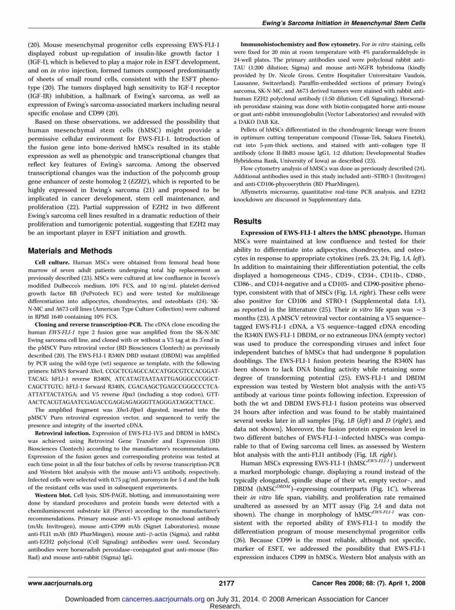
Expression of EWS-FLI-1 alters the hMSC phenotype. HumanMSCs were maintained at low confluence and tested for theirability to differentiate into adipocytes, chondrocytes, and osteo-cytes in response to appropriate cytokines (refs. 23, 24; Fig. 1A, left).In addition to maintaining their differentiation potential, the cellsdisplayed a homogeneous CD45-, CD19-, CD34-, CD11b-, CD80-,CD86-, and CD14-negative and a CD105- and CD90-positive pheno-type, consistent with that of MSCs (Fig. 1A, right). These cells werealso positive for CD106 and STRO-1 (Supplemental data 1A),as reported in the litterature (25). Their in vitro life span was f3months (23). A pMSCV retroviral vector containing a V5 sequence–tagged EWS-FLI-1 cDNA, a V5 sequence–tagged cDNA encodingthe R340N EWS-FLI-1 DBDM, or no extraneous DNA (empty vector)was used to produce the corresponding viruses and infect fourindependent batches of hMSCs that had undergone 8 populationdoublings. The EWS-FLI-1 fusion protein bearing the R340N hasbeen shown to lack DNA binding activity while retaining somedegree of transforming potential (25). EWS-FLI-1 and DBDMexpression was tested by Western blot analysis with the anti-V5antibody at various time points following infection. Expression ofboth the wt and DBDM EWS-FLI-1 fusion proteins was observed24 hours after infection and was found to be stably maintainedseveral weeks later in all samples [Fig. 1B (left) and D (right), anddata not shown]. Moreover, the fusion protein expression level intwo different batches of EWS-FLI-1–infected hMSCs was compa-rable to that of Ewing sarcoma cell lines, as assessed by Westernblot analysis with the anti-FLI1 antibody (Fig. 1B, right).
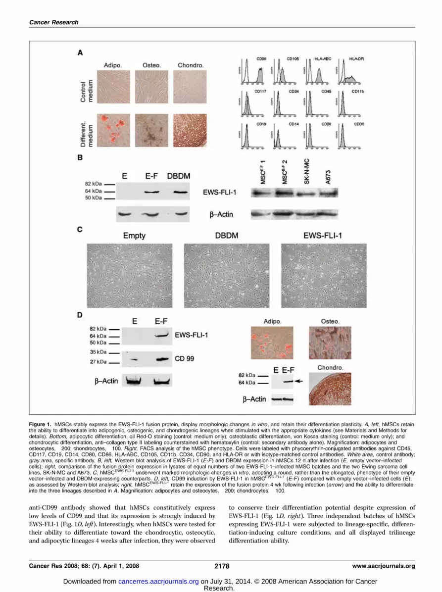
Human MSCs expressing EWS-FLI-1 (hMSCEWS-FLI-1) underwenta marked morphologic change, displaying a round instead of thetypically elongated, spindle shape of their wt, empty vector–, andDBDM (hMSCDBDM)–expressing counterparts (Fig. 1C), whereastheir in vitro life span, viability, and proliferation rate remainedunaltered as assessed by an MTT assay (Fig. 2A and data notshown). The change in morphology of hMSCEWS-FLI-1 was con-sistent with the reported ability of EWS-FLI-1 to modify thedifferentiation program of mouse mesenchymal progenitor cells(26). Because CD99 is the most reliable, although not specific,marker of ESFT, we addressed the possibility that EWS-FLI-1expression induces CD99 in hMSCs. Western blot analysis with an
Ewing’s Sarcoma Initiation in Mesenchymal Stem Cells
www.aacrjournals.org 2177 Cancer Res 2008; 68: (7). April 1, 2008
Research. on July 31, 2014. © 2008 American Association for Cancercancerres.aacrjournals.org Downloaded from
anti-CD99 antibody showed that hMSCs constitutively expresslow levels of CD99 and that its expression is strongly induced byEWS-FLI-1 (Fig. 1D, left). Interestingly, when hMSCs were tested fortheir ability to differentiate toward the chondrocytic, osteocytic,and adipocytic lineages 4 weeks after infection, they were observed
to conserve their differentiation potential despite expression ofEWS-FLI-1 (Fig. 1D, right). Three independent batches of hMSCsexpressing EWS-FLI-1 were subjected to lineage-specific, differen-tiation-inducing culture conditions, and all displayed trilineagedifferentiation ability.
Figure 1. hMSCs stably express the EWS-FLI-1 fusion protein, display morphologic changes in vitro , and retain their differentiation plasticity. A, left, hMSCs retainthe ability to differentiate into adipogenic, osteogenic, and chondrogenic lineages when stimulated with the appropriate cytokines (see Materials and Methods fordetails). Bottom, adipocytic differentiation, oil Red-O staining (control: medium only); osteoblastic differentiation, von Kossa staining (control: medium only); andchondrocytic differentiation, anti–collagen type II labeling counterstained with hematoxylin (control: secondary antibody alone). Magnification: adipocytes andosteocytes, �200; chondrocytes, �100. Right, FACS analysis of the hMSC phenotype. Cells were labeled with phycoerythrin-conjugated antibodies against CD45,CD117, CD19, CD14, CD80, CD86, HLA-ABC, CD105, CD11b, CD34, CD90, and HLA-DR or with isotype-matched control antibodies. White area, control antibody;gray area, specific antibody. B, left, Western blot analysis of EWS-FLI-1 (E-F ) and DBDM expression in hMSCs 12 d after infection (E, empty vector–infectedcells); right, comparison of the fusion protein expression in lysates of equal numbers of two EWS-FLI-1–infected hMSC batches and the two Ewing sarcoma celllines, SK-N-MC and A673. C, hMSCEWS-FLI-1 underwent marked morphologic changes in vitro , adopting a round, rather than the elongated, phenotype of their emptyvector–infected and DBDM-expressing counterparts. D, left, CD99 induction by EWS-FLI-1 in hMSCEWS-FLI-1 (E-F ) compared with empty vector–infected cells (E),as assessed by Western blot analysis; right, hMSCEWS-FLI-1 retain the expression of the fusion protein 4 wk following infection (arrow ) and the ability to differentiateinto the three lineages described in A . Magnification: adipocytes and osteocytes, �200; chondrocytes, �100.
Cancer Research
Cancer Res 2008; 68: (7). April 1, 2008 2178 www.aacrjournals.org
Research. on July 31, 2014. © 2008 American Association for Cancercancerres.aacrjournals.org Downloaded from
hMSCEWS-FLI-1 display induction of genes implicated inneural differentiation and neuroectodermal development aswell as that of known EWS-FLI-1 target genes. Total RNA wasextracted 12 days after infection and selection from each of thefour EWS-FLI-1–, DBDM-, and empty vector–containing hMSCbatches, and the corresponding mRNA was used to perform geneexpression profile analysis using the Affymetrix U133 Plus 2.0Arrays. Statistical analysis, using rank products (27) and a falsediscovery rate of 1%, revealed 614 and 262 probe sets corre-sponding to 393 and 174 genes to be respectively induced and
repressed in hMSCEWS-FLI-1 compared with empty vector–infectedcounterparts, whereas 104 and 158 genes were respectively up-regulated and down-regulated in hMSCDBDM (Supplementarydata 2). Importantly, all four batches of hMSCEWS-FLI-1 cellsrevealed an almost identical gene expression profile, as shown bythe heat map in Supplementary data 1B where the expressionprofile of the four samples is compared with that of cells infectedwith the R340N DBDM.
Among the most potently induced transcripts in hMSCEWS-FLI-1
were genes encoding proteolytic enzymes (primarily matrix
Figure 2. EWS-FLI-1 does not alter hMSC proliferation and induces a neuroectodermal gene expression profile. A, MTT assay analysis of the proliferation rate ofhMSCs expressing DBDM or EWS-FLI 1 compared with their empty vector–infected counterparts. B, comparison of the induction in hMSCEWS-FLI-1 and hMSCDBDM ofselected neuroectodermal markers from the list shown in Table 1A , as assessed by real-time PCR. C, NGFR and MAPT genes were selected from B to furthercharacterize their induction at the protein level by in vitro staining of hMSCEWS-FLI-1 compared with their empty vector– and DBDM-infected counterparts. Magnification,�200. D, the induction of the two candidate EWS-FLI-1 target genes, IGF-I and NKX2-2 , was confirmed by real-time PCR. All real-time PCR experiments werenormalized to cyclophyllin A and done in triplicate. Columns, mean of three independent determinations.
Ewing’s Sarcoma Initiation in Mesenchymal Stem Cells
www.aacrjournals.org 2179 Cancer Res 2008; 68: (7). April 1, 2008
Research. on July 31, 2014. © 2008 American Association for Cancercancerres.aacrjournals.org Downloaded from
metalloproteinases), adhesion receptors (including CLDN1, ICAM1,PCDH7/17, CEACAM1 , and CDH11) , and growth factors (includingIGF1, TGFA/B , and HBEGF ; Supplementary data 1C). Induction ofthese genes is consistent with the alterations in growth, survival,adhesion/migration, and tissue remodeling potential that may beexpected in cells undergoing transformation.
EWS-FLI-1 expression in hMSCs also induced numerous genesthat encode neural cell markers or that are implicated in neuralcrest development and neuronal differentiation, including NYP1R,GRP, MSX1, EGR2, NKX2-2, NGFR, CITED2, CDH11 , and MAPT(Table 1A). Because expression of these genes is consistent with theprimitive neuroectodermal phenotype of ESFT, six of them,including NPY1R, GRP, MAPT, NGFR, CITED2 , and SOX2 , wereselected for validation of their observed expression change. Up-regulation of all six genes in hMSCEWS-FLI-1 was confirmed byquantitative real-time PCR (Fig. 2B). To assess the importance ofDNA binding of EWS-FLI-1 in the induction of these six candidatetarget genes, their expression was assessed in MSCs infected withthe EWS-FLI-1 mutant bearing the R340N loss-of-functionmutation in the DBD of FLI-1. Quantitative real-time PCR analysisrevealed that hMSCs expressing the mutated EWS-FLI-1(hMSCDBDM) failed to display induction of any of the genes,suggesting that an intact DBD is required for EWS-FLI-1–mediatedregulation of their expression (Fig. 2B). To further validate theneuroectodermal phenotype induced by EWS-FLI-1, we assessedNGFR and MAPT protein expression in hMSCs infected with thethree retroviral vectors. Consistent with the gene expression pro-file, strong induction of both neural markers was observed inhMSCEWS-FLI-1 , but not in empty vector– or DBDM-infected coun-terparts (Fig. 2C). The robust induction of NGFRp75 by EWS-FLI-1was further confirmed by fluorescence-activated cell sorting (FACS)analysis of the infected cells (Supplementary data 1E).
Many of the reported candidate EWS-FLI-1 target genes wereinduced in hMSCEWS-FLI-1 , including ID2, IGF1, MMP1, TNC,COL11A1 , and UPP1 (20, 28–30), as well as the more recentlyidentified targets NROB1 and NKX2-2 (refs. 7, 10; Table 1A).Whereas up-regulation of several of these genes by EWS-FLI-1 hasbeen observed in a variety of cellular backgrounds, induction ofIGF-I has thus far been detected only in mouse mesenchymalprogenitor cells (20). Ewing’s sarcoma cells are highly sensitive toIGF-IR blockade, consistent with the possibility that IGF-I mayfacilitate EWS-FLI-1–mediated transformation and promote ESFTcell survival, particularly in the early phases of ESFT development.The relationship between EWS-FLI-1 and NKX2-2 was recentlydiscovered using an inducible rescue approach in ESFT cell lines(10), and expression of the NKX2-2 gene has been suggestedto be relevant to both ESFT differentiation and pathogenesis(10). Quantitative real-time PCR analysis confirmed induction ofboth IGF1 and NKX2-2 genes in hMSCEWS-FLI-1 cells but not inhMSCDBDM cells (Fig. 2D), indicating that regulation of both genesby EWS-FLI-1 requires integrity of the DBD of FLI-1. Transcriptlevels of a panel of Ewing’s sarcoma discriminating genes inMSCEWS-FLI-1 were compared with those of the same genes in afresh primary Ewings’ sarcoma sample. Several of the transcripts,particularly NPY1R, GRP, EZH2 , and IGF1 , were found to beexpressed at comparable levels in MSCEWS-FLI-1 and primaryEwing’s sarcoma cells (Supplementary data 1D).The expression profile of hMSCEWS-FLI-1 closely mimics that
of ESFT but not of other bone and soft tissue tumors. To assessthe degree of relatedness of the hMSC model to ESFT, we com-pared the gene expression profile of MSCEWS-FLI-1 to those of
Table 1. EWS-FLI-1–induced differentiation markers andcandidate target genes
Clone M value Genesymbol
(A) Neuroectodermal differentiation markers205440_s_al 7.004544088 NPY1R
217561_at 5.506065541 CALCA
203413_at 4.293524987 NELL2
206915_at 4.205361231 NKX2-2
214636_at 3.667256337 CALCB
236088_at 3.503590436 NTNG1
228038_at 3.400729183 SOX2
201565_s_at 2.792584692 ID2
204105_s_at 2.571239931 NRCAM205932_s_at 2.652991206 MSX11554485_s_at 2.655283127 TMEM37213479_at 2.366643943 NPTX2218162_at 2.249141903 OLFML3205249_at 2.243181602 EGR2230303_at 2.39988201 SYNPR208605_s_at 2.227463845 NTRK139966_at 2.222217226 CSPG5204869_at 2.146557475 PCSK2221933_at 1.982400408 NLGN4X203929_s_at 1.659102536 MAPT232226_at 1.62194636 LRRC4C205858_at 1.528171209 NGFR207980_s_at 1.461895211 CITED2227933_at 1.209141398 LRRN6A
Candidate EWS-FLI-1 target genes205440_s_at 7.004544088 NPY1R
206915_at 4.205361231 NKX2-2
204475_at 3.88747653 MMP1
201565_s_at 2.792584692 ID2
209541_at 2.366028399 IGF1
201645_at 1.576763978 TNC
37892_at 1.434881719 COL11A1203234_at 1.46967763 UPP1
(B) Staege ESFT signature and hMSC EWS-FLI-1 common genes205440_s_at 7.004544088 NPY1R202746_at 6.728787172 ITM2A219908_at 4.691662572 DKK2203358_s_at 3.103715832 EZH2228636_at 2.688202743 BHLHB51552610_a_at 2.165365299 JAK1205249_al 2.243181602 EGR2225871_at 2.188749054 STEAP2219528_s_at 1.988302947 BCL11B206812_at 1.359914681 ADRB3
241946_at 1.313478058 ZDHHC21
226106_at 1.192636061 RNF141
227933_at 1.209141398 LRRN6A
219825_at 1.544188607 CYP26B1
NOTE: (A) Summary of genes implicated in neural differentiation
and neuroectodermal development, as well as known EWS-FLI-1
target genes, found to be part of the hMSC EWS-FLI-1 gene expressionprofile. Log 2 m values of fold induction are shown. (B) List of the 14
common genes between hMSC EWS-FLI-1 and the ESFT expression
signature published by Staege et al. (21). Log 2 m values of foldinduction are shown.
Cancer Research
Cancer Res 2008; 68: (7). April 1, 2008 2180 www.aacrjournals.org
Research. on July 31, 2014. © 2008 American Association for Cancercancerres.aacrjournals.org Downloaded from

Ewing’s sarcoma and other bone and soft tissue tumors in arecently established sarcoma gene expression database (31). Aremarkable similarity was found between the gene expressionprofile of hMSCEWS-FLI-1 and that of Ewing’s sarcoma samples. Ofthe 225 genes proposed to be ESFT discriminators, 40 wereobserved to be induced in hMSCEWS-FLI-1 (Fig. 3C), comparedwith an expected number of 4.46 (P = 1.95e�25). To determinewhether hMSCEWS-FLI-1 display comparable or greater similarityto ESFT than fibroblastEWS-FLI-1 (17), direct comparison of eachgene expression profile was made to that of native ESFT (31). To doso, only the genes found to be present in all three arrays were used,which resulted in 94 genes in the fibroblast profile list and 235
genes in the MSC profile list. This approach revealed that of the 94genes in the fibroblast profile list, 14 were common to the nativeESFT list, for an expected 1.77 (P = 2.0e�9); of 235 genes in thehMSC list, 29 were common to the ESFT list, for an expected 4.43(P = 3.8 E-16). These observations suggest that fibroblasts providea more restrictive environment than hMSCs for expression of thetarget gene repertoire of the EWS-FLI-1 fusion protein (17).
Importantly, genes that were induced in hMSCEWS-FLI-1 , includingFVT1, DKK2, MAPT, PRKCB1, JAK1, ITM2A, FRZB, CITED2, STEAP1,STEAP2 , and ID2 , were among the top discriminators for Ewings’sarcoma (Fig. 3C). Among the 12 other sarcomas analyzed, onlydermatofibrosarcoma protuberans displayed a gene expression
Figure 3. EWS-FLI-1 expression in hMSCs induces a gene expression profile that closely mimics that of ESFT but not of other bone and soft tissue tumors.A, statistical analysis of the genes common to hMSCEWS-FLI-1 and a publicly available database of soft tissue tumor gene expression profile [Baird et al. (31)],showing the striking similarity of hMSCEWS-FLI-1 to ESFT but not to other sarcomas, with the significant P values indicated. B, Venn diagram representation ofhMSCEWS-FLI-1 profile relatedness to ESFT, dermatofibrosarcoma protuberans, and myxoid liposarcoma, based on the number of shared genes in their respectiveprofiles, as showed in A. C, list of the 40 genes found to be shared by the hMSCEWS-FLI-1 and ESFT gene expression profiles. Asterisks, genes found by Bairdet al. to be top discriminators for Ewing’s sarcoma. Log 2 m values of fold induction are shown. D, statistical analysis of the genes common to hMSCDBDM and the sametumor database used in A , indicating that expression of the EWS-FLI-1 DBDM in hMSCs does not induce a gene expression profile in the infected cells that candiscriminate ESFTs from other sarcomas. EWS, Ewing’s sarcoma; DFSP, dermatofibrosarcoma protuberans; LIPO, liposarcoma; RMS, rhabdomyosarcoma;MFH, malignant fibrous histiocytoma; OS, osteosarcoma; HPC, hemangiopericytoma; GIST, gastrointestinal stromal tumor; LMS, leiomyosarcoma; SS, synovialsarcoma; MULL, mixed mullerian; MPNST, malignant peripheral nerve sheath tumor; FIBRO, fibrosarcoma.
Ewing’s Sarcoma Initiation in Mesenchymal Stem Cells
www.aacrjournals.org 2181 Cancer Res 2008; 68: (7). April 1, 2008
Research. on July 31, 2014. © 2008 American Association for Cancercancerres.aacrjournals.org Downloaded from
profile with marginal similarity to hMSCEWS-FLI-1 (P = 0.011)(Fig. 3A and B). The same statistical analysis was applied to thegene expression profile of hMSCDBDM. In contrast to wt EWS-FLI-1,expression of the EWS-FLI-1 mutant form did not confer on MSCsany statistically significant similarity with Ewing sarcoma (Fig. 3D).This observation supports the notion that the lack of shape changedisplayed by the hMSCDBDM in vitro reflects a different geneticprogram in these cells, and highlights the importance of DNAbinding for the ESFT phenotype–inducing properties of the fusionprotein.
The highly discriminating ability of the hMSCEWS-FLI-1 geneexpression profile for ESFTs was further confirmed by anotherstudy, which assessed the transcriptome of a broad range ofmesenchymal tumors and identified the CALCB, MAPT, andPRKCB1 genes as prominent Ewing’s sarcoma discriminators (32),all of which we found to be induced in hMSCEWS-FLI-1 (Table 1;Supplementary data 2).
Comparison of the genes induced in hMSCEWS-FLI-1 with a set of38 genes (of which only 34 were included on our microarray) foundto be up-regulated in ESFT with respect to a wide spectrum ofnormal tissues and neuroblastomas (21) revealed 14 shared genes(P = 1.12e�15), including the strong ESFT discriminators NPYR1,ITM2A, DKK2, JAK1, STEAP, and EGR2 (Table 1B). Moreover, of asubset of 19 transcripts from the 34-gene set that could clearlydistinguish an ESFT cell line (SK-N-MC) from neuroblastoma celllines (21), 8 were part of the hMSCEWS-FLI-1 profile, whereas none ofthe transcripts in the 34-gene set were identified in the hMSCDBDM
expression profile (expected 0.18, P = 1.00). The same studyreported that introduction of EWS-FLI-1 into HEK 293 cellsinduced only one gene of this subset, namely, CCND1 , furtherhighlighting the selective permissiveness of hMSCs for EWS-FLI-1function (21).Injection of hMSCEWS-FLI-1 into immunocompromised mice.
Following injection into the subcapsular renal compartment ofimmunocompromised mice, hMSCEWS-FLI-1 did not form tumors,suggesting that despite expressing many of the hallmarks ofESFT, these cells require some additional event to become tumori-genic in mice. This observation is not surprising in the sensethat whereas there have been several examples of a single geneticevent transforming mouse progenitor cells (20, 33–35), recentevidence suggests that five events are required to transformhuman MSCs (36). By analogy to the present study, TLS-ERG andTEL-JAK2 respectively initiated a leukemogenic program anderythropoietin-independent erythropoiesis in normal human he-matopoietic cells but fell short of rendering them tumorigenicin vivo (37, 38).EZH2 promotes Ewing’s sarcoma growth. Among transcripts
that were up-regulated in hMSCEWS-FLI-1 , we identified the geneencoding EZH2, a member of the polycomb group proteins(Supplementary data 2), which has recently been found to beexpressed in ESFT (21). Together with EED and SUZ12, EZH2 formsthe polycomb-repressive complex 2, believed to be a key regulatorof embryonic development, stem cell renewal, and differentiation(39, 40). EZH2 is the catalytically active component of polycomb-repressive complex 2 and is believed to silence target genes byacting as a methyltransferase specific for Lys27 of histone H3 andLys26 of histone H1 (reviewed in ref. 22). Overexpression of EZH2has been shown to induce a bypass of the cellular senescenceprogram in mouse embryonic fibroblasts and to prevent mousehematopoietic stem cell exhaustion (41). Conversely, transientknockdown of EZH2 in primary human fibroblasts and a variety of
transformed cells inhibited their proliferation in vitro (42). EZH2expression has been proposed to be controlled by the Rb-E2Fpathway and to be a downstream mediator of E2F-dependentproliferation (42). High EZH2 expression that has been observed ina broad range of tumors has thus far been attributed either to Rbloss or gene amplification (42). Its silencing function is speculatedto target tumor suppressor genes (43), but the precise mechanismof its action on cell proliferation has thus far not been elucidated.
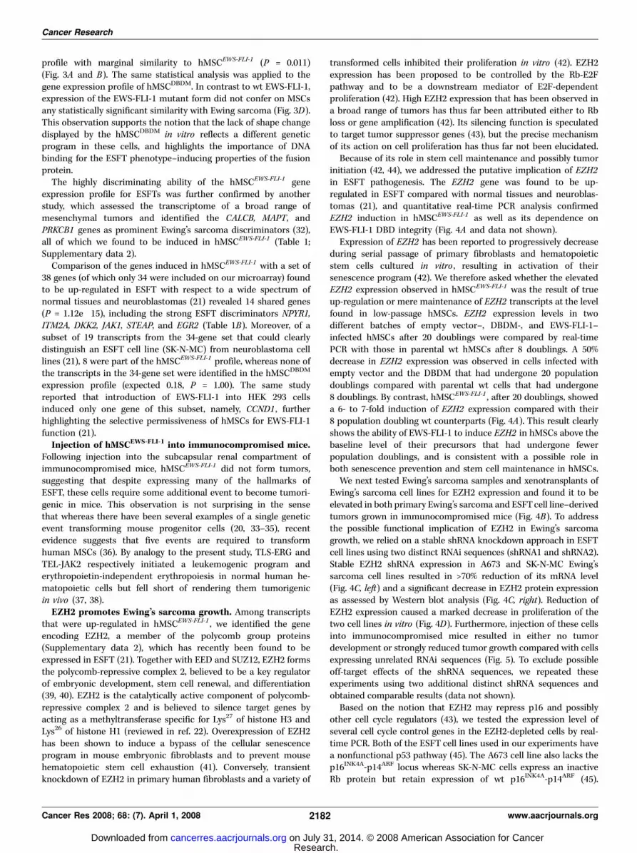
Because of its role in stem cell maintenance and possibly tumorinitiation (42, 44), we addressed the putative implication of EZH2in ESFT pathogenesis. The EZH2 gene was found to be up-regulated in ESFT compared with normal tissues and neuroblas-tomas (21), and quantitative real-time PCR analysis confirmedEZH2 induction in hMSCEWS-FLI-1 as well as its dependence onEWS-FLI-1 DBD integrity (Fig. 4A and data not shown).
Expression of EZH2 has been reported to progressively decreaseduring serial passage of primary fibroblasts and hematopoieticstem cells cultured in vitro , resulting in activation of theirsenescence program (42). We therefore asked whether the elevatedEZH2 expression observed in hMSCEWS-FLI-1 was the result of trueup-regulation or mere maintenance of EZH2 transcripts at the levelfound in low-passage hMSCs. EZH2 expression levels in twodifferent batches of empty vector–, DBDM-, and EWS-FLI-1–infected hMSCs after 20 doublings were compared by real-timePCR with those in parental wt hMSCs after 8 doublings. A 50%decrease in EZH2 expression was observed in cells infected withempty vector and the DBDM that had undergone 20 populationdoublings compared with parental wt cells that had undergone8 doublings. By contrast, hMSCEWS-FLI-1 , after 20 doublings, showeda 6- to 7-fold induction of EZH2 expression compared with their8 population doubling wt counterparts (Fig. 4A). This result clearlyshows the ability of EWS-FLI-1 to induce EZH2 in hMSCs above thebaseline level of their precursors that had undergone fewerpopulation doublings, and is consistent with a possible role inboth senescence prevention and stem cell maintenance in hMSCs.
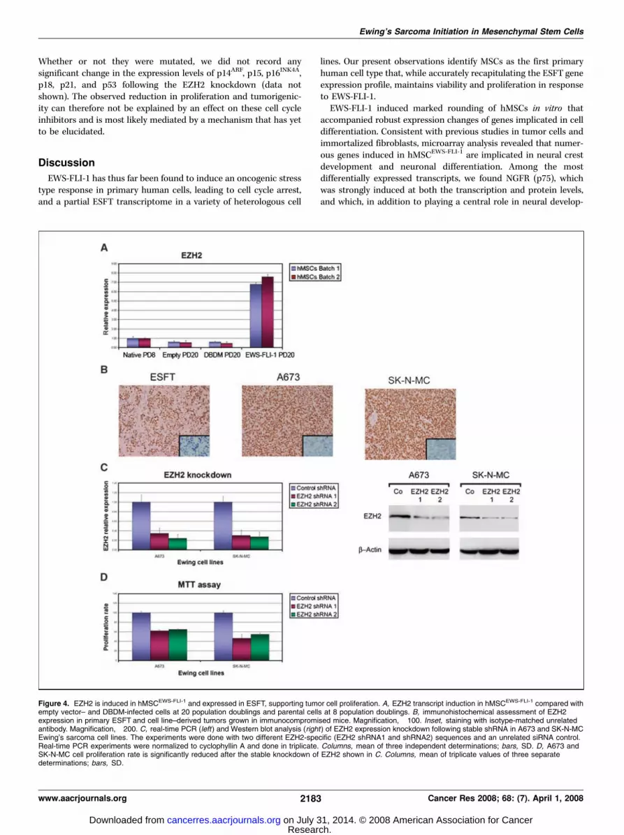
We next tested Ewing’s sarcoma samples and xenotransplants ofEwing’s sarcoma cell lines for EZH2 expression and found it to beelevated in both primary Ewing’s sarcoma and ESFTcell line–derivedtumors grown in immunocompromised mice (Fig. 4B). To addressthe possible functional implication of EZH2 in Ewing’s sarcomagrowth, we relied on a stable shRNA knockdown approach in ESFTcell lines using two distinct RNAi sequences (shRNA1 and shRNA2).Stable EZH2 shRNA expression in A673 and SK-N-MC Ewing’ssarcoma cell lines resulted in >70% reduction of its mRNA level(Fig. 4C, left) and a significant decrease in EZH2 protein expressionas assessed by Western blot analysis (Fig. 4C, right). Reduction ofEZH2 expression caused a marked decrease in proliferation of thetwo cell lines in vitro (Fig. 4D). Furthermore, injection of these cellsinto immunocompromised mice resulted in either no tumordevelopment or strongly reduced tumor growth compared with cellsexpressing unrelated RNAi sequences (Fig. 5). To exclude possibleoff-target effects of the shRNA sequences, we repeated theseexperiments using two additional distinct shRNA sequences andobtained comparable results (data not shown).
Based on the notion that EZH2 may repress p16 and possiblyother cell cycle regulators (43), we tested the expression level ofseveral cell cycle control genes in the EZH2-depleted cells by real-time PCR. Both of the ESFT cell lines used in our experiments havea nonfunctional p53 pathway (45). The A673 cell line also lacks thep16INK4A-p14ARF locus whereas SK-N-MC cells express an inactiveRb protein but retain expression of wt p16INK4A-p14ARF (45).
Cancer Research
Cancer Res 2008; 68: (7). April 1, 2008 2182 www.aacrjournals.org
Research. on July 31, 2014. © 2008 American Association for Cancercancerres.aacrjournals.org Downloaded from
Whether or not they were mutated, we did not record anysignificant change in the expression levels of p14ARF, p15, p16INK4A,p18, p21, and p53 following the EZH2 knockdown (data notshown). The observed reduction in proliferation and tumorigenic-ity can therefore not be explained by an effect on these cell cycleinhibitors and is most likely mediated by a mechanism that has yetto be elucidated.
Discussion
EWS-FLI-1 has thus far been found to induce an oncogenic stresstype response in primary human cells, leading to cell cycle arrest,and a partial ESFT transcriptome in a variety of heterologous cell
lines. Our present observations identify MSCs as the first primaryhuman cell type that, while accurately recapitulating the ESFT geneexpression profile, maintains viability and proliferation in responseto EWS-FLI-1.
EWS-FLI-1 induced marked rounding of hMSCs in vitro thataccompanied robust expression changes of genes implicated in celldifferentiation. Consistent with previous studies in tumor cells andimmortalized fibroblasts, microarray analysis revealed that numer-ous genes induced in hMSCEWS-FLI-1 are implicated in neural crestdevelopment and neuronal differentiation. Among the mostdifferentially expressed transcripts, we found NGFR (p75), whichwas strongly induced at both the transcription and protein levels,and which, in addition to playing a central role in neural develop-
Figure 4. EZH2 is induced in hMSCEWS-FLI-1 and expressed in ESFT, supporting tumor cell proliferation. A, EZH2 transcript induction in hMSCEWS-FLI-1 compared withempty vector– and DBDM-infected cells at 20 population doublings and parental cells at 8 population doublings. B, immunohistochemical assessment of EZH2expression in primary ESFT and cell line–derived tumors grown in immunocompromised mice. Magnification, �100. Inset, staining with isotype-matched unrelatedantibody. Magnification, �200. C, real-time PCR (left ) and Western blot analysis (right ) of EZH2 expression knockdown following stable shRNA in A673 and SK-N-MCEwing’s sarcoma cell lines. The experiments were done with two different EZH2-specific (EZH2 shRNA1 and shRNA2) sequences and an unrelated siRNA control.Real-time PCR experiments were normalized to cyclophyllin A and done in triplicate. Columns, mean of three independent determinations; bars, SD. D, A673 andSK-N-MC cell proliferation rate is significantly reduced after the stable knockdown of EZH2 shown in C. Columns, mean of triplicate values of three separatedeterminations; bars, SD.
Ewing’s Sarcoma Initiation in Mesenchymal Stem Cells
www.aacrjournals.org 2183 Cancer Res 2008; 68: (7). April 1, 2008
Research. on July 31, 2014. © 2008 American Association for Cancercancerres.aacrjournals.org Downloaded from
ment, is a key marker of neuroectodermal stem cells in bothnormal tissues and neural crest–derived tumors (46). Among theother genes that constitute part of the neuroectodermal profile ofhMSCEWS-FLI-1, SOX2 has been shown to maintain neural progenitorfeatures (47). In contrast to observations in mouse mesenchymalprogenitor cells, which were reported to lose osteogenic and adi-pocytic differentiation potential as a result of EWS-FLI-1 expres-sion, hMSCs expressing EWS-FLI-1 retained at least some degreeof trilineage differentiation plasticity. It is plausible that the early-stage neuroectodermal differentiation program induced by EWS-FLI-1 in hMSCs can still be overridden by the supraphysiologicconditions of the in vitro differentiation assays.
Some of the genes that are implicated in neural differentiationhave recently been suggested to play an active role in ESFT patho-genesis. Thus, NROB1 can promote tumorigenesis of Ewing’ssarcoma cell lines (7), whereas NKX2-2 may provide importantfunctions at specific stages of ESFT development because itsrepression strongly reduced ESFT cell line tumorigenicity (10).It is noteworthy that both genes were not only strongly inducedin hMSCEWS-FLI-1 but also remained dependent on EWS-FLI-1expression in established and tumorigenic ESFT cell lines. Theseobservations suggest that they play a role not only during the initialsteps of tumor development but also at late stages of tumorgrowth. Importantly, several of the above genes have been found tobe strong discriminators of ESFT, and their expression has beenused as an argument that ESFT may be of neuroectodermal origin.However, the present experiments show that expression of genesimplicated in neuronal differentiation and neural crest develop-ment can be induced by EWS-FLI-1 in the appropriate primarymesenchymal progenitor cell environment. Thus, a hMSC that canundergo partial neuroectodermal differentiation may constitute theorigin of ESFT, suggesting that these tumors need not arise froma neuroectodermal precursor to explain their primitive neuro-ectodermal phenotype.
Several of the genes observed to be up-regulated in hMSCEWS-FLI-1
and expressed in ESFT have been proposed to be functionallyrelated to Ewings’ sarcoma development and behavior. Thus, ID2 ,observed to be induced by EWS-FLI-1 in both hMSCs and mousemesenchymal progenitor cells (20), may play an important role inpromoting cell cycle entry by inhibiting Rb. Induction of fos thatwas detected as a result of EWS-FLI-1 introduction into hMSCs(Supplementary data 1C) may be of particular interest in light ofrecent evidence that expression of numerous candidate EWS-FLI-1target genes requires cooperation between EWS-FLI-1 andactivator protein-1 (AP-1) (48). Enhanced expression of fos mayexplain, in part, the broad ESFT gene repertoire that was up-regulated in hMSCEWS-FLI-1 , and may therefore underlie the abilityof these cells to recapitulate ESFT features. Moreover, and incontrast to primary fibroblasts, expression of EWS-FLI-1 in hMSCsdid not provoke a p53 response, as shown by the absence of p53
target gene induction in their gene expression profile and thecorresponding growth arrest. EWS-FLI-1 signals may either fail toactivate premature cell senescence and oncogenic stress pathways,including p53 in hMSCs, or trigger mechanisms that neutralize orcircumvent them.
Two candidate EWS-FLI-1 target genes that were induced inhMSCEWS-FLI-1 and that may prevent cell senescence while promotingsurvival and proliferation are IGF1 and EZH2 . We have previouslyshown that IGF1 is induced by EWS-FLI-1 in mouse mesenchymalprogenitor cells (20). Coupled to that observation, the presentfindings suggest that IGF1 may be an important EWS-FLI-1 target inMSCs. It is noteworthy that mouse mesenchymal progenitor cellsand hMSCs are the only cells in which up-regulation of IGF-I byEWS-FLI-1 has been reported thus far. Although the direct or indirectnature of the effect remains to be determined, our experimentsclearly indicate that the DNA binding ability of EWS-FLI-1 is requiredfor IGF1 induction in hMSCs. IGF-IR signaling is observed to becritical for ESFT growth, and its blockade induces growth arrest andapoptosis in ESFT cell lines (49). IGF-I induction may provide asurvival signal that is essential during the initiating phases of tumordevelopment, when the primary cells are subjected to the strongoncogenic stimulus of EWS-FLI-1 expression.
The discovery that EZH2 is induced by EWS-FLI-1 in hMSCs mayprovide an additional explanation about why EWS-FLI-1 does notelicit oncogenic stress–mediated cell cycle arrest in hMSCs. Similarto another polycomb group gene, bmi-1, which cooperates withmyc in B-cell and T-cell lymphoma development by inhibiting myc-induced apoptosis through repression of the Cdkn2a locus, EZH2activity is suggested to participate in blocking cellular response tooncogenic stress and maintain stem cell renewal at the expense ofdifferentiation (22). EZH2 has also been proposed to act as anoncogene in its own right (42) and to become up-regulated inpreneoplastic lesions in the breast (44). Consistent with thesescenarios, EZH2 repression resulted in reduction of ESFT cellproliferation and tumorigenicity. Combined targeting of IGF-IR andEZH2 may provide an attractive therapeutic option in ESFT.
Taken together, our observations provide evidence for the firsttime that hMSCs can sustain EWS-FLI-1 expression and respondby adopting phenotypic and gene expression hallmarks of ESFT,as would be expected of its putative cell of origin. These results aresupported by recent work reporting the appearance of MSCfeatures in Ewing’s sarcoma cell lines subjected to EWS-FLI-1knockdown (50). As such, hMSCEWS-FLI-1 may provide an idealcellular environment for uncovering new candidate genes whoserole in ESFT initiation and subsequent maintenance can be tested.Although tumor initiation by adult hMSCs may require a geneticevent in addition to EWS-FLI-1 expression, the overall geneexpression and phenotypic changes observed in response to EWS-FLI-1 alone argue that hMSCEWS-FLI-1 constitutes a strongcandidate precursor of ESFT.
Figure 5. Reduction of EZH2 expression in A673 andSK-N-MC cell lines causes either no tumor development orreduced tumor growth compared with cells expressingunrelated RNAi sequences.
Cancer Research
Cancer Res 2008; 68: (7). April 1, 2008 2184 www.aacrjournals.org
Research. on July 31, 2014. © 2008 American Association for Cancercancerres.aacrjournals.org Downloaded from

AcknowledgmentsReceived 5/12/2007; revised 11/29/2007; accepted 1/24/2008.
Grant support: Fonds National de la Recherche Scientifique grant 3100A0-105833,Oncosuisse grant 01656-02-2005, and the National Center of Competence in ResearchMolecular Oncology (I. Stamenkovic).
The costs of publication of this article were defrayed in part by the payment of pagecharges. This article must therefore be hereby marked advertisement in accordancewith 18 U.S.C. Section 1734 solely to indicate this fact.
We thank Prof. Pierre Hoffmeyer for fruitful collaboration and for providingsurgical samples, Joan Stalder for technical support, and Carlo Fusco for insightfuldiscussions.
Ewing’s Sarcoma Initiation in Mesenchymal Stem Cells
www.aacrjournals.org 2185 Cancer Res 2008; 68: (7). April 1, 2008
References
1. Delattre O, Zucman J, Plougastel B, et al. Gene fusionwith an ETS DNA-binding domain caused by chromosometranslocation in human tumours. Nature 1992;359:162–5.2. Eliazer S, Spencer J, Ye D, Olson E, Ilaria RL, Jr.Alteration of mesodermal cell differentiation by EWS/FLI-1, the oncogene implicated in Ewing’s sarcoma. MolCell Biol 2003;23:482–92.3. Hu-Lieskovan S, Zhang J, Wu L, Shimada H, SchofieldDE, Triche TJ. EWS-FLI1 fusion protein up-regulatescritical genes in neural crest development and isresponsible for the observed phenotype of Ewing’sfamily of tumors. Cancer Res 2005;65:4633–44.4. May WA, Gishizky ML, Lessnick SL, et al. Ewingsarcoma 11;22 translocation produces a chimerictranscription factor that requires the DNA-bindingdomain encoded by FLI1 for transformation. Proc NatlAcad Sci U S A 1993;90:5752–6.5. Rorie CJ, Thomas VD, Chen P, Pierce HH, O’Bryan JP,Weissman BE. The Ews/Fli-1 fusion gene switches thedifferentiation program of neuroblastomas to Ewingsarcoma/peripheral primitive neuroectodermal tumors.Cancer Res 2004;64:1266–77.6. Thompson AD, Teitell MA, Arvand A, Denny CT.Divergent Ewing’s sarcoma EWS/ETS fusions confer acommon tumorigenic phenotype on NIH3T3 cells.Oncogene 1999;18:5506–13.7. Kinsey M, Smith R, Lessnick SL. NR0B1 is required forthe oncogenic phenotype mediated by EWS/FLI inEwing’s sarcoma. Mol Cancer Res 2006;4:851–9.8. Mendiola M, Carrillo J, Garcia E, et al. The orphannuclear receptor DAX1 is up-regulated by the EWS/FLI1oncoprotein and is highly expressed in Ewing tumors.Int J Cancer 2006;118:1381–9.9. Owen LA, Lessnick SL. Identification of target genes intheir native cellular context: an analysis of EWS/FLI inEwing’s sarcoma. Cell Cycle 2006;5:2049–53.10. Smith R, Owen LA, Trem DJ, et al. Expression profilingof EWS/FLI identifies NKX2.2 as a critical target gene inEwing’s sarcoma. Cancer Cell 2006;9:405–16.11. May WA, Arvand A, Thompson AD, Braun BS,Wright M, Denny CT. EWS/FLI1-induced manic fringerenders NIH 3T3 cells tumorigenic. Nat Genet 1997;17:495–7.12. Kovar H, Aryee DN, Jug G, et al. EWS/FLI-1antagonists induce growth inhibition of Ewing tumorcells in vitro . Cell Growth Differ 1996;7:429–37.13. Tanaka K, Iwakuma T, Harimaya K, Sato H, Iwamoto Y.EWS-Fli1 antisense oligodeoxynucleotide inhibits prolif-eration of human Ewing’s sarcoma and primitive neuro-ectodermal tumor cells. J Clin Invest 1997;99:239–47.14. Prieur A, Tirode F, Cohen P, Delattre O. EWS/FLI-1silencing and gene profiling of Ewing cells revealdownstream oncogenic pathways and a crucial role forrepression of insulin-like growth factor binding protein3. Mol Cell Biol 2004;24:7275–83.15. Riggi N, Stamenkovic I. The Biology of Ewingsarcoma. Cancer Lett 2007;254:1–10.16. Deneen B, Denny CT. Loss of p16 pathways stabilizesEWS/FLI1 expression and complements EWS/FLI1mediated transformation. Oncogene 2001;20:6731–41.17. Lessnick SL, Dacwag CS, Golub TR. The Ewing’ssarcoma oncoprotein EWS/FLI induces a p53-dependent
growth arrest in primary human fibroblasts. Cancer Cell2002;1:393–401.18. Huang HY, Illei PB, Zhao Z, et al. Ewing sarcomaswith p53 mutation or p16/p14ARF homozygous dele-tion: a highly lethal subset associated with poorchemoresponse. J Clin Oncol 2005;23:548–58.19. Castillero-Trejo Y, Eliazer S, Xiang L, Richardson JA,Ilaria RL, Jr. Expression of the EWS/FLI-1 oncogene inmurine primary bone-derived cells Results in EWS/FLI-1-dependent, Ewing sarcoma-like tumors. Cancer Res2005;65:8698–705.20. Riggi N, Cironi L, Provero P, et al. Development ofEwing’s sarcoma from primary bone marrow-derivedmesenchymal progenitor cells. Cancer Res 2005;65:11459–68.21. Staege MS, Hutter C, Neumann I, et al. DNAmicroarrays reveal relationship of Ewing family tumorsto both endothelial and fetal neural crest-derived cellsand define novel targets. Cancer Res 2004;64:8213–21.22. Sparmann A, van Lohuizen M. Polycomb silencerscontrol cell fate, development and cancer. Nat RevCancer 2006;6:846–56.23. Suva D, Garavaglia G, Menetrey J, et al. Non-hematopoietic human bone marrow contains long-lasting, pluripotential mesenchymal stem cells. J CellPhysiol 2004;198:110–8.24. Suva D, Passweg J, Arnaudeau S, Hoffmeyer P, KindlerV. In vitro activated human T lymphocytes veryefficiently attach to allogenic multipotent mesenchymalstromal cells and transmigrate under them. J CellPhysiol 2008;214:588–94.25. Welford SM, Hebert SP, Deneen B, Arvand A, DennyCT. DNA binding domain-independent pathways areinvolved in EWS/FLI1-mediated oncogenesis. J BiolChem 2001;276:41977–84.26. Torchia EC, Jaishankar S, Baker SJ. Ewing tumorfusion proteins block the differentiation of pluripotentmarrow stromal cells. Cancer Res 2003;63:3464–8.27. Breitling R, Armengaud P, Amtmann A, Herzyk P.Rank products: a simple, yet powerful, new method todetect differentially regulated genes in replicated micro-array experiments. FEBS Lett 2004;573:83–92.28. Deneen B, Hamidi H, Denny CT. Functional analysisof the EWS/ETS target gene uridine phosphorylase.Cancer Res 2003;63:4268–74.29. Fuchs B, Inwards CY, Janknecht R. Up-regulation ofthe matrix metalloproteinase-1 gene by the Ewing’ssarcoma associated EWS-ER81 and EWS-Fli-1 oncopro-teins, c-Jun and p300. FEBS Lett 2003;553:104–8.30. Watanabe G, Nishimori H, Irifune H, et al. Inductionof tenascin-C by tumor-specific EWS-ETS fusion genes.Genes Chromosomes Cancer 2003;36:224–32.31. Baird K, Davis S, Antonescu CR, et al. Geneexpression profiling of human sarcomas: insights intosarcoma biology. Cancer Res 2005;65:9226–35.32. Henderson SR, Guiliano D, Presneau N, et al. Amolecular map of mesenchymal tumors. Genome Biol2005;6:R76.33. Codrington R, Pannell R, Forster A, et al. TheEws-ERG fusion protein can initiate neoplasia fromlineage-committed haematopoietic cells. PLoS Biol 2005;3:e242.34. Perez-Losada J, Pintado B, Gutierrez-Adan A, et al.The chimeric FUS/TLS-CHOP fusion protein specifically
induces liposarcomas in transgenic mice. Oncogene2000;19:2413–22.35. Riggi N, Cironi L, Provero P, et al. Expression of theFUS-CHOP fusion protein in primary mesenchymalprogenitor cells gives rise to a model of myxoidliposarcoma. Cancer Res 2006;66:7016–23.36. Funes JM, Quintero M, Henderson S, et al. Transfor-mation of human mesenchymal stem cells increases theirdependency on oxidative phosphorylation for energyproduction. Proc Natl Acad Sci U S A 2007;104:6223–8.37. Kennedy JA, Barabe F, Patterson BJ, et al. Expressionof TEL-JAK2 in primary human hematopoietic cellsdrives erythropoietin-independent erythropoiesis andinduces myelofibrosis in vivo . Proc Natl Acad Sci U S A2006;103:16930–5.38. Pereira DS, Dorrell C, Ito CY, et al. Retroviraltransduction of TLS-ERG initiates a leukemogenicprogram in normal human hematopoietic cells. ProcNatl Acad Sci U S A 1998;95:8239–44.39. Bracken AP, Dietrich N, Pasini D, Hansen KH, HelinK. Genome-wide mapping of Polycomb target genesunravels their roles in cell fate transitions. Genes Dev2006;20:1123–36.40. Caretti G, Di Padova M, Micales B, Lyons GE,Sartorelli V. The Polycomb Ezh2 methyltransferaseregulates muscle gene expression and skeletal muscledifferentiation. Genes Dev 2004;18:2627–38.41. Kamminga LM, Bystrykh LV, de Boer A, et al. ThePolycomb group gene Ezh2 prevents hematopoieticstem cell exhaustion. Blood 2006;107:2170–9.42. Bracken AP, Pasini D, Capra M, Prosperini E, Colli E,Helin K. EZH2 is downstream of the pRB-E2F pathway,essential for proliferation and amplified in cancer.EMBO J 2003;22:5323–35.43. Kotake Y, Cao R, Viatour P, Sage J, Zhang Y, Xiong Y.pRB family proteins are required for H3K27 trimethy-lation and Polycomb repression complexes binding toand silencing p16INK4a tumor suppressor gene. GenesDev 2007;21:49–54.44. Ding L, Kleer CG. Enhancer of zeste 2 as a marker ofpreneoplastic progression in the breast. Cancer Res2006;66:9352–5.45. Kovar H, Jug G, Aryee DN, et al. Among genes involvedin the RB dependent cell cycle regulatory cascade, the p16tumor suppressor gene is frequently lost in the Ewingfamily of tumors. Oncogene 1997;15:2225–32.46. Stemple DL, Anderson DJ. Isolation of a stem cell forneurons and glia from the mammalian neural crest. Cell1992;71:973–85.47. Graham V, Khudyakov J, Ellis P, Pevny L. SOX2functions to maintain neural progenitor identity.Neuron 2003;39:749–65.48. Kim S, Denny CT, Wisdom R. Cooperative DNAbinding with AP-1 proteins is required for transforma-tion by EWS-Ets fusion proteins. Mol Cell Biol 2006;26:2467–78.49. Scotlandi K, Avnet S, Benini S, et al. Expression of anIGF-I receptor dominant negative mutant inducesapoptosis, inhibits tumorigenesis and enhances chemo-sensitivity in Ewing’s sarcoma cells. Int J Cancer 2002;101:11–6.50. Tirode F, Laud-Duval K, Prieur A, Delorme B,Charbord P, Delattre O. Mesenchymal stem cell featuresof Ewing tumors. Cancer Cell 2007;11:421–9.
Research. on July 31, 2014. © 2008 American Association for Cancercancerres.aacrjournals.org Downloaded from