diagnosis and treatment of ewing's sarcoma
TRANSCRIPT
Review Article
Diagnosis and Treatment of Ewing’s Sarcoma
Yukihide Iwamoto
Department of Orthopaedic Surgery, Graduate School of Medical Sciences, Kyushu University, Fukuoka, Japan
Received September 13, 2006; accepted October 12, 2006; published online February 1, 2007
Ewing’s sarcoma is a small round-cell tumor typically arising in the bones, rarely in softtissues, of children and adolescents. Ewing’s sarcoma has retained the most unfavorableprognosis of all primary musculoskeletal tumors. Prior to the use of multi-drug chemotherapy,long-term survival was less than 10%. The development of multi-disciplinary therapy withchemotherapy, irradiation, and surgery has increased current long-term survival rates in mostclinical centers to greater than 50%. In addition, the preferred method of tumor resection haschanged; limb salvage has nearly replaced amputation of the affected limb. Limb salvage pro-cedures can be performed in place of amputation without compromising patient survivalrates. Recent studies have revealed that the pathognomonic translocations involving the EWSgene on chromosome 22 and an ETS-type gene, which is most commonly the Fli1 gene onchromosome 11, are implicated in more than 95% of Ewing’s sarcomas, primitive neuroecto-dermal tumors and Askin’s tumors. Therefore, these lesions have become regarded as asingle entity, dubbed the Ewing’s family of tumors. RT-PCR to detect EWS–ETS genearrangements is widely used to confirm the diagnosis of Ewing’s family of tumors.Experimental results suggest that inhibition of the signaling pathway downstream of theEWS–ETS gene may lead to the development of molecularly targeted therapy in the future.
Key words: Ewing’s sarcoma – diagnosis – treatment
INTRODUCTION
Recent years have seen a remarkable change in the percep-
tion of the histogenesis and the relationship between skeletal
and extra-skeletal Ewing’s sarcoma and primitive neuroecto-
dermal tumor (PNET) (1). In 1918, Stout reported a case
with an ulnar nerve tumor composed of undifferentiated
round cells that form rosettes, subsequently defined as PNET
of soft tissue (2). In 1921, James Ewing reported a case of
round cell tumor in the radius of a 14-year-old girl as a
‘diffuse endothelioma of bone’, proposing an endothelial
derivation (Ewing’s sarcoma) (3). It was in 1975 that
Angervall and Enzinger reported the first case of an Ewing’s
sarcoma arising in soft tissue (extra-skeletal Ewing’s
sarcoma) (4). In 1979, Askin et al. reported a ‘malignant
small-cell tumor of the thoracopulmonary region’ (Askin
tumor) with similar histologic features as PNET (5). In
1984, Jaffe et al. described a small round-cell tumor of
bone, calling it a neuroectodermal tumor of bone (PNET of
bone) (6). Recent clinicopathological studies have revealed
that these lesions have overlapping features, supporting a
common histogenesis. Identification of a common transloca-
tion t(11;22)(q24;q12) (7,8) that results in the formation of
the EWS–ETS fusion gene (9) in cases of Ewing’s sarcoma,
PNET and Askin’s tumor strongly supported the hypothesis
that these tumors are related. Therefore, all these lesions are
now included in the same classification, the Ewing’s
sarcoma family of tumors (EFTs).
Thanks to the development of novel methods for diagnosis
and treatment, the prognosis of EFTs has improved greatly.
This review overviews the updated diagnostic and treatment
methods for management of EFTs.
FREQUENCY
According to data of Bone Tumor Registry Japan, Ewing’s
sarcoma is the third most frequent primary sarcoma of bone
For reprints and all correspondence: Yukihide Iwamoto, Department ofOrthopedic Surgery, Graduate School of Medical Sciences, KyushuUniversity, Maidashi 3-1-1, Higashi-ku, Fukuoka 812-8582, Japan. E-mail:[email protected]
# 2007 Foundation for Promotion of Cancer Research
Jpn J Clin Oncol 2007;37(2)79–89
doi:10.1093/jjco/hyl142
Dow
nloaded from https://academ
ic.oup.com/jjco/article/37/2/79/1795451 by guest on 14 April 2022
after osteosarcoma and chondrosarcoma (10). It is the second
most frequent bone sarcoma after osteosarcoma in patients
younger than 20 years of age. It remains an infrequent neo-
plasm, however; only approximately 20 new cases are regis-
tered per year. Caucasians are much more frequently
affected by Ewing’s sarcoma than Asians, while Africans
and African-Americans rarely suffer from this disease. In the
Surveillance, Epidemiology, and End Results (SEER)
program series in the USA between 1973 and 1985, only
three of 650 cases of Ewing’s sarcoma occurred in black
patients. In North America, 225 patients younger than 20
years old are diagnosed per year with this disease (11).
SEX AND AGE
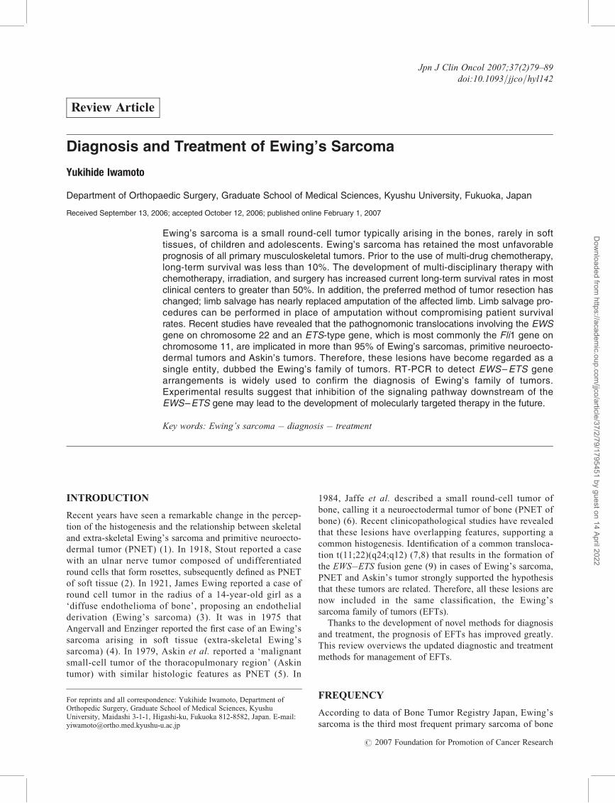
Ewing’s sarcoma has a predilection for the male sex (male/female ratio, 1.3–1.5:1). Ewing’s sarcoma occurs in a wide
range of ages from infants to the elderly, although approxi-
mately 80% of patients afflicted are younger than 20 years of
age. Peak incidence is during the second decade of life,
although 20–30% of cases are diagnosed in the first decade
(Fig. 1). The age of the patient is important diagnostically.
When confronted with patients older than 30 years, the clini-
cian must first eliminate other small round-cell tumors,
including small-cell carcinoma and large-cell lymphoma,
before making a diagnosis of Ewing’s sarcoma. In patients
younger than 5 years, the possibility of metastatic neuroblas-
toma or acute leukemia needs to be ruled out.
LOCALIZATION
Ewing’s sarcoma demonstrates a predilection for the trunk
and long bones. In the truncal skeleton, the pelvis predomi-
nates, followed by the scapula, vertebral column, ribs and
clavicle (Fig. 1). Of the long bones, the most common site is
the femur, followed by the humerus, tibia and bones of
forearm in that order. As opposed to osteosarcoma, Ewing’s
sarcoma of the long bones tends to arise from the diaphysis
rather than the metaphysis.
Ewing’s sarcoma has a strong potential to metastasize.
Metastases most commonly occur in the lungs and bone.
More than 10% of patients present with multiple bone
metastases at initial diagnosis. While metastases in the
lungs, bone, bone marrow, or a combination thereof are
detectable in approximately 25% of patients, metastases to
lymph nodes are rare.
Ewing’s sarcoma primarily occurs in bones, with rare occur-
rences in soft tissues. Most extra-skeletal Ewing’s sarcomas
affect patients between 10 and 30 years of age, with a peak
incidence at approximately 20 years old. The most common
sites are the chest wall, para-vertebral muscles, extremities,
buttocks and retro-peritoneal space. Extra-skeletal Ewing’s
sarcomas present with rapid growth and frequent distant
metastases, similarly to Ewing’s sarcoma of bone.
SYMPTOMS
Ewing’s sarcoma typically progresses quite rapidly. Skeletal
lesions typically progress to large tumors that form in soft
tissues within a few weeks.
The earliest symptom is pain. At first, the pain can be inter-
mittent and mild, but rapidly progresses to the point at which it
becomes so intense as to require the use of analgesic drugs.
When the tumor is vertebral or pelvic in origin, the pain may
be accompanied by paresthesia and treated by irradiation. As
pain can precede definitive diagnosis for weeks or months and
years in some cases, patients with bone pain without defined
trauma should undergo prompt imaging studies.
Tumor growth eventually leads to a visible or palpable
swelling of the affected site. This swelling is tense, elastic,
hard, tender, rapidly increasing and accompanied by local
heat. The tumor bulk, however, may be indiscernible for a
long period of time in the cases of pelvic, spinal, or femoral
tumors that are not palpable as these tumors are deep-seated
Figure 1. Sex, age and localization of Ewing’s sarcoma in Japan (1972–2003) (11) (please note that a colour version of this figure is available as supplemen-
tary data at http://www.jjco.oxfordjournals.org).
80 Diagnosis and treatment of Ewing’s sarcoma
Dow
nloaded from https://academ
ic.oup.com/jjco/article/37/2/79/1795451 by guest on 14 April 2022

or cases in which the Ewing’s sarcoma extends only into the
cancellous bone or along the medullary canal of long bones
without expanding outside the cortex (Fig. 2A).
Other common symptoms include fever, anemia, and non-
specific signs of inflammation, such as increases in sedimen-
tation rate, moderate leukocytosis and an increase in serum
LDH. Conventional blood, serum and urine tests cannot
specifically identify Ewing’s sarcoma. Unlike neuroblastoma,
serum and urine catecholamine levels remain normal.
However, de Alava et al. reported that the EWS–Fli1 fusion
gene is frequently detected in peripheral blood samples from
patients with Ewing’s sarcoma (12). In advanced cases, the
symptoms listed above are frequent; the majority of patients
experience loss of appetite and weight.
DIAGNOSTIC IMAGING
PLAIN RADIOGRAPH
The initial imaging investigation of a suspected bone tumor
is a radiograph in two planes. Tumor-related osteolysis and
periosteal reactions suggest a diagnosis of primary malignant
tumor. Periosteal reactions, the reactive osteogenesis of the
periosteum, are caused by extra-osseous extension of the
tumor. Several types of periosteal reactions have been
observed: (i) an ‘onion skin’ or ‘onion-peel appearance’ is a
prominent multi-layered reaction, (ii) a ‘sunburst’ or ‘spiculae’
pattern is a perpendicular reaction, while (iii) ‘Codman’s
triangle’ is a triangular lifting of the periosteum from the
bone at the site of detachment. Typically, Ewing’s sarcoma
appears as an ill-defined, permeative, or focally moth-eaten,
destructive intramedullary lesion accompanied by a perios-
teal reaction (‘onion skin’) that affects the diaphyses of long
bones (Figs. 2A, 3A). The sunburst type of periosteal reac-
tions can present, but is less common in comparison with its
occurrence in osteosarcoma.
MRI
The most precise definition of the local extent of bone
tumors, including the degree of expansion into the intrame-
dullary portion and the relationship of the lesion to adjacent
blood vessels and nerves, is provided by MRI (Figs. 2B,
3B). When malignant bone tumors are suspected, MRI is
routinely performed for staging and surgical planning. MRI
is particularly important in the imaging of Ewing’s sarcoma
Figure 2. Ewing’s sarcoma of the femur. (A) Plain roentgenogram displays a permeative lesion of the proximal femoral shaft associated with an ‘onion skin’
periosteal reaction (Arrow). Insert is a magnification of ‘onion skin’ periosteal reaction (Arrow). (B) A coronal T1-weighted MR image displays a low-signal
intramedullary tumor involving the long segment of the femoral shaft that extends into the femoral neck. Note that the involvement detected by MRI extends
beyond the anticipated area seen on plain roentgenogram. (C) After resection with wide margins, the affected limb was reconstructed with endoprosthesis.
Jpn J Clin Oncol 2007;37(2) 81
Dow
nloaded from https://academ
ic.oup.com/jjco/article/37/2/79/1795451 by guest on 14 April 2022
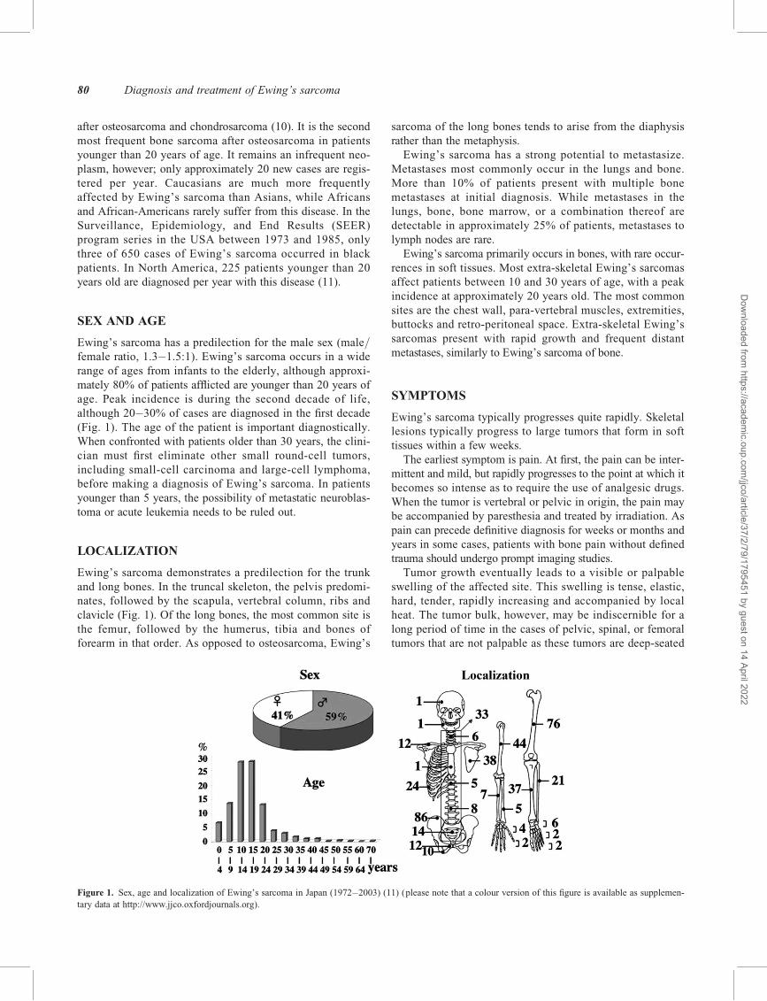
as this tumor is ill-defined on plain radiographs or by
computed tomography (CT). MRI typically demonstrates
lesions that involve large segments of the intramedullary
cavity, which extend beyond the area indicated by plain
radiographs. MRI can also evaluate the extent of soft tissue
masses, which can be quite large.
MRI is widely used to assess responses to neoadjuvant
chemotherapy or irradiation, because regression of the extra-
Figure 3. Ewing’s sarcoma of the humerus. (A) Plain roentgenogram shows an osteolytic lesion of the humeral shaft associated with the periosteal reactions
of ‘Codman’s triangle’ (Arrow). (B) Transverse T2-weighted MR image displays a high-signal large tumor extending into the soft tissues. (C) After pre-
operative adjuvant treatment, including multi-drug chemotherapy and irradiation, the size of the soft tissue tumor was dramatically reduced. (D) After resection
with wide margins, the affected bone was transplanted after autoclaving.
82 Diagnosis and treatment of Ewing’s sarcoma
Dow
nloaded from https://academ
ic.oup.com/jjco/article/37/2/79/1795451 by guest on 14 April 2022
skeletal tumor mass can be precisely defined (Fig. 3B, C).
Currently, MRI is the standard imaging method for such
evaluation. Recent studies have demonstrated, however, that
PET, thallium-201 scintiography and dynamic MRI provide
more valuable information than MRI for assessment of thera-
peutic responses (13).
STAGING
Enneking et al. created a staging system for both benign and
malignant musculoskeletal tumors to support decision
making in treatment and to allow meaningful comparison
between treatment methods (14). The system, based on the
histological grade of the tumor, local extent, and the pre-
sence or absence of metastasis, incorporates the most signifi-
cant prognostic factors into a set of progressive stages that
can help to guide surgical and adjuvant treatments.
High-grade lesions, such as Ewing’s sarcomas, are desig-
nated as stage II tumors, which can be subdivided according
to the extent of local growth. While stage IIA lesions are
contained within well-defined anatomical compartments,
stage IIB lesions extend beyond their compartment of origin.
Stage III includes any lesion that has metastasized, regard-
less of the size or grade of the primary tumor. Almost all
Ewing’s sarcomas fall into stages IIB or III. Many oncolo-
gists stage malignant bone tumors according to the
American Joint Committee on Cancer (AJCC) system, which
is similar to Enneking’s system (15).
Diagnostic staging should include a CT scan of the chest
to determine pulmonary metastases and a technetium-99 m
whole-body radionucleotide bone scan to identify skeletal
metastases. Fluorine-18 fluorodeoxyglucose position emis-
sion tomography (FDG-PET) was recently reported to
increase the sensitivity of detection for both skeletal metas-
tases and therapeutic responses (16). The exact role for this
modality in the management of Ewing’s sarcoma, however,
remains to be defined.
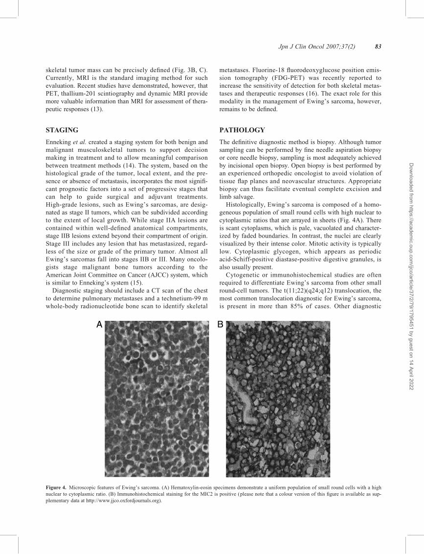
PATHOLOGY
The definitive diagnostic method is biopsy. Although tumor
sampling can be performed by fine needle aspiration biopsy
or core needle biopsy, sampling is most adequately achieved
by incisional open biopsy. Open biopsy is best performed by
an experienced orthopedic oncologist to avoid violation of
tissue flap planes and neovascular structures. Appropriate
biopsy can thus facilitate eventual complete excision and
limb salvage.
Histologically, Ewing’s sarcoma is composed of a homo-
geneous population of small round cells with high nuclear to
cytoplasmic ratios that are arrayed in sheets (Fig. 4A). There
is scant cytoplasms, which is pale, vacuolated and character-
ized by faded boundaries. In contrast, the nuclei are clearly
visualized by their intense color. Mitotic activity is typically
low. Cytoplasmic glycogen, which appears as periodic
acid-Schiff-positive diastase-positive digestive granules, is
also usually present.
Cytogenetic or immunohistochemical studies are often
required to differentiate Ewing’s sarcoma from other small
round-cell tumors. The t(11;22)(q24;q12) translocation, the
most common translocation diagnostic for Ewing’s sarcoma,
is present in more than 85% of cases. Other diagnostic
Figure 4. Microscopic features of Ewing’s sarcoma. (A) Hematoxylin-eosin specimens demonstrate a uniform population of small round cells with a high
nuclear to cytoplasmic ratio. (B) Immunohistochemical staining for the MIC2 is positive (please note that a colour version of this figure is available as sup-
plementary data at http://www.jjco.oxfordjournals.org).
Jpn J Clin Oncol 2007;37(2) 83
Dow
nloaded from https://academ
ic.oup.com/jjco/article/37/2/79/1795451 by guest on 14 April 2022
translocations involving the EWS locus on chromosome 22,
including t(21;22)(q22;q12) and t(7;22)(p22;q12), have also
been identified. Immunohistochemical staining for the MIC2
gene product was reported to be positive in 90% of Ewing’s
sarcomas (Fig. 4B). In addition, Ewing’s sarcomas are often
PAS-positive (owing to intracellular glycogen) and reticulin-
negative; in contrast, lymphomas are PAS-negative and
reticulin-positive. Lymphocyte-derived tumors also stain
positive for leukocyte common antigen and other T and B
cell markers. Embryonal rhabdomyosarcoma stains positive
for desmin, myoglobin and muscle-specific actins.
Hemangiopericytomas stain with antibodies against factor
VIII, while small-cell metastatic carcinomas and melanomas
express detectable cytokeratin.
Some of the more differentiated Ewing’s sarcomas (primi-
tive neuroectodermal tumors, PNET) may exhibit neural
differentiation by light microscopy (Homer Wright rosettes
in more than 20% of tumor tissue) and immunohistochem-
ical staining for neuron-specific enolase (NSE), S-100
protein, Leu-7, and PgP9.5. In addition, neuroendocrine
differentiation can be observed by ultrastructural studies
visualizing the presence of neurosecretary granules. In 1979,
Askin et al. described a small round-cell tumor of the thora-
copulmonary region that affected children (5). In the original
report, the authors postulated that this lesion had a pathogen-
esis different from Ewing’s sarcoma and PNET, but was
microscopically indistinguishable. The pathological distinc-
tion of PNET and Askin’s tumor from Ewing’s sarcoma had
previously been important, as the prognoses of these lesions
were reported to be significantly different from Ewing’s
sarcoma (17). More recent studies, however, have failed to
demonstrate any significant differences in outcomes among
these tumors (18), most likely as a result of the recent devel-
opment of intensive chemotherapy. Recent studies revealed
that pathognomonic translocation between the EWS gene on
chromosome 22 and an ETS-type gene, most commonly the
Fli1 gene on chromosome 11, is implicated in more than
95% of Ewing’s sarcomas, PNETs and Askin’s tumors.
Therefore, these lesions have currently been grouped as the
same entity, dubbed the Ewing’s family of tumors (EFTs).
CYTOGENETIC AND MOLECULARGENETIC INFORMATION
The t(11;22)(q24;q12) translocation, a chromosomal
abnormality specific to the Ewing’s family of tumors
(EFTs), is detected in approximately 85% of cases (11,12).
This translocation results in the formation of the EWS-Fli1
fusion gene, which includes the 50 half of the EWS gene
from chromosome 22 fused to the 30 half of the Fli1 gene
from chromosome 11. In the more rare variant translocations,
EWS is fused to genes closely related to Fli1, such as ERG,
ELAF/ETV4/PEA3, ETV/ER81, or FEV. The rearrange-
ments of EWS with Fli1 or Fli1-related genes comprises
greater than 95% of all EFTs. Thus, at the genetic level,
EFTs are defined by the presence of EWS – ETS gene
arrangements (13,19,20). This discovery has led to the appli-
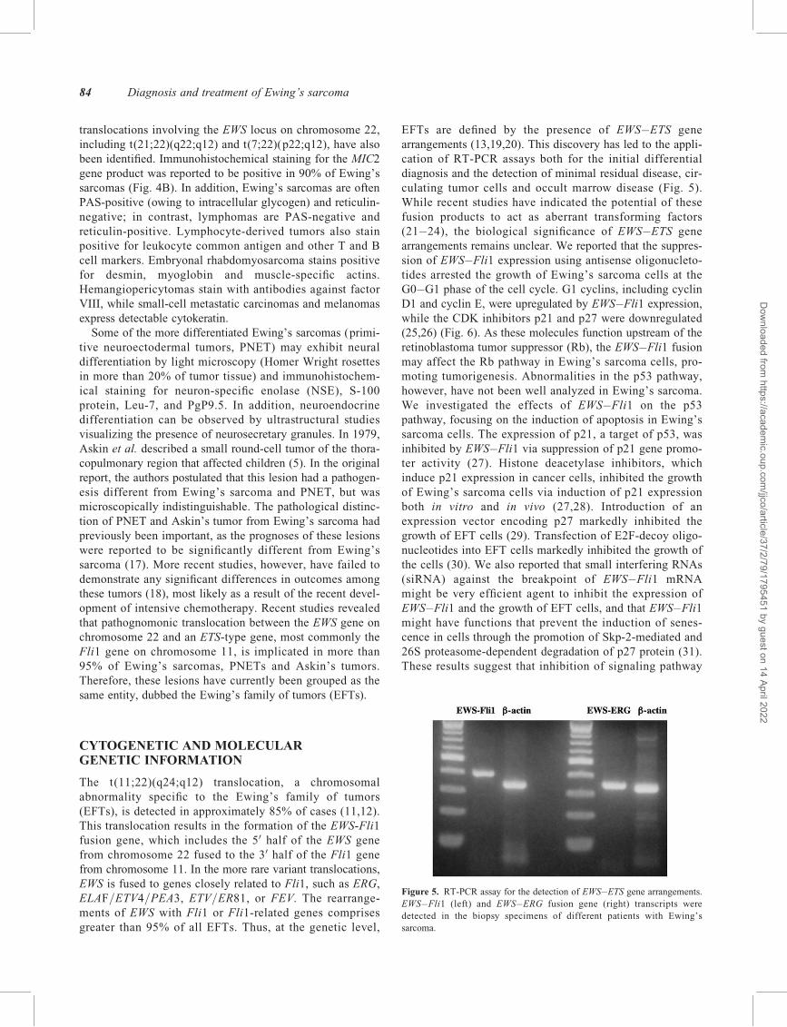
cation of RT-PCR assays both for the initial differential
diagnosis and the detection of minimal residual disease, cir-
culating tumor cells and occult marrow disease (Fig. 5).
While recent studies have indicated the potential of these
fusion products to act as aberrant transforming factors
(21 – 24), the biological significance of EWS – ETS gene
arrangements remains unclear. We reported that the suppres-
sion of EWS–Fli1 expression using antisense oligonucleto-
tides arrested the growth of Ewing’s sarcoma cells at the
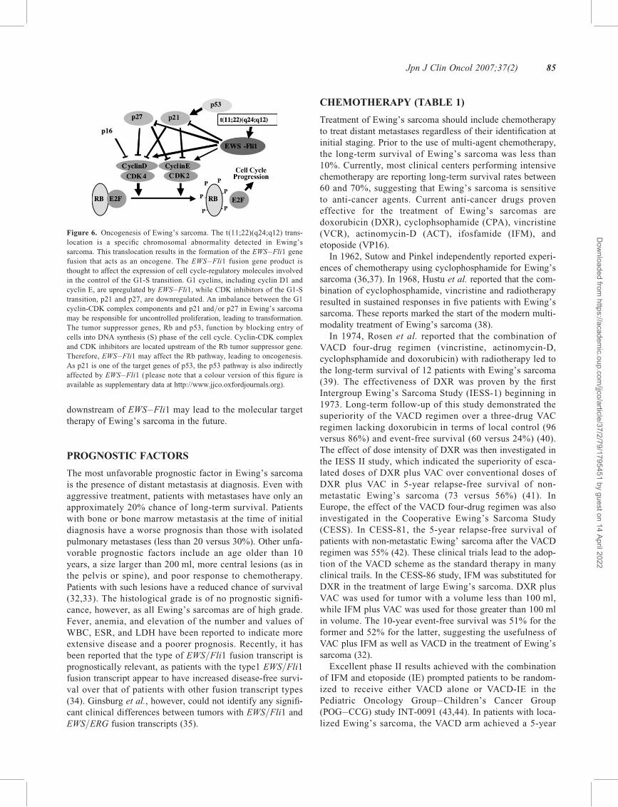
G0–G1 phase of the cell cycle. G1 cyclins, including cyclin
D1 and cyclin E, were upregulated by EWS–Fli1 expression,
while the CDK inhibitors p21 and p27 were downregulated
(25,26) (Fig. 6). As these molecules function upstream of the
retinoblastoma tumor suppressor (Rb), the EWS–Fli1 fusion
may affect the Rb pathway in Ewing’s sarcoma cells, pro-
moting tumorigenesis. Abnormalities in the p53 pathway,
however, have not been well analyzed in Ewing’s sarcoma.
We investigated the effects of EWS – Fli1 on the p53
pathway, focusing on the induction of apoptosis in Ewing’s
sarcoma cells. The expression of p21, a target of p53, was
inhibited by EWS–Fli1 via suppression of p21 gene promo-
ter activity (27). Histone deacetylase inhibitors, which
induce p21 expression in cancer cells, inhibited the growth
of Ewing’s sarcoma cells via induction of p21 expression
both in vitro and in vivo (27,28). Introduction of an
expression vector encoding p27 markedly inhibited the
growth of EFT cells (29). Transfection of E2F-decoy oligo-
nucleotides into EFT cells markedly inhibited the growth of
the cells (30). We also reported that small interfering RNAs
(siRNA) against the breakpoint of EWS – Fli1 mRNA
might be very efficient agent to inhibit the expression of
EWS–Fli1 and the growth of EFT cells, and that EWS–Fli1
might have functions that prevent the induction of senes-
cence in cells through the promotion of Skp-2-mediated and
26S proteasome-dependent degradation of p27 protein (31).
These results suggest that inhibition of signaling pathway
Figure 5. RT-PCR assay for the detection of EWS–ETS gene arrangements.
EWS – Fli1 (left) and EWS –ERG fusion gene (right) transcripts were
detected in the biopsy specimens of different patients with Ewing’s
sarcoma.
84 Diagnosis and treatment of Ewing’s sarcoma
Dow
nloaded from https://academ
ic.oup.com/jjco/article/37/2/79/1795451 by guest on 14 April 2022
downstream of EWS–Fli1 may lead to the molecular target
therapy of Ewing’s sarcoma in the future.
PROGNOSTIC FACTORS
The most unfavorable prognostic factor in Ewing’s sarcoma
is the presence of distant metastasis at diagnosis. Even with
aggressive treatment, patients with metastases have only an
approximately 20% chance of long-term survival. Patients
with bone or bone marrow metastasis at the time of initial
diagnosis have a worse prognosis than those with isolated
pulmonary metastases (less than 20 versus 30%). Other unfa-
vorable prognostic factors include an age older than 10
years, a size larger than 200 ml, more central lesions (as in
the pelvis or spine), and poor response to chemotherapy.
Patients with such lesions have a reduced chance of survival
(32,33). The histological grade is of no prognostic signifi-
cance, however, as all Ewing’s sarcomas are of high grade.
Fever, anemia, and elevation of the number and values of
WBC, ESR, and LDH have been reported to indicate more
extensive disease and a poorer prognosis. Recently, it has
been reported that the type of EWS/Fli1 fusion transcript is
prognostically relevant, as patients with the type1 EWS/Fli1
fusion transcript appear to have increased disease-free survi-
val over that of patients with other fusion transcript types
(34). Ginsburg et al., however, could not identify any signifi-
cant clinical differences between tumors with EWS/Fli1 and
EWS/ERG fusion transcripts (35).
CHEMOTHERAPY (TABLE 1)
Treatment of Ewing’s sarcoma should include chemotherapy
to treat distant metastases regardless of their identification at
initial staging. Prior to the use of multi-agent chemotherapy,
the long-term survival of Ewing’s sarcoma was less than
10%. Currently, most clinical centers performing intensive
chemotherapy are reporting long-term survival rates between
60 and 70%, suggesting that Ewing’s sarcoma is sensitive
to anti-cancer agents. Current anti-cancer drugs proven
effective for the treatment of Ewing’s sarcomas are
doxorubicin (DXR), cyclophsophamide (CPA), vincristine
(VCR), actinomycin-D (ACT), ifosfamide (IFM), and
etoposide (VP16).
In 1962, Sutow and Pinkel independently reported experi-
ences of chemotherapy using cyclophosphamide for Ewing’s
sarcoma (36,37). In 1968, Hustu et al. reported that the com-
bination of cyclophosphamide, vincristine and radiotherapy
resulted in sustained responses in five patients with Ewing’s
sarcoma. These reports marked the start of the modern multi-
modality treatment of Ewing’s sarcoma (38).
In 1974, Rosen et al. reported that the combination of
VACD four-drug regimen (vincristine, actinomycin-D,
cyclophsphamide and doxorubicin) with radiotherapy led to
the long-term survival of 12 patients with Ewing’s sarcoma
(39). The effectiveness of DXR was proven by the first
Intergroup Ewing’s Sarcoma Study (IESS-1) beginning in
1973. Long-term follow-up of this study demonstrated the
superiority of the VACD regimen over a three-drug VAC
regimen lacking doxorubicin in terms of local control (96
versus 86%) and event-free survival (60 versus 24%) (40).
The effect of dose intensity of DXR was then investigated in
the IESS II study, which indicated the superiority of esca-
lated doses of DXR plus VAC over conventional doses of
DXR plus VAC in 5-year relapse-free survival of non-
metastatic Ewing’s sarcoma (73 versus 56%) (41). In
Europe, the effect of the VACD four-drug regimen was also
investigated in the Cooperative Ewing’s Sarcoma Study
(CESS). In CESS-81, the 5-year relapse-free survival of
patients with non-metastatic Ewing’ sarcoma after the VACD
regimen was 55% (42). These clinical trials lead to the adop-
tion of the VACD scheme as the standard therapy in many
clinical trails. In the CESS-86 study, IFM was substituted for
DXR in the treatment of large Ewing’s sarcoma. DXR plus
VAC was used for tumor with a volume less than 100 ml,
while IFM plus VAC was used for those greater than 100 ml
in volume. The 10-year event-free survival was 51% for the
former and 52% for the latter, suggesting the usefulness of
VAC plus IFM as well as VACD in the treatment of Ewing’s
sarcoma (32).
Excellent phase II results achieved with the combination
of IFM and etoposide (IE) prompted patients to be random-
ized to receive either VACD alone or VACD-IE in the
Pediatric Oncology Group – Children’s Cancer Group
(POG–CCG) study INT-0091 (43,44). In patients with loca-
lized Ewing’s sarcoma, the VACD arm achieved a 5-year
Figure 6. Oncogenesis of Ewing’s sarcoma. The t(11;22)(q24;q12) trans-
location is a specific chromosomal abnormality detected in Ewing’s
sarcoma. This translocation results in the formation of the EWS–Fli1 gene
fusion that acts as an oncogene. The EWS– Fli1 fusion gene product is
thought to affect the expression of cell cycle-regulatory molecules involved
in the control of the G1-S transition. G1 cyclins, including cyclin D1 and
cyclin E, are upregulated by EWS–Fli1, while CDK inhibitors of the G1-S
transition, p21 and p27, are downregulated. An imbalance between the G1
cyclin-CDK complex components and p21 and/or p27 in Ewing’s sarcoma
may be responsible for uncontrolled proliferation, leading to transformation.
The tumor suppressor genes, Rb and p53, function by blocking entry of
cells into DNA synthesis (S) phase of the cell cycle. Cyclin-CDK complex
and CDK inhibitors are located upstream of the Rb tumor suppressor gene.
Therefore, EWS–Fli1 may affect the Rb pathway, leading to oncogenesis.
As p21 is one of the target genes of p53, the p53 pathway is also indirectly
affected by EWS–Fli1 (please note that a colour version of this figure is
available as supplementary data at http://www.jjco.oxfordjournals.org).
Jpn J Clin Oncol 2007;37(2) 85
Dow
nloaded from https://academ
ic.oup.com/jjco/article/37/2/79/1795451 by guest on 14 April 2022
event-free survival rate of 54%, while the VACD-IE arm
achieved a rate of 69% (33). Therefore, the VACD-IE
regimen was adopted as standard therapy for localized
Ewing’s sarcoma.
To achieve treatment intensification in Ewing’s sarcoma,
high-dose chemotherapy with autologous hematopoetic stem
cell rescue (HDT) was attempted. In most studies, HDTs
were reserved for high-risk patients, typically those with
metastases or recurrence, because of the considerable tox-
icity of this approach. There has not been a controlled ran-
domized clinical study, however, that was able to prove the
superiority of HDT (45–48).
LOCAL TREATMENT; SURGERYAND/OR IRRADIATION?
Local treatment of the primary lesion remains controversial.
Previous reports demonstrated a decrease in the rate of local
recurrence (,10%) and an increase in the rate of overall
survival with wide resection of the primary tumor. In
addition, retrospective analyses by several groups provide the
impression that local control is preferable when surgery is
possible (49,50). However, there have not been any random-
ized trials comparing local therapy modalities; there may
also be a selection bias favoring a subset of patients for
whom surgery is applicable. Therefore, the choice between
surgery and irradiation as a method for control of the
primary lesion should be made on an individual basis.
If pre-operative imaging suggests that it will likely be
possible to resect the lesion with wide margins, wide
resection without irradiation is the treatment of choice for
primary lesions. If the possibility of achievement of adequate
surgical margins is uncertain, pre-operative radiotherapy
should be added. As Ewing’s sarcomas are sensitive to both
chemotherapy and irradiation, even questionable candidates
for limb salvage may be eligible after neoadjuvant che-
motherapy with or without irradiation. If the surgical
margins are found to be inadequate after surgery, postopera-
tive radiotherapy may also be added. When surgical margins
are certain to be inadequate at preoperative imaging, amputa-
tion may be the only surgical option available. Central,
large, unresectable primary tumors are sometimes treated
with radiation alone. A debulking intralesional procedure
does not improve local control; in the CESS and EICESS
trials, patients who had an intralesional resection followed
by radiotherapy displayed the same local control rate as
those who were treated with radiotherapy alone (49).
SURGICAL MARGIN
The current standard treatment schedules for resectable
Ewing’s sarcoma begin with neoadjuvant chemotherapy,
followed by limb salvage procedure and post-operative
adjuvant chemotherapy. Although amputation had been the
only surgical method for several decades, limb salvage pro-
cedures, which include local resection and reconstruction,
are currently performed in almost all the cases of Ewing’s
sarcomas. Limb salvage procedures can be performed
without compromising survival rates (49–51).
Table 1. Summary of treatment results in Ewing’s sarcoma
Study Period Patients Regimen Results (5-year DFS) Reference
IESS-I 1973–1978 342 VAC 24 40
VACþWLI 44
VACD 60
IESS-II 1978–1982 214 VACD-HD 73
VACD-MD 56
CESS-81 1981–1985 93 VACD Tumor size , 100 ml 41
80% (3 years)
Tumor size . 100 ml
31% (3 years)
CESS-86 1986–1991 301 SR: VACD 52% 32
HR: VAID 51% (10 years)
INT-0091 1988–1992 (non-metastatic) 398 VACD 54% 33
VACD-IE 69%
DFS, disease free survival; WLI, whole lung irradiation; HD, high-dose; MD, moderate-dose; SR, standard risk; HR, high risk; VAC, vincristine, actinomycinD, cyclophosphamide; VACD, vincristine, actinomycin D, cyclophosphamide, doxorubicin; VACD-IE, vincristine, actinomycin D, cyclophosphamide,doxorubicin, ifosfamide, etoposide; VAID, vincristine, actinomycin D, ifosfamide, doxorubicin.
86 Diagnosis and treatment of Ewing’s sarcoma
Dow
nloaded from https://academ
ic.oup.com/jjco/article/37/2/79/1795451 by guest on 14 April 2022
When describing a surgical procedure, it is imperative that
the surgical margin be appropriately defined. The terms
‘amputation’ and ‘resection’ mean little without a modifier
describing the margins, especially when evaluating surgical
procedures and outcomes in the literature. In orthopaedic
oncology, surgical margins can be described by one of four
terms: intralesional, marginal, wide, or radical (52). An
intralesional margin is one in which the plane of surgical
dissection is within the tumor, which is often called ‘debulk-
ing’, because it leaves gross residual tumor behind. A mar-
ginal margin is achieved when the closest plane of dissection
passes through the pseudocapsule of the tumor. The pseudo-
capsule, however, often contains microscopic tumor foci.
Marginal resection often leads to local recurrence if the
remaining tumor cells do not respond to adjuvant chemother-
apy or radiation therapy. Wide margins are achieved when
the plane of dissection is in normal tissue. Wide margins are
the goal for most procedures, especially with high-grade
malignancies such as Ewing’s sarcoma. Radical margins are
achieved when all compartments that contain tumor are
removed en bloc.
RECONSTRUCTION (FIGS. 2 AND 3)
After resection of Ewing’s sarcomas, large bone defects
should be reconstructed to restore the function of the
affected limbs. The main options for reconstruction include
autogenous bone grafts, allogeneic bone grafts and
endoprosthesis.
Autogeneous bone grafts may be vascularized; vascular-
ized bone autograft operations are now performed widely as
a result of the development of microsurgery. As blood flow
can be preserved and the cells in the grafted bone remain
alive, bone formation and bone fusion are vigorous. This
technique has generated remarkable improvement in thera-
peutic success rates (53). Because of the limited amounts of
bone that can be collected, however, it is sometimes difficult
to repair large bone defects; in such cases, allogeneic bone
grafts or endoprosthesis is indicated.
Allografts are a form of reconstruction utilizing dead
bone. Frozen or freeze-dried bone allografts have been
widely used for limb salvage procedures in western
countries. Although fracture and non-union of the grafts can
reduce success rates, acceptable functional limbs can be
recreated with allografts (54). Allografts can be difficult to
obtain in some Asian countries, especially Japan and Korea,
for socio-religious reasons (55,56). Therefore, recycling of
affected bone has been adopted in Japan. Several methods
have been developed to allow re-use of resected bones for
reconstruction, including irradiation (57), autoclaving (58)
(Fig. 3D), pasteurization (59), and treatment with liquid
nitrogen (60).
Endoprosthetic replacement after excision of the tumor
can provide excellent results more rapidly than other
methods (Fig. 2C). Therefore, the most popular
reconstruction method after resection of malignant bone
tumors is prosthetic replacement (61). The late complications
of this method, such as a loosening, infection and fracture of
the prosthesis after replacement, have not been solved. More
successful methods for reconstruction than those in existence
need to be explored in the future.
Acknowledgments
This study was supported in part by Grant-in-Aid for
Clinical Cancer Research and Grants-in-Aid for Cancer
Research (14S-4 and -5) from the Ministry of Health, Labor
and Welfare, Japan.
Conflict of interest statement
None declared.
References
1. Extraskeletal Ewing’s sarcoma/primitive neuroectodermal tumorfamily. In: Enzinger and Weiss’s Soft Tissue Tumors, 4th edn. StLouis: Mosby 2001; 289–1291.
2. Stout AP. A tumor of ulnar nerve. Proc NY Pathol Soc 1918;21:2–12.3. Ewing J. Diffuse endothelioma of bone. Proc NY Pathol Soc
1921;21:17–24.4. Angerval L, Enzinger FM. Extraskeletal neoplasm resembling Ewing’s
sarcoma. Cancer 36:240–251, 19755. Askin FB, Rosai J, Sibley RK, Dehner LP, McAlister WH. Malignant
small cell tumor of the thoracopulmonary region in childhood: adistinctive clinicopathologic entity of uncertain histogenesis. Cancer1979;43:2438–51.
6. Jaffe R, Santamaria M, Yunis EJ, Yunis EJ, Tannery NH, Agostini RMJr, et al. The neuroectodermal tumor of bone. Am J Surg Patol1984;8:885–98.
7. Aurias A, Rimbaut C, Buffe D, Zucker JM, Mazabraud A.Translocation involving chromosome 22 in Ewing’s sarcoma: acytogenetic study of four fresh tumors. Cancer Genet Cytogenet1984;12:21–5.
8. Whang-Peng J, Triche TJ, Knutsen T, Miser J, Douglass EC, Israel MA.Chromosomal translocation in peripheral neuroepithelioma. N Engl JMed 1984;311:584–5.
9. Delattre O, Zucman J, Plougastel B, Desmaze C, Melot T, Peter M,et al. Gene fusion with an ETS DNA-binding domain causedby chromosome translocation in human tumours. Nature 1992;359:162–5.
10. JOA Musculoskeleta Tumor Committee: The Incidence of BoneTumours in Japan, 2003. Tokyo, Japan: National Cancer Institute;2003.
11. Dorfman HD, Czerniak B. Bone cancers. Cancer 1995;75:203–10.12. De Alava E, Lozano MD, Patino A, Sierrasesumaga L, Pardo-
Mindan FJ. Ewing family tumors: potential prognostic value ofreverse-transcriptase polymerase chain reaction detection of minmalresidual disease in peripheral blood samples. Diagn Mol Pathol1998;7:152–7.
13. Van der Woude HJ, Bloem JL, Hogendoorn PC. Preoperative evaluationand monitoring chemotherapy in patients with high-grade osteogenicand Ewing’s sarcoma: review of current imaging modalities. SkeletalRadiol 1998;27:57–71.
14. Enneking WF, Spanier SS, Goodman MA. A system for the surgicalstaging of musculoskeletal sarcoma. Clin Orthop Relat Res 1980;153:106–20.
15. Musculoskeletal Sites. In: American Joint Committee on Cancer. AJCCCancer Staging Manual, 6th edn. New York: Springer 2002;185–200.
16. Daldrup-Link HE, Franzius C, Link TM, Laukamp D,Sciuk J, Jurgens H, et al. Whole-body MR imaging for detection ofbone metastases in children and young adults: comparison with skeletalscintigraphy and FDG PET. Am J Roentgenol 2001;177:229–36.
Jpn J Clin Oncol 2007;37(2) 87
Dow
nloaded from https://academ
ic.oup.com/jjco/article/37/2/79/1795451 by guest on 14 April 2022

17. Primitive neuroectodermal tumors related lesions. In: Enzinger andWeiss’s Soft Tissue Tumors, 4th edn. St Louis: Mosbey 2001;1305–7.
18. Parham DM, Hijazi Y, Steinberg SM, Meyer WH,Horowitz M, Tzen CY, et al. Neuroectodermal differentiation inEwing’s sarcoma family of tumors does not predict tumor behavior.Hum Pathol 1999;30:911–8.
19. Yang L, Chansky HA, Hickstein DD. EWS-Fli1 fusion protein interactswith hyperphosphorylated RNA polymerase II and interferes withserine-arginine protein-mediated RNA splicing. J Biol Chem2000;275:37612–8.
20. Lin PP, Brody RI, Hamelin AC, Bradner JE, Healey JH, Ladanyi M.Differential transactivation by alternative EWS-Fli1 fusion proteinscorrelates with clinical heterogeneity in Ewing’s sarcoma. Cancer Res1999;59:1428–32.
21. De Alava E, Kawai A, Healey JH, Fligman I, Meyers PA, Huvos AG,et al. EWS-Fli1 fusin transcript structure is an independent determinantof prognosis in Ewing’s sarcoma. J Clin Oncol 1998;16:1248–55.
22. Kim J, Pelletier J. Molecular genetics of chromosome translocationinvolving EWS and related family members. Physiol Genomics1999;1:127–38.
23. Ouchida M, Ohno T, Fujimura Y, Rao VN, Reddy ES. Loss oftumorigenicity of Ewing’s sarcoma cells expressing antisense RNA toEWS-fusion transcripts. Oncogene 1995;11:1049–54.
24. May WA, Gishizky ML, Lessnick SL, Lunsford LB,Lewis BC, Delattre O, et al. Ewing sarcoma 11;22 translocationproduces a chimeric transcription factor that require the DNA-bindingdomain encoded by Fli-1 for transformation. Proc Natl Acad Sci USA1993;90:5752–6.
25. Tanaka K, Iwakuma T, Harimaya K, Sato H, Iwamoto Y. EWS-Fli1antisense oligodeoxynucleotide inhibits proliferation of human Ewing’ssarcoma and primitive neuroectodermal tumor cells. J. Clin Invest1997;99:239–47.
26. Matsumoto Y, Tanaka K, Nakatani F, Matsunobu T,Matsuda S, Iwamoto Y. Downregulation and forced expression ofEWS-Fli1 fusion gene results in changes in the expression of G1regulatory genes. Br J Cancer 2001;84:768–75.
27. Nakatani F, Tanaka K, Sakimura R, Matsumoto Y, Matsunobu T, Li X,et al. Identification of p21(WAF1/CIP1) as a direct target of EWS-Fli1oncogenic fusion protein. J Biol Chem 2003;278:15105–15.
28. Sakimura R, Tanaka K, Nakatani F, Matsunobu T, Li X, Hanada M,et al. Antitumor effects of histone deacetylase inhibitor on Ewing’sfamily tumors. Int J Cancer 2005;116:784–92.
29. Matsunobu T, Tanaka k, Matsumoto Y, Nakatani F,Sakimura R, Hanada M, et al. The prognostic and therapeutic relevanceof p27 kip1 in Ewing’s family tumors. Clin Cancer Res 2004;10:1003–12.
30. Li Xu, Tanaka K, Nakatani F, Matsunobu T, Sakimura R, Hanada M,et al. Transactivation of cyclin E gene by EWS – Fli1 and antitumoreffects of cyclin dependent kinase inhibitor on Ewing’s family tumorcells. Int J Cancer 2005;116:385–94.
31. Matsunobu T, Tanaka K, Nakamura T, Nakatani F,Sakimura R, Hanada M, et al. The possible role of EWS – Fli1 inevasion of senescence in Ewing family tumors. Cancer Res2006;66:803–11.
32. Paulussen M, Ahrens S, Dunst J, Winkelmann W, Exner GU,Kotz R, et al. Localized Ewing tumor of bone: final results of thecooperative Ewing’s Sarcoma Study CESS 86. J Clin Oncol2001;19:1818–29.
33. Grier HE, Krailo MD, Tarbell NJ, Link MP, Fryer CJ, Pritchard DJ,et al. Addition of ifosfamide and etoposide to standard chemotherapyfor Ewing’s sarcoma and primitive neuroectodermal tumor of bone. NEngl J Med 2003;348:694–701.
34. De Alava E, Kawai A, Healey JH, Fligman I, Meyers PA, Huvos AG,et al. EWS/Fli1 fusion transcript structure is an independentdeterminant of prognosis in Ewing’s sarcoma. J Clin Oncol 1998;16:1248–55.
35. Ginsberg JP, de Alava E, Ladanyi M, Wexler LH,Kovar H, Paulussen M, et al. EWS/Fli1 and EWS/ERG gene fusionsare associated with similar clinical phenotypes in Ewing’s sarcoma.J Clin Oncol 1999;17:1809–14.
36. Sutow WW, Sullivan MP. Cyclophosphamide therapy in children withEwing’s sarcoma. Cancer Chemother Rep 1962;23:55–60.
37. Pinkel D. Cyclophsphamide in children with cancer. Cancer1962;15:42–9.
38. Hustu HO, Holton C, James D Jr, Pinkel D. Treatment of Ewing’ssarcoma with concurrent radiotherapy and chemotherapy. J Pediatr
1968;73:249–51.39. Rosen G, Wollner N, Tan C, Wu SJ, Hajdu SI, Cham W, et al.
Proceedings: disease-free survival in children with Ewing’s sarcomatreated with radiation therapy and adjuvant four-drug sequentialchemotherapy. Cancer 1974;33:384–93.
40. Nesbit ME Jr, Gehan EA, Burgert EO Jr, Vietti TJ, Cangir A, Tefft M,et al. Multimodal therapy for the management of primary,nonmetastatic Ewing’s sarcoma of bone: a long-term follow-up of theFirst Intergroup Study. J Clin Oncol 1990;8:1664–74.
41. Burgert EO Jr, Nesbit ME, Garnsey LA, Gehan EA,Herrmann J, Vietti TJ, et al. Multimodal therapy for the management ofnonpelvic, localized Ewing’s sarcoma of bone: intergroup study ofIESS-II. J Clin Oncol 1990;8:1514–24.
42. Jurgens H, Exner U, Gadner H, Harms D, Michaelis J, Sauer R, et al.Multidisciplinary treatment of primary Ewing’s sarcoma of bone. A6-year experience of a European Cooperative Trial. Cancer
1988;61:23–32.43. Kung FH, Pratt CB, Vega RA, Jaffe N, Strother D, Schwenn M, et al.
Ifosfamide/etoposide combination in the treatment of recurrentmalignant solid tumors of childhood. A Pediatric Oncology GroupPhase II study. Cancer 1993;71:1898–903.
44. Miser JS, Kinsella TJ, Triche TJ, Tsokos M, Jarosinski P, Forquer R,et al. Ifosfamide with Mesna uroprotection and etoposide: an effectiveregimen in the treatment of recurrent sarcomas and other tumors ofchildren and young adults. J Clin Oncol 1987;5:1191–8.
45. Meyers PA, Krailo MD, Ladanyi M, Chan KW, Sailer SL, Dickman PS,et al. High-dose melphalan, etoposide, total-body irradiation, andautologous stem-cell reconstitution as consolidation therapy forhigh-risk Ewing’s sarcoma does not improve prognosis. J Clin Oncol
2001;19:2812–20.
46. Kinsella TJ, Glaubiger D, Diesseroth A, Makuch R, Waller B, Pizzo P,et al. Intensive combined modality therapy including low-dose TBI inhigh-risk Ewing’s sarcoma patients. Int J Radiat Oncol Biol Phys
1983;9:1955–60.47. Burdach S, Jurgens H, Peters C, Nurnberger W, Mauz-
Korholz C, Korholz D, et al. Myeloabolative radiochemotherapy andhematopoietic stem-cell rescue in poor-prognosis Ewing’s sarcoma. J
Clin Oncol 1993;11:1482–8.48. Tanaka K, Matsunobu T, Sakamoto A, Matsuda S, Iwamoto Y.
High-dose chemotherapy and autologous peripheral blood stem-celltransfusion after conventional chemotherapy for patients with high-riskEwing’s tumors. J Orthop Sci 2002;7:477–82.
49. Schuck A, Ahrens S, Paulussen M, Kuhlen M, Konemann S, Rube C,et al. Local therapy in localized Ewing tumors: results of 1058 patientstreated in the CESS81, CESS86 and EICESS92 trials. Int J Radiat
Oncol Biol Phys 2003;55:168–77.50. Sailer SL, Harmon DC, Mankin HJ, Truman JT, Suit HD. Ewing’s
sarcoma: surgical resection as a prognostic factor. Int J Radiat Oncol
Biol Phys 1988;15:43–52.51. Bacci G, Ferrari S, Bertoni F, Rimondini S, Longhi A, Bacchini P, et al.
Prognostic factors in nonmetastatic Ewin’s srcoma of bone treated withadjuvant chemotherapy: analysis of 359 patients at the IntitutoOrthopedico Rizzoli. J Clin Oncol 2000;18:4–11.
52. Enneking WF. Musculoskeletal Tumor Surgery. New York: ChurchillLivingstone 1983.
53. Wada T, Usui M, Isu K, Yamawaki S, Ishii S. Reconstruction and limbsalvage after resection for malignant bone tumour of the proximalhumerus: a sling procedure using a free vascularized fibular graft.J Bone Joint Surg Br 1999;81:808–13.
54. Mankin HJ, Fogelson FS, Thrasher AZ, Jaffer F. Massive resection andallograft transplantation in the treatment of malignant of bone tumors. N
Engl J Med 1976;294:1247–55.55. Iwamoto Y, Sugioka Y, Chuman H, Matsuda S, Hotokebuchi T,
Kawai S, et al. Nationwide survey of bone grafting performedfrom 1980 through 1989 in Japan. Clin Orthop Relat Res 1997;335:292–7.
56. Iwamoto Y. Clinical results of bone grafting. Jpn Med Assoc J
2002;45:358–60.
57. Tsuboyama T, Toguchida J, Kotoura Y, Kasahara K, Hiraoka M,Nakamura T. Intra-operative radiation therapy for osteosarcoma in theextremities. Int Orthop 2000;24:202–7.
88 Diagnosis and treatment of Ewing’s sarcoma
Dow
nloaded from https://academ
ic.oup.com/jjco/article/37/2/79/1795451 by guest on 14 April 2022

58. Lauritzen C, Alberius P, Santanelli F, Vallfors B, Lilja J, Stephensen H.Repositioning of craniofacial tumourous bone after autoclaving. Scand JPlast Reconstr Surg Hand Surg 1991;25:161–5.
59. Manabe J, Kawaguchi N, Matsumoto S. Pasteurized autogenous bonegraft for reconstrucion after resection of malignant bone and soft tissuetumors: imaging features. Semin Musculoskelet Radiol 2001;5:195–201.
60. Tsuchiya H, Wan SL, Sakayama K, Yamamoto N, Nishida H,Tomita K. Reconstruction using an autograft containing tumour treatedby liquid nitrogen. J Bone Joint Surg Br 2005;87: 218–25.
61. Malawer MM, Chou LB. Prosthetic survival and clinical results withuse of large segment replacements in the treatment of high-grade bonesarcomas. J Bone Joint Surg Am 1995;77:1154–65.
Jpn J Clin Oncol 2007;37(2) 89
Dow
nloaded from https://academ
ic.oup.com/jjco/article/37/2/79/1795451 by guest on 14 April 2022