evaluation of client-specific outcome measures and activity monitoring to measure pain relief in...
TRANSCRIPT

Evaluation of Client-Specific Outcome Measures and ActivityMonitoring to Measure Pain Relief in Cats with Osteoarthritis
B. Duncan X. Lascelles, Bernie D. Hansen, Simon Roe, Venita DePuy, Andrea Thomson,Courtney C. Pierce, Eric S. Smith, and Elizabeth Rowinski
Background: There are no validated systems for measuring pain from osteoarthritis in cats.
Hypothesis: Owner subjective assessments and an activity monitor (AM) can be used to detect pain in cats with
osteoarthritis and to assess efficacy of treatments.
Animals: Thirteen cats older than 10 years old, with owner-assessed decreases in activity, painful arthritic joints, and
clinically normal blood work were included and evaluated for 3 weeks.
Methods: A collar-mounted AM measured activity and a client-specific outcome measure (CSOM) questionnaire
characterized the severity of impairment. Overall global quality of life was also evaluated for each treatment. In weeks 2 and 3,
meloxicam (0.1 mg/kg, day 1; 0.05 mg/kg, days 2–5) or a placebo was administered in a blinded, randomized, cross-over
manner to test the assessment systems.
Results: The cats had a median of 4 arthritic appendicular joints. Activity counts for the week when cats (complete data on
activity; n 5 9) were administered meloxicam were significantly higher than at baseline (P 5 .02) but not after placebo (P 5
.06). Baseline activity counts were not significantly different from placebo (P 5 .6). The CSOM data (n 5 13) showed that
owners considered their cats to be more active on meloxicam compared with baseline (P 5 .001) and placebo (P , .004), and
more active on placebo than at baseline (P , .01). Global quality of life improved significantly with meloxicam (P , .042).
Conclusions and Clinical Importance: Both an AM and a CSOM system can detect behavior associated with pain relief in
cats that are arthritic. Objective activity data might allow subjective assessment systems to be validated for use in clinical
studies.
Key words: Activity; Degenerative joint disease; Feline; Owner; Subjective assessment.
A ppendicular joint osteoarthritis (OA) is present inapproximately 25–30% of dogs1–3 and is a poten-
tially painful condition.4 However, little is known aboutthe incidence of OA or degenerative joint disease in cats.It is assumed that OA is the most common form ofdegenerative joint disease in cats, but this has not beenfully evaluated. To date, the only studies performedwere retrospective evaluations of radiographs of catstaken for various reasons. These studies revealed lesionsconsistent with appendicular limb OA in 17 to 64% ofcats.5–7 Despite the frequency of OA in domestic cats,very little is known about its association with pain, and,indeed, it has been suggested that OA in cats is notpainful,8 although most clinicians believe it can beassociated with pain and impaired mobility.9 Studies todate of pain relief in cats with OA were not placebocontrolled or masked.10,11
There are currently no approved medications for thealleviation of feline OA pain. This is likely, in large part,because of the lack of validated techniques available tomeasure behavior associated with pain relief in feline OA.
Owner assessments of the degree of pain in dogs with OAcorrelate better with force plate evaluations than doassessments by veterinarians.a Subsequent work indicatedthat owners are able to assess chronic pain relief in theirarthritic dogs.12–15 To date, no one has comprehensivelyevaluated owner-based subjective assessment of painrelief in cats with OA.
Pain from knee and hip joints in humans can oftenresult in decreased mobility and decreased distancemoved.16 This decreased distance moved appears to beboth total daily distance and distance moved in 1 effort.Likewise, in dogs and cats, OA is also assumed to impairmobility and daily distance moved.9,10,13,14 Our laborato-ry recently evaluated an accelerometer and found thatactivity counts generated by the activity monitor (AM)correlates well with objectively measured activity incats.b
The study reported here was designed to test thefeasibility of using owner-based assessments and AMs todetect behavioral changes associated with pain relief incats with OA. The hypothesis was that subjectiveassessments by owners and an AM can be used to inferpain relief in cats with OA. By using cats with naturallyoccurring OA in their home environment, the specificaims were to determine if (1) activity counts measuredby a collar-mounted accelerometer-based AM increased,and (2) owner assessment of the ability of cats witharthritis to perform specific spontaneous activitiesshowed improvement after treatment with a nonsteroidalanti-inflammatory drug (NSAID). The NSAID therapyin this study was assumed to provide pain relief.
Materials and Methods
This study was approved by the Animal Care and Use
Committee at North Carolina State University (NCSU) and was
in accordance with the National Institutes of Health and the
From the Comparative Pain Research Laboratory, College of
Veterinary Medicine, North Carolina State University, Raleigh, NC
(Lascelles, Hansen, Roe, Thomson, Pierce, Smith, Rowinski); and
INC Research, Raleigh, NC (DePuy). This study was presented at the
2nd World/33rd Annual Veterinary Orthopedic Society Conference,
Keystone, CO, March 2, 2006, and at the NCSU College of Veterinary
Medicine Annual Research Forum, March 17, 2006.
Reprint requests: Dr B.D.X. Lascelles, Director, Comparative
Pain Research Laboratory, College of Veterinary Medicine, North
Carolina State University, Raleigh, NC 27606; e-mail: Duncan_
Submitted June 23, 2006; Revised September 16, 2006, October
24, 2006; Accepted November 19, 2006.
Copyright E 2007 by the American College of Veterinary Internal
Medicine
0891-6640/07/2103-0006/$3.00/0
J Vet Intern Med 2007;21:410–416
International Association for the Study of Pain policies on the use
of clinical subjects.
Animals
Cats whose owners considered that the cats had slowed down or
had impaired mobility were recruited from faculty, students, and
staff of the NCSU College of Veterinary Medicine (NCSU-CVM),
local practices, and the NCSU-CVM Integrated Pain Management
Service. All owners gave informed signed consent.
Evaluation of Potential StudyCandidates (‘‘Screening’’)
Cats, whose owners considered them as having slowed down or
having impaired mobility, were screened with a physical examina-
tion, orthopedic and neurologic evaluation, CBC, blood chemistry,
urine analysis, and orthogonal radiographs of every appendicular
joint and the entire axial skeleton. A single evaluator (BDXL)
performed the physical examinations. The physical examinations
were performed and recorded before radiographs were made. Each
cat was weighed, and the body condition score was recorded (on
a scale of 1–5: 1, very thin; 2, obvious abdominal waist; 3, ideal; 4,
no observable abdominal waist; 5, obese).c Cats with no detectable
systemic disease and, with at least 1 appendicular joint where
manipulation elicited an aversive response and whose radiographs
showed the presence of OA, were included. Criteria used to
determine the presence of radiographic signs of OA were those
currently used in dogs, because there are no validated descriptions
for the radiographic appearance of OA in cats. To be included in
the study, it was decided a priori that eligible cats were required to
be (a) not currently receiving any anti-inflammatory medications
(administration of glucosamine-chondroitin sulphate preparations
was acceptable as long as the administration of these had been
ongoing for at least 10 weeks and their administration was not
changed during the study period), (b) free from clinically significant
abnormal CBC or blood chemistry values; (c) an indoor-only cat;
(d) have owner-identified mobility impairment, with restriction of
specific activities; and (e) exhibit a painful response upon
manipulation of at least 1 joint that also had radiographic changes
consistent with OA. Cats were excluded if they had any clinically
detectable disease or were pregnant females. Only cats whose
owners were considered to have a stable routine of daily living that
was unlikely to change were included (ie, no impending changes,
such as moving house, marriages, vacations, or introduction of new
pets or people into the household during the proposed study
period).
Experimental Protocol
After the screening evaluation (n 5 19), 13 suitable cats
completed the study. In a randomized, masked (owners and
evaluators were not aware of the treatment being given), cross-over
design, cat activity was assessed by using a client-based subjective
assessment system (client-specific outcome measures [CSOM]) and
a collar-mounted accelerometer-based AMd for 3 weeks. Assess-
ments were made at days 0, 7, 14, and 21. In weeks 2 and 3, each cat
received both a 5-day period of an analgesic (NSAID meloxicame
(0.1 mg/kg on day 1, followed by 0.05 mg/kg daily for 4 days) and
a 5-day period of placebo in a randomized order (computer-
generated randomization). The treatments were administered on
days 3, 4, 5, 6, and 7 of weeks 2 and 3. The last dose of treatment
was given on the morning that the cats returned to the clinic for
evaluation. There was a 2-day break between each round of
medication. The placebo was made to appear and smell identical to
the test drug. The identity of the treatments was not revealed to the
principal investigator (BDXL) until completion of data analysis.
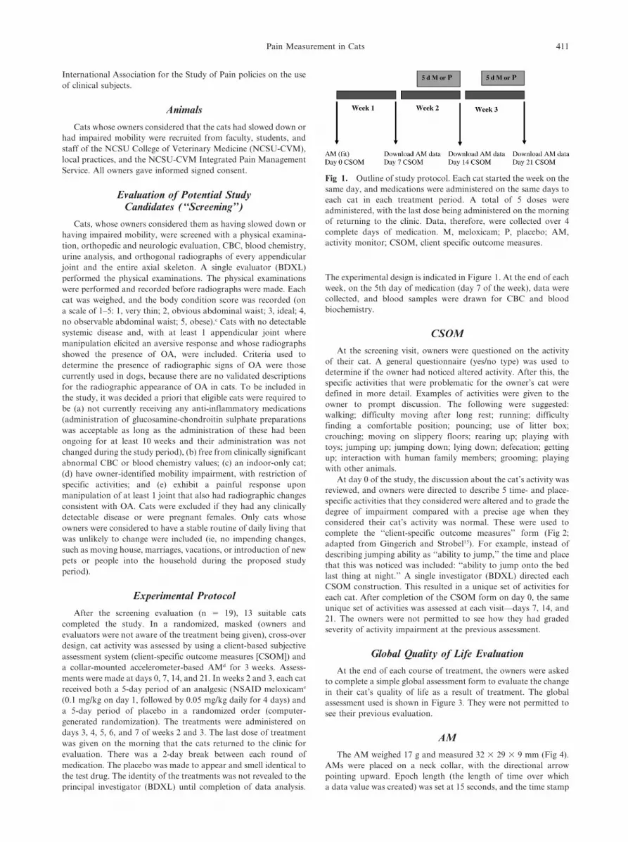
The experimental design is indicated in Figure 1. At the end of each
week, on the 5th day of medication (day 7 of the week), data were
collected, and blood samples were drawn for CBC and blood
biochemistry.
CSOM
At the screening visit, owners were questioned on the activity
of their cat. A general questionnaire (yes/no type) was used to
determine if the owner had noticed altered activity. After this, the
specific activities that were problematic for the owner’s cat were
defined in more detail. Examples of activities were given to the
owner to prompt discussion. The following were suggested:
walking; difficulty moving after long rest; running; difficulty
finding a comfortable position; pouncing; use of litter box;
crouching; moving on slippery floors; rearing up; playing with
toys; jumping up; jumping down; lying down; defecation; getting
up; interaction with human family members; grooming; playing
with other animals.
At day 0 of the study, the discussion about the cat’s activity was
reviewed, and owners were directed to describe 5 time- and place-
specific activities that they considered were altered and to grade the
degree of impairment compared with a precise age when they
considered their cat’s activity was normal. These were used to
complete the ‘‘client-specific outcome measures’’ form (Fig 2;
adapted from Gingerich and Strobel15). For example, instead of
describing jumping ability as ‘‘ability to jump,’’ the time and place
that this was noticed was included: ‘‘ability to jump onto the bed
last thing at night.’’ A single investigator (BDXL) directed each
CSOM construction. This resulted in a unique set of activities for
each cat. After completion of the CSOM form on day 0, the same
unique set of activities was assessed at each visit—days 7, 14, and
21. The owners were not permitted to see how they had graded
severity of activity impairment at the previous assessment.
Global Quality of Life Evaluation
At the end of each course of treatment, the owners were asked
to complete a simple global assessment form to evaluate the change
in their cat’s quality of life as a result of treatment. The global
assessment used is shown in Figure 3. They were not permitted to
see their previous evaluation.
AM
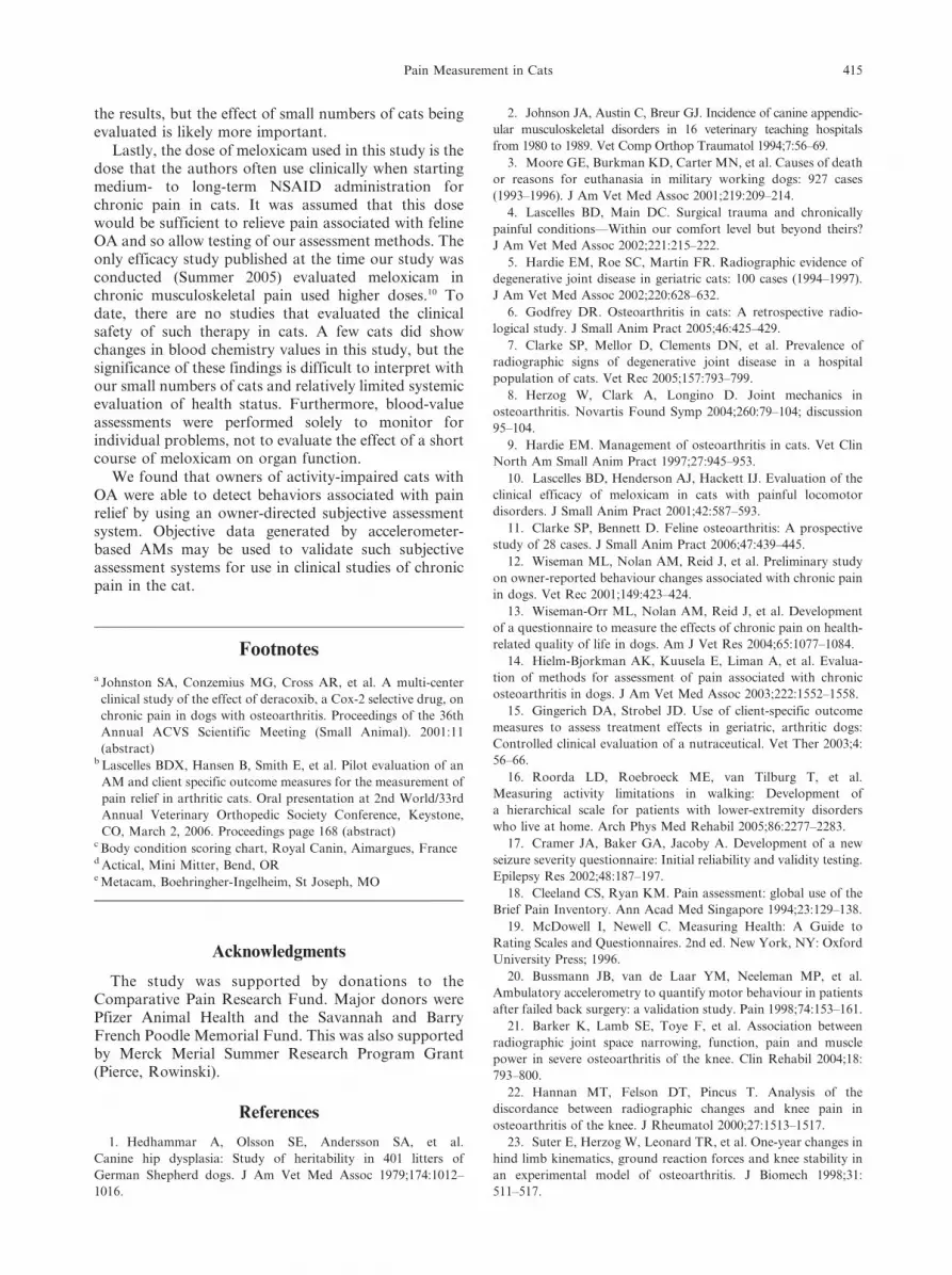
The AM weighed 17 g and measured 32 3 29 3 9 mm (Fig 4).
AMs were placed on a neck collar, with the directional arrow
pointing upward. Epoch length (the length of time over which
a data value was created) was set at 15 seconds, and the time stamp
Fig 1. Outline of study protocol. Each cat started the week on the
same day, and medications were administered on the same days to
each cat in each treatment period. A total of 5 doses were
administered, with the last dose being administered on the morning
of returning to the clinic. Data, therefore, were collected over 4
complete days of medication. M, meloxicam; P, placebo; AM,
activity monitor; CSOM, client specific outcome measures.
Pain Measurement in Cats 411

was synchronized with local time (Eastern Standard Time). After
fitting of the cats with the AM on day 0, it was worn on the collar
in their home environment for 3 weeks. The first week was
considered an acclimatization period. At each visit (day 7, 14, and
21), the AM was removed from the collar and placed on a telemetric
reader to download the data to a personal computer. The AM and
the collar were then replaced on the cat. The owners were asked to
indicate in a diary (also used to record any adverse events) any
times when the collar or the AM or both were removed. Activity
counts were summed for each day of the last 4 complete days of
each week.
Data Analysis
Repeated measures analysis of variance was used to evaluate the
effect of treatment on blood chemistry (serum urea nitrogen [SUN]
and creatinine concentrations, and alkaline phosphatase and alanine
aminotransferase [ALT] activities, and CBC, PCV, and total
protein).
The 5 ratings of impaired mobility (‘‘no problem,’’ ‘‘a little
problematic,’’ ‘‘quite problematic,’’ ‘‘severely problematic,’’ or
‘‘impossible’’) were converted to an ordinal scale, with 0 being ‘‘no
problem’’ and 4 being ‘‘impossible,’’ and were summed for each cat
at each visit, resulting in a possible range of 0 (no problems) to 20
(all listed activities impossible). Because multiple pairwise compar-
isons were being performed on each cat, a ‘‘difference’’ measure-
ment was calculated for each animal for each of the desired
comparisons (baseline day 0 to baseline day 7, day 7 to end of
active drug, day 7 to end of placebo). A nonparametric signed rank
test was used to evaluate whether or not these distributions were
centered at zero, which would equate to no significant changes
between the 2 time periods. All analyses were conducted at
a Bonferonni adjusted alpha 5 0.0167. This method of dividing the
original a 5 .05 by 3 (the number of comparisons) provides more
conservative results for multiple comparisons. Period effect was
evaluated, as was a sequence (carry-over) effect. Because there are
a variety of feasible approaches to this analysis, sensitivity analyses
were also performed to further check this statistical result. One was
based on the median mobility score for each cat at each visit.
Differences in medians were then calculated, and a signed rank test
was used to evaluate whether or not there was a significant change
between the 2 time periods. The second was based on a revised total
mobility score. This was created by combining the first 2 columns
(‘‘no problem’’ and ‘‘a little problematic’’), in addition to
combining the 3rd and 4th columns (‘‘quite problematic’’ and
‘‘severely problematic’’), resulting in a condensed 3-column grid,
with the final column being ‘‘impossible.’’ Subsequent analysis was
similar to the primary CSOM analysis.
For the global assessment, the categories ‘‘worse,’’ ‘‘same,’’
‘‘slightly improved,’’ ‘‘moderately improved,’’ and ‘‘very im-
proved’’ were assigned a numerical index (21, 0, 1, 2, and 3,
respectively). The values for each cat after each treatment were
compared as a group by using the Mann-Whitney rank sum test.
The activity counts for each cat for each of the 4 complete days
of medication or placebo administration were compared across
treatments (none, placebo, and NSAID) by using a Friedman
repeated measures analysis of variance, with a Tukey test for
pairwise multiple comparisons.
All analyses were conducted at a 5 0.05, with Bonferroni
correction as detailed above.
For the global assessment, the activity data were also evaluated
to determine if changes in activity were confined to any particular
time period of the day, with the time periods being 6-hour segments
starting at midnight.
Results
Nineteen cats were fully screened, and 14 met theinclusion criteria. One cat was subsequently removedfrom the study because of increases in SUN over the firstweek (no treatment had been given), and data werecollected from 13 cats. Their mean age was 14 years
Fig 2. Form used by owners to describe specific activities that they considered were altered.
Fig 3. Form used by owners to evaluate the change in their cat’s
quality of life at the end of each course of treatment.
412 Lascelles et al

(range, 10–19 years), mean weight at day 0 was 5.0 kg(range, 2.9–8.25 kg), and median body condition scorewas 2 (range, 1–4). Seven were spayed females, and 6were castrated males.
A median of 4 appendicular joints in each cat hadradiographic signs consistent with OA, with hip (total of16 joints affected) being the most commonly affected,followed by elbow (11 joints) and tarsus (11 joints), thenstifle (10), shoulder (5), and carpus (2). All the cats but 1had radiographic changes in the bones of the spinalcolumn that consisted of spondylosis deformans orradiographic signs or both consistent with intervertebraldisk disease.
The number of joints assessed was 208 (forelimb pes[all joints considered together], carpus, elbow, shoulder,hindlimb pes, hock, stifle, and hip). Pain was detected
upon manipulation of 55 joints; 18 joints had both painon manipulation and radiographic signs of OA; and 37joints had radiographic signs of OA, with no evidence ofpain upon manipulation. Similarly, across all cats, 52regions (cervical, thoracic, lumbar, lumbosacral) of theaxial skeleton were examined. There were 25 regions ofthe axial skeleton with radiographic pathology present,7 regions had both radiographic pathology and evidenceof pain upon manipulation, and 18 regions with noradiographic pathology produced behavioral signs ofpain on manipulation. There was no significant effect ofgroup or treatment with meloxicam or the placebo onthe blood parameters evaluated; however, these statis-tical tests had low power (0.05–0.27). Two cats had highSUN at baseline (48 and 40 mg/dL; reference range, 14–36 mg/dL), that normalized after meloxicam (33 and32 mg/dL). One had normal SUN at baseline (35 mg/dL), and it rose to 57 mg/dL after meloxicam; thecreatinine in this cat was 2.3 mg/dL at baseline(reference range, 0.6–2.4 mg/dL), and it rose to4.2 mg/dL after meloxicam. Subsequent follow-up re-vealed stable high normal SUN and creatinine serumconcentration. Two cats had high ALT activity in serum(114 and 117 U/L; reference range, 10–100 U/L) atbaseline, but subsequent values were within the referencerange. One cat had high ALT activity at baseline (312 U/L), and values remained high (185 U/L after placebo and392 U/L after meloxicam). All other values were withinnormal ranges.
CSOM data were collected on all 13 cats. However,AM data were collected for only 9 cats, because of AMremoval at some point during the data collection periodin 4 cats, thus, rendering those data sets incomplete.
CSOM Data
There was a significant improvement in mobilitywhen the cats were administered the NSAID (melox-icam) compared with being given the placebo (Table 1).There was also a significant improvement over baselinewhen the cats were given a placebo. However, the largestimprovement (lower numbers equate to higher mobility)from baseline occurred when meloxicam was adminis-tered. The difference between treatment and baselinewas greater than the placebo effect, and the difference inmobility between treatment and placebo was significant.Sensitivity test analysis results were consistent with theprimary analysis of CSOM data.
Table 1. Median changes in mobility score (a decreasein score indicates greater ability to perform activities) forthe time-point comparisons.
Comparison
Change in
Total Score
(median)
Range of
Total Score
Change
P Value
(sign ranked
test)
Day 0–7 0 25 to 1 1.00
Placebo—day 7 22.0 25 to 1 .0117
Treatment—day 7 24.0 28 to 1 .001
Treatment—placebo 22.0 26 to 0 .0039
Fig 4. The activity monitor (AM) is designed to detect body
movements in humans. The AM includes an omnidirectional
accelerometer built from a cantilevered rectangular piezoelectric
bimorph plate and seismic mass, which is sensitive to movement in
all directions but most sensitive in the direction parallel with the
longest direction of the case (indicated by arrow). The piezo-
electric sensor generates a voltage when the device undergoes
a change in acceleration. The voltage generated by the sensor is
amplified and filtered by analog circuitry, then passed into an
analog to digital (A/D) converter within a microprocessor to create
a digital value. After internal processing, a digital value for the
designated ‘‘epoch’’ is produced. The epoch is determined by the
investigator and can be set at 15 seconds, 30 seconds, or 1 minute.
The activity count value has no units but corresponds to the
intensity and duration of acceleration changes. The data are
periodically downloaded to a personal computer by placing the
unit on a telemetric reader supplied with the AMs.
Pain Measurement in Cats 413

The test for period effect was not significant (P 5
.73). The test for sequence (carry-over) effect was notsignificant (P 5 .49).
Global Assessment
The difference in median value of improvement wassignificantly different for the 2 treatments (P 5 .042),with the greatest improvement in overall quality of lifeafter administration of meloxicam.
AM Data
The median daily ‘‘counts (range)’’ during thebaseline period (week 1), the NSAID administrationand the placebo administration were 89,765 (42,947–194,978), 95,773 (48,274–299,542), and 85,337 (44,104–189,189), respectively. Activity counts during NSAIDtreatment were significantly greater than during baseline(week 1), (P 5 .02) but not greater than during placebotreatment (not significant, P 5 .06). There was nodifference between counts during the baseline period(week 1) and during placebo administration (P 5 .6).The average percentage increase in activity counts withtreatment over baseline was 9.3%, and the averagechange in activity counts with placebo with respect tobaseline was –1.6%. When the data for each day weredivided into quadrants of midnight to 6:00 AM, 6:00 AM
to 12:00 midday, 12:00 midday to 6:00 PM, and 6:00 PM
to midnight, it was seen that increased activity occurredwith the administration of meloxicam over all periods,but the period 6:00 AM to 12:00 midday contributedmost to the increased activity seen with meloxicam.
Discussion
This study indicates that owners of cats are able todetect changes in behavior associated with administra-tion of an NSAID in cats with OA. This study alsodemonstrates that objective data generated by acceler-ometer-based AMs can to be used to fully validate suchsubjective assessment systems.
Historically, veterinarians have relied on the ownerfor an appraisal of whether therapy for chronic pain isbeing effective. Only recently have the use of ownerassessments played a role in clinical research studies.a
Studies of OA in dogs, where a subjective ownerassessment was included, demonstrated that ownerassessments correlated better with force plate evalua-tions than did assessments by veterinarians. Other workin dogs indicates that owners are able to assess chronicpain relief in their dogs12–15 The results reported hereindicate that, similar to owners of dogs with OA,15
owners of cats with arthritis are able to distinguishaltered behavior associated with pain relief when catsare administered an analgesic compared with a placebo.Because there are a variety of feasible approaches to thisanalysis, sensitivity analyses were also performed tofurther check this statistical result. The results of thesesensitivity analyses were consistent with the primaryanalysis. The global assessment of quality of life alsoshowed that the owners thought the quality of life in
these activity-impaired cats was significantly improvedwith analgesic medication compared with a placebo. Thedata suggest that the CSOM approach may be moresensitive, although this suggestion needs testing witha larger sample size.
Much work is needed to refine the subjectiveassessment system: the factors or impairments that thequestionnaire concentrate on or guide owners toconsider; the design of the questionnaire; the optimalnumber of questions. In conducting the study, we feltowners could easily define 3 activities but oftenstruggled to define the final 2. We, therefore, analyzedthe CSOM data by using only the first 3 activities foreach cat. The results were statistically the same;however, there was some indication that by using thefirst 3 defined activities can slightly increase sensitivity.
Additional studies will be necessary to create a validquestionnaire for use in assessing chronic pain in cats.Firstly, a study similar to one previously performed indogs14 is needed to generate appropriate ‘‘items’’ forinclusion. Secondly, the critique of the assessment formsby cat owners and veterinarians (‘‘face and contentvalidity testing’’17) must be completed. Finally, test-retestreliability and validity testing (by using an objectivemeasure) must be conducted. The present study suggeststhat objective activity monitoring may be used for thispurpose. This approach to developing subjective ratingscales is standard in human medicine.18,19
One study in humans evaluated an accelerometer-based AM to quantify motor behavior in patients afterfailed back surgery.20 No further studies have beenforthcoming, and there are no other studies thatevaluated AMs as a measure of pain relief in naturallyoccurring disease. Although our results look promising,certain assumptions were made, including that OA (asdetected by radiographic appearance) is painful in cats.Although a reasonable assumption, radiographic signsof OA do not correlate well with pain in humans.21,22 Asfar as the authors know, the only existing model of OAin cats is a cruciate transection model.23–25 In a recentlypublished symposium discussion, the investigator, whenusing that model, argued that, despite radiographic signsof progressive OA, cats do not appear painful.8 In ourstudy, 55 joints had radiographic signs of OA, but only18 (33%) of these were painful on manipulation. Thisappears to support the notion that not all radiographicOA is painful is cats. However, pain is difficult toevaluate in feline patients, and further work is needed.Of the 55 joints assessed to be painful, 37 had noradiographic signs of OA. Six of these had otherpathology (synovial osteochondromatosis or meniscalcalcification). This pathology may be associated withpain. Another assumption we made is that cats with OAmove less and that movement will increase if pain isalleviated. Although intuitive, this assumption needs tobe tested in a larger study. It may be that our smallsample of cats was biased to cats that move less withpainful OA. Other cats may become more restless withOA pain, and analgesic treatments may result in lessmovement. Full activity data was not obtained in all ofthe cats, and the loss of data in 4 cats may have biased
414 Lascelles et al
the results, but the effect of small numbers of cats beingevaluated is likely more important.
Lastly, the dose of meloxicam used in this study is thedose that the authors often use clinically when startingmedium- to long-term NSAID administration forchronic pain in cats. It was assumed that this dosewould be sufficient to relieve pain associated with felineOA and so allow testing of our assessment methods. Theonly efficacy study published at the time our study wasconducted (Summer 2005) evaluated meloxicam inchronic musculoskeletal pain used higher doses.10 Todate, there are no studies that evaluated the clinicalsafety of such therapy in cats. A few cats did showchanges in blood chemistry values in this study, but thesignificance of these findings is difficult to interpret withour small numbers of cats and relatively limited systemicevaluation of health status. Furthermore, blood-valueassessments were performed solely to monitor forindividual problems, not to evaluate the effect of a shortcourse of meloxicam on organ function.
We found that owners of activity-impaired cats withOA were able to detect behaviors associated with painrelief by using an owner-directed subjective assessmentsystem. Objective data generated by accelerometer-based AMs may be used to validate such subjectiveassessment systems for use in clinical studies of chronicpain in the cat.
Footnotes
a Johnston SA, Conzemius MG, Cross AR, et al. A multi-center
clinical study of the effect of deracoxib, a Cox-2 selective drug, on
chronic pain in dogs with osteoarthritis. Proceedings of the 36th
Annual ACVS Scientific Meeting (Small Animal). 2001:11
(abstract)b Lascelles BDX, Hansen B, Smith E, et al. Pilot evaluation of an
AM and client specific outcome measures for the measurement of
pain relief in arthritic cats. Oral presentation at 2nd World/33rd
Annual Veterinary Orthopedic Society Conference, Keystone,
CO, March 2, 2006. Proceedings page 168 (abstract)c Body condition scoring chart, Royal Canin, Aimargues, Franced Actical, Mini Mitter, Bend, ORe Metacam, Boehringher-Ingelheim, St Joseph, MO
Acknowledgments
The study was supported by donations to theComparative Pain Research Fund. Major donors werePfizer Animal Health and the Savannah and BarryFrench Poodle Memorial Fund. This was also supportedby Merck Merial Summer Research Program Grant(Pierce, Rowinski).
References
1. Hedhammar A, Olsson SE, Andersson SA, et al.
Canine hip dysplasia: Study of heritability in 401 litters of
German Shepherd dogs. J Am Vet Med Assoc 1979;174:1012–
1016.
2. Johnson JA, Austin C, Breur GJ. Incidence of canine appendic-
ular musculoskeletal disorders in 16 veterinary teaching hospitals
from 1980 to 1989. Vet Comp Orthop Traumatol 1994;7:56–69.
3. Moore GE, Burkman KD, Carter MN, et al. Causes of death
or reasons for euthanasia in military working dogs: 927 cases
(1993–1996). J Am Vet Med Assoc 2001;219:209–214.
4. Lascelles BD, Main DC. Surgical trauma and chronically
painful conditions—Within our comfort level but beyond theirs?
J Am Vet Med Assoc 2002;221:215–222.
5. Hardie EM, Roe SC, Martin FR. Radiographic evidence of
degenerative joint disease in geriatric cats: 100 cases (1994–1997).
J Am Vet Med Assoc 2002;220:628–632.
6. Godfrey DR. Osteoarthritis in cats: A retrospective radio-
logical study. J Small Anim Pract 2005;46:425–429.
7. Clarke SP, Mellor D, Clements DN, et al. Prevalence of
radiographic signs of degenerative joint disease in a hospital
population of cats. Vet Rec 2005;157:793–799.
8. Herzog W, Clark A, Longino D. Joint mechanics in
osteoarthritis. Novartis Found Symp 2004;260:79–104; discussion
95–104.
9. Hardie EM. Management of osteoarthritis in cats. Vet Clin
North Am Small Anim Pract 1997;27:945–953.
10. Lascelles BD, Henderson AJ, Hackett IJ. Evaluation of the
clinical efficacy of meloxicam in cats with painful locomotor
disorders. J Small Anim Pract 2001;42:587–593.
11. Clarke SP, Bennett D. Feline osteoarthritis: A prospective
study of 28 cases. J Small Anim Pract 2006;47:439–445.
12. Wiseman ML, Nolan AM, Reid J, et al. Preliminary study
on owner-reported behaviour changes associated with chronic pain
in dogs. Vet Rec 2001;149:423–424.
13. Wiseman-Orr ML, Nolan AM, Reid J, et al. Development
of a questionnaire to measure the effects of chronic pain on health-
related quality of life in dogs. Am J Vet Res 2004;65:1077–1084.
14. Hielm-Bjorkman AK, Kuusela E, Liman A, et al. Evalua-
tion of methods for assessment of pain associated with chronic
osteoarthritis in dogs. J Am Vet Med Assoc 2003;222:1552–1558.
15. Gingerich DA, Strobel JD. Use of client-specific outcome
measures to assess treatment effects in geriatric, arthritic dogs:
Controlled clinical evaluation of a nutraceutical. Vet Ther 2003;4:
56–66.
16. Roorda LD, Roebroeck ME, van Tilburg T, et al.
Measuring activity limitations in walking: Development of
a hierarchical scale for patients with lower-extremity disorders
who live at home. Arch Phys Med Rehabil 2005;86:2277–2283.
17. Cramer JA, Baker GA, Jacoby A. Development of a new
seizure severity questionnaire: Initial reliability and validity testing.
Epilepsy Res 2002;48:187–197.
18. Cleeland CS, Ryan KM. Pain assessment: global use of the
Brief Pain Inventory. Ann Acad Med Singapore 1994;23:129–138.
19. McDowell I, Newell C. Measuring Health: A Guide to
Rating Scales and Questionnaires. 2nd ed. New York, NY: Oxford
University Press; 1996.
20. Bussmann JB, van de Laar YM, Neeleman MP, et al.
Ambulatory accelerometry to quantify motor behaviour in patients
after failed back surgery: a validation study. Pain 1998;74:153–161.
21. Barker K, Lamb SE, Toye F, et al. Association between
radiographic joint space narrowing, function, pain and muscle
power in severe osteoarthritis of the knee. Clin Rehabil 2004;18:
793–800.
22. Hannan MT, Felson DT, Pincus T. Analysis of the
discordance between radiographic changes and knee pain in
osteoarthritis of the knee. J Rheumatol 2000;27:1513–1517.
23. Suter E, Herzog W, Leonard TR, et al. One-year changes in
hind limb kinematics, ground reaction forces and knee stability in
an experimental model of osteoarthritis. J Biomech 1998;31:
511–517.
Pain Measurement in Cats 415

24. Hasler EM, Herzog W, Leonard TR, et al. In vivo
knee joint loading and kinematics before and after ACL
transection in an animal model. J Biomech 1998;31:253–
262.
25. Herzog W, Adams ME, Matyas JR, et al. Hindlimb
loading, morphology and biochemistry of articular cartilage in
the ACL-deficient cat knee. Osteoarthritis Cartilage 1993;1:
243–251.
416 Lascelles et al