european annals of allergy and clinical immunology
TRANSCRIPT
1/2014
Immunoglobulin G in IgE-mediated allergy and allergen-specific immunotherapy
Sensitization to cockroach allergens in the urban atopic populations living in Campania district (southern Italy). A multicenter study
Evaluation of house dust mite allergy in real life: patients’ characteristics and satisfaction with treatment
Detection of 20 kDa and 32 kDa IgE-binding proteins as the major allergens in Italian sesame seed allergic patients
OCCUPATION study (OCCUPational Asthma: a naTIONal based study): A survey on occupational asthma awareness among italian allergists
Component resolved diagnosis in real life: the risk assessment of food allergy using microarray-based immunoassay
Hair dyes and temporary tattoos are a real hazard for adolescents?
The needle in the haystack: allergic anaphylaxis caused by the local anesthetic articaine
Extraordinary response to omalizumab in a child with severe chronic urticaria
Exclusively breastfed infants at risk for false negative double blind placebo controlled milk challenge
A case of anaphylaxis to Pollinex® Quattro MPL-4
Skin prick test: the only predictive tool of anaphylaxis? A case report
Desensitization to clopidogrel: a tailor-made protocol
Efficacy of omalizumab in severe asthma with fungal sensitisation: a case report
A case of hereditary angioedema who presented with difficulty in urination and globe
NSAIDs are the most frequent medicaments involved in hypersensitivity drug reactions
European Annalsof Allergy and
Clinical Immunology
Issn 1764-1489 Volume 46 n. 1/2014 – January 2014
THE OFFICIAL JOURNAL OF AAITO | ASSOCIAZIONE ITALIANA ALLERGOLOGI IMMUNOLOGI TERRITORIALI E OSPEDALIERI
THE OFFICIAL JOURNAL OF SPAIC | SOCIEDADE PORTUGUESA DE ALERGOLOGIA E IMUNOLOGIA CLINICA
EDITORS IN CHIEFR. Asero (Milano – Italy)
M.Morais - Almeida (Lisbon – Portugal)
HONORARY EDITORA. Sabbah (Angers – France)
ASSOCIATE EDITORSS. Bonini (Roma – Italy), A. Tedeschi (Milano – Italy)
MANAGING EDITORC. Lombardi (Brescia – Italy)
EDITORIAL BOARDM.B. Bilò (Ancona – Italy)
F. Bonifazi (Ancona – Italy)L. Cecchi (Firenze – Italy)
L. Delgado (Oporto – Portugal)P. Demoly (Montpellier – France)
G. D’Amato (Napoli – Italy)M. Drouet (Angers – France)
M. Fernandez-Rivas (Madrid – Spain)A. Fiocchi (Milano – Italy)
D. Macchia (Firenze – Italy)F. Mastrandrea (Taranto – Italy)
D.A. Moneret-Vautrin (Nancy – France)M. Morais-Almeida (Lisbon – Portugal)
C. Nunes (Portimao – Portugal)P. Parronchi (Firenze – Italy)
G. Passalacqua (Genova – Italy)G. Pauli (Strasbourg – France)
A. Perino (Torino – Italy)A. Romano (Roma – Italy)G. Senna (Verona – Italy)
A. Todo Bom (Coimbra – Portugal)S. Voltolini (Genova – Italy)
SCIENTIFIC COMMITTEEL. Antonicelli (Italy)
A. Bener (Qatar)H. Bazin (Belgium)
J. Bellanti (USA)C. Geller-Bernstein (Israel)
S. Bonini (Italy)G.W. Canonica (Italy)
M. Cugno (Italy)B. David (France)
S. Durham (London)R. de Marco (Italy)
G.P. Girolomoni (Italy)R. Jarish (Austria)
S.G.O. Johansson (Sweden)F. Levi-Shaffer (Israel)
P. Lowenstein (Denmark)J.L. Malo (Canada)
A.G. Palma-Carlos (Portugal)G. Scadding (London)G. Scadding (LondonE. Stevens (Belgium)A. Szczeklik (Poland)
R. van Ree (Amsterdam)
FOUNDER AND CORRESPONDING MEMBERG.M. Halpern (USA)
Editors in ChiefRiccardo AseroMário Morais-Almeida
Publishing DirectorNicola Miglino
Publishing EditorChiara [email protected]. 02 88184.257
Production ManagerWalter [email protected]. 02 88184.222
Sales & MarketingLudovico [email protected]. 02 88184.354
TrafficDonatella [email protected]. 02 88184.292
[email protected] Tel. 02 88184.317Fax 02 88184.151
PrintingProntoStampa SrlVia Praga, 124040 Verdellino (BG)
EDRA LSWR SpAVia G. Spadolini, 720141 Milano - ItalyTel. 0039 (0)2-88184.1Fax 0039 (0)2-88184.301www.edizioniedra.it
The contents of this Journal are indexed in PubMed - U.S. National Library of Medicine and Embase/Excerpta Medica
European Annalsof Allergy and
Clinical ImmunologyTHE OFFICIAL JOURNAL OF AAITO
ASSOCIAZIONE ITALIANA ALLERGOLOGI IMMUNOLOGI TERRITORIALI E OSPEDALIERI
THE OFFICIAL JOURNAL OF SPAICSOCIEDADE PORTUGUESA DE ALERGOLOGIA E IMUNOLOGIA CLINICA
AAITOAssociazione Italiana Allergologi Immunologi Territoriali e Ospedalieri
Directory BoarD
PresidentMaria Beatrice Bilò
Designate PresidentAntonino Musarra
Past PresidentGianenrico Senna
Vice PresidentsRiccardo AseroFrancesco Murzilli
TreasurerOliviero Quercia
Honorary PresidentFloriano Bonifazi
MembersLorenzo CecchiDomenico GarganoGiuseppina ManzottiLionello Muratore Eleonora Savi Susanna Voltolini Marcello Zambito
ReviewImmunoglobulin G in IgE-mediated allergy and allergen-specific immunotherapy 6S. Hofmaier, P. comBeriati, P.m. matricarDi
Original ArticlesSensitization to cockroach allergens in the urban atopic populations living in Campania district (southern Italy) A multicenter study 12G. LiccarDi, G. BaLDi, a. ciccareLLi, m. cutajar, m. D’amato, D. GarGano, D. GiannattaSio, G. Leone, m. Lo ScHiavo, f. maDonna, c. montera, a. PiccoLo, a. Pio, m. ruSSo, a. StanzioLa, G. D’amato
Evaluation of house dust mite allergy in real life: patients’ characteristics and satisfaction with treatment 17f. frati , S. Scurati, i. DeLL’aLBani, P. PuccineLLi BS, c. incorvaia, G. PaSSaLacqua
Detection of 20 kDa and 32 kDa IgE-binding proteins as the major allergens in Italian sesame seed allergic patients 22r. aSero, L. ceccHi, m. cervone, m. criveLLaro, f. LoDi rizzini, v. Pravettoni, o. quercia, S. amato, G. miStreLLo
OCCUPATION study (OCCUPational Asthma: a naTIONal based study): A survey on occupational asthma awareness among italian allergists 26G. moScato, P. maeStreLLi, f. Bonifazi, c. troiSe, m. caminati, m. criveLLaro, m. oLivieri, G. Senna
Component resolved diagnosis in real life: the risk assessment of food allergy using microarray-based immunoassay 30L. antoniceLLi, c. maSSacceSi, m. c. BraScHi, B. cinti, m. B. BiLò, f. Bonifazi
Case ReportHair dyes and temporary tattoos are a real hazard for adolescents? 35H. HaLuk akar, S. aDatePe, f. taHan, i. SoLmaz
Table of ConTenTs
3
The needle in the haystack: allergic anaphylaxis caused by the local anesthetic articaine 38c. WieSHuBer, j. StoeveSanDt, a. trautmann
Extraordinary response to omalizumab in a child with severe chronic urticaria 41r. aSero, r. caSaLone, e. iemoLi
Exclusively breastfed infants at risk for false negative double blind placebo controlled milk challenge 43n.c.m. PetruS, e.a. koLe mSc., a.a. ScHoemaker, W.m.c. van aaLDeren, a.B. SPrikkeLman
A case of anaphylaxis to Pollinex® Quattro MPL-4 46L. BorGonovo, S. Piconi, a. fuSi, e. iemoLi
Skin prick test: the only predictive tool of anaphylaxis? A case report 49a. Giannetti, P. meGLio, G. ricci
Desensitization to clopidogrel: a tailor-made protocol 53P. Barreira, S. caDinHa, D. maLHeiro, jP moreira Da SiLva
Efficacy of omalizumab in severe asthma with fungal sensitisation: a case report 56f. Di Stefano, B. cinti anD L. antoniceLLi
A case of hereditary angioedema who presented with difficulty in urination and globe 60H. HaLuk akar, fuLya taHan, tuBa kurt, iSmaiL SoLmaz
CorrespondenceNSAIDs are the most frequent medicaments involved in hypersensitivity drug reactions 63i. Doña, n. BLanca-LoPez, j. a. cornejo-Garcia
Reply 64r. aSero, D. quaratino
4 auThor Guidelines
European Annals of Allergy and Clinical Immunology will accept for publication suitable manuscripts dealing with the ae-tiology, diagnosis, and treatment of allergic and immunologic diseases These might include the study of methods of con-trolling immunologic ad allergic reactions, human and animal models of hypersensitivity and other aspects of basic and applied clinical allergy in its broadest sense We encourage case reports that focus on topic(s) of extreme contemporary interest Paper reporting the results of drug trials will be considered European Annals of Allergy and Clinical Immunology also publishes solicited and usolicited review articles on subjects of topical interest to clinical and experimental allergy
Manuscript
Manuscripts should be sent to:• Prof. Alfred Sabbah – 25, Av Jeanne d’Arc – F-49100 An-
gers – E-mail: alf sabbah@orange fr• Dr. Gianenrico Senna – Servizio di Allergologia, Ospeda-
le Civile Maggiore – Piazza Stefani, 1 – I-37126 Verona –E-mail: gianenrico senna@ospedaleuniverona it
• Dr. Riccardo Asero – Ambulatorio di Allergologia – ClinicaS Carlo – Via Ospedale, 21 – I-20037 Paderno Dugnano(MI) – E-mail: r asero@libero it
• Dr. Carlo Lombardi – Servizio di Allergologia, Unità Oper-ativa di Medicina Generale, Ospedale Sant’Orsola-Fatebene-fratelli – Via Vittorio Emanuele II, 27 – I-25122 Brescia –E-mail: carlo lombardi@poliambulanza it
• Dr. Alberto Tedeschi – Unità Operativa di Allergologiae Immunologia Clinica, Ospedale Maggiore Policlinico,Mangiagalli e Regina Elena – Via Pace, 9 – I-20122 Milano– E-mail: albited@alice it
Typed manuscripts at 30 lines per page: maximum lenght 10 pages, around 300 lines Manuscripts should be typewritten (double spacing) on one side of the paper; on a separate sheet, should bear the title of the paper, name, postal and e-mail address of the Author, together with the name of institution where the work was done Generally, papers should be divided into the following parts and in the order indicated:1 Summary and key words: english, limited to 15 lines 2 Introduction: containing the reasons for doing the work 3 Materials and methods 4 Results: these should be given concisely; the use of tables
and figures to illustrate the same results will only rarely be allowed
5 Discussion: the presentation of results should be separated from a discussion of their significance
6 References
Units and Abbreviations
European Annals of Allergy and Clinical Immunology rec-ognizes the adoption of the International Systems of Units (SI-Units) Abbreviations to be put in a glossary at the foot of page 1 on the text
References
References should be in the order:• the order number corresponding with that of appearance in
the text;• the author’s name(s), followed by initial or first name;• the title of the work, in the original language;• for journals: usual title abbreviations according to interna-
tional nomenclature and in the order: year, volume number,issue number (in parenthesis), first and last page numbers ofthe work
For example:Bodtger U, Linnegerg A Remission of allergic rhinitis: An 8-year observational study J Allergy Clin Immunol 2004; 114(6): 1384-8 • for books: name of the author/editor, title, publisher/institu-
tion, town where published, year of publication, first and lastpage numbers of the work
For example:Paupe J, Scheinman P (Eds ) Allergologie Pédiatrique Flam-marion, Paris, 1988: 324-42
Illustrations
• Figures always on separate numbered sheets and legends onthe back in pencil
• Figures always saved on separate numbered files• Figures, diagrams: JPG, 300 dpi minimum• Radiographs: JPG, 300 dpi minimum
All tables, figures, radiographs, etc. must be referenced in the text.Legends should be put on a separate sheet, saved on a separate file and have the same numbers as the figures
All plates (except the first 4) are in principle to be paid for by the authors (whose property they remain), by prior agreement with the editors The “pdf ” of the article will be sent to the author by e-mail
EDRA LSWR SpAVia Spadolini, 720141 Milano - ItalyTel 0039 (0)2-88184 1Fax 0039 (0)2-88184 301

5
The Editors thank the following colleagues and experts for their invaluable help in reviewing the manuscripts submitted to European Annals of Allergy and Clinical Immunology for the year 2013
Leonardo Antonicelli
Riccardo Asero
Donatella Bignardi
Gianna Bottazzi
Mauro Calvani
Fabio Cardinale
Lorenzo Cecchi
Marcello Cottini
Massimo Cugno
Gennaro D’Amato
Silvia Ferrucci
Iride Dello Iacono
Cristoforo Incorvaia
Gennaro Liccardi
Giorgio Longo
Angelo Valerio Marzano
Lorenza Matarazzo
Paolo Meglio
Stefano Miceli Sopo
Gianni Mistrello
Mauro Pagani
Giovanni Passalacqua
Saveria Pastore
Elide Pastorello
Giacomo Pozzi
Valerio Pravettoni
Donato Quaratino
Antonino Romano
Enrico Scala
Guglielmo Scala
Michele Schiappoli
Nicola Scichilone
Alberto Tedeschi
Salvatore Tripodi
Danilo Villalta
Susanna Voltolini
Laurian Zuidmeer
aCknowledGmenTs
R E V I E W Eur Ann AllErgy Clin immunol Vol 46, N 1, 6-11, 2014
SummaryAllergen-specific IgG antibodies play a significant role in allergen-specific tolerance, either naturally induced or generated by specific immunotherapy. Nevertheless, the underlying mech-anisms are still debated, and allergen-specific IgG determinations are not recommended as a diagnostic tool in IgE-mediated allergy. This review summarizes the latest findings on the immunological and diagnostic role of IgG antibodies in respiratory and food allergies, and during allergen-specific immunotherapy.
Corresponding authorStephanie HofmaierDept. of Pediatric Pneumology and ImmunologyCharité Medical UniversityAugustenburgerplatz, 113353 Berlin, Germany+49 30 450 566 406+49 30 450 566 931E-mail: [email protected]
Key words
Allergy; immunoglobulin G; tolerance; specific immunotherapy; diagnosis
1Department of Pediatric Pneumology and Immunology, Charité Medical School, Berlin, Germany2 Department of Pediatrics, University of Verona, G.B. Rossi Polyclinic, Verona, Italy
Immunoglobulin G in IgE-mediated allergy and allergen-specific immunotherapy
S. Hofmaier1, P. Comberiati2, P.m. matriCardi1
Allergen-specific IgG antibodies in respiratory allergy
Type-1 allergy is mainly based on the production and effects of IgE antibodies (1); however, other immunoglobulin classes such as IgG (2,3) with its subclasses and IgA (4,5) have also gained consid-erable attention in allergy research. Allergen-specific IgG antibod-ies appear increasingly within the course of and after immunother-apy (6,7) and gained the attribute of “blocking antibodies” (8,9) against antigens involved in IgE-mediated allergy. On the other hand, allergen-specific IgG may also play a role in the occurrence of anaphylactic events (10). This broadens the line-up for possible functions of IgG and its subclasses in type-1 allergy.The evidence that IgE deficient mice could still develop anaphy-laxis (11,12) introduced IgG in the group of allergy promoting factors. Recent studies on mouse models confirmed the role of the high-affinity human IgG receptors FcγRIIA (CD32) and FcγRI (CD64) in IgG-mediated allergic inflammation and ana-
phylaxis (10,13). As the binding affinity of IgG antibodies to the antigen is much lower than that of IgE (14), their blocking function seems to be based mainly on the sheer quantity of an-tibodies able to bind the allergen before it reaches the surface of the mast cells. This higher concentration of antibodies is among other factors promoted by the significantly longer half-life of IgG compared to IgE (15,16). Although various studies point out a contributing effect of allergen-specific IgG in the patho-genesis of allergic disease (17,18), the overall results remain controversial and vary according to the antigen and exposure levels (19). For example, the appearance and protective role of IgG antibodies in cat allergy may be related to the dose of ex-posure to the major allergen Fel d 1 (20). By contrast, although the correlation between cat ownership and higher IgG levels, especially of the subclass IgG4, could also be shown in a study on 412 Swedish children (21), no significant protective effect of these antibodies could be demonstrated. Finally, among 227
7Immunoglobulin G in IgE-mediated allergy and allergen-specific immunotherapy
children aged 12 to 14 years (22), those exposed to higher an-tigen concentrations showed a higher risk of being sensitized to house dust mite or cat (OR 4.0, 99% CI 1,49-10,00). With-in this group, only high concentrations of IgG antibodies to Fel d 1 correlated with a decreased prevalence of sensitization. Other studies among children reported a relation of lower IgG4 levels with positive skin prick test (SPT) (5), an increased risk of rhinoconjunctivitis (23), and a modifying effect of IgG (not IgG4) on the association between cat-specific IgE and child-hood wheezing, with decreasing symptoms related to higher IgG levels (24). This goes along with the results of the German Multicentre Allergy Study (MAS), which reported a low risk of wheezing in children with high IgG levels to cat (25). However, these specific IgG levels were only protective in the absence of IgE and not in children with IgE-mediated sensitization. Se-rum levels of mouse related IgG or IgG4 were initially suggested as markers for clinical tolerance among 23 laboratory animal workers (26), but following tests among an increased number of probands (n = 110) could not confirm this evidence (27). Various studies on the above-mentioned antigens (28,29) and on Malassezia (30) or Alternaria (31), report on parallel trends in the appearance of IgE, IgG and IgG4 antibodies, suggesting a complementary role. In addition to these findings, Jenmalm et al. repeatedly discovered a strong correlation between elevated IgG4 serum levels and atopic sensitization to birch, egg and cat allergens in childhood (32,33).
Allergen-specific IgG antibodies in food allergy
Food-specific IgG antibodies can be found in most children at the age of three months, independently from their atopic status (34). In a trial on 89 food-allergic children with eczema, the lev-els of serum and salivary antibodies were examined as potential biomarkers predicting safe reintroduction of previously elimi-nated foods (35). Interestingly, high pre-diet serum IgG4 levels and IgG4/IgE ratios correlated to established allergen-specific tolerance. The importance of allergen-specific IgE/IgG4 ratios in tolerance induction has been repeatedly underlined (36-38) and recently confirmed in 107 egg-allergic children (39) under-going an oral food challenge with baked egg. While children with a low IgE/IgG4 ratio to ovomucoid and/or ovalbumin were able to tolerate baked egg, higher levels of this ratio were related to a positive challenge and even anaphylactic reactions. Then, a low IgE/IgG4 to ovalbumin and ovomucoid has been suggested as a marker for tolerance to baked egg in egg-allergic children. Similarly, tolerance was associated in cow’s milk-aller-gic children with a decrease in epitope binding by IgE in com-bination with an opposed increase in IgG4 binding to the corre-sponding epitopes (40,41). Among 95 infants with eczema, low serum IgG4 levels to ß-lactoglobulin differentiated those with a clear from those with only suspected cow’s milk allergy (4). Ac-
cordingly, various clinical trials showed that the efficacy of oral immunotherapy for different antigens, such as peanut (42,43), milk (44) and egg (45), is related to a significant increase of IgG and IgG4 concentrations. By contrast, some studies reported elevated IgG levels in IgE sensitized children to peanut, milk and egg (46,47), warning that the role of IgG in food allergy or tolerance has not been fully determined yet.
Allergen-specific IgG antibodies in drug allergy
Although food allergens are more frequently the cause of ana-phylactic events, hypersensitivity to drugs can also lead to severe and potentially life threatening allergic reactions (48). Especial-ly adverse reactions to penicillins are reported by patients and can be observed in daily clinical practice, which made their im-munological base a matter of interest already in the 1990s. After reporting on diverse isotypes and specificities of IgG and IgE antibodies to penicillins at individual level (49), a Spanish re-search group evaluated the role of IgG antibodies in immediate allergic reactions to different determinants of benzylpenicillin, amoxicillin, and ampicillin (50). The study on 59 patients could not confirm its hypothesis of a protective role of allergen-specific IgG in the development of anaphylaxis. A later study on 249 pa-tients with penicillin allergy (51) reported on higher IgG levels specific to various allergen components in allergic subjects, also in patients with negative skin tests but typical symptoms. These findings underline the role of allergen-specific IgG antibodies in the development of drug hypersensitivity, but further research on this topic and on reactions to other drugs is still needed.
The role of IgG antibodies in allergen-specific immunotherapy
To date, allergen-specific immunotherapy (SIT) is the only rec-ognized disease modifying and clinical effective treatment for al-lergic rhinitis and allergic asthma, as well as IgE-mediated venom allergy. Unlike symptomatic pharmacotherapy for allergy, SIT can reduce both, symptoms and use of medication, prevent sensi-tization to new allergens, and induce prolonged allergen-specific tolerance after discontinuation of the treatment (52-54). Howev-er, the immunological mechanisms underlying SIT still remain incompletely understood. Successful SIT has been associated with several immunological changes, including reduction in mucosal recruitment of basophils and eosinophils, suppression of periph-eral Th2 effector cells, immune deviation of cytokine responses from an allergic Th2 to a Th1 pattern, and induction of regula-tory T-cells, which suppress the specific Th2 response to allergens through cell-to-cell contact and release of immunosuppressive cytokines (such as TGF-β and interleukin IL-10) (55,56). In addition, there is increasing evidence that clinically effective SIT is associated with an increase in allergen-specific IgG antibodies, particularly the IgG4 subclass. Several studies, involving either
8 S. Hofmaier, P. Comberiati, P.M. Matricardi
sublingual immunotherapy (SLIT) with aeroallergens (57-60) or subcutaneous immunotherapy (SCIT) with aeroallergens (60-64) and hymenoptera venoms (65,66), have documented an induc-tion of allergen-specific IgG and IgG4 in sera. Furthermore, the duration of clinical reactivity (67) or tolerance (68) has also been shown to be related to the level of specific IgG4. It should be stressed that an increase in IgG and IgG4 antibodies has been related not only to a “naturally” acquired food tolerance, but also to the development of tolerance induced by oral immunothera-py (OIT or SOTI) (69-73). Additionally, the specific IgG4/IgG1 ratio as well as the IgG4/IgE ratio have been proposed in some studies as predictive parameters of a beneficial response to SIT (74, 75). However, there is no consensus on using these antibod-ies as biomarkers to predict the clinical response to SIT (76). It is still a matter of debate whether the efficacy of SIT could de-pend on allergen-specific IgG induction. According to the same studies, the induction of allergen-specific IgG antibodies during SIT is mainly an “epiphenomenon”, reflecting the development of favorable conditions for tolerance such as the appearance of IL-10 producing regulatory T-cells, which also increase IgG4 production (77,78). Furthermore, a link between increased aller-gen-specific IgG4 titers and favorable response to SIT has not always been found, particularly with hymenoptera venoms im-munotherapy (79).A possible explanation for the lack of correlation in some studies is that successful SIT seems to induce changes not only in aller-gen-specific-IgG concentrations, but also in their biological activ-ity, which require qualitative rather than quantitative assays for the detection (2). SIT-induced IgG4 antibodies have been shown to act as “blocking antibodies”, which prevent both immediate and late-phase responses by inhibiting IgE-mediated basophils and mast cells degranulation, and allergen presentation to T-cells (3,7-9,80,81). Noteworthy, these blocking activities do not solely depend on allergen-specific-IgG concentrations. Changes in the antigenic reactivity and specificity of SIT-induced IgG antibodies have been reported (82). Moreover, it has been shown that long-term clinical tolerance after discontinuation of SIT is associated with persistence of the IgG4-associated blocking activities (par-ticularly after SIT with aeroallergens). In contrast, SIT-induced allergen-specific IgG4 levels tend to decrease after withdrawal of immunotherapy (83,84). Therefore, the measurement of the IgG inhibitory activities with functional assays, rather than IgG serum titers with quantitative assays, seems a more reliable biomarker to predict the clinical response to SIT (76,85).In light of these evidences, an effective role of allergen-specific IgG antibodies in the induction and maintenance of the beneficial ef-fects of SIT has been reconsidered. In a very recent experimental study in mouse models, the potential therapeutic and preventive ef-fects of passive immunization with allergen-specific IgG antibodies on allergy have been tested, showing promising results (86).
Lack of diagnostic value of allergen-specific IgG in routine clinical practice
Especially in food allergy, an accurate diagnostic procedure is fundamental to avoid potentially life-threatening reactions (87,88); hence, the clinical history, a controlled food challenge or skin prick testing and serum IgE determinations should be used as a combination of diagnostic tools. When the diagnosis of IgE-mediated allergy cannot be established and serologically confirmed, it is not rare that patients seek for alternative test methods to meet their expectations for results. These proce-dures often include the determination of allergen-specific IgG antibodies and subclasses offered by commercial laboratories and pharmacies. These measures are not only expensive for the patient and a burden for any health system but do also lack sufficient scientific background. In 2008, a task force of the European Academy of Allergology and Clinical Immunology (89) comprehensively discussed the use of IgG4 testing against foods in allergy, and got to the clear conclusion that it cannot be recommended as a diagnostic tool. This opinion has been also expressed by the American Academy of Allergy Asthma and Immunology (90). Since then, various studies have been con-ducted to further investigate the role of IgG antibodies in al-lergy diagnosis. Among 150 hen’s egg-allergic children, neither IgG nor IgG4 measurements added any valuable information to the diagnostic procedure of hen’s egg allergy (91), thus sup-porting the position that neither IgG nor IgG4 assays should be included in the diagnostic routine for allergy testing. Similarly, no diagnostic value of IgG and IgA antibodies could be found for cow’s milk allergic patients (92). This unanimity against IgG antibodies and subclasses in allergy diagnosis does not rule out the hypothesis of potential other roles of this serological parameter such as e.g. a predictive value. In the early 1990s, an observational study from the Netherlands showed that high IgG1 levels to food allergens were related to the development of allergy to airborne allergens later in life (93). About 50% of the children with a high IgG1 anti-food score developed an IgE response to grass pollen and/or cat dander, which suggested a predictive value of IgG antibodies to food allergens. Although a cross-sectional approach by the same group could confirm this trend (94), a final prospective study on 397 children was not able to reproduce the results and described the determination of allergen-specific IgG levels as not very useful for the identifi-cation of patients at risk in clinical practice (95). A randomized double-blind allergy prevention trial from Finland also reported on an increased risk of egg allergy in relation to elevated serum IgA and IgG levels to ovalbumin, but could not confirm this trend for other allergens or as a valid predictive tool (96). Thus, although IgG antibodies, especially the subclass IgG4, are cer-tainly of importance in allergy and tolerance induction, they are nowadays still not of value for clinical practice.
9Immunoglobulin G in IgE-mediated allergy and allergen-specific immunotherapy
References
1. Jönsson F, Daëron M. Mast cells and company. Front Immunol. 2012;3:16.
2. Wachholz PA, Durham SR. Mechanisms of immunotherapy: IgG revisited. Curr Opin Allergy Clin Immunol. 2004;4:313-8.
3. Aalberse R. The role of IgG antibodies in allergy and immunother-apy. Allergy. 2011;66Suppl 95:28-30.
4. Savilahti EM, Viljanen M, Kuitunen M, Savilahti E. Cow’s milk and ovalbumin-specific IgG and IgA in children with eczema: low β-lactoglobulin-specific IgG4 levels are associated with cow’s milk allergy. Pediatr Allergy Immunol. 2012;23:590-6.
5. Lúdvíksson BR, Arason GJ, Thorarensen O, Ardal B, Valdimarsson H. Allergic diseases and asthma in relation to serum immunoglob-ulins and salivary immunoglobulin A in pre-school children: a fol-low-up community-based study. Clin Exp Allergy. 2005;35:64-9.
6. Shamji MH, Durham SR. Mechanisms of immunotherapy to aeroallergens. Clin Exp Allergy 2011;41:1235-46.
7. Wachholz PA, Soni NK, Till SJ, Durham SR. Inhibition of aller-gen-IgE binding to B cells by IgG antibodies after grass pollen im-munotherapy. J Allergy Clin Immunol. 2003;112:915-22.
8. Van Neerven RJ, Wikborg T, Lund G et al. Blocking antibodies induced by specific allergy vaccination prevent the activation of CD4+ T cells by inhibiting serum-IgE-facilitated allergen presen-tation. J Immunol. 1999;163:2944-52.
9. Nouri-Aria KT, Wachholz PA, Francis JN et al. Grass pollen im-munotherapy induces mucosal and peripheral IL-10 responses and blocking IgG activity. J Immunol. 2004;172:3252-9.
10. Jönsson F, Mancardi DA, Zhao W et al. Human FcγRIIA induces anaphylactic and allergic reactions. Blood. 2012;119:2533-44.
11. Oettgen HC, Martin TR, Wynshaw-Boris A, Deng C, Drazen JM, and Leder P. Active anaphylaxis in IgE-deficient mice. Nature. 1994;370:367-370.
12. Mehlhop PD, Van De Rijn M, Goldberg AB et al. Allergen-in-duced bronchial hyperreactivity and eosinophilic inflammation occur in the absence of IgE in a mouse model of asthma. Proc Natl Acad Sci USA. 1997;94:1344-1349.
13. Mancardi DA, Albanesi M, Jönsson F et al. The high-affinity human IgG receptor FcγRI (CD64) promotes IgG-mediated in-flammation, anaphylaxis, and antitumor immunotherapy. Blood. 2013;121:1563-73.
14. Hantusch B, Schöll I, Harwanegg C et al. Affinity determinations of purified IgE and IgG antibodies against the major pollen al-lergens Phl p 5a and Bet v 1a: discrepancy between IgE and IgG binding strength. Immunol Lett. 2005;15;97:81-9.
15. Tam SH, McCarthy SG, Brosnan K, Goldberg KM, Scallon BJ. Correlations between pharmacokinetics of IgG antibodies in pri-mates vs. FcRn-transgenic mice reveal a rodent model with predic-tive capabilities. MAbs. 2013;2;5 (e-Pub ahead of print).
16. R.C. Aalberse. Specific IgE and IgG responses in atopic versus nona-topic subjects. Am J Respir Crit Care Med. 2000;162:124-S127.
17. Aydogan M, Mete N, Yazi D et al. Comparison of Der p1-specific antibody levels in children with allergic airway disease and healthy controls. Pediatr Allergy Immunol. 2007;18:320-5.
18. Kemeny DM, Urbanek R, Ewan P et al. The subclass of IgG antibody in allergic disease: II. The IgG subclass of antibod-ies produced following natural exposure to dust mite and grass pollen in atopic and non-atopic individuals. Clin Exp Allergy. 1989;19:545-9.
19. Richard C, Peres G, Guillaume G et al. Specific IgG levels to wheat in wheat tolerant professional cyclists may depend on a homeo-static immune response to a high consumption of wheat. Eur Ann Allergy Clin Immunol. 2012;44(6):243-50.
20. Platts-Mills TA, Vaughan JW, Blumenthal K, Pollart Squillace S, Sporik RB. Serum IgG and IgG4 antibodies to Fel d 1 among children exposed to 20 microg Fel d 1 at home: relevance of a nonallergic modified Th2 response. Int Arch Allergy Immunol. 2001;124:126-9.
21. Hesselmar B, Aberg B, Eriksson B, Björkstén B, Aberg N. High-dose exposure to cat is associated with clinical tolerance–a mod-ified Th2 immune response? Clin Exp Allergy. 2003;33:1681-5.
22. Platts-Mills T, Vaughan J, Squillace S, Woodfolk J, Sporik R. Sensi-tisation, asthma, and a modified Th2 response in children exposed to cat allergen: a population-based cross-sectional study. Lancet. 2001;357:752-6.
23. Kihlström A, Hedlin G, Pershagen G, Troye-Blomberg M, Härfast B, Lilja G. Immunoglobulin G4-antibodies to rBet v 1 and risk of sensitization and atopic disease in the child. Clin Exp Allergy. 2005;35:1542-9.
24. Custovic A, Soderstrom L, Ahlstedt S, Sly PD, Simpson A, Holt PG. Allergen-specific IgG antibody levels modify the relationship between allergen-specific IgE and wheezing in childhood. J Allergy Clin Immunol. 2011;127:1480-5.
25. Lau S, Illi S, Platts-Mills TA et al. Longitudinal study on the re-lationship between cat allergen and endotoxin exposure, sensitiza-tion, cat-specific IgG and development of asthma in childhood–re-port of the German Multicentre Allergy Study (MAS 90). Allergy. 2005;60:766-73.
26. Matsui EC, Diette GB, Krop EJ et al. Mouse allergen-specific im-munoglobulin G and immunoglobulin G4 and allergic symptoms in immunoglobulin E-sensitized laboratory animal workers. Clin Exp Allergy. 2005;35:1347-53.
27. Krop EJ, Doekes G, Heederik DJ, Aalberse RC, van der Zee JS. IgG4 antibodies against rodents in laboratory animal workers do not protect against allergic sensitization. Allergy. 2011;66:517-22.
28. Böttcher MF, Jenmalm MC, Björkstén B. Immune responses to birch in young children during their first 7 years of life. Clin Exp Allergy. 2002;32:1690-8.
29. Miranda DO, Silva DA, Fernandes JF et al. Serum and salivary IgE, IgA, and IgG4 antibodies to Dermatophagoides pteronyssinus and its major allergens, Der p1 and Der p2, in allergic and nonal-lergic children. Clin Dev Immunol. 2011;2011:302739 (e-Pub).
30. Johansson C, Tengvall Linder M, Aalberse RC, Scheynius A. El-evated levels of IgG and IgG4 to Malassezia allergens in atopic eczema patients with IgE reactivity to Malassezia. Int Arch Allergy Immunol. 2004;135:93-100.
31. Vailes LD, Perzanowski MS, Wheatley LM, Platts-Mills TA, Chap-man MD. IgE and IgG antibody responses to recombinant Alt a 1 as a marker of sensitization to Alternaria in asthma and atopic dermatitis. Clin Exp Allergy. 2001;31:1891-5.
32. Jenmalm MC, Björkstén B. Development of immunoglobulin G subclass antibodies to ovalbumin, birch and cat during the first eight years of life in atopic and non-atopic children. Pediatr Allergy Immunol. 1999;10:112-21.
33. Jenmalm M.C., Björksten B. Immune Responses to Birch during the First Seven Pollen Seasons of Life, Int Arch Allergy Immunol. 2001;124:321-323.
10 S. Hofmaier, P. Comberiati, P.M. Matricardi
50. Torres MJ, Mayorga C, Pamies R et al. Immunologic response to different determinants of benzylpenicillin, amoxicillin, and ampi-cillin. Comparison between urticaria and anaphylactic shock. Al-lergy. 1999;54:936-43.
51. Qiao HL, Gao N, Jia LJ, Yang J, Tian X. Specific IgG antibod-ies in sera in patients with penicillin allergy. Clin Exp Med. 2009;9:105-11.
52. Burks AW, Calderon MA, Casale T et al. Update on allergy im-munotherapy: American Academy of Allergy, Asthma & Im-munology/European Academy of Allergy and Clinical Immu-nology/PRACTALL consensus report. J Allergy Clin Immunol. 2013;131:1288-96.
53. Jacobsen L, Wahn U, Bilo MB. Allergen-specific immunotherapy provides immediate, long-term and preventive clinical effects in children and adults: the effects of immunotherapy can be catego-rised by level of benefit -the centenary of allergen specific subcuta-neous immunotherapy. Clin Transl Allergy. 2012;13;2:8.
54. Rajakulasingam K. Early improvement of patients’ condition during allergen-specific subcutaneous immunotherapy with a high-dose hypoallergenic 6-grass pollen preparation. Eur Ann Al-lergy Clin Immunol. 2012;44:128-34.
55. Akdis CA, Akdis M. Mechanisms of allergen-specific immunother-apy. J Allergy Clin Immunol. 2011;127:18-27.
56. Lou W, Wang C, Wang Y, Han D, Zhang L. Responses of CD4+C-D25+Foxp3+ and IL-10-secreting type I T regulatory cells to clus-ter specific immunotherapy for allergic rhinitis in children. Pediatr Allergy Immunol. 2012;23:140-149.
57. Wahn U, Klimek L, Ploszczuk A et al. High-dose sublingual im-munotherapy with single-dose aqueous grass pollen extract in chil-dren is effective and safe: a double-blind, placebo-controlled study. J Allergy Clin Immunol. 2012;130:886-93.
58. Swamy RS, Reshamwala N, Hunter T et al. Epigenetic modifica-tions and improved regulatory T-cell function in subjects under-going dual sublingual immunotherapy. J Allergy Clin Immunol. 2012;130:215-24.
59. Scadding GW, Shamji MH, Jacobson MR et al. Sublingual grass pollen immunotherapy is associated with increases in sublingual Foxp3-expressing cells and elevated allergen-specific immunoglob-ulin G4, immunoglobulin A and serum inhibitory activity for im-munoglobulin E-facilitated allergen binding to B cells. Clin Exp Allergy. 2010;40:598-606.
60. Rossi RE, Monasterolo G, Coco G, Silvestro L, Operti D. Evalua-tion of serum IgG4 antibodies specific to grass pollen allergen com-ponents in the follow up of allergic patients undergoing subcutane-ous and sublingual immunotherapy. Vaccine. 2007;15;25:957-64.
61. Lai X, Li J, Xiao X et al. Specific IgG4 production during house dust mite immunotherapy among age, gender and allergic disease populations. Int Arch Allergy Immunol. 2013;160:37-46.
62. Pfaar O, Wolf H, Klimek L, Schnitker J, Wüstenberg E. Immuno-logic effect and tolerability of intra-seasonal subcutaneous immu-notherapy with an 8-day up-dosing schedule to 10,000 standard-ized quality-units: a double-blind, randomized, placebo-controlled trial. Clin Ther. 2012;34:2072-81.
63. Gadermaier E, Staikuniene J, Scheiblhofer S et al. Recombinant allergen-based monitoring of antibody responses during injection grass pollen immunotherapy and after 5 years of discontinuation. Allergy. 2011;66:1174-82.
64. Keskin O, Tuncer A, Adalioglu G, Sekerel BE, Saçkesen C, Kalayci O. The effects of grass pollen allergoid immunotherapy on clinical
34. Rowntree S, Cogswell JJ, Platts-Mills TA, Mitchell EB. Development of IgE and IgG antibodies to food and inhalant allergens in children at risk of allergic disease. Arch Dis Child. 1985;60:727-35.
35. Tomicić S, Norrman G, Fälth-Magnusson K, Jenmalm MC, De-venney I, Böttcher MF. High levels of IgG4 antibodies to foods during infancy are associated with tolerance to corresponding foods later in life. Pediatr Allergy Immunol. 2009;20:35-41.
36. Noh G, Ahn HS, Cho NY, Lee S, Oh JW. The clinical significance of food specific IgE/IgG4 in food specific atopic dermatitis. Pediatr Allergy Immunol. 2007;18:63-70.
37. Geroldinger-Simic M, Zelniker T, Aberer W et al. Birch pol-len-related food allergy: clinical aspects and the role of aller-gen-specific IgE and IgG4 antibodies. J Allergy Clin Immunol. 2011;127:616-22.
38. Lemon-Mulé H, Sampson HA, Sicherer SH, Shreffler WG, Noone S, Nowak-Wegrzyn A. Immunologic changes in children with egg allergy ingesting extensively heated egg. J Allergy Clin Immunol. 2008;122:977-983.
39. Caubet JC, Bencharitiwong R, Moshier E, Godbold JH, Sampson HA, Nowak-Węgrzyn A. Significance of ovomucoid- and ovalbu-min-specific IgE/IgG(4) ratios in egg allergy. J Allergy Clin Immu-nol. 2012;129:739-47.
40. Savilahti EM, Rantanen V, Lin JS et al. Early recovery from cow’s milk allergy is associated with decreasing IgE and increasing IgG4 binding to cow’s milk epitopes. J Allergy Clin Immunol. 2010;125:1315-1321.
41. Wang J, Lin J, Bardina L, Goldis M et al. Correlation of IgE/IgG4 milk epitopes and affinity of milk-specific IgE antibodies with dif-ferent phenotypes of clinical milk allergy. J Allergy Clin Immunol. 2010;125:695-702.
42. Jones SM, Pons L, Roberts JL et al. Clinical efficacy and immune regulation with peanut oral immunotherapy. J Allergy Clin Immu-nol. 2009;124:292-300.
43. Varshney P, Jones SM, Scurlock AM et al. A randomized controlled study of peanut oral immunotherapy: clinical desensitization and modulation of the allergic response. J Allergy Clin Immunol. 2011;127:654-60.
44. Skripak JM, Nash SD, Rowley H et al. A randomized, double-blind, placebo-controlled study of milk oral immunotherapy for cow’s milk allergy. J Allergy Clin Immunol. 2008;122(6):1154-60.
45. Buchanan AD, Green TD, Jones SM et al. Egg oral immunother-apy in nonanaphylactic children with egg allergy. J Allergy Clin Immunol. 2007;119:199-205.
46. Scott-Taylor TH, O’B Hourihane J, Strobel S. Correlation of al-lergen-specific IgG subclass antibodies and T lymphocyte cytokine responses in children with multiple food allergies. Pediatr Allergy Immunol. 2010;21:935-44.
47. Sverremark-Ekström E, Hultgren EH, Borres MP, Nilsson C. Peanut sensitization during the first 5 yr of life is associated with elevated levels of peanut-specific IgG. Pediatr Allergy Immunol. 2012;23:224-9.
48. Renaudin JM, Beaudouin E, Ponvert C, Demoly P, Moneret-Vau-trin DA. Severe drug-induced anaphylaxis: analysis of 333 cases recorded by the Allergy Vigilance Network from 2002 to 2010. Allergy. Jun2013 (e-Pub ahead of print).
49. Torres MJ, Gonzalez FJ, Mayorga C et al. IgG and IgE anti-bodies in subjects allergic to penicillins recognize different parts of the penicillin molecule. Int Arch Allergy Immunol. 1997;113:342-4.
11Immunoglobulin G in IgE-mediated allergy and allergen-specific immunotherapy
82. James LK, Bowen H, Calvert RA et al. Allergen specificity of Ig-G(4)-expressing B cells in patients with grass pollen allergy under-going immunotherapy. J Allergy Clin Immunol. 2012;130:663-70.
83. Möbs C, Ipsen H, Mayer L et al. Birch pollen immunotherapy re-sults in long-term loss of Bet v 1-specific TH2 responses, transient TR1 activation, and synthesis of IgE-blocking antibodies. J Allergy Clin Immunol. 2012;130:1108-16.
84. James LK, Shamji MH, Walker SM et al. Long-term tolerance after allergen immunotherapy is accompanied by selective persistence of blocking antibodies. J Allergy Clin Immunol. 2011;127:509-16.
85. Shamji MH, Ljørring C, Francis JN et al. Functional rather than immunoreactive levels of IgG4 correlate closely with clinical re-sponse to grass pollen immunotherapy. Allergy. 2012;67:217-26.
86. Flicker S, Linhart B, Wild C, Wiedermann U, Valenta R. Passive immunization with allergen-specific IgG antibodies for treatment and prevention of allergy. Immunobiology. 2013;218:884-91.
87. .Hompes S, Köhli A, Nemat K, et al. Provoking allergens and treat-ment of anaphylaxis in children and adolescents–data from the anaphylaxis registry of German-speaking countries. Pediatr Allergy Immunol. 2011;22:568-74.
88. Calvani M, Cardinale F, Martelli A, et al. Risk factors for severe pediatric food anaphylaxis in Italy. Pediatr Allergy Immunol. 2011;22:813-9.
89. Stapel SO, Asero R, Ballmer-Weber BK et al. Testing for IgG4 against foods is not recommended as a diagnostic tool: EAACI Task Force Report. Allergy. 2008;63:793-6.
90. Bock SA. AAAAI support of the EAACI Position Paper on IgG4. J Allergy Clin Immunol. 2010;125:1410.
91. Ahrens B, Lopes de Oliveira LC, Schulz G, et al. The role of hen’s egg-specific IgE, IgG and IgG4 in the diagnostic procedure of hen’s egg allergy. Allergy. 2010;65:1554-7.
92. Hochwallner H, Schulmeister U, Swoboda I, et al. Patients suffer-ing from non-IgE-mediated cow’s milk protein intolerance cannot be diagnosed based on IgG subclass or IgA responses to milk aller-gens. Allergy. 2011;66:1201-7.
93. Calkhoven PG, Aalbers M, Koshte VL et al. Relationship between IgG1 and IgG4 antibodies to foods and the development of IgE antibodies to inhalant allergens. II. Increased levels of IgG antibod-ies to foods in children who subsequently develop IgE antibodies to inhalant allergens. Clin Exp Allergy. 1991;21:99-107.
94. Eysink PE, De Jong MH, Bindels PJ et al. Relation between IgG antibodies to foods and IgE antibodies to milk, egg, cat, dog and/or mite in a cross-sectional study. Clin Exp Allergy. 1999;29:604-10.
95. Eysink PE, Bindels PJ, Stapel SO, Bottema BJ, Van Der Zee JS, Aalberse RC. Do levels of immunoglobulin G antibodies to foods predict the development of immunoglobulin E antibodies to cat, dog and/or mite? Clin Exp Allergy. 2002;32:556-62.
96. Kukkonen AK, Savilahti EM, Haahtela T, Savilahti E, Kuitunen M. Ovalbumin-specific immunoglobulins A and G levels at age 2 years are associated with the occurrence of atopic disorders. Clin Exp Allergy. 2011;41:1414-21.
and immunological parameters in children with allergic rhinitis. Pediatr Allergy Immunol. 2006;17:396-407.
65. Brown SG, Wiese MD, van Eeden P et al. Ultrarush versus semi-rush initiation of insect venom immunotherapy: a randomized controlled trial. J Allergy Clin Immunol. 2012;130:162-8.
66. Bilò MB, Antonicelli L, Bonifazi F. Honeybee venom immuno-therapy: certainties and pitfalls. Immunotherapy. 2012;4:1153-66.
67. Savilahti EM, Saarinen KM, Savilahti E. Duration of clinical reac-tivity in cow’s milk allergy is associated with levels of specific im-munoglobulin G4 and immunoglobulin A antibodies to beta-lac-toglobulin. Clin Exp Allergy. 2010;40(2):251-6.
68. Ruiter B, Knol EF, van Neerven RJ et al. Maintenance of tol-erance to cow’s milk in atopic individuals is characterized by high levels of specific immunoglobulin G4. Clin Exp Allergy. 2007;37(7):1103-10.
69. Burks AW, Jones SM, Wood RA et al. Consortium of Food Allergy Research (CoFAR). Oral immunotherapy for treatment of egg al-lergy in children. N Engl J Med. 2012;11;367(15):1472.
70. Vickery BP, Lin J, Kulis M et al. Peanut oral immunotherapy mod-ifies IgE and IgG4 responses to major peanut allergens. J Allergy Clin Immunol. 2013;131:128-34.
71. Varshney P, Jones SM, Scurlock AM et al. A randomized controlled study of peanut oral immunotherapy: clinical desensitization and modulation of the allergic response. J Allergy Clin Immunol. 2011;127:654-60.
72. Jones SM, Pons L, Roberts JL et al. Clinical efficacy and immune regulation with peanut oral immunotherapy. J Allergy Clin Immu-nol. 2009;124(2):292-300.
73. Keet CA, Frischmeyer-Guerrerio PA, Thyagarajan A et al. The safety and efficacy of sublingual and oral immunotherapy for milk allergy. J Allergy Clin Immunol. 2012;129:448-55.
74. Gehlhar K, Schlaak M, Becker W, Bufe A. Monitoring allergen im-munotherapy of pollen-allergic patients: the ratio of allergen-spe-cific IgG4 to IgG1 correlates with clinical outcome. Clin Exp Al-lergy. 1999;29:497-506.
75. Lima MT, Wilson D, Pitkin L et al. Grass pollen sublingual im-munotherapy for seasonal rhinoconjunctivitis: a randomized con-trolled trial. Clin Exp Allergy. 2002;32:507-14.
76. Shamji MH, James LK, Durham SR. Serum immunologic markers for monitoring allergen-specific immunotherapy. Immunol Allergy Clin North Am. 2011;31:311-23.
77. Jutel M, Jaeger L, Suck R, Meyer H, Fiebig H, Cromwell O. Aller-gen-specific immunotherapy with recombinant grass pollen aller-gens. J Allergy Clin Immunol. 2005;116:608-13.
78. Möbs C, Slotosch C, Löffler H, Pfutzner W, Hertl M. Cellular and humoral mechanisms of immune tolerance in immediate-type allergy induced by specific immunotherapy. Int Arch Allergy Im-munol. 2008;147:171-8.
79. Ewan PW, Deighton J, Wilson AB, Lachmann PJ. Venom-specific IgG antibodies in bee and wasp allergy: lack of correlation with protection from stings. Clin Exp Allergy. 1993;23:647-660.
80. Marth K, Breyer I, Focke-Tejkl M et al. A nonallergenic birch pollen allergy vaccine consisting of hepatitis PreS-fused bet v 1 peptides fo-cuses blocking IgG toward IgE epitopes and shifts immune responses to a tolerogenic and Th1 phenotype. J Immunol. 2013;190:3068-78.
81. Cady CT, Powell MS, Harbeck RJ et al. IgG antibodies produced during subcutaneous allergen immunotherapy mediate inhibition of basophil activation via a mechanism involving both Fcgamma-RIIA and FcgammaRIIB. Immunol Lett. 2010;130:57-65.
O R I G I N A L A R T I C L E S Eur Ann AllErgy Clin immunol voL 46, n 1, 12-16, 2014
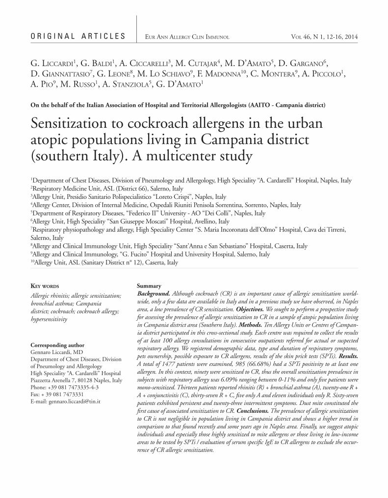
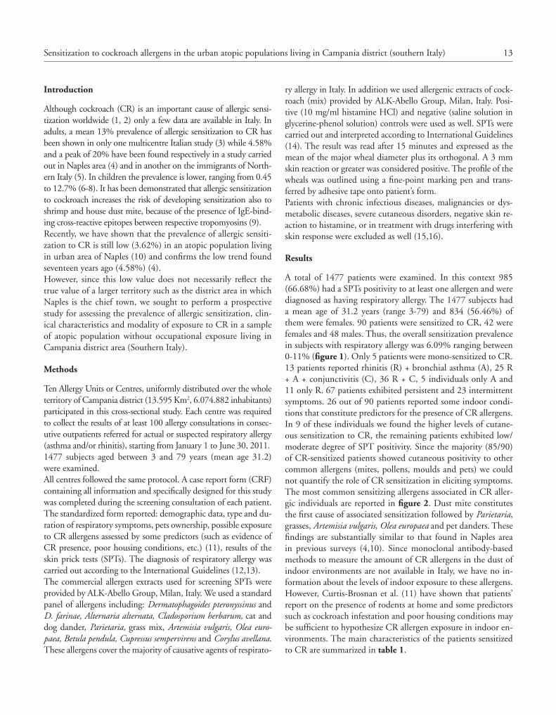
SummaryBackground. Although cockroach (CR) is an important cause of allergic sensitization world-wide, only a few data are available in Italy and in a previous study we have observed, in Naples area, a low prevalence of CR sensitization. Objectives. We sought to perform a prospective study for assessing the prevalence of allergic sensitization to CR in a sample of atopic population living in Campania district area (Southern Italy). Methods. Ten Allergy Units or Centres of Campan-ia district participated in this cross-sectional study. Each centre was required to collect the results of at least 100 allergy consultations in consecutive outpatients referred for actual or suspected respiratory allergy. We registered demographic data, type and duration of respiratory symptoms, pets ownership, possible exposure to CR allergens, results of the skin prick tests (SPTs). Results. A total of 1477 patients were examined, 985 (66.68%) had a SPTs positivity to at least one allergen. In this context, ninety were sensitized to CR, thus the overall sensitization prevalence in subjects with respiratory allergy was 6.09% ranging between 0-11% and only five patients were mono-sensitized. Thirteen patients reported rhinitis (R) + bronchial asthma (A), twenty-one R + A + conjunctivitis (C), thirty-seven R + C, five only A and eleven individuals only R. Sixty-seven patients exhibited persistent and twenty-three intermittent symptoms. Dust mite constituted the first cause of associated sensitization to CR. Conclusions. The prevalence of allergic sensitization to CR is not negligible in population living in Campania district and shows a higher trend in comparison to that found recently and some years ago in Naples area. Finally, we suggest atopic individuals and especially those highly sensitized to mite allergens or those living in low-income areas to be tested by SPTs / evaluation of serum specific IgE to CR allergens to exclude the occur-rence of CR allergic sensitization.
Corresponding authorGennaro Liccardi, MDDepartment of Chest Diseases, Division of Pneumology and AllergologyHigh Speciality “A Cardarelli” Hospital Piazzetta Arenella 7, 80128 Naples, ItalyPhone: +39 081 7473335-4-3 Fax: + 39 081 7473331 E-mail: gennaro liccardi@tin it
Key words
Allergic rhinitis; allergic sensitization; bronchial asthma; Campania district; cockroach; cockroach allergy; hypersensitivity
1Department of Chest Diseases, Division of Pneumology and Allergology, High Speciality “A Cardarelli” Hospital, Naples, Italy2Respiratory Medicine Unit, ASL (District 66), Salerno, Italy
3Allergy Unit, Presidio Sanitario Polispecialistico “Loreto Crispi”, Naples, Italy4Allergy Center, Division of Internal Medicine, Ospedali Riuniti Penisola Sorrentina, Sorrento, Naples, Italy5Department of Respiratory Diseases, “Federico II” University - AO “Dei Colli”, Naples, Italy 6Allergy Unit, High Speciality “San Giuseppe Moscati” Hospital, Avellino, Italy7Respiratory physiopathology and allergy, High Speciality Center “S Maria Incoronata dell’Olmo” Hospital, Cava dei Tirreni, Salerno, Italy 8Allergy and Clinical Immunology Unit, High Speciality “Sant’Anna e San Sebastiano” Hospital, Caserta, Italy9Allergy and Clinical Immunology, “G Fucito” Hospital and University Hospital, Salerno, Italy10Allergy Unit, ASL (Sanitary District n° 12), Caserta, Italy
Sensitization to cockroach allergens in the urban atopic populations living in Campania district (southern Italy) A multicenter study
G. LiccarDi1, G. BaLDi1, a. ciccareLLi3, m. cutajar4, m. D’amato5, D. GarGano6, D. GiannattaSio7, G. Leone8, m. Lo ScHiavo9, f. maDonna10, c. montera9, a. PiccoLo1, a. Pio9, m. ruSSo1, a. StanzioLa5, G. D’amato1
On the behalf of the Italian Association of Hospital and Territorial Allergologists (AAITO - Campania district)
13Sensitization to cockroach allergens in the urban atopic populations living in Campania district (southern Italy)
Introduction
Although cockroach (CR) is an important cause of allergic sensi-tization worldwide (1, 2) only a few data are available in Italy In adults, a mean 13% prevalence of allergic sensitization to CR has been shown in only one multicentre Italian study (3) while 4 58% and a peak of 20% have been found respectively in a study carried out in Naples area (4) and in another on the immigrants of North-ern Italy (5) In children the prevalence is lower, ranging from 0 45 to 12 7% (6-8) It has been demonstrated that allergic sensitization to cockroach increases the risk of developing sensitization also to shrimp and house dust mite, because of the presence of IgE-bind-ing cross-reactive epitopes between respective tropomyosins (9) Recently, we have shown that the prevalence of allergic sensiti-zation to CR is still low (3 62%) in an atopic population living in urban area of Naples (10) and confirms the low trend found seventeen years ago (4 58%) (4) However, since this low value does not necessarily reflect the true value of a larger territory such as the district area in which Naples is the chief town, we sought to perform a prospective study for assessing the prevalence of allergic sensitization, clin-ical characteristics and modality of exposure to CR in a sample of atopic population without occupational exposure living in Campania district area (Southern Italy)
Methods
Ten Allergy Units or Centres, uniformly distributed over the whole territory of Campania district (13 595 Km2, 6 074 882 inhabitants) participated in this cross-sectional study Each centre was required to collect the results of at least 100 allergy consultations in consec-utive outpatients referred for actual or suspected respiratory allergy (asthma and/or rhinitis), starting from January 1 to June 30, 2011 1477 subjects aged between 3 and 79 years (mean age 31 2) were examined All centres followed the same protocol A case report form (CRF) containing all information and specifically designed for this study was completed during the screening consultation of each patient The standardized form reported: demographic data, type and du-ration of respiratory symptoms, pets ownership, possible exposure to CR allergens assessed by some predictors (such as evidence of CR presence, poor housing conditions, etc ) (11), results of the skin prick tests (SPTs) The diagnosis of respiratory allergy was carried out according to the International Guidelines (12,13) The commercial allergen extracts used for screening SPTs were provided by ALK-Abello Group, Milan, Italy We used a standard panel of allergens including: Dermatophagoides pteronyssinus and D. farinae, Alternaria alternata, Cladosporium herbarum, cat and dog dander, Parietaria, grass mix, Artemisia vulgaris, Olea euro-paea, Betula pendula, Cupressus sempervirens and Corylus avellana These allergens cover the majority of causative agents of respirato-
ry allergy in Italy In addition we used allergenic extracts of cock-roach (mix) provided by ALK-Abello Group, Milan, Italy Posi-tive (10 mg/ml histamine HCl) and negative (saline solution in glycerine-phenol solution) controls were used as well SPTs were carried out and interpreted according to International Guidelines (14) The result was read after 15 minutes and expressed as the mean of the major wheal diameter plus its orthogonal A 3 mm skin reaction or greater was considered positive The profile of the wheals was outlined using a fine-point marking pen and trans-ferred by adhesive tape onto patient’s form Patients with chronic infectious diseases, malignancies or dys-metabolic diseases, severe cutaneous disorders, negative skin re-action to histamine, or in treatment with drugs interfering with skin response were excluded as well (15,16)
Results
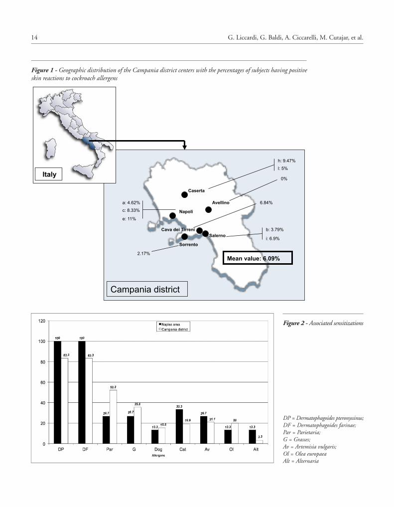
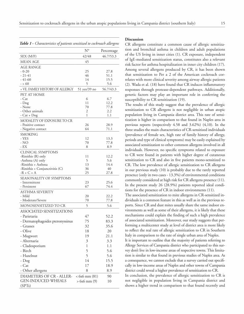
A total of 1477 patients were examined In this context 985 (66 68%) had a SPTs positivity to at least one allergen and were diagnosed as having respiratory allergy The 1477 subjects had a mean age of 31 2 years (range 3-79) and 834 (56 46%) of them were females 90 patients were sensitized to CR, 42 were females and 48 males Thus, the overall sensitization prevalence in subjects with respiratory allergy was 6 09% ranging between 0-11% (figure 1) Only 5 patients were mono-sensitized to CR 13 patients reported rhinitis (R) + bronchial asthma (A), 25 R + A + conjunctivitis (C), 36 R + C, 5 individuals only A and 11 only R 67 patients exhibited persistent and 23 intermittent symptoms 26 out of 90 patients reported some indoor condi-tions that constitute predictors for the presence of CR allergens In 9 of these individuals we found the higher levels of cutane-ous sensitization to CR, the remaining patients exhibited low/moderate degree of SPT positivity Since the majority (85/90) of CR-sensitized patients showed cutaneous positivity to other common allergens (mites, pollens, moulds and pets) we could not quantify the role of CR sensitization in eliciting symptoms The most common sensitizing allergens associated in CR aller-gic individuals are reported in figure 2 Dust mite constitutes the first cause of associated sensitization followed by Parietaria, grasses, Artemisia vulgaris, Olea europaea and pet danders These findings are substantially similar to that found in Naples area in previous surveys (4,10) Since monoclonal antibody-based methods to measure the amount of CR allergens in the dust of indoor environments are not available in Italy, we have no in-formation about the levels of indoor exposure to these allergens However, Curtis-Brosnan et al (11) have shown that patients’ report on the presence of rodents at home and some predictors such as cockroach infestation and poor housing conditions may be sufficient to hypothesize CR allergen exposure in indoor en-vironments The main characteristics of the patients sensitized to CR are summarized in table 1
14 G. Liccardi, G. Baldi, A. Ciccarelli, M. Cutajar, et al.
Avellino
Sorrento
Salerno
Caserta
Napoli
Cava dei Tirreni
0%
h: 9.47%
l: 5%
a: 4.62%
c: 8.33%
e: 11%
b: 3.79%
6.84%
i: 6.9%
2.17%
Italy
Campania district
Mean value: 6.09%
Figure 1 - Geographic distribution of the Campania district centers with the percentages of subjects having positive skin reactions to cockroach allergens
Figure 2 - Associated sensitizations
DP = Dermatophagoides pteronyssinus; DF = Dermatophagoides farinae; Par = Parietaria; G = Grasses; Av = Artemisia vulgaris; Ol = Olea europaeaAlt = Alternaria
15Sensitization to cockroach allergens in the urban atopic populations living in Campania district (southern Italy)
DiscussionCR allergens constitute a common cause of allergic sensitiza-tion and bronchial asthma in children and adult populations of the US living in inner cities (1) CR exposure, independent of IgE-mediated sensitization status, constitutes also a relevant risk factor for asthma hospitalization in inner city children (17) Among several allergens produced by CR, it has been shown that sensitization to Per a 2 of the American cockroach cor-relates with more clinical severity among airway allergic patients (2) Wada et al (18) have found that CR induces inflammatory responses through protease-dependent pathways Additionally, genetic factors may play an important role in conferring the susceptibility to CR sensitization (19) The results of this study suggest that the prevalence of allergic sensitization to CR allergens is not negligible in urban atopic population living in Campania district area This rate of sensi-tization is higher in comparison to that found in Naples area in previous reports (respectively 4 58 and 3 62%) (4,10) In the three studies the main characteristics of CR-sensitized individuals (prevalence of female sex, high rate of family history of allergy, periods and type of clinical symptoms) may be easily explained by associated sensitization to other common allergens involved in all individuals However, no specific symptoms related to exposure to CR were found in patients with higher degree of cutaneous sensitization to CR and also in five patients mono-sensitized to CR The low prevalence of allergic sensitization to CR allergens in our previous study (10) is probably due to the rarely reported presence (only in two cases - 13 3%) of environmental conditions commonly considered at high risk for CR allergens presence (11) In the present study 26 (28 9%) patients reported ideal condi-tions for the presence of CR in indoor environments (11) The associated sensitization to mite allergens in CR-sensitized in-dividuals is a common feature in this as well as in the previous re-ports Since CR and dust mites usually share the same indoor en-vironments as well as some of their allergens, it is likely that these mechanisms could explain the finding of such a high prevalence of associated sensitization Moreover, our study suggests that per-forming a multicenter study at level of district area is more likely to reflect the real rate of allergic sensitization to CR in Southern Italy in comparison to the rate of single urban area of Naples It is important to outline that the majority of patients referring to Allergy Services of Campania district who participated to this sur-vey don’t live in low-income areas of respective towns This limita-tion is similar to that found in previous studies of Naples area As a consequence, we cannot exclude that a survey carried out specifi-cally in low-income areas of Naples and other towns of Campania district could reveal a higher prevalence of sensitization to CR In conclusion, the prevalence of allergic sensitization to CR is not negligible in population living in Campania district and shows a higher trend in comparison to that found recently and
Table 1 - Characteristics of patients sensitized to cockroach allergens
N° PercentageSEX (M/F) 42/48 46 7/53 3
MEAN AGE 45
AGE RANGE- 0-20- 21-41- 41-60- > 60
2546145
27 851 115 55 6
+ VE FAMILY HISTORY OF ALLERGY 51 yes/39 no 56 7/43 3
PET AT HOME- Cat- Dog- None- Other animals- Cat + Dog
6117021
6 712 277 82 21 1
MODALITY OF EXPOSURE TO CR- Positive contact- Negative contact
2664
28 971 1
SMOKING- YES- NO- EX
12708
13 377 88 9
CLINICAL SYMPTOMS-Rinithis (R) only-Asthma (A) only-Rinithis + Asthma-Rinithis + Conjunctivitis (C)-R + C + A
115133625
12 25 614 440
27 8
SEASONALITY OF SYMPTOMS- Intermittent- Persistent
2367
25 674 4
ASTHMA SEVERITY- Mild- Moderate/Severe
2070
22 277 8
MONOSENSITIZED TO CR 5 5 6
ASSOCIATED SENSITIZATIONS- Parietaria- Dermatophagoides pteronyssinus- Grasses- Olive- Mugwort- Alternaria- Cladosporium- Birch- Hazelnut- Dog- Cat- Other allergens
4775321819315514178
52 283 335 620
21 13 31 15 65 615 518 98 9
DIAMETERS OF CR - ALLER-GEN-INDUCED WHEALS(SPTs)
< 6x6 mm (81)> 6x6 mm (9)
9010
16 G. Liccardi, G. Baldi, A. Ciccarelli, M. Cutajar, et al.
6 Peruzzi M, De Luca M, Novembre E, De Martino M, Vierucci A Incidence of cockroach allergy in atopic Italian children Ann Allergy Asthma Immunol 1999;83:167-71
7 Peroni DG, Alfonsi L, Ress M, Milanesi E, Piacentini GL, Boner AL Low rate of cockroach sensitivity in Italian pre-school chil-dren Allergy 2002;58:531
8 La Grutta S, Cibella F, Passalacqua G, Cuttitta G, Liotta G, Ferlisi A, Viegi G Association of Blattella germanica sensitization in pedi-atric allergic patients Pediatr Allergy Immunol 2011;22:521-27
9 Wang J, Calatroni A, Visness CM, Sampson HA Correlation of specific IgE to shrimp with cockroach and dust mite exposure and sensitization in an inner-city population J Allergy Clin Immunol 2011;128:834-37
10 Liccardi G, Salzillo A, Piccolo A, Russo M, D’Amato M, Stanziola
A, Bovenzi D, D’Amato G Is sensitization to cockroach allergens changed during the last seventeen years in the urban atopic pop-ulation living in Naples (southern Italy)? J Investig Allergol Clin Immunol 2013;23:57-59
11 Curtin-Brosnan J, Matsui EC, Breysse P, McCornak MC, Hansel NN, Tonorezos ES, Eggleston P, Williams DC, Diette GB Parent report of pests and pets and indoor allergen levels in inner-city homes Ann Allergy Asthma Immunol 2008;101:517-23
12 Bousquet J and The ARIA Workshop Group Allergic rhinitis and its impact on asthma J Allergy Clin Immunol 2001;108:S147-S336
13 Global Initiative for Asthma http://ginasthma org (December 2012) 14 Dreborg S, Frew A Editors Position Paper: allergen standardiza-
tion and skin tests Allergy 1993;48 Suppl 14:49-82 15 Bousquet J, Michel FB Precision of prick and puncture tests J
Allergy Clin Immunol 1992;90:870-72 16 Wever AMJ, Wever-Hess J Testing for inhalant allergy in asthma
Clin Exp Allergy 1993;23:976-81 17 Rabito FA, Carlson J, Holt EW, Iqbal S, James MA Cockroach
exposure independent of sensitization status and associations for hospitalizations for asthma in inner-city children Ann Allergy Asthma Immunol 2011;106:103-09
18 Wada K, Matsuwaki Y, Moriyama H, Kita H Cockroach indices inflammatory responses through protease-dependent pathways Int Arch Allergy Immunol 2011;155 Suppl 1:135-41
19 Gao P Sensitization to cockroach allergen: immune regulation and genetic determinants Clin Dev Immunol 2012;2012:563760 Doi 10 1155/2012/563760 Epub 2012 Jan 9
some years ago in Naples area Finally, we suggest that atopic in-dividuals, especially those highly sensitized to mite allergens or those living in low-income areas, are tested by SPTs / evaluation of serum specific IgE to CR allergens to exclude the occurrence of CR allergic sensitization We are planning further studies ex-amining exclusively allergic individuals living in some low-in-come areas of Campania district to verify a possible increase in the rate of allergic sensitization to CR
Authorship
All authors contributed equally to the writing and revision of the manuscript
Conflict of interest and financial resources
All authors declare that they have no conflict of interest and that the study has been carried out without any financial support
References
1 Sohn MH, Kim KE The cockroach and allergic diseases Allergy Asthma Immunol Res 2012;4:264-69
2 Lee MF, Song PP, Hwang GH, Lin SJ, Chen YH Sensitization to Per a 2 of the American cockroach correlates with more clini-cal severity among airway allergic patients in Taiwan Ann Allergy Asthma Immunol 2012;108:243-48
3 Riario-Sforza GG, Della Torre F, Antonicelli L, Bonifazi F, Giorda-no T, D’Amato G, Liccardi G, Bettini P, Incorvaia C Sensitization to cockroach in Italy, a multicentric study Allergy Asthma Proc 1997;18:23-28
4 Liccardi G, Russo M, D’Amato M, Granata FP, De Napoli A, D’Amato G Sensitization to cockroach allergens in a sample from the urban population living in Naples (Southern Italy) J Investig Allergol Clin Immunol 1998;8:245-48
5 Lombardi C, Penagos M, Senna GE, Canonica GW, Passalacqua G The clinical characteristics of respiratory allergy in immigrants in Northern Italy Int Arch Allergy Immunol 2008;147:231-34
O R I G I N A L A R T I C L E S Eur Ann AllErgy Clin immunol voL 46, n 1, 17-21, 2014
SummaryBackground. HDMs are a ubiquitous allergen source, with a very well defined biology, but their role in clinical settings and in everyday clinical practice is not well characterized. Aim of this cross-sectional, questionnaire-based study was to assess the clinical characteristics of HDM-related respiratory allergy in a large population of Italian patients. Methods. A structured questionnaire was sent to allergists randomly chosen among those of the Italian Fed-eration of Immunology, Allergy and Clinical Immunology (IFIACI). They were asked to fill it with the clinical data of 10-12 consecutive patients referred for respiratory allergy, positive to HDM skin prick test. The questionnaire assessed type and severity of allergy, demographics, yearly distribution of symptoms, treatment, and satisfaction with the therapy. Results. 45 allergists collected data from 499 patients. Within the evaluated population, 42% had rhi-nitis only, 45% asthma + rhinitis and 13% asthma alone. Rhinitis was moderate/severe in 51% of patients. Asthma was intermittent in 36% of patients, mild in 37% and moderate in 27%. Conjunctivitis was the most frequent comorbidity (36%), followed by rhinosinusitis (16%), adenoid hypertrophy (6%) and polyposis (5%). Out of the population, 56.2% of patients were not at all or partially not satisfied of their treatment for rhinitis, whereas the percentage of dissatisfied patients was about 53% for asthma therapy. 34% patients (n = 170) were monosensitized to HDM. It is confirmed that patients have more symptoms during the fall-winter periods. Conclusion. Patients with HDM allergy have frequently moderate-severe rhinitis, and about 50% of them are not satisfied with their treatment.
Key words
House dust mite; respiratory allergy; treatment; satisfaction
Corresponding authorGiovanni Passalacqua, MDAllergy and Respiratory DiseasesPad Maragliano, Largo R Benzi 1016133 Genoa, Italy
1Stallergenes Italy, Milan, Italy2Stallergenes France, Antony, France 3Allergy/Pulmonary Rehabilitation, ICP Hospital, Milan, Italy4Allergy and Respiratory Diseases, IRCCS University of Genoa, Italy
Evaluation of house dust mite allergy in real life: patients’ characteristics and satisfaction with treatment
f. frati1, S. Scurati2, i. DeLL’aLBani1, P. PuccineLLi1, c. incorvaia3, G. PaSSaLacqua4
Introduction
Sensitization to house dust mite (HDM) is probably the most fre-quent cause of IgE-mediated respiratory allergy all over the world (1,2) Indeed, the prevalence of sensitisation to HDM overcomes that of all other common inhalant allergens, with few exceptions, according to the geographical area (3) In addition, the IgE re-sponse to HDM allergens uniquely starts very early in life, and persists unchanged until adolescence and adulthood (4) HDM belongs to the Arachnida class, is ubiquitous under the altitude of 2000 m and proliferates better in humid and warm environ-ments The demonstration in asthmatic children of the beneficial
effects of sojourning over 2 000 m is part of the history of allergy (5) The allergen components of HDM have been well charac-terized and cloned (6) The major allergens are Group 1 proteins (Der p 1 and Der f 1) and Group 2 proteins (Der p 2 and Der f 2) These are proteases that can per se favour the Th2 response (7) Another important allergen component is tropomyosin (Der p 10), a pan-allergen that has some relevance in food-inhalant aller-gies (8) This type of allergy is usually characterized by persistent symptoms (previously referred to as a perennial disease), since HDM aerodispersed allergens are present around all the year, with a lower burden during summer months The continuous
18 F. Frati, S. Scurati, I. Dell’Albani, P. Puccinelli, C. Incorvaia, G. Passalacqua
was obtained: age, sex, specialty, region of Italy (North, Centre, South) In addition it was asked to estimate when they more frequently experienced symptoms, and in what proportion of them was specific immunotherapy (SIT) prescribed The pa-tient’s questionnaire, filled by the physician according to clinical history and diagnostic procedures, included: • Region of residence (North, Centre, South of Italy)• age range (5≤ years <14, 14≤ years <18, ≥18 years)• type of disease (rhinitis, asthma, both)• severity of rhinitis (intermittent, persistent, mild, moderate/
severe)• severity of asthma (intermittent, mild persistent, moderate,
severe)• duration of symptoms (months/years)• months of the year when symptoms are worse• treatments for rhinitis (topical/systemic antihistamines, top-
ical/oral/injected steroids, leukotriene modifiers, cromones,nose lavages)
• treatments for asthma (short/long acting bronchodilators,inhaled/oral/injected steroids, inhaled anticholinergics, leu-kotriene modifiers, theophylline, others)
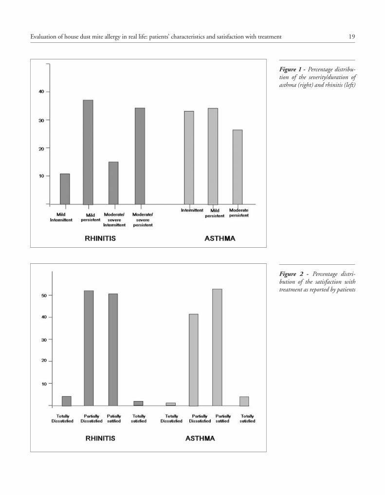
• degree of satisfaction with treatments for asthma and/or andrhinitis (not satisfied at all, partially dissatisfied, partially sat-isfied, fully satisfied)
• presence of clinical symptoms following the ingestion ofshrimps or snails
• additional positive SPTs (grass, Parietaria, birch, olive, rag-weed, cypress, cat, dog, Alternaria, any other tested)
• comorbidities (conjunctivitis, rhinosinusitis, nasal polyps,adenoid hypertrophy, recurrent otitis)
Results
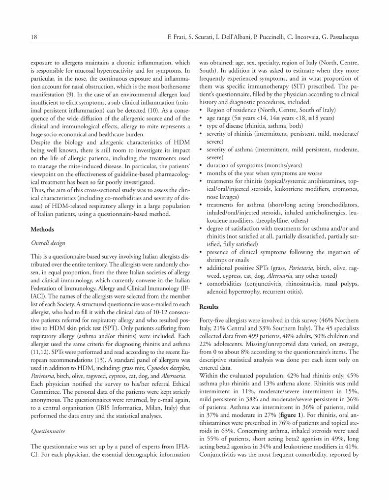
Forty-five allergists were involved in this survey (46% Northern Italy, 21% Central and 33% Southern Italy) The 45 specialists collected data from 499 patients, 48% adults, 30% children and 22% adolescents Missing/unreported data varied, on average, from 0 to about 8% according to the questionnaire’s items The descriptive statistical analysis was done per each item only on entered data Within the evaluated population, 42% had rhinitis only, 45% asthma plus rhinitis and 13% asthma alone Rhinitis was mild intermittent in 11%, moderate/severe intermittent in 15%, mild persistent in 38% and moderate/severe persistent in 36% of patients Asthma was intermittent in 36% of patients, mild in 37% and moderate in 27% (figure 1) For rhinitis, oral an-tihistamines were prescribed in 76% of patients and topical ste-roids in 63% Concerning asthma, inhaled steroids were used in 55% of patients, short acting beta2 agonists in 49%, long acting beta2 agonists in 34% and leukotriene modifiers in 41% Conjunctivitis was the most frequent comorbidity, reported by
exposure to allergens maintains a chronic inflammation, which is responsible for mucosal hyperreactivity and for symptoms In particular, in the nose, the continuous exposure and inflamma-tion account for nasal obstruction, which is the most bothersome manifestation (9) In the case of an environmental allergen load insufficient to elicit symptoms, a sub-clinical inflammation (min-imal persistent inflammation) can be detected (10) As a conse-quence of the wide diffusion of the allergenic source and of the clinical and immunological effects, allergy to mite represents a huge socio-economical and healthcare burden Despite the biology and allergenic characteristics of HDM being well known, there is still room to investigate its impact on the life of allergic patients, including the treatments used to manage the mite-induced disease In particular, the patients’ viewpoint on the effectiveness of guideline-based pharmacolog-ical treatment has been so far poorly investigated Thus, the aim of this cross-sectional study was to assess the clin-ical characteristics (including co-morbidities and severity of dis-ease) of HDM-related respiratory allergy in a large population of Italian patients, using a questionnaire-based method
Methods
Overall design
This is a questionnaire-based survey involving Italian allergists dis-tributed over the entire territory The allergists were randomly cho-sen, in equal proportion, from the three Italian societies of allergy and clinical immunology, which currently convene in the Italian Federation of Immunology, Allergy and Clinical Immunology (IF-IACI) The names of the allergists were selected from the member list of each Society A structured questionnaire was e-mailed to each allergist, who had to fill it with the clinical data of 10-12 consecu-tive patients referred for respiratory allergy and who resulted pos-itive to HDM skin prick test (SPT) Only patients suffering from respiratory allergy (asthma and/or rhinitis) were included Each allergist used the same criteria for diagnosing rhinitis and asthma (11,12) SPTs were performed and read according to the recent Eu-ropean recommendations (13) A standard panel of allergens was used in addition to HDM, including: grass mix, Cynodon dactylon, Parietaria, birch, olive, ragweed, cypress, cat, dog, and Alternaria Each physician notified the survey to his/her referral Ethical Committee The personal data of the patients were kept strictly anonymous The questionnaires were returned, by e-mail again, to a central organization (IBIS Informatica, Milan, Italy) that performed the data entry and the statistical analyses
Questionnaire
The questionnaire was set up by a panel of experts from IFIA-CI For each physician, the essential demographic information
19Evaluation of house dust mite allergy in real life: patients’ characteristics and satisfaction with treatment
Figure 1 - Percentage distribu-tion of the severity/duration of asthma (right) and rhinitis (left)
Figure 2 - Percentage distri-bution of the satisfaction with treatment as reported by patients
20 F. Frati, S. Scurati, I. Dell’Albani, P. Puccinelli, C. Incorvaia, G. Passalacqua
cording to the changes in allergen burden and persistence For this reason, the traditional classification of “perennial” and “sea-sonal” allergic rhinitis has been changed in the Allergic Rhinitis and its Impact on Asthma (ARIA) document into “persistent” and “intermittent” depending on symptoms’ duration (11), with a subdivision according to the intensity of symptoms into “mild” and “moderate/severe” HDMs are a ubiquitous allergen source, with a very well defined biology, but their role in clinical settings is usually the result of deductions, and few data are available on the aspects of mite-in-duced allergy in everyday clinical practice For this reason, we undertook this cross-sectional study within a population of HDM sensitised patients to evaluate their main clinical char-acteristics and their feelings about the treatments received The survey was questionnaire-based, and the database was filled by allergists who evaluated HDM-sensitized patients According to the data collected, most of the patients sensitised to HDM resulted to have a severe and persistent rhinitis, frequently as-sociated with conjunctivitis In addition, it was observed that the worst periods, as far as clinical symptoms were concerned, were limited to autumn-winter, with a relevant improvement during the summer months This increase in symptoms’ se-verity during fall is in accordance with the already described “September epidemic” in allergic patients (15) and also with the
36% of patients, followed by rhinosinusitis (16%), adenoid hy-pertrophy (6%) and polyposis (5%) Out of the population, 56 2% of patients were not at all or partially not satisfied of their treatment for rhinitis, whereas the percentage of dissatisfied patients was about 53% for asthma therapy (figure 2) According to SPT results, performed with the standard panel used, 34% patients (n = 170) resulted to be monosensitized to HDM, whereas 66% (n = 329) were polysensitized, being the association grass + HDM the most frequently recorded (239/499, 48% patients) The yearly distribution of symptoms (worst period) is summarized in figure 3 It is confirmed that patients have more symptoms during the fall-winter periods
Discussion
Despite the features of HDM-induced respiratory allergy be-ing well described in literature, and the biology and molecular aspects of mites being well known, there are few data in the literature focused on patients with HDM allergy regarding their characteristics and response to therapy In the recent past, it was believed that HDM allergy was a “perennial” disease (i e present all around the year) (14), whereas it has been recently acknowledged that symptoms and inflammation may vary, ac-
Figure 3 - Yearly distribution of symptoms (% of patients indi-cating each month as that with worst symptoms)
21Evaluation of house dust mite allergy in real life: patients’ characteristics and satisfaction with treatment
in rhinitis and asthma: ARIA update, in collaboration with GA(2)LEN Allergy 2007;62 Suppl 84:1-41
2 Flohr P Recent perspectives on the global epidemiology of child-hood eczema Allergol Immunopathol (Madr) 2011;39:174-82
3 Thomas WR Geography of house dust mite allergens Asian Pac J Allergy Immunol 2010;28:211-24
4 Melioli G, Marcomini L, Agazzi A, Bazurro G, Tosca, Rossi GA, Minale P, Rossi R, Reggiardo G, Canonica GW, Passalacqua G The IgE repertoire in children and adolescents resolved at com-ponent level: A cross-sectional study Pediatr Allergy immunol 2012;23:433-40
5 Peroni DG, Boner AL, Vallone G, Antolini I, Warner JO Effec-tive allergen avoidance at high altitude reduces allergen-induced bronchial hyperresponsiveness Am J Respir Crit Care Med 1994;149:1442-6
6 Bessot JC, Pauli G Mite allergens: an overview Eur Ann Allergy Clin Immunol 2011;43:141-56
7 Chapman MD, Wünschmann S, Pomés A Proteases as Th2 adju-vants Curr Allergy Asthma Rep 2007;5:363-7
8 Pittner G, Vrtala S, Thomas WR, Weghofer M, Kundi M, Horak F, Kraft D, Valenta R Component-resolved diagnosis of house-dust mite allergy with purified natural and recombinant mite aller-gens Clin Exp Allergy 2004;34:597-603
9 Ciprandi G, Marseglia G, Klersey A, Tosca M Relationships be-tween allergic inflammation and nasal airflow in children with persistent allergic rhinitis due to mite sensitization Allergy 2005;60:957-60
10 Ciprandi G, Buscaglia S, Pesce G, Pronzato C, Ricca V, Parmiani S, Bagnasco M, Canonica GW Minimal persistent inflammation is present at mucosal level in patients with asymptomatic rhinitis and mite allergy J Allergy Clin Immunol 1995;96:971-9
11 Bousquet J Edt ARIA Update 2008 Allergy 2008;63 Suppl 88 12 Global Initiative for the Management of Asthma (GINA) http://
ginasthma org, last accessed October 2012 13 Bousquet J, Heinzerling L, Bachert C, Papadopoulos NG,
Bousquet PJ, et al Practical guide to skin prick tests in allergy to aeroallergens Allergy 2012;67:18-24
14 Van Cauwenberge P, Bachert C, Passalacqua G, Bousquet J, Canon-ica GW, Durham SR, Fokkens WJ, Howarth PH, Lund V, Malling HJ, Mygind N, Passali D, Scadding GK, Wang DY Consensus statement on the treatment of allergic rhinitis European Academy of Allergology and Clinical Immunology Allergy 2000;55:116-34
15 Johnston NW, Johnston SL, Norman GR, Dai J, Sears MR The September epidemic of asthma hospitalization: school children as disease vectors J Allergy Clin Immunol 2006;117:557-62
16 Crisafulli D, Almqvist C, Marks G, Tovey E Seasonal trends in house dust mites in children’s beds over a 7-year period Allergy 2007;62:1394-1400
17 Bousquet J, Bachert C, Canonica GW, Casale TB, Cruz AA, Lock-ey RJ, Zuberbier T; Extended Global Allergy and Asthma Euro-pean Network, World Allergy Organization and Allergic Rhinitis and its Impact on Asthma Study Group Unmet needs in severe chronic upper airway disease (SCUAD) J Allergy Clin Immunol 2009;124:428-33
18 Frew AJ, Powell RJ, Corrigan CJ, Durham SR; UK Immunother-apy Study Group Efficacy and safety of specific immunotherapy with SQ allergen extract in treatment-resistant seasonal allergic rhinoconjunctivitis J Allergy Clin Immunol 2006;117:319-25
reported “seasonality” of HDM In fact, a study from Australia found that the mite concentrations in beds had a two- to three-fold annual fluctuation during a 7-year period of observation, showing the highest values in late autumn and the lowest values in summer (16) HDM-allergic patients we studied seemed to have a more severe disease, with more frequent comorbidities and impairment of their quality of life, when compared to other kind of allergic sensitizations Concerning the treatments, that were prescribed according to current guidelines, for rhinitis oral antihistamines were the most prescribed drugs, followed by top-ical steroids, while for asthma the most prescribed drugs were inhaled steroids, followed by short acting beta2 agonists, long acting beta2 agonists and anti-leukotrienes However, analyz-ing patients’ satisfaction with the treatment of the allergic dis-ease, more than 50% of them reported to be not satisfied with the therapies In particular, 53% of patients were dissatisfied (partially or completely) by asthma therapy, and the rate rose to 56 2% concerning the treatment for rhinitis These cases of allergic rhinitis, mostly satisfying the criteria for severe chronic upper airways disease (SCUAD) (17) are of particular interest because there is a need to control a disease that causes a partic-ularly important impairment of quality of life In a controlled study by Frew et al it was showed that patients with seasonal allergic rhinitis resistant to drug treatment had a clear benefit from specific immunotherapy, with symptom and medication scores resulting significantly better in actively than in placebo treated patients This suggests that specific immunotherapy should be considered as an adjunct to standard therapy in pa-tients with severe allergic rhinitis The main limitations of the study are the questionnaire-based method and the selection bias On the other hand, the large population involved, with a clinically established diagnosis, warrants for a well-selected population In conclusion, we found that in a population of HDM-allergic patients a consistent part of the population suffered from mod-erate to severe allergic disease and that more than half of them were not satisfied by the prescribed drug treatment This calls for specific studies to be addressed on mite-allergic patients, with the aim to improve the management of the disease and consequently their quality of life
Funding and conflict of interest
The study was partially funded by Stallergenes Italy All the co-authors equally contributed in the study design, in collecting and evaluating data and in drafting the final version of the article
References
1 Cruz AA, Popov T, Pawankar R, Annesi-Maesano I, Fokkens W, Kemp J, Ohta K, Price D, Bousquet J ARIA Initiative Scientific Committee Common characteristics of upper and lower airways
O R I G I N A L A R T I C L E S Eur Ann AllErgy Clin immunol voL 46, n 1, 22-25, 2014
SummaryBackground and objective. Sesame seed allergy, a potentially very severe food allergy, seems on the rise worldwide but is still uncommon in Italy. The aim of the present study was to investigate the allergenic profile of Italian sesame seed-allergic patients. Methods. Patients with genuine sesame seed allergy diagnosed over one year in a large number of allergy centers scattered through Italy were considered for the study. Their IgE reactivity to sesame seed aller-gens was characterized by immunoblot analysis. Results. Eleven sesame seed allergic patients were detected and studied. 10/10 patients showed IgE reactivity against a sesame allergen at about 20 kDa, and 7/10 showed an extremely strong reactivity at about 32 kDa. The same 7 sera reacted also against a 28 kDa allergen, although such reactivity was significantly weaker in 6/7 cases. Eight patients showed IgE reactivity at about 48 kDa, and 5 sera reacted against higher m.w. proteins at about 67 kDa. Two sera showed IgE reactivity at about 43 kDa as well. Only one serum appeared to react to 2S-albumin. Conclusions. Italian sesame seed-al-lergic patients react mostly against allergens other than those described so far as major ones. A large number of recombinant sesame allergens will be needed for a comprehensive compo-nent-resolved diagnosis of allergy to this food.
Corresponding authorDr Riccardo AseroAmbulatorio di Allergologia Clinica San CarloPaderno Dugnano (MI), ItalyE-mail: r asero@libero it
Key words
Food allergy; sesame allergy; allergens
1Ambulatorio di Allergologia, Clinica San Carlo, Paderno Dugnano, Italy2Sezione di Allergologia e Immunologia, Azienda Sanitaria di Prato, Italy3Servizio di Allergologia, Ospedale di Atri (TE), Italy4Servizio di Allergologia Dipartimento di Medicina Ambientale e Salute Pubblica, Università di Padova, Italy5S S V D Allergologia, Spedali Civili, Brescia, Italy 6Clinical Allergy and Immunology Unit, Fondazione IRCCS Ca’ Granda Ospedale Maggiore Policlinico, Milan, Italy7Azienda Ospedaliera, Faenza, Italy8Lofarma SpA, Milano, Italy
Detection of 20 kDa and 32 kDa IgE-binding proteins as the major allergens in Italian sesame seed allergic patients
r. aSero1, L. ceccHi2, m. cervone3, m. criveLLaro4, f. LoDi rizzini5, v. Pravettoni6, o. quercia7, S. amato8, G. miStreLLo8
Introduction
Sesamum indicum (Pedaliaceae family) is widely cultivated in many countries in the Middle East, Asia, Latin America and also in the USA Probably due to its widespread use in international fast food restaurants, in bakery products and in snacks and salad dressings, a global increase in sesame seed allergy has been re-corded (1) Following some isolated case reports, the first case series was reported from Switzerland in 1993 (2) Subsequently, reports of sesame seed allergy in both adults and children from
Israel, many European countries, USA, Canada, and Australia have appeared in the literature (3) In certain geographic areas where large amounts of sesame are traditionally present in the common diet, this type of food allergy may occur very early in life (4) To date, 8 allergens have been identified in sesame seeds: Ses i 1 and Ses i 2 (2S-albumin; m w 7-9 kDa), Ses i 3 (7S vicilin; m w 45 kDa), Ses i 4 and Ses i 5 (oleosins; m w 15-17 kDa), Ses i 6 and Ses i 7 (11S globulin, legumin; 52-57 kDa) (5-10) and, although not yet an official IUIS allergen, Ses i 8 (profilin, m w 14 kDa) (11) Sesame seed allergy does not seem
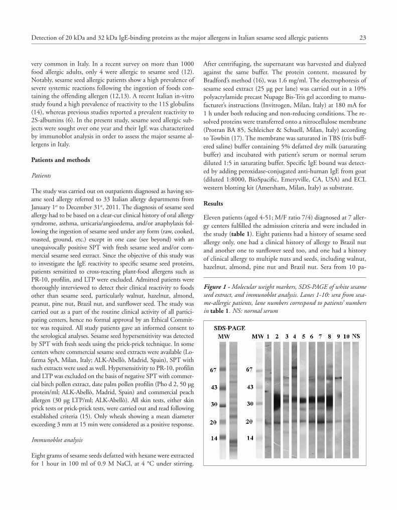
23Detection of 20 kDa and 32 kDa IgE-binding proteins as the major allergens in Italian sesame seed allergic patients
After centrifuging, the supernatant was harvested and dialyzed against the same buffer The protein content, measured by Bradford’s method (16), was 1 6 mg/ml The electrophoresis of sesame seed extract (25 µg per lane) was carried out in a 10% polyacrylamide precast Nupage Bis-Tris gel according to manu-facturer’s instructions (Invitrogen, Milan, Italy) at 180 mA for 1 h under both reducing and non-reducing conditions The re-solved proteins were transferred onto a nitrocellulose membrane (Protran BA 85, Schleicher & Schuell, Milan, Italy) according to Towbin (17) The membrane was saturated in TBS (tris buff-ered saline) buffer containing 5% defatted dry milk (saturating buffer) and incubated with patient’s serum or normal serum diluted 1:5 in saturating buffer Specific IgE bound was detect-ed by adding peroxidase-conjugated anti-human IgE from goat (diluted 1:8000, BioSpacific, Emeryville, CA, USA) and ECL western blotting kit (Amersham, Milan, Italy) as substrate
Results
Eleven patients (aged 4-51; M/F ratio 7/4) diagnosed at 7 aller-gy centers fulfilled the admission criteria and were included in the study (table 1) Eight patients had a history of sesame seed allergy only, one had a clinical history of allergy to Brazil nut and another one to sunflower seed too, and one had a history of clinical allergy to multiple nuts and seeds, including walnut, hazelnut, almond, pine nut and Brazil nut Sera from 10 pa-
very common in Italy In a recent survey on more than 1000 food allergic adults, only 4 were allergic to sesame seed (12) Notably, sesame seed allergic patients show a high prevalence of severe systemic reactions following the ingestion of foods con-taining the offending allergen (12,13) A recent Italian in-vitro study found a high prevalence of reactivity to the 11S globulins (14), whereas previous studies reported a prevalent reactivity to 2S-albumins (6) In the present study, sesame seed allergic sub-jects were sought over one year and their IgE was characterized by immunoblot analysis in order to assess the major sesame al-lergens in Italy
Patients and methods
Patients
The study was carried out on outpatients diagnosed as having ses-ame seed allergy referred to 33 Italian allergy departments from January 1st to December 31st, 2011 The diagnosis of sesame seed allergy had to be based on a clear-cut clinical history of oral allergy syndrome, asthma, urticaria/angioedema, and/or anaphylaxis fol-lowing the ingestion of sesame seed under any form (raw, cooked, roasted, ground, etc ) except in one case (see beyond) with an unequivocally positive SPT with fresh sesame seed and/or com-mercial sesame seed extract Since the objective of this study was to investigate the IgE reactivity to specific sesame seed proteins, patients sensitized to cross-reacting plant-food allergens such as PR-10, profilin, and LTP were excluded Admitted patients were thoroughly interviewed to detect their clinical reactivity to foods other than sesame seed, particularly walnut, hazelnut, almond, peanut, pine nut, Brazil nut, and sunflower seed The study was carried out as a part of the routine clinical activity of all partici-pating centers, hence no formal approval by an Ethical Commit-tee was required All study patients gave an informed consent to the serological analyses Sesame seed hypersensitivity was detected by SPT with fresh seeds using the prick-prick technique In some centers where commercial sesame seed extracts were available (Lo-farma SpA, Milan, Italy; ALK-Abellò, Madrid, Spain), SPT with such extracts were used as well Hypersensitivity to PR-10, profilin and LTP was excluded on the basis of negative SPT with commer-cial birch pollen extract, date palm pollen profilin (Pho d 2, 50 µg protein/ml; ALK-Abellò, Madrid, Spain) and commercial peach allergen (30 µg LTP/ml; ALK-Abellò) All skin tests, either skin prick tests or prick-prick tests, were carried out and read following established criteria (15) Only wheals showing a mean diameter exceeding 3 mm at 15 min were considered as a positive response
Immunoblot analysis
Eight grams of sesame seeds defatted with hexane were extracted for 1 hour in 100 ml of 0 9 M NaCl, at 4 °C under stirring
Figure 1 - Molecular weight markers, SDS-PAGE of white sesame seed extract, and immunoblot analysis. Lanes 1-10: sera from sesa-me-allergic patients, lane numbers correspond to patients’ numbers in table 1. NS: normal serum
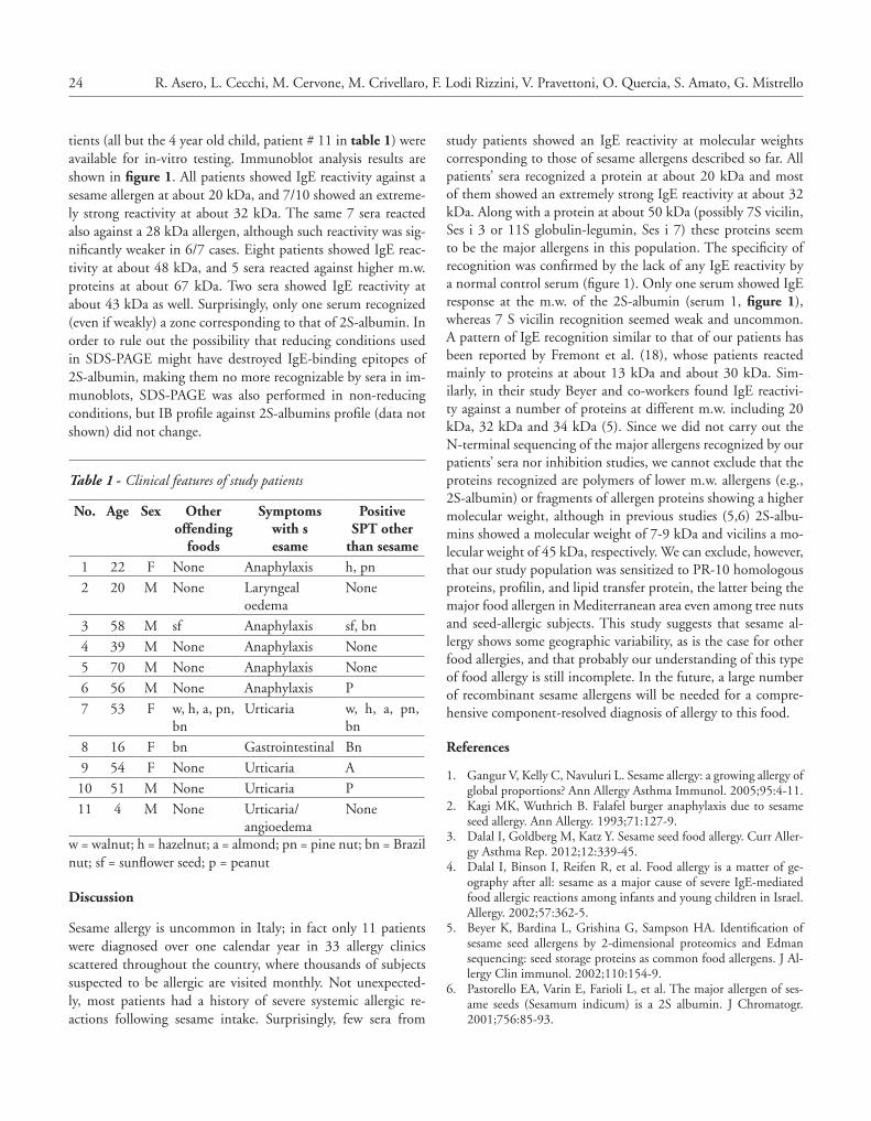
24 R. Asero, L. Cecchi, M. Cervone, M. Crivellaro, F. Lodi Rizzini, V. Pravettoni, O. Quercia, S. Amato, G. Mistrello
study patients showed an IgE reactivity at molecular weights corresponding to those of sesame allergens described so far All patients’ sera recognized a protein at about 20 kDa and most of them showed an extremely strong IgE reactivity at about 32 kDa Along with a protein at about 50 kDa (possibly 7S vicilin, Ses i 3 or 11S globulin-legumin, Ses i 7) these proteins seem to be the major allergens in this population The specificity of recognition was confirmed by the lack of any IgE reactivity by a normal control serum (figure 1) Only one serum showed IgE response at the m w of the 2S-albumin (serum 1, figure 1), whereas 7 S vicilin recognition seemed weak and uncommon A pattern of IgE recognition similar to that of our patients has been reported by Fremont et al (18), whose patients reacted mainly to proteins at about 13 kDa and about 30 kDa Sim-ilarly, in their study Beyer and co-workers found IgE reactivi-ty against a number of proteins at different m w including 20 kDa, 32 kDa and 34 kDa (5) Since we did not carry out the N-terminal sequencing of the major allergens recognized by our patients’ sera nor inhibition studies, we cannot exclude that the proteins recognized are polymers of lower m w allergens (e g , 2S-albumin) or fragments of allergen proteins showing a higher molecular weight, although in previous studies (5,6) 2S-albu-mins showed a molecular weight of 7-9 kDa and vicilins a mo-lecular weight of 45 kDa, respectively We can exclude, however, that our study population was sensitized to PR-10 homologous proteins, profilin, and lipid transfer protein, the latter being the major food allergen in Mediterranean area even among tree nuts and seed-allergic subjects This study suggests that sesame al-lergy shows some geographic variability, as is the case for other food allergies, and that probably our understanding of this type of food allergy is still incomplete In the future, a large number of recombinant sesame allergens will be needed for a compre-hensive component-resolved diagnosis of allergy to this food
References
1 Gangur V, Kelly C, Navuluri L Sesame allergy: a growing allergy of global proportions? Ann Allergy Asthma Immunol 2005;95:4-11
2 Kagi MK, Wuthrich B Falafel burger anaphylaxis due to sesame seed allergy Ann Allergy 1993;71:127-9
3 Dalal I, Goldberg M, Katz Y Sesame seed food allergy Curr Aller-gy Asthma Rep 2012;12:339-45
4 Dalal I, Binson I, Reifen R, et al Food allergy is a matter of ge-ography after all: sesame as a major cause of severe IgE-mediated food allergic reactions among infants and young children in Israel Allergy 2002;57:362-5
5 Beyer K, Bardina L, Grishina G, Sampson HA Identification of sesame seed allergens by 2-dimensional proteomics and Edman sequencing: seed storage proteins as common food allergens J Al-lergy Clin immunol 2002;110:154-9
6 Pastorello EA, Varin E, Farioli L, et al The major allergen of ses-ame seeds (Sesamum indicum) is a 2S albumin J Chromatogr 2001;756:85-93
tients (all but the 4 year old child, patient # 11 in table 1) were available for in-vitro testing Immunoblot analysis results are shown in figure 1 All patients showed IgE reactivity against a sesame allergen at about 20 kDa, and 7/10 showed an extreme-ly strong reactivity at about 32 kDa The same 7 sera reacted also against a 28 kDa allergen, although such reactivity was sig-nificantly weaker in 6/7 cases Eight patients showed IgE reac-tivity at about 48 kDa, and 5 sera reacted against higher m w proteins at about 67 kDa Two sera showed IgE reactivity at about 43 kDa as well Surprisingly, only one serum recognized (even if weakly) a zone corresponding to that of 2S-albumin In order to rule out the possibility that reducing conditions used in SDS-PAGE might have destroyed IgE-binding epitopes of 2S-albumin, making them no more recognizable by sera in im-munoblots, SDS-PAGE was also performed in non-reducing conditions, but IB profile against 2S-albumins profile (data not shown) did not change
Table 1 - Clinical features of study patients
No. Age Sex Other offending
foods
Symptoms with s esame
Positive SPT other
than sesame1 22 F None Anaphylaxis h, pn
2 20 M None Laryngeal oedema
None
3 58 M sf Anaphylaxis sf, bn
4 39 M None Anaphylaxis None
5 70 M None Anaphylaxis None
6 56 M None Anaphylaxis P
7 53 F w, h, a, pn, bn
Urticaria w, h, a, pn, bn
8 16 F bn Gastrointestinal Bn
9 54 F None Urticaria A
10 51 M None Urticaria P
11 4 M None Urticaria/ angioedema
None
w = walnut; h = hazelnut; a = almond; pn = pine nut; bn = Brazil nut; sf = sunflower seed; p = peanut
Discussion
Sesame allergy is uncommon in Italy; in fact only 11 patients were diagnosed over one calendar year in 33 allergy clinics scattered throughout the country, where thousands of subjects suspected to be allergic are visited monthly Not unexpected-ly, most patients had a history of severe systemic allergic re-actions following sesame intake Surprisingly, few sera from
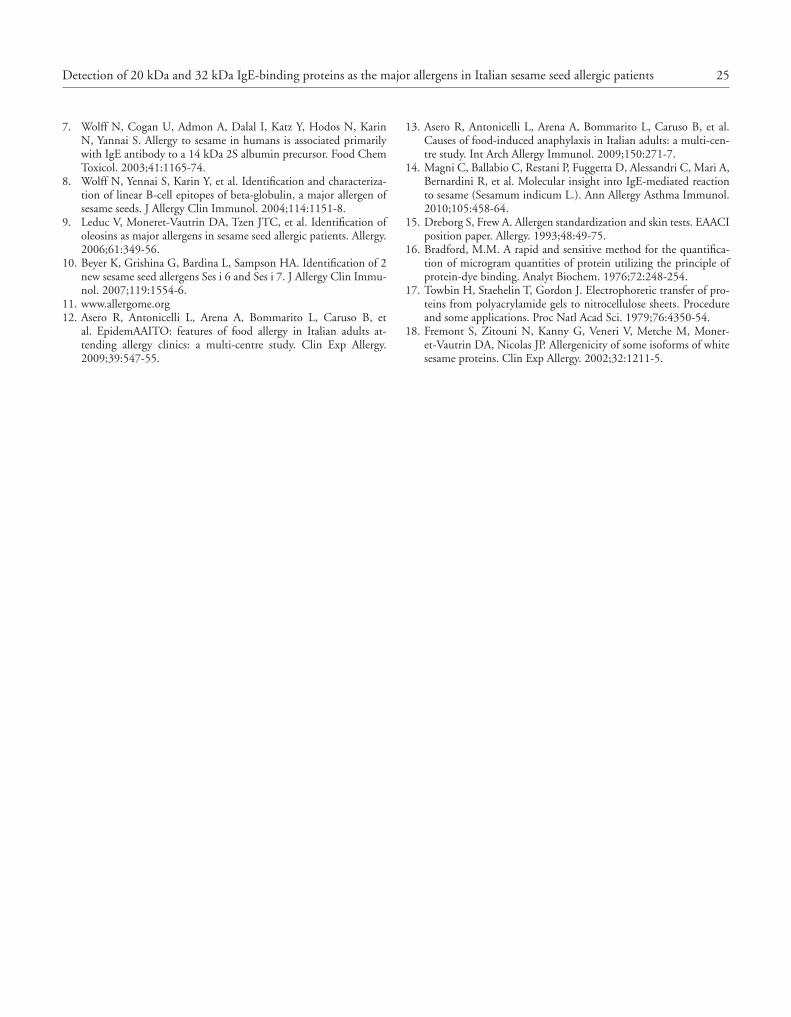
25Detection of 20 kDa and 32 kDa IgE-binding proteins as the major allergens in Italian sesame seed allergic patients
13 Asero R, Antonicelli L, Arena A, Bommarito L, Caruso B, et al Causes of food-induced anaphylaxis in Italian adults: a multi-cen-tre study Int Arch Allergy Immunol 2009;150:271-7
14 Magni C, Ballabio C, Restani P, Fuggetta D, Alessandri C, Mari A, Bernardini R, et al Molecular insight into IgE-mediated reaction to sesame (Sesamum indicum L ) Ann Allergy Asthma Immunol 2010;105:458-64
15 Dreborg S, Frew A Allergen standardization and skin tests EAACI position paper Allergy 1993;48:49-75
16 Bradford, M M A rapid and sensitive method for the quantifica-tion of microgram quantities of protein utilizing the principle of protein-dye binding Analyt Biochem 1976;72:248-254
17 Towbin H, Staehelin T, Gordon J Electrophoretic transfer of pro-teins from polyacrylamide gels to nitrocellulose sheets Procedure and some applications Proc Natl Acad Sci 1979;76:4350-54
18 Fremont S, Zitouni N, Kanny G, Veneri V, Metche M, Moner-et-Vautrin DA, Nicolas JP Allergenicity of some isoforms of white sesame proteins Clin Exp Allergy 2002;32:1211-5
7 Wolff N, Cogan U, Admon A, Dalal I, Katz Y, Hodos N, Karin N, Yannai S Allergy to sesame in humans is associated primarily with IgE antibody to a 14 kDa 2S albumin precursor Food Chem Toxicol 2003;41:1165-74
8 Wolff N, Yennai S, Karin Y, et al Identification and characteriza-tion of linear B-cell epitopes of beta-globulin, a major allergen of sesame seeds J Allergy Clin Immunol 2004;114:1151-8
9 Leduc V, Moneret-Vautrin DA, Tzen JTC, et al Identification of oleosins as major allergens in sesame seed allergic patients Allergy 2006;61:349-56
10 Beyer K, Grishina G, Bardina L, Sampson HA Identification of 2 new sesame seed allergens Ses i 6 and Ses i 7 J Allergy Clin Immu-nol 2007;119:1554-6
11 www allergome org12 Asero R, Antonicelli L, Arena A, Bommarito L, Caruso B, et
al EpidemAAITO: features of food allergy in Italian adults at-tending allergy clinics: a multi-centre study Clin Exp Allergy 2009;39:547-55
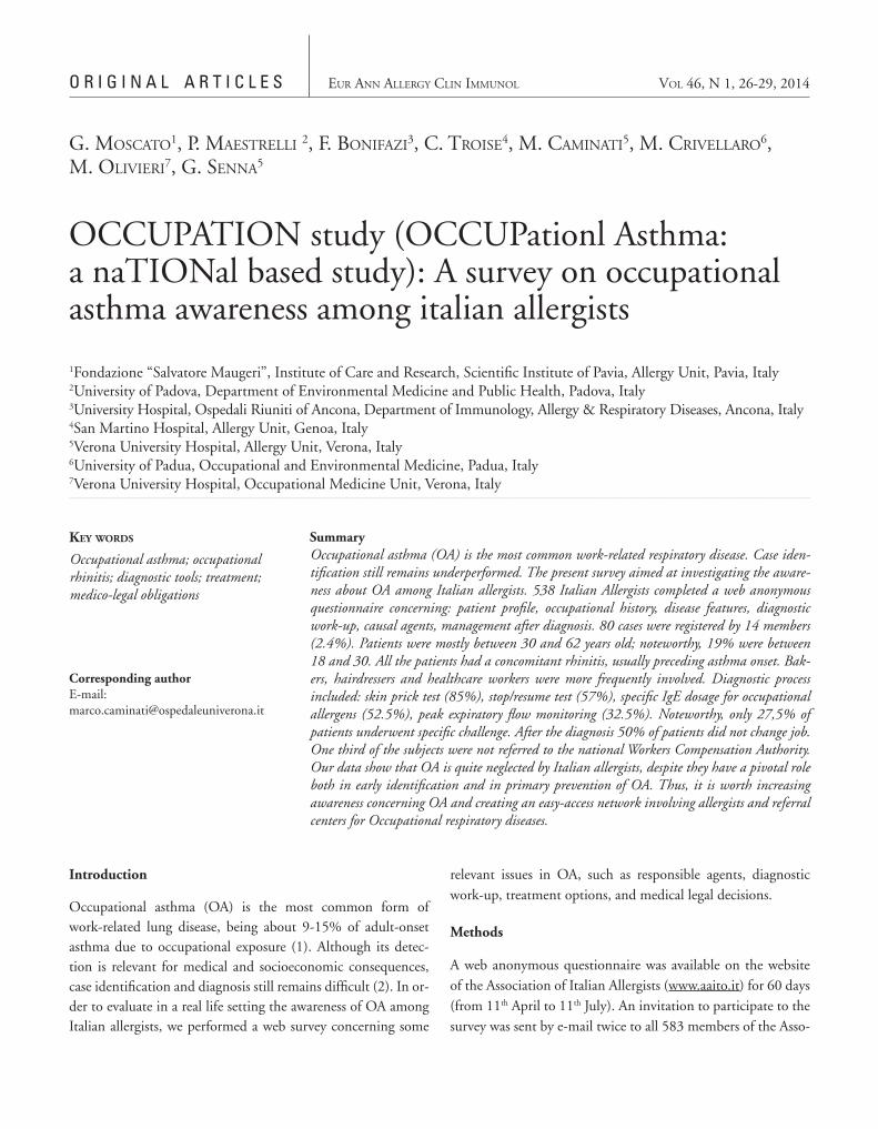
O R I G I N A L A R T I C L E S Eur Ann AllErgy Clin immunol voL 46, n 1, 26-29, 2014
SummaryOccupational asthma (OA) is the most common work-related respiratory disease. Case iden-tification still remains underperformed. The present survey aimed at investigating the aware-ness about OA among Italian allergists. 538 Italian Allergists completed a web anonymous questionnaire concerning: patient profile, occupational history, disease features, diagnostic work-up, causal agents, management after diagnosis. 80 cases were registered by 14 members (2.4%). Patients were mostly between 30 and 62 years old; noteworthy, 19% were between 18 and 30. All the patients had a concomitant rhinitis, usually preceding asthma onset. Bak-ers, hairdressers and healthcare workers were more frequently involved. Diagnostic process included: skin prick test (85%), stop/resume test (57%), specific IgE dosage for occupational allergens (52.5%), peak expiratory flow monitoring (32.5%). Noteworthy, only 27,5% of patients underwent specific challenge. After the diagnosis 50% of patients did not change job. One third of the subjects were not referred to the national Workers Compensation Authority. Our data show that OA is quite neglected by Italian allergists, despite they have a pivotal role both in early identification and in primary prevention of OA. Thus, it is worth increasing awareness concerning OA and creating an easy-access network involving allergists and referral centers for Occupational respiratory diseases.
Key words
Occupational asthma; occupational rhinitis; diagnostic tools; treatment; medico-legal obligations
Corresponding authorE-mail: marco caminati@ospedaleuniverona it
1Fondazione “Salvatore Maugeri”, Institute of Care and Research, Scientific Institute of Pavia, Allergy Unit, Pavia, Italy2University of Padova, Department of Environmental Medicine and Public Health, Padova, Italy3University Hospital, Ospedali Riuniti of Ancona, Department of Immunology, Allergy & Respiratory Diseases, Ancona, Italy4San Martino Hospital, Allergy Unit, Genoa, Italy 5Verona University Hospital, Allergy Unit, Verona, Italy6University of Padua, Occupational and Environmental Medicine, Padua, Italy7Verona University Hospital, Occupational Medicine Unit, Verona, Italy
OCCUPATION study (OCCUPationl Asthma: a naTIONal based study): A survey on occupational asthma awareness among italian allergists
G. moScato1, P. maeStreLLi 2, f. Bonifazi3, c. troiSe4, m. caminati5, m. criveLLaro6, m. oLivieri7, G. Senna5
Introduction
Occupational asthma (OA) is the most common form of work-related lung disease, being about 9-15% of adult-onset asthma due to occupational exposure (1) Although its detec-tion is relevant for medical and socioeconomic consequences, case identification and diagnosis still remains difficult (2) In or-der to evaluate in a real life setting the awareness of OA among Italian allergists, we performed a web survey concerning some
relevant issues in OA, such as responsible agents, diagnostic work-up, treatment options, and medical legal decisions
Methods
A web anonymous questionnaire was available on the website of the Association of Italian Allergists (www aaito it) for 60 days (from 11th April to 11th July) An invitation to participate to the survey was sent by e-mail twice to all 583 members of the Asso-
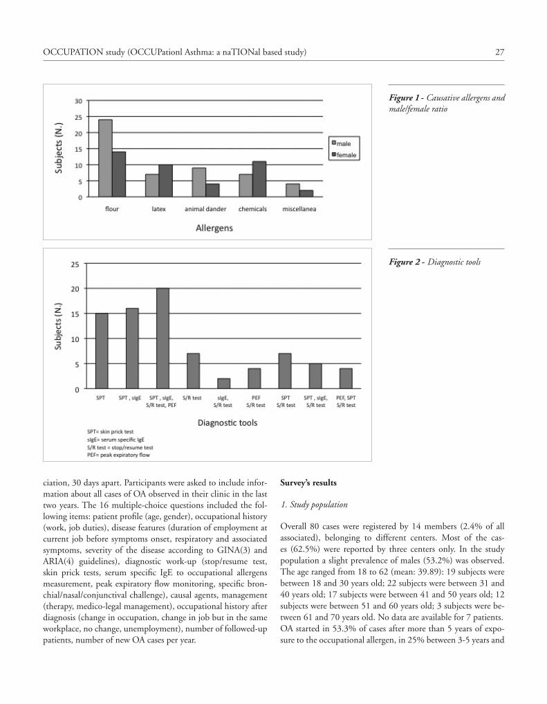
27OCCUPATION study (OCCUPationl Asthma: a naTIONal based study)
Survey’s results
1. Study population
Overall 80 cases were registered by 14 members (2 4% of all associated), belonging to different centers Most of the cas-es (62 5%) were reported by three centers only In the study population a slight prevalence of males (53 2%) was observed The age ranged from 18 to 62 (mean: 39 89): 19 subjects were between 18 and 30 years old; 22 subjects were between 31 and 40 years old; 17 subjects were between 41 and 50 years old; 12 subjects were between 51 and 60 years old; 3 subjects were be-tween 61 and 70 years old No data are available for 7 patients OA started in 53 3% of cases after more than 5 years of expo-sure to the occupational allergen, in 25% between 3-5 years and
ciation, 30 days apart Participants were asked to include infor-mation about all cases of OA observed in their clinic in the last two years The 16 multiple-choice questions included the fol-lowing items: patient profile (age, gender), occupational history (work, job duties), disease features (duration of employment at current job before symptoms onset, respiratory and associated symptoms, severity of the disease according to GINA(3) and ARIA(4) guidelines), diagnostic work-up (stop/resume test, skin prick tests, serum specific IgE to occupational allergens measurement, peak expiratory flow monitoring, specific bron-chial/nasal/conjunctival challenge), causal agents, management (therapy, medico-legal management), occupational history after diagnosis (change in occupation, change in job but in the same workplace, no change, unemployment), number of followed-up patients, number of new OA cases per year
Figure 1 - Causative allergens and male/female ratio
Figure 2 - Diagnostic tools
28 G. Moscato, P. Maestrelli, F. Bonifazi, C. Troise, M. Caminati, M. Crivellaro, M. Olivieri, G. Senna
OA (62 5%) have been reported by three centers only Taken together, these data suggest that, excluding few referral national centers, Italian allergists don’t seem to be focused on OA in daily practice It has to be considered that allergists normally visit pa-tients that are referred by GPs It is therefore possible that GPs refer their patients with suspected occupational respiratory dis-ease directly to specific reference centers (e g workplace health regional centers) On the other hand, as much as 25% of adult asthmatic patients are estimated to have work-related asthma (WRA), the possibility that non-occupational physicians, such as allergists or pneumologists, face WRA in daily practice is high (15) Thus, the overall prevalence of occupational asthma in the present survey may be underestimated Actually, some aspects concerning case identification and diagnostic work-up of occu-pational respiratory allergy still remain problematic and unclear (7,8), despite recent guidelines (7,9) A detailed occupational and medical history collected by every physician would be help-ful for identifying the subjects suspected of having work-related asthma, to address them to in-depth investigations in special-ized centers (15) However, lack of standardized diagnostic tools and referral centers may account for weak awareness of aller-gists about OA It can lead to a delayed diagnosis that is quite common among these patients (1) The high percentage of moderate and severe occupational asthma detected in this study may be explained by a long duration of symptomatic exposure period before diagnosis OA affects working-aged population, which is consistent with causative role of occupational agents Interestingly, in our survey 19% of cases are between 18 and 30 years old This finding suggests that sensitization may take place during apprenticeship The increasing prevalence of aller-gy and asthma in childhood in the last decades accounts for an increased number of young adults entering the workforce affected by respiratory allergy, that is a risk factor for developing sensitization to high molecular weight occupational agents As pointed out by a recent EAACI position paper, allergists play a pivotal role in preventing occupational respiratory allergy, mak-ing their young allergic patients aware of potential work effects on rhinitis and asthma (10) From an epidemiological point of view, our results point out a slight prevalence of males (53 2%) affected by OA, and identify wheat flour as the most recurrent causative agent (47 2%) Our survey does not seem to confirm the epidemiological data in literature of the rising causative role of cleaning substances (11,12) According to our data, a differ-ent male/female ratio was registered according to different aller-gens Male bakers and female hairdressers seem to have a higher risk of developing occupational respiratory disease This proba-bly reflects the different male/female ratio in the respective oc-cupations The model of United Airways Disease has been clear-ly confirmed in occupational field too (13) In most cases our patients suffering from OA have persistent moderate/severe, ac-
in 19 7% within less than three years Patients’ jobs were dis-tributed as follows: bakers (37 5%), hairdressers (15%), health care workers (15%), veterinaries (6%), industrial workers (5%), others (21,5%)
2. Allergens and clinical findings
Wheat flour was the most recurrent cause (47 2%) of OA, fol-lowed by natural rubber latex (23 6%), animal dander (17 8%), persulphates (11 1%) and diisocyanates (8 8%) A different male/female ratio was registered according to different allergens (figure 1) The severity of asthma according to GINA Guide-lines (3) grading was as follows: mild intermittent in 19 7%, mild persistent in 30 3%, moderate persistent in 43 4% and severe persistent in 6 6% A concomitant rhinitis was present in all patients, usually preceding the appearance of asthma Its severity according to ARIA Guidelines (4) was as follows: mild intermittent 6 8%, mild persistent 31 5, moderate/severe inter-mittent 17% and moderate/severe persistent 38 4% Various tools were used in the diagnostic process as reported in figure 2 Twenty-two patients (27,5%) underwent specific challenge with suspected professional agent In 45,5% of cases bronchial chal-lenge only was performed; in 9% specific challenge included both nasal and bronchial exposure; in 45,5% bronchial together with nasal and conjunctival challenge was set up In one patient only nasal challenge was performed
3. Management
Most of patients were treated for both asthma (98%) and rhinitis (87 5%) Nasal steroids were the most frequently used therapy (67%), alone (28 7%) or in combination with anti-histamines (40%) The combination treatment with a long acting beta 2 bronchodilator and an inhaled steroid was the most common therapy for asthma in agreement with a prevalence of moder-ate-persistent severity However, comparing the level of sever-ity and the treatment according to the GINA Guidelines (4) an over-treatment seems to be reported in moderate-persistent asthma, whereas an under-treatment was observed in mild and severe persistent asthma After the diagnosis of OA 50% of patients did not report any change in the job, 32% changed job in the same workplace, while 14% left work or changed occupation One third of the subjects were not referred for an occupational disease to the na-tional Workers Compensation Authority (INAIL)
Discussion
Despite being the most common work-related lung disease (5,6), few cases of OA have been identified by a minority of AAITO associates It is noteworthy that most of the cases of
29OCCUPATION study (OCCUPationl Asthma: a naTIONal based study)
5 Walters GI, McGrath EE, Ayres JG Audit of the recording of occupational asthma in primary care Occup Med (Lond) 2012;62(7):570-3
6 Paris C, Ngatchou-Wandji J, Luc A, McNamee R, Bensefa-Colas L, Larabi L, Telle-Lamberton M, Herin F, Bergeret A, Bonneterre V, Brochard P, Choudat D, Dupas D, Garnier R, Pairon JC, Agius RM, Ameille J; Members of the RNV3P Work-related asthma in France: recent trends for the period 2001-2009 Occup Environ Med 2012;69(6):391-7
7 Baur X, Sigsgaard T, Aasen TB, Burge PS, Heederik D, Henne-berger P, Maestrelli P, Rooyackers J, Schlünssen V, Vandenplas O, Wilken D; ERS Task Force on the Management of Work-related Asthma Guidelines for the management of work-related asthma Eur Respir J 2012;39(3):529-45
8 Cruz MJ, Munoz X The current diagnostic role of the specific occupational laboratory challenge test Curr Opin Allergy Clin Im-munol 2012;12(2):119-25
9 Jares EJ, Baena-Cagnani CE, Gómez RM Diagnosis of oc-cupational asthma: an update Curr Allergy Asthma Rep 2012;12(3):221-31
10 Moscato G, Pala G, Boillat MA, Folletti I, Gerth van Wijk R, Olgiati-Des Gouttes D, Perfetti L, Quirce S, Siracusa A, Walu-siak-Skorupa J, Tarlo SM EAACI position paper: prevention of work-related respiratory allergies among pre-apprentices or ap-prentices and young workers Allergy 2011;66(9):1164-73
11 Fishwick D New occupational and environmental causes of asthma and extrinsic allergic alveolitis Clin Chest Med 2012;33(4):605-16
12 Dumas O, Donnay C, Heederik DJ, Héry M, Choudat D, Kauffmann F, Le Moual N Occupational exposure to cleaning products and asthma in hospital workers Occup Environ Med 2012;69(12):883-9
13 Moscato G, Vandenplas O, Van Wijk RG, Malo JL, Perfetti L, Quirce S, Walusiak J, Castano R, Pala G, Gautrin D, De Groot H, Folletti I, Yacoub MR, Siracusa A European Academy of Allergol-ogy and Clinical Immunolgy EAACI position paper on occupa-tional rhinitis Respir Res 2009;10:16
14 Sublett JW, Bernstein DI Occupational rhinitis Immunol Allergy Clin North Am 2011;31(4):787-96
15 Moscato G, Pala G, Barnig C, De Blay F, Del Giacco SR, Folletti I, Heffler E, Maestrelli P, Pauli G, Perfetti L, Quirce S, Sastre J, Siracusa A, Walusiak-Skorupa J, van Wjik RG European Academy of Allergy and Clinical Immunology EAACI consensus statement for investigation of work-related asthma in non-specialized centres Allergy 2012;67(4):491-501
cording to ARIA classification The severity of both allergic con-ditions is bothersome for patients Moreover, these conditions seem to be independent of the type of causative agent involved Occupational rhinitis should be always assessed and considered as a predictor of subsequent occupational asthma (14) As far as diagnostic work-up is concerned, our survey points out the lack of a homogeneous diagnostic approach Surprisingly, spe-cific challenge is not the main diagnostic tool, despite the fact that most of the cases of OA (62 5%) have been reported by referral centers for occupational allergy Skin prick tests were the most frequently used diagnostic tools (85%) Implementation of current diagnostic guidelines in this field is mandatory to improve case identification and diagnostic work-up of occupa-tional respiratory allergy (15) According to our data, most of patients did not change job or activity after the diagnosis of OA, despite the fact that the most common clinical presentation in our survey is moderate to severe asthma A notification must be submitted to Workers Compensation Authority (INAIL - Isti-tuto Nazionale Assicurazione contro gli Infortuni sul Lavoro) in Italy whenever a worker is diagnosed with OA Our data show that one third of the subjects after the diagnosis of OA were not referred for an occupational disease to INAIL This results in lack of compensation claims for the workers and affects INAIL’s activity in the field of epidemiology and prevention of OA and rhinitis It is thus mandatory to promote the knowledge of med-ico legal obligations among allergists In conclusion, it is well recognized that early diagnosis followed by early removal from exposure is the most important factor that determines a favorable prognosis of OA (7,10) Since many/most respiratory allergic diseases are evaluated by allergists as a first line approach, they have a key role in identifying suspected cases (15) Thus, it is worth increasing awareness concerning OA and creating an easy-access network involving allergists and referral centers for occupational respiratory diseases
References
1 Mapp CE, Boschetto P, Fabbri L, Maestrelli P Occupational asthh-ma Am J Resp Crit Care Med 2005;172:280-305
2 Levy ML, Nicholson PJ Occupational asthma case finding: a role for primary care Br J Gen Pract 2004;54:731-33
3 Global Strategy for Asthma Management and Prevention (GINA) Global Strategy for Asthma Management and Prevention Update http://ginasthma org, 2009
4 Bousquet J, Schunemann HJ, Zuberbier T et al Development and implementation of guidelines in allergic rhinitis An ARIA-GA-2LEN paper Allergy 2010;65:1212-21
O R I G I N A L A R T I C L E S Eur Ann AllErgy Clin immunol voL 46, n 1, 30-34, 2014
Summary Background. The development of component-resolved diagnostics constitutes a potential breakthrough in food allergy testing, as detection of specific IgE (sIgE) to individual allergens may make it possible to establish the risk of a mild versus severe reaction. Objective. To com-pare allergists’ risk assessment based on the current decision making process with that of vir-tual allergen-oriented risk assessment through microarray-based immunoassay. Patients and Methods. An observational, real-life study was performed on 86 adults with food allergy. The prescription of epinephrine was the surrogate marker of a severe reaction. In the same patients, the prescription of epinephrine based on the current decision making of the allergist and the independently established allergen-oriented risk assessment determined by microarray-based immunoassay were compared. Results. Fair degree of agreement between the specialists’ risk assessment and that of the microarray-based immunoassay (k index 0.372 (95% CI: 0.185-0.559) p < 0.001) was documented. Three causes of discrepancy emerged: the poor sensitivity of the allergen microarray-immunoassay (51.9%), the differences in risk assessment established by the specialist and the microarray-immunoassay (33.3%), the non-inclusion of the causative allergen in the microarray-immunoassay platform (14.8%). Conclusion. Improvement of the diagnostic accuracy of microarray-immunoassay, combined with marrying its results to clinical information, could one day soon lead to changes in clinical practice in food allergy.
Corresponding authorLeonardo AntonicelliAllergy Unit, Department of Internal MedicineAzienda Ospedaliero-Universitaria “Ospedali Riuniti” di AnconaVia Conca, 7160126 Ancona, ItalyTel +39 071 5965693 Fax +39 071 5963253E-mail: l antonicelli@ospedaliriuniti marche it
Key words
Food allergy; component-resolved diagnostics; risk-assessment; food allergenic molecules
1Allergy Unit, Department of Internal Medicine, Azienda Ospedaliero-Universitaria “Ospedali Riuniti” di Ancona, Ancona, Italy2Laboratory of Clinical Pathology and Microbiology, General Service Department, Azienda Ospedaliero-Universitaria “Ospedali Riuniti” di Ancona, Ancona, Italy
Component resolved diagnosis in real life: the risk assessment of food allergy using microarray-based immunoassay
L. antoniceLLi1, c. maSSacceSi1, m. c. BraScHi1, B. cinti2, m. B. BiLò1, f. Bonifazi1
Introduction
Food allergies impair quality of life and can culminate in life-threatening reactions, whose prevalence affects an estimated 2-4% of the general adult population (1, 2), with some studies suggesting that the last few decades have seen a rise in the num-ber of cases in industrialized countries (3, 4) Even more striking is the increase in the number of patients in industrialized countries who think they have a food allergy and seek medical assistance (5) Diagnosis of food allergy is necessary both to prevent severe re-actions and to avoid unnecessary dietary restrictions; unfortu-nately, the complex mixture of allergenic proteins of diagnostic
extracts for skin and serological tests makes testing inaccurate and hence unable to predict the likelihood and, in particular, the severity of a future reaction (6) As a result, in current clinical practice, in most patients com-bining their history with the results of skin testing or immuno-assay is a necessary step towards reaching an accurate diagnosis, though controlled food challenge, which is time-consuming and labour intensive, remains the diagnostic gold standard and may sometimes be required (6) The development of component-resolved diagnostics (CRD) is a potential breakthrough in food allergy testing, as the detection of specific IgE (sIgE) to individual allergens in diagnostic extracts could signify diagnostic improvement,
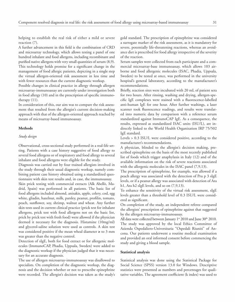
31Component resolved diagnosis in real life: the risk assessment of food allergy using microarray-based immunoassay
gold standard The prescription of epinephrine was considered a surrogate marker of the risk assessment, as it is mandatory for severe, potentially life-threatening reactions, whereas an avoid-ance diet is prescribed for food allergy irrespective of the severity of the reaction Serum samples were collected from each participant and a com-mercial microarray-base immunoassay, which allows 103 air-borne and food allergenic molecules (ISAC, Phadia, Uppsala, Sweden) to be tested at once, was performed in the university hospital’s general laboratory, according to the manufacturer’s recommendations Briefly, reaction sites were incubated with 20 mL of patient sera for two hours After rinsing, washing and drying, allergen-spe-cific IgE complexes were stained with a fluorescence-labelled anti-human IgE for one hour After further washings, a laser scanner took fluorescence readings, and results were translat-ed into numeric data by comparison with a reference serum standardized against ImmunoCAP IgE As a consequence, the results, expressed as standardized ISAC units (ISU/L), are in-directly linked to the World Health Organization IRP 75/502 IgE standard Levels > 0 3 ISU/L were considered positive, according to the manufacturer’s recommendations A physician, blinded to the allergist’s decision making, pre-scribed epinephrine on the basis of the most recently published list of foods which trigger anaphylaxis in Italy (12) and of the available information on the risk of severe reactions associated with the allergenic molecules in the ISAC panel (7,9,13) The prescription of epinephrine, for example, was allowed if a peach allergy was associated with the detection of Pru p 3 sIgE levels, or if a peanut allergy was associated with detection of Ara h1, Ara h2 sIgE levels, and so on (7,9,13) To enhance the sensitivity of the virtual risk assessment, sIgE levels greater than a threshold level of 0 3 ISU/L were consid-ered as significant On completion of the study, an independent referee compared the allergists’ prescription of epinephrine against that suggested by the allergen microarray-immunoassay All data were collected between January 1st 2010 and June 30th 2010 The study was approved by the local Ethics Committee of Azienda Ospedaliero-Universitaria “Ospedali Riuniti” of An-cona Our patients underwent a routine medical examination and provided an oral informed consent before commencing the study and giving a blood sample
Statistical analysis
Statistical analysis was done using the Statistical Package for Social Science (SPSS) version 13 0 for Windows Descriptive statistics were presented as numbers and percentages for quali-tative variables The agreement coefficient (k index) was used to
helping to establish the real risk of either a mild or severe reaction (7) A further advancement in this field is the combination of CRD and microarray technology, which allows testing a panel of one hundred inhalant and food allergens, including recombinant and purified native allergens with very small quantities of serum (8,9) This technology holds promise for a significant change in the management of food allergic patients, depicting in a single step the virtual allergen-oriented risk assessment in less time and with fewer resources than the current diagnostic workup Possible changes in clinical practice in allergy through allergen microarray-immunoassay are currently under investigation both in food allergy (10) and in the prescription of specific immuno-therapy (11) In consideration of this, our aim was to compare the risk assess-ment that resulted from the allergist’s current decision-making approach with that of the allergen-oriented approach reached by means of microarray-based immunoassay
Methods
Study design
Observational, cross-sectional study performed in a real-life set-ting Patients with a case history suggestive of food allergy to several food allergens or of respiratory and food allergy to several inhalant and food allergens were eligible for the study Diagnosis was carried out by four trained allergists involved in the study through their usual diagnostic workup, namely com-bining patient case history obtained using a standardized ques-tionnaire with skin test results and, in case, the immunoassay Skin prick testing with commercial extracts (Alk Abellò, Ma-drid, Spain) was performed in all patients The basic list of food allergens included almond, anisakis, apple, celery, cod, egg white, gliadin, hazelnut, milk, parsley, peanut, profilin, tomato, peach, sunflower, soy, shrimp, walnut and wheat Any further skin tests used in current clinical practice (prick test for inhalant allergens, prick test with food allergens not on the basic list, prick by prick test with fresh food) were allowed if the physician deemed it necessary for the diagnosis Histamine (10mg/ml) and glycerol-saline solution were used as controls A skin test was considered positive if the mean wheal diameter ≥ to 3 mm was greater than the negative control Detection of sIgE, both for food extract or for allergenic mol-ecules (ImmunoCAP, Phadia, Uppsala, Sweden) were added to the diagnostic workup if the physician judged that it was neces-sary for an accurate diagnosis The use of allergen microarray-immunoassay was disallowed to specialists On completion of the diagnostic workup, the diag-nosis and the decision whether or not to prescribe epinephrine were recorded The allergist’s decision was taken as the study’s
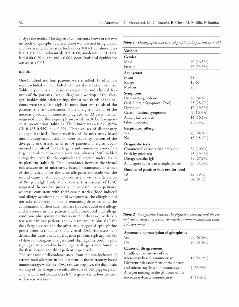
32 L. Antonicelli, C. Massaccesi, M. C. Braschi, B. Cinti, M. B. Bilò, F. Bonifazi
Table 1 - Demographic and clinical profile of the patients (n = 86)
Variable
Gender MaleFemale
40 (46 5%)46 (53 5%)
Age (years) MeanRangeMedian
28 15-67 28
SymptomsUrticaria/angioedemaOral Allergic Symptom (OAS)DyspnoeaGastrointestinal symptomsAnaphylactic shock Glottis oedema
56 (64 4%)25 (28 7%)17 (19 5%) 9 (10 3%)14 (16 1%)2 (2 3%)
Respiratory allergyYesNo
73 (84 8%)13 (15 2%)
Diagnostic testsCommercial extracts skin prick testPrick by prick testDosage specific IgEAll diagnostic tests in a single patient
86 (100%)43 (49 4%)59 (67 8%)30 (34 5%)
Number of positive skin test for food<3>3
22 (19%)64 (81%)
Table 2 - Congruence between the physician work up and the vir-tual risk assessment of the microarray-base immunoassay and causes of disagreement
Agreement in prescription of epinephrineYesNo
59 (68 6%)27 (31 4%)
Causes of disagreement Insufficient sensitivity of the microarray-based immunoassayDifferent risk assessment of the doctor and microarray-based immunoassay Allergen missing in the platform of the microarray-based immunoassay
14 (51 9%)
9 (33 3%)
4 (14 8%)
analyze the results The degree of concordance between the two methods of epinephrine prescription was assessed using Landis and Koch’s interpretive scale for k values: 0 81-1 00, almost per-fect; 0 61-0 80, substantial; 0 41-0 60, moderate; 0 21-0 40, fair; 0 00-0 20, slight; and < 0 001, poor Statistical significance was set at < 0 05
Results
One hundred and four patients were enrolled, 18 of whom were excluded as they failed to meet the selection criteria Table 1 presents the main demographic and clinical fea-tures of the patients In the diagnostic workup of the aller-gist, besides skin prick testing, almost two-thirds of the pa-tients were tested for sIgE In more than two-thirds of the patients, the risk assessment of the allergist and that of the microarray-based immunoassay agreed: in 23 cases neither suggested prescribing epinephrine, while in 36 both suggest-ed its prescription (table 2) The k index was = 0 372 (95% CI: 0 185-0 559) p < 0 001 Three causes of discrepancy emerged (table 2) Poor sensitivity of the microarray-based immunoassay accounted for more than fifty percent of the divergent risk assessments: in 14 patients, allergists docu-mented the role of food allergens and sometimes even of al-lergenic molecules in severe reactions, whereas ISAC yielded a negative score for the equivalent allergenic molecules in its platform (table 3) The discordance between the virtual risk assessment of microarray-based immunoassay and that of the physicians for the same allergenic molecule was the second cause of discrepancy Consistent with the detection of Pru p 3 sIgE levels, the virtual risk assessment of ISAC suggested the need to prescribe epinephrine in six patients, whereas, consistent with their case histories (food-induced oral allergy syndrome or mild symptoms), the allergists did not take this decision In the remaining three patients, the combination of their case histories (food-induced oral allergy and dyspnoea in one patient and food-induced oral allergy syndrome plus systemic urticaria in the other two) with skin test result in one patient, and skin test results plus sIgE for the allergen extracts in the other two, suggested epinephrine prescription to the doctor The virtual ISAC risk assessment denied this decision, as sIgE against profilin, sIgE against Bet v1-like homologous allergens and sIgE against profilin plus sIgE against Bet v1-like homologous allergens were found in the first, second and third patient respectively The last cause of discordance arose from the non-inclusion of certain food allergens in the platform in the microarray-based immunoassay: while the ISAC test was negative, the diagnostic workup of the allergists revealed the role of bell pepper, pista-chio, sesame and peanut (Ara h 9) respectively in four patients with severe reactions
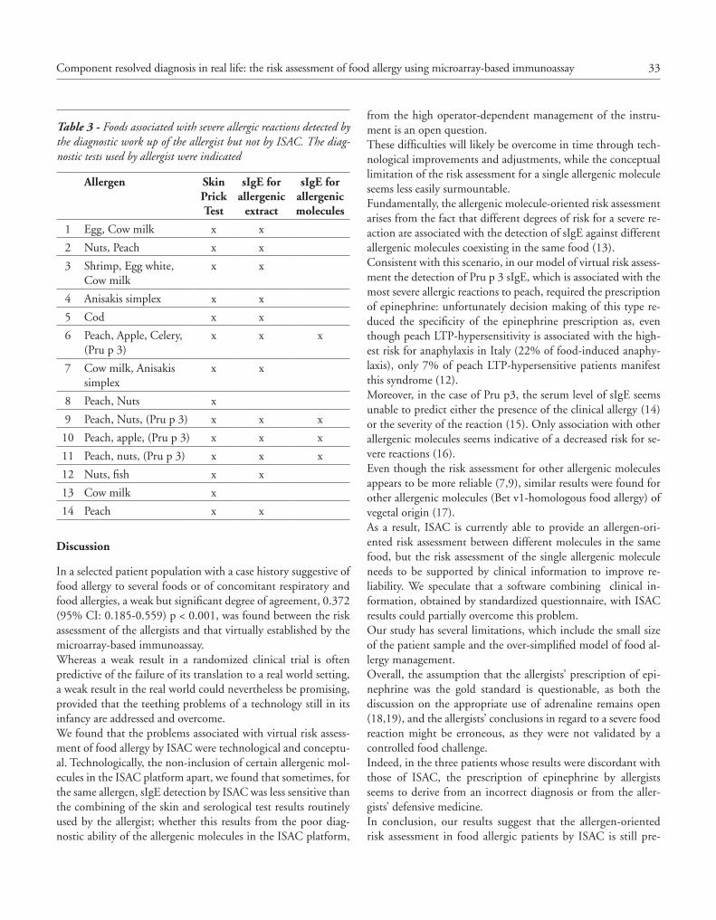
33Component resolved diagnosis in real life: the risk assessment of food allergy using microarray-based immunoassay
from the high operator-dependent management of the instru-ment is an open question These difficulties will likely be overcome in time through tech-nological improvements and adjustments, while the conceptual limitation of the risk assessment for a single allergenic molecule seems less easily surmountable Fundamentally, the allergenic molecule-oriented risk assessment arises from the fact that different degrees of risk for a severe re-action are associated with the detection of sIgE against different allergenic molecules coexisting in the same food (13) Consistent with this scenario, in our model of virtual risk assess-ment the detection of Pru p 3 sIgE, which is associated with the most severe allergic reactions to peach, required the prescription of epinephrine: unfortunately decision making of this type re-duced the specificity of the epinephrine prescription as, even though peach LTP-hypersensitivity is associated with the high-est risk for anaphylaxis in Italy (22% of food-induced anaphy-laxis), only 7% of peach LTP-hypersensitive patients manifest this syndrome (12) Moreover, in the case of Pru p3, the serum level of sIgE seems unable to predict either the presence of the clinical allergy (14) or the severity of the reaction (15) Only association with other allergenic molecules seems indicative of a decreased risk for se-vere reactions (16) Even though the risk assessment for other allergenic molecules appears to be more reliable (7,9), similar results were found for other allergenic molecules (Bet v1-homologous food allergy) of vegetal origin (17) As a result, ISAC is currently able to provide an allergen-ori-ented risk assessment between different molecules in the same food, but the risk assessment of the single allergenic molecule needs to be supported by clinical information to improve re-liability We speculate that a software combining clinical in-formation, obtained by standardized questionnaire, with ISAC results could partially overcome this problem Our study has several limitations, which include the small size of the patient sample and the over-simplified model of food al-lergy management Overall, the assumption that the allergists’ prescription of epi-nephrine was the gold standard is questionable, as both the discussion on the appropriate use of adrenaline remains open (18,19), and the allergists’ conclusions in regard to a severe food reaction might be erroneous, as they were not validated by a controlled food challenge Indeed, in the three patients whose results were discordant with those of ISAC, the prescription of epinephrine by allergists seems to derive from an incorrect diagnosis or from the aller-gists’ defensive medicine In conclusion, our results suggest that the allergen-oriented risk assessment in food allergic patients by ISAC is still pre-
Table 3 - Foods associated with severe allergic reactions detected by the diagnostic work up of the allergist but not by ISAC. The diag-nostic tests used by allergist were indicated
Allergen Skin Prick Test
sIgE for allergenic
extract
sIgE for allergenic molecules
1 Egg, Cow milk x x
2 Nuts, Peach x x
3 Shrimp, Egg white, Cow milk
x x
4 Anisakis simplex x x
5 Cod x x
6 Peach, Apple, Celery, (Pru p 3)
x x x
7 Cow milk, Anisakis simplex
x x
8 Peach, Nuts x
9 Peach, Nuts, (Pru p 3) x x x
10 Peach, apple, (Pru p 3) x x x
11 Peach, nuts, (Pru p 3) x x x
12 Nuts, fish x x
13 Cow milk x
14 Peach x x
Discussion
In a selected patient population with a case history suggestive of food allergy to several foods or of concomitant respiratory and food allergies, a weak but significant degree of agreement, 0 372 (95% CI: 0 185-0 559) p < 0 001, was found between the risk assessment of the allergists and that virtually established by the microarray-based immunoassay Whereas a weak result in a randomized clinical trial is often predictive of the failure of its translation to a real world setting, a weak result in the real world could nevertheless be promising, provided that the teething problems of a technology still in its infancy are addressed and overcome We found that the problems associated with virtual risk assess-ment of food allergy by ISAC were technological and conceptu-al Technologically, the non-inclusion of certain allergenic mol-ecules in the ISAC platform apart, we found that sometimes, for the same allergen, sIgE detection by ISAC was less sensitive than the combining of the skin and serological test results routinely used by the allergist; whether this results from the poor diag-nostic ability of the allergenic molecules in the ISAC platform,
34 L. Antonicelli, C. Massaccesi, M. C. Braschi, B. Cinti, M. B. Bilò, F. Bonifazi
8 Harwanegg C, Laffer S, Hiller R, et al Microarrayed recombinant allergens for diagnosis of allergy Clin Exp Allergy 2003;33:7-13
9 Shreffler WG Microarrayed recombinant allergens for diagnostic testing J Allergy Clin Immunol 2011;127:843-9
10 Lin J, Bruni FM, Fu Z, et al A bioinformatics approach to identify patients with symptomatic peanut allergy using peptide microarray immunoassay J Allergy Clin Immunol 2012;129:1321-1328
11 Sastre J, Landivar ME, Ruiz-García M, Andregnette-Rosigno MV, Mahillo I How molecular diagnosis can change allergen-specific immunotherapy prescription in a complex pollen area Allergy 2012;67:709-11
12 Asero R, Antonicelli L, Arena A, et al Causes of food-induced ana-phylaxis in Italian adults: a multi-centre study Int Arch Allergy Immunol 2009;150:271-7
13 Andersen MB, Hall S, Dragsted LO Identification of european allergy patterns to the allergen families PR-10, LTP, and profilin from Rosaceae fruits Clin Rev Allergy Immunol 2011;41:4-19
14 Asero R, Arena A, Cecchi L, et al Are IgE levels to foods other than rosaceae predictive of allergy in lipid transfer protein-hypersensi-tive patients? Int Arch Allergy Immunol 2011;155(2):149-54
15 Novembre E, Mori F, Contestabile S, Rossi ME, Pucci N Correla-tion of anti-Pru p 3 IgE levels with severity of peach allergy reac-tions in children Ann Allergy Asthma Immunol 2012;108:271-4
16 Pastorello EA, Farioli L, Pravettoni V, et al Pru p 3-sensitised Ital-ian peach-allergic patients are less likely to develop severe symp-toms when also presenting IgE antibodies to Pru p 1 and Pru p 4 Int Arch Allergy Immunol 2011;156(4):362-72
17 Villalta D, Asero R Is the detection of IgE to multiple Bet v 1-ho-mologous food allergens by means of allergen microarray clinically useful? J Allergy Clin Immunol 2010;125:1158-61
18 Niggemann B, Beyer K Adrenaline autoinjectors in food allergy: in for a cent, in for a Euro? Pediatr Allergy Immunol 2012;23:506-8
19 Asero R, Antonicelli L, Arena A, et al Epinephrine autoinjector prescription in food-allergic adults: symptom-based only or al-lergen-based also? An Italian multi-centre study Eur Ann Allergy Clin Immunol 2010;42:25-31
mature for current clinical practice However, the combining of a robust improvement of its diagnostic accuracy and a close linking of its results with clinical information could lead to changes in clinical practice in food allergy, in the not too dis-tant future
Acknowledgements
The authors wish to thank Mr Paul Bowerbank for his help in reviewing the English of the manuscript, and Dr M S Garri-tani and Dr M F Brianzoni for the contribution of their allergic work up The authors also extend their thanks to the members of the Phadia Board, W G Canonica, E Maggi, G Passalacqua
References
1 Kanny G, Moneret-Vautrin DA, Flabbee J, Beaudouin E, Morisset M, Thevenin F Population study of food allergy in France J Aller-gy Clin Immunol 2001;108:133-40
2 Liu AH, Jaramillo R, Sicherer SH, et al National prevalence and risk factors for food allergy and relationship to asthma: results from the National Health and Nutrition Examination Survey 2005-2006 J Allergy Clin Immunol 2010;126:798-806 e13
3 Gupta R, Sheikh A, Strachan DP, Anderson HR Time trends in allergic disorders in the UK Thorax 2007; 62:91-6
4 Sicherer SH, Muñoz-Furlong A, Godbold JH, Sampson HA US prevalence of self-reported peanut, tree nut, and sesame allergy: 11-year follow-up J Allergy Clin Immunol 2010;125:1322-6
5 Teufel M, Biedermann T, Rapps N, et al Psychological burden of food allergy World J Gastroenterol 2007;13:3456-65
6 Sicherer SH, Sampson HA 9 Food allergy J Allergy Clin Immu-nol 2006;117(2 Suppl Mini-Primer):S470-5
7 Sastre J Molecular diagnosis in allergy Clin Exp Allergy 2010;40:1442-60
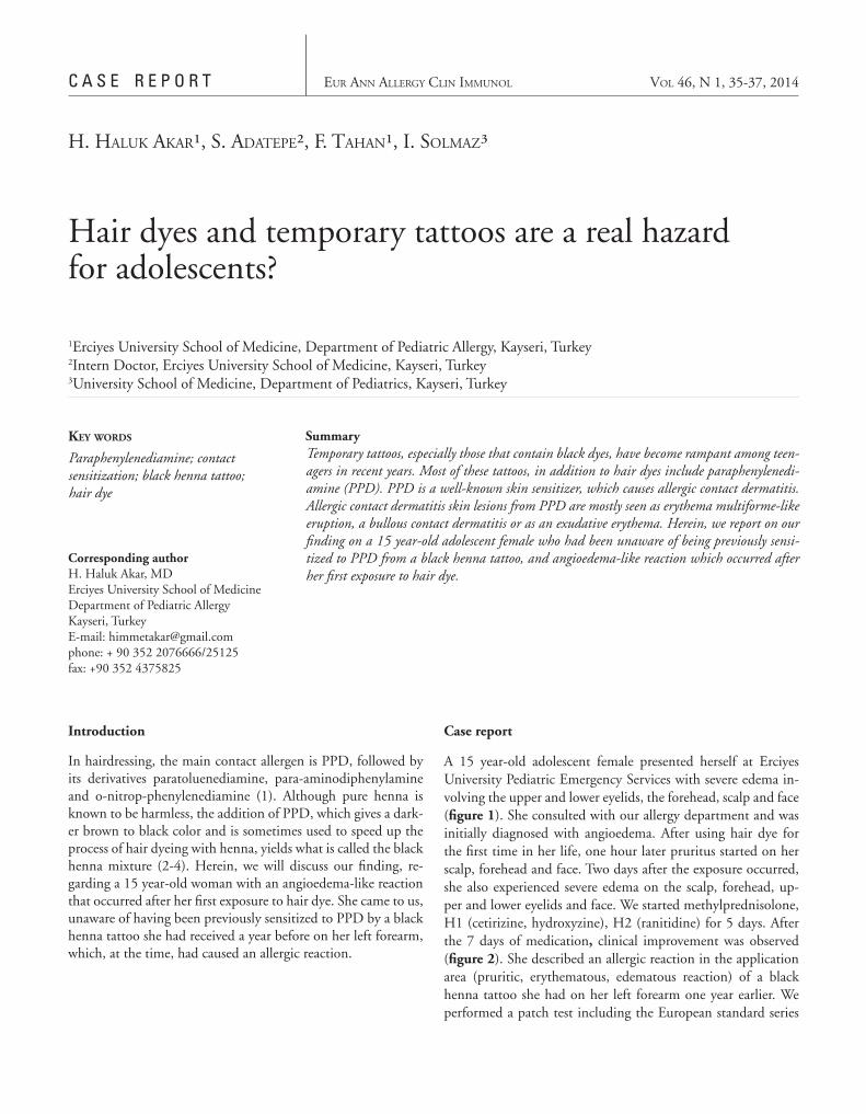
C A S E R E P O R T Eur Ann AllErgy Clin immunol voL 46, n 1, 35-37, 2014
1Erciyes University School of Medicine, Department of Pediatric Allergy, Kayseri, Turkey2Intern Doctor, Erciyes University School of Medicine, Kayseri, Turkey3University School of Medicine, Department of Pediatrics, Kayseri, Turkey
SummaryTemporary tattoos, especially those that contain black dyes, have become rampant among teen-agers in recent years. Most of these tattoos, in addition to hair dyes include paraphenylenedi-amine (PPD). PPD is a well-known skin sensitizer, which causes allergic contact dermatitis. Allergic contact dermatitis skin lesions from PPD are mostly seen as erythema multiforme-like eruption, a bullous contact dermatitis or as an exudative erythema. Herein, we report on our finding on a 15 year-old adolescent female who had been unaware of being previously sensi-tized to PPD from a black henna tattoo, and angioedema-like reaction which occurred after her first exposure to hair dye.
Corresponding author H Haluk Akar, MD Erciyes University School of MedicineDepartment of Pediatric Allergy Kayseri, TurkeyE-mail: himmetakar@gmail com phone: + 90 352 2076666/25125fax: +90 352 4375825
Key words
Paraphenylenediamine; contact sensitization; black henna tattoo; hair dye
Hair dyes and temporary tattoos are a real hazard for adolescents?
H. HaLuk akar¹, S. aDatePe², f. taHan¹, i. SoLmaz³
Introduction
In hairdressing, the main contact allergen is PPD, followed by its derivatives paratoluenediamine, para-aminodiphenylamine and o-nitrop-phenylenediamine (1) Although pure henna is known to be harmless, the addition of PPD, which gives a dark-er brown to black color and is sometimes used to speed up the process of hair dyeing with henna, yields what is called the black henna mixture (2-4) Herein, we will discuss our finding, re-garding a 15 year-old woman with an angioedema-like reaction that occurred after her first exposure to hair dye She came to us, unaware of having been previously sensitized to PPD by a black henna tattoo she had received a year before on her left forearm, which, at the time, had caused an allergic reaction
Case report
A 15 year-old adolescent female presented herself at Erciyes University Pediatric Emergency Services with severe edema in-volving the upper and lower eyelids, the forehead, scalp and face (figure 1) She consulted with our allergy department and was initially diagnosed with angioedema After using hair dye for the first time in her life, one hour later pruritus started on her scalp, forehead and face Two days after the exposure occurred, she also experienced severe edema on the scalp, forehead, up-per and lower eyelids and face We started methylprednisolone, H1 (cetirizine, hydroxyzine), H2 (ranitidine) for 5 days After the 7 days of medication, clinical improvement was observed (figure 2) She described an allergic reaction in the application area (pruritic, erythematous, edematous reaction) of a black henna tattoo she had on her left forearm one year earlier We performed a patch test including the European standard series
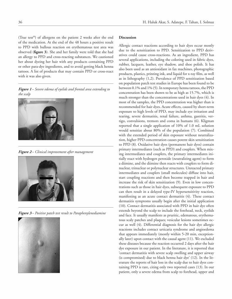
36 H. Haluk Akar, S. Adatepe, F. Tahan, I. Solmaz
Discussion
Allergic contact reactions according to hair dyes occur mostly due to the sensitization to PPD Sensitization to PPD deriv-atives could cause cross-reactions As an ingredient, PPD has several applications, including the coloring used in fabric dyes, rubber, lacquers, leather, eye shadow, and shoe polish It has also been used as an antioxidant in fax machines, photographic products, plastics, printing ink, and liquid for x-ray film, as well as in lithography (1,2) Prevalence of PPD sensitization based on population patch test studies in Europe has been found to be between 0 1% and 1% (5) In temporary henna tattoos, the PPD concentration has been shown to be as high as 15 7%, which is much stronger than the concentrations used in hair dyes (4) In most of the samples, the PPD concentration was higher than is recommended for hair dyes Acute effects, caused by short-term exposure to high levels of PPD, may include eye irritation and tearing, severe dermatitis, renal failure, asthma, gastritis, ver-tigo, convulsions, tremors and coma in humans (6) Kligman reported that a single application of 10% of 1 0 mL solution would sensitize about 80% of the population (7) Combined with the extended period of skin exposure without neutraliza-tion, higher PPD concentration causes potent skin sensitization to PPD (8) Oxidative hair dyes (permanent hair dyes) contain primary intermediates (such as PPD) and couplers When mix-ing intermediates and couplers, the primary intermediates ini-tially react with hydrogen peroxide (neutralizing agent) to form a diimine, and the diimine then reacts with couplers to form di-nuclear, trinuclear or polynuclear structures Unreacted primary intermediates and couplers (small molecules) diffuse into hair, start coupling reactions and then become trapped in hair and increase the risk of skin sensitization (9) Even in low concen-trations such as those in hair dyes, subsequent exposure to PPD can then result in a delayed type-IV hypersensitivity reaction, manifesting as an acute contact dermatitis (4) These contact dermatitis symptoms usually begin after the initial application (10) Contact dermatitis associated with PPD in hair dye often extends beyond the scalp to include the forehead, neck, eyelids and face It usually manifests as pruritic, edematous, erythema-tous scaly patches and plaques; vesicular lesions sometimes oc-cur as well (4) Differential diagnosis for the hair dye allergic reactions includes contact urticaria syndrome and angioedema that appears immediately (mostly within 5-20 min, exception-ally later) upon contact with the causal agent (11) We excluded these diseases because the reaction occurred 2 days after the hair dye exposure in our patient In the literature, it is reported that ‘contact dermatitis with severe scalp swelling and upper airway (is compromised) due to black henna hair dye’ (12) In the lit-erature the reports of hair loss in the scalp due to hair dyes con-taining PPD is rare, citing only two reported cases (13) In our patient, only a severe edema from scalp to forehead, upper and
(True test®) of allergens on the patient 2 weeks after the end of the medication At the end of the 48 hours a positive result to PPD with bullous reaction on erythematous test area was observed (figure 3) She and her family were told that she had an allergy to PPD and cross-reacting substances We cautioned her about dyeing her hair with any products containing PPD or other para-dye ingredients, and to avoid getting black henna tattoos A list of products that may contain PPD or cross-react with it was also given
Figure 1 - Severe edema of eyelids and frontal area extending to the scalp
Figure 2 - Clinical improvement after management
Figure 3 - Positive patch test result to Paraphenylenediamine
37Hair dyes and temporary tattoos are a real hazard for adolescents?
3 Jovanovic DL, Slavkovic-Jovanovic MR Allergic contact dermati-tis from temporary henna tattoo J Dermatol 2009;36 63-5
4 Redlick F, DeKoven J Allergic contact dermatitis to paraphenylen-diamine in hair dye after sensitization from black henna tattoos: a report of 6 cases CMAJ 2007;13Feb;176(4):445-6
5 Heidi Søsted M Sc (pharm ) Allergic contact dermatitis to hair dye ingredients National Allergy Research Centre Gentofte Hos-pital DK-Copenhagen, PhD thesis, Faculty of Health Sciences, University of Copenhagen, Denmark Trykt i forum for Nordic dermato-venereology Supplementum no 13; vol 12, Jan 2007
6 Al-Suwaidi A, Ahmed H Determination of para-phenylenedi-amine (PPD) in henna in the United Arab Emirates Int J Environ Res Public Health 2010Apr;7(4):1681-93
7 Kligman AM The identification of contact allergens by human as-say 3 The maximization test: a procedure for screening and rating contact sensitizers J Invest Dermatol 1966;47:393-409
8 Le Coz CJ, Lefebvre C, Keller F, et al Allergic contact dermati-tis caused by skin painting (pseudotattooing) with black henna, a mixture of henna and p-phenylenediamine and its derivatives ArchDermatol 2000Dec;136(12):1515-7
9 Lee HS, Lin YW Permeation of hair dye ingredients, p-phenylene-diamine and aminophenol isomers, through protective gloves Ann Occup Hyg 2009Apr;53(3):289-9
10 Evans CC, Fleming JD Images in clinical medicine Allergic contact dermatitis from a henna tattoo N Engl J Med 2008Aug;7;359(6):627
11 Goossens A Contact-allergic reactions to cosmetics J Allergy (Cai-ro) 2011;2011:467071
12 Broides A, Sofer S, Lazar I Contact dermatitis with severe scalp swelling and upper airway compromise due to black henna hair dye Pediatr Emerg Care 2011Aug;27(8):745-6
13 Ishida W, Makino T, Shimizu T Severe Hair Loss of the Scalp due to a Hair Dye Containing Para phenylenediamine ISRN Derma-tol 2011;2011:947284
lower eyelids, and face was prominent initially and a misdiagno-sis of angioedema was made in the pediatric emergency room Positive allergic reaction to PPD was confirmed with the patch test bullous reaction on the erythematous test area Our patient was a 15 year-old, had applied hair dye for the first time in her life and the sensitization phase of allergic reaction had been initiated by a black henna tattoo which had been performed one year before An angioedema-like reaction occurred two days after hair dyeing, supporting a delayed type of hypersensitiza-tion Therefore, patch tests results confirmed this relationship between hair dye and black henna tattoos We concluded that the patients with severe edematous reactions after the first application of a hair dye might have previously been sensitized from other PPD-containing materials Black henna tattoos contain PPD It may sensitize users Allergic con-tact dermatitis ought to be considered in patients presenting with angioedema Patch testing should be done on patients who react to hair dyes (PPD and its derivatives) to elucidate the cause and to prevent further severe reactions
References
1 Fernández-Vozmediano JM, Padilla-Moreno M, Armario-Hita JC, et al Pattern of contact sensitization to paraphenylenediamine and its detection in hair dyes Actas Dermosifiliogr 2011;102:206-11
2 Tukenmez Demirci G, Kivanc Altunay I, Atis G, et al Allergic contact dermatitis mimicking angioedema due to paraphenyl-endiamine hypersensitivity: a case report Cutan Ocul Toxicol 2012Sep;31(3):250-2
C A S E R E P O R T Eur Ann AllErgy Clin immunol voL 46, n 1, 38-40, 2014
Corresponding authorDr C WieshuberDepartment of Dermatology and AllergyUniversity Hospital WürzburgJosef-Schneider-Strasse 297080 Würzburg, GermanyTel +49 931 20126710Fax +49 931 20126700E-mail: tinewieshuber@gmx de
Department of Dermatology and Allergy, University Hospital Würzburg, Germany
The needle in the haystack: allergic anaphylaxis caused by the local anesthetic articaine
c. WieSHuBer, j. StoeveSanDt, a. trautmann
Local anesthetics (LA) are extensively used drugs with an excel-lent benefit-risk profile The vast majority of immediate-type adverse reactions can be attributed to non-immune mediated pharmacological effects of LA or psycho-vegetative reactions Clinical symptoms of these reactions often closely resemble anaphylaxis, including hypotension, tachycardia and subjective feelings such as general weakness, heat or vertigo However, true IgE-mediated allergic anaphylaxis due to LA is so exceedingly rare, that the question arises whether it does occur at all Con-sequently, neither commercial skin test reagents nor validated IgE-measurements are available (1) A 28-year-old man requiring dental procedures was referred to our allergy clinic with suspected LA-associated anaphylaxis for further evaluation In March 2011 the patient underwent a dental anesthesia by local injection of the LA SeptanestTM, containing articaine, epinephrine, as well as the preservatives sodium meta-bisulfite and methyl-4-hydroxybenzoate Only a few minutes after injection and before starting dental treat-ment, he suffered a feeling of heat and subsequently developed generalized wheals The patient experienced dizziness due to measurable cardiovascular depression and nausea After emer-gency treatment including fluid replacement, corticosteroids
(betamethasone) and antihistamines (clemastine) the symptoms quickly resolved In the context of this anaphylaxis episode no other potential elicitors such as drugs, foods or concomitant in-fectious diseases could be evaluated Allergologic workup was performed 3 months later Initially, IgE-mediated natural latex allergy was excluded by an incon-spicuous history, negative latex-specific IgE and negative skin testing Thereafter, skin prick testing with a series of different undiluted LA including articaine, articaine combined with epinephrine, mepivacaine, procaine, prilocaine and prilocaine combined with epinephrine, revealed unequivocally positive immediate wheal-and-flare responses to both articaine prepa-rations Intradermal testing with serial dilutions of articaine showed positive immediate reactions even down to the highest dilution A wheal diameter ≥ 5mm was considered as a positive reaction, according to international guidelines Positive respons-es, even in case of diminished erythema by the vasoconstric-tor epinephrine, could be clearly distinguished from the wheal caused by the intradermal injection itself (table 1) Therefore, an IgE-mediated hypersensitivity to articaine was strongly sug-gested A basophil activation test (BAT) with articaine failed to reveal any positive results Following negative skin testing
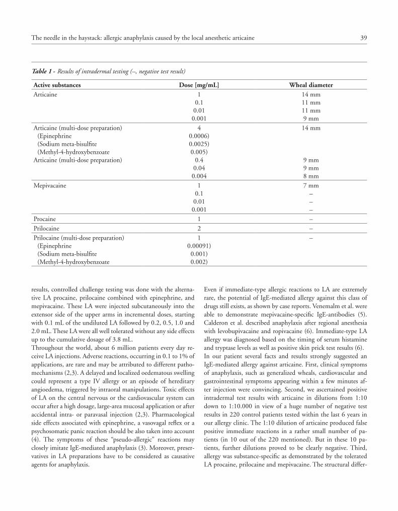
39The needle in the haystack: allergic anaphylaxis caused by the local anesthetic articaine
Even if immediate-type allergic reactions to LA are extremely rare, the potential of IgE-mediated allergy against this class of drugs still exists, as shown by case reports Venemalm et al were able to demonstrate mepivacaine-specific IgE-antibodies (5) Calderon et al described anaphylaxis after regional anesthesia with levobupivacaine and ropivacaine (6) Immediate-type LA allergy was diagnosed based on the timing of serum histamine and tryptase levels as well as positive skin prick test results (6) In our patient several facts and results strongly suggested an IgE-mediated allergy against articaine First, clinical symptoms of anaphylaxis, such as generalized wheals, cardiovascular and gastrointestinal symptoms appearing within a few minutes af-ter injection were convincing Second, we ascertained positive intradermal test results with articaine in dilutions from 1:10 down to 1:10 000 in view of a huge number of negative test results in 220 control patients tested within the last 6 years in our allergy clinic The 1:10 dilution of articaine produced false positive immediate reactions in a rather small number of pa-tients (in 10 out of the 220 mentioned) But in these 10 pa-tients, further dilutions proved to be clearly negative Third, allergy was substance-specific as demonstrated by the tolerated LA procaine, prilocaine and mepivacaine The structural differ-
results, controlled challenge testing was done with the alterna-tive LA procaine, prilocaine combined with epinephrine, and mepivacaine These LA were injected subcutaneously into the extensor side of the upper arms in incremental doses, starting with 0 1 mL of the undiluted LA followed by 0 2, 0 5, 1 0 and 2 0 mL These LA were all well tolerated without any side effects up to the cumulative dosage of 3 8 mL Throughout the world, about 6 million patients every day re-ceive LA injections Adverse reactions, occurring in 0 1 to 1% of applications, are rare and may be attributed to different patho-mechanisms (2,3) A delayed and localized oedematous swelling could represent a type IV allergy or an episode of hereditary angioedema, triggered by intraoral manipulations Toxic effects of LA on the central nervous or the cardiovascular system can occur after a high dosage, large-area mucosal application or after accidental intra- or paravasal injection (2,3) Pharmacological side effects associated with epinephrine, a vasovagal reflex or a psychosomatic panic reaction should be also taken into account (4) The symptoms of these “pseudo-allergic” reactions may closely imitate IgE-mediated anaphylaxis (3) Moreover, preser-vatives in LA preparations have to be considered as causative agents for anaphylaxis
Table 1 - Results of intradermal testing (–, negative test result)
Active substances Dose [mg/mL] Wheal diameterArticaine 1
0 10 010 001
14 mm11 mm11 mm9 mm
Articaine (multi-dose preparation)(Epinephrine(Sodium meta-bisulfite(Methyl-4-hydroxybenzoate
Articaine (multi-dose preparation)
40 0006)0 0025)0 005)
0 40 040 004
14 mm
9 mm9 mm8 mm
Mepivacaine 10 10 010 001
7 mm–––
Procaine 1 –
Prilocaine 2 –
Prilocaine (multi-dose preparation)(Epinephrine(Sodium meta-bisulfite(Methyl-4-hydroxybenzoate
10 00091)0 001)0 002)
–
40 C. Wieshuber, J. Stoevesandt, A. Trautmann
2 Thyssen JP, Menne T, Elberling J, Plaschke P, Johansen JD Hyper-sensitivity to local anaesthetics–update and proposal of evaluation algorithm Contact Dermatitis 2008;59:69-78
3 Ring J, Franz R, Brockow K Anaphylactic Reactions to Local An-esthetics Chem Immunol Allergy 2010;95:190-200
4 Tomoyasu Y, Mukae K, Suda M et al Allergic Reactions to lo-cal Anesthetics in Dental Patients: Analysis of Intracutaneous and Challenge Tests Open Dent J 2011;5:146-149
5 Venemalm L, Degerbeck F, Smith W IgE-mediated reaction to mepivacaine J Allergy Clin Immunol 2008;121:1058-1059
6 Calderon AL, Diot N, Benatir F et al Immediate allergic cross-reactivity to levobupivacaine and ropivacaine Anaesthesia 2013;68:203-205
7 Fuzier R, Lapeyre-Mestre M, Mertes P-M et al Immediate- and delayed-type allergic reactions to amide local anesthetics: clini-cal features and skin testing Pharmacoepidemiol Drug Safety 2009;18:595-601
8 Oertel R, Rahn R, Kirch W Clinical pharmacokinetics of artic-aine Clin Pharmacokinet 1997;33:417-425
ences between articaine, which is a thiophene derivative (with presence of a thiophene ring and an additional ester group), and the amino-acylamides prilocaine and mepivacaine, containing a methylated phenyl ring, reasonably explain this apparent lack of cross-reactivity (7,8) Fourth, by skin and challenge testing a series of LA containing the preservatives sodium meta-bisulfite and methyl-4-hydroxybenzoate, a causative role of these agents could be excluded BAT was proposed as a complementary method for in-vitro diagnosis of drug allergy But until now, the sensitivity of BAT for confirming drug hypersensitivity is generally low
References
1 Gall H, Kaufmann R, Kalveram CM Adverse reactions to lo-cal anesthetics: Analysis of 197 cases J Allergy Clin Immunol 1996;97:933-937
C A S E R E P O R T Eur Ann AllErgy Clin immunol voL 46, n 1, 41-42, 2014
SummaryA case of immediate and definitive response to a single dose of omalizumab in a child with severe ciclosporin-resistant chronic urticaria is reported.
Key words
Chronic urticaria; omalizumab; ciclosporin; therapy
Corresponding authorDr Riccardo Asero, MDAmbulatorio di Allergologia, Clinica San CarloVia Ospedale 2120037 Paderno Dugnano (MI), ItalyE-mail: r asero@libero it
1Ambulatorio di Allergologia, Clinica San Carlo, Paderno Dugnano, Italy2UOD Genetica, Azienda Ospedaliera Ospedale di Circolo e Fondazione Macchi, Polo Universitario, Varese, Italy3Allergy and Clinical Immunology Unit, “L Sacco” Hospital, Milano, Italy
Extraordinary response to omalizumab in a child with severe chronic urticaria
r. aSero1, r. caSaLone2, e. iemoLi3
An 11-year-old boy was recently seen at the allergy department of the Clinica San Carlo He suffered from very severe urticaria with angioedema for 3 months (Urticaria Activity Score = 5) (1), and had a history of seasonal allergic rhino-conjunctivitis for several years, induced by both grass and pellitory pollen The boy suffered from sensineural deafness At the first visit, the pa-tient was taking Hydroxyzine 25 mg/day + Levocetirizine 5 mg/day + Montelukast 10 mg/day + Prednisone 25 mg/day with very little benefit The chronic use of prednisone had caused an increase in body weight of 9 Kg during the last 3 months Exams showed basopenia, an increase in RCP and normal total IgE, normal complement fractions and negative antinuclear an-tibodies and thyroid autoantibodies In view of the sensineural deafness and to exclude a Muckle-Wells disease, gene-sequenc-ing analysis was carried out Analysis of GJB2 and GJB3 genes showed a compound heterozygosity for V153I polymorphic mutation in GJB2 and a -3224G>A mutation in its promoter
UTR region The two mutations were each present in heterozy-gote state in patient’s parents, and it is improbable that, associ-ated in trans, these might cause the bilateral sensineural deafness present in patient Sequencing of exon3 of gene CIASI/NLRP3 was also performed to exclude cold urticaria or Muckle-Well syndromes due to alterations in this gene and no pathogenic mutations were found In view of the very poor response to current treatment and of the severe side effects of oral corticosteroids, after an informed writ-ten consent was obtained by the parents, ciclosporin 3 mg/Kg/day was started, maintaining a single dose of levocetirizine 5 mg in the evening Ciclosporin dosage was increased to 3 8 mg/kg/day after 2 weeks due to the insufficient response, and this dose (250 mg/day) led to an improvement of the clinical situation by 80-90% A further increase in dosage of ciclosporin to 4 6 mg/kg/day (300 mg/day) led to the complete disappearance of both wheals and angioedema After one month, some viral respirato-
42 R. Asero, R. Casalone, E. Iemoli
6 Dreyfus DH Observations on the mechanism of omalizumab as a steroid-sparing agent in autoimmune or chronic idiopathic urticaria and angioedema Ann Allergy Asthma Immunol 2008;100:624-5
7 Godse KV Omalizumab in severe chronic urticaria Indian J Der-matol Venereol Leprol 2008;74:157-8
8 Waibel KH, Reese DA, Hamilton RG, Devillez RL Partial im-provement of solar urticaria after omalizumab J Allergy Clin Im-munol 2009;125:490-1
9 Vestergaard C, Deleuran M Two cases of severe refractory chronic idiopathic urticaria treated with omalizumab Acta Derm Venereol 2010;90:443-4
10 Krause K, Ardelean E, Kessler B, et al Antihistamine-resistant urticaria factitia successfully treated with anti-immunoglobulin E therapy Allergy 2010;65:1494-5
11 Ferrer M, Gamboa P, Sanz ML, et al Omalizumab is effec-tive in nonautoimmune urticaria J Allergy Clin Immunol 2011;127:1300-2
12 Maurer M, Altrichter S, Bieber T, Biedermann T, Braautigam M, et al Efficacy and safety of omalizumab in patients with chronic urticaria who exhibit IgE against thyroperoxidase J Allergy Clin Immunol 2011;128:202-9
13 Saini S, Rosen KE, Hsieh HJ, Wong DA, et al A randomized, placebo-controlled, dose-ranging study of single-dose omalizum-ab in patients with H1-antihistamine-refractort chronic idiopathic urticaria J Allergy Clin Immunol 2011;128:567-73
14 Maurer M, Rosen K, Hsieh HJ, Saini S, Grattan C, Gimenez-Ar-nau A, Agarwal S, Doyle R, Canvin J, Kaplan A, Casale T Omal-izumab for the treatment of chronic idiopathic or spontaneous urticaria N Engl J Med 2013; 368:2340-1
15 Beck LA, Marcotte GV, MacGlashan D, Togias A, Saini S Omal-izumab-induced reductions in mast cell Fc epsilon RI expression and function J Allergy Clin Immunol 2004;114:527-30
16 Eckman JA, Sterna PM, Kelly D, et al Effects of omalizumab on basophil and mast cell responses using an intranasal cat allergen challenge J Allergy Clin Immunol 2010;125:889-95
17 MacGlashan DW, Bochner BS, Ademan DC, et al Down-regu-lation of Fc(epsilon)RI expression on human basophils during in vivo treatment of atopic patients with an anti-IgE antibody J Im-munol 1997;158:1438-45
18 Noga O, Hanf G, Brachmann I, et al Effect of omalizumab treatment on peripheral eosinophil and T-lymphocyte func-tion in patients with allergic asthma J Allergy Clin Immunol 2006;117(6):1493-1499
19 Iemoli E, Piconi S, Fusi A, Borgonovo L, Borelli M, Trabattoni D: Immunological effects of omalizumab in chronic urticaria: a case report J Investig Allergol Clin Immunol 2010;20(3):252-254
ry infections were followed by a relapse of urticaria (UAS = 4), which did not respond to levocetirizine at higher than licensed dose At this point, a marked increase in D-dimer plasma levels (2450 ng/ml; normal 500 ng/ml) and a moderate kidney failure were observed: BUN 70 mg/dl (normal range 19-50 mg/dl) After an informed written consent was obtained by the parents, omalizumab (Xolair) 300 mg was administered subcutaneously The dosing was based on the present recommendations for asthma The day after the administration urticaria completely disappeared, and the boy is still completely urticaria free after 6 months The effectiveness of omalizumab (Humanized monoclonal anti IgE antibodies) in different subsets of CU/angioedema unresponsive to antihistamines is supported by several case-re-ports (2-11), and lately by some multi-center randomized pla-cebo-controlled studies as well (12-14) Two points make our case novel First, the fact that the patient was a 11-year-old boy, and second that the drug worked very quickly and apparently in a resolutive way in a patient that had not been satisfactorily controlled neither by systemic corticosteroids (plus leukotriene receptor antagonists and both first and second generation anti-histamines) nor by ciclosporin The mechanism by which omal-izumab works in patients with chronic idiopathic urticaria is all but established Previous studies showed that the binding of cir-culating IgE by the drug eventually leads to a down regulation of the high affinity IgE receptors, within 2 weeks on basophils and after 8 weeks on mast cells (15-17) Other authors showed that omalizumab induces eosinophil apoptosis, a down-regula-tion of inflammatory cytokines (18) and a reduction in B-cell activation and homing (19) However these observations can-not explain a clinical response that occurs within few days in most cases
References
1 Zuberbier T, Asero R, Bindslev-Jensen C, Canonica GW, Church MK, Gimenez-Arnau A, et al EAACI/GA2LEN;EDF;WAO guideline: definition, classification and diagnosis of urticaria Al-lergy 2009;64:1417-26
2 Boyce JA Successful treatment of cold-induced urticaria/anaphy-laxis with anti-IgE J Allergy Clin Immunol 2006;117:1415-8
3 Spector SL, Tan RA Effect of omalizumab on patients with chron-ic urticaria Ann Allergy Asthma Immunol 2007;99:190-3
4 Sands MF, Blume JW, Schwartz SA Successful treatment of 3 pa-tients with recurrent idiopathic angioedema with omalizumab J Allergy Clin Immunol 2007;120979-81
5 Kaplan AP, Joseph K, Maykut RJ, et al Treatment of chronic au-toimmune urticaria with omalizumab J Allergy Clin Immunol 2008;122:569-73
C A S E R E P O R T Eur Ann AllErgy Clin immunol voL 46, n 1, 43-45, 2014
SummaryThe double blind placebo controlled food challenge (DBPCFC) is the gold standard for di-agnosing cow’s milk allergy (CMA). However, false-negative DBPCFC have been reported. We present 2 cases with a false negative DBPCFC in exclusively breastfed infants suspected of CMA. These cases highlight the occurrence of severe allergic reactions of infants who were exclusively breastfed. Several reported causes of a false negative DBPCFC will be discussed. However, there is currently no clear understanding of the cause of a false negative DBPCFC. This paper highlights that a negative outcome of a DBFCFC must be interpreted with caution, because a severe allergic reaction might occur upon re-introduction of cow’s milk. Therefore, an additional open food challenge under medical supervision is recommended in exclusively breastfed infants with a negative DBPCFC.
Corresponding author N C M Petrus, MDEmma Children’s Hospital AMCDepartment of Paediatric Respiratory Medicine and Allergy (H7-270)Meibergdreef 9, 1105 AZ AmsterdamThe NetherlandsE-mail: n c petrus@amc nlPhone: +31 20 5664748Fax: +31 20 5669683
Key words
Cow’s milk allergy; infants; false negative; double-blind placebo controlled food challenge; open food challenge
1Department of Paediatric Respiratory Medicine and Allergy, Emma Children’s Hospital AMC, Amsterdam, The Netherlands2Nutricia Advanced Medical Nutrition Division, Schiphol, The Netherlands
Exclusively breastfed infants at risk for false negative double blind placebo controlled milk challenge
n.c.m. PetruS1, e.a. koLe2, a.a. ScHoemaker1; W.m.c. van aaLDeren1, a.B. SPrikkeLman1
Abbreviations:AAF: Amino acid formulaCMA: Cow’s milk allergyCMP: Cow’s milk proteinDBPCFC: Double-blind placebo controlled food challengeOFC: Open food challengepHF: Partially hydrolysed formulaw-eHF: Whey-based extensively hydrolysed formulaSPT: Skin prick test
Background
Cow’s milk allergy (CMA) is a common food allergy in infants (1,2) The double-blind placebo controlled food challenge (DB-PCFC) is the gold standard for diagnosing CMA (3) However, false-negative DBPCFC have been reported with a prevalence varying from 3-13% (4-6) We encountered two cases of ex-
clusively breastfed infants who underwent a DBPCFC with a negative outcome, followed by an allergic reaction upon intro-duction of cow’s milk protein (CMP)
Case A
A full term boy with a normal birth weight and APGAR scores had postnatal complications of persisting pulmonary hyper-tension of the neonate and perinatal sepsis, for which he was managed with surfactant, breathing support and antibiotics He was exclusively breastfed from birth on an unrestricted maternal diet until 8 weeks of age, with the exception of one bottle of partially hydrolysed cow’s milk based infant formula (pHF) in the first week of life At 6 weeks he developed severe irritabil-ity, persistent crying and eczema CMA was suspected Since he was taking part in the EuroPrevall Birth Cohort Study, in-vestigations were carried out according to the protocol (7) He
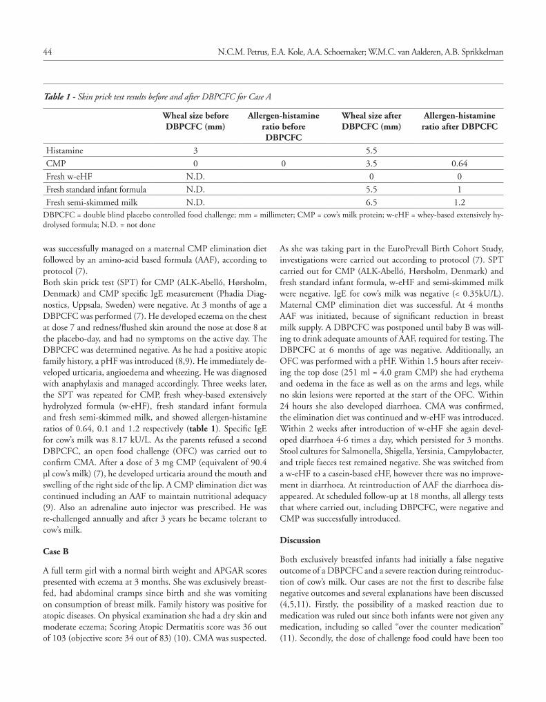
44 N.C.M. Petrus, E.A. Kole, A.A. Schoemaker; W.M.C. van Aalderen, A.B. Sprikkelman
As she was taking part in the EuroPrevall Birth Cohort Study, investigations were carried out according to protocol (7) SPT carried out for CMP (ALK-Abelló, Hørsholm, Denmark) and fresh standard infant formula, w-eHF and semi-skimmed milk were negative IgE for cow’s milk was negative (< 0 35kU/L) Maternal CMP elimination diet was successful At 4 months AAF was initiated, because of significant reduction in breast milk supply A DBPCFC was postponed until baby B was will-ing to drink adequate amounts of AAF, required for testing The DBPCFC at 6 months of age was negative Additionally, an OFC was performed with a pHF Within 1 5 hours after receiv-ing the top dose (251 ml = 4 0 gram CMP) she had erythema and oedema in the face as well as on the arms and legs, while no skin lesions were reported at the start of the OFC Within 24 hours she also developed diarrhoea CMA was confirmed, the elimination diet was continued and w-eHF was introduced Within 2 weeks after introduction of w-eHF she again devel-oped diarrhoea 4-6 times a day, which persisted for 3 months Stool cultures for Salmonella, Shigella, Yersinia, Campylobacter, and triple faeces test remained negative She was switched from a w-eHF to a casein-based eHF, however there was no improve-ment in diarrhoea At reintroduction of AAF the diarrhoea dis-appeared At scheduled follow-up at 18 months, all allergy tests that where carried out, including DBPCFC, were negative and CMP was successfully introduced
Discussion
Both exclusively breastfed infants had initially a false negative outcome of a DBPCFC and a severe reaction during reintroduc-tion of cow’s milk Our cases are not the first to describe false negative outcomes and several explanations have been discussed (4,5,11) Firstly, the possibility of a masked reaction due to medication was ruled out since both infants were not given any medication, including so called “over the counter medication” (11) Secondly, the dose of challenge food could have been too
was successfully managed on a maternal CMP elimination diet followed by an amino-acid based formula (AAF), according to protocol (7) Both skin prick test (SPT) for CMP (ALK-Abelló, Hørsholm, Denmark) and CMP specific IgE measurement (Phadia Diag-nostics, Uppsala, Sweden) were negative At 3 months of age a DBPCFC was performed (7) He developed eczema on the chest at dose 7 and redness/flushed skin around the nose at dose 8 at the placebo-day, and had no symptoms on the active day The DBPCFC was determined negative As he had a positive atopic family history, a pHF was introduced (8,9) He immediately de-veloped urticaria, angioedema and wheezing He was diagnosed with anaphylaxis and managed accordingly Three weeks later, the SPT was repeated for CMP, fresh whey-based extensively hydrolyzed formula (w-eHF), fresh standard infant formula and fresh semi-skimmed milk, and showed allergen-histamine ratios of 0 64, 0 1 and 1 2 respectively (table 1) Specific IgE for cow’s milk was 8 17 kU/L As the parents refused a second DBPCFC, an open food challenge (OFC) was carried out to confirm CMA After a dose of 3 mg CMP (equivalent of 90 4 µl cow’s milk) (7), he developed urticaria around the mouth and swelling of the right side of the lip A CMP elimination diet was continued including an AAF to maintain nutritional adequacy (9) Also an adrenaline auto injector was prescribed He was re-challenged annually and after 3 years he became tolerant to cow’s milk
Case B
A full term girl with a normal birth weight and APGAR scores presented with eczema at 3 months She was exclusively breast-fed, had abdominal cramps since birth and she was vomiting on consumption of breast milk Family history was positive for atopic diseases On physical examination she had a dry skin and moderate eczema; Scoring Atopic Dermatitis score was 36 out of 103 (objective score 34 out of 83) (10) CMA was suspected
Table 1 - Skin prick test results before and after DBPCFC for Case A
Wheal size before DBPCFC (mm)
Allergen-histamine ratio before DBPCFC
Wheal size after DBPCFC (mm)
Allergen-histamine ratio after DBPCFC
Histamine 3 5 5
CMP 0 0 3 5 0 64
Fresh w-eHF N D 0 0
Fresh standard infant formula N D 5 5 1
Fresh semi-skimmed milk N D 6 5 1 2DBPCFC = double blind placebo controlled food challenge; mm = millimeter; CMP = cow’s milk protein; w-eHF = whey-based extensively hy-drolysed formula; N D = not done
45Exclusively breastfed infants at risk for false negative double blind placebo controlled milk challenge
additional funding from Nutricia, the Netherlands and un-restricted grants from Nutricia Advanced Medical Nutrition, the Netherlands, AstraZeneca, the Netherlands, TEVA, the Netherlands and GlaxoSmithKline, the Netherlands These companies were not involved in the design of the study or collecting the data
Acknowledgments
The authors would like to thank the infants and their fam-ilies for their participation in the Dutch EuroPrevall-birth cohort study
References
1 Chafen JJ, Newberry SJ, Riedl MA, et al Diagnosing and man-aging common food allergies: a systematic review JAMA 2010;303:1848-1856
2 Rona RJ, Keil T, Summers C, et al The prevalence of food allergy: a meta-analysis J Allergy Clin Immunol 2007;120:638-646
3 Boyce JA, Assa’ad A, Burks AW, et al Guidelines for the Diagnosis and Management of Food Allergy in the United States: Summary of the NIAID-Sponsored Expert Panel Report J Allergy Clin Im-munol 2010;126:1105-1118
4 Caffarelli C, Petroccione T False-negative food challenges in chil-dren with suspected food allergy Lancet 2001;358:1871-1872
5 Niggemann B, Lange L, Finger A, Ziegert M, Muller V, Beyer K Accurate oral food challenge requires a cumulative dose on a subse-quent day J Allergy Clin Immunol 2012;130:261-263
6 Caffarelli C, Romanini E, Caruana P, Street ME, de’ AG Clinical food hypersensitivity: the relevance of duodenal immunoglobulin E-positive cells Pediatr Res 1998;44:485-490
7 Keil T, McBride D, Grimshaw K, et al The multinational birth cohort of EuroPrevall: background, aims and methods Allergy 2010;65:482-490
8 Host A, Koletzko B, Dreborg S, et al Dietary products used in in-fants for treatment and prevention of food allergy Joint Statement of the European Society for Paediatric Allergology and Clinical Im-munology (ESPACI) Committee on Hypoallergenic Formulas and the European Society for Paediatric Gastroenterology, Hepatology and Nutrition (ESPGHAN) Committee on Nutrition Arch Dis Child 1999;81:80-84
9 Kneepkens CM, Brand PL Clinical practice: Breastfeeding and the prevention of allergy Eur J Pediatr 2010;169:911-917
10 Oranje AP, Glazenburg EJ, Wolkerstorfer A, de Waard-van der Spek FB Practical issues on interpretation of scoring atopic derma-titis: the SCORAD index, objective SCORAD and the three-item severity score Br J Dermatol 2007;157:645-648
11 Niggemann B, Beyer K Pitfalls in double-blind, placebo-con-trolled oral food challenges Allergy 2007;62:729-732
12 Bock SA, Atkins FM Patterns of food hypersensitivity during six-teen years of double-blind, placebo-controlled food challenges J Pediatr 1990;117:561-567
small to elicit symptoms (12) In both cases mentioned above the infants reacted to a lower dose compared to the dose used in the DBPCFC Formula samples were analysed in a labora-tory and exchange of active and placebo foods was ruled out Thirdly, Niggemann and Beyer described a so called short-term specific oral tolerance, which means that the infant develops tol-erance for the allergenic food during the increasing doses of the DBPCFC, but loses this tolerance quickly after the DBPCFC (11) Another possibility may be that during the challenge the infants become sensitised, while the actual clinical reaction oc-curs upon re-introduction of CMP We would like to add that exclusive breastfeeding might be a risk factor of having a false negative DBPCFC, especially since all mentioned studies were performed in children of several ages, while we only describe young infants who were exclusively breastfed (4,5,11,12)
Implications
Despite the fact that DBPCFC is being considered as the gold standard for diagnosing CMA, a false negative outcome remains possible Exclusively breastfed infants are at risk of experiencing a false negative DBPCFC outcome compared to formula-fed infants This could result in severe allergic reactions occurring when CMP is re-introduced Therefore, it is recommended that in exclusively breastfed infants an additional OFC with the for-mula of choice (standard formula or pHF) is performed under medical supervision, rather than introduction of the formula at home (5) After the occurrence of these severe reactions on re-introduction of CMP we have adapted our protocol accordingly
Conclusion
We described two cases of exclusively breastfed infants with a severe allergic reaction after a negative DBPCFC Despite the fact that we are not able to provide a clear explanation for the false negative DBPCFC, an additional OFC with the formula of choice, performed under medical supervision, is necessary in exclusively breastfed infants to avoid severe al-lergic reactions
The institute where the work was conducted
Emma Children’s Hospital AMC, Department of Paediatric Respiratory Medicine and Allergy (H7-270), Meibergdreef 9, 1105 AZ Amsterdam, The Netherlands
Competing interests
The EuroPrevall study is funded by the European Commis-sion (FOOD-CT_2005-514000) The Dutch cohort received
C A S E R E P O R T Eur Ann AllErgy Clin immunol voL 46, n 1, 46-48, 2014
SummaryWe described the first case reported in literature of anaphylactic shock after administration of pollen extract vaccine chemically modified (allergoid) adsorbed onto L-Tyrosine depot adju-vanted with monophosphoryl lipid A (Pollinex® Quattro MPL-4).
Corresponding authorAllergy and Clinical Immunology Unit H “L Sacco”Via G B Grassi 74, 20157 Milano, ItalyE-mail: allergoimmuno@hsacco it
Key words
Anaphylaxis; subcutaneous allergen immunotherapy (SCIT); allergoid; monophosphoryl lipid A
Allergy and Clinical Immunology Unit, H “L Sacco” Milano, Italy
A case of anaphylaxis to Pollinex® Quattro MPL-4
L. BorGonovo, S. Piconi, a. fuSi, e. iemoLi
Introduction
Subcutaneous allergen immunotherapy (SCIT) is an approved efficacious treatment for respiratory allergy, confirmed by WHO Position Paper (1) The incidence of fatal reactions, as reported in an American study, in twelve years of surveillance (1990-2001) is 3 4 per year (41 fatal immunotherapy reactions in 12 years) In particular, the fatality rate is estimated at 1 per 2 5 million injections approx-imately, according to the incidence rates reported by Lockey et al and Reid et al (2) An Italian prospective study conducted on 1738 subjects for a total of 60785 injections over a mean immu-notherapy duration of 3 years (3) suggests that SCIT is safe, since systemic reactions occurred only in 3 6% of patients and 0 15% of injections, with only one grade 4 reaction per > 2000 courses Sublingual immunotherapy (SLIT) is generally considered to have a better safety profile: most reactions are local and transient
and usually do not lead to interruption or cessation of treatment Calderon et al estimated that the incidence of SLIT-induced anaphylaxis is 1 case per 100 million SLIT administrations or per 526000 treatment years, no reaction observed was fatal (4) Modified vaccines with pollen allergoid (chemically modified allergen with a lowered recognition by IgE allergen-specific an-tibodies) have been developed in order to make immunotherapy more effective, reduce side effects and improve compliance Allergy Pollinex® Quattro MPL-4 is a pollen extract vaccine (chemically modified by glutaraldehyde) adsorbed onto L-tyro-sine with addition of the immunostimulatory adjuvant mono-phosphoryl lipid A (MPL) 4MPL is a detoxified, attenuated form of the lipid A component of the lipopolysaccharide of Salmonella Minnesota, a Toll-like receptor 4 agonist The inclusion of MPL reduces the number of required injec-tions (only 4) for effective SCIT and improves compliance (5)
47A case of anaphylaxis to Pollinex® Quattro MPL-4
important to underline that anaphylaxis must be treated imme-diately with an intramuscular injection of epinephrine, as death can occur within minutes All anaphylaxis guidelines recom-mend prompt intramuscular injection of epinephrine, although they differ with regard to the importance of H1-antihistamines, H2-antihistamines, corticosteroids, and bronchodilators other than epinephrine (12) Information should be collected on the role of potential am-plifying factors and co-factors, such as concurrent use of be-ta-blockers or other medication (ACE inhibitors, FANS), viral infection, fever, emotional stress, disruption of routine, premen-strual status in females, and exercise (12) Although ARBs are associated with a lower anaphylactic risk than angiotensin-converting enzyme (ACE) inhibitors (13), in our re-port we cannot exclude that this drug played a facilitating role
Disclosure
The manuscript has not been published elsewhere and it’s not un-der consideration elsewhere The authors disclose any financial or personal relationship which could result in a conflict of interest
References
1 Bousquet J, Lockey R, Malling HJ WHO Position Paper Allergen Immunotherapy: Therapeutic Vaccines for Allergic Diseases Aller-gy 1998; Suppl 44:1-42
2 Bernstein DI, Wanner M, Borish L, Liss GM Twelve-year survey of fatal reactions to allergen injections and skin testing: 1990-2001 J Allergy Clin Immunol 2004;113:1129-36
3 Schiappoli M, Ridolo E, Senna G, Alesina R, Antonicelli L, Asero R, Costantino MT, Longo R, Musarra A, Nettis E, Crivellaro M, Savi E, Massolo A, Passalacqua A prospective Italian survey on the safety of subcutaneous immunotherapy for respiratory allergy Clin Exp Allergy 2009;39:1569-1574
4 Calderon M A , Simons FE, Malling HJ, Lockey RF, Moingeon P, Demoly P Sublingual allergen immunotherapy: mode of action and its relationship with the safety profile Allergy 2012;67:302-311
5 Patel P, Salapatek AM Pollinex Quattro: a novel and well-tol-erated, ultra short-course allergy vaccine Expert Rev Vaccines 2006;5:617-629
6 Puggioni F, Durham SR, Francis JN Monophosphoryl lipid A (MPL) promotes allergen-induced immune deviation in favour of Th1 responses Allergy 2005;60:678-684
7 Drachenberg KJ, Heinzkill M, Urban E, Woroniecki SR Efficacy and tolerability of short-term immunotherapy with pollen aller-goids adjuvanted by monophosphoryl lipid A (MPL) for children and adolescents Allergol Immunopathol 2003;31:270-7
8 Drachenberg KJ, Wheeler AVV, Stuebner P, Horak F A well-tol-erated grass pollen-specific allergy vaccine containing a novel ad-juvant, monophosphoryl lipid A, reduces allergic symptoms after only four preseasonal injections Allergy 2001;498-505
9 Mc Cormack PL, Wagstaff AJ Ultra short course seasonal allergy vaccine (Pollinex® Quattro) Drugs 2006;66:931-8
10 Rosewich M, Schulze J, Fischer von Weikersthal-Drachenberg KJ, Zielen S Ultra-short course Immunotherapy in children and ad-
In vitro studies on peripheral blood mononuclear cells from pa-tients with seasonal allergic rhinitis to grass pollen showed that MPL added to grass pollen extract resulted in the suppression of allergen-induced peripheral Th2 cell responses, in favour a protective Th1 response (6) Several studies demonstrated its clinical efficacy, safety, tolera-bility (7,8,9,10), improvement of patient’s symptoms and com-bined medication/symptom scores (8,5) In addition, this treatment is usually well tolerated Local re-actions, such as redness and swelling in injection site, are the most common reported symptoms Systemic reactions are rare and mild (5) For example, Crivellaro et all (11) reported in their study that only 1 37% of patients (510 in total) experienced an adverse systemic reaction (SR): all SRs were delayed (> 30 minutes), at grade 1 or 2; epinephrine was not required for any of the reac-tions, all resolved spontaneously or after administration of oral antihistamine No report of anaphylaxis is described in literature with this vaccine
Case Description
We report the first case of anaphylaxis after administration of Pollinex® Quattro MPL A 50-year-old Caucasian male with a history of asthma and rhi-nitis since 1992, sensitized to grass, ragweed and birch, usually controlled his symptoms with frequent administration of inhaled salbutamol or disodium cromoglycate and oral antihistamine In February 2012 he started SCIT with Pollinex® Quattro MPL for grass pollen (concentrations at the end of the therapeutic cycle are of 21 5 mcg/ml and 13 64 mcg/ml for the major aller-gens, Phl p 1 and Phl p 5) His clinical history presented arterial hypertension in therapy with beta-blocker, that was pre-emptively substituted with an angiotensin-II receptor blocker (olmesartan) before the start of the immunotherapy The patient, in good health, tolerated the first three injections (300, 800 and 2000 standardized unit SU) However, 10 min-utes after the fourth administration (2000 SU), that had been properly executed, he presented palpebral swelling, conjunctivi-tis, rhinitis, asthma with serrated bronchospasm, hypotension (PA 70/60) The patient was initially treated with oral antihista-mine and methylprednisolone IV but since the symptoms were fast-growing, he was administered twice with epinephrine 0 3 cc IM and beta agonist inhaler, and treatment with hydrocortisone was repeated No biphasic anaphylaxis was observed
Discussion
This case documents that although an allergoid, Pollinex® Quattro MPL is not risk free from systemic reaction It is very
48 L. Borgonovo, S. Piconi, A. Fusi, E. Iemoli
13 Ruëff F, Przybilla B, Biló MB, Müller U, Scheipl F, Aberer W, Birn-baum J, Bodzenta-Lukaszyk A, Bonifazi F, Bucher C, Campi P, Darsow U, Egger C, Haeberli G, Hawranek T, Körner M, Kucha-rewicz I, Küchenhoff H, Lang R, Quercia O, Reider N, Severino M, Sticherling M, Sturm GJ, Wüthrich B Predictors of severe sys-temic anaphylactic reactions in patients with Hymenoptera venom allergy: Importance of baseline serum tryptase–a study of the Eu-ropean Academy of Allergology and Clinical Immunology Interest Group on Insect Venom Hypersensitivity J Allergy Clin Immunol 2009;124:1047-54
olescent during a 3-yrs post-marketing surveillance study Pediatr Allergy immunol 2010;21:e185-e9
11 Crivellaro M, Senna GE, Pappacoda A, Vanzelli R, Spacal B, Marchi G, Recchia G, Makatsori M Safety of ultrashort-term sit with pollen allergoids adjuvanted by monophosphoryl lipid A: a prospective italian survey Eur Ann Allergy Clin Immunol 2011;43:58-60
12 Simons FE, Ardusso LR, Bilò MB, El-Gamal YM, Ledford DK, Ring J, Sanchez-Borges M, Senna GE, Sheikh A, Thong BY World Allergy Organization anaphylaxis guidelines: summary J Allergy Clin Immunol 2011;127:587-593
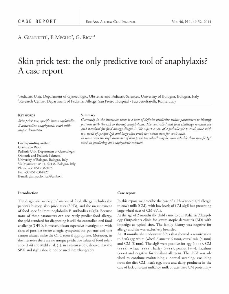
C A S E R E P O R T Eur Ann AllErgy Clin immunol voL 46, n 1, 49-52, 2014
SummaryCurrently, in the literature there is a lack of definite predictive values parameters to identify patients with the risk to develop anaphylaxis. The controlled oral food challenge remains the gold standard for food allergy diagnosis. We report a case of a girl allergic to cow’s milk with low levels of specific IgE and large skin prick test wheal sizes for cow’s milk.In some cases the high diameter of skin prick test wheal may be more reliable than specific IgE levels in predicting an anaphylactic reaction.
Key words
Skin prick test; specific immunoglobulin E antibodies; anaphylaxis; cow’s milk; atopic dermatitis
Corresponding authorGiampaolo RicciPediatric Unit, Department of Gynecologic, Obstetric and Pediatric Sciences, University of Bologna, Bologna, ItalyVia Massarenti n° 11, 40138, Bologna, ItalyPhone: +39 051 6363075Fax: +39 051 6364829E-mail: giampaolo ricci@unibo it
1Pediatric Unit, Department of Gynecologic, Obstetric and Pediatric Sciences, University of Bologna, Bologna, Italy2Research Centre, Department of Pediatric Allergy, San Pietro Hospital - Fatebenefratelli, Rome, Italy
Skin prick test: the only predictive tool of anaphylaxis? A case report
a. Giannetti1, P. meGLio2, G. ricci1
Introduction
The diagnostic workup of suspected food allergy includes the patient’s history, skin prick tests (SPTs), and the measurement of food specific immunoglobulin E antibodies (sIgE) Because none of these parameters can accurately predict food allergy, the gold standard for diagnosing is still the controlled oral food challenge (OFC) However, it is an expensive investigation, with risks of possible severe allergic symptoms for patients and one cannot always make the OFC even if appropriate Moreover, in the literature there are no unique predictive values of food toler-ance (1-4) and Mehl et al (1), in a recent study, showed that the SPTs and sIgEs should not be used interchangeably
Case report
In this report we describe the case of a 25-year-old girl allergic to cow’s milk (CM), with low levels of CM-sIgE but presenting large wheal sizes of CM-SPTs At the age of 2 months the child came to our Pediatric Allergol-ogy Outpatients clinic for severe atopic dermatitis (AD) with impetigo at typical sites The family history was negative for allergy and she was exclusively breastfed At 10 months she underwent SPTs that showed a sensitization to hen’s egg white (wheal diameter 6 mm), cereal mix (4 mm) and CM (8 mm) The sIgE were positive for egg (++++), CM (++++), wheat (++++), barley (++++), peanut (+---), hazelnut (+++-) and negative for inhalant allergens The child was ad-vised to continue maintaining a normal weaning, excluding from the diet CM, hen’s egg, nuts and dairy products; in the case of lack of breast milk, soy milk or extensive CM protein hy-
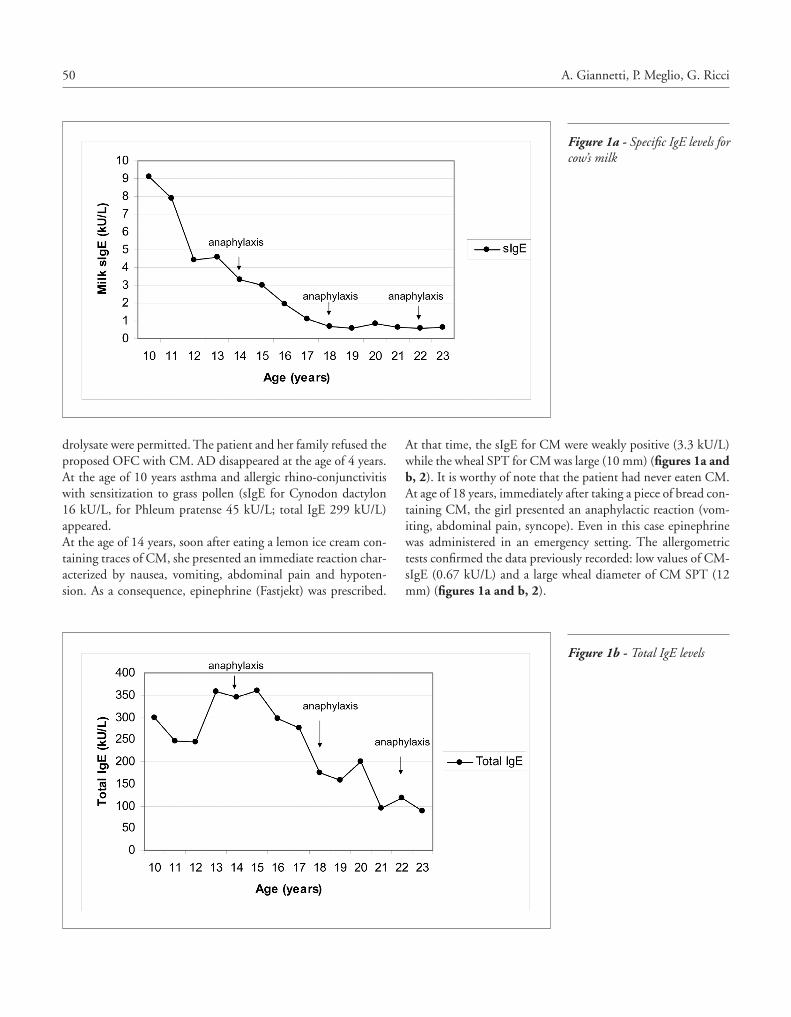
50 A. Giannetti, P. Meglio, G. Ricci
At that time, the sIgE for CM were weakly positive (3 3 kU/L) while the wheal SPT for CM was large (10 mm) (figures 1a and b, 2) It is worthy of note that the patient had never eaten CM At age of 18 years, immediately after taking a piece of bread con-taining CM, the girl presented an anaphylactic reaction (vom-iting, abdominal pain, syncope) Even in this case epinephrine was administered in an emergency setting The allergometric tests confirmed the data previously recorded: low values of CM-sIgE (0 67 kU/L) and a large wheal diameter of CM SPT (12 mm) (figures 1a and b, 2)
drolysate were permitted The patient and her family refused the proposed OFC with CM AD disappeared at the age of 4 years At the age of 10 years asthma and allergic rhino-conjunctivitis with sensitization to grass pollen (sIgE for Cynodon dactylon 16 kU/L, for Phleum pratense 45 kU/L; total IgE 299 kU/L) appeared At the age of 14 years, soon after eating a lemon ice cream con-taining traces of CM, she presented an immediate reaction char-acterized by nausea, vomiting, abdominal pain and hypoten-sion As a consequence, epinephrine (Fastjekt) was prescribed
Figure 1a - Specific IgE levels for cow’s milk
Figure 1b - Total IgE levels
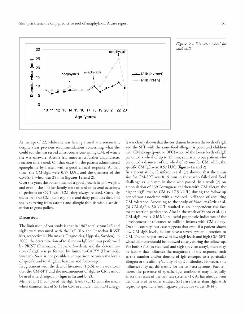
51Skin prick test: the only predictive tool of anaphylaxis? A case report
It was clearly shown that the correlation between the levels of sIgE and the SPT with the same food allergen is poor, and children with CM allergy (positive OFC) who had the lowest levels of sIgE presented a wheal of up to 15 mm, similarly to our patient who presented a diameter of the wheal of 25 mm for CM, whilst the specific CM IgE were 0 57 kU/L (figures 1a and 2) In a recent study, Cianferoni et al (7) showed that the mean size for CM-SPT was 8 15 mm in those who failed oral food challenge vs 4 8 mm in those who passed In a work (2) on a population of 139 Portuguese children with CM allergy, the higher sIgE level to CM (> 17 5 kU/L) during the follow-up period was associated with a reduced likelihood of acquiring CM tolerance According to the study of Vazquez-Ortiz et al (3) CM-sIgE > 50 kU/L resulted as an independent risk fac-tor of reaction persistence Also in the work of Vanto et al (4)
CM-sIgE level > 2 kU/L are useful prognostic indicators of the development of tolerance to milk in infants with CM allergy On the contrary, our case suggests that even if a patient shows low CM-sIgE levels, he can have a severe systemic reaction to CM Therefore, patients with low sIgE levels and high CM-SPT wheal diameter should be followed closely during the follow-up For both SPTs (in vivo test) and sIgE (in vitro assay), there may be factors that influence the magnitude of the response, such as the number and/or density of IgE epitopes to a particular allergen or the affinity/avidity of sIgE antibodies However, this influence may act differently for the two test systems Further-more, the presence of specific IgG antibodies may unequally affect the result of the two test systems (1) As has already been demonstrated in other studies, SPTs are better than sIgE with regard to specificity and negative predictive values (8-14)
At the age of 22, while she was having a meal at a restaurant, despite clear previous recommendations concerning what she could eat, she was served a first course containing CM, of which she was unaware After a few minutes, a further anaphylactic reaction intervened On that occasion the patient administered epinephrine by herself with a good clinical response At that time, the CM-sIgE were 0 57 kU/L and the diameter of the CM-SPT wheal was 25 mm (figures 1a and 2) Over the years the patient has had a good growth height-weight, and even if she and her family were offered on several occasions to perform an OCT with CM, they always refused Currently she is on a free CM, hen’s egg, nuts and dairy products diet, and she is suffering from asthma and allergic rhinitis with a sensiti-zation to grass pollen
Discussion
The limitation of our study is that in 1987 total serum IgE and sIgEs were measured with the IgE RIA and Phadebas RAST kits, respectively (Pharmacia Diagnostics, Uppsala, Sweden); in 2000, the determination of total serum IgE level was performed by PRIST (Pharmacia, Uppsala, Sweden), and the determina-tion of sIgE was performed by Immuno-CAP™ (Pharmacia, Sweden) So it is not possible a comparison between the levels of specific and total IgE at baseline and follow-up In agreement with the data of literature (1,5,6), our case shows that the CM-SPT and the measurement of sIgE to CM cannot be used interchangeably (figures 1a and b, 2) Mehl et al (1) compared the sIgE levels (kU/L) with the mean wheal diameter size of SPTs for CM in children with CM allergy
Figure 2 - Diameter wheal for cow’s milk
52 A. Giannetti, P. Meglio, G. Ricci
7 Cianferoni A, Garrett JP, Naimi DR, Khullar K, Spergel JM Predictive values for food challenge-induced severe reactions: development of a simple food challenge score Isr Med Assoc J 2012;14:24-8
8 Majamaa H, Moisio P, Holm K, Kautiainen H, Turjanmaa K Cow’s milk allergy: diagnostic accuracy of skin prick and patch tests and specific IgE Allergy 1999;54:346-51
9 Vanto T, Juntunen-Backman K, Kalimo K, et al The patch test, skin prick test, and serum milk-specific IgE as diagnostic tools on cow’s milk allergy in infants Allergy 1999;54:837-42
10 Sporik R, Hill DJ, Hosking CS Specificity of allergen skin testing in predicting positive open food challenges to milk, egg and peanut in children Clin Exp Allergy 2000;30:1540-6
11 Verstege A, Mehl A, Rolinck-Werninghaus C, et al The predictive value of the skin prick test wheal size for the outcome of oral food challenges Clin Exp Allergy 2005;35:1220-6
12 Niggemann B When is an oral food challenge positive? Allergy 2010;65:2-6
13 Roehr C, Reibel S, Ziegert M, Sommerfeld C, Wahn U, Nigge-mann B Atopy patch test together challenges in children with atopic dermatitis J Allergy Clin Immunol 2001;107:548-53
14 Fiocchi A, Bouygue GR, Restani P, Bonvini G, Startari R, Ter-racciano L Accuracy of skin prick tests in IgE-mediated adverse reactions to bovine proteins Ann Allergy Asthma Immunol 2002;89:26-32
We conclude that in some cases the positivity of SPTs to CM proteins may be more reliable than sIgE levels in predicting an anaphylactic reaction, especially in case of very high diameter of the CM-SPT wheal
References
1 Mehl A, Niggeman B, Kail T, Wahn U, Beyer K Skin prick test and specific serum IgE in the diagnostic evaluation of suspected cow’s milk and hen’s egg allergy in children: does one replace the other? Clin Exp Allergy 2012;42:1266-72
2 Santos A, Dias A, Pinheiro JA Predictive factors for the persistence of cow’s milk allergy Pediatr Allergy Immunol 2010;21:1127-34
3 Vázquez-Ortiz M, Alvaro-Lozano M, Alsina L, et al Safety and predictors of adverse events during oral immunotherapy for milk allergy: severity of reaction at oral challenge, specific IgE and prick test Clin Exp Allergy 2013;43:92-102
4 Vanto T, Helppila S, Juntunen-Backman K, et al Prediction of the development of tolerance to milk in children with cow’s milk hypersensitivity J Pediatr 2004;144:218-22
5 Rø AD, Saunes M, Smidesang I, et al Agreement of specific IgE and skin prick test in an unselected cohort of two-year-old chil-dren Eur J Pediatr 2012;171:479-84
6 Bartnikas LM, Sheehan WJ, Hoffman EB, et al Predicting food challenge outcomes for baked milk: role of specific IgE and skin prick testing Ann Allergy Asthma Immunol 2012;109:309-313
C A S E R E P O R T Eur Ann AllErgy Clin immunol voL 46, n 1, 53-55, 2014
SummaryClopidogrel is an antiplatelet drug widely used for treatment and prevention of a variety of cardiovascular diseases. We report a successful desensitization to clopidogrel in a 70-year-old Caucasian man with delayed hypersensitivity (HS) reaction. He developed lip, hand and foot swelling, erythematous papular non-pruritic lesions and arthralgias 2 weeks after starting treatment with clopidogrel 75 mg/d. A 3-hour desensitization protocol was started, achieving a cumulative dose of 154 mg without any reaction, and a daily dose of 75 mg was recommend-ed. On the 4th day, the patient developed skin lesions similar to the previously described. He was treated with topical steroids and oral antihistamines, and the daily dose of clopidogrel was reduced to 20 mg. A new desensitization protocol was established, with a slow dose increment, according to the patient’s response. It was only possible to achieve the dose of 75 mg/d after 2 months. Although well tolerated by most patients, HS reactions with clopidogrel may occur and desensitization is rising as a safe alternative in those patients. In delayed reactions with cutaneous lesions, a slower desensitization protocol may be necessary, as in this case.
Corresponding authorPatrícia Cristina Correia BarreiraAllergy and Clinical Immunology DepartmentCentro Hospitalar Vila Nova Gaia / Espinho - EPERua Conceição Fernandes 4434-502, Vila Nova de Gaia, PortugalPhone: +351 22 786 51 00 Fax: +351 22 786 51 78E-mail: pccb23@gmail com
Keywords
Clopidogrel; desensitization; hypersensitivity reaction
Drug Allergy Unit, Allergy and Clinical Immunology Department, Centro Hospitalar de Vila Nova de Gaia / Espinho - EPE, Vila Nova de Gaia, Portugal
Desensitization to clopidogrel: a tailor-made protocol
P. Barreira, S. caDinHa, D. maLHeiro, jP moreira Da SiLva
Case Report
Clopidogrel is a thienopyridine antiplatelet drug widely used for treatment and secondary prevention of a variety of cardio-vascular diseases (1) Its combination with acetylsalicylic acid is considered essential to reduce the risk of stent thrombosis in patients undergoing coronary stenting (2) Although clopi-dogrel is well tolerated in the majority of patients, mucocuta-neous reactions have been described: fixed drug eruption (1), oral erosive lichen planus (3), hemorrhagic herpes zoster (4),
maculopapular pruritic skin rashes (5) and angioedema (1) The alternative thienopyridine, ticlopidine, is more expensive and associated with serious side effects, such as neutropenia, throm-botic thrombocytopenic purpura, bone marrow aplasia and renal failure, limiting its clinical safety and utility (6) Clopi-dogrel is, therefore, the preferred thienopyridine in all cases When hypersensitivity reactions occur, as other treatment op-
tions are limited and frequently not tolerated, desensitization is rising as a safe alternative (7) Drug desensitization is defined as the induction of a temporary state of tolerance to a compound responsible for a hypersensitivity reaction, which can only be maintained by continuous administration of the drug (8) If the drug involved is discontinued, tolerance is lost within a period of time that can vary from a few hours to a few days (8) In this procedure, increasing doses of the drug are administered over a short period of time until the total cumulative therapeutic dose is achieved and tolerated (8) We report a successful desensitization to clopidogrel in a patient with delayed hypersensitivity reaction A 70-year-old Caucasian man with allergic rhinitis, hyperten-sion, dyslipidemia and chronic gastritis was submitted to cor-onary catheterization with placement of a stent and prescribed clopidogrel 75 mg/day Longstanding medication included ramipril, carvedilol, nitrates, indapamide, acetylsalicylic acid
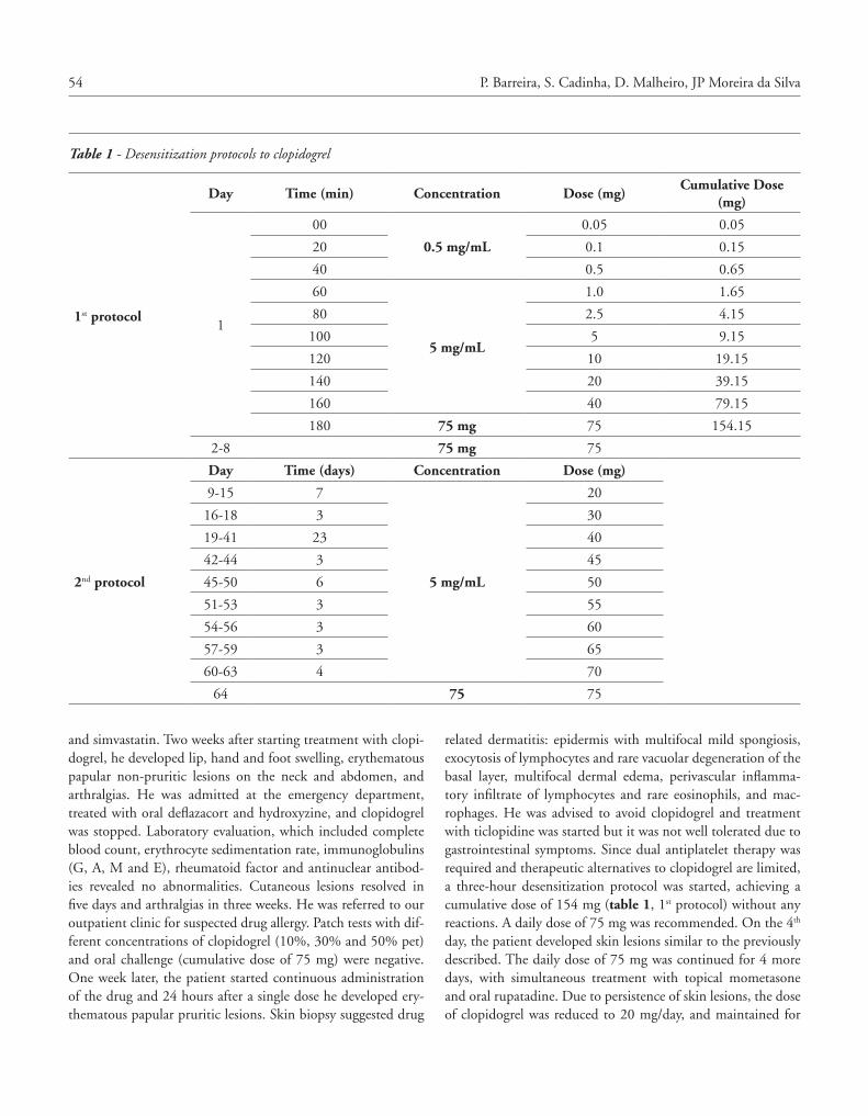
54 P. Barreira, S. Cadinha, D. Malheiro, JP Moreira da Silva
related dermatitis: epidermis with multifocal mild spongiosis, exocytosis of lymphocytes and rare vacuolar degeneration of the basal layer, multifocal dermal edema, perivascular inflamma-tory infiltrate of lymphocytes and rare eosinophils, and mac-rophages He was advised to avoid clopidogrel and treatment with ticlopidine was started but it was not well tolerated due to gastrointestinal symptoms Since dual antiplatelet therapy was required and therapeutic alternatives to clopidogrel are limited, a three-hour desensitization protocol was started, achieving a cumulative dose of 154 mg (table 1, 1st protocol) without any reactions A daily dose of 75 mg was recommended On the 4th day, the patient developed skin lesions similar to the previously described The daily dose of 75 mg was continued for 4 more days, with simultaneous treatment with topical mometasone and oral rupatadine Due to persistence of skin lesions, the dose of clopidogrel was reduced to 20 mg/day, and maintained for
and simvastatin Two weeks after starting treatment with clopi-dogrel, he developed lip, hand and foot swelling, erythematous papular non-pruritic lesions on the neck and abdomen, and arthralgias He was admitted at the emergency department, treated with oral deflazacort and hydroxyzine, and clopidogrel was stopped Laboratory evaluation, which included complete blood count, erythrocyte sedimentation rate, immunoglobulins (G, A, M and E), rheumatoid factor and antinuclear antibod-ies revealed no abnormalities Cutaneous lesions resolved in five days and arthralgias in three weeks He was referred to our outpatient clinic for suspected drug allergy Patch tests with dif-ferent concentrations of clopidogrel (10%, 30% and 50% pet) and oral challenge (cumulative dose of 75 mg) were negative One week later, the patient started continuous administration of the drug and 24 hours after a single dose he developed ery-thematous papular pruritic lesions Skin biopsy suggested drug
Table 1 - Desensitization protocols to clopidogrel
1st protocol
Day Time (min) Concentration Dose (mg)Cumulative Dose
(mg)
1
00
0.5 mg/mL
0 05 0 05
20 0 1 0 15
40 0 5 0 65
60
5 mg/mL
1 0 1 65
80 2 5 4 15
100 5 9 15
120 10 19 15
140 20 39 15
160 40 79 15
180 75 mg 75 154 15
2-8 75 mg 75
2nd protocol
Day Time (days) Concentration Dose (mg)
9-15 7
5 mg/mL
20
16-18 3 30
19-41 23 40
42-44 3 45
45-50 6 50
51-53 3 55
54-56 3 60
57-59 3 65
60-63 4 70
64 75 75
55Desensitization to clopidogrel: a tailor-made protocol
case also highlights the importance of patient awareness for the need of continuous drug administration so that tolerance can be maintained
References
1 Ghosh SK, Bandyopadhyay D Clopidogrel-induced fixed drug eruption J Eur Acad Dermatol Venereol 2009;23:1202-1203
2 Khambekar SK, Kovac J, Gershlick AH Clopidogrel induced urti-carial rash in a patient with left main stem percutaneous coronary intervention: management issues Heart 2004;90:e14
3 Guijarro-Guijarro B, López-Sánchez AF Lichenoid reaction caused by Clopidogrel, a new anti-platelet drug Med Oral 2003;8:33-37
4 Ghosh SK, Bandyopadhyay D Haemorrhagic herpes zos-ter associated with clopidogrel treatment Clin Exp Dermatol 2009;34:409-10
5 Makkar K, Wilensky RL, Julien MB, Herrmann HC, Spinler SA Rash with both clopidogrel and ticlopidine in two patients follow-ing percutaneous coronary intervention with drug-eluting stents Ann Pharmacother 2006;40:1204-1207
6 Von Tiehl KF, Price MJ, Valencia R, Ludington KJ, Teirstein PS, Simon RA Clopidogrel Desensitization After Drug-Eluting Stent Placement J Am Coll Cardiol 2007;50:2039-2043
7 Sanchez-Lopez J, Muñoz-Cano R, Bartra J, Valero A, Pica-do C Desensitization to clopidogrel: a growing need Allergy 2010;65:279-280
8 Cernadas JR, Brockow K, Romano A, Aberer W, Torres MJ, Birch-er A, Campi P, Sanz ML, Castells M, Demoly P, Pichler WJ, for the European Network of Drug Allergy and the EAACI interest group on drug hypersensitivity General considerations on rapid desensitization for drug hypersensitivity - a consensus statement Allergy 2010;65:1357-1366
9 Lee-Wong M, Gadhvi D, Resnick D Clopidogrel desensitization Annals of Allergy, Asthma & Immunology 2006;96;756-757
10 Oppedijk B, Oderkerken DA, van der Wildt, Melissant CF Rapid oral desensitization procedure in clopidogrel hypersensitivity Neth Heart J 2008;16:21-23
11 Scherer K, Brockow K, Aberer W, Gooi JHC, Demoly P, Roma-no A, Schnyder B, Whitaker P, Cernadas JSR, Bircher AJ and for ENDA, the European Network on Drug Allergy and the EAACI Drug Allergy Interest Group Desensitization in delayed drug hy-persensitivity reactions - an EAACI position paper of the Drug Allergy Interest Group Allergy 2013Jun7 (e-Pub ahead of print)
7 days A new desensitization protocol was established, with a slow dose increment, according to the patient’s response (table 1, 2nd protocol) Clopidogrel administration was performed at home and the patient was evaluated in our outpatient depart-ment every 3 days, or less if the clinical situation warranted earlier evaluation During the 2nd protocol, when skin lesions reappeared they were treated with topical corticosteroids and oral antihistamines previously used, and clopidogrel dose was increased only when there was a marked improvement of skin lesions The dose of 75 mg/day was only achieved after two months Even though clopidogrel was well tolerated, the patient decided to stop its administration one month later This means that he did not understand the principle of drug desensitiza-tion, which was explained several times and it was included in the written informed consent The case was discussed with the cardiothoracic surgeon and given the patient’s non-compliance it was decided not to start a new desensitization procedure Although well tolerated by most patients, hypersensitivity reac-tions with clopidogrel may occur Since other treatment options are limited and frequently not tolerated, desensitization is an alternative in patients who require prolonged dual antiplate-let therapy Some protocols for desensitization to clopidogrel have been published, demonstrating its safety and effectiveness (6,7,9,10) Cutaneous hypersensitivity reactions to drugs may be caused by different pathogenic mechanisms Despite negative patch tests to clopidogrel, the authors consider that a specific involvement of T cells may be suggested by histological findings on skin bi-opsy of dermal perivascular inflammatory infiltrate with lym-phocytes and scarce eosinophils and macrophages According to a recent publication of the European Drug Allergy Interest Group there is no universal or consensus drug desensiti-zation protocol for delayed-type hypersensitivity reactions, and protocols vary in duration, ranging from a few hours to several weeks Patients with delayed reactions may benefit from longer protocols, with repetitive and slowly increasing doses, since rush protocols frequently have a higher failure rate (11) The authors present a tailored-made desensitization protocol to clopidogrel that can be used in an outpatient setting This
C A S E R E P O R T Eur Ann AllErgy Clin immunol voL 46, n 1, 56-59, 2014
SummarySevere asthma with fungal sensitisation (SAFS) is characterized by poor symptoms control and frequent hospital admissions for exacerbations despite treatment with high dose inhaled steroids, long-acting beta-2 agonists and leukotriene receptor antagonists. Treatment with oral steroids is usually necessary and courses of antifungal therapy may improve asthma symptoms. We report a case refractory to conventional inhaled therapies, continuous oral steroids and antifungal therapy courses, who was effectively treated with omalizumab.
Corresponding author Fabio Di StefanoPneumology and Allergy Unit Department of Internal Medicine Ospedale Spirito Santo Azienda USL PescaraPhone: + 39 085 4252906-2717E-mail: f distefano@aliceposta it
Key words
Severe asthma with fungal sensitisation; omalizumab
1Laboratory of Clinical Pathology and Microbiology, General Service Department, Azienda Ospedaliero-Universitaria “Ospedali Riuniti” di Ancona, Ancona, Italy2Allergy Unit, Department of Internal Medicine, Azienda Ospedaliero-Universitaria “Ospedali Riuniti” di Ancona, Ancona, Italy
Efficacy of omalizumab in severe asthma with fungal sensitisation: a case reportPneumology and Allergy Unit, Department of Internal Medicine, Ospedale Spirito Santo, Azienda USL Pescara, Pescara, Italy
f. Di Stefano, B. cinti1, L. antoniceLLi2
The link between fungi and severe asthma is complex and not fully understood Both allergy to fungi (spores, hyphae) and the effects of cell wall constituents (glucan, volatile organic com-pounds) on bronchi are involved in asthma (1,2) Distinct pathways of exposure to fungal allergens may be associ-ated with asthma, mainly inhalation of airborne fungal allergens (spores or hyphae) with symptoms due to both persistent indoor exposure (in particular in mouldy and damp houses) or to out-door acute exposure to very high concentration of these allergens (3,4) Fungal infection outside the respiratory tract, most often dermatophyte infection of the skin or nails may also be associated with immediate hypersensitivity, and in these patients courses of antifungal therapy may improve asthma symptoms (5) Chronic or intermittent colonization of the airways by the ubiq-uitous fungus Aspergillus Fumigatus in conjunction with imme-
diate hypersensitivity may cause an abnorm allergic response leading to allergic bronchopulmonary aspergillosis (ABPA) However, many other species of fungi, including Cladosporium, Alternaria, Penicillium, Trichophyton and Candida, may be in-volved leading to a less specific disease named allergic broncho-pulmonary mycosis (ABPM) (1,2,6) Criteria for diagnosis of ABPA and ABPM include presence of severe asthma, peripheral blood eosinophilia, elevated total serum IgE level (> 1000 IU/ml), migrant chest radiographic infiltrates and central bronchi-ectasis easily detected by chest CT scan Additional criteria are usually considered mucus plugs production, Aspergillus or other fungi species immediate skin prick test reactivity, and/or elevat-ed specific IgE antibodies Many patients with severe asthma do not have the diagnostic cri-teria of ABPA and ABPM and are not demonstrably colonized
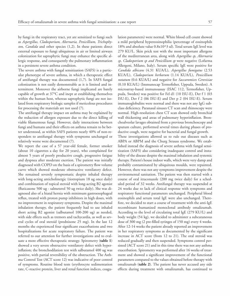
57Efficacy of omalizumab in severe asthma with fungal sensitisation: a case report
lation parameters) were normal White blood cell count showed a mild peripheral hypereosinophilia (percentage of eosinophils 18% and absolute value 0 8x10^3 ul) Total serum IgE level was 279 KU/L Skin prick test with the most important allergens of the mediterranean area, along with Aspergillus sp, Alternaria sp, Cladosporium sp and Penicillium sp were negative (Lofarma Allergeni, Milano, Italy) Serum specific IgE were positive for Candida albicans (4 31 KUA/L), Aspergillus fumigatus (2 53 KUA/L), Cladosporium herbarum (1 14 KUA/L), Penicillium notatum (0 6 KUA/L) and negative for Saccaromices Cerevisiae (0 10 KUA/L) (Immunocap Termofisher, Uppsala, Sweden) A microarray-based immunoassay (ISAC 112, Termofisher, Up-psala, Sweden) was positive for Fel d1 (10 ISU-E), Der f 1 (03 ISU-E), Der f 2 (06 ISU-E) and Der p 2 (04 ISU-E) Serum immunoglobulins were normal and there was not any IgG sub-class deficiency Paranasal sinuses CT scan and rhinoscopy were normal High-resolution chest CT scan showed only bronchial wall thickening and areas of pulmonary hyperinflation Bron-choalveolar lavages obtained from a previous bronchoscopy and sputum culture, performed several times during phases of pro-ductive cough, were negative for bacterial and fungal growth These investigations allowed us to rule out diseases such as ABPA or ABPM and the Churg Strauss syndrome We estab-lished instead the diagnosis of severe asthma with fungal sensi-tization (SAFS) also considering inadequate control and insta-bility of the disease despite the maximal inhalation and systemic therapy Patient’s house indoor walls, which were very damp and probably contaminated by ubiquitous moulds, were repainted However, there was not any symptoms improvement despite the environmental sanitation The patient was then started with a course of oral itraconazole (200 mg twice daily) for a sched-uled period of 32 weeks Antifungal therapy was suspended at 24 weeks due to lack of clinical response with symptoms and respiratory functional parameters unchanged Peripheral blood eosinophils and serum total IgE were also unchanged There-fore, we decided to start a course of treatment with the anti-IgE recombinant humanized monoclonal antibody omalizumab According to the level of circulating total IgE (279 KU/L) and body weight (54 kg), we decided to administer a subcutaneous dose of 300 mg (2 pre-filled syringes of 150 mg) every 4 weeks After 12-14 weeks the patient already reported an improvement in her respiratory symptoms as documented by the significant increase in ACT score (from 12 to 21) The oral steroid was reduced gradually and then suspended Symptoms control per-sisted (ACT score 21) and in this time there was not any asthma exacerbation Spirometry was performed after 16 weeks of treat-ment and showed a significant improvement of the functional parameters compared to the values obtained before therapy with omalizumab (table 2) The patient has never accused any side effects during treatment with omalizumab, has continued to
by fungi in the respiratory tract, yet are sensitized to fungi such as Aspergillus, Cladosporium, Alternaria, Penicillium, Trichophy-ton, Candida and other species (1,2) In these patients direct external exposure to fungi ubiquitous in air or limited airways colonization for saprophytic fungi may enhance the specific al-lergic response, and consequently the pulmonary inflammation in a persistent severe asthma condition The severe asthma with fungal sensitization (SAFS) is a partic-ular phenotype of severe asthma, in which a therapeutic effect of antifungal therapy was documented (1,7) In SAFS fungal colonization is not easily demonstrable as it is limited and in-termittent Moreover the airborne fungi implicated are barely capable of growth at 37°C and inept at establishing themselves within the human host, whereas saprophytic fungi are not iso-lated from respiratory biologic samples if meticolous procedures for processing the materials are not used (7) The antifungal therapy impact on asthma may be explained by the reduction of allergen exposure due to the direct killing of viable filamentous fungi However, daily interactions between fungi and humans and their effects on asthma remain to be bet-ter understood, as within SAFS patients nearly 40% of non-re-sponders to antifungal therapy with symptoms unchanged or relatively worse were documented (7) We report the case of a 57 year-old female, former smoker (about 10 cigarettes a day for 20 years), who complained for almost 5 years of poorly productive cough, progressive fatigue and dyspnea after moderate exertion The patient was initially diagnosed with COPD on the basis of a spirometry flow volume curve which showed moderate obstructive ventilatory defect She remained severely symptomatic despite inhaled therapy with long-acting anticholinergic (tiotropium 18 ug once daily) and combination of topical steroid with long-acting B2 agonist (fluticasone 500 ug - salmeterol 50 ug twice daily) She was di-agnosed also of a hiatal hernia with minimum gastroesophageal reflux, treated with proton pump inhibitors in high doses, with no improvement in respiratory symptoms Despite the maximal inhalation therapy, the patient frequently had to use inhaled short acting B2 agonist (salbutamol 100-200 ug) as needed, with side effects such as tremors and tachycardia, as well as sev-eral cycles of oral steroid (prednisone 25 mg) In the last 12 months she experienced four significant exacerbations and two hospitalizations for acute respiratory failure The patient was referred to our attention for further investigations and to eval-uate a more effective therapeutic strategy Spirometry (table 1) showed a very severe obstructive ventilatory defect with hyper-inflation; the bronchodilation test with salbutamol 400 ug was positive, with partial reversibility of the obstruction The Asth-ma Control Test (ACT score 12) was indicative of poor control of symptoms Routine blood tests (erythrocyte sedimentation rate, C-reactive protein, liver and renal function indices, coagu-
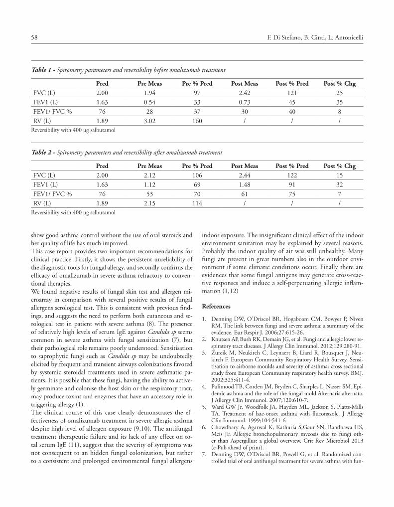
58 F. Di Stefano, B. Cinti, L. Antonicelli
indoor exposure The insignificant clinical effect of the indoor environment sanitation may be explained by several reasons Probably the indoor quality of air was still unhealthy Many fungi are present in great numbers also in the outdoor envi-ronment if some climatic conditions occur Finally there are evidences that some fungal antigens may generate cross-reac-tive responses and induce a self-perpetuating allergic inflam-mation (1,12)
References
1 Denning DW, O’Driscol BR, Hogaboam CM, Bowyer P, Niven RM The link between fungi and severe asthma: a summary of the evidence Eur Respir J 2006;27:615-26
2 Knutsen AP, Bush RK, Demain JG, et al Fungi and allergic lower re-spiratory tract diseases J Allergy Clin Immunol 2012;129:280-91
3 Zureik M, Neukirch C, Leynaert B, Liard R, Bousquet J, Neu-kirch F European Community Respiratory Health Survey Sensi-tisation to airborne moulds and severity of asthma: cross sectional study from European Community respiratory health survey BMJ 2002;325:411-4
4 Pulimood TB, Corden JM, Bryden C, Sharples L, Nasser SM Epi-demic asthma and the role of the fungal mold Alternaria alternata J Allergy Clin Immunol 2007;120:610-7
5 Ward GW Jr, Woodfolk JA, Hayden ML, Jackson S, Platts-Mills TA Treatment of late-onset asthma with fluconazole J Allergy Clin Immunol 1999;104:541-6
6 Chowdhary A, Agarwal K, Kathuria S,Gaur SN, Randhawa HS, Meis JF Allergic bronchopulmonary mycosis due to fungi oth-er than Aspergillus: a global overview Crit Rev Microbiol 2013 (e-Pub ahead of print)
7 Denning DW, O’Driscol BR, Powell G, et al Randomized con-trolled trial of oral antifungal treatment for severe asthma with fun-
show good asthma control without the use of oral steroids and her quality of life has much improved This case report provides two important recommendations for clinical practice Firstly, it shows the persistent unreliability of the diagnostic tools for fungal allergy, and secondly confirms the efficacy of omalizumab in severe asthma refractory to conven-tional therapies We found negative results of fungal skin test and allergen mi-croarray in comparison with several positive results of fungal allergens serological test This is consistent with previous find-ings, and suggests the need to perform both cutaneous and se-rological test in patient with severe asthma (8) The presence of relatively high levels of serum IgE against Candida sp seems common in severe asthma with fungal sensitization (7), but their pathological role remains poorly understood Sensitisation to saprophytic fungi such as Candida sp may be undoubtedly elicited by frequent and transient airways colonizations favored by systemic steroidal treatments used in severe asthmatic pa-tients It is possible that these fungi, having the ability to active-ly germinate and colonise the host skin or the respiratory tract, may produce toxins and enzymes that have an accessory role in triggering allergy (1) The clinical course of this case clearly demonstrates the ef-fectiveness of omalizumab treatment in severe allergic asthma despite high level of allergen exposure (9,10) The antifungal treatment therapeutic failure and its lack of any effect on to-tal serum IgE (11), suggest that the severity of symptoms was not consequent to an hidden fungal colonization, but rather to a consistent and prolonged environmental fungal allergens
Table 1 - Spirometry parameters and reversibility before omalizumab treatment
Pred Pre Meas Pre % Pred Post Meas Post % Pred Post % ChgFVC (L) 2 00 1 94 97 2 42 121 25
FEV1 (L) 1 63 0 54 33 0 73 45 35
FEV1/ FVC % 76 28 37 30 40 8
RV (L) 1 89 3 02 160 / / /Reversibility with 400 µg salbutamol
Table 2 - Spirometry parameters and reversibility after omalizumab treatment
Pred Pre Meas Pre % Pred Post Meas Post % Pred Post % ChgFVC (L) 2 00 2 12 106 2,44 122 15
FEV1 (L) 1 63 1 12 69 1 48 91 32
FEV1/ FVC % 76 53 70 61 75 7
RV (L) 1 89 2 15 114 / / /Reversibility with 400 µg salbutamol
59Efficacy of omalizumab in severe asthma with fungal sensitisation: a case report
10 Lavaud F, Bonniaud P, Dalphin JC, Leroyer C, Muller D, Tannous R, Mangiapan G, De Blay F Usefulness of omalizumab in ten pa-tients with severe occupational asthma Allergy 2013;68:813-5
11 Pizzimenti S, Bussolino C, Badiu I, Rolla G Itraconazole as ‘bridge therapy’ to anti-IgE in a patient with severe asthma with fungal sensitisation BMJ Case Rep 2013 doi:pii: bcr2012008462 10 1136/bcr-2012-008462
12 Fluckiger S, Scapozza L, Mayer C, Blaser K, Folkers G, Crameri R Immunological and structural analysis of IgE mediated cross-reac-tivity between manganese superoxide dismutases Int Arch Allergy Immunol 2002;128:292-303
gal sensitization: The Fungal Asthma Sensitization Trial (FAST) study Am J Respir Crit Care Med 2009;179:11-18
8 O’Driscoll BR, Powell G, Chew F, Niven RM, Miles JF, Vyas A, Denning DW Comparison of skin prick tests with specific serum immunoglobulin E in the diagnosis of fungal sensitization in pa-tients with severe asthma Clin Exp Allergy 2009;39:1677-83
9 Brusselle G, Michils A, Louis R, et al “Real-life” effectiveness of omalizumab in patients with severe persistent allergic asthma: the PERSIST study Respir Med 2009;103:1633-42
C A S E R E P O R T Eur Ann AllErgy Clin immunol voL 46, n 1, 60-62, 2014
SummaryHereditary angioedema (HEA) is a disease characterized by decreased levels or function of C1 esterase inhibitor (C1-INH). The symptoms of HEA in pediatric age group generally consist of recurrent episodes of soft tissue swelling. These symptoms can be transient, subtle, and varied in severity. Genitourinary system is rarely affected in this disease. Here, a three-year-old girl who presented with angioedema on her hands, fingers, and face, and had difficulty in urination and globe is reported. The aim of this case is to focus on this rare disease, hereditary angioede-ma, which presented with difficulty in urination and urinary globe. Corresponding author
Himmet Haluk Akar, MDErciyes University School of MedicineDepartment of Pediatric AllergyKayseri, TurkeyPhone: + 90 352 2076666/25125Fax: + 90 352 4375825 E-mail: himmetakar@gmail com
Key words
Hereditary angioedema; C1 inhibitor deficiency; difficulty in urination; globe
1Erciyes University School of Medicine, Department of Pediatric Allergy and Immunology, Kayseri, Turkey2Erciyes University School of Medicine, Department of Pediatrics, Kayseri, Turkey
A case of hereditary angioedema who presented with difficulty in urination and globe
H. HaLuk akar1, f. taHan1, t. kurt2, i. SoLmaz2
Introduction
C1-INH deficiency can be genetic or acquired HAE is a rare, life-threatening condition manifested by acute attacks of facial, laryngeal, peripheral or genital edema The estimated prevalence of HAE is between 1 in 10,000 and 1 in 150,000 people (1,2) The genetic HEA deficiency is due to mutations in one of the two alleles of the C1-INH gene that result in reduced protein levels in plasma (type I HEA) or in normal protein levels but always in reduced function (type II HEA) A third type has also been reported, occurring exclusively in women who have nor-mal C1-INH levels and function (Type III HEA) It is estimat-ed that 20% to 25% of HAE cases are caused by spontaneous mutations in patients with no family history of the disease (3,5) The development of angioedema in C1-INH-deficient patients involves the inappropriate generation of kinins (particularly bradykinin) that stimulate vascular smooth-muscle relaxation
and induce increased permeability C1-INH is a serine protease inhibitor (serpin), also known as SERPING1, that blocks the activity of some complement components (e g C1r, C1s, Man-nose binding lectin-associated serine protease; MASP-1 and MASP-2) C1-INH also controls contact-kinins, coagulation, and fibrinolytic cascades (6) In this paper, we report a case with rarely manifested hereditary angioedema who presented with difficulty in urination and globe
Case report
One day, a three-year-old girl with HEA was admitted to our emergency pediatric department with swelling of right forearm and urinary globe, and difficulty in urination (figure 1) She didn’t have dysuria, fever or pelvic pain Urinary globe, mild external genital swelling and right forearm angioedema were examined in her physical examination Her urine analyses,
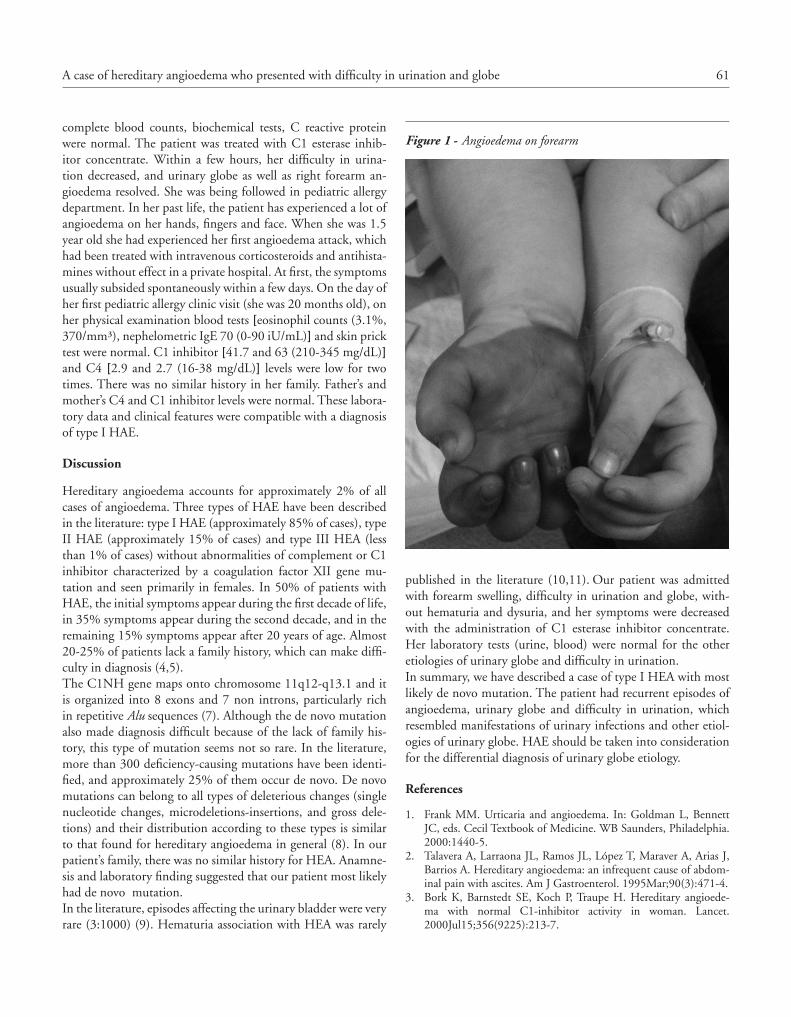
61A case of hereditary angioedema who presented with difficulty in urination and globe
published in the literature (10,11) Our patient was admitted with forearm swelling, difficulty in urination and globe, with-out hematuria and dysuria, and her symptoms were decreased with the administration of C1 esterase inhibitor concentrate Her laboratory tests (urine, blood) were normal for the other etiologies of urinary globe and difficulty in urination In summary, we have described a case of type I HEA with most likely de novo mutation The patient had recurrent episodes of angioedema, urinary globe and difficulty in urination, which resembled manifestations of urinary infections and other etiol-ogies of urinary globe HAE should be taken into consideration for the differential diagnosis of urinary globe etiology
References
1 Frank MM Urticaria and angioedema In: Goldman L, Bennett JC, eds Cecil Textbook of Medicine WB Saunders, Philadelphia 2000:1440-5
2 Talavera A, Larraona JL, Ramos JL, López T, Maraver A, Arias J, Barrios A Hereditary angioedema: an infrequent cause of abdom-inal pain with ascites Am J Gastroenterol 1995Mar;90(3):471-4
3 Bork K, Barnstedt SE, Koch P, Traupe H Hereditary angioede-ma with normal C1-inhibitor activity in woman Lancet 2000Jul15;356(9225):213-7
complete blood counts, biochemical tests, C reactive protein were normal The patient was treated with C1 esterase inhib-itor concentrate Within a few hours, her difficulty in urina-tion decreased, and urinary globe as well as right forearm an-gioedema resolved She was being followed in pediatric allergy department In her past life, the patient has experienced a lot of angioedema on her hands, fingers and face When she was 1 5 year old she had experienced her first angioedema attack, which had been treated with intravenous corticosteroids and antihista-mines without effect in a private hospital At first, the symptoms usually subsided spontaneously within a few days On the day of her first pediatric allergy clinic visit (she was 20 months old), on her physical examination blood tests [eosinophil counts (3 1%, 370/mm³), nephelometric IgE 70 (0-90 iU/mL)] and skin prick test were normal C1 inhibitor [41 7 and 63 (210-345 mg/dL)] and C4 [2 9 and 2 7 (16-38 mg/dL)] levels were low for two times There was no similar history in her family Father’s and mother’s C4 and C1 inhibitor levels were normal These labora-tory data and clinical features were compatible with a diagnosis of type I HAE
Discussion
Hereditary angioedema accounts for approximately 2% of all cases of angioedema Three types of HAE have been described in the literature: type I HAE (approximately 85% of cases), type II HAE (approximately 15% of cases) and type III HEA (less than 1% of cases) without abnormalities of complement or C1 inhibitor characterized by a coagulation factor XII gene mu-tation and seen primarily in females In 50% of patients with HAE, the initial symptoms appear during the first decade of life, in 35% symptoms appear during the second decade, and in the remaining 15% symptoms appear after 20 years of age Almost 20-25% of patients lack a family history, which can make diffi-culty in diagnosis (4,5) The C1NH gene maps onto chromosome 11q12-q13 1 and it is organized into 8 exons and 7 non introns, particularly rich in repetitive Alu sequences (7) Although the de novo mutation also made diagnosis difficult because of the lack of family his-tory, this type of mutation seems not so rare In the literature, more than 300 deficiency-causing mutations have been identi-fied, and approximately 25% of them occur de novo De novo mutations can belong to all types of deleterious changes (single nucleotide changes, microdeletions-insertions, and gross dele-tions) and their distribution according to these types is similar to that found for hereditary angioedema in general (8) In our patient’s family, there was no similar history for HEA Anamne-sis and laboratory finding suggested that our patient most likely had de novo mutation In the literature, episodes affecting the urinary bladder were very rare (3:1000) (9) Hematuria association with HEA was rarely
Figure 1 - Angioedema on forearm
62 H. Haluk Akar, F. Tahan, T. Kurt, I. Solmaz
8 Pappalardo E, Cicardi M, Duponchel C, Carugati A, Choquet S, Agostoni A, Tosi M Frequent de novo mutations and exon de-letions in the C1 inhibitor gene of patients with angioedema J Allergy Clin Immunol 2000Dec;106(6):1147-54
9 Bork K, Meng G, Staubach P, Hardt J Hereditary angioedema: new findings concerning symptoms, affected organs, and course Am J Med 2006Mar;119(3):267-74
10 Van Dellen RC, Myers RP Bladder involvement in hereditary an-gioedema Mayo Clin Proc 1980Apr;55(4):277-8
11 Pan CG, Strife CF, Ward MK, Spitzer RE, McAdams AJ Long-term follow-up of non-systemic lupus erythematosus glomerulone-phritis in patients with hereditary angioedema: report of four cases Am J Kidney Dis 1992 Jun;19(6):526-31
4 Eck SL, Morse JH, Janssen DA, Emerson SG, Markovitz DM An-gioedema presenting as chronic gastrointestinal symptoms Am J Gastroenterol 1993Mar;88(3):436-9
5 Agostini A, Cicardi M Hereditary and acquired C-1 inhibitor deficiency: biological and clinical characteristics in 235 patients Medicine (Baltimore) 1992Jul;71(4):206-15
6 Cugno M, Zanichelli A, Foieni F, Caccia S, Cicardi M C1-inhibi-tor deficiency and angioedema: molecular mechanisms and clinical progress Trends Mol Med 2009Feb;15(2):69-78
7 Carter PE, Duponchel C, Tosi M, Fothergill JE Complete nu-cleotide sequence of the gene for human C1 inhibitor with an unusually high density of Alu elements Eur J Biochem 1991Apr23;197(2):301-8
C O R R E S P O N D E N C E Eur Ann AllErgy Clin immunol voL 46, n 1, 63-64, 2014
i. Doña1, n. BLanca-LoPez2, j. a. cornejo-Garcia3
1Allergy Service Carlos Haya Hospital, Malaga, Spain inmadd@hotmail com2Allergy Service, Infanta Leonor Hospital, Madrid, Spain3Research Laboratory for Allergic Diseases, Carlos Haya Hospital, Malaga, Spain
NSAIDs are the most frequent medicaments involved in hypersensitivity drug reactions
Dear Sir
The editorial by Asero et al in issue n° 1 of Vol 45 of the European Annals of Allergy and Clinical Immunology (1) points out import-ant data on cutaneous hypersensitivity to multiple non-steroidal anti-inflammatory drugs (NSAIDs) in the sense that a certain num-ber of patients may also respond to selective COX-2 inhibitors Al-though the cases of the two patients reported had good tolerance to paracetamol, the authors noted that tolerance to selective COX-2 inhibitors may vary according to previous tolerance to paracetamol Comparing reactivity to different coxibs, figures lower than 10% could correspond to cases who had good tolerance to paracetamol, and higher figures could correspond to those who previously had skin problems after taking paracetamol, as reported by our group (2) However, a more detailed analysis (from the table) shows that tolerance to paracetamol was not tested in every patient (1) It is important also to emphasize the relevance of NSAIDs in the induction of hypersensitivity reactions Asero et al stated in the article that NSAIDs are one of the common causes in-ducing drug hypersensitivity For this purpose, a detailed anal-ysis of the manuscript published by Gomes et al (3) indicates that NSAIDs were the second on the list of implicated drugs in hospital-based populations as well as in outpatients and general populations However, a recent study published by our group (4) showed that after a detailed evaluation of 4460 patients
over a period of 6 years, 966 patients were finally confirmed as positive to NSAIDs, this representing the most frequent group of drugs involved and confirmed as causing allergy A previous study focusing on the hypersensitivity patterns of responses to the NSAID group suggests that 60% were cross-intolerant (5) We believe that with the data now available, these references should be taken into account when referring to drugs involved in hypersensitive reactions
References
1 Asero R, Quaratino D Cutaneous hypersensitivity to multiple NSAIDs: never take tolerance to selective COX-2 inhibitors (COX-IBs) for granted! Eur Ann Allergy Clin Immunol 2013;45(1):3-6
2 Doña I, Blanca-López N, Jagemann LR, Torres MJ, Rondón C, Campo P, et al Response to a selective COX-2 inhibitor in patients with urticaria/angioedema induced by nonsteroidal anti-inflam-matory drugs Allergy 2011;66(11):1428-33
3 Gomes ER, Demoly P Epidemiology of hypersensitivity drug reac-tions Curr Opin Allergy Clin Immunol 2005;5:309-16
4 Doña I, Blanca-Lopez N, Torres MJ, García-Campos J, García-Núñez I, Gómez F, et al Drug hypersensitivity reactions: response patterns, drug involved, and temporal variations in a large series of patients J Investig Allergol Clin Immunol 2012;22:363-71
5 Doña I, Blanca-López N, Cornejo-García JA, Torres MJ, Laguna JJ, Fernández J, et al Characteristics of subjects experiencing hy-persensitivity to non-steroidal anti-inflammatory drugs: patterns of response Clin Exp Allergy 2011;41(1):86-95
64 R Asero, D Quaratino
r. aSero1, D. quaratino2
1Ambulatorio di Allergologia, Clinica San Carlo, Paderno Dugnano, Italy 2Ambulatorio di Allergologia, IDI-IRCCS, Capranica, Italy
Reply
depend on study populations (ethnicities, inpatients or outpa-tients, adults or children), differences in methods for assessing offending drug, and different methods of data analyses (5) However, we believe that an exact ranking of prevalence doesn’t appear very relevant in the evaluation of coxibs as alternative drug in cutaneous hypersensitivity to multiple NSAIDs
References
1 Asero R, Quaratino D Cutaneous hypersensitivity to multiple NSAIDs: never take tolerance to selective COX-2 inhibitors (COX-IBs) for granted! Eur Ann Allergy Clin Immunol 2013;45:3-6
2 Doña I, Blanca-López N, Jagemann LR, Torres MJ, Rondón C, Campo P, et al Response to a selective COX-2 inhibitor in patients with urticaria/angioedema induced by nonsteroidal anti-inflam-matory drugs Allergy 2011;66:1428-33
3 Gomes ER, Demoly P Epidemiology of hypersensitivity drug reac-tions Curr Opin Allergy Clin Immunol 2005;5:309-16
4 Doña I, Blanca-Lopez N, Torres MJ, García-Campos J, García-Núñez I, Gómez F, et al Drug hypersensitivity reactions: response patterns, drug involved, and temporal variations in a large series of patients J Investig Allergol Clin Immunol 2012;22:363-71
5 Thong BY, Tan TC Epidemiology and risk factors for drug allergy Br J Clin Pharmacol 2011;71:684-700
Doña and co-workers’ comments on our editorial (1) underline two aspects of NSAIDs hypersensitivity The first one is parac-etamol tolerance in NSAIDs-hypersensitive patients, particu-larly in those reacting to different NSAIDs, including selective COX-2 inhibitors (coxibs) As reported in the editorial, Doña and co-workers found that intolerance to paracetamol could represent a “marker” of reactivity to etoricoxib and to other cox-ibs (2) This was not the case in our two patients On the other hand, reviewing the outcome of paracetamol tolerance in cox-ibs-reactive patients was out of scope of our article, dealing with challenge tests with coxibs in patients with cutaneous hypersen-sitivity to multiple NSAID The two cases we presented under-lined the need of checking tolerability of alternative NSAIDs (including selective COX2-inhibitors) in a proper setting The same holds obviously true also for paracetamol, as highlighted by Doña et al (2) The second aspect is the prevalence of hypersensitivity reactions to NSAIDs: definitely this class of drugs is one of the most com-mon causes of adverse reactions (3), and in some populations is the most frequent one, as recently reported by Doña et al (4) Differences in the prevalence rate of drug hypersensitivity may

informazionicosti
le più esaurienti e affidabili informazioni sui farmaci da oggi e per tutto il 2014a un prezzo speciale
Le offerte
• volume Parafarmaceutici• volume Indirizzi
a soli 99 euro
• volume Medicinali• volume Indirizzi
a soli 99 euro
• volume Medicinali• volume Parafarmaceutici• volume Indirizzi
a soli 149 euro
© EDRA LSWR SpA
via Spadolini, 7 - 20141 MilanoTel.: 02.88184.317 - Fax: 02.93664.151e-mail: [email protected] - www.edizioniedra.it
DIS
3201
4
Senza titolo-1 1 02/04/14 17.53
INFORMAZIONEPolitica Sanitaria, clinica,
farmaci e terapie e la cronaca quotidiana
dei fatti di attualità
CANALI TEMATICICardiologia, Pediatria,
Ginecologia, Diabetologia, Medicina interna
CAsI CLINICIOltre 120.000 medici coinvolti nei casi clinici messi a disposizione
dei colleghi per un consulto
COMMUNITYL’area Social con i servizi
“L’Esperto risponde”, “Doctor Alert”, “Forum”,
“Preferiti”
DOWNLOAD CENTER
Contenuti di formazione e aggiornamento gratuiti tratti
dalle pubblicazioni Edra Masson
ECM La formazione di qualità
di Doctor33
BACHECA ANNUNCI
La più ricca sezione di annunci sul web per
i professionisti della medicina
Il portale dei protagonisti della salute
www.doctor33.it
NOvITà