erbb receptor stimulation is required for mouse colon
TRANSCRIPT

ErbB Receptor Stimulation Is Required for MouseColon Adenoma Organoids to Form CryptsChin Wee Tan ( [email protected] )
Walter and Eliza Hall Institute of Medical ResearchRuiyan Zhu
Yanshan UniversitySerena R Kane
Walter and Eliza Hall Institute of Medical ResearchMichelle Au
Walter and Eliza Hall Institute of Medical ResearchXiaoyu Zhang
Yanshan UniversityYumiko Hirokawa
Walter and Eliza Hall Institute of Medical ResearchMaree C Faux
Walter and Eliza Hall Institute of Medical ResearchAntony Wilks Burgess
Walter and Eliza Hall Institute of Medical Research
Research Article
Keywords: Colon crypt formation, colon organoids, colon adenoma, ErbB signaling
Posted Date: May 10th, 2021
DOI: https://doi.org/10.21203/rs.3.rs-478160/v1
License: This work is licensed under a Creative Commons Attribution 4.0 International License. Read Full License

ErbB signaling required for colon crypt formation Tan et al
Page 1 of 31
ErbB Receptor stimulation is required for mouse colon adenoma organoids to 1
form crypts 2
3
Chin Wee Tan1,2,^*, Ruiyan Zhu1,4,^, Serena R. Kane1,2, Michelle Au1, Xiaoyu Zhang1,4, 4
Yumiko Hirokawa1, Maree C. Faux1,2,3, #, Antony W. Burgess1, 2,3 * 5
6
1 Personalised Oncology Division, The Walter and Eliza Hall Institute of Medical 7
Research, Parkville, Melbourne, Victoria 3052, Australia. 8
2 Department of Medical Biology, University of Melbourne, Parkville, Victoria 3052, 9
Australia 10 3 Department of Surgery, University of Melbourne, Royal Melbourne Hospital, 11
Parkville, Victoria 3052, Australia 12
4 Applied Chemistry Key Lab of Hebei Province, Department of Bioengineering, 13
Yanshan University, Qinhuangdao 066004, China 14
# current address: Murdoch Children’s Research Institute, 50 Flemington Road, 15
Parkville, Victoria 3052, Australia 16
^ These authors contribute equally to this work. 17
* Corresponding authors 18
Email: [email protected] (AWB) & [email protected] (CWT) 19
20
Key words: Colon crypt formation, colon organoids, colon adenoma, ErbB signaling 21
Correspondence to: 22
Professor Antony W. Burgess, Personalised Oncology Division, The Walter and Eliza 23
Hall Institute of Medial Research,1G Royal Parade, Parkville, VIC 3052, Australia. Tel: 24
+61 (0) 3 9345 2885. E-mail: [email protected]; 25
Dr Chin Wee Tan, Bioinformatics Division, The Walter and Eliza Hall Institute of Medial 26
Research,1G Royal Parade, Parkville, VIC 3052, Australia. Tel: +61 (0) 3 9345 2861. 27
E-mail: [email protected] 28
29

ErbB signaling required for colon crypt formation Tan et al
Page 2 of 31
Abstract 30
The majority of colon adenomas harbor genetic mutations in the APC gene. APC 31
mutation leads to changes in Wnt signalling and cell-cell adhesion: as a consequence, 32
intestinal crypt budding increases and the excess crypts accumulate to form 33
adenomas that progress to colon cancer. When cultured with Wnt, R-spondin, EGF, 34
Noggin, myofibroblast conditioned medium and Matrigel, crypts from normal mouse 35
colon mucosa form crypt-producing organoids and can be passaged continuously. 36
Under the same culture and passage conditions, crypts isolated from colon adenomas 37
derived from Apcmin/+ mice typically grow as spheroidal cysts and do not produce 38
crypts. The adenoma organoid growth requires EGF, but not Wnt, R-spondin or 39
Noggin. However, when mouse colon adenoma spheroids are grown for more than 10 40
days in the presence of EGF, crypt formation occurs. EGF, EREG, β-cellulin, 41
Neuregulin-1 or AREG are sufficient for initiating crypt formation, however, neuregulin-42
1 is more potent than the other EGF-family members. EGFR and ErbB2 inhibitors both 43
prevent crypt formation in adenoma cultures. Either EGFR:ErbB2 or ErbB3:ErbB2 44
signalling is sufficient to initiate adenoma crypt budding and elongation. ErbB2 45
inhibitors may provide a therapeutic avenue for controlling and ablating colon 46
adenomas. 47
48

ErbB signaling required for colon crypt formation Tan et al
Page 3 of 31
Introduction 49
The intestine is a highly regenerative organ with cells being replaced continuously. 50
The epithelium surface is made up of a sheet of epithelial cells folding into glandular 51
like crypts. Under normal homeostatic conditions, regeneration is maintained by the 52
production of new cells from actively cycling stem cells located at the base of each 53
crypt as well as the production of new crypts. During developmental growth or repair 54
of the intestinal epithelium, new crypts are produced by crypt budding 1,2, where the 55
new crypt is initiated at the base of an existing crypt and elongates as the upper rim 56
migrates to the top of the crypt 3-5. Aberrant crypt budding has been implicated as a 57
potential mechanism responsible for colorectal hyperplastic polyps6 and linked to 58
adenoma formation7. These observations have been corroborated in the intestinal and 59
colonic epithelium of Apc min/+ mice during polyp formation 8. 60
Intestinal epithelial cells can be cultured in vitro to produce multicellular three-61
dimensional structures called organoids 9-11. The development of small intestine 11 and 62
colon 9,10,12 organoid cultures allows the direct study of both intra-crypt cell production 63
and crypt budding in organoids. Organoids can be used to measure the effects of 64
factors regulating crypt initiation and survival or intra-crypt cell production in normal, 65
adenomatous or cancerous colon mucosa9,10,12. 66
Previous reports show that factors such as epimorphin13, bone morphogenetic 67
proteins (BMPs)14,15, Wnt16,17, Epidermal Growth Factor (EGF)18, TGFβ19 and 68
Hedgehog16 can affect the formation of the villus-crypt structure in the small intestine. 69
Whereas mouse small intestinal organoid growth requires R-Spondin, Noggin and 70
EGF, but not Wnt, colon organoids require the addition of Wnt12. We have previously 71
reported our results with mouse colon organoids looking at the effect of the structural 72
environment and biochemical factors9,10 on development of crypts in wildtype mouse 73
colon organoids. Our previous study suggests that crypt formation in mouse colon 74
organoids also requires the conditioned medium derived from mouse myo-fibroblast 75
cells (WEHI-YH2)20. In contrast, mouse colon adenomatous organoids grow and 76
passage weekly as spheres or cysts and have not been previously reported to form 77
crypts in vitro21,22. In this study, we report that whilst colon adenoma cells from Apcmin/+ 78
mice initially form spheres, when cultured 10 or more days without splitting the 79
cultures, in the presence of EGF or selected members of the ErbB family of ligands 80 23,24, adenomatous crypts will form. Our results provide insights into the mechanisms 81
initiating crypt production and the aberrant growth of adenomas and colon cancers. 82
83
84

ErbB signaling required for colon crypt formation Tan et al
Page 4 of 31
Results 85
Crypt formation is induced by EGF stimulation in long term colon adenoma 86
cultures 87
The normal mouse colon is lined with a single layer of polarised epithelial cells that 88
form the regular array of crypts which create the flat luminal surface (Fig. 1a). Colon 89
crypts were labelled with phalloidin to visualise F-actin (luminal-specific) and E-90
cadherin (baso-lateral specific). The high resolution 3D imaging reveals the highly 91
organised crypt structures and apical/basolateral polarity (Fig. 1a, Sub Fig. 1a,b). In 92
contrast to the normal colonic epithelial mucosa, adenomatous colonic polyps from 93
Apcmin/+ mice display irregular crypt structure and disorganised packing of the crypts 94
i.e. the crypts no longer stack in perpendicularly to the mucosa surface (Fig. 1b, Sub 95
Fig. 1c,d). The crypts are still tightly packed, but the luminal axes are nolonger aligned. 96
Individual adenomatous crypts retain the apical/basolateral polarity of the epithelial 97
cells (Fig. 1b). 98
Cells from normal colon mucosa form colonospheres in matrigel cultures (with the full 99
complement of growth factors) and from Day 7 the colonospheres begin to produce 100
crypt buds (Fig. 1c). As previously shown, the addition of myofibroblast conditioned 101
media (WEHI-YH2) is required for optimal crypt production9. Most budding crypt 102
organoids produce multiple crypt structures formed from epithelial cells with typical 103
apical/basolateral polarity(Fig. 1c and inset, Fig. 1f). In contrast, cells from Apcmin/+ 104
colon adenomas require only the addition of EGF in the basic culture media to 105
proliferate and form spheroids, (cyst-like) structures (Fig. 1d). The Apcmin/+ adenomas 106
organoids grow similarly with or without the addition of Wnt, RSpondin and Noggin22. 107
The spheroids are readily visible by Day 6 and continue to grow but do not form crypt-108
like structures when in cultured and passaged for up to 10 days (Fig. 1d). Adenoma 109
cultures can be passaged and grown in long term culture, but require passaging after 110
7-10 days and continue to grow as spheroids. Under these conditions, in contrast to 111
the organoids from normal colon, Apcmin/+ adenoma organoids do not appear to 112
produce crypts. This was surprising given that adenoma tissue contains tightly packed 113
and irregular crypt structures (see Fig. 1b) and suggested that the culture conditions 114
did not provide the appropriate micro-environemt to form adenomatous crypts. In order 115
to investigate adenoma organoid growth further, the Apcmin/+ adenoma cultures were 116
allowed to grow for more than 7 days without mechanical disruption and in the 117
presence of EGF, but in the absence of R-spondin and Wnt3a. Under these conditions, 118
the organoids produced crypt-like structures from Day 12 (Fig. 1d). Budding crypt 119
structures were evident in a significant proportion of the organoids, similar to the 120
normal colon organoids (Fig. 1c, d). To ensure that the crypt formation phenomenon 121
presented by the current colon adenoma line was not an isolated event (i.e. specific 122
to a specific organoid line), adenoma cell lines derived from separate Apcmin/+ 123
adenomas were tested and similar crypt-like structures were observed when cultured 124
in the presence of EGF up to 20 days (adenoma line #B13 shown in Supplementary 125
Fig. 2). Time-lapse imaging of another crypt forming organoid suggest that the crypt-126
like structures can occasionally start to appear after 7 days (Supplementary Movies 127
S1 and S2). Apcmin/+ colon adenoma organoids require EGF for both growth of the 128
colonospheres and crypt-like structure production. The adenoma colonospheres grew 129

ErbB signaling required for colon crypt formation Tan et al
Page 5 of 31
poorly without EGF (Fig. 1e, h). The Apcmin/+ adenoma organoid crypt structures have 130
a similar morphology to cultures of normal colon organoids with similar dimensions 131
(eg. diameters of ~40 µm5) but produce many more crypt structures per organoid (Fig. 132
1c, d, f, g, i). High resolution 3D imaging of an immunostained Apcmin/+ adenoma 133
organoid with multiple crypts reveals the structural organisation of the crypts with 134
strong peripheral staining of E-cadherin and β-catenin (Fig. 1i). 135
Effect of EGF on the formation of colon adenoma organoid crypts 136
The EGF signalling pathway is activated in the intestinal epithelial stem cell niche25. 137
EGF increases the proliferation of mouse intestinal stem cells26, mouse embryonic 138
intestinal tissue27 and is an important component for small intestinal11 and colon 139
organoid cultures 9,10,12. To investigate the amount of EGF needed to trigger crypt 140
formation, colon adenoma cultures were treated with different concentrations of EGF 141
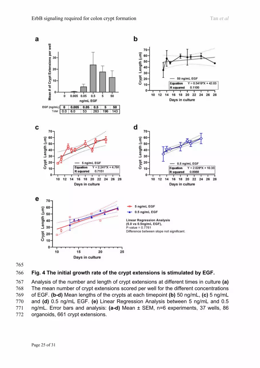
and imaged daily over 21 days. Organoids remained as spheroids at 0.005 ng/ml EGF 142
but produced crypts between 0.05 – 5.0 ng/ml EGF (Fig. 2a-b). The proportion of crypt-143
containing organoids was scored and is presented in Fig. 2b. In the absence or at 144
lower concentrations of EGF the organoids remain as spherical cysts (Fig. 2a, b). The 145
crypt formation colon adenoma cultures increased with increasing EGF concentration, 146
from 0.05 ng/ml to 20 ng/mL (Fig. 2b) and was significantly increased above 0.5 ng/ml 147
(Fig. 2b). 148
Different morphological shapes of colon adenoma organnoids. 149
The colon Apcmin/+ adenoma organoids have four three distinctive morphologies: 150
Spheroids (see Fig 3a up to day 8) , Large body organoids with crypt extensions (Fig 151
3a), Lenticular organoids (Fig 3b) and Hyperbudding organoids (Fig. 3c). The Large 152
body extension organoids had crypts forming after the organoid had grown to a 153
significant size. Crypts extensions then appeared from the main body, leading to the 154
gradual regressing/reduction of the size of the main organoid body. Lenticular 155
organoids started to form crypt extensions as they grew, reshaping the organoid into 156
a series of interconnected tubular crypts. These extensions could be very long ( > 100 157
µm , twice the maximum mean crypt width of normal murine crypt 5) with occasional 158
occurrence of secondary branches (Fig. 3b). Finally, Hyper-budding organoids 159
produced large numbers of small crypt extensions on the surface of the organoid 160
formed at the same time (Fig. 3c). The timing of crypt extensions formation is different 161
for each class of morphology, and varied between day 8 and day 14. For example, the 162
Hyper-budding organoids shown in Fig. 3c formed budging structures on day 11 and 163
day 15, respectively. 164
The elongation rate of crypts in adenoma organoids is independent of EGF 165
concentration. 166
We analysed the number and length of the adenoma crypt extensions over a range of 167
EGF concentrations (Fig. 4a).There was little crypt production at low concentrations 168
of EGF (5 pg/ml) but the number of crypts increased dramatically at 0.5 ng/ml (Fig. 169
4a). The number of crypt extensions per organoid increases with EGF concentration, 170
peaking at 0.5 ng/mL. The crypt lengths over the culture period of 10-25 days at three 171
EGF concentrations (50, 5, and 0.5 ng/mL) were measured (Fig. 4b-d). The mean 172
length of crypts at 50 ng/mL EGF appears to be constant (~ 50 µm) between day 14 173

ErbB signaling required for colon crypt formation Tan et al
Page 6 of 31
and day 24 (Fig. 4b). In contrast, at lower concentrations of 5 ng/mL and 0.5 ng/mL 174
EGF the crypt lengths continued to increase throughout the time course (Fig. 4c, d, 175
respectively), at a rate of 2.24 µm/day and 2.03 µm/day respectively (Fig. 4e). 176
Collectively, this data shows that a minimal concentration of EGF is required for crypt 177
production, at EGF concentrations (i.e. between 0.5~5ng/mL), the crypt extends at a 178
rate of ~2 µm/day. At higher concentrations of EGF, crypt length rapidly reaches ~50 179
µm and does not extend any further 180
Effect of EGF-family ligands and the ErbB signaling pathway in colon adenoma 181
crypt formation. 182
Members of the epidermal growth factor receptor family include EGFR, ErbB2 (also 183
known as HER2), ErbB3/HER3, and ErbB4/HER4. EGFR and ErbB2 have been 184
associated with the growth of many human cancers, including colorectal cancer 28,29 185
(Fig.5a). Ligand binding to the extracellular domain of EGFR, ErbB3 or ErbB4 induces 186
the receptors to form oligomers and consequentially activate the intracellular kinase30. 187
ErbB2 does not bind a ligand but is in a conformation which allows it to bind to other 188
EGFR family members and when the co-receptor is in the ligand-activated 189
conformation, the ErbB2 kinase is activated31. Interestingly, the intracellular domain of 190
ErbB3 has no measurable kinase activity of its own, but when the ligand bound form 191
of ErbB3 combines with ErbB2, the heterodimer (or higher order aggregates of ErbB3 192
and ErbB2) activates the ErbB2 kinase activity. 193
To investigate which ErbB receptors might be driving colon adenoma crypt formation, 194
we used recombinant preparations of EGF-like ligands: Amphiregulin (AREG), 195
Epiregulin (EREG), β-cellulin (BTC) and Neuregulin (NRG-β1, Heregulin-β1) to 196
stimulate the colon adenoma cells in the 3D-Matrigel cultures. Notably, AREG has 197
been implicated in intestinal epithelial regeneration32 and has been implicated in a pro-198
neoplastic role in colorectal carcinogenesis33. Mouse colon adenoma cells were 199
cultured in the presence of different concentrations of each of the EGF-like ligands 200
and the crypts per organoids scored (Fig.5b). All the EGF-like ligands triggered crypt 201
formation with Heregulin /NRG-β1 being the most potent (80-fold more active than 202
EGF) (Fig. 5b). BTC and EGF showed a similar dosage range for stimulating the 203
formation of organoids with crypts; AREG and EREG both required higher 204
concentrations to stimulate crypt formation (Fig. 5b). Given that AREG binds to EGFR 205
but with a significantly lower affinity than that of EGF34,35, it is not surprising that a 206
higher concentration of AREG is required to stimulate the same level of crypt formation 207
in colon adenoma organoid assay (Fig.5b). Similarly, EREG binds to both the EGFR 208
(~2.8 µM) and ErbB4 (>5 µM) but with a significantly lower affinity than EGF binding 209
to these same receptors (1.9 nM and 49 nM, respectively)36, so higher concentrations 210
of EREG were required to stimulate crypt formation (Fig. 5b). 211
Although NRG-β1 (Heregulin-β1) has a high affinity for the ErbB3 and ErbB4 212
homodimers (IC50 ~5 nM), it has an even higher affinity for the ErbB3/ErbB2 213
Erb4/ErbB2 heterodimers (IC50 0.1-0.2 nM)36. EGF’s affinity for EGFR homodimer 214
and the EGFR/ErbB2 heterodimer is between 1.2 to 1.8 nM. Heregulin-β1 stimulates 215
crypt formation more potently than EGF (Fig.5b, Supplementary Fig. 2). This result 216
indicates that a pathway stimulated by ErbB3/ErbB1, ErbB4/ErbB1, ErbB3/ErbB2 or 217

ErbB signaling required for colon crypt formation Tan et al
Page 7 of 31
ErbB4/ErbB2 heterodimer could initiate and maintain adenoma crypt formation. BTC 218
binds to the EGFR homodimer, the EGFR/ErbB2 heterodimer, the ErbB4 homodimer 219
and the ErbB2/ErbB4 heterodimer with relatively high affinities (1.4 nM, 1.7nM, 3.6 nM 220
and 0.2 nM respectively)36. The affinities are very similar to the binding of EGF to the 221
EGFR homodimer and ErbB1/ErbB2 heterodimer and would account for the similarity 222
of their potencies for inducing colon adenoma crypt formation (Fig.5b). 223
The mean number of spheroids and organoids with crypts were also scored for each 224
of the EGF-like ligands (Fig.5c). NRG-β1 and BTC appeared to be the most potent in 225
terms of organoid formation with a consistently higher mean organoids formed, 226
particulary at the lower ligand concentrations. Both EGF and AREG, which stimulate 227
the slowest organoid formation rate, signal through the EGFR homodimer. EREG, 228
which has a faster rate of organoid formation compared to EGF and AREG, can signal 229
through EGFR/ErbB3, E/ErbB2, ErbB1/ErbB4 and ErbB4/ErbB2 heterodimers. With 230
BTC and NRG-β1 predominant signaling through ErbB4/ErbB3, ErbB2/ErbB4 231
heterodimers. 232
It is clear that ErbB signalling plays an essential role in facilitating both colon adenoma 233
organoid formation and crypt formation with combinations of EGFR, ErbB2, ErbB3 and 234
ErbB4 heterodimers identified as key regulators with varying crypt stimulating 235
capacities. ErbB receptor inhibition studies were conducted to further detail the roles 236
each ErbB family member in crypt formation. 237
Inhibition of ErbB Receptor signaling reduces colon adenoma crypt formation. 238
The colon adenoma cultures were treated with different ErbB receptor inhibitors (see 239
Supplementary Table 1 for specificity and IC50s) in the presence of either 0.5 ng/mL 240
EGF (Fig. 2b) or 0.005 ng/mL Heregulin/NRG-β1 (Fig.5b) and the proportion of crypts 241
per organoid was assessed (Fig. 6). Upon EGF stimulation, the EGFR/ErbB2 242
compound PCI-32765 inhibited crypt formation efficiently at concentrations above 0.5 243
pM (Fig. 6a). Both the ErbB2 antagonist CP724714 and the EGFR inhibitor AG1478 244
also inhibited crypt formation but less efficiently, requiring concentrations greater than 245
10 nM (Fig. 6a). These results suggest that EGF-stimulated crypt formation requires 246
both EGFR and ErbB2 signalling. 247
The colon adenoma organoid cultures were stimulated with NRG-β1 (Heregulin-β1) 248
and treated with ErbB inhibitors (Fig. 6b). Crypt formation was reduced with inhibition 249
of ErbB2 (CP724714), suggesting that NRG-β1 (Heregulin-β1) stimulates crypt 250
formation via the ErbB3/ErbB2 heterodimers. EGFR (AG1478) inhibition, which 251
signals through ErbB3/EGFR heterodimers, did not inhibit crypt formation significantly 252
(Fig. 6b). However, the pan-ErbB family inhibitor (HM781-36B) demonstrated the most 253
potent inhibition and was more than 10-fold more effective at inhibiting crypt formation 254
than ErbB2-specific inhibitors (Fig. 6b). This suggests that NRG-b1-stimulated crypt 255
formation can act through both ErbB3 and ErbB2 receptors. ErbB2 signalling is 256
important for crypt formation in the colonadenoma organoids via either EGFR/ErbB2 257
heterodimers upon EGF stimulation, or via ErbB3/ErbB2 heterodimers when 258
stimulated with NRG-β1. 259
260

ErbB signaling required for colon crypt formation Tan et al
Page 8 of 31
Discussion 261
This study demonstrates that EGFR/ErbB family signalling is required for crypt 262
production in organoids produced by mouse colon adenoma stem cells. Although 263
excess crypt formation appears to be the defining feature of adenomas, the current 264
paradigms for intestinal repair37, colon adenoma and colon cancer focuses on excess 265
intra-crypt cell proliferation38. Cell production within normal colon crypts occurs at a 266
rapid rate, indeed the rate of cell production in the intestines39 (including the colon) 267
exceeds all other tissues. The renewal of the colon epithelial cell occurs rapidly and 268
continuously within the crypt, however under normal homeostatic conditions, crypt 269
production is a rare event: less than 1 in 200 crypts in the mouse colon are in the 270
process of budding2,40 and in the normal human colon less than 1 in 2000 crypts are 271
producing crypt buds41. During development42, intestinal renewal after physical 272
damage, radiation, infection and adenoma formation, crypt budding occurs 273
frequently43. In normal tissue, once homeostasis is achieved, crypt budding ceases. 274
However, in adenoma tissue, crypt formation is uncontrolled and leads to the formation 275
of adenomatous polyps. 276
It was surprising that our initial cultures of mouse colon adenomas only yielded 277
spheroids – there were no crypts22. The spheroids grow rapidly, so the cultures were 278
split for passaging after 7 days. Normal mouse colon organoids started to form crypt-279
like structures within 5 days. It was not until we allowed the adenoma organoids to 280
grow for 10 days or more that the spheroids initiated crypt buds. Although normal colon 281
stem cells require Wnt, R-spondin and EGF to proliferate in vitro, Apc+/min mouse colon 282
adenoma cells only require an EGF ligand family member to proliferate and for the 283
spheroids to produce new crypts. The five EGF family members EGF, NRG-1, AREG, 284
EREG and β-cellulin) all stimulated crypt formation; however, NRG-1 (EC50 2 pg/mL) 285
was almost 100-fold more active than the other ligands. EGF and β-cellulin were active 286
at 200 pg/mL, but AREG and EREG required almost 1ng/mL to stimulate crypt 287
formation. 288
NRG-β1 stimulates the aggregation of ErbB3 (HER3) homodimers and heterodimers, 289
included within these aggregates are the ligand-bound ErbB3 in complex with ErbB2 290
(HER2), where the ErbB2 kinase is activated44. In the presence of sufficient 291
concentration of an ErbB2 inhibitor, NRG-β1 stimulated crypt formation is blocked. 292
This inhibiton is amplified if both EGFR and ErbB2 are inhibited. A similar situation 293
occurs when EGF is used to induce crypt formation: the EGF activates both the EGFR 294
kinase and the ErbB2 kinases through ligand stimulated EGFR:ErbB2 hetero-295
oligomers45,46. EGF stimulated crypt formation can be blocked by either an ErbB2 296
inhibitor (CP724714) or an EGFR inhibitor (AG1478), however, inhibiting both 297
receptors with PCI-32765 blocks crypt formation profoundly. When crypt formation is 298
stimulated by NRG-β1 (i.e. via ErbB3), the EGFR inhibitor AG1478 does not block 299
crypt formation, indicating that NRG-β1 stimulates crypt formation through activation 300
of ErbB3:ErbB2 hetero-oligomers rather than EGFR:ErbB3 oligomers. The 301
EGFR:ErbB2 oligomers require the ligand-activated EGFR kinase to stimulate the 302
formation of oligomers of active ErbB2 kinase. Thus, it appears that the activation of 303
the ErbB2 kinase, via ligand activated EGFR:ErbB2 or ErbB2:ErbB3 oligomers, is 304
required to initiate crypt formation. 305

ErbB signaling required for colon crypt formation Tan et al
Page 9 of 31
EGF family members stimulate crypt formation in the context of a mature colon 306
spheroid. It is not clear what changes occur in the adenoma spheroids after 7-10 days 307
in culture, but it is likely that ErbB2 activation leads to changes in key components of 308
cell-cell adhesion47 and/or cytoskeletal proteins at the apical surface48 which induce 309
the cell to form a rhomboidal shape– thus inducing the tissue curvature seen at the 310
base of intestinal crypts. This curvature, together with the proliferation of the crypt stem 311
cells leads to crypt bud formation. Consequential to bud formation, crypt elongation 312
occurs via ligand (EGF-family) stimulated proliferation of progenitor (transit amplifying) 313
cells. 314
Intriguingly, ErbB2 signalling has also been implicated in both mammary duct 315
formation49,50 and alveolar formation in the developing lung49,51 and may be a key 316
regulator of budding morphogenesis in a number of tissues. 317
Crypt cell proliferation and crypt bud formation are different processes. Adenomas are 318
grow by ligand dependent, ErbB2 kinase dependent crypt budding processes, so it is 319
likely that both colon adenoma and colon cancers will be susceptible to drugs or 320
combinations of drugs which inhibit both the EGFR kinase and ErbB2 kinase or to 321
combinations of antagonists such as anti-EGFR, anti-ErbB2 or anti-ErbB3 antibodies) 322
which inhibit the activation of the ErbB3:ErbB2 hetero-oligomers. Once the EGFR 323
family kinase activity is blocked the colon adenoma cancer cancer cells will be more 324
susceptible to pro-apoptotic drugs. 325
326

ErbB signaling required for colon crypt formation Tan et al
Page 10 of 31
Methods 327
Ligands and inhibitors of the ErbB family. The following EGF-like ligands and ErbB 328
kinase inhibitors were used in this study: Recombinant mouse EGF (Peprotech #315-329
09), recombinant human Epiregulin (Peprotech # 100-04), recombinant human β-330
cellulin (Peprotech # 100-50), recombinant human Amphiregulin (Peprotech # 100-331
55B) and recombinant human NRG-β1(Peprotech # 100-03). EGFR tyrosine kinase 332
inhibitor Tyrphostin AG-1478 mesylate (Institute of Drug Technology, IDT Australia, 333
provided by Prof Andrew Scott #T4182), selective inhibitor of ErbB2 CP-724714 334
(Selleck Chemicals #S1167), irreversible pan-HER inhibitor for EGFR/ErbB2/ErbB4 335
Poziotinib (HM781-36B, Selleck Chemicals, Houston, TX, USA. Cat No. S7358) and 336
Btk and EGFR/ ErbB2 inhibitor Ibrutinib (PCI-32765, Selleck Chemicals, Houston, TX, 337
USA. Cat No. S2680). Affinities for the respective ErbB receptors in cell-free assays 338
are as shown in Supplementary Table 1. The ligands were reconstituted in 0.1%(w/v) 339
Bovine Serum Albumin (BSA) in distilled filtered water while the inhibitors were 340
reconstituted in dry dimethyl sulfoxide (DMSO). 341
Normal mouse colon cultures. All animal studies were carried out in accordance 342
with Australian NHMRC Animal Ethics requirements and experimental protocols 343
approved by the Animal Ethics Committee of the Walter and Eliza Hall Institute of 344
Medical Research. C57Bl6 mouse colon tissue was isolated by resection and the 345
tissue opened longitudinally from the anus to the caecum. The tissue was washed with 346
PBS and the distal two-thirds of the colon retained. After 5 minute incubation in PBS 347
containing 0.04% hyperchlorite, the tissue was washed and incubated in PBS 348
containing 3 mM EDTA. Crypts were released by vigourous shaking in PBS (20 349
seconds) and pelleted at 100 xg. Crypts were digested with Dispase (0.1 mg/ml 350
Dispase (Life technologies #17105-041) in DMEM-F12 and then by incubation in 351
TrypLE. Single cell suspensions were prepared by passing the digested crypts through 352
a 26G needle and plated at 500 cells/well in 384 well plates coated with 8 µl Matrigel 353
(BD Biosciences #356231). Growth media (Intesticult (Stem Cell Technologies 354
#06005) supplemented with penicillin/streptomycin (Gibco), 10x concentrated WEHI-355
YH2 conditioned medium 20 and 10 µM Y27632 was added within 30 minutes of plating 356
and cultures were maintained in the same media but without Y27632. 357
Colon adenoma organoid cultures. The colon adenoma crypts were prepared from 358
a polyp taken from an Apc+/min mouse52 colon (kindly provided by Dr Anuratha 359
Sakthianandeswaren, The Walter and Eliza Hall Institute of Medical Research, 360
Victoria, Australia), preparation and culturing of the WEHI-Ad67 colon adenoma 361
organoid line was described previously9. Using the same technique, we prepared 362
another colon adenoma organoid line: WEHI-AdB13 using a polyp from a separate 363
Apc+/min mouse. The colon adenoma organoid lines were passaged weekly (details as 364
described in ref 9) in 96 well plates precoated with 15µL of Matrigel (BD Biosciences 365
#356231) and basic growth medium with recombinant mouse EGF (50 ng/mL, 366
PeproTech, #315-09). The basic colon adenoma organoid growth medium consisted 367
of DMEM-F12 (Gibco/Life Technologies, #10565018) supplemented with additives: 1x 368
penicillin-streptomycin (Gibco/Life Technologies, #15140-122), 1x GlutaMAX 369
(Gibco/Life Technologies, #35050-061), 1x N2 (Gibco/Life Technologies, #17502), 1x 370
B27 (Gibco/Life Technologies, #17504) and HEPES (10 mM). 371

ErbB signaling required for colon crypt formation Tan et al
Page 11 of 31
372
Colon adenoma organoid proliferation and crypt formation assays. Organoid 373
harvesting and preparation: The colon adenoma organoids were harvested from the 374
96 well plates, collected and pipetted with a 26G needle for 5-10 times, visually 375
checking the size of the resulting fragments after each round of pipetting (~10 cell 376
fragments are ideal to work with). The fragments were resuspended with DMEM/F12 377
and centrifuged at 8500 × g for 5 minutes. The supernatant fluid was discarded, and 378
the pellet resuspended with 1mL DMEM/F12. The suspension was centrifuged at 379
10000 × g for 3 minutes and supernatant fluid discarded. After resuspension of the 380
pellet with 150µL basic growth medium, an aliquot of the fragment suspension was 381
removed for counting using a haemocytometer (Hausser Scientific). 382
Based on the number of wells to be plated and required seeding density per well, the 383
volume and fragment concentrations of the seeding mixture were determined as 384
described in Sub Fig. 3a. The seeding mixture is divided equally between two columns 385
of a skirted twin.tec 96-well PCR plate (Eppendorf cat#951020401) sitting on a pre-386
cooled thermo-block (total 14 out of 16 wells, leaving out the first and last well to be 387
filled with buffer solution or water, Sub Fig. 3a). The PCR plate was placed onto the 388
working stage of an Eppendorf epMotion® P5073c liquid handling machine (fitted with 389
a Clean Cap filter to keep the samples sterile) for automatic plating. 390
Automated seeding: The colon adenoma fragments suspended in the 50% Matrigel 391
were pipetted (8 µl) into a 384-well optical plate (Corning, #3985) epMotion®. An 8-392
channel dispensing tool (TM 50, Eppendorf cat# 5280000215) was used to extract the 393
adenoma mixture from the preloaded twin.tec 96-well PCR plate (Eppendorf 394
cat#951020401), kept cool by sitting on a thermo-block (precooled at -20˚C). The 395
robotic arm was programmed and customized to transfer viscous fluid and to dispense 396
near the bottom of the well (Sub Fig. 3c). After dispensing the adenoma cells, the plate 397
was centrifuged for 1 minute at 450 RCF (Eppendorf 5810 R centrifuge) and incubated 398
at 37˚C for 1 hour (Sub Fig. 3d). 399
Reagent preparation and application: The respective EGF-like ligand stocks were 400
prepared in basic growth medium while the ErbB receptor inhibitors were made up in 401
basic growth medium containing recombinant mouse EGF (0.5 ng/mL, PeproTech, 402
#315-09). The study used a 5-point titration of the ligands using 1 in 10 dilutions. The 403
reagents were pipetted into the designated wells of a 96-well deep well plate 404
(Eppendorf cat#951033529) with layout as per described in Sub Fig. 3. The plate was 405
placed on the stage of the Eppendorf epMotion® P5073c along with the previously 406
incubated optical 384-well plate with the plated fragment mixture. The liquid handling 407
machine was programmed and customized to transfer liquid and to dispense from the 408
top of the well. The TM-50 was used to dispense reagents from the 96-well deep well 409
plate to the 384well plate in an alternating manner (Sub Fig. 3f). The 96-well deep well 410
ligand stock plate was covered, sealed and stored at 4˚C for use when the medium 411
was to be replaced. In the case of significant bubble formation, the culture plate was 412
centrifuged for 2 minutes at 450 RCF (Eppendorf 5810 R centrifuge) and before being 413
placed in the at 37˚C incubator. 414
415

ErbB signaling required for colon crypt formation Tan et al
Page 12 of 31
Medium replacement: The crypt forming assays usually ran for three weeks with 416
weekly replacement of medium. The Eppendorf epMotion® P5073c was used for this 417
replacement. Using the 8-channel dispensing tool (TM-50), 40µL of medium was 418
removed and replaced with 40µL of fresh reagent. 419
Colon adenoma organoid imaging assay. The functional imaging assay was 420
conducted with two systems both using an optical 384-well plate. The first system 421
initial imaging data were acquired using a Nikon Eclipse Ti-U microscope with a 4x 422
objective lens and motorised stage (Prior Scientific, H117). Bright field image stacks 423
of each well were captured on a Nikon DS-Ri2 camera (Nikon Inc, MQA17000) using 424
the NIS-Elements software (Nikon, Basic Research version 4.40). The depth images 425
of each FOV (well) were then stacked using the EDF algorithm in Fiji for image 426
processing and analysis. The whole plate assay data were acquired using a 427
customized Nikon High Content Analysis platform consisting of a Nikon C2+ confocal 428
microscope with a LIS/PRIOR automated stage, plate loader and cell culture 429
incubation throughout the experiment (including during the imaging of the plate). 430
Brightfield image stacks of individual 384 well were acquired on a Nikon Qi2 grayscale 431
camera and NIS-Elements High Content Analysis software module (Nikon, High 432
Content Analysis version 5.11). For both systems, each experiment was followed and 433
imaged daily for up to 3 weeks. Each dataset which represents all the wells of a 434
specific timepoint was processed using a customized Fiji script53 to extract each 435
individual well’s image stack, before digitizing, saving as tiff images and organizing 436
into folders for analysis. 437
Computational image analysis for organoid imaging assay. For analysis, a 438
customised Fiji script was used to batch process and organise all the images in each 439
experiment, this includes steps to focus the multiple images at different focal planes 440
for each well, background and debris removal, as well as image segmentation to 441
extract organoids and for feature selection. Specific details of each processing steps 442
are briefly described below. 443
The individual well’s Z-stack bright-field images were flattened using ImageJ/Fiji 444
software53. The projection of z-stack images onto a single in-focus image was carried 445
out using the Stack Focuser plugin (https://imagej.nih.gov/ij/plugins/stack-446
focuser.html) (parameters: select=10 variance=0.000 edge select_only) and exported 447
as high-quality tiff files. 448
Well exterior, background and debris removal as well as organoid selection and 449
feature extraction were performed were achieved using sequential steps of the 450
following operations: conversion to 8-bit, “Auto Local Threshold” (method=Phansalkar, 451
radius=150, parameter_1=0 parameter_2=0 white stack), set Black Background, 452
“Convert to Mask” (method=Default background=Default calculate black list), "Find 453
Edges" as stack, "Invert" stack, set Foreground Color as white, clear boundary (in 454
steps), "Dilate" as stack, "Fill Holes" as stack and "Remove Outliers" (radius=7 455
threshold=50 which=Dark stack). Analyses of object sizes performed using "Analyze 456
Particles" function (size=1000-Infinity circularity=0.30-1.00). 457
The script was used to processes all well images and used to threshold, segment and 458
count the individual organoids. The physical parameters of each organoid, including 459

ErbB signaling required for colon crypt formation Tan et al
Page 13 of 31
shape, size, intensity and circularity were recorded. These features for all organoids 460
identified per well/image were exported as text files (.csv) for analysis and tabulation 461
of the parameters of interest (e.g. mean organoid size). Overlayed images with 462
selection masks were exported for visual curation and validation. The results were 463
curated to remove false positives and to add missing organoids. 464
Whole mount staining and imaging. Colon tissue (normal and adenoma) was 465
cleared and stained as described in ref 54. Briefly, the tissues were fixed, cleared and 466
stained with rat monoclonal anti-E-cadherin (Clone ECCD-2, Thermo Fisher Scientific 467
Cat #13-1900 (1:250)), goat anti-Rat IgG (H+L) Cross-Absorbed Secondary Antibody, 468
Alexa Fluor 488 (Thermo Fisher Scientific Cat #A-11006 (1:500)), Rhodamine 469
Phalloidin (Molecular Probes/Invitrogen(1:200)) and DAPI (Thermo Fisher Scientific 470
Cat# 62248 (1:1000)). For the adenoma tissues, 3D image stacks were acquired on a 471
Leica SP8 Resonance Scanning Confocal microscope using a 20x objective for 472
100µm depth at 0.5 µm per section. For normal tissues, 3D image stacks were 473
acquired on an Olympus IX-81 microscope with an Olympus FV1000 Spectral 474
Confocal attachment and a 20x objective for ~55 µm depth at 1 µm per section. All 475
acquired image data were processed and rendered using Imaris software package 476
(Bitplane, Zürich, Switzerland). 477
Immunofluorescent staining of colon adenoma organoid. Organoid cultures were 478
harvested from selected wells of 384 well plates and transferred into a 1.5 mL 479
Eppendorf microtube. The organoids were either allowed to settle to the bottom of the 480
tube. The supernatant fluid was discarded carefully and in the remaining fluid (~20 µL) 481
the pellet was mixed with an equal volume of Matrigel. The mixture was plated as 482
domes into the wells of an 8-well chamber slide (Ibidi cat#80826) and incubated at 483
37℃ for 1 hour. 300 µL of MT-PBS was added to wash the organoid-Matrigel domes 484
twice. The MT-PBS was removed and 150 μL of 0.1% Paraformaldehyde (PFA) in 485
PBS was added to each well and incubated at 4 ˚C for 48 hours. The 486
paraformaldehyde was removed from the wells and individual wells were washed with 487
150 μL of PBS twice. The MT-PBS was removed, 300 µL of 0.2% (v/v) TritonX-100 in 488
MT-PBS added and the dome incubated for 5 minutes at room temperature. The liquid 489
was removed, and the dome washed twice with 300 µL of blocking buffer [0.2% (w/v) 490
Bovine serum albumin (BSA) in MT-PBS] before leaving in the blocking buffer for 24 491
hr. After removing the blocking solution and 200 µL of the primary antibody solution 492
containing rabbit anti-β-catenin (Sigma-Aldrich #C2206, 1:400) and rat anti-E-cadherin 493
(Invitrogen #13-1900, 1:200, monoclonal ECCD-2) was added and the incubation 494
continued at 4˚C for 24 hr. The antibody-stained domes were washed with 300 µL per 495
well of blocking buffer and followed by 300 µl per well of 0.1% (v/v) TritonX-100 in MT-496
PBS for 5 min at room temperature. After removing the Triton X100 buffer, wash twice 497
with 300 µL of blocking buffer before adding 200 µL of secondary antibody solution 498
(0.2% BSA (w/v) in MT-PBS) containing Alexa Fluor® 546 goat anti-rat IgG (1:400) 499
and Alexa Fluor® 488 goat anti- rabbit IgG (1:400). The domes were incubated with 500
the secondary antibody solution at 4˚C for 18 hours. After removing the secondary 501
antibody solution, the domes were washed twice with the Triton X100 buffer before 502
counter staining the nuclei with 1 nM DAPI (4, 6-diamidino-2-phenyl indole Nucleic 503
Acid Stain, cat# D1306, Molecular Probes Inc, Eugene, OR, Invitrogen) in MT-PBS 504

ErbB signaling required for colon crypt formation Tan et al
Page 14 of 31
(200 µL) for 5 minutes at room temperature in the dark. The organoids were imaged 505
in the 8-well Ibidi slides on the confocal microscope. 506
3D Confocal Fluorescence Imaging. Immunofluorescent images of the organoids 507
were recorded using an Olympus IX-81 microscope with an Olympus FV1000 Spectral 508
Confocal attachment and a 60x water immersion lens. DAPI, β-catenin and E-cadherin 509
fluorescence were excited with the 405 nm, 488 nm and 546 nm laser lines, 510
respectively and the emissions were measured at 405 nm, 473 nm and 559 nm, 511
respectively. 3D image stacks were captured encompassing the entire depth of the 512
organoid in the field of view using Olympus FluoroView software (Version 1.7c). For 513
larger organoids, tiled image stacks were acquired and merged. For quantitative 514
measurements and 3D reconstruction, cubic voxels were acquired (typically up to 0.33 515
µm) for each image stack with the output analogue signal, representing the 516
fluorescence intensities digitized to 16 bits resolution at 65536 levels of grey and 517
saved as Olympus Image Binary (OIB) files. The Bioformats plugin was used to import 518
the images into the ImageJ/Fiji software processing and visualization. 519
520

ErbB signaling required for colon crypt formation Tan et al
Page 15 of 31
References 521
1 Miyoshi, H., Ajima, R., Luo, C. T., Yamaguchi, T. P. & Stappenbeck, T. S. 522
Wnt5a potentiates TGF-beta signaling to promote colonic crypt regeneration 523
after tissue injury. Science 338, 108-113, doi:10.1126/science.1223821 524
(2012). 525
2 Park, H.-S., Goodlad, R. A. & Wright, N. A. Crypt fission in the small intestine 526
and colon. A mechanism for the emergence of G6PD locus-mutated crypts 527
after treatment with mutagens. The American journal of pathology 147, 1416 528
(1995). 529
3 Maskens, A. P. Histogenesis of colon glands during postnatal growth. Acta 530
anatomica 100, 17-26 (1978). 531
4 Preston, S. L. et al. Bottom-up histogenesis of colorectal adenomas: origin in 532
the monocryptal adenoma and initial expansion by crypt fission. Cancer 533
research 63, 3819-3825 (2003). 534
5 Tan, C. W., Hirokawa, Y., Gardiner, B. S., Smith, D. W. & Burgess, A. W. 535
Colon cryptogenesis: asymmetric budding. PloS one 8, e78519, 536
doi:10.1371/journal.pone.0078519 (2013). 537
6 Araki, K., Ogata, T., Kobayashi, M. & Yatani, R. A morphological study on the 538
histogenesis of human colorectal hyperplastic polyps. Gastroenterology 109, 539
1468-1474 (1995). 540
7 Wong, W. et al. Histogenesis of human colorectal adenomas and hyperplastic 541
polyps: the role of cell proliferation and crypt fission. Gut 50, 212-217 (2002). 542
8 Wasan, H. S. et al. APC in the regulation of intestinal crypt fission. The 543
Journal of pathology 185, 246-255, doi:10.1002/(SICI)1096-544
9896(199807)185:3<246::AID-PATH90>3.0.CO;2-8 (1998). 545
9 Tan, C. W., Hirokawa, Y. & Burgess, A. W. Analysis of Wnt signalling 546
dynamics during colon crypt development in 3D culture. Scientific Reports 5, 547
11036, doi:10.1038/srep11036 (2015). 548
10 Yip, H. Y. K., Tan, C. W., Hirokawa, Y. & Burgess, A. W. Colon organoid 549
formation and cryptogenesis are stimulated by growth factors secreted from 550
myofibroblasts. PloS one 13, e0199412, doi:10.1371/journal.pone.0199412 551
(2018). 552
11 Sato, T. et al. Single Lgr5 stem cells build crypt-villus structures in vitro 553
without a mesenchymal niche. Nature 459, 262-265, doi:10.1038/nature07935 554
(2009). 555
12 Sato, T. et al. Long-term expansion of epithelial organoids from human colon, 556
adenoma, adenocarcinoma, and Barrett's epithelium. Gastroenterology 141, 557
1762-1772, doi:10.1053/j.gastro.2011.07.050 (2011). 558
13 Shaker, A. et al. Epimorphin deletion protects mice from inflammation-induced 559
colon carcinogenesis and alters stem cell niche myofibroblast secretion. The 560
Journal of clinical investigation 120, 2081-2093, doi:10.1172/JCI40676 (2010). 561
14 Batts, L. E., Polk, D. B., Dubois, R. N. & Kulessa, H. Bmp signaling is required 562
for intestinal growth and morphogenesis. Developmental dynamics : an official 563
publication of the American Association of Anatomists 235, 1563-1570, 564
doi:10.1002/dvdy.20741 (2006). 565
15 Haramis, A. P. et al. De novo crypt formation and juvenile polyposis on BMP 566
inhibition in mouse intestine. Science 303, 1684-1686, 567
doi:10.1126/science.1093587 (2004). 568

ErbB signaling required for colon crypt formation Tan et al
Page 16 of 31
16 Ormestad, M. et al. Foxf1 and Foxf2 control murine gut development by 569
limiting mesenchymal Wnt signaling and promoting extracellular matrix 570
production. Development 133, 833-843, doi:10.1242/dev.02252 (2006). 571
17 Sato, T. et al. Paneth cells constitute the niche for Lgr5 stem cells in intestinal 572
crypts. Nature 469, 415-418, doi:10.1038/nature09637 (2011). 573
18 Miettinen, P. J. et al. Epithelial immaturity and multiorgan failure in mice 574
lacking epidermal growth factor receptor. Nature 376, 337-341, 575
doi:10.1038/376337a0 (1995). 576
19 Flentjar, N. et al. TGF-betaRII rescues development of small intestinal 577
epithelial cells in Elf3-deficient mice. Gastroenterology 132, 1410-1419, 578
doi:10.1053/j.gastro.2007.02.054 (2007). 579
20 Hirokawa, Y., Yip, K. H. Y., Tan, C. W. & Burgess, A. W. Colonic 580
myofibroblast cell line stimulates colonoid formation. American Journal of 581
Physiology - Gastrointestinal and Liver Physiology 306, G547-G556 (2014). 582
21 Farrall, A. L. et al. Wnt and BMP signals control intestinal adenoma cell fates. 583
International Journal of Cancer 131, 2242-2252, 584
doi:https://doi.org/10.1002/ijc.27500 (2012). 585
22 Yamazaki, D., Hashizume, O., Taniguchi, S., Funato, Y. & Miki, H. Role of 586
adenomatous polyposis coli in proliferation and differentiation of colon 587
epithelial cells in organoid culture. Scientific Reports 11, 3980, 588
doi:10.1038/s41598-021-83590-6 (2021). 589
23 Foroughi, S., Tie, J., Gibbs, P. & Burgess, A. W. Epidermal growth factor 590
receptor ligands: targets for optimizing treatment of metastatic colorectal 591
cancer. Growth Factors 37, 209-225 (2019). 592
24 Mitchell, R. A., Luwor, R. B. & Burgess, A. W. Epidermal growth factor 593
receptor: Structure-function informing the design of anticancer therapeutics. 594
Experimental cell research 371, 1-19 (2018). 595
25 Powell, A. E. et al. The Pan-ErbB Negative Regulator Lrig1 Is an Intestinal 596
Stem Cell Marker that Functions as a Tumor Suppressor. Cell 149, 146-158, 597
doi:10.1016/j.cell.2012.02.042 (2012). 598
26 Suzuki, A., Sekiya, S., Gunshima, E., Fujii, S. & Taniguchi, H. EGF signaling 599
activates proliferation and blocks apoptosis of mouse and human intestinal 600
stem/progenitor cells in long-term monolayer cell culture. Lab Invest 90, 1425-601
1436, doi:10.1038/labinvest.2010.150 (2010). 602
27 Abud, H. E., Watson, N. & Heath, J. K. Growth of intestinal epithelium in 603
organ culture is dependent on EGF signalling. Experimental Cell Research 604
303, 252-262, doi:https://doi.org/10.1016/j.yexcr.2004.10.006 (2005). 605
28 Khayat, D. et al. Epidermal growth factor receptor signaling in colorectal 606
cancer: preclinical data and therapeutic perspectives. Annals of Oncology 16, 607
189-194, doi:10.1093/annonc/mdi057 (2005). 608
29 Arteaga, Carlos L. & Engelman, Jeffrey A. ERBB Receptors: From Oncogene 609
Discovery to Basic Science to Mechanism-Based Cancer Therapeutics. 610
Cancer Cell 25, 282-303, doi:https://doi.org/10.1016/j.ccr.2014.02.025 (2014). 611
30 Yarden, Y. & Sliwkowski, M. X. Untangling the ErbB signalling network. 612
Nature Reviews Molecular Cell Biology 2, 127, doi:10.1038/35052073 (2001). 613
31 Cho, H.-S. et al. Structure of the extracellular region of HER2 alone and in 614
complex with the Herceptin Fab. Nature 421, 756-760, 615
doi:10.1038/nature01392 (2003). 616

ErbB signaling required for colon crypt formation Tan et al
Page 17 of 31
32 Shao, J. & Sheng, H. Amphiregulin promotes intestinal epithelial regeneration: 617
roles of intestinal subepithelial myofibroblasts. Endocrinology 151, 3728-3737, 618
doi:10.1210/en.2010-0319 (2010). 619
33 Guzman, M. J., Shao, J. & Sheng, H. Pro-neoplastic effects of amphiregulin in 620
colorectal carcinogenesis. J Gastrointest Cancer 44, 211-221, 621
doi:10.1007/s12029-012-9474-2 (2013). 622
34 Adam, R. et al. Modulation of the receptor binding affinity of amphiregulin by 623
modification of its carboxyl terminal tail. Biochimica et Biophysica Acta (BBA) - 624
Molecular Cell Research 1266, 83-90, doi:https://doi.org/10.1016/0167-625
4889(94)00224-3 (1995). 626
35 Shoyab, M., Plowman, G. D., McDonald, V. L., Bradley, J. G. & Todaro, G. J. 627
Structure and function of human amphiregulin: A member of the epidermal 628
growth factor family. Science 243, 1074-1076, doi:10.1126/science.2466334 629
(1989). 630
36 Jones, J. T., Akita, R. W. & Sliwkowski, M. X. Binding specificities and 631
affinities of egf domains for ErbB receptors. FEBS Letters 447, 227-231, 632
doi:10.1016/s0014-5793(99)00283-5 (1999). 633
37 Hageman, J. H. et al. Intestinal Regeneration: Regulation by the 634
Microenvironment. Developmental Cell 54, 435-446 (2020). 635
38 Gehart, H. & Clevers, H. Tales from the crypt: new insights into intestinal stem 636
cells. Nature Reviews Gastroenterology & Hepatology 16, 19-34 (2019). 637
39 Marshman, E., Booth, C. & Potten, C. S. The intestinal epithelial stem cell. 638
Bioessays 24, 91-98 (2002). 639
40 Berlanga-Acosta, J., Playford, R., Mandir, N. & Goodlad, R. Gastrointestinal 640
cell proliferation and crypt fission are separate but complementary means of 641
increasing tissue mass following infusion of epidermal growth factor in rats. 642
Gut 48, 803-807 (2001). 643
41 Cummins, A. G. et al. Crypt fission peaks early during infancy and crypt 644
hyperplasia broadly peaks during infancy and childhood in the small intestine 645
of humans. Journal of pediatric gastroenterology and nutrition 47, 153-157 646
(2008). 647
42 Bjerknes, M. & Cheng, H. Clonal analysis of mouse intestinal epithelial 648
progenitors. Gastroenterology 116, 7-14 (1999). 649
43 Yamazaki, M., Fujii, E., Watanabe, T., Kato, A. & Suzuki, M. Histopathological 650
evaluation of crypt fission during intestinal development in neonatal mice. 651
Journal of Toxicologic Pathology 33, 39-46 (2020). 652
44 Steinkamp, M. P. et al. erbB3 is an active tyrosine kinase capable of homo-653
and heterointeractions. Molecular and cellular biology 34, 965-977 (2014). 654
45 HUANG, G. C., OUYANG, X. & EPSTEIN, R. J. Proxy activation of protein 655
ErbB2 by heterologous ligands implies a heterotetrameric mode of receptor 656
tyrosine kinase interaction. Biochemical Journal 331, 113-119 (1998). 657
46 Riese, D. J. & Stern, D. F. Specificity within the EGF family/ErbB receptor 658
family signaling network. Bioessays 20, 41-48 (1998). 659
47 Knust, E. Regulation of epithelial cell shape and polarity by cell-cell adhesion. 660
Molecular membrane biology 19, 113-120 (2002). 661
48 Carraway, C. A. C. & Carraway, K. L. Sequestration and segregation of 662
receptor kinases in epithelial cells: implications for ErbB2 oncogenesis. 663
Science Signaling 2007, re3-re3 (2007). 664

ErbB signaling required for colon crypt formation Tan et al
Page 18 of 31
49 Niemann, C. et al. Reconstitution of mammary gland development in vitro: 665
requirement of c-met and c-erbB2 signaling for branching and alveolar 666
morphogenesis. The Journal of cell biology 143, 533-545 (1998). 667
50 Jackson-Fisher, A. J. et al. ErbB2 is required for ductal morphogenesis of the 668
mammary gland. Proceedings of the National Academy of Sciences 101, 669
17138-17143 (2004). 670
51 Dammann, C. E., Nielsen, H. C. & Carraway III, K. L. Role of neuregulin-1β in 671
the developing lung. American journal of respiratory and critical care medicine 672
167, 1711-1716 (2003). 673
52 Moser, A. R., Dove, W. F., Roth, K. A. & Gordon, J. I. The Min (multiple 674
intestinal neoplasia) mutation: its effect on gut epithelial cell differentiation and 675
interaction with a modifier system. The Journal of cell biology 116, 1517-1526 676
(1992). 677
53 Schindelin, J. et al. Fiji: an open-source platform for biological-image analysis. 678
Nature methods 9, 676-682 (2012). 679
54 Rios, A. C. et al. Intraclonal Plasticity in Mammary Tumors Revealed through 680
Large-Scale Single-Cell Resolution 3D Imaging. Cancer Cell 35, 618-681
632.e616, doi:https://doi.org/10.1016/j.ccell.2019.02.010 (2019). 682
55 Yaish, P., Gazit, A., Gilon, C. & Levitzki, A. Blocking of EGF-dependent cell 683
proliferation by EGF receptor kinase inhibitors. Science 242, 933-935 (1988). 684
56 Jani, J. P. et al. Discovery and pharmacologic characterization of CP-724,714, 685
a selective ErbB2 tyrosine kinase inhibitor. Cancer research 67, 9887-9893 686
(2007). 687
57 Pan, Z. et al. Discovery of selective irreversible inhibitors for Bruton’s tyrosine 688
kinase. ChemMedChem: Chemistry Enabling Drug Discovery 2, 58-61 (2007). 689
58 Cha, M. Y. et al. Antitumor activity of HM781‐36B, a highly effective pan‐690
HER inhibitor in erlotinib‐resistant NSCLC and other EGFR‐dependent 691
cancer models. International journal of cancer 130, 2445-2454 (2012). 692
693

ErbB signaling required for colon crypt formation Tan et al
Page 19 of 31
Acknowledgments 694
This work was supported by the Ludwig Institute for cancer Research, the National 695
Health and Medical Research Council (NHMRC) Project Grant GNT1029628 and, 696
Program Grant #487922, and funds from the Operational Infrastructure Support 697
Program provided by the Victorian Government, Australia. The funders had no role 698
on the study design, data collection and analysis, decision to publish, or preparation 699
of the manuscript. The manuscript is solely the responsibility of the institutions and 700
individual authors and does not reflect the views of the NHMRC. The authors would 701
like to thank Professor Jane Visvader and Professor Geoff Lindeman and Dr Bianca 702
Capaldo from the CRF Cancer Biology and Stem Cells Division (WEHI) for their 703
advice and assistance with tissue clearing and staining protocols. 704
Author contributions 705
R.Z., A.W.B. and C.T. conceived and designed the experiments. R.Z., S.R.K., 706
X.Z., M.C.F and CT. performed the experiments. R.Z., M.A., M.C.F, A.W.B. and 707
C.T. performed the data analysis and interpretation. R.Z., A.W.B., M.C.F and 708
C.T. wrote the manuscript. All authors critically reviewed and approved the 709
manuscript prior to submission. 710
Competing financial interests 711
The authors declare no competing financial interests. 712
713

ErbB signaling required for colon crypt formation Tan et al
Page 20 of 31
Figure Legends 714
715
716

ErbB signaling required for colon crypt formation Tan et al
Page 21 of 31
Fig. 1 Crypt formation in colon Apcmin/+ adenoma organoids requires EGF. 717
Whole-mount mouse colon crypts from (a) an C57Bl6 adult mouse and (b) Apcmin/+ 718
adenoma tissue were immunolabelled for E-cadherin (E-cad) (green) and labelled with 719
phalloidin (magenta) and DAPI (blue) to visualise F-actin and nuclei, respectively. 720
Shown are single section images. (c) Normal mouse colon organoids form crypts in 721
culture. Bright-field extended depth of field (EDF) time-lapse images of wildtype mouse 722
colon organoids grown over 10 days. Insets at Day 9 are shown as enlarged images 723
in panel f. (d) Mouse colon adenoma organoids derived from Apcmin/+ mice form crypts 724
when the cells are cultured in the presence of EGF. Brightfield EDF time-lapse images 725
of Apcmin/+ adenoma organoids grown over 21 days with EGF. Organoids cultured with 726
EGF remain spherical and cyst-like in the first 7-9 days. Crypt extensions start to 727
appear in some organoids after 10 days, some with multiple extensions projecting from 728
the main organoid body. (e) Apcmin/+ adenoma spheroids grown without EGF. 729
Brightfield EDF time-lapse images of Apcmin/+ adenoma organoids grown over 19 days 730
without EGF. Enlarged images of budding organoids from (f) normal mouse colon 731
(Day9) and (g) colon adenoma (Day21) with EGF and (h) colon adenoma (Day19) 732
without EGF. (i) Colon adenoma organoids were immunolabelled with β-catenin 733
(green) and E-cadherin (red) and co-stained with DAPI (blue). Single section images 734
acquired on an Olympus FV100 confocal microscope shown (merged/overlayed 735
image shown in the top left panel). The crypt extensions formed are physiologically 736
similar in shape to crypts isolated from mice colons, with a crypt width of ~ 40 μm, as 737
indicated in the β-catenin image. Merged 3D reconstruction of the stained crypt 738
forming organoid shown in the right panel. Scale bars: (a-b) 50 µm, (f-h) 100 µm. 739
740

ErbB signaling required for colon crypt formation Tan et al
Page 22 of 31
741 Fig. 2 Effect of EGF on crypt formation in colon adenoma organoids. 742
Colon adenoma organoids formed crypts in an EGF dependent manner. Colon 743
organoids were cultured in different amounts of recombinant mouse EGF, monitored 744
and imaged for up to 24 days. The numbers of spheroidal and crypt-forming organoids 745
were quantified and the % of organoids with crypts were tabulated. (a) EDF images of 746
representative wells at different concentrations of EGF (day indicated below each 747
image). (b) The mean proportion of organoids with crypts for different EGF 748
concentrations. Scoring conducted using a customized Fiji script (see Methods 749
section) for first occurrence of organoids with crypts during the duration of the culture. 750
Error bars and analysis: Mean ± SEM, n ≥ 5 experiments, *** p<0.001, ordinary 1-way 751
ANOVA, Holm-Sidak's multiple comparisons test with respect to no added EGF. 752
753
754

ErbB signaling required for colon crypt formation Tan et al
Page 23 of 31
755

ErbB signaling required for colon crypt formation Tan et al
Page 24 of 31
Fig. 3 Colon adenoma crypt-forming organoids can have different 756
morphologies. 757
The mouse Apcmin/+ organoids with crypts can be categorized into (a) large body 758
extension organoids, (b) lenticular organoids and (c) hyper-budding organoids. Large 759
body extensions occur from different sized starting points from a large cyst like body. 760
Lenticular organoids have crypt buds extending in length with reshaping of the 761
organoid. Hyper-budding organoids have large number of crypt buds forming on the 762
organoid. Concentration of EGF used in the respective cultures are as per indicated. 763
764
ErbB signaling required for colon crypt formation Tan et al
Page 25 of 31
765
Fig. 4 The initial growth rate of the crypt extensions is stimulated by EGF. 766
Analysis of the number and length of crypt extensions at different times in culture (a) 767
The mean number of crypt extensions scored per well for the different concentrations 768
of EGF. (b-d) Mean lengths of the crypts at each timepoint (b) 50 ng/mL, (c) 5 ng/mL 769
and (d) 0.5 ng/mL EGF. (e) Linear Regression Analysis between 5 ng/mL and 0.5 770
ng/mL. Error bars and analysis: (a-d) Mean ± SEM, n=6 experiments, 37 wells, 86 771
organoids, 661 crypt extensions. 772

ErbB signaling required for colon crypt formation Tan et al
Page 26 of 31
773

ErbB signaling required for colon crypt formation Tan et al
Page 27 of 31
Fig.5 ErbB ligands stimulate crypt formation in colon adenoma organoids. 774
Colon adenoma organoids were stimulated with different concentrations of EGF-like 775
ligands for up to 21 days and the numbers of spheroids and crypt producing organoids 776
were counted. (a) Schematic diagram of the EGF-like ligands and their corresponding 777
ErbB receptor interactions. AREG: Amphiregulin, EREG: Epiregulin, BTC; β-cellulin, 778
NRG-β1: Neuregulin/Heregulin-β1. (b) The proportion of organoids with crypts and (c) 779
the mean numbers of spheroid and crypt-forming organoids scored were plotted. Data 780
shown as Mean ± SEM for n ≥ 3 experiments, (b) Dose-Response – Stimulation Non-781
linear Analysis: Best-fit values EC50 for EGF: 0.1430 ng/mL, EREG: ~0.1416 ng/mL, 782
Neuregulin/Heregulin-β1: 0.001814 ng/mL 783
784

ErbB signaling required for colon crypt formation Tan et al
Page 28 of 31
785
786

ErbB signaling required for colon crypt formation Tan et al
Page 29 of 31
Fig. 6 Inhibition of ErbB receptors prevents crypt formation in EGF or 787
Neuregulin/Heregulin β1 stimulated colon adenoma organoids. 788
Production of colon adenoma organoids was assessed with (a) 0.5 ng/mL EGF or (b) 789
5 ng/mL Neuregulin/Heregulin β1 in the presences of different concentration of ErbB 790
pathway inhibitors. The cultures were imaged daily for 21 days, and the numbers of 791
crypt-forming organoids scored. The EGF-stimulated cultures (a) were treated with 792
EGFR tyrosine kinase inhibitor AG1478, a selective HER2/ErbB2 inhibitor CP724714 793
or a potent but less selective Brutons tyrosine kinase (Btk), EGFR and ErbB2 inhibitor, 794
Ibrutinib (PCI-32765). The NRG-β1-stimulated cultures (b) were treated with AG1478, 795
CP724714 or an irreversible pan-HER inhibitor (HER1/2/4) Poziotinib (HM781-36B). 796
Data shown as mean ± SEM number of organoids with crypts, n=3 experiments. * p< 797
0.05, ** p<0.01, *** p<0.001, ordinary 1-way ANOVA. Statistical tests: (a) Dunnett's 798
multiple comparisons test with reference to 0.5ng/ml EGF, (b) Dunnett's multiple 799
comparisons test with reference to 5 ng/mL NRG-β1. Dose-Responses – Inhibition 800
Non-linear Analysis, Best-fit values IC50 for (b) HM781-36B: 2.89±2.58 x 10⁻⁵ nM 801
802

ErbB signaling required for colon crypt formation Tan et al
Page 30 of 31
Supplementary Information Legends 803
Supplemental Figure 1 Differences in morphology between normal and 804
adenomatous mouse colon. 805
3D confocal imaging of tissues from normal mouse colon and Apcmin/+ mouse colon 806
adenoma showing DAPI in blue, Phalloidin (F-actin) in magenta, E-cadherin in green. 807
Confocal images of a section of the (a) normal epithelium and the (c) adenoma 808
epithelium indicates the differences in crypt morphology. (b) shows the 3D 809
reconstruction of (a) while panel d is a zoomed image of the boxed region in c. Colon 810
tissue was cleared and stained as described in ref 54. Briefly, the tissues were fixed, 811
cleared and stained with rat monoclonal anti-E-cadherin (Clone ECCD-2, Thermo 812
Fisher Scientific Cat #13-1900 (1:250)), goat anti-Rat IgG (H+L) Cross-Absorbed 813
Secondary Antibody, Alexa Fluor 488 (Thermo Fisher Scientific Cat #A-11006 814
(1:500)), Rhodamine Phalloidin (Molecular Probes/Invitrogen (1:200)) and DAPI 815
(Thermo Fisher Scientific Cat# 62248 (1:1000)). Images were acquired on a Leica SP8 816
Resonant Scanning Confocal microscope with the data processed/rendered using 817
Imaris software package (Bitplane, Zürich, Switzerland). Scale bar: (b) 100µm (c) 818
50µm. 819
820
Supplemental Figure 2 Crypt formation occurs in another colon adenoma 821
organoid line 822
Colon adenoma organoids derived from other Apcmin/+ mice were tested for its crypt 823
formation capability using EGF and other ErbB ligands. Adenoma line #B13 organoids 824
were cultured, cultured with EGF or other ErbB ligands for up to 21 days. 825
Representative EDF images of crypt forming organoids from these #B13 cultures 826
stimulated with (a) 5 ng/mL EGF and (b) 0.5 µg/mL Neuregulin/Heregulin-β1. Inset 827
presents zoomed images of selected regions of the respective well on the left. Scale 828
bars: left panel 500 μm, right panels/inset 100 μm. 829
830
Supplemental Figure 3 Workflow for colon adenoma organoid imaging 831
analyses 832
The workflow had been optimised to maximise the use of a 384well optical plate for 833
analysing the growth and morphology of the organoids. Colon adenoma fragments 834
were mixed with Matrigel 50% (v/v). This mixture was then equally divided into two 835
columns of a precooled 96well PCR plate sitting on a cold thermoblock. An automatic 836
liquid handling machine (Eppendorf epMotion® P5073c) was then used to transfer 8 837
µL per well, 8 wells at a time with an 8-channel pipetting tool. The plate was then 838
centrifuged to move all the cultures to the bottom of the wells before incubating at 839
room temperature for 1 hour. Similarly, media was prepared with the different 840
concentrations of the stock solutions of ligands and inhibitors were prepared in a 96 841
deep well plate and transferred (80 µL) to the adenoma cultures using the epMotion® 842
liquid handling machine. 843

ErbB signaling required for colon crypt formation Tan et al
Page 31 of 31
Supplementary Table 1 844
Summary of the IC50 values for the ErbB receptor kinase inhibitors. 845
846
Supplemental Movie S1 847
Time-lapse of crypt forming organoid followed for 10 days. 848
849
Supplemental Movie S2 850
Time-lapse of crypt forming organoid followed for 24 days. 851

Figures
Figure 1
please see the manuscript �le for the full caption

Figure 2
please see the manuscript �le for the full caption

Figure 3
please see the manuscript �le for the full caption

Figure 4
please see the manuscript �le for the full caption

Figure 5
please see the manuscript �le for the full caption

Figure 6
please see the manuscript �le for the full caption
Supplementary Files
This is a list of supplementary �les associated with this preprint. Click to download.

MovieS2.gif
MScryptformationadenomaorganoidsupplementary20210429A.pdf
MovieS1.gif
MovieS2.gif