enzymatic fingerprinting and modification of acetylated pectins
TRANSCRIPT
Enzymatic fingerprinting and
modification of acetylated pectins
Connie Remoroza
Thesis committee
Promotors Prof. Dr H. Gruppen Professor of Food Chemistry Wageningen University Prof. Dr H.A. Schols Personal chair at the Laboratory of Food Chemistry Wageningen University Other members Prof. Dr L. Dijkhuizen, University of Groningen Prof. Dr M.E. Hendrickx, KU Leuven, Belgium Prof. Dr M.W.F Nielen, Wageningen University Dr M.-C. Ralet, INRA Nantes, France
This research was conducted under the auspices of the Graduate School VLAG (Advanced studies in Food Technology, Agrobiotechnology, Nutrition and Health Sciences).
Enzymatic fingerprinting and
modification of acetylated pectins
Connie Remoroza
Thesis submitted in fulfilment of the requirements for the degree of doctor
at Wageningen University by the authority of the Rector Magnificus
Prof. Dr M.J. Kropff, in the presence of the
Thesis Committee appointed by the Academic Board to be defended in public
on Friday 21 March 2014 at 4 p.m. in the Aula.
Connie Remoroza Enzymatic fingerprinting and modification of acetylated pectins 162 pages
PhD thesis, Wageningen University, Wageningen, NL (2014) With references, with summaries in English and Dutch
ISBN: 978-90-6173-902-5
Abstract To reveal the ester distribution patterns in acetylated pectins, an enzymatic fingerprinting method using a combined endo-polygalacturonase (PG) and pectin lyase (PL) treatment followed by hydrophilic interaction liquid chromatography coupled to electrospray ionization ion trap mass spectrometry with evaporative light scattering detection was developed. This method paved the way for the development of the new quantitative parameters degree of hydrolysis by PG (DHPG) and degree of hydrolysis by PL (DHPL). These parameters distinguished the methylester and acetyl group distribution patterns within different sugar beet pectins (SBPs). In the case of pectin having a degree of methylesterification (DM) of >50 and acetylation of ~20, the above approach was insufficient. Hence, a second digestion was introduced using a fungal pectin methylesterase and a PG. More than 60% of the total GalA residues present in three SBPs were recovered as monomer and oligomers after the two digestions. The first digestion of the acid extracted commercial SBP revealed the presence of small blocks of nonesterified GalA residues and segments containing large blocks of PL degradable methylesterified and /or acetylated GalA residues. Blocks of partly methylesterified, non-acetylated GalA residues were recognized after the second digestion. These results show that the acetylation pattern is non-random.
A pectin acetylesterase (BliPAE) and a pectin methylesterase (BliPME) from Bacillus licheniformis DSM13 were produced, purified and biochemically characterized. The mode of action of BliPAE and BliPME towards acetylated pectins was revealed using the newly developed enzymatic fingerprinting method. BliPAE specifically deacetylates the O-3 linked acetyl groups of nonmethylesterified galacturonic acid residues in the homogalacturan of pectin. BliPME efficiently de-methylesterifies lemon pectins (DM34-76 → DM 0) and SBPs (DM 30-73 → DM 14) in a blockwise manner. BliPME is quite tolerant towards the acetyl groups present within the SBPs. For the first time, a comprehensive experimental characterization was directed to enzymes from B. licheniformis having a PAE and a PME activity.

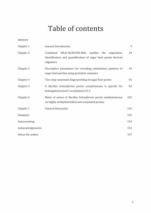
Table of contents Abstract
Chapter 1 General Introduction 9
Chapter 2 Combined HILIC-ELSD/ESI-MSn enables the separation, identification and quantification of sugar beet pectin derived oligomers
29
Chapter 3 Descriptive parameters for revealing substitution patterns of sugar beet pectins using pectolytic enzymes
45
Chapter 4 Two-step enzymatic fingerprinting of sugar beet pectin 65
Chapter 5 A Bacillus licheniformis pectin acetylesterase is specific for homogalacturonan’s acetylated at O-3
85
Chapter 6 Mode of action of Bacillus licheniformis pectin methylesterase on highly methylesterified and acetylated pectins
105
Chapter 7 General Discussion 125
Summary 145
Samenvatting 149
Acknowledgements 153
About the author 157
7

8

Chapter 1
General Introduction
Chapter 1
10
1.1 Background Due to the commercial interest in pectins as gelling and thickening agent, pectin-rich by-products from the food industry are extracted from citrus peel and apple pomace on an industrial scale. Nevertheless, pectin producers are searching for alternative raw materials to be used for pectin production. Pectin-rich raw materials potentially available in large quantities are sugar beet pulp, potato fibre, and sunflower by-products (heads and stalks after seed removal). Contrary to citrus and apple pectins, these pectin raw materials contain non-gelling, acetylated pectins. Chemical deacetylation would not only remove acetyl groups, but also part of the methylesters1 and could lower the molecular mass of pectin by β-elimination within the pectin backbone.2 The enzymatic deacetylation of sugar beet pectin (SBP) is hindered by the presence of methylesters, whereas enzymatic de-methylesterification is hindered by acetyl groups. Finding suitable pectin methylesterases (PMEs) or pectin acetylesterases (PAEs) that are not hindered by acetyl groups or methylesters, respectively, would enable the production of suitable pectins.
The research presented in this PhD thesis aimed to study the precise chemical structure of SBP and esterases able to modify acetylated pectins. This PhD research is part of the PolyModE project. PolyModE (POLYsaccharide MODifying Enzymes) consists of universities, research institutes and industries throughout Europe and was supported by the European Community. One of the goals of the PolyModE project is to modify SBPs by specific esterases in order to behave similarly to non-acetylated commercial pectins from other sources with respect to gelling and stabilizing properties.
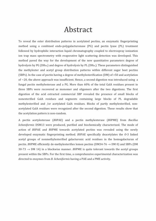
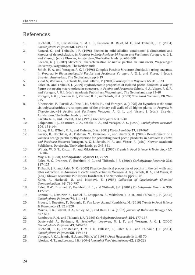
1.2 Chemical structure of pectin Pectin is a heterogeneous, complex polysaccharide with galacturonic acid (GalA) as the main monosaccharide moiety. Depending on the origin and developmental stage of the plant tissue, the precise chemical structure and proportions of the structural elements of pectin may differ significantly. The major elements of pectin are homogalacturonan (HG) and rhamnogalacturonan I (RG-I), the latter containing arabinans and/or galactans as side chains being present within one pectin (Fig. 1.1). Other elements in pectins from specific sources include rhamnogalacturonan (RG-II).3,4 It is present as highly conserved complex parts of the homogalacturonan next to xylogalacturonan.5
Homogalacturonan (HG) region
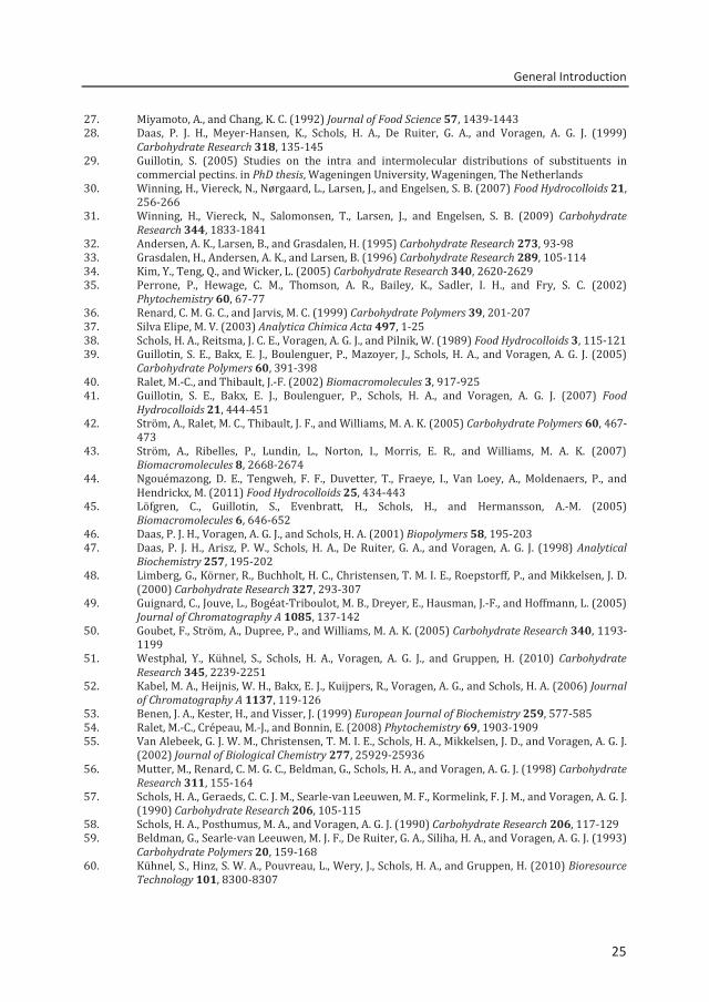
HG, also termed ‘smooth region’, is the backbone of the pectin. HG structural elements consist of approximately 60-100 1,4-linked α-D-galactopyranosyluronic acid residues.6 The GalA residues can be methylesterified at the C6 position and the O-2 and/or O-3 position of the GalA residues can be acetylated (Fig. 1.2).7 The proportion of esterified GalA residues and the distribution of
10
General Introduction
11
these esterified residues along the linear backbone determine most of the functional properties of the pectin.
Figure 1.1. Schematic representation of the pectin structure.
Figure 1.2. Schematic representation of the different substituents present in the homogalacturonan region of pectin. Methylester (O-CH3), acetyl group (CH3COO), carboxyl group (COO-).
Rhamnogalacturonan (RG-I) region
RG-I is composed of alternating rhamnose and galacturonic acid residues.8 The GalA residues of RG-I can be O-acetylated on position O-2 and/or O-3,9 while between 20-80% of the rhamnose residues of RG-I can be substituted with neutral sugars side chains,10 mainly composed of galactose and/or arabinose.3
Rhamnogalacturonan (RG-II) region
RG-II is a highly conserved, complex part of the homogalacturonan structure in some plants.3 The RG-II can contain 12 different monosaccharides, including apiose and aceric acid, linked by more than 20 different linkages.11
Homogalacturonan or ‘smooth region’
Rhamnogalacturonan I or ‘hairy region’
Arabinan Arabinogalactan
O
H
HO OH
C
O
O
OH
C O
H
HO OH
C O
O
CH3
OO
O
H
HO
O-OH
C
O
O
O
C
CH3
=
=
12
54
3
6==
=
OOO OH
OHOH
11
Chapter 1
12
1.3 Pectin gelling properties and applications Approximately 45,000 tons of pectin is used worldwide by the food industry, growing at a 2-5% annual rate.12 The most commercially available pectins nowadays originate from citrus peel and apple pomace.7,13
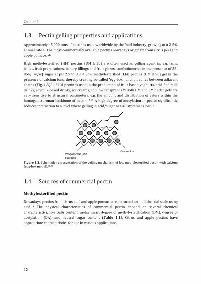
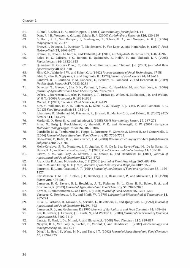
High methylesterified (HM) pectins (DM ≥ 50) are often used as gelling agent in, e.g. jams, jellies, fruit preparations, bakery fillings and fruit glazes, confectioneries in the presence of 55-85% (w/w) sugar at pH 2.5 to 3.8.14 Low methylesterified (LM) pectins (DM ≤ 50) gel in the presence of calcium ions, thereby creating so-called ‘egg-box’ junction zones between adjacent chains (Fig. 1.3).11,15 LM pectin is used in the production of fruit-based yoghurts, acidified milk drinks, soymilk-based drinks, ice creams, and low fat spreads.16 Both HM and LM pectin gels are very sensitive to structural parameters, e.g. the amount and distribution of esters within the homogalacturonan backbone of pectin.17,18 A high degree of acetylation in pectin significantly reduces interaction to a level where gelling in acid/sugar or Ca2+ systems is lost.19
Figure 1.3. Schematic representation of the gelling mechanism of low methylesterified pectin with calcium (egg-box model).20,21
1.4 Sources of commercial pectin Methylesterified pectin
Nowadays, pectins from citrus peel and apple pomace are extracted on an industrial scale using acid.14 The physical characteristics of commercial pectin depend on several chemical characteristics, like GalA content, molar mass, degree of methylesterification (DM), degree of acetylation (DA), and neutral sugar content (Table 1.1). Citrus and apple pectins have appropriate characteristics for use in various applications.
Ca2+ Ca2+ Ca2+ Ca2+ Ca2+
Ca2+ Ca2+ Ca2+ Ca2+ Ca2+
Ca2+
Ca2+Ca2+ Ca2+
Polygacturonic acid backbone
Calcium ion
12
General Introduction
13
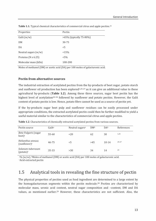
Table 1.1. Typical chemical characteristics of commercial citrus and apple pectins.16
Properties Pectin GalA (w/w) >65% (typically 75-80%) DM 30-75 DA <5 Neutral sugars (w/w) <15% Proteins (N x 6.25) <5% Molecular mass (kDa) 100-200 Moles of methanol (DM) or acetic acid (DA) per 100 moles of galacturonic acid.
Pectin from alternative sources
The industrial extraction of acetylated pectins from the by-products of beet sugar, potato starch and sunflower oil production has been explored1,25-27 as it can give an additional value to these agricultural by-products (Table 1.2). Among these three sources, sugar beet pectin has the highest level of acetylation22-24 followed by sunflower and potato pectins. However, the GalA content of potato pectin is low. Hence, potato fibre cannot be used as a source of pectin yet.
If the by-products sugar beet pulp and sunflower residues can be easily processed under appropriate conditions, the extracted acetylated pectin could then be further modified to yield a useful material similar to the characteristics of commercial citrus and apple pectins. Table 1.2. Characteristics of chemically extracted acetylated pectins from various sources.
Pectin source GalAa Neutral sugarsa DMb DAb References Beta Vulgaris (sugar beet)c 55-60 <20 62 30 1,25
Helianthus annuus (sunflower)c 46-75 <5 >45 10-14 25-27
Solanum tuberosum (potato)c 35-33 >30 34 14 25
a% (w/w); bMoles of methanol (DM) or acetic acid (DA) per 100 moles of galacturonic acid. cAcid-extracted pectin
1.5 Analytical tools in revealing the fine structure of pectin The physical properties of pectins used as food ingredient are determined to a large extent by the homogalacturonan segments within the pectin molecule.28 Pectins are characterized by molecular mass, uronic acid content, neutral sugar composition and –content, DM and DA values, as mentioned earlier.29 However, these characteristics are not sufficient. Also, the
13
Chapter 1
14
methylester (and acetyl group) distribution patterns strongly contribute to the physical behaviour of pectin. To reveal the methylester and acetyl group distribution in pectin, analytical tools that can provide data on such distributions are necessary. Table 1.3 presents an overview of the methods used in the analysis of intact pectin polymer and pectin oligosaccharides.
Analysis of intact pectin
1H nuclear magnetic resonance (NMR) spectroscopy on pectin solutions30,31 has been used to determine the DM and the distribution patterns of methylesters. Although enzymatic de-methylesterification of the pectin polymer by pectin methylesterases from plants or fungi has been studied extensively by NMR,32-34 the complete visualization of the entire pectin molecule is extremely difficult because of the overlapping proton signals of neighboring methylesterified GalA residues. Nevertheless, NMR allows the localization of acetyl substitution to GalA residues (O-2 and/or O-3) in pectin.35,36 The heterogeneity and the high viscosity of pectin samples negatively affects the resolution, which limits the application of NMR.37
Preparative anion exchange chromatography has been proven useful for the analysis of pectins.38,39 Different populations within the pectin preparation can be separated based on charge density.40 However, depending on the ion exchange material used, the separation might also solely be based on the net charge of the pectin molecule.40
Blockwise and random methylesterified pectins having the same DM have been distinguished using capillary electrophoresis (CE).39 Furthermore, CE methods have been developed to analyze pectins according to their charge.41-43 It has been proven that only the net charge is affecting the separation. The drawback of the CE analysis is that only intermolecular charges within a pectin preparation are highlighted, but not the intramolecular charges within a pectin.
Analysis of pectin by enzymatic fingerprinting
Another approach to reveal the ester distribution patterns is the use of specific enzymes. They degrade the polymer into diagnostic oligomers followed by different chromatographic and mass spectrometric techniques for identification and quantification.39,44,45
14
General Introduction
15
Table 1.3. Overview of methods in the analysis of pectin substitution patterns.
Methods References Pectin polymer Spectroscopy 1H NMR Allows the recognition of methylester sequences and the O-2/O-3 localization of acetyl groups in a pectin
30,31,33,34,36
Chromatography Anion exchange column Separation of pectin based on net charge differences Separation of random and blockwise methylesterified pectins having the same DM, based on charge density
38,39 39,40
Electrophoresis Capillary electrophoresis (CE) Separation of pectin polymer based on net charge revealing the intermolecular distribution
41,42
Pectin oligosaccharides Chromatography HPAEC-PAD pH 12, Separation of oligomers by DP; loss of ester information due to the high pH of the eluent HPAEC-PAD pH 5 Separation of partly methylesterified GalA oligomers by DP HPAEC-MS Separation of oligomers by DP and size; Identification of individual components
44,46-48
49
Mass spectrometry MALDI TOF MS Separation of oligomers in the total digest by m/z
41,48
Electrophoresis CE Separation based on total negative charge, quantification of oligomers CE-MS/ CE-LIF (laser induced fluorescence detection) Separation of mole-based detection of neutral and acidic end group labelled oligosaccharides; additional charge leads to co-elution of GalA oligosaccharides
41,50
3,51,52
Pectin degrading enzymes
Different types of enzymes are required for the complete depolymerization of pectin. Since HG and RG-I are the most important structural elements, in this thesis pectin degrading enzymes are classified into HG and RG-I degrading specificities.
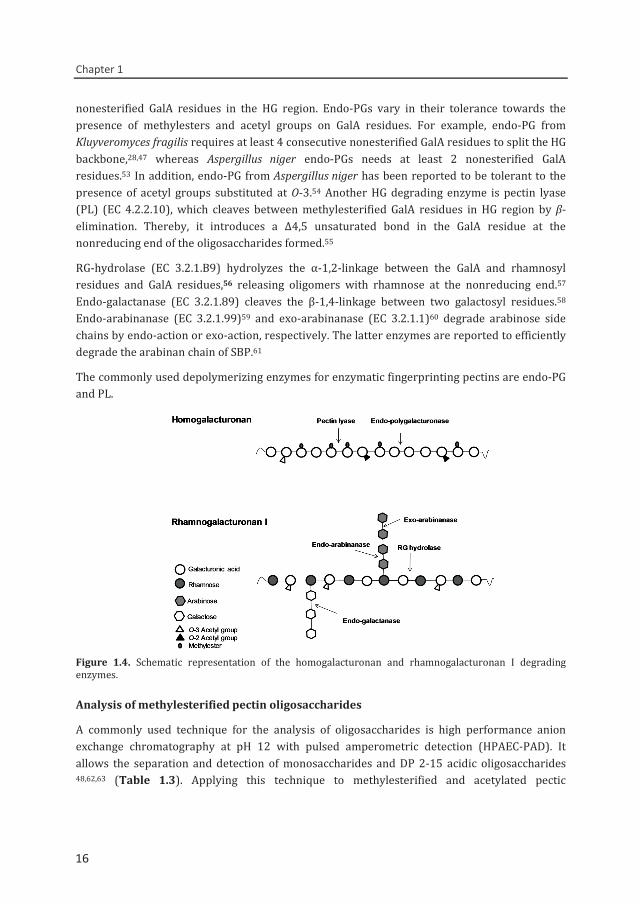
Figure 1.4 shows the modes of action of different HG and RG-I degrading enzymes. Endo-polygalacturonase (endo-PG), EC 3.2.1.15, hydrolyzes α-1,4 glycosidic bonds between two
15
Chapter 1
16
nonesterified GalA residues in the HG region. Endo-PGs vary in their tolerance towards the presence of methylesters and acetyl groups on GalA residues. For example, endo-PG from Kluyveromyces fragilis requires at least 4 consecutive nonesterified GalA residues to split the HG backbone,28,47 whereas Aspergillus niger endo-PGs needs at least 2 nonesterified GalA residues.53 In addition, endo-PG from Aspergillus niger has been reported to be tolerant to the presence of acetyl groups substituted at O-3.54 Another HG degrading enzyme is pectin lyase (PL) (EC 4.2.2.10), which cleaves between methylesterified GalA residues in HG region by β-elimination. Thereby, it introduces a Δ4,5 unsaturated bond in the GalA residue at the nonreducing end of the oligosaccharides formed.55
RG-hydrolase (EC 3.2.1.B9) hydrolyzes the α-1,2-linkage between the GalA and rhamnosyl residues and GalA residues,56 releasing oligomers with rhamnose at the nonreducing end.57 Endo-galactanase (EC 3.2.1.89) cleaves the β-1,4-linkage between two galactosyl residues.58 Endo-arabinanase (EC 3.2.1.99)59 and exo-arabinanase (EC 3.2.1.1)60 degrade arabinose side chains by endo-action or exo-action, respectively. The latter enzymes are reported to efficiently degrade the arabinan chain of SBP.61
The commonly used depolymerizing enzymes for enzymatic fingerprinting pectins are endo-PG and PL.
Figure 1.4. Schematic representation of the homogalacturonan and rhamnogalacturonan I degrading enzymes.
Analysis of methylesterified pectin oligosaccharides
A commonly used technique for the analysis of oligosaccharides is high performance anion exchange chromatography at pH 12 with pulsed amperometric detection (HPAEC-PAD). It allows the separation and detection of monosaccharides and DP 2-15 acidic oligosaccharides 48,62,63 (Table 1.3). Applying this technique to methylesterified and acetylated pectic
16
General Introduction
17
oligosaccharides would, however, lead to unwanted loss of substituents. Matrix-assisted laser desorption ionization time of flight mass spectrometry (MALDI TOF MS) has been used for a fast screening of methylesterified oligosaccharides released after enzymatic digestion,47,63 but the quantification of the individual oligomers is not possible with MALDI TOF MS.
To be able to study not only the nonesterified oligosaccharides, but also methylesterified oligomers, HPAEC-PAD at pH 5 has been developed.47 The separation is based on charge and DP of the oligosaccharides. The annotation of peaks in HPAEC-PAD by online MS is hindered by the high salt concentrations used. The oligosaccharides present in the digest are analyzed by offline MALDI TOF MS.47,63 Until now, this approach is widely used in fingerprinting of non-acetylated, methylesterified citrus pectins.44,63,64 This approach has also led to the introduction of the degree of blockiness (DB) as pectin parameter to discriminate pectins.
Using endo-PG alone, only part of the HG region is elucidated and the highly and/or partly methylesterified blocks are not analyzed. HPAEC-MS pH 5 with online MS identification is now possible by an online desalting step to remove the high salt content of the mobile phase.49 However, this technique is able to only annotate acidic oligosaccharides eluting at relatively low levels of salt. CE is an alternative technique that allows the separation of low DP oligosaccharides in the pectin digest followed by the determination of the degree of blockiness.41 Although CE-LIF and CE-MS has been successfully used to characterize and quantify neutral oligosaccharides after labelling with a fluorescent group at the reducing end.52 However, the use of this method for pectin oligosaccharides is limited due to the additional charge on the oligosaccharides leading to the co-elution of oligosaccharides in a narrow time window.3
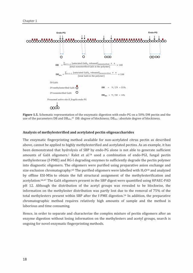
Degree of blockiness of methylesterified pectin
The release of nonesterified GalA monomer, dimer and trimer by endo-PG and the use of the degree of blockiness is illustrated in Figure 1.5.47 DB is calculated as the amount of GalA residues present as nonmethylesterified GalA monomer, dimer, trimer released by endo-PG expressed as percentage of the total amount of nonmethylesterified GalA residues present. The absolute degree of blockiness (DBabs) is calculated as the amount of nonmethylesterified GalA residues released by endo-PG expressed as the percentage of the total GalA residues present in the pectin.39,43,44,62 These studies all concluded that citrus pectins modified by alkali or by fungal pectin methylesterase have in a random distribution. On the contrary, the use of plant pectin methylesterase results in a blockwise distribution of nonesterified GalA residues.
17
Chapter 1
18
Figure 1.5. Schematic representation of the enzymatic digestion with endo-PG on a 50% DM pectin and the use of the parameters DB and DBabs.39 DB: degree of blockiness; DBabs : absolute degree of blockiness.
Analysis of methylesterified and acetylated pectin oligosaccharides
The enzymatic fingerprinting method available for non-acetylated citrus pectin as described above, cannot be applied to highly methylesterified and acetylated pectins. As an example, it has been demonstrated that hydrolysis of SBP by endo-PG alone is not able to generate sufficient amounts of GalA oligomers.1 Ralet et al.54 used a combination of endo-PGI, fungal pectin methylesterase (f-PME) and RG-I degrading enzymes to sufficiently degrade the pectin polymer into diagnostic oligomers. The oligomers were purified using preparative anion exchange and size exclusion chromatography.65 The purified oligomers were labelled with H2O18 and analyzed by offline ESI-MSn to obtain the full structural assignment of the methylesterification and acetylation.66,67 The GalA oligomers present in the SBP digest were quantified using HPAEC-PAD pH 12. Although the distribution of the acetyl groups was revealed to be blockwise, the information on the methylester distribution was partly lost due to the removal of 75% of the total methylesters present within SBP after the f-PME digestion.54 In addition, the preparative chromatographic method requires relatively high amounts of sample and the method is laborious and time consuming.
Hence, in order to separate and characterize the complex mixture of pectin oligomers after an enzyme digestion without losing information on the methylesters and acetyl groups, search is ongoing for novel enzymatic fingerprinting methods.
Endo-PG Endo-PG
DB = 9 / 29 = 31%
58 GalA:
29 methylesterified GalA
29 nonesterified GalADBabs = 9 / 58 = 6%
Presumed active site K. fragilis endo-PG
∑ saturatedGalA released
totalGalAinthepolymer 100
∑ saturatedGalAreleased
totalnonesterifiedGalAinthepolymer 100
18
General Introduction
19
1.6 The use of enzymes to improve the functional properties of commercial acetylated pectin
Pectins with DM above 50% can be converted chemically into LM pectins of DM < 50 % using e.g. acid, alkali and ammonia.14 The disadvantage of alkali treatment is that it can result in uncontrolled depolymerization of pectin,68 thereby losing the viscosity of the pectin in solution. Hence, enzymatic modification using enzymes has been proposed to overcome the challenges for creating pectins in an efficient and environmentally sustainable manner.69 An overiew of pectin modifying esterases is given below.
Pectin methylesterase (PME)
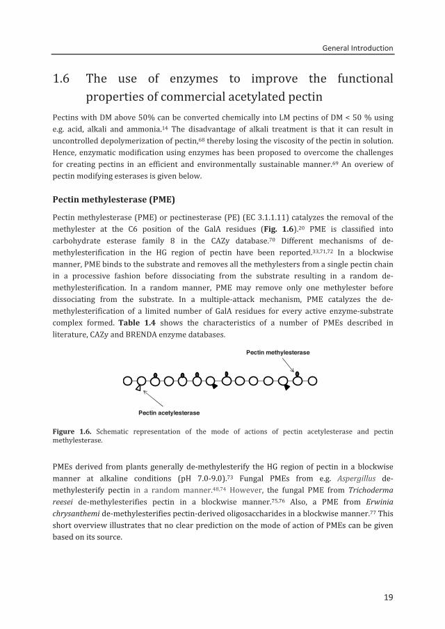
Pectin methylesterase (PME) or pectinesterase (PE) (EC 3.1.1.11) catalyzes the removal of the methylester at the C6 position of the GalA residues (Fig. 1.6).20 PME is classified into carbohydrate esterase family 8 in the CAZy database.70 Different mechanisms of de-methylesterification in the HG region of pectin have been reported.33,71,72 In a blockwise manner, PME binds to the substrate and removes all the methylesters from a single pectin chain in a processive fashion before dissociating from the substrate resulting in a random de-methylesterification. In a random manner, PME may remove only one methylester before dissociating from the substrate. In a multiple-attack mechanism, PME catalyzes the de-methylesterification of a limited number of GalA residues for every active enzyme-substrate complex formed. Table 1.4 shows the characteristics of a number of PMEs described in literature, CAZy and BRENDA enzyme databases.
Figure 1.6. Schematic representation of the mode of actions of pectin acetylesterase and pectin methylesterase. PMEs derived from plants generally de-methylesterify the HG region of pectin in a blockwise manner at alkaline conditions (pH 7.0-9.0).73 Fungal PMEs from e.g. Aspergillus de-methylesterify pectin in a random manner.48,74 However, the fungal PME from Trichoderma reesei de-methylesterifies pectin in a blockwise manner.75,76 Also, a PME from Erwinia chrysanthemi de-methylesterifies pectin-derived oligosaccharides in a blockwise manner.77 This short overview illustrates that no clear prediction on the mode of action of PMEs can be given based on its source.
Pectin methylesterase
Pectin acetylesterase
19
Chapter 1
20
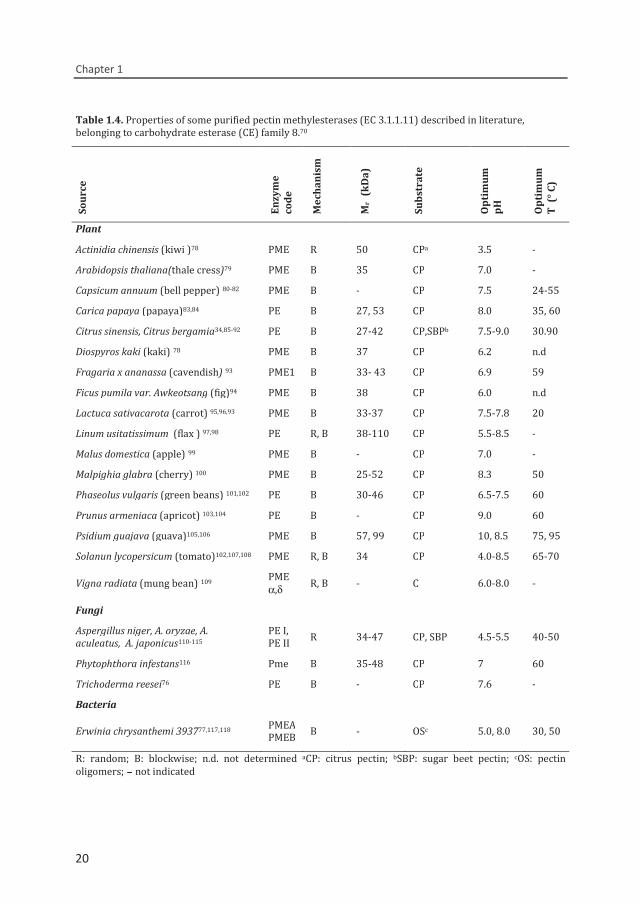
Table 1.4. Properties of some purified pectin methylesterases (EC 3.1.1.11) described in literature, belonging to carbohydrate esterase (CE) family 8.70
Sour
ce
Enzy
me
code
Mec
hani
sm
Mr
(kD
a)
Subs
trat
e
Opt
imum
pH
Opt
imum
T
(° C
)
Plant Actinidia chinensis (kiwi )78 PME R 50 CPa 3.5 - Arabidopsis thaliana(thale cress)79 PME B 35 CP 7.0 - Capsicum annuum (bell pepper) 80-82 PME B - CP 7.5 24-55 Carica papaya (papaya)83,84 PE B 27, 53 CP 8.0 35, 60 Citrus sinensis, Citrus bergamia34,85-92 PE B 27-42 CP,SBPb 7.5-9.0 30.90 Diospyros kaki (kaki) 78 PME B 37 CP 6.2 n.d Fragaria x ananassa (cavendish) 93 PME1 B 33- 43 CP 6.9 59 Ficus pumila var. Awkeotsang (fig)94 PME B 38 CP 6.0 n.d Lactuca sativacarota (carrot) 95,96,93 PME B 33-37 CP 7.5-7.8 20 Linum usitatissimum (flax ) 97,98 PE R, B 38-110 CP 5.5-8.5 - Malus domestica (apple) 99 PME B - CP 7.0 - Malpighia glabra (cherry) 100 PME B 25-52 CP 8.3 50 Phaseolus vulgaris (green beans) 101,102 PE B 30-46 CP 6.5-7.5 60 Prunus armeniaca (apricot) 103,104 PE B - CP 9.0 60 Psidium guajava (guava)105,106 PME B 57, 99 CP 10, 8.5 75, 95 Solanun lycopersicum (tomato)102,107,108 PME R, B 34 CP 4.0-8.5 65-70
Vigna radiata (mung bean) 109 PME α,δ R, B - C 6.0-8.0 -
Fungi Aspergillus niger, A. oryzae, A. aculeatus, A. japonicus110-115
PE I, PE II R 34-47 CP, SBP 4.5-5.5 40-50
Phytophthora infestans116 Pme B 35-48 CP 7 60 Trichoderma reesei76 PE B - CP 7.6 - Bacteria
Erwinia chrysanthemi 393777,117,118 PMEAPMEB B - OSc 5.0, 8.0 30, 50
R: random; B: blockwise; n.d. not determined aCP: citrus pectin; bSBP: sugar beet pectin; cOS: pectin oligomers; - not indicated
20
General Introduction
21
Previous studies have shown that plant PME (Citrus sinensis) and fungal PME (A. niger) are hindered by the presence of acetyl groups in acetylated SBP.1,71 On the contrary, a fungal PME from Aspergillus aculeatus is claimed to be slightly hindered by acetyl groups.66 The previously mentioned PME from Erwinia chrysanthemi77 has not been characterized for its action towards acetylated and non-acetylated pectins.
So far, the information on the biochemical properties and mode of actions for PMEs from various sources has been described in several studies as can be seen in Table 1.4. However, there is limited information on PMEs ability to process acetylated pectins.
Acetylesterases (PAE, RGAE)
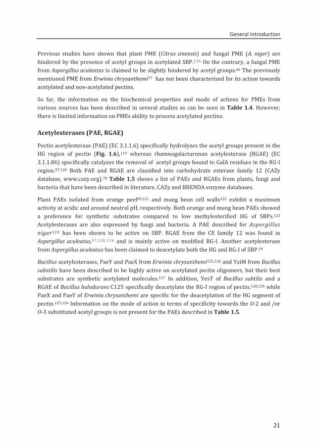
Pectin acetylesterase (PAE) (EC 3.1.1.6) specifically hydrolyzes the acetyl groups present in the HG region of pectin (Fig. 1.6),119 whereas rhamnogalacturonan acetylesterase (RGAE) (EC 3.1.1.86) specifically catalyzes the removal of acetyl groups bound to GalA residues in the RG-I region.57,120 Both PAE and RGAE are classified into carbohydrate esterase family 12 (CAZy database, www.cazy.org).70 Table 1.5 shows a list of PAEs and RGAEs from plants, fungi and bacteria that have been described in literature, CAZy and BRENDA enzyme databases.
Plant PAEs isolated from orange peel85,121 and mung bean cell walls122 exhibit a maximum activity at acidic and around neutral pH, respectively. Both orange and mung bean PAEs showed a preference for synthetic substrates compared to low methylesterified HG of SBPs.122 Acetylesterases are also expressed by fungi and bacteria. A PAE described for Aspergillus niger123 has been shown to be active on SBP. RGAE from the CE family 12 was found in Aspergillus aculeatus,57,120,124 and is mainly active on modified RG-I. Another acetylesterase from Aspergillus aculeatus has been claimed to deacetylate both the HG and RG-I of SBP.19
Bacillus acetylesterases, PaeY and PaeX from Erwinia chrysanthemi125,126 and YxiM from Bacillus substilis have been described to be highly active on acetylated pectin oligomers, but their best substrates are synthetic acetylated molecules.127 In addition, YesT of Bacillus subtilis and a RGAE of Bacillus halodurans C125 specifically deacetylate the RG-I region of pectin,128,129 while PaeX and PaeY of Erwinia chrysanthemi are specific for the deacetylation of the HG segment of pectin.125,126 Information on the mode of action in terms of specificity towards the O-2 and /or O-3 substituted acetyl groups is not present for the PAEs described in Table 1.5.
21
Chapter 1
22
Table 1.5. Properties of some purified acetylesterases described in literature, belonging to carbohydrate esterase (CE) family 12.70
Sour
ce
Enzy
me
code
Spec
ifici
ty
Mr
(kD
a)
Subs
trat
e
Opt
imum
pH
Opt
imum
T
(° C
)
Refe
renc
e
Fungi
Aspergillus aculeatus
RGAE RG-I 26-42 RG-I 5.5-6.0 40-50 120,124,130 RGAE RG-I 42 SBP 5.5 40 19,120 AspacAE RG-I 32-34 SBP 4.5-5.0 45-55 19
Aspergillus niger PAE HG 60 SBP 5.5 50 123 Plant Citrus sinensis (orange) PAE HG 29-42 Triacetin,
SBP 5.0-5.5 30 121
Vigna radiata (mungbean) PAE HG 43 pNPA, SBP 6.5 30-40 122
Bacteria Bacillus halodurans C125 RGAE RG 45 pNPA 8.0 40 129
Bacillus subtilis YxiM HG - pNPA, tobacco HG 8.0 30 127
Bacillus subtilis YesT RG-I 37 pNPA 7.5-8.5 35 128
Erwinia chrysanthemi 3937
PaeX, PaeY HG 35
pNPA, SBP Triacetin
7.5-8.0 30 125,126
SBP: sugar beet pectin; RG-I: rhamnogalaturonan I Synthetic substrates: pNPA: para-nitrophenyl acetate, triacetin (glycerine triacetate) - not indicated
22
General Introduction
23
1.7 Thesis outline The main aim of the research project was to elucidate the precise chemical structure of SBPs before and after modification. However, the current enzymatic fingerprinting method and pectin parameter for the analysis of the distribution patterns in non-acetylated pectin cannot be applied to both methylesterified and acetylated pectin. Hence, there is a necessity to develop techniques for the analysis of acetyl group and methylester distribution patterns. We hypothesized that it is possible to develop a novel enzymatic fingerprinting technique that accurately describes the distribution of methylesters in methylesterified and acetylated pectins. The other aim of this research project is to characterize the newly discovered pectin methylesterase and pectin acetylesterase using the developed analytical tool.
In Chapter 1, the structural elements of pectin, the methods for pectin analysis and characteristics of pectin methylesterases and acetylesterases from various origins were reviewed and presented as background of the project.
Chapters 2 and 3 describe the degradation of different SBPs with various degrees of methylesterification and acetylation into small oligomers by simultaneous action of endo-PG and pectin lyase (PL). The pectin digest was analyzed by HPLC-HILIC with online ESI-IT-MSn and ELSD to identify and quantify individual oligomers. Using this information, novel pectin parameters were developed to distinguish the methylester distribution of SBPs. In order to further degrade highly methylesterified and acetylated SBPs to study their substitution patterns accurately, a second degradation step was introduced. The endo-PG and PL resistant GalA sequences was isolated and digested using fungal pectin methylesterase and endo-PG (Chapter 4). Using the descriptive parameters for both digestions, differences between the ester distributions of the pectins were elucidated.
Thereafter, a putative acetylesterase from Bacillus licheniformis DSM13 was produced, purified and characterized. Furthermore, the enzyme mode of action was revealed using the endo-PG and PL fingerprinting method (Chapter 5). Similarly, detailed characterization and the efficiency of a putative pectin methylesterase from B. licheniformis DSM13 was compared with other PMEs (Chapter 6).
Lastly, the impact of the research project and the potential application of new analytical tools to unravel the complex structure of both non-acetylated and acetylated pectins and to characterize for novel pectolytic enzymes are discussed in chapter 7.
23
Chapter 1
24
References 1. Buchholt, H. C., Christensen, T. M. I. E., Fallesen, B., Ralet, M. C., and Thibault, J. F. (2004)
Carbohydrate Polymers 58, 149-161 2. Renard, C., and Thibault, J.-F. (1996) Pectins in mild alkaline conditions: β-elimination and
kinetics of demethylation. in Progress in Biotechnology:14 Pectins and Pectinases Voragen, A. G. J., and Visser, J. (eds.), Elsevier, Amsterdam, The Netherlands. pp 603-608
3. Coenen, G. J. (2007) Structural characterization of native pectins. in PhD thesis, Wageningen University, Wageningen, The Netherlands
4. Schols, H. A., and Voragen, A. G. J. (1996) Complex Pectins: Structure elucidation using enzymes. in Progress in Biotechnology:14 Pectins and Pectinases Voragen, A. G. J., and Visser, J. (eds.), Elsevier, Amsterdam, The Netherlands. pp 3-19
5. Vidal, S., Williams, P., O'Neill, M., and Pellerin, P. (2001) Carbohydrate Polymers 45, 315-323 6. Ralet, M., and Thibault, J. (2009) Hydrodynamic properties of isolated pectin domains: a way to
figure out pectin macromolecular structure. in Pectins and Pectinases Schols, H. A., Visser, R. G. F., and Voragen, A. G. J. (eds.), Academic Publishers, Wageningen, The Netherlands. pp 35-48
7. Voragen, A. G. J., Coenen, G. J., Verhoef, R. P., and Schols, H. A. (2009) Structural Chemistry 20, 263-275
8. Albersheim, P., Darvill, A., O'neill, M., Schols, H., and Voragen, A. (1996) An hypothesis: the same six polysaccharides are components of the primary cell walls of all higher plants. in Progress in Biotechnology:14 Pectins and Pectinases Voragen, A. G. J., and Visser, J. (eds.), Elsevier, Amsterdam, The Netherlands. pp 47-55
9. Carpita, N. C., and Gibeaut, D. M. (1993) The Plant Journal 3, 1-30 10. Colquhoun, I. J., de Ruiter, G. A., Schols, H. A., and Voragen, A. G. (1990) Carbohydrate Research
206, 131-144 11. Ridley, B. L., O'Neill, M. A., and Mohnen, D. A. (2001) Phytochemistry 57, 929-967 12. Savary, B., Hotchkiss, A., Fishman, M., Cameron, R., and Shatters, R. (2003) Development of a
valencia orange pectin methylesterase for generating novel pectin products. in Advances in Pectin and Pectinase Research Voragen, A. G. J., Schols, H. A., and Visser, R. (eds.), Kluwer Academic Publishers, Dordrecht, The Netherlands. pp 345-361
13. Willats, W. G. T., Knox, J. P., and Mikkelsen, J. D. (2006) Trends in Food Science & Technology 17, 97-104
14. May, C. D. (1990) Carbohydrate Polymers 12, 79-99 15. Ralet, M. C., Dronnet, V., Buchholt, H. C., and Thibault, J. F. (2001) Carbohydrate Research 336,
117-125 16. Thibault, J. F., and Ralet, M. C. (2003) Physico-chemical properties of pectins in the cell walls and
after extraction. in Advances in Pectins and Pectinases Voragen, A. G. J., Schols, H. A., and Visser, R. (eds.), Kluwer Academic Publishers, Dordrecht, The Netherlands. pp 91-105
17. Kohn, R., Markovič, O., and Machová, E. (1983) Collection of Czechoslovak Chemical Communications 48, 790-797
18. Ralet, M.-C., Dronnet, V., Buchholt, H. C., and Thibault, J.-F. (2001) Carbohydrate Research 336, 117-125
19. Bonnin, E., Clavurier, K., Daniel, S., Kauppinen, S., Mikkelsen, J. D. M., and Thibault, J. F. (2008) Carbohydrate Polymers 74, 411-418
20. Fraeye, I., Duvetter, T., Doungla, E., Van Loey, A., and Hendrickx, M. (2010) Trends in Food Science & Technology 21, 219-228
21. Morris, E. R., Powell, D. A., Gidley, M. J., and Rees, D. A. (1982) Journal of Molecular Biology 155, 507-516
22. Rombouts, F. M., and Thibault, J. F. (1986) Carbohydrate Research 154, 177-187 23. Oosterveld, A., Beldman, G., Searle-Van Leeuwen, M. J. F., and Voragen, A. G. J. (2000)
Carbohydrate Polymers 43, 249-256 24. Buchholt, H. C., Christensen, T. M. I. E., Fallesen, B., Ralet, M.-C., and Thibault, J.-F. (2004)
Carbohydrate Polymers 58, 149-161 25. Voragen, A. G. J., Schols, H. A., and Pilnik, W. (1986) Food Hydrocolloids 1, 65-70 26. Iglesias, M. T., and Lozano, J. E. (2004) Journal of Food Engineering 62, 215-223
24
General Introduction
25
27. Miyamoto, A., and Chang, K. C. (1992) Journal of Food Science 57, 1439-1443 28. Daas, P. J. H., Meyer-Hansen, K., Schols, H. A., De Ruiter, G. A., and Voragen, A. G. J. (1999)
Carbohydrate Research 318, 135-145 29. Guillotin, S. (2005) Studies on the intra and intermolecular distributions of substituents in
commercial pectins. in PhD thesis, Wageningen University, Wageningen, The Netherlands 30. Winning, H., Viereck, N., Nørgaard, L., Larsen, J., and Engelsen, S. B. (2007) Food Hydrocolloids 21,
256-266 31. Winning, H., Viereck, N., Salomonsen, T., Larsen, J., and Engelsen, S. B. (2009) Carbohydrate
Research 344, 1833-1841 32. Andersen, A. K., Larsen, B., and Grasdalen, H. (1995) Carbohydrate Research 273, 93-98 33. Grasdalen, H., Andersen, A. K., and Larsen, B. (1996) Carbohydrate Research 289, 105-114 34. Kim, Y., Teng, Q., and Wicker, L. (2005) Carbohydrate Research 340, 2620-2629 35. Perrone, P., Hewage, C. M., Thomson, A. R., Bailey, K., Sadler, I. H., and Fry, S. C. (2002)
Phytochemistry 60, 67-77 36. Renard, C. M. G. C., and Jarvis, M. C. (1999) Carbohydrate Polymers 39, 201-207 37. Silva Elipe, M. V. (2003) Analytica Chimica Acta 497, 1-25 38. Schols, H. A., Reitsma, J. C. E., Voragen, A. G. J., and Pilnik, W. (1989) Food Hydrocolloids 3, 115-121 39. Guillotin, S. E., Bakx, E. J., Boulenguer, P., Mazoyer, J., Schols, H. A., and Voragen, A. G. J. (2005)
Carbohydrate Polymers 60, 391-398 40. Ralet, M.-C., and Thibault, J.-F. (2002) Biomacromolecules 3, 917-925 41. Guillotin, S. E., Bakx, E. J., Boulenguer, P., Schols, H. A., and Voragen, A. G. J. (2007) Food
Hydrocolloids 21, 444-451 42. Ström, A., Ralet, M. C., Thibault, J. F., and Williams, M. A. K. (2005) Carbohydrate Polymers 60, 467-
473 43. Ström, A., Ribelles, P., Lundin, L., Norton, I., Morris, E. R., and Williams, M. A. K. (2007)
Biomacromolecules 8, 2668-2674 44. Ngouémazong, D. E., Tengweh, F. F., Duvetter, T., Fraeye, I., Van Loey, A., Moldenaers, P., and
Hendrickx, M. (2011) Food Hydrocolloids 25, 434-443 45. Löfgren, C., Guillotin, S., Evenbratt, H., Schols, H., and Hermansson, A.-M. (2005)
Biomacromolecules 6, 646-652 46. Daas, P. J. H., Voragen, A. G. J., and Schols, H. A. (2001) Biopolymers 58, 195-203 47. Daas, P. J. H., Arisz, P. W., Schols, H. A., De Ruiter, G. A., and Voragen, A. G. J. (1998) Analytical
Biochemistry 257, 195-202 48. Limberg, G., Körner, R., Buchholt, H. C., Christensen, T. M. I. E., Roepstorff, P., and Mikkelsen, J. D.
(2000) Carbohydrate Research 327, 293-307 49. Guignard, C., Jouve, L., Bogéat-Triboulot, M. B., Dreyer, E., Hausman, J.-F., and Hoffmann, L. (2005)
Journal of Chromatography A 1085, 137-142 50. Goubet, F., Ström, A., Dupree, P., and Williams, M. A. K. (2005) Carbohydrate Research 340, 1193-
1199 51. Westphal, Y., Kühnel, S., Schols, H. A., Voragen, A. G. J., and Gruppen, H. (2010) Carbohydrate
Research 345, 2239-2251 52. Kabel, M. A., Heijnis, W. H., Bakx, E. J., Kuijpers, R., Voragen, A. G., and Schols, H. A. (2006) Journal
of Chromatography A 1137, 119-126 53. Benen, J. A., Kester, H., and Visser, J. (1999) European Journal of Biochemistry 259, 577-585 54. Ralet, M.-C., Crépeau, M.-J., and Bonnin, E. (2008) Phytochemistry 69, 1903-1909 55. Van Alebeek, G. J. W. M., Christensen, T. M. I. E., Schols, H. A., Mikkelsen, J. D., and Voragen, A. G. J.
(2002) Journal of Biological Chemistry 277, 25929-25936 56. Mutter, M., Renard, C. M. G. C., Beldman, G., Schols, H. A., and Voragen, A. G. J. (1998) Carbohydrate
Research 311, 155-164 57. Schols, H. A., Geraeds, C. C. J. M., Searle-van Leeuwen, M. F., Kormelink, F. J. M., and Voragen, A. G. J.
(1990) Carbohydrate Research 206, 105-115 58. Schols, H. A., Posthumus, M. A., and Voragen, A. G. J. (1990) Carbohydrate Research 206, 117-129 59. Beldman, G., Searle-van Leeuwen, M. J. F., De Ruiter, G. A., Siliha, H. A., and Voragen, A. G. J. (1993)
Carbohydrate Polymers 20, 159-168 60. Kühnel, S., Hinz, S. W. A., Pouvreau, L., Wery, J., Schols, H. A., and Gruppen, H. (2010) Bioresource
Technology 101, 8300-8307
25
Chapter 1
26
61. Kuhnel, S., Schols, H. A., and Gruppen, H. (2011) Biotechnology for Biofuels 4, 14 62. Daas, P. J. H., Voragen, A. G. J., and Schols, H. A. (2000) Carbohydrate Research 326, 120-129 63. Guillotin, S. E., Van Kampen, J., Boulenguer, P., Schols, H. A., and Voragen, A. G. J. (2006)
Biopolymers 82, 29-37 64. Fraeye, I., Doungla, E., Duvetter, T., Moldenaers, P., Van Loey, A., and Hendrickx, M. (2009) Food
Hydrocolloids 23, 2069-2077 65. Bonnin, E., Dolo, E., Le Goff, A., and Thibault, J.-F. (2002) Carbohydrate Research 337, 1687-1696 66. Ralet, M. C., Cabrera, J. C., Bonnin, E., Quémenér, B., Hellín, P., and Thibault, J. F. (2005)
Phytochemistry 66, 1832-1843 67. Quéméner, B., Cabrera Pino, J. C., Ralet, M.-C., Bonnin, E., and Thibault, J.-F. (2003) Journal of Mass
Spectrometry 38, 641-648 68. Hills, C. H., White Jr, J. W., and Baker, G. L. (1942) Process Institute of Food Technologist, 47-58 69. Ishii, S., Kiho, K., Sugiyama, S., and Sugimoto, H. (1979) Journal of Food Science 44, 611-614 70. Cantarel, B. L., Coutinho, P. M., Rancurel, C., Bernard, T., Lombard, V., and Henrissat, B. (2009)
Nucleic Acids Research 37, D233-D238 71. Duvetter, T., Fraeye, I., Sila, D. N., Verlent, I., Smout, C., Hendrickx, M., and Van Loey, A. (2006)
Journal of Agricultural and Food Chemistry 54, 7825-7831 72. Oøbro, J., Soørensen, I., Derkx, P., Madsen, C. T., Drews, M., Willer, M., Mikkelsen, J. D., and Willats,
W. G. T. (2009) Proteomics 9, 1861-1868 73. Micheli, F. (2001) Trends in Plant Science 6, 414-419 74. Kim, Y., Williams, M. A. K., Galant, A. L., Luzio, G. A., Savary, B. J., Vasu, P., and Cameron, R. G.
(2013) Food Hydrocolloids 33, 132-141 75. Johansson, K., El-Ahmad, M., Friemann, R., Jörnvall, H., Markovič, O., and Eklund, H. (2002) FEBS
Letters 514, 243-249 76. Markovič, O., Slezárik, A., and Labudová, I. (1985) FEMS Microbiology Letters 27, 267-271 77. Fries, M., Ihrig, J., Brocklehurst, K., Shevchik, V. E., and Pickersgill, R. W. (2007) European
Molecular Biology Organization 26, 3879-3887 78. Ciardiello, M. A., Tamburrini, M., Tuppo, L., Carratore, V., Giovane, A., Mattei, B., and Camardella, L.
(2004) Journal of Agricultural and Food Chemistry 52, 7700-7703 79. De-la-Peña, C., Badri, D. V., and Vivanco, J. M. (2008) Biochimica et Biophysica Acta (BBA)-General
Subjects 1780, 773-783 80. Mejia-Cordova, S. M., Montanez, J. C., Aguilar, C. N., De la Luz Reyes-Vega, M., De la Garza, H.,
Hours, R. A., and Contreras-Esquivel, J. C. (2005) Food Science and Biotechnology 14, 185-189 81. Castro, S. M., Van Loey, A., Saraiva, J. A., Smout, C., and Hendrickx, M. (2004) Journal of
Agricultural and Food Chemistry 52, 5724-5729 82. Arancibia, R. A., and Motsenbocker, C. E. (2006) Journal of Plant Physiology 163, 488-496 83. Lim, Y.-M., and Chung, M. C. (1993) Archives of Biochemistry and Biophysics 307, 15-20 84. Lourenco, E. J., and Catutani, A. T. (1984) Journal of the Science of Food and Agriculture 35, 1120-
1127 85. Christensen, T. M. I. E., Nielsen, J. E., Kreiberg, J. D., Rasmussen, P., and Mikkelsen, J. D. (1998)
Planta 206, 493-503 86. Cameron, R. G., Savary, B. J., Hotchkiss, A. T., Fishman, M. L., Chau, H. K., Baker, R. A., and
Grohmann, K. (2003) Journal of Agricultural and Food Chemistry 51, 2070-2075 87. Körner, B., Zimmermann, G., and Berk, Z. (1980) Journal of Food Science 45, 1203-1206 88. Versteeg, C., Rombouts, F. M., and Pilnik, W. (1978) Lebensmittel-Wissenschaft & Technologie 11,
267-274 89. Rillo, L., Castaldo, D., Giovane, A., Servillo, L., Balestrieri, C., and Quagliuolo, L. (1992) Journal of
Agricultural and Food Chemistry 40, 591-593 90. Cameron, R. G., and Grohmann, K. (1996) Journal of Agricultural and Food Chemistry 44, 458-462 91. Lee, H., Rivner, J., Urbauer, J. L., Garti, N., and Wicker, L. (2008) Journal of the Science of Food and
Agriculture 88, 2102-2110 92. Laratta, B., Masi, L. De., Minasi, P., and Giovane, A. (2008) Food Chemistry 110, 829-837 93. Nguyen, B. L., Van Loey, A., Fachin, D., Verlent, I., and Hendrickx, I. (2002) Biotechnology and
Bioengineering 78, 683-691 94. Ding, J. L., Hsu, J. S., Wang, M. M., and Tzen, J. T. (2002) Journal of Agricultural and Food Chemistry
50, 2920-2925
26
General Introduction
27
95. Markovič, O., Cederlund, E., Griffiths, W., Lipka, T., and Jörnvall, H. (2002) Cellular and Molecular Life Sciences CMLS 59, 513-518
96. Rico, D., Martin-Diana, A. B., Barry-Ryan, C., Henehan, G. T., and Frias, J. M. (2007) Bioscience, Biotechnology, and Biochemistry 71, 2383-2392
97. Al-Qsous, S., Carpentier, E., Klein-Eude, D., Burel, C., Mareck, A., Dauchel, H., Gomord, V., and Balange, A. P. (2004) Planta 219, 369-378
98. Gaffe, J., Morvan, C., Jauneau, A., and Demarty, M. (1992) Phytochemistry 31, 761-765 99. Denès, J.-M., Baron, A., Renard, C. M. G. C., Péan, C., and Drilleau, J.-F. (2000) Carbohydrate
Research 327, 385-393 100. De Assis, S. A., Martins, A. B. G., and De Faria Oliveira, O. M. M. (2007) Journal of the Science of
Food and Agriculture 87, 1845-1849 101. Laats, M. M., Grosdenis, F., Recourt, K., Voragen, A. G., and Wichers, H. J. (1997) Journal of
Agricultural and Food Chemistry 45, 572-577 102. Anthon, G. E., and Barrett, D. M. (2006) Journal of Agricultural and Food Chemistry 54, 204-211 103. Özler, A., Karakuş, E., and Pekyardimci, Ş. (2008) Preparative Biochemistry & Biotechnology 38,
358-375 104. Karakus, E., Özler, A., and Pekyardimci, S. (2008) Artificial Cells, Blood Substitutes and
Biotechnology 36, 535-550 105. Carvalho, A. B., De Assis, S. A., Cerqueira Leite, K. M., Bach, E. E., and de Faria Oliveira, O. M. (2008)
International Journal of Food Sciences and Nutrition 60, 255-265 106. Da Silva Cerqueira Leite, K. M., Tadiotti, A. C., Baldochi, D., and Oliveira, O. M. M. F. (2006) Food
Chemistry 94, 565-572 107. Van den Broeck, I., Ludikhuyze, L. R., Van Loey, A. M., and Hendrickx, M. E. (2000) Journal of
Agricultural and Food Chemistry 48, 551-558 108. Verlent, I., Hendrickx, M., Verbeyst, L., and Van Loey, A. (2007) Enzyme and Microbial Technology
40, 1141-1146 109. Goldberg, R., Pierron, M., Bordenave, M., Breton, C., Morvan, C., and du Penhoat, C. H. (2001)
Journal of Biological Chemistry 276, 8841-8847 110. Lim, J., Fujio, Y., and Ueda, S. (1983) Journal of Applied Biochemistry 5, 91-98 111. Ueda, S., Fujio, Y., and Lim, J. (1982) Journal of Applied Biochemistry 4, 524-532 112. Kitamoto, N., Okada, H., Yoshino, S., Ohmiya, K., and Tsukagoshi, N. (1999) Bioscience,
Biotechnology, and Biochemistry 63, 120-124 113. Plaza, L., Duvetter, T., Plancken, I. V. d., Meersman, F., Loey, A. V., and Hendrickx, M. (2008) Food
Chemistry 111, 912-920 114. Van Alebeek, G. J. W. M., Van Scherpenzeel, K., Beldman, G., Schols, H. A., and Voragen, A. G. J.
(2003) Biochemical Journal 372, 211-218 115. Semenova, M., Grishutin, S., Gusakov, A., Okunev, O., and Sinitsyn, A. (2003) Biochemistry
(Moscow) 68, 559-569 116. Förster, H., and Rasched, I. (1985) Plant Physiology 77, 109-112 117. Laurent, F., Kotoujansky, A., and Bertheau, Y. (2000) Canadian Journal of Microbiology 46, 474-
780 118. Pitkänen, K., Heikinheimo, R., and Pakkanen, R. (1992) Enzyme and Microbial Technology 14, 832-
836 119. Benen, J. A. E., Alebeek, G. J. W. M., Voragen, C. H. L., and Visser, J. (2003) Mode of action analysis
and structure-function relationships Aspergillus Niger pectinolytic enzymes. in Advances in Pectin and Pectinase Research Voragen, A. G. J., Schols, H. A., and Visser, R. (eds.), Kluwer Academic Publishers, Dordrecht, The Netherlands. pp 235-256
120. Searle-van Leeuwen, M. J. F., Broek, L. A. M., Schols, H. A., Beldman, G., and Voragen, A. G. J. (1992) Applied Microbiology and Biotechnology 38, 347-349
121. Williamson, G. (1991) Phytochemistry 30, 445-449 122. Bordenave, M., Goldberg, R., Huet, J. C., and Pernollet, J. C. (1995) Phytochemistry 38, 315-319 123. Searle-van Leeuwen, M. J. F., Vincken, J. P., Schipper, D., Voragen, A. G. J., Beldman, G., Visser, J.,
and Voragen, A. G. J. (1996) Acetyl esterases of Aspergillus niger: purification and mode of action on pectins. in Progress in Biotechnology 14: Pectin and Pectinases Visser, J., and Voragen, A. G. J. (eds.), Elsevier Amsterdam, The Netherlands. pp 793-798
27
Chapter 1
28
124. Kauppinen, S., Christgau, S., Kofod, L. V., Halkier, T., Dorreich, K., and Dalboge, H. (1995) Journal of Biological Chemistry 270, 27172-27178
125. Shevchik, V. E., and Hugouvieux-Cotte-Pattat, N. (2003) Journal of Bacteriology 185, 3091-3100 126. Shevchik, V. E., and Hugouvieux-Cotte-Pattat, N. (1997) Molecular Microbiology 24, 1285-1301 127. Bolvig, P. U., Pauly, M., Orfila, C., Scheller, H. V., and Schnorr, K. (2003) Sequence analysis and
characterisation of a novel pectin acetyl esterase from Bacillus subtilis. in Advances in Pectin and Pectinase Research Voragen, A. G. J., Schols, H. A., and Visser, R. (eds.), Kluwer Academic Publishers, Dordrecht, The Netherlands. pp 315-330
128. Martinez-Martinez, I., Navarro-Fernandez, J., Lozada-Ramirez, J. D., Garcia-Carmona, F., and Sanchez-Ferrer, A. (2008) Proteins-Structure Function and Bioinformatics 71, 379-388
129. Navarro-Fernandez, J., Martinez-Martinez, I., Montoro-Garcia, S., Garcia-Carmona, F., Takami, H., and Sanchez-Ferrer, A. (2008) Journal of Bacteriology 190, 1375-1382
130. Mølgaard, A., Kauppinen, S., and Larsen, S. (2000) Structure 8, 373-383
28
Chapter 2
Combined HILIC-ELSD/ESI-MSn enables
the separation, identification and
quantification of sugar beet pectin
derived oligomers
C. Remoroza, S. Cord-Landwehr, A.G.M. Leijdekkers, B.M. Moerschbacher,
H.A. Schols, H. Gruppen. Carbohydrate Polymers 2012, 90, 41-48
Chapter 2
30
Abstract
The combined action of endo-polygalacturonase (endo-PG), pectin lyase (PL), pectin methyl esterase (fungal PME) and RG-I degrading enzymes enabled the extended degradation of methylesterified and acetylated sugar beet pectins (SBPs). The released oligomers were separated, identified and quantified using hydrophilic interaction liquid chromatography (HILIC) with online electrospray ionization ion trap mass spectrometry (ESI-IT-MSn) and evaporative light scattering detection (ELSD). By MSn, the structures of galacturonic acid (GalA) oligomers having an acetyl group in the O-2 and/or O-3 positions eluting from the HILIC column were elucidated. The presence of methylesterified and/or acetylated galacturonic acid units within an oligomer reduced the interaction with the HILIC column significantly compared to the unsubstituted GalA oligomers. The HILIC column enables a good separation of most oligomers present in the digest. The use of ELSD to quantify oligogalacturonides was validated using pure GalA standards and the signal was found to be independent of the chemical structure of the oligomer being detected. The combination of chromatographic and enzymatic strategies enables to distinguish SBPs having different methylester and acetyl group distribution.
30
Combined HILIC-ELSD/MSn analysis of sugar beet pectin oligomers
31
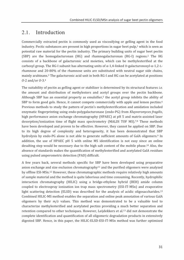
2.1. Introduction Commercially extracted pectin is commonly used as viscosifying or gelling agent in the food industry. Pectic substances are present in high proportions in sugar beet pulp,1 which is seen as potential raw material for the pectin industry. The primary building units of sugar beet pectin (SBP) are the homogalacturonan (HG) and rhamnogalacturonan (RG-I) regions.2 The HG consists of a backbone of galacturonic acid moieties, which can be methylesterified at the carboxyl group. The RG-I subunit has alternating units of α-1,4-linked D-galacturonosyl-α-1,2-L-rhamnose and 20-80% of the rhamnose units are substituted with neutral sugar side chains, mainly arabinans.3 The galacturonic acid unit in both RG-I and HG can be acetylated at positions O-2 and/or O-3.4
The suitability of pectin as gelling agent or stabilizer is determined by its structural features i.e. the amount and distribution of methylesters and acetyl groups over the pectin backbone. Although SBP has an essential property as emulsifier,5 the acetyl group inhibits the ability of SBP to form good gels. Hence, it cannot compete commercially with apple and lemon pectins.6 Previous methods to study the pattern of pectin’s methylesterification and amidation included enzymatic fingerprinting using endo-polygalacturonase (endo-PG) from Kluyveromyces fragilis, high performance anion exchange chromatography (HPAEC) at pH 5 and matrix-assisted laser desorption/ionization time of flight mass spectrometry (MALDI TOF MS).7,8 These methods have been developed and proven to be effective. However, they cannot be applied on SBP. Due to its high degree of complexity and heterogeneity, it has been demonstrated that SBP hydrolysis by endo-PG alone is not able to generate sufficient amounts of GalA oligomers.9 In addition, the use of HPAEC pH 5 with online MS identification is not easy since an online desalting step would be necessary due to the high salt content of the mobile phase.10 Also, the absence of standards makes the quantification of methylesterified and acetylated GalA residues using pulsed amperometric detection (PAD) difficult.
A few years back, several methods specific for SBP have been developed using preparative anion exchange and size exclusion chromatography11 and the purified oligomers were analyzed by offline ESI-MSn.12 However, these chromatographic methods require relatively high amounts of sample material and the method is quite laborious and time consuming. Recently, hydrophilic interaction chromatography (HILIC) using a bridge-ethylene hybrid (BEH) amide column coupled to electrospray ionization ion trap mass spectrometry (ESI-IT-MSn) and evaporative light scattering detection (ELSD) was described for the analysis of acidic oligosaccharides.13 Combined HILIC-MS method enables the separation and online peak annotation of various GalA oligomers by their m/z values. This method was demonstrated to be a valuable tool to characterize methylesterified and acetylated pectins providing a much better separation and retention compared to other techniques. However, Leijdekkers et al.13 did not demonstrate the complete identification and quantification of all oligomeric degradation products in extensively digested SBP. Hence, in this paper, the HILIC-ELSD-ESI-IT-MSn method was further optimized
31
Chapter 2
32
and applied to separate, identify and quantify the complex mixture of SBP oligomers generated by the combined action of pectolytic enzymes.
2.2. Materials and methods
Pectin samples Experimental sugar beet pectins (SBPs), modified by plant PME (P5328) and fungal PME (F5129); and the commercial SBP5317 were obtained from Danisco (Brabrand, Denmark) (Table 2.1). Determination of the neutral sugar composition of SBP5317 by gas chromatography of alditol acetates was achieved after subsequent hydrolysis by 72% (w/w) sulfuric acid and 1M sulfuric acid.14 The uronic acid was determined by an automated colorimetric m-hydroxydiphenyl method as described previously.15 Pectin samples (≈1 mg) were saponified by 1M NaOH to determine the degree of methylesterification (DM) using colorimetric method16 while the degree of acetylation (DA) was analyzed using Megazyme acetic acid kit (Megazyme, Wicklow, Ireland).
Table 2.1. Monosaccharide composition of sugar beet pectin samples.
Pectin GalA Rha Ara Xyl Gal Glc DM (%)a DA (%)a
mg/g of dry matter
P5328b 582 55 109 2 99 4 53 28
F5129b 567 51 116 3 94 2 51 29
SBP5317c 560 30 40 - 80 1 53 17 aMoles methanol (DM) or acetic acid (DA) per 100 mols of galacturonic acid residues. bMonosaccharide composition9 cMonosaccharide composition determined in this study. - not detected
Enzymes Pure and well characterized RG-I and HG degrading enzymes were used to hydrolyse sugar beet pectins. The enzymes used in this study were Aspergillus aculeatus endo-galactanase (EC 3.2.1.89),17 endo-arabinanase (EC 3.2.1.99),18 RG-hydrolase (EC 3.2.1.B9),19 Chrysosporium lucknowense (C1) exo-arabinase (EC 3.2.1.1),20 Aspergillus niger fungal pectin methyl esterase (fungal PME) (EC 3.1.1.11),21 pectin lyase (EC 4.2.2.10)22 and endo-polygalacturonase II (EC 3.2.1.15).23
Enzymatic hydrolysis Sugar beet pectin (SBP5317) in 50 mM sodium citrate buffer pH 5 (5 mg/ml) was digested at 40 °C by RG-I (endo-galactanase + endo/exo arabinase + RG hydrolase) and HG (PL + endo-PGII)
32
Combined HILIC-ELSD/MSn analysis of sugar beet pectin oligomers
33
degrading enzymes to hydrolyse the SBP samples as far as possible. The hydrolysis was done by incubating the pectin solution with RG-I degrading enzymes and PL for 6 hours followed by the addition of endo-PGII and fungal PME followed by the subsequent incubation for another 18 hours. Sugar beet pectins (P5328 and F5129) were digested in the same way although PME addition was omitted during digestion. Enzyme doses were sufficient to degrade theoretically their corresponding substrates within 6 hrs into monomers. Inactivation of enzymes was performed at 100 °C for 6 min and the reaction products were analyzed by high performance size exclusion chromatography (HPSEC) and HPLC-HILIC coupled to ESI-IT-MSn and ELSD detectors.
HPSEC Sugar beet pectin digests were analyzed using HPSEC on an Ultimate 3000 system (Dionex, Sunnyvale, CA, USA). A set of four TSK-Gel super AW columns (Tosoh Bioscience, Tokyo, Japan) was used in series: one guard column (6 mm ID × 40 mm) and the three separation columns 4000, 3000 and 2500 (6 mm × 150 mm). The column temperature was set to 55 °C. Samples (20 µL, 2.5 mg/ml) were eluted with filtered 0.2 M NaNO3 at a flow rate of 0.6 ml/min and the elution was monitored by refractive index detection (Shodex RI 101; Showa Denko K.K., Kawasaki, Japan) and UV235 detection (Dionex Variable Wavelength Detector, Sunnyvale, CA, USA) .
HPLC-ELSD/ESI-MSn Digests were analyzed using HPLC in combination with ESI-IT-MSn and ELSD on a HILIC BEH amide column as described previously.13 The composition of the two mobile phases was (A) 80:20 (v/v) acetonitrile (ACN)/water, and (B) 20:80 (v/v) ACN/water, both containing 0.01 M ammonium formate and 0.05 M formic acid. The following elution profile was used: 0–1 min, isocratic 100% A; 1–60 min, linear from 30% to 80% B; followed by column re-equilibration: 61–67 min, linear from 20% to 100% A; 68-75 min, isocratic 100% A. The eluent was split (1:1) using an ASI flow splitter (Analytical Scientific Instruments, CA, USA) before the ELSD and the ESI-IT-MS detector. Mass spectra were acquired over the scan range m/z 150–2000. Xcalibur software was used to process the data (Thermo). Commercial GalAs degree of polymerization (DP) 1–3 (Sigma-Aldrich, Steinheim, Germany), unsaturated (DP 2–6) and saturated (DP 4–5) galacturonic acid standards were used as purified in our laboratory as described.24 To estimate the amount of oligomers by ELSD, curve fitting of each GalA standard using a power function of f(x) = axb was used, where f(x) is the peak area, x is the sample amount, a is the response factor and b is the slope.25,26 The average responses of DP 1–3 were plotted and the mathematical equation was derived.
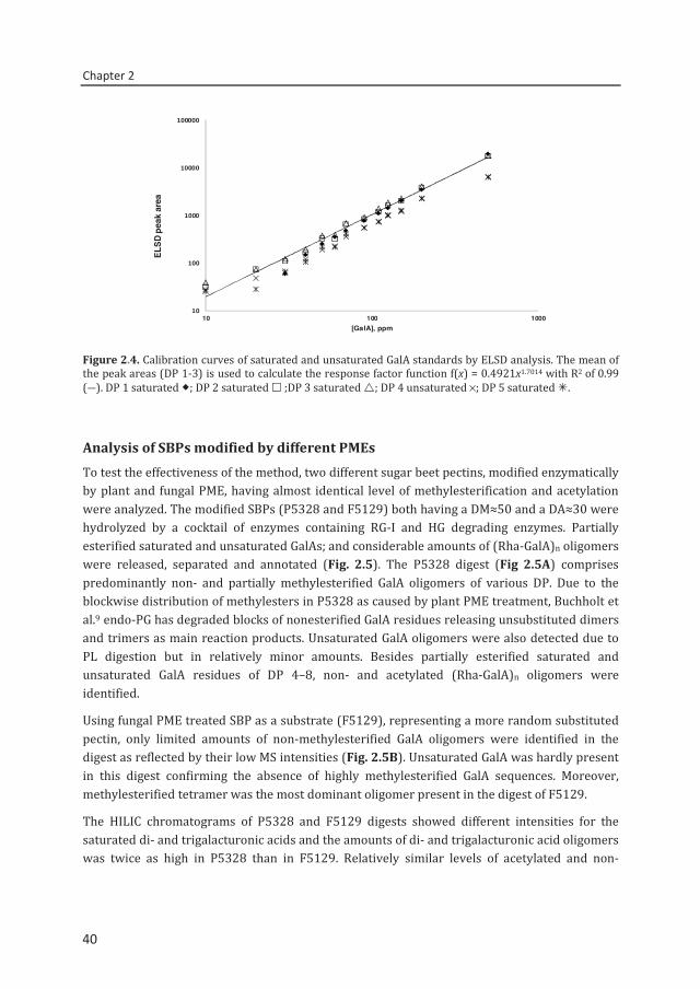
concentration[ppm] = exp ln(ELSDpeakarea 0.4921⁄ )1.7014
33
Chapter 2
34
2.3. Results and discussion
Enzymatic hydrolysis of SBP In this study, RG-I degrading enzymes and PL were added to degrade the pectin side chain and methylesterified HG, respectively. The mixture was then digested with endo-PG and fungal PME. As endo-PG and PL cannot sufficiently degrade highly substituted SBPs9, fungal PME enabled to generate as complete as possible mixture of different GalA oligomers.
In order to monitor the degradation of SBP by pectolytic enzymes, HPSEC with online UV and RI detector was used. Figure 2.1 illustrates the enzyme-treated SBP resulting in a shift in molecular weight (Mw) yielding low Mw oligomers. The UV235 signal indicates that next to the release of saturated GalA oligomers by endo-PG, also unsaturated GalA oligomers resulting from PL action were released. HPSEC analysis indicates clearly that endo-PG and PL together sufficiently degrade SBP to a broad range of diagnostic oligomers (<10 kDa) eluting at retention time >10.5 min.
Figure 2.1. HPSEC elution pattern of SBP5317 (DM 53, DA 17) before (—, RI) and after ( UV235; ---- RI) digestion with RG-I and HG degrading enzymes. Molecular masses of pectin standards (in kDa) are indicated.
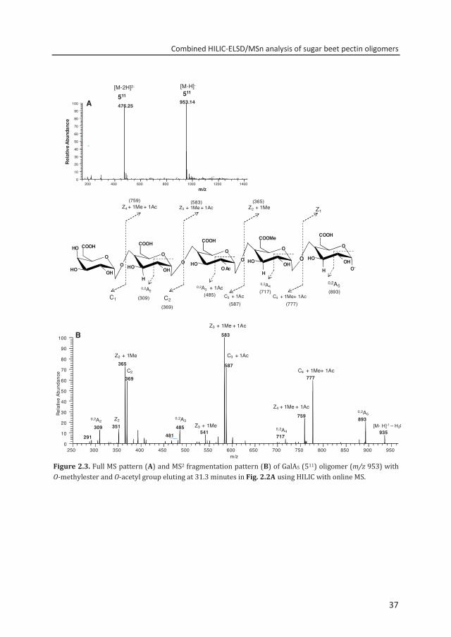
Separation and annotation of the reaction products by HILIC-MS The HILIC elution pattern of SBP5317 (Fig. 2.2A) illustrates that besides the unsubstituted dimer (200) and trimer (300), partially methylesterified and/or acetylated saturated and unsaturated GalA oligomers of different DP were present as main degradation products. Partially acetylated RG-I oligomers i.e. (Rha-GalA)2Ac were identified as well as unsaturated GalA oligomers containing methylesters (U310, U410, U520). The elution behavior of GalA oligomers of the same charge and DP was also influenced by the presence of acetyl group. As
6.0 7.0 8.0 9.0 10.0 11.0 12.0 13.0 14.0
Retention Time (min)
Dete
ctor
resp
onse
56 35 10100 kDa
RI r
espo
nse
Retention time (min)
34
Combined HILIC-ELSD/MSn analysis of sugar beet pectin oligomers
35
Figure 2.2. HILIC elution pattern of SBP5317 digested by RG-I and HG degrading enzymes using (A) ESI-IT-MSn and (B) ELS detection. Peak annotation: 511, DP 5, O-methylester, O-acetyl group. U: unsaturated GalA; Rha: rhamnose; GalA: galacturonic acid; Ac: acetyl group.
an example: tetramer 411 (m/z 777) with a methylester and an acetyl group, eluted before tetramer 410 (m/z 735) with a methylester but without acetyl group.
The efficient separation and rapid identification of a complex SBP digest with HILIC exemplify the advantage of the technique for screening SBP digests compared to the conventional preparative separation.11,12 It is evident that with HILIC analysis, oligosaccharides in pectin digests containing unsubstituted GalAs can be completely differentiated from the oligosaccharides containing methylesterified and acetylated GalA units. In this way, fingerprinting of pectins and determination of the degree of blockiness can be achieved with higher accuracy compared to HPAEC pH 5 separation method.27 The good alignment between
200
(Rha
-Gal
A)2
U410
400/ 7
31621
510 611 /5
01
721 712 / 8
22411
410(R
ha-G
alA)
3
511
521
822/9
32
711
301/ U
520
(Rha
-Gal
A)2A
c
U310
401
U310
200
U410
(Rha
-Gal
A)2
411
521
410
711/
821
B
0 5 10 15 20 25 30 35 40 45Time (min)
(Rha
-Gal
A)2A
c
(Rha
-Gal
A)3
511
621
400/ 7
31
721510 711/
821
711
0 5 10 15 20 25 30 35 40 45
A
822/9
32
401
300300
ELSD
Det
ectio
nM
SDe
tect
ion
301/ U
520
611 /5
01
712 / 8
22
35
Chapter 2
36
MS and ELSD chromatograms (Fig. 2.2B) also allows the possibility of peak identification in the ELSD elution profile, the latter being used for quantification.
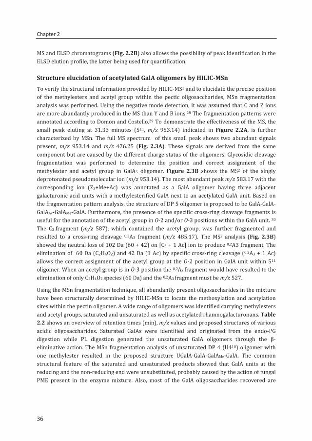
Structure elucidation of acetylated GalA oligomers by HILIC-MSn To verify the structural information provided by HILIC-MS1 and to elucidate the precise position of the methylesters and acetyl group within the pectic oligosaccharides, MSn fragmentation analysis was performed. Using the negative mode detection, it was assumed that C and Z ions are more abundantly produced in the MS than Y and B ions.28 The fragmentation patterns were annotated according to Domon and Costello.29 To demonstrate the effectiveness of the MS, the small peak eluting at 31.33 minutes (511, m/z 953.14) indicated in Figure 2.2A, is further characterized by MSn. The full MS spectrum of this small peak shows two abundant signals present, m/z 953.14 and m/z 476.25 (Fig. 2.3A). These signals are derived from the same component but are caused by the different charge status of the oligomers. Glycosidic cleavage fragmentation was performed to determine the position and correct assignment of the methylester and acetyl group in GalA5 oligomer. Figure 2.3B shows the MS2 of the singly deprotonated pseudomolecular ion (m/z 953.14). The most abundant peak m/z 583.17 with the corresponding ion (Z3+Me+Ac) was annotated as a GalA oligomer having three adjacent galacturonic acid units with a methylesterified GalA next to an acetylated GalA unit. Based on the fragmentation pattern analysis, the structure of DP 5 oligomer is proposed to be GalA-GalA-GalAAc-GalAMe-GalA. Furthermore, the presence of the specific cross-ring cleavage fragments is useful for the annotation of the acetyl group in O-2 and/or O-3 positions within the GalA unit. 30 The C3 fragment (m/z 587), which contained the acetyl group, was further fragmented and resulted to a cross-ring cleavage 0,2A3 fragment (m/z 485.17). The MS2 analysis (Fig. 2.3B) showed the neutral loss of 102 Da (60 + 42) on [C3 + 1 Ac] ion to produce 0,2A3 fragment. The elimination of 60 Da (C2H4O2) and 42 Da (1 Ac) by specific cross-ring cleavage (0,2A3 + 1 Ac) allows the correct assignment of the acetyl group at the O-2 position in GalA unit within 511 oligomer. When an acetyl group is in O-3 position the 0,2A3 fragment would have resulted to the elimination of only C2H4O2 species (60 Da) and the 0,2A3 fragment must be m/z 527.
Using the MSn fragmentation technique, all abundantly present oligosaccharides in the mixture have been structurally determined by HILIC-MSn to locate the methoxylation and acetylation sites within the pectin oligomer. A wide range of oligomers was identified carrying methylesters and acetyl groups, saturated and unsaturated as well as acetylated rhamnogalacturonans. Table 2.2 shows an overview of retention times (min), m/z values and proposed structures of various acidic oligosaccharides. Saturated GalAs were identified and originated from the endo-PG digestion while PL digestion generated the unsaturated GalA oligomers through the β-eliminative action. The MSn fragmentation analysis of unsaturated DP 4 (U410) oligomer with one methylester resulted in the proposed structure UGalA-GalA-GalAMe-GalA. The common structural feature of the saturated and unsaturated products showed that GalA units at the reducing and the non-reducing end were unsubstituted, probably caused by the action of fungal PME present in the enzyme mixture. Also, most of the GalA oligosaccharides recovered are
36
Combined HILIC-ELSD/MSn analysis of sugar beet pectin oligomers
37
Figure 2.3. Full MS pattern (A) and MS2 fragmentation pattern (B) of GalA5 (511) oligomer (m/z 953) with O-methylester and O-acetyl group eluting at 31.3 minutes in Fig. 2.2A using HILIC with online MS.
0,2A5
C4 + 1Me+ 1AcC2C1 C3 + 1Ac
Z1Z2 + 1MeZ3 + 1Me + 1AcZ4 + 1Me + 1Ac
O
HO
HO OH
COOHO
H
HO OH
COOH
O
O
HOOAc
COOHO
H
HO OH
COOMe
OO
O
H
HO
O-OH
COOH
O
0,2A4 0,2A3 + 1Ac0,2A2
A
200 400 600 800 1000 1200 1400 1600 1800 2000m/z
0
10
20
30
40
50
60
70
80
90
100
Rel
ativ
e Ab
unda
nce
953.14476.25511 511
[M-H]-[M-2H]2-
(365)(583)(759)
(309) (485) (893)
(777)
(717)
(587)(369)
250 300 350 400 450 500 550 600 650 700 750 800 850 900 950m/z
0
10
20
30
40
50
60
70
80
90
100
Rela
tive
Abun
danc
e
583
365 587
777369
759 893351 485309
541 935717
Z2 + 1Me
C2
Z3 + 1Me + 1Ac
C3 + 1Ac
C4 + 1Me+ 1Ac
Z4 + 1Me + 1Ac0,2A5
[M- H]-1 – H2OZ2 0,2A3
0,2A4Z3 + 1Me
0,2A2
B
481291
37
Chapter 2
38
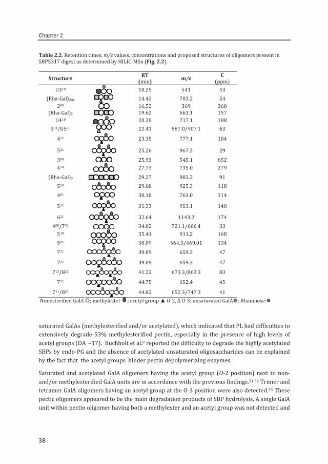
Table 2.2. Retention times, m/z values, concentrations and proposed structures of oligomers present in SBP5317 digest as determined by HILIC-MSn (Fig. 2.2).
Structure RT (min) m/z C
(ppm) U310 10.25 541 43
(Rha-Gal)2Ac 14.42 703.2 54 200 16.52 369 360
(Rha-Gal)2 19.62 661.1 157 U410 20.28 717.1 188
301/U520 22.41 587.0/907.1 63
411 23.35 777.1 184
521
25.26 967.3 29 300 25.93 545.1 652 410 27.73 735.0 279
(Rha-Gal)3 29.27 983.2 91 520 29.68 925.3 118 401 30.18 763.0 114
511 31.33 953.1 140
621 32.64 1143.2 174 400/731 34.02 721.1/666.4 33
510 35.41 911.2 168 501 38.09 564.3/469.01 134 721 39.09 659.3 47
721 39.09 659.3 47
712/822 41.22 673.3/863.3 83
711 44.75 652.4 45
711/821 44.82 652.3/747.3 41 Nonesterified GalA ; methylester ; acetyl group ▲ O-2, Δ O-3; unsaturated GalA; Rhamnose:
saturated GalAs (methylesterified and/or acetylated), which indicated that PL had difficulties to extensively degrade 53% methylesterified pectin, especially in the presence of high levels of acetyl groups (DA ~17). Buchholt et al.9 reported the difficulty to degrade the highly acetylated SBPs by endo-PG and the absence of acetylated unsaturated oligosaccharides can be explained by the fact that the acetyl groups hinder pectin depolymerizing enzymes.
Saturated and acetylated GalA oligomers having the acetyl group (O-2 position) next to non- and/or methylesterified GalA units are in accordance with the previous findings.31,32 Trimer and tetramer GalA oligomers having an acetyl group at the O-3 position were also detected.33 These pectic oligomers appeared to be the main degradation products of SBP hydrolysis. A single GalA unit within pectin oligomer having both a methylester and an acetyl group was not detected and
38
Combined HILIC-ELSD/MSn analysis of sugar beet pectin oligomers
39
such a combination of substitution seems indeed to be rare in native pectin as reported by Ralet et al.12
Calibration curves and quantification by ELSD Quantification of the reaction products present in SBP digest is necessary in order to model the native pectin’s structure. Previous studies have shown that ELSD enables the quantification of the compounds by correlating the peak area of the analyte versus the concentration by using a double logarithmic scale25,26 and the same method was applied in the present study. Figure 2.4 shows that the observed ELSD response of commercial and laboratory made GalA standards at different concentrations was linear with a minimum detection limit of ≈20 ppm. Similar slopes were observed for the monogalacturonic, di- and trigalacturonic acid standards with linear correlation coefficients ranging from 0.988 to 0.999. The mean of DP 1–3 was calculated as f(x) = 0.4921×1.7014 (R2 = 0.99) (Eq. (1)). Besides the quantification of saturated GalA oligomers, an attempt to quantify the amounts of higher saturated and unsaturated oligomers was performed by using the available saturated and unsaturated GalA oligomers (purity ≥85%) as prepared and described by Van Alebeek et al.24 The ELSD response of unsaturated tetragalacturonic acid representing other unsaturated oligomers and of GalA saturated tetramer and pentamer (Fig. 2.4) was found quite similar. The observed deviation among the slopes of different GalA standards was due to the purity of the compounds. The results indicate that the detection method is independent of the molecular structure of the oligomers tested as previously proven by Decroos et al.25 for the quantification of soy saponins. The same type of mathematical equation can be used to quantify saturated, unsaturated, methylesterified and acetylated GalA oligomers without the need of specific standards as would be required in the MS analysis.
The oligomers released after the hydrolysis of an enzyme-treated SBP (Fig. 2.2) as quantified by ELSD, represented 90±5 % of the GalA residues present in the pectin. The calculated amount of GalAs allows a valid reconstruction of the original pectin molecules. Furthermore, quantification by HPAEC-PAD (pH 12) confirmed that 90% of the total GalA oligosaccharides are recovered in the digest.
39
Chapter 2
40
Figure 2.4. Calibration curves of saturated and unsaturated GalA standards by ELSD analysis. The mean of the peak areas (DP 1-3) is used to calculate the response factor function f(x) = 0.4921x1.7014 with R2 of 0.99 (―). DP 1 saturated ; DP 2 saturated ;DP 3 saturated ; DP 4 unsaturated ×; DP 5 saturated .
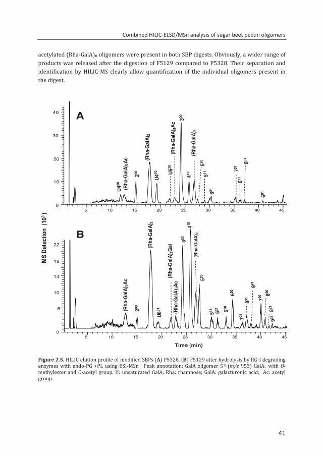
Analysis of SBPs modified by different PMEs To test the effectiveness of the method, two different sugar beet pectins, modified enzymatically by plant and fungal PME, having almost identical level of methylesterification and acetylation were analyzed. The modified SBPs (P5328 and F5129) both having a DM≈50 and a DA≈30 were hydrolyzed by a cocktail of enzymes containing RG-I and HG degrading enzymes. Partially esterified saturated and unsaturated GalAs; and considerable amounts of (Rha-GalA)n oligomers were released, separated and annotated (Fig. 2.5). The P5328 digest (Fig 2.5A) comprises predominantly non- and partially methylesterified GalA oligomers of various DP. Due to the blockwise distribution of methylesters in P5328 as caused by plant PME treatment, Buchholt et al.9 endo-PG has degraded blocks of nonesterified GalA residues releasing unsubstituted dimers and trimers as main reaction products. Unsaturated GalA oligomers were also detected due to PL digestion but in relatively minor amounts. Besides partially esterified saturated and unsaturated GalA residues of DP 4–8, non- and acetylated (Rha-GalA)n oligomers were identified.
Using fungal PME treated SBP as a substrate (F5129), representing a more random substituted pectin, only limited amounts of non-methylesterified GalA oligomers were identified in the digest as reflected by their low MS intensities (Fig. 2.5B). Unsaturated GalA was hardly present in this digest confirming the absence of highly methylesterified GalA sequences. Moreover, methylesterified tetramer was the most dominant oligomer present in the digest of F5129.
The HILIC chromatograms of P5328 and F5129 digests showed different intensities for the saturated di- and trigalacturonic acids and the amounts of di- and trigalacturonic acid oligomers was twice as high in P5328 than in F5129. Relatively similar levels of acetylated and non-
10
100
1000
10000
100000
10 100 1000
ELSD
pea
k ar
ea
[GalA], ppm
40
Combined HILIC-ELSD/MSn analysis of sugar beet pectin oligomers
41
acetylated (Rha-GalA)n oligomers were present in both SBP digests. Obviously, a wider range of products was released after the digestion of F5129 compared to P5328. Their separation and identification by HILIC-MS clearly allow quantification of the individual oligomers present in the digest.
Figure 2.5. HILIC elution profile of modified SBPs (A) P5328, (B) F5129 after hydrolysis by RG-I degrading enzymes with endo-PG +PL using ESI-MSn . Peak annotation: GalA oligomer 511 (m/z 953) GalA5 with O-methylester and O-acetyl group. U: unsaturated GalA; Rha: rhamnose; GalA: galacturonic acid; Ac: acetyl group.
5 10 15 20 25 30 35 40 450
10
20
30
40
MS
Dete
ctio
n (1
03 )
200
(Rha
-Gal
A)2
300
520
621
831
(Rha
-Gal
A)3
410
U420
731
(Rha
-Gal
A)3A
c
611
601
U410 U5
20
511
(Rha
-Gal
A)2A
c
5 10 15 20 25 30 35 40 45
Time (min)
0
6
10
14
18
22
200
520
621
831721
(Rha
-Gal
A)3A
c
510
U631
(Rha
-Gal
A)2G
al
(Rha
-Gal
A)3
620
720 830
941
821(Rha
-Gal
A)2A
c
511
(Rha
-Gal
A)2
300
410
931
A
B
41
Chapter 2
42
2.4. Conclusion A novel method has been developed and applied to analyse and quantify the partially methylesterified and acetylated oligogalacturonides after degradation by pectic enzymes. The results demonstrated that it is remarkably easy to interpret HILIC chromatograms. It implies that the use of the HILIC-MS approach provides essential and detailed information on the composition of pectin structure. ELSD has proven to be a versatile method to quantify the absolute amount of GalA oligomers present in the pectin digest. This method can now be used to accurately determine the degree of blockiness and to predict the functional properties of sugar beet pectins.
Acknowledgements This work has been finacially supported by the European Community within a consortium PolyMode KBBE-2007-3-3-07 and is gratefully acknowledged. We also like to thank Mark Sanders and Tomas Kuijpers for the fruitful discussions.
References 1. Oosterveld, A., Beldman, G., Schols, H. A., and Voragen, A. G. J. (1996) Carbohydrate Research 288,
143-153 2. Guillon, F., Thibault, J.-F., Rombouts, F. M., Voragen, A. G. J., and Pilnik, W. (1989) Carbohydrate
Research 190, 97-108 3. Voragen, A. G. J., Coenen, G. J., Verhoef, R. P., and Schols, H. A. (2009) Structural Chemistry 20, 263-
275 4. Voragen, A. G. J., Pilnik, W., Thibault, J. F., Axelos, M. A. V., and Renard, C. M. G. C. (1995) Pectins. in
Food Polysaccharides and Their Applications Stephen, A. M. (ed.), Marcel Dekker, New York, N.Y., USA. pp 287-339
5. Williams, P. A., Sayers, C., Viebke, C., Senan, C., Mazoyer, J., and Boulenguer, P. (2005) Journal of Agricultural and Food Chemistry 53, 3592-3597
6. Thakur, B. R., Singh, R. K., Handa, A. K., and Rao, M. A. (1997) Critical Reviews in Food Science and Nutrition 37, 47-73
7. Daas, P. J. H., Arisz, P. W., Schols, H. A., De Ruiter, G. A., and Voragen, A. G. J. (1998) Analytical Biochemistry 257, 195-202
8. Guillotin, S. E., Van Kampen, J., Boulenguer, P., Schols, H. A., and Voragen, A. G. J. (2006) Biopolymers 82, 29-37
9. Buchholt, H. C., Christensen, T. M. I. E., Fallesen, B., Ralet, M. C., and Thibault, J. F. (2004) Carbohydrate Polymers 58, 149-161
10. Westphal, Y., Schols, H. A., Voragen, A. G. J., and Gruppen, H. (2010) Journal of Chromatography A 1217, 689-695
11. Bonnin, E., Dolo, E., Le Goff, A., and Thibault, J.-F. (2002) Carbohydrate Research 337, 1687-1696 12. Ralet, M. C., Cabrera, J. C., Bonnin, E., Quémenér, B., Hellín, P., and Thibault, J. F. (2005)
Phytochemistry 66, 1832-1843 13. Leijdekkers, A. G. M., Sanders, M. G., Schols, H. A., and Gruppen, H. (2011) Journal of
Chromatography A 1218, 9227-9235 14. Selvendran, R. R., March, J. F., and Ring, S. G. (1979) Analytical Biochemistry 96, 282-292 15. Kuhnel, S., Schols, H. A., and Gruppen, H. (2011) Biotechnology for Biofuels 4, 14
42
Combined HILIC-ELSD/MSn analysis of sugar beet pectin oligomers
43
16. Guillotin, S. E., Van Loey, A., Boulenguer, P., Schols, H. A., and Voragen, A. G. J. (2007) Food Hydrocolloids 21, 85-91
17. Schols, H. A., Posthumus, M. A., and Voragen, A. G. J. (1990) Carbohydrate Research 206, 117-129 18. Beldman, G., Searle-van Leeuwen, M. J. F., De Ruiter, G. A., Siliha, H. A., and Voragen, A. G. J. (1993)
Carbohydrate Polymers 20, 159-168 19. Mutter, M., Renard, C. M. G. C., Beldman, G., Schols, H. A., and Voragen, A. G. J. (1998) Carbohydrate
Research 311, 155-164 20. Kühnel, S., Hinz, S. W. A., Pouvreau, L., Wery, J., Schols, H. A., and Gruppen, H. (2010) Bioresource
Technology 101, 8300-8307 21. Van Alebeek, G. J. W. M., Van Scherpenzeel, K., Beldman, G., Schols, H. A., and Voragen, A. G. J.
(2003) Biochemical Journal 372, 211-218 22. Harmsen, J. A. M., Kusters-van Someren, M. A., and Visser, J. (1990) Current Genetics 18, 161-166 23. Limberg, G., Körner, R., Buchholt, H. C., Christensen, T. M. I. E., Roepstorff, P., and Mikkelsen, J. D.
(2000) Carbohydrate Research 327, 321-332 24. Van Alebeek, G. J. W. M., Christensen, T. M. I. E., Schols, H. A., Mikkelsen, J. D., and Voragen, A. G. J.
(2002) Journal of Biologial Chemistry 277, 25929-25936 25. Decroos, K., Vincken, J. P., Heng, L., Bakker, R., Gruppen, H., and Verstraete, W. (2005) Journal of
Chromatography A 1072, 185-193 26. Mengerink, Y., De Man, H. C. J., and Van der Wal, S. (1991) Journal of Chromatography 552, 593-
604 27. Daas, P. J. H., Meyer-Hansen, K., Schols, H. A., De Ruiter, G. A., and Voragen, A. G. J. (1999)
Carbohydrate Research 318, 135-145 28. Körner, R., Limberg, G., Christensen, T. M. I. E., Mikkelsen, J. r. D., and Roepstorff, P. (1999)
Analytical Chemistry 71, 1421-1427 29. Domon, B., and Costello, C. E. (1988) Glycoconjugate Journal 5, 397-409 30. Quéméner, B., Cabrera Pino, J. C., Ralet, M.-C., Bonnin, E., and Thibault, J.-F. (2003) Journal of Mass
Spectrometry 38, 641-648 31. Needs, P. W., Rigby, N. M., Colquhoun, I. J., and Ring, S. G. (1998) Phytochemistry 48, 71-77 32. Perrone, P., Hewage, C. M., Thomson, A. R., Bailey, K., Sadler, I. H., and Fry, S. C. (2002)
Phytochemistry 60, 67-77 33. Ralet, M.-C., Crépeau, M.-J., and Bonnin, E. (2008) Phytochemistry 69, 1903-1909
43

44
Chapter 3
Descriptive parameters for revealing
substitution patterns of sugar beet
pectins using pectolytic enzymes
C. Remoroza, H.C. Buchholt, H. Gruppen, H.A. Schols
Carbohydrate Polymers 2014, 101, 1205-1215
Chapter 3
46
Abstract
Enzymatic fingerprinting was applied to sugar beet pectins (SBPs) modified by either plant or fungal pectin methylesterases and alkali catalyzed de-methylesterification to reveal the ester distributions over the pectin backbone. A simultaneous pectin lyase (PL) treatment to the used endo-polygalacturonase (PG) degradation showed to be effective in degrading both high and low methylesterified and/or acetylated homogalaturonan regions of SBP. Using HPLC-HILIC-MS/ELSD, we studied in detail all the diagnostic oligomers present, enabling us to discriminate between differently prepared sugar beet pectins having various levels of methylesterification and acetylation. Furthermore, distinction between commercially extracted and de-methylesterified sugar beet pectin having different patterns of substitution was achieved by using novel descriptive pectin parameters. In addition to DBabs approach for nonmethylesterified sequences degradable by endo-PG, the “degree of hydrolysis” by PG (DHPG) representing all partially saturated methylesterified and/or acetylated galacturonic acid (GalA) moieties was introduced as a new parameter. Consequently, the description degree of hydrolysis by PL (DHPL) has been introduced to quantify all esterified unsaturated GalA oligomers.
46
Descriptive parameters for revealing substitution patterns in sugar beet pectins
47
3.1 Introduction
Pectin is probably the most complex and versatile polysaccharide present in plant cell walls. Pectin consists of different kinds of polymers, which are covalently attached to each other. The primary structural elements of pectin are the homogalacturonan (HG) and rhamnogalacturonan (RG-I) regions.1 The HG consists of a backbone of galacturonic acid moieties, which can be methylesterified at the C-6 of galacturonic acid. The RG-I subunit has repeats of alternating of α-1,4-linked D-galacturonosyl-α-1,2-L-rhamnose residues and the rhamnose units may be substituted with neutral sugar side chains.2 Because of its excellent gelling and thickening properties, industrially extracted pectin is valued highly as a food ingredient. The suitability of pectin as gelling agent or stabilizer is determined by the distribution of methylesters over the homogalacturonan region, galacturonic content, average molar mass, degree of methylesterification (DM) and degree of acetylation (DA).3 Pectins from different sources show different gelling abilities due to variations in these parameters. At present, apple pomace and citrus peels are the main sources of commercially successful pectins and they have been extensively studied. Enzymatic degradation of the pectins with an endo-polygalacturonase (endo-PG) of Kluyveromyces fragilis followed by analysis of the partially methylesterified oligogalacturonides released has been used as a method to distinguish the substitution pattern within citrus pectin structures. In these studies, the Degree of Blockiness (DB) was calculated from the level of oligosaccharides released as quantified by high performance anion exchange chromatography HPAEC (pH 5), while the absolute degree of blockiness (DBabs) was obtained from both capillary electrophoresis and HPAEC (pH 5) analyses of the digests.4-7 Although this approach has been widely used to differentiate substitution patterns of citrus pectins, the focus on nonmethylesterified sequences provides only a view on the relatively low methylesterified region of the pectin structure. Consequently, fingerprinting of highly methylesterified pectins with pectin lyase (PL) in combination with several chromatographic methods available has been used to distinguish series of differently de-methylesterified lemon pectins in order to study the highly methylesterified segment of pectin.8 Recently, the degree of blockiness (DBMe) and absolute degree of blockiness (DBabsMe) for the methylesterified regions present in the HG region of pectin based on pectin lyase digestion was introduced.9
So far, the above-mentioned methods were not yet fully applied for the analysis of more complex pectins e.g acetylated sugar beet pectin. Sugar beet pulp is a by-product of the sugar industry and is seen as a potential raw material of pectin. Sugar beet pectin (SBP), however, cannot compete commercially with citrus and apple pectins as gelling and thickening agent due to the high level of acetyl groups present in the HG regions of SBP.10 The GalA residue in SBP can be acetylated at positions O-2 and/or O-3 in both RG-I and HG. Similar to lemon pectins,11 a series of de-methylesterified SBPs by alkali, plant PME and fungal PME has been characterized by enzymatic fingerprinting using endo and exo-PGs.3 However, the use of only PGs limits the analysis of SBP due to the presence of both methylesters and acetyl groups attached to the homogalacturonan region hindering PG to release sufficient levels of diagnostic oligomers.
47
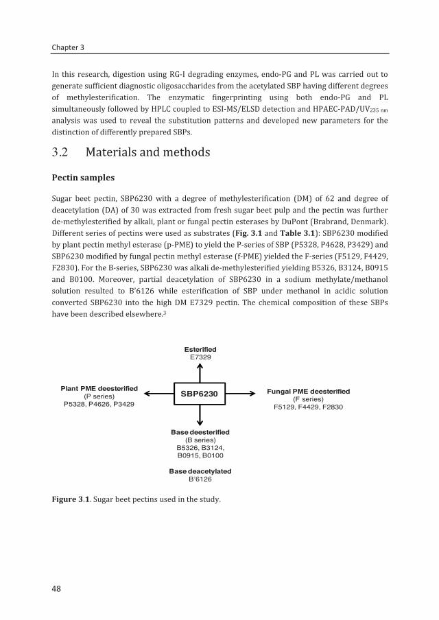
Chapter 3
48
In this research, digestion using RG-I degrading enzymes, endo-PG and PL was carried out to generate sufficient diagnostic oligosaccharides from the acetylated SBP having different degrees of methylesterification. The enzymatic fingerprinting using both endo-PG and PL simultaneously followed by HPLC coupled to ESI-MS/ELSD detection and HPAEC-PAD/UV235 nm analysis was used to reveal the substitution patterns and developed new parameters for the distinction of differently prepared SBPs.
3.2 Materials and methods
Pectin samples
Sugar beet pectin, SBP6230 with a degree of methylesterification (DM) of 62 and degree of deacetylation (DA) of 30 was extracted from fresh sugar beet pulp and the pectin was further de-methylesterified by alkali, plant or fungal pectin esterases by DuPont (Brabrand, Denmark). Different series of pectins were used as substrates (Fig. 3.1 and Table 3.1): SBP6230 modified by plant pectin methyl esterase (p-PME) to yield the P-series of SBP (P5328, P4628, P3429) and SBP6230 modified by fungal pectin methyl esterase (f-PME) yielded the F-series (F5129, F4429, F2830). For the B-series, SBP6230 was alkali de-methylesterified yielding B5326, B3124, B0915 and B0100. Moreover, partial deacetylation of SBP6230 in a sodium methylate/methanol solution resulted to B’6126 while esterification of SBP under methanol in acidic solution converted SBP6230 into the high DM E7329 pectin. The chemical composition of these SBPs have been described elsewhere.3
Figure 3.1. Sugar beet pectins used in the study.
SBP6230Plant PME deesterified
(P series)P5328, P4626, P3429
Fungal PME deesterified(F series)
F5129, F4429, F2830
Base deesterified(B series)
B5326, B3124, B0915, B0100
Base deacetylatedB’6126
EsterifiedE7329
48
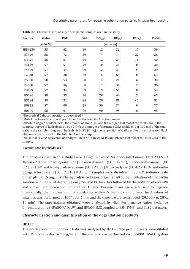
Descriptive parameters for revealing substitution patterns in sugar beet pectins
49
Table 3.1. Characteristics of sugar beet pectin samples used in the study.
Pectins GalAa DMb DAb DBabsc DHPGc DHPLc Yieldc
(w/w %) (mole %) SBP6230 55 62 30 12 22 17 39
E7329 58 73 29 7 14 22 36 B‘6126 56 61 26 12 24 16 40 F5129 57 51 29 10 38 0 38 F4429 57 44 29 12 39 <1 39 F2830 57 28 30 15 65 0 65 P5328 58 53 28 14 24 6 30 P4628 57 46 28 17 34 2 36 P3429 57 34 29 19 54 0 54 B5326 58 53 26 20 64 3 67 B3124 58 31 24 29 81 <1 81 B0915 57 09 15 50 77 0 77 B0100 58 01 00 90 95 0 95
aTheoretical GalA composition as described.3 bMol of methanol/acetic acid per 100 mol of the total GalA in the sample. cAbsolute degree of blockiness: the amount of mono-,di- and triGalA per 100 mol of the total GalA in the sample. cDegree of hydrolysis by PG (DHPG): the amount of saturated GalA residues per 100 mol of the total GalA in the sample. cDegree of hydrolysis by PL (DHPL): the proportion of GalA residues in unsaturated GalA oligomers per 100 mol of the total GalA in the sample. cYield: mol of GalA recovered after digestion of SBPs by endo-PG and PL per 100 mol of the total GalA in the sample.
Enzymatic hydrolysis
The enzymes used in this study were Aspergillus aculeatus endo-galactanase (EC 3.2.1.89),12 Myceliophthora thermophila (C1) exo-arabinase (EC 3.2.1.1), endo-arabinanase (EC 3.2.1.99),13,14 and RG-hydrolase enzyme (EC 3.2.1.B9),15 pectin lyase (EC 4.2.2.10)12 and endo-polygalacturonase II (EC 3.2.1.15).16 All SBP samples were dissolved in 50 mM sodium citrate buffer pH 5.0 (5 mg/ml). The hydrolysis was performed at 40 °C by incubation of the pectin solution with the RG-I degrading enzymes and PL for 6 hrs followed by the addition of endo-PG and subsequent incubation for another 18 hrs. Enzyme doses were sufficient to degrade theoretically their corresponding substrates within 6 hrs into monomers. Inactivation of enzymes was performed at 100 °C for 6 min and the digests were centrifuged (20,000 × g, 20°C, 10 min). The supernatants obtained were analyzed by High Performance Anion Exchange Chromatography (HPAEC-PAD/UV) and HPLC-HILIC coupled to ESI-IT-MSn and ELSD detectors.
Characterization and quantification of the degradation products
HPAEC The precise level of monomeric GalA was analyzed by HPAEC. The pectin digests were diluted with Millipore water to 1 mg/ml and the analysis was performed on ICS5000 HPAEC system
49
Chapter 3
50
with Pulsed Amperometric and UV detection (HPAEC-PAD/UV) (Dionex Corporation, Sunnyvale, CA, USA) equipped with a CarboPac PA-1 column (ID 2 mm ID × 250 mm) and a CarboPac PA guard column (ID 2 mm × 25 mm). Both a ICS5000 ED (PAD) and an Ultimate 3000 Diode Array Detector (Dionex) were connected to detect eluting compounds. The flow rate was set 0.3 ml/min. The two mobile phases were (A) 0.1 M NaOH and (B) 1 M NaOAc in 0.1 M NaOH and the column temperature was 20 °C. The elution profiles were as follows: 0–60 min 20–70% B, 60-65 min 70–100% B, 65–70 min 100% B, 70–70.1 min 100–20% B and finally column re-equilibration by 20% B from 70.1 to 85.0 min. The injection volume was 10 µl. Galacturonic acid (Sigma–Aldrich, Steinheim, Germany) was used as a standard.
HPLC-ELSD/ESI-MSn Pectin digests, diluted to 1 mg/ml in 50% (v/v) acetonitrile, were analyzed using an Accela HPLC system (Thermo Scientific, Waltham, MA, USA) coupled to an evaporative light scattering detector (ELSD; Agilent 1200 series, Gen Tech Scientific Inc., NY, USA) and an ESI-IT-MSn-detector (LTQ Velos Pro ion trap MS, Thermo Scientific). Chromatographic separation was performed on an Acquity UPLC BEH Amide column (1.7 μm, 2.1 mm × 150 mm) in combination with a Van Guard precolumn (1.7 μm, 2.1 mm × 5 mm; Waters Corporation, Milford, MA, USA). Elution was performed at a flow rate of 500 μL/min and a column oven temperature of 35 °C. The injection volume was set to 5 μL. The composition of the three mobile phases were (A) 99:1 (v/v) water/acetonitrile (water/ACN), (B) 100% (v/v) ACN and (C) 200 mM ammonium formate/ 50 mM formic acid buffer (pH 3). For the optimal gradient and reproducibility of results, 5% buffer (C) was constantly added throughout the elution. The following elution profile was used: 0–1min, isocratic 80% B; 1–30 min, linear from 80% to 50% B; followed by column washing: 30–35 min, linear from 50% to 40% B and column re-equilibration: 35–45 isocratic 80% B. The eluent was split into 9:1 using an ASI flow splitter (Analytical Scientific Instruments, Richmond, CA, USA) before leading to the ELSD and the ESI-IT-MSn detector. The drift tube temperature of the ELSD was set to 35 °C and the gain to 12. MS-detection was performed in negative mode with the ion source voltage set to −4.5 kV, heater temperature 225 °C, capillary temperature 350 °C, sheath gas 47 (arbitrary units), auxiliary gas 20 (arbitrary units) and auto-tuned on tetragalacturonic acid (m/z 721). Mass spectra were acquired over the scan range m/z 150–2000. Xcalibur software was used to process the data (Thermo Scientific). The amounts of oligomers were quantified by ELSD using GalA oligomer standards as previously described. 17
Determination of the absolute degree of blockiness
The amount of monomer in the digests obtained in HPAEC-PAD and nonesterified dimer and trimer as determined by HILIC-ELSD were subsequently used to calculate the absolute degree of blockiness (DBabs). This mathematical expression provides information about the number of nonesterified GalA residues present in oligomers relative to all GalA residues present in the polymer.4 DBabs is defined as mole of galacturonic acid residues present as nonesterified mono-, di- and trimer per 100 mole of total GalA units in the polymer generated after endo-PGII digestion.
50
Descriptive parameters for revealing substitution patterns in sugar beet pectins
51
=∑ [saturatedGalAreleased] ×
[totalGalAinthepolymer] × 100
Determination of the degree of hydrolyis by PL (DHPL)
Aside from the saturated nonesterified components as degraded by endo-PGII, the amount of unsaturated GalA oligomers for DP 2–8 by PL action is determined as well. DBabsMe has been defined9 as mole of GalA residues present as unsaturated methylesterified GalA oligomers per 100 mole of total GalA units in the polymer generated after PL digestion. In this study, all GalA residues present as unsaturated oligomers (DP 2–8) both methylesterified and/or acetylated and released by PL action in SBP were quantified and expressed as degree of hydrolysis by PL (DHPL). DHPL is not a new mathematical expression but an adapted terminology based of the previous concept, ‘absolute degree of blockiness’ (DBabsMe) for highly methylesterified stretches.9 DHPL was used rather than DBabsMe since according to our findings oligosaccharides released by PL may not necessarily have a sequence of methylesterified GalA residues only.
=∑ [unsaturatedGalAreleased] ×
[totalGalAinthepolymer] × 100
3.3. Results and discussion
Structure elucidation of the generated oligosaccharides
In order to fingerprint SBP6230 (DM 62, DA 30), a mixture of RG-I degrading enzymes, endo-PG and PL was used to degrade the SBP followed by HPLC-HILIC analysis of the oligosaccharides released. As a result, 39% of the total GalA residues in the polymer was quantified, which is considered as a substantial improvement compared to the exo- and endo-PG digestion of DM ~60 SBPs, yielding <6% w/w of the GalA units in the polymer as oligomers.3 The HILIC-MSn profile of the SBP6230 digest (Fig. 3.2) shows a number of oligomeric fragments originating from the RG-I region (arabinans and rhamnogalactans). However, most of the oligosaccharides detected were released from the HG backbone of pectin (saturated and unsaturated GalA oligomers). All major peaks were annotated following MS/MS analysis as exemplified in two cases. One of the products of the SBP6230 digestion was a saturated DP 5 oligomer (m/z 953) having one methylester and one acetyl group (511). Based on the MSn analysis as described,17 two structures were shown to be present: (GalA)2-GalAMeAc-(GalA)2 and (GalA)2-GalAAc-GalAMe-GalA. The single GalA unit within a pectin oligomer having both a methylester and acetyl group confirmed previous observations by others.18,19 These two GalA pentamers have acetyl substitution on O-2 of GalA residues as extracted from MS2 and internal ring cleavage, which is also consistent with previous findings.20,21 Additionally, Figure 3.2 and Table 3.2 show that pectin lyase generated a wide range of unsaturated oligomers, e.g. U831 (m/z 1491) with three
51
Chapter 3
52
methylesters and one acetyl group. This unsaturated octamer was annotated as UGalAMe-(GalA)2-GalAAc-GalAMe-(GalA)2-GalAMe. The location of the acetyl group was at the O-3 position of the fifth GalA residue from the reducing end. The GalA residues at both the reducing end and non-reducing end within the U831 octamer were methylesterified. Table 3.2 shows that PL also released unsaturated GalA oligomers (e.g. U421 and U520) that do not contain a methylester at the reducing end as has been reported already in literature.8,22 The precise annotation of the degradation products released by endo-PG or by PL clearly demonstrates the wide range of GalA oligomeric structures present in the digest of parental SBP. These results show that by using endo-PG and PL, SBP can be converted into oligomeric fragments to a large extent and the oligomers can be identified and quantified using online HPLC–MS approach.
Figure 3.2. HILIC-MS profile of SBPB6230 after digestion by endo-PG and PL. Peak annotation: 721, DP 7, 2 O-methylesters, 1 O-acetyl group. U: unsaturated GalA. Rha: rhamnose, GalA, galacturonic acid, Ac: acetyl group. Structure elucidation: Galacturonic acid ; methylester ; acetyl group O-3 position Δ; O-2 position ▲; unsaturated GalA .
Since our approach did not include any PME activity, novel oligomeric structures have been found when compared with the structures of oligomers described elsewhere.23 The structures found in this study are summarized in Table 3.2. This approach, as demonstrated for parental SBP, was subsequently applied to differently prepared SBPs having different extents of methylesterification and acetylation.
Fingerprinting of chemically and enzymatically modified SBPs
To study the methylester distribution within SBP samples having the same degree of acetylation (~30) but differing in their functional properties, a series of de-methylesterified SBPs by alkali, p-PME and f-PME originating from SBP6230 was characterized. Because the efficiency of endo-PG and PL to degrade SBP depends on the amount and distribution of methylesters, two series of SBP having a methylesterification of ~50 and ~30 were examined.
0 5 10 15 20 25 30Time (min)Time (min)
Ara 7 U4
21
U420
(Rha
-GalA
) 2
200
U410
(Rha
-GalA
) 2Ac
511 731
U431
U430
Ara 8
U531
U530
411
300
410
621
U630
611
721
831
0
MS de
tectio
n
and
and
andor
and
SBP6230
U320
U541
and
Ara 9
U831
52
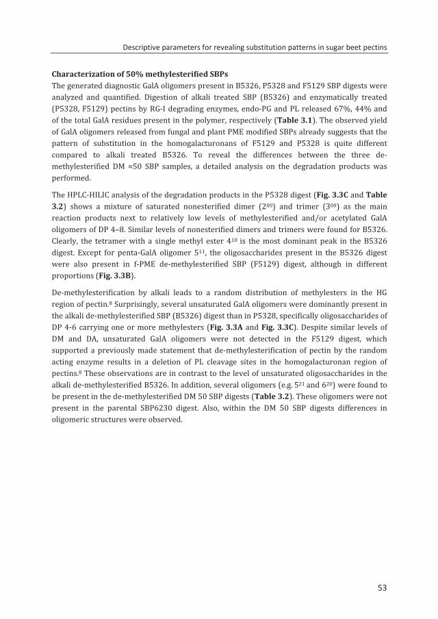
Descriptive parameters for revealing substitution patterns in sugar beet pectins
53
Characterization of 50% methylesterified SBPs The generated diagnostic GalA oligomers present in B5326, P5328 and F5129 SBP digests were analyzed and quantified. Digestion of alkali treated SBP (B5326) and enzymatically treated (P5328, F5129) pectins by RG-I degrading enzymes, endo-PG and PL released 67%, 44% and of the total GalA residues present in the polymer, respectively (Table 3.1). The observed yield of GalA oligomers released from fungal and plant PME modified SBPs already suggests that the pattern of substitution in the homogalacturonans of F5129 and P5328 is quite different compared to alkali treated B5326. To reveal the differences between the three de-methylesterified DM ≈50 SBP samples, a detailed analysis on the degradation products was performed.
The HPLC-HILIC analysis of the degradation products in the P5328 digest (Fig. 3.3C and Table 3.2) shows a mixture of saturated nonesterified dimer (200) and trimer (300) as the main reaction products next to relatively low levels of methylesterified and/or acetylated GalA oligomers of DP 4–8. Similar levels of nonesterified dimers and trimers were found for B5326. Clearly, the tetramer with a single methyl ester 410 is the most dominant peak in the B5326 digest. Except for penta-GalA oligomer 511, the oligosaccharides present in the B5326 digest were also present in f-PME de-methylesterified SBP (F5129) digest, although in different proportions (Fig. 3.3B).
De-methylesterification by alkali leads to a random distribution of methylesters in the HG region of pectin.8 Surprisingly, several unsaturated GalA oligomers were dominantly present in the alkali de-methylesterified SBP (B5326) digest than in P5328, specifically oligosaccharides of DP 4-6 carrying one or more methylesters (Fig. 3.3A and Fig. 3.3C). Despite similar levels of DM and DA, unsaturated GalA oligomers were not detected in the F5129 digest, which supported a previously made statement that de-methylesterification of pectin by the random acting enzyme results in a deletion of PL cleavage sites in the homogalacturonan region of pectins.8 These observations are in contrast to the level of unsaturated oligosaccharides in the alkali de-methylesterified B5326. In addition, several oligomers (e.g. 521 and 620) were found to be present in the de-methylesterified DM 50 SBP digests (Table 3.2). These oligomers were not present in the parental SBP6230 digest. Also, within the DM 50 SBP digests differences in oligomeric structures were observed.
53
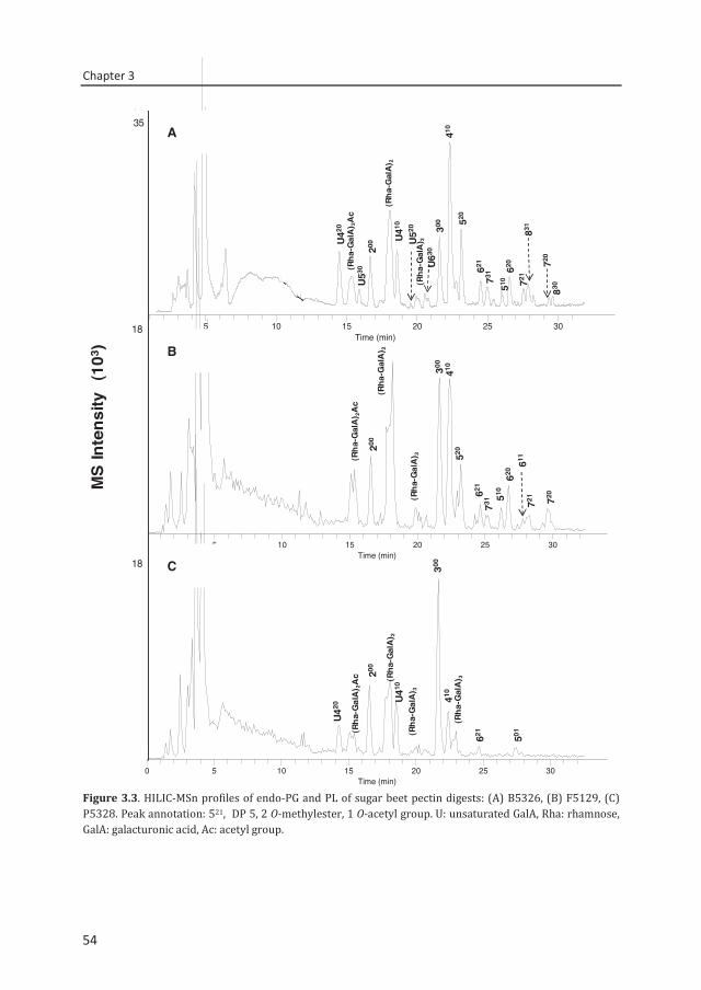
Chapter 3
54
Figure 3.3. HILIC-MSn profiles of endo-PG and PL of sugar beet pectin digests: (A) B5326, (B) F5129, (C) P5328. Peak annotation: 521, DP 5, 2 O-methylester, 1 O-acetyl group. U: unsaturated GalA, Rha: rhamnose, GalA: galacturonic acid, Ac: acetyl group.
410
0 5 10 15 20 25 30Time (min)
0
50000
100000
150000
200000
250000
300000
350000
400000
Inte
nsity
(Rha
-Gal
A) 2
Ac
200
(Rha
-Gal
A) 2
300 52
0
510
831
721
U42
0
U53
0
U41
0
U52
0
U63
0(R
ha-G
alA
) 3
621
731 62
0 720
830
0 5 10 15 20 25 30Time (min)
0
2000
4000
6000
8000
10000
12000
14000
16000
Inte
nsity
(Rha
-Gal
A) 2
Ac
200
300
520
510
721
611
410
(Rha
-Gal
A) 3
621
731
620
720
RT: 0.00 - 34.22
0 5 10 15 20 25 30Time (min)
0
2000
4000
6000
8000
10000
12000
14000
16000
18000
20000
22000
Inte
nsity
300
(Rha
-Gal
A) 2
Ac 20
0
U420
501
621
(Rha
-Gal
A) 2
410
U41
0
(Rha
-Gal
A) 3
(Rha
-Gal
A) 3
B5326
P5328
F5129
(Rha
-Gal
A) 2
MS
Inte
nsity
(1
03)
18
18
35A
B
C
54
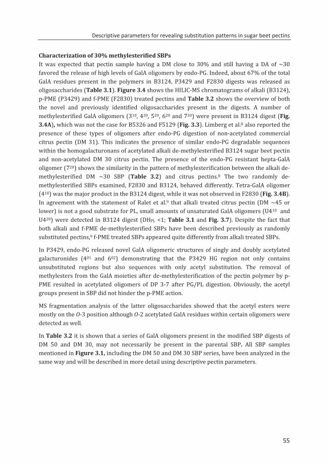
Descriptive parameters for revealing substitution patterns in sugar beet pectins
55
Characterization of 30% methylesterified SBPsIt was expected that pectin sample having a DM close to 30% and still having a DA of ~30 favored the release of high levels of GalA oligomers by endo-PG. Indeed, about 67% of the total GalA residues present in the polymers in B3124, P3429 and F2830 digests was released as oligosaccharides (Table 3.1). Figure 3.4 shows the HILIC-MS chromatograms of alkali (B3124), p-PME (P3429) and f-PME (F2830) treated pectins and Table 3.2 shows the overview of both the novel and previously identified oligosaccharides present in the digests. A number of methylesterified GalA oligomers (310, 420, 520, 620 and 720) were present in B3124 digest (Fig. 3.4A), which was not the case for B5326 and F5129 (Fig. 3.3). Limberg et al.8 also reported the presence of these types of oligomers after endo-PG digestion of non-acetylated commercial citrus pectin (DM 31). This indicates the presence of similar endo-PG degradable sequences within the homogalacturonans of acetylated alkali de-methylesterified B3124 sugar beet pectin and non-acetylated DM 30 citrus pectin. The presence of the endo-PG resistant hepta-GalA oligomer (720) shows the similarity in the pattern of methylesterification between the alkali de- methylesterified DM ~30 SBP (Table 3.2) and citrus pectins.8 The two randomly de-methylesterified SBPs examined, F2830 and B3124, behaved differently. Tetra-GalA oligomer (410) was the major product in the B3124 digest, while it was not observed in F2830 (Fig. 3.4B). In agreement with the statement of Ralet et al.9 that alkali treated citrus pectin (DM ~45 or lower) is not a good substrate for PL, small amounts of unsaturated GalA oligomers (U410 and U420) were detected in B3124 digest (DHPL <1; Table 3.1 and Fig. 3.7). Despite the fact that both alkali and f-PME de-methylesterified SBPs have been described previously as randomly substituted pectins,9 f-PME treated SBPs appeared quite differently from alkali treated SBPs.
In P3429, endo-PG released novel GalA oligomeric structures of singly and doubly acetylated galacturonides (401 and 602) demonstrating that the P3429 HG region not only contains unsubstituted regions but also sequences with only acetyl substitution. The removal of methylesters from the GalA moieties after de-methylesterification of the pectin polymer by p-PME resulted in acetylated oligomers of DP 3-7 after PG/PL digestion. Obviously, the acetyl groups present in SBP did not hinder the p-PME action.
MS fragmentation analysis of the latter oligosaccharides showed that the acetyl esters were mostly on the O-3 position although O-2 acetylated GalA residues within certain oligomers were detected as well.
In Table 3.2 it is shown that a series of GalA oligomers present in the modified SBP digests of DM 50 and DM 30, may not necessarily be present in the parental SBP. All SBP samples mentioned in Figure 3.1, including the DM 50 and DM 30 SBP series, have been analyzed in the same way and will be described in more detail using descriptive pectin parameters.
55
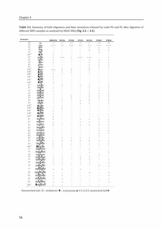
Chapter 3
56
Table 3.2. Summary of GalA oligomers and their structures released by endo-PG and PL after digestion of different SBPs samples as analyzed by HILIC-MSn (Fig. 3.2— 3.4).
Nonesterified GalA ; methylester ; acetyl group ▲ O-2, Δ O-3; unsaturated GalA
Structure SBP6230 B5326 P5328 F5129 B3124 P3429 F2830 200 + + + + + + + + + + 300 + + + + + + + + + + + + + + + + + + 301 - - - - + + + 301 - - - - + + + 301 - - - - - + - U320 + - - - - - -
410 + + + + + + + + + + + - +411 + - - - + + +411 + - - - + + +401 - - - - + + +401 - - - - + + +
U410 + + + + + - + - - U420 + + + - + - - U430 + - - - - - - U421 + - - - - - - U431 + - - - - - - U431 + - - - - - - 510 - + - + + - +511 + - - - + + +511 + - - - + + +501 - - - - + + +501 - - + - + + +501 - - + - + + +520 - + - + + - +521 - - - + + - +
U520 - + - - - - - U530 - + - - - - - U531 + - - - - - -
U541 + - - - - - - U541 + - - - - - - 611 + - - + + + + 611 + - - + + + + 611 + - - + + + + 612 - - - - - + + 601 - - - - - + + 601 - - - - - + + 602 - - - - - + - 620 - + + + + - - 621 + + + + + + + 621 + + + + + + + U630 + + - - - - - 711 - - - - + - - 712 - - - - + - + 702 - - - - - + -
50%
720 - - - + + - - 721 + + - + + - +721 + + - + + - +722 - - - - - + +
731 + + - + - - -
822 - - - - + - +831 + + - - - - +831 + + - - - - +831 + + - - - - +
U831 + - - - - - -
56
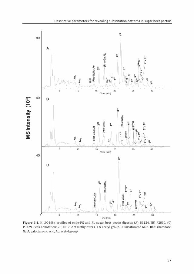
Descriptive parameters for revealing substitution patterns in sugar beet pectins
57
Figure 3.4. HILIC-MSn profiles of endo-PG and PL sugar beet pectin digests: (A) B3124, (B) F2830, (C) P3429. Peak annotation: 721, DP 7, 2 O-methylesters, 1 O-acetyl group. U: unsaturated GalA. Rha: rhamnose, GalA, galacturonic acid, Ac: acetyl group.
0.00 - 35.00 SM: 3B
0 5 10 15 20 25 30Time (min)
2000
4000
6000
8000
10000
12000
14000
16000
520200
(Rha
-Gal
A)2
300
411
401/5
11
U410
510 611 /
721
(Rha
-Gal
A)2A
c 501/ 6
11
720
711
712 /
822
U420Ar
a 7Ar
a 8
301 411
410
511
621 620
0 5 10 15 20 25 30Time (min)
5000
10000
15000
20000
25000
30000
35000
40000
601
200
(Rha
-Gal
A)2
301411
401/5
11
410
611/ 7
21
(Rha
-Gal
A)2A
c
501/ 6
11
621
712/ 8
22
Ara 7
Ara 8
300
(Rha
-Gal
A)3
612/ 7
22
831
B3124
F2830
0.00 - 35.00 SM: 3B
0 5 10 15 20 25 30Time (min)
601
200
(Rha
-Gal
A)2
301 411
401/5
11
721
(Rha
-Gal
A)2A
c
501/ 6
11621
Ara 7
Ara 8
300
(Rha
-Gal
A)3
612/ 7
22
511
602
702
P3429
MS
Inte
nsity
(10
3 )M
S In
tens
ity (1
03 )
40
40
80
A
B
C
57
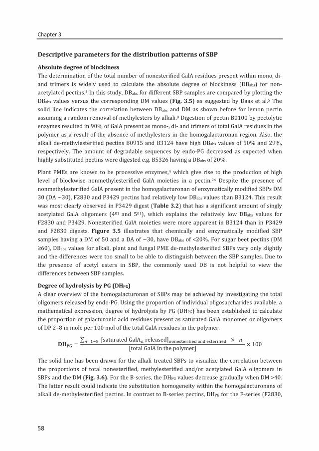
Chapter 3
58
Descriptive parameters for the distribution patterns of SBP
Absolute degree of blockiness The determination of the total number of nonesterified GalA residues present within mono, di- and trimers is widely used to calculate the absolute degree of blockiness (DBabs) for non-acetylated pectins.4 In this study, DBabs for different SBP samples are compared by plotting the DBabs values versus the corresponding DM values (Fig. 3.5) as suggested by Daas et al.5 The solid line indicates the correlation between DBabs and DM as shown before for lemon pectin assuming a random removal of methylesters by alkali.8 Digestion of pectin B0100 by pectolytic enzymes resulted in 90% of GalA present as mono-, di- and trimers of total GalA residues in the polymer as a result of the absence of methylesters in the homogalacturonan region. Also, the alkali de-methylesterified pectins B0915 and B3124 have high DBabs values of 50% and 29%, respectively. The amount of degradable sequences by endo-PG decreased as expected when highly substituted pectins were digested e.g. B5326 having a DBabs of 20%.
Plant PMEs are known to be processive enzymes,4 which give rise to the production of high level of blockwise nonmethylesterified GalA moieties in a pectin.24 Despite the presence of nonmethylesterified GalA present in the homogalacturonan of enzymatically modified SBPs DM 30 (DA ~30), F2830 and P3429 pectins had relatively low DBabs values than B3124. This result was most clearly observed in P3429 digest (Table 3.2) that has a significant amount of singly acetylated GalA oligomers (401 and 501), which explains the relatively low DBabs values for F2830 and P3429. Nonesterified GalA moieties were more apparent in B3124 than in P3429 and F2830 digests. Figure 3.5 illustrates that chemically and enzymatically modified SBP samples having a DM of 50 and a DA of ~30, have DBabs of <20%. For sugar beet pectins (DM ≥60), DBabs values for alkali, plant and fungal PME de-methylesterified SBPs vary only slightly and the differences were too small to be able to distinguish between the SBP samples. Due to the presence of acetyl esters in SBP, the commonly used DB is not helpful to view the differences between SBP samples.
Degree of hydrolysis by PG (DHPG) A clear overview of the homogalacturonan of SBPs may be achieved by investigating the total oligomers released by endo-PG. Using the proportion of individual oligosaccharides available, a mathematical expression, degree of hydrolysis by PG (DHPG) has been established to calculate the proportion of galacturonic acid residues present as saturated GalA monomer or oligomers of DP 2–8 in mole per 100 mol of the total GalA residues in the polymer.
=∑ [saturatedGalAreleased] ×
[totalGalAinthepolymer] × 100
The solid line has been drawn for the alkali treated SBPs to visualize the correlation between the proportions of total nonesterified, methylesterified and/or acetylated GalA oligomers in SBPs and the DM (Fig. 3.6). For the B-series, the DHPG values decrease gradually when DM >40. The latter result could indicate the substitution homogeneity within the homogalacturonans of alkali de-methylesterified pectins. In contrast to B-series pectins, DHPG for the F-series (F2830,
58
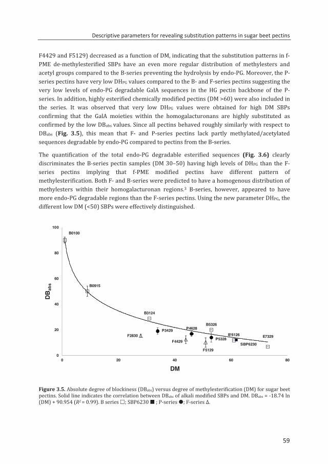
Descriptive parameters for revealing substitution patterns in sugar beet pectins
59
F4429 and F5129) decreased as a function of DM, indicating that the substitution patterns in f-PME de-methylesterified SBPs have an even more regular distribution of methylesters and acetyl groups compared to the B-series preventing the hydrolysis by endo-PG. Moreover, the P-series pectins have very low DHPG values compared to the B- and F-series pectins suggesting the very low levels of endo-PG degradable GalA sequences in the HG pectin backbone of the P-series. In addition, highly esterified chemically modified pectins (DM >60) were also included in the series. It was observed that very low DHPG values were obtained for high DM SBPs confirming that the GalA moieties within the homogalacturonans are highly substituted as confirmed by the low DBabs values. Since all pectins behaved roughly similarly with respect to DBabs (Fig. 3.5), this mean that F- and P-series pectins lack partly methylated/acetylated sequences degradable by endo-PG compared to pectins from the B-series.
The quantification of the total endo-PG degradable esterified sequences (Fig. 3.6) clearly discriminates the B-series pectin samples (DM 30–50) having high levels of DHPG than the F-series pectins implying that f-PME modified pectins have different pattern of methylesterification. Both F- and B-series were predicted to have a homogenous distribution of methylesters within their homogalacturonan regions.3 B-series, however, appeared to have more endo-PG degradable regions than the F-series pectins. Using the new parameter DHPG, the different low DM (<50) SBPs were effectively distinguished.
Figure 3.5. Absolute degree of blockiness (DBabs) versus degree of methylesterification (DM) for sugar beet pectins. Solid line indicates the correlation between DBabs of alkali modified SBPs and DM. DBabs = -18.74 ln (DM) + 90.954 (R2 = 0.99). B series ; SBP6230 ; P-series ; F-series Δ.
E7329B'6126
B5326
B3124
B0915
B0100
F5129
P4628P3429
P5328F4429F2830
SBP6230
0
20
40
60
80
100
0 20 40 60 80
DBab
s
DM
59
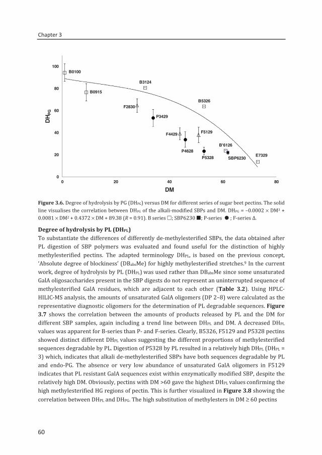
Chapter 3
60
Figure 3.6. Degree of hydrolysis by PG (DHPG) versus DM for different series of sugar beet pectins. The solid line visualises the correlation between DHPG of the alkali-modified SBPs and DM. DHPG = –0.0002 × DM3 + 0.0081 × DM2 + 0.4372 × DM + 89.38 (R = 0.91). B series ; SBP6230 ; P-series ; F-series Δ.
Degree of hydrolysis by PL (DHPL) To substantiate the differences of differently de-methylesterified SBPs, the data obtained after PL digestion of SBP polymers was evaluated and found useful for the distinction of highly methylesterified pectins. The adapted terminology DHPL, is based on the previous concept, ‘Absolute degree of blockiness’ (DBabsMe) for highly methylesterified stretches.9 In the current work, degree of hydrolysis by PL (DHPL) was used rather than DBabsMe since some unsaturated GalA oligosaccharides present in the SBP digests do not represent an uninterrupted sequence of methylesterified GalA residues, which are adjacent to each other (Table 3.2). Using HPLC-HILIC-MS analysis, the amounts of unsaturated GalA oligomers (DP 2–8) were calculated as the representative diagnostic oligomers for the determination of PL degradable sequences. Figure 3.7 shows the correlation between the amounts of products released by PL and the DM for different SBP samples, again including a trend line between DHPL and DM. A decreased DHPL values was apparent for B-series than P- and F-series. Clearly, B5326, F5129 and P5328 pectins showed distinct different DHPL values suggesting the different proportions of methylesterified sequences degradable by PL. Digestion of P5328 by PL resulted in a relatively high DHPL (DHPL = 3) which, indicates that alkali de-methylesterified SBPs have both sequences degradable by PL and endo-PG. The absence or very low abundance of unsaturated GalA oligomers in F5129 indicates that PL resistant GalA sequences exist within enzymatically modified SBP, despite the relatively high DM. Obviously, pectins with DM >60 gave the highest DHPL values confirming the high methylesterified HG regions of pectin. This is further visualized in Figure 3.8 showing the correlation between DHPL and DHPG. The high substitution of methylesters in DM ≥ 60 pectins
E7329
B'6126
B5326
B3124
B0915
B0100
F5129
P4628
P3429
P5328
F4429
F2830
SBP6230
0
20
40
60
80
100
0 20 40 60 80
DHPG
DM
60
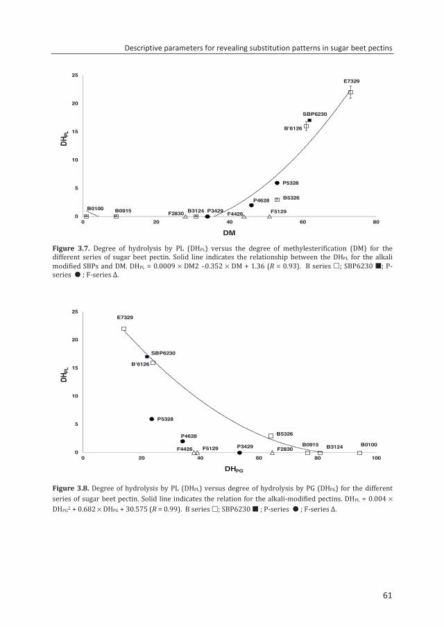
Descriptive parameters for revealing substitution patterns in sugar beet pectins
61
Figure 3.7. Degree of hydrolysis by PL (DHPL) versus the degree of methylesterification (DM) for the different series of sugar beet pectin. Solid line indicates the relationship between the DHPL for the alkali modified SBPs and DM. DHPL = 0.0009 × DM2 –0.352 × DM + 1.36 (R = 0.93). B series ; SBP6230 ; P-series ; F-series Δ.
Figure 3.8. Degree of hydrolysis by PL (DHPL) versus degree of hydrolysis by PG (DHPG) for the different series of sugar beet pectin. Solid line indicates the relation for the alkali-modified pectins. DHPL = 0.004 × DHPG2 + 0.682 × DHPG + 30.575 (R = 0.99). B series ; SBP6230 ; P-series ; F-series Δ.
E7329
B'6126
B5326
B3124B0915B0100
P5328
P4628
P3429 F5129F4426F2830
SBP6230
0
5
10
15
20
25
0 20 40 60 80
DHPL
DM
E7329
B'6126
B5326
B3124B0915 B0100
P5328
P4628
P3429F5129F4426 F2830
SBP6230
0
5
10
15
20
25
0 20 40 60 80 100
DHPL
DHPG
61
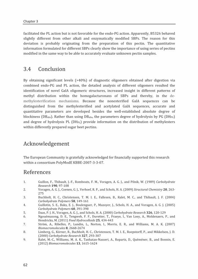
Chapter 3
62
facilitated the PL action but is not favorable for the endo-PG action. Apparently, B5326 behaved slightly different from other alkali and enzymatically modified SBPs. The reason for this deviation is probably originating from the preparation of this pectin. The quantitative information formulated for different SBPs clearly show the importance of using series of pectins modified in the same way to be able to accurately evaluate unknown pectin samples.
3.4 Conclusion
By obtaining significant levels (>40%) of diagnostic oligomers obtained after digestion via combined endo-PG and PL action, the detailed analysis of different oligomers resulted the identification of novel GalA oligomeric structures, increased insight in different patterns of methyl distribution within the homogalacturonans of SBPs and thereby, in the de-methylesterification mechanisms. Because the nonesterified GalA sequences can be distinguished from the methylesterified and acetylated GalA sequences, accurate and quantitative parameters are developed besides the well-established absolute degree of blockiness (DBabs). Rather than using DBabs, the parameters degree of hydrolysis by PG (DHPG) and degree of hydrolysis PL (DHPL) provide information on the distribution of methylesters within differently prepared sugar beet pectins.
Acknowledgement
The European Community is gratefully acknowledged for financially supported this research within a consortium PolyModE KBBE-2007-3-3-07.
References 1. Guillon, F., Thibault, J.-F., Rombouts, F. M., Voragen, A. G. J., and Pilnik, W. (1989) Carbohydrate
Research 190, 97-108 2. Voragen, A. G. J., Coenen, G. J., Verhoef, R. P., and Schols, H. A. (2009) Structural Chemistry 20, 263-
275 3. Buchholt, H. C., Christensen, T. M. I. E., Fallesen, B., Ralet, M. C., and Thibault, J. F. (2004)
Carbohydrate Polymers 58, 149-161 4. Guillotin, S. E., Bakx, E. J., Boulenguer, P., Mazoyer, J., Schols, H. A., and Voragen, A. G. J. (2005)
Carbohydrate Polymers 60, 391-398 5. Daas, P. J. H., Voragen, A. G. J., and Schols, H. A. (2000) Carbohydrate Research 326, 120-129 6. Ngouémazong, D. E., Tengweh, F. F., Duvetter, T., Fraeye, I., Van Loey, A., Moldenaers, P., and
Hendrickx, M. (2011) Food Hydrocolloids 25, 434-443 7. Ström, A., Ribelles, P., Lundin, L., Norton, I., Morris, E. R., and Williams, M. A. K. (2007)
Biomacromolecules 8, 2668-2674 8. Limberg, G., Körner, R., Buchholt, H. C., Christensen, T. M. I. E., Roepstorff, P., and Mikkelsen, J. D.
(2000) Carbohydrate Research 327, 293-307 9. Ralet, M.-C., Williams, M. A. K., Tanhatan-Nasseri, A., Ropartz, D., Quéméner, B., and Bonnin, E.
(2012) Biomacromolecules 13, 1615-1624
62
Descriptive parameters for revealing substitution patterns in sugar beet pectins
63
10. Thakur, B. R., Singh, R. K., Handa, A. K., and Rao, M. A. (1997) Critical Reviews in Food Science and Nutrition 37, 47-73
11. Ralet, M.-C., Dronnet, V., Buchholt, H. C., and Thibault, J.-F. (2001) Carbohydrate Research 336, 117-125
12. Schols, H. A., Posthumus, M. A., and Voragen, A. G. J. (1990) Carbohydrate Research 206, 117-129 13. Beldman, G., Searle-van Leeuwen, M. J. F., De Ruiter, G. A., Siliha, H. A., and Voragen, A. G. J. (1993)
Carbohydrate Polymers 20, 159-168 14. Kühnel, S., Hinz, S. W. A., Pouvreau, L., Wery, J., Schols, H. A., and Gruppen, H. (2010) Bioresource
Technology 101, 8300-8307 15. Mutter, M., Renard, C. M. G. C., Beldman, G., Schols, H. A., and Voragen, A. G. J. (1998) Carbohydrate
Research 311, 155-164 16. Limberg, G., Körner, R., Buchholt, H. C., Christensen, T. M. I. E., Roepstorff, P., and Mikkelsen, J. D.
(2000) Carbohydrate Research 327, 321-332 17. Remoroza, C., Cord-Landwehr, S., Leijdekkers, A. G. M., Moerschbacher, B. M., Schols, H. A., and
Gruppen, H. (2012) Carbohydrate Polymers 90, 41-48 18. Quéméner, B., Cabrera Pino, J. C., Ralet, M.-C., Bonnin, E., and Thibault, J.-F. (2003) Journal of Mass
Spectrometry 38, 641-648 19. Perrone, P., Hewage, C. M., Thomson, A. R., Bailey, K., Sadler, I. H., and Fry, S. C. (2002)
Phytochemistry 60, 67-77 20. Ralet, M.-C., Crépeau, M.-J., and Bonnin, E. (2008) Phytochemistry 69, 1903-1909 21. Needs, P. W., Rigby, N. M., Colquhoun, I. J., and Ring, S. G. (1998) Phytochemistry 48, 71-77 22. Körner, R., Limberg, G., Mikkelsen, J. D., and Roepstorff, P. (1998) Journal of Mass Spectrometry
33, 836-842 23. Ralet, M. C., Cabrera, J. C., Bonnin, E., Quémenér, B., Hellín, P., and Thibault, J. F. (2005)
Phytochemistry 66, 1832-1843 24. Micheli, F. (2001) Trends in Plant Science 6, 414-419
63

64
Chapter 4
Two-step enzymatic fingerprinting of
sugar beet pectin
C. Remoroza, S. Broxterman, H. Gruppen, H.A. Schols
Accepted for publication in Carbohydrate Polymers
Chapter 4
66
Abstract
A two-step enzymatic fingerprinting method was introduced to analyze a highly methylesterified and acetylated sugar beet pectin having a degree of methylesterification (DM) of 62 and acetylation of 30. A cocktail of pectolytic enzymes, including endo-polygalacturonase (endo-PG) and pectin lyase (PL), was used for the first digestion. The endo-PG and PL resistant pectin fragments were isolated and subjected to a second digestion using fungal pectin methylesterase and endo-PG. After the two sequential digestions, 78% of the total GalA residues present in the parental pectin were recovered as mono- and oligomers, sufficient to quantitatively describe the parental SBP. For this reason, the descriptive parameters degree of blockiness (DBabs), degree of hydrolysis by PG (DHPG) and degree of hydrolysis by PL (DHPL) were established for both digestions. The first digestion revealed the presence of short blocks of nonesterified GalA residues and blocks of partly methylesterified and acetylated GalA residues in the parental SBP, in addition to blocks of highly methylesterified and acetylated GalA residues. The second digestion revealed the presence of blocks of methylesterified, partly methylesterified and/or acetylated GalA residues in a sequence not to be degradable by neither endo-PG nor by PL. The acetyl groups were present in a blockwise manner. Application of the method to two differently prepared DM 50 SBPs showed that the two pectins differ in the ratio of blocks of nonesterified and blocks of partly methylesterified and acetylated GalA residues.
66
Two-step enzymatic fingerprinting of sugar beet pectin
67
4.1. Introduction
Pectin isolated from apple pomace and lemon peel is widely used in the food industry as gelling, viscosifying, and stabilizing agent in low pH applications, e.g. fruit preparations, fruit gels, acid beverages, acidified milk-based products, and some types of confectionery. Sugar beet is considered as a potential source of pectin. Sugar beet pectin (SBP) essentially consists of homogalacturonan (HG) and type I rhamnogalacturonan (RG-I) regions.1 About 90% of the galacturonic acid (GalA) residues are present in the HG fragment of SBP and the remaining 10% GalA residues are present within the RG-I structural elements.2 RG-I is constructed of repeating units of α-(1→2)-linked rhamnosyl and α-(1→4)-linked GalA residues, in which the rhamnosyl unit can be substituted with neutral sugar side chains.3 The GalA unit in both RG-I and HG can be acetylated at positions O-2 and/or O-3.4 The level and distribution of esters have important commercial implications because of their effects on the functionality of the pectin.5 Acid extracted commercial SBP may have a degree of acetylation (DA) of 30 and degree of methylesterification (DM) of 62.6
Revealing the ester distribution patterns in SBP is complex due to the fact that the HG is highly decorated with both methylesters and acetyl groups. An enzymatic fingerprinting method has been developed7 for the elucidation of the distribution pattern of acetyl groups in SBP. Although the methylester distribution is highly important as well with respect to functionality of the pectin, it was not addressed in that study. Recently, the simultaneous use of endo-polygalacturonase (endo-PG) and pectin lyase (PL) to degrade a highly methylesterified SBP (SBP6230) and the subsequent analysis of the digest using HPLC-HILIC coupled to online ELSD/MSn was reported.8 This enzymatic degradation of SBP6230 resulted in 40% degradation of the HG region based on the amounts of GalA residues present in monomer and nonesterified, partially methylesterified/acetylated saturated and unsaturated GalA oligomers released. Quantification of these oligosaccharides was used for the determination of the pectin descriptive parameters absolute degree of blockiness (DBabs), degree of hydrolysis by PG (DHPG) and degree of hydrolysis by PL (DHPL).8 These parameters were used to describe the so-called blocks of 1) nonesterified, 2) partly methylesterified, acetylated and 3) highly substituted GalA residues in a pectin by the known modes of action of endo-PG and PL. However, still the non-degradable part of the SBP, representing about 60% of the HG and being endo-PG and PL resistant, was not included in the characterization.
In the present study, a two-step enzymatic fingerprinting approach is introduced to overcome the above mentioned drawback, making it possible to elucidate the distribution patterns in highly methylesterified and acetylated pectins. In the first digestion, RG-I degrading enzymes, endo-PG and PL are used to degrade the pectin, followed by the separation of the high molecular weight (Mw) fragments and the low Mw GalA mono- and oligomers. Subsequently, an enzyme mixture containing
67
Chapter 4
68
fungal pectin methylesterase (f-PME) and endo-PG is used to further digest the high Mw pectin fragments. All diagnostic GalA oligomers generated were analyzed by HPLC-MS/ELSD. Using the pectin parameters the methylester and acetyl distribution over the SBP backbone is described.
4.2. Materials and Methods
Pectin samples
Commercially extracted SBPs, SBP6230 (DM 62, DA 30), P5328 and F5129 were provided by Danisco (Brabrand, Denmark). The chemical characteristics are described in Table 4.1.
Table 4.1. Chemical characteristics of pectin samples used in this study
Mother pectin De-esterification Pectin GalA Rha Ara Gal DMa DAa method % (w/w) (%) (%) SBP6230 SBP6230 59 5 12 10 62 30 p-PME P5328 58 5 11 10 53 28 f-PME F5129 59 5 12 9 51 29 aMoles methanol (DM) or acetic acid (DA) per 100 moles of galacturonic acid
Enzymatic hydrolysis
Purified and well characterized enzymes, Aspergillus niger pectin methylesterase (f-PME) (EC 3.1.1.11),9 HG degrading enzymes, pectin lyase (EC 4.2.2.10)10 and endo-PGII (EC 3.2.1.15)11 and RG-I degrading enzymes8 were used to hydrolyze SBP. Figure 4.1 shows the schematic diagram of the two-step enzymatic fingerprinting approach for the SBPs.
First digestion SBP (10 mg / 2 ml) in 50 mM sodium citrate buffer (pH 5.0) was digested at 40 °C for 24 h by RG-I (endo-galactanase + endo/exo arabinase + RG hydrolase) and HG (endo-PGII + PL) degrading enzymes as described elsewhere.8 The reaction was stopped by heating at 100 °C for 6 min. The total digest after the first digestion was freeze-dried.
Second digestion The degradation products obtained after the first digestion of SBP were fractionated into high Mw fragments and low Mw fragments by size exclusion chromatography (see below). For the second digestion, the high Mw material was dissolved in 2 ml 50 mM sodium acetate buffer (pH 5.0) and digested using Aspergillus niger f-PME (20 U/ml) and endo-PGII (10 U/ml) at 40 °C for 24 h.
68
Two-step enzymatic fingerprinting of sugar beet pectin
69
Inactivation of enzymes was performed by heating at 100 °C for 6 min. This digest was denoted as pool I digest.
Freeze-dried pectin digest (10 mg pectin) after the first enzymatic digestion was dissolved into 100 µl 50 mM sodium citrate buffer (pH 5.0) and applied onto a PD-10 column with packed bed size of 1.45 x 5.0 cm (8.3 ml) containing Sephadex G-25 Medium (GE Healthcare Bio-sciences Uppsala, Sweden). The PD-10 column was equilibrated with 25 ml of 50 mM sodium citrate buffer (pH 5.0) at room temperature. Subsequently, the digest was eluted with 4.90 ml of 50 mM sodium citrate buffer (pH 5.0). The fractionation allows the separation of high Mw fragments from low Mw fragments. Fractions (0.5 ml) were collected and analyzed by HPSEC, followed by pooling on the basis of the Mw of the fragments. Two pools were obtained; Pool I (fractions 1-5; high Mw material) and pool II (fractions 6-10; low Mw material) and both pools were freeze-dried. Subsequently, the freeze-dried high Mw pectin pool I was used for the second digestion (see above).
High performance size exclusion chromatography (HPSEC)
SBP digests were analyzed using an Ultimate 3000 system (Dionex, Sunnyvale, CA, USA). A set of four TSK-Gel super AW columns (Tosoh Bioscience, Tokyo, Japan) was used in series: one guard column (6 mm ID x 40 mm) and the columns 4000, 3000 and 2500 (6 mm x 150 mm) as described previously.12
High performance anion exchange chromatography (HPAEC)
The quantification of monomeric GalA was performed by HPAEC. The SBP digests were diluted with Millipore water to 1 mg/ml and the analysis was performed on ICS5000 HPAEC system (Dionex) with pulsed amperometric and UV detection (HPAEC-PAD/UV) (Dionex) as described elsewhere.8 Galacturonic acid (Sigma-Aldrich, Steinheim, Germany) was used as standard.
69
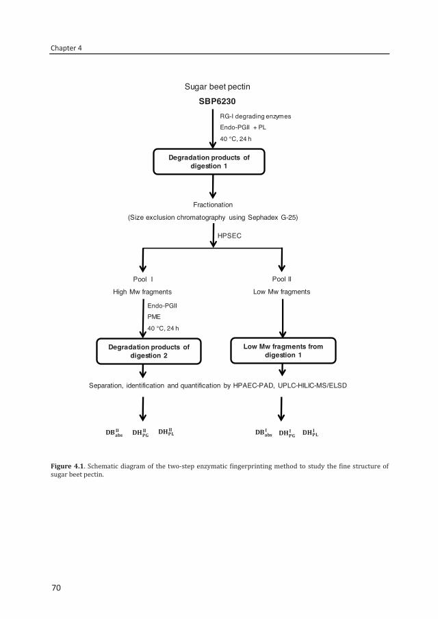
Chapter 4
70
Figure 4.1. Schematic diagram of the two-step enzymatic fingerprinting method to study the fine structure of sugar beet pectin.
Sugar beet pectin SBP6230
RG-I degrading enzymesEndo-PGII + PL
40 °C, 24 h
Separation, identification and quantification by HPAEC-PAD, UPLC-HILIC-MS/ELSD
Endo-PGIIPME
40 °C, 24 h
Pool I
High Mw fragments
Pool II
Low Mw fragments
Fractionation
(Size exclusion chromatography using Sephadex G-25)
Degradation products of digestion 1
Low Mw fragments from digestion 1
Degradation products of digestion 2
HPSEC
70
Two-step enzymatic fingerprinting of sugar beet pectin
71
Hydrophilic interaction liquid chromatography (HILIC-ELSD/MSn)
SBP digests, diluted to 1 mg/ml in 50% (v/v) acetonitrile, were analyzed using an Accela HPLC system (Thermo Scientific, Waltham, MA, USA) coupled to an evaporative light scattering detector (Agilent 1200 series, Gen Tech Scientific, Arcade, NY, USA) and an ESI-IT-MSn-detector (LTQ Velos Pro ion trap MS, Thermo Scientific, San Jose, CA, USA). Chromatographic separation was performed on an Acquity UPLC BEH Amide column as described previously.8
Determination of DM
Pectin samples (≈2mg/ml) were saponified in 100 mM NaOH for 1 hr at room temperature to determine the degree of methylesterification (DM) using the colorimetric method as previously described.13
Determination of the absolute degree of blockiness
The amounts of monomer in the digests of pools I and II, as determined by HPAEC-PAD, and the amounts of nonesterified dimer, and trimer, as determined by HILIC-ELSD, were used to calculate the absolute degree of blockiness (
, ).14 DBabs is defined as moles of galacturonic acid residues present as nonesterified mono-,di- and trimer generated by endo-PGII per 100 moles of total GalA residues in the polymer. The numeral in the formula refers to the results first or second digestion of the pectin.
, =
∑ [saturatedGalAreleased] × [totalGalAinthepolymer] × 100
Degree of hydrolysis by PG (DHPG)
Using the amounts of individual oligosaccharides present after digestion of pool I and II by endo-PGII and PL, the degree of hydrolysis by PG, (
, ) was calculated as the number of moles of galacturonic acid residues present in the digest as saturated GalA monomer and oligomers of DP 2-8 per 100 moles of the total GalA residues in the polymer.8 The numeral in the formula refers to the first and second digestion of the pectin.
, =
∑ [saturatedGalAreleased] × [totalGalAinthepolymer] × 100
71
Chapter 4
72
Determination of the degree of hydrolysis by PL (DHPL)
All unsaturated GalA residues present in unsaturated oligomers (DP 2-8), both methylesterified and/or acetylated released by PL action from SBP, were quantified and expressed as degree of hydrolysis by PL (
,).8 The numeral in the formula refers to the first or second digestion of the pectin.
, =
∑ [unsaturatedGalAreleased] × [totalGalAinthepolymer] × 100
4.3. Results and discussion
Enzymatic digestion of SBPs
Using endo-PG and PL, SBP6230, P5328 and F5129 (DM 51-62 and DA ~30) were partially degraded into mono- and oligosaccharides representing 36%, 34% and 38% of the total GalA residues in the samples, respectively (Table 4.3). These yields were quite comparable with our previous results.8 For each SBP, the remaining not-sufficiently degraded fragments were isolated and subsequently subjected to a second digestion in order to include a larger part of the pectin for modelling its structure on the basis of the type and level of oligosaccharides released. Although all three SBPs in this study were analyzed in this way, the approach will be described in detail only for SBP6230 before discussing the outcome of the other SBPs.
First digestion and isolation of endo-PG and PL resistant fractions Figure 4.2A shows the molecular size distributions of SBP6230 before and after the first digestion. When digested, the average molecular mass of the SBP shifts from 100 kDa to lower than 3 kDa values although still material of ~43 kDa is present.
To analyze the high Mw fragments resistant to endo-PG and PL in detail, a fractionation of the total first digest was carried out using size exclusion chromatography. Ten fractions were collected and two pools (I and II) were made based on the HPSEC analysis of all fractions (supplementary data, Fig. 4.S1). Pool I contains the high Mw fractions eluting at retention times < 11.5 min, whereas pool II consists of the low Mw fractions eluting at retention times > 11.5 min (≤ 3 kDa) (Fig. 4.2B). The high signal at 13 min is typical for salt present in the buffer solution.
72
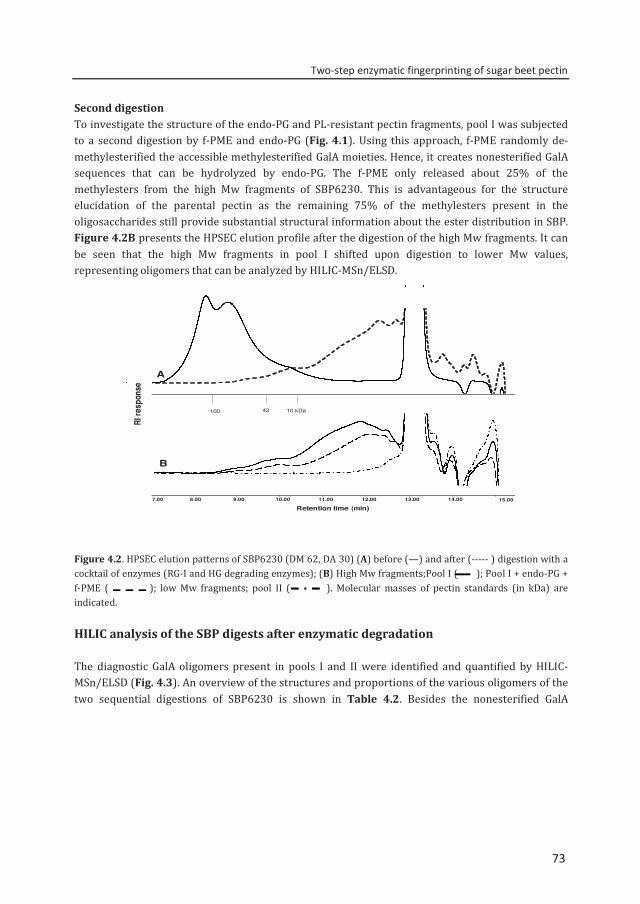
Two-step enzymatic fingerprinting of sugar beet pectin
73
Second digestion To investigate the structure of the endo-PG and PL-resistant pectin fragments, pool I was subjected to a second digestion by f-PME and endo-PG (Fig. 4.1). Using this approach, f-PME randomly de-methylesterified the accessible methylesterified GalA moieties. Hence, it creates nonesterified GalA sequences that can be hydrolyzed by endo-PG. The f-PME only released about 25% of the methylesters from the high Mw fragments of SBP6230. This is advantageous for the structure elucidation of the parental pectin as the remaining 75% of the methylesters present in the oligosaccharides still provide substantial structural information about the ester distribution in SBP. Figure 4.2B presents the HPSEC elution profile after the digestion of the high Mw fragments. It can be seen that the high Mw fragments in pool I shifted upon digestion to lower Mw values, representing oligomers that can be analyzed by HILIC-MSn/ELSD.
Figure 4.2. HPSEC elution patterns of SBP6230 (DM 62, DA 30) (A) before (—) and after (----- ) digestion with a cocktail of enzymes (RG-I and HG degrading enzymes); (B) High Mw fragments;Pool I ( ); Pool I + endo-PG + f-PME ( ); low Mw fragments; pool II ( ). Molecular masses of pectin standards (in kDa) are indicated.
HILIC analysis of the SBP digests after enzymatic degradation
The diagnostic GalA oligomers present in pools I and II were identified and quantified by HILIC-MSn/ELSD (Fig. 4.3). An overview of the structures and proportions of the various oligomers of the two sequential digestions of SBP6230 is shown in Table 4.2. Besides the nonesterified GalA
7.00 8.00 9.00 10.00 11.00 12.00 13.00 14.00
Retention time (min)15.00
100 43 10 kDa
A
B
RI re
spon
se
73
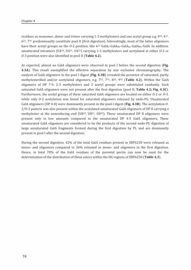
Chapter 4
74
residues as monomer, dimer and trimer carrying 1-3 methylesters and one acetyl group e.g. 410, 411, 621, 731 predominantly constitute pool II (first digestion). Interestingly, most of the latter oligomers have their acetyl groups on the O-2 position, like 621 GalA2-GalAMe-GalAAc-GalAMe-GalA. In addition, unsaturated tetramers (U420, U421, U431) carrying 1-3 methylesters and acetylated at either O-2 or O-3 position were also identified in pool II (Table 4.2).
As expected, almost no GalA oligomers were observed in pool I before the second digestion (Fig. 4.3A). This result exemplified the effective separation by size exclusion chromatography. The analysis of GalA oligomers in the pool I digest (Fig. 4.3B) revealed the presence of saturated, partly methylesterified and/or acetylated oligomers, e.g. 722, 732, 822, 932 (Table 4.2). Within the GalA oligomers of DP 7-9, 2-3 methylesters and 2 acetyl groups were substituted randomly. Such saturated GalA oligomers were not present after the first digestion (pool II, Table 4.2; Fig. 4.3C). Furthermore, the acetyl groups of these saturated GalA oligomers are located on either O-2 or O-3, while only O-2 acetylation was found for saturated oligomers released by endo-PG. Unsaturated GalA oligomers (DP 4-8) were dominantly present in the pool I digest (Fig. 4.3B). The acetylation O-2/O-3 pattern was also present within the acetylated unsaturated GalA oligomers of DP 8 carrying a methylester at the nonreducing end (U822, U831, U832). These unsaturated DP 8 oligomers were present only in low amounts compared to the unsaturated DP 4-5 GalA oligomers. These unsaturated GalA oligomers are considered to be the products of the second endo-PG digestion of large unsaturated GalA fragments formed during the first digestion by PL and are dominantly present in pool I after the second digestion.
During the second digestion, 42% of the total GalA residues present in SBP6230 were released as mono- and oligomers compared to 36% released as mono- and oligomers in the first digestion. Hence, in total 78% of the GalA residues of the parental pectin can now be used for the determination of the distribution of these esters within the HG regions of SBP6230 (Table 4.3).
74
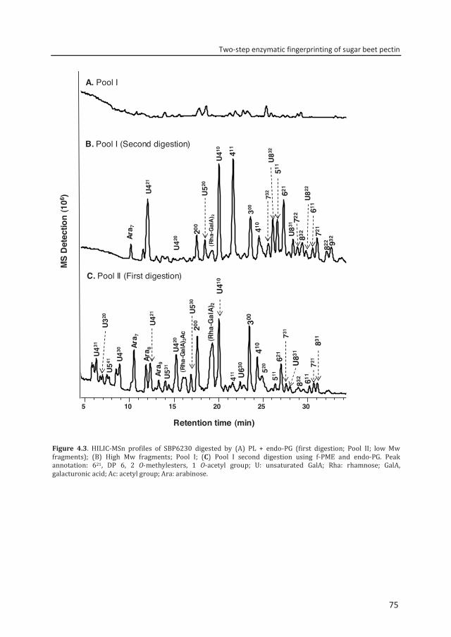
Two-step enzymatic fingerprinting of sugar beet pectin
75
Figure 4.3. HILIC-MSn profiles of SBP6230 digested by (A) PL + endo-PG (first digestion; Pool II; low Mw fragments); (B) High Mw fragments; Pool I; (C) Pool I second digestion using f-PME and endo-PG. Peak annotation: 621, DP 6, 2 O-methylesters, 1 O-acetyl group; U: unsaturated GalA; Rha: rhamnose; GalA, galacturonic acid; Ac: acetyl group; Ara: arabinose.
5 10 15 20 25 30
Retention time (min)
U421
Ara 7
U41
0
200 410
U52
0
411
300
732
U83
2
511
U831
621
721
(Rha
-Gal
A) 2
832
822
Ara 7
U421
U420
200
(Rha
-Gal
A)2
U41
0
300
410
(Rha
-Gal
A)2A
cU5
30
411 U63
0 621
511
731
U431
U541
U320
U430
Ara 8
U531 52
0 U83
1
611
721
831
832
722
U822
611
932
U42
0
Ara 9
MS
Det
ectio
n (1
06 )
C. Pool II (First digestion)
B. Pool I (Second digestion)
A. Pool I
75
Chapter 4
76
Table 4.2. Amount and structure of different GalA oligomers released by endo-PG and pectin lyase after two sequential digestions of sugar beet pectins as analyzed by HILIC-ELSD/ESI-MSn and HPAEC-PAD.
Nonesterified GalA ; methylester ; acetyl group ▲ O-2, Δ O-3; unsaturated GalA aDigestion I: Oligomers (low Mw) generated after the first digestion. bDigestion II: Oligosaccharides released from high Mw fragment after the second digestion. The values indicate the moles of nonesterified and (un)saturated galacturonic residues present as saturated and unsaturated oligomers per 100 moles of the total GalA in the sample. + Traces; - not detected
76
Two-step enzymatic fingerprinting of sugar beet pectin
77
Continuation of Table 4.2
Nonesterified GalA ; methylester ; acetyl group ▲ O-2, Δ O-3; unsaturated GalA aDigestion I: Oligomers (low Mw) generated after the first digestion. bDigestion II: Oligosaccharides released from high Mw fragment after the second digestion. The values indicate the moles of nonesterified and (un)saturated galacturonic residues present as saturated and unsaturated oligomers per 100 moles of the total GalA in the sample. + Traces; - not detected
Structure SBP6230 P5328 F5129 Digestion Ia IIb I II I II
U541 + - - - - -
U541 + - - - - -
611 + + + + - +
611 + + + + - +
611 + + + + - -
DP 6 620 - - + - 2.72 -
621 + 1.02 4.29 3.76 6.38 5.58
621 + + + + + +
U630 + - - - - -
712 - - - - - +
DP 7 721 + + + + 2.87 +
721 + + + + + +
722 - + - + - -
731 + - + - + +
732 - + - - - -
DP 8 822 - + - + - +
831 + - + - 8.31 +
831 + - + - + +
831 + - + - + +
832 + + - - - -
U822 - + - - - -
U822 - + - - - -
U831 - + - - - -
U832 - 1.40 - - - -
U832 - + - - - -
932 - + - - - - DP 9
77
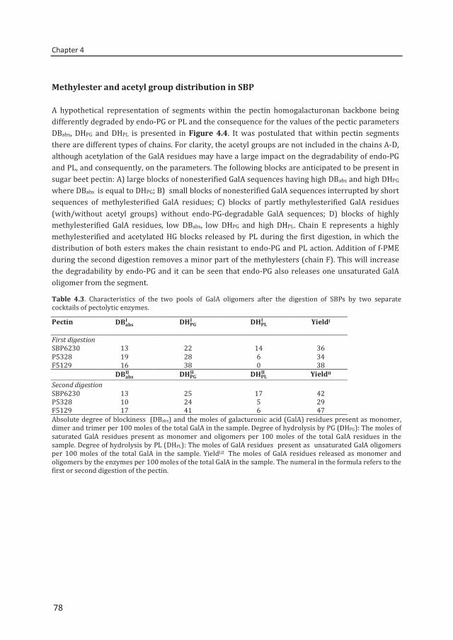
Chapter 4
78
Methylester and acetyl group distribution in SBP
A hypothetical representation of segments within the pectin homogalacturonan backbone being differently degraded by endo-PG or PL and the consequence for the values of the pectic parameters DBabs, DHPG and DHPL is presented in Figure 4.4. It was postulated that within pectin segments there are different types of chains. For clarity, the acetyl groups are not included in the chains A-D, although acetylation of the GalA residues may have a large impact on the degradability of endo-PG and PL, and consequently, on the parameters. The following blocks are anticipated to be present in sugar beet pectin: A) large blocks of nonesterified GalA sequences having high DBabs and high DHPG where DBabs is equal to DHPG; B) small blocks of nonesterified GalA sequences interrupted by short sequences of methylesterified GalA residues; C) blocks of partly methylesterified GalA residues (with/without acetyl groups) without endo-PG-degradable GalA sequences; D) blocks of highly methylesterified GalA residues, low DBabs, low DHPG and high DHPL. Chain E represents a highly methylesterified and acetylated HG blocks released by PL during the first digestion, in which the distribution of both esters makes the chain resistant to endo-PG and PL action. Addition of f-PME during the second digestion removes a minor part of the methylesters (chain F). This will increase the degradability by endo-PG and it can be seen that endo-PG also releases one unsaturated GalA oligomer from the segment.
Table 4.3. Characteristics of the two pools of GalA oligomers after the digestion of SBPs by two separate cocktails of pectolytic enzymes.
Pectin
YieldI
First digestion SBP6230 13 22 14 36 P5328 19 28 6 34 F5129 16 38 0 38
YieldII
Second digestion SBP6230 13 25 17 42 P5328 10 24 5 29 F5129 17 41 6 47 Absolute degree of blockiness (DBabs) and the moles of galacturonic acid (GalA) residues present as monomer, dimer and trimer per 100 moles of the total GalA in the sample. Degree of hydrolysis by PG (DHPG): The moles of saturated GalA residues present as monomer and oligomers per 100 moles of the total GalA residues in the sample. Degree of hydrolysis by PL (DHPL): The moles of GalA residues present as unsaturated GalA oligomers per 100 moles of the total GalA in the sample. YieldI,II The moles of GalA residues released as monomer and oligomers by the enzymes per 100 moles of the total GalA in the sample. The numeral in the formula refers to the first or second digestion of the pectin.
78
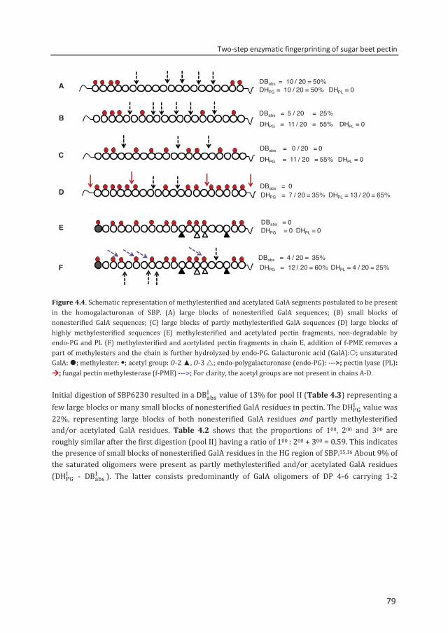
Two-step enzymatic fingerprinting of sugar beet pectin
79
Figure 4.4. Schematic representation of methylesterified and acetylated GalA segments postulated to be present in the homogalacturonan of SBP. (A) large blocks of nonesterified GalA sequences; (B) small blocks of nonesterified GalA sequences; (C) large blocks of partly methylesterified GalA sequences (D) large blocks of highly methylesterified sequences (E) methylesterified and acetylated pectin fragments, non-degradable by endo-PG and PL (F) methylesterified and acetylated pectin fragments in chain E, addition of f-PME removes a part of methylesters and the chain is further hydrolyzed by endo-PG. Galacturonic acid (GalA):; unsaturated GalA: ; methylester: ; acetyl group: O-2 ▲, O-3 ; endo-polygalacturonase (endo-PG): --->; pectin lyase (PL): ; fungal pectin methylesterase (f-PME) --->; For clarity, the acetyl groups are not present in chains A-D.
Initial digestion of SBP6230 resulted in a DB value of 13% for pool II (Table 4.3) representing a few large blocks or many small blocks of nonesterified GalA residues in pectin. The DH value was 22%, representing large blocks of both nonesterified GalA residues and partly methylesterified and/or acetylated GalA residues. Table 4.2 shows that the proportions of 100, 200 and 300 are roughly similar after the first digestion (pool II) having a ratio of 100 : 200 + 300 = 0.59. This indicates the presence of small blocks of nonesterified GalA residues in the HG region of SBP.15,16 About 9% of the saturated oligomers were present as partly methylesterified and/or acetylated GalA residues (DH - DB ). The latter consists predominantly of GalA oligomers of DP 4-6 carrying 1-2
A
B
C
D
E
F
DBabs = 10 / 20 = 50%DHPG = 10 / 20 = 50% DHPL = 0
DBabs = 5 / 20 = 25%DHPG = 11 / 20 = 55% DHPL = 0
DBabs = 0 / 20 = 0DHPG = 11 / 20 = 55% DHPL = 0
DBabs = 0DHPG = 7 / 20 = 35% DHPL = 13 / 20 = 65%
DBabs = 0 DHPG = 0 DHPL = 0
DBabs = 4 / 20 = 35%DHPG = 12 / 20 = 60% DHPL = 4 / 20 = 25%
79
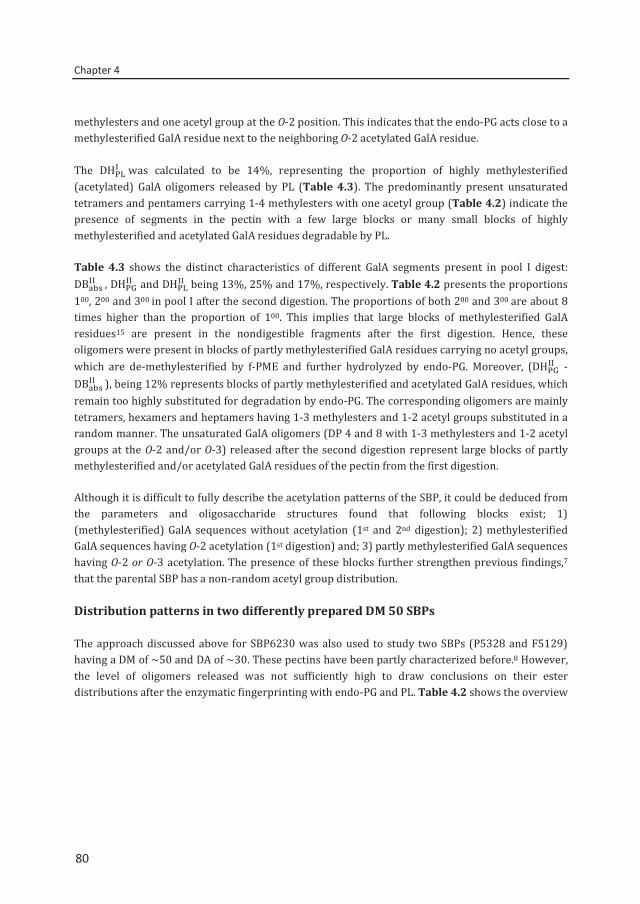
Chapter 4
80
methylesters and one acetyl group at the O-2 position. This indicates that the endo-PG acts close to a methylesterified GalA residue next to the neighboring O-2 acetylated GalA residue.
The DH was calculated to be 14%, representing the proportion of highly methylesterified (acetylated) GalA oligomers released by PL (Table 4.3). The predominantly present unsaturated tetramers and pentamers carrying 1-4 methylesters with one acetyl group (Table 4.2) indicate the presence of segments in the pectin with a few large blocks or many small blocks of highly methylesterified and acetylated GalA residues degradable by PL.
Table 4.3 shows the distinct characteristics of different GalA segments present in pool I digest: DB , DH and DH being 13%, 25% and 17%, respectively. Table 4.2 presents the proportions 100, 200 and 300 in pool I after the second digestion. The proportions of both 200 and 300 are about 8 times higher than the proportion of 100. This implies that large blocks of methylesterified GalA residues15 are present in the nondigestible fragments after the first digestion. Hence, these oligomers were present in blocks of partly methylesterified GalA residues carrying no acetyl groups, which are de-methylesterified by f-PME and further hydrolyzed by endo-PG. Moreover, (DH - DB ), being 12% represents blocks of partly methylesterified and acetylated GalA residues, which remain too highly substituted for degradation by endo-PG. The corresponding oligomers are mainly tetramers, hexamers and heptamers having 1-3 methylesters and 1-2 acetyl groups substituted in a random manner. The unsaturated GalA oligomers (DP 4 and 8 with 1-3 methylesters and 1-2 acetyl groups at the O-2 and/or O-3) released after the second digestion represent large blocks of partly methylesterified and/or acetylated GalA residues of the pectin from the first digestion.
Although it is difficult to fully describe the acetylation patterns of the SBP, it could be deduced from the parameters and oligosaccharide structures found that following blocks exist; 1) (methylesterified) GalA sequences without acetylation (1st and 2nd digestion); 2) methylesterified GalA sequences having O-2 acetylation (1st digestion) and; 3) partly methylesterified GalA sequences having O-2 or O-3 acetylation. The presence of these blocks further strengthen previous findings,7 that the parental SBP has a non-random acetyl group distribution.
Distribution patterns in two differently prepared DM 50 SBPs
The approach discussed above for SBP6230 was also used to study two SBPs (P5328 and F5129) having a DM of ~50 and DA of ~30. These pectins have been partly characterized before.8 However, the level of oligomers released was not sufficiently high to draw conclusions on their ester distributions after the enzymatic fingerprinting with endo-PG and PL. Table 4.2 shows the overview
80
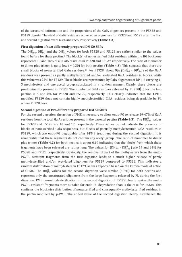
Two-step enzymatic fingerprinting of sugar beet pectin
81
of the structural information and the proportions of the GalA oligomers present in the P5328 and F5129 digests. The yield of GalA residues recovered as oligomers for P5328 and F5129 after the first and second digestion were 63% and 85%, respectively (Table 4.3).
First digestion of two differently prepared DM 50 SBPs The DH , DH and the DH values for both P5328 and F5129 are rather similar to the values found before for these pectins.8 The block(s) of nonesterified GalA residues within the HG backbone represents 19 and 16% of all GalA residues in P5328 and F5129, respectively. The ratio of monomer to dimer plus trimer is quite low (~ 0.30) for both pectins (Table 4.3). This suggests that there are small blocks of nonesterified GalA residues.17 For P5328, about 9% (DH - DB ) of the GalA residues was present as partly methylesterified and/or acetylated GalA residues in blocks, while this value was 22% for F5129. These blocks are represented by GalA oligomers of DP 4-6 carrying 1-3 methylesters and one acetyl group substituted in a random manner. Clearly, these blocks are predominantly present in F5129. The number of GalA residues released by PL (DH ) for the two pectins is 6 and 0% for P5328 and F5129, respectively. This clearly indicates that the f-PME modified F5129 does not contain highly methylesterified GalA residues being degradable by PL where P5328 does.
Second digestion of two differently prepared DM 50 SBPs For the second digestion, the action of PME is necessary to allow endo-PG to release 29-47% of GalA residues from the total GalA residues present in the parental pectins (Table 4.3). The DB values for P5328 and F5129 are 10 and 17, respectively. These values do not indicate the presence of blocks of nonesterified GalA sequences, but blocks of partially methylesterified GalA residues in F5129, which are endo-PG degradable after f-PME treatment during the second digestion. It is remarkable that these segments do not contain any acetyl group. The ratio of monomer to dimer plus trimer (Table 4.2) for both pectins is about 0.10 indicating that the blocks from which these fragments have been released are rather long. The values for (DH - DB ) are 14 and 24% for P5328 and F5129 respectively. Obviously, the removal of part of the methylesters from the endo-PG/PL resistant fragments from the first digestion leads to a much higher release of partly methylesterified and/or acetylated oligomers for F5129 compared to P5328. This indicates a random distribution of methylesters in F5129, as was expected based on the known mode of action of f-PME. The DH values for the second digestion were similar (5-6%) for both pectins and represent only the unsaturated oligomers from the large fragments released by PL during the first digestion. PME de-methylesterification in the second digestion of F5129 clearly makes the endo-PG/PL resistant fragments more suitable for endo-PG degradation than is the case for P5328. This confirms the blockwise distribution of nonesterified and consequently methylesterified residues in the pectin modified by p-PME. The added value of the second digestion clearly established the
81
Chapter 4
82
differences between p-PME and f-PME modified SBPs. This can be seen in sequences of esterified GalA residues, which are not degradable by endo-PG or PL alone.
When taking into account the method of preparation of the two DM 50 pectins, several observations can be stated. Pectin P5328, which has been modified from a parental SBP (DM 62, DA 30) using p-PME is expected to have a blockwise distribution of nonmethylesterified GalA sequences. On the contrary, pectin F5129, which originated from the same parental SBP after a partial removal of the methylesters using f-PME, is expected to have a random distribution of nonmethylesterified GalA sequences. The descriptive parameters indeed confirmed significant differences in the methylester distribution between the two pectins, although less pronounced than found before for lemon pectins.18 Large blocks of nonesterified GalA residues are not present in P5328 since the p-PME was hindered by the presence acetyl groups forcing the enzyme to act less in a blockwise manner than would have been the case for non-acetylated pectins. A typical consequence of a blockwise removal of methylesters is that other blocks of GalA residues remain highly methylesterified. This can be recognized from the higher DH value for P5328 compared to F5129. In addition, a random de-methylesterification pattern of f-PME is recognized from the higher release of partly methylesterified GalA oligomers without acetyl groups in F5129 compared to P5328 for both enzymatic digestions. Our findings support the hypothesis.6 stating that due to the steric hindrance of the acetyl group present, p-PME forced to de-methylesterify SBP in a combined random (Fig, 4 chain C) and blockwise (chain D) manner.
4.4 Conclusions In general, it can be stated that the two-step enzymatic fingerprinting is able to generate additional information on the patterns of SBPs having the same DM and DA compared to the commonly used one-step enzymatic fingerprinting methods. The various proportions of different blocks of nonesterified, partly methylesterified/acetylated and highly methylesterified/acetylated sequences of GalA found for three differently prepared sugar beet pectins describe the ester distributions within the pectins in more detail than has been done before. It should be stated that the two-step fingerprinting method is laborious and, is therefore, far from suitable for routine analysis of pectins. Nevertheless, the additional information to be obtained for complex pectins is important to understand its precise chemical structure and to make the link to their functionality.
82
Two-step enzymatic fingerprinting of sugar beet pectin
83
Acknowledgment
The European Community is gratefully acknowledged for financially supported this research within a consortium PolyModE KBBE-2007-3-3-07.
References 1. Kravtchenko, T. P., Voragen, A. G. J., and Pilnik, W. (1992) Carbohydrate Polymers 18, 17-25 2. Ralet, M., and Thibault, J. (2009) Hydrodynamic properties of isolated pectin domains: a way to figure
out pectin macromolecular structure. in Pectins and Pectinases Schols, H. A., Visser, R. G. F., and Voragen, A. G. J. (eds.), Academic Publishers, Wageningen, The Netherlands. pp 35-48
3. Voragen, A. G. J., Coenen, G. J., Verhoef, R. P., and Schols, H. A. (2009) Structural Chemistry 20, 263-275 4. Quemener, B., Desire, C., Lahaye, M., Debrauwer, L., and Negroni, L. (2003) European Journal of Mass
Spectrometry (Chichester, England) 9, 45-60 5. Voragen, A. G. J., Pilnik, W., Thibault, J. F., Axelos, M. A. V., and Renard, C. M. G. C. (1995) Pectins. in
Food Polysaccharides and Their Applications Stephen, A. M. (ed.), Marcel Dekker, New York, N.Y., USA. pp 287-339
6. Buchholt, H. C., Christensen, T. M. I. E., Fallesen, B., Ralet, M. C., and Thibault, J. F. (2004) Carbohydrate Polymers 58, 149-161
7. Ralet, M.-C., Crépeau, M.-J., and Bonnin, E. (2008) Phytochemistry 69, 1903-1909 8. Remoroza, C., Buchholt, H. C., Gruppen, H., and Schols, H. A. (2014) Carbohydrate Polymers 101, 1205-
1215 9. Van Alebeek, G. J. W. M., Van Scherpenzeel, K., Beldman, G., Schols, H. A., and Voragen, A. G. J. (2003)
Biochemical Journal 372, 211-218 10. Schols, H. A., Posthumus, M. A., and Voragen, A. G. J. (1990) Carbohydrate Research 206, 117-129 11. Limberg, G., Körner, R., Buchholt, H. C., Christensen, T. M. I. E., Roepstorff, P., and Mikkelsen, J. D.
(2000) Carbohydrate Research 327, 321-332 12. Remoroza, C., Cord-Landwehr, S., Leijdekkers, A. G. M., Moerschbacher, B. M., Schols, H. A., and
Gruppen, H. (2012) Carbohydrate Polymers 90, 41-48 13. Guillotin, S. E., Van Loey, A., Boulenguer, P., Schols, H. A., and Voragen, A. G. J. (2007) Food
Hydrocolloids 21, 85-91 14. Guillotin, S. E., Bakx, E. J., Boulenguer, P., Mazoyer, J., Schols, H. A., and Voragen, A. G. J. (2005)
Carbohydrate Polymers 60, 391-398 15. Daas, P. J. H., Meyer-Hansen, K., Schols, H. A., De Ruiter, G. A., and Voragen, A. G. J. (1999) Carbohydrate
Research 318, 135-145 16. Winning, H., Viereck, N., Nørgaard, L., Larsen, J., and Engelsen, S. B. (2007) Food Hydrocolloids 21, 256-
266 17. Daas, P. J. H., Voragen, A. G. J., and Schols, H. A. (2000) Carbohydrate Research 326, 120-129 18. Limberg, G., Körner, R., Buchholt, H. C., Christensen, T. M. I. E., Roepstorff, P., and Mikkelsen, J. D.
(2000) Carbohydrate Research 327, 293-307
83
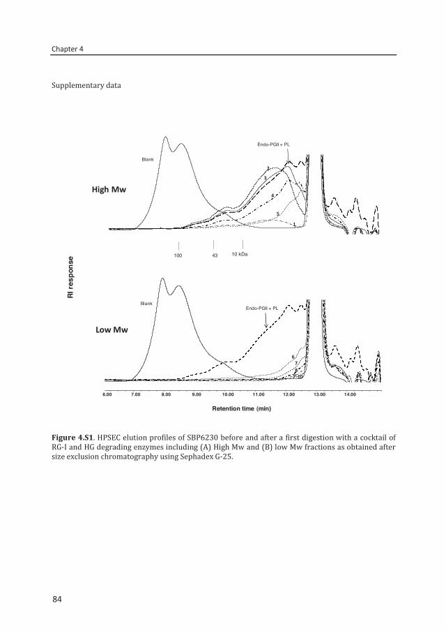
Chapter 4
84
Supplementary data
Figure 4.S1. HPSEC elution profiles of SBP6230 before and after a first digestion with a cocktail of RG-I and HG degrading enzymes including (A) High Mw and (B) low Mw fractions as obtained after size exclusion chromatography using Sephadex G-25.
1
2
3
4
5
Endo-PGII + PL
Blank
6
8109
7
Endo-PGII + PLBlank
6.00 7.00 8.00 9.00 10.00 11.00 12.00 13.00 14.00
RI r
espo
nse
Retention time (min)
High Mw
Low Mw
100 43 10 kDa
84
Chapter 5
A Bacillus licheniformis pectin
acetylesterase is specific for
homogalacturonan’s acetylated at O-3
C. Remoroza, M. Wagenknecht, F. Gu, H.C. Buchholt, B.M. Moerschbacher,
H.A. Schols, H. Gruppen Accepted for publication in Carbohydrate Polymers
Chapter 5
86
Abstract
A recombinant acetylesterase from Bacillus licheniformis DSM13, belonging to carbohydrate esterase family 12, was purified and biochemically characterized. The purified enzyme, termed BliPAE, was capable of deacetylating acetylated pectins, e.g. sugar beet pectin (SBP). Contrary to its provisional annotation as rhamnogalacturonan acetylesterase, the enzyme specifically removed acetyl groups from the homogalacturonan region classifying it as a PAE. The recombinant enzyme has a molecular mass of 26.7 kDa and shows optimal activity at pH 8.0 and 50 °C. It is stable in the range pH 5.0 to 7.0 and below 50 °C. Methylesterification of the galacturonic acid (GalA) moieties reduces the deacetylation efficiency of BliPAE. Hence, the enzyme prefers SBPs with low degree of methylesterification (DM) 9-30, releasing about 75% of the acetyl groups present in the homogalacturonan. Furthermore, 1H NMR of the polymer and HPLC-HILIC-MSn of oligosaccharides generated by endo-PG and PL degradation were used to structurally characterize the BliPAE-modified pectins. The results show that BliPAE removes acetyl groups specifically when substituted at the O-3 position of GalA moieties.
86
A Bacillus licheniformis pectin acetylesterase specific for O-3 acetylation
87
5.1. Introduction
Traditionally, commercial pectin is extracted from citrus peels and apple pomace. The primary structural elements of pectin are homogalacturonan (HG) and rhamnogalacturonan I (RG-I). The HG is composed of (1→4) linked α-D-GalA residues, which can be methylesterified at the C-6 position, while the RG-I backbone is composed of the disaccharide repeating unit [(1→2)-α-L-Rha-(1→4)-α-D-GalA]. Between 20–80% of the rhamnosyl residues can be substituted with side chains consisting of neutral sugars. The GalA residues can be acetylated at positions O-2 and/or O-3 in both RG-I and HG.1 The level of acetylation in commercial pectins from citrus peels and apple pomace is negligible.2
Nowadays, sugar beet pulp and sunflower residue are raw materials seen as potential source of commercial pectins. However, these pectins are rich in acetyl groups and commercial sugar beet pectin (SBP) may have a DA of 30.3 It was reported that about 75% of all acetyl groups are located in the HG and 25% in the RG-I of acid extracted SBP.4 The high degree of acetylation in pectins adversely affects their functional properties.5 Hence, commercialization of SBP is limited due to its poor gelling capability.3 Enzymatic deacetylation may overcome the poor gelling properties of SBP and could provide a promising alternative way for the commercialization of SBP and other acetylated pectins. So far, only a low number of acetylesterases active on acetylated pectin have been identified and characterized. This limits the possibilities for specifically modifying the level of acetylation of SBP and other acetylated pectins.
Pectin acetylesterase (PAE) removes acetyl groups from the HG region of pectin6 and belongs to carbohydrate esterase (CE) family 12 (CAZy database, www.cazy.org). Two PAEs have been detected in orange peel, which exhibit a maximum activity at acidic pH and deacetylate a synthetic substrate more efficiently than the low methylesterified HG of SBPs.7,8 Another type of acetylesterase in the same CE family is the class of rhamnogalacturonan acetylesterase (RGAE). Aspergillus aculeatus RGAE is known to specifically remove acetyl groups bound to GalA residues in the RG-I of pectins and showed an optimal activity at acidic pH.9 Another acetylesterase from A. aculeatus was able to deacetylate both the HG and RG-I of SBP10 Aside from plants and fungi, CE family 12 acetylesterases have been identified in bacteria. PaeY and PaeX, two PAEs from Erwinia chrysanthemi.11,12 and PAE YxiM from Bacillus subtilis13 were highly active towards several synthetic substrates and preferred these substrates over GalA oligomers and acetylated low methylesterified SBP. None of the above mentioned acetylesterases has been distinguished for a possible preference for O-2 or O-3 acetylation.
Bacillus licheniformis DSM13 is known to be an important source for a multitude of biotechnologically important enzymes, among them pectate lyases.14 Pectin acetylesterases, however, have not yet been reported for this species. In this study, we cloned a B. licheniformis DSM13 gene encoding a PAE classified in CE family 12 and expressed it heterologously in
87
Chapter 5
88
Escherichia coli. The recombinant enzyme, termed BliPAE, was purified and characterized for its biochemical properties. 1H NMR analysis and enzymatic fingerprinting of modified SBP using pectolytic enzymes were carried out, followed by HILIC-ESI-MSn analysis in order to determine BliPAE’s mode of action and specificity.
5.2. Materials and methods
Characterization of the acetylated pectins from different sources Substrates Sugar beet pectin, (SBP6230) with degree of methylesterification (DM) of 62 and degree of acetylation (DA) of 30 was extracted from sugar beet pulp and this pectin was further de-esterified by alkali or by plant or fungal pectin esterases to yield different series of pectins (Table 5.1). SBP6230 modified by plant pectin methylesterase (p-PME) yielded the P-series of SBP (P5328, P3429) and SBP6230 modified by fungal pectin methylesterase (f-PME) yielded the F-series (F5129, F3331). For the B-series, SBP6230 was alkali de-esterified yielding B5326, B4626, B3124 and B0915. Partial deacetylation of SBP6230 in a sodium methylate / methanol solution resulted in B’6126, B’6109, B’6023, B’5803 and SBP1713. The chemical preparation and composition of these SBPs have been described elsewhere.3 Mild alkali-treated SBP (SBP5519) and other pectic material from sunflower and potato (DuPont), Okra15 and apple modified hairy region (MHR)16 (Table 1), acetylated xanthan17 and acetylated eucalyptus xylan oligomers18 were also used as substrates for testing enzyme specificity. Constituent monosaccharide composition analysis Determination of the neutral monosaccharide and uronic acid contents of sunflower and potato pectin was carried out as previously described elsewhere.19
Determination of the DM and DA Pectin samples (2 mg/ml) were saponified in 100 mM NaOH and after neutralization the DM was determined using a colorimetric method20 while the DA was analysed using the Megazyme acetic acid kit (Megazyme, Wicklow, Ireland).
Production and purification of BliPAE
Bacterial strains, media, and growth conditions DSM13 and Escherichia coli strains NEB 5-alpha (New England Biolabs, Frankfurt a. Main, Germany) and Rosetta 2(DE3)(pLysSRARE2) (Merck, Darmstadt, Germany) were grown in Luria-Bertani (LB) medium at 37 °C. For plasmid maintenance, E. coli strains were grown in the presence of ampicillin and/or chloramphenicol at a final concentration (f.c.) of 100 µg/ml and 34 µg/ml, respectively. After transformation of the latter strain with the BliPAE expression
88

A Bacillus licheniformis pectin acetylesterase specific for O-3 acetylation
89
plasmid, 40 mM glucose was added to the medium when the strain was grown on LB agar plates.
Table 5.1. Chemical characteristics of pectin from various sources used in this study.
Pectin GalA Rha Ara Xyl Gal Glc DM a DAa
% (w/w) (%) (%) SBP6230 b 55 5 12 0.3 10 0.5 62 30 SBP5519 c 62 4 7 0.1 8 0.5 55 19 SBP1713c 62 4 7 0.1 8 0.5 17 13 B‘6126 56 5 11 0.3 9 0.5 61 26 B‘6109 59 5 11 0.3 8 0.5 61 09 B‘6023 57 5 11 0.3 9 0.6 60 23 B‘5803 60 5 10 0.3 8 0.5 58 03 B5326 58 6 12 0.5 10 0.4 53 26 B4626 60 6 11 0.3 10 0.4 46 26 B3124 55 6 12 0.3 10 0.4 31 24 B0915 57 5 10 0.2 9 0.3 9 15 F5129 57 5 12 0.3 9 0.2 51 29 F3331 57 5 12 0.3 9 0.3 33 31 P5328 58 6 11 0.2 10 0.4 53 28 P3429 66 5 11 0.2 10 0.4 34 28 Sunflower pectinc 75 1 1 n.d. 1 n.d 60 11 Potato pectinc 34 1 4 n.d 40 2 40 18 Apple MHRe 30 14 29 10 16 1 16 35 Okra RG regionf 40 25 n.d n.d 32 10 24 58 aMoles methanol (DM) and acetic acid (DA) per 100 moles of galacturonic acid cMonosaccharide composition was determined in this study. b,d,fMonosaccharide composition3,15,16 n.d. not detected
Cloning of the BliPAE coding sequence The cell pellet of 0.5 ml of a B. licheniformis DSM13 overnight culture was resuspended in 50 µl 10 mM Tris/HCl buffer (pH8.0) and incubated at 99 °C for 5 min. Next, the suspension was centrifuged (13,000 x g, 1 min, room temperature (RT)) and the supernatant was used as template for the PCR amplification of the BliPAE open reading frame (ORF; GenBank accession number (acc no) AAU42913). For this, Phusion High-Fidelity DNA Polymerase (Fermentas, St. Leon-Rot, Germany) and primers 5’-CCGCAATTCCATATGATGCGAGACATTCAGTTGTTTTTG-3’ (forward) and 5’-CCCGAGCTCTAAAGGAATTCCCGCTTCTTTTG-3’ (reverse) were used. Additionally, an NdeI recognition site, overlapping with the start codon, and a SacI recognition site, concomitantly removing the ORF’s stop codon, were introduced. The amplicon obtained was cut with NdeI/SacI and ligated with the likewise cut and dephosphorylated expression vector pET22b-StrepIIc, being a pET-22b(+) (Merck, Darmstadt, Germany) derivative additionally containing a sequence coding for the StrepII affinity tag. Competent cells of E. coli NEB 5-alpha were transformed with the ligation reaction and plasmids were kit-isolated from
89
Chapter 5
90
the transformants obtained. Correctness of insertion and coding sequence was verified by determination of the nucleotide sequence of the insert and the flanking regions (Eurofins MWG, Ebersberg, Germany). The BliPAE expression plasmid generated was termed pET22b-BliPAE-StrepIIc. PCR, agarose gel electrophoresis, DNA restriction endonuclease digestion, ligation and transformation of E. coli were done as described in Sambrook and Russell.21
Synthesis and purification of BliPAE E. coli Rosetta 2(DE3)(pLysSRARE2) was transformed with pET22b-BliPAE-StrepIIc and grown in 500 ml auto-induction medium22 complemented with ampicillin and chloramphenicol. Incubation was done in a shaking incubator for 4 h at 180 rpm and 37 °C and further for 20 h at 120 rpm and 26 °C. Alternatively, for additional induction, isopropyl-β-D-thiogalactopyranoside (IPTG; f.c. 0.5 mM) was added after 15 h of incubation. Cells were harvested by centrifugation (4,200 x g, 10 min, 4 °C), the pellet was resuspended in 30 ml 40 mM triethanolamine buffer (pH 8.0) containing 0.4 M NaCl and frozen at -20°C. The thawed suspension was incubated for 30 min at RT, 75 U benzonase (Merck) were added and the suspension was sonicated. Centrifugation (40,000 x g, 40 min, 4 °C) yielded the crude extract, and BliPAE was purified via affinity chromatography using a 1-ml Strep-Tactin Superflow Plus Column (Qiagen, Hilden, Germany) according to the manufacturer’s instructions. For enzyme concentration and buffer exchange, Vivaspin columns (MWCO 10 kDa; Sartorius, Göttingen, Germany) and 20 mM triethanolamine buffer (pH 7.0) were used. Finally, the enzyme solution was complemented with glycerol (f.c. 10%) and stored at 4 °C.
Verification of enzyme purity
Protein analysis Protein contents were determined using the Pierce BCA Protein Assay Kit (Fisher Scientific, Schwerte, Germany) and BSA as the calibration protein according to the manufacturer’s instructions. SDS-PAGE was carried out using 12% polyacrylamide gels. peqGOLD Protein Marker II (Peqlab Biotechnologie, Erlangen, Germany) served as molecular mass standard. Gels were stained with Coomassie brilliant blue G-250. For Western blot, proteins were transferred to a nitrocellulose membrane. Immunodetection of StrepII-tagged BliPAE was performed using a Strep-Tactin HRP conjugate (1:1000 dilution; IBA, Göttingen, Germany) according to the manufacturer’s instructions. The signal of the horseradish peroxidase coupled to the antibody was imaged in a Fusion-SL Vilber Lourmat (Peqlab Biotechnologie, Erlangen, Germany) by chemiluminescence detection using luminol-based ECL substrate.
Colorimetric determination of acetic acid released
BliPAE activity was measured colorimetrically using SBP, sunflower pectin, potato pectin, apple MHR and okra pectin. The pectin sample (~5 mg) was dissolved in 1 ml 50 mM McIlvaine's buffer (pH 6.5) incubated at 40 °C for 24 h with an enzyme concentration of 0.02% (w/w, on protein-substrate basis). The enzyme was inactivated by heating for 6 min at 100 °C. The
90
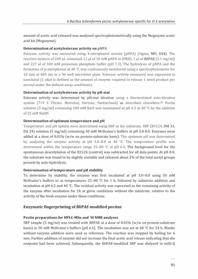
A Bacillus licheniformis pectin acetylesterase specific for O-3 acetylation
91
amount of acetic acid released was analyzed spectrophotometrically using the Megazyme acetic acid kit (Megazyme).
Determination of acetylesterase activity on pNPA Esterase activity was measured using 4-nitrophenol acetate (pNPA) (Sigma, MO, USA). The reaction mixture of 240 µL contained 12 µl of 10 mM pNPA in DMSO, 1 µl of BliPAE (1.1 mg/ml) and 227 µl of 100 mM potassium phosphate buffer (pH 7.3). The hydrolysis of pNPA and the formation of p-nitrophenol at 40 °C was continuously monitored using a spectrophotometer for 10 min at 405 nm in a 96 well microtiter plate. Esterase activity measured was expressed in nanokatal (1 nkat is defined as the amount of enzyme required to release 1 nmol product per second under the defined assay conditions).
Determination of acetylesterase activity by pH-stat Esterase activity was determined by pH-stat titration using a thermostated auto-titration system (719 S Titrino, Metrohm, Herisau, Switzerland) as described elsewhere.23 Pectin solution (5 mg/ml) containing 100 mM NaCl was maintained at pH 6.5 at 40 °C by the addition of 25 mM NaOH.
Determination of optimum temperature and pH Temperature and pH optima were determined using SBP as the substrate. SBP (B3124, DM 31, DA 24) solution (5 mg/ml) containing 50 mM McIlvaine's buffers at pH 3.0-8.0. Enzymes were added at a dose of 0.02% (w/w on protein-substrate basis). The optimum pH was determined by analyzing the enzyme activity at pH 3.0–8.0 at 40 °C. The temperature profile was determined within the temperature range 25–80 °C at pH 6.5. The background level for the spontaneous deacetylation of the B3124 (control) was subtracted for all data points. At pH 8.0, the substrate was found to be slightly unstable and released about 2% of the total acetyl groups present by auto-hydrolysis.
Determination of temperature and pH stability To determine its stability, the enzyme was first incubated at pH 3.0–8.0 using 50 mM McIlvaine’s buffers or at temperatures 25–80 °C for 1 h, followed by substrate addition and incubation at pH 6.5 and 40 °C. The residual activity was expressed as the remaining activity of the enzyme after incubation for 1h at given conditions without the substrate, relative to the activity of the fresh enzyme under these conditions. Enzymatic fingerprinting of BliPAE-modified pectins Pectin preparations for HPLC-MSn and 1H NMR analyses SBP sample (5 mg/ml) was treated with BliPAE at a dose of 0.02% (w/w on protein-substrate basis) in 50 mM McIlvaine's buffers (pH 6.5). The incubation was set at 40 °C for 24 h. Blanks without enzyme addition were used as reference. The reaction was stopped by boiling for 6 min. Further addition of enzyme did not increase the final acetic acid release indicating that the endpoint had been achieved. Subsequently, the BliPAE-modified SBP was dialyzed in milli-Q
91
Chapter 5
92
water overnight, followed by freeze-drying. The freeze-dried SBP obtained was re-dissolved (5 mg/ml) in 50 mM sodium citrate buffer (pH 5.0) and digested using the enzyme cocktail of purified RG-I and HG degrading enzymes at 40 °C for 24 h described elsewhere.24 For 1H NMR analysis, SBP (5 g) was dissolved in 500 mL demineralized water containing 0.1 g sorbic acid and 2.9 g sodium chloride. The sample was adjusted to pH 6.5 with 1M NaOH in a 1 L stirred thermostated reactor and heated to 40 °C. A BliPAE enzyme dose at 1% (w/w protein-substrate basis) was added and the pH was kept at 6.5 by adding of 0.1M NaOH. Incubation of SBP5519 and SBP1713 with BliPAE was 7 h and 4 h, respectively. The reaction was stopped by adjusting pH to 3.5 with 1M hydrochloric acid and heating to 75 °C for 5 minutes. After cooling to RT pectin was precipitated by mixing into 1 L isopropyl alcohol. The precipitate was separated on a screen, dried in an oven at 40 °C overnight and the dry pectin was milled using a 0.5 mm sieve. Hydrophilic interaction liquid chromatography (HILIC)-MSn
SBP digests, diluted to 1 mg/ml in 50% (v/v) acetonitrile, were analysed using an Accela UHPLC system (Thermo Scientific, Waltham, MA, USA) coupled to an evaporative light scattering detector (Agilent 1200 series, Gen Tech Scientific, NY, USA) and an ESI-IT-MSn-detector (LTQ Velos Pro, Thermo Scientific, San Jose, CA, USA). Chromatographic separation was performed on an Acquity UPLC BEH Amide column as described previously.24
NMR spectroscopy
Liquid state 1H NMR spectroscopy was conducted at 14.095 Tesla (600 MHz 1H frequency) with a 5 mm smart probe and a Bruker Advance III spectrometer. The samples (SBP5519, SBP1713) were dissolved (5 mg/ml) in D2O (99.9 atom % D, Sigma-Aldrich, St. Louis, MO, USA). NMR spectra were recorded at a probe temperature of 353 K collecting 64 scans. Chemical shifts are expressed in parts per million (ppm) relative to internal trimethylsilyl propanoic acid (TMSP).
5.3. Results and discussion
Sequence analysis, production and purification of BliPAE
BliPAE, originating from B. licheniformis DSM13, comprises 220 amino acids (aa) corresponding to a calculated molecular mass of 25.0 kDa and based on homology, BliPAE (acc no AAU42913) has been assigned to CE family 12 and annotated as a putative RGAE. BliPAE revealed closest similarity to a 245-aa protein of Bacillus sp. BT1B_CT2 (acc no WP_009329846; 99.6% identity), annotated as a RGAE. Significant similarity was also seen for a putative PAE of B. licheniformis SVD1 (acc no BAL46009; 224 aa, 89.1% identity). Subjecting the deduced aa sequence of BliPAE to a SignalP analysis,25 a signal peptide was not to be predicted.
92
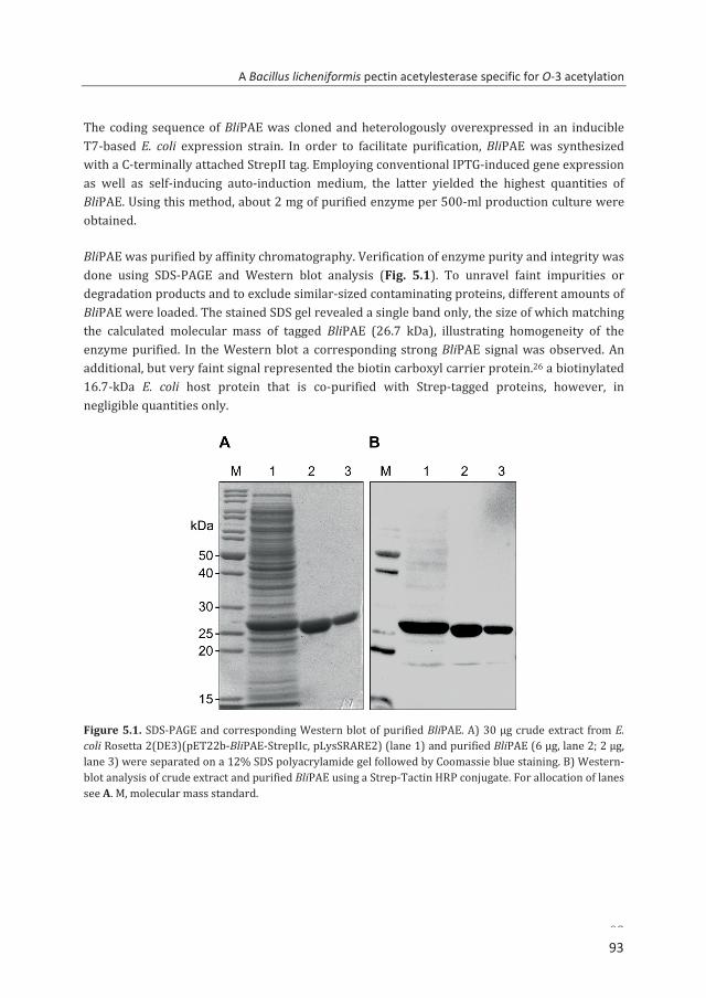
A Bacillus licheniformis pectin acetylesterase specific for O-3 acetylation
93
The coding sequence of BliPAE was cloned and heterologously overexpressed in an inducible T7-based E. coli expression strain. In order to facilitate purification, BliPAE was synthesized with a C-terminally attached StrepII tag. Employing conventional IPTG-induced gene expression as well as self-inducing auto-induction medium, the latter yielded the highest quantities of BliPAE. Using this method, about 2 mg of purified enzyme per 500-ml production culture were obtained.
BliPAE was purified by affinity chromatography. Verification of enzyme purity and integrity was done using SDS-PAGE and Western blot analysis (Fig. 5.1). To unravel faint impurities or degradation products and to exclude similar-sized contaminating proteins, different amounts of BliPAE were loaded. The stained SDS gel revealed a single band only, the size of which matching the calculated molecular mass of tagged BliPAE (26.7 kDa), illustrating homogeneity of the enzyme purified. In the Western blot a corresponding strong BliPAE signal was observed. An additional, but very faint signal represented the biotin carboxyl carrier protein.26 a biotinylated 16.7-kDa E. coli host protein that is co-purified with Strep-tagged proteins, however, in negligible quantities only.
Figure 5.1. SDS-PAGE and corresponding Western blot of purified BliPAE. A) 30 µg crude extract from E. coli Rosetta 2(DE3)(pET22b-BliPAE-StrepIIc, pLysSRARE2) (lane 1) and purified BliPAE (6 µg, lane 2; 2 µg, lane 3) were separated on a 12% SDS polyacrylamide gel followed by Coomassie blue staining. B) Western-blot analysis of crude extract and purified BliPAE using a Strep-Tactin HRP conjugate. For allocation of lanes see A. M, molecular mass standard.
93
Chapter 5
94
Biochemical characterization of BliPAE
pH & temperature optima Optimum pH and temperature of BliPAE were determined on SBP (B3124) (Fig. 5.2). The enzyme was tested in a pH range 3.0–8.0. The purified enzyme was active between pH 5.0-8.0 and showed a maximum activity at pH 8.0 (Fig. 5.2A). The activity profile of BliPAE was also determined in the temperature range 25–80 °C at pH 6.5. The results showed that the enzyme has an optimum temperature at 50 °C. In addition, the enzyme has an activity of >50% of its maximum activity at 25 °C until 60 °C (Fig. 5.2B).
pH & temperature stability To omit the role of auto hydrolysis occurring at pH 8.0 (material and methods) and to allow an extended digestion of the substrate, incubation at pH 6.0-7.0 and 40 °C for 24 h was performed. At these conditions, the enzyme maintained its activity above 70%. Figure 5.2C shows that the enzyme retained more than 90% of its initial activity towards SBP (B3124) between pH 5.0 and 7.0. The enzyme was stable up to 50 °C for 1 h with a residual activity of more than 70%. (Fig. 5.2D).
BliPAE was active and stable at neutral to alkaline conditions, contrary to orange PAEs8 being active at acidic conditions. The findings for BliPAE are consistent with those reported for PAEs of B. subtilis and E. chrysanthemi.13 The optimum temperature found for BliPAE is also similar to Aspergillus RGAE and PAE 27 and 10 °C higher compared to the optimal temperature found for E. chrysanthemi PAEs.12
Specificity of BliPAE towards different substrates
Table 5.2 shows the specific activity of BliPAE and total acetic acid released from different acetylated pectins. Incubation of the enzyme with pNPA showed an activity of 847 nkat/mg. The specific activity towards SBP6230, sunflower pectin, potato pectin and apple modified hairy region (MHR) was 302, 247, 243, 107 nkat/mg, respectively. No activity was observed towards acetylated xanthan and acetylated eucalyptus xylan oligomers indicating that BliPAE is specific towards acetylated pectins. The relatively lower specific activity of the enzyme for natural substrates (e.g. SBP, potato and sunflower pectins) compared to pNPA is in agreement with the previous results for Aspergillus acetylesterases.27
94
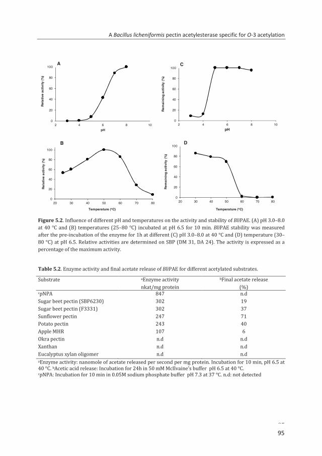
A Bacillus licheniformis pectin acetylesterase specific for O-3 acetylation
95
Figure 5.2. Influence of different pH and temperatures on the activity and stability of BliPAE. (A) pH 3.0–8.0 at 40 °C and (B) temperatures (25–80 °C) incubated at pH 6.5 for 10 min. BliPAE stability was measured after the pre-incubation of the enzyme for 1h at different (C) pH 3.0–8.0 at 40 °C and (D) temperature (30–80 °C) at pH 6.5. Relative activities are determined on SBP (DM 31, DA 24). The activity is expressed as a percentage of the maximum activity.
Table 5.2. Enzyme activity and final acetate release of BliPAE for different acetylated substrates.
Substrate aEnzyme activity bFinal acetate release nkat/mg protein (%) cpNPA 847 n.d Sugar beet pectin (SBP6230) 302 19 Sugar beet pectin (F3331) 302 37 Sunflower pectin 247 71 Potato pectin 243 40 Apple MHR 107 6 Okra pectin n.d n.d Xanthan n.d n.d Eucalyptus xylan oligomer n.d n.d aEnzyme activity: nanomole of acetate released per second per mg protein. Incubation for 10 min, pH 6.5 at 40 °C. bAcetic acid release: Incubation for 24h in 50 mM McIlvaine's buffer pH 6.5 at 40 °C. cpNPA: Incubation for 10 min in 0.05M sodium phosphate buffer pH 7.3 at 37 °C. n.d: not detected
0
20
40
60
80
100
20 30 40 50 60 70 80
Rela
tive
activ
ity (%
)
Temperature (oC)
0
20
40
60
80
100
2 4 6 8 10
Rem
aini
ng a
ctiv
ity (%
)
pH
0
20
40
60
80
100
20 30 40 50 60 70 80
Rem
aini
ng a
ctiv
ity (%
)
Temperature (oC)
B D
C
0
20
40
60
80
100
2 4 6 8 10
Rela
tive
activ
ity (%
)
pH
A
95
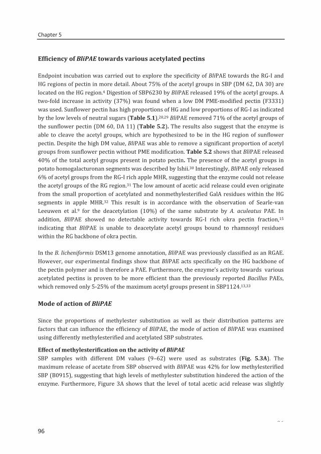
Chapter 5
96
Efficiency of BliPAE towards various acetylated pectins
Endpoint incubation was carried out to explore the specificity of BliPAE towards the RG-I and HG regions of pectin in more detail. About 75% of the acetyl groups in SBP (DM 62, DA 30) are located on the HG region.4 Digestion of SBP6230 by BliPAE released 19% of the acetyl groups. A two-fold increase in activity (37%) was found when a low DM PME-modified pectin (F3331) was used. Sunflower pectin has high proportions of HG and low proportions of RG-I as indicated by the low levels of neutral sugars (Table 5.1).28,29 BliPAE removed 71% of the acetyl groups of the sunflower pectin (DM 60, DA 11) (Table 5.2). The results also suggest that the enzyme is able to cleave the acetyl groups, which are hypothesized to be in the HG region of sunflower pectin. Despite the high DM value, BliPAE was able to remove a significant proportion of acetyl groups from sunflower pectin without PME modification. Table 5.2 shows that BliPAE released 40% of the total acetyl groups present in potato pectin. The presence of the acetyl groups in potato homogalacturonan segments was described by Ishii.30 Interestingly, BliPAE only released 6% of acetyl groups from the RG-I rich apple MHR, suggesting that the enzyme could not release the acetyl groups of the RG region.31 The low amount of acetic acid release could even originate from the small proportion of acetylated and nonmethylesterified GalA residues within the HG segments in apple MHR.32 This result is in accordance with the observation of Searle-van Leeuwen et al.9 for the deacetylation (10%) of the same substrate by A. aculeatus PAE. In addition, BliPAE showed no detectable activity towards RG-I rich okra pectin fraction,15 indicating that BliPAE is unable to deacetylate acetyl groups bound to rhamnosyl residues within the RG backbone of okra pectin.
In the B. licheniformis DSM13 genome annotation, BliPAE was previously classified as an RGAE. However, our experimental findings show that BliPAE acts specifically on the HG backbone of the pectin polymer and is therefore a PAE. Furthermore, the enzyme’s activity towards various acetylated pectins is proven to be more efficient than the previously reported Bacillus PAEs, which removed only 5-25% of the maximum acetyl groups present in SBP1124.13,33
Mode of action of BliPAE
Since the proportions of methylester substitution as well as their distribution patterns are factors that can influence the efficiency of BliPAE, the mode of action of BliPAE was examined using differently methylesterified and acetylated SBP substrates.
Effect of methylesterification on the activity of BliPAE SBP samples with different DM values (9–62) were used as substrates (Fig. 5.3A). The maximum release of acetate from SBP observed with BliPAE was 42% for low methylesterified SBP (B0915), suggesting that high levels of methylester substitution hindered the action of the enzyme. Furthermore, Figure 3A shows that the level of total acetic acid release was slightly
96
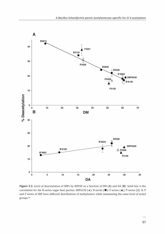
A Bacillus licheniformis pectin acetylesterase specific for O-3 acetylation
97
Figure 5.3. Level of deacetylation of SBPs by BliPAE as a function of DM (A) and DA (B). Solid line is the correlation for the B-series sugar beet pectins. SBP6230 (♦); B-series (); F-series (▲); P-series (Δ). B, P and F series of SBP have different distributions of methylesters while maintaining the same level of acetyl groups.24
B5326B4626
B3124
B'6126
B0915
F5129
F3331
P5328
P3428
SBP6230
0
10
20
30
40
0 10 20 30 40 50 60 70
DM
B'5803
B'6023
B'6109SBP6230
F5129
P5328
B5326
0
10
20
30
40
0 5 10 15 20 25 30 35
DA
A
B
% D
eace
tyla
tion
B’6023
97
Chapter 5
98
different for SBP samples having rather similar DA and DM. This variation in acetate release suggested that the action of BliPAE is influenced by the different methylesterification patterns present in these pectins.24
Effect of acetylation on the activity of BliPAE The effect of acetylation towards BliPAE activity was examined using different SBP series having a rather constant DM values (51-62) and different DA values (3–30). The enzyme released 22% of the total acetyl groups from B5326 while only 13% of all acetyl groups were released for B’5803. This suggests that the activity of BliPAE varies for pectins having different DA. It is postulated that it is not due to the level of acetyl groups but due to different positions of the acetyl groups (O-2 or/and O-3) within the HG part of the substrates. Hence, the site specificity of the enzyme was determined by enzymatic fingerprinting of the BliPAE-modified SBP.
Specificity of BliPAE towards the acetyl group position
It was hypothesized that the BliPAE-modified SBP will lead to a more complete degradation of the HG backbone by endo-PG and PL compared to the unmodified SBP. BliPAE released a maximum of 34% of the total acetyl groups from P3429 resulting in a pectin with a DA 19. In order to elucidate the precise position of acetyl groups not removed by the enzyme, the BliPAE-modified P3429 was digested using endo-PG and PL followed by HILIC-MSn analysis as described elsewhere24 (Fig. 5.4). The RG-I region of the unmodified pectin was degraded and generated (Rha-GalA)n and (Rha-GalA)2Ac (Fig. 5.4A). Figure 5.4B shows that the (Rha-GalA)2Ac oligomers were still present after modification of P3429 by BliPAE, which supports the hypothesis that BliPAE removes only the acetyl groups from the HG segments of pectin. Non- and partially methylesterified and acetylated GalA oligomers ranging from DP 2 to 7 were released from the unmodified pectin by endo-PG and PL. The HILIC elution profile of unmodified P3429 digest shows the presence of different levels of acetylated GalA oligomers, e.g. pentamer DP 5 with a single acetyl group (501), hexamer (601) (Fig. 5.4A) and these GalA oligomers were acetylated either at the O-2 or O-3 position as annotated using MSn.19 An overview of structures identified is presented in Table 5.3. In the BliPAE-modified P3429 digest, GalA oligomers having O-2 acetylation were dominantly present, e.g. 411, 511, 611 621. On the contrary, no GalA oligomers with O-3 acetylation were found. In addition, BliPAE was not able to remove the acetyl group from a single GalA residue having both a methylester and acetyl group as exemplified for GalA oligomer 511 (GalA)2GalAMeAc(GalA)2 (Fig. 5.4). Most of the O-3 acetylated GalA units in the oligomers initially present in unmodified P3429 were not observed in the BliPAE-modified P3429 digest. This result shows that the enzyme specifically removes acetyl groups from GalA residues within HG having the acetylation at the O-3 position.
98
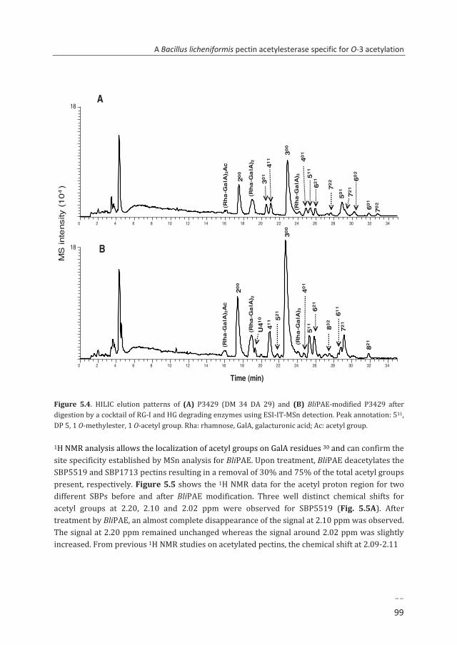
A Bacillus licheniformis pectin acetylesterase specific for O-3 acetylation
99
Figure 5.4. HILIC elution patterns of (A) P3429 (DM 34 DA 29) and (B) BliPAE-modified P3429 after digestion by a cocktail of RG-I and HG degrading enzymes using ESI-IT-MSn detection. Peak annotation: 511, DP 5, 1 O-methylester, 1 O-acetyl group. Rha: rhamnose, GalA, galacturonic acid; Ac: acetyl group.
1H NMR analysis allows the localization of acetyl groups on GalA residues 30 and can confirm the site specificity established by MSn analysis for BliPAE. Upon treatment, BliPAE deacetylates the SBP5519 and SBP1713 pectins resulting in a removal of 30% and 75% of the total acetyl groups present, respectively. Figure 5.5 shows the 1H NMR data for the acetyl proton region for two different SBPs before and after BliPAE modification. Three well distinct chemical shifts for acetyl groups at 2.20, 2.10 and 2.02 ppm were observed for SBP5519 (Fig. 5.5A). After treatment by BliPAE, an almost complete disappearance of the signal at 2.10 ppm was observed. The signal at 2.20 ppm remained unchanged whereas the signal around 2.02 ppm was slightly increased. From previous 1H NMR studies on acetylated pectins, the chemical shift at 2.09-2.11
0 2 4 6 8 10 12 14 16 18 20 22 24 26 28 30 32 34
18
0 2 4 6 8 10 12 14 16 18 20 22 24 26 28 30 32 34
Time (min)
1820
0
300
62130
1
702
411
(Rh
a-G
alA
) 2 401
511
602
50172
2
(Rh
a-G
alA
) 2A
c
(Rh
a-G
alA
) 3
721
601
B
A
MS
inte
nsity
(10
4 )
300
(Rh
a-G
alA
) 2A
c
200
(Rh
a-G
alA
) 2U
410
411 52
1
(Rh
a-G
alA
) 340
151
162
1
832
611
721
821
99
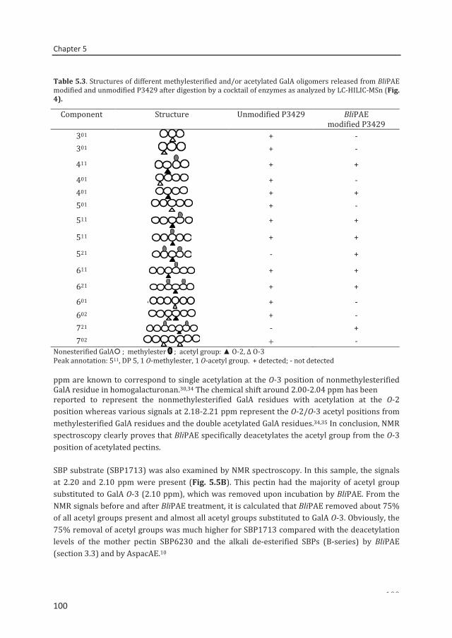
Chapter 5
100
Table 5.3. Structures of different methylesterified and/or acetylated GalA oligomers released from BliPAE modified and unmodified P3429 after digestion by a cocktail of enzymes as analyzed by LC-HILIC-MSn (Fig. 4).
Component Structure Unmodified P3429 BliPAE modified P3429
301 + - 301 + -
411
+ +
401 + - 401 + + 501 + - 511 + +
511 + +
521 - +
611 + +
621 + +
601 + - 602 + - 721 - + 702 + -
Nonesterified GalA ; methylester ; acetyl group: ▲ O-2, Δ O-3 Peak annotation: 511, DP 5, 1 O-methylester, 1 O-acetyl group. + detected; - not detected ppm are known to correspond to single acetylation at the O-3 position of nonmethylesterified GalA residue in homogalacturonan.30,34 The chemical shift around 2.00-2.04 ppm has been reported to represent the nonmethylesterified GalA residues with acetylation at the O-2 position whereas various signals at 2.18-2.21 ppm represent the O-2/O-3 acetyl positions from methylesterified GalA residues and the double acetylated GalA residues.34,35 In conclusion, NMR spectroscopy clearly proves that BliPAE specifically deacetylates the acetyl group from the O-3 position of acetylated pectins.
SBP substrate (SBP1713) was also examined by NMR spectroscopy. In this sample, the signals at 2.20 and 2.10 ppm were present (Fig. 5.5B). This pectin had the majority of acetyl group substituted to GalA O-3 (2.10 ppm), which was removed upon incubation by BliPAE. From the NMR signals before and after BliPAE treatment, it is calculated that BliPAE removed about 75% of all acetyl groups present and almost all acetyl groups substituted to GalA O-3. Obviously, the 75% removal of acetyl groups was much higher for SBP1713 compared with the deacetylation levels of the mother pectin SBP6230 and the alkali de-esterified SBPs (B-series) by BliPAE (section 3.3) and by AspacAE.10
50%
100
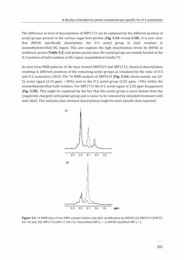
A Bacillus licheniformis pectin acetylesterase specific for O-3 acetylation
101
The difference in level of deacetylation of SBP1713 can be explained by the different position of acetyl groups present in the various sugar beet pectins (Fig. 5.5A versus 5.5B). It is now clear that BliPAE specifically deacetylates the O-3 acetyl group in GalA residues in nonmethylesterified HG region. This also explains the high deacetylation levels by BliPAE in sunflower pectin (Table 5.2) and potato pectin since the acetyl groups are mainly located at the O-3 position of GalA residues in HG region. (unpublished results;30).
As seen from NMR patterns of the base treated SBP5519 and SBP1713, chemical deacetylation resulting in different positions of the remaining acetyl groups as visualized by the ratio of O-3 and O-2 acetylation (30:5). The 1H NMR analysis of SBP5519 (Fig. 5.5A) shows mainly one (O-3) acetyl signal (2.10 ppm; ~30%) next to the O-2 acetyl group (2.02 ppm; ~5%) within the nonmethylesterified GalA residues. For SBP1713 the O-2 acetyl signal at 2.02 ppm disappeared (Fig. 5.5B). This might be explained by the fact that this acetyl group is more distant from the (negatively charged) carboxylate group and is easier to be removed by extended treatment with mild alkali. This indicates that chemical deacetylation might be more specific than expected.
Figure 5.5. 1H NMR data of two SBPs samples before and after modification by BliPAE (A) SBP5519 (DM 55, DA 19) and (B) SBP1713 (DM 17, DA 13). Unmodified SBP (-----); BliPAE-modified SBP (—).
101
Chapter 5
102
5. 4. Conclusions
A recombinant acetylesterase, BliPAE, originating from Bacillus licheniformis DSM13, has been identified and characterized to be a new PAE member of the carbohydrate esterase family 12. It was demonstrated for the first time that a Bacillus PAE specifically removes O-3 linked acetyl groups from the nonmethylesterified HG region of SBP. BliPAE has proven to be more efficient in catalyzing the removal of acetyl groups in SBPs and other O-3 acetylated pectins compared to other bacterial PAEs.
Acknowledgement
This research was supported by the European Community within a consortium PolyModE KBBE-2007-3-3-07 and is gratefully acknowledged. We are grateful to Dr. Stephan Kolkenbrock for gene selection and helpful advice as to enzyme production and purification.
References 1. Quéméner, B., Cabrera Pino, J. C., Ralet, M.-C., Bonnin, E., and Thibault, J.-F. (2003) Journal of Mass
Spectrometry 38, 641-648 2. Thibault, J. F., and Ralet, M. C. (2003) Physico-chemical properties of pectins in the cell walls and
after extraction. in Advances in Pectins and Pectinases Voragen, A. G. J., Schols, H. A., and Visser, R. (eds.), Kluwer Academic Publishers, Dordrecht, The Netherlands. pp 91-105
3. Buchholt, H. C., Christensen, T. M. I. E., Fallesen, B., Ralet, M. C., and Thibault, J. F. (2004) Carbohydrate Polymers 58, 149-161
4. Ralet, M. C., Cabrera, J. C., Bonnin, E., Quémenér, B., Hellín, P., and Thibault, J. F. (2005) Phytochemistry 66, 1832-1843
5. Kim, W. J., Sosulski, F., and Lee, S. C. K. (1978) Journal of Food Science 43, 1436-1439 6. Benen, J. A. E., Alebeek, G. J. W. M., Voragen, C. H. L., and Visser, J. (2003) Mode of action analysis
and structure-function relationships Aspergillus Niger pectinolytic enzymes. in Advances in Pectin and Pectinase Research Voragen, A. G. J., Schols, H. A., and Visser, R. (eds.), Kluwer Academic Publishers, Dordrecht, The Netherlands. pp 235-256
7. Williamson, G. (1991) Phytochemistry 30, 445-449 8. Christensen, T. M. I. E., Nielsen, J. E., Mikkelsen, J. D., Visser, J., and Voragen, A. G. J. (1996)
Isolation, characterization and immuno localization of orange fruit acetyl esterase. in Progress in Biotechnology 14: Pectins and Pectinases Visser, J., Voragen, A. G. J., (eds.), Elsevier, Amsterdam, The Netherlands. pp 723-730
9. Searle-van Leeuwen, M. J. F., Broek, L. A. M., Schols, H. A., Beldman, G., and Voragen, A. G. J. (1992) Applied Microbiology and Biotechnology 38, 347-349
10. Bonnin, E., Clavurier, K., Daniel, S., Kauppinen, S., Mikkelsen, J. D. M., and Thibault, J. F. (2008) Carbohydrate Polymers 74, 411-418
11. Molgaard, A., Kauppinen, S., and Larsen, S. (2000) Structure with Folding & Design 8, 373-383 12. Shevchik, V. E., and Hugouvieux-Cotte-Pattat, N. (2003) Journal of Bacteriology 185, 3091-3100 13. Bolvig, P. U., Pauly, M., Orfila, C., Scheller, H. V., and Schnorr, K. (2003) Sequence analysis and
characterisation of a novel pectin acetyl esterase from Bacillus subtilis. in Advances in Pectin and Pectinase Research Voragen, A. G. J., Schols, H. A., Visser, R. (eds.), Kluwer Academic Publishers, Dordrecht, The Netherlands. pp 315-330
102
A Bacillus licheniformis pectin acetylesterase specific for O-3 acetylation
103
14. Veith, B., Herzberg, C., Steckel, S., Feesche, J., Maurer, K. H., Ehrenreich, P., Bäumer, S., Henne, A., Liesegang, H., Merkl, R., Ehrenreich, A., and Gottschalk, G. (2004) Journal of Molecular Microbiology and Biotechnology 7, 204-211
15. Sengkhamparn, N., Bakx, E. J., Verhoef, R., Schols, H. A., Sajjaanantakul, T., and Voragen, A. G. J. (2009) Carbohydrate Research 344, 1842-1851
16. Schols, H. A., Voragen, A. G. J., and Colquhoun, I. J. (1994) Carbohydrate Research 256, 97-111 17. Kool, M. M., Schols, H. A., Delahaije, R. J. B. M., Sworn, G., Wierenga, P. A., and Gruppen, H. (2013)
Carbohydrate Polymers 97, 368-375 18. Van Gool, M. P., Vancsó, I., Schols, H. A., Toth, K., Szakacs, G., and Gruppen, H. (2011) Bioresource
Technology 102, 6039-6047 19. Remoroza, C., Cord-Landwehr, S., Leijdekkers, A. G. M., Moerschbacher, B. M., Schols, H. A., and
Gruppen, H. (2012) Carbohydrate Polymers 90, 41-48 20. Guillotin, S. E., Van Loey, A., Boulenguer, P., Schols, H. A., and Voragen, A. G. J. (2007) Food
Hydrocolloids 21, 85-91 21. Sambrook, J., and Russell, D. (2001) Molecular Cloning: A Laboratory Manual 2001 Cold Spring
Harbor. in NY Cold Spring Harbor Laboratory Press, New York, USA 22. Studier, F. W. (2005) Protein Expression and Purification 41, 207-234 23. Pouvreau, L., Jonathan, M. C., Kabel, M. A., Hinz, S. W. A., Gruppen, H., and Schols, H. A. (2011)
Enzyme and Microbial Technology 49, 312-320 24. Remoroza, C., Buchholt, H. C., Gruppen, H., and Schols, H. A. (2014) Carbohydrate Polymers 101,
1205-1215 25. Petersen, T. N., Brunak, S., von Heijne, G., and Nielsen, H. (2011) Nature Methods 8, 785-786 26. Choi-Rhee, E., and Cronan, J. E. (2003) Journal of Biological Chemistry 278, 30806-30812 27. Searle-van Leeuwen, M. J. F., Vincken, J. P., Schipper, D., Voragen, A. G. J., Beldman, G., Visser, J.,
and Voragen, A. G. J. (1996) Acetyl esterases of Aspergillus niger: purification and mode of action on pectins. in Progress in Biotechnology 14: Pectin and Pectinases Visser, J., and Voragen, A. G. J. (eds.), Elsevier Amsterdam, The Netherlands. pp 793-798
28. Iglesias, M. T., and Lozano, J. E. (2004) Journal of Food Engineering 62, 215-223 29. Miyamoto, A., and Chang, K. C. (1992) Journal of Food Science 57, 1439-1443 30. Ishii, T. (1997) Plant Physiology 113, 1265-1272 31. Schols, H. A., and Voragen, A. G. J. (1994) Carbohydrate Research 256, 83-95 32. Schols, H. A., Posthumus, M. A., and Voragen, A. G. J. (1990) Carbohydrate Research 206, 117-129 33. Shevchik, V. E., and Hugouvieux-Cotte-Pattat, N. (1997) Molecular Microbiology 24, 1285-1301 34. Perrone, P., Hewage, C. M., Thomson, A. R., Bailey, K., Sadler, I. H., and Fry, S. C. (2002)
Phytochemistry 60, 67-77 35. Renard, C. M. G. C., and Jarvis, M. C. (1999) Carbohydrate Polymers 39, 201-207
103

104
Chapter 6
Mode of action of Bacillus licheniformis
pectin methylesterase on highly
methylesterified and acetylated pectins
C. Remoroza, M. Wagenknecht, H.C. Buchholt, B.M. Moerschbacher,
H. Gruppen, H.A. Schols Submitted for publication
Chapter 6
106
Abstract A gene encoding a putative pectinesterase from Bacillus licheniformis DSM13 was cloned and expressed in Escherichia coli. The resulting recombinant enzyme (BliPME) was purified and characterized as a pectin methylesterase. The enzyme showed a maximum activity at pH 8.0 and 50 oC. BliPME is able to release up to 100% of the methylesters from lime pectin (DM34-76 → DM 0) and up to 73% of all methylesters from SBPs (DM 30-73 → DM 14). BliPME efficiently de-methylesterifies lemon pectins and SBPs in a blockwise manner and is quite tolerant towards the acetyl groups present within the SBPs. Detailed analysis of the BliPME-modified pectins using HILIC-MSn and the classical calcium reactivity measurement showed that the enzyme generates pectins with low methylesterification (lime and SBP) and high acetyl content (SBP) while creating blocks of nonmethylesterified galacturonic acid residues. The high activity of BliPME towards highly methylesterified and acetylated pectins makes the novel esterase more efficient in removing methylesters from highly esterified beet pectin compared to other PMEs, e.g. Aspergillus niger PME.
106
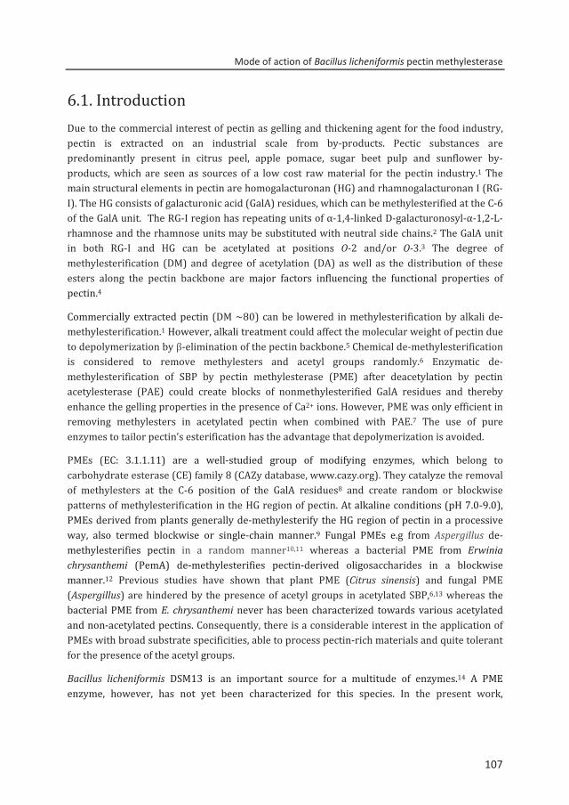
Mode of action of Bacillus licheniformis pectin methylesterase
107
6.1. Introduction Due to the commercial interest of pectin as gelling and thickening agent for the food industry, pectin is extracted on an industrial scale from by-products. Pectic substances are predominantly present in citrus peel, apple pomace, sugar beet pulp and sunflower by-products, which are seen as sources of a low cost raw material for the pectin industry.1 The main structural elements in pectin are homogalacturonan (HG) and rhamnogalacturonan I (RG-I). The HG consists of galacturonic acid (GalA) residues, which can be methylesterified at the C-6 of the GalA unit. The RG-I region has repeating units of α-1,4-linked D-galacturonosyl-α-1,2-L-rhamnose and the rhamnose units may be substituted with neutral side chains.2 The GalA unit in both RG-I and HG can be acetylated at positions O-2 and/or O-3.3 The degree of methylesterification (DM) and degree of acetylation (DA) as well as the distribution of these esters along the pectin backbone are major factors influencing the functional properties of pectin.4 Commercially extracted pectin (DM ~80) can be lowered in methylesterification by alkali de-methylesterification.1 However, alkali treatment could affect the molecular weight of pectin due to depolymerization by β-elimination of the pectin backbone.5 Chemical de-methylesterification is considered to remove methylesters and acetyl groups randomly.6 Enzymatic de-methylesterification of SBP by pectin methylesterase (PME) after deacetylation by pectin acetylesterase (PAE) could create blocks of nonmethylesterified GalA residues and thereby enhance the gelling properties in the presence of Ca2+ ions. However, PME was only efficient in removing methylesters in acetylated pectin when combined with PAE.7 The use of pure enzymes to tailor pectin’s esterification has the advantage that depolymerization is avoided.
PMEs (EC: 3.1.1.11) are a well-studied group of modifying enzymes, which belong to carbohydrate esterase (CE) family 8 (CAZy database, www.cazy.org). They catalyze the removal of methylesters at the C-6 position of the GalA residues8 and create random or blockwise patterns of methylesterification in the HG region of pectin. At alkaline conditions (pH 7.0-9.0), PMEs derived from plants generally de-methylesterify the HG region of pectin in a processive way, also termed blockwise or single-chain manner.9 Fungal PMEs e.g from Aspergillus de-methylesterifies pectin in a random manner10,11 whereas a bacterial PME from Erwinia chrysanthemi (PemA) de-methylesterifies pectin-derived oligosaccharides in a blockwise manner.12 Previous studies have shown that plant PME (Citrus sinensis) and fungal PME (Aspergillus) are hindered by the presence of acetyl groups in acetylated SBP,6,13 whereas the bacterial PME from E. chrysanthemi never has been characterized towards various acetylated and non-acetylated pectins. Consequently, there is a considerable interest in the application of PMEs with broad substrate specificities, able to process pectin-rich materials and quite tolerant for the presence of the acetyl groups.
Bacillus licheniformis DSM13 is an important source for a multitude of enzymes.14 A PME enzyme, however, has not yet been characterized for this species. In the present work,
107
Chapter 6
108
production, purification and characterization of Bacillus licheniformis DSM13 pectin methylesterase (BliPME) were carried out. The purified enzyme was tested on acetylated and non-acetylated pectins with different methylesterification. BliPME-modified pectins were analyzed by enzymatic fingerprinting in combination with HPLC-HILIC-ESI-MSn. The tolerance of BliPME towards the presence of acetyl groups was compared with the well-characterized Aspergillus niger PME.
6.2. Materials and Methods Substrates
Pectins originated from lime pectin, (Mother pectin GrinstedTM Pectin URS 1200: E8100, DM 81, DA 0) and SBP (DM 62, DA 30) were de-esterified by alkali, plant or fungal PMEs by Dupont (Brabrand, Denmark). Esterification of SBP under methanol in acidic solution converted SBP6230 into E7329 pectin (Table 6.1). The chemical properties of the different substrates have been published elsewhere6,15 and are also given in Table 6.1. Methylesterified (saturated and unsaturated) GalA oligomers with degree of polymerization (DP) 2–6 were purified from endo-polygalacturonase II (endo-PGII) and pectin lyase (PL) digests of lime pectin (DM 70, DA 0) as described previously.16
Enzymes
RG-I and HG degrading enzymes were used to hydrolyse SBP as described elsewhere.17 Purified fungal PME from Aspergillus niger (AniPME)18 was used as a comparison to the BliPME.
Amino acid sequence analysis
Homology searches were done using BLAST and databases provided by the NCBI (www.ncbi.nlm.nih.gov). Conserved protein domains were identified using CD-Search, likewise provided by the NCBI. Identification of conserved domains was done also using Pfam.19 For the compilation of multiple alignments and the calculation of identity values ClustalW220 was used. GeneDoc21 was employed for alignment editing and illustration. Secondary structure prediction was done using PredictProtein,22 and SignalP23 and TatP24 were employed for signal peptide predictions.
108
Mode of action of Bacillus licheniformis pectin methylesterase
109
Table 6.1. Chemical characteristics of pectin samples used in this study
Mother pectin De-esterification method
Pectin GalA Rha (%)a
Ara
Gal DM b (%)
DA b (%)
Lime pectin GrinstedTM E 8100 88 1 0.3 4 81 0 Pectin URS
p-PME P5300 90 1 0.3 4 53 0 f-PME F7600 90 2 0.3 3 76 0 F5800 88 1 0.3 3 58 0 Alkali B3400 92 1 0.3 2 34 0 Lime pectinc A6100 75 1 1 5 61 0 Sugar beet pectin
SBP6230 SBP6230 59 5 12 10 62 30 p-PME P5328 58 5 11 10 53 28 f-PME F5129 59 5 12 9 51 29 Alkali B3124 55 6 12 10 31 24 E7329 58 5 8 9 73 29 aMonosaccharide composition in % (w/w).6,15
bMoles methanol (DM) or acetyl groups (DA) per 100 moles of galacturonic acid. cMonosaccharide composition determined in this study.
Production and purification of BliPME
Bacterial strains, media, and growth conditions Bacillus licheniformis DSM13 and Escherichia coli strains NEB 5-alpha (New England Biolabs, Frankfurt a. Main, Germany) and Rosetta 2(DE3)(pLysSRARE2) (Merck, Darmstadt, Germany) were cultivated under conditions specified.17
Cloning of the BliPME coding sequence Template preparation, PCR amplification of the BliPME open reading frame (ORF; GenBank accession number (acc no) AAU42325) using primers 5’-GCAATTCCATATGGTTAATCGGGAGGCAGCTG-3’ (forward) and 5’-CCCGAGCTCTGATTCAACTTTAGGATTCCAGCC-3’ (reverse) and cloning of the ORF were done as described.17 The BliPME expression plasmid generated was termed pET22b-BliPME-StrepIIc.
Synthesis and purification of BliPME E. coli Rosetta 2 (DE3)(pLysSRARE2) was transformed with pET22b-BliPME-StrepIIc and cultivated in 500 ml auto-induction medium25 supplemented with ampicillin and chloramphenicol. Synthesis conditions and details of the purification process can be found elsewhere.17 Other than described there, additional induction using isopropyl-β-D-thiogalactopyranoside was not applied.
109
Chapter 6
110
Characterization of BliPME
Protein analysis Determination of protein contents, SDS-PAGE, staining of gels, Western blot and signal imaging were done as described elsewhere.17
Determination of DM Pectin solution (≈2mg/ml) was saponified in 100 mM NaOH to determine the degree of methylesterification (DM) using a colorimetric method, in which the methanol in the solution is oxidized to formaldehyde with 2,4-pectanedione to the colored product 3,5-diacetyl-1,4-dihydro-2,6-dimethylpyridine.26 This colored product was analyzed spectrophotometrically at 410 nm as previously described.27
Measurement of BliPME activity by titration The PME activity was determined using a thermostated auto-titration system (pH-stat, 719 S Titrino, Metrohm, Herisau, Switzerland) as previously described.17 Pectin solution (5 mg/ml) was maintained at 40 °C, at pH 6.5 (BliPME) and pH 4.7 (AniPME) by the continuous addition of 25 mM NaOH for 10 min. All reaction mixtures were supplemented by NaCl of 100 mM (final concentration) for PME stability and activity enhancement. Enzymes were added at a dose of 0.06% (w/w on protein-substrate basis). PME activity was measured in nanokatal (nkat). One nkat is defined as the amount of enzyme required to release methanol equivalent to 1 nmol NaOH per second under the assay conditions defined.
Determination of optimum temperature and pH Temperature and pH optima were determined using SBP as substrate. SBP E7329 (Table 6.1) was dissolved (5mg/ml) in 50 mM McIlvaine's buffers of different pH. Enzyme was added at a dose of 0.06% (w/w on protein-substrate basis). The pH optimum was determined in a pH range 3.0–9.0 at 40 °C and for 10 min incubation time. Using the same substrate, the temperature optimum was determined within the temperature range 30–80 °C at pH 6.5 for 10 min. The reaction was stopped by heating at 100 °C for 6 min. The methanol released was analyzed spectrophotometrically using the colorimetric method described previously.27 PME activities were calculated relative to the activity at optimal conditions. The background level for the spontaneous de-esterification of E7329 (control) was subtracted for all the data points. After 10 min at pH 8.0-9.0 incubation, about 2% of the total methylesters present was released.
Enzymatic de-methylesterification of pectins Pectin solution (5 mg/ml) containing 100 mM NaCl was maintained at 40 °C and pH 6.5 by the continuous addition of 25 mM NaOH using pH-stat titration. Lime and sugar beet pectins having different DM values were treated with BliPME at a dose of 0.06% (w/w on protein-substrate basis). The incubation was performed for 15 min, 25 min and 24 h. Blanks without enzyme addition were included as references. The reaction was stopped by heating at 100 °C for 6 min. Subsequently, the BliPME-modified and reference pectins were dialyzed against demi-water overnight, followed by freeze-drying. Methylesterified lime pectin oligomers (5 mg/ml) were
110
Mode of action of Bacillus licheniformis pectin methylesterase
111
digested by BliPME at a dose of 0.06% (w/w on protein-substrate basis) in 50 mM McIlvaine's buffers (pH 6.5) containing 100 mM NaCl at 40 °C. The samples were incubated for different time periods (10 min, 1 h and 24 h) and the reaction was stopped by heating at 100 °C for 6 min. The determination of calcium reactivity test was carried out using lime pectin (DM 61, DA 0). Pectin sample (10 mg/ml) was maintained at pH 5.0 and at 40 °C by the continuous addition of 100 mM NaOH for 25 h. BliPME was added at a dose of ~ 0.02% (w/w on protein-substrate basis). Using lime pectin (DM 61, DA 0) sample (10 mg/ml) containing 100 mM NaCl was maintained at pH 5.0 (by addition of 100 mM NaOH) and incubated for 2.5 h. All reactions were stopped by heating at 100 °C for 6 min.
Fingerprinting of BliPME-modified pectins Freeze-dried BliPME modified pectins were dissolved (5 mg/ml) in 50 mM sodium citrate buffer at (pH 5.0) and digested using the pectolytic enzyme cocktail containing RG-I and HG degrading enzymes at 40 °C as described elsewhere.17
Hydrophilic interaction liquid chromatography (HILIC) Pectin digests, diluted to 1 mg/ml in 50% (v/v) acetonitrile, were analysed using an HPLC system coupled to an evaporative light scattering detector (ELSD) and an ESI-IT-MSn-detector. Chromatographic separation was performed on an Acquity UPLC BEH Amide column in combination with a Van Guard pre-column (Waters Corporation, Milford, MA, USA) as described previously.17
Calcium reactivity measurement of the modified lime pectins Non-acetylated BliPME-modified pectin samples were dissolved (10 mg/ml) in 0.5 M sodium acetate buffer (pH 5.0) in the absence or presence of 20-40 mM of Ca2+ ions according to the method described elsewhere.6
6.3. Results Amino acid sequence analysis
BliPME, originating from B. licheniformis DSM13, comprises 317 amino acids (aa) and has a calculated molecular mass of 35.1 kDa. In the corresponding GenBank entry (acc no AAU42325) it is denominated as a putative pectinesterase and in the CAZy database (www.cazy.org) it is assigned to CE family 8.
BLASTP analysis revealed closest relation of BliPME to a 317-aa pectinesterase of B. sonorensis L12 (acc no EME73130; 79.8% aa identity). Similarity was also found for a putative pectinesterase from B. infantis NRRL B-14911 (acc no AGX04064; 313 aa, 60.1% aa identity) and for two larger PME proteins from B. endophyticus (acc no WP_019394632; 495 aa, 59.3% aa identity) and from Clostridium thermocellum DSM1313 (acc no YP_005687040; 567 aa, 49.8% aa identity), respectively. For the esterase from B. endophyticus, similarity was seen for the C-
111
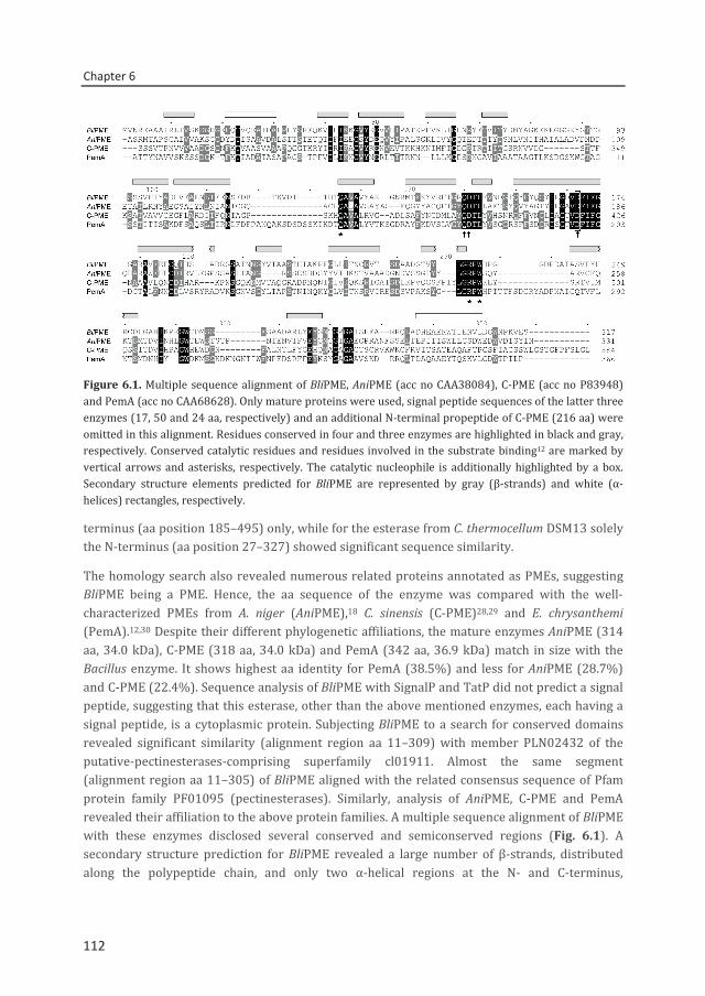
Chapter 6
112
Figure 6.1. Multiple sequence alignment of BliPME, AniPME (acc no CAA38084), C-PME (acc no P83948) and PemA (acc no CAA68628). Only mature proteins were used, signal peptide sequences of the latter three enzymes (17, 50 and 24 aa, respectively) and an additional N-terminal propeptide of C-PME (216 aa) were omitted in this alignment. Residues conserved in four and three enzymes are highlighted in black and gray, respectively. Conserved catalytic residues and residues involved in the substrate binding12 are marked by vertical arrows and asterisks, respectively. The catalytic nucleophile is additionally highlighted by a box. Secondary structure elements predicted for BliPME are represented by gray (β-strands) and white (α-helices) rectangles, respectively.
terminus (aa position 185–495) only, while for the esterase from C. thermocellum DSM13 solely the N-terminus (aa position 27–327) showed significant sequence similarity.
The homology search also revealed numerous related proteins annotated as PMEs, suggesting BliPME being a PME. Hence, the aa sequence of the enzyme was compared with the well-characterized PMEs from A. niger (AniPME),18 C. sinensis (C-PME)28,29 and E. chrysanthemi (PemA).12,30 Despite their different phylogenetic affiliations, the mature enzymes AniPME (314 aa, 34.0 kDa), C-PME (318 aa, 34.0 kDa) and PemA (342 aa, 36.9 kDa) match in size with the Bacillus enzyme. It shows highest aa identity for PemA (38.5%) and less for AniPME (28.7%) and C-PME (22.4%). Sequence analysis of BliPME with SignalP and TatP did not predict a signal peptide, suggesting that this esterase, other than the above mentioned enzymes, each having a signal peptide, is a cytoplasmic protein. Subjecting BliPME to a search for conserved domains revealed significant similarity (alignment region aa 11–309) with member PLN02432 of the putative-pectinesterases-comprising superfamily cl01911. Almost the same segment (alignment region aa 11–305) of BliPME aligned with the related consensus sequence of Pfam protein family PF01095 (pectinesterases). Similarly, analysis of AniPME, C-PME and PemA revealed their affiliation to the above protein families. A multiple sequence alignment of BliPME with these enzymes disclosed several conserved and semiconserved regions (Fig. 6.1). A secondary structure prediction for BliPME revealed a large number of β-strands, distributed along the polypeptide chain, and only two α-helical regions at the N- and C-terminus,
112
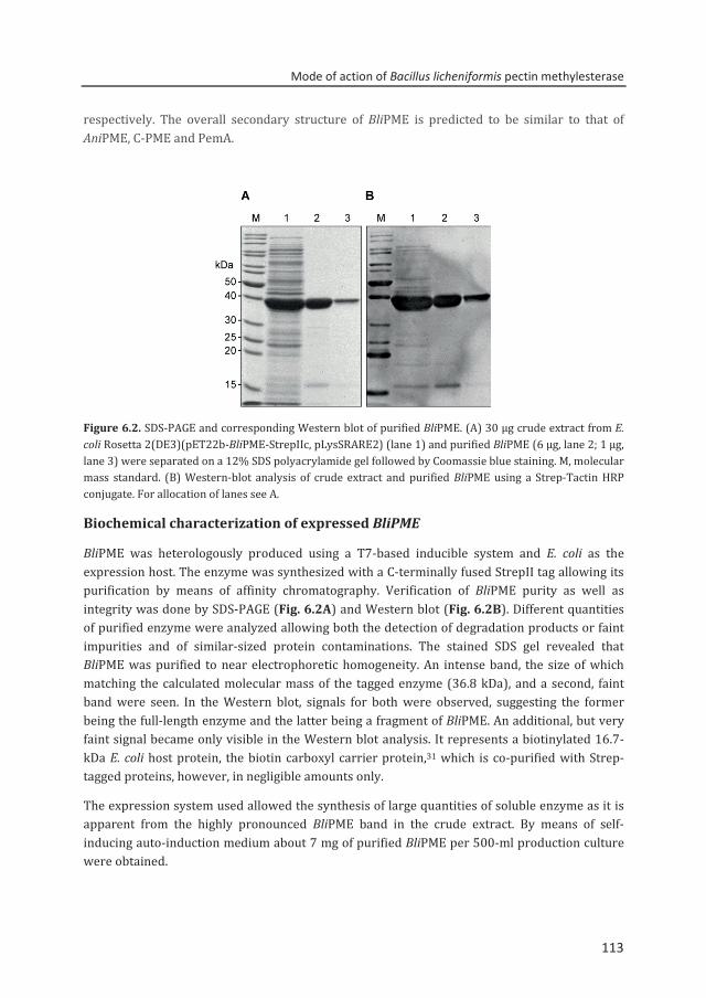
Mode of action of Bacillus licheniformis pectin methylesterase
113
respectively. The overall secondary structure of BliPME is predicted to be similar to that of AniPME, C-PME and PemA.
Figure 6.2. SDS-PAGE and corresponding Western blot of purified BliPME. (A) 30 µg crude extract from E. coli Rosetta 2(DE3)(pET22b-BliPME-StrepIIc, pLysSRARE2) (lane 1) and purified BliPME (6 µg, lane 2; 1 µg, lane 3) were separated on a 12% SDS polyacrylamide gel followed by Coomassie blue staining. M, molecular mass standard. (B) Western-blot analysis of crude extract and purified BliPME using a Strep-Tactin HRP conjugate. For allocation of lanes see A.
Biochemical characterization of expressed BliPME
BliPME was heterologously produced using a T7-based inducible system and E. coli as the expression host. The enzyme was synthesized with a C-terminally fused StrepII tag allowing its purification by means of affinity chromatography. Verification of BliPME purity as well as integrity was done by SDS-PAGE (Fig. 6.2A) and Western blot (Fig. 6.2B). Different quantities of purified enzyme were analyzed allowing both the detection of degradation products or faint impurities and of similar-sized protein contaminations. The stained SDS gel revealed that BliPME was purified to near electrophoretic homogeneity. An intense band, the size of which matching the calculated molecular mass of the tagged enzyme (36.8 kDa), and a second, faint band were seen. In the Western blot, signals for both were observed, suggesting the former being the full-length enzyme and the latter being a fragment of BliPME. An additional, but very faint signal became only visible in the Western blot analysis. It represents a biotinylated 16.7-kDa E. coli host protein, the biotin carboxyl carrier protein,31 which is co-purified with Strep-tagged proteins, however, in negligible amounts only.
The expression system used allowed the synthesis of large quantities of soluble enzyme as it is apparent from the highly pronounced BliPME band in the crude extract. By means of self-inducing auto-induction medium about 7 mg of purified BliPME per 500-ml production culture were obtained.
113
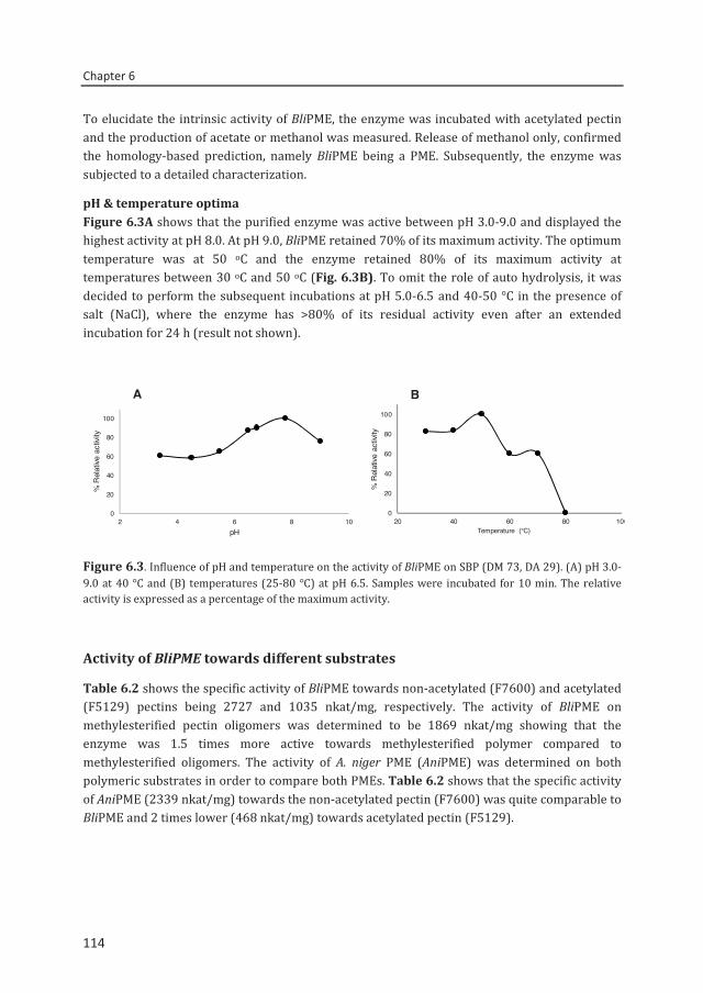
Chapter 6
114
To elucidate the intrinsic activity of BliPME, the enzyme was incubated with acetylated pectin and the production of acetate or methanol was measured. Release of methanol only, confirmed the homology-based prediction, namely BliPME being a PME. Subsequently, the enzyme was subjected to a detailed characterization.
pH & temperature optima Figure 6.3A shows that the purified enzyme was active between pH 3.0-9.0 and displayed the highest activity at pH 8.0. At pH 9.0, BliPME retained 70% of its maximum activity. The optimum temperature was at 50 oC and the enzyme retained 80% of its maximum activity at temperatures between 30 oC and 50 oC (Fig. 6.3B). To omit the role of auto hydrolysis, it was decided to perform the subsequent incubations at pH 5.0-6.5 and 40-50 °C in the presence of salt (NaCl), where the enzyme has >80% of its residual activity even after an extended incubation for 24 h (result not shown).
Figure 6.3. Influence of pH and temperature on the activity of BliPME on SBP (DM 73, DA 29). (A) pH 3.0-9.0 at 40 °C and (B) temperatures (25-80 °C) at pH 6.5. Samples were incubated for 10 min. The relative activity is expressed as a percentage of the maximum activity.
Activity of BliPME towards different substrates
Table 6.2 shows the specific activity of BliPME towards non-acetylated (F7600) and acetylated (F5129) pectins being 2727 and 1035 nkat/mg, respectively. The activity of BliPME on methylesterified pectin oligomers was determined to be 1869 nkat/mg showing that the enzyme was 1.5 times more active towards methylesterified polymer compared to methylesterified oligomers. The activity of A. niger PME (AniPME) was determined on both polymeric substrates in order to compare both PMEs. Table 6.2 shows that the specific activity of AniPME (2339 nkat/mg) towards the non-acetylated pectin (F7600) was quite comparable to BliPME and 2 times lower (468 nkat/mg) towards acetylated pectin (F5129).
0
20
40
60
80
100
2 4 6 8 10
% R
elat
ive a
ctivi
ty
pH
0
20
40
60
80
100
20 40 60 80 100
% R
elat
ive a
ctivi
ty
Temperature (°C)
A B
114
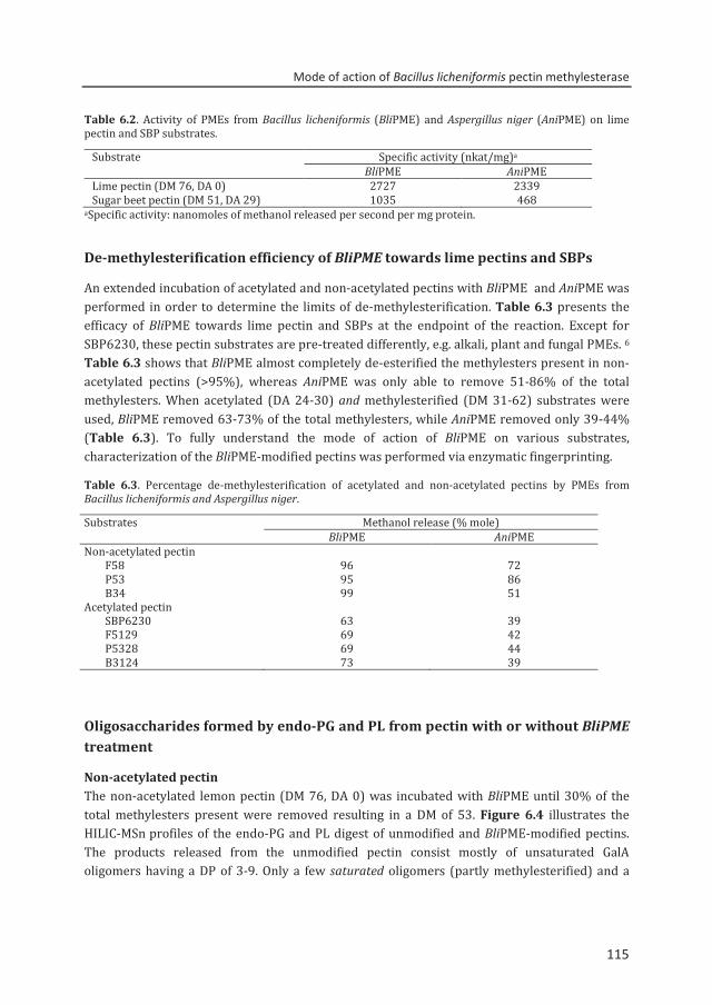
Mode of action of Bacillus licheniformis pectin methylesterase
115
Table 6.2. Activity of PMEs from Bacillus licheniformis (BliPME) and Aspergillus niger (AniPME) on lime pectin and SBP substrates.
Substrate Specific activity (nkat/mg)a BliPME AniPME Lime pectin (DM 76, DA 0) 2727 2339 Sugar beet pectin (DM 51, DA 29) 1035 468
aSpecific activity: nanomoles of methanol released per second per mg protein.
De-methylesterification efficiency of BliPME towards lime pectins and SBPs
An extended incubation of acetylated and non-acetylated pectins with BliPME and AniPME was performed in order to determine the limits of de-methylesterification. Table 6.3 presents the efficacy of BliPME towards lime pectin and SBPs at the endpoint of the reaction. Except for SBP6230, these pectin substrates are pre-treated differently, e.g. alkali, plant and fungal PMEs. 6 Table 6.3 shows that BliPME almost completely de-esterified the methylesters present in non-acetylated pectins (>95%), whereas AniPME was only able to remove 51-86% of the total methylesters. When acetylated (DA 24-30) and methylesterified (DM 31-62) substrates were used, BliPME removed 63-73% of the total methylesters, while AniPME removed only 39-44% (Table 6.3). To fully understand the mode of action of BliPME on various substrates, characterization of the BliPME-modified pectins was performed via enzymatic fingerprinting.
Table 6.3. Percentage de-methylesterification of acetylated and non-acetylated pectins by PMEs from Bacillus licheniformis and Aspergillus niger.
Substrates Methanol release (% mole) BliPME AniPME Non-acetylated pectin
F58 96 72 P53 95 86 B34 99 51
Acetylated pectin SBP6230 63 39 F5129 69 42 P5328 69 44 B3124 73 39
Oligosaccharides formed by endo-PG and PL from pectin with or without BliPME treatment
Non-acetylated pectin The non-acetylated lemon pectin (DM 76, DA 0) was incubated with BliPME until 30% of the total methylesters present were removed resulting in a DM of 53. Figure 6.4 illustrates the HILIC-MSn profiles of the endo-PG and PL digest of unmodified and BliPME-modified pectins. The products released from the unmodified pectin consist mostly of unsaturated GalA oligomers having a DP of 3-9. Only a few saturated oligomers (partly methylesterified) and a
115
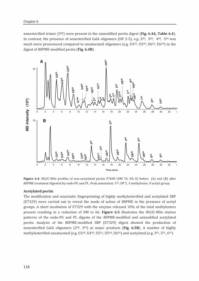
Chapter 6
116
nonesterified trimer (300) were present in the unmodified pectin digest (Fig. 6.4A, Table 6.4). In contrast, the presence of nonesterified GalA oligomers (DP 2-5), e.g. 200, 300, 400, 500 was much more pronounced compared to unsaturated oligomers (e.g. U320, U540, U620, U630) in the digest of BliPME-modified pectin (Fig. 6.4B).
Figure 6.4. HILIC-MSn profiles of non-acetylated pectin F7600 (DM 76, DA 0) before (A) and (B) after BliPME treatment digested by endo-PG and PL. Peak annotation: 530, DP 5, 3 methylester, 0 acetyl group.
Acetylated pectin The modification and enzymatic fingerprinting of highly methylesterified and acetylated SBP (E7329) were carried out to reveal the mode of action of BliPME in the presence of acetyl groups. A short incubation of E7329 with the enzyme released 10% of the total methylesters present resulting in a reduction of DM to 66. Figure 6.5 illustrates the HILIC-MSn elution patterns of the endo-PG and PL digests of the BliPME-modified and unmodified acetylated pectin. Analysis of the BliPME-modified SBP (E7329) digest showed the production of nonesterified GalA oligomers (200, 300) as major products (Fig. 6.5B). A number of highly methylesterified unsaturated (e.g. U320, U430, U541, U540, U650) and acetylated (e.g. 301, 501, 601)
0 2 4 6 8 10 12 14 16 18 20 22 24 26 28 30 32 34
0 2 4 6 8 10 12 14 16 18 20 22 24 26 28 30 32 34
Time (min)
25
25
MS
inte
nsity
(10
4 )
A
B
U320
U430
U540
520
U650
U760
U730
U840
310
930
1030
U420
U410
U630
300 410
510
620
U940
720
U530
U640
530
U830
300
200
400
500
U630
U320
U650
U530 U
620
U430 U
540
U420
U410
530
U830
820 920U930
300
116
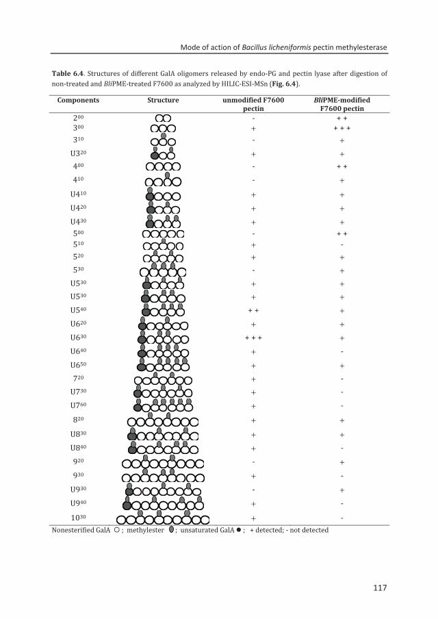
Mode of action of Bacillus licheniformis pectin methylesterase
117
Table 6.4. Structures of different GalA oligomers released by endo-PG and pectin lyase after digestion of non-treated and BliPME-treated F7600 as analyzed by HILIC-ESI-MSn (Fig. 6.4).
Components Structure unmodified F7600 pectin
BliPME-modified F7600 pectin
200 - + + 300 + + + + 310 - +
U320 + +
400 - + + 410 - +
U410 + + U420 + + U430 + + 500 - + + 510 + - 520 + + 530 - +
U530 + + U530
+ + U540 + + + U620
+ + U630 + + + + U640 + - U650 + + 720 + -
U730 + -
U760 + -
820 + +
U830 + +
U840 + -
920 - +
930 + -
U930 - +
U940 + -
1030 + -
Nonesterified GalA ; methylester ; unsaturated GalA ; + detected; - not detected
117
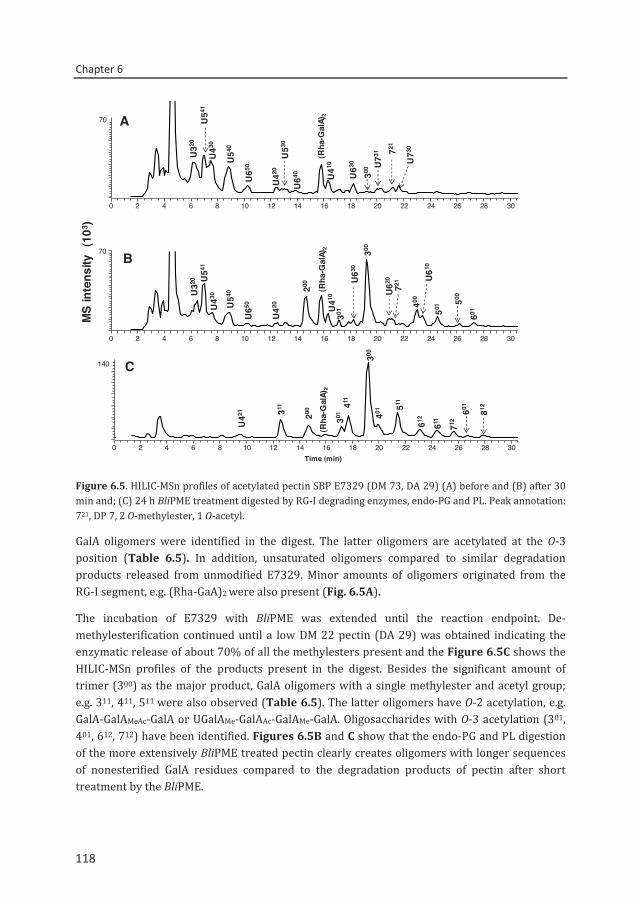
Chapter 6
118
Figure 6.5. HILIC-MSn profiles of acetylated pectin SBP E7329 (DM 73, DA 29) (A) before and (B) after 30 min and; (C) 24 h BliPME treatment digested by RG-I degrading enzymes, endo-PG and PL. Peak annotation: 721, DP 7, 2 O-methylester, 1 O-acetyl.
GalA oligomers were identified in the digest. The latter oligomers are acetylated at the O-3 position (Table 6.5). In addition, unsaturated oligomers compared to similar degradation products released from unmodified E7329. Minor amounts of oligomers originated from the RG-I segment, e.g. (Rha-GaA)2 were also present (Fig. 6.5A).
The incubation of E7329 with BliPME was extended until the reaction endpoint. De-methylesterification continued until a low DM 22 pectin (DA 29) was obtained indicating the enzymatic release of about 70% of all the methylesters present and the Figure 6.5C shows the HILIC-MSn profiles of the products present in the digest. Besides the significant amount of trimer (300) as the major product, GalA oligomers with a single methylester and acetyl group; e.g. 311, 411, 511 were also observed (Table 6.5). The latter oligomers have O-2 acetylation, e.g. GalA-GalAMeAc-GalA or UGalAMe-GalAAc-GalAMe-GalA. Oligosaccharides with O-3 acetylation (301, 401, 612, 712) have been identified. Figures 6.5B and C show that the endo-PG and PL digestion of the more extensively BliPME treated pectin clearly creates oligomers with longer sequences of nonesterified GalA residues compared to the degradation products of pectin after short treatment by the BliPME.
0 2 4 6 8 10 12 14 16 18 20 22 24 26 28 30
0 2 4 6 8 10 12 14 16 18 20 22 24 26 28 30Time (min)
0 2 4 6 8 10 12 14 16 18 20 22 24 26 28 30
U410
300
(Rha
-Gal
A)2
U63
0
U42
0
U43
0U
541
U540U
320
U65
0 U73
1U530
U640
721
U73
0
U410
200 (Rha
-Gal
A)2
501
301
U63
0
U42
0U430
U54
1
U540U
320
U65
0
U610
400 500
601
300
511411
U42
1 812
601
401311
612200
301
300
MS
inte
nsity
(10
3 )
70
70
140
611
712
U620
721
A
B
C
(Rha
-Gal
A)2
118
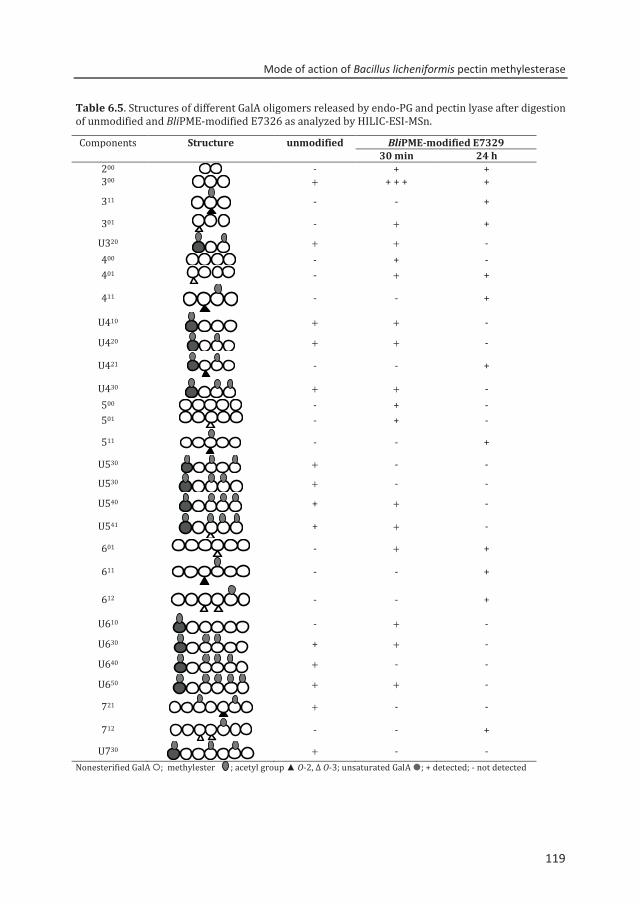
Mode of action of Bacillus licheniformis pectin methylesterase
119
Table 6.5. Structures of different GalA oligomers released by endo-PG and pectin lyase after digestion of unmodified and BliPME-modified E7326 as analyzed by HILIC-ESI-MSn.
Components Structure unmodified BliPME-modified E7329 30 min 24 h
200 - + + 300 + + + + +
311
- - +
301 - + +
U320 + + -
400 - + - 401 - + +
411
- - +
U410 + + -
U420 + + -
U421
- - +
U430 + + - 500 - + - 501 - + -
511
- - +
U530 + - -
U530 + - -
U540 + + -
U541
+ + -
601 - + +
611
- - +
612
- - +
U610 - + -
U630 + + -
U640 + - -
U650 + + -
721
+ - -
712 - - +
U730 + - -
Nonesterified GalA ; methylester ; acetyl group ▲ O-2, Δ O-3; unsaturated GalA ; + detected; - not detected
119
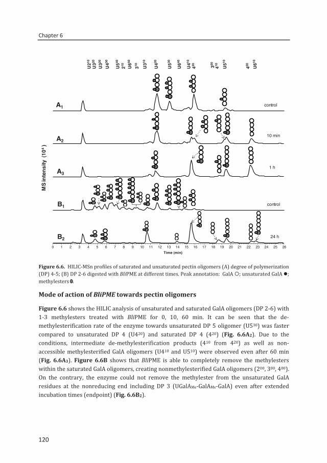
Chapter 6
120
Figure 6.6. HILIC-MSn profiles of saturated and unsaturated pectin oligomers (A) degree of polymerization (DP) 4-5; (B) DP 2-6 digested with BliPME at different times. Peak annotation: GalA ; unsaturated GalA ; methylesters .
Mode of action of BliPME towards pectin oligomers
Figure 6.6 shows the HILIC analysis of unsaturated and saturated GalA oligomers (DP 2-6) with 1-3 methylesters treated with BliPME for 0, 10, 60 min. It can be seen that the de-methylesterification rate of the enzyme towards unsaturated DP 5 oligomer (U530) was faster compared to unsaturated DP 4 (U420) and saturated DP 4 (420) (Fig. 6.6A2). Due to the conditions, intermediate de-methylesterification products (410 from 420) as well as non-accessible methylesterified GalA oligomers (U410 and U510) were observed even after 60 min (Fig. 6.6A3). Figure 6.6B shows that BliPME is able to completely remove the methylesters within the saturated GalA oligomers, creating nonmethylesterified GalA oligomers (200, 300, 400). On the contrary, the enzyme could not remove the methylester from the unsaturated GalA residues at the nonreducing end including DP 3 (UGalAMe-GalAMe-GalA) even after extended incubation times (endpoint) (Fig. 6.6B2).
control
10 min
1 h
0 1 2 3 4 5 6 7 8 9 10 11 12 13 14 15 16 17 18 19 20 21 22 23 24 25 26Time (min)
control
24 h
A1
B1
MS
inte
nsity
(10
4)
A2
A3
B2
U210
U320
U320
U430
U540
210 U650
U310
310 U420
U530
U640
420U410
410 U510
400 U610
300
120
Mode of action of Bacillus licheniformis pectin methylesterase
121
Calcium reactivity test of the modified lime pectins
Lime pectin (DM 61, DA 0) and BliPME-modified lime pectins (DM 58, DA 0) and (DM 40, DA 0) were subjected to a calcium reactivity test. The BliPME-modified pectin (DM 58, DA 0) slightly precipitated when Ca2+ ions were added at concentrations of 20 and 40 mM, whereas the unmodified pectin (DM 61, DA 0) did not precipitate upon Ca2+ ions addition. Moreover, the strongly BliPME-treated pectin, which resulted to low methylesterified pectin (DM 40, DA 0), showed precipitation in the presence of 20 and 40 mM Ca2+ ions.
6.4. Discussion In this study, we produced and characterized a bacterial PME from B. licheniformis DSM13. Comparison of its protein sequence with the well-studied PMEs PemA, AniPME and C-PME revealed similar sizes and several conserved and semiconserved regions (Fig. 6.1). Among such enzymes, highest overall aa sequence identity (38.5%) was seen for BliPME and the likewise bacterial PemA of E. chrysanthemi. Based on crystallographic studies of the latter enzyme, a right-handed parallel β-helix structure was found,30 being distinct from the common α/β hydrolase fold and of the catalytic Ser-His-Asp triad of most esterases.32 The high level of resemblance in the predicted secondary structures of the Bacillus enzyme and PemA suggests BliPME to be a parallel β-helix protein as well.
In PemA, Asp178 acts as the general acid-base catalyst while Asp199 is the nucleophile in the reaction mechanism.12 Gln177 is suggested to form the oxyanion hole, stabilizing the negative charge of the transition state. Gln153, Arg267 and Trp269 are involved in the substrate binding. All above six residues are present in BliPME (Asp149, Asp170, Gln148; Gln126, Arg232, Trp234) (Fig. 6.1), suggesting these two bacterial PMEs to share the same reaction mechanism. Besides the high similarity of conserved regions as well as the predicted secondary structure between BliPME and PemA, both bacterial enzymes have their optimal activity at neutral and alkaline conditions.12,33 On the contrary, Aspergillus PMEs are generally active at more acidic conditions34,35 compared to bacterial or plant- derived PMEs. Moreover, BliPME performed optimally at low and moderately high temperatures. Although quite similar, both bacterial PMEs (BliPME and PemA) cannot be compared in great detail since PemA (or other PMEs) was not analyzed on various methylesterified acetylated and non-acetylated pectins.
As far as we know, this is the first time that detailed experimental evidence is provided on an enzyme from B. licheniformis DSM13 having a PME activity. Obviously, BliPME has distinct features compared with the bacterial PemA, fungal AniPME and plant C-PME enzymes. Apart from the substantial higher de-methylesterification activity toward acetylated pectins tested, BliPME de-methylesterifies the pectins in a blockwise manner irrespective of the methylesters and/or acetyl groups distribution.
In this study, BliPME shows to be 2.5 times more efficient in catalyzing the removal of methylesters in non-acetylated pectin compared to acetylated pectin and methylesterified
121
Chapter 6
122
pectin oligomers. Although the degree of methylesterification (DM 34-58) and distribution of methylesters are different in the non-acetylated lime pectins,11 BliPME rapidly de-methylesterified the lime pectins almost completely. This can be attributed to high binding affinity of BliPME towards the substrate. In addition, the bacterial enzyme was not limited by the methylester distribution in non-acetylated lime pectins and could also release the last methylesters from modified low DM pectins. On the contrary, the de-methylesterification of the same pectins using AniPME stopped when DM reached ~16. These values are in agreement with previous studies.11,15
The rate of de-methylesterification was different when acetylated substrates were used. BliPME is clearly active towards methylesterified and acetylated pectin and its activity was two times higher compared to AniPME. The findings for BliPME demonstrate that the enzyme has a high tolerance on highly acetylated SBPs, hence it is able to release ~70% of the total methylesters present in SBPs. Apparently, BliPME is quite tolerant for the presence of acetyl groups. So far, this information has not been reported for other bacterial PME.
Enzymatic fingerprinting of non-acetylated and acetylated pectin was done in order to understand the mode of action of BliPME. From the oligosaccharides profile as shown in Figures 6.4 and 6.5 it can be concluded that the enzyme creates blocks of nonmethylesterified GalA sequences. Although the blockwise de-methylesterification pattern was observed theoretically leading to blockwise methylesterified segments remaining, unsaturated GalA oligomers were present in rather lower amounts compared to the nonesterified GalA oligomers in the digests of non-acetylated and acetylated pectins. This indicates the formation of relative short blocks of nonmethylesterified sequences in the HG region of BliPME-treated pectin. The blockwise acting BliPME strongly binds to non-acetylated lime pectin (F7600) and acetylated SBP (F5129) having a random distribution of methylesters.
The influence of acetyl groups on BliPME activity was also demonstrated. After BliPME treatment and enzymatic fingerprinting (Fig. 6.5), generation of both nonesterified and nonmethylesterified O-3-acetylated GalA oligomers such as 401, 501, 601 was observed. Such degradation products have also been observed after the enzymatic fingerprinting of plant PME-modified SBPs having a low DM as described elsewhere.36 On the contrary, the BliPME could not completely remove the methylesters from the GalA residues within a pectin, especially when the adjacent or the same GalA residue is O-2 acetylated, e.g. 311 GalA-GalAMeAc-GalA. This indicates that BliPME can release more methylesters from the methylesterified GalA residues in case that the acetylation is at the O-3 position and not at the O-2 position (Table 6.5). Based on the above results, it can be hypothesized that a steric shielding effect of the acetyl groups at the O-2 position hinder the enzyme from binding with the substrate.
When testing saturated and unsaturated methylesterified GalA oligomers, BliPME de-methylesterify completely the methylesters from the saturated oligomers, which generates nonmethylesterified oligomers. BliPME could not remove the methylester from the unsaturated GalA residue within a GalA oligomer, which is in contrast with the previous findings.18 AniPME
122
Mode of action of Bacillus licheniformis pectin methylesterase
123
de-methylesterifies the methylester from the unsaturated GalA on its non-reducing end. Exceptionally, unsaturated DP 3 oligomer with 2 methylesters, UGalAMe-GalAMe-GalA could be one of the limitations of the BliPME enzyme. It can be predicted that the presence of a nonesterified GalA residue and a methylesterified GalA residue is the enzyme requirement as -1 and +1 subsites for binding. From the available methylesterified GalA oligomers (Fig. 6.6), it cannot be concluded whether BliPME is able to remove a methylester from both the non-reducing end or reducing end of the saturated GalA oligomers.
Based on the above observation, BliPME seemed to introduce a single-chain or blockwise removal of methylesters on non-acetylated pectins. This is confirmed by the results of the Ca2+ precipitation. The enzyme prefers pectin polymers rather than pectin oligomers. In contrast to plant PMEs, BliPME does not form large de-methylesterified blocks but creates many small blocks of nonesterified GalA sequences.
Furthermore, BliPME is quite tolerant to acetyl groups at the O-3 position present in the HG region of SBP and it has been proven to be more efficient in creating a blockwise structure on acetylated pectins compared to other reported PMEs.
Acknowledgements This research was supported by the European Community within a consortium PolyModE KBBE-2007-3-3-07. We are grateful to Dr. Stephan Kolkenbrock for gene selection and helpful advice as to enzyme production and purification. We also like to thank Rianne Willems of the Laboratory of Food Chemistry, Wageningen University, The Netherlands for providing the pectin oligomers used in the study.
References 1. May, C. D. (1990) Carbohydrate Polymers 12, 79-99 2. Voragen, A. G. J., Coenen, G. J., Verhoef, R. P., and Schols, H. A. (2009) Structural Chemistry 20, 263-
275 3. Voragen, A. G. J., Pilnik, W., Thibault, J.-F., Axelos, M. A. V., and Renard, C. M. G. C. (1995) Pectins. in
Food Polysaccharides and Their Applications Stephen, A. M. (ed.), M. Dekker, New-York, N.Y., USA. pp 287-339
4. Willats, W. G. T., Knox, J. P., and Mikkelsen, J. D. (2006) Trends in Food Science & Technology 17, 97-104
5. Renard, C., and Thibault, J.-F. (1996) Pectins in mild alkaline conditions: β-elimination and kinetics of demethylation. in Progress in Biotechnology:14 Pectins and Pectinases Voragen, A. G. J., and Visser, J. (eds.), Elsevier, Amsterdam, The Netherlands. pp 603-608
6. Buchholt, H. C., Christensen, T. M. I. E., Fallesen, B., Ralet, M. C., and Thibault, J. F. (2004) Carbohydrate Polymers 58, 149-161
7. Oosterveld, A., Beldman, G., Searle-Van Leeuwen, M. J. F., and Voragen, A. G. J. (2000) Carbohydrate Polymers 43, 249-256
8. Fraeye, I., Duvetter, T., Doungla, E., Van Loey, A., and Hendrickx, M. (2010) Trends in Food Science & Technology 21, 219-228
9. Micheli, F. (2001) Trends in Plant Science 6, 414-419
123
Chapter 6
124
10. Kim, Y., Williams, M. A. K., Galant, A. L., Luzio, G. A., Savary, B. J., Vasu, P., and Cameron, R. G. (2013) Food Hydrocolloids 33, 132-141
11. Limberg, G., Körner, R., Buchholt, H. C., Christensen, T. M. I. E., Roepstorff, P., and Mikkelsen, J. D. (2000) Carbohydrate Research 327, 293-307
12. Fries, M., Ihrig, J., Brocklehurst, K., Shevchik, V. E., and Pickersgill, R. W. (2007) European Molecular Biology Organization 26, 3879-3887
13. Duvetter, T., Fraeye, I., Sila, D. N., Verlent, I., Smout, C., Hendrickx, M., and Van Loey, A. (2006) Journal of Agricultural and Food Chemistry 54, 7825-7831
14. Veith, B., Herzberg, C., Steckel, S., Feesche, J., Maurer, K. H., Ehrenreich, P., Bäumer, S., Henne, A., Liesegang, H., Merkl, R., Ehrenreich, A., and Gottschalk, G. (2004) Journal of Molecular Microbiology and Biotechnology 7, 204-211
15. Ralet, M.-C., Dronnet, V., Buchholt, H. C., and Thibault, J.-F. (2001) Carbohydrate Research 336, 117-125
16. Van Alebeek, G. J. W. M., Zabotina, O., Beldman, G., Schols, H. A., and Voragen, A. G. J. (2000) Journal of Mass Spectrometry 35, 831-840
17. Remoroza, C., Wagenknecht, M., Gu, F., Buchholt, H. C., Moerschbacher, B. M., Schols, H. A., and Gruppen, H. (2014) Accepted for publication in Carbohydrate Polymers. This thesis, chapter 5
18. Van Alebeek, G. J. W. M., Van Scherpenzeel, K., Beldman, G., Schols, H. A., and Voragen, A. G. J. (2003) Biochemical Journal 372, 211-218
19. Punta, M., Coggill, P. C., Eberhardt, R. Y., Mistry, J., Tate, J., Boursnell, C., Pang, N., Forslund, K., Ceric, G., and Clements, J. (2012) Nucleic Acids Research 40, D290-D301
20. Larkin, M., Blackshields, G., Brown, N., Chenna, R., McGettigan, P. A., McWilliam, H., Valentin, F., Wallace, I. M., Wilm, A., and Lopez, R. (2007) Bioinformatics 23, 2947-2948
21. Nicholas, K., Nicholas H., and Deerfield, D. EMBNET News. (1997); 4: 1-4. 22. Rost, B., Yachdav, G., and Liu, J. (2004) Nucleic Acids Research 32, W321-W326 23. Petersen, T. N., Brunak, S., von Heijne, G., and Nielsen, H. (2011) Nature Methods 8, 785-786 24. Bendtsen, J. D., Kiemer, L., Fausbøll, A., and Brunak, S. (2005) BMC Microbiology 5, 58 25. Studier, F. W. (2005) Protein Expression and Purification 41, 207-234 26. Wood, P. J., and Siddiqui, I. R. (1971) Analytical Biochemistry 39, 418-428 27. Guillotin, S. E., Van Loey, A., Boulenguer, P., Schols, H. A., and Voragen, A. G. J. (2007) Food
Hydrocolloids 21, 85-91 28. Versteeg, C., Rombouts, F. M., and Pilnik, W. (1978) Lebensmittel-Wissenschaft & Technologie 11,
267-274 29. Christensen, T. M. I. E., Nielsen, J. E., Kreiberg, J. D., Rasmussen, P., and Mikkelsen, J. D. (1998)
206, 493-503 30. Jenkins, J., Mayans, O., Smith, D., Worboys, K., and Pickersgill, R. W. (2001) Journal of Molecular
Biology 305, 951-960 31. Choi-Rhee, E., and Cronan, J. E. (2003) Journal of Biological Chemistry 278, 30806-30812 32. Huo, Y., Miao, J., Liu, B., Yang, Y., Zhang, Y., Tahara, Y., Meng, Q., He, Q., Kitano, H., and Wu, X.
(2012) Plant Breeding 131, 211-216 33. Laurent, F., Kotoujansky, A., and Bertheau, Y. (2000) Canadian Journal of Microbiology 46, 474-
780 34. Semenova, M., Grishutin, S., Gusakov, A., Okunev, O., and Sinitsyn, A. (2003) Biochemistry
(Moscow) 68, 559-569 35. Plaza, L., Duvetter, T., Plancken, I. V. d., Meersman, F., Loey, A. V., and Hendrickx, M. (2008) Food
Chemistry 111, 912-920 36. Remoroza, C., Buchholt, H. C., Gruppen, H., and Schols, H. A. (2014) Carbohydrate Polymers 101,
1205-121
124

Chapter 7
General Discussion
Chapter 7
126
7.1. Research strategy The aim of this PhD research was to develop methods to characterize acetylated and methylesterified pectins (e.g. sugar beet pectin) with respect to the distribution patterns of both the methylesters and the acetyl groups. The fingerprinting method developed could subsequently be used to study the mode of action of a selected pectin acetylesterase (PAE) and a pectin methylesterase (PME) from bacterial origin. For this reason, a fingerprinting method for the characterization of sugar beet pectin (SBP) was developed, using an endo-polygalacturonase (PG) and a pectin lyase (PL) digestion in order to degrade the pectin sufficiently. Hence, this approach generated sufficient representative diagnostic oligosaccharides to study the methylester and acetyl group distribution of the parental pectin. Subsequently, hydrophilic interaction liquid chromatography (HILIC) coupled to electrospray ionization ion trap mass spectrometry (ESI-IT-MSn) with evaporative light scattering detection (ELSD) was used. This enabled the separation, identification and quantification of these oligomers and the development of parameters that describe the characteristics of acetylated and methylesterified pectins.
7.2. Acetylated pectins used in this thesis A wide range of sugar beet pectins with different degrees of methylesterification (DM 1-62) and acetylation (DA 3-73) has been used in this study as summarized in Table 7.1. Three sugar beet pectins (SBPs), SBP6230, SBP5519 and SBP5317 were commercially acid extracted. SBP6230 modified by alkali, plant PME (p-PME) and by fungal PME (f-PME) yielded the B-, P- and F-series of SBPs, respectively. Due to a more random mechanism of de-methylesterification by the alkali treatment and f-PME treatment, and a more blockwise mechanism of removal by p-PME, these pectins are considered to differ in methylester distribution patterns.1,2
7.3. Method development to elucidate the distribution patterns of methylesterified and acetylated pectin
Next to the level of esterification, the methylester and acetyl group distribution is extremely important in understanding the specific pectin functionality,1 both before and after enzymatic modification by PME or PAE. To understand the precise structure of acetylated pectin, pectolytic enzymes have been used to sufficiently degrade the polymer into smaller oligosaccharides followed by a detailed characterization of pectin oligosaccharides by different chromatographic strategies and mass spectrometry techniques.
126
General Discussion
127
Table 7.1. Chemical characteristics of acetylated pectins from various sources used in the study.
Pectin GalA Neutral sugars DM (%)a DA (%)a
% (w/w)
SBP6230 b 55 27 62 30 SBP5519 b 62 19 55 19 SBP5317b 56 15 53 17 SBP1713b 62 19 17 13 E7329 58 23 73 29 B‘6126 56 25 61 26 B‘6109 59 24 61 9 B‘6023 57 25 60 23 B‘5803 60 23 58 3 B5326 58 28 53 26 B4626 60 27 46 26 B3124 55 28 31 24 B0915 57 24 9 15 F5129 57 26 51 29 F4429 57 25 44 29 F3331 57 26 33 31 F2830 57 27 28 30 P5328 58 27 53 28 P4628 57 26 46 28 P3429 66 26 34 28 Sunflowerc pectpectinpectin
75 3 60 11 Potato pectinc 34 47 40 18 Apple MHRc 30 70 16 35 Okra RG regionc 40 60 24 58
aMoles methanol (DM) or acetic acid (DA) per 100 moles of galacturonic acid bMonosaccharide composition2-4 cMonosaccharide composition was determined in this study (chapter 5).
Enzymatic hydrolysis of pectin
So far, enzymatic fingerprinting of commercial non-acetylated lime pectins has been commonly performed by digestion using endo-PGs.5-7 Since endo-PG does not work on highly methylesterified pectin,1,8 fingerprinting by PL alone was also used to characterize the highly methylesterified sequences present in non-acetylated lime pectins having different levels and distributions of methylesters. However, endo-PG or PL alone is not sufficient to fully degrade a highly substituted pectin backbone, such as present in SBP. A combined exo- and endo-PG digestion of SBP (DM 62, DA 30) yielded about 6% (w/w) of the total galacturonic acid (GalA) residues in the polymer recovered as monomer and oligomers.2 For this reason, enzymatic digestion of SBP has been extended by a combined endo-PG and PL action to degrade HG backbone of SBP into analyzable oligosaccharides. RG-I degrading enzymes, e.g. arabinanase,
127
Chapter 7
128
galactanase and rhamnogalacturonase hydrolase, were added to avoid any hindrance of RG-I on HG degradation.
As a result of this approach, substantial improvement was observed in releasing a complex mixture of oligosaccharides among them partially methylesterified and acetylated GalA oligomers in various degrees of polymerization (DP) as well as acetylated and non-acetylated (Rha-GalA)n oligomers (chapters 2 and 3).
HPLC-HILIC-MSn/ELSD analysis of pectin oligosaccharides
HPLC-HILIC coupled to ESI-MSn/ELSD was introduced as an alternative analytical tool for the characterization of acidic and neutral oligosaccharides (chapter 2). HPLC-HILIC-MSn analysis provides a sophisticated method to separate and identify complex mixtures of acidic oligosaccharides of DP 2-10 using online ESI-MSn. The (non)-methylesterified and/or acetylated saturated and unsaturated GalA oligomers as well as non- and acetylated rhamnogalacturonan oligomers have been separated based on their total negative charge and size (m/z). The HPLC-HILIC elution behavior of GalA oligomers of the same negative charge and DP can be influenced by the presence of methylesters and/or acetyl groups. For example, a DP4 GalA oligomer with one methylester and one acetyl group (411) elute at a different time than a DP4 GalA oligomer with only one acetyl group (401) (chapter 2). For this reason, various oligomers present in the digest are eluted at different retention times providing additional information on the structure of pectin. The full annotation of the peaks and the precise location of the methylesters and the acetyl groups at the O-2/O-3 position within the oligosaccharides can be determined by on-line MSn analysis (chapter 2). The quantification of the individual oligomers present in the pectin digest is necessary in order to model the parental pectin structure. ELSD enables the quantification of compounds. The ELSD peak areas of the available saturated and unsaturated GalA standards correlate with the concentrations. The ELSD response was similar for saturated and unsaturated oligomers and no effect was seen for the presence of esters as well. This allows the quantification of saturated, unsaturated, methylesterified and acetylated GalA oligomers without the need of specific standards as would be required when using mass spectrometry for quantification (chapter 2).
In conclusion, the separation, identification and quantification technique developed can be effectively used for the analysis of complex mixtures of pectin oligomers. This method can now be used to accurately determine different pectin parameters after the enzymatic fingerprinting of pectin.
Enzymatic fingerprinting of pectins
Enzymatic degradation of pectin followed by the analysis of complex mixtures of oligomers has been the routine method to distinguish the substitution pattern of methylesters within pectin structures. Figure 7.1 shows a decision tree indicating the enzymatic fingerprinting of pectin using endo-PG and /or PL and f-PME. The level of methylesterification as well as amount of
128
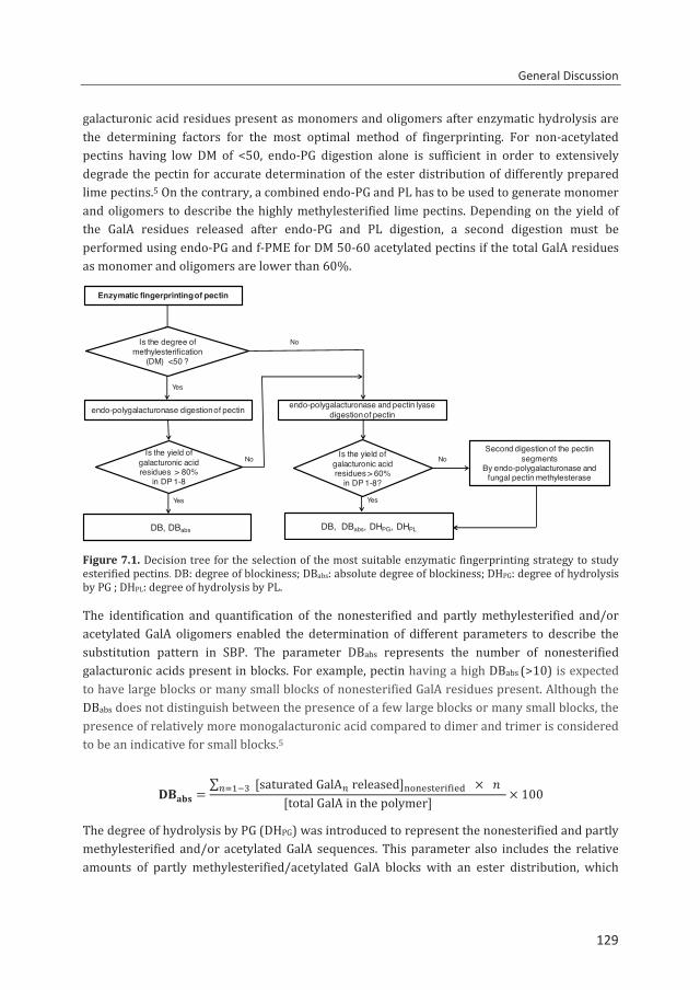
General Discussion
129
galacturonic acid residues present as monomers and oligomers after enzymatic hydrolysis are the determining factors for the most optimal method of fingerprinting. For non-acetylated pectins having low DM of <50, endo-PG digestion alone is sufficient in order to extensively degrade the pectin for accurate determination of the ester distribution of differently prepared lime pectins.5 On the contrary, a combined endo-PG and PL has to be used to generate monomer and oligomers to describe the highly methylesterified lime pectins. Depending on the yield of the GalA residues released after endo-PG and PL digestion, a second digestion must be performed using endo-PG and f-PME for DM 50-60 acetylated pectins if the total GalA residues as monomer and oligomers are lower than 60%.
Figure 7.1. Decision tree for the selection of the most suitable enzymatic fingerprinting strategy to study esterified pectins. DB: degree of blockiness; DBabs: absolute degree of blockiness; DHPG: degree of hydrolysis by PG ; DHPL: degree of hydrolysis by PL. The identification and quantification of the nonesterified and partly methylesterified and/or acetylated GalA oligomers enabled the determination of different parameters to describe the substitution pattern in SBP. The parameter DBabs represents the number of nonesterified galacturonic acids present in blocks. For example, pectin having a high DBabs (>10) is expected to have large blocks or many small blocks of nonesterified GalA residues present. Although the DBabs does not distinguish between the presence of a few large blocks or many small blocks, the presence of relatively more monogalacturonic acid compared to dimer and trimer is considered to be an indicative for small blocks.5
=∑ [saturatedGalAreleased] ×
[totalGalAinthepolymer] × 100
The degree of hydrolysis by PG (DHPG) was introduced to represent the nonesterified and partly methylesterified and/or acetylated GalA sequences. This parameter also includes the relative amounts of partly methylesterified/acetylated GalA blocks with an ester distribution, which
Enzymatic fingerprinting of pectin
No
endo-polygalacturonase digestion of pectin
Yes
DB, DBabs
endo-polygalacturonase and pectin lyasedigestion of pectin
Is the degree of methylesterification
(DM) <50 ?
Yes
Is the yield of galacturonic acid residues > 80%
in DP 1-8
Yes
NoSecond digestion of the pectin
segmentsBy endo-polygalacturonase and
fungal pectin methylesterase
DB, DBabs, DHPG, DHPL
NoIs the yield of galacturonic acid residues > 60%
in DP 1-8?
129
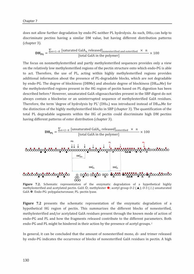
Chapter 7
130
does not allow further degradation by endo-PG neither PL hydrolysis. As such, DHPG can help to discriminate pectins having a similar DM value, but having different distribution patterns (chapter 3).
=∑ [saturatedGalAreleased] ×
[totalGalAinthepolymer] × 100
The focus on nonmethylesterified and partly methylesterified sequences provides only a view on the relatively low methylesterified regions of the pectin structure onto which endo-PG is able to act. Therefore, the use of PL, acting within highly methylesterified regions provides additional information about the presence of PL-degradable blocks, which are not degradable by endo-PG. The degree of blockiness (DBMe) and absolute degree of blockiness (DBabsMe) for the methylesterified regions present in the HG region of pectin based on PL digestion has been described before.8 However, unsaturated GalA oligosaccharides present in the SBP digest do not always contain a blockwise or an uninterrupted sequence of methylesterified GalA residues. Therefore, the term ‘degree of hydrolysis by PL’ (DHPL) was introduced instead of DBabsMe for the distinction of the highly methylesterified blocks in SBP (chapter 3). The quantification of the total PL degradable segments within the HG of pectin could discriminate high DM pectins having different patterns of ester distribution (chapter 3).
=∑ [unsaturatedGalAreleased] ×
[totalGalAinthepolymer] × 100
Figure 7.2. Schematic representation of the enzymatic degradation of a hypothetical highly methylesterified and acetylated pectin. GalA ; methylester ; acetyl group O-2 (▲); O-3 () unsaturated GalA . Endo-PG: polygalacturonase; PL: pectin lyase.
Figure 7.2 presents the schematic representation of the enzymatic degradation of a hypothetical HG region of pectin. This summarizes the different blocks of nonesterified, methylesterified and/or acetylated GalA residues present through the known mode of action of endo-PG and PL and how the fragments released contribute to the different parameters. Both endo-PG and PL might be hindered in their action by the presence of acetyl groups. 2 In general, it can be concluded that the amount of nonesterified mono, di- and trimer released by endo-PG indicates the occurrence of blocks of nonesterified GalA residues in pectin. A high
PG PL PL
130
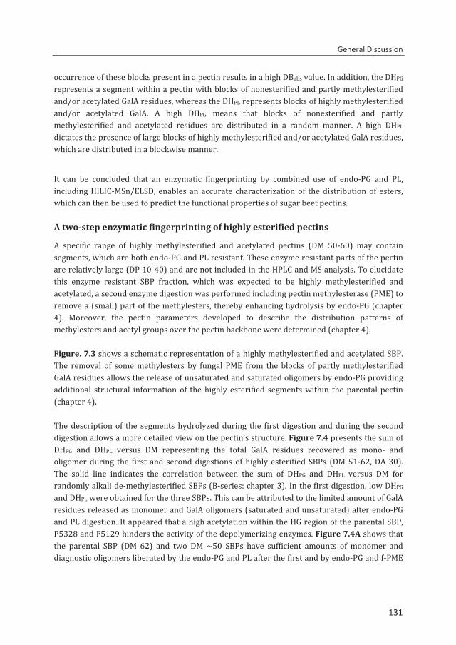
General Discussion
131
occurrence of these blocks present in a pectin results in a high DBabs value. In addition, the DHPG represents a segment within a pectin with blocks of nonesterified and partly methylesterified and/or acetylated GalA residues, whereas the DHPL represents blocks of highly methylesterified and/or acetylated GalA. A high DHPG means that blocks of nonesterified and partly methylesterified and acetylated residues are distributed in a random manner. A high DHPL dictates the presence of large blocks of highly methylesterified and/or acetylated GalA residues, which are distributed in a blockwise manner.
It can be concluded that an enzymatic fingerprinting by combined use of endo-PG and PL, including HILIC-MSn/ELSD, enables an accurate characterization of the distribution of esters, which can then be used to predict the functional properties of sugar beet pectins.
A two-step enzymatic fingerprinting of highly esterified pectins
A specific range of highly methylesterified and acetylated pectins (DM 50-60) may contain segments, which are both endo-PG and PL resistant. These enzyme resistant parts of the pectin are relatively large (DP 10-40) and are not included in the HPLC and MS analysis. To elucidate this enzyme resistant SBP fraction, which was expected to be highly methylesterified and acetylated, a second enzyme digestion was performed including pectin methylesterase (PME) to remove a (small) part of the methylesters, thereby enhancing hydrolysis by endo-PG (chapter 4). Moreover, the pectin parameters developed to describe the distribution patterns of methylesters and acetyl groups over the pectin backbone were determined (chapter 4). Figure. 7.3 shows a schematic representation of a highly methylesterified and acetylated SBP. The removal of some methylesters by fungal PME from the blocks of partly methylesterified GalA residues allows the release of unsaturated and saturated oligomers by endo-PG providing additional structural information of the highly esterified segments within the parental pectin (chapter 4). The description of the segments hydrolyzed during the first digestion and during the second digestion allows a more detailed view on the pectin’s structure. Figure 7.4 presents the sum of DHPG and DHPL versus DM representing the total GalA residues recovered as mono- and oligomer during the first and second digestions of highly esterified SBPs (DM 51-62, DA 30). The solid line indicates the correlation between the sum of DHPG and DHPL versus DM for randomly alkali de-methylesterified SBPs (B-series; chapter 3). In the first digestion, low DHPG and DHPL were obtained for the three SBPs. This can be attributed to the limited amount of GalA residues released as monomer and GalA oligomers (saturated and unsaturated) after endo-PG and PL digestion. It appeared that a high acetylation within the HG region of the parental SBP, P5328 and F5129 hinders the activity of the depolymerizing enzymes. Figure 7.4A shows that the parental SBP (DM 62) and two DM ~50 SBPs have sufficient amounts of monomer and diagnostic oligomers liberated by the endo-PG and PL after the first and by endo-PG and f-PME
131
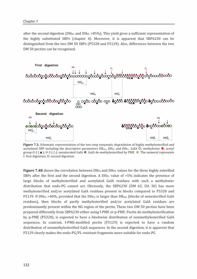
Chapter 7
132
after the second digestion (DHPG and DHPL >85%). This yield gives a sufficient representation of the highly substituted SBPs (chapter 4). Moreover, it is apparent that SBP6230 can be distinguished from the two DM 50 SBPs (P5328 and F5129). Also, differences between the two DM 50 pectins can be recognized.
Figure 7.3. Schematic representation of the two-step enzymatic degradation of highly methylesterified and acetylated SBP including the descriptive parameters DBabs, DHPG and DHPL. GalA ; methylester ; acetyl group O-2 (▲); O-3 (); unsaturated GalA , GalA de-methylesterified by PME . The numeral represents I: first digestion; II: second digestion.
Figure 7.4B shows the correlation between DHPL and DHPG values for the three highly esterified SBPs after the first and the second digestion. A DHPL value of >5% indicates the presence of large blocks of methylesterified and acetylated GalA residues with such a methylester distribution that endo-PG cannot act. Obviously, the SBP6230 (DM 62, DA 30) has more methylesterified and/or acetylated GalA residues present in blocks compared to P5328 and F5129. If DHPG >40%, provided that the DHPG is larger than DBabs (blocks of nonesterified GalA residues), then blocks of partly methylesterified and/or acetylated GalA residues are predominantly present within the HG region of the pectin. These two DM 50 pectins have been prepared differently from SBP6230 either using f-PME or p-PME. Pectin de-methylesterification by p-PME (P5328), is expected to have a blockwise distribution of nonmethylesterified GalA sequences. In contrast, f-PME-modified pectin (F5129) is expected to have a random distribution of nonmethylesterified GalA sequences. In the second digestion, it is apparent that F5129 clearly makes the endo-PG/PL resistant fragments more suitable for endo-PG
Second digestionPG PG
First digestion
PG PL PL
132
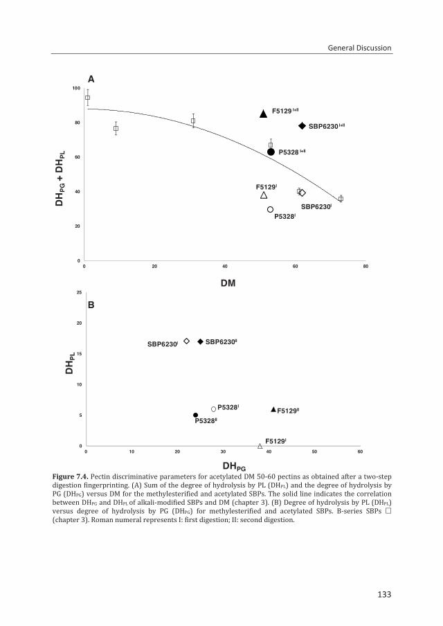
General Discussion
133
Figure 7.4. Pectin discriminative parameters for acetylated DM 50-60 pectins as obtained after a two-step digestion fingerprinting. (A) Sum of the degree of hydrolysis by PL (DHPL) and the degree of hydrolysis by PG (DHPG) versus DM for the methylesterified and acetylated SBPs. The solid line indicates the correlation between DHPG and DHPL of alkali-modified SBPs and DM (chapter 3). (B) Degree of hydrolysis by PL (DHPL) versus degree of hydrolysis by PG (DHPG) for methylesterified and acetylated SBPs. B-series SBPs (chapter 3). Roman numeral represents I: first digestion; II: second digestion.
P5328I
F5129I
SBP6230I
SBP6230 I+II
P5328 I+II
F5129 I+II
0
20
40
60
80
100
0 20 40 60 80
DH
PG+
DH
PL
DM
P5328I
F5129I
SBP6230I SBP6230II
F5129II
P5328II
0
5
10
15
20
25
0 10 20 30 40 50 60
DH
PL
DHPG
A
B
133
Chapter 7
134
degradation than is the case for P5328. This confirms the blockwise distribution of nonesterified and consequently methylesterified residues in the pectin being modified by p-PME. Large blocks of nonesterified GalA residues are not present in P5328, probably because the p-PME was hindered by the presence of acetyl groups forcing the enzyme to act less in a blockwise manner than would have been the case for non-acetylated pectins. As a result of the blockwise de-methylesterification of pectin, a part of the pectin remains highly methylesterified. This can be seen by the high DH values for both DM pectins. In conclusion, the descriptive parameters clearly focus on the significant differences in the methylester distribution among acetylated pectins, although less pronounced than found before for lemon pectins.1 At the moment, the interest for using descriptive fingerprinting methods is growing, especially because it is seen as a potential tool to link the distribution of methylesters and acetyl groups in pectins or pectic oligomers with functional behavior.28 The well-established “degree of blockiness” and the new parameter “degree of hydrolysis” by PL or degree of hydrolysis by PG are powerful parameters for efficient exploitation of the method (chapters 3 and 4). The application of the above mentioned enzymatic fingerprinting can be useful for other acetylated pectins. As an example, the characterization of a highly esterified sunflower pectin (DM 60, DA 11) is described below.
Characterization of sunflower pectin
Sunflower pectin, obtained from heads and stalks, which remained on the field during harvesting, is a naturally occurring LM pectin and is potentially available to extract pectins on an industrial scale.9,10 Although the extraction and chemical characterization of pectin from sunflower heads has been investigated,9,11 so far, the distribution of methylesters and acetyl groups of sunflower pectin has not been described. Table 7.2 presents the characteristics of acid extracted pectins from sunflower heads and of lemon and sugar beet pectin for comparison. Sunflower pectin has a relatively high level of uronic acid (75%) compared to SBP (55%), but has lower uronic acid content than lemon pectin (87%). The DM and the neutral sugar content of sunflower pectin are in between those of SBP and citrus pectin. The enzymatic fingerprinting and the determination of the descriptive parameters were applied to sunflower pectin. A two-step enzymatic fingerprinting method was not considered to be necessary for sunflower pectin since 85% of the total GalA residues were recovered as mono- and oligomers. The recovery of total GalA residues present in sunflower pectin is comparable to lemon pectin (Table 7.2).
The HPLC-HILIC-MS elution pattern of the PG/PL digest of sunflower pectin is shown in Figure 7.5. Besides the unsubstituted dimer (200) and trimer (300), partially methylesterified and/or acetylated saturated and unsaturated GalA oligomers of different degrees of polymerization (DP) were present as major products. It can be seen that oligomers with high levels of methylesters and acetyl groups are predominantly present in sunflower pectin. Using MSn
134
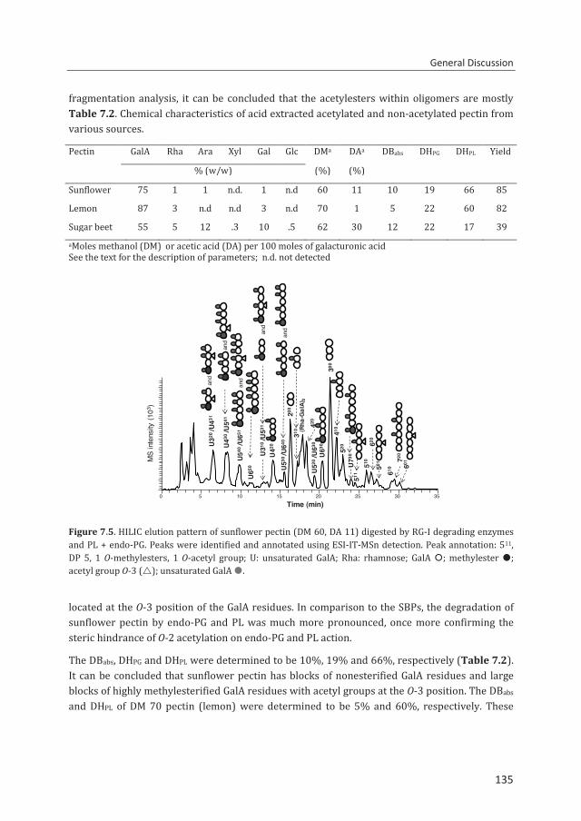
General Discussion
135
fragmentation analysis, it can be concluded that the acetylesters within oligomers are mostly Table 7.2. Chemical characteristics of acid extracted acetylated and non-acetylated pectin from various sources.
Pectin GalA Rha Ara Xyl Gal Glc DMa DAa DBabs DHPG DHPL Yield
% (w/w) (%) (%)
Sunflower 75 1 1 n.d. 1 n.d 60 11 10 19 66 85
Lemon 87 3 n.d n.d 3 n.d 70 1 5 22 60 82
Sugar beet 55 5 12 .3 10 .5 62 30 12 22 17 39 aMoles methanol (DM) or acetic acid (DA) per 100 moles of galacturonic acid See the text for the description of parameters; n.d. not detected
Figure 7.5. HILIC elution pattern of sunflower pectin (DM 60, DA 11) digested by RG-I degrading enzymes and PL + endo-PG. Peaks were identified and annotated using ESI-IT-MSn detection. Peak annotation: 511, DP 5, 1 O-methylesters, 1 O-acetyl group; U: unsaturated GalA; Rha: rhamnose; GalA ; methylester ; acetyl group O-3 (); unsaturated GalA .
located at the O-3 position of the GalA residues. In comparison to the SBPs, the degradation of sunflower pectin by endo-PG and PL was much more pronounced, once more confirming the steric hindrance of O-2 acetylation on endo-PG and PL action.
The DBabs, DHPG and DHPL were determined to be 10%, 19% and 66%, respectively (Table 7.2). It can be concluded that sunflower pectin has blocks of nonesterified GalA residues and large blocks of highly methylesterified GalA residues with acetyl groups at the O-3 position. The DBabs and DHPL of DM 70 pectin (lemon) were determined to be 5% and 60%, respectively. These
0 5 10 15 20 25 30 35Time (min)
U320
/U43
1
(Rha
-Gal
A)2
MS
inte
nsity
(10
3 )
U43
0/U
541
U65
0
U31
0/U
531
U42
0
U53
0 /U
640
200
310
U63
042
0U
520
/U63
0
300
410
520
U73
051
1
510
620
501
610
720
601
and
and
U540
/U65
1an
d
and
and
135
Chapter 7
136
values indicate the presence of small blocks of nonesterified sequences and large blocks of highly methylesterified sequences degradable by PL.
Based on these results, it can be postulated that both sunflower pectin and lemon pectin differ in the ratio between blocks of nonesterified and blocks of methylesterified GalA residues present within their homogalacturonans. Sunflower pectin has a DA 10 while for lemon the DA is negligible. According to the standard characteristics for commercial pectin,12 sunflower pectin qualifies to be a commercial pectin despite its acetylation.
7.4. Pectin methyl- and acetylesterases able to modify acetylated pectins
Bacterial PMEs, PAEs, and RGAEs have been reported in several studies.13-17 Interestingly, pectolytic enzymes of potential interest for food applications were found in Bacillus licheniformis DSM13; e.g. pectate lyases.18 However, pectin acetylesterases and pectin methylesterases have not been characterized for this organism so far.
PAE from Bacillus licheniformis DSM13
For the first time, an acetylesterase was cloned from B. licheniformis and produced with high purity and in relatively large quantities using an E. coli expression system (chapter 5). The purified enzyme BliPAE was characterized in detail and was capable of deacetylating sugar beet pectin (SBP), sunflower pectin and potato pectin. Contrary to its provisional annotation (CAZy database) as rhamnogalacturonan acetylesterase, the enzyme specifically removed acetyl groups from the homogalacturonan region classifying it as a PAE. The enzyme was active and stable at neutral to alkaline conditions. In addition, 1H NMR analysis of the polymer and enzymatic fingerprinting of the BliPAE-modified by endo-PG and PL including HPLC-HILIC-MSn clearly showed that BliPAE specifically removes acetyl groups linked to O-3 of GalA residues (chapter 5).
PAE from from Bacillus subtilis
In a separate study, a Bacillus subtilis PAE, termed YesY has been produced, purified and characterized19 on the basis of homology with BliPAE (chapter 5). The two bacterial acetylesterases were compared in terms of their activity towards SBPs. Similar to BliPAE, YesY was active on SBP at pH 6.0 to 8.0 and has a maximum activity at pH 8.0 and 50 ˚C. A parental SBP (DM 62 and DA 30) and B-, F- and P-series pectins of SBP were used as substrates to examine the activity of YesY. Figure 7.6 shows the levels of deacetylation of different SBPs by YesY and BliPAE. It can be seen that YesY released a maximum of 60% of the total acetyl groups from SBP substrates compared to maximal 40% for BliPAE (chapter 5; Fig
136
General Discussion
137
7.6). This suggests that YesY is less hindered by the level of methylesters present in SBPs. To gain more insight of its mode of action, enzymatic fingerprinting of SBP3124 (DM 31, DA 24)
Figure 7.6. The deacetylation by Bacillus subtilis PAE YesY () and Bacillus licheniformis PAE BliPAE () of different SBP substrates as a function of DM.
Figure 7.7. HILIC elution pattern of (A) SBP3124 (DM 31, DA 24) and (B) YesY-modified SBP3124, digested by RG-I degrading enzymes and PL + endo-PG. Peaks were identified and annotated using ESI-IT-MSn
detection. Peak annotation: 611, DP 6, 1 O-methylester, 1 O-acetyl group.
SBP6230
B3124B4626
B3124
B0915
0
10
20
30
40
50
60
70
0 10 20 30 40 50 60 70
% D
eace
tyla
tion
DM
MS
inte
nsity
(103 )
0 2 4 6 8 10 12 14 16 18 20 22 24 26 28 30 32 34
200
511
721
420
300
410
520
621
510620
830 821
930
411
70B
0 2 4 6 8 10 12 14 16 18 20 22 24 26 28 30 32 34
Time (min)
0
200
411
300
410
70
520
511
510 620
611721
822
821
A
137
Chapter 7
138
and the YesY-modified SBP was performed and the products generated were analyzed on HPLC-MSn. Figure 7.7 presents the HPLC-MSn profiles of the products formed after endo-PG and PL digestion of the non-treated and YesY treated SBP in order to understand the mode of action of the enzyme. Non- and partially methylesterified and/or acetylated GalA oligomers ranging from DP 2 to 8 were released after endo-PG and PL digestion (Fig. 7.7A). Analysis of the YesY-modified pectin reveals that most of the O-3 acetylated GalA units in the oligomers released from the non-treated pectin were not observed in the oligomers released from the YesY-modified pectin anymore. The presence of GalA oligomers, e.g. 511, 621, 721 having acetyl groups at the O-2 position of GalA illustrates that the enzyme specifically deacetylates the acetyl group from O-3 substituted GalA residues. The initial results presented for YesY are promising since the enzyme show higher activity towards SBP compared to BliPAE. This indicates that YesY is less hindered by the presence of methylesters in the pectin compared to BliPAE. In-depth analysis of its mode of action is needed to confirm its specificity towards different acetylated pectins from various substrates and preference for acetyl group positions.
Microbial and plant PMEs
A bacterial PME from Bacillus licheniformis DSM13, termed as BliPME was produced and characterized in detail (chapter 6). This PME can be produced with high purity and in large quantities using an E. coli expression system. This novel PME was shown to remove up to the last methylester present on non-acetylated lime pectins (DM34-76 → DM 0) and up to 60-73% of all methylesters from acetylated pectin (SBP) (DM 30-73 → DM 14). As an extension of the research (chapter 6), the activity of BliPME towards highly methylesterified and acetylated pectins is compared to Aspergillus niger PME20 (AniPME) and Citrus sinensis PME21 (Fig. 7.8). The esterase activity was carried out at their optimum pH and temperature conditions with an enzyme dose of 0.02% (w/w) protein-substrate basis. It can be seen that the activity of BliPME and AniPME towards lime pectin (Fig. 7.8A) for the first 15 min was very similar, but BliPME proceeds further in de-methylesterifying up to the last methylester left in the substrate (chapter 6). On the contrary, C-PME has low activity for lime pectin. The activity of PMEs was also tested on acetylated SBP (Fig. 7.8B). The BliPME seemed to tolerate the presence of acetyl groups much more than AniPME. The presence of O-2 substituted acetyl groups on a GalA residue hinders the enzyme from binding to the substrate. Thereby, BliPME could not remove a specific part of the methylesters within pectin as demonstrated by the fingerprinting method (chapter 6). In addition, the mode of action of BliPME was studied using lime pectin and lime pectin oligomers and as substrates. It was revealed that BliPME is a processive enzyme able to create blockwise nonmethylesterified regions in acetylated and non-acetylated pectins.
138
General Discussion
139
Figure 7.8. Activity of PMEs from Bacillus licheniformis, Aspergillus niger and Citrus sinensis towards (A) non-acetylated (DM 76, DA 0) and (B) acetylated pectin (DM 51, DA 29) incubated at pH 6.5, pH 5.0 and pH 7.0, respectively at 40 °C using pH-stat analysis.
Many PMEs have been recently extracted from various plants (e.g. onion flower, pea)22,23 and bacteria (e.g. xanthamonas campetris, Yersinia enterocolitica).24,25 However, many of these PMEs are not fully characterized yet. A comprehensive understanding of their properties and their modes of action would support the targeted modification of pectins. The fingerprinting approach described in this thesis (chapter 6) proved to be a quite successful method to determine the precise modes of action of different PMEs.
7.5. Future perspectives
On-going search to characterize pectins
It was postulated in this thesis that a powerful enzymatic fingerprinting is key in understanding of the ester distribution pattern in industrially extracted pectins. The method developed is a big step forward in pectin characterization.
0
20
40
60
80
0 1000 2000 3000 4000
% D
e-m
ethy
lest
erifi
catio
n
0
10
20
30
40
50
60
0 1000 2000 3000 4000
Time (s)
A
B
BliPME
AniPME
Citrus-PME
BliPME
AniPME
Citrus-PME
139
Chapter 7
140
Our method would be even more powerful and time-saving if we could include the quantification of monogalacturonic acid in the HILIC analysis. In addition, the current method is limited to the analysis of oligosaccharides having a DP < 9. Novel (HILIC) column materials might become available in the near future to further strengthen our method. In addition, mass spectrometry is evolving rapidly and new interfaces, ionization- and in-MS ion-separation techniques are introduced continuously.
ESI-IT-MSn of oligosaccharides has been used extensively in this study. Although this MS technique is a powerful tool for the identification of oligosaccharides in complex mixtures, new MS tools, such as ion mobility spectrometry (IMS), are promising. The advantage of ESI-ion mobility MS is that the separation is based on size as well as on conformational differences. Isomeric oligosaccharides having the same m/z values can be separated with ion mobility. Recently, a rapid travelling-wave ion mobility mass spectrometry (TWIMS-MS) method has been applied for the analysis of pectin digest.26 Due to the (in)sensitivity of the IMS system used, screening of the pectin oligomers on TWIMS-MS was done after direct infusion of the enzyme digest rather than after LC-separation. TWIMS-MS analysis allows the precise identification of isomeric structures separated by IM through MS fragmentation analysis. This annotation was further simplified by using a chemical label, e.g. 3-aminoquinoline (3-AQ) at the reducing end of the oligosaccharide analysis, which promotes a directed fragmentation from the nonreducing end.27 As an example of this new separation and labelling technique, the identification of various isomeric GalA oligomers present in the endo-PG digest is presented. Figure 7.9 presents the product ion mass spectra of the heptamer 73 isomers with three methylesters ([M +H]+; m/z 1419) for the IM peaks separated by drift time (at 2.89 and 4.56 ms). The MS spectra show that two isomers are present in different ratios for each drift time peak.
The new analytical approach developed in this study (chapters 2-4) and the TWIMS-MS technique opens new perspectives in coupling HILIC-ELSD to online TWIMS-MS for the separation, identification and quantification of oligosaccharides. Especially, since currently available TWIMS-MS apparatus have gained in sensitivity and accuracy. Such a combination of techniques could be a future tool for the elucidation of the chemical structure of oligomers co-eluting or having similar m/z values.
140
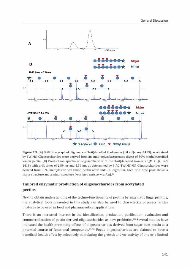
General Discussion
141
Figure 7.9. (A) Drift time graph of oligomers of 3-AQ-labelled 73 oligomer ([M +H]+; m/z1419), as obtained by TWIMS. Oligosaccharides were derived from an endo-polygalacturonase digest of 30% methylesterified lemon pectin. (B) Product ion spectra of oligosaccharides of the 3-AQ-labelled isomer 73([M +H]+; m/z 1419) with drift times of 2.89 ms and 4.56 ms, as determined by 3-AQ-TWIMS-MS. Oligosaccharides were derived from 30% methylesterified lemon pectin after endo-PG digestion. Each drift time peak shows a major structure and a minor structure (reprinted with permission).26
Tailored enzymatic production of oligosaccharides from acetylated pectins
Next to obtain understanding of the techno-functionality of pectins by enzymatic fingerprinting, the analytical tools presented in this study can also be used to characterize oligosaccharides mixtures to be used in food and pharmaceutical applications.
There is an increased interest in the identification, production, purification, evaluation and commercialization of pectin-derived oligosaccharides as new prebiotics.28 Several studies have indicated the health promoting effects of oligosaccharides derived from sugar beet pectin as a potential source of functional compounds.29,30 Pectic oligosaccharides are claimed to have a beneficial health effect by selectively stimulating the growth and/or activity of one or a limited
141
Chapter 7
142
number of beneficial bacterial species in the human colon.31 In addition, pectic oligosaccharides may affect the immune system, as has been supported by clinical trials and animal studies.31,32 It is claimed that pectic oligosaccharides supplement in infant’s early life could lead to the prevention of the development of food allergy, allergic asthma or dermatitis. It is thought that the immune modulating effect of these pectic oligosaccharides are mediated either via modification of the intestinal microflora or in a microbiota-independent manner by direct interaction on cells of the immune system.31 The pharmaceutical properties of pectic oligosaccharides are very promising. However, so far quite complex mixtures of oligosaccharides have been used in both in vitro and in vivo studies. The identification and characterization of particular oligosaccharides (e.g. specific DP, ester substitution pattern) using the analytical techniques described in this thesis could highly contribute to understanding the structure-function relationship of these oligosaccharides.
References 1. Limberg, G., Körner, R., Buchholt, H. C., Christensen, T. M. I. E., Roepstorff, P., and Mikkelsen, J. D.
(2000) Carbohydrate Research 327, 293-307 2. Buchholt, H. C., Christensen, T. M. I. E., Fallesen, B., Ralet, M. C., and Thibault, J. F. (2004)
Carbohydrate Polymers 58, 149-161 3. Sengkhamparn, N., Bakx, E. J., Verhoef, R., Schols, H. A., Sajjaanantakul, T., and Voragen, A. G. J.
(2009) Carbohydrate Research 344, 1842-1851 4. Schols, H. A., Voragen, A. G. J., and Colquhoun, I. J. (1994) Carbohydrate Research 256, 97-111 5. Daas, P. J. H., Voragen, A. G. J., and Schols, H. A. (2000) Carbohydrate Research 326, 120-129 6. Guillotin, S. E., Bakx, E. J., Boulenguer, P., Mazoyer, J., Schols, H. A., and Voragen, A. G. J. (2005)
Carbohydrate Polymers 60, 391-398 7. Ngouémazong, D. E., Tengweh, F. F., Duvetter, T., Fraeye, I., Van Loey, A., Moldenaers, P., and
Hendrickx, M. (2011) Food Hydrocolloids 25, 434-443 8. Ralet, M.-C., Williams, M. A. K., Tanhatan-Nasseri, A., Ropartz, D., Quéméner, B., and Bonnin, E.
(2012) Biomacromolecules 13, 1615-1624 9. Iglesias, M. T., and Lozano, J. E. (2004) Journal of Food Engineering 62, 215-223 10. May, C. D. (1990) Carbohydrate Polymers 12, 79-99 11. Miyamoto, A., and Chang, K. C. (1992) Journal of Food Science 57, 1439-1443 12. Thibault, J. F., and Ralet, M. C. (2003) Physico-chemical properties of pectins in the cell walls and
after extraction. in Advances in Pectins and Pectinases Voragen, A. G. J., Schols, H. A., and Visser, R. ed.(eds.), Kluwer Academic Publishers, Dordrecht, The Netherlands. pp 91-105
13. Shevchik, V. E., and Hugouvieux-Cotte-Pattat, N. (1997) Molecular Microbiology 24, 1285-1301 14. Shevchik, V. E., and Hugouvieux-Cotte-Pattat, N. (2003) Journal of Bacteriology 185, 3091-3100 15. Martinez-Martinez, I., Navarro-Fernandez, J., Lozada-Ramirez, J. D., Garcia-Carmona, F., and
Sanchez-Ferrer, A. (2008) Proteins-Structure Function and Bioinformatics 71, 379-388 16. Jenkins, J., Mayans, O., Smith, D., Worboys, K., and Pickersgill, R. W. (2001) Journal of Molecular
Biology 305, 951-960 17. Bolvig, P. U., Pauly, M., Orfila, C., Scheller, H. V., and Schnorr, K. (2003) Sequence analysis and
characterisation of a novel pectin acetyl esterase from Bacillus subtilis. in Advances in Pectin and Pectinase Research Voragen, A. G. J., Schols, H. A., and Visser, R. ed.(eds.), Kluwer Academic Publishers, Dordrecht, The Netherlands. pp 315-330
18. Veith, B., Herzberg, C., Steckel, S., Feesche, J., Maurer, K. H., Ehrenreich, P., Bäumer, S., Henne, A., Liesegang, H., Merkl, R., Ehrenreich, A., and Gottschalk, G. (2004) Journal of Molecular Microbiology and Biotechnology 7, 204-211
19. Wagenknecht, M., Remoroza, C., Singh, R., Kool, M. M., Schols, H. A., and Moerschbacher, B. M., To be submitted
142
General Discussion
143
20. Van Alebeek, G. J. W. M., Van Scherpenzeel, K., Beldman, G., Schols, H. A., and Voragen, A. G. J. (2003) Biochemical Journal 372, 211-218
21. Cameron, R. G., Luzio, G. A., Goodner, K., and Williams, M. A. K. (2008) Carbohydrate Polymers 71, 287-299
22. Huo, Y., Miao, J., Liu, B., Yang, Y., Zhang, Y., Tahara, Y., Meng, Q., He, Q., Kitano, H., and Wu, X. (2012) Plant Breeding 131, 211-216
23. Gómez, M. D., Renau-Morata, B., Roque, E., Polaina, J., Beltrán, J. P., and Cañas, L. A. (2013) Sexual Plant Reproduction 26, 245-254
24. Boraston, A. B., and Abbott, D. W. (2012) Acta Crystallographica Section F: Structural Biology and Crystallization Communications 68, 129-133
25. Hsiao, Y.-M., Liu, Y.-F., Huang, Y.-L., and Lee, P.-Y. (2011) Research in Microbiology 162, 270-278 26. Huang, J.-H., Bakx, E. J., Gruppen, H., and Schols, H. A. (2013) Rapid Communications in Mass
Spectrometry 27, 2279-2285 27. Rohmer, M., Meyer, B., Mank, M., Stahl, B., Bahr, U., and Karas, M. (2010) Analytical Chemistry 82,
3719-3726 28. Gullón, B., Gómez, B., Martínez-Sabajanes, M., Yáñez, R., Parajó, J. C., and Alonso, J. L. (2013)
Trends in Food Science and Technology 30, 153-161 29. Holck, J., Hjernø, K., Lorentzen, A., Vigsnæs, L. K., Hemmingsen, L., Licht, T. R., Mikkelsen, J. D., and
Meyer, A. S. (2011) Process Biochemistry 46, 1039-1049 30. Concha Olmos, J., and Zúñiga Hansen, M. E. (2012) Chemical Engineering Journal 192, 29-36 31. Rijnierse, A., Jeurink, P. V., van Esch, B. C. A. M., Garssen, J., and Knippels, L. M. J. (2011) European
Journal of Pharmacology 668, Supplement 1, S117-S123 32. Moro, G., Arslanoglu, S., Stahl, B., Jelinek, J., Wahn, U., and Boehm, G. (2006) Archives of Disease in
Childhood 91, 814-819
143

144

Summary
Samenvatting
Summary, Samenvatting
146
Summary The aim of this research project was to develop methods to reveal the distribution of methylesters and acetyl groups within sugar beet pectins (SBPs) and to use these methods to characterize newly discovered pectin esterases: pectin acetylesterase (PAE) and pectin methylesterase (PME). It was hypothesized in this study that it is possible to develop enzymatic approaches to sufficiently degrade SBP into diagnostic oligosaccharides in order to use the amounts and structures of the oligosaccharides to reconstruct the parental pectin. For the separation, identification and quantification of such diagnostic oligosaccharides, a powerful analytical method would be essential. The above described fingerprinting method developed could then be used to describe the action of PAE and PME towards SBP by analyzing its ester distribution before and after incubation with the enzymes.
The background of the research project is presented in chapter 1 with a review of literature on general information about the pectin structure, pectin analysis, as well as the characteristics of pectin methylesterases and acetylesterases from various origins.
The optimization and development of hydrophilic interaction liquid chromatography (HILIC) with online electrospray ionization ion trap mass spectrometry (ESI-IT-MSn) and evaporative light scattering detection (ELSD) for the analysis of complex mixtures of oligosaccharides after an endo-polygalacturonase (endo-PG) and pectin lyase (PL) digestion is described (chapter 2). HILIC-MS analysis provides a sophisticated method to separate and to identify various oligomers present in a complex mixture. Saturated and unsaturated GalA oligomers of various DPs with or without methylesters and acetyl groups were separated according to their total negative charge and size as identified based on their m/z values using online ESI-MSn. The precise location of methylesters and the O-2/O-3 position of the acetyl groups within the oligomers were elucidated. Furthermore, ELS detection allowed the quantification of individual GalA oligomers present in the pectin digest in absolute amounts.
The above mentioned analytical method was used to accurately characterize the distribution of esters in SBPs. Degradation of different SBPs with various degrees of methylesterification (DM) and acetylation (DA) to small oligomers by the simultaneous action of endo-PG and PL is shown in chapter 3. Individual oligomers were identified and quantified. Using this information, new pectin parameters DHPG and DHPL based on the well-established absolute degree of blockiness (DBabs) were defined in order to distinguish different ester distribution patterns in SBPs. DBabs is a measure of the amount of nonesterified GalA residues present as monomer, dimer and trimer. Degree of hydrolysis by PG (DHPG) represents the nonesterified and partly methylesterified and/or acetylated sequences degradable by endo-PG. All GalA residues present as unsaturated GalA oligomers (DP 2-8) both methylesterified and/or acetylated released by PL action in SBP were quantified and expressed via the degree of hydrolysis by PL (DHPL). These pectin parameters were used to describe the ester distribution patterns of chemically and enzymatically modified SBP samples within the range of DM 1-73 and DA 0-30 (chapter 3).
146
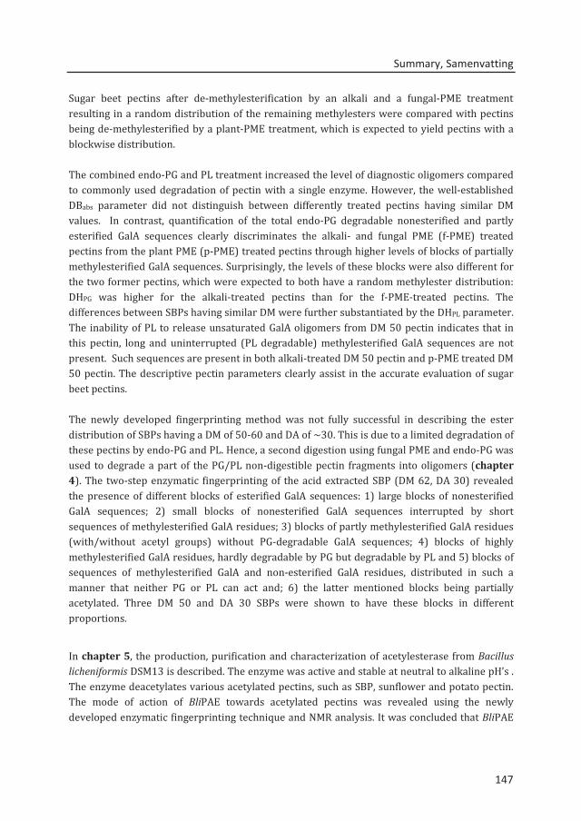
Summary, Samenvatting
147
Sugar beet pectins after de-methylesterification by an alkali and a fungal-PME treatment resulting in a random distribution of the remaining methylesters were compared with pectins being de-methylesterified by a plant-PME treatment, which is expected to yield pectins with a blockwise distribution. The combined endo-PG and PL treatment increased the level of diagnostic oligomers compared to commonly used degradation of pectin with a single enzyme. However, the well-established DBabs parameter did not distinguish between differently treated pectins having similar DM values. In contrast, quantification of the total endo-PG degradable nonesterified and partly esterified GalA sequences clearly discriminates the alkali- and fungal PME (f-PME) treated pectins from the plant PME (p-PME) treated pectins through higher levels of blocks of partially methylesterified GalA sequences. Surprisingly, the levels of these blocks were also different for the two former pectins, which were expected to both have a random methylester distribution: DHPG was higher for the alkali-treated pectins than for the f-PME-treated pectins. The differences between SBPs having similar DM were further substantiated by the DHPL parameter. The inability of PL to release unsaturated GalA oligomers from DM 50 pectin indicates that in this pectin, long and uninterrupted (PL degradable) methylesterified GalA sequences are not present. Such sequences are present in both alkali-treated DM 50 pectin and p-PME treated DM 50 pectin. The descriptive pectin parameters clearly assist in the accurate evaluation of sugar beet pectins. The newly developed fingerprinting method was not fully successful in describing the ester distribution of SBPs having a DM of 50-60 and DA of ~30. This is due to a limited degradation of these pectins by endo-PG and PL. Hence, a second digestion using fungal PME and endo-PG was used to degrade a part of the PG/PL non-digestible pectin fragments into oligomers (chapter 4). The two-step enzymatic fingerprinting of the acid extracted SBP (DM 62, DA 30) revealed the presence of different blocks of esterified GalA sequences: 1) large blocks of nonesterified GalA sequences; 2) small blocks of nonesterified GalA sequences interrupted by short sequences of methylesterified GalA residues; 3) blocks of partly methylesterified GalA residues (with/without acetyl groups) without PG-degradable GalA sequences; 4) blocks of highly methylesterified GalA residues, hardly degradable by PG but degradable by PL and 5) blocks of sequences of methylesterified GalA and non-esterified GalA residues, distributed in such a manner that neither PG or PL can act and; 6) the latter mentioned blocks being partially acetylated. Three DM 50 and DA 30 SBPs were shown to have these blocks in different proportions.
In chapter 5, the production, purification and characterization of acetylesterase from Bacillus licheniformis DSM13 is described. The enzyme was active and stable at neutral to alkaline pH’s . The enzyme deacetylates various acetylated pectins, such as SBP, sunflower and potato pectin. The mode of action of BliPAE towards acetylated pectins was revealed using the newly developed enzymatic fingerprinting technique and NMR analysis. It was concluded that BliPAE
147
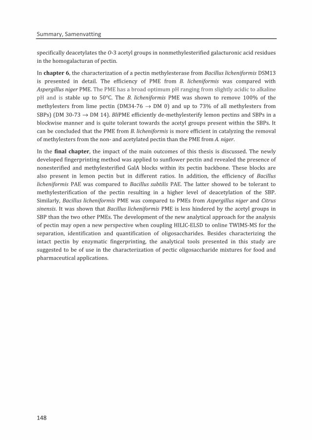
Summary, Samenvatting
148
specifically deacetylates the O-3 acetyl groups in nonmethylesterified galacturonic acid residues in the homogalacturan of pectin.
In chapter 6, the characterization of a pectin methylesterase from Bacillus licheniformis DSM13 is presented in detail. The efficiency of PME from B. licheniformis was compared with Aspergillus niger PME. The PME has a broad optimum pH ranging from slightly acidic to alkaline pH and is stable up to 50°C. The B. licheniformis PME was shown to remove 100% of the methylesters from lime pectin (DM34-76 → DM 0) and up to 73% of all methylesters from SBPs) (DM 30-73 → DM 14). BliPME efficiently de-methylesterify lemon pectins and SBPs in a blockwise manner and is quite tolerant towards the acetyl groups present within the SBPs. It can be concluded that the PME from B. licheniformis is more efficient in catalyzing the removal of methylesters from the non- and acetylated pectin than the PME from A. niger.
In the final chapter, the impact of the main outcomes of this thesis is discussed. The newly developed fingerprinting method was applied to sunflower pectin and revealed the presence of nonesterified and methylesterified GalA blocks within its pectin backbone. These blocks are also present in lemon pectin but in different ratios. In addition, the efficiency of Bacillus licheniformis PAE was compared to Bacillus subtilis PAE. The latter showed to be tolerant to methylesterification of the pectin resulting in a higher level of deacetylation of the SBP. Similarly, Bacillus licheniformis PME was compared to PMEs from Aspergillus niger and Citrus sinensis. It was shown that Bacillus licheniformis PME is less hindered by the acetyl groups in SBP than the two other PMEs. The development of the new analytical approach for the analysis of pectin may open a new perspective when coupling HILIC-ELSD to online TWIMS-MS for the separation, identification and quantification of oligosaccharides. Besides characterizing the intact pectin by enzymatic fingerprinting, the analytical tools presented in this study are suggested to be of use in the characterization of pectic oligosaccharide mixtures for food and pharmaceutical applications.
148
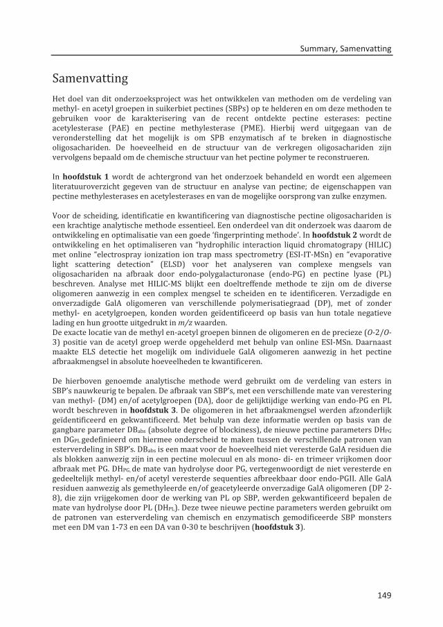
Summary, Samenvatting
149
Samenvatting Het doel van dit onderzoeksproject was het ontwikkelen van methoden om de verdeling van methyl- en acetyl groepen in suikerbiet pectines (SBPs) op te helderen en om deze methoden te gebruiken voor de karakterisering van de recent ontdekte pectine esterases: pectine acetylesterase (PAE) en pectine methylesterase (PME). Hierbij werd uitgegaan van de veronderstelling dat het mogelijk is om SPB enzymatisch af te breken in diagnostische oligosachariden. De hoeveelheid en de structuur van de verkregen oligosachariden zijn vervolgens bepaald om de chemische structuur van het pectine polymer te reconstrueren. In hoofdstuk 1 wordt de achtergrond van het onderzoek behandeld en wordt een algemeen literatuuroverzicht gegeven van de structuur en analyse van pectine; de eigenschappen van pectine methylesterases en acetylesterases en van de mogelijke oorsprong van zulke enzymen. Voor de scheiding, identificatie en kwantificering van diagnostische pectine oligosachariden is een krachtige analytische methode essentieel. Een onderdeel van dit onderzoek was daarom de ontwikkeling en optimalisatie van een goede ‘fingerprinting methode’. In hoofdstuk 2 wordt de ontwikkeling en het optimaliseren van “hydrophilic interaction liquid chromatograpy (HILIC) met online “electrospray ionization ion trap mass spectrometry (ESI-IT-MSn) en “evaporative light scattering detection” (ELSD) voor het analyseren van complexe mengsels van oligosachariden na afbraak door endo-polygalacturonase (endo-PG) en pectine lyase (PL) beschreven. Analyse met HILIC-MS blijkt een doeltreffende methode te zijn om de diverse oligomeren aanwezig in een complex mengsel te scheiden en te identificeren. Verzadigde en onverzadigde GalA oligomeren van verschillende polymerisatiegraad (DP), met of zonder methyl- en acetylgroepen, konden worden geïdentificeerd op basis van hun totale negatieve lading en hun grootte uitgedrukt in m/z waarden. De exacte locatie van de methyl en-acetyl groepen binnen de oligomeren en de precieze (O-2/O-3) positie van de acetyl groep werde opgehelderd met behulp van online ESI-MSn. Daarnaast maakte ELS detectie het mogelijk om individuele GalA oligomeren aanwezig in het pectine afbraakmengsel in absolute hoeveelheden te kwantificeren. De hierboven genoemde analytische methode werd gebruikt om de verdeling van esters in SBP’s nauwkeurig te bepalen. De afbraak van SBP’s, met een verschillende mate van verestering van methyl- (DM) en/of acetylgroepen (DA), door de gelijktijdige werking van endo-PG en PL wordt beschreven in hoofdstuk 3. De oligomeren in het afbraakmengsel werden afzonderlijk geïdentificeerd en gekwantificeerd. Met behulp van deze informatie werden op basis van de gangbare parameter DBabs (absolute degree of blockiness), de nieuwe pectine parameters DHPG en DGPL gedefinieerd om hiermee onderscheid te maken tussen de verschillende patronen van esterverdeling in SBP’s. DBabs is een maat voor de hoeveelheid niet veresterde GalA residuen die als blokken aanwezig zijn in een pectine molecuul en als mono- di- en trimeer vrijkomen door afbraak met PG. DHPG, de mate van hydrolyse door PG, vertegenwoordigt de niet veresterde en gedeeltelijk methyl- en/of acetyl veresterde sequenties afbreekbaar door endo-PGII. Alle GalA residuen aanwezig als gemethyleerde en/of geacetyleerde onverzadige GalA oligomeren (DP 2-8), die zijn vrijgekomen door de werking van PL op SBP, werden gekwantificeerd bepalen de mate van hydrolyse door PL (DHPL). Deze twee nieuwe pectine parameters werden gebruikt om de patronen van esterverdeling van chemisch en enzymatisch gemodificeerde SBP monsters met een DM van 1-73 en een DA van 0-30 te beschrijven (hoofdstuk 3).
149
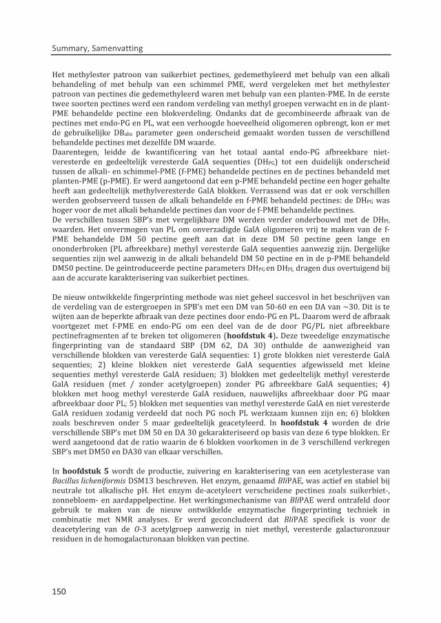
Summary, Samenvatting
150
Het methylester patroon van suikerbiet pectines, gedemethyleerd met behulp van een alkali behandeling of met behulp van een schimmel PME, werd vergeleken met het methylester patroon van pectines die gedemethyleerd waren met behulp van een planten-PME. In de eerste twee soorten pectines werd een random verdeling van methyl groepen verwacht en in de plant-PME behandelde pectine een blokverdeling. Ondanks dat de gecombineerde afbraak van de pectines met endo-PG en PL, wat een verhoogde hoeveelheid oligomeren opbrengt, kon er met de gebruikelijke DBabs parameter geen onderscheid gemaakt worden tussen de verschillend behandelde pectines met dezelfde DM waarde. Daarentegen, leidde de kwantificering van het totaal aantal endo-PG afbreekbare niet-veresterde en gedeeltelijk veresterde GalA sequenties (DHPG) tot een duidelijk onderscheid tussen de alkali- en schimmel-PME (f-PME) behandelde pectines en de pectines behandeld met planten-PME (p-PME). Er werd aangetoond dat een p-PME behandeld pectine een hoger gehalte heeft aan gedeeltelijk methylveresterde GalA blokken. Verrassend was dat er ook verschillen werden geobserveerd tussen de alkali behandelde en f-PME behandeld pectines: de DHPG was hoger voor de met alkali behandelde pectines dan voor de f-PME behandelde pectines. De verschillen tussen SBP’s met vergelijkbare DM werden verder onderbouwd met de DHPL waarden. Het onvermogen van PL om onverzadigde GalA oligomeren vrij te maken van de f-PME behandelde DM 50 pectine geeft aan dat in deze DM 50 pectine geen lange en ononderbroken (PL afbreekbare) methyl veresterde GalA sequenties aanwezig zijn. Dergelijke sequenties zijn wel aanwezig in de alkali behandeld DM 50 pectine en in de p-PME behandeld DM50 pectine. De geintroduceerde pectine parameters DHPG en DHPL dragen dus overtuigend bij aan de accurate karakterisering van suikerbiet pectines. De nieuw ontwikkelde fingerprinting methode was niet geheel succesvol in het beschrijven van de verdeling van de estergroepen in SPB’s met een DM van 50-60 en een DA van ~30. Dit is te wijten aan de beperkte afbraak van deze pectines door endo-PG en PL. Daarom werd de afbraak voortgezet met f-PME en endo-PG om een deel van de de door PG/PL niet afbreekbare pectinefragmenten af te breken tot oligomeren (hoofdstuk 4). Deze tweedelige enzymatische fingerprinting van de standaard SBP (DM 62, DA 30) onthulde de aanwezigheid van verschillende blokken van veresterde GalA sequenties: 1) grote blokken niet veresterde GalA sequenties; 2) kleine blokken niet veresterde GalA sequenties afgewisseld met kleine sequenties methyl veresterde GalA residuen; 3) blokken met gedeeltelijk methyl veresterde GalA residuen (met / zonder acetylgroepen) zonder PG afbreekbare GalA sequenties; 4) blokken met hoog methyl veresterde GalA residuen, nauwelijks afbreekbaar door PG maar afbreekbaar door PL; 5) blokken met sequenties van methyl veresterde GalA en niet veresterde GalA residuen zodanig verdeeld dat noch PG noch PL werkzaam kunnen zijn en; 6) blokken zoals beschreven onder 5 maar gedeeltelijk geacetyleerd. In hoofdstuk 4 worden de drie verschillende SBP’s met DM 50 en DA 30 gekarakteriseerd op basis van deze 6 type blokken. Er werd aangetoond dat de ratio waarin de 6 blokken voorkomen in de 3 verschillend verkregen SBP’s met DM50 en DA30 van elkaar verschillen. In hoofdstuk 5 wordt de productie, zuivering en karakterisering van een acetylesterase van Bacillus licheniformis DSM13 beschreven. Het enzym, genaamd BliPAE, was actief en stabiel bij neutrale tot alkalische pH. Het enzym de-acetyleert verscheidene pectines zoals suikerbiet-, zonnebloem- en aardappelpectine. Het werkingsmechanisme van BliPAE werd ontrafeld door gebruik te maken van de nieuw ontwikkelde enzymatische fingerprinting techniek in combinatie met NMR analyses. Er werd geconcludeerd dat BliPAE specifiek is voor de deacetylering van de O-3 acetylgroep aanwezig in niet methyl, veresterde galacturonzuur residuen in de homogalacturonaan blokken van pectine.
150
Summary, Samenvatting
151
In hoofdstuk 6 wordt de karakterisering van een pectine methylesterase van Bacillus licheniformis DSM13 uitvoerig beschreven. Substraat specificiteit en efficiëntie van PME van B. licheniformis wordt vergeleken met die van Aspergillus niger PME. Het enzym, genaamd BliPME, heeft een breed pH optimum in het gebied van licht zuur tot alkalische pH en is stabiel tot 50°C. Er werd aangetoond dat de BliPME in staat is om 100% van de methylesters van limoen pectine (DM 34-76 → DM 0) te verwijderen en tot 73% van alle methylesters van SBP’s (DM 30-73 → DM 14). BliPME bleek citroen pectine en SBP’s bloksgewijs te kunnen demethyleren en is bijzonder tolerant voor de aanwezigheid van acetyl groepen in de SBP’s. Geconcludeerd kan worden dat PME van B. licheniformis efficiënter is in het hydrolyseren van de methylesters van zowel niet- als wel geacetyleerd pectine dan PME van A. niger. In het laatste hoofdstuk wordt de impact van de belangrijkste resultaten van dit proefschrift besproken. Daarnaast wordt de nieuw ontwikkelde fingerprinting method ook toegepast op zonnebloempectine, wat de aanwezigheid van niet veresterde en methylveresterde GalA blokken in de zonnebloempectine keten onthulde. Deze blokken zijn ook aanwezig in citroen pectine maar in verschillende proporties. Daarnaast werd Bacillus licheniformis PAE vergeleken met Bacillus subtilis PAE. De laatse bleek tolerant te zijn voor methyl verestering van pectine wat resulteerde in een hogere de-acetylering van SBP. Ook werd Bacillus licheniformis PME vergeleken met PME’s van Aspergillus niger en Citrus sinensis. Er werd aangetoond dat Bacillus licheniformis PME minder gehinderd werd door de acetyl groepen in SBP dan de twee andere PME’s. Het online koppelen van HILIC-ELSD aan TWIM-MS biedt nieuwe perspectieven voor de scheiding, identificering en kwantificering van oligosachariden. Naast karakterisering van intact pectine door enzymatische fingerprinting, kunnen de analytische methoden beschreven in dit onderzoek nuttig zijn bij het karakteriseren van pectine oligosachariden mengsels in levensmiddelen en in farmaceutische toepassingen.
151

152

Acknowledgements
Thank you
Bedankt, Dank je wel
Tak
Salamat

Thank you, Bedankt, Tak, Salamat
154
Time quickly passes when you are working on an interesting and challenging PhD project with the added benefit of a supportive environment around you. As I conclude my thesis, I would like to thank the people who have made tremendous impact on my work.
First of all, I wish to acknowledge, want to thank the Laboratory of Food Chemistry, management and staff, for the fruitful years since my MSc thesis in 2006. As I have always mentioned in the R and O meeting, FCH department is almost ideal. PhD students are well-provided with facilities and there’s so much freedom in exploring and accomplishing quality research. In addition, the training program provided for PhD students to become independent scientists was excellent.
Harry, my promotor, I am so privileged to have a dedicated mentor with such a broad area of knowledge. I value your critical remarks on my papers by providing the needed push to go for above average research. I deeply appreciate the kind of guidance you gave weeks before the submission of my thesis. I salute you for the extraordinary kindness extended to me and my family when I was ill four years ago.
To my daily supervisor, promotor, mentor and almost like a father, Henk, whose passion, efficiency, and dedication as a researcher stimulated and motivated me to complete my work with such a great success. I would like to express my great appreciation for the helpful discussions, analytical inputs, endless patience, and constant guidance all throughout the experimental period and manuscripts writing, which made me finish my PhD thesis. I will never forget the fruitful discussions we had about PECTIN. Your eyes spark with great enthusiasm whenever pectin is the topic. I could not say thank you enough for all the help when I was very ill and up to the completion of my thesis. BEDANKT Henk for opportunities you gave me to fulfil my dream and to pursue higher education. Thank you also to Anita, your wife, for being so supportive and understanding whenever I had long discussions of my thesis at your place.
Jean-Paul and Marian, thanks a lot for the nice life chats and dinners at your lovely house in Renkum.
Special thanks to Jolanda, dank je wel for all the nice life chats at your office and for being so helpful in many ways. To Margaret, Mark and Peter: Bedankt for your help whenever the HPLC, LC-MS and GC were not working properly. Hey Edwin, thanks for the nice tandem supervision of the MALDITOF MS, Red for being so patient whenever I have complaints with the UPLC-1. Rene and Rianne for sharing to me some of your encouraging experiences in life.
To PolyModE team, I enjoyed the project meetings and social activities (boat trips, dinners and sightseeing) we had in several countries in Europe. I got the chance to see different places in Europe and enjoyed the combination of both work and leisure.
To my work package 5 team, Bruno, Martin and Hans; bedankt, mange tak for your great contributions to my thesis.
154

Thank you, Bedankt, Tak, Salamat
155
Marijn, I was very happy to see you up there on the stage defending your thesis. Our struggles and frustrations in the beginning up to the final year of our projects on xanthan and pectin shaped us as good scientists.
Melliana, thank you very much for your interest and the nice discussions we had on pectin. Since our MSc years, it is always fruitful to talk about ideas, difficulties and many other things with you.
Claire, my constant office mate (in 503 and now 1-23), merci, for all the great time together and for being my paranymph. To my FCH colleagues, though I rarely joined the coffee breaks, I just want to say thank you!!! My roommates and lab mates; your cheerful smiles that greeted me in the morning were heartfelt.
I give my deep appreciation to my MSc students, Stefan (Cord-Landwehr), Laura, Suzanne, Ibnu and Fangjie for the valuable inputs to my project, which resulted to several publications. I am very happy that most of you are now PhD students.
Chantal, thank you for translating the summary of my thesis in Dutch language. I am grateful for you always try to help me immediately as I ask for it and for making me as part of your family. Your kids are lovely!
To my Dutch contact family Bep and Johan, I could not thank you enough for the whole time I’m here in the Netherlands. You are almost my second parents and NL seems to be my second home.
To my care groups, Koos, Carolien, Peter, Yuni, Cris, Kathelijn, Maiga, Gerson, Sebastianne, Marije, Koos vdz, Simon, Heleen, Johan and Elly. Thank you very much for the support system. Elise, Menno, Monique and Wilfred dank je wel for being there when I needed you most.
Sa mga Kapuso sa Wageningen, Dabarkads Tita Nora, Ate Eva, Jean and Ate Dolores, WUR Pinoys, salamat sa panahon at walang sawang kwentuhan sa harap ng masasarap na hapunan. Verna girl for being my internal co-paranymph, Melissa, Maricar, Pere and friends. Lane, dakal salamat sa matiyagang pag layout ng mga chapters ng thesis ko. Ate Ching, Kuya Bani, Liza and the whole WIN family of Utrecht and Schiedam, maraming salamat po sa inyong lahat sa pagtulong sa aking selebrasyon.
Sa mga Kapamilya, inaalay ko ang thesis na ito sa aking mga magulang, Eddie at Lina at mga kapatid na sila Rocel, Jing, Meann, Alex, Marivic at Ten. Para sa aking anak Aliyah at mga pamangkin Tricia, Allen, Carlo at Mican. Para sa aking Ama, ang pag ibig at kabaitan Nya ang dahilan ng lahat para magawa ko ang mga bagay na higit sa kayang gawin ng sino man.
VÉÇÇ|x
155

156

About the author
Curriculum vitae
List of publications
Overview of completed training
activities
158
Curriculum vitae
Concepcion Connie A. Remoroza was born on August 6, 1971 in Quezon City, Philippines where she was raised and had her primary and secondary education. In 1988, she entered Mapua Institute of Technology in Manila and pursued a degree of Bachelor of Chemical Engineering. She enrolled courses in MSc Chemistry at the University of the Philippines, Diliman while she was teaching General Chemistry at Mapua Institute of Technology. Later as faculty member at the College of Science and Mathematics at Western Mindanao State University, Zamboanga City for more than 10 years. Around that time, Connie also served as technical assistant to the Vice President for Corporate Affairs at Western Mindanao State University, Zamboanga City, Philippines.
In 2006, she was awarded a MSc fellowship by Nuffic Fellowship Program where she earned her MSc in Biotechnology at Wageningen University. Her MSc thesis entitled “Enzymatic saccharification of wheat bran for bioethanol” was done at the Laboratory of Food Chemistry. Dyadic Nederland B.V. employed her in 2008 and served as a researcher mainly on purification and characterization of hemicellulases.
In 2009, Laboratory of Food Chemistry, Wageningen University awarded her a PhD fellowship that culminated in this thesis. She is now employed as post-doctoral researcher at the Laboratory of Food Chemistry, Wageningen University.
158
158
Curriculum vitae
Concepcion Connie A. Remoroza was born on August 6, 1971 in Quezon City, Philippines where she was raised and had her primary and secondary education. In 1988, she entered Mapua Institute of Technology in Manila and pursued a degree of Bachelor of Chemical Engineering. She enrolled courses in MSc Chemistry at the University of the Philippines, Diliman while she was teaching General Chemistry at Mapua Institute of Technology. Later as faculty member at the College of Science and Mathematics at Western Mindanao State University, Zamboanga City for more than 10 years. Around that time, Connie also served as technical assistant to the Vice President for Corporate Affairs at Western Mindanao State University, Zamboanga City, Philippines.
In 2006, she was awarded a MSc fellowship by Nuffic Fellowship Program where she earned her MSc in Biotechnology at Wageningen University. Her MSc thesis entitled “Enzymatic saccharification of wheat bran for bioethanol” was done at the Laboratory of Food Chemistry. Dyadic Nederland B.V. employed her in 2008 and served as a researcher mainly on purification and characterization of hemicellulases.
In 2009, Laboratory of Food Chemistry, Wageningen University awarded her a PhD fellowship that culminated in this thesis. She is now employed as post-doctoral researcher at the Laboratory of Food Chemistry, Wageningen University.
159
List of publications
Remoroza, C., Cord-Landwehr, S., Leijdekkers, A.G.M., Moerschbacher, B.M., Schols, H.A., Gruppen, H. Combined HILIC-ELSD/ESI-MSn enables the separation, identification and quantification of sugar beet pectin derived oligomers. Carbohydrate Polymers 2012, 90, 41-48.
Remoroza, C., Buchholt, H. C., Gruppen, H., Schols, H. A. Descriptive parameters for revealing substitution patterns of sugar beet pectins using pectolytic enzymes. Carbohydrates Polymers 2014, 101, 1205-1215. Remoroza, C., Broxterman, S., Gruppen, H., Schols, H. A. Two-step enzymatic fingerprinting of sugar beet pectin. Carbohydrate Polymers. Accepted for publication in Carbohydrate Polymers . Remoroza, C., Wagenknecht M., Gu, F., Buchholt, H.C., Moerschbacher, B.M., Schols, H.A., Gruppen, H. A Bacillus licheniformis pectin acetylesterase is specific for homogalacturonan’s acetylated at O-3. Carbohydrate Polymers 2014. Accepted for publication in Carbohydrate Polyemrs. Remoroza, C., Wagenknecht M., Buchholt, H.C., Moerschbacher, B.M., Gruppen, H., Schols, H.A. Mode of action Bacillus licheniformis pectin methylesterase on highly methylesterified and acetylated pectins. Submitted for publication.
Wagenknecht, M., Remoroza, C., Singh, R., Kool, M.M., Schols, H.A., Moerschbacher, B.M. YesY a versatile carbohydrate esterase-Biochemical and bioinformatics characterization. To be submitted. Ognyanov, M., Remoroza, C., Schols, H.A., Georgiev, Y., Kratchanova, M., Gruppen, H., Kratchanov, Chr. Isolation and revealing structure of pectin polysaccharide from rose hip fruit (Rosa Canina L). To be submitted. Yan, K. P., Remoroza, C., Kastner, H., Einhorn-Stoll, U., Gruppen, H., Schols, H.A. Revealing differences in the methyl-ester distribution of lemon pectins by enzymatic fingerprinting. To be submitted.
159
159
List of publications
Remoroza, C., Cord-Landwehr, S., Leijdekkers, A.G.M., Moerschbacher, B.M., Schols, H.A., Gruppen, H. Combined HILIC-ELSD/ESI-MSn enables the separation, identification and quantification of sugar beet pectin derived oligomers. Carbohydrate Polymers 2012, 90, 41-48.
Remoroza, C., Buchholt, H. C., Gruppen, H., Schols, H. A. Descriptive parameters for revealing substitution patterns of sugar beet pectins using pectolytic enzymes. Carbohydrates Polymers 2014, 101, 1205-1215. Remoroza, C., Broxterman, S., Gruppen, H., Schols, H. A. Two-step enzymatic fingerprinting of sugar beet pectin. Carbohydrate Polymers. Accepted for publication in Carbohydrate Polymers . Remoroza, C., Wagenknecht M., Gu, F., Buchholt, H.C., Moerschbacher, B.M., Schols, H.A., Gruppen, H. A Bacillus licheniformis pectin acetylesterase is specific for homogalacturonan’s acetylated at O-3. Carbohydrate Polymers 2014. Accepted for publication in Carbohydrate Polyemrs. Remoroza, C., Wagenknecht M., Buchholt, H.C., Moerschbacher, B.M., Gruppen, H., Schols, H.A. Mode of action Bacillus licheniformis pectin methylesterase on highly methylesterified and acetylated pectins. Submitted for publication. Wagenknecht, M., Remoroza, C., Singh, R., Kool, M.M., Schols, H.A., Moerschbacher, B.M. YesY a versatile carbohydrate esterase-Biochemical and bioinformatics characterization. To be submitted. Ognyanov, M., Remoroza, C., Schols, H.A., Georgiev, Y., Kratchanova, M., Gruppen, H., Kratchanov, Chr. Isolation and revealing structure of pectin polysaccharide from rose hip fruit (Rosa Canina L). To be submitted. Yan, K. P., Remoroza, C., Kastner, H., Einhorn-Stoll, U., Gruppen, H., Schols, H.A. Revealing differences in the methyl-ester distribution of lemon pectins by enzymatic fingerprinting. To be submitted.
160
Conference talks
• Modification and elucidation of sugar beet pectin by LC-MS. Presented at the 17th Gums and Stabilisers for the Food Industry, Wrexham, UK, 2013
• Enzymatic and chromatographic strategies to characterize sugar beet pectin. Presented at the 11th International Hydrocolloids Conference, West-Lafayette, Indiana, USA, 2012
Conference posters
• Analysis of sunflower pectin oligosaccharides by LC-HILIC-MS and MALDITOF/TOF-MS. Presented at the 17th Gums and Stabilisers for the Food Industry, Wrexham, UK, 2013
• LC-MSn/ELS analysis of sugar beet pectin oligomers. Presented at the 17th Gums and Stabilisers for the Food Industry, Wageningen, The Netherlands, 2011
• LC-HILIC-ESI-MSn analysis of lemon pectin oligomers. Presented at the 16th Gums and Stabilisers for the Food Industry, Wageningen, The Netherlands, 2011, Best poster
Overview of completed training activities
Discipline specific activities
Courses • Summer Course Glycosciences (VLAG), Wageningen, The Netherlands, 2012 • Tools in Polysaccharides Engineering, (VLAG), Wageningen, The Netherlands, 2011 • Food Enzymology and Biorefinery, (VLAG), Wageningen, The Netherlands, 2011 • Advanced Food Analysis, (VLAG), Wageningen, The Netherlands, 2010
Conferences • 17th Gums and Stabilisers for the Food Industry, (Food Hydrocolloids Trust), Wrexham, UK,
2013 • 11th International Hydrocolloids Conference, (Whistler Centre for Carbohydrate Research),
West-Lafayette, Indiana, USA, 2012 • European Polysaccharide Network of Excellence (EPNOE) Meeting, Wageningen, The
Netherlands, 2011 • 16th Gums and Stabilisers for the Food Industry, (Food Hydrocolloids Trust), Wageningen,
The Netherlands, 2011 • 8th Carbohydrate Bioengineering Meeting, Ischia, Italy, 2009
160
161
General courses
• Techniques of Writing and Presenting a Scientific Paper (WGS), Wageningen, The Netherlands, 2013
• Advanced Course Guide to Scientific Artwork, (WGS), Wageningen, The Netherlands, 2013 • PhD Competence Assessment, (WGS), Wageningen, The Netherlands, 2011 • Information Literacy including the Endnote, (WGS), Wageningen, The Netherlands, 2011 • VLAG PhD Introduction Week, Maastricht, The Netherlands, 2009
Additional activities
• Member of the organization committee for the PhD trip to Singapore and Malaysia, 2012 • Food Chemistry study trip to Ghent, Belgium, 2009 • PolyModE project meetings, 2009-2013 • PhD presentations, Wageningen, The Netherlands, 2009-2013 • MSc/BSc student presentations and colloquia, Wageningen, The Netherlands, 2009-2013 • Preparation PhD research proposal
161
162
The study presented in this thesis was performed at the Laboratory of Food Chemistry, Wageningen University, the Netherlands.
This research was financially supported by the Commission of the European Communities within the 7th Framework programme for research and technological development, Grant agreement No. 222628.
Cover design by Jimmy Perdana and Connie Remoroza
This thesis was printed by TG Grafishe Dienstein, Nijmegen, The Netherlands
Connie Remoroza, 2014
162