endolithic cyanobacteria in halite rocks from the hyperarid core of the atacama desert
TRANSCRIPT
1
1
ASTROBIOLOGY (in press) Volume 6, Number 4, 2006 © Mary Ann Liebert, Inc. WIERZCHOS ET AL. ENDOLITHS IN THE ATACAMA DESERT Research Paper Endolithic Cyanobacteria in Halite Rocks from the Hyperarid Core of the Atacama Desert JACEK WIERZCHOS,1 CARMEN ASCASO,2 and CHRISTOPHER P. MCKAY3
1Servei de Microscopía Electrònica, Universitat de Lleida, Lleida, Spain. 2Instituto Recursos Naturales, Consejo Superior de Investigaciones Científicas, Madrid, Spain. 3Space Science Division, Ames Research Center, NASA, Moffett Field, California.
ABSTRACT In the driest parts of the Atacama Desert there are no visible life forms on soil or rock surfaces. The soil in this region contains only minute traces of bacteria distributed in patches, and conditions are too dry for cyanobacteria that live under translucent stones. Here we show that halite evaporite rocks from the driest part of the Atacama Desert are colonized by cyanobacteria. This colonization takes place just a few millimeters beneath the rock surface, occupying spaces among salt crystals. Our work reveals that these communities are composed of extremely resistant Chroococcidiopsis morphospecies of cyanobacteria and associated heterotrophic bacteria. This newly discovered endolithic environment is an extremely dry and, at the same time, saline microbial habitat. Photosynthetic microorganisms within dry evaporite rocks could be an important and previously unrecognized target for the search for life within our Solar System. Key Words: Atacama Desert—Chroococcidiopsis—Endoliths—Halite—Mars. Astrobiology 6, xxx–xxx. INTRODUCTION N VIRTUALLY ALL of Earth’s hot or cold deserts, photosynthetic microorganisms occupy endolithic
and hypolithic habitats (Nienow and Friedmann, 1993). Endolithic cyanobacteria grow within the pore spaces of rocks close enough to the surface to receive sunlight. Hypolithic cyanobacteria grow below translucent rocks. In both cases the organisms are in a microenvironment that retains more moisture than ambient conditions and provides protection from temperature fluctuations and solar ultraviolet radiation.
The Atacama Desert is possibly the driest desert on Earth and provides a test for the ability of life to survive in extreme dry conditions. The Atacama Desert extends across 1,000 km from 30°S to 20°S along the Pacific coast of South America. The extreme aridity of the Atacama Desert is due to the cold offshore water of the northward flowing Humbolt current, the strong Pacific anticyclone that prevents the northward motion of moist air from further south, and the blockage of moist air by the Andes (Miller 1976; Rundel et al., 1991). Geological and soil mineralogical evidence suggests that extreme arid conditions have persisted in the southern Atacama for 10–15 million years (Ericksen, 1983),
which makes it one of the oldest, if not the oldest, deserts on Earth.
The entire Atacama is arid and receives very little rain. However, many locations in the desert receive marine fog, which provides sufficient moisture for hypolithic algae, lichens, and even cacti (Rundel et al., 1991; Warren-Rhodes et al., 2006). However, there is a hyperarid core region that extends from about 24°S to 25°S. Here, the crest-line of the coastal range averages 2,500 m in altitude and blocks the inflow of marine fog, which creates a "fog shadow" effect. Rech et al. (2003) have used sulfur isotopes to trace out the areas affected by marine input, and their results are consistent with the coastal range blocking the marine fog in the hyperarid core region.
Soils within the hyperarid core region have three "Mars-like" characteristics (Navarro-González et al., 2003). The first is that there are very low levels of organic material, and the organics that are present are refractory. They do not decompose at the temperatures reached by the Viking Gas Chromatograph Mass Spectrometer (500°C). The second is that, in this zone, there are patches of soil with virtually no detectable soil bacteria either by culture or DNA amplification (Navarro-González et al., 2003) or by limulus amebocyte lysate (M.
I
2
2
Turnbull, personal communication). Maier et al. (2004) reported very low numbers of cultured bacteria (DNA recovered) in soil samples from 20–30 cm below the surface. And, finally, the soil equally oxidizes L and D amino acids and L and D sugars (Navarro-González et al., 2003), presumably as a result of a chemical oxidant rather than biological activity. Soils to the south of the hyperarid core region do not show these characteristics.
Warren-Rhodes et al. (2006) have reported that the presence of hypolithic algae under quartz stones decreases from about 30% in the south of the hyperarid zone to virtually zero within it. This is significant because hypolithic algae are found in every other desert of the world that has been studied and are present in the wetter and foggy regions of the Atacama.
Four years of environmental monitoring at the Yungay station located in the center of the hyperarid region of the Atacama (McKay et al., 2003) detected one incident of rain in which 2.4 mm reached the ground, and no fogs heavy enough to moisten the underside of quartz stones. However, because of large swings in air temperature the relative nighttime humidity would often rise to over 70%—a value above which halite absorbs moisture from the air.
In the Yungay area of the Atacama Desert, halite non-marine evaporites occur as bottom-growth crusts that form the round shape structures shown in Fig. 1A and B. The geological history, formation processes, and geochemistry of these halite rocks in the different zones of the Atacama Desert have been studied and described by Pueyo et al. (2001). These rocks are residual forms, shaped by long-term dissolution by the occasional rain and fog and by aeolian action.
In this paper, we report abundant endolithic, in fact endoevaporitic, colonization by Chroococcidiopsis morphospecies of cyanobacteria and associated heterotrophic bacteria within the halite rocks in the Yungay area. These represent the only life forms known that thrive in the hyperarid core of the Atacama Desert. MATERIALS AND METHODS Sample description
During field work in June 2005 in the Yungay area of the Atacama Desert, we observed that many of the evaporite (halite) rocks in this zone contain a 2–5-mm-thick gray layer 3–7 mm below to the rock surface. We suspected that this gray layer might be colonized by endolithic microorganisms. The rocks that contain this gray layer were widely distributed across the area. Halite rock samples were collected from the vicinity of the University of
Antofagasta Desert Research Station located in the Yungay area in the Atacama Desert from two sites: 24°05' 08.65" S, 69°55' 17.14" W; and 24°05' 10.47" S, 69°54' 44.70" W, both at an elevation of 970 m. More than 20 rock bulk samples (~5 cm across) were collected, stored in sterile polyethylene bags, and kept at ambient temperature.
X-ray diffraction (XRD) analysis
The mineralogical composition of the halite rocks was determined by X-ray powder diffraction measurements using a Bragg-Brentano theta/2theta PANalytical X'Pert PRO alpha1 diffractometer, CuK(α) radiation, and an X'celerator detector, Siemens D-500 (Karlsruhe, Germany). Halite rock subsamples were taken from: (a) a sample from a portion of rock above the gray layer “Sup”; (b) a sample from the gray layer—colonization zone “Col”; and (c) the rock below the gray layer “Bot.” Rock samples were dried at 60°C and powdered in agate mortar. Semiquantitative analysis (% wt/wt) of the major minerals was obtained from the XRD spectra by applying the Reference Intensity Ratios method (results are given in Table 1).
TABLE 1. MINERAL SEMIQUANTITATIVE COMPOSITION OF THE HALITE ROCK SUBSAMPLES
Mineral (wt/wt %)
Subsample Halite Gypsum Sylvine (sodium) Quartz
A–Sup 99 1 <1 <1 B–Col 97 3 <1 <1 C–Bot 96 3 <1 1
A–Sup, surface sample from the layer above the colonized gray zone; B–Col, sample from the gray colonization zone; C–Bot, sample from the rock layer below the colonization zone.
Light microscopy (LM)
Fifteen fragments of halite rocks taken from the different sampling zones were maintained for 6 h in 100% relative humidity at 4°C in the dark and for 2 h at 20°C under natural light conditions. These conditions of relative humidity, temperature, and light illumination were selected to simulate nighttime and early morning fog (relative humidity near 100%) and dew events. The overlap of nighttime fog and dew and morning light is likely to be the optimal condition for photosynthetic activity in the natural setting. After this simulation, small pieces of rock (about 0.2 g) were removed from the grayish layers of the halite using a sterile knife, and they were placed into the sterile Eppendorf tubes. To dissolve the rock substrate, which is mainly composed of halite (NaCl), 2 ml of distilled water was added to each tube. The tubes were gently shaken until the
3
3
rock dissolved and then centrifuged for 15 min at 13,000 rpm (centrifugal force, 3.5 g). The resultant pellet was composed of endoevaporitic microorganisms and a small quantity of undissolved minerals. The pellets prepared in this manner were also used for confocal and transmission electron microscopy (TEM) studies as explained below. The pellets for study with light microscopy were resuspended in 10 µl of distilled water and placed on a microscope slide. Once covered with a coverslip, the specimens were examined under an Olympus (Hamburg, Germany) BX50 microscope using a PlanApo 60×/1.40 oil immersion objective and transmission light. Images were acquired using a CCD Nikon (Tokyo, Japan) camera DXM1200 and digitally stored. More than 100 micrographs of green-yellow cyanobacteria were taken. Our morphological identification of the cyanobacteria was confirmed by A. Quesada, Universidad Autonoma, Madrid, Spain.
Confocal laser scanning microscopy (CLSM)
Pellets composed mainly of endoevaporitic microorganisms and prepared as for LM were examined using an LSM 310 Zeiss (Jena, Germany) confocal microscope equipped with a Plan-Apochromat 63×/1.40 oil immersion objective. CLSM was used to detect the fluorescence produced by SYBR® Green I dye (Molecular Probes, Eugene, OR), which is a highly specific stain for bacterial double-stranded DNA (Lunau et al., 2005). To achieve optimal fluorescence signals, the SYBR Green I stock solution was diluted 1:50 (2 × 10⁻²) in 0.1 M phosphate-buffered saline, and 10 µl of this solution was used for resuspension of the pellet and staining of the endoevaporitic microorganisms. After 10 min the SYBR Green I-stained suspension (10 µl) of microorganisms was transferred to a microscope slide and covered with a coverslip, and then the specimen was examined under CLSM. A 488 nm Ar laser was used to excite the SybrGreen I-stained bacterial double-stranded DNA (fluorescence bright green filtered with a 515–545 nm band pass filter). The instrument’s He/Ne (543 nm) laser was also used to generate the excitation beam necessary to produce red autofluorescence of cyanobacteria chlorophyll, which was detected using a long pass filter (> 570 nm). Both fluorescence channels (green and red, respectively) were displayed and stored simultaneously using LSM 310 Zeiss microscope software.
Low temperature scanning electron microscopy (LT-SEM)
Fragments of halite rock colonized by endoevaporitic microorganisms were placed under the relative humidity, temperature, and light
conditions as described for LM. After a simulation of heavy fog and dew conditions, the small fragments of rock were then extracted from the colonized zone (grayish layer) using sterile tweezers, processed for LT-SEM as described elsewhere (De Los Ríos et al., 2005), and examined in a DMS960 Zeiss scanning electron microscope operating at 15 kV. By using LT-SEM, samples were examined frozen (cryofixed), and microorganisms were observed in their natural state of hydration after a simulated fog and dew event.
TEM
TEM was used to characterize the ultrastructure of the endoevaporitic cyanobacterial cells. For the TEM study, fractured pieces of halite rock colonized by endoevaporitic microorganisms were maintained under relative humidity, temperature, and light conditions as for LM preparation, and then the pellet (composed mostly of cyanobacteria) was processed as described above for the LM study. The pellet was resuspended in a solution of 2.5% glutaraldehyde in 5 M NaCl and stored for 16 h at 4°C in the dark. After centrifugation, the cells were washed three times (15 min) in 0.1 M phosphate-buffered saline (pH 7.4). Next the pellet was dehydrated in ethanol (i.e., 30%, 50%, 70%, 90%, and three series of 100%). After passing through the last 100% ethanol solution, the pellet was rinsed in a 1:1 solution of LR White resin (London Resin Company Ltd, Berkshire, England) and ethanol (15 min) and three times (30 min) in 100% LR White resin. The pellet was polymerized in LR White resin for 48 h at 60°C. Ultrathin sections (90 nm) were then cut from the block using a Reichert (Leica, Vienna, Austria) Ultracut-S instrument. Finally, the sections were stained with uranyl acetate and lead citrate and observed using an EM910 Zeiss transmision electron microscope operating at 80 kV acceleration potential as previously described (De Los Ríos et al., 2005).
Chlorophyll a measurements
The results of the LM, CLSM, LT-SEM, and TEM observations revealed that endoevaporitic cyanobacteria are colonizing the grayish layer, and therefore about 2 g of halite rock was separated from this zone using sterile tweezers. This sample was crushed and powdered in agate mortar. Ten milliliters of 90% acetone was added to the vial containing 2 g of halite powder, and this suspension was maintained for 12 h at 4°C in the dark. The extraction of chlorophyll a and subsequent concentration measurements were performed according to a method described by Stal et al. (1985). A Shimadzu (Duisburg, Germany) UV-160A UV-
4
4
VIS Recording Spectrophotometer was used for absorbance measurements. Chlorophyll a was determined and normalized to the mass of the sample, because the depth and thickness of the colonized layer varied significantly in a single rock fragment.
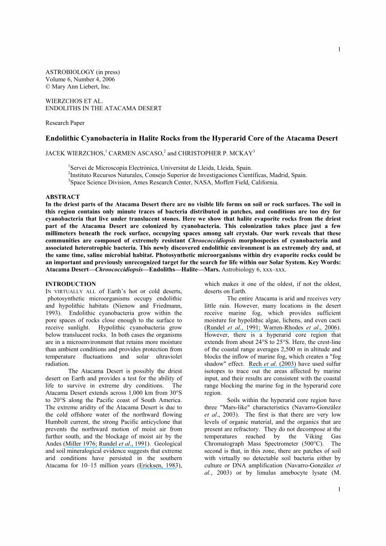
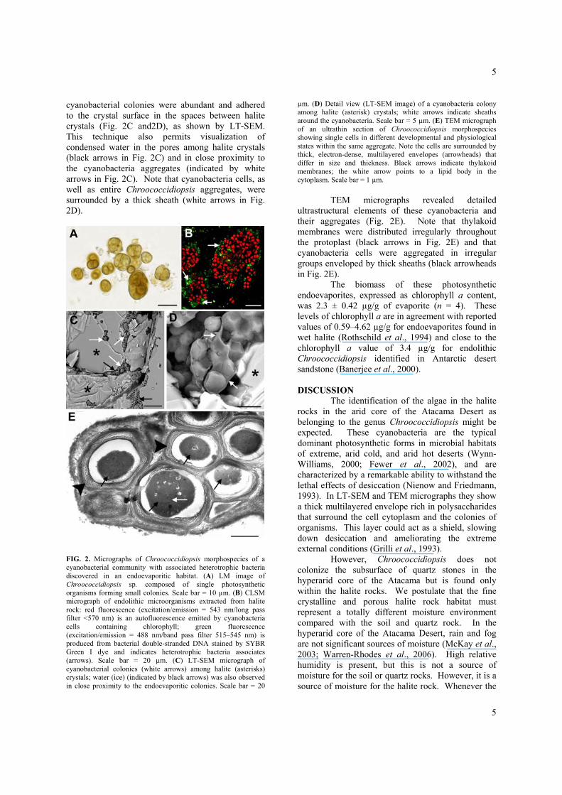
FIG. 1. Halite evaporite formations in the Yungay area of the Atacama Desert containing endolithic cyanobacteria. (A) General view of the round halite rocks harboring endoliths. (B) Close-up of the halite rock showing a gray layer parallel to the surface (blue arrows). (C) A cross fracture of the rock reveals a distinct gray layer representing the zone colonized by endoevaporitic cyanobacterial colonies. This layer appears 3–7 mm below the rock surface (arrows). The box indicates the area enlarged in (D), which is a stereoscopic micrograph of endolithic microorganism aggregates appearing as black spots among the halite crystals. RESULTS
Evaporite rocks in the Yungay area are essentially composed of halite (96–99%) with minor amounts of gypsum (1–3%) and traces (≤1%) of sylvine and quartz (detailed XRD data are given in Table 1). The upper exposed surface of the halite rocks appear brownish in color, apparently as a result of sand and dust, as can be observed in Fig. 1B. However, a considerable number of round rocks in
the sampling area had a dark gray layer beneath the rock surface (arrows in Fig. 1B). On closer
inspection, this layer was found to be 2–5 mm thick and 3–7 mm below the rock surface (Fig. 1C). Observations with the stereoscopic microscope (Fig.
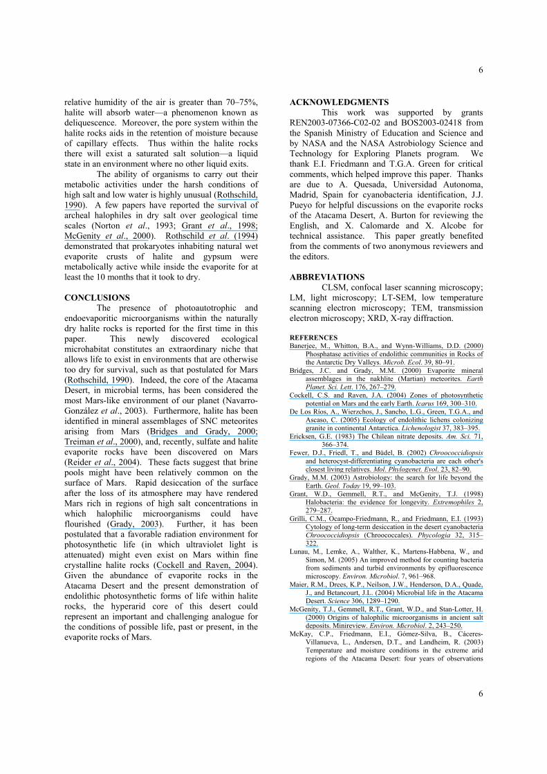
1D) revealed small dark spots within the grayish layer, which correspond to the colonies of endolithic microorganisms. LM examination (15 samples) of residues from the halite grayish layer after removal of the salt by aqueous dissolution revealed a homogeneous and possibly monospecific population of photosynthetic primary producers that belong to the genus Chroococcidiopsis (Fig. 2A). The same observation also shows that Chroococcidiopsis aggregates are sometimes composed of hundreds of single cells. Note that baeocyte cells appear spherical or slightly irregular, are 2–3 µm in diameter, and have a green-yellowish homogeneous cytoplasm. The cyanobacteria cell cytoplasm emits an intense chlorophyll autofluorescence red signal under the confocal microscope, as is shown in Fig. 2B. The use of SYBR Green I fluorescence assay reagent, which stains bacterial double-stranded DNA, indicated the presence of rod-shaped heterotrophic bacteria (green fluorescence in Fig. 2B) associated with the Chroococcidiopsis aggregates. Endoevaporitic
5
5
cyanobacterial colonies were abundant and adhered to the crystal surface in the spaces between halite crystals (Fig. 2C and2D), as shown by LT-SEM. This technique also permits visualization of condensed water in the pores among halite crystals (black arrows in Fig. 2C) and in close proximity to the cyanobacteria aggregates (indicated by white arrows in Fig. 2C). Note that cyanobacteria cells, as well as entire Chroococcidiopsis aggregates, were surrounded by a thick sheath (white arrows in Fig. 2D).
FIG. 2. Micrographs of Chroococcidiopsis morphospecies of a cyanobacterial community with associated heterotrophic bacteria discovered in an endoevaporitic habitat. (A) LM image of Chroococcidiopsis sp. composed of single photosynthetic organisms forming small colonies. Scale bar = 10 µm. (B) CLSM micrograph of endolithic microorganisms extracted from halite rock: red fluorescence (excitation/emission = 543 nm/long pass filter <570 nm) is an autofluorescence emitted by cyanobacteria cells containing chlorophyll; green fluorescence (excitation/emission = 488 nm/band pass filter 515–545 nm) is produced from bacterial double-stranded DNA stained by SYBR Green I dye and indicates heterotrophic bacteria associates (arrows). Scale bar = 20 µm. (C) LT-SEM micrograph of cyanobacterial colonies (white arrows) among halite (asterisks) crystals; water (ice) (indicated by black arrows) was also observed in close proximity to the endoevaporitic colonies. Scale bar = 20
µm. (D) Detail view (LT-SEM image) of a cyanobacteria colony among halite (asterisk) crystals; white arrows indicate sheaths around the cyanobacteria. Scale bar = 5 µm. (E) TEM micrograph of an ultrathin section of Chroococcidiopsis morphospecies showing single cells in different developmental and physiological states within the same aggregate. Note the cells are surrounded by thick, electron-dense, multilayered envelopes (arrowheads) that differ in size and thickness. Black arrows indicate thylakoid membranes; the white arrow points to a lipid body in the cytoplasm. Scale bar = 1 µm.
TEM micrographs revealed detailed ultrastructural elements of these cyanobacteria and their aggregates (Fig. 2E). Note that thylakoid membranes were distributed irregularly throughout the protoplast (black arrows in Fig. 2E) and that cyanobacteria cells were aggregated in irregular groups enveloped by thick sheaths (black arrowheads in Fig. 2E).
The biomass of these photosynthetic endoevaporites, expressed as chlorophyll a content, was 2.3 ± 0.42 µg/g of evaporite (n = 4). These levels of chlorophyll a are in agreement with reported values of 0.59–4.62 µg/g for endoevaporites found in wet halite (Rothschild et al., 1994) and close to the chlorophyll a value of 3.4 µg/g for endolithic Chroococcidiopsis identified in Antarctic desert sandstone (Banerjee et al., 2000). DISCUSSION
The identification of the algae in the halite rocks in the arid core of the Atacama Desert as belonging to the genus Chroococcidiopsis might be expected. These cyanobacteria are the typical dominant photosynthetic forms in microbial habitats of extreme, arid cold, and arid hot deserts (Wynn-Williams, 2000; Fewer et al., 2002), and are characterized by a remarkable ability to withstand the lethal effects of desiccation (Nienow and Friedmann, 1993). In LT-SEM and TEM micrographs they show a thick multilayered envelope rich in polysaccharides that surround the cell cytoplasm and the colonies of organisms. This layer could act as a shield, slowing down desiccation and ameliorating the extreme external conditions (Grilli et al., 1993).
However, Chroococcidiopsis does not colonize the subsurface of quartz stones in the hyperarid core of the Atacama but is found only within the halite rocks. We postulate that the fine crystalline and porous halite rock habitat must represent a totally different moisture environment compared with the soil and quartz rock. In the hyperarid core of the Atacama Desert, rain and fog are not significant sources of moisture (McKay et al., 2003; Warren-Rhodes et al., 2006). High relative humidity is present, but this is not a source of moisture for the soil or quartz rocks. However, it is a source of moisture for the halite rock. Whenever the
6
6
relative humidity of the air is greater than 70–75%, halite will absorb water—a phenomenon known as deliquescence. Moreover, the pore system within the halite rocks aids in the retention of moisture because of capillary effects. Thus within the halite rocks there will exist a saturated salt solution—a liquid state in an environment where no other liquid exits.
The ability of organisms to carry out their metabolic activities under the harsh conditions of high salt and low water is highly unusual (Rothschild, 1990). A few papers have reported the survival of archeal halophiles in dry salt over geological time scales (Norton et al., 1993; Grant et al., 1998; McGenity et al., 2000). Rothschild et al. (1994) demonstrated that prokaryotes inhabiting natural wet evaporite crusts of halite and gypsum were metabolically active while inside the evaporite for at least the 10 months that it took to dry.
CONCLUSIONS
The presence of photoautotrophic and endoevaporitic microorganisms within the naturally dry halite rocks is reported for the first time in this paper. This newly discovered ecological microhabitat constitutes an extraordinary niche that allows life to exist in environments that are otherwise too dry for survival, such as that postulated for Mars (Rothschild, 1990). Indeed, the core of the Atacama Desert, in microbial terms, has been considered the most Mars-like environment of our planet (Navarro-González et al., 2003). Furthermore, halite has been identified in mineral assemblages of SNC meteorites arising from Mars (Bridges and Grady, 2000; Treiman et al., 2000), and, recently, sulfate and halite evaporite rocks have been discovered on Mars (Reider et al., 2004). These facts suggest that brine pools might have been relatively common on the surface of Mars. Rapid desiccation of the surface after the loss of its atmosphere may have rendered Mars rich in regions of high salt concentrations in which halophilic microorganisms could have flourished (Grady, 2003). Further, it has been postulated that a favorable radiation environment for photosynthetic life (in which ultraviolet light is attenuated) might even exist on Mars within fine crystalline halite rocks (Cockell and Raven, 2004). Given the abundance of evaporite rocks in the Atacama Desert and the present demonstration of endolithic photosynthetic forms of life within halite rocks, the hyperarid core of this desert could represent an important and challenging analogue for the conditions of possible life, past or present, in the evaporite rocks of Mars.
ACKNOWLEDGMENTS This work was supported by grants
REN2003-07366-C02-02 and BOS2003-02418 from the Spanish Ministry of Education and Science and by NASA and the NASA Astrobiology Science and Technology for Exploring Planets program. We thank E.I. Friedmann and T.G.A. Green for critical comments, which helped improve this paper. Thanks are due to A. Quesada, Universidad Autonoma, Madrid, Spain for cyanobacteria identification, J.J. Pueyo for helpful discussions on the evaporite rocks of the Atacama Desert, A. Burton for reviewing the English, and X. Calomarde and X. Alcobe for technical assistance. This paper greatly benefited from the comments of two anonymous reviewers and the editors. ABBREVIATIONS
CLSM, confocal laser scanning microscopy; LM, light microscopy; LT-SEM, low temperature scanning electron microscopy; TEM, transmission electron microscopy; XRD, X-ray diffraction. REFERENCES Banerjee, M., Whitton, B.A., and Wynn-Williams, D.D. (2000)
Phosphatase activities of endolithic communities in Rocks of the Antarctic Dry Valleys. Microb. Ecol. 39, 80–91.
Bridges, J.C. and Grady, M.M. (2000) Evaporite mineral assemblages in the nakhlite (Martian) meteorites. Earth Planet. Sci. Lett. 176, 267–279.
Cockell, C.S. and Raven, J.A. (2004) Zones of photosynthetic potential on Mars and the early Earth. Icarus 169, 300–310.
De Los Ríos, A., Wierzchos, J., Sancho, L.G., Green, T.G.A., and Ascaso, C. (2005) Ecology of endolithic lichens colonizing granite in continental Antarctica. Lichenologist 37, 383–395.
Ericksen, G.E. (1983) The Chilean nitrate deposits. Am. Sci. 71, 366–374.
Fewer, D.J., Friedl, T., and Büdel, B. (2002) Chroococcidiopsis and heterocyst-differentiating cyanobacteria are each other's closest living relatives. Mol. Phylogenet. Evol. 23, 82–90.
Grady, M.M. (2003) Astrobiology: the search for life beyond the Earth. Geol. Today 19, 99–103.
Grant, W.D., Gemmell, R.T., and McGenity, T.J. (1998) Halobacteria: the evidence for longevity. Extremophiles 2, 279–287.
Grilli, C.M., Ocampo-Friedmann, R., and Friedmann, E.I. (1993) Cytology of long-term desiccation in the desert cyanobacteria Chroococcidiopsis (Chroococcales). Phycologia 32, 315–322.
Lunau, M., Lemke, A., Walther, K., Martens-Habbena, W., and Simon, M. (2005) An improved method for counting bacteria from sediments and turbid environments by epifluorescence microscopy. Environ. Microbiol. 7, 961–968.
Maier, R.M., Drees, K.P., Neilson, J.W., Henderson, D.A., Quade, J., and Betancourt, J.L. (2004) Microbial life in the Atacama Desert. Science 306, 1289–1290.
McGenity, T.J., Gemmell, R.T., Grant, W.D., and Stan-Lotter, H. (2000) Origins of halophilic microorganisms in ancient salt deposits. Minireview. Environ. Microbiol. 2, 243–250.
McKay, C.P., Friedmann, E.I., Gómez-Silva, B., Cáceres-Villanueva, L., Andersen, D.T., and Landheim, R. (2003) Temperature and moisture conditions in the extreme arid regions of the Atacama Desert: four years of observations
7
7
including the El Niño of 1997–1998. Astrobiology 3, 393–406.
Miller, A. (1976) The climate of Chile. In World Survey of Climatology, Vol. 12: Climate of Central and South America, edited by W. Schwerdfeger, Elsevier, Amsterdam, pp. 113–145.
Navarro-González, R., Rainey, F.A., Molina, P., Bagaley, D.R., Hollen, B.J., de la Rosa, J., Small, A.M., Quinn, R.C., Grunthaner, F.J., Cáceres, L., Gomez-Silva, B., and McKay, C.P. (2003) Mars-like soils in the Atacama Desert, Chile, and the dry limit of microbial life. Science 302, 1018–1021.
Nienow, J.A. and Friedmann, E.I. (1993) Terrestrial lithophytic (rock) communities. In Antarctic Microbiology, edited by E.I. Friedmann, Wiley-Liss, New York, pp. 243–412.
Norton, C.F., McGenity, T.J., and Grant, W.D. (1993) Archeal halophiles (halobacteria) from two British salt mines. J. Gen. Microbiol. 139, 1077–1081.
Pueyo, J.J., Chong, G., and Jensen, A. (2001) Neogene evaporites in desert volcanic environments: Atacama Desert, northern Chile. Sedimentology 48, 1411–1431.
Rech, J., Quade, J., and Hart, W. (2003) Isotopic evidence for the source of Ca and S in soil gypsum, anhydrite and calcite in the Atacama Desert, Chile. Geochim. Cosmochim. Acta 67, 575–586.
Reider, R., Gellert, R., Anderson, R.C., Brückner, J., Clark, B.C., Dreibus, G., Economou, T., Klingelhöfer, G., Lugmair, G.W., Ming, D.W., Squyres, S.W., d'Uston, C., Wänke, H., Yen, A., and Zipfel, J. (2004) Chemistry of rocks and soils at Meridiani Planum from the Alpha Particle X-ray Spectrometer. Science 306, 1746–1749.
Rothschild, L.J. (1990) Earth analogs for Martian life. Microbes in evaporites, a new model system for life on Mars. Icarus 88, 246–260.
Rothschild, L.J., Giver, L.J., White, M.R., and Mancinelli, R.L. (1994) Metabolic activity of microorganisms in evaporites. J. Phycol. 30, 431–438.
Rundel, P.W., Dillon, M.O., Palma, B., Hooney, H.A., Gulmon, S.L., and Ehleringer, J.R. (1991) The phytogeography and ecology of the coastal Atacama and Peruvian deserts. Aliso 13, 1–49.
Stal, L.J., van Gemerden, H., and Krumbein, W.F. (1985) Structure and development of a benthic microbial mat. FEMS Microbiol. Ecol. 31, 111–125.
Treiman, A.H., Gleason, J.D., and Bogard, D.D. (2000) The SNC meteorites are from Mars. Planet. Space Sci. 48, 1213–1230.
Warren-Rhodes, K.A., Rhodes, K. L., Pointing, S.B., Ewing, S., Lacap, D.C., Gómez-Silva, B., Amundson, R., Friedmann, E.I., and McKay, C.P. (2006) Hypolithic cyanobacteria, dry limit of photosynthesis and microbial ecology in the hyperarid Atacama Desert. Microb. Ecol. (in press).
Wynn-Williams, D.D. (2000) Cyanobacteria in deserts—life at the limit? In The Ecology of Cyanobacteria—Their Diversity in Time and Space, edited by B.A. Whitton and M. Potts, Kluwer Academic Publishers, Dordrecht, The Netherlands, pp. 341–366.
Address reprint requests to: Jacek Wierzchos Servei de Microscopía Electrònica Universitat de Lleida c/ Rovira Roure 44 25198 Lleida, Spain E-mail: [email protected]