effect of ceria nano-coating on the formation of a protective
TRANSCRIPT
EasyChair Preprint№ 2536
Effect of Ceria Nano-Coating on the Formation ofa Protective Oxide-Scale Layer on the Surface ofFeCrAl Fibers
Omar Yousef, Isameldeen Daffallah and Osama Ibrahim
EasyChair preprints are intended for rapiddissemination of research results and areintegrated with the rest of EasyChair.
February 4, 2020
Arab International Conference and Exhibition on Nanotechnology, March 17-19, 2020, KISR, Kuwait
1
Effect of nano-ceria coating on the formation of a protective oxide-scale layer on the surface of
FeCrAl fibers
Omar A. Yousef, Isameldeen E. Daffallah, Osama M. Ibrahim
Mechanical Engineering Department, Faculty of Engineering and Petroleum, Kuwait University, Safat
13060, Kuwait
Abstract
The objective of this study is to investigate the effect of nano-ceria coating on the formation
of the protective oxide-scale layer on the surface of metal fibers made of FeCrAl alloy. FeCrAl
alloy, which contains about 72% iron, 21% chromium, and 5% aluminum, forms an oxide-
scale layer dominated by aluminum oxide to protect the base metal alloy from further oxidation
at high temperatures. A small fraction of rare earth elements, such as yttrium and cerium, are
commonly added to FeCrAl alloy to enhance its oxidation characteristics at high temperature
and improve the adhesion between the oxide-scale layer and the base metal alloy. In this study,
the nano-ceria coating was added to the surface of the fibers. Three samples were considered
in this investigation. The first sample was kept without ceria coating as a baseline. The second
sample was coated with 2.5%, by weight, of the nano-ceria solution, while the third sample
was coated with 7.5%, by weight, of the nano-ceria solution. Thermo-Gravimetric Analysis
(TGA) was conducted for 10 hours at 800°C to quantify and compare the weight gain and
oxidation rate for the three samples. The surface micromorphology of the samples was
examined using a field-emission Scanning Electron Microscope (SEM). Chemical analyses of
the surface of the samples were performed using Energy Dispersive x-ray Spectroscopy (EDS).
The results clearly show that the ceria nano-coating reduces the oxidation rate of FeCrAl
significantly; however, excess material on the surface of the fibers was observed. Different
coating techniques were then tried to optimize the coating process.
Keywords: Metal Fibers, FeCrAl alloy, Coating, Nano-ceria, Oxidation.
Omar A. Yousef is a graduate student at the Mechanical Engineering Department, Kuwait
University.
Isameldeen E. Daffallah is a senior research assistant at the Nanotechnology Research Facility,
Kuwait University.
Osama M. Ibrahim completed his Ph.D. from the University of Wisconsin-Madison, USA. Currently,
he is an Associate Professor of Mechanical Engineering at Kuwait University.
Arab International Conference and Exhibition on Nanotechnolog, March 17-19, 2020, KISR, Kuwait
2
Introduction
The thermally grown oxide scale layer plays a vital role in limiting the environmental degradation of many
high-temperature alloys. FeCrAl is a well-known alloy that contains mostly Iron (72%), Chromium
(21%), and Aluminum (6%) [1]. FeCrAl alloy is used for the fabrication of components working at high
temperatures such as heating elements, fire grid, honeycomb catalytic converters, and metal fibers. High-
temperature oxidation of FeCrAl alloy results in the formation of an oxide scale layer on the alloy surface
[2]. Trunov et al. [3] reported that there is considerable interest in the formation and adhesion of these
oxide scale layers, and a complete understanding of the growth mechanism is essential in designing
oxidation resistant alloys.
The formation of aluminum oxide scale layer (Al2O3) on the metal surface is classified into two categories
[4]: (1) Metastable; and (2) Stable. The metastable1-transient formation of alumina includes γ-Al2O3, θ-
Al2O3, and δ-Al2O3; while the stable form of alumina includes α-Al2O3.
Kadiri et al. [4] and Prescott and Graham [5] reported that forming alumina on the alloys is very crucial
for many advanced high-temperature oxidation coatings in the aeronautical and power generation sectors.
It has been reported that during the initial stages of oxidation, fast-growing metastable alumina form in
the shapes of platelets and needles on the alloy surface. As the heating process continues, these platelets
transformation accompanied by a reduction in oxide volume leading to tensile stresses and radial cracks
detrimental to the adherence of the final α-Al2O3 layer. The features of the alumina layer and the scale
growth mechanism depends on the processing method, as well as, on the additives to the base of the
FeCrAl alloy. Studies by Jeurgens et al. [6, 7] explained the formation of alumina as a function of
temperature. Generally, the formation of aluminum oxide happens through four stages, as summarized in
Table 1. In the first stage, a layer of amorphous alumina diffuses slowly to the surface at temperatures
between 300 to 550 °C. The rate of this stage is controlled by the outward diffusion of Aluminum cations.
The second stage starts at a temperature of 550 °C and ends at 650 °C. This stage accompanied by an
increase in the rate of oxidation, and as the openings in the oxide coating heal, the rate of oxidation
decreases. During this stage, the layer of amorphous alumina starts to transform into γ–Al2O3 phase. As
the temperature approaches 650 °C, an arranged polycrystalline film of γ- Al2O3 phase formed. The third
stage occurs at a temperature higher than 650 °C. The growth of γ-alumina, at this third stage, continues
at a rate that is limited by the inward diffusion of oxygen anions along grain boundaries, resulting in a
dense film of γ-Al2O3 phase on the surface. During thermal oxidation of this stage, a combination of
chemical and thermal transformation converts γ- Al2O3 phase into δ-Al2O3 and θ-Al2O3 phases. At a higher
thermal oxidation temperature of 950 °C to 1050 °C, which characterizes the end of the third stage and
the beginning of the fourth stage, δ-Al2O3 and θ-Al2O3 phases transform into α-Al2O3 phase. In the fourth
stage, at a temperature higher than 1050 °C, the oxide film on the surface is totally converted to the coarse
and dense film of α-Al2O3 crystallites on the surface.
Table 1. Stages at which the alumina layer is formed, adapted from Trunov et al. [3].
Transition Stages Temperature Ranges Observation
Stage 1 T < 550℃ Growth of amorphous alumina
Stage 2 550℃ < T < 650℃ Transition of amorphous alumina into γ- Al2O3 phase
Stage 3 650℃ < T < 1050℃ Growth of γ-alumina on the surface Converts γ- Al2O3 phase into δ- Al2O3 and θ- Al2O3
Stage 4 1050℃ < T δ- Al2O3 and θ- Al2O3 phases transform into α- Al2O3 phase.
Arab International Conference and Exhibition on Nanotechnolog, March 17-19, 2020, KISR, Kuwait
3
Due to the failure of the protective layer α- Al2O3 alloy under high load of thermal oxidation, which
results in an extensive scale spallations, many researchers attempted to enhance the adherence of alumina
scales by adding the rare earth elements. The rare earth elements improve the adherence of alumina scales
by limiting the oxygen diffusion through grain boundaries [8]. The rare earth elements can be added
directly to the composition of metals and alloys or by using them as coating materials. [9]. Whereas the
last one is the commonly preferred once since it does not alternate the alloys’ mechanical properties [9,
10].
Numerous studies were performed on coating metals or alloys with rare earth elements for improving the
oxidation resistance [10-21]. For the FeCrAl alloys, a couple of research studies were accomplished in
this field. Ishii et al. [8] investigated the effect of rare earth elements (Y,La,Ce, Pr, Nd, SM) on FeCrAl
alloy foils under a cyclic oxidation test at high temperature (1100 and 1200 °C). And they found that the
rare earth elements enhanced the resistance to high-temperature oxidation. Pillis and Ramanathan [22]
studied a coated FeCrAl alloy with lanthanum chromite at isothermal oxidation tests at 1000 °C and up to
200 hours. Also, their results showed good alumina scale adherence. Nguyen et al. [23] coated FeCrAl
alloy with cerium by the sol-gel technique and tested it at high isothermal oxidation temperature (1100
°C). For monitoring the start-up of the coated ceria particle size under the oxidation tests, they performed
heat treatments under argon media to the coated FeCrAl samples at different temperatures from 600 to
1000 °C. And they found that the annealed sample at 600° C shows the best improvement in the oxidation
resistance. Che˛cmanowski et al. [24] studied the influence of SiO2–Al2O3–CeO2 oxides gel on a FeCrAl
alloy. They concluded that the amount of cerium used to obtain the coating layer of SiO2–Al2O3–CeO2
influenced the morphology and the oxidation resistance. Fernandes et al. [9] investigated the effect of
several oxide gels of rare earth elements (CeO2, La2O3) and their combination on increasing the high-
temperature oxidation resistance of alumina forming on the FeCrAl alloy at 1000 °C. They concluded that
coating with the combined oxides of CeO2 and La2O3 approved the oxidation resistance significantly
compared to coating with single oxides.
Motivation
Metal fibers are used in many industrial applications related to filtration, composite reinforcement, and
heat transfer applications [25]. Metal fibers have a porous structure and high surface to volume ratio,
which make them vulnerable to high-temperature thermal oxidation. The aim of this study is to protect
the base metal of the fibers from depletion by forming a reliable protective oxide-scale layer of alumina
by using a nano-material coating on the surface of the fibers to control the oxide-scale layer thickness,
adhesion, and growth rate.
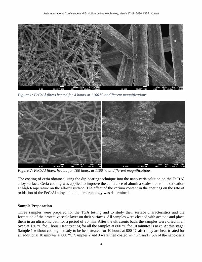
Figure 1 shows the FeCrAl metal fiber under the electron microscope after heating it for 4 hours at 1100℃.
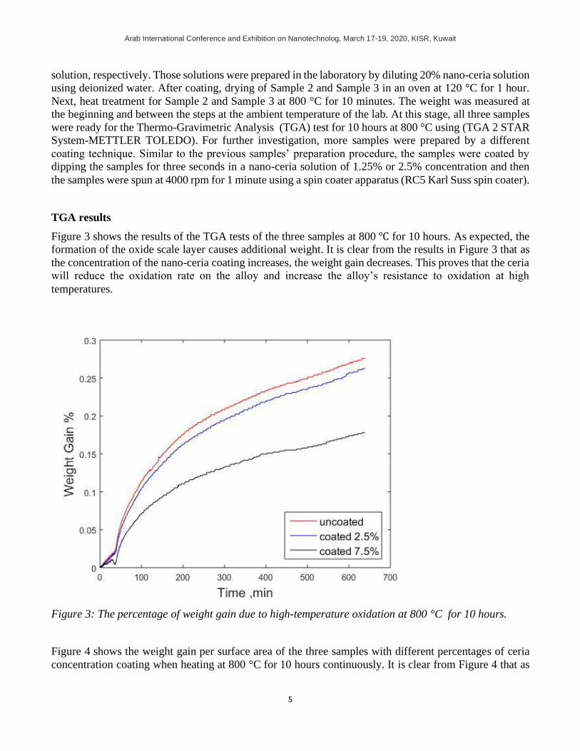
alumina was formed on the fiber’s surface to protect the base metal from depletion. But, when the fibers
are exposed to a long duration of further heating oxidization occurs, causing the fibers to fail and the metal
to deplete as shown in Figure 2 for a 100 hours heating at 1100℃. Nano-particles of rare elements, such
as ceria, zirconia, and yttria, are considered as nano-coatings to the surface of the fiber. The oxides of rare
elements can increase the thermal resistance of metals [8]. This paper presents the results of nano-ceria
coating on the surface of FeCrAl fibers.
Arab International Conference and Exhibition on Nanotechnolog, March 17-19, 2020, KISR, Kuwait
4
Figure 1: FeCrAl fibers heated for 4 hours at 1100 ℃ at different magnifications.
Figure 2: FeCrAl fibers heated for 100 hours at 1100 ℃ at different magnifications.
The coating of ceria obtained using the dip-coating technique into the nano-ceria solution on the FeCrAl
alloy surface. Ceria coating was applied to improve the adherence of alumina scales due to the oxidation
at high temperature on the alloy’s surface. The effect of the cerium content in the coatings on the rate of
oxidation of the FeCrAl alloy and on the morphology was determined.
Sample Preparation
Three samples were prepared for the TGA testing and to study their surface characteristics and the
formation of the protective scale layer on their surfaces. All samples were cleaned with acetone and place
them in an ultrasonic bath for a period of 30 min. After the ultrasonic bath, the samples were dried in an
oven at 120 °C for 1 hour. Heat treating for all the samples at 800 °C for 10 minutes is next. At this stage,
Sample 1 without coating is ready to be heat-treated for 10 hours at 800 °C after they are heat-treated for
an additional 10 minutes at 800 °C. Samples 2 and 3 were then coated with 2.5 and 7.5% of the nano-ceria
Arab International Conference and Exhibition on Nanotechnolog, March 17-19, 2020, KISR, Kuwait
5
solution, respectively. Those solutions were prepared in the laboratory by diluting 20% nano-ceria solution
using deionized water. After coating, drying of Sample 2 and Sample 3 in an oven at 120 °C for 1 hour.
Next, heat treatment for Sample 2 and Sample 3 at 800 °C for 10 minutes. The weight was measured at
the beginning and between the steps at the ambient temperature of the lab. At this stage, all three samples
were ready for the Thermo-Gravimetric Analysis (TGA) test for 10 hours at 800 °C using (TGA 2 STAR
System-METTLER TOLEDO). For further investigation, more samples were prepared by a different
coating technique. Similar to the previous samples’ preparation procedure, the samples were coated by
dipping the samples for three seconds in a nano-ceria solution of 1.25% or 2.5% concentration and then
the samples were spun at 4000 rpm for 1 minute using a spin coater apparatus (RC5 Karl Suss spin coater).
TGA results
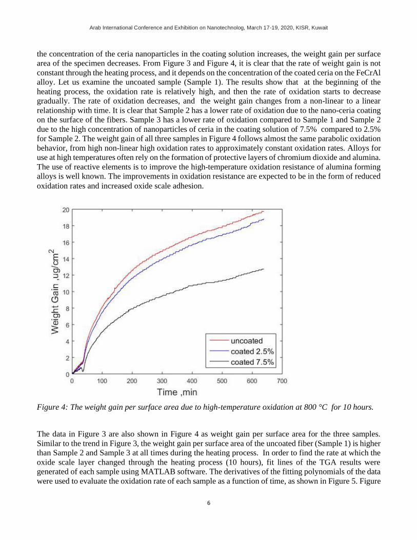
Figure 3 shows the results of the TGA tests of the three samples at 800 ℃ for 10 hours. As expected, the
formation of the oxide scale layer causes additional weight. It is clear from the results in Figure 3 that as
the concentration of the nano-ceria coating increases, the weight gain decreases. This proves that the ceria
will reduce the oxidation rate on the alloy and increase the alloy’s resistance to oxidation at high
temperatures.
Figure 3: The percentage of weight gain due to high-temperature oxidation at 800 °C for 10 hours.
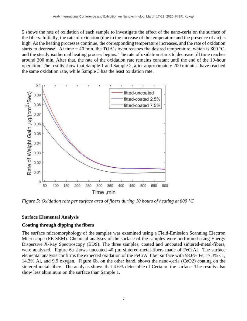
Figure 4 shows the weight gain per surface area of the three samples with different percentages of ceria
concentration coating when heating at 800 °C for 10 hours continuously. It is clear from Figure 4 that as
Arab International Conference and Exhibition on Nanotechnolog, March 17-19, 2020, KISR, Kuwait
6
the concentration of the ceria nanoparticles in the coating solution increases, the weight gain per surface
area of the specimen decreases. From Figure 3 and Figure 4, it is clear that the rate of weight gain is not
constant through the heating process, and it depends on the concentration of the coated ceria on the FeCrAl
alloy. Let us examine the uncoated sample (Sample 1). The results show that at the beginning of the
heating process, the oxidation rate is relatively high, and then the rate of oxidation starts to decrease
gradually. The rate of oxidation decreases, and the weight gain changes from a non-linear to a linear
relationship with time. It is clear that Sample 2 has a lower rate of oxidation due to the nano-ceria coating
on the surface of the fibers. Sample 3 has a lower rate of oxidation compared to Sample 1 and Sample 2
due to the high concentration of nanoparticles of ceria in the coating solution of 7.5% compared to 2.5%
for Sample 2. The weight gain of all three samples in Figure 4 follows almost the same parabolic oxidation
behavior, from high non-linear high oxidation rates to approximately constant oxidation rates. Alloys for
use at high temperatures often rely on the formation of protective layers of chromium dioxide and alumina.
The use of reactive elements is to improve the high-temperature oxidation resistance of alumina forming
alloys is well known. The improvements in oxidation resistance are expected to be in the form of reduced
oxidation rates and increased oxide scale adhesion.
Figure 4: The weight gain per surface area due to high-temperature oxidation at 800 °C for 10 hours.
The data in Figure 3 are also shown in Figure 4 as weight gain per surface area for the three samples.
Similar to the trend in Figure 3, the weight gain per surface area of the uncoated fiber (Sample 1) is higher
than Sample 2 and Sample 3 at all times during the heating process. In order to find the rate at which the
oxide scale layer changed through the heating process (10 hours), fit lines of the TGA results were
generated of each sample using MATLAB software. The derivatives of the fitting polynomials of the data
were used to evaluate the oxidation rate of each sample as a function of time, as shown in Figure 5. Figure
Arab International Conference and Exhibition on Nanotechnolog, March 17-19, 2020, KISR, Kuwait
7
5 shows the rate of oxidation of each sample to investigate the effect of the nano-ceria on the surface of
the fibers. Initially, the rate of oxidation (due to the increase of the temperature and the presence of air) is
high. As the heating processes continue, the corresponding temperature increases, and the rate of oxidation
starts to decrease. At time = 40 min, the TGA’s oven reaches the desired temperature, which is 800 ℃,
and the steady isothermal heating process begins. The rate of oxidation starts to decrease till time reaches
around 300 min. After that, the rate of the oxidation rate remains constant until the end of the 10-hour
operation. The results show that Sample 1 and Sample 2, after approximately 200 minutes, have reached
the same oxidation rate, while Sample 3 has the least oxidation rate.
Figure 5: Oxidation rate per surface area of fibers during 10 hours of heating at 800 °C.
Surface Elemental Analysis
Coating through dipping the fibers
The surface micromorphology of the samples was examined using a Field-Emission Scanning Electron
Microscope (FE-SEM). Chemical analyses of the surface of the samples were performed using Energy
Dispersive X-Ray Spectroscopy (EDS). The three samples, coated and uncoated sintered-metal-fibers,
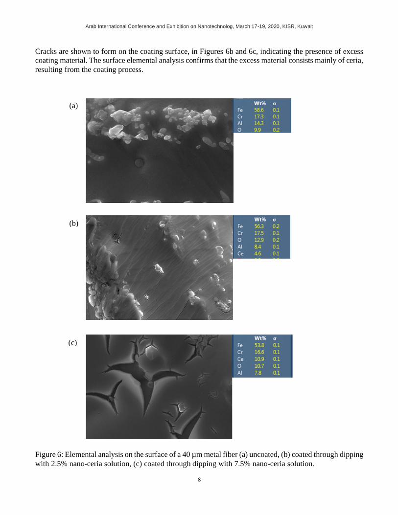
were analyzed. Figure 6a shows uncoated 40 µm sintered-metal-fibers made of FeCrAl. The surface
elemental analysis confirms the expected oxidation of the FeCrAl fiber surface with 58.6% Fe, 17.3% Cr,
14.3% Al, and 9.9 oxygen. Figure 6b, on the other hand, shows the nano-ceria (CeO2) coating on the
sintered-metal-fibers. The analysis shows that 4.6% detectable.of Ceria on the surface. The results also
show less aluminum on the surface than Sample 1.
Arab International Conference and Exhibition on Nanotechnolog, March 17-19, 2020, KISR, Kuwait
8
Cracks are shown to form on the coating surface, in Figures 6b and 6c, indicating the presence of excess
coating material. The surface elemental analysis confirms that the excess material consists mainly of ceria,
resulting from the coating process.
Figure 6: Elemental analysis on the surface of a 40 µm metal fiber (a) uncoated, (b) coated through dipping
with 2.5% nano-ceria solution, (c) coated through dipping with 7.5% nano-ceria solution.
(a)
(b)
(c)
Arab International Conference and Exhibition on Nanotechnolog, March 17-19, 2020, KISR, Kuwait
9
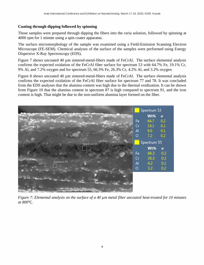
Coating through dipping followed by spinning
Those samples were prepared through dipping the fibers into the ceria solution, followed by spinning at
4000 rpm for 1 minute using a spin coater apparatus.
The surface micromorphology of the sample was examined using a Field-Emission Scanning Electron
Microscope (FE-SEM). Chemical analyses of the surface of the samples were performed using Energy
Dispersive X-Ray Spectroscopy (EDS).
Figure 7 shows uncoated 40 µm sintered-metal-fibers made of FeCrAl. The surface elemental analysis
confirms the expected oxidation of the FeCrAl fiber surface for spectrum 53 with 64.7% Fe, 19.1% Cr,
9% Al, and 7.2% oxygen and for spectrum 55, 66.3% Fe, 26.3% Cr, 4.2% Al, and 3.2% oxygen.
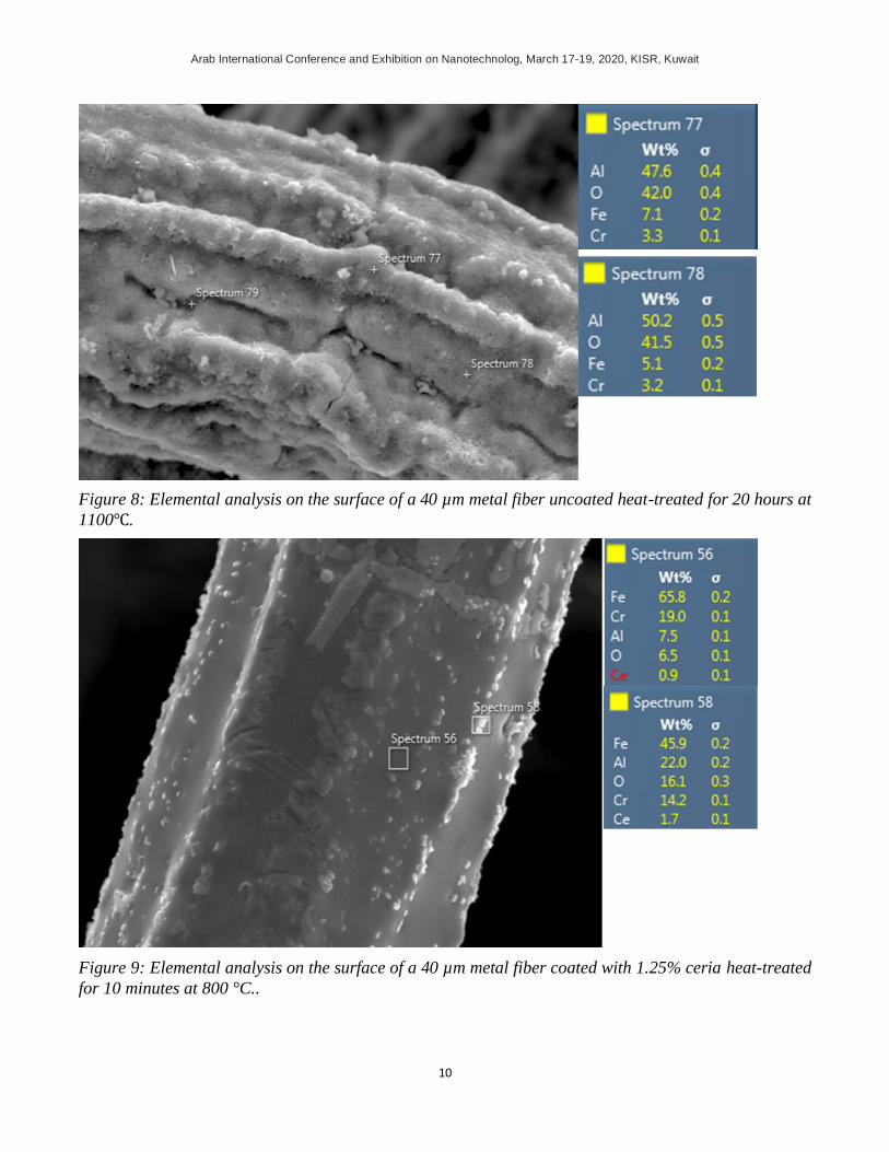
Figure 8 shows uncoated 40 µm sintered-metal-fibers made of FeCrAl. The surface elemental analysis
confirms the expected oxidation of the FeCrAl fiber surface for spectrum 77 and 78. It was concluded
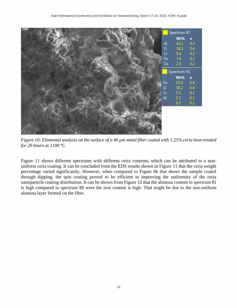
from the EDS analyses that the alumina content was high due to the thermal oxidization. It can be shown
from Figure 10 that the alumina content in spectrum 87 is high compared to spectrum 91, and the iron
content is high. That might be due to the non-uniform alumina layer formed on the fiber.
Figure 7: Elemental analysis on the surface of a 40 µm metal fiber uncoated heat-treated for 10 minutes
at 800℃.
Arab International Conference and Exhibition on Nanotechnolog, March 17-19, 2020, KISR, Kuwait
10
Figure 8: Elemental analysis on the surface of a 40 µm metal fiber uncoated heat-treated for 20 hours at
1100℃.
Figure 9: Elemental analysis on the surface of a 40 µm metal fiber coated with 1.25% ceria heat-treated
for 10 minutes at 800 °C..
Arab International Conference and Exhibition on Nanotechnolog, March 17-19, 2020, KISR, Kuwait
11
Figure 10: Elemental analysis on the surface of a 40 µm metal fiber coated with 1.25% ceria heat-treated
for 20 hours at 1100 ℃.
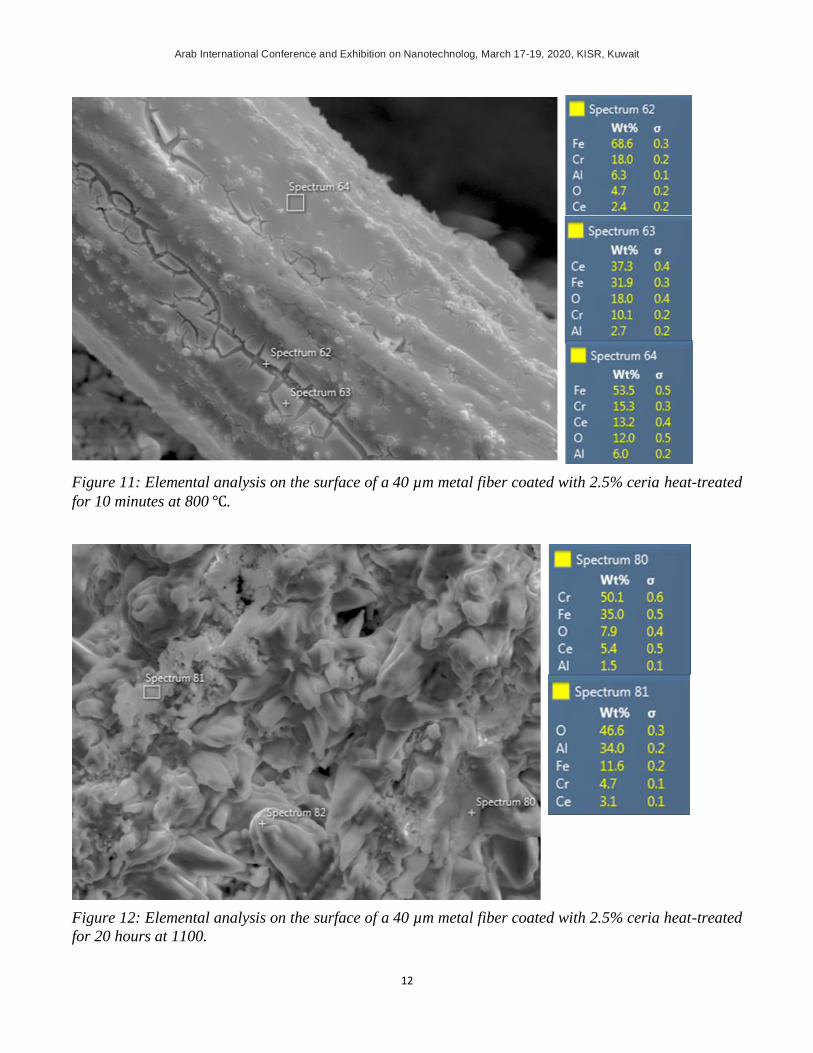
Figure 11 shows different spectrums with different ceira contents, which can be attributed to a non-
uniform ceria coating. It can be concluded from the EDS results shown in Figure 11 that the ceria weight
percentage varied significantly. However, when compared to Figure 6b that shows the sample coated
through dipping, the spin coating proved to be efficient in improving the uniformity of the ceria
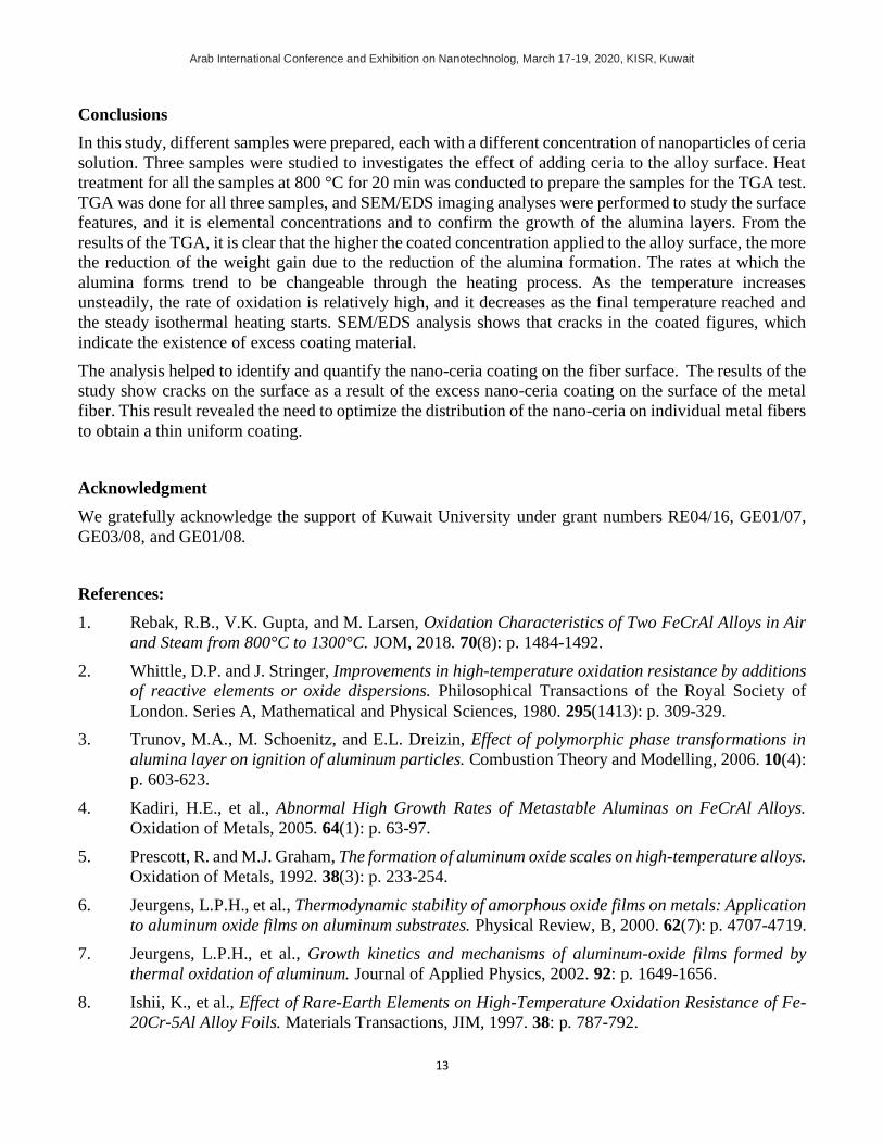
nanoparticle coating distribution. It can be shown from Figure 12 that the alumina content in spectrum 81
is high compared to spectrum 80 were the iron content is high. That might be due to the non-uniform
alumina layer formed on the fiber.
Arab International Conference and Exhibition on Nanotechnolog, March 17-19, 2020, KISR, Kuwait
12
Figure 11: Elemental analysis on the surface of a 40 µm metal fiber coated with 2.5% ceria heat-treated
for 10 minutes at 800 ℃.
Figure 12: Elemental analysis on the surface of a 40 µm metal fiber coated with 2.5% ceria heat-treated
for 20 hours at 1100.
Arab International Conference and Exhibition on Nanotechnolog, March 17-19, 2020, KISR, Kuwait
13
Conclusions
In this study, different samples were prepared, each with a different concentration of nanoparticles of ceria
solution. Three samples were studied to investigates the effect of adding ceria to the alloy surface. Heat
treatment for all the samples at 800 °C for 20 min was conducted to prepare the samples for the TGA test.
TGA was done for all three samples, and SEM/EDS imaging analyses were performed to study the surface
features, and it is elemental concentrations and to confirm the growth of the alumina layers. From the
results of the TGA, it is clear that the higher the coated concentration applied to the alloy surface, the more
the reduction of the weight gain due to the reduction of the alumina formation. The rates at which the
alumina forms trend to be changeable through the heating process. As the temperature increases
unsteadily, the rate of oxidation is relatively high, and it decreases as the final temperature reached and
the steady isothermal heating starts. SEM/EDS analysis shows that cracks in the coated figures, which
indicate the existence of excess coating material.
The analysis helped to identify and quantify the nano-ceria coating on the fiber surface. The results of the
study show cracks on the surface as a result of the excess nano-ceria coating on the surface of the metal
fiber. This result revealed the need to optimize the distribution of the nano-ceria on individual metal fibers
to obtain a thin uniform coating.
Acknowledgment
We gratefully acknowledge the support of Kuwait University under grant numbers RE04/16, GE01/07,
GE03/08, and GE01/08.
References:
1. Rebak, R.B., V.K. Gupta, and M. Larsen, Oxidation Characteristics of Two FeCrAl Alloys in Air
and Steam from 800°C to 1300°C. JOM, 2018. 70(8): p. 1484-1492.
2. Whittle, D.P. and J. Stringer, Improvements in high-temperature oxidation resistance by additions
of reactive elements or oxide dispersions. Philosophical Transactions of the Royal Society of
London. Series A, Mathematical and Physical Sciences, 1980. 295(1413): p. 309-329.
3. Trunov, M.A., M. Schoenitz, and E.L. Dreizin, Effect of polymorphic phase transformations in
alumina layer on ignition of aluminum particles. Combustion Theory and Modelling, 2006. 10(4):
p. 603-623.
4. Kadiri, H.E., et al., Abnormal High Growth Rates of Metastable Aluminas on FeCrAl Alloys.
Oxidation of Metals, 2005. 64(1): p. 63-97.
5. Prescott, R. and M.J. Graham, The formation of aluminum oxide scales on high-temperature alloys.
Oxidation of Metals, 1992. 38(3): p. 233-254.
6. Jeurgens, L.P.H., et al., Thermodynamic stability of amorphous oxide films on metals: Application
to aluminum oxide films on aluminum substrates. Physical Review, B, 2000. 62(7): p. 4707-4719.
7. Jeurgens, L.P.H., et al., Growth kinetics and mechanisms of aluminum-oxide films formed by
thermal oxidation of aluminum. Journal of Applied Physics, 2002. 92: p. 1649-1656.
8. Ishii, K., et al., Effect of Rare-Earth Elements on High-Temperature Oxidation Resistance of Fe-
20Cr-5Al Alloy Foils. Materials Transactions, JIM, 1997. 38: p. 787-792.
Arab International Conference and Exhibition on Nanotechnolog, March 17-19, 2020, KISR, Kuwait
14
9. Fernandes, S.M.d.C., O.V. Corea, and L.V. Ramanathan. Nanocrystalline Rare Earth Oxide
Coatings for Increased Protection of Iron-chromium Alloys at High Temperatures. 2014.
10. Rahman, A. and R. Jayaganthan, Study of Nanostructured CeO2 Coatings on Superalloy. Surface
Engineering, 2016. 32(10): p. 771-778.
11. Carvalho, F. and L. Ramanathan, Rare earth oxide coatings to decrease high-temperature
degradation of chromia forming alloys. Materials Research, 2004. 7.
12. Kumar, S., et al. Effect of CeO2 in Cr3C2-NiCr Coating on Superni 600 at High Temperature☆.
2014.
13. Pillis, M.F., E.G.d. Araújo, and L.V. Ramanathan. Effect of Addition of Rare Earth Oxide
Concentrates on Oxidation Resistance of AISI 304L. 2006.
14. Fernandes, S. and L. Ramanathan, Effect of surface deposited rare earth oxide gel characteristics
on cyclic oxidation behavior of Fe20Cr alloys. Materials Research-Ibero-American Journal of
Materials - MATER RES-IBERO-AM J MATER, 2006. 9.
15. Ghosh, D., et al., CeO2 based coating for high-temperature oxidation protection. Surface
Engineering, 2014. 31: p. 1743294414Y.000.
16. Meng, J.-s. and J. Ze-sheng. Effect of La2O3/CeO2 particle size on high-temperature oxidation
resistance of electrodeposited Ni–La2O3/CeO2 composites. 2014.
17. Papaiacovou, P., et al., The effect of CeO2 coatings on the oxidation behaviour of Fe20Cr alloys
in O2 at 1173 K. Corrosion Science - CORROS SCI, 1990. 30: p. 451-460.
18. Rhys-Jones, T.N., H.J. Grabke, and H. Kudielka, The High-temperature oxidation of iron-
chromium alloys containing 0.1 wt. % cerium or cerium oxide. Materials and Corrosion, 1987.
38(2): p. 65-72.
19. Roure, S., F. Czerwinski, and A. Petric, Influence of CeO2-coating on the high-temperature
oxidation of chromium. Oxidation of Metals, 1994. 42: p. 75-102.
20. Huiming, J., Z. Linnan, and L. Xiaojun, Influence of nanometric CeO2 coating on high-
temperature oxidation of Cr. Progress in Natural Science, 2007. 17(5): p. 601-606.
21. Amano, T., Rare earth application for heat-resisting alloys. Journal of Rare Earths, 2010. 28: p.
12-21.
22. Pillis, M.F., and L.V. Ramanathan, High-temperature oxidation resistance of rare earth chromite
coated Fe-20Cr and Fe-20Cr-4Al alloys. Materials Research, 2007. 10: p. 279-282.
23. Nguyen, C., et al., The effect of cerium oxide argon-annealed coatings on the high-temperature
oxidation of a FeCrAl alloy. Applied Surface Science, 2009. 255: p. 9480-9486.
24. Chȩcmanowski, J., et al., High-temperature oxidation of FeCrAl alloy with alumina-silica–ceria
coatings deposited by sol-gel method. Journal of Thermal Analysis and Calorimetry, 2013. 113: p.
311-318.
25. Xi, Z., et al., Progress of Application Researches of Porous Fiber Metals. Materials (Basel,
Switzerland), 2011. 4(4): p. 816-824.