dysregulated expression of mig/cxcl9, ip-10/cxcl10 and cxcl16 and their receptors in systemic...
TRANSCRIPT

RESEARCH ARTICLE Open Access
Dysregulated expression of MIG/CXCL9,IP-10/CXCL10 and CXCL16 and their receptorsin systemic sclerosisBradley J Rabquer1*, Pei-Suen Tsou1, Yong Hou1, Eshwar Thirunavukkarasu1, G Kenneth Haines 3rd2,Ann J Impens1, Kristine Phillips1, Bashar Kahaleh3, James R Seibold1,4, Alisa E Koch1,5
Abstract
Introduction: Systemic sclerosis (SSc) is characterized by fibrosis and microvascular abnormalities includingdysregulated angiogenesis. Chemokines, in addition to their chemoattractant properties, have the ability tomodulate angiogenesis. Chemokines lacking the enzyme-linked receptor (ELR) motif, such as monokine induced byinterferon-g (IFN-g) (MIG/CXCL9) and IFN-inducible protein 10 (IP-10/CXCL10), inhibit angiogenesis by bindingCXCR3. In addition, CXCL16 promotes angiogenesis by binding its unique receptor CXCR6. In this study, wedetermined the expression of these chemokines and receptors in SSc skin and serum.
Methods: Immunohistology and enzyme-linked immunosorbent assays (ELISAs) were used to determinechemokine and chemokine receptor expression in the skin and serum, respectively, of SSc and normal patients.Endothelial cells (ECs) were isolated from SSc skin biopsies and chemokine and chemokine receptor expression wasdetermined by quantitative PCR and immunofluorescence staining.
Results: Antiangiogenic IP-10/CXCL10 and MIG/CXCL9 were elevated in SSc serum and highly expressed in SScskin. However, CXCR3, the receptor for these chemokines, was decreased on ECs in SSc vs. normal skin. CXCL16was elevated in SSc serum and increased in SSc patients with early disease, pulmonary arterial hypertension, andthose that died during the 36 months of the study. In addition, its receptor CXCR6 was overexpressed on ECs inSSc skin. At the mRNA and protein levels, CXCR3 was decreased while CXCR6 was increased on SSc ECs vs. humanmicrovascular endothelial cells (HMVECs).
Conclusions: These results show that while the expression of MIG/CXCL9 and IP-10/CXCL10 are elevated in SScserum, the expression of CXCR3 is downregulated on SSc dermal ECs. In contrast, CXCL16 and CXCR6 are elevatedin SSc serum and on SSc dermal ECs, respectively. In all, these findings suggest angiogenic chemokine receptorexpression is likely regulated in an effort to promote angiogenesis in SSc skin.
IntroductionSystemic sclerosis (scleroderma, SSc) is a multisystemdisorder that is characterized by fibrosis of the skin andinternal organs, early inflammation, and vascular altera-tions. As the disease progresses, a loss of vasculature isobserved in many organs, including the skin [1]. How-ever, despite the loss of vasculature, compensatory angio-genesis is dysregulated and does not occur normally [2].
Angiogenesis is a highly regulated process of newblood vessel formation from pre-existing vessels. It isinitiated by either proangiogenic mediators which pro-mote the release of proteolytic enzymes or those thatactivate endothelial cells (ECs), inducing proliferation ormigration [3]. Several types of proangiogenic mediatorshave been identified including growth factors, cytokines,and chemokines.Chemokines are a family of small proteins that have
leukocyte activation and chemoattractant properties. Inaddition, we and others have shown that some chemo-kines modulate angiogenesis [4,5]. CXC chemokines
* Correspondence: [email protected] of Internal Medicine, University of Michigan Medical School,109 Zina Pitcher Dr., Ann Arbor, MI 48109, USAFull list of author information is available at the end of the article
Rabquer et al. Arthritis Research & Therapy 2011, 13:R18http://arthritis-research.com/content/13/1/R18
© 2011 Rabquer et al.; licensee BioMed Central Ltd. This is an open access article distributed under the terms of the Creative CommonsAttribution License (http://creativecommons.org/licenses/by/2.0), which permits unrestricted use, distribution, and reproduction inany medium, provided the original work is properly cited.
containing the ELR motif (Glu-Leu-Arg), such as inter-leukin-8 (IL-8/CXCL8), are potent angiogenic factors[5]. In addition, CXCL16 is a proangiogenic chemokinethat promotes angiogenesis by binding CXCR6 on thesurface of ECs [6]. By contrast, CXC chemokines lackingthe ELR motif, including monokine induced by inter-feron-g (MIG/CXCL9) and interferon-g inducible pro-tein 10 (IP-10/CXCL10), are natural inhibitors ofangiogenesis [5]. These chemokines inhibit angiogenesisby binding CXCR3 on the surface of ECs [7].In SSc, previous studies have suggested a net increase
in proangiogenic factors locally in the skin and systemi-cally in the serum, including the overexpression of selectchemokines [8]. Of these, several proangiogenic chemo-kines are upregulated in SSc serum including IL-8/CXCL8, growth-regulated oncogene-a (Gro-a/CXCL1),and monocyte chemoattractant protein-1 (MCP-1/CCL2) [9-14]. In addition, potent antiangiogenic chemo-kines such as platelet factor 4 (PF4/CXCL4) [15] andIP-10/CXCL10 [16] have been shown to be upregulatedin SSc serum. However, the expression of their receptorshas not been examined. Therefore, we examined theexpression of antiangiogenic MIG/CXCL9 and IP-10/CXCL10, and proangiogenic CXCL16 in SSc serum andskin, and their receptors in SSc skin and on ECs derivedfrom the skin of patients with SSc. Our results suggestthat while both pro- and antiangiogenic chemokines areelevated systemically in SSc, their receptors may beregulated in an effort to promote angiogenesis in SScskin.
Materials and methodsPatients and controlsSSc patient and normal volunteer characteristics aregiven in Table 1. Punch biopsy skin samples (4 mm)were obtained from subjects with SSc and normal volun-teers. Two biopsies were taken from SSc patients, onefrom the proximal arm, which was less clinically involved,and the other from the distal forearm, which was moreclinically involved [17]. One biopsy was taken from theforearm of healthy control subjects. Peripheral bloodsamples were also collected. All SSc patients fulfilled theAmerican College of Rheumatology criteria for classifica-tion of SSc and also met the criteria for diffuse SSc [18].Biopsies were taken after informed consent, and thisstudy was approved by the University of Michigan Insti-tutional Review Board. Complete medical histories werealso taken at the time of biopsy, which included age, dis-ease duration, and the presence of immunomodulatingtherapy. Clinical symptoms were defined as: interstitiallung disease = ground glass opacification or evidence ofpulmonary fibrosis by high resolution computed tomo-graphy; renal disease = history of hypertensive sclero-derma renal crisis; pulmonary arterial hypertension =
determined by right heart catheterization; digital ulcers =ischemic ulcer on digital tip; gastrointestinal disease =esophageal dysmotility or small intestinal dysmotility;gastric antral vascular ectasia = diagnosed by endoscopy.Patients were also stratified according to early (less thanfive years) or late (greater than five years) disease andthose that died during the 36-month study.
ImmunohistologyWe performed immunohistologic staining on cryosectionsfrom SSc and normal skin, as described previously [19].Antibodies against MIG/CXCL9 (R&D Systems, Minneapo-lis, MN, USA), IP-10/CXCL10 (Peprotech, Rocky Hill, NJ,USA), CXCL16 (Peprotech), CXCR3 (R&D Systems), andCXCR6 (R&D Systems) were used as primary antibodies.Purified nonspecific isotype matched IgG was used as acontrol. An antibody against von Willebrand factor (vWF)was used to confirm the presence of ECs in normal and SScskin sections as previously described [17]. Staining was eval-uated in duplicate under blinded conditions and graded bya pathologist. Entire tissue sections were examined for cel-lular immunoreactivity and cell types were distinguishedbased on their characteristic morphology. For quantifica-tion, the percentage of positive cells was calculated asstained cells in proportion to all cells of a distinctive subset.
Table 1 SSc patient and normal volunteer characteristics
SSc patients Normalvolunteers
Age (years) 52.5 ± 1.8a 51.2 ± 4.4
Women 18 3
Men 2 7
Diffuse SSc 19 NAb
Raynaud’s 19 NA
Disease duration (years) 3.7 ± 0.8 NA
Early diseasec 13 NA
Late disease 7 NA
Deceased 4 NA
Renal involvement 1 NA
Interstitial lung disease 10 NA
Pulmonary arterialhypertension
2 NA
Digital ulcers 10 NA
Gastrointestinal disease 19 NA
Gastric antral vascular ectasia 3 NA
Immunomodulatory therapy 3 NA
SSc patients serum CXCL16 (ng/ml)
With Without
Pulmonary arterialhypertension
5.7 ± 1.2 (n = 2)d 4.5 ± 0.1 (n = 18)
Early disease 4.9 ± 0.2 (n = 13)d 4.0 ± 0.1 (n = 7)
Deceased 5.6 ± 0.4 (n = 4)d 4.4 ± 0.1 (n = 16)aMean ± SEM. bNA = Not applicable. cEarly disease = less than five years.dP < 0.05.
Rabquer et al. Arthritis Research & Therapy 2011, 13:R18http://arthritis-research.com/content/13/1/R18
Page 2 of 10

Enzyme-linked immunosorbent assays (ELISAs)Commercial ELISA kits were purchased and used fol-lowing the manufacturer’s instructions to determine theconcentrations of IP-10/CXCL10 (Invitrogen, Carlsbad,CA, USA), CXCL16 (Peprotech), and MIG/CXCL9(R&D Systems) in SSc and normal serum.
Isolation of ECs from SSc skinMicrovascular ECs from SSc skin were isolated as pre-viously described [20,21]. Briefly, the epidermis and sub-cutaneous layers were mechanically removed from skinbiopsies, leaving the vascularized dermal layer. Micro-vascular colonies were selected and ECs were positivelyselected using Dynabeads CD31 (Invitrogen). Confirma-tion of EC selection was made using antibodies againstCD31 and von Willebrand factor (vWF).
SSc EC and human dermal microvascular endothelial cell(HMVEC) cell cultureHMVECs were obtained from Lonza (Basel, Switzerland)and cultured using complete EC basal medium (EBM)-2medium with 5% FBS and EC growth medium-2 Single-Quots (Lonza) as previously described [22]. SSc ECswere cultured in complete EBM-2 media with ECgrowth medium-2 SingleQuots with 20% FBS. HMVECsand SSc ECs were used between passage 5 and 11. SScECs and HMVECs were plated on gelatin coated six-well plates and grown to confluence. The cells wereserum starved overnight in EBM media with 0.1% FBSprior to RNA isolation.
RNA isolation and quantitative PCR (qPCR)Total RNA was extracted from SSc ECs and HMVECsand qPCR was performed using a Mastercycler ep real-plex (Eppendof, Hauppauge, NY, USA) as previouslydescribed [23]. The primer sets used were CXCR35’TGGCCGAGAAAGCAGGG3’ and 5’AGGCGCAA-GAGCAGCATC3’; CXCR6 5’ATGGCAATGTCTT-TAATCTCGACAA3’ and 5’TGAAAGCTGGTCATGGCATAGTATT3’; and CXCL16 5’ACTACACGAGGTTC-CAGCTCC3’ and 5’CTTTGTCCGAGGACAGTGATC3’[24,25]. Primers specific for b-actin were used as acontrol.
ImmunofluorescenceImmunofluorescence staining was performed as pre-viously described [26]. Primary antibodies specific forvWF (Dako, Glostrup, Denmark), CXCR3 (R&D Sys-tems), CXCR6 (R&D Systems), or mouse IgG (Thermo-Fisher, Waltham, MA, USA) (10 μg/ml) were used.Binding was detected using Alexa Fluor 555-conjugateddonkey anti-mouse antibody (Molecular Probes, Eugene,OR, USA) and 4’,6-diaminido-2-phenylindole (DAPI,Molecular Probes) nuclear stain was added to observe
nuclei. Staining was detected using an Olympus fluores-cence microscope (Olympus America, Melville, NY,USA). Images were taken at 400×.
Statistical analysisStudent’s t-tests and, where appropriate, ANOVAs wereperformed, and P-values less than 0.05 were consideredsignificant. All values presented were the mean ± stan-dard error of the mean (SEM).
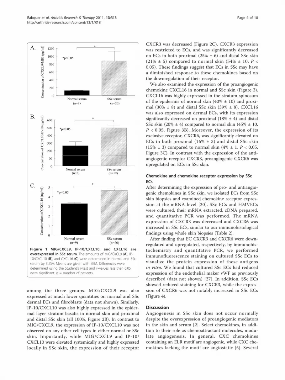
ResultsChemokine expression in SSc serumSerum from patients with SSc and healthy volunteerswas assayed using ELISAs to determine the expressionof pro- and antiangiogenic chemokines. SSc serum hadsignificantly elevated levels of antiangiogenic chemo-kines compared to normal controls (Figure 1A). Theexpression of MIG/CXCL9 was significantly greater inSSc serum (mean 876 pg/ml ± SEM 250 pg/ml) com-pared to normal serum (126 pg/ml ± 1, P < 0.05). Inaddition, IP-10/CXCL10 was significantly elevated inSSc serum (495 pg/ml ± 38) compared to normal serum(263 pg/ml ± 53, P < 0.05, Figure 1B). Similar resultswere found when a representative proangiogenic chemo-kine was assayed in SSc and normal serum. The expres-sion of CXCL16 was significantly greater in SSc serum(4.6 ng/ml ± 0.2) compared to normal serum (3.3 ng/ml± 0.1, P < 0.05, Figure 1C). These results demonstratethat both pro- and antiangiogenic chemokines are sig-nificantly upregulated in the serum of patients with SSc.In patients with SSc, CXCL16 was significantly ele-
vated in those with pulmonary arterial hypertension (5.7ng/ml ± 1.2, n = 2) compared to those without (4.5 ng/ml ± 0.1, n = 18, P < 0.05, Table 1). CXCL16 was alsosignificantly increased in SSc patients that died duringthe 36 months of the study (5.6 ng/ml ± 0.4, n = 4)compared to surviving patients (4.4 ng/ml ± 0.1, n = 16,P < 0.05). Moreover, SSc patients with early disease hadsignificantly increased levels of CXCL16 (4.9 ng/ml ±0.2, n = 13) compared to those with late disease (4.0 ng/ml ± 0.1, n = 7, P < 0.05). However, no differences wereobserved in any clinical characteristics with respect toserum MIG/CXCL9 and IP-10/CXCL10.
Chemokine and chemokine receptor expression in SScskinAfter finding elevated expression of the antiangiogenicchemokines MIG/CXCL9 and IP-10/CXCL10 in SScserum, we sought to determine their expression, andthe expression of their receptor CXCR3, in SSc skin(Figure 2). MIG/CXCL9 was primarily expressed in thestratum spinosum of the epidermis in normal skin (48%± 15) and both proximal (52% ± 9) and distal SSc skin(57% ± 8), with no significant differences observed
Rabquer et al. Arthritis Research & Therapy 2011, 13:R18http://arthritis-research.com/content/13/1/R18
Page 3 of 10
among the three groups. MIG/CXCL9 was alsoexpressed at much lower quantities on normal and SScdermal ECs and fibroblasts (data not shown). Similarly,IP-10/CXCL10 was also highly expressed in the epider-mal layer stratum basalis in normal skin and proximaland distal SSc skin (all 100%, Figure 2B). In contrast toMIG/CXCL9, the expression of IP-10/CXCL10 was notobserved on any other cell types in either normal or SScskin. Importantly, while MIG/CXCL9 and IP-10/CXCL10 were elevated systemically and highly expressedlocally in SSc skin, the expression of their receptor
CXCR3 was decreased (Figure 2C). CXCR3 expressionwas restricted to ECs, and was significantly decreasedon ECs in both proximal (25% ± 6) and distal SSc skin(21% ± 5) compared to normal skin (54% ± 10, P <0.05). These findings suggest that ECs in SSc may havea diminished response to these chemokines based onthe downregulation of their receptor.We also examined the expression of the proangiogenic
chemokine CXCL16 in normal and SSc skin (Figure 3).CXCL16 was highly expressed in the stratum spinosumof the epidermis of normal skin (40% ± 10) and proxi-mal (30% ± 8) and distal SSc skin (39% ± 8). CXCL16was also expressed on dermal ECs, with its expressionsignificantly decreased on proximal (18% ± 4) and distalSSc skin (20% ± 4) compared to normal skin (45% ± 10,P < 0.05, Figure 3B). Moreover, the expression of itsexclusive receptor, CXCR6, was significantly elevated onECs in both proximal (16% ± 3) and distal SSc skin(15% ± 3) compared to normal skin (4% ± 1, P < 0.05,Figure 3C). In contrast with the expression of the anti-angiogenic receptor CXCR3, proangiogenic CXCR6 wasupregulated on ECs in SSc skin.
Chemokine and chemokine receptor expression by SScECsAfter determining the expression of pro- and antiangio-genic chemokines in SSc skin, we isolated ECs from SScskin biopsies and examined chemokine receptor expres-sion at the mRNA level [20]. SSc ECs and HMVECswere cultured, their mRNA extracted, cDNA prepared,and quantitative PCR was performed. The mRNAexpression of CXCR3 was decreased and CXCR6 wasincreased in SSc ECs, similar to our immunohistologicalfindings using whole skin biopsies (Table 2).After finding that EC CXCR3 and CXCR6 were down-
regulated and upregulated, respectively, by immunohis-tochemistry and quantitative PCR, we performedimmunofluorescence staining on cultured SSc ECs tovisualize the protein expression of these antigensin vitro. We found that cultured SSc ECs had reducedexpression of the endothelial maker vWF as previouslydescribed (data not shown) [27]. In addition, SSc ECsshowed reduced staining for CXCR3, while the expres-sion of CXCR6 was not notably increased in SSc ECs(Figure 4).
DiscussionAngiogenesis in SSc skin does not occur normallydespite the overexpression of proangiogenic mediatorsin the skin and serum [2]. Select chemokines, in addi-tion to their role as chemoattractant molecules, modu-late angiogenesis. In general, CXC chemokinescontaining an ELR motif are angiogenic, while CXC che-mokines lacking the motif are angiostatic [5]. Several
1000
1200
MIG
(pg/
ml) *
*p<0.05
A.
Normal serum SSc serum0
200
400
600
800
Con
cent
ratio
n of
CX
CL9
/M
(n=8) (n=20)
200
300
400
500
600
ion
of C
XC
L10/
IP-1
0 (p
g/m
l)
*
*p<0.05
B.
Normal serum SSc serum0
100
Con
cent
rat
(n=8) (n=19)
4
5
L16
(ng/
ml)
*
*p<0.05
C.
Normal serum SSc serum0
1
2
3
Con
cent
ratio
n of
CX
CL
(n=9) (n=20)
Figure 1 MIG/CXCL9, IP-10/CXCL10, and CXCL16 areoverexpressed in SSc serum. The amounts of MIG/CXCL9 (A), IP-10/CXCL10 (B), and CXCL16 (C) were determined in normal and SScserum by ELISA. Means are given with SEM. Differences weredetermined using the Student’s t-test and P-values less than 0.05were significant. n = number of patients.
Rabquer et al. Arthritis Research & Therapy 2011, 13:R18http://arthritis-research.com/content/13/1/R18
Page 4 of 10

other chemokines have been shown to be proangiogenicincluding MCP-1/CCL2, macrophage inflammatory pro-tein-1a (MIP-1a/CCL3), and CXCL16 [7]. Collectively,these and other studies established the role of chemo-kines in angiogenesis.Angiogenic chemokines have previously been asso-
ciated with SSc pathogenesis [8]. We and others haveshown that IL-8/CXCL8 is a potent proangiogenic che-mokine overexpressed in both SSc serum and skin[9,11,14,28]. In addition, it has been associated with SSc
lung pathology, as it is elevated in pulmonary bronchiallavage fluid and secreted by alveolar macrophages [29].Moreover, the angiogenic CXC chemokines Gro-a/CXCL1 and stromal derived factor-1 (SDF-1/CXCL12)have also been found to be upregulated in SSc serum[9,10,30]. Among CC chemokines that are angiogenic,MCP-1/CCL2 and MIP-1a/CCL3 have been shown tobe upregulated in SSc [10,12,29]. PF4/CXCL4, which bycontrast is an antiangiogenic chemokine, has also beenshown to be upregulated in SSc serum [15].
A.
50
60
70
sum
pos
itive
for
pres
sion
Normal Skin Proximal SSc Skin Distal SSc Skin0
10
20
30
40
Perc
ent o
f stra
tum
spin
os
(n=10) (n=20) (n=20)
CX
CL9
/MIG
exp
C.
20
30
40
50
60
70
f end
othe
lial c
ells
pos
itive
for
CX
CR
3 ex
pres
sion
**
*p<0.05
Normal Skin Proximal SSc Skin Distal SSc Skin0
10
Perc
ent o
f
(n=10) (n=20) (n=20)
C
D.
Normal skinNormal skin
B.
708090
100
salis
pos
itive
for
pres
sion
Normal Skin Proximal SSc Skin Distal SSc Skin0
102030405060
Perc
ent o
f stra
tum
bas
(n=8) (n=18) (n=19)
CX
CL1
0/IP
-10
exp
E.
Distal SSc skinDistal SSc skin
Figure 2 MIG/CXCL9 and IP-10/CXCL10 are highly expressed, but CXCR3 is decreased, in SSc and normal skin. Frozen sections ofproximal and distal SSc and normal skin were stained for MIG/CXCL9, IP-10/CXCL10, or CXCR3. (A), MIG/CXCL9 was highly expressed in thestratum spinosum of normal (48%), proximal SSc (52%), and distal SSc (57%) skin. (B), IP-10/CXCL10 was uniformly expressed in the stratumbasalis of normal, proximal SSc, and distal SSc skin at 100%. (C), CXCR3 was significantly decreased on ECs in proximal SSc (25%) and distal SScskin (21%) compared to normal skin (54%). Representative photos of CXCR3 immunohistological staining in normal (D) and distal SSc skin (E) areshown. Arrows indicate positive staining. Means are given with the SEM. n = the number of patients. P < 0.05 was considered significant.
Rabquer et al. Arthritis Research & Therapy 2011, 13:R18http://arthritis-research.com/content/13/1/R18
Page 5 of 10

A.
40
50
60um
pos
itive
for
essi
on
Normal Skin Proximal SSc Skin Distal SSc Skin0
10
20
30
Perc
ent o
f stra
tum
spin
osu
(n=10) (n=20) (n=20)
CX
CL1
6 ex
pre
C.
20
ositi
ve fo
r
**E.
Normal skin
Normal Skin Proximal SSc Skin Distal SSc Skin0
5
10
15
Perc
ent o
f end
othe
lial c
ells
po
CX
CR
6 ex
pres
sion *p<0.05
Normal Skin Proximal SSc Skin Distal SSc Skin(n=8) (n=19) (n=19)
F.
Normal skin
B.
40
50
60
cells
pos
itive
for
ssio
n
**
*p<0.05
Normal Skin Proximal SSc Skin Distal SSc Skin0
10
20
30
40
Perc
ent o
f end
othe
lial c
(n=10) (n=20) (n=20)
CX
CL1
6 ex
pre
D.
Distal SSc skin
G.
Distal SSc skin
Figure 3 CXCL16 and CXCR6 are highly expressed in SSc compared to normal skin. Frozen sections of proximal and distal SSc and normalskin were stained for CXCL16 or CXCR6. (A), CXCL16 was highly expressed in the stratum spinosum of normal (40%), proximal SSc (30%), anddistal SSc (39%) skin. (B), CXCL16 was significantly decreased on ECs in proximal SSc (18%) and distal SSc skin (20%) compared to normal skin(45%). Representative photos of CXCL16 immunohistological staining in normal (C) and distal SSc skin (D) are shown. (E), CXCR6 was significantlyincreased on ECs in proximal SSc (16%) and distal SSc skin (15%) compared to normal skin (4%). Representative photos of CXCR6immunohistological staining in normal (F) and distal SSc skin (G) are shown. Arrows indicate positive staining. Means are given with the SEM.n = the number of patients. P < 0.05 was considered significant.
Rabquer et al. Arthritis Research & Therapy 2011, 13:R18http://arthritis-research.com/content/13/1/R18
Page 6 of 10

In this study, we examined the expression of selectpro- and antiangiogenic chemokines and their receptorsin SSc. We found that antiangiogenic MIG/CXCL9 wassignificantly elevated in SSc serum compared to normalcontrols. The only other study to examine its expressionin SSc serum found that MIG/CXCL9 was detected atsimilar rates in diffuse and limited SSc serum and nor-mal control serum [16]. However, the assays employedin that study were not as sensitive as the methods usedhere, as their analysis was limited to detection, whereaswe were able to quantify MIG/CXCL9 expression. Wealso found that IP-10/CXCL10 was elevated in SScserum compared to normal controls. Similar to ourresults, Fujii et al. demonstrated that IP-10/CXCL10was detected more often in SSc serum compared tohealthy control serum [16]. In addition, a more recentstudy has demonstrated that IP-10/CXCL10 is signifi-cantly elevated in early SSc serum compared to controls,and that its level significantly decreases after five years
[31]. Moreover, the level of IP-10/CXCL10 correlatedwith lung and kidney involvement in SSc patients [31].In accordance with these findings, we found that both
MIG/CXCL9 and IP-10/CXCL10 were highly expressedin the epidermis. Mediators produced by keratinocytesin the epidermis may cross into the dermal layer andaffect the function of fibroblasts and other cell types inthe dermis [32]. This suggests that chemokinesexpressed in the epidermis may act on dermal vascula-ture, along with the circulating chemokines in theserum.After examining the expression of the antiangiogenic
chemokines MIG/CXCL9 and IP-10/CXCL10 in SScskin and serum, we then sought to determine theexpression of their receptor. The ELR negative chemo-kines PF4/CXCL4, MIG/CXCL9, and IP-10/CXCL10mediate their leukocyte chemoattractant and angiogeniceffects by interacting with CXCR3 [7]. To date, threesplice variants of CXCR3 have been identified; CXCR3-A, CXCR3-B, and CXCR3-alt [33]. However, the angio-static effects of PF4/CXCL4, MIG/CXCL9, and IP-10/CXCL10 are mediated solely by CXCR3-B [34,35]. Wefound that CXCR3 is decreased on ECs in both proxi-mal and distal SSc skin compared to normal skin. More-over, we observed that SSc ECs have reduced mRNAlevels of CXCR3 compared to HMVECs, and thatin vitro CXCR3 cell surface expression is decreased onSSc ECs compared to HMVECs. Previous studiesdescribing the expression of CXCR3 on ECs in vitrohave yielded inconsistent results. CXCR3 has been
Table 2 CXCR3 is decreased while CXCR6 is increased onSSc ECs
Average relative abundancea
HMVECb SSc ECc Ratio
CXCR3 3.8 E-04 ± 9.1 E-06 1.5 E-04 ± 8.8 E-05 0.4
CXCR6 2.2 E-05 ± 1.1 E-05 3.2 E-05 ± 8.1 E-06 1.5
CXCL16 1.1 E-05 ± 1.6 E-06 1.4 E-04 ± 3.7 E-05 13.4aChemokine or chemokine receptor/b-actin. bn = three replicates. cn = threepatient derived lines. dMean ± SEM.
A.
HMVECs
B.
CXCR3IgG control
SSc ECs
CXCR63
Figure 4 CXCR3 expression is decreased while CXCR6 expression is unchanged on SSc ECs compared to HMVECs. HMVECs (A) and SScECs (B) were cultured on gelatin-coated chambers and immunofluorescence stained. Staining with specific antibodies against CXCR3, CXCR6, orirrelevant IgG control is shown. Nuclei were counter stained with DAPI. Staining was detected using a fluorescence microscope and images weretaken at 400×.
Rabquer et al. Arthritis Research & Therapy 2011, 13:R18http://arthritis-research.com/content/13/1/R18
Page 7 of 10
shown to be expressed on HMVECs, but not on humanumbilical vein ECs (HUVECs) [33,36]. This may be dueto the different nature of microvascular ECs and venousECs, as Chi et al. has previously shown that ECs fromdifferent anatomical sites have disparate gene expressionprofiles [37]. Thus HMVECs isolated from adult skinare a more suitable control population for our SSc ECsthan HUVECs. In addition, differences in cytokine andgrowth factor expression may alter CXCR3 expression,as tumor necrosis factor (TNF)-a downregulatesCXCR3 expression [38]. TNF-a and other proinflamma-tory cytokines are known to be upregulated in SSc skinand serum, which may account for the decreasedCXCR3 expression we observed in vitro and in vivo[12,39,40].In addition, we also determined the expression of the
proangiogenic receptor CXCR6 and its ligand CXCL16.CXCL16 is a recently described CXC chemokine that isfound in both membrane bound and soluble forms, thelatter following cleavage by a disintegrin and metallo-proteinase 10 (ADAM10) [41]. It has a unique receptorin CXCR6, which the membrane bound form ofCXCL16 binds to facilitate firm adhesion of emigratingleukocytes, and the soluble form utilizes to mediate itschemotactic properties [42]. CXCL16 is expressed on anumber of different cell types, including leukocytes, ECsand keratinocytes [43].We found that CXCL16 was significantly upregulated
in SSc serum. In addition, it was the only chemokine inour study that was associated with specific SSc clinicalfeatures. Serum CXCL16 was elevated in SSc patientsthat died between the time of biopsy and the time ofdata analysis, and in those with pulmonary arterialhypertension and early disease. A recent study byYanaba et al. found similar results and showed thatCXCL16 serum levels correlated with the extent of skinsclerosis [43]. In addition, they demonstrated thatCXCL16 levels decrease as SSc skin sclerosis improves[43]. Collectively, these findings implicate CXCL16 as achemokine of interest in SSc that warrants further study.In SSc skin biopsies, we found that CXCL16 was
highly expressed in the stratum spinosum layer of theepidermis, and decreased on dermal ECs compared tonormal skin. In contrast, our in vitro results indicatethat CXCL16 mRNA is modestly increased in SSc ECs.It is likely that this discrepancy is caused by the differ-ences in detection techniques, and the fact that CXCL16exists in both secreted and membrane bound forms.The receptor for CXCL16, CXCR6, is expressed by
several cell types, most notably memory and activated Tcells, cancer cells, and ECs [44]. We observed thatCXCR6 was significantly upregulated on dermal ECs inboth proximal and distal SSc skin biopsies. Moreover,we found a similar expression pattern on cultured SSc
ECs compared to HMVECs. To date, little is knownregarding the regulation of CXCR6 expression. However,hypoxia has been shown to be a strong inducer ofCXCR6 expression via hypoxia-inducible factor-1a [6].Hypoxia is a characteristic feature of SSc [45]. In SSc,the hypoxic environment is thought to be a factor in theoverexpression of potent angiogenic factors such asVEGF [45]. Likewise, it seems probable that skinhypoxia could result in an increase of CXCR6 expres-sion on SSc ECs. In addition, CXCR6-expressingHUVECs migrate in response to CXCL16 [6]. As ECchemotaxis is an initial step in the angiogenic cascade,these findings suggest that CXCL16/CXCR6 may beimportant in mediating SSc angiogenesis.
ConclusionsDiminished dermal vasculature and dysregulated angio-genesis are hallmarks of SSc. Our results indicate a sys-temic overexpression of proangiogenic CXCL16 andantiangiogenic MIG/CXCL9 and IP-10/CXCL10 in SScserum. However, differential expression of CXCR3 andCXCR6 was observed on SSc dermal ECs. Collectively,these results argue that despite high serum levels ofantiangiogenic chemokines, the downregulation of theirreceptor on SSc ECs prevents them from directly contri-buting to the hypovascular state of SSc skin. In addition,the increase of CXCR6 on SSc ECs points to CXCL16being a relevant angiogenic factor in SSc that warrantsfurther study.
AbbreviationsADAM10: a disintegrin and metalloproteinase 10; DAPI: 4’,6-diaminido-2-phenylindole; ECs: endothelial cells; ELISA: enzyme-linked immunosorbentassay; Glu-Leu-Arg: ELR motif; Gro-α/CXCL1: growth-regulated oncogene-α;HMVECs: human microvascular endothelial cells; HUVECs: human umbilicalvein ECs; IFN-γ: interferon-γ; IL: interleukin; IP-10/CSCL10: IFN-inducibleprotein 10; MCP-1/CCL2: monocyte chemoattractant protein-1; MIG/CXCL9:monokine induced by IFN-γ; MIP-1α/CCL3: macrophage inflammatoryprotein-1α; PF4/CXCL4: platelet factor 4; qPCR: quantitative polymerase chainreaction; SDF-1/CXCL12: stromal derived factor-1; SEM: standard error of themean; SSC: systemic sclerosis; vWF: von Willebrand factor.
AcknowledgementsThis work was supported by the National Institute of Health (grantsHL094017 to BJR, and AR48267 to AEK), the Office of Research andDevelopment, Medical Research Service, Department of Veterans Affairs, theFrederick G. L. Huetwell and William D. Robinson, MD, Professorship inRheumatology, Scleroderma Research Foundation (CA), NIH General ClinicalResearch Center grant M01-RR-00042, NIH Center for Translational ScienceActivities grant UL1-RR-024986, the clinical research unit at the University ofMichigan, American College of Rheumatology Research and EducationFoundation grant (ARA to KP), the Linda Dolce Scleroderma Research Fund,the Marvin and Betty Danto and the Jonathan and Lisa Rye Endowments forScleroderma Research at the University of Michigan, and by the SclerodermaFoundation (Mark Flapan Award).
Author details1Department of Internal Medicine, University of Michigan Medical School,109 Zina Pitcher Dr., Ann Arbor, MI 48109, USA. 2Department of Pathology,Yale University, 200 South Frontage Rd., New Haven, CT 06510, USA.3Department of Medicine, University of Toledo, 3000 Arlington Ave., Toledo,
Rabquer et al. Arthritis Research & Therapy 2011, 13:R18http://arthritis-research.com/content/13/1/R18
Page 8 of 10

OH 43614, USA. 4Current address: Scleroderma Research Consultants, LLC, 97Deer Run, Avon, CT 06001, USA. 5Department of Veterans Affairs, VA MedicalService, 2215 Fuller Rd., Ann Arbor, MI 48105, USA.
Authors’ contributionsBJR conceived the study, and participated in the immunohistology, ELISA,cell culture, qPCR, immunofluorescence, and data analysis and drafted themanuscript. PT participated in the immunohistology, ELISA, qPCR, and dataanalysis. YH participated in the immunohistology and ELISAs. ET participatedin the immunohistology and ELISAs. GKH scored the immunohistology. AJIperformed the statistical analysis. KP conceived the study and collectedclinical data. BK isolated ECs from SSc skin. JRS conceived the study andcollected clinical data. AEK conceived the study and drafted the manuscript.All authors read and approved the final manuscript.
Competing interestsThe authors declare that they have no competing interests.
Received: 31 August 2010 Revised: 10 January 2011Accepted: 8 February 2011 Published: 8 February 2011
References1. Gabrielli A, Avvedimento EV, Krieg T: Scleroderma. N Engl J Med 2009,
360:1989-2003.2. Distler JH, Gay S, Distler O: Angiogenesis and vasculogenesis in systemic
sclerosis. Rheumatology (Oxford) 2006, 45:iii26-27.3. Szekanecz Z, Gaspar L, Koch AE: Angiogenesis in rheumatoid arthritis.
Front Biosci 2005, 10:1739-1753.4. Koch AE, Polverini PJ, Kunkel SL, Harlow LA, DiPietro LA, Elner VM, Elner SG,
Strieter RM: Interleukin-8 as a macrophage-derived mediator ofangiogenesis. Science 1992, 258:1798-1801.
5. Strieter RM, Polverini PJ, Kunkel SL, Arenberg DA, Burdick MD, Kasper J,Dzuiba J, Van Damme J, Walz A, Marriott D: The functional role of the ELRmotif in CXC chemokine-mediated angiogenesis. J Biol Chem 1995,270:27348-27357.
6. Lin S, Sun L, Hu J, Wan S, Zhao R, Yuan S, Zhang L: Chemokine C-X-Cmotif receptor 6 contributes to cell migration during hypoxia. CancerLett 2009, 279:108-117.
7. Strieter RM, Burdick MD, Gomperts BN, Belperio JA, Keane MP: CXCchemokines in angiogenesis. Cytokine Growth Factor Rev 2005, 16:593-609.
8. Yamamoto T: Chemokines and chemokine receptors in scleroderma. IntArch Allergy Immunol 2006, 140:345-356.
9. Furuse S, Fujii H, Kaburagi Y, Fujimoto M, Hasegawa M, Takehara K, Sato S:Serum concentrations of the CXC chemokines interleukin 8 and growth-regulated oncogene-alpha are elevated in patients with systemicsclerosis. J Rheumatol 2003, 30:1524-1528.
10. Hasegawa M, Sato S, Takehara K: Augmented production ofchemokines (monocyte chemotactic protein-1 (MCP-1), macrophageinflammatory protein-1alpha (MIP-1alpha) and MIP-1beta) in patientswith systemic sclerosis: MCP-1 and MIP-1alpha may be involved inthe development of pulmonary fibrosis. Clin Exp Immunol 1999,117:159-165.
11. Reitamo S, Remitz A, Varga J, Ceska M, Effenberger F, Jimenez S, Uitto J:Demonstration of interleukin 8 and autoantibodies to interleukin 8 inthe serum of patients with systemic sclerosis and related disorders. ArchDermatol 1993, 129:189-193.
12. Scala E, Pallotta S, Frezzolini A, Abeni D, Barbieri C, Sampogna F, De Pita O,Puddu P, Paganelli R, Russo G: Cytokine and chemokine levels in systemicsclerosis: relationship with cutaneous and internal organ involvement.Clin Exp Immunol 2004, 138:540-546.
13. Yamamoto T, Eckes B, Hartmann K, Krieg T: Expression of monocytechemoattractant protein-1 in the lesional skin of systemic sclerosis.J Dermatol Sci 2001, 26:133-139.
14. Koch AE, Kronfeld-Harrington LB, Szekanecz Z, Cho MM, Haines GK,Harlow LA, Strieter RM, Kunkel SL, Massa MC, Barr WG, et al: In situexpression of cytokines and cellular adhesion molecules in the skin ofpatients with systemic sclerosis. Their role in early and late disease.Pathobiology 1993, 61:239-246.
15. Macko RF, Gelber AC, Young BA, Lowitt MH, White B, Wigley FM,Goldblum SE: Increased circulating concentrations of thecounteradhesive proteins SPARC and thrombospondin-1 in systemic
sclerosis (scleroderma). Relationship to platelet and endothelial cellactivation. J Rheumatol 2002, 29:2565-2570.
16. Fujii H, Shimada Y, Hasegawa M, Takehara K, Sato S: Serum levels of a Th1chemoattractant IP-10 and Th2 chemoattractants, TARC and MDC, areelevated in patients with systemic sclerosis. J Dermatol Sci 2004, 35:43-51.
17. Hou Y, Rabquer BJ, Gerber ML, Del Galdo F, Jimenez SA, Haines GK,Barr WG, Massa MC, Seibold JR, Koch AE: Junctional adhesion molecule-Ais abnormally expressed in diffuse cutaneous systemic sclerosis skin andmediates myeloid cell adhesion. Ann Rheum Dis 2010, 69:249-254.
18. LeRoy EC, Black C, Fleischmajer R, Jablonska S, Krieg T, Medsger TA Jr,Rowell N, Wollheim F: Scleroderma (systemic sclerosis): classification,subsets and pathogenesis. J Rheum 1988, 15:202-205.
19. Rabquer BJ, Hou Y, Del Galdo F, Haines GK, Gerber ML, Jimenez SA,Seibold JR, Koch AE: The proadhesive phenotype of systemic sclerosisskin promotes myeloid cell adhesion via ICAM-1 and VCAM-1.Rheumatology (Oxford) 2009, 48:734-740.
20. D’Alessio S, Fibbi G, Cinelli M, Guiducci S, Del Rosso A, Margheri F, Serrati S,Pucci M, Kahaleh B, Fan P, Annunziato F, Cosmi L, Liotta F, Matucci-Cerinic M, Del Rosso M: Matrix metalloproteinase 12-dependent cleavageof urokinase receptor in systemic sclerosis microvascular endothelialcells results in impaired angiogenesis. Arthritis Rheum 2004, 50:3275-3285.
21. Scott PA, Bicknell R: The isolation and culture of microvascularendothelium. J Cell Sci 1993, 105:269-273.
22. Rabquer BJ, Amin MA, Teegala N, Shaheen MK, Tsou P, Ruth JH, Lesch CA,Imhof BA, Koch AE: Junctional adhesion molecule-C is a soluble mediatorof angiogenesis. J Immunol 2010, 185:1777-1785.
23. Ahmed S, Marotte H, Kwan K, Ruth JH, Campbell PL, Rabquer BJ, Pakozdi A,Koch AE: Epigallocatechin-3-gallate inhibits IL-6 synthesis and suppressestranssignaling by enhancing soluble gp130 production. Proc Natl AcadSci U S A 2008, 105:14692-14697.
24. Hanamoto H, Nakayama T, Miyazato H, Takegawa S, Hieshima K, Tatsumi Y,Kanamaru A, Yoshie O: Expression of CCL28 by Reed-Sternberg cellsdefines a major subtype of classical Hodgkin’s disease with frequentinfiltration of eosinophils and/or plasma cells. Am J Pathol 2004,164:997-1006.
25. Notohamiprodjo M, Segerer S, Huss R, Hildebrandt B, Soler D,Djafarzadeh R, Buck W, Nelson PJ, von Luettichau I: CCR10 is expressed incutaneous T-cell lymphoma. Int J Cancer 2005, 115:641-647.
26. Rabquer BJ, Pakozdi A, Michel JE, Gujar BS, Haines GK, Imhof BA, Koch AE:Junctional adhesion molecule-C mediates leukocyte adhesion to therheumatoid arthritis synovium. Arthritis Rheum 2008, 58:3020-3029.
27. Freemont AJ, Hoyland J, Fielding P, Hodson N, Jayson MI: Studies of themicrovascular endothelium in uninvolved skin of patients with systemicsclerosis: direct evidence for a generalized microangiopathy. Br JDermatol 1992, 126:561-568.
28. Kadono T, Kikuchi K, Ihn H, Takehara K, Tamaki K: Increased production ofinterleukin 6 and interleukin 8 in scleroderma fibroblasts. J Rheumatol1998, 25:296-301.
29. Bolster MB, Ludwicka A, Sutherland SE, Strange C, Silver RM: Cytokineconcentrations in bronchoalveolar lavage fluid of patients with systemicsclerosis. Arthritis Rheum 1997, 40:743-751.
30. Cipriani P, Franca Milia A, Liakouli V, Pacini A, Manetti M, Marrelli A,Toscano A, Pingiotti E, Fulminis A, Guiducci S, Perricone R, Kahaleh B,Matucci-Cerinic M, Ibba-Manneschi L, Giacomelli R: Differential expressionof stromal cell-derived factor 1 and its receptor CXCR4 in the skin andendothelial cells of systemic sclerosis patients: pathogeneticimplications. Arthritis Rheum 2006, 54:3022-3033.
31. Antonelli A, Ferri C, Fallahi P, Ferrari SM, Giuggioli D, Colaci M, Manfredi A,Frascerra S, Franzoni F, Galetta F, Ferrannini E: CXCL10 (alpha) and CCL2(beta) chemokines in systemic sclerosis–a longitudinal study.Rheumatology (Oxford) 2008, 47:45-49.
32. Werner S, Smola H: Paracrine regulation of keratinocyte proliferation anddifferentiation. Trends Cell Biol 2001, 11:143-146.
33. Lasagni L, Francalanci M, Annunziato F, Lazzeri E, Giannini S, Cosmi L,Sagrinati C, Mazzinghi B, Orlando C, Maggi E, Marra F, Romagnani S,Serio M, Romagnani P: An alternatively spliced variant of CXCR3 mediatesthe inhibition of endothelial cell growth induced by IP-10, Mig, and I-TAC, and acts as functional receptor for platelet factor 4. J Exp Med 2003,197:1537-1549.
34. Vandercappellen J, Van Damme J, Struyf S: The role of CXC chemokinesand their receptors in cancer. Cancer Lett 2008, 267:226-244.
Rabquer et al. Arthritis Research & Therapy 2011, 13:R18http://arthritis-research.com/content/13/1/R18
Page 9 of 10
35. Yang J, Richmond A: The angiostatic activity of interferon-inducibleprotein-10/CXCL10 in human melanoma depends on binding to CXCR3but not to glycosaminoglycan. Mol Ther 2004, 9:846-855.
36. Schutyser E, Su Y, Yu Y, Gouwy M, Zaja-Milatovic S, Van Damme J,Richmond A: Hypoxia enhances CXCR4 expression in humanmicrovascular endothelial cells and human melanoma cells. Eur CytokineNetw 2007, 18:59-70.
37. Chi JT, Chang HY, Haraldsen G, Jahnsen FL, Troyanskaya OG, Chang DS,Wang Z, Rockson SG, van de Rijn M, Botstein D, Brown PO: Endothelial celldiversity revealed by global expression profiling. Proc Natl Acad Sci U S A2003, 100:10623-10628.
38. Feil C, Augustin HG: Endothelial cells differentially express functionalCXC-chemokine receptor-4 (CXCR-4/fusin) under the control of autocrineactivity and exogenous cytokines. Biochem Biophys Res Commun 1998,247:38-45.
39. Denton CP, Bickerstaff MC, Shiwen X, Carulli MT, Haskard DO, Dubois RM,Black CM: Serial circulating adhesion molecule levels reflect diseaseseverity in systemic sclerosis. Br J Rheumatol 1995, 34:1048-1054.
40. Sollberg S, Peltonen J, Uitto J, Jimenez SA: Elevated expression of beta 1and beta 2 integrins, intercellular adhesion molecule 1, and endothelialleukocyte adhesion molecule 1 in the skin of patients with systemicsclerosis of recent onset. Arthritis Rheum 1992, 35:290-298.
41. Gough PJ, Garton KJ, Wille PT, Rychlewski M, Dempsey PJ, Raines EW: Adisintegrin and metalloproteinase 10-mediated cleavage and sheddingregulates the cell surface expression of CXC chemokine ligand 16.J Immunol 2004, 172:3678-3685.
42. Matloubian M, David A, Engel S, Ryan JE, Cyster JG: A transmembrane CXCchemokine is a ligand for HIV-coreceptor Bonzo. Nat Immunol 2000,1:298-304.
43. Yanaba K, Muroi E, Yoshizaki A, Hara T, Ogawa F, Shimizu K, Yozaki M,Hasegawa M, Fujimoto M, Takehara K, Sato S: Serum CXCL16concentrations correlate with the extent of skin sclerosis in patientswith systemic sclerosis. J Rheumatol 2009, 36:1917-1923.
44. Unutmaz D, Xiang W, Sunshine MJ, Campbell J, Butcher E, Littman DR: Theprimate lentiviral receptor Bonzo/STRL33 is coordinately regulated withCCR5 and its expression pattern is conserved between human andmouse. J Immunol 2000, 165:3284-3292.
45. Beyer C, Schett G, Gay S, Distler O, Distler JH: Hypoxia. Hypoxia in thepathogenesis of systemic sclerosis. Arthritis Res Ther 2009, 11:220.
doi:10.1186/ar3242Cite this article as: Rabquer et al.: Dysregulated expression of MIG/CXCL9, IP-10/CXCL10 and CXCL16 and their receptors in systemicsclerosis. Arthritis Research & Therapy 2011 13:R18.
Submit your next manuscript to BioMed Centraland take full advantage of:
• Convenient online submission
• Thorough peer review
• No space constraints or color figure charges
• Immediate publication on acceptance
• Inclusion in PubMed, CAS, Scopus and Google Scholar
• Research which is freely available for redistribution
Submit your manuscript at www.biomedcentral.com/submit
Rabquer et al. Arthritis Research & Therapy 2011, 13:R18http://arthritis-research.com/content/13/1/R18
Page 10 of 10