dynamic neural networks supporting memory retrieval
TRANSCRIPT

Dynamic Neural Networks Supporting Memory Retrieval
Peggy L. St. Jacques1,*, Philip A. Kragel1,2,*, and David C. Rubin2,3
1 Center for Cognitive Neuroscience, Duke University, Durham, NC, 27708, USA2 Department of Psychology and Neuroscience, Duke University, Durham, NC, 27708, USA3 Center on Autobiographical Memory Research, Aarhus University, Aarhus, 800C Denmark
AbstractHow do separate neural networks interact to support complex cognitive processes such asremembrance of the personal past? Autobiographical memory (AM) retrieval recruits a consistentpattern of activation that potentially comprises multiple neural networks. However, it is unclearhow such large-scale neural networks interact and are modulated by properties of the memoryretrieval process. In the present functional MRI (fMRI) study, we combined independentcomponent analysis (ICA) and dynamic causal modeling (DCM) to understand the neuralnetworks supporting AM retrieval. ICA revealed four task-related components consistent with theprevious literature: 1) Medial Prefrontal Cortex (PFC) Network, associated with self-referentialprocesses, 2) Medial Temporal Lobe (MTL) Network, associated with memory, 3) FrontoparietalNetwork, associated with strategic search, and 4) Cingulooperculum Network, associated withgoal maintenance. DCM analysis revealed that the medial PFC network drove activation withinthe system, consistent with the importance of this network to AM retrieval. Additionally, memoryaccessibility and recollection uniquely altered connectivity between these neural networks.Recollection modulated the influence of the medial PFC on the MTL network during elaboration,suggesting that greater connectivity among subsystems of the default network supports greater re-experience. In contrast, memory accessibility modulated the influence of frontoparietal and MTLnetworks on the medial PFC network, suggesting that ease of retrieval involves greater fluencyamong the multiple networks contributing to AM. These results show the integration betweenneural networks supporting AM retrieval and the modulation of network connectivity by behavior.
KeywordsEpisodic Memory; Autobiographical Memory; Retrieval; Large-Scale Brain Network; fMRI;Functional Neuroimaging
1. IntroductionThe brain is intrinsically organized into multiple neural networks that contribute to higher-order cognitive processes through their interaction (Bressler & Menon, 2010; Fuster, 2009;
© 2011 Elsevier Inc. All rights reserved.Corresponding Author: Peggy L. St. Jacques, Department of Psychology, Harvard University, William James Hall, Rm. 864,Cambridge, MA 02143, USA, Phone: (617) 495-9031, Fax: (617) 496-3122, [email protected].*Equal AuthorshipPublisher's Disclaimer: This is a PDF file of an unedited manuscript that has been accepted for publication. As a service to ourcustomers we are providing this early version of the manuscript. The manuscript will undergo copyediting, typesetting, and review ofthe resulting proof before it is published in its final citable form. Please note that during the production process errors may bediscovered which could affect the content, and all legal disclaimers that apply to the journal pertain.
NIH Public AccessAuthor ManuscriptNeuroimage. Author manuscript; available in PMC 2012 July 15.
Published in final edited form as:Neuroimage. 2011 July 15; 57(2): 608–616. doi:10.1016/j.neuroimage.2011.04.039.
NIH
-PA Author Manuscript
NIH
-PA Author Manuscript
NIH
-PA Author Manuscript

Rubin, 2006). How large-scale brain networks interact to support complex cognitiveprocesses such as autobiographical memory (AM) retrieval is largely unknown. AMretrieval involves strategic search processes that are guided by knowledge about one’s selfand by current goals, the recovery of memory traces involving a rich sense of re-experience,and monitoring and other control processes (Conway & Pleydell-Pearce, 2000; Norman &Bobrow, 1976). Thus, it is not surprising that recalling memories from our personal pastinvolves a distributed set of brain regions (Cabeza & St. Jacques, 2007; Svoboda,McKinnon, & Levine, 2006) that encompass separate systems, such as those supporting self-reference, memory consolidation and storage, search and goal-related processes (Greenberg& Rubin, 2003; Rubin, 2005, 2006). In the present functional MRI (fMRI) study weinvestigate the integration between neural networks supporting AM retrieval and therelationship between behavior and neural network dynamics.
Functional neuroimaging studies of AM studies have identified a number of typical regionsinvolved during memory retrieval, including the medial and lateral prefrontal cortices (PFC),lateral and medial temporal lobes (MTL; hippocampus, parahippocampus), ventral parietalcortex, and posterior cingulate cortex (Cabeza & St. Jacques, 2007; McDermott, Szpunar, &Christ, 2009; Spreng, Mar, & Kim, 2009; Svoboda, et al., 2006). Several studies have alsoexamined the interaction among the brain regions supporting AM retrieval (Addis,McIntosh, Moscovitch, Crawley, & McAndrews, 2004; Burianova & Grady, 2007;Greenberg, et al., 2005; Levine, et al., 2004; Maguire, Mummery, & Buchel, 2000; St.Jacques, Conway, Lowder, & Cabeza, In Press), with some studies observing that lesionsalter these interactions (Addis, Moscovitch, & McAndrews, 2007; Maguire, Vargha-Khadem, & Mishkin, 2001). However, one limitation of previous approaches relying onsubtraction paradigms, and task or behavior based multivariate approaches, is that they donot distinguish the coactivation of distinct networks (Friston, et al., 1996). Thus, less isknown whether the “core network” recruited during AM retrieval actually involves thecontribution of multiple networks and their interaction (although see Andrews-Hanna,Reidler, Sepulcre, Poulin, & Buckner, 2010; also see Hassabis, Kumaran, & Maguire,2007a).
Functional connectivity analysis of spontaneous blood oxygen level dependent fluctuationsduring passive resting states has revealed a number of correlated brain regions comprisingneural networks (e.g., Damoiseaux, et al., 2006; Greicius, Krasnow, Reiss, & Menon, 2003;Raichle, et al., 2001; for reviews see Buckner, Andrews-Hanna, & Schacter, 2008; Fox &Raichle, 2007) and these networks overlap with structural connections (for a review seeDamoiseaux & Greicius, 2009). In particular, four networks have been identified that maycontribute to the self-reference, memory, search and goal-related processes important forAM retrieval. First, the medial PFC network comprises dorsal medial PFC, posteriorcingulate, and ventral parietal cortices, and is linked to the construction of self-referentialsimulations (Andrews-Hanna, et al., 2010; for a review see Buckner, et al., 2008). Second,the MTL network comprises hippocampal, ventral medial PFC, retrosplenial, and ventralparietal cortices, which support memory processes and the construction of mental scenes(Andrews-Hanna, et al., 2010; Kahn, Andrews-Hanna, Vincent, Snyder, & Buckner, 2008;Vincent, et al., 2006). Together, the medial PFC and MTL Networks comprise the defaultnetwork, the set of brain regions that are coactive during passive resting states (Andrews-Hanna, et al., 2010; Buckner, et al., 2008), and commonly engaged during AM retrieval,future thinking and theory of mind (e.g., Spreng & Grady, 2010; Spreng, et al., 2009). Third,the frontoparietal network, which is similar to the central executive network, compriseslateral PFC, anterior cingulate and inferior parietal regions, and is linked to adaptivecognitive control processes (Dosenbach, et al., 2007; Seeley, et al., 2007; Vincent, Kahn,Snyder, Raichle, & Buckner, 2008), which may support strategic search processes. Fourth,the cinguloopercular network, which is similar to the salience network, comprises
St. Jacques et al. Page 2
Neuroimage. Author manuscript; available in PMC 2012 July 15.
NIH
-PA Author Manuscript
NIH
-PA Author Manuscript
NIH
-PA Author Manuscript

frontopolar, anterior insular/frontal operculum, and dorsal anterior cingulate cortices, and islinked to the maintenance of goals (Dosenbach, et al., 2007) and the processing of salientevents in the environment (Menon & Uddin, 2010; Seeley, et al., 2007). Neural networks,however, are not completely segregated from one another but contribute to cognitionthrough the interaction of sparse connections potentially mediated by cortical hubs (Achard,Salvador, Whitcher, Suckling, & Bullmore, 2006; Buckner, et al., 2009). Few studies,however, have examined effective connectivity between networks, or the influence of onenetwork on another, and how the connections between these networks are modulated bybehavior (although see Stevens, Kiehl, Pearlson, & Calhoun, 2007). In addition, similaranalytic techniques applied to task related, as opposed to resting state tasks, find comparablenetworks and others that correspond to additional cognitive functions involved in AMincluding visual and auditory processing (Botzung, LaBar, Kragel, Miles, & Rubin, 2010).In the current study we identified multiple networks contributing to AM and we examinedhow the interaction between these networks was modulated by characteristics of memoryretrieval.
Recollection, the ability to retrieve contextual details and to re-experience or relive a pastevent, is a central feature of AM that distinguishes episodic (e.g., unique) from semantictypes of personal memories (e.g., repeated events; Brewer, 1986; Rubin, Schrauf, &Greenberg, 2003; Tulving, 1983). The ability to mentally time travel or re-experience thepersonal past is linked to the frontal lobes (Wheeler, Stuss, & Tulving, 1997), and issupported by the recovery of contextual details via posterior brain regions such as the MTL,retrosplenial cortex and lateral parietal cortices (Cabeza & St. Jacques, 2007; St Jacques &Cabeza, In Press; Svoboda, et al., 2006). AMs that are richly recollected also tend to bevoluntarily retrieved more quickly often via direct or associative retrieval processes(Conway & Pleydell-Pearce, 2000), although longer retrieval times involving qualitativelydifferent generative retrieval processes can elicit recollection (St Jacques & Cabeza, InPress; Svoboda, et al., 2006). Thus, it is important to consider the contribution of bothrecollection and memory accessibility to understand the neural correlates supporting AMretrieval.
To investigate the dynamic interaction between neural networks during AM retrieval and themodulation of network connectivity by recollection and memory accessibility, we used ageneric cue method to elicit unprepared memories during fMRI scanning. Participantssearched for and constructed AMs, indicated once a memory was formed by a self-pacedbutton press, elaborated on the retrieved memory, and made online ratings includingrecollection (e.g., Daselaar, et al., 2008). The goal of the current study was to examine howspatially extensive whole-brain networks interact. Thus, we employed independentcomponent analysis (ICA; Calhoun, Adali, Pearlson, & Pekar, 2001b) to distinguish thecoactivation of spatially distinct networks contributing to AM retrieval from activationwithin a single “core network.” We then combined the output of the ICA (i.e., the networktime-courses) and dynamic causal modeling (DCM; Friston, Harrison, & Penny, 2003), amethod developed by Stevens et al. (2007), to identify interactions between the large-scaleneural networks contributing to AM retrieval and the modulation of these connections byboth recollection and memory accesibility.
2. Materials and Methods2.1. Participants
There were seventeen young (18–35 years of age) participants who were healthy, right-handed and without history of neurological or psychiatric episodes. All participants reportedthat they were not taking medication known to affect cognitive function. Participants gavewritten informed consent for a protocol approved by the Duke University Institutional
St. Jacques et al. Page 3
Neuroimage. Author manuscript; available in PMC 2012 July 15.
NIH
-PA Author Manuscript
NIH
-PA Author Manuscript
NIH
-PA Author Manuscript

Review Board. One young adult was excluded due to symptoms of depression as indicatedby scores > 13 on the Beck Depression Inventory (Beck, Steer, & Brown, 1996).Furthermore, two young were excluded from the analyses because of problems withcompleting the task as instructed. Thus, the reported results are based on data from fourteenyoung (7 females; mean Age = 24.43, SD = 3.73) participants.
2.2. MaterialsMemory cues consisted of 60 emotionally arousing words selected from the affective normsfor English words database (Bradley & Lang, 1999), such that there were 30 positive(Valence mean = 7.93, SD = 0.45; Arousal Mean = 5.96; SD = 0.83) and 30 negative(Valence Mean = 2.17, SD = 0.52; Arousal Mean = 6.00; SD = 1.03) words that wereequally arousing. In order to create auditory cues the words were recorded in a female voiceand constrained to an equal duration of 1 s.
2.3. ProcedureThe procedure was based on Daselaar et al. (2008). During scanning participants were askedto search for and construct autobiographical memories (AMs) triggered by the auditory cuewords. Participants were instructed to retrieve an AM with specific spatiotemporalcoordinates. They indicated when a specific AM was found by making a response on thebutton-box and then continued to elaborate on the retrieved event in as much detail aspossible for the rest of the trial. Thirty seconds following the onset of the auditory cueparticipants were given auditory instructions to rate reliving (low to high) associated withthe memory and the amount of emotion (negatively arousing to positively arousing) on an 8-point scale. Emotion ratings were included for a separate analysis (St Jacques, Botzung,Miles, & Rubin, 2010). The order of the ratings was counterbalanced between participants.Rating responses were self-paced (up to 6 s) and separated by at least 0.5 s. Since the ratingswere self-paced up to 6 s any remaining time after pressing the button was added to thefixation to ensure that the next trial began on the TR. There were 6 functional runs, with 10memory cues in each run (5 positive words and 5 negative words), and an inter-trial intervalat least 1.5 to 7.5 s. Participants were instructed to keep their eyes closed for the duration ofeach run.
2.4. fMRI Methods2.4.1. Image Acquisition—Scanning was conducted using a 4T GE magnet. Auditorystimuli were presented using headphones and behavioral responses were recorded using aneight-button fiber optic response box (Resonance Technology, Northridge, CA). Headmotion was minimized using foam pads and a headband. Anatomical scanning started with aT1-weighted sagittal localizer series, and then 3D fast spoiled gradient echo recalled (SPGR)structural images were acquired in the coronal plane (2562 matrix, TR = 12.3 ms, TE = 5.4ms, flip angle = 20°, FOV = 240, 68 slices, 1.9 mm slice thickness). Coplanar functionalimages were subsequently acquired using an inverse spiral sequence (642 image matrix, TR= 2000 ms, TE = 6 ms, FOV = 240, flip angle = 60°, 34 slices, 3.8 mm slice thickness).
2.4.2. Image Processing—Image processing and analyses were performed usingStatistical Parametric Mapping software in MATLAB (SPM5; Wellcome Department ofImaging Neuroscience). Functional images were corrected for slice acquisition order,realigned to correct for motion artifacts, and then spatially normalized to a standardstereotactic space, using the template implemented in SPM5. Subsequently, the functionalimages were spatially smoothed using an 8 mm FWHM isotropic Gaussian kernel.
St. Jacques et al. Page 4
Neuroimage. Author manuscript; available in PMC 2012 July 15.
NIH
-PA Author Manuscript
NIH
-PA Author Manuscript
NIH
-PA Author Manuscript

2.4.3 Independent Component Analysis—ICA was used to determine spatiallydistinct networks contributing to AM retrieval unbiased with respect to an underlyingtemporal model (for conventional analysis using a general linear model see St. Jacques,Rubin, & Cabeza, 2010). These spatially independent networks were identified with ICA asimplemented in the Group ICA of fMRI Toolbox (GIFT; Calhoun, Adali, Pearlson, & Pekar,2001a; Stephan, et al., 2010). This method involves performing ICA on functional dataconcatenated over every participant, creating a series of spatial maps and associated timecourses for the group. Back reconstruction is then used to create individual time courses andspatial maps from each participant’s functional data. The number of components wasestimated to be 30 using the minimum description length criteria. The amplitude ofindividual time courses and spatial maps were calibrated to percent signal change forcomparison across participants. Percent signal change was calculated by regressingcomponent time courses onto the original data at maximal voxels for a component, scalingto reflect percent change from the mean, and then applying additional scaling so that themaximal component voxel contains the maximal percent signal change value. Spatialvariability of components across subjects was examined by performing one sample t-tests onthese scaled component maps. For subsequent DCM analysis, component time courses werehigh pass filtered with a cutoff of 128 seconds and adjusted for confounds (session mean andmotion parameters).
2.4.4. Component Identification—Components with time courses related to theexperimental design were identified using multiple regression using the temporal sortingfeature of the GIFT toolbox. AM retrieval operations were modeled by convolving stimulifunctions with the canonical haemodynamic response. This model, implemented in SPM5,had separate regressors for the Cue, Construction, Response, Elaboration, and the Ratings inthe task. Stimuli functions varied between regressors as either zero duration Dirac deltafunctions (Cue, Response, Ratings) or as variable duration box-car functions (Construction,Elaboration) that depend on the time of the motor response. The primary interest of thepresent study was to determine components associated with construction and elaborationphases of AM retrieval. We conducted a regression analysis to determine β weightsassociating the components with construction and elaboration phases separately in eachparticipant. One sample t-tests on these β weights showed four components that weresignificantly related (p < .001) to construction and elaboration phases, which were subject tofurther examination.
2.4.5. Dynamic Causal Model—To examine the interaction among the spatially distinctnetworks identified by ICA, the effective connectivity of the four task related componentswas analyzed using bilinear DCM (Friston, et al., 2003) as implemented in SPM5. Unlikeother measures of effective connectivity, DCM examines the strength of causal connectionsby accounting for both the bilinear and dynamic nature of neuronal interactions and theperturbation of this system by experimental manipulation (i.e., driving or modulatoryinputs). Utilizing ICA as a feature selection tool for DCM differs significantly from themore commonly utilized volume of interest approach. Typically, several regions related to aprocess of interest are selected and representative time courses are created by taking the firstprincipal component over all voxels in the volume of interest. Due to current practicalconstraints a maximum of eight regions can be selected in a DCM. Because of this, it is notpossible to model all regions or subnetworks that may interact with key regions of interest,and the occurrence of unmodeled indirect connections is a possibility. As an alternative, ICAcan be used to separate functional data into spatially independent networks withcorresponding time courses and has the potential to account for what would be unmodeledregions (for a similar method see Stevens, et al., 2007).
St. Jacques et al. Page 5
Neuroimage. Author manuscript; available in PMC 2012 July 15.
NIH
-PA Author Manuscript
NIH
-PA Author Manuscript
NIH
-PA Author Manuscript

As there was no a priori hypothesis about the relationship between the components, wecreated a large model space (for the full model space please see Supplemental Figure 1) andused Bayesian Model Selection (BMS) as implemented in SPM8 to select an optimal model(Stephan, Penny, Daunizeau, Moran, & Friston, 2009). Components and correspondingtimecourses related to the Construction and Elaboration phases were used as inputs, creatinga network comprised of four components. Because two of the task-related components wereadditionally associated with the presentation of the Cue, we included the Cue phase as apossible input in the creation of the model space. Memory retrieval involves externalsources that drive activity (i.e., auditory cue), as well as internal sources (i.e., memoryretrieval that prompts further recovery of memory during construction and elaborationphases); thus, we considered both external and internal sources as driving inputs in theDCM. Different models were created for all possible permutations of component-phasepairings (e.g., Construction Phase as driving input on the Medial Prefrontal CortexComponent, etc.), so long as every task phase (i.e., Cue, Construction, Elaboration) had atleast one input in the model. Each model tested included all possible intrinsic connections.Further, we used a bilinear interaction term to determine how the context of re-experienceand ease of retrieval modulated connectivity for all connections in each model, separatelyfor each phase. We note that modulation here refers to alteration of connectivity betweennetworks. Accessibility was determined by the response time to retrieve an AM as indicatedby the button press, and operationalized as those trials that were less than median responsetime as calculated separately for each subject. Recollection was determined by participantratings that followed each trial, and was operationalized as trials that were greater than themedian reliving rating as calculated separately within each subject. In the model space, weallowed ease of retrieval to potentially modulate both phases because it could impact bothhow a memory is constructed and elaborated. For example, a memory that is moreaccessible may involve less effortful initial construction processes, as well as greaterelaboration of the recovered memory. We adopted a similar unbiased approach whenexamining the impact of reliving by allowing it to modulate both construction andelaboration phases. The final model space included 36 models that were estimated for eachparticipant and replicated over the six sessions. The estimated models were included in arandom effects BMS procedure to determine the most likely model. We conducted onesample t-tests (p < .001) on each parameter coefficient (intrinsic connectivity, modulatoryinputs, and driving inputs) separately on the chosen model for further inference.
3. Results3.1. Behavioral Results
Participants recalled a memory elicited by the cue on 99% of the trials (SD = .02), whichwere associated with an average retrieval speed of 6.55 sec (SD = 2.11) and average relivingratings of 5.03 (SD = 1.07). Additionally, retrieval speed and reliving were negativelycorrelated on each trial within participants (Mean R = −.29, SD = 0.18; t (13) = 5.47, p < .001), showing that AMs associated with reliving were also retrieved more quickly.
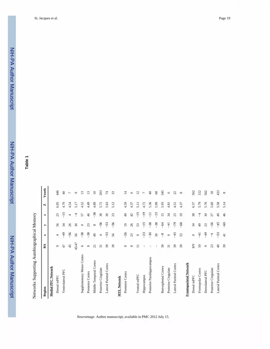
3.2. fMRI Results3.2.1. Independent Component Analysis—We used ICA to identify neural networkscontributing to construction and elaboration during AM retrieval. ICA revealed four task-related networks (p’s < .001; see Table 1, Figure 1; all activations are reported at FDRcorrected p < .001) corresponding to large-scale networks that have previously been found(Damoiseaux, et al., 2006) and which overlap with structural connectivity (Skudlarski, et al.,2008). The first component showed a pattern of integrated left-lateralized frontal andparietal cortices, including dorsal mPFC and striatum, which overlaps with the frontoparietalnetwork and similar central executive network (Dosenbach, et al., 2007; Seeley, et al., 2007;
St. Jacques et al. Page 6
Neuroimage. Author manuscript; available in PMC 2012 July 15.
NIH
-PA Author Manuscript
NIH
-PA Author Manuscript
NIH
-PA Author Manuscript

also see Vincent, et al., 2008). Interestingly, the frontoparietal network we observed herewas left-lateralized, consistent with evidence that this network sometimes splits into left andright hemispheres (Damoiseaux, et al., 2006) and is lateralized (Vincent, et al., 2008). Thesecond component showed an integrated pattern of bilateral frontopolar cortex and leftinsula, along with dorsal anterior cingulate, which is consistent with the cinguloperculumnetwork and related salience network (Dosenbach, et al., 2007; Seeley, et al., 2007; also seeVincent, et al., 2008). The third component showed an integrated pattern along the midlineconsisting primarily of dorsal medial PFC and posterior cingulate, along with bilateral VPC,which is consistent with the medial PFC network (Buckner, et al., 2008). Finally, the fourthcomponent showed an integrated pattern of medial temporal lobe (MTL), ventromedial PFC,retrosplenial cortex, and bilateral VPC, which is consistent with the MTL network thatsupports declarative memory (Vincent, et al., 2006).
As indicated by the regression analysis (see Table 2), the frontoparietal andcingulooperculum network were associated with the construction phase, whereas medialPFC and MTL networks were associated with both phases (for visualization see Figure 2).Further, we note that the strength of the association of both the frontoparietal andcinguloopercular networks was greater for the construction phase when compared to theelaboration phase, but there were no differences between these two phases in the othercomponents.
3.2.2. Dynamic Causal Model—To determine how the neural networks identified byICA interacted during AM retrieval we employed DCM and a Bayesian Model Selection(BMS) procedure to find the most likely model within a large model space (Stevens, et al.,2007). Model selection showed that the most likely model within the model space had anexceeded probability of 0.17, which was on average 7.36 times more likely than othermodels (see Supplemental Figure 2). The DCM (see Figure 3) revealed that construction andelaboration phases of AM retrieval were driving inputs (i.e., produced evoked responses) inthe medial PFC network (for coefficient values see Table 3). In contrast, presentation of thegeneric memory cue evoked activity in the system via the frontoparietal andcingulooperculum networks. The cue coefficient is negative because inhibitory activityoriginating in frontoparietal and cinguloperculum networks cascades to the neural networkscontributing to AM retrieval during processing associated with the cue (i.e., auditoryanalysis; also see Figure 2). These results suggest that the medial PFC network drives theconstruction and elaboration of AMs. There were reciprocal direct connections betweensome, but not all of the neural networks contributing to AM retrieval. There was a reciprocalpositive influence between the frontoparietal and cingulooperculum networks, thefrontoparietal and medial PFC networks, and the medial PFC and MTL networks.
Recollection and accessibility of AMs also modulated the connectivity among the neuralnetworks. First, AMs that were more accessible were associated with greater influence of thefrontoparietal and MTL networks on the medial PFC network. Accessibility modulated theconnectivity between the frontoparietal and medial PFC networks, such that thefrontoparietal network influenced activity in the medial PFC network during theconstruction phase, whereas the medial PFC network influenced the frontoparietal networkduring elaboration. The latter result suggests that accessibility may also impact how amemory is elaborated, with greater connectivity between medial PFC and frontoparietalnetworks. However, these effects were not greater for accessibility when compared torecollection. Accessibility was also associated with more bottom-up influence of the MTLon the medial PFC network during construction, and there was a trend showing that thiseffect was also greater in the context of accessibility vs. recollection, t (14) = −1.37, p = .19.These results suggests that accessibility impacts both construction and elaboration phases ofAM. Second, recollection of AMs was associated with greater connectivity between the
St. Jacques et al. Page 7
Neuroimage. Author manuscript; available in PMC 2012 July 15.
NIH
-PA Author Manuscript
NIH
-PA Author Manuscript
NIH
-PA Author Manuscript

medial PFC and MTL networks during elaboration, potentially as the result of increasedrecovery of memory details, and this effect was significantly greater for recollection vs.accessibility, t (14) = 2.04, p < .06.
3.2.2. Post-Hoc Analysis of Nodes Common to Networks—To identify nodeswithin each network that were common across the networks identified by the DCM, weemployed a conjunction approach to examine the intersection of pairs of networks. An FDRcorrected p < .001 and a 5 voxel extent threshold for each contrast was used. The resultsrevealed that there was some overlap between the networks (ranging from 0.94% to 8.84%of possible voxels), especially those having a direct influence on one another. Frontoparietaland cinguoloopercular networks overlapped in the recruitment of the dorsal anteriorcingulate/medial frontal (81 voxels; MNI: 0, 30, 38) near Brodmann Area (BA) 32/9. MedialPFC and frontoparietal networks both recruited lateral parietal cortex (25 voxels; MNI: −53,−53, 30; BA 40), dorsal medial PFC (36 voxels; MNI: −4, 56, 38; BA 9), ventrolateral PFC(5 voxels; MNI: −45, 30, −19; BA 47) and dorsolateral PFC (9 voxels; MNI: −38, 23, 46;BA 9). Finally, medial PFC and MTL networks overlapped in the recruitment of theposterior cingulate (24 voxels; MNI: 0, −68, 34; BA 23/31). In contrast, there was lessoverlap among the networks that were not directly connected as identified by the DCM. TheMTL and frontoparietal networks showed overlap in a region in the premotor cortex (5voxels; MNI: −26, 19, 46; BA 8), and the medial PFC and cingulopercular networksoverlapped in premotor (13 voxels; MNI: −38, 0, 57; BA 6) and supplementary motor areas(7 voxels; MNI: 11, 19, 65; BA 6). There was no overlap between the MTL andcinguloopercular networks.
4. DiscussionThe brain is organized into multiple neural networks and higher-order cognitive processes,such as AM, are constructed through their interaction (Fuster, 2009; Rubin, 2005, 2006).Understanding how networks interact to support complex cognitive processes is critical forAM retrieval, because it relies on a distributed set of brain regions (Cabeza & St. Jacques,2007; Maguire, 2001; Svoboda, et al., 2006) that may encompass separate networks.Previous functional neuroimaging studies have identified the brain regions supporting AM(for review see Cabeza & St. Jacques, 2007; St Jacques & Cabeza, In Press; St. Jacques, InPress) and have examined the interaction among coactive regions (Addis, Pan, Vu, Laiser, &Schacter, 2009; Burianova & Grady, 2007; Burianova, McIntosh, & Grady, 2010; Spreng &Grady, 2009; for meta-analysis see Svoboda, et al., 2006), but they do not distinguish the co-activation of multiple neural networks supporting AM retrieval. Here we used a novelapproach to understand these issues (e.g., Stevens, et al., 2007) by combining ICA and DCMto examine the interaction among large-scale neural supporting AM retrieval, and themodulation of network connections by behavior. The results of the ICA revealed four task-related components: 1) a left-lateralized frontoparietal network, 2) the cinguolooperculumnetwork, 3) the medial PFC network, and 4) the MTL network. DCM on these neuralnetworks, represented by ICA time courses, revealed two main findings demonstrating theimportance of the medial PFC network. First, the medial PFC network drove neuronalactivation among the other networks, confirming the integral contribution of this subsystemof the default network to AM construction. Second, connectivity with the medial PFCnetwork and other networks increased when AMs were recollected and accessible,suggesting that this network has a substantial influence on AM retrieval. We discuss thesefindings below.
St. Jacques et al. Page 8
Neuroimage. Author manuscript; available in PMC 2012 July 15.
NIH
-PA Author Manuscript
NIH
-PA Author Manuscript
NIH
-PA Author Manuscript

4.1. Neural Networks Supporting AM RetrievalPrevious functional neuroimaging studies of AM have observed a “core network” of regionsthat are typically coactive and interact during AM retrieval (Cabeza & St. Jacques, 2007;McDermott, et al., 2009; Spreng, et al., 2009; Svoboda, et al., 2006). The current resultssuggest that the regions recruited during AM retrieval comprise functionally dissociablenetworks including the frontoparietal network, cinguolooperculum network, MTL networkand medial PFC network.
Two of the networks identified by ICA contributed primarily to the construction phaseduring AM retrieval. First, we found a frontoparietal network, which is linked to adaptivecontrolled processes (Dosenbach, Fair, Cohen, Schlaggar, & Petersen, 2008; Dosenbach, etal., 2007; Vincent, et al., 2008). Controlled operations that act on memory are associatedwith the engagement of frontal and parietal cortices (Cabeza, Ciaramelli, Olson, &Moscovitch, 2008; Moscovitch & Winocur, 2002). The predominantly left-lateralizedrecruitment of the frontoparietal network during construction is consistent with previousfunctional neuroimaging studies of AM (for a meta-analysis see Svoboda, et al., 2006),which observed recruitment of left-lateralized brain regions overlapping with those foundhere. Moreover, the left-lateralized pattern of the frontoparietal network suggests that it maycontribute more specifically to strategic verbal retrieval processes relying on semanticinformation that guide the construction of a specific AM (Cabeza & St. Jacques, 2007),perhaps as the retrieval cue is processed and elaborated upon. Second, we observed thecingulooperculum network linked to goal-maintenance (Dosenbach, et al., 2008; Dosenbach,et al., 2007) and to salience processing (Seeley, et al., 2007). The finding that frontoparietaland cingulooperculum components were associated with construction rather than elaborationsuggests that the initial processes involved in AM elicited by generic cues relies uponadditional control and goal-related processes. Conjointly the frontoparietal andcinguloopercular networks may potentially contribute to retrieval mode, cue specification ormonitoring and verification operations (for a review see Rugg & Wilding, 2000) that lead tothe recovery and validation of memory.
The remaining two components were associated with both construction and elaborationphases during AM retrieval, and included the medial PFC network, which previous researchhas suggested may support self-referential simulations (for a review see Buckner, et al.,2008), and the MTL network associated with declarative memory (Kahn, et al., 2008;Vincent, et al., 2006) and scene construction (Andrews-Hanna, et al., 2010; also seeHassabis, et al., 2007a). The finding that the medial PFC and MTL networks were sustainedacross the construction and elaboration phases is consistent with the iterative nature of AMretrieval, which would involve multiple construction-memory mappings. The MTL andmedial PFC networks overlap with the set of regions engaged by the default network(Greicius, et al., 2003; Raichle, et al., 2001) and compose separate subsystems (Andrews-Hanna, et al., 2010). For example, Andrews-Hanna et al. (2010) identified subsystems of thedefault network and their relationship to cognitive functions. They found that an MTLsubsystem contributed to the construction of mental scenes, whereas a dorsal medial PFCsubsystem contributed to self-referential functions. Understanding the role of separablenetworks contributing to AM retrieval may be particular important for understanding thecomponent processes of memory and for distinguishing memory from other tasks relying onthe same core network (Hassabis, Kumaran, & Maguire, 2007b; Spreng & Grady, 2010).
4.2. Neural Network Dynamics during AM Retrieval and Modulation by BehaviorThe medial PFC network drove neuronal activation within the system during bothconstruction and elaboration of AM. In contrast, the frontoparietal and cinguloopercularnetworks drove activity during the presentation of the cue. It is interesting that the medial
St. Jacques et al. Page 9
Neuroimage. Author manuscript; available in PMC 2012 July 15.
NIH
-PA Author Manuscript
NIH
-PA Author Manuscript
NIH
-PA Author Manuscript

PFC network associated with self-referential processes (Gusnard, Akbudak, Shulman, &Raichle, 2001; Kelley, et al., 2002), rather than other networks associated with memoryrelated or controlled processes, was found to initiate and maintain AM retrieval. Self-reference is a critical and defining component of AM, which may importantly modulate theconstruction of AMs (Cabeza & St. Jacques, 2007; Conway, 2005; Conway & Pleydell-Pearce, 2000; P. L. St. Jacques, In Press). Consistent with this idea, in a previous fMRIstudy (Daselaar, et al., 2008) we found that medial PFC region were activated to a greaterextent during the initial construction of AM. Similarly, Muscatell, Addis and Kensinger(2010) directly modulated the extent of self-involvement and found increased effectiveconnectivity with the medial PFC during AM retrieval. Future studies should examine thepotential interactions between both self-referential and memory processes on the networkssupporting AM retrieval.
We found direct intrinsic connections between some but all of the networks supporting AM.First, there was a reciprocal positive influence between the frontoparietal andcingulooperculum networks, suggesting that these networks directly influence one anotherduring AM retrieval. These results are in line with previous studies suggesting interregionalinteractions between these top-down control networks, with the frontoparietal networkinitiating and flexibly adjusting controlled processes that are maintained by thecingulooperculum network (Dosenbach, et al., 2008; Dosenbach, et al., 2007). Second, therewas a reciprocal connection between the frontoparietal and medial PFC networks.Frontoparietal cortex may contribute to the integration between externally directed attentionand internally directed thought, given that it is anatomically juxtaposed between the dorsalattention and default networks (Vincent, et al., 2008). Third, there was reciprocalconnectivity between the medial PFC and MTL networks, which suggests that AM retrievalrelies on the connectivity between these subsystems of the default network (Andrews-Hanna, et al., 2010; Buckner, et al., 2008).
Neural networks are thought to interact with one another through sparse connectionspotentially mediated by cortical hubs, which are regions that demonstrate high levels ofcortical functional connectivity (Achard, et al., 2006; Buckner, et al., 2009). Supporting thissuggestion, we found greater overlap among the networks with reciprocal connections. Forexample, there was overlap in the regions comprising the MTL and medial PFC networks,but no overlap between the indirectly connected MTL and cinguloopercular networks.Interestingly, the regions of overlap observed here were generally located in heteromodalassociation areas that are ideally situated for integrating divergent information across brainsystems (Mesulam, 1998), and which have previously been identified as cortical hubs(Achard, et al., 2006; Buckner, et al., 2009; Hagmann, et al., 2008). In particular, we foundthat the medial PFC and MTL networks overlapped in the recruitment of the posteriorcingulate, a region that is part of a midline core interspersed between dorsal medial PFC andMTL subsystems of the default network (Andrews-Hanna, et al., 2010; Buckner, et al.,2008). Cinguloopercular and frontoparietal networks both engaged the dorsal anteriorcingulate/medial PFC, a region that also has been shown to overlap in the salience andexecutive control networks (Seeley, et al., 2007). Further, there was overlap in lateralparietal cortex and dorsal medial PFC between the medial PFC and frontoparietal networks.It will be important for future work to determine how these overlapping nodes potentiallycontribute to the mechanisms of connectivity between networks.
Memory accessibility and recollection modulated the intrinsic connections among the neuralnetworks, particular with the medial PFC network. AM retrieval occurs via effortfulprocesses that engage top-down control processes or directly through associative retrievalprocesses involving bottom-up recovery of the memory trace (Conway & Pleydell-Pearce,2000; Moscovitch, 1992). Here we show that when AMs are more easily retrieved both the
St. Jacques et al. Page 10
Neuroimage. Author manuscript; available in PMC 2012 July 15.
NIH
-PA Author Manuscript
NIH
-PA Author Manuscript
NIH
-PA Author Manuscript

top-down and bottom-up processes impinge on the medial PFC network that drives activitywithin the system during construction. In contrast, the influence of recollection wasobserved during elaboration via the modulation of the medial PFC network on the MTLnetwork. These results are consistent with evidence that recollection depends uponactivation that occurs later during retrieval once a memory is found and can be elaboratedupon (Daselaar, et al., 2008; for review see St Jacques & Cabeza, In Press). Increasedinteraction between the MTL network and medial PFC network during elaboration ofrecollected memories may be associated with additional visualization of the constructedscene as participants were asked to recall additional details during this phase. Interestingly, adirect comparison of modulation by accessibility versus recollection revealed that theconnectivity between MTL and Medial PFC networks also tended to show context specificeffects. The bottom-up influence of the MTL network on the medial PFC network tended toincrease for memory accessibility more than the recollective outcome of retrieval, whereasthe top-down influence of the medial PFC on the MTL network increased to a greater extentfor recollection than memory accessibility. In sum, recollection and memory accessibilityuniquely modulated connectivity among some of the networks contributing to AM retrieval.
5. ConclusionsThe findings support the proposal that AM retrieval is supported by the interaction amongseparable neural networks which have been associated with self-referential, memory, searchand goal-directed processes (Rubin, 2006). DCM of the neural networks supporting AMretrieval revealed that the medial PFC network drives neuronal activation, and thatreciprocal connectivity with this network increased when memories were accessible andrecollected. In sum, the data suggest that the medial PFC network importantly contributes toAM retrieval.
Elucidating neural network dynamics has important implications for understanding andpotentially distinguishing higher-order cognitive functions. The neural networks identifiedin the current study during AM retrieval may also support tasks relying on similarcomponent processes, such as envisioning the future and taking another person’s perspective(i.e., Theory of Mind; Hassabis & Maguire, 2007; Schacter, Addis, & Buckner, 2007).Examination of the recruitment of particular neural networks, their interaction, andmodulation of behavior may help to further distinguish these tasks. Our findings highlightthe important role of large-scale neural networks in the generation of complex cognitiveprocesses.
Supplementary MaterialRefer to Web version on PubMed Central for supplementary material.
AcknowledgmentsWe thank Dr. Roberto Cabeza for helpful comments on an earlier draft and Amanda Miles for help with participanttesting. This research was supported by a National Institute of Aging RO1 AG023123 to DCR, and a post-doctoralNRSA AG038079 and L’Oreal USA for Women in Science Fellowship to PLS.
ReferencesAchard S, Salvador R, Whitcher B, Suckling J, Bullmore E. A resilient, low-frequency, small-world
human brain functional network with highly connected association cortical hubs. J Neurosci. 2006;26(1):63–72. [PubMed: 16399673]
St. Jacques et al. Page 11
Neuroimage. Author manuscript; available in PMC 2012 July 15.
NIH
-PA Author Manuscript
NIH
-PA Author Manuscript
NIH
-PA Author Manuscript

Addis DR, McIntosh AR, Moscovitch M, Crawley AP, McAndrews MP. Characterizing spatial andtemporal features of autobiographical memory retrieval networks: a partial least squares approach.Neuroimage. 2004; 23(4):1460–1471. [PubMed: 15589110]
Addis DR, Moscovitch M, McAndrews MP. Consequences of hippocampal damage across theautobiographical memory network in left temporal lobe epilepsy. Brain. 2007; 130(Pt 9):2327–2342. [PubMed: 17681983]
Addis DR, Pan L, Vu MA, Laiser N, Schacter DL. Constructive episodic simulation of the future andthe past: distinct subsystems of a core brain network mediate imagining and remembering.Neuropsychologia. 2009; 47(11):2222–2238. [PubMed: 19041331]
Andrews-Hanna JR, Reidler JS, Sepulcre J, Poulin R, Buckner RL. Functional-anatomic fractionationof the brain’s default network. Neuron. 2010; 65(4):550–562. [PubMed: 20188659]
Beck, AT.; Steer, RA.; Brown, GK. Manual for Beck Depression Inventory. San Antonio, TX:Psychology Coporation; 1996.
Botzung A, LaBar KS, Kragel P, Miles A, Rubin DC. Component neural systems for the creation ofemotional memories during free viewing of a complex, real-world event. [Original Research].Frontiers in Human Neuroscience. 2010:4. [PubMed: 20198130]
Bradley, MM.; Lang, PJ. Affective norms for English words (ANEW). Gainesville, FL: The NIMHCenter for the Study of Emotion and Attention, University of Florida; 1999.
Bressler SL, Menon V. Large-scale brain networks in cognition: emerging methods and principles.Trends Cogn Sci. 2010; 14(6):277–290. [PubMed: 20493761]
Brewer, WF. What is autobiographical memory?. In: Rubin, DC., editor. Autobiographical Memory.NY: Cambridge UP; 1986. p. 25-49.
Buckner RL, Andrews-Hanna JR, Schacter DL. The brain’s default network: anatomy, function, andrelevance to disease. Annals of the New York Academy of Science. 2008; 1124:1–38.
Buckner RL, Sepulcre J, Talukdar T, Krienen FM, Liu H, Hedden T, et al. Cortical hubs revealed byintrinsic functional connectivity: mapping, assessment of stability, and relation to Alzheimer’sdisease. J Neurosci. 2009; 29(6):1860–1873. [PubMed: 19211893]
Burianova H, Grady CL. Common and unique neural activations in autobiographical, episodic, andsemantic retrieval. J Cogn Neurosci. 2007; 19(9):1520–1534. [PubMed: 17714013]
Burianova H, McIntosh AR, Grady CL. A common functional brain network for autobiographical,episodic, and semantic memory retrieval. Neuroimage. 2010; 49(1):865–874. [PubMed:19744566]
Cabeza R, Ciaramelli E, Olson IR, Moscovitch M. The parietal cortex and episodic memory: anattentional account. Nat Rev Neurosci. 2008; 9(8):613–625. [PubMed: 18641668]
Cabeza R, St Jacques PL. Functional neuroimaging of autobiographical memory. Trends Cogn Sci.2007; 11(5):219–227. [PubMed: 17382578]
Calhoun VD, Adali T, Pearlson GD, Pekar JJ. A method for making group inferences from functionalMRI data using independent component analysis. Hum Brain Mapp. 2001a; 14(3):140–151.[PubMed: 11559959]
Calhoun VD, Adali T, Pearlson GD, Pekar JJ. Spatial and temporal independent component analysis offunctional MRI data containing a pair of task-related waveforms. Hum Brain Mapp. 2001b; 13(1):43–53. [PubMed: 11284046]
Conway MA. Memory and the self. Journal of Memory and Language. 2005; 53:594–628.Conway MA, Pleydell-Pearce CW. The construction of autobiographical memories in the self-memory
system. Psychological Review. 2000; 107(2):261–288. [PubMed: 10789197]Damoiseaux JS, Greicius MD. Greater than the sum of its parts: a review of studies combining
structural connectivity and resting-state functional connectivity. Brain Struct Funct. 2009; 213(6):525–533. [PubMed: 19565262]
Damoiseaux JS, Rombouts SA, Barkhof F, Scheltens P, Stam CJ, Smith SM, et al. Consistent resting-state networks across healthy subjects. Proc Natl Acad Sci U S A. 2006; 103(37):13848–13853.[PubMed: 16945915]
Daselaar SM, Rice HJ, Greenberg DL, Cabeza R, LaBar KS, Rubin DC. The spatiotemporal dynamicsof autobiographical memory: neural correlates of recall, emotional intensity, and reliving. CerebCortex. 2008; 18(1):217–229. [PubMed: 17548799]
St. Jacques et al. Page 12
Neuroimage. Author manuscript; available in PMC 2012 July 15.
NIH
-PA Author Manuscript
NIH
-PA Author Manuscript
NIH
-PA Author Manuscript

Dosenbach NU, Fair DA, Cohen AL, Schlaggar BL, Petersen SE. A dual-networks architecture of top-down control. Trends Cogn Sci. 2008; 12(3):99–105. [PubMed: 18262825]
Dosenbach NU, Fair DA, Miezin FM, Cohen AL, Wenger KK, Dosenbach RA, et al. Distinct brainnetworks for adaptive and stable task control in humans. Proc Natl Acad Sci U S A. 2007;104(26):11073–11078. [PubMed: 17576922]
Fox MD, Raichle ME. Spontaneous fluctuations in brain activity observed with functional magneticresonance imaging. Nature Reviews Neuroscience. 2007; 8(9):700–711.
Friston KJ, Harrison L, Penny W. Dynamic causal modelling. Neuroimage. 2003; 19(4):1273–1302.[PubMed: 12948688]
Friston KJ, Price CJ, Fletcher P, Moore C, Frackowiak RS, Dolan RJ. The trouble with cognitivesubtraction. Neuroimage. 1996; 4(2):97–104. [PubMed: 9345501]
Fuster JM. Cortex and memory: emergence of a new paradigm. J Cogn Neurosci. 2009; 21(11):2047–2072. [PubMed: 19485699]
Greenberg DL, Rice HJ, Cooper JJ, Cabeza R, Rubin DC, LaBar KS. Co-activation of the amygdala,hippocampus and inferior frontal gyrus during autobiographical memory retrieval.Neuropsychologia. 2005; 43(5):659–674. [PubMed: 15721179]
Greenberg DL, Rubin DC. The neuropsychology of autobiographical memory. Cortex. 2003; 39(4–5):687–728. [PubMed: 14584549]
Greicius MD, Krasnow B, Reiss AL, Menon V. Functional connectivity in the resting brain: a networkanalysis of the default mode hypothesis. Proc Natl Acad Sci U S A. 2003; 100(1):253–258.[PubMed: 12506194]
Gusnard DA, Akbudak E, Shulman GL, Raichle ME. Medial prefrontal cortex and self-referentialmental activity: relation to a default mode of brain function. Proc Natl Acad Sci U S A. 2001;98(7):4259–4264. [PubMed: 11259662]
Hagmann P, Cammoun L, Gigandet X, Meuli R, Honey CJ, Wedeen VJ, et al. Mapping the structuralcore of human cerebral cortex. PLoS Biol. 2008; 6(7):e159. [PubMed: 18597554]
Hassabis D, Kumaran D, Maguire EA. Using imagination to understand the neural basis of episodicmemory. Journal of Neuroscience. 2007a; 27(52):14365–14374. [PubMed: 18160644]
Hassabis D, Kumaran D, Maguire EA. Using imagination to understand the neural basis of episodicmemory. J Neurosci. 2007b; 27(52):14365–14374. [PubMed: 18160644]
Hassabis D, Maguire EA. Deconstructing episodic memory with construction. Trends in CognitiveScience. 2007; 11(7):299–306.
Kahn I, Andrews-Hanna JR, Vincent JL, Snyder AZ, Buckner RL. Distinct cortical anatomy linked tosubregions of the medial temporal lobe revealed by intrinsic functional connectivity. JNeurophysiol. 2008; 100(1):129–139. [PubMed: 18385483]
Kelley WM, Macrae CN, Wyland CL, Caglar S, Inati S, Heatherton TF. Finding the self? An event-related fMRI study. J Cogn Neurosci. 2002; 14(5):785–794. [PubMed: 12167262]
Levine B, Turner GR, Tisserand D, Hevenor SJ, Graham SJ, McIntosh AR. The functionalneuroanatomy of episodic and semantic autobiographical remembering: a prospective functionalMRI study. J Cogn Neurosci. 2004; 16(9):1633–1646. [PubMed: 15601525]
Maguire EA. Neuroimaging studies of autobiographical event memory. Philos Trans R Soc Lond BBiol Sci. 2001; 356(1413):1441–1451. [PubMed: 11571035]
Maguire EA, Mummery CJ, Buchel C. Patterns of hippocampal-cortical interaction dissociate temporallobe memory subsystems. Hippocampus. 2000; 10(4):475–482. [PubMed: 10985287]
Maguire EA, Vargha-Khadem F, Mishkin M. The effects of bilateral hippocampal damage on fMRIregional activations and interactions during memory retrieval. Brain. 2001; 124(Pt 6):1156–1170.[PubMed: 11353732]
McDermott KB, Szpunar KK, Christ SE. Laboratory-based and autobiographical retrieval tasks differsubstantially in their neural substrates. Neuropsychologia. 2009; 47(11):2290–2298. [PubMed:19159634]
Menon V, Uddin LQ. Saliency, switching, attention and control: a network model of insula function.Brain Struct Funct. 2010; 214(5–6):655–667. [PubMed: 20512370]
Mesulam MM. From sensation to cognition. Brain. 1998; 121(Pt 6):1013–1052. [PubMed: 9648540]
St. Jacques et al. Page 13
Neuroimage. Author manuscript; available in PMC 2012 July 15.
NIH
-PA Author Manuscript
NIH
-PA Author Manuscript
NIH
-PA Author Manuscript

Moscovitch M. Memory and Working-with-Memory. Journal of Cognitive Neuroscience. 1992; 4(3):257–267.
Moscovitch, M.; Winocur, G. The frontal cortex and working with memory. In: Stuss, DT.; Knight,RT., editors. Principles of frontal lobe function. London: Oxford University Press; 2002. p.188-209.
Muscatell KA, Addis DR, Kensinger EA. Self-involvement modulates the effective connectivity of theautobiographical memory network. Soc Cogn Affect Neurosci. 2010; 5(1):68–76. [PubMed:20007242]
Norman, DA.; Bobrow, DG. On the role of active memory processes in perception and cognition. In:Cofer, CN., editor. The structure of human memory. San Francisco: Freeman; 1976.
Raichle ME, MacLeod AM, Snyder AZ, Powers WJ, Gusnard DA, Shulman GL. A default mode ofbrain function. Proc Natl Acad Sci U S A. 2001; 98(2):676–682. [PubMed: 11209064]
Rubin DC. A basic-systems approach to autobiographical memory. Current Directions inPsychological Science. 2005; 14(2):79–83.
Rubin DC. The basic-systems model of episodic memory. Perspectives in Psychological Science.2006; 1(1):277–311.
Rubin DC, Schrauf RW, Greenberg DL. Belief and recollection of autobiographical memories. MemCognit. 2003; 31(6):887–901.
Rugg MD, Wilding EL. Retrieval processing and episodic memory. Trends Cogn Sci. 2000; 4(3):108–115. [PubMed: 10689345]
Schacter DL, Addis DR, Buckner RL. Remembering the past to imagine the future: the prospectivebrain. Nature Reviews Neuroscience. 2007; 8(9):657–661.
Seeley WW, Menon V, Schatzberg AF, Keller J, Glover GH, Kenna H, et al. Dissociable intrinsicconnectivity networks for salience processing and executive control. J Neurosci. 2007; 27(9):2349–2356. [PubMed: 17329432]
Skudlarski P, Jagannathan K, Calhoun VD, Hampson M, Skudlarska BA, Pearlson G. Measuring brainconnectivity: diffusion tensor imaging validates resting state temporal correlations. Neuroimage.2008; 43(3):554–561. [PubMed: 18771736]
Spreng RN, Grady CL. Patterns of Brain Activity Supporting Autobiographical Memory, Prospection,and Theory-of-Mind and Their Relationship to the Default Mode Network. Journal of CognitiveNeuroscience. 2009
Spreng RN, Grady CL. Patterns of Brain Activity Supporting Autobiographical Memory, Prospection,and Theory-of-Mind and Their Relationship to the Default Mode Network. Journal of CognitiveNeuroscience. 2010; 22(6):1112–1123. [PubMed: 19580387]
Spreng RN, Mar RA, Kim AS. The common neural basis of autobiographical memory, prospection,navigation, theory of mind, and the default mode: a quantitative meta-analysis. J Cogn Neurosci.2009; 21(3):489–510. [PubMed: 18510452]
St Jacques PL, Botzung A, Miles A, Rubin DC. Functional neuroimaging of emotionally intenseautobiographical memories in post-traumatic stress disorder. Journal of Psychiatric Research.201010.1016/j.jpsychires.2010.1010.1011
St Jacques, PL.; Cabeza, R. Neural Basis of Autobiographical Memory. In: Ghetti, S.; Bauer, PJ.,editors. Origins and development of recollection: Perspectives from psychology and neuroscience.New York: Oxford University Press; (In Press)
St Jacques, PL. Functional neuroimaging of autobiographcial memory. In: Berntsen, D.; Rubin, DC.,editors. Understanding Autobiographical Memory: Theories and Approaches. (In Press)
St Jacques PL, Conway MA, Lowder MW, Cabeza R. Watching my mind unfold vs. yours: An fMRIstudy using a novel camera technology to examine neural differences in self-projection of self vs.other perspectives. Journal of Cognitive Neuroscience. (In Press).
St Jacques PL, Rubin DC, Cabeza R. Age-related effects on the neural correlates of autobiographicalmemory retrieval. Neurobiology of Aging. 201010.1016/j.neurobiolaging.2010.1011.1007
Stephan KE, Penny WD, Daunizeau J, Moran RJ, Friston KJ. Bayesian model selection for groupstudies. Neuroimage. 2009; 46(4):1004–1017. [PubMed: 19306932]
Stephan KE, Penny WD, Moran RJ, den Ouden HE, Daunizeau J, Friston KJ. Ten simple rules fordynamic causal modeling. Neuroimage. 2010; 49(4):3099–3109. [PubMed: 19914382]
St. Jacques et al. Page 14
Neuroimage. Author manuscript; available in PMC 2012 July 15.
NIH
-PA Author Manuscript
NIH
-PA Author Manuscript
NIH
-PA Author Manuscript

Stevens MC, Kiehl KA, Pearlson GD, Calhoun VD. Functional neural networks underlying responseinhibition in adolescents and adults. Behav Brain Res. 2007; 181(1):12–22. [PubMed: 17467816]
Svoboda E, McKinnon MC, Levine B. The functional neuroanatomy of autobiographical memory: ameta-analysis. Neuropsychologia. 2006; 44(12):2189–2208. [PubMed: 16806314]
Tulving, E. Elements of Memory. Oxford: Clarendon Press; 1983.Vincent JL, Kahn I, Snyder AZ, Raichle ME, Buckner RL. Evidence for a frontoparietal control
system revealed by intrinsic functional connectivity. J Neurophysiol. 2008; 100(6):3328–3342.[PubMed: 18799601]
Vincent JL, Snyder AZ, Fox MD, Shannon BJ, Andrews JR, Raichle ME, et al. Coherent spontaneousactivity identifies a hippocampal-parietal memory network. J Neurophysiol. 2006; 96(6):3517–3531. [PubMed: 16899645]
Wheeler MA, Stuss DT, Tulving E. Toward a theory of episodic memory: the frontal lobes andautonoetic consciousness. Psychol Bull. 1997; 121(3):331–354. [PubMed: 9136640]
St. Jacques et al. Page 15
Neuroimage. Author manuscript; available in PMC 2012 July 15.
NIH
-PA Author Manuscript
NIH
-PA Author Manuscript
NIH
-PA Author Manuscript

Figure 1.The task-related independent components supporting AM retrieval. Activation is displayedat FDR corrected p < .001, 5 voxels.
St. Jacques et al. Page 16
Neuroimage. Author manuscript; available in PMC 2012 July 15.
NIH
-PA Author Manuscript
NIH
-PA Author Manuscript
NIH
-PA Author Manuscript

Figure 2.The time course of each independent component across the AM retrieval task reveals thatfrontoparietal and cingulooperculum networks contribute to construction, whereas, themedial temporal lobe (MTL) and medial prefrontal cortex (PFC) networks contribute to bothconstruction and elaboration phases.
St. Jacques et al. Page 17
Neuroimage. Author manuscript; available in PMC 2012 July 15.
NIH
-PA Author Manuscript
NIH
-PA Author Manuscript
NIH
-PA Author Manuscript

Figure 3.The dynamic causal model of the integration between neural networks supportingautobiographical memory retrieval. Red Square = modulation by accessibility duringconstruction (constr), Blue Square = modulation by accessibility during elaboration (elab),Blue Circle = modulation by recollection during elaboration.
St. Jacques et al. Page 18
Neuroimage. Author manuscript; available in PMC 2012 July 15.
NIH
-PA Author Manuscript
NIH
-PA Author Manuscript
NIH
-PA Author Manuscript
NIH
-PA Author Manuscript
NIH
-PA Author Manuscript
NIH
-PA Author Manuscript
St. Jacques et al. Page 19
Tabl
e 1
Net
wor
ks S
uppo
rting
Aut
obio
grap
hica
l Mem
ory
Reg
ion
BA
xy
zZ
Vox
els
Med
ial P
FC N
etw
ork
D
orsa
l mPF
C9
456
236.
0544
6
V
entro
late
ral P
FC47
−49
34−15
4.70
40
45−56
264
4.54
7
45/4
756
30−4
5.17
9
Su
pple
men
tary
Mot
or C
orte
x6
−38
057
4.52
13
Pr
emot
or C
orte
x8
−38
2346
4.49
13
M
iddl
e Te
mpo
ral C
orte
x21
498
−38
4.89
10
Po
ster
ior C
ingu
late
314
−56
385.
7220
3
La
tera
l Par
ieta
l Cor
tex
39−53
−53
305.
6374
3956
−56
235.
1233
MT
L N
etw
ork
Pr
emot
or C
orte
x8
−26
1949
4.59
14
823
2642
4.37
6
V
entra
l mPF
C11
053
−15
5.11
12
H
ippo
cam
pus
–−23
−15
−19
4.72
7
Po
ster
ior P
arah
ippo
cam
pus
–−30
−38
−11
5.36
40
–30
−38
−15
5.99
68
R
etro
sple
nial
Cor
tex
30−8
−64
155.
9334
5
Po
ster
ior C
ingu
late
310
−41
344.
816
La
tera
l Par
ieta
l Cor
tex
39−45
−79
234.
5122
3953
−68
194.
378
Fron
topa
riet
al N
etw
ork
D
orsa
l mPF
C8/
90
3438
6.37
562
Fr
onto
pola
r Cor
tex
10−41
49−4
5.79
152
D
orso
late
ral P
FC9
−49
2330
5.76
562
Po
ster
ior C
ingu
late
23−4
−26
275.
6018
La
tera
l Par
ieta
l Cor
tex
40−53
−45
495.
5843
3
3941
−60
465.
148
Neuroimage. Author manuscript; available in PMC 2012 July 15.

NIH
-PA Author Manuscript
NIH
-PA Author Manuscript
NIH
-PA Author Manuscript
St. Jacques et al. Page 20
Reg
ion
BA
xy
zZ
Vox
els
C
auda
te–
−11
011
4.87
8
C
ereb
ellu
m–
−8
−30
−30
4.86
8
Cin
gulo
oper
culu
m N
etw
ork
A
nter
ior C
ingu
late
328
3427
6.12
811
Fr
onto
pola
r Cor
tex
10−26
4923
5.54
112
1034
4130
4.70
35
In
sula
/Fro
ntal
Ope
rcul
um13
−38
158
4.82
78
C
laus
trum
-34
154
5.20
64
641
−4
494.
6649
Pr
ecun
eus
7−4
−75
534.
615
MN
I Coo
rdin
ates
Rep
orte
d. B
A, B
road
man
n’s A
rea
PFC
= P
refr
onta
l Cor
tex;
mPF
C =
Med
ial P
refr
onta
l Cor
tex;
MTL
= M
edia
l Tem
pora
l Lob
e
Neuroimage. Author manuscript; available in PMC 2012 July 15.

NIH
-PA Author Manuscript
NIH
-PA Author Manuscript
NIH
-PA Author Manuscript
St. Jacques et al. Page 21
Tabl
e 2
Inde
pend
ent C
ompo
nent
Ana
lysi
s
Com
pone
ntC
ueC
onst
ruct
ion
Ela
bora
tion
Con
st >
Ela
b
MSD
t(13)
pM
SDt(1
3)p
MSD
t(13)
pt(2
6)p
Fron
topa
rieta
l−4.46
242
−7.15
< .0
001
0.37
0.34
4.27
< .0
010.
140.
301.
770.
102.
05<
.05
Cin
gulo
oper
culu
m−2.76
1.37
−7.79
< .0
001
0.61
0.47
5.03
< .0
010.
190.
362.
060.
062.
750.
01
Med
ial P
FC−0.02
1.66
−0.06
0.96
0.85
0.63
5.19
< .0
010.
790.
329.
53<
.000
10.
330.
75
MTL
−1.37
1.50
−3.54
< .0
10.
550.
534.
08<
.01
0.63
0.31
7.95
< .0
001
−0.51
0.62
Neuroimage. Author manuscript; available in PMC 2012 July 15.

NIH
-PA Author Manuscript
NIH
-PA Author Manuscript
NIH
-PA Author Manuscript
St. Jacques et al. Page 22
Tabl
e 3
Coe
ffic
ient
s in
the
Dyn
amic
Cau
sal M
odel
Nam
eM
SDt(1
3)P
Dri
ving
Inpu
t
Con
stru
ctio
n on
Med
ial P
FC0.
030
0.03
33.
360.
005
Elab
orat
ion
on M
edia
l PFC
0.04
20.
024
6.47
< .0
001
Cue
on
Fron
topa
rieta
l−0.170
0.15
64.
080.
001
Cue
on
Cin
gulo
oper
culu
m−0.230
0.11
97.
25<
.000
1
Intr
insi
c C
onne
ctio
n
Med
ial P
FC o
n M
TL0.
272
0.14
27.
18<
.000
01
Med
ial P
FC o
n Fr
onto
parie
tal
0.09
60.
123
2.93
0.01
2
MTL
on
Med
ial P
FC0.
461
0.19
13.
74<
.000
1
Fron
topa
rieta
l on
Med
ial P
FC0.
108
0.13
33.
030.
010
Fron
topa
rieta
l on
Cin
gulo
oper
culu
m0.
191
0.11
26.
41<
.000
1
Cin
gulo
oper
culu
m o
n Fr
onto
parie
tal
0.12
20.
125
3.66
0.00
3
Mod
ulat
ory
Inpu
t
Acc
essi
bilit
y, C
onst
ruct
ion:
MTL
on
Med
ial P
FC0.
019
0.03
12.
320.
035
Acc
essi
bilit
y, C
onst
ruct
ion:
Fro
ntop
arie
tal o
n M
edia
l PFC
0.04
50.
063
2.68
0.01
9
Acc
essi
bilit
y, E
labo
ratio
n: M
edia
l PFC
on
Fron
topa
rieta
l0.
049
0.05
43.
420.
004
Rec
olle
ctio
n, E
labo
ratio
n: M
TL o
n M
edia
l PFC
0.11
70.
089
4.93
< 0.
001
PFC
= P
refr
onta
l Cor
tex,
MTL
= M
edia
l Tem
pora
l Lob
e
Neuroimage. Author manuscript; available in PMC 2012 July 15.