as time goes by: hippocampal connectivity changes with remoteness of autobiographical memory...
TRANSCRIPT
As Time Goes By: Hippocampal Connectivity Changes WithRemoteness of Autobiographical Memory Retrieval
Hedvig Soderlund,1* Morris Moscovitch,2,3 Namita Kumar,2 Marina Mandic,2 andBrian Levine2,3,4
ABSTRACT: The hippocampus is crucial for episodic autobiographicalmemory retrieval. Functional neuroimaging evidence suggests that it issimilarly engaged in recent and remote retrieval when memories arematched on vividness and personal importance. Far fewer studies haveinvestigated the nature of hippocampal-neocortical coactivation in rela-tion to memory remoteness. The purpose of this study was to examinehippocampal activity and functional connectivity as a function of mem-ory age. Unlike most studies of autobiographical memory, we includedautobiographical memories formed in the days and weeks before scan-ning, in addition to truly remote memories on the order of months andyears. Like previous studies, we found that the hippocampus was activebilaterally regardless of memory age, with anterior activity increasingup to 1 yr and then decreasing, and with posterior activity being lesssensitive to memory age. More importantly, hippocampal functionalconnectivity varied with memory age. Retrieving recent memories(�1 yr) showed a late coactivation of the hippocampus and areas of theautobiographical memory network, whereas retrieving remote memories(10 yrs) showed an early negative coactivation of the hippocampus andleft inferior frontal gyrus followed by a positive coactivation with ante-rior cingulate. This finding may reflect that the hippocampus is morestrongly integrated with the autobiographical memory network forrecent than for remote memories, and that more effort is required torecover remote memories. VVC 2011 Wiley-Liss, Inc.
KEY WORDS: autobiographical memory; connectivity; hippocampus;memory age; retrieval
INTRODUCTION
Retrieving an autobiographical memory implicates a palette of cogni-tive processes that is mirrored by a broad pattern of brain activation(Maguire, 2001; Svoboda et al., 2006; Cabeza and St Jacques, 2007).For autobiographical memories that are episodic in nature, the hippo-campal formation (including the CA fields, dentate gyrus, and subicularcomplex), hereafter referred to as the hippocampus, is a key structure.
This is reflected in studies of patients with hippocam-pal damage (e.g., Kirwan, 2008; Rosenbaum, 2008).Similarly, hippocampal activity in healthy adults isassociated with factors such as emotionality and vivid-ness that are important to episodic recollection (Ryanet al., 2001; Addis et al., 2004b; Gilboa et al., 2004).There is disagreement, however, as to the necessity ofthe hippocampus in relation to the age of memories,with the Standard Model of Consolidation (Squire andAlvarez, 1995) holding that memories are temporarilystored in the hippocampus and later consolidated in theneocortex, and Multiple Trace Theory (Nadel andMoscovitch, 1997; Moscovitch et al., 2005) holdingthat the hippocampus is required for the retrieval of allvivid or detailed autobiographical memories regardlessof age. The two theories also differ regarding hippocam-pal-neocortical connectivity: Standard Model of Consol-idation predicts that connectivity diminishes with age,whereas Multiple Trace Theory maintains that it per-sists, though the structures implicated may change.
In fMRI studies of healthy adults, most have foundhippocampal involvement regardless of memory age,supporting Multiple Trace Theory (Piolino et al., 2004;Rekkas and Constable, 2005; Steinvorth et al., 2006;Viard et al., 2007), especially when considering vivid-ness (Ryan et al., 2001; Addis et al., 2004b; Gilboaet al., 2004). Where age effects were reported, thesewere confounded with vividness (Niki and Luo, 2002;Piefke et al., 2003) or time allowed for retrieval andactivation (Maguire and Frith, 2003). Within the hippo-campus, recent memories may engage the anteriorregion, whereas remote memories are more diffuselyrepresented along the anterior-posterior plane (Gilboaet al., 2004; see also Rekkas and Constable, 2005).
The hippocampus, however, does not act in isolation.Its interaction with other brain structures, its ‘‘functionalconnectivity,’’ during autobiographical memory retrievalhas been explored in a few studies (Maguire et al., 2000;Addis et al., 2004a; Greenberg et al., 2005). For retrievalof memories a few years old, these studies suggestedhippocampal connectivity with frontal areas (e.g., rightinferior and left medial frontal gyrus), amygdala, para-hippocampus, and cerebellum, among others (Maguireet al., 2000; Addis et al., 2004a; Greenberg et al.,2005). Comparing memories across different lifeperiods, Viard et al. (2010) found that the strongesthippocampal-neocortical associations for vivid memorieswere from intermediate time periods. Significant correla-
1Department of Psychology, Uppsala University, Uppsala, Sweden;2 The Rotman Research Institute, Baycrest Centre for Geriatric Care,Toronto, Ontario, Canada; 3Department of Psychology, University ofToronto, Toronto, Ontario, Canada; 4Department of Medicine(Neurology), University of Toronto, Toronto, Ontario, CanadaAdditional Supporting Information may be found in the online version ofthis article.Grant sponsor: Canadian Institutes of Health Research; Grant number:MOP-62963; Grant sponsor: NIH-NICHD; Grant number: HD42385-01;Heart and Stroke Foundation of Ontario, Centre for Stroke Recovery*Correspondence to: Hedvig Soderlund, Department of Psychology,Uppsala University, Box 1225, Uppsala 751 42, Sweden.E-mail: [email protected] for publication 17 December 2010DOI 10.1002/hipo.20927Published online in Wiley Online Library (wileyonlinelibrary.com).
HIPPOCAMPUS 00:000–000 (2011)
VVC 2011 WILEY-LISS, INC.

tions between the hippocampus and other brain structuresthroughout all time periods (except 18–30 yrs) were interpretedas support of the Multiple Trace Theory.
Many studies contrasting recent and remote memories desig-nated events of 12–24 months in age as ‘‘recent’’ (for exceptions,see Maguire et al., 2001; Rekkas and Constable, 2005). However,assuming an exponential function of memory trace strength asfound in behavioral studies (Rubin and Schulkind, 1997), andextrapolating from consolidation time in rats which ranges from aday to a month, and whose life span is about 2–3 yrs (Ghirardiet al., 1995), there may be more of a phenomenological andneural difference in humans between events that are 2 weeks oldand 2 yrs old than between events that are 2 yrs old and 20 yrsold. Hence, studies that find similar activation in the hippocam-pus during retrieval of recent and remote events may do sobecause the so-called recent events have since long been consoli-dated and are not much different from the remote events.
Following an exponential function (Rubin and Shulkind,1997), we included in this study events that were 1 week, 1month, 1 yr, and 10 yrs old. We hypothesized that the hippo-campus would be active throughout all time periods, but thatits interaction with other brain structures would differ as afunction of memory age. Crucially, we measured the temporalpattern of the brain connectivity across both life periods andepochs of autobiographical memory retrieval within each lifeperiod. Remote memories that are usually less vividly re-experi-enced may require more retrieval effort relative to the veryrecent memories utilized in this study, and may entail differentneocortical-hippocampal interaction as compared to morerecent memories. As such, more vividly re-experienced memo-ries may also show a larger extent and greater sustenance ofhippocampal interaction with neocortical areas that confer viv-idness within episodic autobiographical memory, such as thosethat mediate visual imagery.
MATERIALS AND METHODS
Participants
Participants were 12 healthy, right-handed subjects (33.7 66.1 yrs old; 16.3 6 3.1 yrs of education; five men), free ofsignificant medical illnesses, including psychiatric disease (asdetermined by the Structured Clinical Interview for Diagnosis;SCID; First, 1995), neurological disease, and significant trau-matic brain injury. These participants were scanned as part of alarger study on the effects of depression electroconvulsive ther-apy on autobiographical memory. Just like the depressedpatients, these healthy controls were rescanned 6–10 weekslater. However, this report is restricted to the first scan in thehealthy participants.
Scanning
Participants performed two tasks during scanning: autobio-graphical memory retrieval and odd/even number judgments, a
recommended comparison task in studies of hippocampal func-tion (Stark and Squire, 2001). The two tasks were randomlydistributed within each run, with 10 trials of each task. Eachrun lasted �11 min. Participants were familiarized with the tasksand performed two practice trials of each prior to scanning.
Autobiographical memory
Two days before scanning, while still at home, participantsgenerated, dated, and gave titles to 10 events from each of fourdifferent time periods (i.e., 40 events in total, 10 events pertime period): the last week (14 days, excluding the last 2 days;hereafter referred to as 1 w); the last month (3–7 weeks; 1 m);the last year (6–18 months; 1 yr); the last 10 yrs (5–10 yrs, 10yrs). Participants were instructed to provide events that werespecific to a single time and place. They were told that as such,‘‘My vacation in Belize’’ was not acceptable, but that ‘‘My firstsurfing lesson in Belize’’ was. All event titles were randomizedacross the runs.
Each event title was presented for 18 s, preceded by a 1 s fixa-tion and a 4 s cue of the upcoming task (i.e., ‘‘Autobiographicalmemory’’). During the presentation of an autobiographicalmemory title, the participants were instructed to re-experience theevent as vividly as possible, recalling thoughts, feelings, and visualimages associated with the event. After 18 s, participants rated theamount of re-experiencing from 1 to 8 via fMRI-compatibleresponse boxes. Eight seconds were provided for rating. Tenevents were included in each run, with 2 from each of 2 timeperiods, and 3 from each of the other 2 time periods. Withineach run, the events, and hence the time periods, were presentedin a random order.
Immediately after scanning, participants rated each event onsix different dimensions: visualization, emotional valence, emo-tional change at the time of the event, importance at the timeof the event, importance at the time of scanning, and frequencyof thinking about or talking about the event. Ratings rangedfrom 1 to 6, except the last dimension which was more finelygrained (going from 0: never; to 11: constantly). Emotionalvalence ranged from negative (low) to positive (high) along the6-point scale, but was transformed into 2 measures, going from1 to 3. Ratings were performed after scanning rather thanbefore to reduce potential contamination of recent recollection.
Odd/Even judgment
The odd/even number judgment (odd/even) task consistedof the presentation of nine numbers, one at a time, and waspreceded by a 1 s fixation and a 4 s cue (i.e., ‘‘Odd/Even?’’).Participants were instructed to determine whether the numberwas odd or even without making an overt response. Each num-ber was presented for 1,900 ms, with a 100 ms interstimulusinterval. After the presentation of the last number, participantsrated the degree of re-experiencing, from 1 to 8 as describedabove. This rating was included as a manipulation check todetermine the degree of contamination of the comparison taskby any unbidden extraneous memories. Again, 8 s were pro-vided for rating.
2 SODERLUND ET AL.
Hippocampus

fMRI Data Acquisition
Scanning was conducted on a whole body 3.0T (SiemensMagnetom Trio Tim, Numaris/4Syngo MR B13; Siemens,Germany) with a standard quadrature bird-cage head coil.Participants were placed in the scanner in supine position, withtheir head firmly placed in a vacuum pillow to minimize headmovement. Earplugs and headphones were provided to reducethe noise from the scanner, and sensors were placed on partici-pants’ left big toe and around the chest, to monitor heart rateand respiration. A volumetric anatomical MRI was performedbefore functional scanning, using a MP-RAGE sequence (TR/TE 5 2,000/2.63 ms, 176 coronal slices perpendicular to thehippocampus, 256 3 256 acquisition matrix, voxel size 5 1mm3, FOV 5 25.6 cm). Functional imaging was performed tomeasure the blood oxygenation level-dependent (BOLD) effect(Ogawa et al., 1990). Scans were obtained using a single-shotT2*-weighted pulse with spiral in-out (TR/TE 5 2,000/30 ms,flip angle 708, 64 3 64 acquisition matrix, 32 coronal slicesperpendicular to the hippocampus, 5 mm thick, voxel size 53.1 3 3.1 mm, slice spacing 5 0, FOV 5 20 cm). To allowmagnetization to reach equilibrium, stimulus presentation wasdelayed by 20 s at the start of each experimental run.
Data Analysis
Univariate task analysis
Data processing and analyses were performed using Analysisof Functional NeuroImages software (AFNI; Cox and Hyde,1997). Time series data were spatially co-registered (alignedvolumetrically to a reference image within the run, using the3dvolreg program in AFNI) to correct for small head motionusing a 3D Fourier transform interpolation, and the lineartrends were removed. Uncorrected head motion (spikes) wasidentified through visual inspection and reduced through aver-aging the two surrounding time points. Physiological motion(respiration and heart beat) was also removed through linearfiltering. The data were normalized temporally and thereafterdeconvolved, using the AFNI plugin 3dDeconvolve. T-statisticscontrasting autobiographical memory retrieval for each timeperiod and odd/even processing to a baseline consisting of allnon-event time points (e.g., fixation) were calculated for eachparticipant to create statistical maps. These activation mapswere then transformed into stereotaxic space (Talairach andTournoux, 1988; Cox and Hyde, 1997) and spatially smoothedwith a Gaussian filter with a full width at half maximum valueof 6.0 mm to minimize individual variation of the anatomicallandmarks and to increase the signal-to-noise ratio. These lasttwo steps were performed to facilitate the subsequent groupanalysis, which consisted of voxelwise, mixed effects (conditionsfixed, participants random), two-way ANOVAs with time pe-riod as a within-subject factor.
Voxel of Interest (VOI) Analyses
Because of the theoretical importance of hippocampal activa-tion in this study, we further interrogated the peak activation
voxels within the right and left hippocampi. These voxels wereselected from the overall autobiographical memory versus odd/even contrast (and therefore unbiased with respect to timeperiod; see Supporting Information Table 1). These includedright anterior and left posterior hippocampal peaks, supple-mented by two most active voxels selected in the opposite loca-tions (left anterior; right posterior) to ensure any hemisphericdifferences were not in fact effects of axis location. Hence,there were four VOIs, two per hemisphere; with one beinganterior and one relatively posterior (223, 210, 28; 226,226, 21; 24, 217, 29; 21, 230, 4). The voxels wereextracted from each subject’s activation map, and entered into arepeated-measures 3-way ANOVA [Time (4) 3 Laterality (2)3 Longitudinal axis (2: ant; post)].
Multivariate Functional Connectivity Analysis
The data were also analyzed with a multivariate method, spa-tiotemporal partial least squares (PLS; McIntosh et al., 1996).Generally speaking, spatiotemporal PLS assesses the relationshipbetween patterns of whole brain activation across time to oneor more other variables, such as behavior, experimental condi-tions (task PLS), or, of main interest here, co-activation in oneor more seed voxel (seed PLS). These relationships areexpressed as mutually orthogonal latent variables that describedifferences and similarities in activation patterns in relation tothe other selected variables. Each brain voxel has a particularweight (‘‘salience’’) on each latent variable, and can be positiveor negative depending on how this voxel is related to the pat-tern described by that latent variable.
We began with a task PLS assessing the patterns of whole-brain activation in relation to overall autobiographical memoryretrieval. As expected, this replicated the findings of the univari-ate analysis and further provided a left anterior hippocampalpeak from which a seed voxel was extracted. A seed PLS wassubsequently run to examine whole-brain patterns of functionalconnectivity to this hippocampal peak as a function of memoryage. Finally, a task PLS relating patterns of brain activation tomemory ratings (e.g., vividness, personal importance, etc.) wasattempted, but did not result in any clear associations and is notreported here. Possibly, there was not sufficient variation in theratings or some of them (e.g., emotionality, vividness) may haveworked against each other, reducing potential patterns. The rat-ings are nevertheless important in characterizing the retrievedmemories, and are presented along with the brain imaging data.
The functional data that had been corrected for physiologicaland head motion were used for the PLS analyses. They werefirst spatially transformed to Talairach space (Talairach andTournoux, 1988) using adwarp in AFNI, and then into voxelsof 4 3 4 3 4 mm which is the format used by PLS. There-after, the data were spatially smoothed with an 8-mm full-width half-maximum Gaussian filter to reduce the effect ofbetween-subject anatomical variation. The reliability of theidentified voxels is assessed with a bootstrap estimation of thesalience standard errors with 500 resamplings. The voxel sali-ence was considered reliable when the salience-to-standard error
HIPPOCAMPAL CONNECTIVITY IN RECENT AND REMOTE RETRIEVAL 3
Hippocampus

ratio, corresponding to a z score, was above 3.3 (P < 0.001).No correction for multiple comparisons is required as image-wide statistical assessment is performed in a single analytic step.The reliability of the extracted latent variables is assessedthrough a permutation test, which was also run 500 times. Thebrain response is described across the event at different lagsthat each last 1 TR. In our case, there were 8 lags, each lasting2 s, beginning at 4 s post stimulus onset.
RESULTS
Behavioral Findings
There was an overall effect of time period on the amount ofre-experiencing as rated following each memory in the scanner(as a within-subject effect, and as a quadratic but not linearwithin-subject contrast), F(3, 33) 5 4.76; P 5 0.007, due to sig-nificant differences between time periods: 1 w < 1 m > 10 yrs,and 1 yr > 10 yrs. Re-experiencing of potential extraneousmemories during the odd/even task was significantly lower thanthat during autobiographical memory retrieval at all time peri-ods (Ps < 0.00020.01), showing that this manipulation waseffective in having participants not engage in autobiographicalmemory retrieval during the control task [through runs 1 to 4the M and (SD) of re-experiencing during odd/even were 2.81(1.34); 3.40 (1.26); 3.37 (1.28); 3.36 (1.17)]. For the post-scan-ner measures, there were main effects of time for the measures vis-ualize [F(3, 27) 5 3.57, P < 0.05], emotional change [F(3, 27) 55.93, P < 0.005], important then [F(3, 27) 5 13.2, P < 0.0001],and often think [F(3, 27) 5 4.38, P < 0.05]. In general (see Table 1notes for specifics), the extent to which participants could
visualize the event and how often they thought of it decreasedwith remoteness, whereas emotional change and importance ofthe event when it happened increased with remoteness.
Functional Neuroimaging Findings
Overall autobiographical memory activity
In comparison to the odd/even task, autobiographical mem-ory retrieval (regardless of time period) activated the thalamus,prefrontal areas, anterior and posterior cingulate, middle tem-poral gyrus, the hippocampus and parahippocampus, precuneusand cerebellum (see Supporting Information Table 1). Mostactivations were bilateral, except that of the lingual gyrus whichwas left-lateralized. This pattern corresponds to the corenetwork of autobiographical memory activation as reported inprevious studies (Maguire, 2001; Svoboda et al., 2006). Deacti-vations (odd/even > autobiographical memory retrieval) wereobserved bilaterally in the pre- and post-central gyri and theinferior parietal lobule, and unilaterally in the left middleoccipital/inferior temporal gyrus, the right middle temporalgyrus, and the right claustrum.
Autobiographical memory activationas a function of time period
A modulation of brain activity as a function of memoryremoteness would be reflected in a main effect of time period.Such an effect was indeed observed bilaterally in the cuneus andposterior cingulate, unilaterally in the left precuneus, the rightanterior cingulate, and the left cerebellar tonsils (ts: 9.7–16.9).
Direct comparisons contrasting all time periods (see Table 2)showed that recency was most frequently associated with theprecuneus bilaterally, with greater activation in 1 w versus 1 yr,
TABLE 1.
Ratings of Re-Experiencing During Retrieval and Memory Characteristics as a Function of Age of the Memory
Rating 1 Week 1 Month 1 yr 10 yrs
In-scanner rating
Re-experiencing: AM dataa 5.49 (1.23) 5.93 (1.22) 5.63 (1.06) 5.08 (1.43)
Post-scanner ratings
Visualizeb 5.10 (0.67) 4.76 (0.82) 4.67 (0.46) 4.31 (0.93)
Positive (1–3) 1.14 (0.60) 1.33 (0.74) 1.43 (0.58) 1.43 (0.66)
Negative (1–3) 0.48 (0.40) 0.57 (0.49) 0.53 (0.31) 0.61 (0.34)
Emotional changec 3.17 (1.28) 3.51 (1.00) 3.85 (1.04) 4.17 (0.85)
Important thend 3.83 (0.72) 4.34 (0.66) 4.67 (0.67) 4.97 (0.62)
Important now 2.34 (0.54) 2.52 (0.53) 2.60 (0.86) 2.71 (0.80)
Often thinke 3.37 (1.77) 3.45 (1.88) 2.76 (1.17) 2.11 (1.08)
Average post-scanner ratings 2.78 (0.47) 2.92 (0.43) 2.93 (0.41) 2.90 (0.53)
All ratings were done on scales 1–6 with 6 being maximum, except re-experiencing which was 1–8, positive and negative, which were 1–3, and often think whichwas 0–11, going from never to constantly. Indicated differences between time periods were significant at P < 0.05, except important then, 1 w > 1 yr–10 yrs,which were significant at P � 0.001.a1 w < 1 m; 1 m–1 yr > 10 yrs.b1 w > 1 m–10 yrs.c1 w < 1 yr–10 yrs; 1 m < 10 yrs.d1 w < 1 m–10 yrs; 1 m < 10 yrs.e1 w–1 yr > 10 yrs.
4 SODERLUND ET AL.
Hippocampus
1 w versus 10 yrs, 1 m versus 1 yr. Additional areas showing‘‘recency effects’’ were the posterior cingulate gyrus (1 w >10 yrs bilaterally; 1 m > 10 yrs in the right), inferior parietallobe (1 w > 1 yr in the right; 1 yr > 10 yrs in the left), theright paracentral lobule (1 m > 1 yr), the right middle temporalgyrus and the cerebellum (1 m > 10 yrs). The only exceptionsto the overall finding of greater activation with recency were inthe right medial frontal gyrus, which was more activated in 1 mthan in 1 w, accompanied by the right posterior hippocampalcluster, which fell short of the cluster size threshold. Also, the leftinferior parietal gyrus was more activated in 1 yr than in 10 yrs.
Hippocampal activity as a functionof time period (VOI analyses)
Hippocampal activity was significantly greater in the mem-ory condition than in the control condition across all VOI’s, asindicated by analysis of 95% confidence intervals around
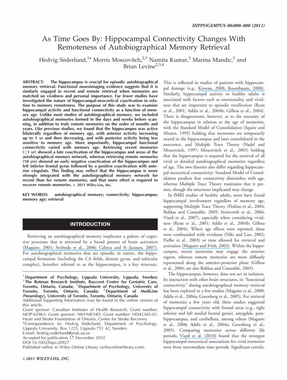
each VOI (see Supporting Information Table 2). Variation inhippocampal activity across time period, laterality, and the ante-rior–posterior axis was assessed by a three-way repeated measuresANOVA. There were main effects of Laterality (left > right)and Axis (anterior > posterior), and interactions between Timeand Axis, and Laterality and Axis. As can be seen in Figure 1,the Time 3 Axis interaction was due to increased activationwith remoteness in the anterior hippocampus bilaterally, exceptfor Time 4, and slightly decreasing activation with remotenessin the posterior hippocampus. In the anterior hippocampus, theleft hemisphere showed significant differences between timeperiods: 1 w < 1 m 5 1 yr (Ps � 0.05), and the right betweentime periods: 1 w < 1 yr > 10 yrs (Ps < 0.05). In the posteriorhippocampus, there was a slight but non-significant decrease inthe left hemisphere (P 5 0.09), and a significant decrease in theright hemisphere [F(1, 11) 5 6.7, P < 0.05], although none ofthe pair-wise comparisons between time periods was significant,suggesting a weak effect of time overall.
TABLE 2.
Brain Areas Differing in Activity Between Time Periods (P < 0.001)
Region Cluster size (ml) BA X Y Z t
1 w < 1 m
R med fro gyr/ant cing 183 32 18 9 47 6.64
R parahippocampus/hippocampus* 125 30 25 238 21 5.79
1 w > 1 yr
R inf par lob 202 40 48 243 29 9.40
R precuneus 1,330 7 6 269 38 7.23
L precuneus 347 7 213 273 35 7.16
1 w > 10 yrs
BL cingulate gyr 457 23 28 222 31 12.44
R thal 199 23 227 0 8.16
R precuneus 672 7 6 268 38 5.80
1 m > 1 yr
R paracentral lobule 295 4 8 239 67 4.73
L precuneus 165 7 222 258 49 6.10
L precuneus 154 7 224 265 28 5.23
R precuneus 299 31 12 266 23 6.45
1 m > 10 yrs
L fusiform 197 37 238 253 26 6.07
R cing gyr 234 31 13 255 27 6.52
R midd temp 166 19 34 260 11 5.45
R midd temp gyr 330 37 48 266 7 6.55
L cerebellar tonsil 599 25 247 237 7.00
L cerebellum 331 219 250 237 5.83
R declive 200 19 255 214 6.40
1 yr > 10 yrs
R cing gyr 218 16 23 27 6.60
R parahippocampus* 99 35 16 223 217 5.27
1 yr < 10 yrs
L inf par lobule 185 40 242 261 42 5.56
Coordinates are in standardized space of Talairach and Tournoux (1988). T values > 4.8 are significant at P 5 0.005; T values > 5.8 at P 5 0.0001. Positive t valuessuggest greater activity for the first of the two compared time periods, and negative t values suggest greater activity for the second.BA, Brodmann area; L, left; R, right; med, medial; fro, frontal; gyr, gyrus; ant, anterior; cing, cingulate; inf, inferior; par, parietal; BL, bilateral; thal, thalamus;midd, middle; temp, temporal.*Even though cluster threshold is 150 ml, these areas are included since the hippocampal region is a primary focus in this study.
HIPPOCAMPAL CONNECTIVITY IN RECENT AND REMOTE RETRIEVAL 5
Hippocampus

The Laterality 3 Axis interaction was due to more activityin the left than the right posterior hippocampus [F(1, 11) 515.7, P < 0.005], but similar activity in the left and rightanterior hippocampus (F < 1). Overall, there was a strikingresemblance in activation patterns across hemispheres, suggest-ing a more crucial role of anterior–posterior axis location thanlaterality in terms of memory remoteness.
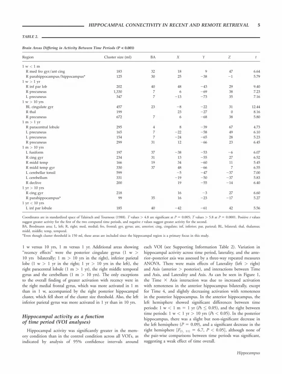
Functional connectivity of the hippocampusduring autobiographical memory retrieval (PLS)
A task PLS yielded a single latent variable that distinguishedbetween autobiographical memory retrieval across all time peri-ods and the odd/even task (P < 0.0001; it accounted for 68%of the cross-correlation variance). This pattern corresponded tothe main effect of autobiographical memory retrieval reportedabove for the univariate analysis. There were no further latentvariables. Our main goal in applying PLS was to examinedifferences in hippocampal-neocortical coactivation, functionalconnectivity, as a function of time period, which may be inde-pendent from level of hippocampal activity across time periods.The most reliable active hippocampal voxel derived from thetask PLS was noted in the left hemisphere (228, 212, 216;bootstrap ratio: 27.2), in the anterior sector of the hippocam-pus. This served as the seed voxel (see Fig. 2A) in the seedPLS, assessing functional connectivity between this hippocam-pal voxel and the rest of the brain.
The first significant latent variable showed overall similaritiesbetween time periods (P < 0.0001; 43% of cross-correlationvariance), including functional connections between the hippo-campal voxel and various areas of the autobiographical memorynetwork, such as mainly left prefrontal structures (e.g., insula,middle frontal cortex) and mediotemporal areas (hippocampus,parahippocampus). Of greater theoretical interest, an additionalsignificant latent variable showed differential connectivity pat-
terns across the 4 time periods of this voxel, dissociating timeperiods 1–3 (1 w – 1 yr) and time period 4 (10 yrs; P 50.006; 6% of cross-correlation variance). Figure 2 shows thecorrelations between activity in the seed and the rest of thebrain during retrieval from the 4 time periods as a function oflag (i.e., scanning epoch), with 95% CIs yielded by the boot-strap procedure. There were significant associations betweentime period and patterns of hippocampal connectivity, but thesediffered according to lag. There were no significant correlationsat lag 3 (6 s), so these data are not reported. For time periods
FIGURE 1. BOLD % change (mean and SD) in the hippocam-pus during autobiographical memory retrieval as compared to theodd/even task, as a function of memory age and hippocampal loca-tion. L, Left; Ant, Anterior; Post, Posterior; R, Right. Activity wasstrikingly similar within location across hemispheres, whereas itdiffered along the hippocampal axis within the same hemisphere. Inthe anterior hippocampus, most significantly in the right, activityincreased somewhat for memories up to 1 yr back and thendecreased, whereas there were no significant differences in the poste-rior hippocampus between time periods.
FIGURE 2. Results of seed PLS indicating coactivationsbetween (A) the left anterior hippocampal seed and (B) the rest ofthe brain as a function of memory age. The line figure indicatescorrelations between seed activity and activity in other brainregions across lags (time in seconds) as a function of memory age.The correlations are represented on brain bootstrap plots (below),with yellow voxels indicating positive bootstrap ratios, and bluevoxels indicating negative bootstrap ratios. The right side of thebrain images represents the right hemisphere. The sign of the cor-relation or bootstrap ratio alone is arbitrary; the direction of theassociation between activity in the seed voxel and other regions isdetermined by the correspondence of the sign of the correlationwith the sign of the bootstrap ratio. At 4 and 8 s, there was a neg-ative coactivation (i.e., negative correlation; positive bootstrapratios) between the left anterior hippocampus and the left inferiorfrontal gyrus, the left superior temporal gyrus and the right cuneusfor the 10 yrs condition. This coactivation was positive for themore recent time periods, but it did not reach significance. At 10s for the 10 yrs condition, there was a positive coactivation(i.e., negative correlation, negative bootstrap ratios) with the ante-rior cingulate bilaterally. During retrieval of the more recent mem-ories, the left hippocampus showed later (12–14 s) coactivationswith prefrontal areas, the precuneus, and caudate (see Table 3 fora summary of all areas). *Significant correlation where the 95% CIdoes not include 0.
6 SODERLUND ET AL.
Hippocampus
1–3 (1 w to 1 yr), at 12–14 s into retrieval, the hippocampuswas functionally connected to several midline posterior and fron-tal structures (see negative saliences in lags 6 and 7 in Table 3/blue areas in Fig. 2B), including the anterior and posterior cin-gulate, precuneus, and anteromedial prefrontal regions as well asthe bilateral caudate and the hippocampus itself. This patternwas not observed for time period 4 (10 yrs), where hippocampalactivation was negatively correlated with activation in the left in-ferior frontal gyrus, the left superior temporal gyrus, and theright cuneus at early lags (4–8 s). At 10 s, the pattern forthis time period shifted such that there was a positive hippo-campal/anterior cingulate coactivation. These results suggestthat the hippocampus has different functional connectionswhen retrieving events from 1 week and up to 1 yr ago ascompared to 10 yrs ago.
DISCUSSION
This study examined the neural correlates of retrieving auto-biographical memories of varying remoteness, including a finergrained analysis of recent memories than in previous studies.
We found that, in addition to activation of the autobiographi-cal memory network for all time periods, the hippocampus wasactivated bilaterally regardless of memory age. A dissociationwas observed between the anterior and posterior parts of thehippocampus bilaterally, with activity in the anterior partincreasing for memories up to 1 yr old, and then decreasing(but not below the level of the 1-week old memories), andactivity in the posterior hippocampus showing a slight butnon-significant decrease over time. This dissociation along thehippocampal axis was strikingly similar across hemispheres.Finally, the hippocampus showed a different pattern of coacti-vation with other brain areas when retrieving remote (10 yrs)as compared to more recent autobiographical memories(1 week to 1 yr back).
This study extends previous findings of hippocampal activa-tion regardless of memory age (Ryan et al., 2001; Gilboa et al.,2004; Piolino et al., 2004; Rekkas and Constable, 2005; Stein-vorth et al., 2006; Viard et al., 2007) to a fuller autobiographi-cal time course extending from the very recent past (1 week) tothe remote past (10 yrs). Previous research suggests hippocam-pal activation can be expected during memories this recent (~2weeks; Hassabis et al., 2007). The only other study comparingmemories as recent as ours to remote memories (2.5 days old
TABLE 3.
Brain Structures Functionally Connected to the Left Hippocampal Seed as a Function of Lag and Bootstrap Ratio Direction
Brain structure Number of voxels X Y Z Bootstrap ratio
Lag 2: 4 s: positive ratio
L inferior frontal gyrus 43 248 23 14 6.02
Lag 4: 8 s: positive ratio
L superior temporal gyrus 12 239 247 18 4.02
R cuneus 18 13 273 8 4.44
Lag 5: 10 s: negative ratio
Anterior cingulate (BL) 10 2 34 12 24.38
Lag 6: 12 s: negative ratio
R precuneus, BA 7 11 6 276 39 25.63
L middle frontal gyrus, BA 8 14 232 27 37 24.89
R precuneus, BA 7 20 10 264 37 24.81
R cingulate gyrus, BA 31 13 6 228 40 24.76
L precuneus, BA 31 12 224 274 23 24.75
R medial frontal gyrus, BA 9 21 5 50 15 24.65
L superior/middle frontal gyrus, BA 10 21 227 47 15 24.55
R anterior cingulate, BA 32 15 3 37 14 24.46
Lag 7: 14 s: negative ratio
L hippocampus 22 233 224 28 26.03
L superior/medial frontal gyrus, BA 10 103 218 49 2 25.98
R precuneus, BA 7 77 6 264 35 25.57
L precuneus 35 226 257 30 25.17
R caudate body 13 7 16 11 24.87
L caudate body 24 210 20 16 24.76
R superior frontal gyrus 48 19 11 48 24.63
Coordinates are reported in Talairach space.The bootstrap ratio is the parameter estimate for that voxel over its standard error and is proportional to a z score.L, left; R, right; BA, Brodmann area; BL, bilateral.
HIPPOCAMPAL CONNECTIVITY IN RECENT AND REMOTE RETRIEVAL 7
Hippocampus

vs. 8 yrs old; Rekkas and Constable, 2005) observed left-lateral-ized hippocampal activation for the recent memories, and bilat-eral activation for the remote. This was interpreted as supportof Multiple Trace Theory, since more remote memories shouldhave more traces and thus provoke a larger activation. Variationin laterality has been observed in a few studies with the lefthippocampus active throughout and the right varying in activ-ity as a function of memory age (Maguire and Frith, 2003;Addis et al., 2004b; Viard et al., 2007). We did not observesuch a laterality effect, but instead observed a bilateral activa-tion across time-periods with an anterior–posterior differentia-tion of activation intensity within the hippocampus that wassimilar across hemispheres.
A functional differentiation between the anterior and poste-rior hippocampus has previously been suggested using labora-tory materials as a function of stimulus novelty (Strange et al.,1999; Poppenk et al., 2010), stimulus material/modality (Smallet al., 2001), memory function (Moser and Moser, 1998), andmemory process (Lepage et al., 1998). Similarly, for autobio-graphical memory, we found that both sectors are activatedduring retrieval, but there may be differences in the characteris-tics of that retrieval that engage them differently. The anteriorpart showed an increase in activity with remoteness up to 1 yrback in time, which thereafter decreased (although not belowthe level of the 1-week-old memories). This finding corre-sponds to the sensitivity of the anterior, but not posterior,hippocampus to reduced activation with repetition of autobio-graphical memories that are matched for age (Svoboda andLevine, 2009). Similarly, Gilboa and colleagues (2004) foundthat hippocampal activation associated with recent memories(up to 5 yrs old;1.75 6 1.61 on average) was clustered in theanterior sector of the hippocampus, whereas hippocampal acti-vation associated with remote memories (mean years of age 532.3) was distributed throughout the anterior-posterior axis.Our results of anterior hippocampal activity increasing for upto 1-yr-old memories and then decreasing somewhat for 10-yr-old memories are consistent with these findings.
For very recent memories, our findings also correspond tothose of Rekkas and Constable (2005), who observed left pos-terior hippocampal activity (222, 224, 29) for 2.5 days oldmemories, as we found greater posterior than anterior activityfor the 1 week old memories in the left hemisphere. For moreremote memories (10 yrs) we observed, on average, greater an-terior than posterior activity, although posterior activity wasalso present. This is in accordance with Rekkas and Constable(2005) where remote memories (from childhood and teenageyears) activated the anterior hippocampus bilaterally (y: 213/212). These findings may, however, appear contradictory toGilboa et al. (2004) who suggested anterior activation forrecent memories, but what they call ‘‘recent’’ (up to 5 yrs)partly overlap with what we call ‘‘remote’’ (10 yrs). It is in gen-eral difficult to compare these studies and ours to each other asthey differ on several dimensions such as memory age, retrievalcues and duration, control conditions, and pre-scan procedures.What can be concluded, nevertheless, is that the hippocampus isactivated during retrieval from all time periods, but that there
may be regional variation as a function of memory age, and, aswith retrieval of memories for laboratory materials, with otheraspects of the retrieval process and the memory that is retrieved.
Although many functional neuroimaging studies, includingthis one, have shown similarity in hippocampal activation asso-ciated with episodic autobiographical memory across recent andremote time periods, functional neuroimaging studies can onlybe regarded as complementary to lesion studies concerning thenecessity of a given region to a given function. On the otherhand, a human lesion study cannot address questions concern-ing real-time functional connectivity that also is crucial to theunderstanding of autobiographical memory retrieval. The pat-tern of hippocampal engagement with neocortical elements ofthe autobiographical memory network likely modulates the phe-nomenological experience associated with retrieving an autobio-graphical memory (cf. Addis et al., 2004b; Viard et al., 2010).Consistent with Multiple Trace Theory, we found that both thehippocampus and the autobiographical memory network wereactive regardless of memory age, but the nature of the hippo-campal-neocortical interactions was not invariant across timeperiods. Autobiographical memory retrieval is extended overtime and consists of retrieving the memory and then elaboratingon it. Generally, retrieval is initially associated with mainly (left)frontal activity and then with posterior temporal and occipitalactivity during elaboration and maintenance of the memory(Conway et al., 2001). We found that during retrieval of memo-ries from 1 w to 1 yr old, the hippocampus was coactivatedwith both midline frontal and posterior elements of the autobio-graphical memory network late into the retrieval process (12–14s). Similar areas (i.e., precuneus, left prefrontal) peaked around12 s in an autobiographical memory retrieval study of Daselaaret al. (2008) during the elaboration phase of the memory, and itis possible that hippocampal coactivation with these areasreflects a higher extent of elaboration and reliving of the morerecent autobiographical memories than of the most remotememories. By contrast, for the 10 yrs old memories, the hippo-campus showed negative interaction with the left inferior fron-tal, left superior temporal, and right precuneus at the earlyepochs, followed by positive coactivation with the anterior cin-gulate, perhaps reflecting an initial absence of memory recovery,and thereafter increased retrieval effort (Schacter et al., 1996)and reliving during the elaboration of the memory (Daselaaret al., 2008). Negative functional connectivity between the hip-pocampus and areas of the autobiographical memory networkhas been observed in previous research where memories olderthan 1 yr were used (Addis et al., 2004a). The dissociation infunctional connectivity of the hippocampus between the mostremote time period and the others was paralleled by decreasingvisualizability and re-experiencing of autobiographical memoriesover time. This is in line with Viard et al. (2010) who observedmore interaction between medial temporal lobe regions andneocortical areas during retrieval of currently emotional memo-ries from intermediate time periods, than during less emotionalmemories from the most recent and most remote time periods.
The functional connectivity of the hippocampus in relationto autobiographical memory remoteness, and changes in the
8 SODERLUND ET AL.
Hippocampus

quality of those memories with time and other factors, providenew information about the relation of memory age to hippo-campal-neocortical interactions. Evidence of continued connec-tivity between the hippocampus and neocortical structuresacross time argues against the Standard Model of Consolidationwhich predicts reduced connectivity with increasing memoryage. Although the sustained connectivity with time is consistentwith Multiple Trace Theory, variations in the pattern ofconnectivity indicate that the process of retrieving remotememories involves hippocampal-neocortical interactions thatare more complex than had previously been observed. Whereasfunctional connectivity between the hippocampus and corticalareas for the more recent memories was positive throughout theretrieval period, the connectivity for remote memories changeddirection, from negative at the beginning, to positive at the end.A possible interpretation of these findings is that for recentmemories, the cue (i.e., event title) activates the hippocampallymediated memory from the very beginning, whereas for the moreremote memories, the cue may not be as effective immediately;first, it may be necessary to determine precisely which event thecue specifies without interference from hippocampally-mediatedmemories. Once that has been settled, the process proceeds in asimilar fashion as for recent events. Further studies are needed totest this interpretation and, more broadly, to determine whetherthe differences in pattern of connectivity observed in this studyare a reliable distinguishing feature between recent and remotememories.
Turning to other regions, the autobiographical memory net-work (Maguire, 2001; Svoboda et al., 2006) was activatedthroughout all time periods, although there was some varia-tion in areas and extent. Midline posterior regions, includingthe precuneus and posterior cingulate, were more activatedwith recent memories. A positive association between precu-neus activation and recency is in line with earlier research(Niki and Luo, 2002; Rekkas and Constable, 2005; Viardet al., 2007). This structure has been associated with imagery(Fletcher et al., 1995), vivid and context-rich retrieval, re-experiencing (Gilboa et al., 2004) and retrieval success (Kapuret al., 1995), all of which are likely higher for recent memo-ries than for remote ones. The posterior cingulate has previ-ously been associated with recency (Niki and Luo, 2002;Piefke et al., 2003), and is thought to be involved in integrat-ing self-referential stimuli into a person’s autobiographical con-text (Northoff and Bermpohl, 2004). Less frequently, recencyeffects were also observed in the inferior parietal lobe, the rightparacentral lobule, the right middle temporal gyrus and thecerebellum. The right medial frontal/paracingulate region wasmore active for 1-month-old memories as compared to 1-weekold memories. The 1-month old memories were personally sa-lient, as reflected in ratings of re-experiencing and importanceat the time of the event. This finding is difficult to accommo-date within the autobiographical memory literature as thisregion is not strongly associated with autobiographical memoryimportance or vividness and is in fact more strongly activatedin conjunction with laboratory than autobiographical materials(Cabeza et al., 2004).
The memory stimulus generation in this study consisted ofparticipants generating events 2 days before scanning ratherthan only once in the scanner, which may have contaminatedrecollection during scanning. Earlier functional neuroimagingstudies, however, suggest that hippocampal activation associatedwith autobiographical stimuli holds in spite of pre-scanrehearsal (Maguire et al., 2001; Ryan et al., 2001; Levine andSvoboda, 2009). The same holds for re-encoding in retrievalstudies. Each time a memory is retrieved it may also be re-encoded, which would give rise to encoding-related hippocam-pal activity regardless of memory age. Confronting similarproblems, Gilboa et al. (2004) showed that such effects aresmall relative to retrieval effects. Likewise, in our study, ifre-encoding accounted for the observed hippocampal activationthat accompanied retrieval of recent and remote memories, nodifferences between them should have emerged since re-encod-ing would be quite similar for all events.
Our data indicate that the hippocampus is active duringepisodic autobiographical memory retrieval for events occurringone week and up to 10 yrs ago. Although both the hippocam-pus and the rest of the autobiographical memory network areactive across all time periods when considered in isolation, theway different parts of the network interact with one anotherdiffers as a function of memory remoteness and variations inthe quality of those memories with time. Studying the hippo-campus alone is an initial step in determining how memory isconsolidated in the brain, but exploring its interactions withthe rest of the brain is the next necessary step to understandfully a memory’s journey from inception to retrieval.
Acknowledgments
The authors are grateful to Marilyne Ziegler for program-ming support and Allison Mackey for image analysis assistance.
REFERENCES
Addis DR, McIntosh AR, Moscovitch M, Crawley AP, McAndrewsMP. 2004a. Characterizing spatial and temporal features of auto-biographical memory retrieval networks: a partial least squaresapproach. Neuroimage 23:1460–1471.
Addis DR, Moscovitch M, Crawley AP, McAndrews MP. 2004b.Recollective qualities modulate hippocampal activation duringautobiographical memory retrieval. Hippocampus 14:752–762.
Cabeza R, Prince SE, Daselaar SM, Greenberg DL, Budde M, DolcosF. 2004. Brain activity during episodic retrieval of autobiographicaland laboratory events: an fMRI study using a novel photo para-digm. J Cogn Neurosci 16:1583–1594.
Cabeza R, St Jacques P. 2007. Functional neuroimaging of autobio-graphical memory. Trends Cogn Sci 11:219–227.
Conway MA, Pleydell-Pearce CW, Whitecross SE. 2001. The neuro-anatomy of autobiographical memory: A slow cortical potentialstudy of autobiographical memory retrieval. J Mem Lang 45:493–524.
Cox RW, Hyde JS. 1997. Software tools for analysis and visualizationof fMRI data. NMR Biomed 10:171–178.
Daselaar SM, Rice HJ, Greenberg DL, Cabeza R, LaBar KS, RubinDC. 2008. The spatiotemporal dynamics of autobiographical
HIPPOCAMPAL CONNECTIVITY IN RECENT AND REMOTE RETRIEVAL 9
Hippocampus

memory: Neural correlates of recall, emotional intensity, and reliv-ing. Cereb Cortex 18:217–229.
First M, Spitzer R, Gibbon M, Williams J. 1995. Structured ClinicalInterview for DSM-IV Axis 1 Disorders (SCID-I/P). New York: Bio-metrics Research Department, New York State Psychiatric Institute.
Fletcher PC, Frith CD, Baker SC, Shallice T, Frackowiak RS, DolanRJ. 1995. The mind’s eye: Precuneus activation in memory-relatedimagery. Neuroimage 2:195–200.
Ghirardi O, Cozzolino R, Guaraldi D, Giuliani A. 1995. Within- andbetween-strain variability in longevity of inbred and outbred ratsunder the same environmental conditions. Exp Gerontol 30:485–494.
Gilboa A, Winocur G, Grady CL, Hevenor SJ, Moscovitch M. 2004.Remembering our past: Functional neuroanatomy of recollection ofrecent and very remote personal events. Cereb Cortex 14:1214–1225.
Greenberg DL, Rice HJ, Cooper JJ, Cabeza R, Rubin DC, Labar KS.2005. Co-activation of the amygdala, hippocampus and inferiorfrontal gyrus during autobiographical memory retrieval. Neuropsy-chologia 43:659–674.
Hassabis D, Kumaran D, Maguire EA. 2007. Using imagination tounderstand the neural basis of episodic memory. J Neurosci27:14365–14374.
Kapur S, Craik FIM, Jones C, Brown GM, Houle S, Tulving E. 1995.Functional role of the prefrontal cortex in retrieval of memories: APET study. NeuroReport 6:1880–1884.
Kirwan CB, Bayley PJ, Galvan VV, Squire LR. 2008. Detailed recol-lection of remote autobiographical memory after damage to themedial temporal lobe. Proc Natl Acad Sci USA 105:2676–2680.
Lepage M, Habib R, Tulving E. 1998. Hippocampal PET activationsof memory encoding and retrieval: The HIPER model. Hippocam-pus 8:313–322.
Maguire EA. 2001. Neuroimaging studies of autobiographical eventmemory. Philos Trans R Soc Lond B Biol Sci 356:1441–1451.
Maguire EA, Frith CD. 2003. Lateral asymmetry in the hippocampalresponse to the remoteness of autobiographical memories. J Neuro-sci 23:5302–5307.
Maguire EA, Henson RN, Mummery CJ, Frith CD. 2001. Activity inprefrontal cortex, not hippocampus, varies parametrically with theincreasing remoteness of memories. Neuroreport 12:441–444.
Maguire EA, Mummery CJ, Buchel C. 2000. Patterns of hippocam-pal-cortical interaction dissociate temporal lobe memory subsys-tems. Hippocampus 10:475–482.
McIntosh AR, Bookstein FL, Haxby JV, Grady CL. 1996. Spatialpattern analysis of functional brain images using partial leastsquares. Neuroimage 3:143–157.
Moscovitch M, Rosenbaum RS, Gilboa A, Addis DR, Westmacott R,Grady C, McAndrews MP, Levine B, Black S, Winocur G, et al.2005. Functional neuroanatomy of remote episodic, semantic andspatial memory: A unified account based on multiple trace theory.J Anat 207:35–66.
Moser MB, Moser EI. 1998. Functional differentiation in the hippo-campus. Hippocampus 8:608–619.
Northoff G, Bermpohl F. 2004. Cortical midline structures and theself. Trends Cogn Sci 8:102–107.
Nadel L, Moscovitch M. 1997. Memory consolidation, retrogradeamnesia and the hippocampal complex. Curr Opin Neurobiol7:217–227.
Niki K, Luo J. 2002. An fMRI study on the time-limited role of themedial temporal lobe in long-term topographical autobiographicmemory. J Cogn Neurosci 1:500–507.
Ogawa S, Lee TM, Kay AR, Tank DW. 1990. Brain magnetic reso-nance imaging with contrast dependent on blood oxygenation.Proc Natl Acad Sci USA 87:9868–9872.
Piefke M, Weiss PH, Zilles K, Markowitsch HJ, Fink GR. 2003.Differential remoteness and emotional tone modulate the neuralcorrelates of autobiographical memory. Brain 126:650–668.
Piolino P, Giffard-Quillon G, Desgranges B, Chetelat G, Baron JC,Eustache F. 2004. Re-experiencing old memories via hippocampus:A PET study of autobiographical memory. Neuroimage 22:1371–1383.
Poppenk J, McIntosh AR, Craik FI, Moscovitch M. 2010. Past experi-ence modulates the neural mechanisms of episodic memory forma-tion. J Neurosci 30:4707–4716.
Rekkas PV, Constable RT. 2005. Evidence that autobiographic mem-ory retrieval does not become independent of the hippocampus:An fMRI study contrasting very recent with remote events. J CognNeurosci 17:1950–1961.
Rosenbaum RS, Moscovitch M, Foster JK, Schnyer DM, Gao F, Kova-cevic N. 2008. Patterns of autobiographical memory loss in medial-temporal lobe amnesic patients. J Cogn Neurosci 20:1490–1506.
Rubin DC, Schulkind MD. 1997. The distribution of autobiographi-cal memories across the lifespan. Mem Cogn 25:859–866.
Ryan L, Nadel L, Keil K, Putnam K, Schnyer D, Trouard T, Mosco-vitch M. 2001. Hippocampal complex and retrieval of recent andvery remote autobiographical memories: Evidence from functionalmagnetic resonance imaging in neurologically intact people.Hippocampus 11:707–714.
Schacter DL, Alpert NM, Savage CR, Rauch SL, Albert MS. 1996.Conscious recollection and the human hippocampal formation:Evidence from positron emission tomography. Proc Natl Acad SciUSA 93:321–325.
Small SA, Nava AS, Perera GM, DeLaPaz R, Mayeux R, Stern Y.2001. Circuit mechanisms underlying memory encoding andretrieval in the long axis of the hippocampal formation. NatNeurosci 4:442–449.
Squire LR, Alvarez P. 1995. Retrograde amnesia and memory consoli-dation: A neurobiological perspective. Curr Opin Neurobiol5:169–177.
Stark CE, Squire LR. 2001. When zero is not zero: The problem ofambiguous baseline conditions in fMRI. Proc Natl Acad Sci USA98:12760–12766.
Steinvorth S, Corkin S, Halgren E. 2006. Ecphory of autobiographicalmemories: An fMRI study of recent and remote memory retrieval.Neuroimage 30:285–298.
Strange BA, Fletcher PC, Henson RN, Friston KJ, Dolan RJ. 1999.Segregating the functions of human hippocampus. Proc Natl AcadSci USA 96:4034–4039.
Svoboda E, McKinnon MC, Levine B. 2006. The functional neuroan-atomy of autobiographical memory: A meta-analysis. Neuropsycho-logia 44:2189–2208.
Svoboda E, Levine B. 2009. The effects of rehearsal on the functionalneuroanatomy of episodic autobiographical and semantic remem-bering: a functional magnetic resonance imaging study. J Neurosci29:3073–3082.
Talairach J, Tournoux P. 1988. Co-Planar Stereotaxic Atlas of theHuman Brain. New York: Thieme Medical Publishers.
Viard A, Piolino P, Desgranges B, Chetelat G, Lebreton K, Landeau B,Young A, De La Sayette V, Eustache F. 2007. Hippocampal activa-tion for autobiographical memories over the entire lifetime inhealthy aged subjects: An fMRI study. Cereb Cortex 17:2453–2467.
Viard A, Lebreton K, Chetelat G, Desgranges B, Landeau B, Young A,De La Sayette V, Eustache F, Piolino P. 2010. Patterns of hippo-campal-neocortical interactions in the retrieval of episodic autobio-graphical memories across the entire life-span of aged adults.Hippocampus 20:153–165.
10 SODERLUND ET AL.
Hippocampus