dna-based species level detection of glomeromycota : one pcr primer set for all arbuscular...
TRANSCRIPT
Research
212 New Phytologist (2009) 183: 212–223 © The Authors (2009)212 www.newphytologist.org Journal compilation © New Phytologist (2009)
Blackwell Publishing LtdOxford, UKNPHNew Phytologist0028-646X1469-8137© The Authors (2009). Journal compilation © New Phytologist (2009)283510.1111/j.1469-8137.2009.02835.xMarch 200900212???223???Original ArticleXX XX
DNA-based species level detection of Glomeromycota: one PCR primer set for all arbuscular mycorrhizal fungi
Manuela Krüger, Herbert Stockinger, Claudia Krüger and Arthur SchüßlerLudwig-Maximilians-University Munich, Dept Biology I, Genetics, Großhaderner Strasse 4, D–82152 Planegg-Martinsried, Germany
Summary
• At present, molecular ecological studies of arbuscular mycorrhizal fungi (AMF)are only possible above species level when targeting entire communities. To improvemolecular species characterization and to allow species level community analyses inthe field, a set of newly designed AMF specific PCR primers was successfully tested.• Nuclear rDNA fragments from diverse phylogenetic AMF lineages weresequenced and analysed to design four primer mixtures, each targeting one bindingsite in the small subunit (SSU) or large subunit (LSU) rDNA. To allow species resolution,they span a fragment covering the partial SSU, whole internal transcribed spacer(ITS) rDNA region and partial LSU.• The new primers are suitable for specifically amplifying AMF rDNA from materialthat may be contaminated by other organisms (e.g., samples from pot culturesor the field), characterizing the diversity of AMF species from field samples, andamplifying a SSU-ITS-LSU fragment that allows phylogenetic analyses with specieslevel resolution.• The PCR primers can be used to monitor entire AMF field communities, based ona single rDNA marker region. Their application will improve the base for deepsequencing approaches; moreover, they can be efficiently used as DNA barcodingprimers.
Author for correspondence:Manuela KrügerTel: +49 89 2180 74714Email: [email protected]
Received: 12 December 2008Accepted: 23 February 2009
New Phytologist (2009) 183: 212–223 doi: 10.1111/j.1469-8137.2009.02835.x
Key words: arbuscular mycorrhizal fungi (AMF), DNA barcoding, ITS region, LSU rRNA gene, molecular community analyses, rDNA, species level resolution, specific primers.
Introduction
Arbuscular mycorrhizal fungi (AMF) are associated with70–90% of land plants (Smith & Read, 2008) in a symbiosiscalled arbuscular mycorrhiza (AM), that has existed for> 400 million yr (Parniske, 2008; Schüßler et al., 2009). Theeconomic and ecological importance of these ancient biotrophicplant symbionts is therefore obvious. Arbuscular mycorrhizalfungi transfer inorganic nutrients and water to the plant andreceive carbohydrates in exchange. By driving this bidirectionalnutrient transport between soil and plants, they are highlyrelevant for global phosphorus (P), nitrogen (N) and CO2cycles. Moreover, they affect directly and indirectly thediversity and productivity of land-plant communities (vander Heijden et al., 1998) by their central role at the soil–plantinterface (van der Heijden et al., 2008). They can also improvehost plant pathogen resistance (Vigo et al., 2000; de la Penaet al., 2006) and drought stress tolerance (Michelson &Rosendahl, 1990; Aroca et al., 2007).
Despite the enormous role of AMF in the entire terrestrialecosystem, their biodiversity in relation to functional aspects
is little understood. Most of the 214 currently describedspecies (www.amf-phylogeny.com) are characterized onlyby spore morphology and the majority have not yet beencultured. Moreover, from molecular ecological studies weknow that the species described represent only a small fractionof the existing AMF diversity (Kottke et al., 2008; Öpik et al.,2008). Problems with identification of AMF result fromtheir hidden, biotrophic lifestyle in the soil, few morpho-logical characters, and the potential formation of dimorphicspores. This led to many AMF species, phylogeneticallybelonging to different orders, being placed in one genus(Glomus) and, conversely, individual species forming differentspore morphs being described as members of different orders.
Another drawback of morphologically monitoring AMFby their resting spores (Oehl et al., 2005; Wang et al., 2008)is that the presence of spores may not reflect a symbioticallyactive organism community. Furthermore, many speciescannot be reliably identified at all from heterogeneous fieldsamples, and when identifying described species (likely torepresent less than 5% of the existing species diversity) similarmorphotypes may be erroneously determined as a single species.
© The Authors (2009) New Phytologist (2009) 183: 212–223Journal compilation © New Phytologist (2009) www.newphytologist.org
Research 213
To reveal functional and ecological aspects of distinct AMFcommunities associated with different plants and/or underdifferent environmental conditions it is essential to detectAMF communities in the field on the species level. However,there are as yet no unbiased methods for this purpose, notonly for morphological identification but also for molecularmethods. Principally, DNA sequence based methods are mostuseful for detecting organisms at different community levels,but for ecological work they also depend on reliable baselinedatabases and tools. For example, fingerprinting methodssuch as random amplification of polymorphic DNA (RAPD),inter-simple sequence repeat PCR (ISSR) and amplifiedfragment length polymorphism (AFLP) are expected to be errorprone in uncharacterized environments because of too many‘unknowns’ in the background, which hampers interpretationof specificity (Mathimaran et al., 2008). A similar problemexists for DNA array techniques. Nevertheless, suitablemolecular methods are crucial to overcome the limitationsof morphological identification (Walker & Schüßler, 2004;Walker et al., 2007; Gamper et al., 2009; Stockinger et al., 2009).
But how are DNA or RNA sequence data for communityanalyses obtained and how can the current limitations ofmolecular tools be overcome? Molecular characterization ofAMF is in most cases achieved by PCR on DNA from rootsof host plants, spores or soil samples. Several primers targetingthe rDNA regions as molecular marker were claimed to beAMF specific. Most of these amplify only a restricted numberof glomeromycotan taxa or DNA of nontarget organisms. Themost comprehensive taxon sampling for the Glomeromycotacovers the small subunit (SSU) rDNA region (Schüßleret al., 2001a,b), for which a new, AMF specific primer pairwas recently published (AML1 and AML2; Lee et al., 2008).Unlike the often used AM1 primer (Helgason et al., 1998) itis perhaps suitable to amplify sequences from all AMF taxa,but the SSU rDNA is inadequate for species resolution ofAMF. Inclusion of the internal transcribed spacer (ITS) andthe large subunit (LSU) rDNA region allows both robustphylogenetic analyses and species level resolution (Gamper et al.,2009; Stockinger et al., 2009).
The available public database sequences are scatteredthrough SSU, ITS and LSU rDNA subsets with varyinglengths, often only 500–800 bp. In most cases this does notallow species level analyses, and short sequences obtainedwith primers that have inaccurately defined specificity mayresult in errors. For example, some short database sequenceslabelled as Gigaspora (Jansa et al., 2003) cluster with those ofGlomus versiforme BEG47 (Diversisporaceae) (Gamper et al.,2009). Because of the relatively few LSU sequences in thepublic databases, the design of improved primers is challengingor even impossible. We therefore sequenced the ITS regionand the 5′ part of the LSU rDNA of a set of well-characterized,but phylogenetically diverse AMF, and designed new primersfrom the resulting database. These primers are suited toamplify DNA from members of all known glomeromycotan
lineages and, by allowing elaboration of a more accuratebaseline dataset, could be a breakthrough for molecularcommunity analyses of AMF.
Materials and Methods
Fungal and plant material for primer tests
We first tested different samples as DNA templates for PCRto confirm the specificity of the newly designed primers.These included plasmid inserts (Table 1), DNA extractionsfrom single AMF spores and root samples from the Andes(Ecuador) and the Spessart Mountains (Germany). Primerswere tested for specificity by PCR with plasmids carrying rDNAfragments with known sequences. All these plasmids had beenamplified from single spore DNA extracts with the SSUrDNA primer SSUmAf, described here, and the LSU rDNAprimer LR4+2 (modified from LR4; www.aftol.org). Thespecificity of SSUmAf could therefore not be investigated directly.
DNA extraction for primer tests
All vials, tips, beads, solutions, and other equipment usedwere sterile and DNA free.
From cleaned, single AMF spores DNA was extractedwith the Dynabead DNA DIRECT Universal Kit (Invitrogen,Karlsruhe, Germany) as described in Schwarzott & Schüßler(2001).
Roots potentially colonized by AMF were cut into ten0.5 cm pieces and collected in a single 1.5 ml Eppendorf tubecontaining one tungsten carbide bead (diameter 3 mm;Qiagen, Hilden, Germany). They were immediately frozen inliquid N2 within the closed tube, placed in liquid N2 precooledTeflon holders, and ground to a fine powder in a MM2000bead-mill (Retsch, Haan, Germany). Extraction was done byeither an innuPREP Plant DNA Kit (Analytik Jena, Jena,Germany) following the instructions of the manufacturer,or a cetyltrimethylammonium bromide (CTAB) protocolmodified from Allen et al. (2006). For the CTAB protocol,prewarmed extraction buffer (750 µl for 75 mg tissue) wasadded to each sample of frozen, ground tissue, followed byincubation at 60°C for 30 min. Next, one volume of achloroform–isoamylalcohol mixture (24 : 1) was added. Thesamples were centrifuged for 5 min at 2570 g and the upperphase was transferred into a new tube. After addition of 2.5 µlRNase A (10 mg ml−1) this was incubated at 37°C for 30 min.One volume chloroform–isoamylalcohol (24 : 1) was thenadded and the tube was centrifuged as above. The supernatantwas collected and two-thirds volumes of isopropanol added.The samples were incubated at 4°C for 15 min. After centrifu-gation (10290 g for 10 min) the pellet was washed in 70%ethanol, air dried, and eluted in 100 µl of molecular biologygrade H2O. Volumes of 2–5 µl of each DNA extract wereused as PCR template.
New Phytologist (2009) 183: 212–223 © The Authors (2009)www.newphytologist.org Journal compilation © New Phytologist (2009)
Research214
PCR conditions
The Phusion High-Fidelity DNA polymerase 2× mastermix(Finnzymes, Espoo, Finland) was used for PCR with theSSUmAf–LSUmAr or SSUmCf–LSUmBr primer pairs.SSUmCf and LSUmBr were also applied as nested primers(see Fig. 1c). The final concentration of the reaction mixcontained 0.02 U µl−1 Phusion polymerase, 1× Phusion HFBuffer with 1.5 mm MgCl2, 200 µm of each dNTP and0.5 µm of each primer. Thermal cycling was done in anEppendorf Mastercycler Gradient (Eppendorf, Hamburg,Germany) with the following conditions for the first PCR:5 min initial denaturation at 99°C; 40 cycles of 10 sdenaturation at 99°C, 30 s annealing at 60°C and 1 minelongation at 72°C; and a 10 min final elongation. The sameconditions were used for the nested PCR primers except thatthe annealing temperature was 63°C and only 30 cycles werecarried out. The PCR products were loaded on 1% agarosegels (Agarose NEEO; Carl Roth, Karlsruhe, Germany) with1× sodium borate buffer (Brody & Kern, 2004) at 220 V, andvisualized after ethidium bromide staining (1 µg ml−1).
Cloning, restriction fragment length polymorphism (RFLP) and sequencing
Polymerase chain reaction products were cloned with theZero Blunt TOPO PCR Cloning Kit (Invitrogen) followingthe instructions of the manufacturer, except that to reducecosts only one-third of the specified volume of all componentswas used. Only SOC medium for initial bacterial growth aftertransformation was used in the volume as per the instructions.From each cloning we analysed up to 48 clones for correctlength of plasmid inserts. In some instances fewer cloneswere available because of low cloning efficiency. Colony-PCR
was performed with the GoTaq DNA Polymerase (5 U µl−1;Promega, Mannheim, Germany) and modified M13F andM13R primers. To roughly detect intrasporal and intersporalsequence variability in the clones, RFLP was performed in10 µl reaction volume, containing 5 µl colony-PCR product,one of the restriction enzymes Hinf I (1 U), RsaI (1 U), orMboI (0.5 U) and the specific buffer. One or two clones foreach restriction pattern were sequenced, using M13 primers,by the LMU Sequencing Service Unit on an ABI capillarysequencer with the BigDye v3.1 (Applied Biosystems, FosterCity, CA, USA) sequencing chemistry. The sequences wereassembled and edited in seqassem (www.sequentix.de) anddeposited in the EMBL/GenBank/DDBJ databases with theaccession numbers FM876780 to FM876839.
Primer design
For the design of new AMF specific primers a sequencealignment was established with the programs align(www.sequentix.de) and arb (Ludwig et al., 2004). Thealignments contained all AMF sequences present in the publicdatabases and our new data. In total > 1000 AMF sequences,covering all known phylogenetic lineages, were analysed todesign the SSU and LSU rDNA primers. To allow com-parison to the existing SSU rDNA datasets the primers weredesigned to overlap (approx. 250 bp) with the SSU rDNA.We used blast against the public databases and the probematch tool in arb to test the specificity of the newlydesigned primers in silico. For the alignment in the arbdatabase a combination of our new dataset and the 94threlease version of the SILVA database (Pruesse et al., 2007,www.arb-silva.de) was used. The oligonucleotides werethen synthesized as standard primers (25 nmol, desalted)by Invitrogen.
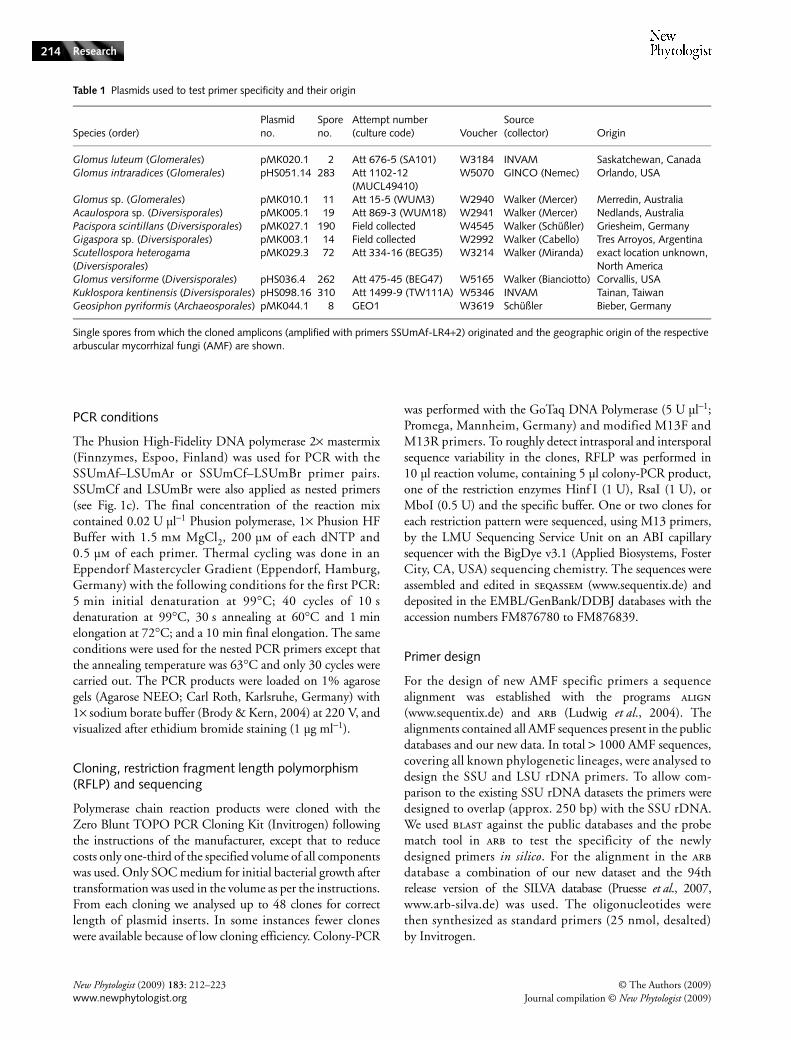
Table 1 Plasmids used to test primer specificity and their origin
Species (order)Plasmid no.
Spore no.
Attempt number (culture code) Voucher
Source (collector) Origin
Glomus luteum (Glomerales) pMK020.1 2 Att 676-5 (SA101) W3184 INVAM Saskatchewan, CanadaGlomus intraradices (Glomerales) pHS051.14 283 Att 1102-12
(MUCL49410)W5070 GINCO (Nemec) Orlando, USA
Glomus sp. (Glomerales) pMK010.1 11 Att 15-5 (WUM3) W2940 Walker (Mercer) Merredin, AustraliaAcaulospora sp. (Diversisporales) pMK005.1 19 Att 869-3 (WUM18) W2941 Walker (Mercer) Nedlands, AustraliaPacispora scintillans (Diversisporales) pMK027.1 190 Field collected W4545 Walker (Schüßler) Griesheim, GermanyGigaspora sp. (Diversisporales) pMK003.1 14 Field collected W2992 Walker (Cabello) Tres Arroyos, ArgentinaScutellospora heterogama (Diversisporales)
pMK029.3 72 Att 334-16 (BEG35) W3214 Walker (Miranda) exact location unknown, North America
Glomus versiforme (Diversisporales) pHS036.4 262 Att 475-45 (BEG47) W5165 Walker (Bianciotto) Corvallis, USAKuklospora kentinensis (Diversisporales) pHS098.16 310 Att 1499-9 (TW111A) W5346 INVAM Tainan, TaiwanGeosiphon pyriformis (Archaeosporales) pMK044.1 8 GEO1 W3619 Schüßler Bieber, Germany
Single spores from which the cloned amplicons (amplified with primers SSUmAf-LR4+2) originated and the geographic origin of the respective arbuscular mycorrhizal fungi (AMF) are shown.
© The Authors (2009) New Phytologist (2009) 183: 212–223Journal compilation © New Phytologist (2009) www.newphytologist.org
Research 215
(a) SSUmAf1 TGGGTAATCTTTTGAAACTTYA...---------------------- SSUmAf2 TGGGTAATCTTRTGAAACTTCA...---------------------- SSUmCf1 ----------------------...--TCGCTCTTCAACGAGGAATC SSUmCf2 ----------------------...TATTGTTCTTCAACGAGGAATC SSUmCf3 ----------------------...TATTGCTCTTNAACGAGGAATC Gl. caledonium BEG20 Y17635 TGGGTAATCTTTTGAAACTTCA...TATTGCTCTTCAACGAGGAATC Gl. mosseae UT101 AY635833, Gl. geosporum BEG11 AJ132664 TGGGTAATCTTTTGAAACTTCA...TATTGCTCTTCAACGAGGAATC Gl. sp. 'intraradices' DAOM197198 AY635831 TGGGTAATCTTTTGAAACTTCA...TATTGCTCTTGAACGAGGAATC Gl. claroideum BEG14 AJ301851 TGGGTAATCTTTTGAAACTTTA...TATCGCTCTTCAACGAGGAATC Gl. luteum SA101 AJ276089 TGGGTAATCTTTKGAAACTTTA...TATCGCTCTTCAACGAGGAATC Ac. laevis AU211 AJ250847 TGGGTAATCTTTTGAAACTTCA...TATTGCTCTTAAACGAGGAATC Ac. longula W3302 AJ306439, Ac. rugosa WV949 Z14005 TGGGTAATCTTTTGAAACTTCA...TATTGCTCTTCAACGAGGAATC Ac. scrobiculata BEG33 AJ306442, Ac. spinosa WV860 Z14004 TGGGTAATCTTTTGAAACTTCA...TATTGCTCTTCAACGAGGAATC Ac. sp. W3424 AJ306440 TGGGTAATCTTTTGAAACTTCA...TATTGCTCTTTAACGAGGAATC Ku. colombiana WV877 Z14006 TGGGTAATCTTTTGAAACTTCA...TATTGCTCTTCAACGAGGAATC Di. spurca ex-type W3239 AJ276077 TGGGTAATCTTTTGAAACTTCA...TATTGCTCTTTAACGAGGAATC Gl. versiforme BEG47 X86687, G. sp. W2423 AJ301863 TGGGTAATCTTTTGAAACTTCA...TATTGCTCTTCAACGAGGAATC Gl. eburneum AZ420 AM713405 TGGGTAATCTTGTGAAACTTCA...TATTGCTCTTCAACGAGGAATC Gl. eburneum AM713406, Gl. fulvum AM418548, Ot. bareai AM905318 TGGGTAATCTTTTGAAACTTCA...TATTGCTCTTCAACGAGGAATC Gi. candida BEG17 AJ276091 TGGGTAATCTTTTGAAACTTTA...TATTGCTCTTCAACGAGGAATC Gi. cf. margarita W2992 AJ276090 TGGGTAATCTTTTGAAACTTCA...TATTGCTCTTTAACGAGGAATC Gi. rosea DAOM194757 X58726 TGGGTAATCTTTTGAAACTTCA...TATTGCTCTTCAACGAGGAATC Sc. cerradensis MAFF520056 AB041345 TGGGTAATCTTTTGAARCTTCA...TATTGCTCTTCAACGAGGAATC Sc. heterogama FL225 AY635832 TGGGTAATCTTTTGAAACTTCA...TATTGCTCTTCAACGAGGAATC Pac. scintillans W3793 AJ619940 TGGGTAATCTTTTGAAACTTCA...TATTGYTCTTAAACGAGGAAYC Ge. pyriformis AM183923 TGGGTAGTCTTATGAAACTTCA...TATTGCTCTTCAACGAGGAATC Am. fennica W3847 AM268194, W4752 AM268196 TGGGTAATCTTGTGAAACTTCA...TATTGCTCTTCAACGAGGAATC Am. leptoticha MAFF520055 AB047304, NC176 AJ006466 TGGGTAATCTTGTGAAACTTCA...TATTGCTCTTCAACGAGGAATC Ar. trappei NB112 AJ243420 TGGGTAATCTTTTGAAACTTCA...TATTGCTCTTAAACGAGGAATC In. schenkii CL401 AM743189 TGGGTAATCTTTTGAAACTTCA TATTGCTCCTAAACGAGGAATC Pa. brasilianum WV219 AJ012112, Pa. occultum IA702 AJ276081 TGGGTAATCTTGTGAAACTTCA...TATTGTTCTTCAACGAGGAATC Ichthyophonus hoferi U25637 CGGGTAATCTTTTGAAACCTTA...TATTGATCTTCAACGAGGAATT Neurospora crassa X04971 CGGGTAATCTTGTTAAACTGTG...TATTGCTCTTCAACGAGGAATC Parasitella parasitica AF157149 TGGGTAAACTTTT-AAATTTCA...TATTGCTCTTCAACGAGGAATT Penicillium notatum M55628 TGGGTAATCTTGTTAAACCCTG...TATTGCTCTTCAACGAGGAATG Peridermium/Endocronartium harknessii M94339 TGGGTAATCTTGTGAAACTTGG...TATTGCTCTTCAACGAGGAATA Peziza badia L37539 TGGGTAATCTTGTGAAACTCTG...TATTGCTCTTCAACGAGGAATT Russula compacta U59093 TGGGTAATCTTGTGAAACTCTG...TATTGCTCTNCAACNAGGAAAT Saccharomyces cerevisiae J01353 TTGGTAATCTTGTGAAACTCCG...TATTGCTCTTCAACGAGGAATT
(b) LSUmAr1 -GCTCACACTCAAATCTATCAAA...---------------------- LSUmAr2 -GCTCTAACTCAATTCTATCGAT...---------------------- LSUmAr3 TGCTCTTACTCAAATCTATCAAA...---------------------- LSUmAr4 -GCTCTTACTCAAACCTATCGA-...---------------------- LSUmBr1 -----------------------...DAACACTCGCATATATGTTAGA LSUmBr2 -----------------------....AACACTCGCACACATGTTAGA LSUmBr3 -----------------------....AACACTCGCATACATGTTAGA LSUmBr4 -----------------------...AAACACTCGCACATATGTTAGA LSUmBr5 -----------------------....AACACTCGCATATATGCTAGA Gl. etunicatum BEG92 AF145749 TGTTCTTACTCAAATCTATCAAA...GAACACTCGCATATATGTTAGA Gl. etunicatum AJ623309 TGCTCTTACTCAAATCTATCAAA...GAACACTCGCATATATGTTAGA Gl. etunicatum AJ623310 AGNTCTTACTCAAATGTATCAAA...GAACACTCGCACATATGTTAGA Gl. luteum SA101 FM876809, Gl. sp. W3349 FM876804 TGCTCTTACTCAAATCTATCAAA...GAACACTCGCATATATGCTAGA Gl. sp. WUM3 FM876813 TGCTCTTACTCAAATCTATCAAA...AAACACTCGCATATATGTTAGA Gl. coronatum W3582 FM876794, BEG28 AF145739 TGCTCTCACTCAAATCTATCAAA...AAACACTCGCATATATGTTAGA Gl. coronatum BEG49 AF145740, Gl. mosseae BEG25 AF145735 TGCTCTTACTCAAATCTATCAAA...AAACACTCGCATATATGTTAGA Gl. sp. 'intraradices' DAOM197198 DQ273790 TGCTCTTACTCAAATCTATCAAA...TAACACTCGCATATATGTTAGA Gl. claroideum BEG14 AF235007 TGCTCTTACTCAAATCTATCAAA...AAACACTCGCATATATGCTAGA Gl. constrictum BEG130 AF145741 TGCTCTTACTCAAATCTATCAAA...AAACACTCGCATATATGTTAGA Gl. fragilistratum BEG05 AF145747 TGC-CTTACTCAAATCTATCAAA...AAACACTCGCATATATGTTAGA Ac. laevis WUM11 FM876787 TGCTCACACTCAAATCTATCAAA...AAACACTCGCACACATGTTAGA Ac. sp. WUM18 FM876792 TGCTCGTACTCAAATCTATCAAA...AAACACTCGCACACATGTTAGA Ac. scrobiculata BEG33 FM876788 TGCTCTTACTCAAATCTATCAAA...AAACACTCGCACACATGTTAGA Di. celata BEG231 AM713417, Gl. versiforme BEG47 FM876814 TGCTCTTACTCAAATCTATCAAA...AAACACTCGCACATATGTTAGA Gi. sp. W2992 FM876803, Sc. heterogama BEG35 FM876837 TGCTCTAACTCAATTCTATCGAT...TAACACTCGCATACATGTTAGA Sc. heterogama FL225 DQ273792 TGCTCTGACTCAATCCTATCGAT...TAACACTCGCATACATGTTAGA Sc. sp. W3009 FM876833 TGCTTTAACTCAATTCTATCGAT...TAACACTCGCATACATGTTAGA Pac. scintillans W4545 FM876831 TGCTCTTACTCAAATCTATCAAA...AAACACTCGCATATATGTTAGA Ge. pyriformis GEO1 AM183920 TGCTCTAACTCAAATCTATCAAA...AAACACTCGCACGTATGTTAGA Pa. occultum IA702 DQ273827 TGCTCTTACTCAAACCTATCGAT...AAACACTCGCACATATGCTAGA Aspergillus niger AM270051 CGCTCTTACTCAAATCCATCCGA...GAACACTCGCGTAGATGTTAGA Endogone pisiformis DQ273811 TGCTCTTACTCAAATCTATCCAA...AAACACTTGCATATATGTTAGA Laccaria bicolor DQ071702 TGCTCTACCGCAGAATCGTCACA...AAATACTCGCAGGCATGTTAGA Malassezia cf. restricta HN312 DQ789978 TGCTCTTACGCAGACCCATCCGA...AAAAACTCGCACACATGTTAGA Mortierella sp. MS-6 DQ273786 TACTCTTACTCAATCCCAGTCAC...AAACACTCGCATATATGTTAGA Mucor racemosus M26190 TGCTTTACCTCGGTCATTTCAGT...AAATACTTGCACTTATGGTGGA Saccharomyces cerevisiae Z73326 TGCTCTTACTCAAATCCATCCGA...AAACACTCGCATAGACGTTAGA
(c)
5.8S
ITS1SSUmCf
SSUmAf LSUmArLSUmBr
SSU LSU
ITS2
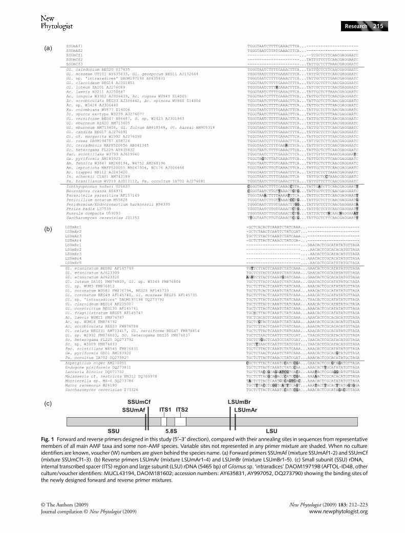
Fig. 1 Forward and reverse primers designed in this study (5′–3′ direction), compared with their annealing sites in sequences from representative members of all main AMF taxa and some non-AMF species. Variable sites not represented in any primer mixture are shaded. When no culture identifiers are known, voucher (W) numbers are given behind the species name. (a) Forward primers SSUmAf (mixture SSUmAf1-2) and SSUmCf (mixture SSUmCf1-3). (b) Reverse primers LSUmAr (mixture LSUmAr1-4) and LSUmBr (mixture LSUmBr1-5). (c) Small subunit (SSU) rDNA, internal transcribed spacer (ITS) region and large subunit (LSU) rDNA (5465 bp) of Glomus sp. ‘intraradices’ DAOM197198 (AFTOL-ID48, other culture/voucher identifiers: MUCL43194, DAOM181602; accession numbers: AY635831, AY997052, DQ273790) showing the binding sites of the newly designed forward and reverse primer mixtures.
New Phytologist (2009) 183: 212–223 © The Authors (2009)www.newphytologist.org Journal compilation © New Phytologist (2009)
Research216
Results
Primer design
Potentially suited binding sites for primers that match AMFsequences but discriminate against plant and non-AM fungal(non-AMF) sequences were identified for the SSU rDNA andLSU rDNA. They were located at positions 1484 and 1532on the SSU, and at positions 827 and 928 on the LSU rDNA(based on Glomus sp. ‘intraradices’ DAOM197198 sequence;Fig. 1c). Sequence variation made it impossible to deriveindividual primer sequences that specifically amplify allGlomeromycota. Thus, a set of four primer mixtures wasdesigned, each targeting one binding site (Table 2, Fig. 1).Certain non-3′ located mismatches that only slightly alteredmelting temperature and some mismatches (Glomus etunicatum)that were perhaps caused by low sequence quality wereaccepted for primer design (Fig. 1). To discriminate againstnontarget organisms mismatches at the 3′ end of the primerswere included. blast searches indicated high specificity of thenew primer pairs for AMF.
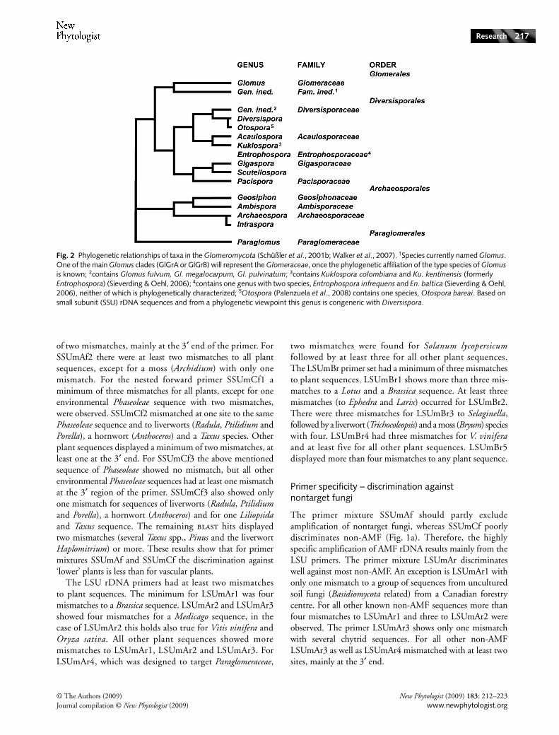
Glomeromycota sequences that represent the knownvariability at the primer binding sites are shown in Fig. 1. Weaimed to include as many main phylogenetic lineages (Fig. 2)for primer design as possible. However, the following taxacould not be included for LSU rDNA binding sites analyses:Entrophosporaceae, containing only two species lackingsequence data; Archaeosporaceae, because available sequencesdid not cover the LSU rDNA binding sites; Otospora forwhich only two nonoverlapping partial SSU rDNA sequencesare known; Intraspora, represented by only one SSU rDNAdatabase sequence.
Primer specificity – discrimination against plants
The discrimination of primer SSUmAf1 against ‘lower’ plantsis weak and exemplified by only one mismatch to databasesequences from mosses (Polytrichastrum, Leptodontiumand Pogonatum), a liverwort (Trichocoleopsis), a hornwort(Phaeoceros) and a clubmoss (Selaginella). Burmannia, onePhaseoleae sp. and some other plant sequences also showedonly one mismatch. All other plant sequences had a minimum
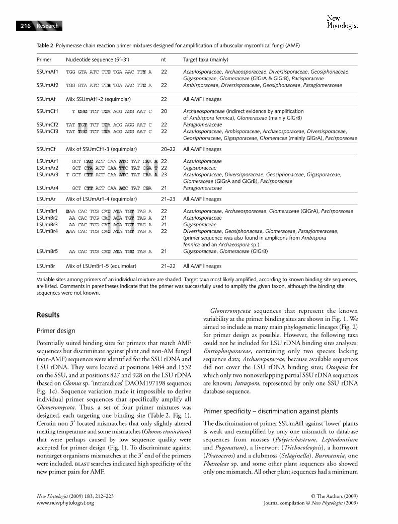
Table 2 Polymerase chain reaction primer mixtures designed for amplification of arbuscular mycorrhizal fungi (AMF)
Primer Nucleotide sequence (5′–3′) nt Target taxa (mainly)
SSUmAf1 TGG GTA ATC TTT TGA AAC TTY A 22 Acaulosporaceae, Archaeosporaceae, Diversisporaceae, Geosiphonaceae, Gigasporaceae, Glomeraceae (GlGrA & GlGrB), Pacisporaceae
SSUmAf2 TGG GTA ATC TTR TGA AAC TTC A 22 Ambisporaceae, Diversisporaceae, Geosiphonaceae, Paraglomeraceae
SSUmAf Mix SSUmAf1-2 (equimolar) 22 All AMF lineages
SSUmCf1 T CGC TCT TCA ACG AGG AAT C 20 Archaeosporaceae (indirect evidence by amplification of Ambispora fennica), Glomeraceae (mainly GlGrB)
SSUmCf2 TAT TGT TCT TCA ACG AGG AAT C 22 ParaglomeraceaeSSUmCf3 TAT TGC TCT TNA ACG AGG AAT C 22 Acaulosporaceae, Ambisporaceae, Archaeosporaceae, Diversisporaceae,
Geosiphonaceae, Gigasporaceae, Glomeracea (mainly GlGrA), Pacisporaceae
SSUmCf Mix of SSUmCf1-3 (equimolar) 20–22 All AMF lineages
LSUmAr1 GCT CAC ACT CAA ATC TAT CAA A 22 AcaulosporaceaeLSUmAr2 GCT CTA ACT CAA TTC TAT CGA T 22 GigasporaceaeLSUmAr3 T GCT CTT ACT CAA ATC TAT CAA A 23 Acaulosporaceae, Diversisporaceae, Geosiphonaceae, Gigasporaceae,
Glomeraceae (GlGrA and GlGrB), PacisporaceaeLSUmAr4 GCT CTT ACT CAA ACC TAT CGA 21 Paraglomeraceae
LSUmAr Mix of LSUmAr1-4 (equimolar) 21–23 All AMF lineages
LSUmBr1 DAA CAC TCG CAT ATA TGT TAG A 22 Acaulosporaceae, Archaeosporaceae, Glomeraceae (GlGrA), Pacisporaceae LSUmBr2 AA CAC TCG CAC ACA TGT TAG A 21 AcaulosporaceaeLSUmBr3 AA CAC TCG CAT ACA TGT TAG A 21 GigasporaceaeLSUmBr4 AAA CAC TCG CAC ATA TGT TAG A 22 Diversisporaceae, Geosiphonaceae, Glomeraceae, Paraglomeraceae,
(primer sequence was also found in amplicons from Ambispora fennica and an Archaeospora sp.)
LSUmBr5 AA CAC TCG CAT ATA TGC TAG A 21 Gigasporaceae, Glomeraceae (GlGrB)
LSUmBr Mix of LSUmBr1-5 (equimolar) 21–22 All AMF lineages
Variable sites among primers of an individual mixture are shaded. Target taxa most likely amplified, according to known binding site sequences, are listed. Comments in parentheses indicate that the primer was successfully used to amplify the given taxon, although the binding site sequences were not known.
© The Authors (2009) New Phytologist (2009) 183: 212–223Journal compilation © New Phytologist (2009) www.newphytologist.org
Research 217
of two mismatches, mainly at the 3′ end of the primer. ForSSUmAf2 there were at least two mismatches to all plantsequences, except for a moss (Archidium) with only onemismatch. For the nested forward primer SSUmCf1 aminimum of three mismatches for all plants, except for oneenvironmental Phaseoleae sequence with two mismatches,were observed. SSUmCf2 mismatched at one site to the samePhaseoleae sequence and to liverworts (Radula, Ptilidium andPorella), a hornwort (Anthoceros) and a Taxus species. Otherplant sequences displayed a minimum of two mismatches, atleast one at the 3′ end. For SSUmCf3 the above mentionedsequence of Phaseoleae showed no mismatch, but all otherenvironmental Phaseoleae sequences had at least one mismatchat the 3′ region of the primer. SSUmCf3 also showed onlyone mismatch for sequences of liverworts (Radula, Ptilidiumand Porella), a hornwort (Anthoceros) and for one Liliopsidaand Taxus sequence. The remaining blast hits displayedtwo mismatches (several Taxus spp., Pinus and the liverwortHaplomitrium) or more. These results show that for primermixtures SSUmAf and SSUmCf the discrimination against‘lower’ plants is less than for vascular plants.
The LSU rDNA primers had at least two mismatchesto plant sequences. The minimum for LSUmAr1 was fourmismatches to a Brassica sequence. LSUmAr2 and LSUmAr3showed four mismatches for a Medicago sequence, in thecase of LSUmAr2 this holds also true for Vitis vinifera andOryza sativa. All other plant sequences showed moremismatches to LSUmAr1, LSUmAr2 and LSUmAr3. ForLSUmAr4, which was designed to target Paraglomeraceae,
two mismatches were found for Solanum lycopersicumfollowed by at least three for all other plant sequences.The LSUmBr primer set had a minimum of three mismatchesto plant sequences. LSUmBr1 shows more than three mis-matches to a Lotus and a Brassica sequence. At least threemismatches (to Ephedra and Larix) occurred for LSUmBr2.There were three mismatches for LSUmBr3 to Selaginella,followed by a liverwort (Trichocoleopsis) and a moss (Bryum) specieswith four. LSUmBr4 had three mismatches for V. viniferaand at least five for all other plant sequences. LSUmBr5displayed more than four mismatches to any plant sequence.
Primer specificity – discrimination against nontarget fungi
The primer mixture SSUmAf should partly excludeamplification of nontarget fungi, whereas SSUmCf poorlydiscriminates non-AMF (Fig. 1a). Therefore, the highlyspecific amplification of AMF rDNA results mainly from theLSU primers. The primer mixture LSUmAr discriminateswell against most non-AMF. An exception is LSUmAr1 withonly one mismatch to a group of sequences from unculturedsoil fungi (Basidiomycota related) from a Canadian forestrycentre. For all other known non-AMF sequences more thanfour mismatches to LSUmAr1 and three to LSUmAr2 wereobserved. The primer LSUmAr3 shows only one mismatchwith several chytrid sequences. For all other non-AMFLSUmAr3 as well as LSUmAr4 mismatched with at least twosites, mainly at the 3′ end.
Fig. 2 Phylogenetic relationships of taxa in the Glomeromycota (Schüßler et al., 2001b; Walker et al., 2007). 1Species currently named Glomus. One of the main Glomus clades (GlGrA or GlGrB) will represent the Glomeraceae, once the phylogenetic affiliation of the type species of Glomus is known; 2contains Glomus fulvum, Gl. megalocarpum, Gl. pulvinatum; 3contains Kuklospora colombiana and Ku. kentinensis (formerly Entrophospora) (Sieverding & Oehl, 2006); 4contains one genus with two species, Entrophospora infrequens and En. baltica (Sieverding & Oehl, 2006), neither of which is phylogenetically characterized; 5Otospora (Palenzuela et al., 2008) contains one species, Otospora bareai. Based on small subunit (SSU) rDNA sequences and from a phylogenetic viewpoint this genus is congeneric with Diversispora.
New Phytologist (2009) 183: 212–223 © The Authors (2009)www.newphytologist.org Journal compilation © New Phytologist (2009)
Research218
For the (nested) LSUmBr primer mixture the specificity islower; for example, LSUmBr1 showed no mismatch to somefungi in the more ancestral lineages, namely Endogone lactifluaand Mortierellaceae species, chytrids (Rhizophlyctis andGonapodya), an uncultured alpine tundra soil fungus andmatched one ascomycete sequence (Catenulostroma). ForLSUmBr2, no mismatches occurred for sequences of somebasidiomycetes (Bulleribasidium, Paullicorticium and Russula)and a zygomycete (Spiromyces minutus). Only one mismatchwas observed for sequences including basidiomycetes(Calocera, Calostoma and Ramaria) and ascomycetes (Pyxidi-ophora, Eremithallus and Phaeococcus), and some other fungi.LSUmBr3 discriminates well against other fungi with at leastthree mismatches, except for one uncultured soil fungussequence (Cryptococcus related) that matched completely.The primer LSUmBr4 showed no mismatch to Clavulinagriseohumicola and only one to some fungal sequencesincluding ascomycetes (Pyxidiophora and Phaeococcus) andbasidiomycetes (Cryptococcus spp.). LSUmBr5 showed onlyone mismatch to fungal sequences of Mortierella spp., a chytrid(Rhizophlyctis rosea), and some ascomycetes (Schizosaccharomyces,Verrucocladosporium, Passalora and Catenulostroma). In generalthe LSUmAr primers discriminate better against non-AMFthan the nested primers LSUmBr.
Primer efficiency – tests on plasmids and DNA extracts from single spores
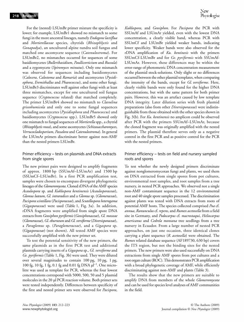
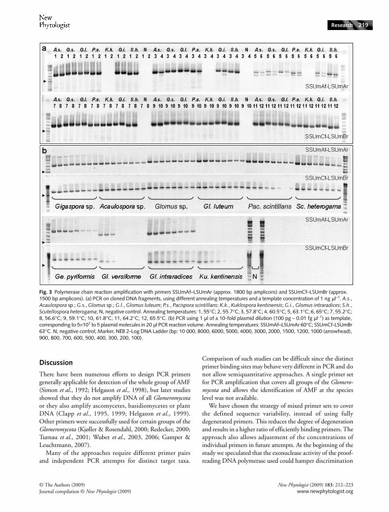
The new primer pairs were designed to amplify fragmentsof approx. 1800 bp (SSUmAf–LSUmAr) and 1500 bp(SSUmCf–LSUmBr). In a first PCR amplification test,samples were chosen to encompass divergent phylogeneticlineages of the Glomeromycota. Cloned rDNA of the AMF speciesAcaulospora sp. and Kuklospora kentinensis (Acaulosporaceae),Glomus luteum, Gl. intraradices and a Glomus sp. (Glomeraceae),Pacispora scintillans (Pacisporaceae), and Scutellospora heterogama(Gigasporaceae) were used (Table 1, Fig. 3a). In addition,rDNA fragments were amplified from single spore DNAextracts from Geosiphon pyriformis (Geosiphonaceae), Gl. mosseae(Glomeraceae), Gl. eburneum and Gl. versiforme (Diversisporaceae),a Paraglomus sp. (Paraglomeraceae), and a Gigaspora sp.(Gigasporaceae) (not shown). All tested AMF species weresuccessfully amplified with the new primer set.
To test the potential sensitivity of the new primers, thesame plasmids as in the first PCR test and additionalplasmids carrying inserts of a Gigaspora sp., Gl. versiforme andGe. pyriformis (Table 1, Fig. 3b) were used. They were dilutedover several magnitudes to contain 100 pg, 10 pg, 1 pg,100 fg, 10 fg, 1 fg, 0.1 fg and 0.01 fg DNA µl−1. One micro-litre was used as template for PCR, whereas the four lowestconcentrations correspond with 5000, 500, 50 and 5 plasmidmolecules in the 20 µl PCR reaction volume. Both primer setswere tested independently. Differences between specificity ofthe first and nested primer sets were observed for Pacispora,
Kuklospora, and Geosiphon. For Pacispora the PCR withSSUmAf and LSUmAr yielded, even with the lowest DNAconcentration, a clearly visible band, whereas PCR withSSUmCf and LSUmBr yielded weaker bands, indicatinglower specificity. Weaker bands were also observed for therDNA amplification of Ku. kentinesis with the primersSSUmCf-LSUmBr and for Ge. pyriformis with SSUmAf-LSUmAr. However, these differences may be within theerror-range of photometric DNA concentration measurementof the plasmid stock-solutions. Only slight or no differencesoccurred between the other plasmid templates, when comparingthe intensity of the bands, except for Gl. versiforme. Here,clearly visible bands were only found for the higher DNAconcentrations, but with the same pattern for both primerpairs. However, this was an artefact caused by low templateDNA integrity. Later dilution series with fresh plasmidpreparations (also from other Diversisporaceae) were indistin-guishable from those obtained with the other species shown inFig. 3(b). For Ku. kentinensis no amplicon could be observedafter PCR with the primers SSUmAf–LSUmAr, becausethe cloned fragment was originally amplified with the nestedprimers. The plasmid therefore serves only as a negativecontrol in the first PCR and as positive control for the PCRwith the nested primers.
Primer efficiency – tests on field and nursery sampled roots and spores
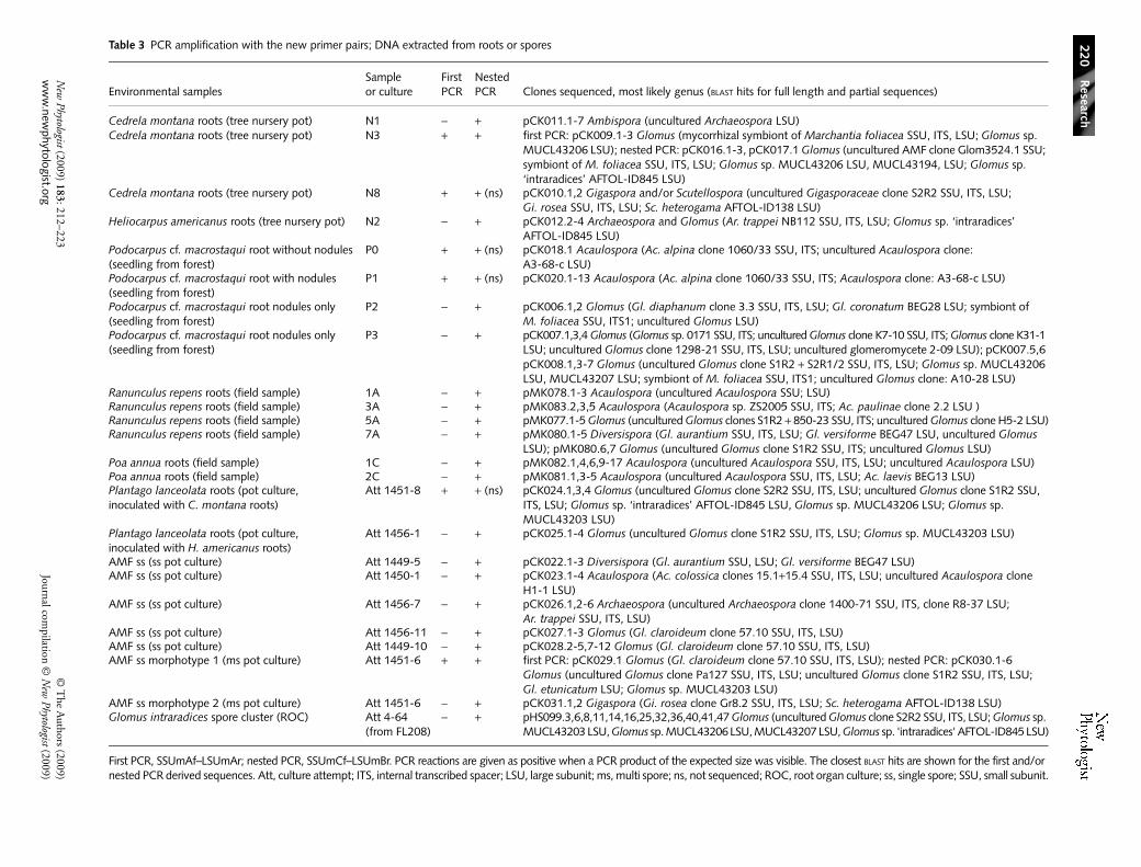
To test whether the newly designed primers discriminateagainst nonglomeromycotan fungi and plants, we used themon DNA extracted from single spores from pot cultures,environmental root samples, and root samples from a treenursery, in nested PCR approaches. We observed not a singlenon-AMF contaminant sequence in the 12 environmentalroot and 40 single spore samples processed. The discriminationagainst plants was tested with DNA extracts from roots ofpotential AMF hosts. The species collected comprised Poa cf.annua, Ranunculus cf. repens, and Rumex acetosella from a fieldsite in Germany, and Podocarpus cf. macrostaqui, Heliocarpusamericanus and Cedrela montana tree seedlings from a treenursery in Ecuador. From a large number of nested PCRapproaches, on just one occasion, three identical clonescarrying a plant sequence (R. acetosella) were obtained. TheRumex related database sequence (AF189730, 630 bp) coversthe ITS region, but not the binding sites for the nestedprimers. The new primers were also used successfully on DNAextractions from single AMF spores from pot cultures and aroot organ culture (ROC). This demonstrates PCR amplificationwith a broad phylogenetic coverage of AMF, while efficientlydiscriminating against non-AMF and plants (Table 3).
The results show that the new primers are suitable toamplify DNA from members of the whole Glomeromycotaand can be used for species level analyses of AMF communitiesin the field.
© The Authors (2009) New Phytologist (2009) 183: 212–223Journal compilation © New Phytologist (2009) www.newphytologist.org
Research 219
Discussion
There have been numerous efforts to design PCR primersgenerally applicable for detection of the whole group of AMF(Simon et al., 1992; Helgason et al., 1998), but later studiesshowed that they do not amplify DNA of all Glomeromycotaor they also amplify ascomycetes, basidiomycetes or plantDNA (Clapp et al., 1995, 1999; Helgason et al., 1999).Other primers were successfully used for certain groups of theGlomeromycota (Kjøller & Rosendahl, 2000; Redecker, 2000;Turnau et al., 2001; Wubet et al., 2003, 2006; Gamper &Leuchtmann, 2007).
Many of the approaches require different primer pairsand independent PCR attempts for distinct target taxa.
Comparison of such studies can be difficult since the distinctprimer binding sites may behave very different in PCR and donot allow semiquantitative approaches. A single primer setfor PCR amplification that covers all groups of the Glomero-mycota and allows the identification of AMF at the specieslevel was not available.
We have chosen the strategy of mixed primer sets to coverthe defined sequence variability, instead of using fullydegenerated primers. This reduces the degree of degenerationand results in a higher ratio of efficiently binding primers. Theapproach also allows adjustment of the concentrations ofindividual primers in future attempts. At the beginning of thestudy we speculated that the exonuclease activity of the proof-reading DNA polymerase used could hamper discrimination
Fig. 3 Polymerase chain reaction amplification with primers SSUmAf–LSUmAr (approx. 1800 bp amplicons) and SSUmCf–LSUmBr (approx. 1500 bp amplicons). (a) PCR on cloned DNA fragments, using different annealing temperatures and a template concentration of 1 ng µl−1. A.s., Acaulospora sp.; G.s., Glomus sp.; G.l., Glomus luteum; P.s., Pacispora scintillans; K.k., Kuklospora kentinensis; G.i., Glomus intraradices; S.h., Scutellospora heterogama; N, negative control. Annealing temperatures: 1, 55°C; 2, 55.7°C; 3, 57.8°C; 4, 60.5°C; 5, 63.1°C; 6, 65°C; 7, 55.2°C; 8, 56.6°C; 9, 59.1°C; 10, 61.8°C; 11, 64.2°C; 12, 65.5°C. (b) PCR using 1 µl of a 10-fold plasmid dilution (100 pg – 0.01 fg µl−1) as template, corresponding to 5×10⁷ to 5 plasmid molecules in 20 µl PCR reaction volume. Annealing temperatures: SSUmAf–LSUmAr 60°C; SSUmCf-LSUmBr 63°C. N, negative control; Marker, NEB 2-Log DNA Ladder (bp: 10 000, 8000, 6000, 5000, 4000, 3000, 2000, 1500, 1200, 1000 (arrowhead), 900, 800, 700, 600, 500, 400, 300, 200, 100).
New
Phytologist (2009) 183: 212–223©
The A
uthors (2009)w
ww
.newphytologist.org
Journal compilation ©
New
Phytologist (2009)
Research
220Table 3 PCR amplification with the new primer pairs; DNA extracted from roots or spores
Environmental samplesSample or culture
First PCR
Nested PCR Clones sequenced, most likely genus (BLAST hits for full length and partial sequences)
Cedrela montana roots (tree nursery pot) N1 − + pCK011.1-7 Ambispora (uncultured Archaeospora LSU)Cedrela montana roots (tree nursery pot) N3 + + first PCR: pCK009.1-3 Glomus (mycorrhizal symbiont of Marchantia foliacea SSU, ITS, LSU; Glomus sp.
MUCL43206 LSU); nested PCR: pCK016.1-3, pCK017.1 Glomus (uncultured AMF clone Glom3524.1 SSU; symbiont of M. foliacea SSU, ITS, LSU; Glomus sp. MUCL43206 LSU, MUCL43194, LSU; Glomus sp. ‘intraradices’ AFTOL-ID845 LSU)
Cedrela montana roots (tree nursery pot) N8 + + (ns) pCK010.1,2 Gigaspora and/or Scutellospora (uncultured Gigasporaceae clone S2R2 SSU, ITS, LSU; Gi. rosea SSU, ITS, LSU; Sc. heterogama AFTOL-ID138 LSU)
Heliocarpus americanus roots (tree nursery pot) N2 − + pCK012.2-4 Archaeospora and Glomus (Ar. trappei NB112 SSU, ITS, LSU; Glomus sp. ‘intraradices’AFTOL-ID845 LSU)
Podocarpus cf. macrostaqui root without nodules (seedling from forest)
P0 + + (ns) pCK018.1 Acaulospora (Ac. alpina clone 1060/33 SSU, ITS; uncultured Acaulospora clone: A3-68-c LSU)
Podocarpus cf. macrostaqui root with nodules (seedling from forest)
P1 + + (ns) pCK020.1-13 Acaulospora (Ac. alpina clone 1060/33 SSU, ITS; Acaulospora clone: A3-68-c LSU)
Podocarpus cf. macrostaqui root nodules only (seedling from forest)
P2 − + pCK006.1,2 Glomus (Gl. diaphanum clone 3.3 SSU, ITS, LSU; Gl. coronatum BEG28 LSU; symbiont of M. foliacea SSU, ITS1; uncultured Glomus LSU)
Podocarpus cf. macrostaqui root nodules only (seedling from forest)
P3 − + pCK007.1,3,4 Glomus (Glomus sp. 0171 SSU, ITS; uncultured Glomus clone K7-10 SSU, ITS; Glomus clone K31-1 LSU; uncultured Glomus clone 1298-21 SSU, ITS, LSU; uncultured glomeromycete 2-09 LSU); pCK007.5,6 pCK008.1,3-7 Glomus (uncultured Glomus clone S1R2 + S2R1/2 SSU, ITS, LSU; Glomus sp. MUCL43206 LSU, MUCL43207 LSU; symbiont of M. foliacea SSU, ITS1; uncultured Glomus clone: A10-28 LSU)
Ranunculus repens roots (field sample) 1A − + pMK078.1-3 Acaulospora (uncultured Acaulospora SSU; LSU)Ranunculus repens roots (field sample) 3A − + pMK083.2,3,5 Acaulospora (Acaulospora sp. ZS2005 SSU, ITS; Ac. paulinae clone 2.2 LSU )Ranunculus repens roots (field sample) 5A − + pMK077.1-5 Glomus (uncultured Glomus clones S1R2 + 850-23 SSU, ITS; uncultured Glomus clone H5-2 LSU)Ranunculus repens roots (field sample) 7A − + pMK080.1-5 Diversispora (Gl. aurantium SSU, ITS, LSU; Gl. versiforme BEG47 LSU, uncultured Glomus
LSU); pMK080.6,7 Glomus (uncultured Glomus clone S1R2 SSU, ITS; uncultured Glomus LSU)Poa annua roots (field sample) 1C − + pMK082.1,4,6,9-17 Acaulospora (uncultured Acaulospora SSU, ITS, LSU; uncultured Acaulospora LSU)Poa annua roots (field sample) 2C − + pMK081.1,3-5 Acaulospora (uncultured Acaulospora SSU, ITS, LSU; Ac. laevis BEG13 LSU)Plantago lanceolata roots (pot culture, inoculated with C. montana roots)
Att 1451-8 + + (ns) pCK024.1,3,4 Glomus (uncultured Glomus clone S2R2 SSU, ITS, LSU; uncultured Glomus clone S1R2 SSU, ITS, LSU; Glomus sp. ‘intraradices’ AFTOL-ID845 LSU, Glomus sp. MUCL43206 LSU; Glomus sp. MUCL43203 LSU)
Plantago lanceolata roots (pot culture, inoculated with H. americanus roots)
Att 1456-1 − + pCK025.1-4 Glomus (uncultured Glomus clone S1R2 SSU, ITS, LSU; Glomus sp. MUCL43203 LSU)
AMF ss (ss pot culture) Att 1449-5 − + pCK022.1-3 Diversispora (Gl. aurantium SSU, LSU; Gl. versiforme BEG47 LSU)AMF ss (ss pot culture) Att 1450-1 − + pCK023.1-4 Acaulospora (Ac. colossica clones 15.1+15.4 SSU, ITS, LSU; uncultured Acaulospora clone
H1-1 LSU)AMF ss (ss pot culture) Att 1456-7 − + pCK026.1,2-6 Archaeospora (uncultured Archaeospora clone 1400-71 SSU, ITS, clone R8-37 LSU;
Ar. trappei SSU, ITS, LSU)AMF ss (ss pot culture) Att 1456-11 − + pCK027.1-3 Glomus (Gl. claroideum clone 57.10 SSU, ITS, LSU)AMF ss (ss pot culture) Att 1449-10 − + pCK028.2-5,7-12 Glomus (Gl. claroideum clone 57.10 SSU, ITS, LSU)AMF ss morphotype 1 (ms pot culture) Att 1451-6 + + first PCR: pCK029.1 Glomus (Gl. claroideum clone 57.10 SSU, ITS, LSU); nested PCR: pCK030.1-6
Glomus (uncultured Glomus clone Pa127 SSU, ITS, LSU; uncultured Glomus clone S1R2 SSU, ITS, LSU; Gl. etunicatum LSU; Glomus sp. MUCL43203 LSU)
AMF ss morphotype 2 (ms pot culture) Att 1451-6 − + pCK031.1,2 Gigaspora (Gi. rosea clone Gr8.2 SSU, ITS, LSU; Sc. heterogama AFTOL-ID138 LSU)Glomus intraradices spore cluster (ROC) Att 4-64
(from FL208)− + pHS099.3,6,8,11,14,16,25,32,36,40,41,47 Glomus (uncultured Glomus clone S2R2 SSU, ITS, LSU; Glomus sp.
MUCL43203 LSU, Glomus sp. MUCL43206 LSU, MUCL43207 LSU, Glomus sp. 'intraradices' AFTOL-ID845 LSU)
First PCR, SSUmAf–LSUmAr; nested PCR, SSUmCf–LSUmBr. PCR reactions are given as positive when a PCR product of the expected size was visible. The closest BLAST hits are shown for the first and/or nested PCR derived sequences. Att, culture attempt; ITS, internal transcribed spacer; LSU, large subunit; ms, multi spore; ns, not sequenced; ROC, root organ culture; ss, single spore; SSU, small subunit.
© The Authors (2009) New Phytologist (2009) 183: 212–223Journal compilation © New Phytologist (2009) www.newphytologist.org
Research 221
by terminal 3′ primer mismatches, but no such problemswere detected.
Primer specificity
The primers designed show some mismatches to AMFsequences at the 5′ end (Fig. 1), which do not hinder PCRamplification (Bru et al., 2008). Primer mismatches suchas C–T, T–C and T–G do not impair amplification stronglyeven when situated at the 3′ end of the primer (Kwok et al.,1990). The forward primers SSUmAf as well as the reverseprimers LSUmBr mismatched once with Ge. pyriformis, butdid not hamper amplification. The LSU rDNA primers showsufficient sequence similarity to the target organisms, as themismatches are either in the middle or at the 5′ end.LSUmAr primers displayed individual mismatches tosequences of Scutellospora spp., Gl. etunicatum, and oneAcaulospora sp. (Fig. 1). Nevertheless, DNA of these specieswas successfully amplified from environmental samples andin the primer efficiency test (Fig. 3). Ambisporaceae andArchaeosporaceae species could not be included in the designof the LSU primers, but Ambispora fennica DNA from a singlespore extraction (not shown) and Archaeospora sp. from singlespores and roots of an Ecuadorian tree seedling (Table 3) couldbe amplified with the new primers, indicating well matchingbinding sites. Sequences from Otospora (Diversisporaceae;Palenzuela et al., 2008; matching the SSU primers), Intraspora(closely related to Archaeospora), and Entrophospora (sensuOehl & Sieverd.; with two species only) are either not or onlypartly characterized and therefore could not be included inseveral aspects of primer design. Otospora and Intraspora arevery closely related to their sister genera (maybe congeneric),so the lack of LSU rDNA sequences was therefore interpretedas a minor limitation.
We could successfully amplify all AMF tested with the newprimers, but because of the lower number of LSU rDNAsequences available for AMF an optimization of the LSUprimers might be reasonable in future. The discriminationagainst non-AMF and plant DNA is excellent, as shown onDNA extracts from environmental samples and spores frompot cultures. To discriminate against non-AMF, LSUmArworks much better than the nested primers LSUmBr. Thecloned plant (Rumex) rDNA fragment that originated fromroot material can be interpreted as an ‘outlier’. The primerbinding sites could not be investigated for Rumex, because oflacking sequence coverage. It should be indicated in thiscontext that we did not use HPLC-purified primers. Thismeans a certain fraction of primers may not be fully synthesizedand could result in less specific amplification. All plasmidsused in the plasmid test carried inserts that were originallyamplified with SSUmAf. Therefore, the efficiency of thisprimer could not be validated, but because of the highnumber of SSU rDNA sequences known, it can be statedthat the binding sites in the cloned fragments correspond to a
realistic situation. The efficient amplification from spore DNAextracts was, moreover, confirmed in numerous former PCR.
Advantages over previously used PCR primer sets
In most former field studies SSU rDNA phylotypes wereanalysed for molecular detection of AMF. However, thisregion does not allow species resolution and each definedphylotype, irrespective of the used distance threshold value orphylogenetic analysis method, may hide a number of species(Walker et al., 2007). In general, the LSU rDNA regionallows species resolution, and thus the LSU primer pairFLR3–FLR4 (Gollotte et al., 2004) was used for species-level community analyses. However, in particular, FLR4is not phylogenetically inclusive (Gamper et al., 2009)and discriminates many lineages, including Diversisporales,Archaeosporales and Paraglomerales, which results in a strongbias in community analyses towards the Glomeraceae. Theprimer FLR3 binds to DNA of many nontarget fungi as itshows no mismatch to > 1300 basidiomycete sequences andsome ascomycete sequences in the public databases. Suchproblems obviously may bias tRFLP community analyses(Mummey & Rillig, 2008) and seminested PCR approaches(Pivato et al., 2007) using FLR3 and/or FLR4. The primerpair SSUGlom1–LSUGlom1 (Renker et al., 2003) amplifiesmany non-AMF and plants. Combined with the primersITS5–ITS4 in a nested PCR (Hempel et al., 2007) thisresulted in a 5.8S rDNA phylogenetic analysis, whichresolved only the genus level. Even the ITS region does notalways resolve species for AMF (Stockinger et al., 2009).
In some cases, species-specific detection tools are availablefor individual species or certain well-defined and closelyrelated species. The three closely related AM fungi Gl. mosseae,Gl. caledonium and Gl. geosporum were detected by usingLSU primers in field studies (Stukenbrock & Rosendahl,2005; Rosendahl & Matzen, 2008), but these primers weredesigned to only amplify subgroups or certain taxa in theGlomeromycota. For the well-studied Gl. intraradices relatedAMF (e.g. DAOM197198), which are, however, not conspecificwith Gl. intraradices (Stockinger et al. 2009), microsatellitemarkers are available for their detection in the field (Crollet al., 2008; Mathimaran et al., 2008). Some mtLSU regionmarkers were also studied (Börstler et al., 2008), but becauseof the high length variation observed (1070–3935 bp) and thedifficulty in amplifying this region it is not very promising forcommunity analyses. Thus, such markers cannot be used forgeneral AMF community analyses.
The new primers described in the present study wereused to amplify efficiently and specifically target rDNA fromenvironmental samples of the main phylogenetic groups inthe Glomeromycota. For the first time, this will allow molecularecological studies covering all AMF lineages to be carried outwith only one primer set. Furthermore, the long sequencesallow robust phylogenetic analyses and species level resolution
New Phytologist (2009) 183: 212–223 © The Authors (2009)www.newphytologist.org Journal compilation © New Phytologist (2009)
Research222
by inclusion of the variable ITS and LSU rDNA region(Walker et al., 2007; Gamper et al., 2009; Stockinger et al.2009), whereas formerly used primers mainly amplifiedrDNA fragments of up to 800 bp (Helgason et al., 1999;Redecker, 2000; Lee et al., 2008).
Potential application as DNA barcoding primers
The new primers are suited to amplify the most likely primaryDNA barcode region for fungi, the ITS region (already onlineat the Barcode of Life Data Systems (BOLD) website;www.barcodinglife.org). In general ‘barcode primers’ shouldamplify short fragments and for the ITS region the ampliconsgenerated by our primers are in fact too long. However, themain criterion for DNA barcodes is the resolution at specieslevel. Since for Glomeromycota this is difficult or impossibleto achieve with the ITS region only (Stockinger et al., 2009),the inclusion of the 5′ LSU rDNA fragment is stronglyrecommended. Our new primer set (SSUmAf, SSUmCf,LSUmAr and LSUmBr) appears to be well suited as barcodingprimers for Glomeromycota. The primers will be helpful forthe molecular characterization of AMF, including speciesdescriptions (Gamper et al., 2009), resulting in a sequencedatabase that allows the design of further primers for thedetection of AMF from field samples. LSUmAr and LSUmBr,located approximately at positions 930–950 and 830–850 onthe LSU rRNA gene, may be used in combination with newforward LSU primers for amplification of fragments withinthe variable D1/D2 LSU regions. Based on such amplicons,deep sequencing approaches with the now feasible longerreads of the new 454 FLX-titanium chemistry will allowspecies level detection of the ‘unknown’ AMF community, infuture molecular ecological studies.
Acknowledgements
The grants for M.K., C.K. and A.S. were financed by theGerman Research Foundation (DFG). The grant for H.S. wasfunded by the Marie Curie Early Stage Research TrainingFellowship of the European Community’s Sixth frameworkProgramme (MEST-CT-2005-021016).
References
Allen GC, Flores-Vergara MA, Krasnyanski S, Kumar S, Thompson WF. 2006. A modified protocol for rapid DNA isolation from plant tissues using cetyltrimethylammonium bromide. Nature Protocols 1: 2320–2325.
Aroca R, Porcel R, Ruiz-Lozano JM. 2007. How does arbuscular mycorrhizal symbiosis regulate root hydraulic properties and plasma membrane aquaporins in Phaseolus vulgaris under drought, cold or salinity stresses? New Phytologist 173: 808–816.
Börstler B, Raab PA, Thiery O, Morton JB, Redecker D. 2008. Genetic diversity of the arbuscular mycorrhizal fungus Glomus intraradices as determined by mitochondrial large subunit rRNA gene sequences is considerably higher than previously expected. New Phytologist 180: 452–465.
Brody JR, Kern SE. 2004. Sodium boric acid: a Tris-free, cooler conductive medium for DNA electrophoresis. Biotechniques 36: 214–215.
Bru D, Martin-Laurent F, Philippot L. 2008. Quantification of the detrimental effect of a single primer–template mismatch by real-time PCR using the 16S rRNA gene as an example. Applied and Environmental Microbiology 74: 1660–1663.
Clapp JP, Fitter AH, Young JPW. 1999. Ribosomal small subunit sequence variation within spores of an arbuscular mycorrhizal fungus, Scutellospora sp. Molecular Ecology 8: 915–922.
Clapp JP, Young JPW, Merryweather JW, Fitter AH. 1995. Diversity of fungal symbionts in arbuscular mycorrhizas from a natural community. New Phytologist 130: 259–265.
Croll D, Wille L, Gamper HA, Mathimaran N, Lammers PJ, Corradi N, Sanders IR. 2008. Genetic diversity and host plant preferences revealed by simple sequence repeat and mitochondrial markers in a population of the arbuscular mycorrhizal fungus Glomus intraradices. New Phytologist 178: 672–687.
De la Pena E, Rodriguez Echeverria S, van der Putten WH, Freitas H, Moens M. 2006. Mechanism of control of root-feeding nematodes by mycorrhizal fungi in the dune grass Ammophila arenaria. New Phytologist 169: 829–840.
Gamper H, Leuchtmann A. 2007. Taxon-specific PCR primers to detect two inconspicuous arbuscular mycorrhizal fungi from temperate agricultural grassland. Mycorrhiza 17: 145–152.
Gamper H, Walker C, Schüßler A. 2009. Diversispora celata sp. nov.: molecular ecology and phylotaxonomy of an inconspicuous arbuscular mycorrhizal fungus. New Phytologist 182: 495–506.
Gollotte A, van Tuinen D, Atkinson D. 2004. Diversity of arbuscular mycorrhizal fungi colonising roots of the grass species Agrostis capillaris and Lolium perenne in a field experiment. Mycorrhiza 14: 111–117.
Helgason T, Daniell TJ, Husband R, Fitter AH, Young JPW. 1998. Ploughing up the wood-wide web? Nature 394: 431.
Helgason T, Fitter AH, Young JPW. 1999. Molecular diversity of arbuscular mycorrhizal fungi colonising Hyacinthoides nonscripta (bluebell) in a seminatural woodland. Molecular Ecology 8: 659–666.
Hempel S, Renker C, Buscot F. 2007. Differences in the species composition of arbuscular mycorrhizal fungi in spore, root and soil communities in a grassland ecosystem. Environmental Microbiology 9: 1930–1938.
Jansa J, Mozafar A, Kuhn G, Anken T, Ruh R, Sanders IR, Frossard E. 2003. Soil tillage affects the community structure of mycorrhizal fungi in maize roots. Ecological Applications 13: 1164–1176.
Kjøller R, Rosendahl S. 2000. Detection of arbuscular mycorrhizal fungi (Glomales) in roots by nested PCR and SSCP (single stranded conformation polymorphism). Plant and Soil 226: 189–196.
Kottke I, Haug I, Setaro S, Suárez JP, Weiß M, Preußing M, Nebel M, Oberwinkler F. 2008. Guilds of mycorrhizal fungi and their relation to trees, ericads, orchids and liverworts in a neotropical mountain rain forest. Basic and Applied Ecology 9: 13–23.
Kwok S, Kellogg DE, McKinney N, Spasic D, Goda L, Levenson C, Sninsky JJ. 1990. Effects of primer–template mismatches on the polymerase chain reaction: human immunodeficiency virus type 1 model studies. Nucleic Acids Research 18: 999–1005.
Lee J, Lee S, Young JPW. 2008. Improved PCR primers for the detection and identification of arbuscular mycorrhizal fungi. FEMS Microbiology Ecology 65: 339–349.
Ludwig W, Strunk O, Westram R, Richter L, Meier H, Yadhukumar, Buchner A, Lai T, Steppi S, Jobb G et al. 2004. ARB: a software environment for sequence data. Nucleic Acids Research 32: 1363–1371.
Mathimaran N, Falquet L, Ineichen K, Picard C, Redecker D, Boller T, Wiemken A. 2008. Microsatellites for disentangling underground networks: Strain-specific identification of Glomus intraradices, an arbuscular mycorrhizal fungus. Fungal Genetics and Biology 45: 812–817.
Michelson A, Rosendahl S. 1990. The effect of VA mycorrhizal fungi, phosphorus and drought stress on the growth of Acacia nilotica and Leucaena leucocephala seedlings. Plant and Soil 124: 7–13.
© The Authors (2009) New Phytologist (2009) 183: 212–223Journal compilation © New Phytologist (2009) www.newphytologist.org
Research 223
Mummey DL, Rillig MC. 2008. Spatial characterization of arbuscular mycorrhizal fungal molecular diversity at the submetre scale in a temperate grassland. FEMS Microbiology Ecology 64: 260–270.
Oehl F, Sieverding E, Ineichen K, Ris EA, Boller T, Wiemken A. 2005. Community structure of arbuscular mycorrhizal fungi at different soil depths in extensively and intensively managed agroecosystems. New Phytologist 165: 273–283.
Öpik M, Moora M, Zobel M, Saks Ü, Wheatley R, Wright F, Daniell T. 2008. High diversity of arbuscular mycorrhizal fungi in a boreal herb-rich coniferous forest. New Phytologist 179: 867–876.
Palenzuela J, Ferrol N, Boller T, Azcon-Aguilar C, Oehl F. 2008. Otospora bareai, a new fungal species in the Glomeromycetes from a dolomitic shrub land in Sierra de Baza National Park (Granada, Spain). Mycologia 100: 296–305.
Parniske M. 2008. Arbuscular mycorrhiza: the mother of plant root endosymbioses. Nature Reviews Microbiology 6: 763–775.
Pivato B, Mazurier S, Lemanceau P, Siblot S, Berta G, Mougel C, van Tuinen D. 2007. Medicago species affect the community composition of arbuscular mycorrhizal fungi associated with roots. New Phytologist 176: 197–210.
Pruesse E, Quast C, Knittel K, Fuchs BM, Ludwig W, Peplies J, Glöckner FO. 2007. SILVA: a comprehensive online resource for quality checked and aligned ribosomal RNA sequence data compatible with ARB. Nucleic Acids Research 35: 7188–7196.
Redecker D. 2000. Specific PCR primers to identify arbuscular mycorrhizal fungi within colonized roots. Mycorrhiza 10: 73–80.
Renker C, Heinrichs J, Kaldorf M, Buscot F. 2003. Combining nested PCR and restriction digest of the internal transcribed spacer region to characterize arbuscular mycorrhizal fungi on roots from the field. Mycorrhiza 13: 191–198.
Rosendahl S, Matzen HB. 2008. Genetic structure of arbuscular mycorrhizal populations in fallow and cultivated soils. New Phytologist 179: 1154–1161.
Schüßler A, Gehrig H, Schwarzott D, Walker C. 2001a. Analysis of partial Glomales SSU rRNA gene sequences: implications for primer design and phylogeny. Mycological Research 105: 5–15.
Schüßler A, Krüger M, Walker C. 2009. Phylogeny, evolution and origin of the ‘plant-symbiotic’ phylum Glomeromycota. In: Wöstemeyer J, Martin W, eds. The Mycota XIV – evolution of fungi and fungal-like organisms. Berlin, Germany: Springer-Verlag, in press.
Schüßler A, Schwarzott D, Walker C. 2001b. A new fungal phylum, the Glomeromycota: phylogeny and evolution. Mycological Research 105: 1413–1421.
Schwarzott D, Schüßler A. 2001. A simple and reliable method for SSU rRNA gene DNA extraction, amplification, and cloning from single AM fungal spores. Mycorrhiza 10: 203–207.
Sieverding E, Oehl F. 2006. Revision of Entrophospora and description of Kuklospora and Intraspora, two new genera in the arbuscular mycorrhizal Glomeromycetes. Journal of Applied Botany and Food Quality 80: 69–81.
Simon L, Lalonde M, Bruns TD. 1992. Specific amplification of 18S fungal ribosomal genes from vesicular–arbuscular endomycorrhizal fungi colonizing roots. Applied Environmental Microbiology 58: 291–295.
Smith SE, Read DJ. 2008. Mycorrhizal symbiosis. Cambridge, UK: Academic Press.
Stockinger H, Walker C, Schüßler A. 2009. ‘Glomus intraradices DAOM197198’, a model fungus in arbuscular mycorrhiza research, is not Glomus intraradices. New Phytologist, in press.
Stukenbrock EH, Rosendahl S. 2005. Development and amplification of multiple co-dominant genetic markers from single spores of arbuscular mycorrhizal fungi by nested multiplex PCR. Fungal Genetics and Biology 42: 73–80.
Turnau K, Ryszka P, Gianinazzi-Pearson V, van Tuinen D. 2001. Identification of arbuscular mycorrhizal fungi in soils and roots of plants colonizing zinc wastes in southern Poland. Mycorrhiza 10: 169–174.
Van der Heijden MGA, Bardgett RD, van Straalen NM. 2008. The unseen majority: soil microbes as drivers of plant diversity and productivity in terrestrial ecosystems. Ecology Letters 11: 296–310.
Van der Heijden MGA, Klironomos JN, Ursic M, Moutoglis P, Streitwolf-Engel R, Boller T, Wiemken A, Sanders IR. 1998. Mycorrhizal fungal diversity determines plant biodiversity, ecosystem variability and productivity. Nature 396: 69–72.
Vigo C, Norman JR, Hooker JE. 2000. Biocontrol of the pathogen Phytophthora parasitica by arbuscular mycorrhizal fungi is a consequence of effects on infection loci. Plant Pathology 49: 509–514.
Walker C, Schüßler A. 2004. Nomenclatural clarifications and new taxa in the Glomeromycota. Mycological Research 108: 981–982.
Walker C, Vestberg M, Demircik F, Stockinger H, Saito M, Sawaki H, Nishmura I, Schüßler A. 2007. Molecular phylogeny and new taxa in the Archaeosporales (Glomeromycota): Ambispora fennica gen. sp nov., Ambisporaceae fam. nov., and emendation of Archaeospora and Archaeosporaceae. Mycological Research 111: 137–153.
Wang YY, Vestberg M, Walker C, Hurme T, Zhang XP, Lindström K. 2008. Diversity and infectivity of arbuscular mycorrhizal fungi in agricultural soils of the Sichuan Province of mainland China. Mycorrhiza 18: 59–68.
Wubet T, Weiß M, Kottke I, Oberwinkler F. 2003. Morphology and molecular diversity of arbuscular mycorrhizal fungi in wild and cultivated yew (Taxus baccata). The Canadian Journal of Botany 81: 255–266.
Wubet T, Weiß M, Kottke I, Teketay D, Oberwinkler F. 2006. Phylogenetic analysis of nuclear small subunit rDNA sequences suggests that the endangered African Pencil Cedar, Juniperus procera, is associated with distinct members of Glomeraceae. Mycological Research 110: 1059–1069.