arbuscular mycorrhizal symbiosis leads to differential ... - mdpi
TRANSCRIPT
Citation: Mendoza-Soto, A.B.;
Rodríguez-Corral, A.Z.;
Bojórquez-López, A.; Cervantes-Rojo,
M.; Castro-Martínez, C.;
Lopez-Meyer, M. Arbuscular
Mycorrhizal Symbiosis Leads to
Differential Regulation of Genes and
miRNAs Associated with the Cell
Wall in Tomato Leaves. Biology 2022,
11, 854. https://doi.org/10.3390/
biology11060854
Academic Editors: Aria Dolatabadian
and Mohammad Sayari
Received: 29 March 2022
Accepted: 24 May 2022
Published: 2 June 2022
Publisher’s Note: MDPI stays neutral
with regard to jurisdictional claims in
published maps and institutional affil-
iations.
Copyright: © 2022 by the authors.
Licensee MDPI, Basel, Switzerland.
This article is an open access article
distributed under the terms and
conditions of the Creative Commons
Attribution (CC BY) license (https://
creativecommons.org/licenses/by/
4.0/).
biology
Article
Arbuscular Mycorrhizal Symbiosis Leads to DifferentialRegulation of Genes and miRNAs Associated with the CellWall in Tomato LeavesAna Belén Mendoza-Soto , Amada Zulé Rodríguez-Corral, Adriana Bojórquez-López, Maylin Cervantes-Rojo,Claudia Castro-Martínez and Melina Lopez-Meyer *
Departamento Biotecnología Agrícola, Instituto Politécnico Nacional, CIIDIR-Sinaloa, Blv. Juan de Dios Bátiz 250,Guasave 81000, Mexico; [email protected] (A.B.M.-S.); [email protected] (A.Z.R.-C.);[email protected] (A.B.-L.); [email protected] (M.C.-R.); [email protected] (C.C.-M.)* Correspondence: [email protected]
Simple Summary: Tomato can interact with arbuscular mycorrhizal fungi (AMF) to form a symbioticassociation called arbuscular mycorrhiza. This symbiosis, in addition to providing nutritional benefitsto plants, induces a plant defense response against biotic and abiotic stresses locally in the roots,and systemically throughout the entire plant. However, the mechanisms underlying these conferredsystemic resistance-induced responses are largely unknown. This work aimed to identify whichregulatory molecules could be involved in the response mechanisms elicited during priming. Thefindings presented here provide valuable information on the molecules that could participate in theseresponses, with the aim of elucidating the whole mechanism.
Abstract: Arbuscular mycorrhizal symbiosis is an association that provides nutritional benefits toplants. Importantly, it induces a physiological state allowing plants to respond to a subsequentpathogen attack in a more rapid and intense manner. Consequently, mycorrhiza-colonized plantsbecome less susceptible to root and shoot pathogens. This study aimed to identify some of themolecular players and potential mechanisms related to the onset of defense priming by mycorrhizacolonization, as well as miRNAs that may act as regulators of priming genes. The upregulation ofcellulose synthases, pectinesterase inhibitors, and xyloglucan endotransglucosylase/hydrolase, aswell as the downregulation of a pectinesterase, suggest that the modification and reinforcement ofthe cell wall may prime the leaves of mycorrhizal plants to react faster and stronger to subsequentpathogen attack. This was confirmed by the findings of miR164a-3p, miR164a-5p, miR171e-5p, andmiR397, which target genes and are also related to the biosynthesis or modification of cell wallcomponents. Our findings support the hypothesis that the reinforcement or remodeling of the cellwall and cuticle could participate in the priming mechanism triggered by mycorrhiza colonization,by strengthening the first physical barriers upstream of the pathogen encounter.
Keywords: arbuscular mycorrhiza; priming; tomato; defense; symbiosis; miRNA; cell wall
1. Introduction
Tomato plants can establish symbiotic interactions with arbuscular mycorrhizal fungi(AMF) to give rise to a mutualistic association called arbuscular mycorrhiza, which canoccur between the majority of land plants and fungi within the phylum Glomeromycota [1].This interaction takes place in the roots, where the fungus colonizes the cortex and ob-tains carbon compounds in the form of carbohydrates and lipids from the plant. Thesecomponents are required to complete the fungus life cycle while facilitating the transferof mineral nutrients, such as phosphate, to the root cells through arbuscules, which aredifferentiated and highly branched intracellular fungal structures [2]. In addition to thenutritional benefit, it has been observed that mycorrhiza colonization induces tolerance to
Biology 2022, 11, 854. https://doi.org/10.3390/biology11060854 https://www.mdpi.com/journal/biology
Biology 2022, 11, 854 2 of 20
abiotic stresses such as drought, salinity, extreme temperature, herbivory, and metals, aswell as biotic stress, such as pathogen attack in roots and aerial organs [3–15].
It has been postulated that a priming mechanism is activated when plants establishmycorrhizal associations [16]. The increase in basal defenses in colonized plants hasbeen defined as mycorrhiza-induced resistance (MIR), and several studies suggest thatthis priming mechanism is important for MIR [12,17,18]. The priming mechanism is aphysiological state in which the plant is capable of reacting faster and more intensely to theattack of a pathogen [19,20]. This implies that several molecular and biochemical eventsmust occur when the plant establishes this symbiosis, but before the potential attack of apathogen [12].
The response of mycorrhiza-colonized plants to biotic stress is both local (at the roots)and systemic (throughout the entire plant) [21]. MIR has been documented in a variety ofcrop species. For example, in tomatoes, Cordier et al. (1998) showed that Glomus mosseae canconfer protection against Phytophthora parasitica in roots [22]. Moreover, in tomato plantscolonized with the AMFs Rhizophagus irregularis and Funneliformis sp., damage caused bythe nematode Nacobbus aberrans was significantly reduced [23]. In shoots, the symbiosisof tomato with G. mosseae and G. intraradices provides a systemic and local defense totomato plants against parasitic Phytophthora [24]. Furthermore, (arbuscular mycorrhiza)AM symbiosis has been reported in tomatoes to confer reduced susceptibility to shootpathogens such as Alternaria solani [25], Xanthomonas campestris pv. vesicatoria [14], andBotrytis cinerea [26], among others.
Several changes must occur locally and systemically in mycorrhizal plants in orderto show resistance to pathogens. In this respect, this regulation has been shown to occurthrough different molecules that act in signaling pathways such as jasmonic acid (JA),salicylic acid (SA), abscisic acid (ABA), and ethylene (ET) [17,20,27]. Recently, Goddardet al. (2021) found strong induction of the expression of PR (pathogenesis-related) proteinsin roots in grapevine inoculated with Rhizophagus irregularis, suggesting that these proteinscould play a role in mycorrhiza development as well as conferring higher resistance toroot pathogens. In leaves, metabolic changes induced by AM fungal colonization are lessevident, and higher levels of linoleic and linolenic acids and lower levels of sucrose, quinicacid, and shikimic acid have been observed. Furthermore, mycorrhiza colonization isreported to result in enhanced JA and SA levels in foliar tissues [28]. In order to gaininsight into the changes occurring in leaves of mycorrhizal plants, Cervantes-Gámez et al.(2016) performed a differential transcriptomic analysis of tomato leaves of colonized andnon-colonized plants using the AMF Rhizophagus irregularis [15]. These authors foundthat several genes related to the cell wall are differentially regulated. In another study,mycorrhizal tomato plants also inoculated with Rhizophagus irregularis displayed calloseaccumulation following Botrytis cinerea infection. The fact that the callose inhibitor 2-deoxy-D-glucose abolished MIR confirms the relevance of callose to the bioprotectionphenomena [26]. However, the mechanisms of mycorrhiza-induced systemic resistanceresponses, as well as defense priming, remain unknown.
MicroRNAs (miRNAs) are small non-coding RNA molecules of 21–24 nucleotidesin length that regulate the expression of genes at the post-transcriptional level [29–31].These regulatory molecules occupy a unique position within the hierarchy of geneticregulators; however, unlike conventional transcription factors, they can act as fine-tuningmolecules of programmed transcription. The regulatory functions of miRNAs are essentialfor different biological processes in plants, such as development, metabolism, and theresponses to biotic and abiotic stress [32–39]. The reason for this is that they can recognizespecific regions of a target mRNA by base complementarity, promoting their cleavage byincorporating the RISC (RNA-induced silencing complex) with consequent silencing ofthe complementary messenger RNA, thereby inhibiting its subsequent translation [29,40].The ability of miRNAs to repress their target genes depends on their expression levels [30].However, the role of miRNAs in the priming induced by arbuscular mycorrhizal symbiosisin shoots has not yet been documented. We therefore analyzed miRNA expression in
Biology 2022, 11, 854 3 of 20
the leaves of mycorrhiza-colonized tomato plants in order to identify potential functionsduring these defense-enhancing responses in the aerial part of plants during this symbiosis.
In this work, we aimed to analyze the transcriptional and post-transcriptional re-sponses related to the cell wall in order to identify players induced by mycorrhiza coloniza-tion potentially involved in defense priming. The results obtained here support the hypoth-esis that reinforcement of the cell wall plays a role in the priming mechanism triggered bymycorrhiza symbiosis, by strengthening the first physical barriers against pathogens.
2. Materials and Methods2.1. Plant Growth and Tissue Collection
Tomato seeds (Solanum lycopersicum var. Missouri) were surface-sterilized for 5 min in70% ethanol, and 30 min in 5% sodium hypochlorite. Next, seeds were rinsed five timesin sterile distilled water. Seeds were planted in sterilized vermiculite/sand (3:1 v/v), andfive days later, plantlets were inoculated with 400 spores of the AMF R. irregularis per plant(MYC). Control plants (CTR) were mock-inoculated with water from the last rinse of thespores, and grown under the same conditions as colonized plants. Plants were kept in agrowth room (25 ◦C; 12 h light/12 h dark). Four weeks after planting, tomato plants wereindividually transplanted to 500-mL pots with the same substrate and maintained underthe same growing conditions for four additional weeks. Tomato plants were therefore eightweeks old when harvested.
Plants were watered once per week with distilled water and twice per week withthe following modified Hoagland’s solution: (Ca(NO3)2·4H2O, 2.5 mM; KNO3, 2.5 mM;MgSO4·7H2O, 1 mM; NaFe EDTA, 0.05 mM; H3BO3, 10 µM; Na2MoO4·2H2O, 0.2 µM;ZnSO4·7H2O, 1 µM; MnCl2·4H2O, 2.0 µM; CuSO4·5H2O, 0.5 µM; CoCl2·6H2O, 0.2 µM;HCL, 25 µM; MES buffer, 0.5 mM) [41]. The phosphate concentration of the solution wasadjusted to 0.05 mM KH2PO4 to favor mycorrhiza colonization. Eight weeks after planting,plants were harvested. The shoots and roots of each plant were immediately frozen inliquid nitrogen and stored at −70 ◦C. All leaves were pooled from each plant and groundto a fine powder in liquid nitrogen. The experiment was repeated three times.
2.2. Sclerotinia Sclerotiorum Inoculum
Sclerotia collected in agricultural fields in the state of Sinaloa, Mexico, were surface ster-ilized for 1 min in 0.05% sodium hypochlorite and rinsed three times with sterile distilledwater. Subsequently, sclerotia were placed in potato dextrose agar (PDA) plates and incu-bated at 19 ◦C for germination. Mycelia were transferred to fresh PDA plates and incubatedfor three more days. Mycelium agar discs (0.3 cm in diameter) from the active growingzone in the plate were used for infection experiments [14]. The molecular identificationof S. sclerotiorum was performed by sequencing the ITS1, 5.8 S, and ITS2 regions of rDNAfragment amplicons, using the ITS1 and ITS4 primers (accession number ON430518).
2.3. Infection Assays of Tomato Leaves with the Foliar Pathogen S. sclerotiorum
Wet chambers were prepared by placing a wet paper towel on the bottom of Petri dishes.A tomato leaflet was placed in the Petri dish, and an agar disc containing S. sclerotiorummycelium was placed on the leaflet. Petri dishes were sealed with parafilm and incubatedat 19 ◦C. The level of infection was monitored by measuring the diameter of the necroticlesions caused by the pathogen at 27 and 36 h post-infection (hpi). Since necrotic lesionswere not perfect circles, we recorded the lengths of the longest and shortest axes, and theaverage of the two lengths was calculated and considered to be the diameter of the lesion.Two leaflets (from the second true leaf) per plant were used for the assay. Five mycorrhiza-colonized (MYC) and five mock-inoculated controls (CTR) were used in the experiments.
2.4. RNA Extraction
Total RNA from the leaves of three biological replicates (individual plants) of MYCand CTR plants was obtained using TRIzol® reagent (Ambion; Carlsbad, CA, USA). The
Biology 2022, 11, 854 4 of 20
concentration of total RNA, as well as the A260/280 and A260/230 ratios, was estimatedusing a NanoDrop 2000 c Spectrometer (Thermo; Waltham, MA, USA). All RNA sam-ples were treated with the Turbo DNA-free™ kit (Invitrogen by Thermo Fisher Scientific)to remove any genomic DNA contaminant before qRT-PCR analyses, according to themanufacturer’s instructions.
2.5. cDNA Synthesis and Quantitative RT-PCR (qPCR)
cDNA was synthesized from 300 ng of total RNA (Superscript III reverse transcriptasekit, Invitrogen, Waltham, MA, USA), using the manufacturer’s instructions. cDNA frommiRNA was synthesized from 300 ng of total RNA with the MirX miRNA First-StrandSynthesis kit (Takara Bio, Kusatsu City, Japan), according to the manufacturer’s instructions.
qPCR reactions were performed in triplicate for each of the three biological replicatesin a Rotor-Gene Q real-time PCR system (Qiagen, Venlo, The Netherlands). The totalreaction volume per reaction was 10 µL, including 500 nM of each primer, 10 ng of cDNAand 5 µL of SYBR Green master mix (Qiagen). Non-template controls were included. ThePCR program included an initial step at 95 ◦C (5 min), followed by 40 cycles of steps at95 ◦C for 5 s and 60 ◦C for 10 s. Dissociation curves were performed at the end of each run.
The expression of ubiquitin and elongation factor 1α (EF-1α) was used for the normal-ization of gene expression, and the U6 snRNA was used for miRNAs. All primers usedin this work are listed in Tables A1–A3. Primers were designed in the exon-exon junctionregions. Relative gene expression was calculated according to the 2−∆∆Ct method using theCTR treatment as the reference condition.
2.6. Determination of Mycorrhiza Colonization
To confirm mycorrhiza colonization, the root systems were collected and frozen imme-diately in liquid nitrogen. Total RNA was extracted and cDNA synthesized as explainedabove. End-point PCR was performed using these cDNA samples as templates and primersfor the mycorrhiza-specific phosphate transporter gene (Solyc06g051850), according toHo-Plágaro et al. (2018) [42]. No bands were detected in the roots of CTR plants, whereas aband corresponding to the expected PCR product size was observed in roots of MYC plants(a representative experiment is presented in Figure A1).
2.7. Cellulose Quantification
Ten leaflets per plant were pooled and lyophilized to determine cellulose contentfrom MYC and CTR tomato plants. Cellulose determination was based on the methodreported by Updegraff (1969) [43]. Fifty mg of lyophilized tissue was added to 1 mL of80% ethanol and incubated at 80 ◦C for 1 h. Ethanol was then eliminated, and 200 µL of90% DMSO was added and incubated for 1 h at room temperature, and finally centrifugedfor 3 min at 2500 rpm. The pellet was rinsed twice in 96% acetone and three times indeionized water, and centrifuged again for 3 min at 2500 rpm. The residue was incubatedwith 5 U α-amylase (Sigma, Cat. No. A7095) in 100 mM ammonium formate for 72 h atroom temperature. Acetone and water rinses were repeated. Then, the residue was addedto 1 mL acetic acid:water:nitric acid (8:2:1 v/v/v) and heated at 100 ◦C for 30 min. Thesamples were allowed to cool down, and were then centrifuged at 2500 rpm for 5 min. Thesupernatant was decanted and the sediment was suspended in 1 mL of deionized water,centrifuged again, and the supernatant was discarded. Next, 1 mL of 72% sulfuric acidwas added to the pellet and incubated at 50 ◦C for 1 h with agitation at 120 rpm, and thencentrifuged at 2500 rpm for 5 min. The supernatant was transferred to a 10-mL volumetricflask and water was added to 10 mL. In a 1.5-mL Eppendorf tube, 10 µL of the flask contentwas mixed by inversion with 1 mL of cold anthrone solution (0.2 g of anthrone in 100 mLof 72% sulfuric acid) and left on ice for 2 min. The sample was then heated at 100 ◦C for15 min and allowed to cool down. Spectrophotometric reads were taken on the samples at620 nm, and a standard curve with cellulose was used.
Biology 2022, 11, 854 5 of 20
3. Results3.1. Leaves of AM Tomato Plants Are Less Susceptible to the Foliar Pathogen S. sclerotiorum
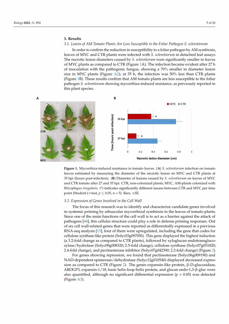
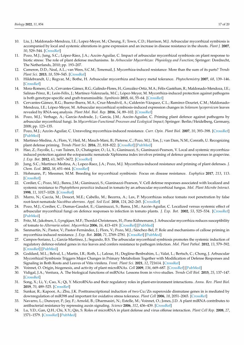
In order to confirm the reduction in susceptibility to a foliar pathogen by AM symbiosis,leaves of MYC and CTR plants were infected with S. sclerotiorum in detached leaf assays.The necrotic lesion diameters caused by S. sclerotiorum were significantly smaller in leavesof MYC plants as compared to CTR (Figure 1A). The infection became evident after 27 hof inoculation with the pathogenic fungus, showing a 70% smaller in diameter lesionsize in MYC plants (Figure A2); at 35 h, the infection was 50% less than CTR plants(Figure 1B). These results confirm that AM tomato plants are less susceptible to the foliarpathogen S. sclerotiorum showing mycorrhiza-induced resistance, as previously reported inthis plant species.
Biology 2022, 11, x 5 of 20
centrifuged again, and the supernatant was discarded. Next, 1 mL of 72% sulfuric acid
was added to the pellet and incubated at 50 °C for 1 h with agitation at 120 rpm, and then
centrifuged at 2500 rpm for 5 min. The supernatant was transferred to a 10-mL volumetric
flask and water was added to 10 mL. In a 1.5-mL Eppendorf tube, 10 µL of the flask con-
tent was mixed by inversion with 1 mL of cold anthrone solution (0.2 g of anthrone in 100
mL of 72% sulfuric acid) and left on ice for 2 min. The sample was then heated at 100 °C
for 15 min and allowed to cool down. Spectrophotometric reads were taken on the sam-
ples at 620 nm, and a standard curve with cellulose was used.
3. Results
3.1. Leaves of AM Tomato Plants Are Less Susceptible to the Foliar Pathogen S. sclerotiorum
In order to confirm the reduction in susceptibility to a foliar pathogen by AM symbi-
osis, leaves of MYC and CTR plants were infected with S. sclerotiorum in detached leaf
assays. The necrotic lesion diameters caused by S. sclerotiorum were significantly smaller
in leaves of MYC plants as compared to CTR (Figure 1A). The infection became evident
after 27 h of inoculation with the pathogenic fungus, showing a 70% smaller in diameter
lesion size in MYC plants (Figure A2); at 35 h, the infection was 50% less than CTR plants
(Figure 1B). These results confirm that AM tomato plants are less susceptible to the foliar
pathogen S. sclerotiorum showing mycorrhiza-induced resistance, as previously reported
in this plant species.
Figure 1. Mycorrhiza-induced resistance in tomato leaves. (A) S. sclerotiorum infection on tomato
leaves estimated by measuring the diameter of the necrotic lesion on MYC and CTR plants at 35 hpi
(hours post-infection). (B) Diameter of lesions caused by S. sclerotiorum on leaves of MYC and CTR
tomato after 27 and 35 hpi. CTR, non-colonized plants; MYC, AM-plants colonized with Rhizophagus
irregularis. (*) indicates significantly different means between CTR and MYC per time point (Stu-
dent’s t-test, p ≤ 0.05, n = 5). Bars, ±SE.
3.2. Expression of Genes Involved in the Cell Wall
The focus of this research was to identify and characterize candidate genes involved
in systemic priming by arbuscular mycorrhizal symbiosis in the leaves of tomato plants.
Since one of the main functions of the cell wall is to act as a barrier against the attack of
pathogens [44], this cellular structure could play a role in defense priming responses. Out
of six cell wall-related genes that were reported as differentially expressed in a previous
RNA-seq analysis [15], four of them were upregulated, including the gene that codes for
cellulose synthase-like protein (Solyc03g097050). This gene displayed the highest induc-
tion (a 3.2-fold change as compared to CTR plants), followed by xyloglucan endotransglu-
cosylase/hydrolase (Solyc09g008320; 2.5-fold change), cellulose synthase
(Solyc07g051820; 2.4-fold change), and pectinesterase inhibitor (Solyc07g042390; 2.2-fold
change) (Figure 2).
Figure 1. Mycorrhiza-induced resistance in tomato leaves. (A) S. sclerotiorum infection on tomatoleaves estimated by measuring the diameter of the necrotic lesion on MYC and CTR plants at35 hpi (hours post-infection). (B) Diameter of lesions caused by S. sclerotiorum on leaves of MYCand CTR tomato after 27 and 35 hpi. CTR, non-colonized plants; MYC, AM-plants colonized withRhizophagus irregularis. (*) indicates significantly different means between CTR and MYC per timepoint (Student’s t-test, p ≤ 0.05, n = 5). Bars, ±SE.
3.2. Expression of Genes Involved in the Cell Wall
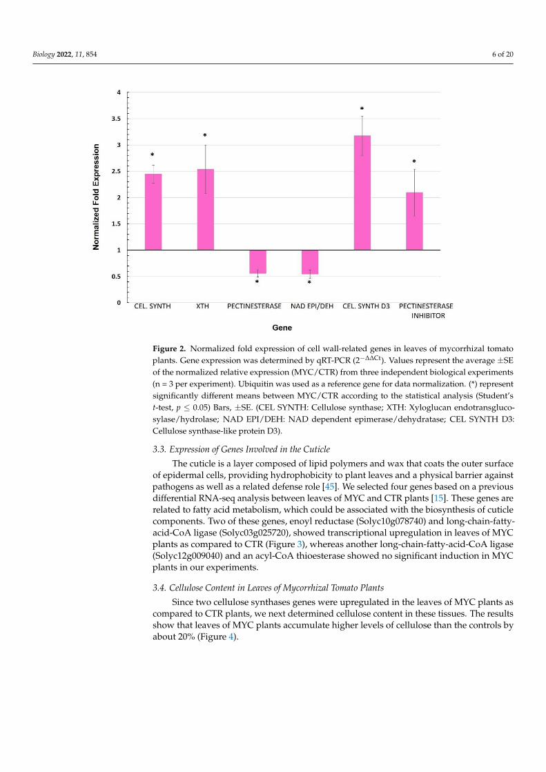
The focus of this research was to identify and characterize candidate genes involvedin systemic priming by arbuscular mycorrhizal symbiosis in the leaves of tomato plants.Since one of the main functions of the cell wall is to act as a barrier against the attack ofpathogens [44], this cellular structure could play a role in defense priming responses. Outof six cell wall-related genes that were reported as differentially expressed in a previousRNA-seq analysis [15], four of them were upregulated, including the gene that codes forcellulose synthase-like protein (Solyc03g097050). This gene displayed the highest induction(a 3.2-fold change as compared to CTR plants), followed by xyloglucan endotransgluco-sylase/hydrolase (Solyc09g008320; 2.5-fold change), cellulose synthase (Solyc07g051820;2.4-fold change), and pectinesterase inhibitor (Solyc07g042390; 2.2-fold change) (Figure 2).
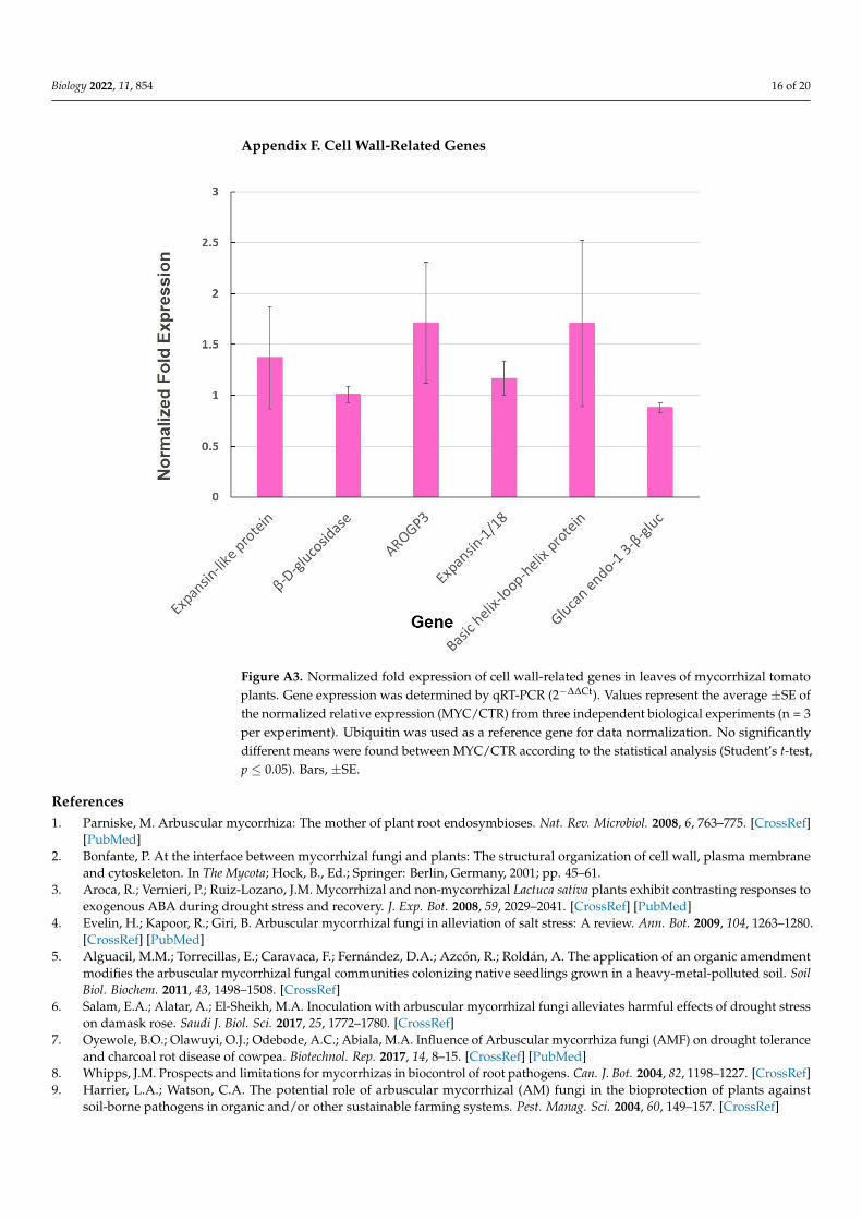
For genes showing repression, we found that pectinesterase (Solyc06g009190) andNAD-dependent epimerase/dehydratase (Solyc12g010540) displayed decreased expres-sion as compared to CTR (Figure 2). The genes expansin-like protein, β-D-glucosidase,AROGP3, expansin-1/18, basic helix-loop-helix protein, and glucan endo-1,3-β-gluc werealso quantified, although no significant differential expression (p < 0.05) was detected(Figure A3).
Biology 2022, 11, 854 6 of 20
Biology 2022, 11, x 6 of 20
Figure 2. Normalized fold expression of cell wall-related genes in leaves of mycorrhizal tomato
plants. Gene expression was determined by qRT-PCR (2−∆∆Ct). Values represent the average ±SE of
the normalized relative expression (MYC/CTR) from three independent biological experiments (n =
3 per experiment). Ubiquitin was used as a reference gene for data normalization. (*) represent sig-
nificantly different means between MYC/CTR according to the statistical analysis (Student’s t-test,
p ≤ 0.05) Bars, ±SE. (CEL SYNTH: Cellulose synthase; XTH: Xyloglucan endotransglucosylase/hy-
drolase; NAD EPI/DEH: NAD dependent epimerase/dehydratase; CEL SYNTH D3: Cellulose syn-
thase-like protein D3).
For genes showing repression, we found that pectinesterase (Solyc06g009190) and
NAD-dependent epimerase/dehydratase (Solyc12g010540) displayed decreased expres-
sion as compared to CTR (Figure 2). The genes expansin-like protein, β-D-glucosidase,
AROGP3, expansin-1/18, basic helix-loop-helix protein, and glucan endo-1,3-β-gluc were
also quantified, although no significant differential expression (p < 0.05) was detected (Fig-
ure A3).
3.3. Expression of Genes Involved in the Cuticle
The cuticle is a layer composed of lipid polymers and wax that coats the outer surface
of epidermal cells, providing hydrophobicity to plant leaves and a physical barrier against
pathogens as well as a related defense role [45]. We selected four genes based on a previ-
ous differential RNA-seq analysis between leaves of MYC and CTR plants [15]. These
genes are related to fatty acid metabolism, which could be associated with the biosynthe-
sis of cuticle components. Two of these genes, enoyl reductase (Solyc10g078740) and long-
chain-fatty-acid-CoA ligase (Solyc03g025720), showed transcriptional upregulation in
leaves of MYC plants as compared to CTR (Figure 3), whereas another long-chain-fatty-
acid-CoA ligase (Solyc12g009040) and an acyl-CoA thioesterase showed no significant in-
duction in MYC plants in our experiments.
Figure 2. Normalized fold expression of cell wall-related genes in leaves of mycorrhizal tomatoplants. Gene expression was determined by qRT-PCR (2−∆∆Ct). Values represent the average ±SEof the normalized relative expression (MYC/CTR) from three independent biological experiments(n = 3 per experiment). Ubiquitin was used as a reference gene for data normalization. (*) representsignificantly different means between MYC/CTR according to the statistical analysis (Student’st-test, p ≤ 0.05) Bars, ±SE. (CEL SYNTH: Cellulose synthase; XTH: Xyloglucan endotransgluco-sylase/hydrolase; NAD EPI/DEH: NAD dependent epimerase/dehydratase; CEL SYNTH D3:Cellulose synthase-like protein D3).
3.3. Expression of Genes Involved in the Cuticle
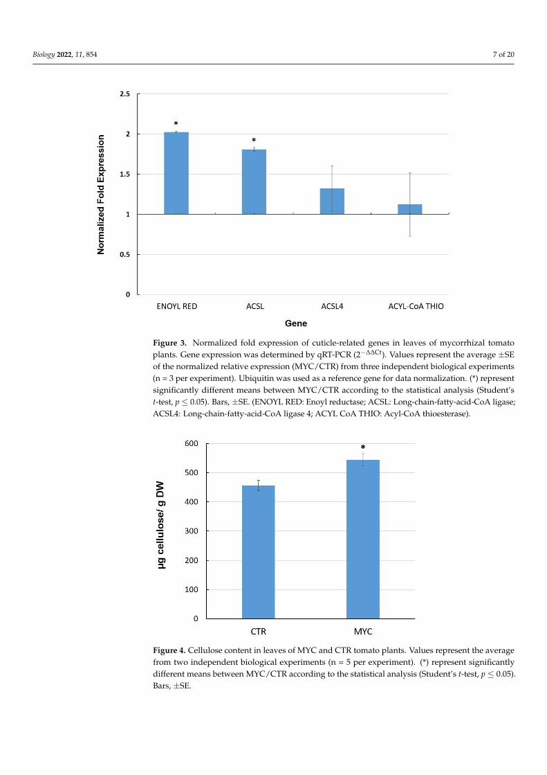
The cuticle is a layer composed of lipid polymers and wax that coats the outer surfaceof epidermal cells, providing hydrophobicity to plant leaves and a physical barrier againstpathogens as well as a related defense role [45]. We selected four genes based on a previousdifferential RNA-seq analysis between leaves of MYC and CTR plants [15]. These genes arerelated to fatty acid metabolism, which could be associated with the biosynthesis of cuticlecomponents. Two of these genes, enoyl reductase (Solyc10g078740) and long-chain-fatty-acid-CoA ligase (Solyc03g025720), showed transcriptional upregulation in leaves of MYCplants as compared to CTR (Figure 3), whereas another long-chain-fatty-acid-CoA ligase(Solyc12g009040) and an acyl-CoA thioesterase showed no significant induction in MYCplants in our experiments.
3.4. Cellulose Content in Leaves of Mycorrhizal Tomato Plants
Since two cellulose synthases genes were upregulated in the leaves of MYC plants ascompared to CTR plants, we next determined cellulose content in these tissues. The resultsshow that leaves of MYC plants accumulate higher levels of cellulose than the controls byabout 20% (Figure 4).
Biology 2022, 11, 854 7 of 20Biology 2022, 11, x 7 of 20
Figure 3. Normalized fold expression of cuticle-related genes in leaves of mycorrhizal tomato plants.
Gene expression was determined by qRT-PCR (2−∆∆Ct). Values represent the average ±SE of the nor-
malized relative expression (MYC/CTR) from three independent biological experiments (n = 3 per
experiment). Ubiquitin was used as a reference gene for data normalization. (*) represent signifi-
cantly different means between MYC/CTR according to the statistical analysis (Student’s t-test, p ≤
0.05). Bars, ±SE. (ENOYL RED: Enoyl reductase; ACSL: Long-chain-fatty-acid-CoA ligase; ACSL4:
Long-chain-fatty-acid-CoA ligase 4; ACYL CoA THIO: Acyl-CoA thioesterase).
3.4. Cellulose Content in Leaves of Mycorrhizal Tomato Plants
Since two cellulose synthases genes were upregulated in the leaves of MYC plants as
compared to CTR plants, we next determined cellulose content in these tissues. The results
show that leaves of MYC plants accumulate higher levels of cellulose than the controls by
about 20% (Figure 4).
Figure 3. Normalized fold expression of cuticle-related genes in leaves of mycorrhizal tomatoplants. Gene expression was determined by qRT-PCR (2−∆∆Ct). Values represent the average ±SEof the normalized relative expression (MYC/CTR) from three independent biological experiments(n = 3 per experiment). Ubiquitin was used as a reference gene for data normalization. (*) representsignificantly different means between MYC/CTR according to the statistical analysis (Student’st-test, p ≤ 0.05). Bars, ±SE. (ENOYL RED: Enoyl reductase; ACSL: Long-chain-fatty-acid-CoA ligase;ACSL4: Long-chain-fatty-acid-CoA ligase 4; ACYL CoA THIO: Acyl-CoA thioesterase).
Biology 2022, 11, x 7 of 20
Figure 3. Normalized fold expression of cuticle-related genes in leaves of mycorrhizal tomato plants.
Gene expression was determined by qRT-PCR (2−∆∆Ct). Values represent the average ±SE of the nor-
malized relative expression (MYC/CTR) from three independent biological experiments (n = 3 per
experiment). Ubiquitin was used as a reference gene for data normalization. (*) represent signifi-
cantly different means between MYC/CTR according to the statistical analysis (Student’s t-test, p ≤
0.05). Bars, ±SE. (ENOYL RED: Enoyl reductase; ACSL: Long-chain-fatty-acid-CoA ligase; ACSL4:
Long-chain-fatty-acid-CoA ligase 4; ACYL CoA THIO: Acyl-CoA thioesterase).
3.4. Cellulose Content in Leaves of Mycorrhizal Tomato Plants
Since two cellulose synthases genes were upregulated in the leaves of MYC plants as
compared to CTR plants, we next determined cellulose content in these tissues. The results
show that leaves of MYC plants accumulate higher levels of cellulose than the controls by
about 20% (Figure 4).
Figure 4. Cellulose content in leaves of MYC and CTR tomato plants. Values represent the averagefrom two independent biological experiments (n = 5 per experiment). (*) represent significantlydifferent means between MYC/CTR according to the statistical analysis (Student’s t-test, p ≤ 0.05).Bars, ±SE.
Biology 2022, 11, 854 8 of 20
3.5. Expression Analysis of Selected miRNAs and Their Target Genes in Leaves ofMycorrhiza-Colonized Tomato
Considering that miRNAs are essential post-transcriptional regulators in response tobiotic and abiotic stress, one of our goals was to identify differentially regulated miRNAsin leaves of MYC plants that could be relevant in defense priming in tomato, i.e., in MYCplants before any pathogen attack. Since there is no published information on miRNA inthe leaves of mycorrhizal plants, we selected miRNAs that are reported to be differentiallyregulated in previous studies in mycorrhizal tomato roots [46], as well as miRNAs thatwere identified as targeting cell wall-related genes in other plants such as sorghum [47].
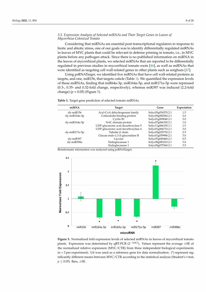
Using psRNATarget, we identified five miRNAs that have cell wall-related proteins astargets, and one, miR156, that targets cuticle (Table 1). We quantified the expression levelsof these miRNAs, finding that miR64a-3p, miR164a-5p, and miR171e-5p were repressed(0.3-, 0.35- and 0.32-fold change, respectively), whereas miR397 was induced (2.2-foldchange) (p < 0.05) (Figure 5).
Table 1. Target gene prediction of selected tomato miRNAs.
miRNA Target Gene Expectation
sly-miR156 Acyl-CoA dehydrogenase family Solyc05g054370.2.1 1.5sly-miR164a-3p Calmodulin-binding protein Solyc09g082560.2.1 2.0
Cyclin B1 Solyc01g009040.2.1 3.0sly-miR164a-5p NAC domain protein Solyc07g066330.2.1 1.0
UDP-glucuronic acid decarboxylase 5 Solyc11g066150.1.1 1.5UDP-glucuronic acid decarboxylase 4 Solyc01g066710.2.1 3.0
sly-miR171e-5p Tubulin β chain Solyc03g025730.2.1 2.5Glucan endo-1,3-β-glucosidase B Solyc01g059980.2.1 3.0
sly-miR397 Laccase Solyc07g049460.2.1 0.5sly-miR398a Endoglucanase 1 Solyc08g083210.2.1 3.0
Endoglucanase 1 Solyc09g075360.2.1 3.5Bioinformatic information was analyzed using psRNATarget.
Biology 2022, 11, x 9 of 20
Figure 5. Normalized fold expression levels of selected miRNAs in leaves of mycorrhizal tomato plants. Expression was determined by qRT-PCR (2−∆∆Ct). Values represent the average ±SE of the normalized relative expression (MYC/CTR) from three independent biological experiments (n = 3 per experiment). U6 was used as a reference gene for data normalization. (*) represent significantly different means between MYC/CTR according to the statistical analysis (Student’s t-test, p ≤ 0.05). Bars, ±SE.
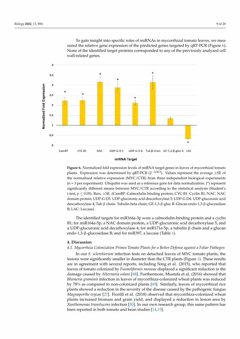
Figure 6. Normalized fold expression levels of miRNA target genes in leaves of mycorrhizal tomato plants. Expression was determined by qRT-PCR (2−∆∆Ct). Values represent the average ±SE of the normalized relative expression (MYC/CTR) from three independent biological experiments (n = 3 per experiment). Ubiquitin was used as a reference gene for data normalization. (*) represent signif-icantly different means between MYC/CTR according to the statistical analysis (Student’s t-test, p ≤ 0.05). Bars, ±SE. (CamBP: Calmodulin binding protein; CYC B1: Cyclin B1; NAC: NAC domain pro-tein; UDP-G-D5: UDP-glucoronic acid descarboxylase 5; UDP-G-D4: UDP-glucoronic acid descar-boxylase 4; Tub β chain: Tubulin beta chain; GE-1,3-β-gluc B: Glucan endo-1,3-β-glucosidase B; LAC: Laccase).
Figure 5. Normalized fold expression levels of selected miRNAs in leaves of mycorrhizal tomatoplants. Expression was determined by qRT-PCR (2−∆∆Ct). Values represent the average ±SE ofthe normalized relative expression (MYC/CTR) from three independent biological experiments(n = 3 per experiment). U6 was used as a reference gene for data normalization. (*) represent sig-nificantly different means between MYC/CTR according to the statistical analysis (Student’s t-test,p ≤ 0.05). Bars, ±SE.
Biology 2022, 11, 854 9 of 20
To gain insight into specific roles of miRNAs in mycorrhizal tomato leaves, we mea-sured the relative gene expression of the predicted genes targeted by qRT-PCR (Figure 6).None of the identified target proteins corresponded to any of the previously analyzed cellwall-related genes.
Biology 2022, 11, x 9 of 20
Figure 5. Normalized fold expression levels of selected miRNAs in leaves of mycorrhizal tomato
plants. Expression was determined by qRT-PCR (2−∆∆Ct). Values represent the average ±SE of the
normalized relative expression (MYC/CTR) from three independent biological experiments (n = 3
per experiment). U6 was used as a reference gene for data normalization. (*) represent significantly
different means between MYC/CTR according to the statistical analysis (Student’s t-test, p ≤ 0.05).
Bars, ±SE.
Figure 6. Normalized fold expression levels of miRNA target genes in leaves of mycorrhizal tomato
plants. Expression was determined by qRT-PCR (2−∆∆Ct). Values represent the average ±SE of the
normalized relative expression (MYC/CTR) from three independent biological experiments (n = 3
per experiment). Ubiquitin was used as a reference gene for data normalization. (*) represent signif-
icantly different means between MYC/CTR according to the statistical analysis (Student’s t-test, p ≤
0.05). Bars, ±SE. (CamBP: Calmodulin binding protein; CYC B1: Cyclin B1; NAC: NAC domain pro-
tein; UDP-G-D5: UDP-glucoronic acid descarboxylase 5; UDP-G-D4: UDP-glucoronic acid descar-
boxylase 4; Tub β chain: Tubulin beta chain; GE-1,3-β-gluc B: Glucan endo-1,3-β-glucosidase B;
LAC: Laccase).
Figure 6. Normalized fold expression levels of miRNA target genes in leaves of mycorrhizal tomatoplants. Expression was determined by qRT-PCR (2−∆∆Ct). Values represent the average ±SE ofthe normalized relative expression (MYC/CTR) from three independent biological experiments(n = 3 per experiment). Ubiquitin was used as a reference gene for data normalization. (*) representsignificantly different means between MYC/CTR according to the statistical analysis (Student’st-test, p ≤ 0.05). Bars, ±SE. (CamBP: Calmodulin binding protein; CYC B1: Cyclin B1; NAC: NACdomain protein; UDP-G-D5: UDP-glucoronic acid descarboxylase 5; UDP-G-D4: UDP-glucoronic aciddescarboxylase 4; Tub β chain: Tubulin beta chain; GE-1,3-β-gluc B: Glucan endo-1,3-β-glucosidaseB; LAC: Laccase).
The identified targets for miR164a-3p were a calmodulin-binding protein and a cyclinB1; for miR164a-5p, a NAC domain protein, a UDP-glucuronic acid decarboxylase 5, anda UDP-glucuronic acid decarboxylase 4; for miR171e-5p, a tubulin β chain and a glucanendo-1,3-β-glucosidase B; and for miR397, a laccase (Table 1).
4. Discussion4.1. Mycorrhiza Colonization Primes Tomato Plants for a Better Defense against a Foliar Pathogen
In our S. sclerotiorum infection tests on detached leaves of MYC tomato plants, thelesions were significantly smaller in diameter than the CTR plants (Figure 1). These resultsare in agreement with several reports, including Song et al. (2015), who reported thatleaves of tomato colonized by Funneliformis mosseae displayed a significant reduction in thedamage caused by Alternaria solani [48]. Furthermore, Mustafa et al. (2016) showed thatBlumeria graminis infection in leaves of mycorrhiza-colonized wheat plants was reducedby 78% as compared to non-colonized plants [49]. Similarly, leaves of mycorrhizal riceplants showed a reduction in the severity of the disease caused by the pathogenic fungusMagnaporthe oryzae [27]. Fiorilli et al. (2018) observed that mycorrhiza-colonized wheatplants increased biomass and grain yield, and displayed a reduction in lesion area byXanthomonas translucens infection [50]. In our own research group, this same pattern hasbeen reported in both tomato and bean studies [14,15].

Biology 2022, 11, 854 10 of 20
Although there is abundant evidence for the occurrence of mycorrhiza-induced re-sistance, much work must still be performed to elucidate the biochemical and molecularmechanisms involved when the mycorrhiza plant is defending itself against a pathogen, aswell as before the attack occurs, i.e., during the priming stage. The discrete modificationand/or remodeling of some cell wall components such as cellulose fibrils, lignin, and evencuticle could be important for mycorrhiza plants before their encounter with a pathogen, asa means to mount a faster and stronger defense reaction in a subsequent pathogen attack.In addition, it is well known that the cell wall is remodeled and reinforced at specific sitesof interaction with pathogens [51].
4.2. Cell Wall and Cuticle Genes in Mycorrhiza Defense Priming
We observed that a cellulose synthase and a cellulose synthase-like protein were bothupregulated in the leaves of MYC plants (Figure 2). Since this regulation was accompaniedby an increase in cellulose content in leaves of MYC plants as compared to CTR (Figure 4),it is possible that that the proteins encoded by these genes might be involved in thereinforcement of the cell wall, as a priming response before any interaction with a pathogen.Cellulose, the main constituent of cell walls (40%), complexes with other components suchas proteins, high molecular weight polysaccharides and aromatic substances, and is capableof dynamic changes [52–54].
Previous studies have linked the regulation of cellulose synthase genes to defense:in rice, cellulose synthase genes were markedly upregulated in an RSV (rice stripe virus)-resistant cultivar, but downregulated in a susceptible cultivar [55]. Cell wall strengtheninghas been widely documented in response to pathogen attack [56–58]. In addition, celluloseaccumulation has been reported to occur in response to environmental stress, implying thatthe cell wall is reinforced to avoid external stress [59]. Here, however, we are reporting theaccumulation of cellulose in leaf tissue that has not yet been attacked by a pathogen. Wetherefore take this accumulation to be a response to the establishment of symbiosis, whichcan be interpreted as a step in priming, since it occurs as a response to the interaction witha beneficial microorganism (in this case a mycorrhizal fungus) and before any attack bya pathogen.
Interestingly, we observed the downregulation of a pectinesterase gene in leavesof MYC plants, while a pectinesterase inhibitor gene was also upregulated (Figure 2).Pectinesterases catalyze the hydrolysis of pectin methyl esters, thus decreasing their degreeof esterification, reducing intracellular adhesiveness and tissue rigidity. Pectinesteraseshave been shown to have an important function in response to fungal pathogens and arenecessary for the systemic spread of tobacco mosaic virus throughout a plant [60,61]. Instudies on cotton plants, pectinesterase inhibitor (PMEI) was shown to participate in plantresponses to fungal infection, including Verticillium dahliae in cotton plants; its ectopicexpression also increased pectin methyl esterification and limited fungal disease [62]. Onthe other hand, in pepper plants, silencing of the PMEI gene resulted in enhanced diseasesusceptibility to infection by the virulent Xanthomonas campestris pv. vesicatoria, whereasits overexpression in Arabidopsis showed enhanced resistance to Pseudomonas syringaepv. tomato [63]. These results suggest that the repression of a pectinesterase gene and theupregulation of a pectinesterase inhibitor may hinder the success of a subsequent pathogeninfection, indicating that these two genes might be mycorrhiza priming genes.
Other genes that we found to be upregulated in leaves of MYC plants include anenoyl reductase and a long-chain-fatty-acid-CoA ligase (Figure 3), which are involved infatty acid metabolism as well as the biosynthesis of cuticle components. In addition toits essential role in limiting water loss, the cuticle protects the plant against xenobioticsand pathogens. Components of the cuticle are perceived by pathogenic fungi, and theyinduce several processes during pathogenesis. Furthermore, modifications of the cuticlecan result in resistance to necrotrophs [45]. Thus, alteration of the cuticle may also be partof the priming mechanism in leaves of mycorrhizal plants, as a means to more efficientlyreact to a subsequent pathogen attack.
Biology 2022, 11, 854 11 of 20
4.3. miRNAs as Potential Players in Mycorrhiza Defense Priming
The function of miRNAs as post-transcriptional regulators suggests that they couldplay a role in the mycorrhizal systemic response in leaves. In the present work, threemiRNAs (miR164a-3p, miR164a-5p, and miR171e-5p) were found to be repressed in leavesof MYC plants (Figure 5), whereas their predicted target genes (Table 1) exhibited aninverse expression relative to their corresponding miRNAs (Figure 6), suggesting thatthese miRNAs could have a post-transcriptional regulatory action on the predicted targets.This miRNA/target gene expression pattern indicates that these molecular entities mightbe part of the response of the shoot to AM establishment in the root that is involved indefense priming.
One of the predicted target genes of miR164a-3p is a calmodulin-binding protein(CaMBP). These genes belong to the IQD/SUN gene family, and in Populus plants anIDQ/SUN gene has been associated with the signaling of cell wall biosynthesis [64]. Plantresistance to diseases involves a number of signaling response pathways including cal-cium/calmodulin, reactive oxygen species, and phytohormones [65]. Calmodulin regulatesplant disease responses through CaMBPs, often by affecting the biosynthesis or signalingof JA and SA [66]. The induction of CaMBPs has been associated with defense responses inrice against M. grisea [67], and in Arabidopsis against Botrytis cinerea through the regulationof JA synthesis [66]. On the other hand, jasmonates are reported to accumulate in leaveson mycorrhiza-colonized plants [11,16,28], which could be mediated by a CaMBP suchas the one induced in the present work. Although the genome-wide identification andexpression analysis of this gene family in tomato has been published [68], no informationon the role of these genes or their associated miRNAs in AM colonized plants has beenreported. However, its potential role in the signaling of cell wall biosynthesis could be inaccordance with its involvement in cell wall reinforcement by mycorrhizal priming.
Cyclin B1, another predicted target of miR164a-3p, also showed an inverse expression(upregulation) to this miRNA (Figure 6). The expression of cyclin B1 in Arabidopsisaccelerates the proliferation of root cells and promotes DNA repair [69,70]. Recent studiesby Ambastha and Leshem (2020) provide evidence for the intense activity of cyclin B1during salt stress as a means to repair damaged DNA in Arabidopsis [71]. Since mycorrhizacolonization also induces resistance to abiotic stress, cyclin B1 might be involved in theresponse to abiotic instead of biotic stress.
Another downregulated miRNA in the leaves of MYC plants is miRNA164a-5p,and we detected the upregulation of one of its predicted targets, a NAC domain pro-tein (Figure 6). NAC is an important family of plant transcription factors that are associatedwith developmental processes, and together with miR164 they are required for the sub-sequent formation of the limits of the lateral organs in the apical meristem, in additionto leaf development in tomato plants [72]. NACs are also implicated in leaf senescenceand secondary wall formation, as well as responses to abiotic and biotic stresses [73–75].In rice, the NAC TF gene (ONAC063) has been shown to respond to high-temperaturestress (Yokotani et al. 2009) [76]. Similarly, overexpressing the NAC transcription factorJUNGBRUNNEN1 (JUB1; ANAC042) extends longevity and increases tolerance to heatstress in Arabidopsis thaliana [77]. Furthermore, the induction of miR164 and repression of itsrespective target gene in response to aluminum toxicity have been observed in nodulatedbean plants [37].
The expression and regulation of lignin biosynthetic genes are determined by varioustranscription factors including NACs, whose repression leads to a reduction in secondarywall thickening in fibers [78–80]. The secondary wall is mainly composed of lignin, apolyphenolic biopolymer responsible for contributing to cell rigidity and protection againstpathogens, as well as covering the interior of the vessels to facilitate hydrophilic transport.
NAC transcription factors also play an important role in the regulation of plantdefense responses during pathogen stress, such as insect wounds [75,81,82] and nematodeinteraction [83]. In addition to the participation of NAC proteins in the defense mechanismagainst pathogens, the possible regulation of lignin biosynthesis by miR164a-5p during
Biology 2022, 11, 854 12 of 20
mycorrhiza colonization could be part of the priming mechanism. This in turn could resultin the reinforcement of the cell wall to provide better defense in a subsequent pathogenattack, as shown in the present work.
UDP-glucuronic acid decarboxylases 4 and 5 were induced in leaves of MYC plants(Figure 6) whereas miR164a-5p was repressed (Figure 5), suggesting that UDP-glucuronicacid decarboxylases 4 and 5 genes are the target. The enzyme UDP-glucuronic acid decar-boxylase is responsible for converting UDP-glucuronic acid (UDP-GLcA) into UDP-xylose,and is also involved in the formation of xylans during the biosynthesis of non-cellulosicpolysaccharides of the cell wall [84,85]. Xylans play an important role in the integrity ofthe plant cell wall and increase its resistance to enzymatic digestion, thus helping plants todefend themselves against herbivores and pathogens. Crowe et al. (2021) demonstratedthat xylans are critical for the proper bundling and alignment of cellulose microfibrils inplant secondary cell walls [86].
The interactions between the three main structural biopolymers xylan, cellulose andlignin seem to be essential for providing the rigidity of plant cell walls [87,88]. The possibleregulation of two UDP-glucuronic decarboxylases and a NAC domain protein by miR164a-5p, along with the accumulation of cellulose possibly due to the upregulation of cellulosesynthase genes, could therefore be part of the priming responses that favor secondary cellwall reinforcement, even occurring before the pathogen attack.
MiR171e-5p was also identified as being repressed in the leaves of MYC plants(Figure 5), and one of its targets showing upregulation was a tubulin β chain gene (Figure 6).Tubulin is an essential protein for eukaryotic cells formed by two homologous globularproteins, α-tubulin and β-tubulin. This protein plays a major role in defining the shapeof the cell and organizing its cytoplasm, as well as in cell division and the orientation ofcomponents in the cell wall [89,90]. Spokevicius et al. (2007) showed that β-tubulin isinvolved in determining the orientation of cellulose microfibrils in plant secondary fibercell walls; in this way, β-tubulins participate in the flexibility, strength, and resistance ofplants [91]. Together with the aforementioned analyses, our findings indicate that miR171and its regulation of β-tubulin could participate in priming by mycorrhiza colonization toresult in an improved defense after a pathogen attack, due to the structural role of β-tubulinin the orientation of cellulose microfibrils, an essential part of the secondary cell wall.
Another selected miRNA, miR397, showed upregulation expression (Figure 5), whereasits target, a laccase gene, was downregulated (Figure 6). Laccases are a family of multi-copper oxidoreductase enzymes reported to be involved in the biosynthesis of lignin bypolymerizing monolignols into lignin [92]. Consequently, their downregulation should im-ply a reduction in lignin polymerization. Interestingly, Lee et al. (2012) reported that lignincontent was lower in arbuscular mycorrhizal plants as compared to non-mycorrhizal con-trols in drought-stressed perennial ryegrass [93]. Furthermore, Baslam et al. (2013) observedthat the leaves of mycorrhizal alfalfa plants exposed to CO2 showed increased hemicel-lulose and decreased lignin concentrations in cell walls as compared to non-mycorrhizalplants [94]. Determining the lignin content in leaves of mycorrhizal tomato will help sup-port the hypothesis that this laccase, regulated by miR397, has an effect on lignin and itspotential role in priming. Although the downregulation of laccase and upregulation ofNAC domain protein seem to have contrasting effects on lignin content, these two potentialstrategies for regulating lignin composition might reflect that more than one mechanism isneeded to fine-tune the accumulation of such an important cell wall component as lignin.
In contrast, Pan et al. (2017) observed in rice that miR397 negatively regulates laccases,which induced the expression of three oxidase/peroxidase genes and promoted an increasein herbicide tolerance related to a better response to oxidative stress. This places miR397as a potentially important element in the regulation of responses to oxidative stress [95],which could certainly have a role in the plant response to mycorrhiza colonization, aspreviously noted [96].
Biology 2022, 11, 854 13 of 20
It is important to mention that some of the analyzed targets have already been vali-dated by degradome sequencing in other tomato studies, such as the case of NAC domainprotein as a target of miR164a-5p [97], and laccase as a target of miR397 [98].
Although there is abundant evidence for the role of cell wall modifications as an im-portant mechanism in plant defense against pathogens, our results support the idea that thepriming mechanism induced by arbuscular mycorrhiza colonization also involves the rein-forcement of the cell wall. Additional studies will be needed in order to completely elucidatethe role of the cell wall in the priming mechanism induced by mycorrhiza colonization.
5. Conclusions
The present work demonstrates that mycorrhiza colonization induces a priming statethat confers an increase in defense against the foliar pathogen Sclerotinia sclerotiorum intomato leaves. This could be associated with the observed differential expression of cellwall- and cuticle-related genes, as these two structures form the first physical barriersto pathogens. The induction of cellulose synthase genes is consistent with the highercellulose content observed in the leaves of mycorrhizal plants as compared to control plants.Modifications of the cell wall and cuticle in response to the establishment of the symbiosiscould be part of the priming mechanisms that prepare the plant tissues to respond ina faster and stronger manner to a subsequent pathogen encounter. Although abundantscientific work supports the idea that the cell wall is strengthened during a pathogen attack,literature concerning cell wall reinforcement in leaves as a result of mycorrhiza symbiosisis scarce.
In this study, we identified differentially expressed miRNAs/targets for the firsttime in the leaves of mycorrhizal tomato plants. These molecules could be part of asophisticated and efficient strategy to improve defense induced by mycorrhiza colonization.Further analyses can build upon this work to demonstrate the specific roles of candidatetomato genes and miRNAs/target nodes in the priming mechanism, using genetic and/orfunctional approaches.
Author Contributions: Conceptualization: A.B.M.-S. and M.L.-M.; methodology: A.B.M.-S., A.Z.R.-C.,A.B.-L., M.C.-R. and C.C.-M.; formal analysis: A.B.M.-S. and M.L.-M.; investigation: A.B.M.-S.,A.Z.R.-C., A.B.-L. and M.C.-R.; writing—original draft preparation: A.B.M.-S.; writing—reviewand editing: A.B.M.-S. and M.L.-M.; supervision: A.B.M.-S. and M.L.-M.; project administration:M.L.-M.; funding acquisition: M.L.-M. All authors have read and agreed to the published version ofthe manuscript.
Funding: This research was supported by CONACyT (Project A1_S_31400) and SIP-IPN (20196531),Consejo Nacional de Ciencia y Tecnología (CONACYT) postdoctoral fellowship (CVU: 336932), and amaster’s scholarship (CVU: 829480).
Informed Consent Statement: Not applicable.
Data Availability Statement: The data presented in this study are available on request from thecorresponding author.
Acknowledgments: We are grateful to Claudia María Ramírez-Douriet for technical support.
Conflicts of Interest: The authors declare no conflict of interest.
Biology 2022, 11, 854 14 of 20
Appendix A
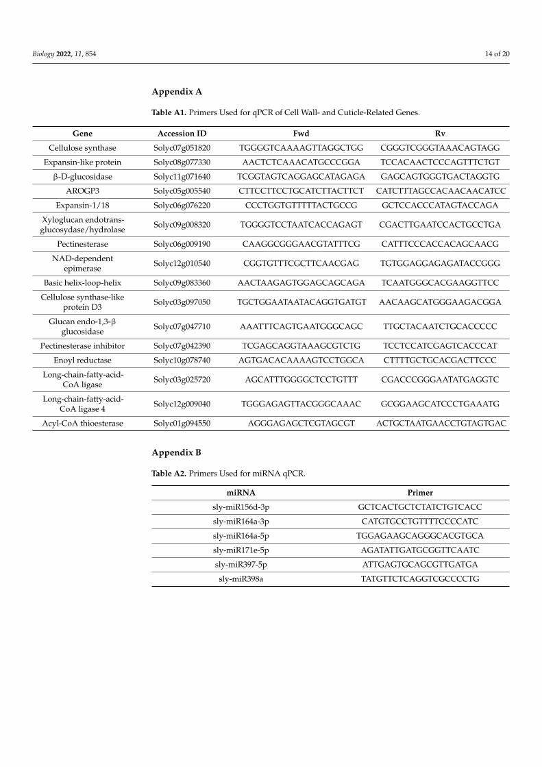
Table A1. Primers Used for qPCR of Cell Wall- and Cuticle-Related Genes.
Gene Accession ID Fwd Rv
Cellulose synthase Solyc07g051820 TGGGGTCAAAAGTTAGGCTGG CGGGTCGGGTAAACAGTAGG
Expansin-like protein Solyc08g077330 AACTCTCAAACATGCCCGGA TCCACAACTCCCAGTTTCTGT
β-D-glucosidase Solyc11g071640 TCGGTAGTCAGGAGCATAGAGA GAGCAGTGGGTGACTAGGTG
AROGP3 Solyc05g005540 CTTCCTTCCTGCATCTTACTTCT CATCTTTAGCCACAACAACATCC
Expansin-1/18 Solyc06g076220 CCCTGGTGTTTTTACTGCCG GCTCCACCCATAGTACCAGA
Xyloglucan endotrans-glucosydase/hydrolase Solyc09g008320 TGGGGTCCTAATCACCAGAGT CGACTTGAATCCACTGCCTGA
Pectinesterase Solyc06g009190 CAAGGCGGGAACGTATTTCG CATTTCCCACCACAGCAACG
NAD-dependentepimerase Solyc12g010540 CGGTGTTTCGCTTCAACGAG TGTGGAGGAGAGATACCGGG
Basic helix-loop-helix Solyc09g083360 AACTAAGAGTGGAGCAGCAGA TCAATGGGCACGAAGGTTCC
Cellulose synthase-likeprotein D3 Solyc03g097050 TGCTGGAATAATACAGGTGATGT AACAAGCATGGGAAGACGGA
Glucan endo-1,3-βglucosidase Solyc07g047710 AAATTTCAGTGAATGGGCAGC TTGCTACAATCTGCACCCCC
Pectinesterase inhibitor Solyc07g042390 TCGAGCAGGTAAAGCGTCTG TCCTCCATCGAGTCACCCAT
Enoyl reductase Solyc10g078740 AGTGACACAAAAGTCCTGGCA CTTTTGCTGCACGACTTCCC
Long-chain-fatty-acid-CoA ligase Solyc03g025720 AGCATTTGGGGCTCCTGTTT CGACCCGGGAATATGAGGTC
Long-chain-fatty-acid-CoA ligase 4 Solyc12g009040 TGGGAGAGTTACGGGCAAAC GCGGAAGCATCCCTGAAATG
Acyl-CoA thioesterase Solyc01g094550 AGGGAGAGCTCGTAGCGT ACTGCTAATGAACCTGTAGTGAC
Appendix B
Table A2. Primers Used for miRNA qPCR.
miRNA Primer
sly-miR156d-3p GCTCACTGCTCTATCTGTCACC
sly-miR164a-3p CATGTGCCTGTTTTCCCCATC
sly-miR164a-5p TGGAGAAGCAGGGCACGTGCA
sly-miR171e-5p AGATATTGATGCGGTTCAATC
sly-miR397-5p ATTGAGTGCAGCGTTGATGA
sly-miR398a TATGTTCTCAGGTCGCCCCTG
Biology 2022, 11, 854 15 of 20
Appendix C
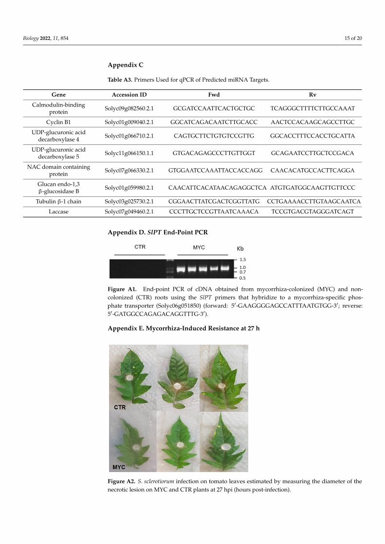
Table A3. Primers Used for qPCR of Predicted miRNA Targets.
Gene Accession ID Fwd Rv
Calmodulin-bindingprotein Solyc09g082560.2.1 GCGATCCAATTCACTGCTGC TCAGGGCTTTTCTTGCCAAAT
Cyclin B1 Solyc01g009040.2.1 GGCATCAGACAATCTTGCACC AACTCCACAAGCAGCCTTGC
UDP-glucuronic aciddecarboxylase 4 Solyc01g066710.2.1 CAGTGCTTCTGTGTCCGTTG GGCACCTTTCCACCTGCATTA
UDP-glucuronic aciddecarboxylase 5 Solyc11g066150.1.1 GTGACAGAGCCCTTGTTGGT GCAGAATCCTTGCTCCGACA
NAC domain containingprotein Solyc07g066330.2.1 GTGGAATCCAAATTACCACCAGG CAACACATGCCACTTCAGGA
Glucan endo-1,3β-glucosidase B Solyc01g059980.2.1 CAACATTCACATAACAGAGGCTCA ATGTGATGGCAAGTTGTTCCC
Tubulin β-1 chain Solyc03g025730.2.1 CGGAACTTATCGACTCGGTTATG CCTGAAAACCTTGTAAGCAATCA
Laccase Solyc07g049460.2.1 CCCTTGCTCCGTTAATCAAACA TCCGTGACGTAGGGATCAGT
Appendix D. SlPT End-Point PCR
Biology 2022, 11, x 15 of 20
Appendix C.
Table A3. Primers Used for qPCR of Predicted miRNA Targets.
Gene Accession ID Fwd Rv
Calmodulin-binding
protein Solyc09g082560.2.1 GCGATCCAATTCACTGCTGC TCAGGGCTTTTCTTGCCAAAT
Cyclin B1 Solyc01g009040.2.1 GGCATCAGACAATCTTGCACC AACTCCACAAGCAGCCTTGC
UDP-glucuronic acid
decarboxylase 4 Solyc01g066710.2.1 CAGTGCTTCTGTGTCCGTTG GGCACCTTTCCACCTGCATTA
UDP-glucuronic acid
decarboxylase 5 Solyc11g066150.1.1 GTGACAGAGCCCTTGTTGGT GCAGAATCCTTGCTCCGACA
NAC domain contain-
ing protein Solyc07g066330.2.1 GTGGAATCCAAATTACCACCAGG CAACACATGCCACTTCAGGA
Glucan endo-1,3 β-
glucosidase B Solyc01g059980.2.1
CAACATTCACATAACAGAGGCTC
A ATGTGATGGCAAGTTGTTCCC
Tubulin β-1 chain Solyc03g025730.2.1 CGGAACTTATCGACTCGGTTATG CCTGAAAACCTTGTAA-
GCAATCA
Laccase Solyc07g049460.2.1 CCCTTGCTCCGTTAATCAAACA TCCGTGACGTAGGGATCAGT
Appendix D. SlPT End-Point PCR
Figure A1. End-point PCR of cDNA obtained from mycorrhiza-colonized (MYC) and non-colonized
(CTR) roots using the SlPT primers that hybridize to a mycorrhiza-specific phosphate transporter
(Solyc06g051850) (forward: 5′-GAAGGGGAGCCATTTAATGTGG-3′; reverse: 5′-
GATGGCCAGAGACAGGTTTG-3′).
Appendix E. Mycorrhiza-Induced Resistance at 27 h
Figure A1. End-point PCR of cDNA obtained from mycorrhiza-colonized (MYC) and non-colonized (CTR) roots using the SlPT primers that hybridize to a mycorrhiza-specific phos-phate transporter (Solyc06g051850) (forward: 5′-GAAGGGGAGCCATTTAATGTGG-3′; reverse:5′-GATGGCCAGAGACAGGTTTG-3′).
Appendix E. Mycorrhiza-Induced Resistance at 27 h
Biology 2022, 11, x 15 of 20
Appendix C.
Table A3. Primers Used for qPCR of Predicted miRNA Targets.
Gene Accession ID Fwd Rv
Calmodulin-binding
protein Solyc09g082560.2.1 GCGATCCAATTCACTGCTGC TCAGGGCTTTTCTTGCCAAAT
Cyclin B1 Solyc01g009040.2.1 GGCATCAGACAATCTTGCACC AACTCCACAAGCAGCCTTGC
UDP-glucuronic acid
decarboxylase 4 Solyc01g066710.2.1 CAGTGCTTCTGTGTCCGTTG GGCACCTTTCCACCTGCATTA
UDP-glucuronic acid
decarboxylase 5 Solyc11g066150.1.1 GTGACAGAGCCCTTGTTGGT GCAGAATCCTTGCTCCGACA
NAC domain contain-
ing protein Solyc07g066330.2.1 GTGGAATCCAAATTACCACCAGG CAACACATGCCACTTCAGGA
Glucan endo-1,3 β-
glucosidase B Solyc01g059980.2.1
CAACATTCACATAACAGAGGCTC
A ATGTGATGGCAAGTTGTTCCC
Tubulin β-1 chain Solyc03g025730.2.1 CGGAACTTATCGACTCGGTTATG CCTGAAAACCTTGTAA-
GCAATCA
Laccase Solyc07g049460.2.1 CCCTTGCTCCGTTAATCAAACA TCCGTGACGTAGGGATCAGT
Appendix D. SlPT End-Point PCR
Figure A1. End-point PCR of cDNA obtained from mycorrhiza-colonized (MYC) and non-colonized
(CTR) roots using the SlPT primers that hybridize to a mycorrhiza-specific phosphate transporter
(Solyc06g051850) (forward: 5′-GAAGGGGAGCCATTTAATGTGG-3′; reverse: 5′-
GATGGCCAGAGACAGGTTTG-3′).
Appendix E. Mycorrhiza-Induced Resistance at 27 h
Figure A2. S. sclerotiorum infection on tomato leaves estimated by measuring the diameter of thenecrotic lesion on MYC and CTR plants at 27 hpi (hours post-infection).
Biology 2022, 11, 854 16 of 20
Appendix F. Cell Wall-Related Genes
Biology 2022, 11, x 16 of 20
Figure A2. S. sclerotiorum infection on tomato leaves estimated by measuring the diameter of the
necrotic lesion on MYC and CTR plants at 27 hpi (hours post-infection).
Appendix F. Cell Wall-Related Genes
Figure A3. Normalized fold expression of cell wall-related genes in leaves of mycorrhizal tomato
plants. Gene expression was determined by qRT-PCR (2−∆∆Ct). Values represent the average ±SE of
the normalized relative expression (MYC/CTR) from three independent biological experiments (n =
3 per experiment). Ubiquitin was used as a reference gene for data normalization. (*) represent sig-
nificantly different means between MYC/CTR according to the statistical analysis (Student’s t-test,
p ≤ 0.05). Bars, ±SE.
References
1. Parniske, M. Arbuscular mycorrhiza: The mother of plant root endosymbioses. Nat. Rev. Microbiol. 2008, 6, 763–775.
https://doi.org/10.1038/nrmicro1987.
2. Bonfante, P. At the interface between mycorrhizal fungi and plants: The structural organization of cell wall, plasma membrane
and cytoskeleton. In The Mycota; Hock, B., Ed.; Springer: Berlin, Germany, 2001; pp. 45–61.
3. Aroca, R.; Vernieri, P.; Ruiz-Lozano, J.M. Mycorrhizal and non-mycorrhizal Lactuca sativa plants exhibit contrasting responses
to exogenous ABA during drought stress and recovery. J. Exp. Bot. 2008, 59, 2029–2041.
4. Evelin, H.; Kapoor, R.; Giri, B. Arbuscular mycorrhizal fungi in alleviation of salt stress: A review. Ann. Bot. 2009, 104, 1263–
1280. https://doi.org/10.1093/aob/mcp251.
5. Alguacil, M.M.; Torrecillas, E.; Caravaca, F.; Fernández, D.A.; Azcón, R.; Roldán, A. The application of an organic amendment
modifies the arbuscular mycorrhizal fungal communities colonizing native seedlings grown in a heavy-metal-polluted soil. Soil
Biol. Biochem. 2011, 43, 1498–1508.
6. Salam, E.A.; Alatar, A.; El-Sheikh, M.A. Inoculation with arbuscular mycorrhizal fungi alleviates harmful effects of drought
stress on damask rose. Saudi J. Biol. Sci. 2017, 25, 1772–1780. https://doi.org/10.1016/j.sjbs.2017.10.015.
7. Oyewole, B.O.; Olawuyi, O.J.; Odebode, A.C.; Abiala, M.A. Influence of Arbuscular mycorrhiza fungi (AMF) on drought toler-
ance and charcoal rot disease of cowpea. Biotechnol. Rep. 2017, 14, 8–15. https://doi.org/10.1016/j.btre.2017.02.004.
Figure A3. Normalized fold expression of cell wall-related genes in leaves of mycorrhizal tomatoplants. Gene expression was determined by qRT-PCR (2−∆∆Ct). Values represent the average ±SE ofthe normalized relative expression (MYC/CTR) from three independent biological experiments (n = 3per experiment). Ubiquitin was used as a reference gene for data normalization. No significantlydifferent means were found between MYC/CTR according to the statistical analysis (Student’s t-test,p ≤ 0.05). Bars, ±SE.
References1. Parniske, M. Arbuscular mycorrhiza: The mother of plant root endosymbioses. Nat. Rev. Microbiol. 2008, 6, 763–775. [CrossRef]
[PubMed]2. Bonfante, P. At the interface between mycorrhizal fungi and plants: The structural organization of cell wall, plasma membrane
and cytoskeleton. In The Mycota; Hock, B., Ed.; Springer: Berlin, Germany, 2001; pp. 45–61.3. Aroca, R.; Vernieri, P.; Ruiz-Lozano, J.M. Mycorrhizal and non-mycorrhizal Lactuca sativa plants exhibit contrasting responses to
exogenous ABA during drought stress and recovery. J. Exp. Bot. 2008, 59, 2029–2041. [CrossRef] [PubMed]4. Evelin, H.; Kapoor, R.; Giri, B. Arbuscular mycorrhizal fungi in alleviation of salt stress: A review. Ann. Bot. 2009, 104, 1263–1280.
[CrossRef] [PubMed]5. Alguacil, M.M.; Torrecillas, E.; Caravaca, F.; Fernández, D.A.; Azcón, R.; Roldán, A. The application of an organic amendment
modifies the arbuscular mycorrhizal fungal communities colonizing native seedlings grown in a heavy-metal-polluted soil. SoilBiol. Biochem. 2011, 43, 1498–1508. [CrossRef]
6. Salam, E.A.; Alatar, A.; El-Sheikh, M.A. Inoculation with arbuscular mycorrhizal fungi alleviates harmful effects of drought stresson damask rose. Saudi J. Biol. Sci. 2017, 25, 1772–1780. [CrossRef]
7. Oyewole, B.O.; Olawuyi, O.J.; Odebode, A.C.; Abiala, M.A. Influence of Arbuscular mycorrhiza fungi (AMF) on drought toleranceand charcoal rot disease of cowpea. Biotechnol. Rep. 2017, 14, 8–15. [CrossRef] [PubMed]
8. Whipps, J.M. Prospects and limitations for mycorrhizas in biocontrol of root pathogens. Can. J. Bot. 2004, 82, 1198–1227. [CrossRef]9. Harrier, L.A.; Watson, C.A. The potential role of arbuscular mycorrhizal (AM) fungi in the bioprotection of plants against
soil-borne pathogens in organic and/or other sustainable farming systems. Pest. Manag. Sci. 2004, 60, 149–157. [CrossRef]
Biology 2022, 11, 854 17 of 20
10. Liu, J.; Maldonado-Mendoza, I.E.; Lopez-Meyer, M.; Cheung, F.; Town, C.D.; Harrison, M.J. Arbuscular mycorrhizal symbiosis isaccompanied by local and systemic alterations in gene expression and an increase in disease resistance in the shoots. Plant J. 2007,50, 529–544. [CrossRef]
11. Pozo, M.J.; Jung, S.C.; López-Ráez, J.A.; Azcón-Aguilar, C. Impact of arbuscular mycorrhizal symbiosis on plant response tobiotic stress: The role of plant defense mechanisms. In Arbuscular Mycorrhizas: Physiology and Function; Springer: Dordrecht,The Netherlands, 2010; pp. 193–207.
12. Cameron, D.D.; Neal, A.L.; van Wees, S.C.M.; Tonemail, J. Mycorrhiza-induced resistance: More than the sum of its parts? TrendsPlant Sci. 2013, 18, 539–545. [CrossRef]
13. Hildebrandt, U.; Regvar, M.; Bothe, H. Arbuscular mycorrhiza and heavy metal tolerance. Phytochemistry 2007, 68, 139–146.[CrossRef]
14. Mora-Romero, G.A.; Cervantes-Gámez, R.G.; Galindo-Flores, H.; González-Ortíz, M.A.; Félix-Gastélum, R.; Maldonado-Mendoza, I.E.;Salinas-Pérez, R.; León-Félix, J.; Martínez-Valenzuela, M.C.; López-Meyer, M. Mycorrhiza-induced protection against pathogensis both genotype-specific and graft-transmissible. Symbiosis 2015, 66, 55–64. [CrossRef]
15. Cervantes-Gámez, R.G.; Bueno-Ibarra, M.A.; Cruz-Mendívil, A.; Calderón-Vázquez, C.L.; Ramírez-Douriet, C.M.; Maldonado-Mendoza, I.E.; López-Meyer, M. Arbuscular mycorrhizal symbiosis-induced expression changes in Solanum lycopersicum leavesrevealed by RNA-seq analysis. Plant Mol. Biol. Rep. 2016, 34, 89–102. [CrossRef]
16. Pozo, M.J.; Verhage, A.; García-Andrade, J.; García, J.M.; Azcón-Aguilar, C. Priming plant defence against pathogens byarbuscular mycorrhizal fungi. In Mycorrhizas-Functional Processes and Ecological Impact; Springer: Berlin/Heidelberg, Germany,2008; pp. 123–135.
17. Pozo, M.J.; Azcón-Aguilar, C. Unraveling mycorrhiza-induced resistance. Curr. Opin. Plant Biol. 2007, 10, 393–398. [CrossRef][PubMed]
18. Martinez-Medina, A.; Flors, V.; Heil, M.; Mauch-Mani, B.; Pieterse, C.; Pozo, M.J.; Ton, J.; van Dam, N.M.; Conrath, U. Recognizingplant defense priming. Trends Plant Sci. 2016, 21, 818–822. [CrossRef] [PubMed]
19. Hao, Z.; Fayolle, L.; van Tuinen, D.; Chatagnier, O.; Li, X.; Gianinazzi, S.; Gianinazzi-Pearson, V. Local and systemic mycorrhiza-induced protection against the ectoparasitic nematode Xiphinema index involves priming of defence gene responses in grapevine.J. Exp. Bot. 2012, 63, 3657–3672. [CrossRef]
20. Jung, S.C.; Martinez-Medina, A.; Lopez-Raez, J.A.; Pozo, M.J. Mycorrhiza-induced resistance and priming of plant defenses. J.Chem. Ecol. 2012, 38, 651–664. [CrossRef]
21. Hohmann, P.; Messmer, M.M. Breeding for mycorrhizal symbiosis: Focus on disease resistance. Euphytica 2017, 213, 113.[CrossRef]
22. Cordier, C.; Pozo, M.J.; Barea, J.M.; Gianinazzi, S.; Gianinazzi-Pearson, V. Cell defense responses associated with localized andsystemic resistance to Phytophthora parasitica induced in tomato by an arbuscular mycorrhizal fungus. Mol. Plant Microbe Interact.1998, 11, 1017–1028. [CrossRef]
23. Marro, N.; Caccia, M.; Doucet, M.E.; Cabello, M.; Becerra, A.; Lax, P. Mycorrhizas reduce tomato root penetration by falseroot-knot nematode Nacobbus aberrans. Appl. Soil Ecol. 2018, 124, 262–265. [CrossRef]
24. Pozo, M.J.; Cordier, C.; Dumas-Gaudot, E.; Gianinazzi, S.; Barea, J.M.; Azcón-Aguilar, C. Localized versus systemic effect ofarbuscular mycorrhizal fungi on defence responses to infection in tomato plants. J. Exp. Bot. 2002, 53, 525–534. [CrossRef][PubMed]
25. Fritz, M.; Jakobsen, I.; Lyngkjaer, M.F.; Thordal-Christensen, H.; Pons-Kühnemann, J. Arbuscular mycorrhiza reduces susceptibilityof tomato to Alternaria solani. Mycorrhiza 2006, 16, 413–419. [CrossRef] [PubMed]
26. Sanmartín, N.; Pastor, V.; Pastor-Fernández, J.; Flors, V.; Pozo, M.J.; Sánchez-Bel, P. Role and mechanisms of callose priming inmycorrhiza-induced resistance. J. Exp. Bot. 2020, 71, 2769–2781. [CrossRef] [PubMed]
27. Campos-Soriano, L.; García-Martínez, J.; Segundo, B.S. The arbuscular mycorrhizal symbiosis promotes the systemic induction ofregulatory defence-related genes in rice leaves and confers resistance to pathogen infection. Mol. Plant Pathol. 2012, 13, 579–592.[CrossRef] [PubMed]
28. Goddard, M.L.; Belval, L.; Martin, I.R.; Roth, L.; Laloue, H.; Deglène-Benbrahim, L.; Valat, L.; Bertsch, C.; Chong, J. ArbuscularMycorrhizal Symbiosis Triggers Major Changes in Primary Metabolism Together with Modification of Defense Responses andSignaling in Both Roots and Leaves of Vitis vinifera. Front. Plant Sci. 2021, 12, 721614. [CrossRef]
29. Voinnet, O. Origin, biogenesis, and activity of plant microRNAs. Cell 2009, 136, 669–687. [CrossRef] [PubMed]30. Vidigal, J.A.; Ventura, A. The biological functions of miRNAs: Lessons from in vivo studies. Trends Cell Biol. 2015, 25, 137–147.
[CrossRef]31. Song, X.; Li, Y.; Cao, X.; Qi, Y. MicroRNAs and their regulatory roles in plant-environment interactions. Annu. Rev. Plant Biol.
2019, 70, 489–525. [CrossRef]32. Sunkar, R.; Kapoor, A.; Zhu, J.K. Posttranscriptional induction of two Cu/Zn superoxide dismutase genes in is mediated by
downregulation of miR398 and important for oxidative stress tolerance. Plant Cell 2006, 18, 2051–2065. [CrossRef]33. Navarro, L.; Dunoyer, P.; Jay, F.; Arnold, B.; Dharmasiri, N.; Estelle, M.; Voinnet, O.; Jones, J.D. A plant miRNA contributes to
antibacterial resistance by repressing auxin signaling. Science 2006, 312, 436–439. [CrossRef]34. Lu, Y.D.; Gan, Q.H.; Chi, X.Y.; Qin, S. Roles of microRNA in plant defense and virus offense interaction. Plant Cell Rep. 2008, 27,
1571–1579. [CrossRef] [PubMed]
Biology 2022, 11, 854 18 of 20
35. Cui, L.G.; Shan, J.X.; Shi, M.; Gao, J.P.; Lin, H.X. The miR156-SPL9-DFR pathway coordinates the relationship between develop-ment and abiotic stress tolerance in plants. Plant J. 2014, 80, 1108–1117. [CrossRef] [PubMed]
36. Naya, L.; Paul, S.; Valdés-López, O.; Mendoza-Soto, A.B.; Nova-Franco, B.; Sosa-Valencia, G.; Reyes, J.L.; Hernández, G.Regulation of copper homeostasis and biotic interactions by microRNA 398b in common bean. PLoS ONE 2014, 9, e84416.[CrossRef] [PubMed]
37. Mendoza-Soto, A.B.; Naya, L.; Leija, A.; Hernández, G. Responses of symbiotic nitrogen-fixing common bean to aluminumtoxicity and delineation of nodule responsive microRNAs. Front. Plant Sci. 2015, 6, 587. [CrossRef]
38. Xie, M.; Zhang, S.; Yu, B. microRNA biogenesis, degradation and activity in plants. Cell Mol. Life Sci. 2015, 72, 87–99. [CrossRef]39. Kouhi, F.; Sorkheh, K.; Ercisli, S. MicroRNA expression patterns unveil differential expression of conserved miRNAs and target
genes against abiotic stress in safflower. PLoS ONE 2020, 15, e0228850. [CrossRef]40. Axtell, M.J.; Westholm, J.O.; Lai, E.C. Vive la différence: Biogenesis and evolution of microRNAs in plants and animals. Genome
Biol. 2011, 12, 221. [CrossRef]41. Ziv, C.; Zhao, Z.; Gao, Y.G.; Xia, Y. Multifunctional Roles of Plant Cuticle During Plant-Pathogen Interactions. Front. Plant Sci.
2018, 9, 1088. [CrossRef]42. Ho-Plágaro, T.; Huertas, R.; Tamayo-Navarrete, M.I.; Ocampo, J.A.; García-Garrido, J.M. An improved method for Agrobacterium
rhizogenes-mediated transformation of tomato suitable for the study of arbuscular mycorrhizal symbiosis. Plant Methods 2018,14, 34. [CrossRef]
43. Updegraff, D.M. Semimicro determination of cellulose inbiological materials. Anal. Biochem. 1969, 32, 420–424. [CrossRef]44. Engelsdorf, T.; Will, C.; Hofmann, J.; Schmitt, C.; Merritt, B.B.; Rieger, L.; Frenger, M.S.; Marschall, A.; Franke, R.B.;
Pattathil, S.; et al. Cell wall composition and penetration resistance against the fungal pathogen Colletotrichum higginsianum areaffected by impaired starch turnover in Arabidopsis mutants. J. Exp. Bot. 2017, 68, 701–713. [CrossRef] [PubMed]
45. Serrano, M.; Coluccia, F.; Torres, M.; L’Haridon, F.; Métraux, J.P. The cuticle and plant defense to pathogens. Front. Plant Sci. 2014,5, 274. [CrossRef] [PubMed]
46. Wu, P.; Wu, Y.; Liu, C.C.; Liu, L.W.; Ma, F.F.; Wu, X.Y.; Wu, M.; Hang, Y.Y.; Chen, J.Q.; Shao, Z.Q.; et al. Identification of arbuscularmycorrhiza (AM)-responsive microRNAs in Tomato. Front. Plant Sci. 2016, 7, 429. [CrossRef] [PubMed]
47. Rai, K.M.; Thu, S.W.; Balasubramanian, V.K.; Cobos, C.J.; Disasa, T.; Mendu, V. Identification, characterization, and expressionanalysis of cell wall related genes in Sorghum bicolor (L.) moench, a food, fodder, and biofuel crop. Front. Plant Sci. 2016, 7, 1287.[CrossRef]
48. Song, Y.; Chen, D.; Lu, K.; Sun, Z.; Zeng, R. Enhanced tomato disease resistance primed by arbuscular mycorrhizal fungus. Front.Plant Sci. 2015, 6, 786. [CrossRef]
49. Mustafa, G.; Randoux, B.; Tisserant, B.; Fontaine, J.; Magnin-Robert, M.; Lounès-Hadj Sahraoui, A.; Reignault, P. Phosphorussupply, arbuscular mycorrhizal fungal species, and plant genotype impact on the protective efficacy of mycorrhizal inoculationagainst wheat powdery mildew. Mycorrhiza 2016, 26, 685–697. [CrossRef]
50. Fiorilli, V.; Vannini, C.; Ortolani, F.; Garcia-Seco, D.; Chiapello, M.; Novero, M.; Domingo, G.; Terzi, V.; Morcia, C.;Bagnaresi, P.; et al. Omics approaches revealed how arbuscular mycorrhizal symbiosis enhances yield and resistance to leafpathogen in wheat. Sci. Rep. 2018, 8, 9625. [CrossRef]
51. Underwood, W. The plant cell wall: A dynamic barrier against pathogen invasion. Front. Plant Sci. 2012, 3, 85. [CrossRef]52. Carpita, N.C.; McCann, M. The cell wall. In Biochemistry and Molecular Biology of Plants; Rockville, M.D., American Society of
Plant Physiologists, Buchanan, B., Gruissem, W., Jones, R.L., Eds.; Wiley: New York, NY, USA, 2000; pp. 52–108.53. Chylinska, M.; Szymanska-Chargot, M.; Zdunek, A. Imaging of polysaccharides in the tomato cell wall with Raman microspec-
troscopy. Plant Methods 2014, 10, 14. [CrossRef]54. Lunn, D.; Phan, T.D.; Tucker, G.A.; Lycett, G.W. Cell wall composition of tomato fruit changes during development and inhibition
of vesicle trafficking is associated with reduced pectin levels and reduced softening. Plant Physiol. Biochem. 2013, 66, 91–97.[CrossRef]
55. Zheng, W.; Ma, L.; Zhao, J.; Li, Z.; Sun, F.; Lu, X. Comparative transcriptome analysis of two rice varieties in response to ricestripe virus and small brown planthoppers during early interaction. PLoS ONE 2013, 8, e82126. [CrossRef] [PubMed]
56. Nishimura, M.T.; Stein, M.; Hou, B.-H.; Vogel, J.P.; Edwards, H.; Somerville, S.C. Loss of a callose synthase results in salicylicacid-dependent disease resistance. Science 2003, 301, 969–972. [CrossRef] [PubMed]
57. Zeyen, R.J.; Carver, T.L.W.; Lyngkjaer, M.F. Epidermal cell papillae. In The Powdery Mildews: A Comprehensive Treatise;Belanger, R.R., Buschnell, W.R., Dik, A.J., Carver, T.L.W., Eds.; APS Press: Saint Paul, MN, USA, 2002; pp. 107–125.
58. Vega-Sánchez, M.E.; Verhertbruggen, Y.; Christensen, U.; Chen, X.; Sharma, V.; Varanasi, P.; Jobling, S.A.; Talbot, M.; White, R.G.;Joo, M.; et al. Loss of Cellulose synthase-like F6 function affects mixed-linkage glucan deposition, cell wall mechanical properties,and defense responses in vegetative tissues of rice. Plant Physiol. 2012, 159, 56–69. [CrossRef] [PubMed]
59. Kesten, C.; Menna, A.; Sanchez-Rodriguez, C. Regulation of cellulose synthesis in response to stress. Curr. Opin. Plant Biol. 2017,40, 106–113. [CrossRef]
60. Wiethölter, N.; Graessner, B.; Mierau, M.; Mort, A.J.; Moerschbacher, B.M. Differences in the methyl ester distribution ofhomogalacturonans from near-isogenic wheat lines resistant and susceptible to the wheat stem rust fungus. Mol. Plant MicrobeInteract. 2003, 16, 945–952. [CrossRef] [PubMed]
Biology 2022, 11, 854 19 of 20
61. Chen, M.H.; Citovsky, V. Systemic movement of a tobamovirus requires host cell pectin methylesterase. Plant J. 2003, 35, 386–392.[CrossRef]
62. Liu, N.; Sun, Y.; Pei, Y.; Zhang, X.; Wang, P.; Li, X.; Li, F.; Hou, Y. A pectin methylesterase inhibitor enhances resistance toVerticillium wilt. Plant Physiol. 2018, 176, 2202–2220. [CrossRef]
63. An, S.H.; Sohn, K.H.; Choi, H.W.; Hwang, I.S.; Lee, S.C.; Hwang, B.K. Pepper pectin methylesterase inhibitor protein CaPMEI1 isrequired for antifungal activity, basal disease resistance and abiotic stress tolerance. Planta 2008, 228, 61–78. [CrossRef]
64. Badmi, R.; Payyavula, R.S.; Bali, G.; Guo, H.B.; Jawdy, S.S.; Gunter, L.E.; Yang, X.; Winkeler, K.A.; Collins, C.; Rottmann, W.H.; et al.A New Calmodulin-Binding Protein Expresses in the Context of Secondary Cell Wall Biosynthesis and Impacts Biomass Propertiesin Populus. Front. Plant Sci. 2018, 9, 1669. [CrossRef]
65. Lecourieux, D.; Mazars, C.; Pauly, N.; Ranjeva, R.; Pugin, A. Analysis and effects of cytosolic free calcium increases in response toelicitors in Nicotiana plumbaginifolia cells. Plant Cell 2002, 14, 2627–2641. [CrossRef]
66. Lv, T.; Li, X.; Fan, T.; Luo, H.; Xie, C.; Zhou, Y.; Tian, C.E. The calmodulin-binding protein IQM1 interacts with CATALASE2 toaffect pathogen defense. Plant Physiol. 2019, 181, 1314–1327. [CrossRef] [PubMed]
67. Kim, M.C.; Lee, S.H.; Kim, J.K.; Chun, H.J.; Choi, M.S.; Chung, W.S.; Moon, B.C.; Kang, C.H.; Park, C.Y.; Yoo, J.H.; et al. Mlo, amodulator of plant defense and cell death, is a novel calmodulin-binding protein. Isolation and characterization of a rice Mlohomologue. J. Biol. Chem. 2002, 277, 19304–19314. [CrossRef] [PubMed]
68. Huang, Z.; Van Houten, J.; Gonzalez, G.; Xiao, H.; van der Knaap, E. Genome-wide identification, phylogeny and expressionanalysis of SUN, OFP and YABBY gene family in tomato. Mol. Genet. Genomics. 2013, 288, 111–129. [CrossRef] [PubMed]
69. Criqui, M.C.; Parmentier, Y.; Derevier, A.; Shen, W.H.; Dong, A.; Genschik, P. Cell cycle-dependent proteolysis and ectopicoverexpression of cyclin B1 in tobacco BY2 cells. Plant J. 2000, 24, 763–773. [CrossRef]
70. Weimer, A.K.; Biedermann, S.; Harashima, H.; Roodbarkelari, F.; Takahashi, N.; Foreman, J.; Guan, Y.; Pochon, G.; Heese, M.; VanDamme, D.; et al. The plant-specific CDKB1-CYCB1 complex mediates homologous recombination repair in Arabidopsis. EMBO J.2016, 35, 2068–2086. [CrossRef]
71. Ambastha, V.; Leshem, Y. Cyclin B1;1 activity is observed in lateral roots but not in the primary root during lethal salinity and saltstress recovery. Plant Signal Behav. 2020, 15, 1776026. [CrossRef]
72. Berger, Y.; Harpaz-Saad, S.; Brand, A.; Melnik, H.; Sirding, N.; Alvarez, J.P.; Zinder, M.; Samach, A.; Eshed, Y.; Ori, N. TheNAC-domain transcription factor GOBLET specifies leaflet boundaries in compound tomato leaves. Development 2009, 136,823–832. [CrossRef]
73. Guo, Y.; Gan, S. AtNAP, a NAC family transcription factor, has an important role in leaf senescence. Plant J. 2006, 46, 601–612.[CrossRef]
74. Mitsuda, N.; Seki, M.; Shinozaki, K.; Ohme-Takagi, M. The NAC transcription factors NST1 and NST2 of Arabidopsis regulatesecondary wall thickenings and are required for anther dehiscence. Plant Cell 2005, 17, 2993–3006. [CrossRef]
75. Nuruzzaman, M.; Sharoni, M.; Kikuchi, S. Roles of NAC transcription factors in the regulation of biotic and abiotic stressresponses in plants. Front. Microbiol. 2013, 4, 248. [CrossRef]
76. Yokotani, N.; Ichikawa, T.; Kondou, Y.; Matsui, M.; Hirochika, H.; Iwabuchi, M.; Oda, K. Tolerance to various environmentalstresses conferred by the salt-responsive rice gene ONAC063 in transgenic Arabidopsis. Planta 2009, 229, 1065–1075. [CrossRef][PubMed]
77. Shahnejat-Bushehri, S.; Mueller-Roeber, B.; Balazadeh, S. Arabidopsis NAC transcription factor JUNGBRUNNEN1 affectsthermomemory-associated genes and enhances heat stress tolerance in primed and unprimed conditions. Plant Signal Be-hav. 2012, 7, 1518–1521. [CrossRef] [PubMed]
78. Xu, Q.; Wang, W.; Zeng, J.; Zhang, J.; Grierson, D.; Li, X.; Yin, X.; Chen, K. A NAC transcription factor, EjNAC1, affects lignificationof loquat fruit by regulating lignin. Postharvest Biol. Technol. 2015, 102, 25–31. [CrossRef]
79. Zhong, R.; Richardson, E.A.; Ye, Z.H. Two NAC domain transcription factors, SND1 and NST1, function redundantly in regulationof secondary wall synthesis in fibers of Arabidopsis. Planta 2007, 225, 1603–1611. [CrossRef] [PubMed]
80. Sun, Y.; Jiang, C.; Jiang, R.; Wang, F.; Zhang, Z.; Zeng, J. A Novel NAC Transcription Factor from Eucalyptus, EgNAC141,Positively Regulates Lignin Biosynthesis and Increases Lignin Deposition. Front. Plant Sci. 2021, 12, 642090. [CrossRef]
81. Hu, H.; Dai, M.; Yao, J.; Xiao, B.; Li, X.; Zhang, Q.; Xiong, L. Overexpressing a NAM, ATAF, and CUC (NAC) transcription factorenhances drought resistance and salt tolerance in rice. Proc. Natl. Acad. Sci. USA 2006, 103, 12987–12992. [CrossRef]
82. Nakashima, K.; Tran, L.S.; Van Nguyen, D.; Fujita, M.; Maruyama, K.; Todaka, D.; Ito, Y.; Hayashi, N.; Shinozaki, K.; Yamaguchi-Shinozaki, K. Functional analysis of a NAC-type transcription factor OsNAC6 involved in abiotic and biotic stress-responsivegene expression in rice. Plant J. 2007, 51, 617–630. [CrossRef]
83. Kaur, P.; Shukla, N.; Joshi, G.; VijayaKumar, C.; Jagannath, A.; Agarwal, M.; Goel, S.; Kumar, A. Genome-wide identification andcharacterization of miRNAome from tomato (Solanum lycopersicum) roots and root-knot nematode (Meloidogyne incognita) duringsusceptible interaction. PLoS ONE 2017, 12, e0175178. [CrossRef]
84. Zheng, H.Y.; York, W.S.; Darvill, A.G. Important new players in secondary wall synthesis. Trends Plant Sci. 2006, 11, 162–164.85. Pan, Y.X.; Wang, X.F.; Liu, H.W.; Zhang, G.Y.; Ma, Z.Y. Molecular cloning of three UDP-glucuronate decarboxylase genes that are
preferentially expressed in gossypium fibers from elongation to secondary cell wall synthesis. J. Plant Biol. 2010, 53, 367–373.[CrossRef]
Biology 2022, 11, 854 20 of 20
86. Crowe, J.D.; Hao, P.; Pattathil, S.; Pan, H.; Ding, S.Y.; Hodge, D.B.; Jensen, J.K. Xylan Is Critical for Proper Bundling and Alignmentof Cellulose Microfibrils in Plant Secondary Cell Walls. Front. Plant Sci. 2021, 12, 737690. [CrossRef] [PubMed]
87. Simmons, T.J.; Mortimer, J.C.; Bernardinelli, O.D.; Pöppler, A.-C.; Brown, S.P.; de Azevedo, E.R.; Dupree, R.; Dupree, P. Folding ofxylan onto cellulose fibrils in plant cell walls revealed by solid-state NMR. Nat. Commun. 2016, 7, 13902. [CrossRef]
88. Kang, X.; Kirui, A.; Dickwella Widanage, M.C.; Mentink-Vigier, F.; Cosgrove, D.J.; Wang, T. Lignin-polysaccharide interactions inplant secondary cell walls revealed by solid-state NMR. Nat. Commun. 2019, 10, 347. [CrossRef] [PubMed]
89. Ludueña, R.F. Multiple forms of tubulin: Different gene products and covalent modifications. Int. Rev. Cytol. 1998, 178, 207–275.[CrossRef]
90. Nogales, E. Structural insights into microtubule function. Annu. Rev. Biochem. 2000, 69, 277–302. [CrossRef]91. Spokevicius, A.V.; Southerton, S.G.; MacMillan, C.P.; Qiu, D.; Gan, S.; Tibbits, J.F.; Moran, G.F.; Bossinger, G. beta-tubulin affects
cellulose microfibril orientation in plant secondary fibre cell walls. Plant J. 2007, 51, 717–726. [CrossRef] [PubMed]92. Gavnholt, B.; Larsen, K. Molecular biology of plant laccases in relation to lignin formation. Physiol. Plant. 2002, 116, 273–280.
[CrossRef]93. Lee, B.R.; Muneer, S.; Jung, W.J.; Avice, J.C.; Ourry, A.; Kim, T.H. Mycorrhizal colonization alleviates drought-induced oxidative
damage and lignification in the leaves of drought-stressed perennial ryegrass (Lolium perenne). Physiol. Plant. 2012, 145, 440–449.[CrossRef]
94. Baslam, M.; Antolín, M.C.; Gogorcena, Y.; Muñoz, F.; Goicoechea, N. Changes in alfalfa forage quality and stem carbohydratesinduced by arbuscular mycorrhizal fungi and elevated atmospheric CO2. Ann. Appl. Biol. 2013, 164, 190–199. [CrossRef]
95. Pan, L.; Zhao, H.; Yu, Q.; Bai, L.; Dong, L. miR397/Laccase Gene Mediated Network Improves Tolerance to Fenoxprop-P-ethyl inBeckmannia syzigachne and Oryza sativa. Front. Plant Sci. 2017, 8, 879. [CrossRef]
96. Chen, J.; Zhang, H.; Zhang, X.; Tang, M. Arbuscular mycorrhizal symbiosis mitigates oxidative injury in black locust under saltstress through modulating antioxidant defence of the plant. Environ. Exp. Bot. 2020, 175, 104034. [CrossRef]
97. Cui, J.; Jiang, N.; Hou, X.; Wu, S.; Zhang, Q.; Meng, J.; Luan, Y. Genome-Wide Identification of lncRNAs and Analysis of ceRNANetworks During Tomato Resistance to Phytophthora infestans. Phytopathology 2020, 110, 456–464. [CrossRef] [PubMed]
98. Sarkar, D.; Maji, R.K.; Dey, S.; Sarkar, A.; Ghosh, Z.; Kundu, P. Integrated miRNA and mRNA expression profiling reveals theresponse regulators of a susceptible tomato cultivar to early blight disease. DNA Res. 2017, 24, 235–250. [CrossRef] [PubMed]