differential response of human gingival fibroblasts to titanium- and titanium-zirconium-modified...
TRANSCRIPT

Seediscussions,stats,andauthorprofilesforthispublicationat:https://www.researchgate.net/publication/255693050
Differentialresponseofhumangingivalfibroblaststotitanium‐andtitanium‐zirconium‐modifiedsurfaces
ARTICLEinJOURNALOFPERIODONTALRESEARCH·AUGUST2013
ImpactFactor:2.47·DOI:10.1111/jre.12121·Source:PubMed
CITATIONS
8
READS
113
7AUTHORS,INCLUDING:
ManuelGomez-Florit
UniversityofOslo
19PUBLICATIONS90CITATIONS
SEEPROFILE
RongeXing
InnerMongoliaAgriculturalUniversity
111PUBLICATIONS2,396CITATIONS
SEEPROFILE
HåvardJHaugen
UniversityofOslo
88PUBLICATIONS696CITATIONS
SEEPROFILE
StaalePetterLyngstadaas
UniversityofOslo
170PUBLICATIONS3,182CITATIONS
SEEPROFILE
Availablefrom:HåvardJHaugen
Retrievedon:04February2016

Differential response ofhuman gingival fibroblaststo titanium- and titanium-zirconium-modified surfaces
G�omez-Florit M, Ramis JM, Xing R, Taxt-Lamolle S, Haugen HJ, Lyngstadaas
S P, Monjo M. Differential response of human gingival fibroblasts to titanium-
and titanium-zirconium-modified surfaces. J Periodont Res 2014; 49: 425–436.
© 2013 John Wiley & Sons A/S. Published by John Wiley & Sons Ltd
Background and Objective: Gingival fibroblasts are responsible for the constant
adaptation, wound healing and regeneration of gingival connective tissue. New
titanium-zirconium (TiZr) abutment surfaces have been designed to improve soft
tissue integration and reduce implant failure compared with titanium (Ti). The
aim of the present study was first to characterize a primary human gingival
fibroblast (HGF) model and secondly to evaluate their differential response to
Ti and TiZr polished (P), machined (M) and machined + acid-etched (modMA)
surfaces, respectively.
Material and Methods: HGF were cultured on tissue culture plastic or on the
different Ti and TiZr surfaces. Cell morphology was evaluated through confocal
and scanning electron microscopy. A wound healing assay was performed to
evaluate the capacity of HGF to close a scratch. The expression of genes was
evaluated by real-time RT-PCR, addressing: (i) extracellular matrix organization
and turnover; (ii) inflammation; (iii) cell adhesion and structure; and (iv) wound
healing. Finally, cells on Ti/TiZr surfaces were immunostained with anti-ITGB3
antibodies to analyze integrin b3 production. Matrix metalloproteinase-1
(MMP1) and inhibitor of metallopeptidases-1 (TIMP1) production were ana-
lyzed by enzyme-linked immunosorbent assays.
Results: On tissue culture plastic, HGF showed no differences between donors
on cell proliferation and on the ability for wound closure; a-smooth muscle
actin was overexpressed on scratched monolayers. The differentiation profile
showed increased production of extracellular matrix components. Ti and TiZr
showed similar biocompatibility with HGF. TiZr increased integrin-b3 mRNA
and protein levels, compared with Ti. Cells on TiZr surfaces showed higher
MMP1 protein than Ti surfaces, although similar TIMP1 protein production. In
this in vitro experiment, P and M surfaces from both Ti and TiZr showed better
HGF growth than modMA.
Conclusion: Taking into account the better mechanical properties and bioactivity
of TiZr compared with Ti, the results of the present study show that TiZr is a
potential clinical candidate for soft tissue integration and implant success.
M. G�omez-Florit1, J. M. Ramis1,
R. Xing2, S. Taxt-Lamolle2, H. J.
Haugen2, S. P. Lyngstadaas2,
M. Monjo1
1Department of Fundamental Biology and
Health Sciences, Research Institute on Health
Sciences (IUNICS), University of Balearic
Islands, Palma de Mallorca, Spain and2Department of Biomaterials, Institute for
Clinical Dentistry, University of Oslo, Oslo,
Norway
Marta Monjo, PhD, Department of Fundamental
Biology and Health Sciences, Research
Institute on Health Sciences, IUNICS,
University of Balearic Islands, Cra. de
Valldemossa, km 7.5 (E-07122), Palma de
Mallorca, Spain
Tel: +34 971 259960
Fax: +34 971 173184
e-mail: [email protected]
Key words: primary human gingival fibroblasts;
soft tissue; titanium; titanium-zirconium
Accepted for publication June 25, 2013
J Periodont Res 2014; 49: 425–436All rights reserved
© 2013 John Wiley & Sons A/S.
Published by John Wiley & Sons Ltd
JOURNAL OF PERIODONTAL RESEARCH
doi:10.1111/jre.12121
Soft tissue integration (STI) is one of
the critical points for dental implant
success as it establishes an effective
biological seal between the oral cavity
and the implant. This integration at
the dental implant’s abutments pro-
tects bone and implant from bacterial
penetration, avoiding gingival reces-
sion and inflammation-driven bone
resorption (1), and inhibits epithelial
downgrowth (2). A soft tissue barrier,
with gingival tissue attached to the
implant’s abutment, may improve this
protective function.
Several cell types have been identi-
fied within gingival connective tissue,
among these, gingival fibroblasts
account for most connective tissue
cells. They synthesize many extracellu-
lar matrix (ECM) components and
are responsible for the constant adap-
tation of gingival connective tissue as
well as for wound healing repair and
regeneration (3,4). Fibroblasts are
mesenchymal cells with many vital
functions during development and in
adult organisms. Although fibroblasts
are among the most accessible mam-
malian cells to culture in vitro, they
remain poorly defined in molecular
terms. Cultured fibroblasts from
diverse sites displayed different and
characteristic transcriptional patterns
although they showed similar mor-
phology, appearing as spindle-shaped
cells, positive for vimentin immuno-
staining and without markers for
other cell lineages, thus, suggesting
that fibroblasts at different locations
in the body should be considered dif-
ferentiated cell types (5).
Titanium (Ti) is the most widely
used material in implantology because
of its mechanical strength, resistance
to corrosion and excellent biocompat-
ibility (6). However, its mechanical
properties are limited in the case of
small diameter implants when placed
in a narrow bone space (e.g. maxilla
front) (7). To enhance its strength, Ti
can be alloyed with other elements. Ti
alloys containing zirconium (Zr) have
shown improved tensile and fatigue
strength than Ti (8–10) and with simi-
lar biocompatibility (11,12). The
surface properties of implants influ-
ence adhesion and differentiation of
cells surrounding implants, including
gingival fibroblasts (13,14). Different
modifications, such as generation of
nano-topographies and surface rough-
ness, have been used to improve
osteoblast cell attachment and osseo-
integration of oral dental implants
(15). Unlike osteoblasts, nano-topog-
raphy reduces the adhesion and
spreading of fibroblasts and epithelial
cells, which have a higher affinity for
smooth or finely grooved surfaces
(1,16).
Although the same surface modifi-
cations can be applied to Ti and TiZr,
it is possible that these two materials
may end up with different surface
characteristics. The physicochemical
surface characteristics of TiZr implant
abutments might thus elicit different
fibroblastic responses compared with
Ti. The purposes of the present
research were to characterize three
different donors of primary human
gingival fibroblasts (HGF) through
analysis over time of cell attachment,
growth, morphology, wound healing
and gene expression of several genes
to establish a valuable model for
screening new abutment surfaces,
addressing: (i) ECM organization and
turnover; (ii) cell adhesion and struc-
ture; (iii) inflammation; and (iv)
wound healing. Then, the response of
HGF to surface-modified TiZr
implant abutments was compared to
Ti surfaces.
Material and methods
Ti and TiZr discs
Coin-shaped samples (diameter 4.39
mm, thickness 2 mm) made of grade
IV Ti and TiZr, containing 13–17%zirconium, were kindly provided by
Institut Straumann (Basel, Switzer-
land) as machined (M) and machined
+ acid-etched (modMA) surfaces.
Mechanical polishing was used to fab-
ricate the polished (P) surfaces.
The P samples were prepared by
grinding and polishing M surfaces
using a polishing machine (Phoenix
4000; Buehler GmbH, Dusseldorf,
Germany), and then washed in ultra-
sonic baths with NaOH (40 wt.%)
and HNO3 (50 wt.%) as reported ear-
lier (17). The modMA surfaces were
produced from M surfaces acid-etched
in HCl/H2SO4 at 125–130°C for
5 min. They were then rinsed in NaCl
under N2 protection and stored in
0.9% NaCl solution, producing a
hydrophilic surface (18). All samples
were dipped in phosphate-buffered
saline (PBS) before placing them in
wells before cell seeding.
Surface characterization
Scanning electron microscope (Quanta
200F, Eindhoven, The Netherlands)
was used to visualize surface topogra-
phy of the modified samples.
Topographical parameters were
recorded by using a blue light laser
profilometer (Pll 2300; Sensofar, Ter-
rassa, Spain) to scan areas of
255 9 191 lm2. Two samples from
each group were scanned on four areas
randomly selected. Surface area
roughness (Sa, absolute average height
deviation from a mean plane) was cal-
culated from the scanned areas with
SensoMap software (SensoMap Plus
4.1; Sensofar).
Cell culture
Three different donors of primary
HGF (Provitro GmbH, Berlin, Ger-
many) were used: HGF-A (27 years,
Caucasian, female, lot number
313X100401), HGF-B (19 years, Cau-
casian, male, lot number 322X070501)
and HGF-C (47 years, Caucasian,
male, lot number 323X070501). HGF
cells were cultured at 37°C in a
humidified atmosphere of 5% CO2,
and maintained in fibroblast growth
medium (Provitro GmbH) supple-
mented with 10% fetal calf serum and
50 ng amphotericin/mL and 50 lggentamicin/mL (Provitro GmbH).
Culture media was changed every
other day. Cells were subcultured
before reaching confluence using PBS
and trypsin/EDTA, as recommended
by the supplier. Trypan blue stain was
used to determine total and viable cell
number. Experiments were performed
with HGF cells between passages 7
and 8 after isolation.
To characterize different HGF
donors, 2.0 9 104 cells were seeded
on to tissue culture plastic surfaces
426 G�omez-Florit et al.
(TCPS, ø 1.9 cm2). HGF cells were
maintained in complete fibroblast
growth medium and harvested after 1,
7, 14, 21 or 28 d to perform the
experiments.
To test the performance of the dif-
ferent surfaces, coin-shaped implant
abutments were placed in 96-well half
area plates (Corning, Lowell, MA,
USA). HGF-A was used to perform
the experiments and 3.5 9 103 cells
were seeded on each coin-shaped
implant. The same number of cells was
cultured in parallel on TCPS. HGF
cells were maintained up to 14 d in
complete fibroblast growth medium.
Cell number determination by DNA
quantification
Culture media was removed from
wells, and 100 or 200 lL of distilled
water were added to each well for cells
on implant abutments or on TCPS,
respectively. Plates were incubated for
1 h at room temperature. Plates were
frozen at �80°C to lysate cells. Plates
were thawed until reaching room tem-
perature and 100 or 200 lL of Hoechst
33258 (Sigma-Aldrich, St. Louis, MO,
USA) at 20 lg/mL in TNE buffer were
added, for cells on implant abutments
or on TCPS, respectively. Two hundred
lL aliquots were transferred to 96-well
fluorescence plates and a spectropho-
tometer was set to record fluorescence.
Relative fluorescence units are corre-
lated with the cell number using a linear
standard curve.
Cell cytotoxicity: lactate
dehydrogenase activity in the
culture media
The presence of lactate dehydrogenase
(LDH) activity in the culture media
after 24 h of incubation of HGF on
the implant abutment surfaces was
used as an index of cell death. LDH
activity was determined spectrophoto-
metrically after 30 min of incubation
at 25°C of 50 lL of culture media
and 50 lL of the reaction mixture, by
measuring the oxidation of NADH at
490 nm in the presence of pyruvate,
according to the manufacturer’s kit
instructions (Roche Diagnostics,
Mannheim, Germany). Toxicity was
presented relative to the LDH activity
in the media of cells seeded on TCPS
without treatment (low control, 0%
of cell death) and on cells grown on
TCPS treated with 1% Triton X-100
(high control, 100% of death), using
the following equation:Cytotoxicity
(%) = (exp. value � low control)/(high
control � low control) 9 100.
Immunofluorescence
Cells grown for 2 and/or 14 d on the
surfaces were fixed for 15 min with
4% formaldehyde in PBS at room
temperature. For actin cytoskeleton
visualization, cells were stained with
phalloidin–fluorescein isothiocyanate
5 lg/mL (Sigma, St. Louis, MO,
USA) in PBS with Triton X-100 0.2%
for 30 min. For integrin beta-3 visuali-
zation, cells were incubated with 0.1%
Triton X-100 for 5 min, 1% bovine
serum albumin for 30 min and then
with anti-integrin beta-3 antibody
(AB47584, Abcam, Cambridge, UK)
for 1 h at 1 : 50 dilution in PBS. Then,
a Cy3-conjugated goat antirabbit IgG
(Molecular Probes, Eugene, OR,
USA) was used as secondary antibody
for 1.5 h at 1 : 200 dilution in PBS.
Cells were washed with PBS and coin-
shaped samples were placed on slides.
Finally, a drop of FluoroshieldTM with
DAPI (Sigma) was added and cover
glasses were mounted on the samples.
Two samples of each group were
used to perform the experiment and
two images of each sample were taken
with the confocal microscope (Leica
DMI 4000B equipped with a Leica
TCS SPE laser system).
RNA isolation and real-time RT-PCR
analysis
Total RNA was isolated using Tri-
pure� (Roche Diagnostics) and was
quantified at 260 nm using a Nano-
drop spectrophotometer (NanoDrop
Technologies, Wilmington, DE,
USA). The same amount of RNA
was reverse transcribed to cDNA at
42°C for 60 min using High Capacity
RNA-to-cDNA kit (Applied Biosys-
tems, Foster City, CA, USA), accord-
ing to the protocol of the supplier.
Aliquots of each cDNA were frozen
(�20°C) until the PCR reactions were
carried out.
Real-time PCR was performed for
two reference genes, glyceraldehyde-3-
phosphate dehydrogenase (GAPDH)
and beta-actin (ACTBL2), and several
target genes (Table 1). Real-time PCR
was performed in the Lightcycler 480�
(Roche Diagnostics) using SYBR
green detection. Each reaction con-
tained 7 lL Lightcycler-FastStart
DNA MasterPLUS SYBR Green I
(containing Fast Start Taq polymerase,
reaction buffer, dNTPs mix, SYBR-
Green I dye and MgCl2), 0.5 lM(except for COL3A1, COL5A1 and
ITGA2 that was 0.25 lM) of each, thesense and the antisense specific primers
(Table 1) and 3 lL of the cDNA dilu-
tion in a final volume of 10 lL. Theamplification program consisted of a
pre-incubation step for denaturation of
the template cDNA (5 min 95°C), fol-lowed by 45 cycles consisting of a
denaturation step (10 s 95°C), an
annealing step (10 s 60°C, except for
ITGB3, which was 62°C) and an exten-
sion step (10 s 72°C). After each cycle,
fluorescence was measured at 72°C. Anegative control without a cDNA tem-
plate was run in each assay.
Real-time efficiencies (E) were cal-
culated from the given slopes in the
LightCycler 480 software using serial
dilutions, showing all the investigated
transcripts high real-time PCR effi-
ciency rates, and high linearity when
different concentrations were used.
PCR products were subjected to a
melting curve analysis on the Light-
Cycler and subsequently 2% agarose/
TAE gel electrophoresis to confirm
amplification specificity, Tm and
amplicon size, respectively.
All samples were normalized by the
geometric mean of the expression
levels of ACTBL2 and GAPDH and
fold changes were related to the con-
trol groups using the mathematical
model described by Pfaffl (19), where
Cp is the crossing point of the reac-
tion amplification curve as determined
by the LightCycler 480 software. The
stability of reference genes was calcu-
lated using the BestKeeper tool (20).
The Cp variation of the reference
genes among samples was lower than
0.6. Moreover, a good consistence of
Gingival fibroblast response to Ti/TiZr surfaces 427
the BestKeeper index was proved, as
its contributing reference genes were
tightly correlated with it (0.841 < r
< 0.894), with a p value of 0.001 for
both reference genes.
For each gene, the coefficient of
variation (CV) of the expression was
calculated. The CV equals the stan-
dard deviation divided by the mean
(expressed as a percentage). The CV
is used as a statistic for comparing
the degree of variation between genes,
even if the mean expressions are dras-
tically different from each other (21).
Wound healing assay and scanning
electron microscopy
When HGF cells were confluent, the
monolayer was scraped in a straight
line to create a scratch. Two hundred
lL sterile pipette tips were used to
scratch the monolayers grown on
24-well plates, and 10 lL tips to scratch
monolayers grown on coin-shaped
implant abutments. Then, monolayers
were washed once with growth medium
to remove debris and detached cells.
A bright field inverted microscope
(Leica DM IRB, Wetzlar, Germany)
Table 1. Sense (S) and antisense (A) primers used in real-time PCR of reference and target genes
Related function Gene Primer sequence (5′–3′)Product
size (bp)
GenBank
accession
number
ECM component Collagen I a1 (COL1A1) S: AGAGCATGACCGATGGATTC 122 NM_000088.3
A: TTCTTGAGGTTGCCAGTC
ECM component Collagen III a1 (COL3A1) S: GGCCTACTGGGCCTGGTGGT 190 NM_000090.3
A: CCACGTTCACCAGGGGCACC
ECM component Collagen IV a2 (COL4A2) S: CCAGAGCGTCTTGGCGGGTG 176 NM_001846.2
A: TACGTCCCTGCAGTCCCGGG
ECM component Collagen V a1 (COL5A1) S: CTCCAGCAGGCCAGGTTGGC 198 NM_000093.3
A: ATCACTCCCAGCCCGACCCC
ECM component Collagen XII a1 (COL12A1) S: GCGGCCCTGCTCCTGTCTTC 159 NM_004370.5
A: AGGCCCATCCGTTGTAGGGTCC
ECM component Decorin (DCN) S: ATCTCAGCTTTGAGGGCTCC 146 NM_001920.3
A: GCCTCTCTGTTGAAACGGTC
ECM component Versican (VCAN) S: GCCGCCTTCCAAGGCCAAGA 129 NM_004385.4
A: GAGAGGGAGCCCCTCACCGG
ECM component Osteonectin (SPARC) S: GCGGTCCTTCAGACTGCCCG 138 NM_003118
A: CTTGCTGAGGGGCTGCCAAGG
ECM turnover Matrix metalloproteinase-1
(MMP1)
S: TGTCAGGGGAGATCATCGGGACA 177 NM_002421.3
A: TGGCCGAGTTCATGAGCTGCA
ECM turnover Metallopeptidase
inhibitor 1 (TIMP1)
S: TTCCGACCTCGTCATCAGGG 144 NM_003254.2
A: TAGACGAACCGGATGTCAGC
ECM component/cell
adhesion
Fibronectin (FN1) S: CGGAGAGACAGGAGGAAATAGCCCT 150 NM_212482.1
A: TTGCTGCTTGCGGGGCTGTC
Cell adhesion Integrin a2 (ITGA2) S: AGCCTCGGCTTGATCCTCACCA 170 NM_002203.3
A: GGGCAGGGCTGAGTTGCAGG
Cell adhesion Integrin a8 (ITGA8) S: GCCCGCACAGCGAGTGTCTT 124 NM_003638.1
A: TCTGCCTGCACTGCGCAGAC
Cell adhesion Integrin b3 (ITGB3) S: GAGGCGGACGAGATGCGAGC 192 NM_000212.2
A: GCCCAGAGGCAGGGCCTCAT
Cell structure Vimentin (VIM) S: GGCCGCCTGCAGGATGAGATTC 153 NM_003380.3
A: CAGAGAAATCCTGCTCTCCTCGC
Pro-inflamatory cytokine Interleukin-6 (IL6) S: AGGAGACTTGCCTGGTGAAA 196 NM_000600.3
A: GCATTTGTGGTTGGGTCAG
Pro-inflamatory cytokine Tumor necrosis
factor-alpha (TNF)
S: CTATCTGGGAGGGGTCTTCC 181 NM_000594.3
A: GGGGGTAATAAAGGGATTGG
Anti-inflammatory cytokine Interleukin-10 (IL10) S: TTATCTTGTCTCTGGGCTTGG 139 NM_000572.2
A: ATGAAGTGGTTGGGGAATGA
Wound healing/fibrogenic a-smooth muscle actin
(ACTA2)
S: TAAGACGGGAATCCTGTGAAGC 184 NM_001141945.1
A: TGTCCCATTCCCACCATCAC
Wound healing/fibrogenic Transforming growth
factor-b1 (TGFB1)
S: TGTCACCGGAGTTGTGCGGC 131 NM_000660.4
A: GGCCGGTAGTGAACCCGTTG
Wound healing/fibrogenic Endothelin-1 (EDN1) S: ACGGCGGGGAGAAACCCACT 147 NM_001955.4
A: ACGGAACAACGTGCTCGGGA
Reference gene Beta-actin (ACTBL2) S: CTGGAACGGTGAAGGTGACA 136 NM_001101.3
A: AAGGGACTTCCTGTAACAATGCA
Reference gene Glyceraldehyde 3-phosphate
dehydrogenase (GAPDH)
S: TGCACCACCAACTGCTTAGC 87 NM_002046.3
A: GGCATGGACTGTGGTCATGAG
ECM, extracellular matrix.
428 G�omez-Florit et al.
was used to take images 0, 24 and
48 h after the scratch of the same
areas from cells grown on TCPS.
Then, culture media was removed and
RNA was isolated as described before
to study gene expression. The images
were quantitatively analyzed with
TScratch software using the default
parameter settings (22). The open
wound area (%) at a time point was
defined as 100 9 (uncovered image
area at 24 or 48 h/uncovered image
area at 0 h).
A scanning electron microscope
(Hitachi S-3400N, Krefeld, Germany)
using back-scattered electrons, 40 Pa
of pressure and 10 kV of voltage was
used to acquire images of cells grown
on samples. Cells were washed twice
with PBS and fixed with glutaralde-
hyde 4% in PBS for 2 h. The fixative
solution was removed, and the cells
were washed with distilled water three
times. At 30 min intervals, the cells
were dehydrated by the addition of
50%, 70%, 90% and 100% ethanol
solutions. Finally, the ethanol was
removed, and the cells were left at
room temperature to evaporate the
remaining ethanol before analysis.
Enzyme-linked immunosorbent
assays
The detection of TIMP1 and MMP1
was performed from cell lysates after
14 d of cell culture (n = 6) by com-
mercially available ELISA kits
(Sigma) according to kit instructions.
Briefly, cells were washed with PBS
and lysed with 1% Nonidet P-40,
20 mM Tris–HCl pH 7.4, 100 mM
NaCl and protease inhibitors (Com-
plete; Roche Diagnostics). The absor-
bance was measured with a
microplate reader at 450 nm.
Statistical analysis
All data are presented as
means � SEM. The Kolmogorov–Smirnov test was done to assume
parametric or non-parametric distribu-
tions for the normality tests. Differ-
ences between groups were assessed by
two-way analysis of variance (ANO-
VA) test followed by post-hoc pairwise
comparisons using the DMS test.
When data were not parametric or
ANOVA test was not suitable, the
Mann–Whitney test or Student t-test
were run depending on their normal
distribution. CV over time was calcu-
lated to analyze stability of gene
expression. To measure correlation
among variables, Pearson or Spear-
man correlation analysis were used.
SPSS� program for Windows, version
17.0 (SPSS Inc., Chicago, IL, USA)
was used. Results were considered sta-
tistically significant at p ≤ 0.05.
Results
Human gingival fibroblasts
characterization
We first characterized HGF from three
different donors through the analysis
over time of cell number, cell adhesion,
gene expression and a wound healing
assay. Cells reached confluence after
7 d of culture and continued to prolif-
erate as multilayers until day 21. No
differences between the three donors
were found in cell proliferation on
TCPS (data not shown).
The expression of different target
genes was studied over time (Fig. 1).
Only VIM and ITGA8 expression was
maintained over time. Although the
ANOVA test revealed significant dif-
ferences in gene expression between
donors, it can be observed that the
three donors followed similar expres-
sion patterns for all the genes ana-
lyzed. Expression of genes coding for
ECM proteins followed different pat-
terns during differentiation. Thus,
while relative mRNA levels of
COL3A1, COL5A1, COL12A1, DCN,
VCAN, SPARC and TIMP1 increased
over time, the mRNA levels of
COL4A2 and MMP1 decreased. It is
noteworthy that COL3A1 and DCN
were highly upregulated over time (up
to eightfold at the last time point) and
that the MMP1/TIMP1 mRNA ratio
decreased over time (Fig. S1A).
Regarding expression of genes
involved in cell adhesion and cell
integrity, the expression levels of VIM
together with FN1 and ITGA8 tended
to be stable over time (CV of 21%,
25% and 19%, respectively). Never-
theless, gene expression of ITGA2 and
ITGB3 was upregulated after 7 d of
cell culture. Among the pro-fibrotic-
related genes, while EDN1 expression
decreased, ACTA2 expression
increased after 7 d of cell culture.
To study the recovery of the mono-
layer after a scratch, a wound healing
assay was performed (Fig. S2).
We found that the percentage of open
wound area after 24 and 48 h was
statistically lower than the initial
scratched area at 0 h (Fig. S2A). In the
healed monolayer, 48 h after the
scratch, ACTA2 expression increased
and MMP1 expression decreased, while
EDN1 and TGFB1 did not change,
compared with the unscratched mono-
layer (Fig. S2B).
Surface characterization
Six candidate surfaces were created by
three methods and two materials with
different surface area roughness (Sa)
(Fig. 2). Roughness of M surfaces
was not statistically different when
comparing Ti and TiZr. However,
despite the same treatment, P and
modMA surfaces displayed signifi-
cantly different Sa depending on the
material, but, while roughness was
higher in TiZr compared with Ti for
P treatment, modMA Ti was higher
(above 1 lm) compared with modMA
TiZr (190 nm).
In accordance to Sa values, scan-
ning electron microscope images
(Fig. 2) revealed similar grooved tex-
tures on M Ti and M TiZr surfaces
and different smooth surfaces for P Ti
and P TiZr samples. For modMA
surfaces, scanning electron microscope
images showed that TiZr preserved
the original grooved morphology of
M and was less roughened than mod-
MA Ti.
Cell behavior on Ti- and TiZr-
modified surfaces
Performance of TiZr surfaces was
compared with that of Ti surfaces
through the analysis of cell cytotoxic-
ity, DNA content, morphology,
recovery after a scratch and gene
expression.
We found no differences in cell tox-
icity when comparing Ti and TiZr
Gingival fibroblast response to Ti/TiZr surfaces 429

Fig. 1. Gene expression levels at different times of COL1A1, COL3A1, COL4A2, COL5A1, COL12A1, DCN, VCAN, SPARC, TIMP1,
VIM, ITGA2, ITGA8, ITGB3, FN1, MMP1, IL6, IL10, TNF, ACTA2, EDN1 and TGFB1 of HGF cells seeded on to tissue culture plas-
tic surfaces. Data represent fold changes of target genes normalized to beta-actin and GAPDH (reference genes) expressed relative to cells
at day 1 that were set at 100%. Values represent the mean � SEM (n = 6 for each donor). (t) effect of time (p ≤ 0.05); (d) effect of donor
(p ≤ 0.05), as assessed by ANOVA. HGF, human gingival fibroblasts.
430 G�omez-Florit et al.
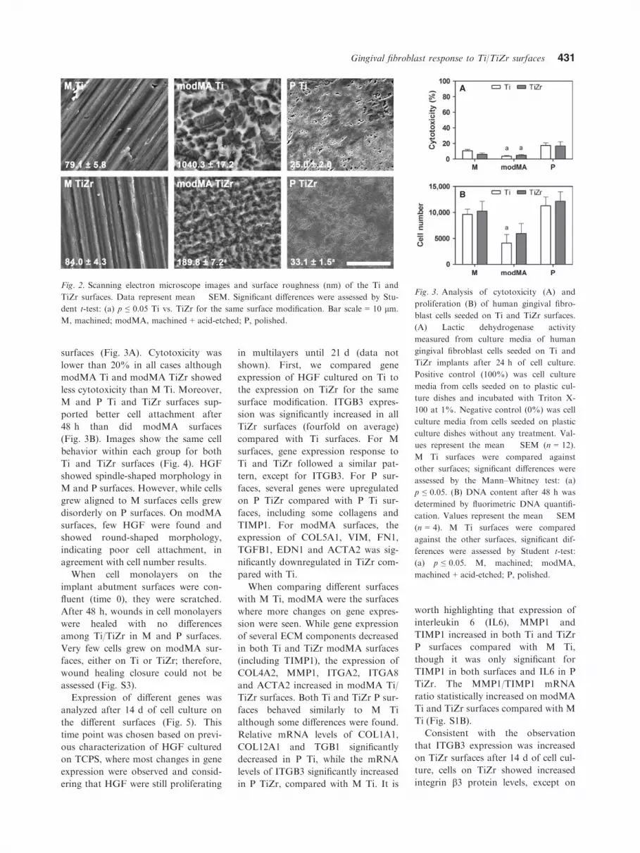
surfaces (Fig. 3A). Cytotoxicity was
lower than 20% in all cases although
modMA Ti and modMA TiZr showed
less cytotoxicity than M Ti. Moreover,
M and P Ti and TiZr surfaces sup-
ported better cell attachment after
48 h than did modMA surfaces
(Fig. 3B). Images show the same cell
behavior within each group for both
Ti and TiZr surfaces (Fig. 4). HGF
showed spindle-shaped morphology in
M and P surfaces. However, while cells
grew aligned to M surfaces cells grew
disorderly on P surfaces. On modMA
surfaces, few HGF were found and
showed round-shaped morphology,
indicating poor cell attachment, in
agreement with cell number results.
When cell monolayers on the
implant abutment surfaces were con-
fluent (time 0), they were scratched.
After 48 h, wounds in cell monolayers
were healed with no differences
among Ti/TiZr in M and P surfaces.
Very few cells grew on modMA sur-
faces, either on Ti or TiZr; therefore,
wound healing closure could not be
assessed (Fig. S3).
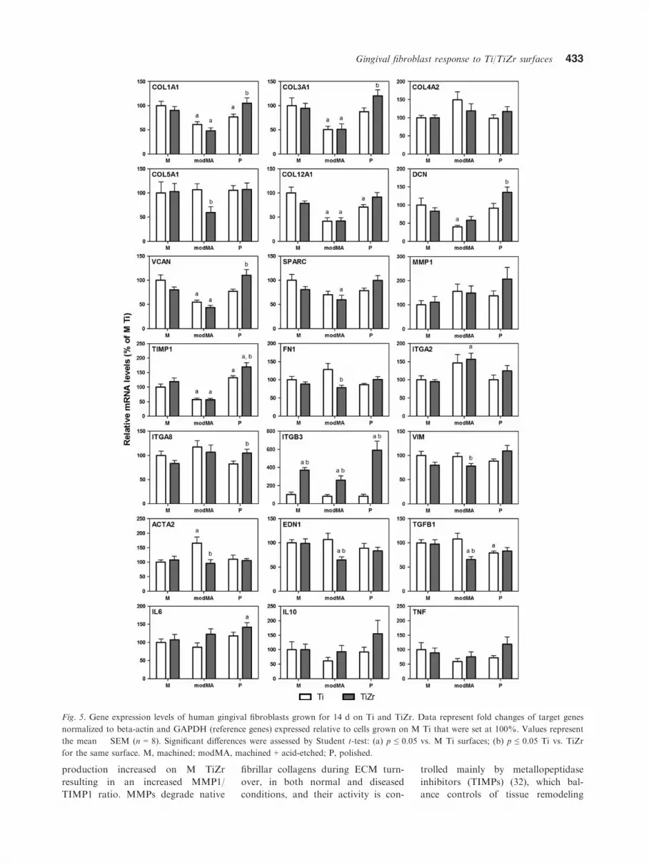
Expression of different genes was
analyzed after 14 d of cell culture on
the different surfaces (Fig. 5). This
time point was chosen based on previ-
ous characterization of HGF cultured
on TCPS, where most changes in gene
expression were observed and consid-
ering that HGF were still proliferating
in multilayers until 21 d (data not
shown). First, we compared gene
expression of HGF cultured on Ti to
the expression on TiZr for the same
surface modification. ITGB3 expres-
sion was significantly increased in all
TiZr surfaces (fourfold on average)
compared with Ti surfaces. For M
surfaces, gene expression response to
Ti and TiZr followed a similar pat-
tern, except for ITGB3. For P sur-
faces, several genes were upregulated
on P TiZr compared with P Ti sur-
faces, including some collagens and
TIMP1. For modMA surfaces, the
expression of COL5A1, VIM, FN1,
TGFB1, EDN1 and ACTA2 was sig-
nificantly downregulated in TiZr com-
pared with Ti.
When comparing different surfaces
with M Ti, modMA were the surfaces
where more changes on gene expres-
sion were seen. While gene expression
of several ECM components decreased
in both Ti and TiZr modMA surfaces
(including TIMP1), the expression of
COL4A2, MMP1, ITGA2, ITGA8
and ACTA2 increased in modMA Ti/
TiZr surfaces. Both Ti and TiZr P sur-
faces behaved similarly to M Ti
although some differences were found.
Relative mRNA levels of COL1A1,
COL12A1 and TGB1 significantly
decreased in P Ti, while the mRNA
levels of ITGB3 significantly increased
in P TiZr, compared with M Ti. It is
worth highlighting that expression of
interleukin 6 (IL6), MMP1 and
TIMP1 increased in both Ti and TiZr
P surfaces compared with M Ti,
though it was only significant for
TIMP1 in both surfaces and IL6 in P
TiZr. The MMP1/TIMP1 mRNA
ratio statistically increased on modMA
Ti and TiZr surfaces compared with M
Ti (Fig. S1B).
Consistent with the observation
that ITGB3 expression was increased
on TiZr surfaces after 14 d of cell cul-
ture, cells on TiZr showed increased
integrin b3 protein levels, except on
Fig. 2. Scanning electron microscope images and surface roughness (nm) of the Ti and
TiZr surfaces. Data represent mean � SEM. Significant differences were assessed by Stu-
dent t-test: (a) p ≤ 0.05 Ti vs. TiZr for the same surface modification. Bar scale = 10 lm.
M, machined; modMA, machined + acid-etched; P, polished.
A
B
Fig. 3. Analysis of cytotoxicity (A) and
proliferation (B) of human gingival fibro-
blast cells seeded on Ti and TiZr surfaces.
(A) Lactic dehydrogenase activity
measured from culture media of human
gingival fibroblast cells seeded on Ti and
TiZr implants after 24 h of cell culture.
Positive control (100%) was cell culture
media from cells seeded on to plastic cul-
ture dishes and incubated with Triton X-
100 at 1%. Negative control (0%) was cell
culture media from cells seeded on plastic
culture dishes without any treatment. Val-
ues represent the mean � SEM (n = 12).
M Ti surfaces were compared against
other surfaces; significant differences were
assessed by the Mann–Whitney test: (a)
p ≤ 0.05. (B) DNA content after 48 h was
determined by fluorimetric DNA quantifi-
cation. Values represent the mean � SEM
(n = 4). M Ti surfaces were compared
against the other surfaces, significant dif-
ferences were assessed by Student t-test:
(a) p ≤ 0.05. M, machined; modMA,
machined + acid-etched; P, polished.
Gingival fibroblast response to Ti/TiZr surfaces 431

modMA TiZr surfaces (Fig. 6). M Ti/
TiZr surfaces showed similar integrin
b3 after 2 d of cell culture, although
after 14 d its presence decreased on
M Ti while on M TiZr was main-
tained. P TiZr increased ITGB3 levels
after 2 and 14 d of cell culture com-
pared with P Ti. In general, ITGB3
immunostaining was low on modMA
surfaces, as there were few cells.
MMP1 production significantly
increased on TiZr surfaces compared
with Ti (1.5 vs. 0.9 ng/mL) although
TiZr did not affect TIMP1 production
compared with Ti. Compared with M
Ti, MMP1 increased on M TiZr,
modMA TiZr and P Ti/TiZr surfaces
(Fig. 7A); TIMP1 decreased on mod-
MA Ti (Fig. 7B); and the MMP1/
TIMP1 ratio increased on modMA
Ti/TiZr and on M TiZr (Fig. 7C).
Interestingly, the production of
TIMP1 was higher than the produc-
tion of MMP1 (an average of 55 ng/
mL for TIMP1 vs. 1.2 ng/mL for
MMP1), as also found at mRNA lev-
els (a difference of sixfold, taking into
account similar PCR efficiencies for
both pairs of primers).
Discussion
STI and hard tissue integration is
mandatory for dental implant success.
While over the past years, research
and development of dental implant bi-
omaterials has been focused on osseo-
integration, nowadays STI is gaining
ground, as dental implants require a
soft tissue barrier to prevent bacterial
penetration and inhibit epithelial
downgrowth. After installation of a
dental implant, fibroblasts from the
oral connective tissue are the pre-
ferred cells to form a collagen-rich
connective tissue to repopulate the
wound (23). In the present research,
we first established an in vitro model
with primary HGF for later evalua-
tion of their biological response to
surface-modified TiZr implant abut-
ments and compare it with the gold
standard in implantology, Ti.
We show that HGF produce an
organized ECM, do not tend to fibro-
lytic or proinflammatory situations
and have the capacity for in vitro
differentiation into myofibroblasts.
This was characterized by increased
gene expression over time of ECM
constituents such as several collagens,
proteoglycans and glycoproteins
(production of an organized ECM);
decreased MMP1/TIMP1 mRNA ratio
over time [lack of tendency to a fibro-
lytic situation (24)]; steady expression
of IL6 together with decreased expres-
sion of tumor necrosis factor-alpha
[lack of a proinflammatory situation
(25)]; re-establishment of the full
monolayer architecture in 48 h after a
wound scratch and increased expres-
sion of ACTA2 [hallmark of myofibro-
blast differentiation and fibrogenic
conditions (26)]. However, the elevated
levels of ITGA2 could be a feature of
a fibrotic phenotype (27).
Regarding the in vitro test system,
monolayers of either gingival epithe-
lial keratinocytes or fibroblasts have
been used to test STI (1). Complex
interactive co-cultures of gingival
keratinocytes and fibroblasts have
also been used as a test platform for
STI evaluation (28), although kerat-
inocytes induce fibroblasts to differen-
tiate into myofibroblasts (29). Cell
co-cultures are more time and
resource consuming than single cell
type cultures; therefore, we considered
that co-cultures were not the optimal
test system to perform high-through-
put analysis.
In this study, we compared differ-
ent Ti and TiZr surfaces to evaluate
their applicability for STI. TiZr was
as biocompatible as Ti, as previously
demonstrated (11,12). In addition,
TiZr supported cell attachment and
had little influence on either cell mor-
phology or alignment. We found that
integrin b3 mRNA and protein levels
increased on TiZr compared with Ti.
Integrin avb3 is involved in cell
attachment (30), thus, this finding
could be related to the slight but not
significant, enhanced cell attachment
on to TiZr compared with Ti. Besides
the strong ITGB3 increase, Ti and
TiZr caused different responses in
HGF, as previously found with osteo-
blasts (31). Regarding P and modMA
surfaces, although Ti and TiZr
showed different roughness, we only
found few differences among materi-
als, including the decreased expression
of the pro-fibrotic markers in mod-
MA TiZr (ACTA2, EDN1 and
TGFB1) compared with modMA Ti
and increased expression of some
ECM markers in P TiZr (COL1A1,
COL3A1, DCN, VCAN and TIMP1)
compared with P Ti. For M surfaces,
with a similar roughness, MMP1
Fig. 4. Cell morphology on the different Ti
and TiZr surfaces. Confocal microscopy
was used to visualize actin immunostaining
(green) and nuclei (blue) after 48 h (left
column). Scanning electron microscopy
was used to visualize human gingival fibro-
blasts on the surfaces after 9 d (right col-
umn). Bar scale = 100 lm. M, machined;
modMA, machined + acid-etched; P, pol-
ished.
432 G�omez-Florit et al.
production increased on M TiZr
resulting in an increased MMP1/
TIMP1 ratio. MMPs degrade native
fibrillar collagens during ECM turn-
over, in both normal and diseased
conditions, and their activity is con-
trolled mainly by metallopeptidase
inhibitors (TIMPs) (32), which bal-
ance controls of tissue remodeling
Fig. 5. Gene expression levels of human gingival fibroblasts grown for 14 d on Ti and TiZr. Data represent fold changes of target genes
normalized to beta-actin and GAPDH (reference genes) expressed relative to cells grown on M Ti that were set at 100%. Values represent
the mean � SEM (n = 8). Significant differences were assessed by Student t-test: (a) p ≤ 0.05 vs. M Ti surfaces; (b) p ≤ 0.05 Ti vs. TiZr
for the same surface. M, machined; modMA, machined + acid-etched; P, polished.
Gingival fibroblast response to Ti/TiZr surfaces 433

(33). An increased MMP1/TIMP1
ratio in M TiZr surfaces could reflect
either the remodeling of newly synthe-
sized collagen (34) or an accelerated
ECM degradation, which has been
associated with periodontitis (35,36).
Interestingly, it has been shown that
MMP production is regulated by the
ECM through integrin receptor sig-
naling (37) and that MMP1 increases
in parallel with ITGB3 expression in
smooth muscle cells from wounding
sites (38). Thus, the greater integrin
b3 production on TiZr surfaces could
be responsible for the increased
MMP1 production on TiZr surfaces.
Furthermore, MMP1 and ITGB3
expression were positively correlated
(Pearson’s correlation coefficient =0.365; p = 0.012). Future studies
should explore the pathways between
MMP1 and ITGB3 in gingival
fibroblasts to understand further the
mechanisms implicated under this
hypothesis.
We also evaluated the performance
of the different surface modifications
comparing with the reference surface,
M Ti. We considered M Ti the refer-
ence surface as machined is the stan-
dard implant neck and abutment
surface of dental products in the mar-
ket and Ti is the gold standard in im-
plantology (6). For modMA surfaces,
the decreased cell number, abnormal
morphology and easy-stained con-
densed nuclei suggest impaired growth
and even apoptosis. Moreover, mod-
MA surfaces decreased gene expres-
sion of several ECM components and
vimentin and increased the mRNA lev-
els of MMP1, ITGA2, ITGA8, IL6,
the pro-fibrotic markers and the
MMP1/TIMP1 mRNA and protein
ratio suggesting impaired HFG growth
and a pro-fibrotic situation on to these
surfaces. As the initial cell attachment
to a surface is a critical step for cell
survival, for this in vitro study we
hypothesize that although modMA
surfaces were not cytotoxic, initial cell
attachment was impaired, and was, in
consequence, cell survival and growth.
This can be explained by the increased
roughness of modMA surface as cell
number and the expression of several
genes encoding for ECM proteins
(COL1A1, COL3A1, COL12A1,
DCN, VCAN and TIMP1) were nega-
tively correlated with surface rough-
ness (Spearman’s correlation coeffi-
cient �0.425 ≤ r ≤ �0.771; p < 0.05).
This impairment in cell attachment
and growth might be also explained by
surface hydrophilicity (14,39), even
though we did not measure the contact
angle in the present study. Thus,
although modMA do not seem to be
applicable for STI, hydrophilic rough
surfaces downregulated the gene
expression of key proinflammatory
cytokines of macrophages (40),
increased the osteogenic differentiation
of osteoblasts (15) and, in vivo, favored
blood wetting and the formation of a
fibrin network, resulting in superior
osseointegrative properties (41) and
connective tissue attachment (42,43).
For M and P surfaces, data suggest
a normal fibroblast growth, M sur-
faces created a pattern of mechanical
stress that caused cell alignment (44)
to the grooves, which is related to a
higher density of focal contacts (45),
while no clear orientation was seen on
P surfaces. Previous studies have
shown similar results with fibroblasts
Fig. 6. Confocal microscopy was used to
visualize integrin b3 (red) and nuclei (blue)
after 2 and 14 d of cell culture on the Ti
and TiZr surfaces. Bar scale = 50 lm. M,
machined; modMA, machined + acid-
etched; P, polished.
A
B
C
Fig. 7. Effect of Ti and TiZr surfaces on
MMP1 (A) and TIMP1 (B) production
and the MMP1/TIMP1 ratio (C), after
14 d of cell culture. Values represent the
mean � SEM (n = 6). Significant differ-
ences were assessed by Student t-test: (a)
p ≤ 0.05 vs. M Ti surfaces. M, machined;
MMP1, matrix metalloproteinase 1; mod-
MA, machined + acid-etched; P, polished;
TIMP1, metallopeptidase inhibitor 1.
434 G�omez-Florit et al.

and rough surfaces (Sa from 500 nm
to 1 lm) compared with M
(Sa = 119 nm) and smooth surfaces
(Sa from 14 to 293 nm) (14,28,46).
However, some studies have demon-
strated that it is the combination of
micro-roughness with further acid
etching that promotes HGF adhesion
and proliferation (47,48).
In conclusion, we show that mono-
cultures of HGF are a valuable in vitro
model for high-throughput screening
of new abutment surfaces designed to
improve STI. We also prove in this in
vitro study that: (i) TiZr has the same
biocompatibility than Ti surfaces on
primary HGF; (ii) TiZr surfaces show
higher integrin b3 mRNA and protein
levels together with increased MMP1
protein compared with Ti; and (iii) M
surfaces caused better alignment than
the other studied surfaces. Taking into
account the better mechanical proper-
ties and bioactivity of TiZr compared
with Ti, the results of the present study
show that TiZr is a potential clinical
candidate for STI and implant success.
Acknowledgements
Authors have no conflict of interest.
This work was supported by the Min-
isterio de Ciencia e Innovaci�on del
Gobierno de Espa~na (Torres Quevedo
contract to MG and JMR, and
Ram�on y Cajal contract to MM).
Titanium and titanium-zirconium
coins were kindly provided by Institut
Straumann AG (Basel, Switzerland).
We are especially grateful to Dr. Fer-
ran Hierro (University of the Balearic
Islands) for the excellent technical
support with the scanning electron
microscope.
Supporting Information
Additional Supporting Information
may be found in the online version of
this article:
Figure S1 Ratio MMP1/TIMP1.
(A) MMP1/TIMP1 mRNA ratio over
time of HGF cells seeded on to
TCPS. Values represent the mean �SEM (n = 6 for each donor); (t) effect
of time (p < 0.05). (B) MMP1/TIMP1
mRNA ratio of HGF cells seeded on
to Ti/TiZr-modified surfaces. Values
represent the mean � SEM (n = 8).
Significant differences were assessed
by Student t-test: (a) p ≤ 0.05 versus
machined Ti surfaces (M Ti).
Figure S2 Wound healing assay. Cell
monolayers were scratched at 0 h (C)
and allowed to heal up to 48 h (D, E;
dotted line indicates scratch mark). (A)
The open wound area was calculated
for each donor and then the mean �SEM (n = 3) of the three donors was
calculated. (B) Gene expression levels
48 h after scratching HGF monolayers
ofMMP1, ACTA2, EDN1 and TGFB1
of HGF cells seeded on to TCPS. Data
represent fold changes of target genes
normalized to beta-actin and GAPDH
(reference genes) expressed relative to
cells before scratching the monolayers
that were set at 100%. Values represent
the median and the 5–95 percentile
(n = 3 for each donor). Significant
differences were assessed by Student t-
test: (a) p ≤ 0.05.
Figure S3 Wound healing assay.
Scanning electron microscopy images
of HGF grown on Ti and TiZr
implants 48 h after scratching the sur-
face. Dotted line indicates the scratch
mark. Bar scale = 2 mm.
References
1. Rompen E, Domken O, Degidi M, Pon-
tes AE, Piattelli A. The effect of material
characteristics, of surface topography
and of implant components and connec-
tions on soft tissue integration: a litera-
ture review. Clin Oral Implants Res
2006;17(Suppl 2):55–67.
2. Chehroudi B, Gould TR, Brunette DM.
The role of connective tissue in inhibiting
epithelial downgrowth on titanium-
coated percutaneous implants. J Biomed
Mater Res 1992;26:493–515.
3. Bartold PM, Walsh LJ, Narayanan AS.
Molecular and cell biology of the gin-
giva. Periodontol 2000 2000;24:28–55.
4. Abiko Y, Hiratsuka K, Kiyama-Kishika-
wa M, Tsushima K, Ohta M, Sasahara
H. Profiling of differentially expressed
genes in human gingival epithelial cells
and fibroblasts by DNA microarray.
J Oral Sci 2004;46:19–24.
5. Chang HY, Chi JT, Dudoit S et al.
Diversity, topographic differentiation,
and positional memory in human fibro-
blasts. Proc Natl Acad Sci USA
2002;99:12877–12882.
6. Steinemann SG. Titanium—the material
of choice? Periodontol 2000 1998;17:7–21.
7. Saulacic N, Bosshardt DD, Bornstein
MM, Berner S, Buser D. Bone apposi-
tion to a titanium-zirconium alloy
implant, as compared to two other tita-
nium-containing implants. Eur Cells
Mater 2012;23:273–286; discussion 286-
278.
8. Ho WF, Chen WK, Wu SC, Hsu HC.
Structure, mechanical properties, and
grindability of dental Ti-Zr alloys. J
Mater Sci Mater Med 2008;19:3179–
3186.
9. Kobayashi E, Matsumoto S, Doi H,
Yoneyama T, Hamanaka H. Mechanical
properties of the binary titanium-zirco-
nium alloys and their potential for bio-
medical materials. J Biomed Mater Res
1995;29:943–950.
10. Thoma DS, Jones AA, Dard M, Grize
L, Obrecht M, Cochran DL. Tissue inte-
gration of a new titanium-zirconium den-
tal implant: a comparative histologic and
radiographic study in the canine. J Peri-
odontol 2011;82:1453–1461.
11. Linkevicius T, Apse P. Influence of abut-
ment material on stability of peri-implant
tissues: a systematic review. Int J Oral
Maxillofac Implants 2008;23:449–456.
12. Pae A, Lee H, Kim HS, Kwon YD,
Woo YH. Attachment and growth
behaviour of human gingival fibroblasts
on titanium and zirconia ceramic sur-
faces. Biomed Mater 2009;4:025005.
13. Kokubu E, Hamilton DW, Inoue T, Bru-
nette DM. Modulation of human gingi-
val fibroblast adhesion, morphology,
tyrosine phosphorylation, and ERK 1/2
localization on polished, grooved and
SLA substratum topographies. J Biomed
Mater Res A 2009;91:663–670.
14. Oates CJ, Wen W, Hamilton DW. Role
of titanium surface topography and sur-
face wettability on focal adhesion kinase
mediated signaling in fibroblasts. Materi-
als 2011;4:893–907.
15. Le Guehennec L, Soueidan A, Layrolle
P, Amouriq Y. Surface treatments of
titanium dental implants for rapid osseo-
integration. Dent Mater 2007;23:844–854.
16. Kononen M, Hormia M, Kivilahti J,
Hautaniemi J, Thesleff I. Effect of sur-
face processing on the attachment, orien-
tation, and proliferation of human
gingival fibroblasts on titanium. J Bio-
med Mater Res 1992;26:1325–1341.
17. Lamolle SF, Monjo M, Lyngstadaas SP,
Ellingsen JE, Haugen HJ. Titanium
implant surface modification by cathodic
reduction in hydrofluoric acid: surface
characterization and in vivo perfor-
mance. J Biomed Mater Res A
2009;88:581–588.
18. Rupp F, Scheideler L, Olshanska N, de
Wild M, Wieland M, Geis-Gerstorfer J.
Enhancing surface free energy and hydro-
philicity through chemical modification of
Gingival fibroblast response to Ti/TiZr surfaces 435

microstructured titanium implant sur-
faces. J Biomed Mater Res A 2006;76A:
323–334.
19. Pfaffl MW. A new mathematical model
for relative quantification in real-time
RT-PCR. Nucleic Acids Res 2001;29:e45.
20. Pfaffl MW, Tichopad A, Prgomet C,
Neuvians TP. Determination of stable
housekeeping genes, differentially regu-
lated target genes and sample integrity:
BestKeeper–Excel-based tool using pair-
wise correlations. Biotechnol Lett
2004;26:509–515.
21. Novak JP, Sladek R, Hudson TJ. Char-
acterization of variability in large-scale
gene expression data: implications for
study design. Genomics 2002;79:104–113.
22. Geback T, Schulz MM, Koumoutsakos
P, Detmar M. TScratch: a novel and
simple software tool for automated anal-
ysis of monolayer wound healing assays.
Biotechniques 2009;46:265–274.
23. Palaiologou AA, Yukna RA, Moses R,
Lallier TE. Gingival, dermal, and peri-
odontal ligament fibroblasts express dif-
ferent extracellular matrix receptors. J
Periodontol 2001;72:798–807.
24. Soell M, Elkaim R, Cathepsin TH, C, .
Matrix metalloproteinases, and their tis-
sue inhibitors in gingiva and gingival cre-
vicular fluid from periodontitis-affected
patients. J Dent Res 2002;81:174–178.
25. Bartold PM, Narayanan AS. Molecular
and cell biology of healthy and diseased
periodontal tissues. Periodontol 2000
2006;40:29–49.
26. Hinz B, Phan SH, Thannickal VJ, Galli
A, Bochaton-Piallat ML, Gabbiani G.
The myofibroblast: one function, multi-
ple origins. Am J Pathol 2007;170:1807–
1816.
27. Guo F, Carter DE, Mukhopadhyay A, Le-
ask A. Gingival fibroblasts display reduced
adhesion and spreading on extracellular
matrix: a possible basis for scarless tissue
repair? PLoSONE 2011;6:e27097.
28. Schulz S, Tomakidi P, Mauth C, Kohal
R, Steinberg T. Interactive fibroblast-
keratinocyte co-cultures: an in vivo-like
test platform for dental implant-based
soft tissue integration. Tissue Eng Part C
Methods 2012;18:785–796.
29. Shephard P, Martin G, Smola-Hess S,
Brunner G, Krieg T, Smola H. Myofi-
broblast differentiation is induced in
keratinocyte-fibroblast co-cultures and is
antagonistically regulated by endogenous
transforming growth factor-beta and
interleukin-1. Am J Pathol 2004;164:20-
55–2066.
30. Hormia M, Kononen M. Immunolocal-
ization of fibronectin and vitronectin
receptors in human gingival fibroblasts
spreading on titanium surfaces. J Peri-
odontal Res 1994;29:146–152.
31. Chakravorty N, Ivanovski S, Prasadam
I, Crawford R, Oloyede A, Xiao Y. The
microRNA expression signature on mod-
ified titanium implant surfaces influences
genetic mechanisms leading to osteogenic
differentiation. Acta Biomater 2012;8:
3516–3523.
32. Sakagami G, Sato E, Sugita Y et al.
Effects of nifedipine and interleukin-
1alpha on the expression of collagen,
matrix metalloproteinase-1, and tissue
inhibitor of metalloproteinase-1 in
human gingival fibroblasts. J Periodontal
Res 2006;41:266–272.
33. Domeij H, Modeer T, Yucel-Lindberg T.
Matrix metalloproteinase-1 and tissue
inhibitor of metalloproteinase-1 produc-
tion in human gingival fibroblasts: the
role of protein kinase C. J Periodontal
Res 2004;39:308–314.
34. Dasu MR, Hawkins HK, Barrow RE,
Xue H, Herndon DN. Gene expression
profiles from hypertrophic scar fibro-
blasts before and after IL-6 stimulation.
J Pathol 2004;202:476–485.
35. Birkedal-Hansen H. Role of matrix me-
talloproteinases in human periodontal
diseases. J Periodontol 1993;64:474–484.
36. Kubota T, Nomura T, Takahashi T,
Hara K. Expression of mRNA for
matrix metalloproteinases and tissue
inhibitors of metalloproteinases in peri-
odontitis-affected human gingival tissue.
Arch Oral Biol 1996;41:253–262.
37. Werb Z. ECM and cell surface proteoly-
sis: regulating cellular ecology. Cell
1997;91:439–442.
38. Bendeck MP, Irvin C, Reidy M et al.
Smooth muscle cell matrix metallopro-
teinase production is stimulated via avb3integrin. Arterioscler Thromb Vasc Biol
2000;20:1467–1472.
39. Ponsonnet L, Reybier K, Jaffrezic N
et al. Relationship between surface prop-
erties (roughness, wettability) of titanium
and titanium alloys and cell behaviour.
Mater Sci Eng C-Mater Biol Appl
2003;23:551–560.
40. Hamlet S, Alfarsi M, George R, Ivanov-
ski S. The effect of hydrophilic titanium
surface modification on macrophage
inflammatory cytokine gene expression.
Clin Oral Implants Res 2012;23:584–590.
41. Donos N, Hamlet S, Lang NP et al.
Gene expression profile of osseointegra-
tion of a hydrophilic compared with a
hydrophobic microrough implant sur-
face. Clin Oral Implants Res 2011;22:
365–372.
42. Schwarz F, Ferrari D, Herten M et al.
Effects of surface hydrophilicity and mic-
rotopography on early stages of soft and
hard tissue integration at non-submerged
titanium implants: an immunohistochem-
ical study in dogs. J Periodontol 2007;78:
2171–2184.
43. Schwarz F, Herten M, Sager M, Wieland
M, Dard M, Becker J. Histological and
immunohistochemical analysis of initial
and early subepithelial connective tissue
attachment at chemically modified and
conventional SLA�titanium implants. A
pilot study in dogs. Clin Oral Investig
2007;11:245–255.
44. Walboomers XF, Jansen JA. Cell and
tissue behavior on micro-grooved sur-
faces. Odontology 2001;89:2–11.
45. Eisenbarth E, Meyle J, Nachtigall W,
Breme J. Influence of the surface struc-
ture of titanium materials on the adhe-
sion of fibroblasts. Biomaterials
1996;17:1399–1403.
46. Yamano S, Ma AK, Shanti RM, Kim
SW, Wada K, Sukotjo C. The influence
of different implant materials on human
gingival fibroblast morphology, prolifera-
tion, and gene expression. Int J Oral
Maxillofac Implants 2011;26:1247–1255.
47. Lee SW, Kim SY, Lee MH, Lee KW,
Leesungbok R, Oh N. Influence of
etched microgrooves of uniform dimen-
sion on in vitro responses of human gin-
gival fibroblasts. Clin Oral Implants Res
2009;20:458–466.
48. Kim SY, Oh N, Lee MH, Kim SE,
Leesungbok R, Lee SW. Surface microg-
rooves and acid etching on titanium sub-
strata alter various cell behaviors of
cultured human gingival fibroblasts. Clin
Oral Implants Res 2009;20:262–272.
436 G�omez-Florit et al.