biologic width around titanium implants. a histometric analysis of the implanto-gingival junction...
TRANSCRIPT
186
Biologic Width Around TitaniumImplants. A Histometric Analysis of theImplanto-Gingival Junction AroundUnloaded and Loaded NonsubmergedImplants in the Canine MandibleDavid L. Cochran* Joachim S. Hermann* Robert K. Schenk/ Frank L.Higginbottom,* and Daniel Buser*
The use of endosseous dental implants as transmucosal devices necessitates thesuccessful integration of three different tissues: bone, connective tissue, and epitheli-um. So far, studies have predominantly focused on hard tissue integration. Much lessis known about soft tissues. This study examined the dimensions of the implanto-gingival junction in relation to clinically healthy unloaded and loaded nonsubmergedimplants. In total, 69 titanium plasma-sprayed (TPS) and sandblasted acid-etched(SLA) implants were placed in an alternating fashion in six foxhounds and allowedto heal for 3 months. Two dogs were sacrificed after the initial healing period. Theremaining four dogs had crowns fabricated that were allowed to function for up to 12months. These animals were sacrificed after 3 and 12 months of loading. Histometricanalysis of undecalcified histologie sections included the evaluation of the sulcus depth(SD), the dimensions of the junctional epithelium (JE), and the connective tissue con-
tact (CTC). Mean values in the 3 month unloaded group were 0.49 mm for SD, 1.16mm for JE, and 1.36 mm for CTC. These dimensions were 0.50 mm for SD, 1.44mm for JE, and 1.01 mm for CTC for the 3 month loaded group. After 12 months ofloading, these values were 0.16 mm for SD, 1.88 mm for JE, and 1.05 mm for CTC.The sum of these measurements was similar for the different time points and similarto the same dimensions around teeth. TPS and SLA surfaces had no influence on theevaluated parameters (P > 0.05). The data suggest that a biologic width exists aroundunloaded and loaded nonsubmerged one-part titanium implants and that this is a phys-iologically formed and stable dimension as is found around teeth. / Periodontol 1997;68:186-198.
Key Words: Dental implants/endosseous; biologic width; titanium; soft tissue; animalstudies.
In the past 25 years, basic research in implant dentistryhas focused mainly on the bone-to-implant interface ofendosseous implants. This has been primarily due to thefact that predictable implant anchorage requires directbone-to-implant contact. Original investigations examined
*Department of Periodontics, Dental School, University of Texas HealthScience Center at San Antonio, TX.institute of Pathophysiology, University of Berne, Switzerland.'Department of Oral and Maxillofacial Surgery and Pharmacology, Bay-lor College of Dentistry, Dallas, TX.'Department of Oral Surgery, School of Dental Medicine, University ofBerne, Switzerland.
parameters of surgical procedures and characteristics ofimplant shape and surface to achieve hard tissue integra-tion. Brânemark et al.1 using submerged implants estab-lished criteria to predictably achieve direct bone-to-im-plant contact called "osseointegration." Schroeder et al.2-3utilizing intentionally nonsubmerged implants recognizedthat oral implants must integrate with three tissues; name-
ly, bone, connective tissue, and epithelium. These inves-tigators examined parameters for a bone-to-implant con-
tact, called "functional ankylosis," and, in addition, de-scribed for the first time histologically the contact of softtissues to the transmucosal portion of titanium implants.4
Volume 68Number 2 COCHRAN, HERMANN, SCHENK, HIGGINBOTTOM, BUSER 187
One of the prerequisites for successful implant integra-tion recommended by Brânemark et al.' was to cover theimplant with soft tissues. After an initial healing period,another implant component, following a second surgicalprocedure, was placed on the submerged implant. Thiscomponent penetrated the soft tissues resulting in a trans-mucosal device. Thus, a space or gap always exists be-tween implant and abutment at or below the osseous crest.A rationale for the need for submerging the implant wasdue to "occasionally" exposed implants to the oral cav-
ity.1 No numbers were given as to the percentage thatbecame exposed. Furthermore, no oral hygiene procedu-res were performed on these animals. Thus, plaque ac-
cumulated and inflammation was found on the exposedimplants compared to implants covered by soft tissues.Brânemark et al.,1-5 therefore, recommended submergingendosseous implants below the soft tissues in order toachieve osseous integration. Albrektsson6 in a review ofthis work stated that the soft tissues over the submergedimplant provided "... a barrier toward downgrowth ofgranulation tissue during the phase of bone deminerali-zation. .
.
" However, in a later paper by Albrektsson andSennerby,7 these authors suggested ". .. there is insuffi-cient evidence to back up the necessity for a 2-stage sur-
gical procedure ..." Ruggeri et al.8 stated that their re-
sults from studies on supracrestal collagen fibers aroundnonsubmerged implants disagree with previous findingsthat submerged implants are required to avoid local in-flammation and epithelial downgrowth. In a later articleby Ruggeri et al.9 they conclude, "... keeping an implantnonsubmerged at the time of surgical placement does notinfluence its survival..." Thus, there are conflicting dataon soft tissues around endosseous implants.
Transmucosal devices in the oral cavity must penetratethe soft tissues comprised of gingival connective tissueand gingival epithelium. Therefore, implants should forma seal at the soft tissue interface to ensure the integrityof the integument. The implanto-gingival tissues by def-inition thus have to serve a similar barrier function as
dento-gingival tissues, and integration of an implant ne-
cessitates the integration of all three types of tissue—bone, connective tissue, and epithelium. Many longitu-dinal clinical studies have shown that both submerged andnonsubmerged implants can be clinically successful.510-21It has also been shown that there is no difference in thehard tissue healing when comparing submerged and non-
submerged implants.22 Gingival connective tissue appearsto form a scar-like connective tissue contact adjacent tosmooth nonsubmerged titanium implant surfaces,23-24whereas epithelium forms a hemidesmosomal attachmentto the implant surface at the ultrastructural level.25-27
Unloaded nonsubmerged titanium implants have dem-onstrated favorable soft tissue healing with resultantstructures similar to that around teeth.24-28-29 Histometricanalyses carried out in these studies indicated similar di-
mensions for implanto-gingival tissues compared to den-to-gingival segments.30-31 However, these investigationswere performed in unloaded implants with short-termhealing periods. The purpose of this study was to examinethe dimensions and relationships of implanto-gingival tis-sues surrounding endosseous nonsubmerged one-part ti-tanium implants under unloaded and loaded conditions inthe canine mandible over longer periods.MATERIALS AND METHODS
Implant Design and SurfacesTwo different types of nonsubmerged cylindrical one-parttitanium implants11 with a hollow-screw design were madefrom grade-IV commercially pure titanium. The outer di-ameter was 4.1 mm, and total length was 9 mm. Thesuprabony, smooth portion of each implant had a ma-
chined surface, whereas the intrabony portion was 6 mm
in length and had either a titanium plasma-sprayed (TPS)surface11 with typical roughness and porosity values of 30to 50 µ or a sandblasted and HC1/H2S04 acid-etched(SLA) surface with two levels of roughness, one 20 to 40µ peak to peak, and a superimposed second level at 2to 4 µ peak to peak.AnimalsSix male, lab-bred American foxhounds were used in thisstudy. At the beginning, these animals were about 2 yearsold and weighed about 30 to 35 kg. All animals were freeof heart worms and were quarantined.
Surgical Procedures: ExtractionTooth extractions were performed under general anesthe-sia and sterile conditions in an operating room using 4%thiopenthal-Na solution I.V. (0.4 ml/kg bw)1 as a premedi-cation. The dogs were placed on a heating pad, intubated,and inhalated with 1.5-2% isoflurane* and monitored withan electrocardiogram. After disinfection of the surgicalsite with 10% povidone-iodine solution/1% titratable io-dine,** 2% lidocaine HCl with epinephrine 1:100,000»was administered by infiltration at the buccal aspect ofthe lower jaw. Crevicular incisions were made, and allfour premolars (P,-P4) and the first molar (M,) were care-
fully extracted. Prior to extraction, P2-M, were sectionedto avoid tooth fracturé. Adaptation of the wound marginswas achieved with interrupted sutures. Finally, the re-
maining teeth were scaled and cleaned.The day of surgery, the dogs received 20 mg of the
analgesic nalbuphine s.c. BID (10 mg/ml).** Three ml of
"Institut Straumann AG, Waldenburg, Switzerland.'Pentothal, Abbott Laboratories, North Chicago, IL."AErane, Ohmeda Carbide Inc., Liberty Corner, NJ.**Clinidine, Clinipad Co., Guilford, CT."Henry Schein Inc., Port Washington, NY."Nubain, Astra Pharmaceutical Products Inc., Westborough, MA.
188 BIOLOGIC WIDTH AROUND TITANIUM IMPLANTSJ Periodontol
February 1997
SacrificeGroup A
SacrificeGroup
SacrificeGroup C
mplantation
Loading-+- -t-
-3 0 3 6
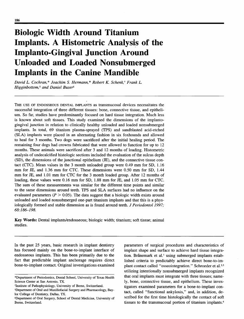
Figure 1. Study design.15 t(mo)
the antibiotic benzathine penicillin (150,000 I.U.) withprocaine penicillin G (150,000 LU.) was administered s.c.SID every 48 hours for 7 to 10 days.§§ For suture removal,after a period of 7 to 10 days, the animals were brieflyanesthetized utilizing a combination (1.1 ml/15 kg bw) ofxylazine (7.1 mg/ml),m acepromazine (2.1 mg/ml),n at-
ropine (0.1 mg/ml),** and ketamine (50.0 mg/ml)*** i.v.Prior to suture removal the local wound area was care-
fully cleaned with 0.12% Chlorhexidine digluconate-soaked gauze.m
Surgical Procedures: Implant PlacementEndosseous, nonsubmerged titanium implants were in-serted under the same surgical conditions as the toothextractions (sterility, operating room, anesthesia) after a
healing period of 3 months (Fig. 1). A crestal incisionwas made maximizing keratinized tissue on each side ofthe incision. Mucoperiosteal flaps were carefully reflectedon the lingual and buccal aspect. Mental foramina were
exposed prior to implant placement. The edentulous os-seous ridge was carefully flattened with an acrylic burand copious irrigation with chilled sterile physiologic sa-
line. Measurements were made using a boley gauge to
help distribute 6 test implants on each side of the man-
dible. Implant osteotomy was performed with torque re-
duction rotary instruments at 500 rpm using again chilledsaline. Dogs were assigned randomly to the three differenttreatment groups A, B, and C (Fig. 1). According to a
randomized starting selection, 3 of each of the test im-plants were placed in an alternating manner per side andhealing screws were placed on top of the implants. In thisfashion, no implant type had a biased position in the arch.Due to narrowing of the ridge in the anterior area, 3 ofpossible 72 implants could not be placed, resulting in a
total of 69 inserted implants.If necessary, periosteal relieving and contouring inci-
sions were made on the buccal and lingual aspects toachieve tension-free wound closure, with a close adapta-
tion of the mucosa to the transmucosal portion of thenonsubmerged implants. Horizontal mattress and inter-rupted sutures were placed. The day of surgery the dogsreceived 20 mg of the analgesic nalbuphine s.c. BID (10mg/ml)." Three ml of the antibiotic benzathine penicillin(150,000 I.U.) with procaine penicillin G (150,000 LU.)was administered s.c. SID every 48 hours for 14 days;100 mg of the antibiotic gentamicin was given s.c. on day1 BID, and the same dosage SID from day 2 through 10(50 mg/ml).*** To reduce swelling, the dogs received 4mg of the anti-inflammatory dexamethasone i.m. SID day1 and day 4.m Sutures were removed after 7 to 10 daysas described above. A soft diet was utilized throughoutthe study. Oral hygiene procedures were carried out 3times a week using a 0.2% Chlorhexidine gel111111 in com-bination with a soft toothbrush and a soft sponge.
Prosthetic ReconstructionFour out of the six dogs constituted the loaded implantgroups, and C (Fig. 1). Two months after implant place-ment in these animals, individual impressions (polyvinylsiloxane) were made™ and screw-retained gold crownsfabricated (Fig. 4). To imitate the natural dentition of thedogs, the P, area had single crowns placed, whereas inthe P2-M, area, connected crowns on two implants were
made. Octagonal abutments were placed in the implantsand precise impressions were taken with standard com-
ponents including repositional transfer copings.*** Implantanalogs were placed in the impressions and models madefor fabrication of the restorations.
Gold copings from the implant manufacturer were in-corporated into the wax-ups for the crowns and bridges.The restorations were inserted 3 months after implantplacement (Fig. 1). At that time, each bridge was carefullyevaluated for passive fit by alternating occlusal screw
tightening and evaluating movement of the restoration. Incases where any movement was detected, the bridgeswere removed and sectioned on the models and placed as
separate units to assure passivity of fit. Finally, the res-torations were seated using 4 mm occlusal screws andadjusted in the mouth to assure that the crowns were ei-ther out of occlusion or had only light contact. Premolareare not in occlusion in the foxhound and the occlusionwas maintained as naturally as possible by taking modelsof the dogs before extraction and duplicating each dog'socclusion. Over time, most dogs exhibited wear patternson the molar restorations as well as some premolars.
S5Pen-B, Pfizer Inc., Lee's Summit, MO. Miles Inc., Shawnee Mission, KS."Burns Veterinary Supply, Oakland, CA."Burns Veterinary Supply, Rockville Center, NY.***Fort Dodge Laboratories Inc., Fort Dodge, IA.,tfPeridex, Procter & Gamble Co., Cincinnati, OH.
"'Gentocin, Schering-Plough Animal Health Corp., Kenilworth, NJ.§8§Dexaject, Burns Veterinary Supply, Oakland, CA."PlakOut Gel, Hawe-Neos AG, Bioggio/TI, Switzerland."President, Coltène/Whaledent Inc., Mahwah, NJ. Dental Implant System, Institut Straumann AG, Waldenburg/BL,Switzerland.
Volume 68Number 2 COCHRAN, HERMANN, SCHENK, HIGGINBOTTOM, BUSER 189
Surgical Procedures: SacrificeTwo dogs (Group A) were sacrificed after a healing periodof 3 months and constituted the unloaded implant group(Fig. 1). The other 4 dogs were sacrificed after loading,2 after 3 months (Group B) and the other 2 after 12months of loading (Group C). Euthanasia was performedwith an overdose of pentobarbital sodium i.v. (0.2 ml =
65 mg/kg bw).**** Mandibles were block-resected withan oscillating autopsy sawmt and the recovered segmentswith the implants were fixed in 4% formaldehyde/1%CaCl2 for histologie preparation and analysis.
Histologie PreparationEach implant with surrounding tissues was prepared forhistology as described by Schenk et ai.32 Nondecalcifiedspecimens were embedded in methyl-methacrylate andstained with basic fuchsin. Two orofacial sections with a
thickness of approximately 80 µ were obtained forGroups A and and four for Group C.
Histometric AnalysisHistometric quantification was carried out utilizing a
high-resolution video camera**** interfaced to a videomonitor.§§§§ This optical system was associated with a dig-itizing pad and a bone morphometry software packagewith image capturing capabilities.11111111 All sections were an-
alyzed under several magnifications of light microscopyto locate anatomical reference points. In a few sections ineach group of dogs the landmarks were not visible. There-fore, the following numbers of implant sites were evalu-ated: 76 out of 86 in Group A, 93 out of 96 in Group B,and 172 out of 196 in Group C. The following measure-
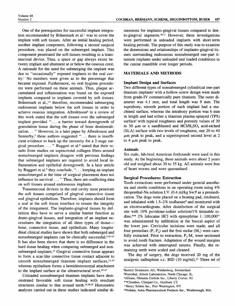
ments were taken or calculated (Fig. 2; shown in Figs. 5,6, 7, and 9).
1. Distance between the gingival margin (GM) and themost coronal point of the junctional epithelium (cJE) =
sulcus depth (SD).2. Distance between cJE and the most apical point of
the junctional epithelium (aJE) = junctional epithelium(JE).
3. Distance between aJE and the first bone-to-implantcontact (fBIC) = connective tissue contact (CTC).
4. SD + JE + CTC = biologic width (BW).5. Distance between the bone margin (BM) and fBIC
= crestal bone loss (cBL).6. Distance between the microgap (MG) and the GM.7. Distance between the MG and cJE.8. Distance between the MG and aJE.
****Euthanasia-5 Solution, Henry Schein Inc., Port Washington, NY.m,Stryker Co., Kalamazoo, MI.'"•CCD-color video camera, Sony Corp., Fujisawa, Japan.5§§§Hyper HAD video monitor, Sony Corp., Fujisawa, Japan.
11 "Bioquant bone morphometry software, R & M, Biometrics Inc., Nash-ville, TN.
Figure 2. Schematic of histometric evaluation with the following mea-
surements: Distance between the gingival margin (GM) and the mostcoronal point of the junctional epithelium (cJE) = sulcus depth (SD).Distance between cJE and the most apical point of the junctional epi-thelium (aJE) = junctional epithelium (JE). Distance between aJE andthe first bone-to-implant contact (fBIC) = connective tissue contact
(CTC). SD + JE + CTC = biologic width (BW). Distance between thebone margin (BM) and fBIC = crestal bone loss (cBL). Distances be-tween the microgap (MG) and the GM, cJE, aJE, BM, and the fBIC.
9. Distance between the MG and fBIC.10. Distance between the MG and BM.
Statistical AnalysisEach of the readings for the measurements taken from theorofacial sections were averaged so that each implant hada single value for each measurement. Analysis of variancewas done to compare the location of the implant site inthe jaws. The location was ordered from most distal tomost mesial. Also, Student unpaired i-tests were per-formed to determine any differences between the types ofimplants. In addition to the measurements indicated in theHistometric Analysis, the proportion of BW attributableto SD, JE, and CTC was also analyzed.
RESULTS
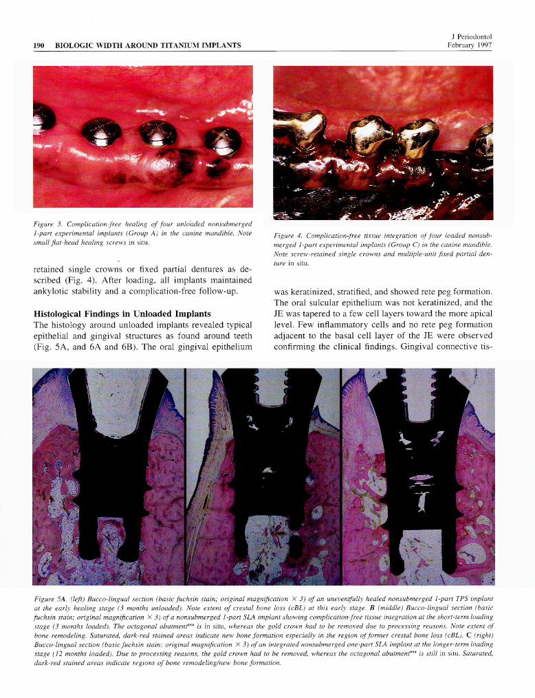
Clinical FindingsPostoperative healing following implant placement was
uneventful in all dogs. After 3 months of healing, all 69implants demonstrated successful tissue integration withankylotic stability and no clinical signs of peri-implantinfection (Fig. 3). No continuous peri-implant radiolucen-cies were apparent on the radiographs.33 Therefore, all 48implants of Groups and C could be restored with screw-
190 BIOLOGIC WIDTH AROUND TITANIUM IMPLANTSJ Periodontol
February 1997
Figure 3. Complication-free healing of four unloaded nonsubmerged1-part experimental implants (Group A) in the canine mandible. Notesmall flat-head healing screws in situ.
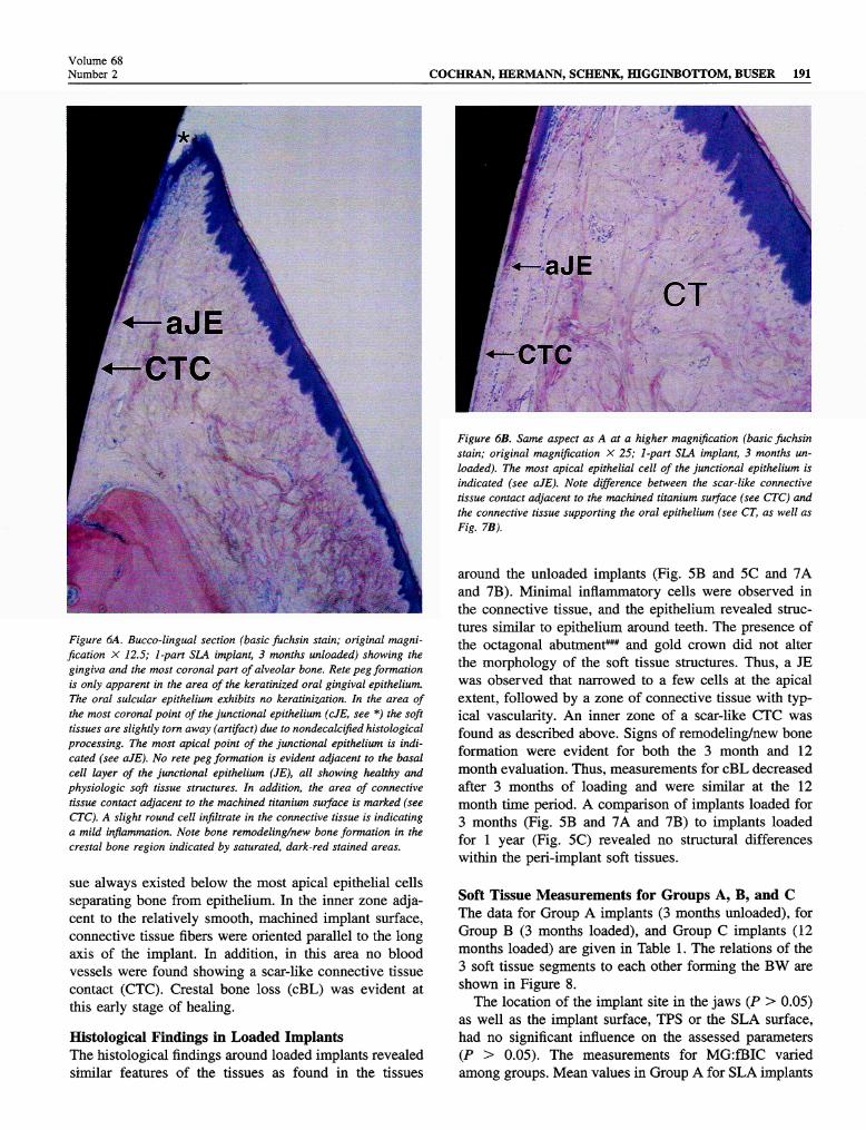
retained single crowns or fixed partial dentures as de-scribed (Fig. 4). After loading, all implants maintainedankylotic stability and a complication-free follow-up.
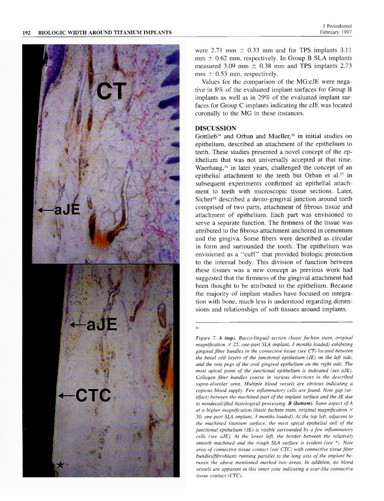
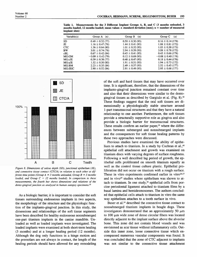
Histological Findings in Unloaded ImplantsThe histology around unloaded implants revealed typicalepithelial and gingival structures as found around teeth(Fig. 5A, and 6A and 6B). The oral gingival epithelium
Figure A. Complication-free tissue integration of four loaded nonsub-merged 1-part experimental implants (Group C) in the canine mandible.Note screw-retained single crowns and multiple-unit fixed partial den-ture in situ.
was keratinized, stratified, and showed rete peg formation.The oral sulcular epithelium was not keratinized, and theJE was tapered to a few cell layers toward the more apicallevel. Few inflammatory cells and no rete peg formationadjacent to the basal cell layer of the JE were observedconfirming the clinical findings. Gingival connective tis-
Figure 5A. (left) Bucco-lingual section (basic fuchsin stain; original magnification X 3) of an uneventfully healed nonsubmerged 1-part TPS implantat the early healing stage (3 months unloaded). Note extent of eresiai bone loss (cBL) at this early stage. (middle) Bucco-lingual section (basicfuchsin stain; original magnification X 3) of a nonsubmerged 1-part SLA implant showing complication-free tissue integration at the short-term loadingstage (3 months loaded). The octagonal abutment*** is in situ, whereas the gold crown had to be removed due to processing reasons. Note extent ofbone remodeling. Saturated, dark-red stained areas indicate new bone formation especially in the region offormer eresiai bone loss (cBL). C (right)Bucco-lingual section (basic fuchsin stain; original magnification X 3) of an integrated nonsubmerged one-part SLA implant at the longer-term loadingstage (12 months loaded). Due to processing reasons, the gold crown had to be removed, whereas the octagonal abutmenf" is still in situ. Saturated,dark-red stained areas indicate regions of bone remodeling/new bone formation.
Volume 68Number 2 COCHRAN, HERMANN, SCHENK, HIGGINBOTTOM, BUSER 191
Figure 6A. Buceo-lingual section (basic fiichsin stain; original magni-fication X 12.5; 1-part SLA implant, 3 months unloaded) showing thegingiva and the most coronal part of alveolar bone. Rete peg formationis only apparent in the area of the keratinized oral gingival epithelium.The oral sulcular epithelium exhibits no keratinization. In the area ofthe most coronal point of the junctional epithelium (cJE, see *) the softtissues are slightly torn away (artifact) due to nondecalcified histologicalprocessing. The most apical point of the junctional epithelium is indi-cated (see aJE). No rete peg formation is evident adjacent to the basalcell layer of the junctional epithelium (JE), all showing healthy andphysiologic soft tissue structures. In addition, the area of connectivetissue contact adjacent to the machined titanium surface is marked (seeCTC). A slight round cell infiltrate in the connective tissue is indicatinga mild inflammation. Note bone remodeling/new bone formation in theeresiai bone region indicated by saturated, dark-red stained areas.
sue always existed below the most apical epithelial cellsseparating bone from epithelium. In the inner zone adja-cent to the relatively smooth, machined implant surface,connective tissue fibers were oriented parallel to the longaxis of the implant. In addition, in this area no bloodvessels were found showing a scar-like connective tissuecontact (CTC). Crestal bone loss (cBL) was evident atthis early stage of healing.Histological Findings in Loaded ImplantsThe histological findings around loaded implants revealedsimilar features of the tissues as found in the tissues
Figure 6B. Same aspect as A at a higher magnification (basic fuchsinstain; original magnification X 25; 1-part SLA implant, 3 months un-
loaded). The most apical epithelial cell of the junctional epithelium isindicated (see aJE). Note difference between the scar-like connectivetissue contact adjacent to the machined titanium surface (see CTC) andthe connective tissue supporting the oral epithelium (see CT, as well as
Fig. 7B).
around the unloaded implants (Fig. 5B and 5C and 7Aand 7B). Minimal inflammatory cells were observed inthe connective tissue, and the epithelium revealed struc-tures similar to epithelium around teeth. The presence ofthe octagonal abutment*** and gold crown did not alterthe morphology of the soft tissue structures. Thus, a JEwas observed that narrowed to a few cells at the apicalextent, followed by a zone of connective tissue with typ-ical vascularity. An inner zone of a scar-like CTC wasfound as described above. Signs of remodeling/new boneformation were evident for both the 3 month and 12month evaluation. Thus, measurements for cBL decreasedafter 3 months of loading and were similar at the 12month time period. A comparison of implants loaded for3 months (Fig. 5B and 7A and 7B) to implants loadedfor 1 year (Fig. 5C) revealed no structural differenceswithin the peri-implant soft tissues.
Soft Tissue Measurements for Groups A, B, and CThe data for Group A implants (3 months unloaded), forGroup (3 months loaded), and Group C implants (12months loaded) are given in Table 1. The relations of the3 soft tissue segments to each other forming the BW are
shown in Figure 8.The location of the implant site in the jaws (P > 0.05)
as well as the implant surface, TPS or the SLA surface,had no significant influence on the assessed parameters(P > 0.05). The measurements for MG:fBIC variedamong groups. Mean values in Group A for SLA implants
192 BIOLOGIC WIDTH AROUND TITANIUM IMPLANTSJ Periodontol
February 1997
were 2.71 mm ± 0.33 mm and for TPS implants 3.11mm ± 0.62 mm, respectively. In Group SLA implantsmeasured 3.09 mm ± 0.38 mm and TPS implants 2.73mm ±- 0.53 mm, respectively.
Values for the comparison of the MGxJE were nega-tive in 8% of the evaluated implant surfaces for Group implants as well as in 29% of the evaluated implant sur-faces for Group C implants indicating the cJE was locatedcoronally to the MG in these instances.
DISCUSSIONGottlieb34 and Orban and Mueller,35 in initial studies on
epithelium, described an attachment of the epithelium toteeth. These studies presented a novel concept of the ep-ithelium that was not universally accepted at that time.Waerhaug,36 in later years, challenged the concept of an
epithelial attachment to the teeth but Orban et al.37 insubsequent experiments confirmed an epithelial attach-ment to teeth with microscopic tissue sections. Later,Sicher38 described a dento-gingival junction around teethcomprised of two parts, attachment of fibrous tissue andattachment of epithelium. Each part was envisioned toserve a separate function. The firmness of the tissue wasattributed to the fibrous attachment anchored in cementumand the gingiva. Some fibers were described as circularin form and surrounded the tooth. The epithelium wasenvisioned as a "cuff" that provided biologic protectionto the internal body. This division of function betweenthese tissues was a new concept as previous work hadsuggested that the firmness of the gingival attachment hadbeen thought to be attributed to the epithelium. Becausethe majority of implant studies have focused on integra-tion with bone, much less is understood regarding dimen-sions and relationships of soft tissues around implants.
<—
Figure 7. A (top). Bucco-lingual section (basic fuchsin stain, originalmagnification X 25, one-part SLA implant, 3 months loaded) exhibitinggingival fiber bundles in the connective tissue (see CT) located betweenthe basal cell layers of the junctional epithelium (JE) on the left side,and the rete pegs of the oral gingival epithelium on the right side. Themost apical point of the junctional epithelium is indicated (see aJE).Collagen fiber bundles course in various directions in the describedsupra-alveolar area. Multiple blood vessels are obvious indicating a
copious blood supply. Few inflammatory cells are found. Note gap (ar-tifact) between the machined part of the implant surface and the JE dueto nondecalcified histological processing. (bottom). Same aspect ofAat a higher magnification (basic fuchsin stain, original magnification X
50, one-part SLA implant, 3 months loaded). At the top left, adjacent tothe machined titanium surface, the most apical epithelial cell of thejunctional epithelium (JE) is visible surrounded by a few inflammatorycells (see aJE). At the lower left, the border between the relativelysmooth machined and the rough SLA surface is evident (see *). Notearea of connective tissue contact (see CTC) with connective tissue fiberbundles/fibroblasts running parallel to the long axis of the implant be-tween the above mentioned marked two areas. In addition, no bloodvessels are apparent in this inner zone indicating a scar-like connectivetissue contact (CTC).
Volume 68Number 2 COCHRAN, HERMANN, SCHENK, HIGGINBOTTOM, BUSER 193
Table 1. Measurements for the 3 Different Implant Groups A, B, and C (3 months unloaded, 3months loaded, 12 months loaded; mean values ± standard deviation [mm]; = number of measuredimplant sites)
Variable(s) Group A (n) _ Group ( ) Group C (n)SD 0.49 ± 0.32 (77) 0.50 ± 0.30 (93) 0.16 ± 0.14 (178)JE 1.16 ± 0.47 (76) 1.44 ± 0.41 (93) 1.88 ± 0.81 (172)CTC 1.36 ± 0.64(80) 1.01 ± 0.32 (93) 1.05 ± 0.38 (173)BW 3.01 ± 0.74 (76) 2.94 ± 0.59 (93) 3.08 ± 0.78 (172)cBL 0.67 ± 0.42 (84) 0.43 ± 0.41 (93) 0.43 ± 0.66 (178)MG.-GM -0.09 ± 0.42 (79) 0.12 ± 0.69 (93) -0.08 ± 0.40 (176)MGxJE 0.39 ± 0.38 (77) 0.48 ± 0.47 (93) 0.18 ± 0.44 (176)MG:aJE 1.52 ± 0.50 (80) 1.91 ± 0.51 (93) 1.94 ± 0.73 (172)MGBM 2.23 ± 0.35 (84) 2.49 ± 0.57 (93) 2.52 ± 0.45 (177)MG:fBIC 2.90 ± 0.52 (84) 2.91 ± 0.49 (93) 2.95 ± 0.68 (177)
SD
je
CTC/A
ABC TeethFigure 8. Dimensions of sulcus depth (SD), junctional epithelium (JE),and connective tissue contact (CTC/A) in relation to each other at dif-ferent time points (Group A = 3 months unloaded, Group = 3 monthsloaded, and Group C = 12 months loaded). In comparison to thesemeasurements, the fourth bar shows dimensions and relations of thedento-gingival junction as analyzed in human autopsy specimens.1"
As a biologic barrier, it is important to consider the softtissues surrounding endosseous implants in two aspects,the morphology of the structure and the physiologic func-tion of the implanto-gingival junction. In this study, thedimensions and relationships of the soft tissue segmentshave been described for healthy endosseous nonsubmergedone-part titanium implants in the canine mandible. Un-loaded as well as loaded implants were investigated. Theloaded implants were examined at both short-term healing(3 months) and at a longer healing period (12 months).Although the dog only functions in a hinge motion andthe premolare are not always in contact, the length of thehealing periods should have allowed for any remodeling
of the soft and hard tissues that may have occurred overtime. It is significant, therefore, that the dimensions of theimplanto-gingival junction remained constant over timeand also that their dimensions were similar to the dento-gingival tissues as described by Gargiulo et al. (Fig. 8).30These findings suggest that the oral soft tissues are di-mensionally a physiologically stable structure around1-part transmucosal structures and that they have a naturalrelationship to one another. Furthermore, the soft tissuesprovide a structurally supportive role as gingiva and alsoprovide a biologic barrier for transmucosal structures.These results confirm an earlier paper,39 where the differ-ences between submerged and nonsubmerged implantsand the consequences for soft tissue healing patterns bythese two approaches were discussed.
Previous studies have examined the ability of epithe-lium to attach to titanium. In a study by Cochran et al.,40epithelial cell attachment and growth was examined on
titanium discs with varying degrees of surface roughness.Following a well described lag period of growth, the ep-ithelial cells proliferated on smooth titanium equally aswell as the control tissue culture plastic. Epithelial pro-liferation did not occur on titanium with a rough surface.These in vitro experiments confirmed earlier in vitro26,27and in vivo25 studies where epithelium was shown to at-tach to titanium. In one study,26 epithelial cells from por-cine periodontal ligament attached to titanium films by abasal lamina and hemidesmosomes. The authors conclud-ed that epithelial cells attach to titanium in vitro the same
way epithelium attaches to a tooth surface in vivo.Buser et al.24 described the connective tissue contact to
nonsubmerged titanium implants in beagle dogs. Theseinvestigators demonstrated that an approximately 50 µ to 100 µ wide zone of dense circular fibers was locateddirectly adjacent to the implant surface above the alveolarbone. This zone did not contain blood vessels and wasenvisioned as scar tissue without inflammatory cells. Out-side this inner zone, loose connective tissue which en-
compassed numerous vascular components was found. Itwas concluded that the zone of CTC adjacent to implantswas not similar to the connective tissue attachment
194 BIOLOGIC WIDTH AROUND TITANIUM IMPLANTSJ Periodontol
February 1997
around teeth since inserting perpendicular fibers were notfound. These findings were confirmed at the electron mi-croscopic level.41 The functional difference in the implantconnective tissue zone compared to the gingival connec-tive tissue around teeth is, however, unknown.
The findings in the present study support clinical find-ings of nonsubmerged titanium implants.14·24-28·29·42 For in-stance, in a study of 70 partially edentulous patients with100 implants, greater than 80% of the sites had a probingdepth < 3 mm.14 The mean probing depth was found tobe 2.74 mm and the distance between the MG and theGM was between —3 mm and +3 mm. Thus, similar tothe mean of the 12-month loaded implants in the presentstudy, implants in the study by Buser et al.14 had a slightlysubgingival implant shoulder. The mean value for the dis-tance MG:GM was —0.12 mm in the human study com-
pared to -0.08 mm for the same distance in Group Cimplants in the present study. These results are in contrastto findings reported by Apse et al.43 for submerged im-plants in patients, where mucosal shrinkage was observedaround the implant abutment over time as demonstratedby an increased abutment height measurement; i.e., moreof the abutment became exposed.
The exact dimensions and relationships of soft tissuesaround endosseous titanium implants placed in a sub-merged approach compared to one-part titanium implantsinserted according to an intentionally nonsubmerged ap-proach are not known. Evidence suggests, however, thatdifferences exist between these two approaches to implanttherapy. Weber et al.29 demonstrated in a canine modelusing experimental implants that the epithelial attachmentwas more apical and always located below the microgapin submerged implants compared to nonsubmerged im-plants. The dimension for mean epithelial attachment was1.71 mm ±0.13 mm for submerged implants comparedto 1.18 mm ± 0.27 mm for nonsubmerged implants.These and other investigators have concluded that the oralepithelium around nonsubmerged implants was similar inappearance to epithelium around teeth.4-24'28 29 Indirect ev-
idence on probing and soft tissues around submerged im-plants with abutments compared to teeth also suggestsdifferences in soft tissues around submerged and nonsub-merged implants. Ericsson and Lindhe,44 for instance,demonstrated that under healthy conditions a probe tippenetrates the tissues around submerged implants to with-in 0.2 mm of the bone crest level while Lang et al45showed that the probe tip does not penetrate the tissuesas far around nonsubmerged implants, stopping at 0.6 mm
from the bone crest. Although less force was applied tothe probe in the study on nonsubmerged implants (0.2 vs. 0.5 N), the contribution of the epithelium and the con-nective tissue resistance to this finding is unknown. Fur-thermore, in two papers by Berglundh et al.,23-46 the di-mensions of the soft tissues were greater around sub-merged implants compared to teeth (see Table 1 in either
paper). This is contrary to the findings presented in thispaper where the BW in nonsubmerged implants was sim-ilar to the BW in teeth. Thus, the findings by Berglundhet al.23-46 and the findings in the present study are consis-tent with the results of Weber et al.29 discussed above.The material presented by Berglundh et al.23-46 revealed a
distance between GM and fBIC of 3.17 mm for teethcompared to 3.80 mm for submerged implants. Similarly,the distance from the GM to the aJE was 2.05 mm forteeth compared to 2.14 mm for submerged implants.Thus, the epithelium appears to migrate further apicallyaround submerged implants as shown by Berglundh etal.23-46 and Weber et al.29
Evaluating radiographie and clinical data also suggeststhat epithelium migrates below the implant-abutmentjunction (microgap) in initially submerged implants. Sev-eral longitudinal radiographie studies indicate that crestalbone loss around smooth submerged titanium implants isaround 0.9 mm to 1.6 mm over the first year of loading.After the first year, an annual crestal bone loss of ap-proximately 0.05 mm to 0.13 mm occurs.5-10-12-19-21-47-51These findings are so consistent that measurements of 1.5mm and less for the first year and less than 0.2 mm forevery following year have been suggested as criteria ofsuccess for endosseous implants in general.52-53 It shouldbe noted, however, based upon the findings reported inthis paper and the discussion above that these measure-ments may only be appropriate for submerged smoothtitanium screws. It is proposed based upon the soft tissuefindings around nonsubmerged 1-part implants, that thesemeasurements may not be appropriate for all titanium en-
dosseous implants, and in particular, nonsubmerged 1-parttitanium implants with rough surfaces in areas of osseous
integration and smooth surfaces in areas of connectivetissue and epithelial integration. As of today, it is clearfrom many in vitro as well as in vivo studies that boneapposition is greater adjacent to rough surfaced implantscompared to implants with a smooth surface.54-68 Ber-glundh et al.23 concluded that "... at both tooth and im-plant sites the apical cells of the junctional epitheliumterminated approximately 1.0 mm to 1.5 mm coronal tothe alveolar bone crest... " Given the radiographie dataindicating 1.5 mm of bone loss around the smooth tita-nium screw (submerged approach), this places the epithe-lium consistently below the microgap as reported by We-ber et al.29 using submerged experimental implants. Thisfinding has recently been confirmed using beagle dogsand loaded submerged machined screw implants placedin a two-stage approach.69 The authors found "... thejunctional epithelium extended a short distance apical tothe implant rim.
..
" The relevance of the soft tissue dif-ferences between submerged and nonsubmerged implantsis not known but may impact on the long-term mainte-nance of the implants and the health of the peri-implanttissues. Many papers document long-term success of both
Volume 68Number 2 COCHRAN, HERMANN, SCHENK, HIGGINBOTTOM, BUSER 195
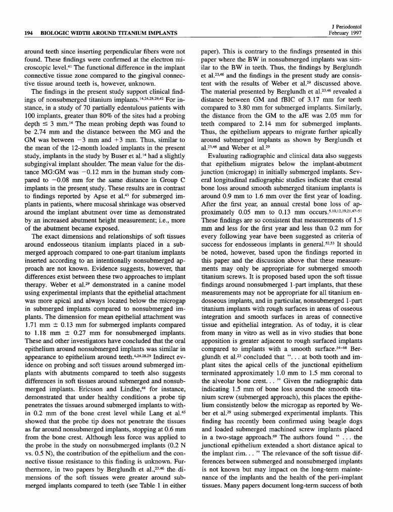
Early healing Short-term loading Longer-term loadingFigure 9. Model of peri-implant soft tissue integration. Note constant value for biologic width (BW) over
time.
approaches to endosseous implant dentistry. However, itis not possible to determine morphology of the soft tissuesand effectiveness of the implanto-gingival barrier underclinical inspection.
In the canine model used in this study where the tissueswere cleaned 3 times per week, there was no significantdifference between unloaded and loaded implants as re-
gards the overall dimensions of the implanto-gingival softtissues. This would suggest that the tissues, once healed,reach a level of maturity that does not change signifi-cantly over time unless an environmental change occurs
such as inflammation, toothbrush trauma, etc. This ap-pears to be the case with dento-gingival soft tissue andthe present data support such a concept around healthynonsubmerged 1-part implants. It is unknown if the im-planto-gingival soft tissues will serve as a barrier similarto dento-gingival soft tissues due to the morphologicaldifferences in these two situations. Periodontal ligamentfibers are found apical to the dento-gingival connectivetissues, whereas bone is present in the implanto-gingivalcase. The data support the fact that the dimensions of thetissues are physiologically stable for periods greater thanone year and thus may be stable during longer mainte-nance periods. The oral hygiene performed in this studyincluded both chemical (chlorhexidine-digluconate111111) andmechanical (soft toothbrush plus soft sponge) therapy.The data demonstrate for the first time that routine me-
chanical and chemical therapy for up to 15 months willnot significantly alter, at the histological level, the softtissue dimensions around healthy integrated nonsub-merged 1-part titanium implants.
A model of peri-implant soft tissue integration for en-
dosseous nonsubmerged titanium implants is shown inFigure 9. The findings reported in this study permit a
concept that the BW is physiologically constant for 1 -part
oral transmucosal structures including teeth and nonsub-merged implants. These implants represented early stagesof healing; i.e., from time of implant placement until timeof loading (3 months unloaded), short-term loaded (3months loaded and 6 months healed) and longer-termloaded (12 months loaded and 15 months healed) im-plants. As in the case of teeth, the CTC in nonsubmergedimplants remains relatively stable, while the soft tissuedifferences that occurred largely took place in the dimen-sion of the JE and the SD. These data therefore suggestthat perturbation of the oral soft tissues with the place-ment of a nonsubmerged 1-part implant results in a heal-ing process such that the connective tissue is formed andmatures to a state similar to surrounding tissues and thatthe JE and SD are adaptable with their dimensions beingdynamic with time of healing. These findings are sup-ported by the fact that implants with two different roughendosseous surfaces were examined in this study and thatno significant differences were found concerning the BWwhen comparing the two surfaces. Furthermore, no dif-ferences were found in soft tissue dimensions comparingimplant sites in the more posterior area of the mandibleversus the more anterior sites in spite of a more narrow
alveolar ridge in the anterior implant sites.
ConclusionThis study indicates that the dimensions and relationshipsof the implanto-gingival junction of healthy nonsub-merged 1-part titanium implants are similar to dento-gin-gival tissues. Thus, both junctions have relative physio-logic dimensions. The measurements of the tissues weresimilar under unloaded and loaded conditions. Routinemaintenance therapy including mechanical and chemicaltreatment for up to 15 months did not alter the overalldimensions of the soft tissues. These data suggest that a
196 BIOLOGIC WIDTH AROUND TITANIUM IMPLANTSJ Periodontol
February 1997
biologic width exists around nonsubmerged 1-part tita-nium implants as well as around teeth and that this is a
physiologically formed and stable structure at least in thecase of nonsubmerged titanium implants.
AcknowledgmentsThe authors gratefully acknowledge Sonja A. Bustamante,H.T. (ASCP), for her valuable contributions throughoutthe study. We also thank Richard J. Haines, DVM, andhis colleagues for exemplary care of the animals. Wegreatly appreciate the help of James P. Simpson, BSc(Eng), PhD, and the Institut Straumann AG for manufac-turing the test implants; John D. Schoolfield, MS, for sta-tistical evaluation and Nancy Place (CMI) for graphic ser-
vices. This study was supported by grant" 18-92/053 fromthe ITI foundation, Waldenburg/BL, Switzerland.
REFERENCES1. Brânemark Pl, Breine U, Adell R, Hansson BO, Lindström J, Ohls-
son A. Intra-osseous anchorage of dental prostheses. I. Experimentalstudies. Scand J Plast Reconstr Surg 1969;3:81-100.
2. Schroeder A, Pohler O, Sutter F. Gewebsreaktion auf ein Titan-Hohl-zylinderimplantat mit Titan-Spritzschichtoberfläche. Schweiz MschrZahnheilk 1976;86:713-727.
3. Schroeder A, Stich H, Straumann F, Sutter F. Über die Anlagerungvon Osteozement an einen belasteten Implantatkörper. SchweizMschr Zahnheilk 1978;88:1051-1058.
4. Schroeder A, van der Zypen E, Stich H, Sutter F. The reactions ofbone, connective tissue, and epithelium to endosteal implants withtitanium-sprayed surfaces. J Maxillofac Surg 1981;9:15-25.
5. Brânemark PI, Hansson BO, Adell R, et al. Osseointegrated implantsin the treatment of the edentulous jaw. Experience from a 10-yearperiod. Scand J Plast Reconstr Surg 1977:ll(Suppl. 16):1-132.
6. Albrektsson T. Direct bone anchorage of dental implants. / ProsthetDent 1983;50:255-261.
7. Albrektsson T, Sennerby L. State of the art in oral implants. / ClinPeriodontol 1991;18:474-481.
8. Ruggeri A, Franchi M, Marini , Trial , Piattelli . Supracrestalcircular collagen fiber network around nonsubmerged titanium im-plants. Clin Oral Impl Res 1992;3:169-175.
9. Ruggeri A, Franchi M, Trisi , Piattelli A. Histologie and ultrastruc-tural findings of gingival circular ligament surrounding osseointe-grated nonsubmerged loaded titanium implants. Int J Oral Maxil-lofac Implants 1994;9:636-643.
10. Adell R, Lekholm U, Rockier B, Brânemark PI. A 15-year study ofosseointegrated implants in the treatment of the edentulous jaw. IntJ Oral Surg 1981;10:387-416.
11. Babbush CA, Kent JN, Misiek DL. Titanium plasma-sprayed (TPS)screw implants for the reconstruction of the edentulous mandible. JOral Maxillofac Surg 1986;44:274-282.
12. Cox TE Zarb GA. The longitudinal clinical efficacy of osseointe-grated dental implants: A 3-year report. Int J Oral Maxillofac Im-plants 1987;2:91-100.
13. Adell R, Eriksson B, Lekholm U, Brânemark PI, Jemt T. A long-term follow-up study of osseointegrated implants in the treatmentof totally edentulous jaws. Int J Oral Maxillofac Implants 1990;5:347-359.
14. Buser D, Weber HP, Lang NP. Tissue integration of non-submergedimplants. 1-year results of a prospective study with 100 ITI hollow-cylinder and hollow-screw implants. Clin Oral Impl Res 1990; 1:33-40.
15. ten Bruggenkate CM, Mutter K, Oosterbeek HS. Clinical evaluation
of the ITI (F-type) hollow cylinder implant. Oral Surg Oral MedOral Pathol 1990;70:693-697.
16. Buser D, Weber HP, Brägger U, Balsiger C. Tissue integration ofone-stage ITI implants: 3-year results of a longitudinal study withhollow-cylinder and hollow-screw implants. Int J Oral MaxillofacImplants 1991;6:405-412.
17. Behneke A, Behneke N, Wagner W. Klinische Ergebnisse mit trans-
gingival inserierten enossalen Implantaten (Bonefit-System). Zahnärztl Implantol 1992;8:97-102.
18. Schmitt A, Zarb GA. The longitudinal clinical effectiveness of os-
seointegrated dental implants for single-tooth replacement. Int JProsthodont 1993;6:197-202.
19. Laney WR, Jemt T, Harris D, et al. Osseointegrated implants forsingle-tooth replacement: Progress report from a multicenter pro-spective study after 3 years. Int J Oral Maxillofac Implants 1994;9:49-54.
20. Mericske-Stem R, Steinlin Schaffner , Marti , Geering AH. Peri-implant mucosal aspects of ITI implants supporting overdentures. Afive-year longitudinal study. Clin Oral Impl Res 1994;5:9-18.
21. Jemt , Lekholm U. Implant treatment in edentulous maxillae: A5-year follow-up report on patients with different degrees of jawrésorption. Int J Oral Maxillofac Implants 1995;10:303-311.
22. Gotfredsen K, Rostrup E, Hj0rting-Hansen E, Stoltze , Budtz-J0r-gensen E. Histological and histomorphometrical evaluation of tissuereactions adjacent to endosteal implants in monkeys. Clin Oral ImplRes 1991;2:30-37.
23. Berglundh T, Lindhe J, Ericsson I, Marinello CP, Liljenberg B,Thomsen P. The soft tissue barrier at implants and teeth. Clin OralImpl Res 1991;2:81-90.
24. Buser D, Weber HP, Donath , Fiorellini JP, Paquette DW, WilliamsRC. Soft tissue reactions to non-submerged unloaded titanium im-plants in beagle dogs. J Periodontol 1992;63:226-236.
25. James RA, Schultz R. Hemidesmosomes and the adhesion of junc-tional epithelial cells to metal implants -a preliminary report. J OralImplantol 1974;4:294-302.
26. Gould T, Brunette D, Westbury L. The attachment mechanism ofepithelial cells to titanium in vitro. J Periodont Res 1981;16:611-616.
27. Jansen JA, de Wijn JR, Wolters-Lutgerhorst JML, van Müllem PJ.Ultrastructural study of epithelial cell attachment to implant mate-rials. J Dent Res 1985;64:891-896.
28. Buser D, Stich H, Krekeler G, Schroeder A. Faserstrukturen derperiimplantären Mukosa bei Titanimplantaten. Eine tierexperimen-telle Studie am Beagle-Hund. Zahnärztl Implantol 1989;5:15-23.
29. Weber HP, Buser D, Donath K, et al. Comparison of healed tissuesadjacent to submerged and nonsubmerged unloaded titanium dentalimplants. A histometric study in beagle dogs. Clin Oral Impl Res1996;7:11-19.
30. Gargiulo AW, Wentz FM, Orban B. Dimensions and relations of thedentogingival junction in humans. J Periodontol 1961;32:261-267.
31. Vacek JS, Gher ME, Assad DA, Richardson AC, Giambarresi LI.The dimensions of the human dentogingival junction. Int J Perio-dontics Restorative Dent 1994;14:155-165.
32. Schenk RK, Olah AJ, Herrmann W. Preparation of calcified tissuesfor light microscopy. In: Dickson GR, ed. Methods of Calcified Tis-sue Preparation, vol. 1. Amsterdam, New York, Oxford: ElsevierScience Publishers B.V.; 1984:1-56.
33. Cochran DL, Nummikoski PV, Higginbottom FL, Hermann JS,Makins SR, Buser D. Evaluation of an endosseous titanium implantwith a sandblasted and acid-etched surface in the canine mandible.Radiographie results. Clin Oral Impl Res 1996;7:240-252.
34. Gottlieb B. Der Epithelansatz am Zahne. Dtsch Monatsschr Zahn-heilk 1921;5:142-147.
35. Orban B, Mueller E. The gingival crevice. J Am Dent Assoc1929;16:1206-1242.
36. Waerhaug J. The gingival pocket. Odont Tidscrift 1952;60:(Suppl.):l.
Volume 68Number 2 COCHRAN, HERMANN, SCHENK, HIGGINBOTTOM, BUSER 197
37. Orban B, Bhatia H, Kollar JA, Wentz FM. The epithelial attachment(the attached epithelial cuff). J Periodontol 1956;27:167-180.
38. Sicher H. Changing concepts of the supporting dental structures.Oral Surg Oral Med Oral Pathol 1959;12:31-35.
39. Cochran DL, Mahn DH. Dental implants and regeneration. Part I.Overview and biological considerations. In: Hardin J, ed. Clark'sClinical Dentistry, 5th edition. Philadelphia: J. B. Lippincott Com-pany; 1992:1-7.
40. Cochran DL, Simpson J, Weber HP, Buser D. Attachment andgrowth of periodontal cells on smooth and rough titanium. Int J OralMaxillofac Implants 1994;9:289-297.
41. Listgarten MA, Buser D, Steinemann SG, Donath , Lang NP, We-ber HP. Light and transmission electron microscopy of the intactinterfaces between non-submerged titanium-coated epoxy resin im-plants and bone or gingiva. J Dent Res 1992;71:364-371.
42. Weber HP, Buser D, Fiorellini JP, Williams RC. Radiographic eval-uation of crestal bone levels adjacent to nonsubmerged titanium im-plants. Clin Oral Impl Res 1992;3:181-188·.
43. Apse R Zarb GA, Schmitt A, Lewis DW. The longitudinal effec-tiveness of osseointegrated dental implants. The Toronto study: Peri-implant mucosal response. Int J Periodontics Restorative Dent1991;11:95-111. .
44. Ericsson I, Lindhe J. Probing depth at implants and teeth. An ex-
perimental study in the dog. J Clin Periodontol 1993;20:623-627.45. Lang NP, Wetzel AC, Stich , Caffesse RG. Histologie probe pen-
etration in healthy and inflamed peri-implant tissues. Clin Oral ImplRes 1994;5:191-201.
46. Berglundh T, Lindhe J, Mannello C, Ericsson I, Liljenberg B. Softtissue reaction to de novo plaque formation on implants and teeth.An experimental study in the dog. Clin Oral Impl Res 1992;3:1-8.
47. Adell R, Lekholm U, Rockler B, et al. Marginal tissue reactions at
osseointegrated titanium fixtures. (I). A 3-year longitudinal prospec-tive study. Int J Oral Maxillofac Surg 1986;15:39-52.
48. Lekholm U, Adell R, Lindhe J, et al. Marginal tissue reactions at
osseointegrated titanium fixtures. (II) A cross-sectional retrospectivestudy. Int J Oral Maxillofac Surg 1986;15:53-61.
49. Ahlqvist J, Borg , Günne J, Nilson , Olsson M, Âstrand P. Os-seointegrated implants in edentulous jaws: A 2-year longitudinalstudy. Int J Oral Maxillofac Implants 1990;5:155-163.
50. Jemt , Lekholm U, Grondarti . A 3-year follow up study of earlysingle implant restorations ad modum Brânemark. Int J PeriodonticsRestorative Dent 1990;10:341-349.
51. Quirynen M, Naert I, van Steenberghe D, Teerlinck J, Dekeyser C,Theuniers G. Periodontal aspects of osseointegrated fixtures sup-porting an overdenture. A 4-year retrospective study. J Clin Perio-dontol 1991;18:719-728.
52. Albrektsson T, Zarb G, Worthington P, Eriksson AR. The long-termefficacy of currently used dental implants: A review and proposedcriteria of success. Int J Oral Maxillofac Implants 1986; 1:11—25.
53. Smith DE, Zarb GA. Criteria for success of osseointegrated end-osseous implants. J Prosthet Dent 1989;62:567-572.
54. Wennerberg A, Albrektsson T, Andersson B, Eng M. Design andsurface characteristics of 13 commercially available oral implantsystems. Int J Oral Maxillofac Implants 1993;8:622-633.
55. Martin JY, Schwartz Z, Hummert TW, et al. Effect of titanium sur-
face roughness on proliferation, differentiation, and protein synthesisof human osteoblast-like cells (MG63). J Biomed Mater Res1995;29:389-401.
56. Martin JY, Dean DD, Cochran DL, Simpson J, Boyan BD, Schwartz . Proliferation, differentiation, and protein synthesis of human os-
teoblast-like cells (MG63) cultured on previously used titanium sur-
faces. Clin Oral Impl Res 1996;7:27-37.57. Schwartz Z, Martin JY, Dean DD, Simpson J, Cochran DL, Boyan
BD. Effect of titanium surface roughness on chondrocyte prolifer-ation, matrix production, and differentiation depends on the state ofcell maturation. J Biomed Mater Res 1996;30:145-155.
58. Claes L, Hutschenreuter P, Pohler O. Lösemomente von Corticalis-zugschrauben in Abhängigkeit von Implantationszeit und Oberfläch-en-beschaffenheit. Archiv Orthopädischen Unfall-Chirurgie 1976;85:155-159.
59. Kirsch A, Donath K. Tierexperimentelle Untersuchungen zur Be-deutung der Mikromorphologie von Titanimplantatoberflächen.Fortschr Zahnärztl Implantai 1984;1:35-40.
60. Thomas KA, Cook S. An evaluation of variables influencing implantfixation by direct bone apposition. J Biomed Mater Res 1985;19:875-901.
61..Steinemann SG, Eulenberger J, Mäusli PA, Schroeder A. Adhesionof bone to titanium. In: Christel P, Meunier A, Lee AJC, eds. Ad-vances in Biomaterials -Biological and Biomechanical Performanceof Biomaterials (Vol. 6). Amsterdam: Elsevier Publishers; 1986:409-414.
62. Block MS, Kent JN, Kay JF. Evaluation of hydroxylapatite-coatedtitanium dental implants in dogs. J Oral Maxillofac Surg 1987;45:601-607.
63. Carlsson L, Röstlund , Albrektsson , Albrektsson T. Removal tor-
ques for polished and rough titanium implants. Int J Oral MaxillofacImplants 1988;3:21-24.
64. Wilke HJ, Claes L, Steinemann S. The influence of various titaniumsurfaces on the interface shear strength between implants and bone.In: Heimke G, Soltész U, Lee AJC, eds. Advances in BiomaterialsClinical Implant Materials (Vol. 9). Amsterdam: Elsevier Publish-ers; 1990:309-314.
65. Buser D, Schenk RK, Steinemann S, Fiorellini JR Fox CH, StichH. Influence of surface characteristics on bone integration of tita-nium implants. A histomorphometric study in miniature pigs. J Bio-med Mater Res 1991;25:889-902.
66. Gottlander M, Albrektsson , Carlsson LV. A histomorphometricstudy of unthreaded hydroxyapatite-coated and titanium-coated im-plants in rabbit bone. Int J Oral Maxillofac Implants 1992;7:485-490.
67. Weinlaender M, Kenney EB, Lekovic V, Moy PK. Histomorphom-etry of bone apposition around three types of endosseous dentalimplants. Int J Oral Maxillofac Implants 1992;7:491-496.
68. Wennerberg A, Albrektsson T, Andersson B, Krol JJ. A histomor-phometric and removal torque study of screw-shaped titanium im-plants with three different surface topographies. Clin Oral Impl Res1995;6:24-30.
69. Hürzeler MB, Quiñones CR, Schupbach , Vlassis JM, Strub JR,Caffesse RG. Influence of the suprastrueture on the peri-implanttissues in beagle dogs. Clin Oral Impl Res 1995;6:139-148.
Send reprint requests to: Dr. David L. Cochran, University of Texas,Health Science Center, Department of Periodontics, 7703 Floyd Curl Dr.,San Antonio, TX 78284-7894.
Accepted for publication July 19, 1996.
s