secretory microrna-29 expression in gingival crevicular fluid
TRANSCRIPT
RESEARCH ARTICLE
Secretory microRNA-29 expression in gingival
crevicular fluid during orthodontic tooth
movement
Phimon Atsawasuwan1*, Paul Lazari1, Yinghua Chen1, Xiaofeng Zhou2, Grace Viana1,
Carla A. Evans3
1 Department of Orthodontics, University of Illinois at Chicago, Chicago, Illinois, United States of America,
2 Department of Periodontics, University of Illinois at Chicago, Chicago, Illinois, United States of America,
3 Department of Orthodontics, Boston University, Boston, Massachusetts, United States of America
Abstract
Secretory microRNAs (miRNAs) have been used increasingly as biomarkers for cancers,
autoimmune diseases and inflammatory diseases. They are reported as being freely circu-
lated or encapsulated in microvesicles such as exosomes. This study was performed to eluci-
date the presence of miRNAs with exosomes in human gingival crevicular fluid (GCF), and
the expression profile of miRNA-29 during orthodontic tooth movement. Four healthy volun-
teer and fifteen orthodontic patients were enrolled in the study. Secretory miRNA in GCF was
collected and analyzed using a bioanalyzer, realtime PCR and Western blot analysis. The
expression profile of secretory miR-29 family in GCF was analyzed during the course of
canine retraction for 6 weeks. The results demonstrated the presence of miRNAs in the GCF.
After series of ultracentrifugation and RT-PCR array, exosome-depleted fractions and pellets
were isolated and we found that secretory miRNAs were detected in both the exosome-asso-
ciated fraction and the exosome-depleted supernatant fraction; however, the concentration of
miRNAs was higher in the exosome-associated fraction than in the exosome-depleted frac-
tion suggesting a close association between the secretory miRNAs and exosomes in GCF.
We also demonstrated the increased expression profiles of miR-29 family during six weeks of
orthodontic tooth movement in humans. Secretory miRNAs are present in GCF and secretory
miRNA-29 family expression profiles increase during the tooth movement in humans. Secre-
tory miRNA-29 in GCF could serve as potential biomarkers for periodontal remodeling.
Introduction
MicroRNAs (miRNAs) are small non-coding RNAs, which are important in development,
organogenesis, and homeostasis[1]. Several reports showed that miRNAs could be isolated
from fresh or fixed tissues[2,3] and body fluids[4,5]. Secretory miRNAs have been isolated and
shown to exist with remarkable stability in various types of body fluids[6]. The stability of
secretory miRNA results from the formation of complexes between secretory miRNA and spe-
cific proteins[7]. Some secretory miRNAs were demonstrated to be packaged inside exosomes
PLOS ONE | https://doi.org/10.1371/journal.pone.0194238 March 8, 2018 1 / 11
a1111111111
a1111111111
a1111111111
a1111111111
a1111111111
OPENACCESS
Citation: Atsawasuwan P, Lazari P, Chen Y, Zhou
X, Viana G, Evans CA (2018) Secretory microRNA-
29 expression in gingival crevicular fluid during
orthodontic tooth movement. PLoS ONE 13(3):
e0194238. https://doi.org/10.1371/journal.
pone.0194238
Editor: Sompop Bencharit, Virginia Commonwealth
University, UNITED STATES
Received: January 14, 2018
Accepted: February 27, 2018
Published: March 8, 2018
Copyright: This is an open access article, free of all
copyright, and may be freely reproduced,
distributed, transmitted, modified, built upon, or
otherwise used by anyone for any lawful purpose.
The work is made available under the Creative
Commons CC0 public domain dedication.
Data Availability Statement: All relevant data are
within the paper and its Supporting Information
files.
Funding: This work was supported by the National
Institute of Dental and Craniofacial research:
DE024531 to PA and the American Association of
Orthodontists Foundation: biomedical research
award to PA. The funders had no role in study
design, data collection and analysis decision to
publish, or preparation of the manuscript.

[8], which are small membranous vesicles about 30–100 nm in size and derived from the endo-
some[9]. The exosomes contain proteins and nucleic acids and can be secreted by many cell
types[10]. The existence of secretory miRNAs in human serum and saliva has been reported to
be concentrated in exosomes[4]. MiRNAs from unfractionated whole serum, urine, saliva,
cerebrospinal fluid and exosomes could be promising biological tools as diagnostic biomarkers
[8,11]. Several miRNAs play crucial roles in bone remodeling by controlling osteoblast/clast
differentiation and function[12]. miRNAs-29a/b/c were involved in regulation of osteoblasts/
clasts differentiation and could influence expression of certain extracellular matrix molecules
i.e. collagens and their modifying enzymes[13–15]. Kagiya and Nakamura[16] found an
increase in miRNA-29b during osteoclast differentiation in TNF-α/RANKL-treated cells, sug-
gesting this miRNA plays a role in TNFα -regulated osteoclast differentiation. Franceschetti
et al[17] found an increase in expression of all three members of miRNA-29 family during
osteoclastogenesis, and repression of osteoclastogenesis process by inhibition of miRNA-29.
The finding implicated that miRNA-29 promoted osteoclastogenesis.
Gingival crevicular fluid (GCF) is a serum transudate found in the gingival sulcus[18]. Irri-
tation and inflammation of the gingival tissue increase the flow and alter the constituents of
crevicular fluid. Serum is the primary source of the aqueous component of the GCF. The usual
volume range of the GCF in the undisturbed sulcus is between 0.5–1μL[19]. A number of stud-
ies have measured cytokine and protein levels in GCF and analyzed this fluid to find biomark-
ers for several oral diseases such as gingivitis, periodontitis, root resorption and systemic
diseases[20–22]. During orthodontic tooth movement (OTM), osteoclast plays a crucial role
and its activities increases leading to alveolar bone resorption and tooth movement[23].
Changes in different substances found in GCF were reported in presence of orthodontic forces
and various classes of molecules related to osteoclast activities such as Receptor activator of
nuclear factor kappa-Β ligand (RANKL) and osteoprotegerin (OPG) contained in GCF have
been reported as potential biomarkers for OTM[24,25]. This study we focused on miR-29 fam-
ily due to their expression patterns in human periodontal ligament under loading[26] and
their direct association with osteoclast function[16,17].This study was performed to elucidate
the presence of miRNAs with exosomes in human gingival crevicular fluid (GCF), and the
expression profile of miRNA-29 during orthodontic tooth movement.
Materials and methods
Human subjects and GCF colllection
The study was approved by the Institutional Review Boards of the University of Illinois at Chi-
cago (IRB protocol #2013–0183). All subjects and guardians signed a written informed consent
and assent. To evaluate the presence of microRNA in gingival crevicular fluid (GCF), the GCF
samples was collected from 4 healthy adult male volunteers aged 26–28 years old with excellent
periodontal health. To study the profiles of secretory miR-29 family expression during maxil-
lary canine retraction to close extraction spaces, seventy orthodontic patients aged 10–17 years
old were screened and fifteen orthodontic patients was recruited for the study. Inclusion crite-
ria were the following: (1) patients with excellent oral hygiene throughout the study period, (2)
patients require at least maxillary first premolar extraction so canine retraction will be per-
formed as a part of the treatment, (3) available to come back at the clinic at the times of sample
collection. The exclusion criteria were (1) poor oral hygiene and/or bleeding on probing, (2)
use medications, steroids or other anti-inflammatory medications, (3) systemic health prob-
lems or smoking. The collecting times were the following: T0: prior to bonding the fixed
orthodontic appliances, T1: on the day of canine retraction, 60 minutes after engaging the elas-
tomeric powerchains (American Orthodontics, Sheboygan,WI) onto the canine bracket. The
Secretory microRNA in gingival crevicular fluid
PLOS ONE | https://doi.org/10.1371/journal.pone.0194238 March 8, 2018 2 / 11
Competing interests: The authors have declared
no competing interest exists.
powerchain for all the subjects was placed with an initial force of approximately 250g as mea-
sured by the Dontrix Force Gauge (Orthopli Corp, Philadelphia, Pa), T2: 24 hours after initia-
tion of canine retraction, T3: 7 days after initiation of canine retraction, T4: 6 weeks after
initiation of canine retraction. The timepoints of sample collection were shown in Fig 1.To col-
lect the GCF samples, gingival sulcus of subjects was carefully isolated with cotton rolls to pre-
vent a contamination from saliva. The GCF was collected by placing periopapers (Oraflow,
Smithtown, NY) gently in gingival sulcus mesial to the tooth for 1 min. The collection process
was repeated 4 times in the same location to collect adequate amount of GCF. The collected
periopapers were stored in 200 μL cold sterile, RNase-DNase free phosphate buffered saline
(PBS) and directly processed for the analysis.
Exosome isolation
The samples from the 4 healthy individuals were centrifuged at 1,500g for 10 minutes to remove
the periopapers and debris. The collected supernatant was centrifuged again at 17,000g for 15
minutes. An aliquot of the supernatant was subjected in an ultracentrifuged at 160,000g for 1
hour to isolate exosomes[4]. All centrifugations were performed at 4˚C. The pellet containing
exosomes and exosome-depleted supernatant were then processed for further experiments.
RNA isolation and PCR array
Total RNA was extracted from all orthodontic patients’s total GCF samples and the 4 healthy
subjects’ GCF samples, which were composed of non-ultracentrifuged supernatant, and
exosome pellet and the exosome-depleted supernatant fractions after ultracentrifugation.
miRNaeasy kit (Qiagen, Valencia, CA) was used for the total RNA isolation following the man-
ufacturer’s instructions. The quality and quantity of the extracted RNA was assessed using an
Agilent 2200 Tapestation bioanalyzer (Agilent Technologies, Santa Clara, CA) and NanoDrop
1000 spectrophotometer (Thermoscientific, Waltham, MA). For the Tapestation, 4μl R6K
sample buffer and 1μl of RNA sample was mixed, heated at 72˚C for 3 min then placed on ice
for 2 min. Five microliters of DNA ladder and the samples were loaded into R6K screentape
(Agilent Technologies,). The screentape was placed in the 2200 Tapestation and processed
using the 2200 Tapestation Controller Software A.01.04 (Agilent Technologies). Quantitative
real-time PCR (qPCR) was performed according to the manufacturer’s instructions (Qiagen)
to profile the miRNA distribution in the exosome pellet and exosome-depleted-supernatant
fractions of GCF. In brief, 5μl total RNA was used to generate cDNA using the miScript
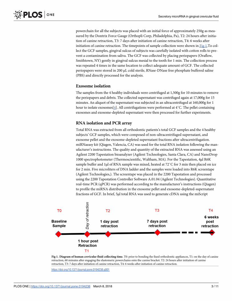
Fig 1. Diagram of human crevicular fluid collecting time. T0: prior to bonding the fixed orthodontic appliances, T1: on the day of canine
retraction, 60 minutes after engaging the elastomeric powerchains onto the canine bracket. T2: 24 hours after initiation of canine
retraction, T3: 7 days after initiation of canine retraction, T4: 6 weeks after initiation of canine retraction.
https://doi.org/10.1371/journal.pone.0194238.g001
Secretory microRNA in gingival crevicular fluid
PLOS ONE | https://doi.org/10.1371/journal.pone.0194238 March 8, 2018 3 / 11
Reverse Transcription kit (Qiagen). The cDNA from each fraction was subject to miScript
PCR array MIHS-001Z (Qiagen) to investigate the species and presence of secretory miRNAs
in GCF. The RT-realtime PCR for quantification of miRNA-29 family was performed using
commercially available primers (Life Technologies).
Electron microscopy
To verify the presence of exosomes in GCF, the exosome pellet was fixed with cold 2% v/v glu-
taraldehyde in 0.1M PBS, rinsed in PBS, dehydrated through a graded series of ethanol and
embedded in Epon. Ultra-thin sections (65nm) were stained with uranyl acetate and Reynold’s
lead citrate. A JEOL 100CX II transmission electron microscope was used for imaging.
Western blotting
Proteins were extracted from the ultracentrifuged pellets and subject to SDS-PAGE then trans-
ferred to a PVDF membrane. The blotting membrane was blocked with skimmed milk and
incubated with anti-human CD63 and CD9 antibodies (Abcam, Cambridge, MA) followed by
incubation with horseradish peroxide-coupled secondary antibody (Abcam, Cambridge, MA).
The proteins were detected using enhanced chemiluminescence (Thermo Scientific, Rockford,
IL).
Statistical analysis
The distribution of data was investigated using the Shapiro-Wilk test. Due to the distribution
of the data and nature of the methods, Kruskal-Wallis statistical analyses were performed
using IBM SPSS Statistics for Windows, V22.0, (Armonk, NY: IBM Corp.). Statistical signifi-
cance was set at p� 0.05. Descriptive statistical analysis was reported as mean + standard
deviation.
Results
Secretory miRNAs were present in human gingival crevicular fluids
No subjects in this study had oral or periodontal pathology or were smokers. No pain or com-
plication related to the noninvasive GCF collection using absorbent paper strips was reported.
After the GCF collection from the 4 healthy subjects, the cell debris and paper strips were iso-
lated from supernatant of the GCF by serial centrifugation at 1,500 and 17,000g. The superna-
tant was then subjected for total RNA isolation. After the total RNA isolation, examination of
the isolated total RNA was immediately performed using a Tapestation 2200 and the results
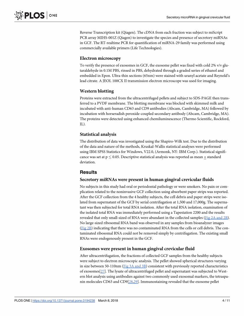
revealed that only small-sized of RNA were abundant in the collected samples (Fig 2A and 2B).
No large-sized ribosomal RNA band was observed in any samples from bioanalyzer images
(Fig 2B) indicating that there was no contaminated RNA from the cells or cell debris. The con-
taminated ribosomal RNA could not be removed simply by centrifugation. The existing small
RNAs were endogenously present in the GCF.
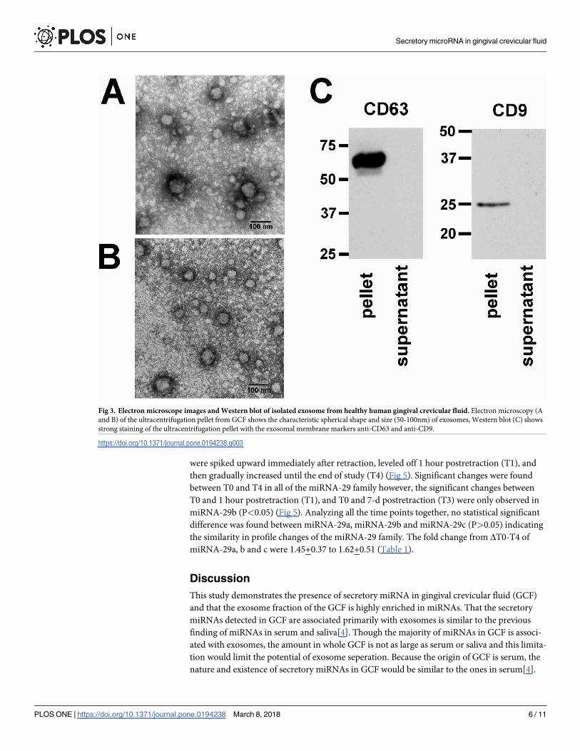
Exosomes were present in human gingival crevicular fluid
After ultracentrifugation, the fractions of collected GCF samples from the healthy subjects
were subject to electron microscopic analysis. The pellet showed spherical structures varying
in size between 50-110nm (Fig 3A and 3B) consistent with previously reported characteristics
of exosomes[27]. The lysate of ultracentrifuged pellet and supernatant was subjected to West-
ern blot analysis using antibodies against two commonly used exosomal markers, the tetraspa-
nin molecules CD63 and CD9[28,29]. Immunostaining revealed that the exosome pellet
Secretory microRNA in gingival crevicular fluid
PLOS ONE | https://doi.org/10.1371/journal.pone.0194238 March 8, 2018 4 / 11
fraction showed strong staining of CD63 and CD9 compared to negative staining of the exo-
some-depleted supernatant fraction (Fig 3C and S1 Fig).
Majority of secretory miRNAs were associated with exosomes
To quantify and determine whether miRNA in GCF is associated with exosomes or is freely
circulated, we extracted the RNA from the exosome pellet fraction and from the exosome-
depleted supernatant fraction. The whole exosomal pellet and entire volume of the supernatant
were used for RNA isolation. Equal amounts of RNA in the pellet fraction and supernatant
fraction were used for quantitative RT-PCR and subjected to miScript PCR array to examine
species of secretory miRNAs in each fraction. The distribution of miRNAs in exosome pellet
and exosome-depleted supernatant from each subject demonstrated that the majority of miR-
NAs detected in GCF was associated with the exosome pellet (S2 Fig). The expression levels of
miRNAs in GCF were quantified and their amounts in exosome pellet and exosome-depleted
supernatant were compared using quantitative RT-PCR. The RT-PCR revealed that the aver-
age number of CT of selected miRNAs in exosome pellet was about 5 cycles lower than the one
in the supernatant (Fig 4A) and the same results were found when the CT of each miRNA was
normalized using total amount of RNA in each fraction as internal control miRNAs (Fig 4B).
Increased expression profiles of secretory miRNA-29 family were detected
during canine retraction
Due to the limited quantity of GCF sample collected from a single canine during the canine
retraction, the total GCF sample not fractionated sample from each canine was tested in this
part of study. Along the course of canine retraction, after normalization with internal miRNA
controls (Let-7d, g and i), the change in miRNA-29 family expression patterns from pretreat-
ment (T0) to 6 weeks postretraction (T4) showed statistical significance consistently (P<0.05).
The expression patterns of miR-29 family expression demonstrated that the expression levels
Fig 2. Presence of miRNA in healthy human crevicular fluid. “gel-like” images of R6K ScreenTape (A) after
subjected to Tapestation bioanalyzer showed RNA ladder (L) on left lane and only small-sized RNA present in the
sample lanes (middle and right lane). The positive bands approximately 50 nucleotides (nt) were present in
representative GCF samples (A1 and B1). Electropherogram (B) corresponding to the gel-like images on the (A) figure.
The x-axis on the electropherogram represents RNA size (nt), while the y-axis represents the measurement response of
fluorescence units (FUs).
https://doi.org/10.1371/journal.pone.0194238.g002
Secretory microRNA in gingival crevicular fluid
PLOS ONE | https://doi.org/10.1371/journal.pone.0194238 March 8, 2018 5 / 11
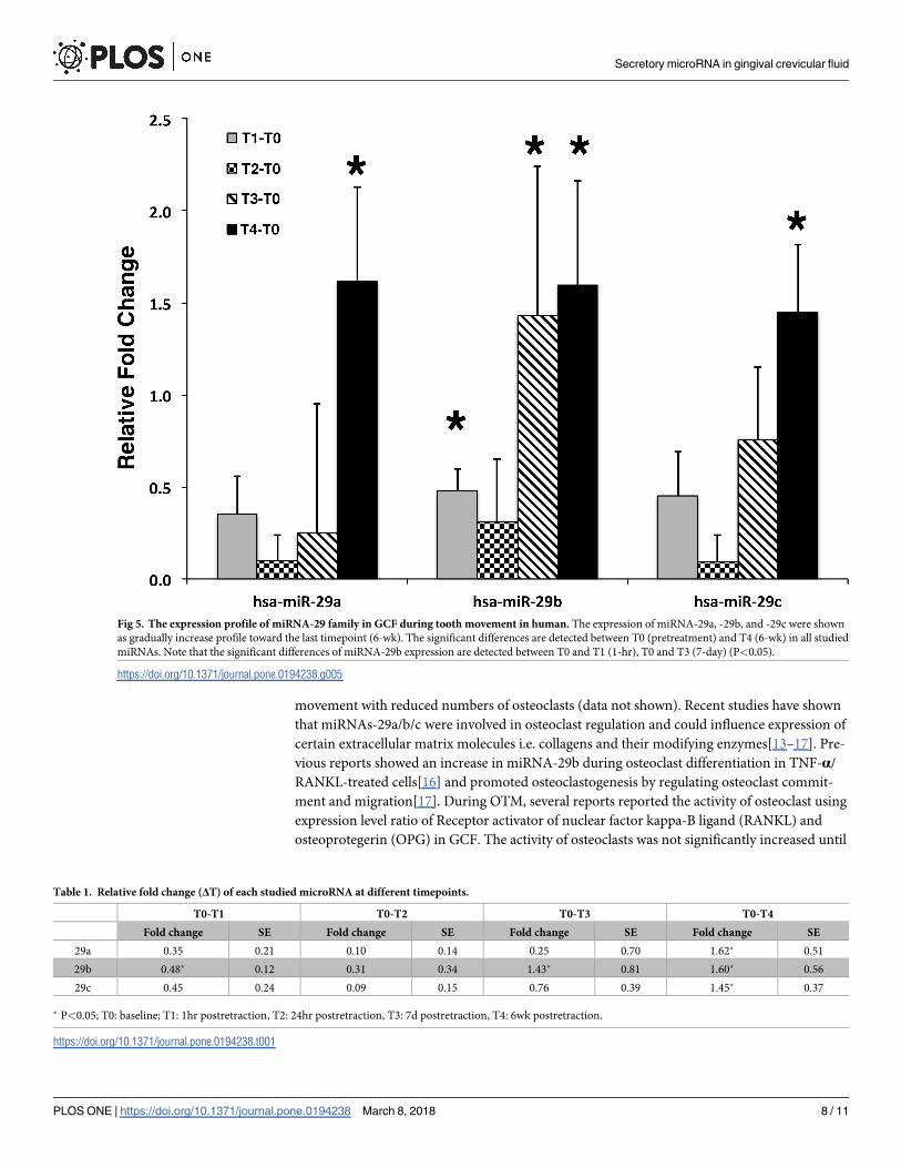
were spiked upward immediately after retraction, leveled off 1 hour postretraction (T1), and
then gradually increased until the end of study (T4) (Fig 5). Significant changes were found
between T0 and T4 in all of the miRNA-29 family however, the significant changes between
T0 and 1 hour postretraction (T1), and T0 and 7-d postretraction (T3) were only observed in
miRNA-29b (P<0.05) (Fig 5). Analyzing all the time points together, no statistical significant
difference was found between miRNA-29a, miRNA-29b and miRNA-29c (P>0.05) indicating
the similarity in profile changes of the miRNA-29 family. The fold change from ΔT0-T4 of
miRNA-29a, b and c were 1.45+0.37 to 1.62+0.51 (Table 1).
Discussion
This study demonstrates the presence of secretory miRNA in gingival crevicular fluid (GCF)
and that the exosome fraction of the GCF is highly enriched in miRNAs. That the secretory
miRNAs detected in GCF are associated primarily with exosomes is similar to the previous
finding of miRNAs in serum and saliva[4]. Though the majority of miRNAs in GCF is associ-
ated with exosomes, the amount in whole GCF is not as large as serum or saliva and this limita-
tion would limit the potential of exosome seperation. Because the origin of GCF is serum, the
nature and existence of secretory miRNAs in GCF would be similar to the ones in serum[4].
Fig 3. Electron microscope images and Western blot of isolated exosome from healthy human gingival crevicular fluid. Electron microscopy (A
and B) of the ultracentrifugation pellet from GCF shows the characteristic spherical shape and size (50-100nm) of exosomes, Western blot (C) shows
strong staining of the ultracentrifugation pellet with the exosomal membrane markers anti-CD63 and anti-CD9.
https://doi.org/10.1371/journal.pone.0194238.g003
Secretory microRNA in gingival crevicular fluid
PLOS ONE | https://doi.org/10.1371/journal.pone.0194238 March 8, 2018 6 / 11

Normalization of secretory miRNAs, a crucial step in determining changes in expression of
miRNA between samples, is necessary to remove variation between samples and isolate the
change due to treatment effect[30]. There are traditionally used housekeeping genes that are
used in quantitative studies of miRNA, depending on the source medium of the miRNA. Since
there was no previous comparative study that analyzed miRNA in GCF, a serum derivative, we
selected a group of miRNAs (Let-7d, Let-7g, Let-7i) that had shown stable expression level in
various serum samples and were reported to be suitable options to be used as internal control
for serum miRNA normalization[31]. Recently a pilot study showed the presence of miRNA in
crevicular fluid of periodontitis patients[32]. However, the present study was the first to report
the change of secretory miRNA expression profile in gingival crevicular fluid during tooth
movement in humans. We reported that different orientation of forces affected the expression
pattern of miRNA-29 in human periodontal ligament cells[26]. MiRNA-29 family (a/b/c) were
selected for this study based on their potential involvement in molecular and cellular pathways
associated with OTM. In our laboratory, miR-29 sponge mouse demonstrated delayed tooth
Fig 4. Distribution of miRNAs in exosome pellet and exosome-depleted supernatant fractions from crevicular crevicular
fluid. Crevicular fluid miRNAs in all subjects are predominantly in exosomes; however, some miRNAs are present in exosome-
depleted supernatant (A). The higher CT cycles are detected in exosome-depleted supernatant than in exosome pellet after
normalized with amount of RNA (ng) (B) and the ΔCT is the difference between CT of supernatant-CT of exosome pellet after
normalization with let-7d and g. Positive numbers show higher concentrations in the exosome pellet whereas negative numbers
indicate higher concentrations in the exosome-depleted supernatant (C).
https://doi.org/10.1371/journal.pone.0194238.g004
Secretory microRNA in gingival crevicular fluid
PLOS ONE | https://doi.org/10.1371/journal.pone.0194238 March 8, 2018 7 / 11
movement with reduced numbers of osteoclasts (data not shown). Recent studies have shown
that miRNAs-29a/b/c were involved in osteoclast regulation and could influence expression of
certain extracellular matrix molecules i.e. collagens and their modifying enzymes[13–17]. Pre-
vious reports showed an increase in miRNA-29b during osteoclast differentiation in TNF-α/
RANKL-treated cells[16] and promoted osteoclastogenesis by regulating osteoclast commit-
ment and migration[17]. During OTM, several reports reported the activity of osteoclast using
expression level ratio of Receptor activator of nuclear factor kappa-B ligand (RANKL) and
osteoprotegerin (OPG) in GCF. The activity of osteoclasts was not significantly increased until
Fig 5. The expression profile of miRNA-29 family in GCF during tooth movement in human. The expression of miRNA-29a, -29b, and -29c were shown
as gradually increase profile toward the last timepoint (6-wk). The significant differences are detected between T0 (pretreatment) and T4 (6-wk) in all studied
miRNAs. Note that the significant differences of miRNA-29b expression are detected between T0 and T1 (1-hr), T0 and T3 (7-day) (P<0.05).
https://doi.org/10.1371/journal.pone.0194238.g005
Table 1. Relative fold change (ΔT) of each studied microRNA at different timepoints.
T0-T1 T0-T2 T0-T3 T0-T4
Fold change SE Fold change SE Fold change SE Fold change SE
29a 0.35 0.21 0.10 0.14 0.25 0.70 1.62� 0.51
29b 0.48� 0.12 0.31 0.34 1.43� 0.81 1.60� 0.56
29c 0.45 0.24 0.09 0.15 0.76 0.39 1.45� 0.37
� P<0.05; T0: baseline; T1: 1hr postretraction, T2: 24hr postretraction, T3: 7d postretraction, T4: 6wk postretraction.
https://doi.org/10.1371/journal.pone.0194238.t001
Secretory microRNA in gingival crevicular fluid
PLOS ONE | https://doi.org/10.1371/journal.pone.0194238 March 8, 2018 8 / 11
day 7 of force application followed by significantly increased after 42 days of force application
[33,34]. Interestingly the expression profiles of miRNA-29 family in this study were similar to
the reported activity of osteoclasts as their expression significantly increased after day 35 of
canine retraction implicating secretory miRNA-29 family expression in GCF were associated
with osteoclast activity during the tooth movement.
We demonstrated that secretory miRNAs were present in GCF and seems to be associated
with exosomes. In addition, expression of secretory specific miRNAs such as miRNA-29 family
seems to be correlated with osteoclast function suggesting the potential of secretory miRNA-
29 family during the canine retraction.
Supporting information
S1 Fig. Full gel Western blot of CD63 and CD 9 in exosomal precipitate and exosome
depleted supernatant fraction. CD9 and CD63 were positive only in exosomal precipitate
fraction and no exosome in the supernatant fraction.
(TIF)
S2 Fig. Distribution of each miRNA in each fraction of samples after subjected to the miS-
cript PCR array MIHS-001Z. Majority of miRNAs were identified in the exosome pellet frac-
tion (red color) while only minority of miRNAs were identified in the supernatant fraction
(green color) and some of miRNAs could be detected in both fractions (black color).
(TIF)
Author Contributions
Conceptualization: Phimon Atsawasuwan.
Data curation: Phimon Atsawasuwan, Paul Lazari.
Formal analysis: Phimon Atsawasuwan, Paul Lazari, Grace Viana.
Funding acquisition: Phimon Atsawasuwan.
Investigation: Phimon Atsawasuwan, Paul Lazari, Yinghua Chen.
Methodology: Phimon Atsawasuwan, Paul Lazari, Xiaofeng Zhou.
Project administration: Phimon Atsawasuwan.
Resources: Phimon Atsawasuwan.
Supervision: Phimon Atsawasuwan.
Validation: Phimon Atsawasuwan, Carla A. Evans.
Visualization: Phimon Atsawasuwan, Paul Lazari.
Writing – original draft: Phimon Atsawasuwan, Paul Lazari.
Writing – review & editing: Phimon Atsawasuwan, Carla A. Evans.
References1. Ebert MS, Sharp PA. Roles for microRNAs in conferring robustness to biological processes. Cell. 2012;
149(3):515–524. https://doi.org/10.1016/j.cell.2012.04.005 PMID: 22541426.
2. Lehmann U. MicroRNA-profiling in formalin-fixed paraffin-embedded specimens. Methods Mol Biol.
2010; 667:113–125. https://doi.org/10.1007/978-1-60761-811-9_8 PMID: 20827530.
3. Liu A, Xu X. MicroRNA isolation from formalin-fixed, paraffin-embedded tissues. Methods Mol Biol.
2011; 724:259–267. https://doi.org/10.1007/978-1-61779-055-3_16 PMID: 21370018.
Secretory microRNA in gingival crevicular fluid
PLOS ONE | https://doi.org/10.1371/journal.pone.0194238 March 8, 2018 9 / 11
4. Gallo A, Tandon M, Alevizos I, Illei GG. The Majority of MicroRNAs Detectable in Serum and Saliva Is
Concentrated in Exosomes. PloS one. 2012; 7(3),:e30679. https://doi.org/10.1371/journal.pone.
0030679 PMID: 22427800.
5. Weber JA, Baxter DH, Zhang S, Huang DY, Huang KH, Lee MJ, et al. The microRNA spectrum in 12
body fluids. Clin Chem. 2010; 56(11):1733–1741. https://doi.org/10.1373/clinchem.2010.147405 PMID:
20847327.
6. Cortez MA, Bueso-Ramos C, Ferdin J, Lopez-Berestein G, Sood AK, Calin GA. MicroRNAs in body flu-
ids—the mix of hormones and biomarkers. Nat Rev Clin Oncol. 2011; 8(8):467–477. https://doi.org/10.
1038/nrclinonc.2011.76 PMID: 21647195.
7. Mitchell PS, Parkin RK, Kroh EM, Fritz BR, Wyman SK, Pogosova-Agadjanyan EL, et al. Circulating
microRNAs as stable blood-based markers for cancer detection. P Natl Acad Sci USA. 2008; 105
(30):10513–10518. https://doi.org/10.1073/pnas.0804549105 PMID: 18663219.
8. Valadi H, Ekstrom K, Bossios A, Sjostrand M, Lee JJ, Lotvall JO. Exosome-mediated transfer of
mRNAs and microRNAs is a novel mechanism of genetic exchange between cells. Nat Cell Biol. 2007;
9(6):654–659. https://doi.org/10.1038/ncb1596 PMID: 17486113.
9. Raposo G, Stoorvogel W. Extracellular vesicles: exosomes, microvesicles, and friends. J Cell Biol.
2013; 200(4):373–383. https://doi.org/10.1083/jcb.201211138 PMID: 23420871.
10. Heijnen HF, Schiel AE, Fijnheer R, Geuze HJ, Sixma JJ. Activated platelets release two types of mem-
brane vesicles: microvesicles by surface shedding and exosomes derived from exocytosis of multivesi-
cular bodies and alpha-granules. Blood. 1999; 94(11):3791–3799. PMID: 10572093.
11. Kosaka N, Yoshioka Y, Hagiwara K, Tominaga N, Katsuda T, Ochiya T. Trash or treasure: extracellular
microRNAs and cell-to-cell communication. Front Genet. 2013; 4:173. https://doi.org/10.3389/fgene.
2013.00173 PMID: 24046777.
12. PI C, Li YP, Zhou X, Gao B. The expression and function of microRNAs in bone hoemostasis. Front
Biosci. 2015; 20:119–38. PMID: 25553443.
13. Toyono T, Usui T, Villarreal G Jr, Kallay L, Matthaei M, Vianna LM, et al. MicroRNA-29b Overexpression
Decreases Extracellular Matrix mRNA and Protein Production in Human Corneal Endothelial Cells. Cor-
nea. 2016; 35(11):1466–70. https://doi.org/10.1097/ICO.0000000000000954 PMID: 27490049.
14. Zhang Y, Ghazwani M, Li J, Sun M, Stolz DB, He F, et al. MiR-29b inhibits collagen maturation in
hepatic stellate cells through down-regulating the expression of HSP47 and lysyl oxidase. Biochem Bio-
phys Res Commun. 2014; 446(4):940–4. https://doi.org/10.1016/j.bbrc.2014.03.037 PMID: 24650661.
15. Villarreal G Jr, Oh DJ, Kang MH, Rhee DJ. Coordinated regulation of extracellular matrix synthesis by
the microRNA-29 family in the trabecular meshwork. Invest Ophthalmol Vis Sci. 2011; 52(6):3391–7.
https://doi.org/10.1167/iovs.10-6165 PMID: 21330653.
16. Kagiya T, Nakamura S. Expression profiling of miRNAs in RAW264.7 cells treated with a combination
of tumor necrosis factor alpha and RANKL during osteoclast differentiation. J Periodontal Res. 2013; 48
(3):373–85. https://doi.org/10.1111/jre.12017 PMID: 23078176.
17. Franceschetti T, Kessler CB, Lee SK, Delany AM. miR-29 promotes murine osteoclastogenesis by reg-
ulating osteoclast commitment and migration. J Biol Chem. 2013; 288(46):33347–60. https://doi.org/10.
1074/jbc.M113.484568 PMID: 24085298.
18. Alfano MC. The origin of gingival fluid. J Theoret Biol. 1974; 47(1):127–36. PMID: 4617812.
19. Lamster IB, Hartley LJ, Vogel RI. Development of a biochemical profile for gingival crevicular fluid.
Methodological considerations and evaluation of collagen-degrading and ground substance-degrading
enzyme activity during experimental gingivitis. J Periodontol. 1985; 56(11 suppl);13–21. https://doi.org/
10.1902/jop.1985.56.11s.13 PMID: 3001265.
20. George A, Evans CA. Detection of root resorption using dentin and bone markers. Orthod Craniofac
Res. 2009; 12(3): 229–35. https://doi.org/10.1111/j.1601-6343.2009.01457.x PMID: 19627525.
21. Lamster IB, Ahlo JK. Analysis of gingival crevicular fluid as applied to the diagnosis of oral and systemic
diseases. Ann NY Acad Sci. 2007; 1098:216–29. https://doi.org/10.1196/annals.1384.027 PMID:
17435131.
22. Taba M Jr, Kinney J, Kim AS, Giannobile WV. Diagnostic biomarkers for oral and periodontal diseases.
Dent Clin North Am. 2005; 49(3):551–571. https://doi.org/10.1016/j.cden.2005.03.009 PMID:
15978241.
23. Huang H, Williams RC, Kyrkanides S. Accelerated orthodontic tooth movement: molecular mecha-
nisms. Am J Orthod Dentofacial Orthop. 2014; 146(5):620–32. https://doi.org/10.1016/j.ajodo.2014.07.
007 PMID: 25439213.
24. Alhashimi N., Frithiof L., Brudvik P., and Bakhiet M. Orthodontic tooth movement and de novo synthesis
of proinflammatory cytokines. Am J Orthod Dentofac Orthop. 2001; 119(3):307–12. https://doi.org/10.
1067/mod.2001.110809 PMID: 11244425.
Secretory microRNA in gingival crevicular fluid
PLOS ONE | https://doi.org/10.1371/journal.pone.0194238 March 8, 2018 10 / 11
25. Kapoor P, Kharbanda OP, Monga N, Miglani R, Kapila S. Effect of orthodontic forces on cytokine and
receptor levels in gingival crevicular fluid: a systematic review. Prog Orthod. 2014; 15:65. https://doi.
org/10.1186/s40510-014-0065-6 PMID: 25487828.
26. Chen Y, Mohammed A, Oubaidin M, Evans CA, Zhou X, Luan X, et al. Cyclic stretch and compression
forces alter microRNA-29 expression of human periodontal ligament cells. Gene. 2015; 566(1):13–7.
https://doi.org/10.1016/j.gene.2015.03.055 PMID: 25827718.
27. Silverman JM, Reiner NE. Exosomes and other microvesicles in infection biology: organelles with unan-
ticipated phenotypes. Cell Microbiol. 2011; 13(1):1–9. https://doi.org/10.1111/j.1462-5822.2010.01537.
x PMID: 21040357.
28. Thery C, Zitvogel L, Amigorena S. Exosomes: composition, biogenesis and function. Nat Rev Immunol.
2002; 2(8):569–79. https://doi.org/10.1038/nri855 PMID: 12154376.
29. Thery C, Ostrowski M, Segura E. Membrane vesicles as conveyors of immune responses. Nat Rev
Immunol. 2009; 9(8): 581–93. https://doi.org/10.1038/nri2567 PMID: 19498381.
30. Sanders R, Mason DJ, Foy CA, Huggett JF. Considerations for accurate gene expression measure-
ment by reverse transcription quantitative PCR when analysing clinical samples. Anal Bioanal Chem.
2014; 406(26):6471–83. https://doi.org/10.1007/s00216-014-7857-x PMID: 24858468.
31. Chen X, Liang H, Guan D, Wang C, Hu X, Cui L, et al. A combination of Let-7d, Let-7g and Let-7i serves
as a stable reference for normalization of serum microRNAs. PloS one. 2013; 8(11):e79652. https://doi.
org/10.1371/journal.pone.0079652 PMID: 24223986.
32. Saito A, Horie M, Ejiri K, Aoki A, Katagiri S, Maekawa S, et al. MicroRNA profiling in gingival crevicular
fluid of periodontitis-a pilot study. FEBS Open Bio. 2017; 7(7):981–94. https://doi.org/10.1002/2211-
5463.12238 PMID: 28680811.
33. Grant M, Wilson J, Rock P, Chapple I. Induction of cytokines, MMP9, TIMPs, RANKL and OPG during
orthodontic tooth movement. Eur J Orthod. 2013; 35(5): 644–51. https://doi.org/10.1093/ejo/cjs057
PMID: 22987319.
34. Nishijima Y, Yamaguchi M, Kojima T, Aihara N, Nakajima R, Kasai K. Levels of RANKL and OPG in gin-
gival crevicular fluid during orthodontic tooth movement and effect of compression force on releases
from periodontal ligament cells in vitro. Orthod Craniofac Res. 2006; 9(2):63–70. https://doi.org/10.
1111/j.1601-6343.2006.00340.x PMID: 16764680.
Secretory microRNA in gingival crevicular fluid
PLOS ONE | https://doi.org/10.1371/journal.pone.0194238 March 8, 2018 11 / 11