are rhoptries in apicomplexan parasites secretory granules or secretory lysosomal granules?
TRANSCRIPT

Molecular Microbiology (2004)
52
(6), 1531–1541 doi:10.1111/j.1365-2958.2004.04056.x
© 2004 Blackwell Publishing Ltd
Blackwell Science, LtdOxford, UKMMIMolecular Microbiology0950-382XBlackwell Publishing Ltd, 2004
? 2004
52
615311541
Review Article
Rhoptry biogenesis in ApicomplexansH. M. Ngô, M. Yang and K. A. Joiner
Accepted 26 January, 2004. *For correspondence. [email protected]; Tel. (+1) 203 785 2115; Fax (+1) 203 7853864.
†
Present address: Sheridan Communication and TechnologyMiddle School, New Haven, CT 06515, USA.
MicroReview
Are rhoptries in Apicomplexan parasites secretory granules or secretory lysosomal granules?
Huân M. Ngô,
†
Mei Yang and Keith A. Joiner*
Section of Infectious Diseases, Department of Internal Medicine, Yale University School of Medicine, New Haven, CT 06520-8022, USA.
Summary
The club-shaped rhoptries in Apicomplexan parasitesare one of the most unusual secretory organellesamong the eukaryotes, containing unusual lipid andprotein cargo that is specialized for intracellular par-asitism. Rhoptries have traditionally been viewedstrictly as regulated secretory granules. We discussin this article recent data on the cargo, function andbiogenesis of rhoptries in two parasitic model sys-tems,
Toxoplasma
and
Plasmodium
. Current findingssuggest that rhoptries receive products from bothbiosynthetic and endocytic pathways and, therefore,they are most analogous to secretory lysosomal gran-ules found in mammalian cells.
Introduction
Regulated secretion is essential for proper functions ofspecialized secretory cells in multicellular organisms. Forexample, exocrine, endocrine and neuroendrocine cellsstore and secrete bioactive molecules such as hormonesand peptides in secretory granules that are synthesizedalong the secretory pathway (reviewed by Glombik andGerdes, 2000; Burgoyne and Morgan, 2003). In contrast,many cells use specialized lysosomes as secretoryorganelles; hence, both endocytic and secretory routescontribute to the biosynthesis of these lysosomal granules(reviewed by Denzer
et al
., 2000; Blott and Griffith, 2002;Luzio
et al
., 2003). Unlike conventional lysosomes, whichare traditionally viewed as dead-end organelles thatdegrade intracellular proteins and endocytosed macro-
molecules, the primary function of secretory lysosomes isto process and store effector macromolecules to be exo-cytosed for extracellular functions.
Protozoans use regulated secretion for a wide rangeof functions ranging from capture of prey, defenceagainst predators and active parasitism of multicellularorganisms (reviewed by Hausmann, 1978). As signifi-cant pathogens of humans and livestock, intracellularparasites of the Apicomplexan phylum deploy three dif-ferent types of secretory organelles (micronemes, rhop-tries and dense granules) for active invasion, formationof the parasitophorous vacuole and replication in thehost cell (reviewed by Ngô
et al
., 2000; Joiner andRoos, 2002). Whereas dense granules appear to beconstitutively secreted, microneme and rhoptry secretionappears to require physiological triggers. Ca
2+
-mediatedmicroneme secretion mediates saltatory gliding motilityand active penetration of host cells, whereas rhoptrysecretion coincides with formation of the delimitingmembrane (parasitophorous vacuole membrane orPVM) that separates the parasite from the host cyto-plasm and thus prevents the parasites from entering thehost endocytic pathways, where they may be exposed todegradation.
In this review, we focus on the rhoptry compartment,with two specific aims. The first goal is to provide a syn-thesis of the structure, function and biogenesis of rhop-tries. The second goal is to evaluate the multiple lines ofevidence leading to the logical hypothesis that rhoptrieshave characteristics of secretory lysosomes, i.e. thatsecretory and endocytic pathways converge for thebiogenesis of secretory rhoptries in Apicomplexanparasites.
Secretory granules versus secretory lysosomes
Since the pioneering study of regulated protein secre-tion in pancreatic exocrine cells (Palade, 1975), secre-tory granules have been studied extensively, but onlyrecently have secretory lysosomes been delineated fromthis group. The widespread distribution and functions ofsecretory granules and lysosomes are best documented

1532
H. M. Ngô, M. Yang and K. A. Joiner
© 2004 Blackwell Publishing Ltd,
Molecular Microbiology
,
52
, 1531–1541
in mammalian cells (Burgoyne and Morgan, 2003).Secretory granules and secretory lysosomes are funda-mentally distinct in their structure and function (Fig. 1),primarily because of the different sorting mechanismsthat direct specific molecules to each secretoryorganelle.
Morphology
As visualized by electron microscopy, secretory granulestypically have a dense core that is a highly condensedmatrix of secretory materials in an osmotically inert form.Secretory lysosomes have more diverse types of struc-tures; some have a multilamellar or multivesicular appear-ance (for example, MHC class II compartments indendritic cells), some exhibit dense cores (for example,platelet-dense granules), and yet others have a unique
and distinctive morphology (for example, melanosomes inneural crest in vertebrates).
Cell distribution
Secretory lysosomes are most well documented in pro-fessional immune cells that are specialized for phagocy-tosis and processing and presenting antigens (Blott andGriffith, 2002). They are, however, more widely used inother eukaryotic and mammalian cell lineages. Melano-cytes secrete melanin from lysosome-like melanosomesto keratinocytes to produce hair, eye and skin pigmenta-tion. Secretion of active acid hydrolases from spermcells suggests that the acrosome may be a lysosomal-like granule. Secretion from a lysosomal-like organellehas also been documented in
Dictyostelium
. In a num-ber of protozoan parasites, lysosomal proteases are
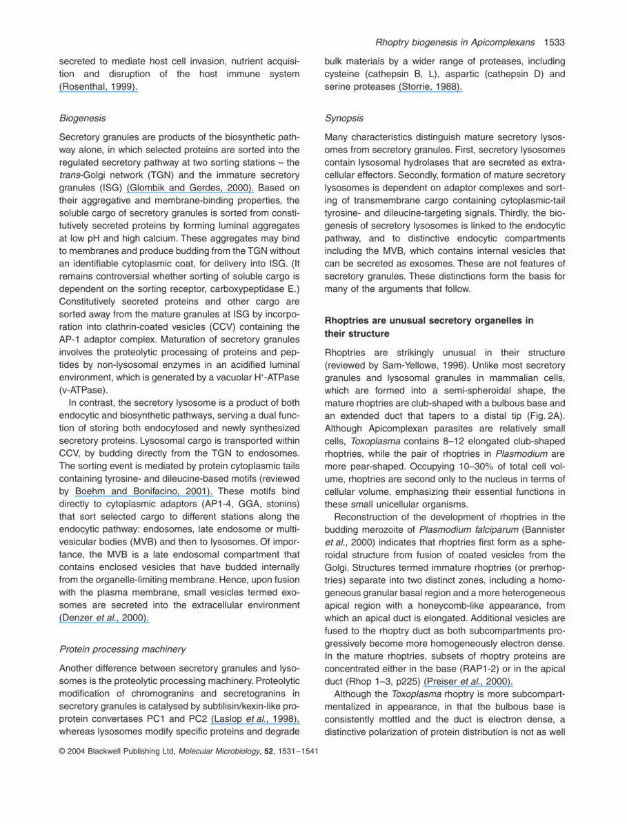
Fig. 1.
Contents in cells with conventional lysosomes, secretory granules and secretory lysosomes.A. All cell types contain a lysosomal compartment, which contains lysosomal hydrolases, transmembrane proteins and internal vesicles. Transmembrane proteins often contain tyrosine and dileucine sorting signals in the cytoplasmic tail. Internal vesicles are invaginations of the limiting membrane carrying selected proteins and lipids.B. Secretory cells also contain set of secretory proteins specific for each cell type, such as secretogranin II and chromogranins.C. Cells that have secretory lysosomes contain specific cell type secretory proteins in the modified lysosomal compartment, such as histamine and serotonin in mast cells, MHCII (major histocompatibility complex, class II) in macrophages and Fas ligand, soluble granzyme A and perforin in T and NK cells (modified from Blott and Griffiths, 2002).
Rhoptry biogenesis in Apicomplexans
1533
© 2004 Blackwell Publishing Ltd,
Molecular Microbiology
,
52
, 1531–1541
secreted to mediate host cell invasion, nutrient acquisi-tion and disruption of the host immune system(Rosenthal, 1999).
Biogenesis
Secretory granules are products of the biosynthetic path-way alone, in which selected proteins are sorted into theregulated secretory pathway at two sorting stations – the
trans
-Golgi network (TGN) and the immature secretorygranules (ISG) (Glombik and Gerdes, 2000). Based ontheir aggregative and membrane-binding properties, thesoluble cargo of secretory granules is sorted from consti-tutively secreted proteins by forming luminal aggregatesat low pH and high calcium. These aggregates may bindto membranes and produce budding from the TGN withoutan identifiable cytoplasmic coat, for delivery into ISG. (Itremains controversial whether sorting of soluble cargo isdependent on the sorting receptor, carboxypeptidase E.)Constitutively secreted proteins and other cargo aresorted away from the mature granules at ISG by incorpo-ration into clathrin-coated vesicles (CCV) containing theAP-1 adaptor complex. Maturation of secretory granulesinvolves the proteolytic processing of proteins and pep-tides by non-lysosomal enzymes in an acidified luminalenvironment, which is generated by a vacuolar H
+
-ATPase(v-ATPase).
In contrast, the secretory lysosome is a product of bothendocytic and biosynthetic pathways, serving a dual func-tion of storing both endocytosed and newly synthesizedsecretory proteins. Lysosomal cargo is transported withinCCV, by budding directly from the TGN to endosomes.The sorting event is mediated by protein cytoplasmic tailscontaining tyrosine- and dileucine-based motifs (reviewedby Boehm and Bonifacino, 2001). These motifs binddirectly to cytoplasmic adaptors (AP1-4, GGA, stonins)that sort selected cargo to different stations along theendocytic pathway: endosomes, late endosome or multi-vesicular bodies (MVB) and then to lysosomes. Of impor-tance, the MVB is a late endosomal compartment thatcontains enclosed vesicles that have budded internallyfrom the organelle-limiting membrane. Hence, upon fusionwith the plasma membrane, small vesicles termed exo-somes are secreted into the extracellular environment(Denzer
et al
., 2000).
Protein processing machinery
Another difference between secretory granules and lyso-somes is the proteolytic processing machinery. Proteolyticmodification of chromogranins and secretogranins insecretory granules is catalysed by subtilisin/kexin-like pro-protein convertases PC1 and PC2 (Laslop
et al
., 1998),whereas lysosomes modify specific proteins and degrade
bulk materials by a wider range of proteases, includingcysteine (cathepsin B, L), aspartic (cathepsin D) andserine proteases (Storrie, 1988).
Synopsis
Many characteristics distinguish mature secretory lysos-omes from secretory granules. First, secretory lysosomescontain lysosomal hydrolases that are secreted as extra-cellular effectors. Secondly, formation of mature secretorylysosomes is dependent on adaptor complexes and sort-ing of transmembrane cargo containing cytoplasmic-tailtyrosine- and dileucine-targeting signals. Thirdly, the bio-genesis of secretory lysosomes is linked to the endocyticpathway, and to distinctive endocytic compartmentsincluding the MVB, which contains internal vesicles thatcan be secreted as exosomes. These are not features ofsecretory granules. These distinctions form the basis formany of the arguments that follow.
Rhoptries are unusual secretory organelles intheir structure
Rhoptries are strikingly unusual in their structure(reviewed by Sam-Yellowe, 1996). Unlike most secretorygranules and lysosomal granules in mammalian cells,which are formed into a semi-spheroidal shape, themature rhoptries are club-shaped with a bulbous base andan extended duct that tapers to a distal tip (Fig. 2A).Although Apicomplexan parasites are relatively smallcells,
Toxoplasma
contains 8–12 elongated club-shapedrhoptries, while the pair of rhoptries in
Plasmodium
aremore pear-shaped. Occupying 10–30% of total cell vol-ume, rhoptries are second only to the nucleus in terms ofcellular volume, emphasizing their essential functions inthese small unicellular organisms.
Reconstruction of the development of rhoptries in thebudding merozoite of
Plasmodium falciparum
(Bannister
et al
., 2000) indicates that rhoptries first form as a sphe-roidal structure from fusion of coated vesicles from theGolgi. Structures termed immature rhoptries (or prerhop-tries) separate into two distinct zones, including a homo-geneous granular basal region and a more heterogeneousapical region with a honeycomb-like appearance, fromwhich an apical duct is elongated. Additional vesicles arefused to the rhoptry duct as both subcompartments pro-gressively become more homogeneously electron dense.In the mature rhoptries, subsets of rhoptry proteins areconcentrated either in the base (RAP1-2) or in the apicalduct (Rhop 1–3, p225) (Preiser
et al
., 2000).Although the
Toxoplasma
rhoptry is more subcompart-mentalized in appearance, in that the bulbous base isconsistently mottled and the duct is electron dense, adistinctive polarization of protein distribution is not as well

1534
H. M. Ngô, M. Yang and K. A. Joiner
© 2004 Blackwell Publishing Ltd,
Molecular Microbiology
,
52
, 1531–1541
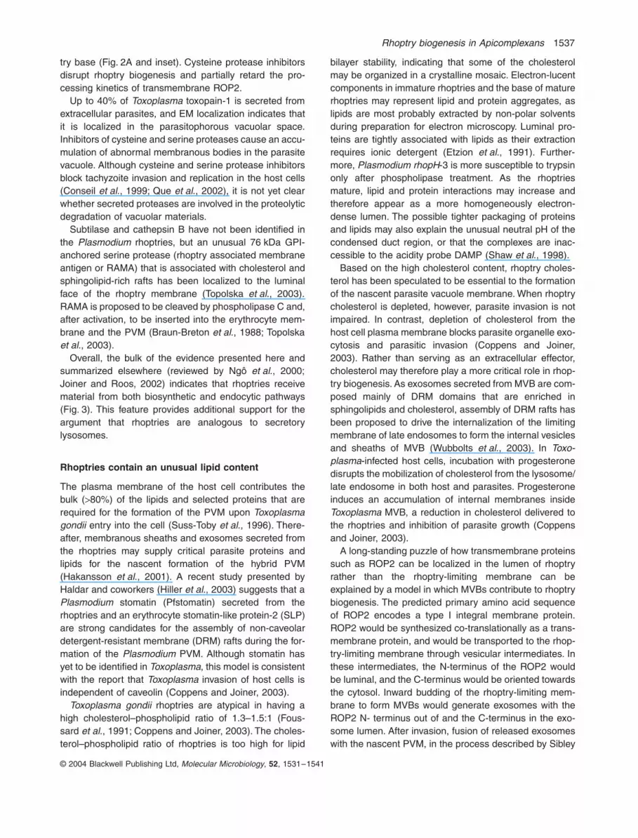
Fig. 2.
Protein localization to rhoptries and multivesicular body-like endosomes by cryoimmunoelectron microscopy. A
T. gondii
cysteine protease (TgCP1), identified as a lysosomal cathepsin B, is localized to the core region of the base of mature rhoptries (A and inset) and is found as residual cargo associated with vesicles in the core region of a discharged rhoptry (B). Multivesicular bodies (MVB) are lucent vacuoles of 400–900 nm that typically contain internal vesicles. MVBs are labelled with toxopain-1 (C) and endosomal markers Tg
m
1 (D) and TgRab5 (E). Electron micrographs are modified from Que
et al
. (2002) (A and C), Ngô
et al
. (2003) (D) and Robibaro
et al
. (2002) (E). Scale bars: A = 500 nm, A (inset) = 250 nm, B–E = 200 nm.
B
C
D
E
A

Rhoptry biogenesis in Apicomplexans
1535
© 2004 Blackwell Publishing Ltd,
Molecular Microbiology
,
52
, 1531–1541
defined as in
Plasmodium
. Earlier ultrastructural localiza-tion indicates that native ROP1 is localized to the rhoptrybase (Saffer
et al
., 1992). When expression of the solublerhoptry antigen ROP1 is abolished (Soldati
et al
., 1995),the rhoptry base is no longer mottled, suggesting thatthere is a domain-specific function of ROP1 in the twosubcompartments. It is also unclear in
Toxoplasma
whether all 8–12 rhoptries have formed from a centralizedimmature rhoptry, or whether each mature rhoptry origi-nates from a separate immature rhoptry. Serial recon-struction of parasites undergoing endodyogeny supportsthe latter model as each forming daughter contains atleast six visible immature rhoptries (Pelletier
et al
., 2002).Whether there is a heterogeneity of rhoptries in
Toxo-plasma
with differential cargo and functions has not beenaddressed. The discharge of only a subset of the 8–12rhoptries during host cell invasion suggests, however, atleast a differential level of exocytotic competency.
The remarkable structure and morphogenesis of rhop-tries are hence unlike any known secretory lysosome orgranule of higher mammalian systems. The mechanismof how elongated spindle secretory organelles are formedand maintained is probably more universal in protozoans(Hausmann, 1978), such as trichocysts in ciliates (Plattnerand Kissmehl, 2003).
Rhoptry functions are explicit to intracellular parasitism
Few other secretory organelles have been designed withas wide a range of functions as the rhoptries in the Api-complexan parasites. Secreted macromolecules from theparasite rhoptries are involved in the selection and adhe-sion to host cells, in forming an intracellular environmentfor parasite survival and in ‘hijacking’ host organelles (seediscussion below for references).
In
Plasmodium
, rhoptries appear to be secreted simul-taneously with micronemes, as micronemes are dockedand fused to the rhoptry ducts; hence, compound exocy-tosis of both secretory organelles contributes to all stagesof parasitic invasion. In
Toxoplasma
, micronemes are trig-gered first to mediate host cell attachment and penetra-tion, whereas rhoptry contents are secreted secondarilyfor contributing to PVM biogenesis and host parasite inter-actions, although it is still unclear whether micronemesundergo heterotypic fusion with the rhoptry duct duringexocytosis in
Toxoplasma
.Although rhoptry morphogenesis appears to be princi-
pally conserved between
Toxoplasma
and
Plasmodium
,each parasite loads a different set of protein cargoes intotheir secretory rhoptries (summarized by Sam-Yellowe,1996). None of the malaria rhoptry proteins (summarizedby Preiser
et al
., 2000) have homologues in
Toxoplasma
,and vice versa.
A striking example in the divergence of cargoesbetween the two related parasites is the subtilases, inwhich two isozymes (SUB1, SUB2) are found in
Plasmo-dium
dense granules (Blackman
et al
., 1998; Hackett
et al
., 1999). In
Toxoplasma
, SUB1 and SUB2 are sortedand secreted from the micronemes and rhoptries respec-tively (Miller
et al
., 2001; 2003).This differential sorting of secreted effectors may reflect
the differences between
Toxoplasma
and
Plasmodium
inhost cell range. Most of the malarial rhoptry proteins areimplicated in erythrocyte recognition and invasion. Theseinclude the low-molecular-weight complex of RAP 1–3,the high-molecular-weight complex of Rhop 1–3, rhoptrymultigene families of Pf60 and Py235 (Preiser
et al
., 2000)and RAMA (Topolska
et al
., 2003). For example, thesecreted oligomeric complex of Rhop-H and SERA(120 kDa serine-rich protein) binds to erythrocyte mem-branes, vesicles and liposomes. Vesicles containing phos-phatidylethanolamine can inhibit the association of rhoptryprotein complexes with mouse erythrocytes (Sam-Yellowe, 1996). Monoclonal antibodies to some malarialrhoptry proteins block merozoite invasion of red bloodcells
in vitro
and inhibit parasite replication
in vivo
.In
Toxoplasma
, nine major rhoptries proteins (ROP1–9)have been identified. The primary sequences of ROP1,ROP2, ROP3, ROP4, ROP8 and ROP9 indicate that onlyROP2, ROP3, ROP4 and ROP8 are highly similar andcontain predicted transmembrane motifs. Two rhoptryhydrolases (cathepsin B, subtilase) have been identifiedrecently and are discussed in a later section. SolubleROP1 is found in secreted exosomes (Hakansson
et al
.,2001) and is a component of the parasite protein fractionthat enhances invasion
in vitro
(Saffer
et al
., 1992). AsROP1 knock-out parasites show no defects in invasionkinetics, or virulence in mice, ROP1 function remains elu-sive. Although ROP9 carries an integrin-like RGD motif(Reichman
et al
., 2002), its potential role in mediating hostcell interaction is undetermined.
Peculiar to
Toxoplasma
invasion of nucleated eukaryoticcells is the recruitment and adherence of host mitochon-dria and endoplasmic reticulum (ER) to adhere to theparasite vacuole. Transmembrane ROP2 is secretedfrom the rhoptries and inserted into the PVM with its N-terminus facing the host cytosol (Beckers
et al
., 1994). Amitochondrial targeting signal in the N-terminus of ROP2has been proposed to mediate the tight association of hostmitochondria and ER (Sinai and Joiner, 2001). Depletionof endogenous ROP2 by antisense RNA expression con-fers a significant (90%) decrease in host mitochondriaassociation to the PVM (Nakaar
et al
., 2002).A common feature between
Toxoplasma
and
Plasmo-dium
invasion is the formation of the PVM to enclose theparasite in the host cell. Secretion of proteins, lipids andmultivesicular structures contribute to the formation of the

1536
H. M. Ngô, M. Yang and K. A. Joiner
© 2004 Blackwell Publishing Ltd,
Molecular Microbiology
,
52
, 1531–1541
nascent PVM, which is a hybrid between parasite and hostmaterials (Fig. 3). Sibley and coworkers (Hakansson
et al
., 2001) demonstrated that exosomes and membra-nous sheaths secreted from
Toxoplasma
rhoptries into thehost cytoplasm will subsequently undergo heterotypicfusion with the nascent parasitophorous vacuole, contrib-uting parasite lipids and proteins to the PVM (Fig. 3).
Rhoptry proteins are processed in an acidified lumen
All rhoptry proteins identified to date are synthesized aslarger precursors (prepro-protein) with a typical signalsequence that is cleaved presumably after entry into theendoplasmic reticulum. A second processing step entailsthe proteolytic cleavage of an N-terminal pro domain togenerate a mature polypeptide. In
Toxoplasma
, matrix-assisted laser desorption ionization time-of-flight (MALDI-TOF) mass spectrometry has identified E83/A84 as the
cleavage site in soluble pro-ROP1 (Bradley and Boo-throyd, 1999). The rhoptry subtilase (TgSUB2) has thesame cleavage site as ROP1, as demonstrated by aE686A mutation resulting in a reduction in TgSUB2 pro-cessing (Miller
et al
., 2003). A similar EA dipeptide isdetected in the N-terminal regions of transmembraneROP2 and ROP8, although the mature N-terminus of theprotein has not been determined. Disruption of the secre-tory pathway by brefeldin A treatment, or temperatureblock, inhibits the normal processing of rhoptry proteinsthat occurs within 20 min of protein synthesis (Howardand Schmidt, 1995; Soldati, 1998). Hence, pro-domaintrimming occurs late in the rhoptry pathway, presumablyat the immature/maturing rhoptry.
Typically, proper protein processing and packaging insecretory granules, or hydrolysis of macromolecules inlysosomes, requires an acidified luminal environment thatis generated by proton pumping by a vacuolar H
+
-ATPase(v-ATPase). Studies using the electron microscopic probeDAMP (Shaw
et al
., 1998) indicate that the only acidicorganelles in
Toxoplasma
are immature (pH 3.5–5.5) andmature (pH 5.0–7.0) rhoptries or multivesicular bodies(pH 5.3–6.5). No DAMP labelling is observed in the rhop-try duct or at the base of 25% of mature rhoptries.Although the electron-dense appearance in the lumen ofthese two subdomains suggests that the cargoes aremore condensed, non-partitioning of DAMP may reflectthe novel rhoptry content, which is presumed to be atightly packaged gel of proteins and lipids. Cytochemicalstudies also suggest that rhoptries contain an acidic phos-phatase and Ca
2+
-, Mg
2+
-ATPase activities, as well as ahigh concentration of calcium (Bouchot
et al
., 2001).
Rhoptries contain cysteine and serine proteases
Specific inhibitors of cysteine (cathepsin inhibitor III) andserine (TPCK and subtilisin inhibitor III) proteases causeswelling of the ER and nuclear envelope, collapse of theGolgi and specific disruption of rhoptry biogenesis in
Tox-oplasma
(Shaw
et al
., 2002). Subtilisin inhibitor III alsogenerates multivesicular bodies (see below) that accumu-late luminal vesicles.
The subtilisin-type serine protease (TgSUB2) is local-ized to the
Toxoplasma
rhoptries (Miller
et al
., 2003). AsTgSUB2 undergoes autocatalytic cleavage and is co-pre-cipitated with soluble ROP1, the subtilase is proposed tobe involved in rhoptry biogenesis as a maturase.
Toxo-plasma
rhoptries contain at least one cysteine protease,a cathepsin B that is termed toxopain-1 (Que
et al
., 2002).Toxopain-1 is one candidate for processing of rhoptryproteins, based on the evidence that this protease islocalized to acidic compartments in which active proteinmodification occurs, i.e. multivesiculated endosomes(Fig. 2B), immature rhoptries and the core region of rhop-
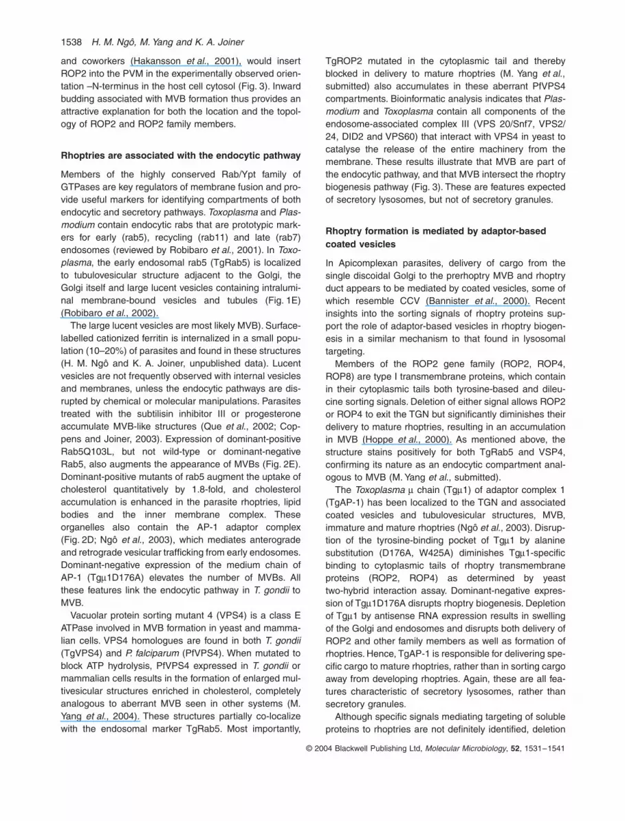
Fig. 3.
Biogenesis and exocytosis of rhoptry contents. This model proposes that rhoptries are formed by contributions from both secre-tory and endocytic pathways. Secretory cargo from the TGN is trans-ported to the mature rhoptry via the immature rhoptry. Some cargo is delivered directly from the TGN to the immature rhoptry (possibly subtilase), while other cargo transits the endosomal pathway through an MVB en route to the immature rhoptry. The MVB intersects the rhoptry biogenesis pathway by both delivering cargo to and probably receiving cargo from the immature rhoptry. Selected cargo is then subcompartmentalized to form the tapering apical duct (blue), a region that is potentially fusogenic with secreting micronemes, most notably in
Plasmodium
. When activated by an unknown trigger, rhop-tries secrete internal vesicles (exosomes), which then fuse with the parasitophorous vacuole membrane (Hakansson
et al
., 2001).
Rhoptry biogenesis in Apicomplexans
1537
© 2004 Blackwell Publishing Ltd,
Molecular Microbiology
,
52
, 1531–1541
try base (Fig. 2A and inset). Cysteine protease inhibitorsdisrupt rhoptry biogenesis and partially retard the pro-cessing kinetics of transmembrane ROP2.
Up to 40% of
Toxoplasma
toxopain-1 is secreted fromextracellular parasites, and EM localization indicates thatit is localized in the parasitophorous vacuolar space.Inhibitors of cysteine and serine proteases cause an accu-mulation of abnormal membranous bodies in the parasitevacuole. Although cysteine and serine protease inhibitorsblock tachyzoite invasion and replication in the host cells(Conseil
et al
., 1999; Que
et al
., 2002), it is not yet clearwhether secreted proteases are involved in the proteolyticdegradation of vacuolar materials.
Subtilase and cathepsin B have not been identified inthe
Plasmodium
rhoptries, but an unusual 76 kDa GPI-anchored serine protease (rhoptry associated membraneantigen or RAMA) that is associated with cholesterol andsphingolipid-rich rafts has been localized to the luminalface of the rhoptry membrane (Topolska et al., 2003).RAMA is proposed to be cleaved by phospholipase C and,after activation, to be inserted into the erythrocyte mem-brane and the PVM (Braun-Breton et al., 1988; Topolskaet al., 2003).
Overall, the bulk of the evidence presented here andsummarized elsewhere (reviewed by Ngô et al., 2000;Joiner and Roos, 2002) indicates that rhoptries receivematerial from both biosynthetic and endocytic pathways(Fig. 3). This feature provides additional support for theargument that rhoptries are analogous to secretorylysosomes.
Rhoptries contain an unusual lipid content
The plasma membrane of the host cell contributes thebulk (>80%) of the lipids and selected proteins that arerequired for the formation of the PVM upon Toxoplasmagondii entry into the cell (Suss-Toby et al., 1996). There-after, membranous sheaths and exosomes secreted fromthe rhoptries may supply critical parasite proteins andlipids for the nascent formation of the hybrid PVM(Hakansson et al., 2001). A recent study presented byHaldar and coworkers (Hiller et al., 2003) suggests that aPlasmodium stomatin (Pfstomatin) secreted from therhoptries and an erythrocyte stomatin-like protein-2 (SLP)are strong candidates for the assembly of non-caveolardetergent-resistant membrane (DRM) rafts during the for-mation of the Plasmodium PVM. Although stomatin hasyet to be identified in Toxoplasma, this model is consistentwith the report that Toxoplasma invasion of host cells isindependent of caveolin (Coppens and Joiner, 2003).
Toxoplasma gondii rhoptries are atypical in having ahigh cholesterol–phospholipid ratio of 1.3–1.5:1 (Fous-sard et al., 1991; Coppens and Joiner, 2003). The choles-terol–phospholipid ratio of rhoptries is too high for lipid
bilayer stability, indicating that some of the cholesterolmay be organized in a crystalline mosaic. Electron-lucentcomponents in immature rhoptries and the base of maturerhoptries may represent lipid and protein aggregates, aslipids are most probably extracted by non-polar solventsduring preparation for electron microscopy. Luminal pro-teins are tightly associated with lipids as their extractionrequires ionic detergent (Etzion et al., 1991). Further-more, Plasmodium rhopH-3 is more susceptible to trypsinonly after phospholipase treatment. As the rhoptriesmature, lipid and protein interactions may increase andtherefore appear as a more homogeneously electron-dense lumen. The possible tighter packaging of proteinsand lipids may also explain the unusual neutral pH of thecondensed duct region, or that the complexes are inac-cessible to the acidity probe DAMP (Shaw et al., 1998).
Based on the high cholesterol content, rhoptry choles-terol has been speculated to be essential to the formationof the nascent parasite vacuole membrane. When rhoptrycholesterol is depleted, however, parasite invasion is notimpaired. In contrast, depletion of cholesterol from thehost cell plasma membrane blocks parasite organelle exo-cytosis and parasitic invasion (Coppens and Joiner,2003). Rather than serving as an extracellular effector,cholesterol may therefore play a more critical role in rhop-try biogenesis. As exosomes secreted from MVB are com-posed mainly of DRM domains that are enriched insphingolipids and cholesterol, assembly of DRM rafts hasbeen proposed to drive the internalization of the limitingmembrane of late endosomes to form the internal vesiclesand sheaths of MVB (Wubbolts et al., 2003). In Toxo-plasma-infected host cells, incubation with progesteronedisrupts the mobilization of cholesterol from the lysosome/late endosome in both host and parasites. Progesteroneinduces an accumulation of internal membranes insideToxoplasma MVB, a reduction in cholesterol delivered tothe rhoptries and inhibition of parasite growth (Coppensand Joiner, 2003).
A long-standing puzzle of how transmembrane proteinssuch as ROP2 can be localized in the lumen of rhoptryrather than the rhoptry-limiting membrane can beexplained by a model in which MVBs contribute to rhoptrybiogenesis. The predicted primary amino acid sequenceof ROP2 encodes a type I integral membrane protein.ROP2 would be synthesized co-translationally as a trans-membrane protein, and would be transported to the rhop-try-limiting membrane through vesicular intermediates. Inthese intermediates, the N-terminus of the ROP2 wouldbe luminal, and the C-terminus would be oriented towardsthe cytosol. Inward budding of the rhoptry-limiting mem-brane to form MVBs would generate exosomes with theROP2 N- terminus out of and the C-terminus in the exo-some lumen. After invasion, fusion of released exosomeswith the nascent PVM, in the process described by Sibley
1538 H. M. Ngô, M. Yang and K. A. Joiner
© 2004 Blackwell Publishing Ltd, Molecular Microbiology, 52, 1531–1541
and coworkers (Hakansson et al., 2001), would insertROP2 into the PVM in the experimentally observed orien-tation –N-terminus in the host cell cytosol (Fig. 3). Inwardbudding associated with MVB formation thus provides anattractive explanation for both the location and the topol-ogy of ROP2 and ROP2 family members.
Rhoptries are associated with the endocytic pathway
Members of the highly conserved Rab/Ypt family ofGTPases are key regulators of membrane fusion and pro-vide useful markers for identifying compartments of bothendocytic and secretory pathways. Toxoplasma and Plas-modium contain endocytic rabs that are prototypic mark-ers for early (rab5), recycling (rab11) and late (rab7)endosomes (reviewed by Robibaro et al., 2001). In Toxo-plasma, the early endosomal rab5 (TgRab5) is localizedto tubulovesicular structure adjacent to the Golgi, theGolgi itself and large lucent vesicles containing intralumi-nal membrane-bound vesicles and tubules (Fig. 1E)(Robibaro et al., 2002).
The large lucent vesicles are most likely MVB). Surface-labelled cationized ferritin is internalized in a small popu-lation (10–20%) of parasites and found in these structures(H. M. Ngô and K. A. Joiner, unpublished data). Lucentvesicles are not frequently observed with internal vesiclesand membranes, unless the endocytic pathways are dis-rupted by chemical or molecular manipulations. Parasitestreated with the subtilisin inhibitor III or progesteroneaccumulate MVB-like structures (Que et al., 2002; Cop-pens and Joiner, 2003). Expression of dominant-positiveRab5Q103L, but not wild-type or dominant-negativeRab5, also augments the appearance of MVBs (Fig. 2E).Dominant-positive mutants of rab5 augment the uptake ofcholesterol quantitatively by 1.8-fold, and cholesterolaccumulation is enhanced in the parasite rhoptries, lipidbodies and the inner membrane complex. Theseorganelles also contain the AP-1 adaptor complex(Fig. 2D; Ngô et al., 2003), which mediates anterogradeand retrograde vesicular trafficking from early endosomes.Dominant-negative expression of the medium chain ofAP-1 (Tgm1D176A) elevates the number of MVBs. Allthese features link the endocytic pathway in T. gondii toMVB.
Vacuolar protein sorting mutant 4 (VPS4) is a class EATPase involved in MVB formation in yeast and mamma-lian cells. VPS4 homologues are found in both T. gondii(TgVPS4) and P. falciparum (PfVPS4). When mutated toblock ATP hydrolysis, PfVPS4 expressed in T. gondii ormammalian cells results in the formation of enlarged mul-tivesicular structures enriched in cholesterol, completelyanalogous to aberrant MVB seen in other systems (M.Yang et al., 2004). These structures partially co-localizewith the endosomal marker TgRab5. Most importantly,
TgROP2 mutated in the cytoplasmic tail and therebyblocked in delivery to mature rhoptries (M. Yang et al.,submitted) also accumulates in these aberrant PfVPS4compartments. Bioinformatic analysis indicates that Plas-modium and Toxoplasma contain all components of theendosome-associated complex III (VPS 20/Snf7, VPS2/24, DID2 and VPS60) that interact with VPS4 in yeast tocatalyse the release of the entire machinery from themembrane. These results illustrate that MVB are part ofthe endocytic pathway, and that MVB intersect the rhoptrybiogenesis pathway (Fig. 3). These are features expectedof secretory lysosomes, but not of secretory granules.
Rhoptry formation is mediated by adaptor-based coated vesicles
In Apicomplexan parasites, delivery of cargo from thesingle discoidal Golgi to the prerhoptry MVB and rhoptryduct appears to be mediated by coated vesicles, some ofwhich resemble CCV (Bannister et al., 2000). Recentinsights into the sorting signals of rhoptry proteins sup-port the role of adaptor-based vesicles in rhoptry biogen-esis in a similar mechanism to that found in lysosomaltargeting.
Members of the ROP2 gene family (ROP2, ROP4,ROP8) are type I transmembrane proteins, which containin their cytoplasmic tails both tyrosine-based and dileu-cine sorting signals. Deletion of either signal allows ROP2or ROP4 to exit the TGN but significantly diminishes theirdelivery to mature rhoptries, resulting in an accumulationin MVB (Hoppe et al., 2000). As mentioned above, thestructure stains positively for both TgRab5 and VSP4,confirming its nature as an endocytic compartment anal-ogous to MVB (M. Yang et al., submitted).
The Toxoplasma m chain (Tgm1) of adaptor complex 1(TgAP-1) has been localized to the TGN and associatedcoated vesicles and tubulovesicular structures, MVB,immature and mature rhoptries (Ngô et al., 2003). Disrup-tion of the tyrosine-binding pocket of Tgm1 by alaninesubstitution (D176A, W425A) diminishes Tgm1-specificbinding to cytoplasmic tails of rhoptry transmembraneproteins (ROP2, ROP4) as determined by yeasttwo-hybrid interaction assay. Dominant-negative expres-sion of Tgm1D176A disrupts rhoptry biogenesis. Depletionof Tgm1 by antisense RNA expression results in swellingof the Golgi and endosomes and disrupts both delivery ofROP2 and other family members as well as formation ofrhoptries. Hence, TgAP-1 is responsible for delivering spe-cific cargo to mature rhoptries, rather than in sorting cargoaway from developing rhoptries. Again, these are all fea-tures characteristic of secretory lysosomes, rather thansecretory granules.
Although specific signals mediating targeting of solubleproteins to rhoptries are not definitely identified, deletion

Rhoptry biogenesis in Apicomplexans 1539
© 2004 Blackwell Publishing Ltd, Molecular Microbiology, 52, 1531–1541
mapping indicates that they are embedded in two domainsof ROP1, the propeptide (amino acids 24–85) and a cen-tral peptide (amino acids 198–345) (Bradley and Boo-throyd, 2001; Striepen et al., 2001). The prodomains ofmost lysosomal proteins are glycosylated in the Golgicomplex and targeted to lysosomes by MPRs, which rec-ognize the modified asparagine-linked oligosaccharides.As minimal N-glycosylation occurs in Apicomplexan par-asites, ROP1 is not post-translationally modified, and thegenome does not encode the MPR sorting machinery,sorting of soluble proteins must use a non-MPRmechanism.
One possible MPR-independent sorting processdepends on the membrane-binding motif (nine residues)at the N-terminus of prodomains in mammalian procathe-psin L (McIntyre and Erickson, 1993) and trypanosomecruzipain (Huete-Perez et al., 1999). It remains to bedetermined whether Toxoplasma ROP1 has membrane-binding potential as tentatively reported (He et al., 2001)and, if so, whether the sorting motifs of rhoptry-solubleproteins will bind to the luminal domain of the ROP2transmembrane protein family. In Plasmodium, solubleRAP2 and RAP3 form an oligomeric complex with RAP1,which contains at least one predicted transmembranemotif. A gene deletion that generates a truncated RAP1,containing the N-terminal 344 amino acids of the 782-amino-acid wild-type RAP1, abolishes heterocomplex for-mation between RAP1 and 2. Consequently, RAP2 isretained in the lumen of ER and possibly Golgi, whereasRAP1 is still faithfully targeted to the rhoptries (Baldi et al.,2000). Tyrosine sorting signals (YAKL, YFAF) are pre-dicted at the C-tail of RAP1 that can potentially interactwith adaptor complex and adaptor-like proteins.
As the T. gondii ROP1 prodomain targets a VSG fusionto both the rhoptry base and duct whereas mature ROP1is restricted to the basal subcompartment (Saffer et al.,1992; Bradley and Boothroyd, 2001), the motif that medi-ates the basal retention of ROP1 is encoded in the matureprotein. It remains to be determined whether the octapep-tide repeat regions in ROP1 and ROP9 that are enrichedin proline may mediate this sorting function.
In summary, the biogenesis of rhoptries is mediated byadaptor-based vesicular trafficking, most closely relatedto the sorting mechanism for secretory lysosomes. Futurestudies will hopefully identify the transmembrane proteinsthat mediate the sorting of soluble proteins into coatedvesicles for forward transport to the rhoptries.
Conclusion: are rhoptries secretory granules or secretory lysosomes?
Three possible scenarios currently exist for the biogene-sis of rhoptries. The secretory granule model indicatesthat rhoptry proteins and lipids aggregate at the TGN via
sorting receptors/escorters, or DRM rafts, and are exclu-sively sorted to the immature rhoptry. An alternativemodel is that rhoptries are formed exclusively via theendocytic pathway. Finally, the ‘secretory lysosome’model suggests that both secretory and endocytic path-ways contribute to formation of the organelle (Fig. 3).The preponderance of evidence supports the lastmodel. Altogether, we believe that rhoptries representone of the earliest models of secretory lysosomes.
Acknowledgements
We are grateful to members of our laboratory and collabora-tors who have contributed to studies of membrane traffickingpathways and organelle biogenesis in Apicomplexan para-sites. This work was supported by NIH grants AI30060 andTW01594, and by a Burroughs-Wellcome Fund Scholaraward to K.A.J. H.M.N. was supported by NIH grants5T32AI07404 and 5F32AI10044.
References
Baldi, D.L., Andrews, K.T., Waller, R.F., Roos, D.S., Howard,R.F., Crabb, B.S., and Cowman, A.F. (2000) RAP1 con-trols rhoptry targeting of RAP2 in the malaria parasitePlasmodium falciparum. EMBO J 19: 2435–2443.
Bannister, L.H., Hopkins, J.M., Fowler, R.E., Krishna, S., andMitchell, G.H. (2000) Ultrastructure of rhoptry developmentin Plasmodium falciparum erythrocytic schizonts. Parasitol-ogy 121: 273–287.
Beckers, C.J.M., Dubremetz, J.F., Mercereau-Puijalon, O.,and Joiner, K.A. (1994) The Toxoplasma gondii rhoptryprotein ROP2 is inserted into the parasitophorous vacuolemembrane, surrounding the intracellular parasite, and isexposed to the host cell cytoplasm. J Cell Biol 127: 947–961.
Blackman, M.J., Fujioka, H., Stafford, W.H., Sajid, M.,Clough, B., Fleck, S.L., et al. (1998) A subtilisin-like proteinin secretory organelles of Plasmodium falciparum merozo-ites. J Biol Chem 273: 23398–23409.
Blott, E.J., and Griffiths, G.M. (2002) Secretory lysosomes.Nature Rev Mol Cell Biol 2002 (3): 122–131.
Boehm, M., and Bonifacino, J.S. (2001) Adaptins. The finalrecount. Mol Biol Cell 12: 2907–2920.
Bouchot, A., Jaillet, J.D., Bonhomme, A., Alessandro, N.P.,Laquerriere, P., Kilian, L., et al. (2001) Detection and local-ization of a Ca2+-ATPase activity in Toxoplasma gondii.Cell Struct Funct 26: 49–60.
Bradley, P.J., and Boothroyd, J.C. (1999) Identification ofthe pro-mature processing site of Toxoplasma ROP1by mass spectrometry. Mol Biochem Parasitol 100:103–109.
Bradley, P.J., and Boothroyd, J.C. (2001) The pro region ofToxoplasma ROP1 is a rhoptry-targeting signal. Int J Par-asitol 31: 1177–1186.
Braun-Breton, C., Rosenberry, T.L., and da Silva, L.P. (1988)Induction of the proteolytic activity of a membrane proteinin Plasmodium falciparum by phosphatidyl inositol-specificphospholipase C. Nature 332: 457–459.

1540 H. M. Ngô, M. Yang and K. A. Joiner
© 2004 Blackwell Publishing Ltd, Molecular Microbiology, 52, 1531–1541
Burgoyne, R.D., and Morgan, A. (2003) Secretory granuleexocytosis. Physiol Rev 83: 581–632.
Conseil, V., Soete, M., and Dubremetz, J.F. (1999) Serineprotease inhibitors block invasion of host cells by Toxo-plasma gondii. Antimicrob Agents Chemother 43: 1358–1361.
Coppens, I., and Joiner, K.A. (2003) Host but not parasitecholesterol controls Toxoplasma entry by modulatingorganelle discharge. Mol Biol Cell 14: 3804–3820.
Denzer, K., Kleijmeer, M.J., Heijnen, H.F., Stoorvogel, W.,and Geuze, H.J. (2000) Exosome: from internal vesicle ofthe multivesicular body to intercellular signaling device. JCell Sci 113: 3365–3374.
Etzion, Z., Murray, M., and Perkins, M. (1991) Isolation andcharacterization of rhoptries of Plasmodium falciparum.Mol Biochem Parasitol 47: 51–62.
Foussard, F., Leriche, M.A., and Dubremetz, J.F. (1991)Characterization of the lipid content of Toxoplasma gondiirhoptries. Parasitology 102: 367–370.
Glombik, M.M., and Gerdes, H.H. (2000) Signal-mediatedsorting of neuropeptides and prohormones: secretorygranule biogenesis revisited. Biochimie 82: 315–326.
Hackett, F., Sajid, M., Withers-Martinez, C., Grainger, M.,and Blackman, M.J. (1999) PfSUB-2: a second subtilisin-like protein in Plasmodium falciparum merozoites. Mol Bio-chem Parasitol 103: 183–195.
Hakansson, S., Charron, A.J., and Sibley, L.D. (2001) Toxo-plasma evacuoles: a two-step process of secretion andfusion forms the parasitophorous vacuole. EMBO J 20:3132–3144.
Hausmann, K. (1978) Extrusive organelles in protists. Int RevCytol 25: 197–276.
He, C.Y., Shaw, M.K., Pletcher, C.H., Striepen, B., Tilney,L.G., and Roos, D.S. (2001) A plastid segregation defectin the protozoan parasite Toxoplasma gondii. EMBO J 20:330–339.
Hiller, N.L., Akompong, T., Morrow, J.S., Holder, A.A., andHaldar, K. (2003) Identification of a stomatin orthologue invacuoles induced in human erythrocytes by malaria para-sites: a role for microbial raft-protein in apicomplexan vac-uole biogenesis. J Biol Chem 278: 48413–48421.
Hoppe, H.C., Ngo, H.M., Yang, M., and Joiner, K.A. (2000)Targeting to rhoptry organelles of Toxoplasma gondiiinvolves evolutionarily conserved mechanisms. Nature CellBiol 2: 449–456.
Howard, R.F., and Schmidt, C.M. (1995) The secretary path-way of Plasmodium falciparum regulates transport of p82/RAP1 to the rhoptries. Mol Biochem Parasitol 74: 43–54.
Huete-Perez, J.A., Engel, J.C., Brinen, L.S., Mottram, J.C.,and McKerrow, J.H. (1999) Protease trafficking in two prim-itive eukaryotes is mediated by a prodomain protein motif.J Biol Chem 274: 16249–16256.
Joiner, K.A., and Roos, D.S. (2002) Secretory traffic in theeukaryotic parasite Toxoplasma gondii: less is more. J CellBiol 157: 557–563.
Laslop, A., Weiss, C., Savaria, D., Eiter, C., Tooze, S.A.,Seidah, N.G., and Winkler, H. (1998) Proteolytic process-ing of chromogranin B and secretogranin II by prohormoneconvertases. J Neurochem 70: 374–383.
Luzio, J.P., Poupon, V., Lindsay, M.R., Mullock, B.M.,Piper, R.C., and Pryor, P.R. (2003) Membrane dynamics
and the biogenesis of lysosomes. Mol Membr Biol 20:141–154.
McIntyre, G.F., and Erickson, A.H. (1993) The lysosomalproenzyme receptor that binds procathepsin L to microso-mal membranes at pH 5 is a 43-kDa integral membraneprotein. Proc Natl Acad Sci USA 90: 10588–10592.
Miller, S.A., Binder, E.M., Blackman, M.J., Carruthers, V.B.,and Kim, K. (2001) A conserved subtilisin-like proteinTgSUB1 in microneme organelles of Toxoplasma gondii. JBiol Chem 276: 45341–45348.
Miller, S.A., Thathy, V., Ajoika, J.W., Blackman, M.J., andKim, K. (2003) TgSUB2 is a Toxoplasma gondii rhoptryorganelle processing proteinase. Mol Microbiol 49: 883–894.
Nakaar, V., Ngô, H.M., Aronson, E.P., Coppens, I., Stedman,T.T., and Joiner, K.A. (2002) Pleiotropic effect due to tar-geted depletion of secretory rhoptry protein ROP2 in Tox-oplasma gondii. J Cell Sci 116: 2311–2320.
Ngô, H., Hoppe, H.C., and Joiner, K.A. (2000) Differentialsorting and post-secretory targeting of proteins in parasiticinvasion. Trends Cell Biol 10: 67–72.
Ngô, H.M., Yang, M., Pypaert, M., Hoppe, H.C., and Joiner,K.A. (2003) Biogenesis of secretory organelles in an api-complexan parasite is dependent on the AP-1 adaptin. JBiol Chem 278: 5343–5352.
Palade, G. (1975) Intracellular aspects of the process ofprotein synthesis. Science 189: 347–358.
Pelletier, L., Stern, C.A., Pypaert, M., Sheff, D., Ngo, H.M.,Roper, N., et al. (2002) Golgi biogenesis in Toxoplasmagondii. Nature 418: 548–552.
Plattner, H., and Kissmehl, R. (2003) Molecular aspects ofmembrane trafficking in paramecium. Int Rev Cytol 232:185–216.
Preiser, P., Kaviratne, M., Khan, S., Bannister, L., and Jarra,W. (2000) The apical organelles of malaria merozoites:host cell selection, invasion, host immunity and immuneevasion. Microbes Infect 2: 1461–1477.
Que, X., Ngo, H., Lawton, J., Gray, M., Liu, Q., Engel, J., etal. (2002) The cathepsin B of Toxoplasma gondii, toxopain-1, is critical for parasite invasion and rhoptry protein pro-cessing. J Biol Chem 277: 25791–25797.
Reichmann, G., Dlugonska, H., and Fischer, H.G. (2002)Characterization of TgROP9 (p36), a novel rhoptry proteinof Toxoplasma gondii tachyzoites identified by T cell clone.Mol Biochem Parasitol 119: 43–54.
Robibaro, B., Hoppe, H.C., Yang, M., Coppens, I., Ngo, H.M.,Stedman, T.T., et al. (2001) Endocytosis in different life-styles of protozoan parasitism: role in nutrient uptake withspecial reference to Toxoplasma gondii. Int J Parasitol 31:1343–1353.
Robibaro, B., Stedman, T.T., Coppens, I., Ngo, H.M.,Pypaert, M., Bivona, T., et al. (2002) Toxoplasma gondiiRab5 enhances cholesterol acquisition from host cells. CellMicrobiol 4: 139–152.
Rosenthal, P.J. (1999) Proteases of protozoan parasites. AdvParasitol 43: 105–159.
Saffer, L.D., Mercereau-Puijalon, O., Dubremetz, J.F., andSchwartzman, J. (1992) Localization of a Toxoplasmagondii rhoptry protein by immunoelectron microscopy dur-ing and after host cell penetration. J Protozool 39: 526–530.

Rhoptry biogenesis in Apicomplexans 1541
© 2004 Blackwell Publishing Ltd, Molecular Microbiology, 52, 1531–1541
Sam-Yellowe, T.Y. (1996) Rhoptry organelles of the apicom-plexa: their role in host cell invasion and intracellular sur-vival. Parasitol Today 12: 308–316.
Shaw, M.K., Roos, D.S., and Tilney, L.G. (1998) Acidic com-partments and rhoptry formation in Toxoplasma gondii.Parasitol 117: 435–443.
Shaw, M.K., Roos, D.S., and Tilney, L.G. (2002) Cysteineand serine protease inhibitors block intracellular develop-ment and disrupt the secretory pathway of Toxoplasmagondii. Microbes Infect 4: 119–132.
Sinai, A.P., and Joiner, K.A. (2001) The Toxoplasma gondiiprotein ROP2 mediates host organelle association with theparasitophorous vacuole membrane. J Cell Biol 154: 95–108.
Soldati, D. (1998) Processing of Toxoplasma ROP1 proteinin nascent rhoptries. Mol Biochem Parasitol 96: 37–48.
Soldati, D., Kim, K., Kampmeier, J., Dubremetz, J.F., andBoothroyd, J.C. (1995) Complementation of a Toxoplasmagondii ROP1 knock-out mutant using phleomycin selection.Mol Biochem Parasitol 74: 87–97.
Storrie, B. (1988) Assembly of lysosomes: perspectives fromcomparative molecular cell biology. Int Rev Cytol 111: 53–105.
Striepen, B., Soldati, D., Garcia-Reguet, N., Dubremetz, J.F.,and Roos, D.S. (2001) Targeting of soluble proteins to therhoptries and micronemes in Toxoplasma gondii. Mol Bio-chem Parasitol 113: 45–63.
Suss-Toby, E., Zimmerberg, J., and Ward, G.E. (1996)Toxoplasma invasion: the parasitophorous vacuole isformed from host cell plasma membrane and pinches offvia a fission pore. Proc Natl Acad Sci USA 93: 8413–8418.
Topolska, A.E., Lidgett, A., Truman, D., Fujioka, H., andCoppel, R.L. (2003) Characterisation of a membrane-associated rhoptry protein of Plasmodium falciparum. JBiol Chem 279: 4648–4656.
Wubbolts, R., Leckie, R.S., Veenhuizen, P.T., Schwarzmann,G., Mobius, W., Hoernschemeyer, J., et al. (2003) Pro-teomic and biochemical analyses of human B cell-derivedexosomes. Potential implications for their function and mul-tivesicular body formation. J Biol Chem 278: 10963–10972.
Yang, M., Coppens, I., Wormsley, S., Baevova, P., Hoppe,H.C., and Joiner, K.A. (2004) The Plasmodium falciparumVps4 homolog mediates multivesicular body formation. JCell Sci (in press).