development of active biofilms of quinoa (chenopodium quinoa w.) starch containing gold...
TRANSCRIPT
Food Chemistry 173 (2015) 755–762
Contents lists available at ScienceDirect
Food Chemistry
journal homepage: www.elsevier .com/locate / foodchem
Development of active biofilms of quinoa (Chenopodium quinoa W.)starch containing gold nanoparticles and evaluation of antimicrobialactivity
http://dx.doi.org/10.1016/j.foodchem.2014.10.0680308-8146/� 2014 Elsevier Ltd. All rights reserved.
⇑ Corresponding author. Tel.: +55 051 3308 9789.E-mail address: [email protected] (S.H. Flôres).
Carlos H. Pagno a, Tania M.H. Costa b, Eliana W. de Menezes b, Edilson V. Benvenutti b, Plinho F. Hertz a,Carla R. Matte a, Juliano V. Tosati c, Alcilene R. Monteiro c, Alessandro O. Rios a, Simone H. Flôres a,⇑a Laboratório de Compostos Bioativos, Instituto de Ciência e Tecnologia dos Alimentos (ICTA), Universidade Federal do Rio Grande do Sul (UFRGS), Porto Alegre, RS, Brazilb Instituto de Química, Universidade Federal do Rio Grande do Sul, Porto Alegre, RS, Brazilc Departamento de Engenharia de Alimentos, Universidade Federal de Santa Catarina, Florianópolis, SC, Brazil
a r t i c l e i n f o
Article history:Received 4 June 2014Received in revised form 9 October 2014Accepted 14 October 2014Available online 28 October 2014
Keywords:NanocompositeAntibacterialFood packagingMetal nanoparticlesQuinoa starch
a b s t r a c t
Active biofilms of quinoa (Chenopodium quinoa, W.) starch were prepared by incorporating gold nanopar-ticles stabilised by an ionic silsesquioxane that contains the 1,4-diazoniabicyclo[2.2.2]octane chloridegroup. The biofilms were characterised and their antimicrobial activity was evaluated against Escherichiacoli and Staphylococcus aureus. The presence of gold nanoparticles produces an improvement in themechanical, optical and morphological properties, maintaining the thermal and barrier propertiesunchanged when compared to the standard biofilm. The active biofilms exhibited strong antibacterialactivity against food-borne pathogens with inhibition percentages of 99% against E. coli and 98% againstS. aureus. These quinoa starch biofilms containing gold nanoparticles are very promising to be used asactive food packaging for the maintenance of food safety and extension of the shelf life of packaged foods.
� 2014 Elsevier Ltd. All rights reserved.
1. Introduction
Interest in the maintenance and/or improvement of the qualityof packaged products and the reduction of waste packaging hasencouraged the exploration of new packaging materials, such asbiodegradable films formulated with raw materials derived fromrenewable sources, called biofilms.
In recent years, many researchers focused on the production ofedible films based on biopolymers, such as polysaccharides andproteins (Andreuccetti, Carvalho, Galicia-García, Martínez-Bustos,& Grosso, 2011; Kanmani & Rhim, 2014; Mei, Yuan, Wu, & Li,2013; Nascimento, Calado, & Carvalho, 2012; Souza et al., 2012;Souza, Goto, Mainardi, Coelho, & Tadini, 2013). Starch, a renewablebiopolymer consisting of amylose and amylopectin, is the mostcommonly used agricultural raw material for edible film manufac-turing because it is inexpensive, relatively easy to handle, totallybiodegradable, and widely available in nature from varioussources, such as cereals, roots, tubers (Nascimento et al., 2012),and more recently rediscovered pseudocereals, such as amaranthand quinoa (Araujo-Farro, Podadera, Sobral, & Menegalli, 2010).
The quinoa seed (Chenopodium quinoa, Willdenow) is a graintypically found in the South American Andean highlands. It is com-posed of significant amounts of starch (up to 80%), has an amylosecontent of 10–21% (depending on the variety), and a small starchgranule size (�1 lm), which are characteristics that allow its easierdispersion and thus make this starch a promising material for film(Araujo-Farro et al., 2010). This starch may be able to form trans-parent biodegradable edible films without any prior chemicaltreatment.
In order to maintain the quality of foods, it is necessary to selectthe correct materials and appropriate technologies for productionof the packaging. Thus, current trends include the developmentof packaging that interacts with food. Many of these biofilmsmay be incorporated with different compounds aiming at provid-ing specific properties; as a result, the resulting biofilms are calledactive biofilms or active packaging. The promising biofilms includebiofilms with antimicrobial activity (Kechichian, Ditchfield, Veiga-Santos, & Tadini, 2010).
Nanocomposites with antimicrobial function are highly usefulfor the minimisation of the growth of contaminant microorgan-isms during the processing or storage of food and thereby theextension of shelf life and improvement of food safety (Rhim,Wang, & Hong, 2013). One of the most widely studied nanocom-posites used in antimicrobial food packaging is based on the
756 C.H. Pagno et al. / Food Chemistry 173 (2015) 755–762
incorporation of silver nanoparticles (AgNPs) into biofilms(Incoronato, Buonocore, Conte, Lavorgna, & Nobile, 2010; Llorens,Lloret, Picouet, Trbojevich, & Fernandez, 2012; Rhim et al., 2013;Yoksan & Chirachanchai, 2010). Other metallic nanocomposites,such as zinc oxide (Emamifar, Kadivar, Shahedi, & Soleimanian-Zad, 2011), titanium oxide (Bodaghi et al., 2013), and copper(Cioffi et al., 2005), have also been incorporated in biofilms. How-ever, as far as we know, the development of starch biofilms incor-porated with gold nanoparticles as antimicrobial agent has notbeen previously described. The proposal for synthesis of gold nano-particles in the present work is based on the reduction of gold saltsin solution containing the ionic silsesquioxane as stabiliser. Thisstabiliser is essential for the nanoparticle synthesis additionally itcontains quaternary ammonium groups that promote water solu-bility and are known for their inhibitory and antimicrobial effectmaking this system very promising in the preparation of bioactivefilms.
The mechanical properties of the films are also very importantbecause these allow the handling, storage, and transport of thefood without damage. It was reported that the incorporation ofmetal nanoparticles can affect the mechanical properties of thebiofilms, however a more precise approach of this influence is nec-essary. This evaluation is of highest importance, since fragile pack-ages may be inappropriate for good food storage (Yoksan &Chirachanchai, 2010).
In this context, this work aims at developing a biodegradablebiofilm of quinoa starch, with active packaging by the addition ofgold nanoparticles (AuNPs) stabilised with an ionic silsesquioxanecontaining the organic group 1,4-diazoniabicyclo[2.2.2]octanechloride, and evaluating their physical, mechanical and microbio-logical properties.
2. Materials and methods
2.1. Quinoa starch production
Quinoa starch was extracted from quinoa seeds (C. quinoa Will-denow), purchased at the Municipal Market, Bolivia Glycerol(MARK), using the methodology developed by Araujo-Farro et al.(2010). The seeds were immersed in distilled water at a ratio of1:2 and refrigerated for 8 h. The softened grain suspension wasmilled in a kitchen blender (ARNO Mod.wwB3 400W, Brazil), andthe resulting suspension was removed by sieves (80, 150 mesh)and washed with distilled water. After, the material was centri-fuged (1000 g/15 min/4 �C) (HITACHI High-Speed Refrigerated CR21GIII, Ibaraki, Japan), and the supernatant was removed; this pro-cedure was repeated three times. The starch was suspended inaqueous 0.20% (w/w) NaOH solution at an alkaline pH of 10.5,gently stirred for 5 min to solubilise the proteins, and centrifuged.The starch was redispersed in deionised water and neutralised bythe addition of 1 mol L�1 HCl. Afterwards, the starch was centri-fuged and redispersed in water 5 times to remove residual proteinand salt. The purified starch was then frozen, lyophilised, andstored refrigerated until use.
2.2. Proximate analysis of the quinoa starch
The contents of protein, ash, ether extractable lipids, total fibre,and water were determined using the standard AOAC methodsAOAC (1995). The total protein content was determined by theKjeldahl method using a correction factor of 6.25. The lipid contentwas determined using a Soxhlet extractor (Foss Soxtec, model2055, Denmark). The ash content was determined in a muffle fur-nace (Elektro Therm Linn, 312.6 SO LM 1729, Germany) set to550 �C. The moisture content was determined by maintaining the
lyophilised sample at 105 �C (Oven DeLeo, model 48 TLK, Brazil)for approximately 4 h, and the measurement was determinedthrough the weight difference. The carbohydrate content was cal-culated by subtracting from 100 the sum of the percentages ofwater, protein, lipid, ash and dietary fibre. All of the analyses wereperformed in triplicate. The results are expressed as grams per100 g of dry matter (DM).
2.3. Obtaining a gold nanoparticle dispersion
The aqueous gold nanoparticles dispersion was obtained usingas stabiliser a ionic silsesquioxane containing the divalent 1,4-diazoniabicyclo[2.2.2]octane chloride group, synthesised asdescribed by Arenas et al. (2008). For the preparation of gold nano-particles, aqueous solution (30 mL) containing 600 mg of ionic sils-esquioxane was mixed with 7.5 mL of 5 � 10�3 mol L�1 HAuCl4. Tothis mixture, it was added 30 mL of 0.02 mol L�1 NaBH4 freshlyprepared, always under stirring. The aqueous colloidal dispersionof gold nanoparticles, stabilised by ionic silsesquioxane was desig-nated by AuNPs. For comparison with AuNPs dispersion, a refer-ence solution, without metal addition, was also prepared, and itwas designated as blank.
2.4. Preparation of starch quinoa biofilms
The biofilms were produced through the casting technique(Fakhouri et al., 2013). The film-forming solution was preparedwith a suspension of 4% starch from quinoa (4 g/100 g of total filmsolution). The dispersion of quinoa starch was gelatinised at 82 �Cfor 30 min with constant stirring in a water bath (DeLeo B450).Glycerol was then added to a concentration of 1%, and the resultingmixture was considered the standard formulation. Five types ofbiofilms were produced: standard film (film with starch only), bio-films containing 5% (v/v) of gold nanoparticles dispersion (AuNPs5)and 2.5% (v/v) of gold nanoparticles dispersion (AuNPs2.5), andtwo control biofilms containing 5% (v/v) (C5) and 2.5% (v/v)(C2.5) of the blank solution. An amount of 0.24 g cm�2 of thefilm-forming solution was then poured evenly onto acrylic plates.The biofilms were dried in an oven with forced air circulation (DeL-eo, model B5AFD, Brazil) at 35 �C for 16 h.
2.5. Characterisation of quinoa starch biofilms
The quinoa starch biofilms were characterised by the determi-nation of their mechanical properties, solubility, optical properties(colour and opacity), gas permeability, and water vapour perme-ability, morphology and thermal stability. The biofilms were condi-tioned in desiccators under 58% RH at 25 �C for 48 h before beingsubjected to analyses (Pelissari, Andrade-Mahecha, Sobral, &Menegalli, 2013).
2.5.1. Mechanical propertiesThe biofilms were cut into strips (100 mm–25 mm), and their
thickness was measured using a digital micrometer (DIGIMESSPrecision 0.001 mm, resolution/0 mm � 25 mm, Brazil) at five ran-dom positions in each strip. The tensile strength (TS) [MPa] andpercent elongation at break (E) [%] were evaluated through a ten-sile test performed on a texture analyser (TA.XT2i e Stable MicroSystems, UK) with a load cell of 5 kg using the A/TGT self-tighten-ing roller grips fixture, according to ASTM D882-09 (2009). Tenstrips were cut, and each one was mounted between the grips ofthe equipment for testing; the initial distance between the gripsand the test speed were set to 50 mm and 0.8 mm s�1, respectively(Souza et al., 2013).
C.H. Pagno et al. / Food Chemistry 173 (2015) 755–762 757
2.5.2. Water vapour permeability (WVP)The WVP was determined gravimetrically according to the
method described by Mei et al. (2013) with some modifications.The samples were placed in permeation cells (inner diame-ter = 63 mm, height = 25 mm) filled with granular anhydrous cal-cium chloride and hermetically sealed. The permeation cells wereplaced in a glass chamber with a saturated sodium chloride solu-tion to obtain RH gradients of 0%/75% at 25 �C. The mass gainwas determined by weighing the cell permeation on an analyticalbalance (AY 220, Shimadzu) at intervals of 1 h during the first24 h period and of 12 h after 24 h. The water vapour permeabilityof the samples was determined in triplicate using Eq. (1):
WVP ¼ w � LA � t � Dp
ð1Þ
where W is the weight of the water that permeated through the film(g), L is the thickness of the film (m), A is the permeation area (m2), tis the time of permeation (h), and Dp is the water vapour pressuredifference between the two sides of the film (Pa).
2.5.3. SolubilityThe solubility was calculated as the percentage of dry matter in
the film solubilised after immersion for 24 h in water at 25 �C.Discs of the film (2 cm in diameter) were cut, weighed, immersedin 30 mL of distilled water, and slowly and periodically agitated.The amounts of dry matter in the initial and final samples weredetermined by drying the samples at 105 �C for 24 h. The solubilitywas calculated using Eq. (2) (Pelissari et al., 2013):
DE ¼ 100wi�wf
wi
� �ð2Þ
where wi is the initial dry weight of the sample (g), and wf is thefinal dry weight of the sample (g).
2.5.4. Optical properties (colour and opacity)The opacity was determined by measuring the film absorbance
at 210 and 500 nm using a UV spectrophotometer (Shimadzu UV-1800). The biofilms were cut into a rectangle and directly placed ina spectrophotometer test cell. An empty test cell was used as thereference. The opacity of the biofilms was calculated by dividingthe values of the absorbance (nm) by the thickness of the biofilm(mm) (Wang, Dong, Men, Tong, & Zhou, 2013).
The colour of the biofilms was determined with a colorimeter(Hunter Lab system, model Miniscan XE, USA) operated with D65(daylight) and using the CIELab colour parameters. The parametersL⁄ (luminosity), a⁄ (red-green), and b⁄ (yellow-blue) were deter-mined. A white disk (L0
⁄ = 97.5; a0⁄ = 0.13 and b0
⁄ = 1.7) was used asthe standard. The colour difference (DE) compared with a whitestandard was calculated using Eq. (3) (Rotta et al., 2009):
DE ¼ffiffiffiffiffiffiffiffiffiffiffiffiffiffiðDL�Þ2
qþ ðDa�Þ2 þ ðDb�Þ2 ð3Þ
In Eq. (3), DL⁄ = L⁄ � L0⁄, Da⁄ = a⁄ � a0
⁄, Db⁄ = b⁄ � b0⁄, where L0
⁄, a0⁄,
and b0⁄ are the colour values of the standards, and L⁄, a⁄, and b⁄ are
the biofilm colour values.
2.5.5. Gas permeabilityThe experimental setup used to measure the film permeability
was designed and built in the lab scale. The apparatus is stainlessand composed of a cylinder, manual on/off valves, micrometricvalves, and two closed chambers, and the temperature and mea-sured pressure are controlled by transducers. The measurementswere performed using the method of the American Society forTesting and Materials (ASTM D1434). The sample was mountedin a gas transmission cell to form a sealed semi-barrier betweenthe two chambers. One chamber contains the test gas at a specific
high pressure, and the other chamber, which is maintained at alower pressure, receives the permeating gas. Either of the followingprocedures was used: (1) the lower pressure chamber was initiallyevacuated, and the transmission of the gas through the film wasindicated by an increase in the pressure; or (2) the high pressurechamber was maintained near atmospheric pressure, and thetransmission of the gas through the film was indicated by a changethe pressure.
The coefficient of permeability was obtained from a mass bal-ance for O2 and CO2, in chamber the lower pressure measured bypressure transducer and using Eq. (4):
P2ðnþ1Þ ¼ P1 � ðP1 � P2iÞ exp �A � R � T � t � Pe
V � l
� �ð4Þ
where A is the permeable area (m2), V is the chamber volume (m3), tis the time (s), R is the gas constant (m3 Pa K�1 mol�1), T is the gastemperature inside the chamber (K), l is the film thickness (lm), Pe
is the permeability (mol lm m�2 s�1 Pa�1), P2(n + 1) is the pressureat time (Pa), P1 is the pressure under constant flux (Pa), and P2i is thepressure in the lower-pressure chamber (Pa). The routine programused the software Matlab (R2012a).
2.5.6. Scanning electron microscopy (SEM)SEM images were obtained for standard film, AuNPs5 and AuN-
Ps2.5. The biofilms were cut and pasted in a double sided conduct-ing tape on an aluminium support and coated with a thin film ofplatinum using a Balted SCD 050 Sputter Coater apparatus (Scotia,New York, USA). The images were obtained using a scanning elec-tron microscope, model JSM 5800 LV, JEOL (Tokyo, Japan), con-nected to a secondary electron detector with energy dispersiveX-ray spectroscopy (EDS). The micrograph was obtained with amagnification of 1000� operating at an accelerating voltage of5 kV. Several images were taken from various parts of the solidphase, to assure the reproducibility of final image taken as repre-sentative of the whole sample.
2.5.7. Transmission electron microscopy (TEM)TEM images of AuNPs5 and AuNPs2.5 were recorded using a
JEOL JEM-1220 microscope, operated at an acceleration voltage of80 kV. The biofilms were cut into small pieces and placed in a ves-sel containing liquid N2. After evaporation of the liquid N2 the bio-films were crushed, dispersed into isopropanol, using ultrasoundfor 60 min, and two drops of this mixture were used to place ontoa carbon-coated Cu grid, followed by drying at ambient conditions.
2.5.8. Thermogravimetric analysisThe thermogravimetric analysis of the biofilms was performed
under argon flow on a Shimadzu Instrument model TGA-50 2, witha heating rate of 10 �C min�1, from room temperature up to 600 �C.
2.6. Antimicrobial activity
The antimicrobial activity of the biofilms with metal nanoparti-cles was tested against the Gram-negative bacteria Escherichia coli(ATCC 25972) and Gram-positive bacteria Staphylococcus aureus(ATCC 1901) according to the methodology proposed by Liao,Anchun, Zhu, and Quan (2010) with some modifications. Firstly,the microorganism were inoculated in trypticase soy broth (TSB),and incubated at 37 �C/24 h, until the exponential growth phasewas reached. After centrifuged and resuspended in saline solution(0.9%). For the assay, the antimicrobial activity of each bacterial theinoculum density was then adjusted to 1 � 106 colony-formingunits (CFUs mL�1) in saline solution (0.9%). The growth was moni-tored at 1 h intervals by O.D.600 nm and by viable cell counts. Thebiofilms were cut into squares of 20 mm2 and accommodated in
758 C.H. Pagno et al. / Food Chemistry 173 (2015) 755–762
eppendorf tubes. Then, 500 lL of the bacterial suspensions wasapplied to the biofilms with gold nanoparticles and to the controlbiofilm. After incubation for 24 h with gentle agitation, an aliquotof 100 lL the bacterial suspensions were transferred separatelyinto tubes containing sterilised saline solution to obtain serial dilu-tions of the suspensions, and the resulting mixtures were vigor-ously mixed. Then, 10 lL of the bacterial solutions from themixtures were spread evenly on TSA (trypticase soy agar) mediumagar plates. The plates were incubated at 37 �C in an aerobic Petridish for 10 h. The viable cells on each of the plates were counted byquantifying the CFUs, and the resulting counts were compared.Each test was performed in duplicate. The antibacterial effect ineach group was calculated as the percentage of inhibition, whichwas calculated using Eq. (5):
%inhibition ¼ CFU standard� CFU experimental groupCFU standard
� �� 100
ð5Þ
2.7. Statistical analysis
The results were evaluated by analysis of variance (ANOVA) andTukey’s test at a significance level of 0.05 using the Statistica 10.0software (STATSOFT Inc.).
3. Results and discussion
3.1. Composition of quinoa starch
The extraction of starch using the proposed methodologyyielded a material containing, approximately 11.39 ± 0.04 mois-ture, and other constituents on a dry basis (db), 3 ± 0.03% totalfibre, 0.20 ± 0.001% total ash, 0.02 ± 0.001% total lipids,1.3 ± 0.0002% total proteins.
3.2. AuNPs aqueous dispersion
Metal nanoparticles are thermodynamically unstable in solu-tion and have a tendency to coalesce via particle–particle interac-tions during the preparation process, which demands the use of astabiliser to avoid this interaction (de Menezes et al., 2012). Thenanoparticles used in this study were stabilised with an ionic sils-esquioxane containing the organic group 1,4-diazoniabicy-clo[2.2.2]octane chloride bonded in a bridged way. This stabiliseris a silica based hybrid polymer that presents appreciable water-solubility and, in previous reports, it showed ability to stabilisegold nanoparticles (Nunes et al., 2012). The concentration of ionicsilsesquioxane was 15.6 � 10�6 mol mL�1, and the concentration ofgold was 18.5 � 10�6 g m L�1, in the form of nanoparticles. TheAuNPs dispersion presents reddish colour and a UV–Vis absorptionspectrum is showed in Fig. 4 of Supplementary Material, showing aband with a maximum at 517 nm. The TEM image of nanoparticlesdispersion is also showed in the Supplementary Material Fig. 3. Thenanoparticles presented spherical form and the average diameterof AuNPs, determined by transmission electron microscopy(TEM), using Quantikov software, was of 5.5 nm with a standarddeviation of 1.4 nm.
3.3. Biofilm characterisation
The Table 1 shows physical properties of the standard biofilm,control biofilms and biofilms containing gold nanoparticles(AuNPs2.5 and AuNPs5). It is possible to observe that thicknessof all developed biofilms, was not significantly different(p < 0.05), ranging from 79.3 ± 5 to 86.7 ± 1.5 lm.
3.3.1. Mechanical propertiesThe mechanical properties of standard biofilm, control biofilms
and biofilms containing gold nanoparticles are presented onTable 1. It is possible to observe that the developed biofilms showa significant increase (p < 0.05) in the tensile strength in relation tothe standard biofilm, with control biofilms showing higher valuesthan the biofilms containing AuNPs.
The higher values for TS in the control biofilms can be attrib-uted to the presence of the silsesquioxane polymer chain that aredispersed in the filmogenic solution and during the drying processof the biofilm they become randomly entangled with the quinoastarch chains, thus increasing the mechanical resistance of the bio-films. This effect has been very explored in polymeric materialswhere silica based moieties were used as reinforcing fillers(Xiong, Tang, Tang, & Zou, 2008). It was observed a decrease inthe TS for the biofilms containing gold nanoparticles, AuNPs2.5and AuNPs5. This decrease was interpreted by taking into accountthat, in the presence of gold nanoparticles, the silsesquioxanepolymer acts as stabiliser of the nanoparticles, being partiallyconsumed in this task and consequently decreasing its availabilityin the initial filmogenic solution to perform the entanglementwith the starch chains. No significant difference was observed(p > 0.05) between the AuNPs5 (9.9 ± 0.7 MPa) and AuNPs2.5(11.0 ± 0.9 MPa) biofilms.
It was reported by Kanmani and Rhim (2014) that the mechan-ical properties of gelatin biofilms containing silver nanoparticlesresulted in a significant reduction (p < 0.05) in the TS from35.0 ± 4.7 MPa for the biofilm standard to 26.9 ± 3.8 MPa. Accord-ing to these authors, this change may be due to a reduction inthe protein–protein interactions in the biofilms containing thenanoparticles. On the other hand, an inverse effect was reportedwhen silver nanoparticles were incorporated into biofilms basedon starch and chitosan (p < 0.05) in the TS from 66.8 ± 3.3 MPafor the standard biofilm to 74.6 ± 5.6 MPa for the biofilms contain-ing the silver nanoparticles (Yoksan & Chirachanchai, 2010).
Therefore, the TS of the biofilms seem to depend on the polymerused in the filmogenic solution, on the stabilisers used for themetal nanoparticles and also on their relative concentration. Thehighest value of TS was obtained for the C5 biofilms(22.2 ± 1.1 MPa), and these values are comparable to thosereported for other plastic biofilms in the literature, such as high-density polyethylene (HDPE – 22 to 23 MPa), low-density polyeth-ylene (LDPE – 19 to 44 MPa), polypropylene (PP – 31 to 38 MPa),and polylactic acid biofilms (PLA – 47.6 to 53.1 MPa) (Rhim et al.,2013).
The assessment of the elongation percent E (%) (Table 1)revealed that all of the prepared formulations showed a significantdecrease (p < 0.05) in this value when compared with the standardbiofilm (5.2 ± 0.6%); however, the values obtained for these pre-pared formulations did not differ significantly (p > 0.05), varyingfrom 2.7 to 3.1 ± 0.6%.
3.3.2. Water vapour permeability (WVP)The standard biofilm formulated with quinoa starch showed a
WVP of 0.393 ± 0.054 g mm m�2 h�1 kPa�1 (Table 1), which ishigher than that obtained by Araujo-Farro et al. (2010) for biofilmsbased on quinoa starch (0.204 ± 0.12 g mm m�2 h�1 kPa�1). How-ever, the value obtained in the present study was similar to valuesobtained by other authors for biofilms produced with cassavastarch: 0.307 ± 0.00012 g mm m�2 h�1 kPa�1 (Nascimento et al.,2012) and 0.283 ± 0.013 g mm m�2 h�1 kPa�1 (Souza et al., 2012).
The comparison of the WVP values of standard biofilms withthose of biofilms with AuNPs and control biofilms revealed no sig-nificant differences (p > 0.05), demonstrating that the presence ofsilsesquioxane and AuNPs do not interfere with the WVP of thebiofilms. On the other hand, it was reported a significant difference
Table 1Thickness, tensile strength (TS), elongation at break (E), water vapour permeability (WVP), and solubility of standard biofilm, control biofilms, and biofilms containing goldnanoparticles.
Thickness (lm) (TS) [MPa] (E) [%] WVP (g mm/m2 h kPa) Solubility (%)
Standard 86. 7 ± 1.5a 7.6 ± 0.6d 5.2 ± 0.6a 0.393 ± 0.054a 21.51 ± 0.59a
C5 88.3 ± 6.1a 22.2 ± 1.1a 2.7 ± 0.3b 0.375 ± 0.040a 8.22 ± 0.39d
C2.5 85.3 ± 2.3a 15.7 ± 0.9b 2.7 ± 0.2b 0.314 ± 0.083a 14.76 ± 0.31c
AuNPs5 80.3 ± 3.2a 9.9 ± 0.7c 3.0 ± 0.6b 0.338 ± 0.015a 18.75 ± 0.43b
AuNPs2.5 79.3 ± 5.8a 11.0 ± 0.9c 3.1 ± 0.4b 0.321 ± 0.058a 17.90 ± 0.83b
The results are represented as the means ± standard deviation of the three repetitions for thickness, WVP and solubility and ten for TS e E. Values with the same superscript inthe same column are not significantly different (p < 0.05), for the standard biofilm, control biofilms with concentrations of stabiliser 2.5% (C2.5) and 5% (C5), and biofilmscontaining gold nanoparticles with concentrations of 2.5% (AuNPs2.5) and 5% (AuNPs5).
C.H. Pagno et al. / Food Chemistry 173 (2015) 755–762 759
in the WVP between agar standard biofilm (1.97 ± 0.31 � 10�9
g m m�2 s�1 Pa�1) and the formulation obtained with the additionof silver nanoparticles (1.47 ± 0.01 � 10�9 g m m�2 s�1 Pa�1) (Rhimet al., 2013). Similar results were found by Kanmani and Rhim(2014), where the control gelatin biofilm showed WVP of3.02 ± 0.14 � 10�9 g m m�2 s�1 Pa�1, and, after the addition ofsilver nanoparticles, there was a slight decrease to 2.97 ±0.01 � 10�9 g m m�2 s�1 Pa�1.
The WVP values are essential for defining the possible packag-ing applications of biofilms. A material that is very permeable towater vapour may be suitable for the packaging of fresh products,whereas a slightly permeable biofilm may be useful for the packag-ing of dehydrated products. In their attempt to develop biofilmscomposed of starch with different concentrations of potassiumsorbate for the conservation of fresh pasta, Andrade-Molina et al.(2013) obtained WVP values varying from 6 � 10�6 g m�1 Pa�1
day�1 to 14 � 10�6 g m�1 Pa�1 day�1, which are similar to theaverage value obtained in this work after the appropriate unitconversion 8 ± 0.3 � 10�6 g m�1 Pa�1 day�1.
3.3.3. SolubilityThe biofilms produced with the dispersion containing the nano-
particles and the control biofilms exhibited a decrease in solubilitycompared with that of the standard biofilm (21.51 ± 0.59%)(Table 1). Lower solubility values were obtained for biofilms C5(8.22 ± 0.39%) and C2.5 (14.76 ± 0.31%). This behavior may berelated to the highest TS values found for these formulations. Itis possible that the entanglement between silsesquioxane and qui-noa starch chains, which helped to improve the mechanical prop-erties, resulted also in a decrease in the biofilm solubility.
The comparison of the solubility of biofilms composed of flourand starch from different botanical sources with the addition of dif-ferent compounds is a challenge because the solubility is related tomany factors, including the type of material used to form the poly-mer matrix, the type of interaction that occurs in the matrix, theplasticiser used, and the process conditions (Pelissari et al., 2013).
3.3.4. Optical properties (colour and opacity)The comparison of the optical properties of biofilms (Table 2)
revealed that the main difference is that the biofilms prepared with
Table 2Optical properties of colour and opacity of standard biofilms, control biofilms, and biofilm
Colour parameters
L⁄ a⁄ b⁄
Standard 96.09 ± 0.20a 4.93 ± 0.05c �0.65 ± 0.21C5 96.08 ± 0.10a 4.95 ± 0.04c �0.48 ± 0.16C2.5 96.26 ± 0.14a 4.97 ± 0.02c �0.57 ± 0.10AuNPs5 92.23 ± 0.29c 9.29 ± 0.40a �0.95 ± 0.09AuNPs2.5 93.99 ± 0.34b 7.43 ± 0.18b �0.62 ± 0.09
The results are represented as the means ± standard deviation. Values with the same lettwith concentrations of stabiliser 2.5% (C2.5) and 5% (C5), and biofilms containing gold n
AuNPs showed higher values of DE, 25.92 ± 1.33 and 13.01 ± 0.48for AuNPs5 and AuNPs2.5, respectively, which are significantly dif-ferent (p < 0.05) from those obtained for the standard and controlbiofilms. No differences were observed between the standard andcontrol biofilms. Higher values of DE indicate biofilms with highercolour intensity (Rotta et al., 2009). The DE value obtained for stan-dard biofilm (5.35 ± 0.08) was higher than the DE value obtainedby Araujo-Farro et al. (2010) for a biofilm composed of starch qui-noa (1.33 ± 0.03). Rhim et al. (2013) developed biofilms of agarwith silver nanoparticles and obtained a standard biofilm withDE value of 6.42 ± 0.49, similar to the values obtained in the pres-ent work. These authors observed a significant increase (p < 0.05)in DE with the increase in the concentration of silver nanoparticles.The AuNPs5 and AuNPs2.5 biofilms also showed higher values ofthe parameter a⁄ (Table 2), indicating that they have a more red-dish colour compared with the standard and control biofilms.
The UV–Vis absorption spectrum (400–700 nm) of AuNPs5,AuNPs2.5 and showed absorption bands with maximum near520 nm for AuNPs5 and AuNPs2.5, similar to the maximumobtained for aqueous dispersion precursor (Section 3.2). These val-ues are typical of gold nanoparticles with diameter lower than10 nm (de Menezes et al., 2012). The characteristic colour of thesegold nanoparticles is due to collective oscillation of the electronsknown as the localised surface plasmon resonance absorption(Willets & Duyne, 2007).
In the analysis of opacity (Table 2), high absorbance values indi-cate less transparency and a high degree of opacity. The evaluationof the opacity in the visible region (500 nm) revealed that, in a gen-eral way, all of the tested biofilms showed low absorbance values,indicating their high transparency and low opacity. However, theAuNPs5 and AuNPs2.5 biofilms exhibited slightly higher absor-bance values, 2.44 ± 0.19 A mm�1 and 2.14 ± 0.16 A mm�1, respec-tively, demonstrating that the increase in the concentration ofAuNPs leads to an increase in opacity. Wang et al. (2013) devel-oped biofilms of chitosan using polyphenols as antioxidants andnoted an increase in opacity with increasing concentrations ofthe antioxidants. These authors obtained values for the absorbanceat 600 nm ranging from 0.489 ± 0.045 (standard biofilm) to2.898 ± 0.158 for the maximum concentration of the antioxidant.In their evaluation of the opacity of biofilms composed of gelatin
s containing gold nanoparticles.
Opacity (A mm�1)
DE⁄ 210 nm 500 nm
a 5.35 ± 0.08c 35.75 ± 1.99d 1.86 ± 0.16abc
a 5.37 ± 0.07c 36.93 ± 1.64d 1.48 ± 0.05bc
a 5.43 ± 0.03c 40.71 ± 1.63c 1.43 ± 0.10c
a 25.92 ± 1.33a 57.13 ± 1.04a 2.44 ± 0.19a
a 13.01 ± 0.48b 46.72 ± 0.52b 2.14 ± 0.16ab
er are not significantly different (p > 0.05), for the standard biofilm, control biofilmsanoparticles with concentrations of 2.5% (AuNPs2.5) and 5% (AuNPs5).
760 C.H. Pagno et al. / Food Chemistry 173 (2015) 755–762
with different concentrations of silver nanoparticles through theanalysis of the reading transmittance at 660 nm, Kanmani andRhim (2014) observed a similar effect: an increase in the concen-tration of silver nanoparticles caused a reduction in the transpar-ency of the biofilm from 89.1 ± 0.0% for the standard biofilm to49.6 ± 0.1% for the biofilm with the maximum concentration of sil-ver nanoparticles.
As shown on the Table 2, the UV band at 210 nm exhibitedhigher absorbance values for biofilms containing gold nanoparti-cles, AuNPs5 and AuNPs2.5 when compared to the standard andcontrol biofilms (p < 0.05). This result indicates that the biofilmscontaining the AuNPs are able to protect against UV radiation,which can accelerate the oxidation process of lipids.
3.3.5. Gas permeabilityThe gas permeabilities of the biofilms developed in this work
were less than 50 cm3/dia m2 atm for CO2 and less than 30 cm3/dia m2 atm for O2, which is the minimum detection limit of theequipment used to quantify the permeation of gases. The quinoastarch biofilms developed by Araujo-Farro et al. (2010) exhibitedan oxygen permeability value of 4.36 cm3/dia m2 atm, after theappropriate unit conversions. The presence of metal nanoparticlesor nanocomposites in biofilms can lead to a greater reduction inpermeability because such elements can be considered an extrabarrier to the permeation of gases and thereby offer a delay in oxy-gen transport due to an increased tortuosity in the oxygen path-way. Biofilms with low oxygen permeability are very useful forfood preservation because oxygen can trigger or accelerate oxida-tion and facilitate the growth of aerobic microorganisms, therebylowering food quality and shortening the shelf life. Strategies lead-ing to an increase in the gas barrier properties include the use ofactive oxygen scavengers in the packaging sachets, labels, or thepolymer layers and the use of passive nanocomposites (Llorenset al., 2012).
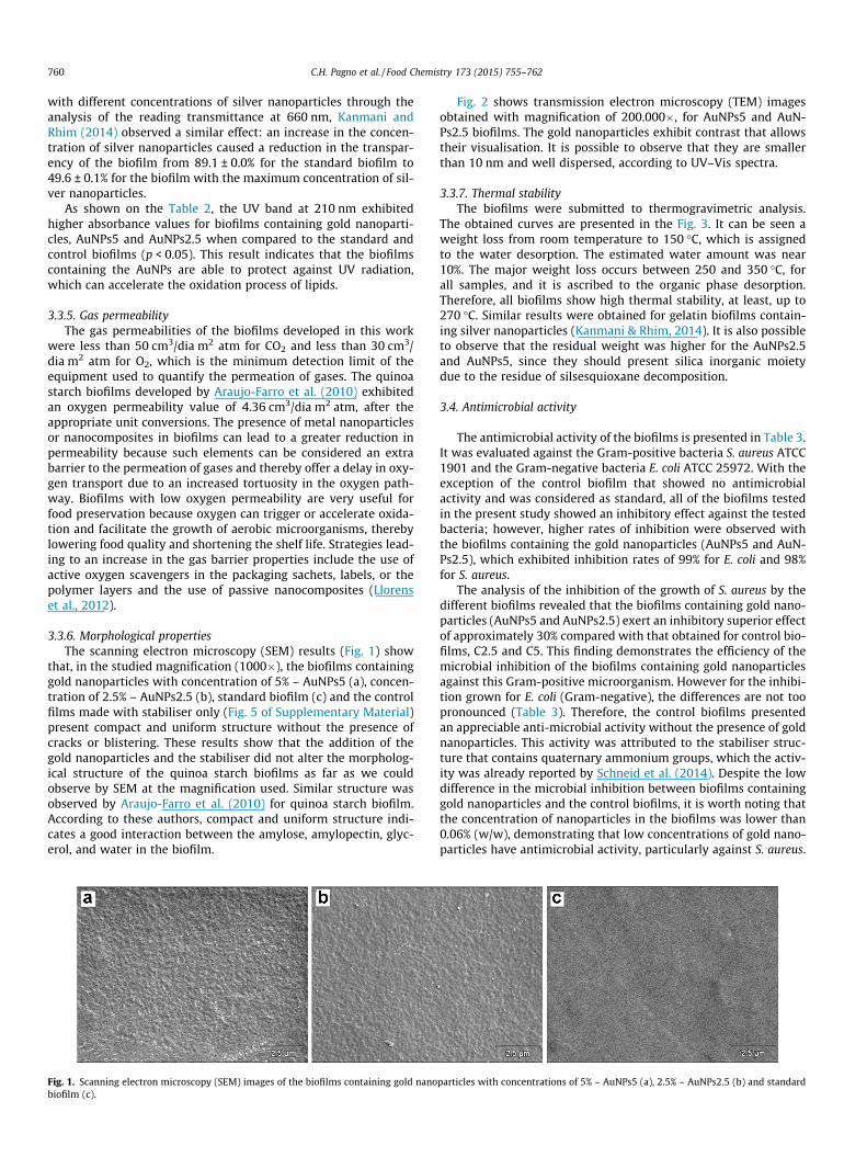
3.3.6. Morphological propertiesThe scanning electron microscopy (SEM) results (Fig. 1) show
that, in the studied magnification (1000�), the biofilms containinggold nanoparticles with concentration of 5% – AuNPs5 (a), concen-tration of 2.5% – AuNPs2.5 (b), standard biofilm (c) and the controlfilms made with stabiliser only (Fig. 5 of Supplementary Material)present compact and uniform structure without the presence ofcracks or blistering. These results show that the addition of thegold nanoparticles and the stabiliser did not alter the morpholog-ical structure of the quinoa starch biofilms as far as we couldobserve by SEM at the magnification used. Similar structure wasobserved by Araujo-Farro et al. (2010) for quinoa starch biofilm.According to these authors, compact and uniform structure indi-cates a good interaction between the amylose, amylopectin, glyc-erol, and water in the biofilm.
Fig. 1. Scanning electron microscopy (SEM) images of the biofilms containing gold nanobiofilm (c).
Fig. 2 shows transmission electron microscopy (TEM) imagesobtained with magnification of 200.000�, for AuNPs5 and AuN-Ps2.5 biofilms. The gold nanoparticles exhibit contrast that allowstheir visualisation. It is possible to observe that they are smallerthan 10 nm and well dispersed, according to UV–Vis spectra.
3.3.7. Thermal stabilityThe biofilms were submitted to thermogravimetric analysis.
The obtained curves are presented in the Fig. 3. It can be seen aweight loss from room temperature to 150 �C, which is assignedto the water desorption. The estimated water amount was near10%. The major weight loss occurs between 250 and 350 �C, forall samples, and it is ascribed to the organic phase desorption.Therefore, all biofilms show high thermal stability, at least, up to270 �C. Similar results were obtained for gelatin biofilms contain-ing silver nanoparticles (Kanmani & Rhim, 2014). It is also possibleto observe that the residual weight was higher for the AuNPs2.5and AuNPs5, since they should present silica inorganic moietydue to the residue of silsesquioxane decomposition.
3.4. Antimicrobial activity
The antimicrobial activity of the biofilms is presented in Table 3.It was evaluated against the Gram-positive bacteria S. aureus ATCC1901 and the Gram-negative bacteria E. coli ATCC 25972. With theexception of the control biofilm that showed no antimicrobialactivity and was considered as standard, all of the biofilms testedin the present study showed an inhibitory effect against the testedbacteria; however, higher rates of inhibition were observed withthe biofilms containing the gold nanoparticles (AuNPs5 and AuN-Ps2.5), which exhibited inhibition rates of 99% for E. coli and 98%for S. aureus.
The analysis of the inhibition of the growth of S. aureus by thedifferent biofilms revealed that the biofilms containing gold nano-particles (AuNPs5 and AuNPs2.5) exert an inhibitory superior effectof approximately 30% compared with that obtained for control bio-films, C2.5 and C5. This finding demonstrates the efficiency of themicrobial inhibition of the biofilms containing gold nanoparticlesagainst this Gram-positive microorganism. However for the inhibi-tion grown for E. coli (Gram-negative), the differences are not toopronounced (Table 3). Therefore, the control biofilms presentedan appreciable anti-microbial activity without the presence of goldnanoparticles. This activity was attributed to the stabiliser struc-ture that contains quaternary ammonium groups, which the activ-ity was already reported by Schneid et al. (2014). Despite the lowdifference in the microbial inhibition between biofilms containinggold nanoparticles and the control biofilms, it is worth noting thatthe concentration of nanoparticles in the biofilms was lower than0.06% (w/w), demonstrating that low concentrations of gold nano-particles have antimicrobial activity, particularly against S. aureus.
particles with concentrations of 5% – AuNPs5 (a), 2.5% – AuNPs2.5 (b) and standard
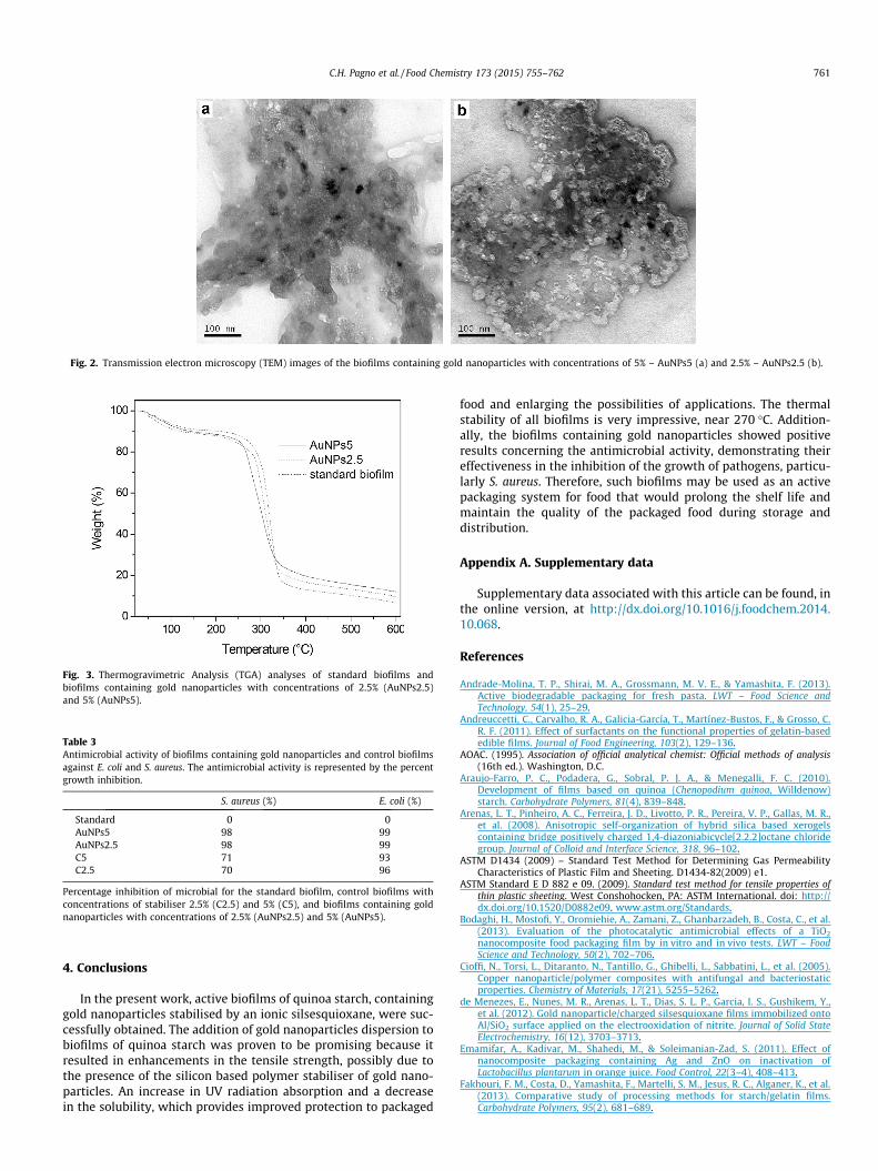
Fig. 2. Transmission electron microscopy (TEM) images of the biofilms containing gold nanoparticles with concentrations of 5% – AuNPs5 (a) and 2.5% – AuNPs2.5 (b).
Fig. 3. Thermogravimetric Analysis (TGA) analyses of standard biofilms andbiofilms containing gold nanoparticles with concentrations of 2.5% (AuNPs2.5)and 5% (AuNPs5).
Table 3Antimicrobial activity of biofilms containing gold nanoparticles and control biofilmsagainst E. coli and S. aureus. The antimicrobial activity is represented by the percentgrowth inhibition.
S. aureus (%) E. coli (%)
Standard 0 0AuNPs5 98 99AuNPs2.5 98 99C5 71 93C2.5 70 96
Percentage inhibition of microbial for the standard biofilm, control biofilms withconcentrations of stabiliser 2.5% (C2.5) and 5% (C5), and biofilms containing goldnanoparticles with concentrations of 2.5% (AuNPs2.5) and 5% (AuNPs5).
C.H. Pagno et al. / Food Chemistry 173 (2015) 755–762 761
4. Conclusions
In the present work, active biofilms of quinoa starch, containinggold nanoparticles stabilised by an ionic silsesquioxane, were suc-cessfully obtained. The addition of gold nanoparticles dispersion tobiofilms of quinoa starch was proven to be promising because itresulted in enhancements in the tensile strength, possibly due tothe presence of the silicon based polymer stabiliser of gold nano-particles. An increase in UV radiation absorption and a decreasein the solubility, which provides improved protection to packaged
food and enlarging the possibilities of applications. The thermalstability of all biofilms is very impressive, near 270 �C. Addition-ally, the biofilms containing gold nanoparticles showed positiveresults concerning the antimicrobial activity, demonstrating theireffectiveness in the inhibition of the growth of pathogens, particu-larly S. aureus. Therefore, such biofilms may be used as an activepackaging system for food that would prolong the shelf life andmaintain the quality of the packaged food during storage anddistribution.
Appendix A. Supplementary data
Supplementary data associated with this article can be found, inthe online version, at http://dx.doi.org/10.1016/j.foodchem.2014.10.068.
References
Andrade-Molina, T. P., Shirai, M. A., Grossmann, M. V. E., & Yamashita, F. (2013).Active biodegradable packaging for fresh pasta. LWT – Food Science andTechnology, 54(1), 25–29.
Andreuccetti, C., Carvalho, R. A., Galicia-García, T., Martínez-Bustos, F., & Grosso, C.R. F. (2011). Effect of surfactants on the functional properties of gelatin-basededible films. Journal of Food Engineering, 103(2), 129–136.
AOAC. (1995). Association of official analytical chemist: Official methods of analysis(16th ed.). Washington, D.C.
Araujo-Farro, P. C., Podadera, G., Sobral, P. J. A., & Menegalli, F. C. (2010).Development of films based on quinoa (Chenopodium quinoa, Willdenow)starch. Carbohydrate Polymers, 81(4), 839–848.
Arenas, L. T., Pinheiro, A. C., Ferreira, J. D., Livotto, P. R., Pereira, V. P., Gallas, M. R.,et al. (2008). Anisotropic self-organization of hybrid silica based xerogelscontaining bridge positively charged 1,4-diazoniabicycle[2.2.2]octane chloridegroup. Journal of Colloid and Interface Science, 318, 96–102.
ASTM D1434 (2009) – Standard Test Method for Determining Gas PermeabilityCharacteristics of Plastic Film and Sheeting. D1434-82(2009) e1.
ASTM Standard E D 882 e 09. (2009). Standard test method for tensile properties ofthin plastic sheeting. West Conshohocken, PA: ASTM International. doi: http://dx.doi.org/10.1520/D0882e09. www.astm.org/Standards.
Bodaghi, H., Mostofi, Y., Oromiehie, A., Zamani, Z., Ghanbarzadeh, B., Costa, C., et al.(2013). Evaluation of the photocatalytic antimicrobial effects of a TiO2
nanocomposite food packaging film by in vitro and in vivo tests. LWT – FoodScience and Technology, 50(2), 702–706.
Cioffi, N., Torsi, L., Ditaranto, N., Tantillo, G., Ghibelli, L., Sabbatini, L., et al. (2005).Copper nanoparticle/polymer composites with antifungal and bacteriostaticproperties. Chemistry of Materials, 17(21), 5255–5262.
de Menezes, E., Nunes, M. R., Arenas, L. T., Dias, S. L. P., Garcia, I. S., Gushikem, Y.,et al. (2012). Gold nanoparticle/charged silsesquioxane films immobilized ontoAl/SiO2 surface applied on the electrooxidation of nitrite. Journal of Solid StateElectrochemistry, 16(12), 3703–3713.
Emamifar, A., Kadivar, M., Shahedi, M., & Soleimanian-Zad, S. (2011). Effect ofnanocomposite packaging containing Ag and ZnO on inactivation ofLactobacillus plantarum in orange juice. Food Control, 22(3–4), 408–413.
Fakhouri, F. M., Costa, D., Yamashita, F., Martelli, S. M., Jesus, R. C., Alganer, K., et al.(2013). Comparative study of processing methods for starch/gelatin films.Carbohydrate Polymers, 95(2), 681–689.
762 C.H. Pagno et al. / Food Chemistry 173 (2015) 755–762
Incoronato, A. L., Buonocore, G. G., Conte, A., Lavorgna, M., & Nobile, M. A. (2010).Active systems based on silver-montmorillonite nanoparticles embedded intobio-based polymer matrices for packaging applications. Journal of FoodProtection, 73(12), 2256–2262.
Kanmani, P., & Rhim, J.-W. (2014). Physicochemical properties of gelatin/silver nanoparticle antimicrobial composite films. Food Chemistry, 148,162–169.
Kechichian, V., Ditchfield, C., Veiga-Santos, P., & Tadini, C. C. (2010). Naturalantimicrobial ingredients incorporated in biodegradable films based on cassavastarch. LWT – Food Science and Technology, 43(7), 1088–1094.
Liao, J., Anchun, M., Zhu, Z., & Quan, Y. (2010). Antibacterial titanium platedeposited by silver nanoparticles exhibits cell compatibility. InternationalJournal of Nanomedicine, 5, 337–342.
Llorens, A., Lloret, E., Picouet, P. A., Trbojevich, R., & Fernandez, A. (2012). Metallic-based micro and nanocomposites in food contact materials and active foodpackaging. Trends in Food Science & Technology, 24(1), 19–29.
Mei, J., Yuan, Y., Wu, Y., & Li, Y. (2013). Characterization of edible starch–chitosanfilm and its application in the storage of Mongolian cheese. International Journalof Biological Macromolecules, 57, 17–21.
Nascimento, T. A., Calado, V., & Carvalho, C. W. P. (2012). Development andcharacterization of flexible film based on starch and passion fruit mesocarpflour with nanoparticles. Food Research International, 49(1), 588–595.
Nunes, M. R., Gushikem, Y., Landers, R., Dupont, J., Costa, T. M. H., & Benvenutti, E. V.(2012). Charged silsesquioxane used as a vehicle for gold nanoparticles toperform the synthesis of catalyst xerogels. Journal of Sol-Gel Science andTechnology, 63(2), 258–265.
Pelissari, F. M., Andrade-Mahecha, M. M., Sobral, P. J. d. A., & Menegalli, F. C. (2013).Comparative study on the properties of flour and starch films of plantainbananas (Musa paradisiaca). Food Hydrocolloids, 30(2), 681–690.
Rhim, J. W., Wang, L. F., & Hong, S. I. (2013). Preparation and characterization ofagar/silver nanoparticles composite films with antimicrobial activity. FoodHydrocolloids, 33(2), 327–335.
Rotta, J., Ozório, R. Á., Kehrwald, A. M., de Oliveira Barra, G. M., de Melo CastanhoAmboni, R. D., & Barreto, P. L. M. (2009). Parameters of color, transparency,water solubility, wettability and surface free energy of chitosan/hydroxypropylmethylcellulose (HPMC) films plasticized with sorbitol.Materials Science and Engineering C, 29(2), 619–623.
Schneid, A. C., Roesch, E. W., Sperb, F., Matte, U., da Silveira, N. P., Costa, T. M. H.,et al. (2014). Silver nanoparticle–ionic silsesquioxane: A new system proposedas an antibacterial agent. Journal of Materials Chemistry B, 2, 1079–1086.
Souza, A. C., Benze, R., Ferrão, E. S., Ditchfield, C., Coelho, A. C. V., & Tadini, C. C.(2012). Cassava starch biodegradable films: Influence of glycerol and claynanoparticles content on tensile and barrier properties and glass transitiontemperature. LWT – Food Science and Technology, 46(1), 110–117.
Souza, A. C., Goto, G. E. O., Mainardi, J. A., Coelho, A. C. V., & Tadini, C. C. (2013).Cassava starch composite films incorporated with cinnamon essential oil:Antimicrobial activity, microstructure, mechanical and barrier properties. LWT– Food Science and Technology., 54(2), 346–352.
Wang, L., Dong, Y., Men, H., Tong, J., & Zhou, J. (2013). Preparation andcharacterization of active films based on chitosan incorporated teapolyphenols. Food Hydrocolloids, 32(1), 35–41.
Willets, K. A., & Duyne, R. P. V. (2007). Localized surface plasmon resonancespectroscopy and sensing. Annual Review of Physical Chemistry, 58, 267–297.
Xiong, H., Tang, S., Tang, H., & Zou, P. (2008). The structure and properties of astarch-based biodegradable film. Carbohydrate Polymers, 71(2), 263–268.
Yoksan, R., & Chirachanchai, S. (2010). Silver nanoparticle-loaded chitosan–starchbased films: Fabrication and evaluation of tensile, barrier and antimicrobialproperties. Materials Science and Engineering C, 30(6), 891–897.