mathematical description of microbial biofilms
TRANSCRIPT

SIAM REVIEW c© 2010 Society for Industrial and Applied MathematicsVol. 52, No. 2, pp. 000–000
Mathematical Description ofMicrobial Biofilms∗
Isaac Klapper†
Jack Dockery†
Abstract. We describe microbial communities denoted biofilms and efforts to model some of their im-portant aspects, including quorum sensing, growth, mechanics, and antimicrobial tolerancemechanisms.
Key words. biofilms, quorum sensing, biomechanics, antimicrobial tolerance
AMS subject classifications. AUTHOR MUST PROVIDE
DOI. 10.1137/080739720
1. Introduction.
1.1. Microbial Ecology. Compared to the plant and animal kingdoms, the di-versity of microbial life is considerably less explored and less understood (even thenotion of microbial species is a current topic of debate [41, 235]), in part because clas-sification and characterization of microbes have been comparatively difficult tasks.Recently, though, new efficient technologies for genetic and genomic cataloging ofmicrobial communities are changing the landscape of prokaryotic and archael ecol-ogy, uncovering surprising diversity in the process. Such information is important asmicroorganisms are closely connected with geochemical cycling as well as with produc-tion and degradation of organic materials. Thus a set of guiding principles is neededto understand the distribution and diversity of microorganisms in the context of theirmicro- and macroenvironments as a step toward the ultimate goal of constructingdescriptions of full microbial ecosystems.
Prokaryotes (bacteria and archaea) are estimated to make up approximately halfof the extant biomass [244]. Their numbers are staggering; for example, each humanharbors an estimated 100 trillion microbes (bacteria and archaea), ten times more mi-crobes than human cells [19], and during the course of a normal lifetime, the number ofEscherichia coli inhabiting a given person will easily exceed the number of people whoever lived. To paraphrase Stephen Jay Gould, though we may think we are living ina human-dominated world, it may be more truthful to say that we live in the “Age ofBacteria” [90]. The familiar view of microbes in their free (planktonic) state, however,is not the norm; rather, it is believed that much of the microbial biomass is locatedin close-knit communities, designated biofilms and microbial mats, each consisting oflarge numbers of organisms living within a self-secreted matrix constructed of poly-mers and other molecules [1, 47, 46, 49, 96, 210]; see Figures 1 through 4. (Microbes in
∗Received by the editors November 2, 2008; accepted for publication (in revised form) June 15,2009; published electronically May 6, 2010. This work was supported by grant NSF/DMS 0856741.
http://www.siam.org/journals/sirev/52-2/73972.html†Department of Mathematical Sciences, Center for Biofilm Engineering, Montana State University,
Bozeman, MT 59717 ([email protected]).
1

2 ISAAC KLAPPER AND JACK DOCKERY
Fig. 1 Mixed-species photosynthetic mat, Biscuit Basin, Yellowstone National Park. Reprinted bypermission of Macmillan Publishers Ltd: Nature Reviews Microbiology from [96].
Fig. 2 Microbial structures in a mixed-species photosynthetic mat, Mushroom Spring, YellowstoneNational Park.

MATHEMATICAL DESCRIPTION OF MICROBIAL BIOFILMS 3
Fig. 3 Laboratory-grown Pseudomonas aeruginosa biofilm. Reprinted by permission of BlackwellPublishing from [126].
Fig. 4 Aquificales streamers (pink), Octopus Spring, Yellowstone National Park.

4 ISAAC KLAPPER AND JACK DOCKERY
collective behave very differently from their planktonic state; even genetic expressionpatterns change [110, 210].) These matrices anchor and protect their communities infavorable locations [76], generally wet or damp but not always so [237], while provid-ing a framework in which structured populations can differentiate and self-organize.In this way, as a result of internal spatial variation, microbial community ecology isdetermined not only by classical ecological interactions such as competition and co-operation (as, for example, in chemostat communities [199]) but also by local physicaland chemical stresses and accompanying physical and chemical material properties.One can expect that, in both directions, the connection between physics and ecologyin microbial communities is profound at multiple scales and that these ecosystems arenot merely the sum of their parts.
Microbial mats and biofilms have a long lineage—it is interesting and suggestiveto note that biofilm formation is observed today in the Aquificales [183] (Figure 4),the lowest known branch of bacteria (on the phylogenetic tree of life), and, likewise,Korarcheota [111], the corresponding lowest known branch of archaea. Prokaryoticmats may once have been a dominant life-form on earth and may have played an eco-logically crucial role in precambrian earth chemistry. Within those billions of years ofdominance, enormous generation of genetic diversity occurred; today, prokaryotes areestimated to carry 100 times as many genes as eukaryotes [244]. These days, possiblydue to the existence of grazers, mats are mostly found in extreme environments (e.g.,high or low temperature, high salinity, deep ocean vents), where they typically consistof communities of photoautotrophs and attending heterotrophs, though communitiesbased on chemoautotrophy also exist; see, e.g., Figure 4. However, while microbialmats are now relatively rare, their thinner cousins, microbial biofilms, are ubiquitous.One can and will find them in almost any damp or wet environment, and they areoften key players in problems such as human and animal infections [43, 82], foulingof industrial equipment and water systems [18], and waste treatment and remedia-tion [42, 218], just to name a few. Medical relevance is dramatic as well [26]. Quotingfrom the National Institutes of Health [178],
Biofilms are clinically important, accounting for over 80 percent of micro-
bial infections in the body. Examples include: infections of the oral soft tis-
sues, teeth and dental implants; middle ear; gastrointestinal tract; urogeni-
tal tract; airway/lung tissue; eye; urinary tract prostheses; peritoneal mem-
brane and peritoneal dialysis catheters, in-dwelling catheters for hemodial-
ysis and for chronic administration of chemotherapeutic agents (Hickman
catheters); cardiac implants such as pacemakers, prosthetic heart valves,
ventricular assist devices, and synthetic vascular grafts and stents; pros-
theses, internal fixation devices, percutaneous sutures; and tracheal and
ventilator tubing.
Biofilms can be dangerous and opportunistic pathogenic structures; in fact, biofilm-associated nosocomial (hospital acquired) infection is by itself a leading cause of deathin the United States [241].
1.2. Biofilm Development and Structure. Biofilm development is often char-acterized as a multistage process [159] (see Figure 5). In the first stage, isolatedplanktonic-state microbes attach to a surface or interface. Once attached to a surfacea phenotypic transformation (change of gene expression pattern) into a biofilm stateoccurs. At this point, cells begin manufacturing and excreting extracellular matrixmaterial. The exact nature of this material can vary greatly depending on micro-

MATHEMATICAL DESCRIPTION OF MICROBIAL BIOFILMS 5
Fig. 5 Stages of the biofilm life cycle, courtesy of the Montana State University Center for BiofilmEngineering, P. Dirckx.
bial strain and, even within a single strain, on environmental conditions and history,though its function shares commonality. With formation of an anchoring matrix,the nascent biofilm begins to grow. Secondary colonizing strains might arrive andjoin the new community. But as the biofilm matures, meso- and macroscale physicalrealities like diffusion-reaction limitations and mechanical stress distribution becomemore important, probably to the extent that they significantly influence structureand ecology. Finally, the fully mature biofilm reaches a quasi-steady state wheregrowth is balanced by loss through erosion and detachment due to mechanical stress,self-induced dispersal from starved subregions, and predation and loss to grazers andviruses. We recommend a remarkably forward-looking overview written by a pioneerof the subject of biofilm mechanics and modeling, Bill Characklis [32].
On close inspection, we note a wide variation of relevant time scales (see Figure 6),ranging from fast time scales for bulk fluid dynamics and reaction-diffusion chemicalsubprocesses to slow time scales of biological development. The roughly ten orders ofmagnitude variation in range is a challenge to modelers, of course, one that necessitatescompromises. The usual practice is to introduce equilibrium in the fast processes: bulkfluid flow, when considered, is assumed steady over a quasi-static biofilm, and thenadvection-reaction-diffusion processes are also assumed to be quasi-steady relative tothe given fluid velocity field. Given those fluid velocity and chemical profiles, themodeler can take advantage of scale separation and focus on the slow growth andloss processes that are typically the ones of interest. Notably missing from Figure 6are time scales related to viscoelastic mechanics within the biofilm itself that mayinconveniently straddle several time scales. Also missing are external forcing timescales, e.g., the day–night cycle, which can also significantly effect biofilm communitystructure [55].
Biofilms are housed in a self-secreted matrix composed of polysaccharides, pro-teins, DNA, and other materials (detritus from lysed cells, abiotic material like pre-cipitates and corrosive products, etc.) which performs many roles [78, 214]. Foremostit serves as an anchoring structure for its inhabitants, keeping them in place andprotected from external mechanical forces as well as hostile predators and dangerous

6 ISAAC KLAPPER AND JACK DOCKERY
Fig. 6 Various time scales for biofilm-related processes. Box 1 includes processes related to bulkfluid (measured with respect to a biofilm-length scale). Box 2 includes processes related tochemical processes (again, measured with respect to a biofilm-length scale). Box 3 includesbiological processes (essentially independent of biofilm-length scale). Reprinted from [169]with permission from John Wiley & Sons.
chemicals. However, in the words of H.-C. Flemming, “a biofilm is not a prison,”meaning that it is necessary for biofilm inhabitants to be able to escape in order toform new colonies elsewhere. These topics will be further pursued later. For now wenote that the matrix is thought to accomplish a number of other purposes as well.It provides a food source when needed and it protects from dehydration. By keepingmicrobes in proximity to each other, the matrix plays a role in community ecology,facilitating signaling, ecological interactions, and exchange of genetic material [134].Conversely, it has been suggested that production of matrix material can serve as asort of exclusion zone by pushing competing microorganisms away, thus improvingaccess to resources [256].
The physical makeup of a biofilm creates a heterogeneous environment, in partdue to reaction-diffusion limitations on both small and large scales, resulting in a richstructure of ecological niches [207, 260]. As a simple but important example, oxygenconsumption in a layer at the top of a biofilm can create a low-oxygen environmentin the sublayer below that is favorable for anaerobic organisms (those that functionwithout oxygen). One such instance of this commensalism is surface corrosion byanaerobic sulfate-reducing bacteria protected in the depths of a biofilm [139, 224], aphenomenon that might be exploitable in the form of microbial fuel cells [184]. Evensingle-species biofilms, though, can present phenotypic heterogeneity in their ecologyby changing genetic expression patterns in response to spatially varying environmentalconditions within the biofilm. Regarding the biofilm as a whole organism, spatialheterogeneity-induced niche structure confers considerable flexibility and may be oneof the reasons that bacteria have such a broad role in geocycling.

MATHEMATICAL DESCRIPTION OF MICROBIAL BIOFILMS 7
2. Quorum Sensing. It has been known for some time that bacteria can com-municate with each other using chemical signaling molecules. As in higher organisms,these signaling processes allow bacteria to synchronize activities and behave as multi-cellular microorganisms. Many bacteria produce, release, and respond to small signalmolecules in order to monitor their own population density and control the expres-sion of specific genes in response to change in population density. This type of generegulation is known as quorum sensing (QS) [238].
Quorum sensing systems have been shown to regulate the expression of genesinvolved in plasmid transfer, population mobility, biofilm formation, biofilm matu-ration, dissolution of biofilms and virulence [246, 185, 193, 22]. While there havebeen a number of articles on the connections of biofilm formation to quorum sensing[51, 99, 161], the specific relationships are still a matter of debate [122, 162]. The ex-periments are apparently difficult; for example, Stoodley et al. [208] initially reportedthat under high shear conditions, biofilms formed by a QS mutant were visually in-distinguishable from wild-type biofilms. It was found later through a more refinedanalysis that there are discernable differences (see, e.g., [176]).
There are many different types of quorum sensing systems and circuits, and manybacteria have multiple signaling systems. The receptors for some signaling moleculesare very specific, which implies that some types of signaling are designed for in-traspecies communication. On the other hand, there are universal communicationsignaling molecules produced by different bacterial species as well as eukaryotic cellswhich promote interspecies communication. Some species of bacteria can manipulatethe signaling of, and interfere with, other species’ abilities to assess and respond cor-rectly to changes in cell population density. Cell-to-cell signaling, and interferencewith it, could have important ramifications for eukaryotes in the maintenance of nor-mal microflora and in protection from pathogenic bacteria. For recent reviews of thevarious types of quorum sensing systems see [109, 246, 247].
One type of quorum sensing signaling in Gram-negative bacteria is mediatedby the secretion of low-molecular-weight compounds called acyl homoserine lactones(AHL). Because these water-soluble molecules are small and can permeate cell mem-branes, they cannot accumulate to critical concentrations within the cells unless thebacteria are at a sufficiently large cell density or there is some type of restriction onchemical diffusion. AHL signals are used to trigger expression of specific genes whenthe bacteria have reached a critical density. This “census-taking” enables the groupas a whole to express specific genes only at large population densities.
Most often the processes regulated by quorum sensing are ones that would beunproductive when undertaken by an individual bacterium but become effective wheninitiated by the collective. For example, virulence factors are molecules producedby a pathogen that influence their host’s function to allow the pathogen to thrive.It would not be beneficial for a small population to produce these factors if therewere not sufficient numbers to mount a viable attack on the host. This cell-to-cellcommunication tactic appears to be extremely important in the pathogenic bacteriumPseudomonas aeruginosa. It has been estimated that 6% to 10% of all genes in P.aeruginosa are controlled by AHL quorum sensing signaling systems [194, 225].
Perhaps the best characterized example of AHL QS is in the autoinduction ofluminescence in the symbiotic marine bacterium Vibrio fischeri, which colonizes lightorgans of the Hawaiian squid Euprymna scolopes [80]. When the bioluminescentbacteria were grown in liquid cultures it was noted that the cultures produced lightonly when sufficiently large numbers of the bacteria were present [91]. The originalthought was that the growthmedia itself contained an inhibitor which was degraded by
8 ISAAC KLAPPER AND JACK DOCKERY
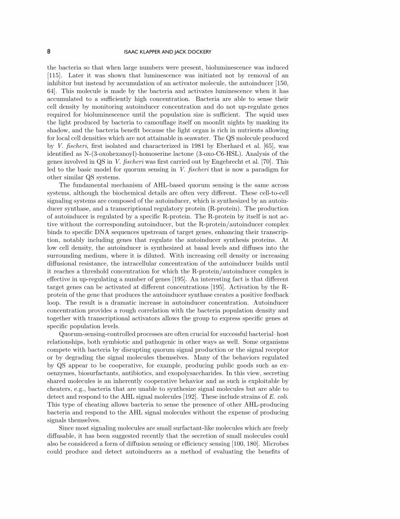
the bacteria so that when large numbers were present, bioluminescence was induced[115]. Later it was shown that luminescence was initiated not by removal of aninhibitor but instead by accumulation of an activator molecule, the autoinducer [150,64]. This molecule is made by the bacteria and activates luminescence when it hasaccumulated to a sufficiently high concentration. Bacteria are able to sense theircell density by monitoring autoinducer concentration and do not up-regulate genesrequired for bioluminescence until the population size is sufficient. The squid usesthe light produced by bacteria to camouflage itself on moonlit nights by masking itsshadow, and the bacteria benefit because the light organ is rich in nutrients allowingfor local cell densities which are not attainable in seawater. The QS molecule producedby V. fischeri, first isolated and characterized in 1981 by Eberhard et al. [65], wasidentified as N-(3-oxohexanoyl)-homoserine lactone (3-oxo-C6-HSL). Analysis of thegenes involved in QS in V. fischeri was first carried out by Engebrecht et al. [70]. Thisled to the basic model for quorum sensing in V. fischeri that is now a paradigm forother similar QS systems.
The fundamental mechanism of AHL-based quorum sensing is the same acrosssystems, although the biochemical details are often very different. These cell-to-cellsignaling systems are composed of the autoinducer, which is synthesized by an autoin-ducer synthase, and a transcriptional regulatory protein (R-protein). The productionof autoinducer is regulated by a specific R-protein. The R-protein by itself is not ac-tive without the corresponding autoinducer, but the R-protein/autoinducer complexbinds to specific DNA sequences upstream of target genes, enhancing their transcrip-tion, notably including genes that regulate the autoinducer synthesis proteins. Atlow cell density, the autoinducer is synthesized at basal levels and diffuses into thesurrounding medium, where it is diluted. With increasing cell density or increasingdiffusional resistance, the intracellular concentration of the autoinducer builds untilit reaches a threshold concentration for which the R-protein/autoinducer complex iseffective in up-regulating a number of genes [195]. An interesting fact is that differenttarget genes can be activated at different concentrations [195]. Activation by the R-protein of the gene that produces the autoinducer synthase creates a positive feedbackloop. The result is a dramatic increase in autoinducer concentration. Autoinducerconcentration provides a rough correlation with the bacteria population density andtogether with transcriptional activators allows the group to express specific genes atspecific population levels.
Quorum-sensing-controlled processes are often crucial for successful bacterial–hostrelationships, both symbiotic and pathogenic in other ways as well. Some organismscompete with bacteria by disrupting quorum signal production or the signal receptoror by degrading the signal molecules themselves. Many of the behaviors regulatedby QS appear to be cooperative, for example, producing public goods such as ex-oenzymes, biosurfactants, antibiotics, and exopolysaccharides. In this view, secretingshared molecules is an inherently cooperative behavior and as such is exploitable bycheaters, e.g., bacteria that are unable to synthesize signal molecules but are able todetect and respond to the AHL signal molecules [192]. These include strains of E. coli.This type of cheating allows bacteria to sense the presence of other AHL-producingbacteria and respond to the AHL signal molecules without the expense of producingsignals themselves.
Since most signaling molecules are small surfactant-like molecules which are freelydiffusable, it has been suggested recently that the secretion of small molecules couldalso be considered a form of diffusion sensing or efficiency sensing [100, 180]. Microbescould produce and detect autoinducers as a method of evaluating the benefits of

MATHEMATICAL DESCRIPTION OF MICROBIAL BIOFILMS 9
secreting more expensive molecules. A bacterium could use the signal to estimatemass-transfer properties of the media and spatial distribution of cells as well as celldensities. The thought is that it would be an inefficient use of resources to produceenzymes if they are going to be rapidly transported away by diffusion or some othermeans, like advection. Note that this need not be a cooperative strategy since a singlebacterium might find itself in a confined region where the autoinducer could reachthreshold without any other cells present.
2.1. Modeling of QS. While quorum sensing has been described as “the mostconsequential molecular microbiology story of the last decade” [27, 252], the historyof mathematical modeling of QS is fairly brief. For a good survey up to about 2005see Ward [236]. Modeling of QS at the molecular level was started by the almostsimultaneous publications of James et al. [108] and Dockery and Keener [58]. Thesemodels indicate that QS works as a switch via a positive feedback loop which givesrise to bistability for sufficient cell density, an up-regulated and down-regulated state.There have been several extensions of these types of deterministic models, includingmuch more regulatory network detail [73, 74, 149, 88, 222] as well as stochasticity[89, 142, 189].
2.2. The Model. The quorum-sensing system of P. aeruginosa is unusual be-cause it has two somewhat redundant regulatory systems [166, 167] . The first systemwas shown to regulate expression of the elastase LasB and was therefore named thelas system. The two enzymes, LasB elastase and LasA elastase, are responsible forelastolytic activity which destroys elastin-containing human lung tissue and causespulmonary hemorrhages associated with P. aeruginosa infections. The las system iscomposed of lasI, the autoinducer synthase gene responsible for synthesis of the au-toinducer 3-oxo-C12-HSL, and the lasR gene that codes for transcriptional activatorprotein. The LasR/3-oxo-C12-HSL dimer, which is the activated form of LasR, ac-tivates a variety of genes ([251, 163, 164, 167, 101]), but preferentially promotes lasIactivity ([195]). The las system is positively controlled by both GacA and Vfr, whichare needed for transcription of lasR. The transcription of lasI is also repressed by theinhibitor RsaL.
The second quorum-sensing system in P. aeruginosa is named the rhl system be-cause of its ability to control the production of rhamnolipid [155]. Rhamnolipid has adetergent-like structure and is responsible for the degradation of lung surfactant andinhibits the mucociliary transport and ciliary function of human respiratory epithe-lium. This system is composed of rhlI, the synthase gene for the autoinducer C4-HSL,and the rhlR gene encoding a transcriptional activator protein. A diagram depictingthese two systems is shown in Figure 7 [221].
We make several simplifying assumptions as in [58, 72]. First, if we assumethat there is no shortage of substrate for autoinducer production, then there is noneed to explicitly model the biosynthesis of 3-oxo-C12-HSL and C4-HSL by LasI andRhlI. Second, there is evidence that many proteins are more stable than the mRNAthat code for them (see, for example, [6, 29, 68]). Assuming this is the case, thenLasR mRNA, lasI mRNA, rhlI mRNA, and rsaL mRNA are much shorter lived thanLasR, LasI, RhlR and RsaL, respectively. If we assume that all messenger RNAsare produced by DNA at rates that are Michaelis–Menten in type, then, assumingthe mRNAs are in quasi-steady state, it follows (see, e.g., [58]) that production ofLasR, RasL, and RhlR follows Michaelis–Menten kinetics. We will also assume thatthe production of 3-oxo-C12-HSL and C4-HSL also follow Michaelis–Menten kinetics.Finally, we assume that the autoinducers freely diffuse across the cell membrane.

10 ISAAC KLAPPER AND JACK DOCKERY
Fig. 7 Schematic diagram showing gene regulation for the las and rhl systems in P. aeruginosa.
Following [72], the intercellular dynamics is described by a system of eight cou-pled differential equations that model the intracellular concentrations of LasR, RhlR,RasL, 3-oxo-C12-HSL, C4-HSL, the LasR/3-oxo-C12-HSL complex, the RhlR/C4-HSL complex, and the RhlR/3-oxo-C12-HSL complex. We introduce variables for allthe concentrations (shown in Table 1) as well as the external concentrations of theautoinducers, 3-oxo-C12-HSL and C4-HSL.
If we assume that all the dimers C1, C2 and C3 are formed via the law of massaction at rate rCi and dissociate at the rate dCi, i = 1, 2, 3, then
dC1
dt= rC1P1A1 − dc1C1,(1)
dC2
dt= rC2P2A2 − dc2C2,(2)
dC3
dt= rC3P2A1 − dc3C3.(3)
The equations for production of the proteins under the assumptions described above

MATHEMATICAL DESCRIPTION OF MICROBIAL BIOFILMS 11
Table 1 Variables used to identify concentrations.
Variable Concentration
A1 [3-oxo-C12-HSL]
A2 [C4-HSL]
P1 [LasR]
P2 [RhlR]
P3 [RsaL]
C1 [LasR/3-oxo-C12-HSL]
C2 [RhlR/C4-HSL]
C3 [RhlR/3-oxo-C12-HSL]
E1 (External) [3-oxo-C12-HSL]
E2 (External) [C4-HSL]
are given by
dP1
dt= −rC1P1A1 + dc1C1 − dp1P1(4)
+VP1
C1
KP1 + C1+ P 0
1 ,
dP2
dt= −rC2P2A2 + dc2C2 − dp2P2(5)
+VP2
C1
KP2 + C1+ P 0
2
− rC3P2A1 + dc3C3,
dP3
dt= −dP3P3 + VP3
C1
KP3 + C1+ P 0
3 .(6)
Similarly, for the autoinducers,
dA1
dt= −rC1P1A1 + dC1C1 − rC3P2A1 + dC3C3 − dA1A1(7)
+VA1
C1
C1 +KA1(1 + P3KP3r
)+A0
1 + δ1(E1 −A1),
dA2
dt= −rC2P2A2 + dC2C2 + VA2
C2
C2 +KA2+A0
2 + δ2(E2 −A2).(8)
Next, we need to determine how the density of organisms controls the activityof this network. We assume that autoinducer Ai (i = 1, 2) diffuses across the cellmembrane and that the local volume fraction of cells is ρ. Then, by assumption,the local volume fraction of extracellular space is 1 − ρ. As indicated above, theextracellular autoinducer is assumed to diffuse freely across the cell membrane withconductance δi and to naturally degrade at rate kEi . We remark that there is someevidence that the transport of the autoinducer may involve both passive diffusion anda cotransport mechanism [165]. For this model we assume that diffusion alone actsto transport the autoinducer (as is apparently correct for the rhl system [165]).
If we suppose that the density of cells is uniform and the extracellular space iswell mixed, then the concentration of autoinducer in the extracellular space, denoted

12 ISAAC KLAPPER AND JACK DOCKERY
Fig. 8 Bifurcation Diagram for (1) through (8) with respect to the density parameter ρ for theparameter values [72]: rc1 = rc2 = rc3 = 0.17 (1/µMs), dc1 = dc2 = dc3 = 0.25 (1/s),VP1 = Vp2 = 0.6 (µM/s), KP1 = k (µM), KP2 = 1.2 (µM), VP3 = 0.9 (µM/s), KP3 = 1(µM/S), VA1 = 1.2 (µM/s), VA2 = 1 (µM/s), KA1 = 0.4 (µM), KA2 = 1.0 (µM),dP1 = dP2 = 0.2 (1/s), dP3 = 0.25 (1/s), δ1 = 0.2 (1/s), δ2 = 0.4 (1/s), KE1 = KE2 = 0.2(µM), dA1 = 0.2 (1/s), dA2 = 0.4 (1/s), P 0
1 = P 02 = P 0
3 = 0.004 (µM), KP3r = 2.5 (µM),A0
1 = 0.4 (µM), A02 = 0.004 (µM).
Ei, is governed by the equation
(9) (1− ρ)(dEi
dt+ kEiEi
)= δi(Ai − Ei) for i = 1, 2.
Here we emphasize that the factor 1 − ρ must be included to scale for the differencebetween concentration in the extracellular space and concentration viewed as amountper unit total volume. Similarly, the governing equation for intracellular autoinducerAi must be modified to account for the cell density. For example, (8) becomes
dA2
dt= −rC2P2A2 + dC2C2 + VA2
C2
C2 +KA2
+A02 +
δ2ρ(E2 −A2).(10)
A bifurcation diagram for the steady states for (1) through (8) is shown in Fig-ure 8. The parameter ρ provides a switch between the two stable steady solutions.For small values of ρ, there is a unique stable steady state with small values of A1 andA2. As ρ increases, two more solutions appear (a saddle node bifurcation), and asρ increases yet further, the small solution disappears (another saddle node), leavinga unique solution, thus initiating a “switch.” For intermediate values of ρ, there arethree steady solutions. The small and large values are stable steady solutions, whilethe intermediate solution is unstable, a saddle point. It is easy to arrange parameter

MATHEMATICAL DESCRIPTION OF MICROBIAL BIOFILMS 13
values that have this switching behavior, and the switch can be adjusted to occur atany desired density level.
One can give the following verbal explanation of how quorum sensing works. Thequantity A1, the autoinducer, is produced by cells at some nominal rate. However,each cell must dump its production of A1 or else the autocatalytic reaction wouldturn on. As the density of cells increases, the dumping process becomes less effective,and so the autocatalytic reaction is enabled.
It is interesting that, to the best of the authors’ knowledge, bistability has notyet been observed experimentally. This may be due to difficulties such as the re-quirement for a reporter gene product with an extremely short half life. Haseltineand Arnold [98] have shown experimentally in an artificial quorum-sensing networkthat simple changes in network architecture can change the steady state behavior ofa QS network. In particular, it is possible to obtain graded threshold and bistableresponses via plasmid manipulations. They also have shown that threshold responseis more consistent with the wild-type lux operon than the bistable response. It wouldseem possible to conduct experiments to explore the multistability in synthetic geneticcircuits by using the approach described by Angeli et al. [10].
The first population-level models were developed by Ward et al. [233], includingpopulation growth. This system models subpopulations of up-regulated and down-regulated cells and a single QS molecule species, predicting that QS occurs due to thecombination of the effects of increased QS molecule concentration as the populationsize increases and the induced increase of the up-regulated subpopulation. In a rel-atively short time (compared to the time scale for growth) there is a rapid increaseof the QS molecule concentration. Ward et al. [234] extended these models to in-clude a negative feedback mechanism known to be involved in QS. A similar modelingapproach has been developed to investigate QS in a wound [127].
Since the virulence of P. aeruginosa is controlled by QS, there is the possibilitythat agents designed to block cell-to-cell communication can act as novel antibacte-rials. One advantage of this approach is that it is nonlethal and hence less selectivefor resistance than lethal methods. Anguige et al. [7] have extended the basic modeldeveloped in [233] to investigate therapies that are designed to disrupt QS. Two typesof QS blockers were investigated, one that degrades the AHL signal and anotherthat disrupts the production of the signal by diffusing through the cell membraneand destabilizing one of the proteins necessary for signaling molecule production. Aconventional antibiotic is also investigated. Through numerical simulations and math-ematical analysis it is shown that while it is possible to reduce the QS signal (andhence virulence) to a negligible level, the qualitative response to treatment is sensitiveto parameter values.
Koerber et al. have published an interesting application of deterministic popu-lation level models [128]. They use a deterministic model for QS to help develop astochastic model for the escape of Staphylococcus aureus from the endosome. Thedeterministic model includes the QS molecule, up- and down-regulated cell types, anexoenzyme, and the thickness of the endosome. The deterministic model is used tovalidate the escape time asymptotically for the stochastic model of a single cell.
Muller et al. [149] have developed a deterministic model that includes the regula-tory network for QS in a single cell and have incorporated this single-cell model intoa population-level model. The population model is a spatially structured model. Thescaling behavior with regard to cell size is studied and the model is validated withexperimental data. It is interesting that the submodel for the regulatory system hasbistable behavior but a threshold effect at the population level. The analysis shows
14 ISAAC KLAPPER AND JACK DOCKERY
that a single cell is mainly influenced by its own signal and thus is computing diffusionsensing. However, interaction with other cells does provide a higher-order effect. Notethat it is assumed that distances between cells are large compared to cell sizes.
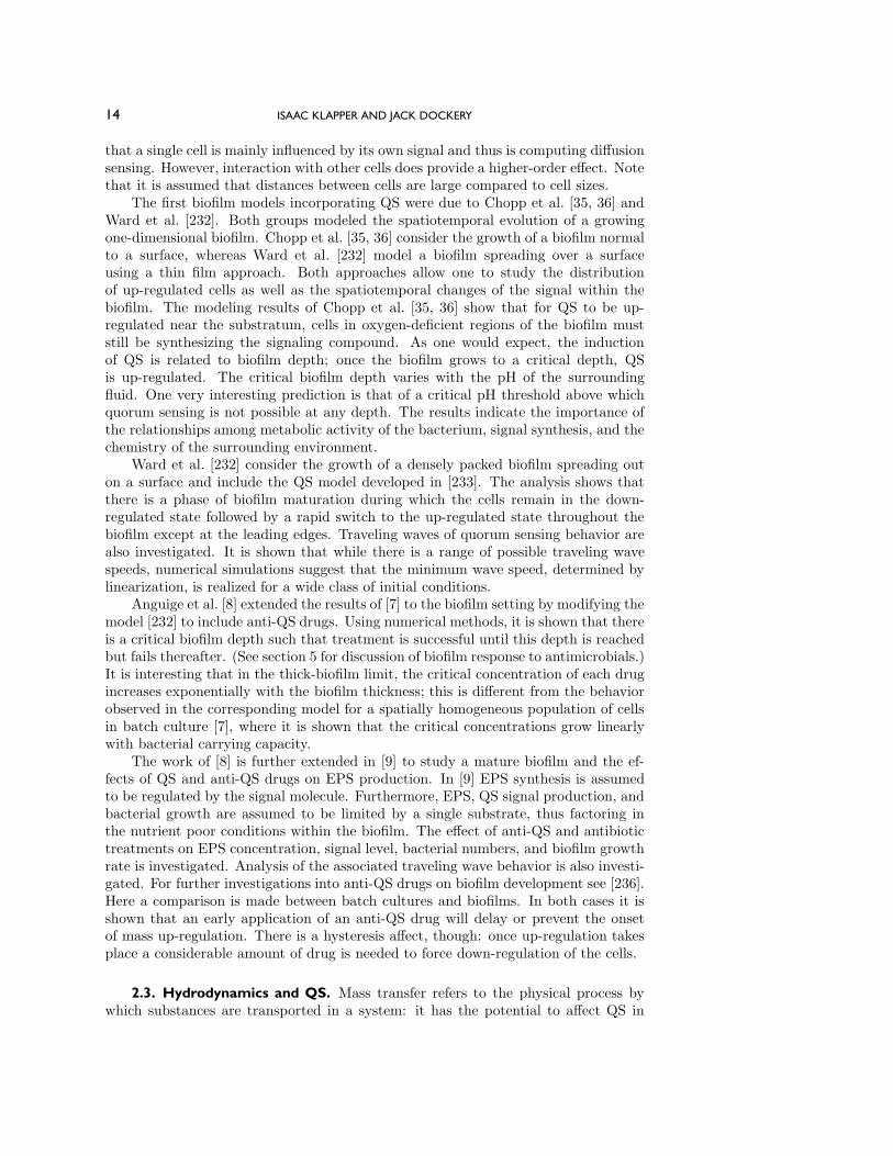
The first biofilm models incorporating QS were due to Chopp et al. [35, 36] andWard et al. [232]. Both groups modeled the spatiotemporal evolution of a growingone-dimensional biofilm. Chopp et al. [35, 36] consider the growth of a biofilm normalto a surface, whereas Ward et al. [232] model a biofilm spreading over a surfaceusing a thin film approach. Both approaches allow one to study the distributionof up-regulated cells as well as the spatiotemporal changes of the signal within thebiofilm. The modeling results of Chopp et al. [35, 36] show that for QS to be up-regulated near the substratum, cells in oxygen-deficient regions of the biofilm muststill be synthesizing the signaling compound. As one would expect, the inductionof QS is related to biofilm depth; once the biofilm grows to a critical depth, QSis up-regulated. The critical biofilm depth varies with the pH of the surroundingfluid. One very interesting prediction is that of a critical pH threshold above whichquorum sensing is not possible at any depth. The results indicate the importance ofthe relationships among metabolic activity of the bacterium, signal synthesis, and thechemistry of the surrounding environment.
Ward et al. [232] consider the growth of a densely packed biofilm spreading outon a surface and include the QS model developed in [233]. The analysis shows thatthere is a phase of biofilm maturation during which the cells remain in the down-regulated state followed by a rapid switch to the up-regulated state throughout thebiofilm except at the leading edges. Traveling waves of quorum sensing behavior arealso investigated. It is shown that while there is a range of possible traveling wavespeeds, numerical simulations suggest that the minimum wave speed, determined bylinearization, is realized for a wide class of initial conditions.
Anguige et al. [8] extended the results of [7] to the biofilm setting by modifying themodel [232] to include anti-QS drugs. Using numerical methods, it is shown that thereis a critical biofilm depth such that treatment is successful until this depth is reachedbut fails thereafter. (See section 5 for discussion of biofilm response to antimicrobials.)It is interesting that in the thick-biofilm limit, the critical concentration of each drugincreases exponentially with the biofilm thickness; this is different from the behaviorobserved in the corresponding model for a spatially homogeneous population of cellsin batch culture [7], where it is shown that the critical concentrations grow linearlywith bacterial carrying capacity.
The work of [8] is further extended in [9] to study a mature biofilm and the ef-fects of QS and anti-QS drugs on EPS production. In [9] EPS synthesis is assumedto be regulated by the signal molecule. Furthermore, EPS, QS signal production, andbacterial growth are assumed to be limited by a single substrate, thus factoring inthe nutrient poor conditions within the biofilm. The effect of anti-QS and antibiotictreatments on EPS concentration, signal level, bacterial numbers, and biofilm growthrate is investigated. Analysis of the associated traveling wave behavior is also investi-gated. For further investigations into anti-QS drugs on biofilm development see [236].Here a comparison is made between batch cultures and biofilms. In both cases it isshown that an early application of an anti-QS drug will delay or prevent the onsetof mass up-regulation. There is a hysteresis affect, though: once up-regulation takesplace a considerable amount of drug is needed to force down-regulation of the cells.
2.3. Hydrodynamics and QS. Mass transfer refers to the physical process bywhich substances are transported in a system: it has the potential to affect QS in

MATHEMATICAL DESCRIPTION OF MICROBIAL BIOFILMS 15
many ways. Delivery of nutrients to the active biomass from which the QS moleculesare made is one example (see, e.g., [198]) that could affect the production rates ofthe signal. Mass transfer can be affected by the hydrodynamics of the bulk fluid andthe geometry of the biofilm community. These two factors affect each other becausea biofilm community both shapes and is shaped by its external environment.
Few studies have directly addressed the effect of hydrodynamics on quorum sens-ing in structured communities. Purevdorj et al. [176] found that at relatively highflow rates, flow velocity was a stronger determinant of P. aeruginosa biofilm structurethan quorum-sensing-required functions. Of course it may be the case that at theseflow rates, the flow had a diluting effect on signaling molecules. In the absence ofQS-inducing concentrations, it may be expected that there would be little differencebetween the wild type (WT) and QS mutant biofilms. Yarwood et al. [258] grew WTand QS mutant biofilms of S. aureus by batch culture under static conditions, bybatch culture on spinning disks, and in flow cells. The QS mutation had the greatesteffect under static conditions, resulting in an increase in biofilm formation, consistentwith the hypotheses that flow can have a diluting effect on the signaling molecules.
The only modeling results known to the authors along these lines are due toKirisits et al. [120]. They provide experimental and modeling evidence that the hy-drodynamic environment can affect quorum sensing in a P. aeruginosa biofilm. Asone would expect, the amount of biofilm biomass required for full QS induction of thepopulation increased as the flow rate increased. The experimental data and modelresult suggest that signal washout might explain why QS fails to fully induce at thehighest flow rate, although one cannot rule out mass transfer effects on nutrient gra-dients. The data also suggest that QS may not be fully operative at high flow ratesand thus may not play a significant role in biofilm formation.
For a recent review of how other environmental conditions may effect QS andbiofilm formation see, e.g., [105].
3. Growth. Much of the earliest work in biofilm modeling was, and contin-ues to be, directed toward predicting growth balance, often for practical engineer-ing applications, such as waste remediation, the work of Wanner and Gujer be-ing particularly influential [11, 33, 121, 148, 228, 229]. These are generally one-dimensional (1D) models combining reaction-diffusion equations for nutrient andother substrates (such as electron donors or acceptors) with some sort of growth-generated velocity and a moving boundary (see below). Close examination, con-firmed by confocal microscopy, however, revealed that 1D approximations could besuspect [21, 86]. First efforts at multidimensionality made use of cellular automata-based models [102, 103, 137, 154, 168, 172, 173, 250] (work by Picioreanu [171] hashad great impact), and subsequently individual-based models [132, 133], to studygrowth processes in two and three dimensions. In these systems, new-growth bio-material was introduced by pushing old material out of the way according to givenrules—for example, newly created biomaterial displaced biomaterial in an occupiedneighboring cell, which in turn displaced biomaterial in a next neighboring cell, etc.,until an open, unoccupied location could be found. Substrate was treated differently;in contrast to discrete processing of biomass, in most cases substrate transport andconsumption was handled as a continuum reaction-diffusion process. While appealingin their simplicity, these semidiscrete models suffer a certain degree of arbitrariness intheir choice of rules. Hence, more recently, fully continuum models based on standardcontinuum mechanics have been introduced; this is where we place our attention here.The idea is to treat the biofilm as a viscous or viscoelastic material that expands in

16 ISAAC KLAPPER AND JACK DOCKERY
response to growth-induced pressure, much like avascular tumor models [92, 93]. Wenote that other continuum models based on chemotactic and diffusive mechanismshave also been considered [2, 66, 117], and continuum and individual-based methodswere combined in [3]. In the absence of observational evidence distinguishing theseapproaches (see [126], however), and while recognizing that different methodologiesmay have the advantage in different situations, we focus on the classical continuummechanics platform. We remark that connecting continuum models to the cellularscale has been considered in [253, 254], but, lacking many details of the microscale,this line of research is currently difficult.
3.1. Example: 1D, single-species, single limiting substrate model. We con-sider the half-space z > 0 in R
3 (Figure 9) with a wall placed at z = 0. A single-species biofilm occupies the domain (in the 1D case) 0 < z < h(t) with height functionh(t). The portion of the domain z > h(t) is the bulk fluid region. A reaction-limitingsubstrate, e.g., oxygen, with concentration c(z, t) is present throughout z > 0. It isassumed that the bulk fluid is itself divided into a well-mixed “free stream,” in whicha constant substrate concentration c0 is maintained, and a diffusive boundary layerof thickness L adjacent to the biofilm in which substrate freely diffuses [114]. Withinthe biofilm, substrate diffuses and is consumed. Overall, c obeys
ct = (κ(z)cz)z −H(h− z)r(c), 0 < z < h+ L,
where H is the Heaviside function and r is a consumption rate function. We assumer(0) = 0, and r(c) > 0 for c > 0. Generally, we can expect a saturating form forr such as in Figure 10, e.g., the Monod form r(c) = kc(ch + c)−1 [231], although insome circumstances a linear function r(c) = kc +m (with k or m possibly zero) isadequate. The diffusivity κ(z) is discontinuous across the biofilm–bulk fluid interface(and may vary within the biofilm as well), but in practice the variation is not thatlarge, especially for smaller molecules such as oxygen because biofilms are watery andapparently fairly porous [206]. The diffusive time scale �2/κ, where � is a systemlength scale (� = h, say, in the present setup), is usually very small compared to otherrelevant time scales (see Figure 6), in which case the quasi-steady approximation
(11) (κ(z)cz)z = H(h− z)r(c), 0 < z < h+ L,
is generally appropriate. Boundary conditions are c(h+L, t) = c0, i.e., constant sub-strate concentration in the well-mixed zone, and cz(0, t) = 0, i.e., no flux of substratethrough the wall.
Substrate consumption rate r(c) drives biofilm growth at rate g(r(c)) (typically aform g = (r(c)− b)Y is used, where Y is a yield coefficient and b is a base subsistencelevel), which in turn generates a pressure p(z, t) that drives deformation with velocityu(z, t). We balance pressure stress with friction, obtaining
(12)1λu = −pz, 0 < z < h,
where λ is a friction coefficient [59]. Alternatively, a pressure–viscosity balance wasused in [37]. More elaborate stress balances will be described later.
Biofilm, being mostly water [44], can be considered to be incompressible, withthe consequence in one dimemsion that u is determined by
(13) uz = g, 0 < z < h.

MATHEMATICAL DESCRIPTION OF MICROBIAL BIOFILMS 17
L
h
BOUNDARY LAYER
BULK FLUID(WELL MIXED)
z
Fig. 9 Biofilm occupies the 1D layer 0 < z < h and limiting substrate diffuses in from z = h + L.
r(c)
c
Saturation
Half
Max
Fig. 10 Typical form of saturating rate function r(c) = kc(ch + c)−1. The parameters k, ch are themaximum and half-saturation, respectively.
Combining (12) with (13) we obtain
(14) pzz = − 1λg, 0 < z < h.
At the wall, u = 0 so that pz|z=0 = 0. Note that the biofilm–bulk fluid interfacemoves according to
(15) ht = u(h, t) = −λpz(h, t) =∫ h
0
g(c(z, t)) dz,
so that the interface moves to exactly accommodate new growth or decay. Alsoobserve that p is linear in 1/λ and so, for this same reason, that the interface speedht is independent of the friction parameter λ. Completing the system description,a second boundary condition for p is provided by the assumption that pressure isconstant in the bulk fluid, so we set p = 0 at z = h.
In order to evaluate the right-hand side of (15), we need to solve (11) for thesubstrate concentration c(z, t), 0 < z < h. For z > h, c is linear in z and a shortcalculation reduces (11) on 0 < z < h to
(16) czz =r(c)κ
, cz|z=0 = 0, c|z=h + Lcz|z=h = c0.

18 ISAAC KLAPPER AND JACK DOCKERY
c0
c0 /L
c0
a + Lb =
a
b
Fig. 11 Phase plane representation of solutions of system (17) corresponding to boundary conditionsgiven in (16).
Introducing the variables a = c, b = cz, results in the system
(17)a = b,
b = r(a)/κ,
and phase-plane analysis reveals (see Figure 11) that a solution of (16) correspondsto an orbit in the positive quadrant connecting the a axis to the line a+Lb = c0 over“time” z = 0 to z = h. Under mild smoothness conditions on r, we observe that theorbit starting from the origin takes infinite time to reach the target line a+ Lb = c0(corresponding to an infinitely deep layer) and the orbit starting from a = c0 takes 0time (corresponding to a degenerate layer), with continuous variation of orbit time,i.e., depth, between. Thus for a layer of given depth, (11) has a solution. This solutionis unique if r is monotone but otherwise may not be. Note that if h is large, then thesolution spends most of its time near the (a, b)-plane origin, i.e., c(z) is close to zeroexcept near z = h.
3.2. Active Layers and Microenvironments. System (17) illustrates an impor-tant physical phenomenon of biofilms, namely, the existence of sharp transitions re-sulting in what are sometimes called microenvironments [45]. As just noted, if layerheight h is sufficiently large, then solutions of (17) spend most of their time close tothe origin, the so-called substrate limited regime. In this limit, r(c) ≈ r′(0)c and (17)can be approximated by
a = b,
b = (r′(0)/κ)a,
i.e., a = (r′(0)/κ)a. Thus solutions behave like exp((z − h)/�) with decay length� =
√κ/r′(0). In terms of the biofilm, this indicates exponential decay of substrate
concentration and hence a sharp transition between high and low biological activity.Typical parameter values used in models amount to � ≈ 10 µm [231], a numberconsistent with observations.

MATHEMATICAL DESCRIPTION OF MICROBIAL BIOFILMS 19
Fig. 12 Active layer (counterstained green) in a Pseudomonas aeruginosa biofilm (stained red).Scale bar is 100 µm. Reproduced with permission of the American Society for Microbiologyfrom [257].
The existence of active and inactive layers (see Figure 12) is a central componentof biofilm behavior, as will be discussed later, and has been well studied, especiallysince the introduction of microsensor and CLSM technology [113, 114, 119, 203, 249,242, 243, 257, 259]. Appearance of other types of microenvironments demarcated bysharp localized chemical transitions is also a frequently observed phenomenon andan indicator of ecological diversity [179]. Sharp boundary layers are often seen, forexample, at electron acceptor transitions, usually separating important niche regions.These boundaries can even move in time, for example, due to the day–night cycle andits control of photosynthetic oxygenesis [182].
3.3. Multidimensional, Multispecies, Multisubstrate Models. The simple modelof the previous section presents a useful picture of the role of diffusive transport; wewill return to it later. However, biofilms are often not 1D systems; rather, channelsand mushroomlike structures are frequently observed [21] (Figure 5) with importantconsequences [53] that can be addressed only by models allowing multidimensionalstructures [37, 59, 62]. Further, we have neglected much important ecology—in real-ity, biofilms are usually composed of many species, upwards of 500 in dental biofilms,for example [144], including both autotrophs (users of inorganic materials and exter-nal energy) and heterotrophs (users of organic products of autotrophs) cycling andthroughputting numerous products and byproducts. Even within individual species,considerable behavioral variation exists (indeed, as already mentioned even the no-tion of microbial species is unsettled). Inactive and inert material also affects spatialdistribution and hence influences ecology [121]. So we introduce Nb different “bio-phases” [229, 230]—material phases that could be species or other quantities likephenotypic variants, extracellular material, inert biomass, free water, etc.—each withconcentration bi(x, t), i = 1, . . . , Nb. Similarly, confronted with the ecology of an in-teractive microflora, simplifying to a single limiting substrate is also likely insufficient.So we generalize to a set of substrates with concentrations ci(x, t), i = 1, . . . , Nc, sat-

20 ISAAC KLAPPER AND JACK DOCKERY
isfying mass balance equations
ci,t = ∇ · (κi∇ci) + χBri,
where χB(x, t) is the characteristic function of the biofilm-occupied region and ri arethe multispecies usage rate functions. As before, the time derivatives typically can beneglected, leaving the quasistatic approximations
(18) ∇ · (κi∇ci) = −χBri.
We retain the same biofilm stress balance as before, namely,
(19)1λu = −∇p,
and continue to consider only growth-induced pressure balanced by friction. Note theimplicit introduction of the assumption in (19) that friction is strong enough so that allphases are locked together, i.e., all phases are advected with the same velocity u (thisassumption will be relaxed later). The result is that we obtain biophase transportequations
(20) bi,t +∇ · (ubi) = gi,
i = 1, . . . , Nb, where gi are the growth functions. (Note that we neglect the possibilityof diffusion as is common practice. Diffusion of biomaterial is generally considered tobe insignificant, though we are not aware of measurements of diffusion coefficients.)Defining θi(x, t) and ρi(x, t) to be, respectively, the volume fraction and specific den-sity of biophase i, we have bi = ρiθi. The various biophases are generally mostlycomposed of water and hence can be regarded as incompressible, i.e., ρi(x, t) is aconstant ρ. Then (20) reduces to
(21) θi,t +∇ · (uθi) =gi
ρ.
Using the constraintNb∑i=1
θi = 1
and summing (21) we obtain the incompressibility condition
(22) ∇ · u =1ρ
Nb∑i=1
gi,
which can be combined with the divergence of (19), resulting in
(23) −λ∇2p =1ρ
Nb∑i=1
gi.
The biofilm–bulk fluid interface moves with normal velocity
(24) γ = −λ∇p · n,where n is a unit normal pointing into the bulk fluid. As a remark, note that equations(21) can be rewritten in the forms
(25) θi,t − λ∇p · ∇θi =gi
ρ− θi
ρ
Nb∑i=1
gi.

MATHEMATICAL DESCRIPTION OF MICROBIAL BIOFILMS 21
Fig. 13 Simulation of a two-component system (active and inert biomass) with a single limitingsubstrate (oxygen). Left: molecular oxygen concentration (kg/m3). Right: active (green)and inert (blue) biomass.
From (23), observe as in the 1D single-phase, single-substrate case, that p is pro-portional to 1/λ and hence the interface velocity and biophase volume fractions areindependent of λ. In fact, excepting a scaling factor of p, the friction coefficient λ isarbitrary and, as before, can be set equal to 1. This simplification is a consequenceof the choice of momentum balance (19).
Equations (18), (23), (24), and (25) together with boundary conditions andchoices for the utilization rate functions ri and growth rate functions gi constitute amodel system. See [231] for typical rate function choices (often multilinear or multi-linear with saturation forms). Boundary conditions are ∇c · n = ∇p · n = 0 on thesolid boundary (where n is a normal vector), p = 0 at the biofilm–bulk fluid interface,and ci = constanti at a boundary above the biofilm. This boundary is sometimesplaced at a fixed location and is sometimes allowed to move to a fixed distance abovethe highest point of the biofilm (imitating the top of a fluid diffusive boundary layer).
We have delayed mention of one important component of these types of growthmodels, namely, biomaterial loss. Without some loss mechanism, the biofilm regioncan grow without bound, so, in order to study the long-time behavior, some methodof material removal is necessary. Material loss is often taken into account by allowingthe growth rate functions gi(x, t) to be negative in sum or by including an erosionterm at the biofilm–bulk fluid interface through addition of an inward directed termto the right-hand side of (24) [201, 229, 255].
Growth models are popular predictive tools in engineering applications such aswaste and water treatment [231]. See Figure 13 for an example computation of thesort reported in [5]. These sorts of models can produce pictures that look good, withindeed some work on quantitative verification, but there is not a good deal of ex-perimental data appropriate for comparison because the computationally convenientsetup described so far is generally not the convenient one in the laboratory. In par-

22 ISAAC KLAPPER AND JACK DOCKERY
ticular, the stress balance posited in equation (19) omits important mechanics thatcan be significant in laboratory systems, which often include nonnegligible fluid shearstress.
3.4. Linear Analysis. Nevertheless, these models seem to do a reasonable job atdescribing some important zeroth order quantities like approximate net production ofbiomaterial and net kinetics. We wish, however, to explore another direction, namely,creation of structured biofilm (as opposed to flat ones).
One might expect that 1D solutions would be susceptible to a Mullins–Sekerkatype instability, i.e., would be unstable to perturbation of a surface protrusion thatextends into richer substrate and hence tends to be amplified. In order to studystability, then, we consider linear perturbations to the upper biofilm surface (thebiofilm–bulk fluid interface) of the form
h(x, t) = h(0)(t) + h(1)(t)eik·x
with k �= 0 and where x = (x, y) is the vector of horizontal coordinates. Here h(0)(t)is the location of the unperturbed interface; see (15). The first order perturbationof the surface normal n is orthogonal to the zeroth order normal n0 = z to the z-direction and hence orthogonal to the zeroth order pressure gradient which is parallelto z. Thus the interface velocity is, to first order,
(26) h = −n0 · ∇p|z=h = h(0) + eik·x[h(1)(t)g(0)(h(0)(t), t)− p(1)z (h(0)(t), t)]
(see (15)) where superscript (i) refers to the ith order quantity. The first term in theperturbed velocity, eik·xh(1)(t)g(0)(h(0)(t), t), indicates perturbation enhancement dueto bumps extending into richer substrate. The second term, −eik·xp(1)
z (h(0)(t), t), isconsidered below. Note that this pressure gradient term can be expected to opposeinstability since the perturbed pressure increases toward the top of bumps, whereperturbed growth expansion is strongest.
Perturbation effects might be expected to be most noticeable in the substratelimited case—under growth limitation (i.e., substrate saturation), substrate variationhas little effect, so surface protrusions receive little advantage. Specializing thenas previously to the substrate limitation case where r(c) ∼= r′(0)c, equation (11)linearizes, with κ constant, to
(27) c(1)zz = (k2 +H(h(0) − z)κ−1r′(0))c(1)
with accompanying boundary conditions and interface (at z = h(0)) conditions. Forz > h(0), (27) reduces to c
(1)zz − k2c(1) = 0, which can be solved explicitly, and thus
the interface problem (27) in turn reduces to a boundary value problem
(28) c(1)zz = (k2 + κ−1r′(0))c(1)
on 0 < z < h(0) with computed boundary conditions that can also be obtainedexplicitly (see [59]). Note that substrate perturbation within the biofilm decays likeexp(−α(h(0) − z)), α =
√k2 + �−2 (recall � =
√κ/r′(0) is a measure of active layer
depth), away from the interface. Relative to zeroth order quantities, short wavelengths (k2 �−1) decay quickly while long wave lengths (k2 �−1) decay on aroughly k-independent depth scale determined instead by the active layer depth.
Given the perturbed substrate concentration c(1), we can then determine theperturbation to the pressure field p by linearizing the multidimensional generalization

MATHEMATICAL DESCRIPTION OF MICROBIAL BIOFILMS 23
of (14) (setting λ = 1):
(29) p(1)zz = k2p(1) −G(c(0))c(1),
where G(c(0)) = g′(r(c(0)))r′(c(0)) ∼= g′(0)r′(0) > 0. Equation (29) has solutions ofthe form
p(1)(z) = Cek(z−h(0)) − k−1
∫ z
0
Gc(1) sinh[k(z − s)]ds
on 0 < z < h(0) with C > 0, and so
−p(1)z (h(0), t) = −Ck +
∫ h(0)
0
Gc(1) cosh[k(h(0) − s)]ds.
The first term, −Ck, is the expected suppressive term; increased growth at the top ofbumps induces increased pressure and hence a positive perturbation to the pressuregradient. This effect dominates for large k. The integral term mitigates (due toreduction of extra growth because of substrate usage and diffusion), but mitigationis small for k2 �−1, where c(1) decays quickly. Hence, putting everything togetherin (26), we see a long-wave instability regularized at large k by pressure—in theabsence of mechanical forcing other than growth-induced expansive pressure, fingerformation is predicted with a preferred wavelength.
4. Biofilm Mechanics. The simple mechanical environment assumed so far isnot always the reality. Rather, the presence of exterior driving forces is probablycommon. Though composed of microorganisms, a biofilm is a macroscale structurethat interacts with its environment as a macroscale material. Indeed it may well bethat a principal fitness benefit of extracellular matrix formation (which is, after all, aprocess with a cost) is the ability of biofilms to withstand external mechanical stress.Large-scale physics and small-scale biology are thus intertwined, and description ofthe long-time behavior of biofilm-environmental interactions would seemingly requirean adequate model of biofilm mechanics including constitutive behavior.
Observations of biofilm mechanics have mainly been of two types: measurementsof constitutive response (e.g., linear regime stress-strain relations) [34, 123, 130, 131,197, 209, 219, 220] and measurement of material failure (e.g., detachment due to me-chanical failure) [143, 146, 156, 157, 158, 175, 211]. The latter has not been addressedin great detail in modeling efforts—detachment tends to be treated in an ad hoc man-ner, the most popular technique being use of an erosion mechanism where material islost locally from the biofilm–bulk fluid interface at a rate proportional to the square ofthe local biofilm height or by a similar rule [201, 229, 255]. It is clear that balance ofnew growth by more or less concurrent biomaterial elimination is a key element of longtime quasi-stationarity in biofilms and it seems that detachment and hence generalaspects of constitutive response play an important role in loss [31, 104, 186, 248]. Notethat mechanically determined detachment or erosion is not the only loss mechanism;dispersal [177] and predation [107, 138] can also be significant.
There have been some attempts to use mechanical principles in models in simpli-fied ways [4, 23, 56, 57, 63, 170]. We describe here one method for generalizing setupsof the previous sections through use of a mixture model [37, 39, 124, 261, 262], for ex-ample, following ideas for polymer-solvent two-phase (or more) theory [61, 145, 217].In the simplest version, the system is divided into two phases: a sticky, viscoelastic

24 ISAAC KLAPPER AND JACK DOCKERY
biomaterial fraction consisting mainly of cells and matrix and a solvent fraction con-sisting mainly of free water. We denote the biomaterial and solvent volume fractionsby φb(x, t) and φs(x, t) with φb + φs = 1. The corresponding specific densities withrespect to volume fraction are denoted ρb(x, t) and ρs(x, t). As the biomaterial is toa large extent also water, we assume both specific densities are constant. We defineub to be the velocity of the biofilm fraction and us to be the velocity of the solventfraction, and set average velocity u = φbub +φsus. Again, as biofilm is mostly water,we impose incompressibility on u, i.e., ∇ · u = 0. The aim then is to find (and solve)equations describing the time course of the volume fractions which incorporate therelevant physical and biological influences.
4.1. Cohesion. We begin by considering forces of cohesion—what is it that keepsa biofilm together, or, conversely, how would one pull a biofilm apart [77]? This isan interesting and important question because biofilms need to balance their need forstructural integrity with a capability for effective dispersal. Indeed, measurementssuggest that floc (i.e., clump; see Figure 5) detachment, as opposed to erosion ofsingle or small numbers of cells, is significant, which is notable since flocs retain manyof the advantages of biofilms [81]. It is believed that the biofilm matrix is sticky inthe sense that it includes a mesh of polymers with a tendency to form physical linksand entanglements [79]. We can construct a model of this tendency by introducing aso-called cohesion energy E(φb) of the form
(30) E(φb) =∫ (
f(φb) +κ
2|∇φb|2
)dV,
where f(φb) is an energy density of mixing (Figure 14) with a minimum at a vol-ume fraction φb = φb,0, where attractive forces are balanced by repulsive, packinginteractions [124]. If attractive forces are weak at long distances, as is the case for
Density
Energy
Biomaterial Volume Fraction
0 1
Fig. 14 Representative mixing energy density. Note that biomaterial volume fraction φb is confinedbetween 0 and 1. As φb approaches 1, energy density may become large or infinite.

MATHEMATICAL DESCRIPTION OF MICROBIAL BIOFILMS 25
hydrogen bonding, for example, then f flattens as φb → 0. The term (κ/2)|∇φb|2is a distortional energy which penalizes formation of small-scale structure (necessaryif for no other reason than for regularization) and also introduces a surface energypenalty. We remark that we neglect mixing entropy effects in (30) but that theycan also be included, in which case E can be characterized as a free energy [37]. Analternative approach to modeling biomaterial adhesion in biofilms using Potts modelscan be found in [174]. We also note that surfactants may have an important role incohesion [50, 160].
Using (d/dt)φb +∇ · (φbub) = 0 (and neglecting change of φb due to growth), wecan calculate
(31)dE
dt= −
∫(φb∇ ·Π) · ubdV,
where
Π = −[f ′(φb)− κ∇2φb]I
is a cohesive stress tensor. Equation (31) takes the form of a work integral and thusindicates that biomaterial motion as dictated by biomaterial velocity ub does work.Hence the term φb∇ ·Π must be present in the biomaterial force balance equation inorder to maintain consistency. Introducing a frictional coupling between biomaterialand solvent fractions of the form ζ(ub − us), we obtain force balance equations
0 = φb∇ · Π− ζ(ub − us) + φb∇p,(32)0 = −ζ(us − ub) + φs∇p,(33)
where p is hydrostatic pressure. Equations (32) and (33) are stripped-down forcebalances: we neglect inertial forces and assume for the moment that viscous andviscoelastic forces are small compared to friction. With some manipulation, we obtainfrom (32) and (33) that
(34)∂φb
∂t+∇ · (φbu) = ∇ · [a(φb)f ′′(φb)∇φb]− κ∇ · [a(φb)∇∇2φb],
where we suppose ζ = ζ0φbφs and set a(φb) = ζ−10 φbφs = ζ−1
0 φb(1 − φb). In the 1Dcase, incompressibility and boundary conditions dictate that u = 0, leaving us witha modified Cahn–Hilliard equation. For sufficiently small φb, f ′′ < 0, resulting ininstability, the so-called spinodal decomposition [28]. As a consequence, low-volumefractions of biomaterial will tend to condense and phase-separate into a higher-volumefraction concentrates, i.e., cohesive biofilms, expelling solvent in the process (Fig-ure 15). That is, introduction of a cohesion energy has the effect of transforming ourmodel biofilm into a true, coherent material phase.
4.2. Viscoelasticity. There is by now a fairly extensive literature on viscoelastic-ity of biofilms, in particular on their characterization as viscoelastic fluids [106, 123,130, 188, 197, 209, 220]; biofilms respond elastically to mechanical stress on shorttime scales and as a viscous fluid on long time scales. In order to accurately de-scribe biofilm mechanics, then, at least over short time scales, we need to replace thefrictional balance described previously with a suitable viscoelastic dynamics [75]. Al-though the biomaterial phase consists of a complicated collection of a variety of typesof cells, polymers, and other materials, for ease we will simplify the dynamical descrip-tion for mechanical respects only, by supposing it to be made up of uniform polymers.Following [60, 227] we introduce a function ψ(x,q, t), tracking a weighting of polymers

26 ISAAC KLAPPER AND JACK DOCKERY
Fig. 15 Phase separation: low-volume fraction biomaterial (left) is unstable to spinodal decom-position, resulting in phase separation (right) with one phase of high biomaterial volumefraction (dark) and the other with no biomaterial.
with center-of-mass x and end-to-end displacement q. Under this framework,
φb(x, t) =∫
ψ(x,q, t) dq.
Note that ψ/φb, with ψ/φb defined to be 0 when φb = 0, is a probability density inq. We can now extend the energy (30) to include elastic contributions as
E(ψ) =∫ (
f(φb) + fe(ψ) +κ
2|∇φb|2
)dV
with, in a simple version,
(35) fe(ψ) = νkT
∫ (ln(ψ/φb) + q2
)ψ dq,
where ν is a polymer matrix density parameter. The log term is an entropy contri-bution that opposes, for example, polymer extension in any one particular direction.The quadratic term is a contribution of a standard linear spring potential.
Proceeding as before, we calculate dE/dt with respect to the advective influenceof a biomaterial velocity ub, obtaining the same form (31) with, in this more generalsetting,
(36) Π = −[f ′(φb)− κ∇2φb + νkT (q2 + ln(ψ/φb))]I.
Assuming that biomaterial is deformed by the biomaterial velocity ub, then ψ can besupposed to obey a Smoluchowski equation [60] of the form
(37)∂ψ
∂t+∇ · (ψu) = ∇ · (ζψ∇ ·Π)−∇q · (ψ∇ubq),
where ζ is a mobility coefficient. The first term on the left is due to chemical mixingstress (as in the previous section) and the second is due to viscoelastic stress. Notethat averaging over q reproduces equation (34) for φb.
Classical linear molecular models indicate that the viscoelastic stress tensor isapproximated by M = 2kTλ
∫qqψ dq, where λ is an elasticity parameter [136]. To
obtain M , we can multiply (37) by qq and average over q, resulting in the equation
∂M
∂t+∇ · (uM)−∇uT
b M −M∇ub = ∇ · 〈ζqqψ∇ ·Π〉q,where 〈·〉 denotes averaging over q. The average on the right-hand side reproducesthe chemical forcing terms from earlier as well as new viscoelastic terms (from thethird term in Π above, equation (36)). In the case of the simple elastic energy densitycontribution (35), the averaging process produces a Maxwell fluidlike contribution.More complicated laws can arise using more involved physics [136]. See [227] forcomputational examples.

MATHEMATICAL DESCRIPTION OF MICROBIAL BIOFILMS 27
5. Tolerance and Disinfection. One of the singular aspects of bacterial biofilmsis their relative tolerance to chemical attack as compared to free-floating, planktonicpopulations [147]. Many studies have noted that bacterial cells within a biofilm requirehigher minimum antimicrobial concentrations (MIC) (or minimum biofilm eliminat-ing concentration), even by a factor of 103 or more for effective control as comparedto the MIC for those same bacteria in their planktonic state, e.g., [118, 196]. Suchdosages are expensive and may translate into dosages that exceed therapeutically safelevels. However, sub-MIC application can result, for example, in an infection whosesymptoms are only temporarily subdued by even an extended treatment regime butwhich recur following treatment cessation. It is important to emphasize that we arenot referring to strain-specific genetic mechanisms whereby individual cells from aspecial, resistant strain in some manner acquire altered genes able to transform thespecific target or access to that target by a particular class of antimicrobials [240],but rather to a nonspecific modulated population tolerance to general chemical at-tack. That is, we consider a situation whereby the same population of cells thatwould be susceptible in their free-swimming form becomes much less so within theprotection of the biofilm phenotype and vice versa. Indeed, it has even been suggestedthat competitive disadvantage (for nutrients and space) would preclude resistant ge-netic variant strains from surviving long, in the absence of antimicrobial challenge, inenvironmental biofilms where competitive pressures are high [84].
Identical cells have the same genetic potential whether situated in a biofilm or free-floating in a planktonic state. So an effective defensive strategy available in biofilmsbut not planktonic states must either rely on special regulatory networks that operateonly in the biofilm state or else on the special environmental conditions created inthe biofilm. Lacking, presently, evidence for the former [245], we concentrate onthe latter. This is not in any way to say that regulation will not be found to beimportant, but at least it seems that one key factor is not genetic at all but insteadphysical: spatially varied environments created by reaction-diffusion processes. Theconsequence is spatial variation in antimicrobial concentration as well as in microbialphysiology and activity. We will argue below that physically modulated processescan either defeat antimicrobial attack or at least slow it sufficiently that biologicalresponses such as altered gene expression patterns can occur.
We concentrate on discussion and mathematical description of four particulartolerance mechanisms, all related in varying extents to reaction-diffusion barriers [30,140, 205]: (1) diffusive and reactive penetration barriers, (2) adaptive stress responsebelow a reactive layer, (3) nutrient limitation and slow growth below the active layer,and (4) presence of persister cells. For illustrative purposes it will be sufficient toconfine our attention to 1D descriptions, i.e., flat biofilms with variation in the heightdirection only, though higher-dimensional effects may have consequence as well, due,for example, to fluid transport influences [38, 67].
5.1. Penetration Barriers. It is not surprising that a biofilm matrix would actas a protective barrier against large-scale attack, for instance, by host-defense phago-cyte cells [223]—its microbe-matrix structure may simply provide a physical obstruc-tion to engulfment (although the detailed mode of defense may be more subtle; see,e.g., [112]). However, down at the molecular scale, penetration into and through thematrix by diffusion can be rather free, even for relatively large molecules [206]. Nev-ertheless, for many antimicrobials, penetration is slowed (e.g., delayed penetration ofaminoglycoside-type antibiotics due to binding to matrix material [87, 152]) or eveneffectively halted altogether (e.g., limited depth penetration of chlorine into P. aerug-

28 ISAAC KLAPPER AND JACK DOCKERY
inosa and Klebsiella pneumoniae biofilms [52]), so somehow a matrix barrier seemsto provide shelter even against small invaders.
Within a biofilm, the main transport mechanism is diffusion, as advective pro-cesses are generally extremely slow comparatively. One should not overlook the factthat there are also reactive processes present that might influence transport rates sig-nificantly however. Following Stewart [204] we list three possibilities: (i) interactionwith charged matrix material can lead to reversible adsorption (as suggested for exam-ple in studies of antibiotics vancomycin [48], tobramycin [153], and ciprofloxacin [212]),(ii) interaction with biofilm material (perhaps as a part of the antimicrobial process)can lead to irreversible reaction through which process both antimicrobial and reac-tant are neutralized, for example, reduction of oxidizing agents like chlorine [52], and(iii) reaction with a catalytic agent in which antimicrobial is neutralized but the react-ing agent is unaltered by the process. An example of the latter is enzymatic activityof extracellular catalase in protecting against hydrogen peroxide [69]. In each of thethree cases, the biofilm context is important—even if matrix material is not directlyinvolved in defense, the confined and concentrated biofilm environment (relative tothe planktonic environment) makes these defense mechanisms much more effective.
For purposes of describing these possible mechanisms mathematically, we consideras before a 1D biofilm domain 0 < z < h with the substratum at z = 0 and the bulkfluid interface at z = h and set B(z, t) to be the concentration of free antimicrobial.Height h is assumed constant, i.e., we suppose that no significant growth occurs overthe time scale of antimicrobial application and so do not consider here extended dis-infection schedules. B satisfies a no-flux condition Bz |z=0 = 0 at the substratum anda Dirichlet condition B|z=h = B0 at the bulk fluid interface. A diffusive boundarylayer above the interface z = h could be included to allow for mass transfer resistancebut does not qualitatively affect results. We suppose initial conditions B(z, 0) = 0,0 < z < h, i.e., application beginning at t = 0 of antimicrobial to an initiallyantimicrobial-free biofilm.
The aim is to describe the spatial profile of B as a function of time in order tomodel the effects of penetration barriers. The concentration B satisfies an equationof the from
(38) Bt = κBzz + f,
where f describes the three reactive mechanisms listed above [153, 204]. We set
f(B,S,Xb) = −St −R(B)Xb − C(B).
Here (i) S is the sorbed antimicrobial concentration. We assume in fact that thisreversible component of sorption equilibrates quickly relative to other relevant pro-cesses, i.e.,
(39) S = g(B),
where g is a monotonic, saturating function of the sort illustrated in Figure 10, e.g.,say, the Monod form g(B) = kSB/(B +KB). We also assume that S, being matrixassociated, does not diffuse. (ii) Xb is a density of binding sites, and R is a reactionrate function, also monotonic and saturating (with respect to B only—we assume thatbiofilms do not choose to produce so many reaction binding sites such that saturationof f in Xb would occur), measuring removal rate of antimicrobial due to irreversibleadsorption. The function Xb(z, t), 0 < z < h, is the concentration of free binding

MATHEMATICAL DESCRIPTION OF MICROBIAL BIOFILMS 29
sites (for antimicrobial) and satisfies the equation
(40) Xb,t = −R(B)Yb
Xb, Xb(z, 0) = Xb0,
where Yb is a yield coefficient. (iii) C is a catalysis rate function, again monotonic andsaturating with respect to its argument, measuring removal rate of antimicrobial dueto catalytic destruction. Note that g, R, and C may all depend on other factors suchas microbial activity as one example, but these extra complications are neglected forpurposes of clarity.
Using (39), equation (38) can be rewritten as
(41) (1 + g′(B))Bt = κBzz −R(B)Xb − C(B).
At the top of the biofilm, if the biocide dosage is saturating, then (41) simplifies to,approximately,
Bt = κBzz − C0,
where the constant C0 is the saturation level of C(B), so that antimicrobial shows aquadratically decreasing profile.
We focus attention on an antimicrobial depletion zone in the deeper parts of thebiofilm where B is small, although this zone may exist only for a finite time. Thenequation (41) reduces approximately (away from z = h) to
(42) Bt = κBzz − αB,
where
κ =κ
1 + g′(0), α =
R1Xb + C1
1 + g′(0)
and R1 and C1 are first order rate constants. Note that κ < κ, i.e., that the effectivediffusivity is smaller than the true diffusivity as a consequence of mechanism (i),reversible sorption. Based on available data, Stewart estimated the decrease to beapproximately by a factor of 10, leading to a penetration delay for typical biofilmsof about 1 to 100 minutes [204]. While transient, this delay may be sufficient toallow other defensive responses; see below. The balance of diffusion and reactionin (42) results in a decay distance � =
√κ0/(R1Xb0 + C1), similar to that seen in
section 3.2 in the context of substrate limitation and active layer formation, withresulting antimicrobial limitation below a reactive layer located at the top of thebiofilm if � is smaller than h. Note that reversible sorption plays no role. A part ofthe reactive term, though, namely −R1XbB/(1 + g′(0)), corresponds to irreversiblesorption and is temporary within any reactive layer; Xb decays on time scale Yb/R1B0
as binding sites are occupied. The decay distance � will thus also increase in time upto a maximum of � =
√κ0/C1.
On longer times, (38) equilibrates to
C(B) = κBzz,
Below the resulting reactive layer, where B is small and C(B) ≈ C1B, antimicrobialconcentration is depleted exponentially with length scale � =
√κ/C1 [203]. Thus
for those antimicrobials like hydrogen peroxide that are susceptible to enzymaticdeactivation, a permanent reactive penetration layer results (at least, permanent onthe time scales considered here).

30 ISAAC KLAPPER AND JACK DOCKERY
5.2. Altered Microenvironment. But many antimicrobials do not appear sus-ceptible to a defensive mechanism like enzymatic degradation and in fact are able tofully penetrate the biofilm at concentrations roughly equal to the externally appliedconcentration. Nevertheless, even when full penetration occurs at levels above theMIC, significant killing of microbes in the deeper regions of the biofilm is often notobserved; see as an example [48]. This lack of efficacy seems related to decreased po-tency against inactive microbes; in most cases antimicrobial-induced killing proceedsin the active layer where metabolic activity is high, while the same antimicrobial agentmay not be as effective against less active organisms [25, 71, 226] such as those whichpredominate below the active layer; microbes that are not “eating” will not ingestpoison as easily, so to speak. Other microenvironmental parameters that vary withdepth, such as pH, may also be relevant.
A basic model of this defense would look much like the previous one for antimi-crobial penetration barrier except with a focus on a limiting substrate concentration,e.g., oxygen concentration, replacing antimicrobial concentration. That is, (42) wouldbe replaced by
(43) Ct = κCzz − αC,
where C is, say, oxygen concentration. Equation (43) is supplemented by a killingrate equation of the form
(44) bt = −k(C,B, b),where b(z, t) is the concentration of microbes and k is a killing function that ap-proaches 0 as C, B, or b approach 0. Even assuming that the antimicrobial is able topenetrate the entire biofilm in quantity without significant resistance, it would still bethe case that below the active layer, where C is exponentially small, killing is limited.Note, however, that in (43) substrate usage generally depends on presence of live or-ganisms, i.e., α = α(b) with α approaching 0 as b does. Thus this model predicts thatsubstrate penetration depth would gradually increase as disinfection extinguishes livemicrobes in upper parts of the biofilm. Hence, it can generally be expected that, bar-ring other defense, eventually the biofilm will be uniformly penetrated by substrateand protected environmental conditions (absent antimicrobial) that favor inactivitymay disappear in time.
5.3. Adaptive Stress Response. Thus one method for overcoming diffusive andreactive barriers could be application of a sustained low-level dosage of antimicrobialagent. The motivation is just this: on the one hand, diffusive resistance is temporaryfor many antimicrobials and can be overcome by sustained attack, and on the otherhand, reactive resistance, while perhaps not transient, cannot prevent disinfection ofan upper layer of biofilm which over time will be shed in the process, exposing pre-viously protected microorganisms. Further, as disinfection proceeds, organisms nearthe surface are killed and cease substrate usage so that the active layer may extendfarther into the biofilm, thus improving susceptibility. So a patient antimicrobialcourse might be expected to be eventually rewarded with effective disinfection.
However, it seems that, in actuality, this strategy does not necessarily workwell [15, 191]. Indeed, available evidence suggests that high, short-duration dos-ing can be a more effective disinfection method than low, long-duration control at-tempts [94]. Why does this happen? In at least some cases, microbes apparentlyrespond to sublethal antimicrobial dosage by acquiring increased tolerance, for ex-ample, by up-regulating production of protective enzymes [69, 85] or of extracellular

MATHEMATICAL DESCRIPTION OF MICROBIAL BIOFILMS 31
polysaccharides [13, 190], among other means [245]. This tolerance is specific to theapplied antimicrobial [16] unlike the general tolerance exhibited by persisters; seebelow. It is worth emphasizing that these adaptive or acclimitizing defenses are indi-vidually modulated and hence available to organisms in both biofilm and free-floatingplanktonic state; the difference is that even at super-MIC antimicrobial levels, suffi-ciently thick biofilms can result in a “safe” zone where either antimicrobial does notpenetrate in strength or else metabolic activity is low. Hence biofilms can containmicrobes which are locally protected and thus allowed sufficient time to mount a ge-netically controlled defense [215], a strategy not available to planktonic communities.
As an aside, we remark that it is perhaps not so surprising to discover that mi-croorganisms have the know-how to mount a responsive defense to a wide spectrumof known antimicrobials. The natural world is full of antimicrobial substances de-ployed in an intranecine microbe-microbe conflict billions of years old, not to mentionmulticellular organisms and their responses, providing a rich source for discovery andsubsequent use for our own advantage. However, those same billions of years have pro-vided plenty of opportunity for microbial responses to be constructed. And biofilmsmay be a likely place for those defenses to operate effectively for reasons alreadydiscussed.
To describe adaptive response mathematically within a simple 1D biofilm model,we can return to the multispecies, multisubstrate setup (18)–(24) particularizing tothree species and two substrates, namely, the three species θu = live unadapted cellvolume fraction, θa = adapted cell volume fraction, θd = dead (or more generallyinactive) cell volume fraction, and the two substrates cs = limiting substrate concen-tration, cB = antimicrobial concentration (previously denoted as B). Live cells growin the presence of limiting substrate; dead cells do not obviously but neverthelessneed to be tracked since they take up space (although one might allow dead cells tolyse and then disappear, possibly releasing substrate in the process). Cells can dieboth from natural causes and from disinfection by an antimicrobial. Unadapted andadapted cells interconvert with rates depending on the antimicrobial concentration.
The equations for the cell densities are
θu,t + (uθu)z = −ϕ(cB)θu − λ(cB)θu + αu(cs)θu + γ(cB)θa,
θa,t + (uθa)z = λ(cB)θu − ψ(cB)θa + αa(cs)θa − γ(cB)θa,
θd,t + (uθd)z = φ(cB)θu + ψ(cB)θa
on the domain z ∈ [0, h(t)]. Here ϕ and ψ are death rates (combining natural anddisinfection fatality), αu and αa are growth rates, λ is the adaption rate, and γ is thereversion rate. Quantities cs and cB satisfy
Dscs,zz = rs(θu, θa, θd),DBcB,zz = rB(θu, θa, θd),
where rs and rB are reactive terms, Ds and DB are diffusivities, and, due to therelative rapidity of diffusive equilibration, we have neglected advective transport aswell as the explicit time variation. The incompressibility condition θu + θa + θd = 1translates into the equation
uz = ρ−1(αu(cs)θu + αa(cs)θa)
for velocity u, where ρ is the specific density; see (22). Boundary conditions are asdescribed in section 3.3.

32 ISAAC KLAPPER AND JACK DOCKERY
If we modify the interface velocity (15) to include an erosion term [95, 201], as iscommonly done in 1D models, then h(t) satisfies
ht = u(h, t)− e(h, θu, θa, θd).
Here e is an erosion function (usually chosen to be e(h) = αh2) that allows, throughbalance of growth and erosion, for nontrivial stable steady state solutions. Existenceof such steady states (not necessarily unique) for a similar system was shown in [216]with the necessary assumption that e(h)/h → ∞ as h → ∞. An important resultshown in [216] is that if adapted cells are not subject to disinfection and if theygrow faster than they revert (to the unadapted state), then biofilm eradication isnot possible at any level of disinfectant dosage. Thus the ratio of adaption rate toreversion rate is crucial in designing a dosage scheme. Not surprisingly, this ratioseems to be relatively large [17].
5.4. Persisters. There are certain antibiotics that are able to defeat all of theabove defenses; as an example, fluoroquinolones are able to rapidly penetrate biofilmsand also to kill nongrowing bacteria, albeit less effectively than growing ones [24]. Nev-ertheless, even in this case, upon removal of antibiotic challenge a small number ofsurviving cells are able to repopulate and return the biofilm to viability. These specialcells, called persisters, are in fact not particular in their tolerance to fluoroquinolonesor to any one organism and have been consistently observed in antimicrobially dosedplanktonic bacterial cultures since at least the 1940s, when a study observed thatabout 1 out of 106 cells of staphylococci cultures survives sustained exposure to peni-cillin [20]. A signature of these supertolerant cells is a biphasic killing curve, i.e., apopulation versus disinfection time of the form p(t) = C1 exp(−k1t) + C2 exp(−k2t)with k1 > k2 and C1 C2 (see Figure 16) with C1 being the initial populationof normal cells and C2 the initial population of tolerant cells; see, e.g., [14]. Per-sister cells are not specific to biofilms; indeed, most studies of them have focusedon planktonic populations. However, as recently realized by Lewis [140], persistencemay be most dangerous in biofilms where surviving cells, though few in number, canhide from predators and have access to dead, lysed cells that provide a nutrient-richenvironment. In contrast, in a planktonic culture, isolated surviving cells are morevulnerable to predators or immune defenses.
Characterization of persisters has been uncertain. Persisters would seem not tobe genetic variants (they seed regrowth of populations indistinguishable from theoriginal) nor cells in a sporelike state (spores are not observed) nor cells caught coin-cidentally in a special, quiescent phase of cell division (the proportion of persister cellsis too small) [14, 116, 140]. Unlike adaptive responders, they show cross tolerance toantimicrobials—survivors of a challenge from one type of antimicrobial can also toler-ate challenges from representatives of widely different families of antimicrobials [213].Persister cells appear to be slowly growing cells, and, in fact, the percentage of persis-ters is larger in more slowly growing cultures (so-called stationary phase) as comparedto faster growing cultures (so-called log phase) [116, 213].
So what are these mysterious persister cells? Despite many years of observations,there is still no definitive answer to this question. A common hypothesis is that apersister cell is a special, protected phenotype of the main population that is slow-growing and generally tolerant in some way [140]; cells are able to switch in and outof the persister state, with a greater tendency toward persistence when environmentalconditions are unfavorable, e.g., in stationary state conditions. In some versions,switching is triggered by presence of antimicrobial stress [40, 54] and in others there

MATHEMATICAL DESCRIPTION OF MICROBIAL BIOFILMS 33
0 0.5 1 1.5 2 2.5 3 3.5 4 4.5 50
1
2
3
4
5
6
7
TIME
Log
p
Fig. 16 Biphasic killing curve. In the initial phase, a large susceptible population is rapidly reduced,leaving, in the final phase, a small (initially 1/10000 of the size of the susceptible group)but tolerant population. For illustration purposes the total population p(t) = 107 exp(−5t)+103 exp(−0.5t) is plotted.
is an everpresent stochastic jump rate [135, 187]. Mathematically, these mechanismsare similar to the adaptive stress response discussed in section 5.3, one difference beingthat the growth rate αa would be supposed to be very small for persisters. (Recallalso that adaptive response is antimicrobial specific.) We thus consider here insteadan alternative—persister cells are senescent cells [12, 125]—based on the observationthat bacterial cells can demonstrate aging effects [200]. The idea is that senescent cellsare slow-growing, and slow-growing cells have increased tolerance to antimicrobials,so perhaps persister cells are defined by their senescence.
In order to consider implications of a persister-senescence connection we constructa model of senescing microbial populations, necessitating introduction of structuredpopulation densities [239] as follows. Set b(σ, t) to be the bacterial population at timet of senescence level σ in a well-mixed batch culture. Then b satisfies
(45) bt(σ, t) + (v(σ, t)b(σ, t))σ = −rd(σ,C,B)b(σ, t), σ > 0, t > 0,
where v is a senescing rate function (we will set v = 1 here, i.e., senescence = chrono-logical age, for simplicity), and rd is a death rate function which may depend onsenescence level σ, C is available limiting substrate concentration, and B is antimi-crobial concentration. More senescent organisms are supposedly less susceptible toantimicrobials than less senescent ones. For simplicity we neglect C-dependence andthen a simple form for rd would be
(46) rd(σ,B) = kd +H(δB − σ)kK ,
where kd is a natural death rate and kK is a disinfection rate. The parameter δ is atolerance coefficient. The form (46) indicates that sufficiently senesced cells (σ > δB)

34 ISAAC KLAPPER AND JACK DOCKERY
0 20 40 60 8010
0
101
102
103
104
105
106
107
(a)
time (hr)
popu
latio
n
all cellspersisters
0 10 20 30 40 50 60 7010
0
101
102
103
104
105
106
107
(b)
time (hr)po
pula
tion
all cellspersisters
Fig. 17 Exposure of a batch culture to an antimicrobial with senescence determined by chronologicalage. (a) Antimicrobial applied during stationary phase at t = 17 h and removed at t =27 h, at which time the surviving cells are recultured at the t = 0 h nutrient level. (b)Antimicrobial applied during exponential phase at t = 5 h and removed at t = 15 h, at whichtime the surviving cells are recultured at the t = 0 h nutrient level. Solid lines, all cells;dashed lines, persisters. Reprinted by permission of the Society for General Microbiologyfrom [125].
are entirely tolerant. This is a rather extreme choice, but the exact form of tolerancerelation does not effect observations to follow very much.
Equation (45) is a linear wave equation in the region σ > 0, t > 0, with charac-teristics emanating from both the σ- and t-axes, and thus requires initial conditionsb(σ, 0) = b0(σ) and also a birth boundary condition at σ = 0 of the form
(47) b(0, t) =∫ ∞
0
rb(σ,C)b(σ, t) dσ,
where rb is a birth rate function. Equation (45) and condition (47) are supplementedby
Ct = −∫ ∞
0
rC(σ,C)b(σ, t) dσ,
an equation for substrate consumption, where rC is a substrate usage function.A typical computational experiment (from [125]) using parameter estimates from
the general literature is reprinted in Figure 17. The solid lines follow populationsof cultures of cells subject to antimicrobial challenge at hour 17 and then reculturedat hour 27. The dashed line indicates the number of those sufficiently aged cellsas to qualify as persisters. Note the biphasic killing during dosage, as well as therelative scarcity of persisters during growth periods as opposed to during stationary,especially late stationary phases. This latter difference is due to abundance of youngcells during exponential growth as well as the time lag for young cells to age theirway into senescence.
The senescence model of persistence would seem to provide a reasonable qualita-tive match to available data, but so does the phenotype-switching model. In fact, the

MATHEMATICAL DESCRIPTION OF MICROBIAL BIOFILMS 35
two are difficult to distinguish at the population-modeling level. The reason for thisis that a nonsenescent/senescent divide is rather similar to a dual phenotype com-partmentalization. Nonsenescent cells form a compartment and senescent cells form acompartment with conversion of a sort occurring between the two: nonsenescent cellseventually become senescent, while senescent cells produce, albeit at a slow rate, new(and nonsenescent) offspring. So resolution awaits further observational data. And ofcourse, it would not be a great surprise if neither explanation were sufficient.
6. Conclusion. The physiology and ecology of microbes is a fast-growing field ofstudy. Advancing technologies in microsensing and molecular analysis, and accom-panying rapidly lowering barriers for their use, are transforming understanding andappreciation of these organisms in vivo and of the central roles they play in geo andbio processes. New and rapidly accumulating knowledge will be important not onlyin advances in medical and industrial fields but also in the context of key geochemicalcycles like the carbon and nitrogen cycles. But microbial systems are complicatedenough that effective modeling may be essential for fully exploiting this knowledge.The applied mathematics community would be well advised to take note.
In this review we have aimed to raise questions of how macroscale physical factorsmight influence composition, structure, and function of ecosystems within microbialcommunities. Historically, as in many biological contexts, ecological phenomena andphysical forces acting on and within a given community have been treated separately.This dichotomy is unfortunate as, in many microbial systems, community structureand function are intimately connected with chemical and energetic processes and withprotection from the physical environment. Of course, embedding all the complicationsof a living, reacting ecosystem in a moving interface, multiphase material system raisesmany difficult problems; physics, chemistry, and biology all combine at a variety oftime and length scales. We have attempted to convey some of those difficulties here.That said, there are nevertheless many important topics that have been omitted,especially a detailed discussion of microbial ecology and its role in biofilms and mats.Also on the omitted list are the role of viruses, mechanisms and influence of horizontalgene transfer, applications such as bioremediation, etc. All of these are additional ripetopics for mathematical modeling.
Acknowledgments. The authors would like to thank Erik Alpkvist, Eric Becraft,Shane Nowack, Phil Stewart, Bruce Ayati, and Tianyu Zhang for their assistance andcontributions.
REFERENCES
[1] D.G. Allison, The biofilm matrix, Biofouling, 19 (2003), pp. 139–150.[2] E. Alpkvist, N. Overgaard, S. Gustafsson, and A. Heyden, A new mathematical model
for chemotactic bacterial colony growth, Wat. Sci. Tech., 49 (2004), pp. 187–192.[3] E. Alpkvist, C. Picioreanu, M.C.M. van Loosdrecht, and A. Heyden, Three-dimensional
biofilm model with individual cells and continuum EPS matrix, Biotech. Bioeng., 94 (2006),pp. 961–979.
[4] E. Alpkvist and I. Klapper, Description of mechanical response including detachment usinga novel particle method of biofilm/flow interaction, Wat. Sci. Tech., 55 (2007), pp. 265–273.
[5] E. Alpkvist and I. Klapper, A multidimensional multispecies continuum model for heteroge-nous biofilm, Bull. Math. Biol., 69 (2007), pp. 765–789.
[6] J.B. Anderson, C. Sternberg, L.K. Poulsen, M. Givskov, and S. Molin, New unstablevariants of green fluorescent protein for studies of transient gene expression in bacteria,Appl. Environ. Microbiol., 64 (1998), pp. 2240–2246.

36 ISAAC KLAPPER AND JACK DOCKERY
[7] K. Anguige, J.R. King, J.P. Ward, and P. Williams, Mathematical modelling of therapiestargeted at bacterial quorum sensing, Math. Biosci., 192 (2004), pp. 39–83.
[8] K. Anguige, J.R. King, and J.P. Ward, Modelling antibiotic- and anti-quorum sensing treat-ment of a spatially-structured Pseudomonas aeruginosa population, J. Math. Biol., 51(2005), pp. 557–594.
[9] K. Anguige, J.R. King, and J.P. Ward, A multi-phase mathematical model of quorum sensingin a maturing Pseudomonas aeruginosa biofilm, Math. Biosci., 203 (2006), pp. 240–276.
[10] D. Angeli, J.E. Ferrell, Jr., and E.D. Sontag, Detection of multistability, bifurcations,and hysteresis in a large class of biological positive-feedback systems, Proc. Natl. Acad.Sci. USA, 101 (2004), pp. 1822–1827.
[11] B. Atkinson and I.J. Davies, The overall rate of substrate uptake (reaction) by microbialfilms, Trans. Inst. Chem. Engrs., 52 (1974), pp. 248–259.
[12] B.P. Ayati and I. Klapper, A multiscale model of biofilm as a senescence-structured fluid,Multiscale Model. Sim., 6, (2007) pp. 347–365.
[13] N. Bagge, M. Schuster, M. Hentzer, O. Ciofu, M. Givskov, E.P. Greenberg, and
N. Høiby, Pseudomonas aeruginosa biofilms exposed to imipenem exhibit changes inglobal gene expression and beta-lactamase and alginate production, Antimicrob. AgentsChemother., 48 (2004), pp. 1175–1187.
[14] N.Q. Balaban, J. Merrin, R. Chait, L. Kowalik, and S. Leibler, Bacterial persistence asa phenotypic switch, Science, 305 (2004), pp. 1622–1625.
[15] M.L. Barclay, E.J. Begg, and S.T. Chambers, Adaptive resistance following single dosesof gentamicin in a dynamic in vitro model, Antimicrob. Agents Chemother., 36 (1992),pp. 1951–1957.
[16] M.L. Barclay, E.J. Begg, S.T. Chambers, and B.A. Peddie, The effect of aminoglycoside-induced adaptive resistance on the antibacterial activity of other antibiotics against Pseu-domonas aeruginosa in vitro, J. Antimicrob. Chemother., 38 (1996), pp. 853–858.
[17] M.L. Barclay and E.J. Begg, Aminoglycoside adaptive resistance: Importance for effectivedosing regimens, Drugs, 61 (2001), pp. 713–721.
[18] I.B. Beech and J. Sunner, Biocorrosion: Towards understanding interactions between biofilmsand metals, Current Opinion Biotech., 15 (2004), pp. 181–186.
[19] R.D. Berg, The indigenous gastrointestinal microflora, Trends Microbiol., 4 (1996), pp. 430–435.
[20] J.W. Bigger, Treatment of staphylococcal infections with penicillin by intermittent steriliza-tion, Lancet, 2 (1944), pp. 497–500.
[21] P.L. Bishop, Biofilm structure and kinetics, Wat. Sci. Tech., 36 (1997), pp. 287–294.[22] B.R. Boles and A.R. Horswill, Agr-mediated dispersal of Staphylococcus aureus biofilms,
PLoS Pathogens, 4 (4) (2008), article e1000052.[23] M. Bol, R.B. Mohle, M. Haesner, T.R. Neu, H. Horn, and R. Krull, 3D finite element
model of biofilm detachment using real biofilm structures from CLSM data, Biotech. Bio-eng., 103 (2009), pp. 177–186.
[24] A. Brooun, S. Liu, and K. Lewis, A dose-response study of antibiotic resistance in Pseu-domonas aeruginosa biofilms, Antimicrob. Agents Chemother., 44 (2000), pp. 640–646.
[25] M.R.W. Brown, P.J. Collier, and P. Gilbert, Influence of growth-rate on the susceptibilityto antimicrobial agents: Modification of the cell envelope and batch and continuous culture,Antimicrob. Agents Chemother., 34 (1990), pp. 1623–1628.
[26] J.D. Bryers, Medical biofilms, Biotech. Bioeng., 100 (2007), pp. 1–18.[27] S. Busby and V. de Lorenzo, Cell regulation: Putting together pieces of the big puzzle,
Current Opinion Microbiol., 4 (2001), pp. 117–118.[28] P.M. Chaikin and T.C. Lubensky, Principles of Condensed Matter Physics, Cambridge Uni-
versity Press, Cambridge, UK, 2000.[29] M. Chalfie, Y. Tu, G. Euskirchen, W.W. Ward, and D.C. Prasher, Green fluorescent
protein as a marker for gene expression, Science, 263 (1994), pp. 802–805.[30] J.D. Chambless, S.M. Hunt, and P.S. Stewart, A three-dimensional computer model of
four hypothetical mechanisms protecting biofilms from antimicrobials, Appl. Environ. Mi-crobiol., 72 (2006), pp. 2005–2013.
[31] J.D. Chambless and P.S. Stewart, A three-dimensional computer model analysis of threehypothetical biofilm detachment mechanisms, Biotech. Bioeng., 97 (2007), pp. 1573–1584.
[32] W.G. Characklis, Fouling biofilm development: A process analysis, Biotech. Bioeng., 23(1981), pp. 1923–1960.
[33] M.A.S. Chaudhry and S.A. Beg, A review on the mathematical modeling of biofilm processes:Advances in fundamentals of biofilm modeling, Chem. Eng. Technol., 9 (1998), pp. 701–710.
[34] X. Chen and P.S. Stewart, Role of electrostatic interactions in cohesion of bacterial biofilms,Appl. Microbiol. Biotechnol., 59 (2002), pp. 718–720.

MATHEMATICAL DESCRIPTION OF MICROBIAL BIOFILMS 37
[35] D.L. Chopp, M.J. Kirisits, M.R. Parsek, and B. Moran, A mathematical model of quorumsensing in a growing P. aeruginosa biofilm, J. Indust. Microbiol. Biotechnol., 29 (2002),pp. 339–346.
[36] D.L. Chopp, M.J. Kirisits, M.R. Parsek, and B. Moran, The dependence of quorum sensingon the depth of a growing biofilm, Bull. Math. Biol., 65 (2003), pp. 1053–1079.
[37] N.G. Cogan and J.P. Keener, The role of the biofilm matrix in structural development, Math.Med. Biol., 21 (2004), pp. 147–166.
[38] N.G. Cogan, R. Cortez, and L. Fauci, Modeling physiological resistance in bacterial biofilms,Bull. Math. Biol., 21 (2005), pp. 831–853.
[39] N.G. Cogan and J.P. Keener, Channel formation in gels, SIAM J. Appl. Math., 65 (2005),pp. 1839–1854.
[40] N.G. Cogan, Effects of persister formation on bacterial response to dosing, J. Theor. Biol.,238 (2006), pp. 694–703.
[41] F.M. Cohan and E.B. Perry, A systematics for discovering the fundamental units of bacterialdiversity, Current Biol., 17 (2007), pp. R373–R386.
[42] Y. Cohen, Bioremediation of oil by marine microbial mats, Int. Microbiol., 5 (2002), pp. 189–193.
[43] J.W. Costerton, P.S. Stewart, and E.P. Greenberg, Bacterial biofilms: A common causeof persistent infections, Science, 284 (1999), pp. 1318–1322.
[44] J.W. Costerton, K.J. Cheng, G.G. Geesey, T.I. Ladd, J.C. Nickel, M. Dasgupta, and
T.J. Marrie, Bacterial biofilms in nature and disease, Annu. Rev. Microbiol., 41 (1987),pp. 435–464.
[45] J.E. Costerton, Z. Lewandowski, D.DeBeer, D. Caldwell, D. Korber, and G. James,Biofilms, the customized microniche, J. Bacteriol., 176 (1994), pp. 2137–2142.
[46] J.W. Costerton, The Biofilm Primer, Springer, Berlin, 2007.[47] J.W. Costerton, Z. Lewandowski, D.E. Caldwell, D.R. Korber, and H.M. Lappin-
Scott, Microbial biofilms, Annu. Rev. Microbiol., 49 (1995), pp. 711–745.[48] R.O. Darouiche, A. Dhir, A.J. Miller, G.C. Landon, I.I Raad, and D.M Musher, Van-
comycin penetration into biofilm covering infected prostheses and effect on bacteria, J.Infectious Diseases, 170 (1994), pp. 720–723.
[49] M.E. Davey and G.A. O’Toole, Microbial biofilms: From ecology to molecular genetics,Microbiol. Mol. Biol. Rev., 64 (2000), pp. 847–867.
[50] M.E. Davey, N.C. Caiazza, and G.A. O’Toole, Rhamnolipid surfactant production affectsbiofilm architecture in Pseudomonas aeruginosa PAO1, J. Bacteriol., 185 (2003), pp. 1027–1036.
[51] D.G. Davies, J.P. Pearson, B.H. Iglewski, J.W. Costerton, E.P. Greenberg, and
M.R. Parsek, The involvement of cell-to-cell signals in the development of bacterialbiofilm, Science, 280 (1998), pp. 295–298.
[52] D. de Beer, R. Srinivasan, and P.S. Stewart, Direct measurement of chlorine penetrationinto biofilms during disinfection, Appl. Environ. Microbiol., 60 (1994), pp. 4339–4344.
[53] D. de Beer, P. Stoodley, and Z. Lewandowski, Effects of biofilm structure on oxygendistribution and mass transport phenomenon, Biotech. Bioeng., 43 (1994), pp. 1131–1138.
[54] P. De Leenheer and N. Cogan, Failure of antibiotic treatment in microbial populations, J.Math. Biol., 59 (2009), pp. 563–579.
[55] J.G. Dillon, S. Miller, M. Hullar, N. Pinel, and D.A. Stahl, Spatial and temporal vari-ability in a stratified hypersaline microbial mat community, FEMS Microbiol. Ecol., 68(2009), pp. 46–58.
[56] R. Dillon and L. Fauci, A microscale model of bacterial and biofilm dynamics in porousmedia, Biotech. Bioeng., 68 (2000), pp. 536–547.
[57] R. Dillon, L. Fauci, A. Fogelson, and D. Gaver, Modeling biofilm processes using theimmersed boundary method, J. Comput. Phys., 129 (1996), pp. 57–73.
[58] J.D. Dockery and J.P. Keener, A mathematical model for quorum sensing in Pseudomonasaeruginosa, Bull. Math. Bio., 63 (2001), pp. 95–116.
[59] J.D. Dockery and I. Klapper, Finger formation in biofilm layers, SIAM J. Appl. Math., 62(2001), pp. 853–869.
[60] M. Doi and S.F. Edwards, The Theory of Polymer Dynamics, Oxford Science Publications,Clarendon Press, Oxford, UK, 1988.
[61] M. Doi and A. Onuki, Dynamic coupling between stress and composition in polymer-solutionsand blends, J. Phys. II, 2 (1992), pp. 1631–1656
[62] R. Duddu, S. Bordas, D.L. Chopp, and B. Moran, A combined extended finite elementand level set method for biofilm growth, Internat. J. Numer. Methods Engrg., 74 (2007),pp. 848–870.

38 ISAAC KLAPPER AND JACK DOCKERY
[63] H.J. Dupin, P.K. Kitanidis, and P.L. McCarty, Pore-scale modeling of biological clog-ging due to aggregate expansion: A material mechanics approach, Water Resour. Res.,37 (2001), pp. 2965–2979.
[64] A. Eberhard, Inhibition and activation of bacterial luciferase synthesis, J. Bacteriol., 109(1972), pp. 1101–1105.
[65] A. Eberhard, A.L. Burlingame, C. Eberhard, G.L. Kenyon, K.H. Nealson, and N.J. Op-
penheimer, Structural identification of autoinducer of Photobacterium fischeri luciferase,Biochemistry, 20 (1981), pp. 2444–2449.
[66] H.J. Eberl, D.F. Parker, and M.C.M. van Loosdrecht, A new deterministic spatio-temporal continuum model for biofilm development, J. Theor. Med., 3 (2001), pp. 161–175.
[67] H.J. Eberl and R. Sudarsan, Exposure of biofilms to slow flow fields: The convective contri-bution to growth and disinfection, J. Theor. Biol., 253 (2008), pp. 788–807.
[68] M. Ehrenberg and A. Sverredal, A model for copy number control of the plasmid R1, J.Mol. Biol., 246 (1995), pp. 472–485.
[69] J.G Elkins, D.J. Hassett, P.S. Stewart, H.P. Schweizer, and T.R. McDermott, Protec-tive role of catalase in Pseudomonas aeruginosa biofilm resistance to hydrogen peroxide,Appl. Environ. Microbiol., 65 (1999), pp. 4594-4600.
[70] J. Engebrecht, K. Nealson, and M. Silverman, Bacterial bioluminescence: Isolation andgenetic analysis of functions from Vibrio fischeri, Cell., 32 (1983), pp. 773–781.
[71] D.J. Evans, D.G. Allison, M.R.W. Brown, and P. Gilbert, Susceptibility of Pseudomonasaeruginosa and Escherichia coli biofilms towards ciprofloxacin: Effect of specific growthrate, J. Antimicrob. Chemother., 27 (1991), pp. 177–184.
[72] M.G. Fagerlind, Mathematical Modelling of Bacterial Quorum Sensing and Biofilm Forma-tion, thesis, The University of New South Wales, Sydney, Australia.
[73] M.G. Fagerlind, S.A. Rice, P. Nilsson, M. Harlen, S. James, T. Charlton, and S.
Kjelleberg, The role of regulators in the expression of quorum-sensing signals in Pseu-domonas aeruginosa, J. Mol. Microbiol. Biotechnol., 6 (2003), pp. 88–100.
[74] M.G. Fagerlind, P. Nilsson, M. Harlen, S. Karlsson, S.A. Rice, and S. Kjelleberg,Modeling the effect of acylated homoserine lactone antagonists in Pseudomonas aerugi-nosa, Biosystems, 80 (2005), pp. 201–213.
[75] J.D. Ferry, Viscoelastic Properties of Polymers, John Wiley & Sons, New York, 1980.[76] H.-C. Flemming, Biofilms and environmental protection, Wat. Sci. Technol., 27 (1993), pp. 1–
10.[77] H.-C. Flemming, J. Wingender, C. Mayer, V. Korstgens, and W. Borchard, Cohesive-
ness in biofilm matrix polymers, in Community Structure and Cooperation in Biofilms,D. Allison, P. Gilbert, H.M. Lappin-Scott, and M. Wilson, eds., SGM Sympos. Ser. 59,Cambridge University Press, Cambridge, UK, 2000, pp. 87–105.
[78] H.-C. Flemming and J. Wingender, Relevance of microbial extracellular polymeric substances(EPSs)—Part I: Structural and ecological aspects, Wat. Sci. Technol., 43 (2001), pp. 1–8.
[79] H.-C. Flemming, T.R. Neu, and D. Wozniak, The EPS matrix: The “house of biofilm cells,”J. Bacteriol., 189 (2007), pp. 7945–7947.
[80] C. Fuqua, S. C. Winans, and E. P. Greenberg, Census and consensus in bacterial ecosys-tems: The luxR-luxI family of quorum-sensing transcriptional regulators, Annu. Rev. Mi-crobiol., 50 (1996), pp. 727–751.
[81] C.A. Fux, S. Wilson, and P. Stoodley, Detachment characteristics and oxacillin resistanceof Staphylococcus aureus biofilm emboli in an in vitro catheter infection model, J. Bacte-riol., 186 (2004), pp. 4486–4491.
[82] C.A. Fux, J.W. Costerton, P.S. Stewart, and P. Stoodley, Survival strategies of infectiousbiofilms, Trends in Microbiol., 13 (2005), pp. 34–40.
[83] V. Gadani, P. Villon, J. Manem, and B. Rittman, A new method to solve a non-steady-statemultispecies biofilm model, Bull. Math. Biol., 55 (1993), pp. 1039–1061.
[84] P. Gilbert, D.G. Allison, and A.J. McBain, Biofilms in vitro and in vivo: Do singularmechanisms imply cross-resistance?, J. Appl. Microbiol., 92 (2002), pp. 98S–110S.
[85] B. Giwercman, E.T. Jensen, N. Hoiby, A. Kharazmi, and J.W. Costerton, Induc-tion of β-lactamase production in Pseudomonas aeruginosa biofilms, Antimicrob. AgentsChemother., 35 (1991), pp. 1008–1010.
[86] A. Gjaltema, P.A.M. Arts, M.C.M. van Loosdrecht, J.G. Kuenen, and J.J. Heijnen,Heterogeneity of biofilms in rotating annular reactors: Occurrence, structure, and conse-quences, Biotech. Bioeng., 44 (1994), pp. 194–204.
[87] C.A. Gordon, N.A. Hodges, and C. Marriot, Antibiotic interaction and diffusion throughalginate and exopolussacharide of cystic fibrosis-derived Pseudomonas aeruginosa, J. An-timicrob. Chemother., 22 (1988), pp. 667–674.

MATHEMATICAL DESCRIPTION OF MICROBIAL BIOFILMS 39
[88] A.B. Goryachev, D.J. Toh, and T. Lee, Systems analysis of a quorum sensing network:Design constraints imposed by the functional requirements, network topology and kineticconstants, Biosystems, 83 (2006), pp. 178-187.
[89] A.B. Goryachev, D.-J. Toh, K.B. Wee, T. Lee, H.-B. Zhang, and L.-H. Zhang, Transitionto quorum sensing in an agrobacterium population: A stochastic model, PLoS Comput.Biol., 1 (2005), article e37.
[90] S.J. Gould, Planet of the bacteria, Washington Post Horizon, 119 (1996), p. 344.[91] E.P. Greenberg, Quorum sensing in Gram-negative bacteria, ASM News, 63 (1997), pp. 371–
377.[92] H.P. Greenspan, On the self-inhibited growth of cell cultures, Growth, 38 (1974), pp. 81–95.[93] H.P. Greenspan, On the growth and stability of cell cultures and solid tumors, J. Theor. Biol.,
56 (1975), pp. 229–242.[94] K.J. Grobe, J. Zahller, and P.S. Stewart, Role of dose concentration in biocide efficacy
against Pseudomonas aeruginosa biofilms, J. Ind. Microbiol. Biotechnol., 29 (2002), pp. 10–15.
[95] W. Gujer and O. Wanner, Modeling mixed population biofilms, in Biofilms, John Wiley &Sons, New York, 1990.
[96] L. Hall-Stoodley, J.W. Costerton, and P. Stoodley, Bacterial biofilms: From the naturalenvironment to infectious diseases, Nature Rev. Microbiol., 2 (2004), pp. 95–108.
[97] G.W. Hanlon, Bacteriophages: An appraisal of their role in the treatment of bacterial infec-tions, Internat. J. Antimicrobial Agents, 30 (2007), pp. 118–128.
[98] E.L. Haseltine and F.H. Arnold, Implications of rewiring bacterial quorum sensing, Appl.Environ. Microbiol., 74 (2008), pp. 437–445.
[99] D.J. Hassett, J.F. Ma, J.G. Elkins, T.R. McDermott, U.A. Ochsner, S.E. West, C.T.
Huang, J. Fredericks, S. Burnett, P.S. Stewart, G. McFeters, L. Passador, and
B.H. Iglewski, Quorum sensing in Pseudomonas aeruginosa controls expression of cata-lase and superoxide dismutase genes and mediates biofilm susceptibility to hydrogen per-oxide, Mol. Microbiol., 34 (1999), pp. 1082–1093.
[100] B.A. Hense, C. Kuttler, J. Muller, M. Rothballer, A. Hartmann, and J.-U. Kreft,Does efficiency sensing unify diffusion and quorum sensing?, Nat. Rev. Micro., 5 (2007),pp. 1740–1526.
[101] M. Hentzer, H. Wu, J.B. Andersen, K. Riedel, T.B. Rasmussen, N. Bagge, N. Ku-
mar, M.A. Schembri, Z. Song, P. Kristoffersen, M. Manefield, J.W. Costerton,
S. Molin, L. Eberl, P. Steinberg, S. Kjelleberg, N. Hoiby, and M. Givskov, Atten-uation of Pseudomonas aeruginosa virulence by quorum sensing inhibitors, EMBO J., 22(2003), pp. 3803–3815.
[102] S.W. Hermanowicz, A model of two-dimensional biofilm morphology, Wat. Sci. Tech., 37(1998), pp. 219–222.
[103] S.W. Hermanowicz, A simple 2D biofilm model yields a variety of morphological features,Math. Biosci., 169 (2001), pp. 1–14.
[104] H. Horn, H. Reiff, and E. Morgenroth, Simulation of growth and detachment in biofilmsystems under defined hydrodynamic conditions, Biotech. Bioeng., 81 (2002), pp. 607–617.
[105] A.R. Horswill, P. Stoodley, P.S. Stewart, and M.R. Parsek, The impact of the chemical,biological, and physical environment on quorum sensing in structured microbial commu-nities, Analyt. Bioanalyt. Chem., 387 (2007), pp. 371–380.
[106] A. Houaria, J. Picarda, H. Habaroub, L. Galasc, H. Vaudryc, V. Heimd, and P. Di
Martinoa, Rheology of biofilms formed at the surface of NF membranes in a drinkingwater production unit, Biofouling, 24 (2008), pp. 235–240.
[107] S.A. Huws, A.J. McBain, and P. Gilbert, Protozoan grazing and its impact upon populationdynamics in biofilm communities, J. Appl. Microbiol., 98 (2005), pp. 238–244.
[108] S. James, P. Nilsson, S. Kjelleberg, and T. Fagerstrom, Luminescence control in themarine bacterium Vibrio fischeri: An analysis of the dynamics of lux regulation, J. Mol.Biol., 296 (2000), pp. 1127–1137.
[109] A. Jayaraman and T.K. Wood, Bacterial quorum sensing: Signals, circuits, and implicationsfor biofilms and disease, Annu. Rev. Biomed., 10 (2008), pp. 8.1–8.23.
[110] K.K. Jefferson, What drives bacteria to produce biofilms?, FEMS Microbiol. Lett., 236(2004), pp. 163–173.
[111] L.L. Jahnke, W. Eder, R. Huber, J.M. Hope, K.U. Hinrichs, J.M. Hayes, D.J. Des
Marais, S.L. Cady, and R.E. Summons, Signature lipids and stable carbon isotope anal-yses of octopus spring hyperthermophilic communities compared with those of Aquificalesrepresentatives, Appl. Environ. Microbiol., 67 (2001), pp. 5179–5189.
[112] P.O. Jensen, T. Bjarnsholt, T.B. Rasmussen, H. Calum, C. Moser, T. Pressler, M.
Givskov, and N. Hoiby, Rapid necrotic killing of PMNs is caused by quorum sensing

40 ISAAC KLAPPER AND JACK DOCKERY
controlled production of rhamnolipid by Pseudomonas aeruginosa, Microbiology, 3 (2006),pp. 225–231.
[113] B.B. Jørgensen and D.J. Des Marais, Optical properties of benthic photosynthetic commu-nities: studies of cyanobacterial mats, Limnol. Oceanogr., 33 (1988), pp. 99–113.
[114] B.B. Jørgensen and D.J. Des Marais, The diffusive boundary layer of sediments: oxygenmicrogradients over a microbial mat, Limnol. Oceanogr., 35 (1990), pp. 1343–1355.
[115] E.S. Kemper and F.E. Hanson, Aspects of light production by Photobacterium fischeri, J.Bacteriol., 95 (1968), pp. 975–979.
[116] I. Keren, N. Kaldalu, A. Spoering, Y. Wang, and K. Lewis, Persister cells and toleranceto antimicrobials, FEMS Microbiol. Lett., 230 (2004), pp. 13–18.
[117] H. Khassehkhan and H.J. Eberl, Interface tracking for a non-linear, degenerated diffusion-reaction equation describing formation of bacterial biofilms, Dyn. Contin. Discrete Impuls.Syst. Ser. A Math. Anal., 13 (2006), pp. 131–144 (suppl.).
[118] A. Khoury, K. Lam, B.D. Ellis, and J.W. Costerton, Practical measures to control device-related bacterial infections, Int. J. Artif. Organs, 38 (1991), pp. M174–M178.
[119] S.L. Kinniment and J.W.T. Wimpenny, Measurements of the distribution of adenylate con-centrations and adenylate energy charge across Pseudomonas aeruginosa biofilms, Appl.Environ. Microbiol., 58 (1992), pp. 1629–1635.
[120] M.J. Kirisits, J.J. Margolis, B.L. Purevdorj-Gage, B. Vaughan, D.L. Chopp, P. Stood-
ley, and M.R. Parsek, Influence of the hydrodynamic environment on quorum sensingin Pseudomonas aeruginosa biofilms, J. Bacteriology, 189 (2007), pp. 8357–8360.
[121] J.C. Kissel, P.L. McCarty, and R.L. Street, Numerical simulation of mixed-culturebiofilm, J. Environ. Eng., 110 (1984), pp. 393–411.
[122] S. Kjelleberg and S. Molin, Is there a role for quorum sensing signals in bacterial biofilms?,Current Opinion Microbiol., 5 (2002), pp. 254–258.
[123] I. Klapper, C.J. Rupp, R. Cargo, B. Purevdorj, and P. Stoodley, A viscoelastic fluiddescription of bacterial biofilm material properties, Biotech. Bioeng., 80 (2002), pp. 289–296.
[124] I. Klapper and J. Dockery, Role of cohesion in material description of biofilms, Phys. Rev.E, 74 (2006), article 031902.
[125] I. Klapper, P. Gilbert, B.P. Ayati, J. Dockery, and P.S. Stewart, Senescence can explainmicrobial persistence, Microbiology, 153 (2007), pp. 3623–3630.
[126] B.J. Klayman, I. Klapper, P.S. Stewart, and A.K. Camper, Measurements of accumula-tion and displacement at the single cell cluster level in Pseudomonas aeruginosa biofilms,Environ. Microbiol., 10 (2008), pp. 2344–2354.
[127] A.J. Koerber, J.R. King, J.P. Ward, P. Williams, J.M. Croft, and R.E. Sockett, Amathematical model of partial-thickness burn-wound infection by Pseudomonas aerugi-nosa: Quorum sensing and the build-up to invasion, Bull. Math. Biol., 64 (2002), pp. 239–259.
[128] A.J. Koerber, J.R. King, and P. Williams, Deterministic and stochastic modelling of endo-some escape by Staphylococcus aureus: “Quorum” sensing by a single bacterium, J. Math.Biol., 50 (2005), pp. 440–488.
[129] A. Konopka, Microbial ecology: Searching for principles, Microbe, 1 (2006), pp. 175–179.[130] V. Korstgens, H.C. Flemming, J. Wingender, and W. Borchard, Uniaxial compression
measurement device for investigation of the mechanical stability of biofilms, J. Microbiol.Methods, 46 (2001), pp. 9–17.
[131] V. Korstgens, H.C. Flemming, J. Wingender, and W. Borchard, Influence of calciumions on the mechanical properties of a model biofilm of mucoid Pseudomonas aeruginosa,Wat. Sci. Technol., 43 (2001), pp. 49–57.
[132] J.U. Kreft, G. Booth, and J.W.T. Wimpenny, BacSim, a simulator for individual-basedmodelling of bacterial colony growth, Microbiology, 144 (1998), pp. 3275–3287.
[133] J.U. Kreft, C. Picioreanu, J.W.T. Wimpenny, and M.C.M. van Loosdrecht, Individual-based modelling of biofilms, Microbiology, 147 (2001), pp. 2897–2912.
[134] J.U. Kreft and J.W.T. Wimpenny, Effect of EPS on biofilm structure and function asrevealed by an individual-based model of biofilm growth, Wat. Sci. Tech., 43 (2001), pp. 135–141.
[135] E. Kussell, R. Kishony, N.Q. Balaban, and S. Leibler, Bacterial persistence: A model ofsurvival in changing environments, Genetics, 169 (2005), pp. 1807–1814.
[136] R.G. Larson, Constitutive Equations for Polymer Melts and Solutions, Butterworth-Heinemann Ser. Chem. Engrg., Butterworth, Boston, 1988.
[137] C.S. Laspidou and B.E. Rittmann, Modeling the development of biofilm density includingactive bacteria, inert biomass, and extracellular polymeric substances, Wat. Res., 38 (2004),pp. 3349–3361.

MATHEMATICAL DESCRIPTION OF MICROBIAL BIOFILMS 41
[138] J.R. Lawrence, B. Scharf, G. Packroff, and T.R. Neu, Microscale evaluation of theeffects of grazing by invertebrates with contrasting feeding modes on river architecture andcomposition, Microb. Ecol., 44 (2002), pp. 199–207.
[139] A.K. Lee and D.K. Newman, Microbial iron respiration: Impacts on corrosion processes,Appl. Microbiol. Biotechnol., 62 (2003), pp. 134–139.
[140] K. Lewis, Riddle of bioflm resistance, Antimicrob. Agents. Chemo., 45 (2001), pp. 999–1007.[141] R.E. Ley, R. Knight, and J.I. Gordon, The human microbiome: Eliminating the biomedi-
cal/environmental dichotomy in microbial ecology, Environ. Microbiol., 9 (2007), pp. 3–4.[142] J. Li, L. Wang, Y. Hashimoto, C.Y. Tsao, T.K. Wood, J.J. Valdes, E. Zafiriou, and
W.E. Bentley, A stochastic model of Escherichia coli AI-2 quorum signal circuit revealsalternative synthesis pathways, Mol. Syst. Biol., 2 (2006), article 67.
[143] B. Manz, F. Volka, D. Goll, and H. Horn, Investigation of biofilm structure, flow patternsand detachment with magnetic resonance imaging, Wat. Sci. Tech., 52 (2005), pp. 1–6.
[144] P.D. Marsh, Dental plaque: Biological significance of a biofilm and community life-style, J.Clin. Periodentol., 32 (2005), pp. 7–15.
[145] S.T. Milner, Hydrodynamics of semidilute polymer-solutions, Phys. Rev. Lett., 66 (1991),pp. 1477–1480.
[146] R.B. Mohle, T. Langemann, M. Haesner, W. Augustin, S. Scholl, T.R. Neu, D.C.
Hempel, and H. Horn, Structure and shear strength of microbial biofilms as determinedwith confocal laser scanning microscopy and fluid dynamic gauging using a novel rotatingdisc biofilm reactor, Biotech. Bioeng., 98 (2007), pp. 747–755.
[147] D. Monroe, Looking for chinks in the armor of bacterial biofilms, PLoS Biol., 5 (2007), articlee307.
[148] E. Morgenroth, H.J. Eberl, M.C.M. van Loosdrecht, D.R. Noguera, G.E. Pizarro,
C. Picioreanu, B.E. Rittmann, A.O. Schwarz, and O. Wanner, Comparing biofilmmodels for a single species biofilm system, Wat. Sci. Tech., 49 (2004), pp. 145–154.
[149] J. Muller, C. Kuttler, B.A. Hense, M. Rothballer, A. Hartmann, Cell-cell communica-tion by quorum sensing and dimension-reduction, J. Math. Biol., 53 (2006), pp. 672–702.
[150] K.H. Nealson, T. Platt, J. Woodland Hastings, Cellular control of the synthesis andactivity of the bacterial luminescent system, J. Bacteriology, 104 (1970), pp. 313–322.
[151] V.T. Nguyen, E. Morgenroth, and H.J. Eberl, A mesoscale model for hydrodynamics inbiofilms that takes flow effects into account, Wat. Sci. Tech., 52 (2005), pp. 167–172.
[152] W.W. Nichols, S.M. Dorrington, M.P.E. Slack, and H.L. Walmsley, Inhibition of to-bramycin diffusion by binding to alginate, Antimicrob. Agents Chemother., 32 (1988),pp. 518–523.
[153] W.W. Nichols, M.J. Evans, M.P.E. Slack, and H.L. Walmsley, The penetration of an-tibiotics into aggregates of mucoid and non-mucoid Pseudomonas aeruginosa, J. Gen.Microbiol., 135 (1989), pp. 1291–1303.
[154] D.R. Noguera, G. Pizarro, D.A. Stahl, and B.E. Rittman, Simulation of multispeciesbiofilm development in three dimensions, Wat. Sci. Tech., 39 (1999), pp. 123–130.
[155] U.A. Ochsner, A.K. Koch, A. Fiechter, and J. Reiser, Isolation and characterization of aregulatory gene affecting rhamnolipid biosurfactant synthesis in Pseudomonas aeruginosa,J. Bacteriol., 176 (1994), pp. 2044–2054.
[156] A. Ohashi and H. Harada, Adhesion strength of biofilm developed in an attached growthreactor, Wat. Sci. Tech., 29 (1994), pp. 281–288.
[157] A. Ohashi and H. Harada, A novel concept for evaluation of biofilm adhesion strength byapplying tensile force and shear force, Wat. Sci. Tech., 34 (1996), pp. 201–211.
[158] A. Ohashi, T. Koyama, K. Syutsubo, and H. Harada, A novel method for evaluation ofbiofilm tensile strength resisting erosion, Wat. Sci. Tech., 39 (1999), pp. 261–268.
[159] G. O’Toole, H.B. Kaplan, and R. Kolter, Biofilm formation as microbial development,Annu. Rev. Microbiol., 54 (2000), pp. 49–79.
[160] S.J. Pamp and T. Tolker-Nielsen, Multiple roles of biosurfactants in structural biofilmdevelopment by Pseudomonas aeruginosa, J. Bacteriol., 189 (2007), pp. 2531–2539.
[161] M.R. Parsek and E.P. Greenberg, Acyl-homoserine lactone quorum sensing in Gram-negative bacteria: A signaling mechanism involved in associations with higher organisms,Proc. Natl. Acad. Sci. USA, 97 (1997), pp. 8789–8793.
[162] M.R. Parsek and E.P. Greenberg, Sociomicrobiology: The connections between quorumsensing and biofilms, Trends in Microbiol., 13 (2005), pp. 27–33.
[163] L. Passador, K.D. Tucker, K.R. Guertin, M.P. Journet, A.S. Kende, and B.H. Iglewski,Functional analysis of the Pseudomonas aeruginosa autoinducer PAI, J. Bacteriol., 178(1996), pp. 5995–6000.

42 ISAAC KLAPPER AND JACK DOCKERY
[164] J.P. Pearson, E.C. Pesci, and B.H. Iglewski, Roles of Pseudomonas aeruginosa las andrhl quorum-sensing systems in control of elastase and rhamnolipid biosynthesis genes, J.Bacteriol., 179 (1997), pp. 5756–5767.
[165] J.P. Pearson, C. Van Delden, and B.H. Iglewski, Active efflux and diffusion are in-volved in transport of pseudomonas aeruginosa cell-to-cell signals, J. Bacteriol., 181 (1999),pp. 1203–1210.
[166] E.C. Pesci, J.P. Pearson, P.C. Seed, and B.H. Iglewski, Regulation of las and rhl quorumsensing in Pseudomonas aeruginosa, J. Bacteriol., 179 (1997), pp. 3127–3132.
[167] E.C. Pesci and B.H. Iglewski, Signalling in Pseudomonas aeruginosa, in Microbial Signallingand Communication, Cambridge University Press, Cambridge, UK, 1999, pp. 105–115.
[168] C. Picioreanu, J.J. Heijnen, and M.C. van Loosdrecht, A new combined differential-discrete cellular automaton approach for biofilm modeling: Application for growth in gelbeads, Biotech. Bioeng., 57 (1998), pp. 718–731.
[169] C. Picioreanu, M.C. van Loosdrecht, and J.J. Heijnen, Effect of diffusive and convectivesubstrate transport on biofilm structure formation: A two-dimensional modeling study,Biotech. Bioeng., 69 (2000), pp. 504–515.
[170] C. Picioreanu, M.C. van Loosdrecht, and J.J. Heijnen, Two-dimensional model of biofilmdetachment caused by internal stress from liquid flow, Biotech. Bioeng., 72 (1998), pp. 205–218.
[171] C. Picioreanu, Multidimensional Modeling of Biofilm Structure, Ph.D. thesis, Department ofBiotechnology, TU Delft, Netherlands, 1999.
[172] G. Pizarro, D. Griffeath, and D.R. Noguera, Quantitative cellular automaton model forbiofilms, J. Environment. Engrg., 127 (2001), pp. 782–789.
[173] G.E. Pizarro, C. Garcia, R. Moreno, and M.E. Sepulveda, Two-dimensional cellularautomaton model for mixed-culture biofilm, Wat. Sci. Tech., 49 (2004), pp. 193–198.
[174] N.J. Poplawski, A. Shirinifard, M. Swat, and J.A. Glazier, Simulation of single-speciesbacterial-biofilm growth using the Glazier-Graner-Hogeweg model and the Compucell3Dmodeling environment, Math. Biosci. Eng., 8, (2008) pp. 356–388.
[175] E.H. Poppele and R.M. Hozalski, Micro-cantilever method for measuring the tensile strengthof biofilms and microbial flocs, J. Microbiol. Meth., 55 (2007), pp. 607–615.
[176] B. Purevdorj, J.W. Costerton, and P. Stoodley, Influence of hydrodynamics and cell sig-naling on the structure and behavior of Pseudomonas aeruginosa biofilms, Appl. Environ.Microbiol., 68 (2002), pp. 4457–4464.
[177] B. Purevdorj-Gage, J.W. Costerton, and P. Stoodley, Phenotypic differentiation andseeding dispersal in non-mucoid and mucoid Pseudomonas aeruginosa biofilms, Microbi-ology, 151 (2005), pp. 1569–1576.
[178] National Institutes of Health, Immunology of Biofilms (R01), grants.nih.gov/grants/guide/pa-files/PA-07-288.html (2007).
[179] S.A. Rani, B. Pitts, H. Beyenal, R.A. Veluchamy, Z. Lewandowski, W.M. Davison, K.
Buckingham-Meyer, and P.S. Stewart, Spatial patterns of DNA replication, proteinsynthesis, and oxygen concentration within bacterial biofilms reveal diverse physiologicalstates, J. Bacteriol., 189 (2007), pp. 4223–4233.
[180] R.J. Redfield, Is quorum sensing a side effect of diffusion sensing?, Trends in Microbiol., 10(2002), pp. 365–370.
[181] P. Reichert, Aquasim: A tool for simulation and data analysis of aquatic systems, WaterSci. Tech., 30 (1994), pp. 21–30.
[182] N.P. Revsbech and D.M. Ward, Microelectrode studies of interstitial water chemistry andphotosynthetic activity in a hot spring microbial mat, Appl. Environ. Microbiol., 48 (1984),pp. 270–275.
[183] A.L. Reysenbach, M. Ehringer, and K. Hershberger, Microbial diversity at 83 degrees Cin Calcite Springs, Yellowstone National Park: Another environment where the Aquificalesand Korarchaeota coexist, Extremophiles, 4 (2000), pp. 61–67.
[184] A. Rhoads, H. Beyenal, and Z. Lewandoski, Microbial fuel cell using anaerobic respirationas an anodic reaction and biomineralized manganese as a cathodic reactant, Environ. Sci.Technol., 39 (2005), pp. 4666–4671.
[185] S.A. Rice, K.S. Koh, S.Y. Queck, M. Labbate, K.W. Lam, and S. Kjelleberg, Biofilmformation and sloughing in Serratia marcescens are controlled by quorum sensing andnutrient cues, J. Bacteriol., 187 (2005), pp. 3477–3485.
[186] B.E. Rittmann, The effect of shear stress on biofilm loss rate, Biotech. Bioeng., 24 (1982),pp. 501–506.
[187] M.E. Roberts and P.S. Stewart, Modelling protection from antimicrobial agents in biofilmsthrough the formation of persister cells, Microbiology, 151 (2005), pp. 75–80.

MATHEMATICAL DESCRIPTION OF MICROBIAL BIOFILMS 43
[188] S.S. Rogers, C. van der Walle, and T.A. Waigh, Microrheology of bacterial biofilmsin vitro: Staphylococcus aureus and Pseudomonas aeruginosa, Langmuir, 24 (2208),pp. 13549–13555.
[189] F.J. Romero-Campero and M.J. Perez-Jimenez, A model of the quorum sensing system invibrio fischeri using P systems, Artificial Life, 14 (2008), pp. 95–109.
[190] F.C. Sailer, B.M. Meberg, and K.D. Young, β-lactam induction of colanic acid gene ex-pression in Escherichia coli, FEMS Microbiol. Lett., 226 (2003), pp. 245–249.
[191] S.S. Sanderson and P.S. Stewart, Evidence of bacterial adaptation to monochloramine inPseudomonas aeruginosa biofilms and evaluation of biocide action model, Biotech. Bioeng.,56 (1997), pp. 201–209.
[192] K.M. Sandoz, S.M. Mitzimberg, and M. Schuster, Social cheating in Pseudomonas aerug-inosa quorum sensin, Proc. Natl. Acad. Sci. USA, 104 (2007), pp. 15876–15881.
[193] S.R. Schooling, U.K. Charaf, D.G. Allison, and P. Gilbert, A role for rhamnolipid inbiofilm dispersion, Biofilms, 1 (2004), pp. 91–99.
[194] M. Schuster, C.P. Lostroh, T. Ogi, and E.P. Greenberg, Identification, timing, andsignal specificity of Pseudomonas aeruginosa quorum-controlled genes: A transcriptomeanalysis, J. Bacteriol., 185 (2003), pp. 2066–2079.
[195] P.C. Seed, L. Passador, and B.H. Iglewski, Activation of the Pseudomonas aeruginosalasI gene by LasR and the Pseudomonas autoinducer PAI: An autoinduction regulatoryhierarchy, J. Bacteriol., 177 (1995), pp. 654–659.
[196] F. Sepandj, H. Ceri, A Gibb, R. Read, and M. Olson, Minimum inhibitory concentration(MIC) versus minimum biofilm eliminating concentration (MBEC) in evaluation of antibi-otic sensitivity of Gram-negative bacilli causing peritonitis, Peritoneal Dialysis Internat.,24 (2004), pp. 65–67.
[197] T. Shaw, M. Winston, C. Rupp, I. Klapper, and P. Stoodley, Commonality of elasticrelaxation times in biofilms, Phys. Rev. Lett., 93 (2004), article 098102.
[198] J.D. Shrout, D.L. Chopp, C.L. Just, M. Hentzer, M. Givskov, and M.R. Parsek, Theimpact of quorum sensing and swarming motility on Pseudomonas aeruginosa biofilmformation is nutritionally conditional, Mol. Microbiol., 62 (2006), pp. 1264–1277.
[199] H.L. Smith and P. Waltman, The Theory of the Chemostat: Dynamics of Microbial Com-petition, Cambridge University Press, Cambridge, UK, 1995.
[200] E.J. Stewart, R. Madden, G. Paul, and F. Taddei, Aging and death in an organism thatreproduces by morphologically symmetric division, PLoS Biol., 3 (2005), article e45.
[201] P.S. Stewart, A model of biofilm detachment, Biotech. Bioeng., 41 (1993), pp. 111–117.[202] P.S. Stewart and J. Raquepas, Implications of reaction-diffusion theory for the disinfec-
tion of microbial biofilms by reactive antimicrobial agents, Chem. Engrg. Sci., 50 (1995),pp. 3099–3104.
[203] P.S. Stewart, R. Murga, R. Srinivasan, and D. DeBeer, Biofilm structural heterogeneityvisualized by 3 microscopic methods, Wat. Res., 29 (1995), pp. 2006–2009.
[204] P.S. Stewart, Theoretical aspects of antibiotic diffusion into microbial biofilms, Antimicrob.Agents Chemother., 40 (1996), pp. 2517–2522.
[205] P.S. Stewart, Mechanisms of antibiotic resistance in bacterial biofilms, Internat. J. Med.Microbiol., 292 (2002), pp. 107–113.
[206] P.S. Stewart, Diffusion in biofilms, J. Bacteriol., 185 (2003), pp. 1485–1491.[207] P.S. Stewart and M.J. Franklin, Physiological heterogeneity in biofilms, Nat. Rev. Micro-
biol., 6 (2008), pp. 199–210.[208] P. Stoodley, F. Jorgensen, P. Williams, and H.M. Lappin-Scott, The role of hydrody-
namics and ahl signaling molecules as determinants of structure of Pseudomonas aerug-inosa biofilms, in Biofilms: The Good, the Bad, and the Ugly, R. Bayston et al., eds..BioLine Press, Cardiff, UK, 1999, pp. 323–330.
[209] P. Stoodley, Z. Lewandowski, J.D. Boyle, and H.M. Lappin-Scott, Structural deforma-tion of bacterial biofilms caused by short term fluctuations in flow velocity: An in-situdemonstration of biofilm viscoelasticity, Biotech. Bioeng., 65 (1999), pp. 83–92.
[210] P. Stoodley, K. Sauer, D.G. Davies, and J.W. Costerton, Biofilms as complex differen-tiated communities, Annu. Rev. Microbiol., 56 (2002), pp. 187–209.
[211] R. Sudarsan, K. Milferstedt, E. Morgenroth, and H.J. Eberl, Quantification of detach-ment forces on rigid biofilm colonies in a roto torque reactor using computational fluiddynamics tools, Wat. Sci. Tech., 52 (2005), pp. 149–154.
[212] P. Suci, M.W. Mittelman, F.P. Yu, and G.G. Geesey, Investigation of ciprofloxacin pene-tration into Pseudomonas aeruginosa biofilms, Antimicrob. Agents Chemother., 38 (1994),pp. 2125–2133.

44 ISAAC KLAPPER AND JACK DOCKERY
[213] N. Sufya, D.G. Allison, and P. Gilbert, Clonal variation in maximum specific growth rateand susceptibility towards antimicrobials, J. Appl. Microbiol., 95 (2003), pp. 1261–1267.
[214] I.W. Sutherland, The biofilm matrix—an immobilized but dynamic microbial environment,Trends in Microbiol., 9 (2001), pp. 222–227.
[215] B. Szomolay, I. Klapper, J. Dockery, and P.S. Stewart, Adaptive responses to antimi-crobial agents in biofilms, Environ. Microbiol., 8 (2005), pp. 1186–1191.
[216] B. Szomolay, Analysis of a moving boundary value problem arising in biofilm modelling,Math. Methods Appl. Sci., 31 (2008), pp. 1835–1859.
[217] H. Tanaka, Viscoelastic model of phase separation, Phys. Rev. E, 56 (1997), pp. 4451–4462.[218] G. Tchobanoglous, F.L. Burton, and H.D Stensel, Wastewater Engineering: Treatment
and Reuse, McGraw–Hill, New York, 2002.[219] B.W. Towler, A. Cunningham, P. Stoodley, and L. McKittrick, A model of fluid-biofilm
interaction using a Burger material law, Biotech. Bioeng., 96 (2007), pp. 259–271.[220] B.W. Towler, C.J. Rupp, A. Cunningham, and P. Stoodley, Viscoelastic properties of a
mixed culture biofilm from rheometer creep analysis, Biofouling, 19 (2003), pp. 279–285.[221] C. Van Delden and B. H. Iglewski, Cell-to-cell signaling and Pseudomonas aeruginosa
infections, Emerging Infectious Diseases, 4 (1998), pp. 551–560.[222] A.U. Viretta and M. Fussenegger, Modeling the quorum sensing regulatory network of
humanpathogenic Pseudomonas aeruginosa, Biotechnol. Progr., 20 (2004), pp. 670–678.[223] P.E. Vaudaux, G. Zulian, E. Huggler, and F.A. Waldvogel, Attachment of Staphylococcus
aureus to polymethylmethacrylate increases its resistance to phagocytosis in foreign bodyinfection, Infection and Immunity, 50 (1985), pp. 472–477.
[224] H.A. Videla and L.K. Herrera, Microbiologically influenced corrosion: Looking forward tothe future, Int. Microbiol., 8 (2005), pp. 169–180.
[225] V.E. Wagner, D. Bushnell, L. Passador, A.I. Brooks, and B.H. Iglewski, Microarrayanalysis of Pseudomonas aeruginosa quorum-sensing regulons: Effects of growth phaseand environment, J. Bacteriol., 185 (2003), pp. 2080–2095.
[226] M.C. Walters, F. Roe, A. Bugnicourt, M.J. Franklin, and P.S. Stewart, Contributionsof antibiotic penetration, oxygen limitation, and low metabolic activity to tolerance ofPseudomonas aeruginosa biofilms to ciprofloxacin and tobramycin, Antimicrob. AgentsChemother., 47 (2003), pp. 317–323.
[227] Q. Wang and T. Zhang, Kinetic Theories for Biofilms, preprint, 2008.[228] O. Wanner and W. Gujer, Competition in biofilms, Wat. Sci. Tech., 17 (1984), pp. 27–44.[229] O. Wanner and W. Gujer, A multispecies biofilm model, Biotech. Bioeng., 28 (1986), pp. 314–
328.[230] O. Wanner and P. Riechert, Mathematical modeling of mixed-culture biofilm, Biotech. Bio-
eng., 49 (1996), pp. 172–184.[231] O. Wanner, H. Eberl, E. Morgenroth, D. Noguera, C. Picioreanu, B. Rittmann, and
M. van Loosdrecht, Mathematical Modeling of Biofilms, Report 18, IWA Task Group onBiofilm Modeling, Scientific and Technical IWA Publishing, London, 2006.
[232] J.P. Ward, J.R. King, A.J. Koerber, J.M. Croft, R.E. Sockett, and P. Williams, Earlydevelopment and quorum sensing in bacterial biofilms, J. Math. Biol., 47 (2003), pp. 23–55.
[233] J.P. Ward, J.R. King, A.J. Koerber, P. Williams, J.M. Croft, and R.E. Sockett, Math-ematical modelling of quorum sensing in bacteria, IMA J. Math. Appl. Med. Biol., 18(2001), pp. 263–292.
[234] J.P. Ward, J.R. King, A.J. Koerber, J.M. Croft, R.E. Sockett, and P. Williams, Cellsignalling repression in bacterial quorum sensing, Math. Med. Biol., 21 (2004), pp. 169–204.
[235] D.M. Ward, A microbiological perspective on microbial species, Microbe, 1 (2006), pp. 269–278.
[236] J. Ward, Mathematical modelling of quorum sensing control in biofilms, in The Control ofBiofilm Infections by Signal Manipulation, N. Balaban, ed., Springer, Berlin, 2008, pp. 79–108.
[237] K.A. Warren-Rhodes, K.L. Rhodes, S.B. Pointing, S.A. Ewing, D.C. Lacap, B. Gomez-
Silva, R. Amundson, E.I. Friedmann, and C.P. McKay, Hypolithic cyanobacteria, drylimit of photosynthesis, and microbial ecology in the hyperarid Atacama desert, MicrobialEcol., 52 (2006), pp. 389–398.
[238] C.M. Waters and B.L. Bassler, Quorum sensing: Cell-to-cell communication in bacteria,Annu. Rev. Cell Dev. Biol., 21 (2005), pp. 319–346.
[239] G.F. Webb, Theory of Nonlinear Age-Dependent Population Dynamics, Marcel Dekker, NewYork, 1989.

MATHEMATICAL DESCRIPTION OF MICROBIAL BIOFILMS 45
[240] G.F. Webb, E.M.C. D’Agata, P. Magal, and S. Ruan, A model of antibiotic-resistantbacterial epidemics in hospitals, Proc. Natl. Acad. Sci. USA, 102 (2005), pp. 13343–13348.
[241] R.P. Wenzel, Health care-associated infections: Major issues in the early years of the 21stcentury, Clin. Infect. Dis., 45 (2007), pp. S85–S88 (Suppl. 1).
[242] E. Wentland, P.S. Stewart, C.-T. Huang, and G.A. McFeters, Spatial variations ingrowth rate within Klebsiella pneumoniae colonies and biofilm, Biotech. Prog., 12 (1996),pp. 316–321.
[243] E. Werner, F. Roe, A. Bugnicourt, M.J. Franklin, A. Heydorn, S. Molin, B. Pitts,
and P.S. Stewart, Stratified growth in Pseudomonas aeruginosa biofilms, Appl. Environ.Microbiol., 70 (2004), pp. 6188–6196.
[244] W.B. Whitman, D.C. Coleman, and W.J. Wiebe, Prokaryotes: The unseen majority, Proc.Natl. Acad. Sci. USA, 95 (1998), pp. 6578–6583.
[245] M. Whitely, M.G. Bangera, R.E. Bumgarner, M.R. Parsek, G.M. Teitzel, S. Lory,
and E.P. Greenberg, Gene expression in Pseudomonas aeruginosa biofilms, Nature, 413(2001), pp. 860–864.
[246] P. Williams, Quorum sensing, communication and cross-kingdom signalling in the bacterialworld, Microbiology, 153 (2007), pp. 3923–3938.
[247] P. Williams, K. Winzer, W. Chan, and M. Camara, Look who’s talking: Communicationand quorum sensing in the bacterial world, Philos. Trans. R. Soc. Lond. Ser. B Biol. Sci.,362 (2007), pp. 1119–1134.
[248] S. Wilson, M.A. Hamilton, G.C. Hamilton, M.R. Schumann, and P. Stoodley, Statisticalquantification of detachment rates and size distributions of cell clumps from wild-type(PA01) and cell signalling mutant (JP1) Pseudomonas aeruginosa biofilms, Appl. Environ.Microbiol., 70 (2004), pp. 5847–5852.
[249] J.W.T. Wimpenny and S.L. Kinniment, Biochemical reactions and the establishment of gra-dients within biofilms, in Microbial Biofilms, H.M. Lappin-Scott and J.W. Costerton, eds.,Cambridge University Press, Cambridge, UK, 1995.
[250] J.W.T. Wimpenny and R. Colasanti, A unifying hypothesis for the structure of microbialbiofilms based on cellular automaton models, FEMS Microbiol. Ecol., 22 (1997), pp. 1–16.
[251] M.K. Winson, M. Camara, A. Latifi, M. Foglino, S.R. Chhabra, M. Daykin, M. Bally,
V. Chapon, G.P.C. Salmond, B.W. Bycroft, A. Lazdunski, G.S.A.B. Stewart, and
P. Williams, Multiple N-acyl-l-homoserine lactone signal molecules regulate productionof virulence determinants and secondary metabolites in Pseudomonas aeruginosa, Proc.Natl. Acad. Sci. USA, 92 (1995), pp. 9427–9431.
[252] K. Winzer, K.H. Hardie, and P. Williams, Bacterial cell-to-cell communication: Sorrycan’t talk now—gone to lunch!, Current Opinion Microbiol., 5 (2002), pp. 216–222.
[253] B.D. Wood and S. Whitaker, Diffusion and reaction in biofilms, Chem. Engrg. Sci., 53(1998), pp. 397–425.
[254] B.D. Wood and S. Whitaker, Cellular growth in biofilms, Biotech. Bioeng., 64 (1999),pp. 656–670.
[255] J.B. Xavier, C. Picioreanu, and M.C.M. van Loosdrecht, A general description of detach-ment for multidimensional modelling of biofilms, Biotech. Bioeng., 91 (2005), pp. 651–669.
[256] J.B. Xavier and K.R. Foster, Cooperation and conflict in microbial biofilms, Proc. Natl.Acad. Sci. USA, 104 (2007), pp. 876–881.
[257] K.D. Xu, P.S. Stewart, F. Xia, C.-T. Huang, and G.A. McFeters, Spatial physiologi-cal heterogeneity in Pseudomonas aeruginosa biofilm is determined by oxygen availability,Appl. Environ. Microbiol., 64 (1998), pp. 4035–4039.
[258] J.M. Yarwood, D.J. Bartels, E.M. Volper, and E.P. Greenberg, Quorum sensing inStaphylococcus aureus biofilms, J. Bacteriol, 186 (2004), pp. 1838–1850.
[259] T.C. Zhang and P.L. Bishop, Experimental determination of the dissolved oxygen boundarylayer and mass transfer resistance near the fluid-biofilm interface, Wat. Sci. Tech., 30(1994), pp. 47–58.
[260] T.C. Zhang, Y.C. Fu, and P.L. Bishop, Competition in biofilms, Wat. Sci. Tech., 29 (1994),pp. 263–270.
[261] T. Zhang, N.C. Cogan, and Q. Wang, Phase field models for biofilms. I. Theory and one-dimensional simulations, SIAM J. Appl. Math., 69 (2008), pp. 641–669.
[262] T. Zhang, N. Cogan, and Q. Wang, Phase-field models for biofilms II. 2-D numerical sim-ulations of biofilm-flow interactions, Comm. Comput. Phys., 4 (2008), pp. 72–101.