development of a serum-free co-culture of human intestinal epithelium cell-lines (caco-2/ht29-5m21
TRANSCRIPT
BioMed CentralBMC Cell Biology
ss
Open AcceResearch articleDevelopment of a serum-free co-culture of human intestinal epithelium cell-lines (Caco-2/HT29-5M21)Géraldine Nollevaux1, Christelle Devillé1, Benaïssa El Moualij2, Willy Zorzi2, Patricia Deloyer1, Yves-Jacques Schneider3, Olivier Peulen†1 and Guy Dandrifosse*†1Address: 1diGESD (Study Group of Digestive System), Center of Immunology, Institute of Pathology, University of Liege, 4000 Liege, Belgium, 2Department of human histology, Center of Immunology, CRPP, Institute of Pathology, University of Liege CHU, 4000 Liege, Belgium and 3Department of Cellular Biochemistry, Catholic University of Louvain, 1348 Louvain-la-Neuve, Belgium
Email: Géraldine Nollevaux - [email protected]; Christelle Devillé - [email protected]; Benaïssa El Moualij - [email protected]; Willy Zorzi - [email protected]; Patricia Deloyer - [email protected]; Yves-Jacques Schneider - [email protected]; Olivier Peulen - [email protected]; Guy Dandrifosse* - [email protected]
* Corresponding author †Equal contributors
AbstractBackground: The absorptive and goblet cells are the main cellular types encountered in theintestine epithelium. The cell lineage Caco-2 is a model commonly used to reproduce the featuresof the bowel epithelium. However, there is a strong debate regarding the value of Caco-2 cellculture to mimick in vivo situation. Indeed, some authors report in Caco-2 a low paracellularpermeability and an ease of access of highly diffusible small molecules to the microvilli, due to analmost complete lack of mucus. The HT29-5M21 intestinal cell lineage is a mucin-secreting cellularpopulation. A co-culture system carried out in a serum-free medium and comprising both Caco-2and HT29-5M21 cells was developed. The systematic use of a co-culture system requires thecharacterization of the monolayer under a given experimental procedure.
Results: In this study, we investigated the activity and localization of the alkaline phosphatase andthe expression of IAP and MUC5AC genes to determine a correlation between these markers andthe cellular composition of a differentiated monolayer obtained from a mixture of Caco-2 andHT29-5M21 cells. We observed that the culture conditions used (serum-free medium) did notchange the phenotype of each cell type, and produced a reproducible model. The alkalinephosphatase expression characterizing Caco-2 cells was influenced by the presence of HT29-5M21cells.
Conclusion: The culture formed by 75% Caco-2 and 25% HT29-5M21 produce a monolayercontaining the two main cell types of human intestinal epithelium and characterized by a reducedpermeability to macromolecules.
BackgroundAmong human intestinal diseases, intestinal inflamma-
tory disorders, such as food allergies and Crohn's disease,are characterized by an increase in intestinal permeability
Published: 02 May 2006
BMC Cell Biology 2006, 7:20 doi:10.1186/1471-2121-7-20
Received: 03 April 2006Accepted: 02 May 2006
This article is available from: http://www.biomedcentral.com/1471-2121/7/20
© 2006 Nollevaux et al; licensee BioMed Central Ltd. This is an Open Access article distributed under the terms of the Creative Commons Attribution License (http://creativecommons.org/licenses/by/2.0), which permits unrestricted use, distribution, and reproduction in any medium, provided the original work is properly cited.
Page 1 of 11(page number not for citation purposes)
BMC Cell Biology 2006, 7:20 http://www.biomedcentral.com/1471-2121/7/20
[1-4]. The in vitro study of these pathologies as well as ofpermeability modulators requires the use of a cell modelrepresenting as closely as possible the physiological con-ditions.
The small intestinal epithelium is the main barrier pre-venting the molecules from the lumen (e.g. food, toxins)to reach the blood compartment [5]. This epithelium iscomposed of several cell types: enterocyte, goblet cell,paneth cell, endocrine cell and stem cell [6,7]. The absorp-tive and goblet cells are the two main cellular types in theintestinal epithelium. The apical side of the enterocytes ischaracterized by a brush border which contains severalenzymes and which increases the surface for nutrientabsorption. The goblet cells secrete a mucus [8,9], whichcovers the apical membrane of intestinal cells and limitsmolecule absorption [10,11]. The cellular phenotypes inthe epithelium are influenced by cell-cell and cell-matrixinteractions and are defined by the expression of specificgenes [12].
In vitro models are increasingly developed to study drugand nutrient transport across the intestinal epithelium[13-16]. The intestinal cell lineage Caco-2 is the mostcommonly used cell model [17,18]. However, someauthors report in Caco-2 a low paracellular permeability[14,16,19] and an ease of access of highly diffusible smallmolecules to the microvilli, due to an almost completelack of mucus [16]. Caco-2 cells were obtained fromhuman colon adenocarcinoma [20]. It differentiatesspontaneously when it grows to confluence [21]. Thealkaline phosphatase (EC 3.1.3.1.), coded by the IAPgene, is an enzyme widely used as a marker of differentia-tion in the Caco-2 cell type [22,23]. The parental HT29cell line was obtained from human colorectal cancer [24].The HT29-5M21 cell line results from the isolation ofHT29 cells adapted to methotrexate (MTX, 10-5 M)[25,26]. The differentiation of goblet cells is characterizedby the secretion of several mucins [27]. The MUC5ACmucin gene, usually expressed in the stomach, accountsfor the major expressed mucin gene in HT29-MTX[28,29].
The aim of our study is the development of a serum-freeco-culture of human intestinal epithelium cell lines. Thisreproducible co-culture will form an epithelial monolayerexhibiting the two main cellular types encountered in thehuman intestinal epithelium. The systematic use of co-culture systems requires the characterization of the mon-olayer under a given experimental procedure. In thisstudy, we investigated the activity and localization of alka-line phosphatase and the expression of IAP and MUC5ACgenes to determine a correlation between these markersand the cellular composition of a differentiated monol-
ayer obtained from a mixture of Caco-2 and HT29-5M21intestinal cells.
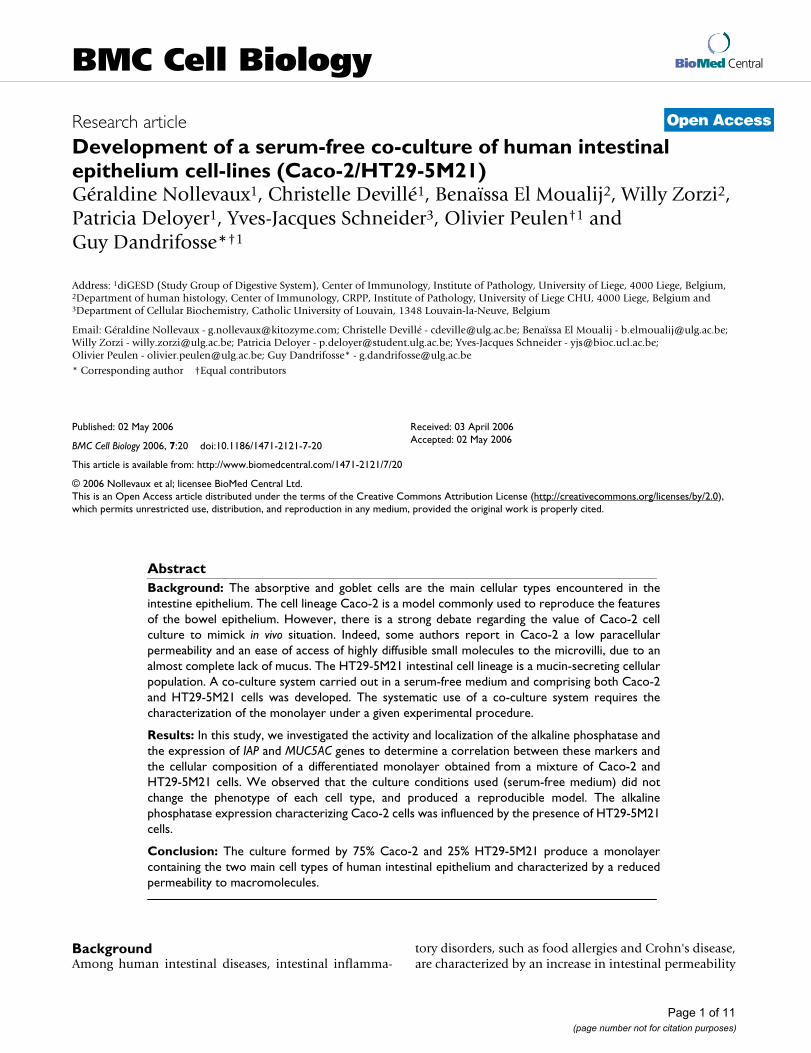
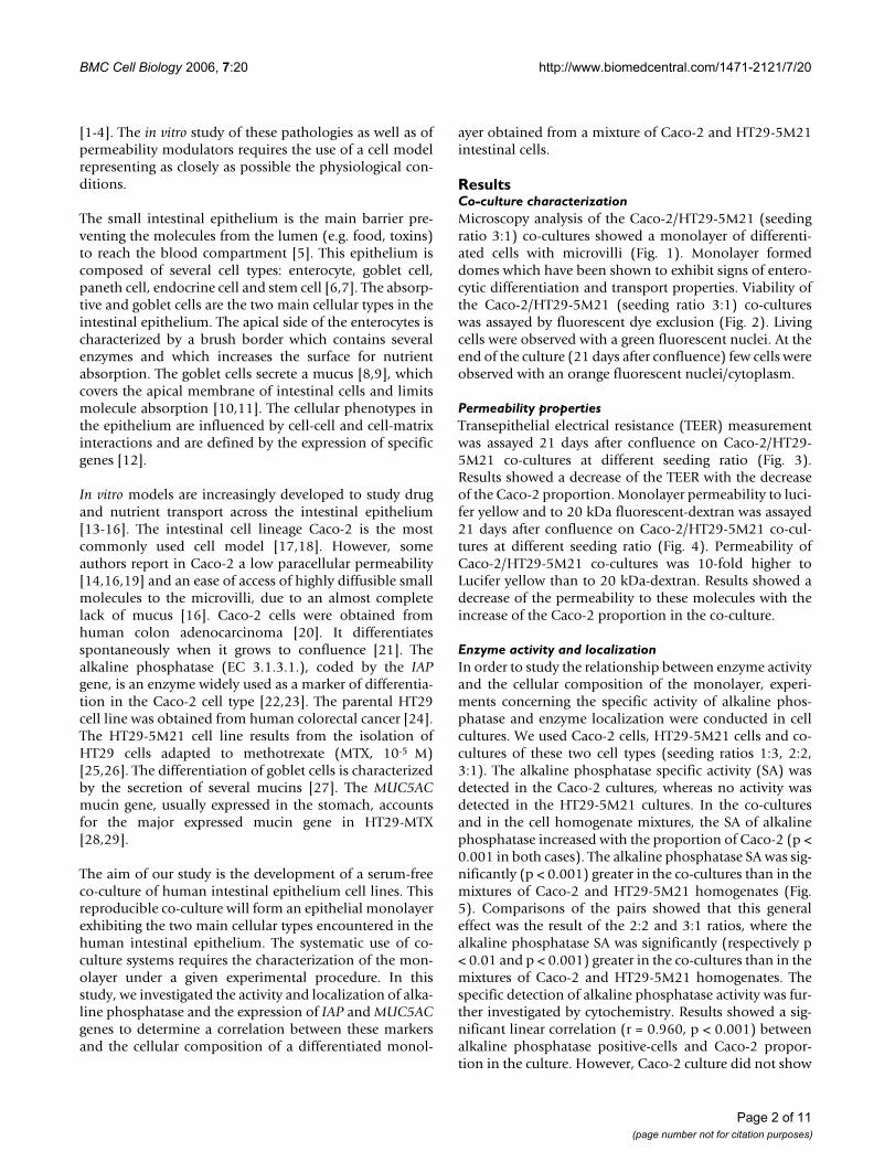
ResultsCo-culture characterizationMicroscopy analysis of the Caco-2/HT29-5M21 (seedingratio 3:1) co-cultures showed a monolayer of differenti-ated cells with microvilli (Fig. 1). Monolayer formeddomes which have been shown to exhibit signs of entero-cytic differentiation and transport properties. Viability ofthe Caco-2/HT29-5M21 (seeding ratio 3:1) co-cultureswas assayed by fluorescent dye exclusion (Fig. 2). Livingcells were observed with a green fluorescent nuclei. At theend of the culture (21 days after confluence) few cells wereobserved with an orange fluorescent nuclei/cytoplasm.
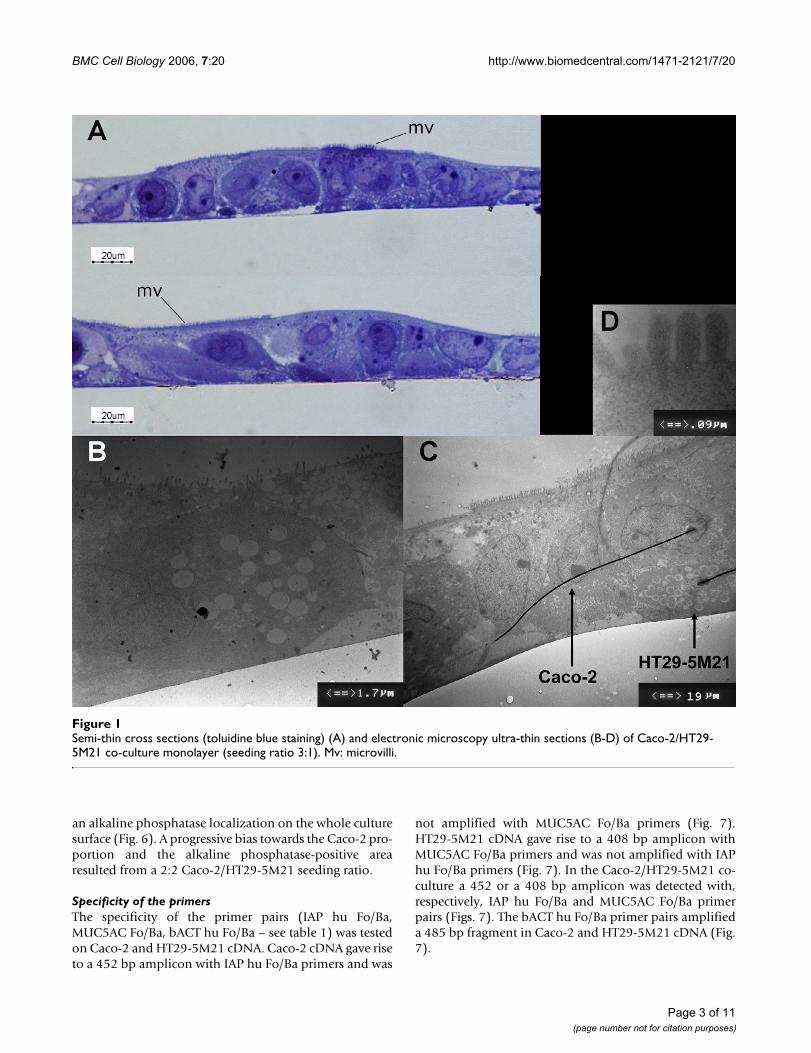
Permeability propertiesTransepithelial electrical resistance (TEER) measurementwas assayed 21 days after confluence on Caco-2/HT29-5M21 co-cultures at different seeding ratio (Fig. 3).Results showed a decrease of the TEER with the decreaseof the Caco-2 proportion. Monolayer permeability to luci-fer yellow and to 20 kDa fluorescent-dextran was assayed21 days after confluence on Caco-2/HT29-5M21 co-cul-tures at different seeding ratio (Fig. 4). Permeability ofCaco-2/HT29-5M21 co-cultures was 10-fold higher toLucifer yellow than to 20 kDa-dextran. Results showed adecrease of the permeability to these molecules with theincrease of the Caco-2 proportion in the co-culture.
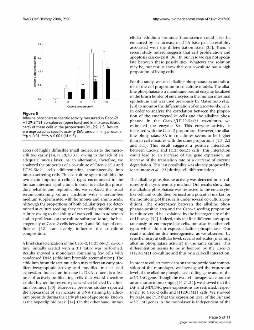
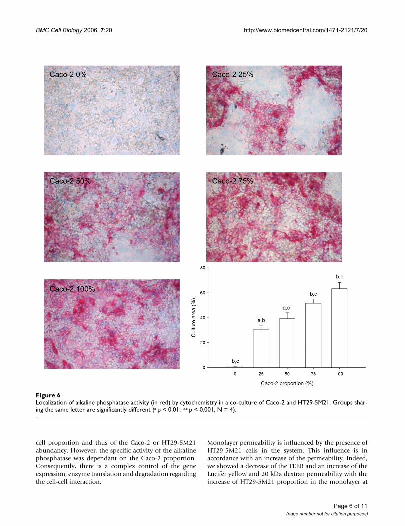
Enzyme activity and localizationIn order to study the relationship between enzyme activityand the cellular composition of the monolayer, experi-ments concerning the specific activity of alkaline phos-phatase and enzyme localization were conducted in cellcultures. We used Caco-2 cells, HT29-5M21 cells and co-cultures of these two cell types (seeding ratios 1:3, 2:2,3:1). The alkaline phosphatase specific activity (SA) wasdetected in the Caco-2 cultures, whereas no activity wasdetected in the HT29-5M21 cultures. In the co-culturesand in the cell homogenate mixtures, the SA of alkalinephosphatase increased with the proportion of Caco-2 (p <0.001 in both cases). The alkaline phosphatase SA was sig-nificantly (p < 0.001) greater in the co-cultures than in themixtures of Caco-2 and HT29-5M21 homogenates (Fig.5). Comparisons of the pairs showed that this generaleffect was the result of the 2:2 and 3:1 ratios, where thealkaline phosphatase SA was significantly (respectively p< 0.01 and p < 0.001) greater in the co-cultures than in themixtures of Caco-2 and HT29-5M21 homogenates. Thespecific detection of alkaline phosphatase activity was fur-ther investigated by cytochemistry. Results showed a sig-nificant linear correlation (r = 0.960, p < 0.001) betweenalkaline phosphatase positive-cells and Caco-2 propor-tion in the culture. However, Caco-2 culture did not show
Page 2 of 11(page number not for citation purposes)
BMC Cell Biology 2006, 7:20 http://www.biomedcentral.com/1471-2121/7/20
an alkaline phosphatase localization on the whole culturesurface (Fig. 6). A progressive bias towards the Caco-2 pro-portion and the alkaline phosphatase-positive arearesulted from a 2:2 Caco-2/HT29-5M21 seeding ratio.
Specificity of the primersThe specificity of the primer pairs (IAP hu Fo/Ba,MUC5AC Fo/Ba, bACT hu Fo/Ba – see table 1) was testedon Caco-2 and HT29-5M21 cDNA. Caco-2 cDNA gave riseto a 452 bp amplicon with IAP hu Fo/Ba primers and was
not amplified with MUC5AC Fo/Ba primers (Fig. 7).HT29-5M21 cDNA gave rise to a 408 bp amplicon withMUC5AC Fo/Ba primers and was not amplified with IAPhu Fo/Ba primers (Fig. 7). In the Caco-2/HT29-5M21 co-culture a 452 or a 408 bp amplicon was detected with,respectively, IAP hu Fo/Ba and MUC5AC Fo/Ba primerpairs (Figs. 7). The bACT hu Fo/Ba primer pairs amplifieda 485 bp fragment in Caco-2 and HT29-5M21 cDNA (Fig.7).
Semi-thin cross sections (toluidine blue staining) (A) and electronic microscopy ultra-thin sections (B-D) of Caco-2/HT29-5M21 co-culture monolayer (seeding ratio 3:1)Figure 1Semi-thin cross sections (toluidine blue staining) (A) and electronic microscopy ultra-thin sections (B-D) of Caco-2/HT29-5M21 co-culture monolayer (seeding ratio 3:1). Mv: microvilli.
Page 3 of 11(page number not for citation purposes)
BMC Cell Biology 2006, 7:20 http://www.biomedcentral.com/1471-2121/7/20
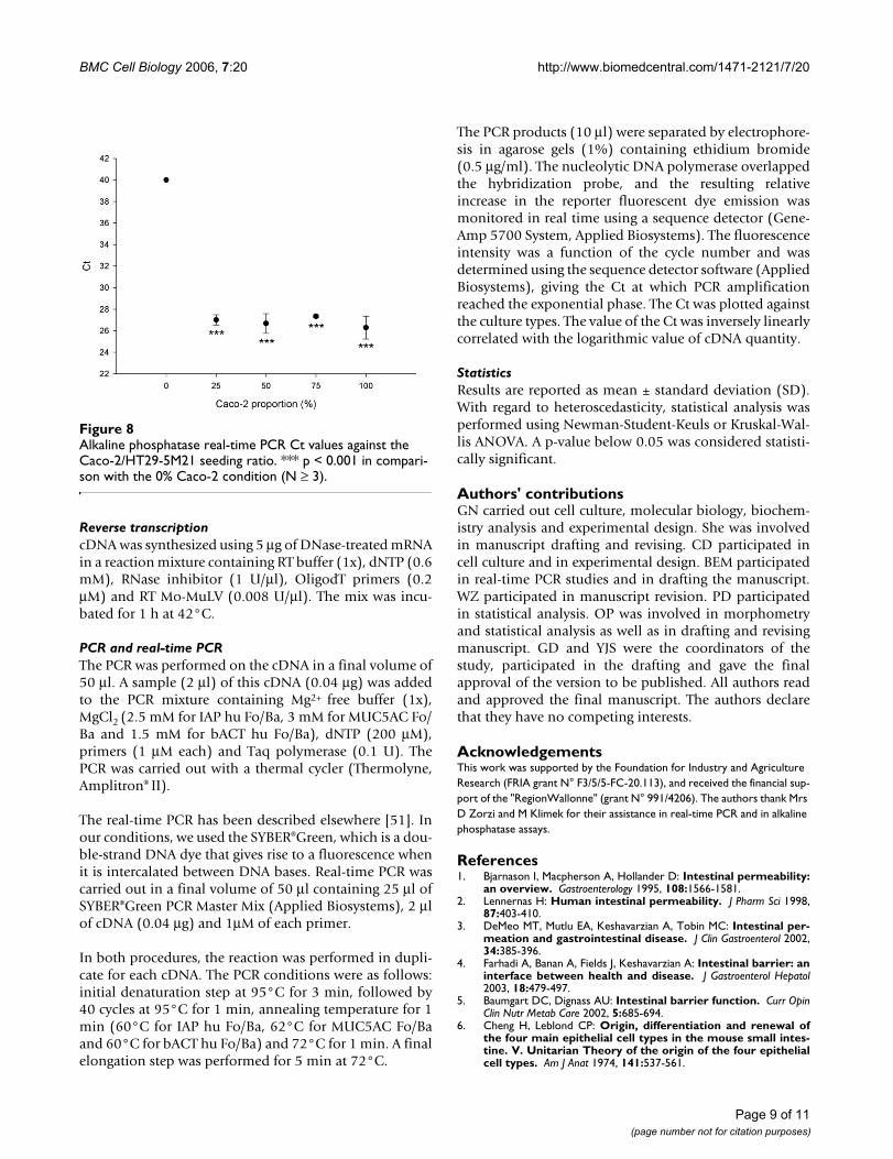
Expression of IAP and MUC5AC genesThe expression level of IAP and MUC5AC genes was deter-mined by real-time PCR in the Caco-2 and HT29-5M21co-cultures. After 40 PCR cycles, the fluorescence, corre-sponding to SYBER Green® binding, was recorded andplotted against the cycle number (data not shown).
Our results showed that the threshold cycle number (Ct)value, corresponding to the amount of IAP cDNA, is thesame in the Caco-2 culture and in co-cultures at different
seeding Caco-2/HT29-5M21 proportions (1:3, 2:2 and3:1; Fig. 8). With regard to the HT29-5M21 cDNA, noamplification product was detected until a Ct value corre-sponding to the value of the negative control wasobtained without template. The Ct value did not differ sig-nificantly with the proportion of Caco-2 cells in the cul-ture. The same results (Ct ~ 27) were obtained forMUC5AC (data not shown).
DiscussionTo understand the cellular events leading to a leaky gut inpathologies such as inflammatory bowel diseases, an invitro approach using cell models is required. The intestinalcell lineage Caco-2 is the cell model most commonly usedto reproduce the features of the human small intestineepithelium [17,18]. Nevertheless, the Caco-2 cell model isdifferent in some respects to the human epithelium. Forexample, it has a low paracellular permeability and allows
Lucifer yellow (452 Da) and dextran (20 kDa) fluorescence in the basolateral pole compartment of Caco-2/HT29-5M21 co-culture monolayer 21 days after the confluenceFigure 4Lucifer yellow (452 Da) and dextran (20 kDa) fluorescence in the basolateral pole compartment of Caco-2/HT29-5M21 co-culture monolayer 21 days after the confluence. Basolateral pole compartment samples were analysed 30 to 180 minutes after permeability tracer addition into the apical compart-ment. Results are expressed as % of total fluorescence.
Fluorescent dye exclusion in Caco-2/HT29-5M21 co-culture monolayer (seeding ratio 3:1) 21 days after the confluenceFigure 2Fluorescent dye exclusion in Caco-2/HT29-5M21 co-culture monolayer (seeding ratio 3:1) 21 days after the confluence.
Transepithelial Electrical resistance of Caco-2/HT29-5M21 co-culture monolayer 21 days after the confluenceFigure 3Transepithelial Electrical resistance of Caco-2/HT29-5M21 co-culture monolayer 21 days after the confluence. Results are expressed as Ω.cm2. Groups sharing the same letter are significantly different (a p < 0.001, N ≥ 4).
Page 4 of 11(page number not for citation purposes)
BMC Cell Biology 2006, 7:20 http://www.biomedcentral.com/1471-2121/7/20
access of highly diffusible small molecules to the micro-villi too easily [14,17,19,30,31], owing to the lack of anadequate mucus layer. As an alternative, therefore, weanalyzed the properties of a co-culture of Caco-2 cells andHT29-5M21 cells differentiating spontaneously intomucus-secreting cells. This co-culture system exhibits thetwo main important cellular types encountered in thehuman intestinal epithelium. In order to make this proce-dure reliable and reproducible, we replaced the usualserum-containing culture medium with a serum-freemedium supplemented with hormones and amino acids.Although the proportions of both cellular types are deter-mined at culture seeding, they can rapidly modify duringculture owing to the ability of each cell line to adhere toand to proliferate on the culture substrate. More, the het-erogeneity of Caco-2 cells between 0 and 30 days of con-fluence [32] can deeply influence the co-culturecomposition.
A brief characterization of the Caco-2/HT29-5M21 co-cul-ture, initially seeded with a 3:1 ratio, was performed.Results showed a monolayer containing few cells withcondensed DNA (ethidium bromide accumulation). Theethidium bromide accumulation may reflect an early pro-liferative/apoptotic activity and modified nucleic acidexpression. Indeed, an increase in DNA content is a fea-ture of actively-proliferating cells that would thereforeexhibit higher fluorescence peaks when labeled by ethid-ium bromide [33]. Moreover, previous studies reportedthe appearance of an increase in DNA staining by ethid-ium bromide during the early phases of apoptosis, knownas the hyperdiploid peak, [34]. On the other hand, intrac-
ellular ethidium bromide fluorescence could also beenhanced by an increase in DNA base pair accessibilityassociated with the differentiation state [35]. Then, arecent study indeed suggests that cell proliferation andapoptosis can co-exist [36]. In our case we can not specu-late between these possibilities. Whatever the solutionmay be, our results show that our co-culture has a highproportion of living cells.
For this study, we used alkaline phosphatase as an indica-tor of the cell proportion in co-culture models. The alka-line phosphatase is a membrane-bound enzyme localizedin the brush border of enterocytes in the human intestinalepithelium and was used previously by Matsumoto et al.[23] to monitor the differentiation of enterocyte-like cells.In order to analyze the correlation between the propor-tion of the enterocyte-like cells and the alkaline phos-phatase in the Caco-2/HT29-5M21 co-cultures, weestimated the enzyme SA. This enzyme activity isincreased with the Caco-2 proportion. However, the alka-line phosphatase SA in co-cultures seems to be higherthan in cell mixtures with the same proportions (1:3, 2:2and 3:1). This result suggests a positive interactionbetween Caco-2 and HT29-5M21 cells. This interactioncould lead to an increase of the gene expression, anincrease of the translation rate or a decrease of enzymedegradation. This last possibility was already proposed byMatsumoto et al. [23] during cell differentiation.
The alkaline phosphatase activity was detected in co-cul-tures by the cytochemistry method. Our results show thatthe alkaline phosphatase was restricted to the enterocyte-like cell and could then be used as a potential marker forthe monitoring of these cells under several co-culture con-ditions. The discrepancy between the alkaline phos-phatase-positive area and the Caco-2 seeding proportionin culture could be explained by the heterogeneity of thecell lineage [32]. Indeed, this cell line differentiates spon-taneously in enterocyte-like cells, but also in other celltypes which do not express alkaline phosphatase. Ourresults underline this heterogeneity, as we observed, bycytochemistry at cellular level, several red-scales (meaningalkaline phosphatase activity) in the same culture. Thisdifferentiation seems to be influenced by the Caco-2/HT29-5M21 co-culture and thus by a cell-cell interaction.
In order to collect more data on the proportionate compo-sition of the monolayer, we investigated the expressionlevel of the alkaline phosphatase coding gene and of theMUC5AC gene. Though the two cell lineages were both ofan adenocarcinoma origin [16,21,24], we showed that theIAP and MUC5AC gene expressions are restricted, respec-tively, to Caco-2 cells and HT29-5M21 cells. We showedby real-time PCR that the expression level of the IAP andMUC5AC genes in the monolayer is independent of the
Alkaline phosphatase specific activity measured in Caco-2/HT29-5M21 co-cultures (open bars) and in mixtures (black bars) of these cells in the proportions 3:1, 2:2, 1:3Figure 5Alkaline phosphatase specific activity measured in Caco-2/HT29-5M21 co-cultures (open bars) and in mixtures (black bars) of these cells in the proportions 3:1, 2:2, 1:3. Results are expressed as specific activity (SA, μmol/min.mg protein). **p < 0.01, ***p < 0.001 (N = 3).
Page 5 of 11(page number not for citation purposes)
BMC Cell Biology 2006, 7:20 http://www.biomedcentral.com/1471-2121/7/20
cell proportion and thus of the Caco-2 or HT29-5M21abundancy. However, the specific activity of the alkalinephosphatase was dependant on the Caco-2 proportion.Consequently, there is a complex control of the geneexpression, enzyme translation and degradation regardingthe cell-cell interaction.
Monolayer permeability is influenced by the presence ofHT29-5M21 cells in the system. This influence is inaccordance with an increase of the permeability. Indeed,we showed a decrease of the TEER and an increase of theLucifer yellow and 20 kDa dextran permeability with theincrease of HT29-5M21 proportion in the monolayer at
Localization of alkaline phosphatase activity (in red) by cytochemistry in a co-culture of Caco-2 and HT29-5M21Figure 6Localization of alkaline phosphatase activity (in red) by cytochemistry in a co-culture of Caco-2 and HT29-5M21. Groups shar-ing the same letter are significantly different (a p < 0.01; b,c p < 0.001, N = 4).
Page 6 of 11(page number not for citation purposes)
BMC Cell Biology 2006, 7:20 http://www.biomedcentral.com/1471-2121/7/20
seeding. The cell ratio is established at seeding and isprobably not indicative of the situation when permeabil-ity is measured. However, HT29-5M21 cells seem toremain in sufficient proportion in the culture to increasecell permeability. The range of molecular masses (Luciferyellow 452 Da & dextran 20 kDa) used in this study toexplore monolayer permeability allow to characterizepara-cellular permeability [37,38]. Lucifer yellow is notabsorbed by epithelial cell but is able to pass through celljunctions as mannitol does. Dextrans are largely used asmacromolecule permeability tracer.
ConclusionThe phosphatase alkaline, a well-established marker forenterocyte-like cells, could be proposed, in combinationwith MUC5AC, as a new marker of great interest for mon-itoring the cellular composition of the co-culture. We arecurrently developing an immuno-labelling system toquantify the level of this protein.
On the basis of the enterocyte/goblet cell ratio observed invivo [39,40], the cell proportion 3:1 allows us to decreasethe permeability to macromolecules who becomes closerto the situation observed in a human intestinal epithe-lium.
Permeability studies, using this co-culture, indicate thatCaco-2/HT29-5M21 co-culture is more permissive thanCaco-2 cell culture regarding to para-cellular permeabil-ity. This characteristic is probably in accordance with thein vivo permeability. We can expect to use the model tostudy some human diseases involving permeability alter-ations as coeliac disease.
MethodsChemicalsAll chemicals were purchased from Sigma-Aldrich (Poole,Dorset, UK) and from VWR (Brussels, Belgium). Cell cul-ture media were obtained from Gibco BRL (Merelbeke,Belgium) and from Sigma-Aldrich. The molecular biology
reagents were obtained from Promega (Madison, USA).Lucifer yellow and 20 kDa-dextran were obtained fromSigma-Aldrich.
Cell lines and culture conditionsThe HT29-5M21 cell line, derived from the HT29-MTX 10-
5M cell line [27], was obtained at passage 9 from Dr. T.Lesuffleur (INSERM U505, Villejuif, France). Caco-2 cellswere obtained at passage 22 from Cambrex Bio Science(Verviers, Belgium). These cells were progressivelyadapted (until passage 30 for HT29-5M21 and passage 40for Caco-2) to a serum-free medium: the basal definedmedium (BDM) [41] supplemented with a mixture ofnon-essential amino acids and growth factors [42]. Ini-tially, Caco-2 and HT29-5M21 cells were cultured in Dul-becco's Modified Eagle Medium (DMEM) containingglucose (4.15 mg/l) buffered with HEPES (25 mM) con-taining fetal calf serum (10%) and antibiotics (1%: peni-cillin [1 U/ml], streptomycin [1 μg/ml] and amphotericinas fungizone® [2.5 ng/ml]). Then, these cells were progres-sively sub-cultured in BDM containing glucose (4.15 mg/l) supplemented with non-essential amino acids andgrowth factors [42]. Cells were used at passages 30 (HT29-5M21) and 40 (Caco-2) to avoid dedifferentiation [43].The cells were seeded at 4 × 10⁴ cells/cm2 in plastic flasks(Greiner labortechnik SA, Wemmel, Belgium) and cul-tured at 37°C in a CO2-air (5:95) atmosphere (HeraeusEK incubator). The culture medium was changed everyday.
Co-culturesFive types of culture cells were produced: Caco-2 cellsalone, HT29-5M21 cells alone and co-cultures of thesecells in various seeding ratios (1:3, 2:2 and 3:1). These cul-tures (4 × 10⁴ cells/cm2 per flask) were maintained inBDM supplemented with non-essential amino acids, amixture of growth factors [42] and antibiotics (see above).The medium was changed every two days. Co-cultureswere confluent 7 days after the seeding. The Caco-2 andHT29-5M21 cells were maintained in culture during 21
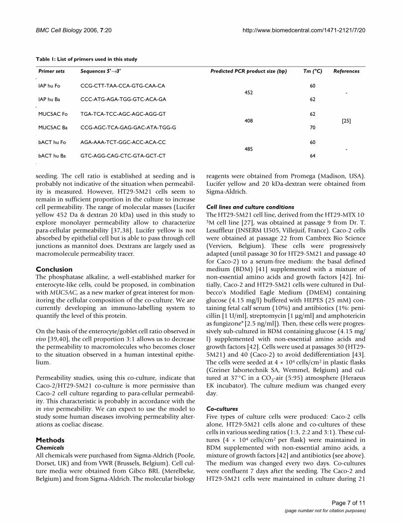
Table 1: List of primers used in this study
Primer sets Sequences 5'→3' Predicted PCR product size (bp) Tm (°C) References
IAP hu Fo CCG-CTT-TAA-CCA-GTG-CAA-CA 60452 -
IAP hu Ba CCC-ATG-AGA-TGG-GTC-ACA-GA 62
MUC5AC Fo TGA-TCA-TCC-AGC-AGC-AGG-GT 62408 [25]
MUC5AC Ba CCG-AGC-TCA-GAG-GAC-ATA-TGG-G 70
bACT hu Fo AGA-AAA-TCT-GGC-ACC-ACA-CC 60485 -
bACT hu Ba GTC-AGG-CAG-CTC-GTA-GCT-CT 64
Page 7 of 11(page number not for citation purposes)

BMC Cell Biology 2006, 7:20 http://www.biomedcentral.com/1471-2121/7/20
days of differentiation after confluence. Under these con-ditions, the maximum expression of intestinal differentia-tion-specific markers was achieved. For the enterocytes,the differentiation marker was the alkaline phosphataseactivity associated with the microvilli [17,30,31]. Withregard to the goblet cells, the differentiation marker wasthe mucin synthesis [44]. Cells were used for mRNAextraction, enzyme assays and localization. Cells in co-culture were briefly characterized by phase contrast micro-scopy, light microscopy (semi-thin cross sections stainedwith toluidine blue), electronic microscopy and vital dye(ethidium bromide/acridine orange) exclusion [45].
Permeability assaysPermeability analyses were performed on cells culturedon 0.4 μm polyethylene terephtalate membrane coatedwith collagen as described above. Transepithelial electri-cal resistance measurement was measured in Hank'sbuffer [46,47]. Lucifer yellow and fluorescent dextranwere measured in the baso-lateral pole compartment witha fluorimeter (excitation 430 nm – emission 540 nm).
Enzyme assaysEnzyme activity assays were performed on cell homoge-nates. Just before use, the monolayer was washed withPBS buffer (pH 7.2) and 1 ml of water was added. Cellswere collected by scraping and were disrupted by ultra-sonication (3 times for 20 sec each; ultrasonicer MSEPE818). Enzyme activity was assayed on co-culturehomogenates and on Caco-2 and HT29-5M21 homoge-nate mixtures in 1:3, 2:2 and 3:1 ratios. Alkaline phos-phatase activity was assayed according to the methoddescribed by Garen and Levinthal [48], with p-nitrophe-nolphosphate as substrate. Results were expressed as mil-liunit mg-1 of protein (specific activity = SA). One unit wasdefined as the activity equal to one μmol of substratehydrolyzed per min at 37°C. Proteins were assayed by themethod described by Bradford [49]. Phosphatase alkalineactivity was localized on cell culture by cytochemistryusing the fast red substrate (naphtol combined to fast red;Roche Diagnostics GmbH, Penzberg, Germany). Labelledcell-culture pictures were analyzed using ImageJ 1.33usoftware (Wayne Rasband, NIH, USA). Images were con-verted in 256 greyscale files, then a threshold was definedin order to include the red channel in the selection. Thearea of the selection was measured as a percentage of thepicture surface.
PCR primersAll the PCR primers used in this study were obtained fromEurogentec (Seraing, Belgium). Three pairs of primerswere chosen: IAP hu Fo/Ba was a specific of an alkalinephosphatase gene; MUC5AC Fo/Ba was a specific of theMUC5AC mucin gene [29]; and bACT hu Fo/Ba was a spe-cific of the cytosolic beta actin coding-gene. The primer3program [50] was used to design the IAP hu Fo/Ba andbACT hu Fo/Ba primers. The sequences of the primersused in this study are presented in Table I.
RNA isolationTotal mRNA was isolated using a kit manufactured byPromega (RNagents® total RNA isolation system) fromconfluent monolayer cells. RNA integrity was checked byelectrophoresis in agarose gel (1%) containing ethidiumbromide (0.5 μg/ml). RNA concentration was estimatedby spectrophotometry at 260 nm.
IAP, MUC5AC and beta ACTIN PCR on mRNA isolated from cell culturesFigure 7IAP, MUC5AC and beta ACTIN PCR on mRNA isolated from cell cultures. Lanes: M: 100 bp DNA ladder. Analysis was repeated three times.
Page 8 of 11(page number not for citation purposes)
BMC Cell Biology 2006, 7:20 http://www.biomedcentral.com/1471-2121/7/20
Reverse transcriptioncDNA was synthesized using 5 μg of DNase-treated mRNAin a reaction mixture containing RT buffer (1x), dNTP (0.6mM), RNase inhibitor (1 U/μl), OligodT primers (0.2μM) and RT Mo-MuLV (0.008 U/μl). The mix was incu-bated for 1 h at 42°C.
PCR and real-time PCRThe PCR was performed on the cDNA in a final volume of50 μl. A sample (2 μl) of this cDNA (0.04 μg) was addedto the PCR mixture containing Mg2+ free buffer (1x),MgCl2 (2.5 mM for IAP hu Fo/Ba, 3 mM for MUC5AC Fo/Ba and 1.5 mM for bACT hu Fo/Ba), dNTP (200 μM),primers (1 μM each) and Taq polymerase (0.1 U). ThePCR was carried out with a thermal cycler (Thermolyne,Amplitron® II).
The real-time PCR has been described elsewhere [51]. Inour conditions, we used the SYBER®Green, which is a dou-ble-strand DNA dye that gives rise to a fluorescence whenit is intercalated between DNA bases. Real-time PCR wascarried out in a final volume of 50 μl containing 25 μl ofSYBER®Green PCR Master Mix (Applied Biosystems), 2 μlof cDNA (0.04 μg) and 1μM of each primer.
In both procedures, the reaction was performed in dupli-cate for each cDNA. The PCR conditions were as follows:initial denaturation step at 95°C for 3 min, followed by40 cycles at 95°C for 1 min, annealing temperature for 1min (60°C for IAP hu Fo/Ba, 62°C for MUC5AC Fo/Baand 60°C for bACT hu Fo/Ba) and 72°C for 1 min. A finalelongation step was performed for 5 min at 72°C.
The PCR products (10 μl) were separated by electrophore-sis in agarose gels (1%) containing ethidium bromide(0.5 μg/ml). The nucleolytic DNA polymerase overlappedthe hybridization probe, and the resulting relativeincrease in the reporter fluorescent dye emission wasmonitored in real time using a sequence detector (Gene-Amp 5700 System, Applied Biosystems). The fluorescenceintensity was a function of the cycle number and wasdetermined using the sequence detector software (AppliedBiosystems), giving the Ct at which PCR amplificationreached the exponential phase. The Ct was plotted againstthe culture types. The value of the Ct was inversely linearlycorrelated with the logarithmic value of cDNA quantity.
StatisticsResults are reported as mean ± standard deviation (SD).With regard to heteroscedasticity, statistical analysis wasperformed using Newman-Student-Keuls or Kruskal-Wal-lis ANOVA. A p-value below 0.05 was considered statisti-cally significant.
Authors' contributionsGN carried out cell culture, molecular biology, biochem-istry analysis and experimental design. She was involvedin manuscript drafting and revising. CD participated incell culture and in experimental design. BEM participatedin real-time PCR studies and in drafting the manuscript.WZ participated in manuscript revision. PD participatedin statistical analysis. OP was involved in morphometryand statistical analysis as well as in drafting and revisingmanuscript. GD and YJS were the coordinators of thestudy, participated in the drafting and gave the finalapproval of the version to be published. All authors readand approved the final manuscript. The authors declarethat they have no competing interests.
AcknowledgementsThis work was supported by the Foundation for Industry and Agriculture Research (FRIA grant N° F3/5/5-FC-20.113), and received the financial sup-port of the "RegionWallonne" (grant N° 991/4206). The authors thank Mrs D Zorzi and M Klimek for their assistance in real-time PCR and in alkaline phosphatase assays.
References1. Bjarnason I, Macpherson A, Hollander D: Intestinal permeability:
an overview. Gastroenterology 1995, 108:1566-1581.2. Lennernas H: Human intestinal permeability. J Pharm Sci 1998,
87:403-410.3. DeMeo MT, Mutlu EA, Keshavarzian A, Tobin MC: Intestinal per-
meation and gastrointestinal disease. J Clin Gastroenterol 2002,34:385-396.
4. Farhadi A, Banan A, Fields J, Keshavarzian A: Intestinal barrier: aninterface between health and disease. J Gastroenterol Hepatol2003, 18:479-497.
5. Baumgart DC, Dignass AU: Intestinal barrier function. Curr OpinClin Nutr Metab Care 2002, 5:685-694.
6. Cheng H, Leblond CP: Origin, differentiation and renewal ofthe four main epithelial cell types in the mouse small intes-tine. V. Unitarian Theory of the origin of the four epithelialcell types. Am J Anat 1974, 141:537-561.
Alkaline phosphatase real-time PCR Ct values against the Caco-2/HT29-5M21 seeding ratioFigure 8Alkaline phosphatase real-time PCR Ct values against the Caco-2/HT29-5M21 seeding ratio. *** p < 0.001 in compari-son with the 0% Caco-2 condition (N ≥ 3).
Page 9 of 11(page number not for citation purposes)
BMC Cell Biology 2006, 7:20 http://www.biomedcentral.com/1471-2121/7/20
7. Potten CS: Stem cells in gastrointestinal epithelium: num-bers, characteristics and death. Philos Trans R Soc Lond B Biol Sci1998, 353:821-830.
8. Moe H: Mucus-producing goblet cells of the small intestine.Nature 1953, 172:309.
9. Bierring F: Electron microscopic observations on the mucusproduction in human and rat intestinal goblet cells. ActaPathol Microbiol Scand 1962, 54:241-252.
10. Allen JD, Martin GP, Marriott C, Hassan I, Williamson I: Drug trans-port across a novel mucin secreting cell model: Comparisonwith the CaCo-2 cell system [abstract]. J Pharm Pharmacol1991, 43:63P.
11. Meaney C, O'Driscoll C: Mucus as a barrier to the permeabilityof hydrophilic and lipophilic compounds in the absence andpresence of sodium taurocholate micellar systems using cellculture models. Eur J Pharm Sci 1999, 8:167-175.
12. Simon-Assmann P, Kedinger M, De Arcangelis A, Rousseau V, Simo P:Extracellular matrix components in intestinal development.Experientia 1995, 51:883-900.
13. Wikman-Larhed A, Artursson P: Co-cultures of human intestinalgoblet (HT29-H) and absorptive (Caco-2) cells for studies ofdrug and peptide absorption. Eur J Pharm Sci 1995, 3:171-193.
14. Walter E, Janich S, Roessler BJ, Hilfinger JM, Amidon GL: HT29-MTX/Caco-2 cocultures as an in vitro model for the intesti-nal epithelium: in vitro-in vivo correlation with permeabilitydata from rats and humans. J Pharm Sci 1996, 85:1070-1076.
15. Hilgendorf C, Spahn-Langguth H, Regardh CG, Lipka E, Amidon GL,Langguth P: Caco-2 versus Caco-2/HT29-MTX co-cultured celllines: permeabilities via diffusion, inside- and outside-directed carrier-mediated transport. J Pharm Sci 2000,89:63-75.
16. Artursson P, Palm K, Luthman K: Caco-2 monolayers in experi-mental and theoretical predictions of drug transport. AdvDrug Deliv Rev 2001, 46:27-43.
17. Meunier V, Bourrie M, Berger Y, Fabre G: The human intestinalepithelial cell line Caco-2; pharmacological and pharmacok-inetic applications. Cell Biol Toxicol 1995, 11:187-194.
18. Ingels FM, Augustijns PF: Biological, pharmaceutical, and analyt-ical considerations with respect to the transport media usedin the absorption screening system, Caco-2. J Pharm Sci 2003,92:1545-1558.
19. Menon RM, Barr WH: Comparison of ceftibuten transportacross Caco-2 cells and rat jejunum mounted on modifiedUssing chambers. Biopharm Drug Dispos 2003, 24:299-308.
20. Pinto M, Robine-Léon S, Appay MD, Kedinger M, Triadou N, Dus-saulx E, Lacroix B, Simon-Assmann P, Haffen K, Fogh J, Zweibaum A:Enterocyte-like differentiation and polarization of thehuman colon carcinoma cell line Caco-2 in culture. Biol Cell1983, 47:323-330.
21. Rousset M: The human colon carcinoma cell lines HT-29 andCaco-2: two in vitro models for the study of intestinal differ-entiation. Biochimie 1986, 68:1035-1040.
22. Chantret I, Barbat A, Dussaulx E, Brattain MG, Zweibaum A: Epithe-lial polarity, villin expression, and enterocytic differentiationof cultured human colon carcinoma cells: a survey of twentycell lines. Cancer Res 1988, 48:1936-1942.
23. Matsumoto H, Erickson RH, Gum JR, Yoshioka M, Gum E, Kim YS:Biosynthesis of alkaline phosphatase during differentiation ofthe human colon cancer cell line Caco-2. Gastroenterology 1990,98:1199-1207.
24. Fogh J, Trempe G: New human tumor cell lines. In Human TumorCells in vitro Edited by: Fogh J. New-York: Plenum Publishing Corp;1975:115-141.
25. Lesuffleur T, Barbat A, Luccioni C, Beaumatin J, Clair M, KornowskiA, Dussaulx E, Dutrillaux B, Zweibaum A: Dihydrofolate reduct-ase gene amplification-associated shift of differentiation inmethotrexate-adapted HT-29 cells. J Cell Biol 1991,115:1409-1418.
26. Wils P, Warnery A, Phung-Ba V, Scherman D: Differentiated intes-tinal epithelial cell lines as in vitro models for predicting theintestinal absorption of drugs. Cell Biol Toxicol 1994, 10:393-397.
27. Lesuffleur T, Porchet N, Aubert JP, Swallow D, Gum JR, Kim YS, RealFX, Zweibaum A: Differential expression of the human mucingenes MUC1 to MUC5 in relation to growth and differentia-tion of different mucus-secreting HT-29 cell subpopulations.J Cell Sci 1993, 106:771-783.
28. Fergie N, Guo L, Sithole J, Pearson JP, Birchall JP: Influence of pred-nisolone on the secretion of mucin from the HT29-MTX cellline. Clin Otolaryngol Allied Sci 2003, 28:39-42.
29. Gouyer V, Wiede A, Buisine MP, Dekeyser S, Moreau O, LesuffleurT, Hoffmann W, Huet G: Specific secretion of gel-formingmucins and TFF peptides in HT-29 cells of mucin-secretingphenotype. Biochim Biophys Acta 2001, 1539:71-84.
30. Quaroni A, Beaulieu JF: Cell dynamics and differentiation ofconditionally immortalized human intestinal epithelial cells.Gastroenterology 1997, 113:1198-1213.
31. Artursson P, Borchardt RT: Intestinal drug absorption andmetabolism in cell cultures: Caco-2 and beyond. Pharm Res1997, 14:1655-1658.
32. Vachon PH, Beaulieu JF: Transient mosaic patterns of morpho-logical and functional differentiation in the Caco-2 cell line.Gastroenterology 1992, 103:414-423.
33. Bonaly J, Mestre JC: Flow fluorometric study of DNA contentin nonproliferative Euglena gracilis cells and during prolifer-ation. Cytometry 1981, 2:35-38.
34. Ferlini C, Di CS, Rainaldi G, Malorni W, Samoggia P, Biselli R, Fat-torossi A: Flow cytometric analysis of the early phases ofapoptosis by cellular and nuclear techniques. Cytometry 1996,24:106-115.
35. Darzynkiewicz Z, Traganos F, Kapuscinski J, Staiano-Coico L, Mela-med MR: Accessibility of DNA in situ to various fluoro-chromes: relationship to chromatin changes duringerythroid differentiation of Friend leukemia cells. Cytometry1984, 5:355-363.
36. Nagy Z, Esiri MM: Neuronal cyclin expression in the hippocam-pus in temporal lobe epilepsy. Exp Neurol 1998, 150:240-247.
37. Konsoula R, Barile FA: Correlation of in vitro cytotoxicity withparacellular permeability in Caco-2 cells. Toxicol In Vitro 2005,19:675-684.
38. Satsu H, Yokoyama T, Ogawa N, Fujiwara-Hatano Y, Shimizu M:Effect of neuronal PC12 cells on the functional properties ofintestinal epithelial Caco-2 cells. Biosci Biotechnol Biochem 2003,67:1312-1318.
39. Cheng H, Leblond CP: Origin, differentiation and renewal ofthe four main epithelial cell types in the mouse small intes-tine. I. Columnar cell. Am J Anat 1974, 141:461-479.
40. Rubio CA, Lindholm J, Rodensjo M: Mapping intestinal metapla-sia by histochemistry and morphometry. Pathol Res Pract 1989,184:525-528.
41. Schneider YJ: Optimisation of hybridoma cell growth andmonoclonal antibody secretion in a chemically defined,serum- and protein-free culture medium. J Immunol Methods1989, 116:65-77.
42. Halleux C, Schneider YJ: Iron absorption by intestinal epithelialcells: 1. CaCo2 cells cultivated in serum-free medium, onpolyethyleneterephthalate microporous membranes, as anin vitro model. In Vitro Cell Dev Biol 1991, 27A:293-302.
43. Yu H, Cook TJ, Sinko PJ: Evidence for diminished functionalexpression of intestinal transporters in Caco-2 cell monolay-ers at high passages. Pharm Res 1997, 14:757-762.
44. Hennebicq-Reig S, Lesuffleur T, Capon C, De BC, Kim I, Moreau O,Richet C, Hemon B, Recchi MA, Maes E, Aubert JP, Real FX,Zweibaum A, Delannoy P, Degand P, Huet G: Permanent expo-sure of mucin-secreting HT-29 cells to benzyl-N-acetyl-alpha-D-galactosaminide induces abnormal O-glycosylationof mucins and inhibits constitutive and stimulated MUC5ACsecretion. Biochem J 1998, 334:283-295.
45. Foglieni C, Meoni C, Davalli AM: Fluorescent dyes for cell viabil-ity: an application on prefixed conditions. Histochem Cell Biol2001, 115:223-229.
46. Lu S, Gough AW, Bobrowski WF, Stewart BH: Transport proper-ties are not altered across Caco-2 cells with heightenedTEER despite underlying physiological and ultrastructuralchanges. J Pharm Sci 1996, 85:270-273.
47. Yamashita S, Furubayashi T, Kataoka M, Sakane T, Sezaki H, TokudaH: Optimized conditions for prediction of intestinal drug per-meability using Caco-2 cells. Eur J Pharm Sci 2000, 10:195-204.
48. Garen A, Levinthal C: A fine-structure genetic and chemicalstudy of the enzyme alkaline phosphatase of E. coli. I. Purifi-cation and characterization of alkaline phosphatase. BiochimBiophys Acta 1960, 38:470-483.
Page 10 of 11(page number not for citation purposes)
BMC Cell Biology 2006, 7:20 http://www.biomedcentral.com/1471-2121/7/20
Publish with BioMed Central and every scientist can read your work free of charge
"BioMed Central will be the most significant development for disseminating the results of biomedical research in our lifetime."
Sir Paul Nurse, Cancer Research UK
Your research papers will be:
available free of charge to the entire biomedical community
peer reviewed and published immediately upon acceptance
cited in PubMed and archived on PubMed Central
yours — you keep the copyright
Submit your manuscript here:http://www.biomedcentral.com/info/publishing_adv.asp
BioMedcentral
49. Bradford MM: A rapid and sensitive method for the quantita-tion of microgram quantities of protein utilizing the princi-ple of protein-dye binding. Anal Biochem 1976, 72:248-254.
50. Rozen S, Skaletsky H: Primer3 on the WWW for general usersand for biologist programmers. Methods Mol Biol 2000,132:365-386.
51. Latil A, Vidaud D, Valeri A, Fournier G, Vidaud M, Lidereau R, Cuss-enot O, Biache I: htert expression correlates with MYC over-expression in human prostate cancer. Int J Cancer 2000,89:172-176.
Page 11 of 11(page number not for citation purposes)