disposition mechanisms of raloxifene in the human intestinal caco-2 model
TRANSCRIPT

JPET #63925
1
Disposition Mechanisms of Raloxifene in the Human Intestinal Caco-2 Model
Eun Ju Jeong1, Huimin Lin1 and Ming Hu1, 2
JPET Fast Forward. Published on March 12, 2004 as DOI:10.1124/jpet.103.063925
Copyright 2004 by the American Society for Pharmacology and Experimental Therapeutics.
This article has not been copyedited and formatted. The final version may differ from this version.JPET Fast Forward. Published on March 12, 2004 as DOI: 10.1124/jpet.103.063925
at ASPE
T Journals on January 6, 2016
jpet.aspetjournals.orgD
ownloaded from

JPET #63925
2
1Department of Pharmaceutical Sciences, College of Pharmacy, Washington State University,
Pullman, Washington
2Address correspondence to:
Ming Hu, Ph.D.
Department of Pharmaceutical Sciences
College of Pharmacy
Washington State University
Pullman, WA 99164-6510
Tel: (509)-335-4184
Fax :( 509)-335-5092
Email: [email protected]
ABBREVIATIONS: HBSS, Hank’s balanced salt solution; MRP, multidrug resistance related
protein; OAT, organic anionic transporter; PAPS, 3’-phosphoadenosine 5’-phosphosulfate;
SERM, selective estrogen receptor modulator; UGT, UDP-glucuronosyltransferases; PBA,
secretory permeability; PAB, absorptive permeability
This article has not been copyedited and formatted. The final version may differ from this version.JPET Fast Forward. Published on March 12, 2004 as DOI: 10.1124/jpet.103.063925
at ASPE
T Journals on January 6, 2016
jpet.aspetjournals.orgD
ownloaded from

JPET #63925
3
ABSTRACT
The purpose of this study was to determine the mechanisms responsible for transport of
raloxifene and its hydrophilic conjugates. Human intestinal Caco-2 cell culture model and Caco-
2 cell lysate were used for the studies. The results indicated that absorptive permeability (PAB) of
raloxifene was lower than its secretory permeability (PBA). As the concentration increased, the
efflux ratio (PBA/PAB) decreased, but PAB increased. PAB was also increased in the presence of
verapamil and cyclosporine A, two p-glycoprotein inhbitors. Raloxifene was extensively
metabolized into sulfated and glucuronidated conjugates. The extent of metabolism or clearance
was decreased as the concentration increased from 3.4 µM (96%) to 30 µM (22%). MRP
inhibitors MK-571 and leukotriene C4 significantly decreased (maximal 80%) apical efflux of
both conjugates. They also significantly decreased (maximal 85%) basolateral efflux of
glucuronides but not sulfates. On the other hand, organic anion inhibitor (OAT) estrone sulfate
and estrone glucuronide significantly decreased (maximal 50%) the efflux of sulfate from both
sides, but had variable effects on glucuronide efflux. Inhibition of conjugate efflux with OAT
inhibitor estrone sulfate was concentration dependent, which resulted in increased transport of
intact raloxifene (maximal 90%). This increase in raloxifene transport was also observed in the
presence of another OAT inhibitor estrone glucuronide (70%). In conclusion, this is the first
report that inhibition of an efflux transporter responsible for the transport of metabolites can
result in increase in the transport of the intact compound. It also provides additional explanation
why raloxifene has low bioavailability but a long half-life.
This article has not been copyedited and formatted. The final version may differ from this version.JPET Fast Forward. Published on March 12, 2004 as DOI: 10.1124/jpet.103.063925
at ASPE
T Journals on January 6, 2016
jpet.aspetjournals.orgD
ownloaded from

JPET #63925
4
Raloxifene, a selective estrogen receptor modulator (SERM) (Duschek et al., 2003;
Colacurci et al., 2003), is used for the treatment of osteoporosis. Raloxifene blocks the adverse
effects of estrogen in breast tissues and uterine endometrium while mimicking the beneficial
estrogen effects on bone and lipid metabolism (MacGregor and Jordan, 1998; Scott et al., 1999).
Data from both animal and human studies demonstrated that raloxifene has minimal effects on
the uterus and caused no significant changes in the histological appearance of the endometrium
(Scott et al, 1999 and references therein). Raloxifene is being considered as a chemopreventive
agent for breast cancer (Delmas et al., 1997). Short-term clinical trials showed that raloxifene
did not increase the risk of breast cancer and long-term multicenter trials are currently ongoing
(Scott et al, 1999; Jordan and Morrow, 1999).
Raloxifene is reported by its manufacturer to be rapidly absorbed after oral administration,
but its absolute bioavailability is only 2%. This poor bioavailability is thought to be the result of
extensive phase II metabolism, since it is absorbed rapidly and approximately 60% of dose was
absorbed after oral dosing (Hochner-Celnikier, 1999). According to its manufacturer, raloxifene
exhibits linear pharmacokinetics with high within-subject variability (approximately 30% CV)
(Hochner-Celnikier, 1999). It undergoes extensive phase II biotransformation, primarily
glucuronidation, but was not metabolized by the cytochrome P450s (Eli Lilly, 1998). Raloxifene
has a plasma elimination half-life of approximately 27 hours, which is attributed to its reversible
systemic metabolism and significant enterohepatic cycling (Eli Lilly, 1998). The major human
intestinal enzymes responsible for its phase II conjugation have been shown to be UGT1A8 and
UGT1A10 (Kemp et al., 2002).
This article has not been copyedited and formatted. The final version may differ from this version.JPET Fast Forward. Published on March 12, 2004 as DOI: 10.1124/jpet.103.063925
at ASPE
T Journals on January 6, 2016
jpet.aspetjournals.orgD
ownloaded from

JPET #63925
5
Although a variety of pharmacokinetic information has been provided by its
manufacturer (as summarized previously), mechanisms responsible for absorption of the
raloxifene, and excretion of raloxifene conjugates have not been reported. Studies have not
established the dynamic relationship between absorption, metabolism and excretion of raloxifene
in the intestine, which deserves more attention since it is likely to be the main site of presystemic
clearance (Kemp et al., 2002). In the present research, we focused on the study of
absorption/excretion pathways responsible for intestinal disposition of raloxifene using the
human intestinal Caco-2 cell model using the cloned TC7 variant.
We chose Caco-2 TC7 cells, one of the cloned Caco-2 variants, for increased
homogeneity and stability of cell population (Ranaldi et al., 2003, Caro et al., 1995). TC variant
exhibits morphological characteristics similar to those of the parental Caco-2 cells (Gres et al.,
1998), and shows similar expression of MDR1 and MRP1-5 as human jejunal biopsies (Pfrunder
et al., 2003). It has been used by many investigators to study human intestinal disposition (Hu et
al., 1999; 2000; 2003; Sabolovic et al., 2000; Pontier et al., 2001; Bohets et al., 2001).
Therefore, the objective of the present study was to determine if enteric recycling and
other intestinal disposition events could help explain why raloxifene has a low bioavailability but
a long half-life. The process of enteric recycling, which was first proposed to explain the
disposition of flavonoids (Liu and Hu, 2002), is analogous to enterohepatic recycling except the
phase II conjugates are produced and excreted by the enterocytes instead of hepatocytes.
This article has not been copyedited and formatted. The final version may differ from this version.JPET Fast Forward. Published on March 12, 2004 as DOI: 10.1124/jpet.103.063925
at ASPE
T Journals on January 6, 2016
jpet.aspetjournals.orgD
ownloaded from

JPET #63925
6
MATERIALS AND METHODS
Materials
Cloned Caco-2 cells (TC7) were a kind gift from Dr. Moniqué Rousset (Institute National
de la Santé et de la Recherche zU178, Villejuit, France). Raloxifene was extracted from Evista®
tablet (Eli Lilly and Company, Indianapolis, IN) using 100% ethanol and concentration was then
verified by using raloxifene hydrochloride purchased from National Cancer Institute Chemical
Standard Repository managed by Midwest Research Institute (Kansas City, MO). β-
Glucuronidase, sulfatase, Hank’s balanced salt solution (HBSS; powder form), cyclosporin A,
verapamil, estrone glucuronide and estrone sulfate were purchased from Sigma (St. Louis, MO).
Leukotriene C4 and MK-571 were purchased from Biomol (Plymouth Meeting, PA). All other
materials were analytical grade or better and used as received.
Cell culture
Cell culture conditions for growing Caco-2 cells have been described previously (Liu and
Hu, 2002; Hu et al., 1994a,b; Chen et al., 2003). The seeding density was 100,000 cells/cm2 (4.2
cm2 per monolayer) and Dulbecco’s modified Eagle’s medium supplemented with 10% fetal
bovine serum was used as growth media. Quality control criteria were the same as described
previously (Hu et al., 1994a,b). Cell monolayers from 19 to 22 days past seeding were used for
the experiments.
Transport Experiments in the Caco-2 Cell Model
This article has not been copyedited and formatted. The final version may differ from this version.JPET Fast Forward. Published on March 12, 2004 as DOI: 10.1124/jpet.103.063925
at ASPE
T Journals on January 6, 2016
jpet.aspetjournals.orgD
ownloaded from

JPET #63925
7
The protocols for performing transport experiments were similar to those described
previously (Liu and Hu, 2002). Briefly, the cell monolayers were washed three times with 37ºC
HBSS (pH 7.4). The transepithelial electrical resistance values were measured and those with the
values less than 420 ohms x cm2 were discarded. Various concentrations (ranging from 1.5 to 30
µM) of raloxifene were loaded at the apical or basolateral side of the Caco-2 cell monolayer, and
the concentration of raloxifene and its metabolites at both sides were measured by HPLC.
Inhibitors, when used, were loaded at the apical, basolateral or both sides of the monolayer. Six
samples (650 µl each) were taken at different times (0, 1, 2, 3, 4 and 7 or 24 h after incubation)
from both donor and receiver side (total volume of each chamber is 2.5 ml) and same volume of
donor solution (containing raloxifene) or transport medium was replaced after each sampling.
Forty-five µl of internal standard solution (100 µM testosterone in methanol) was immediately
added to 200 µl of samples to stabilize them until analysis.
Extraction of Raloxifene from Caco-2 Cell Monolayers
After the transport experiments, cells were washed three times with ice-cold saline
solution and the polycarbonate membranes containing cells were removed from the insert (4.2
cm2, Nalge Nunc International, Rochester, NY). One ml of HBSS (pH 7.4) was added to each
membrane and the solution was frozen (in liquid nitrogen) and thawed (in 37ºC water bath) for
three times to disrupt cell membranes. After centrifugation at 13,000 rpm for 8 min, 0.5 ml of
supernatant was collected. Half ml of methanol was added to the remaining suspension and the
tubes were centrifuged again to extract the remaining raloxifene.
This article has not been copyedited and formatted. The final version may differ from this version.JPET Fast Forward. Published on March 12, 2004 as DOI: 10.1124/jpet.103.063925
at ASPE
T Journals on January 6, 2016
jpet.aspetjournals.orgD
ownloaded from

JPET #63925
8
Preparation of Caco-2 Cell Lysate
After six mature (19-22 days post-seeding) Caco-2 cell monolayers were washed twice
with 3 ml of 37ºC HBSS (pH 7.4), they were cut out together with the porous polycarbonate
membranes, immersed into 6 ml of 50 mM potassium phosphate buffer (pH 7.4) and sonicated in
an ice bath (4ºC) for 30 min as described previously (Hu et al., 2003). Afterwards, the cell lysate
was centrifuged at 1000 rpm for 5 min to remove the polycarbonate membrane. The protein
concentration of the cell lysate was then determined using a commercial protein assay kit (Bio-
Rad, Hercules, CA).
Glucuronidation of Raloxifene in Caco-2 Cell Lysate
Glucuronidation of raloxifene by Caco-2 cell lysate was measured using procedures
described previously (Hu et al., 2003). The cell lysate (final concentration ≈ 0.65 mg/ml) was
mixed with magnesium chloride (0.88 mM), saccharolactone (4.4 mM) and alamethicin (0.022
mg/ml). Raloxifene or raloxifene plus estrone sulfate in 50 mM potassium phosphate buffer (pH
7.4) were then added. Uridine diphosphoglucuronic acid (3.5 mM) was added last to the reaction
mixture (total volume 200 µl) and the mixture was incubated in a 37ºC shaking (200 rpm) water
bath for 4 h. The reaction was stopped by the addition of 50 µl solution of 94% acetonitril/6%
glacial acetic acid containing 100 µM of testosterone as the internal standard.
Sulfation of Raloxifene in Caco-2 Cell Lysate
The cell lysate (final concentration of about 0.91 mg/ml) was mixed with raloxifene or
raloxifene plus estrone sulfate in 50 mM potassium phosphate buffer (pH 7.4). The cofactor 3’-
phosphoadenosine 5’-phosphosulfate (PAPS) (0.1 mM) was added last to the reaction mixture
This article has not been copyedited and formatted. The final version may differ from this version.JPET Fast Forward. Published on March 12, 2004 as DOI: 10.1124/jpet.103.063925
at ASPE
T Journals on January 6, 2016
jpet.aspetjournals.orgD
ownloaded from
JPET #63925
9
(total volume 200 µl) and the mixture was incubated in a 37ºC shaking (200 rpm) water bath for
4 h. The reaction was stopped by the addition of 50 µl solution of 94% acetonitril/6% glacial
acetic acid containing 100 µM of testosterone as the internal standard.
Hydrolysis of Raloxifene Metabolites by Hydrolases.
A portion of 24 h samples of raloxifene (7 µM) transport and metabolism experiments
was extracted with methylene chloride to remove intact raloxifene. The remaining aqueous phase
was incubated with glucuronidase (20 units per reaction) or sulfatase (0.5 unit per reaction) at
37ºC for 4 h to reconvert conjugated raloxifene to raloxifene, which is used to identify the
raloxifene metabolite peaks and amounts of metabolites (via reconversion) in HPLC
chromatograms (Fig.1).
HPLC Analysis of Raloxifene and its Conjugates
The HPLC conditions were as follows: system, Agilent 1090 controlled by Chemstation
with a dioarray detector and an autosampler (Agilent Technologies, Palo Alto, CA); column,
Aqua 5 µm, 150 X 4.60 mm (Phenomenex, Gilroy, CA); mobile phase A, 0.04% (v/v)
phosphoric acid plus 0.06% (v/v) triethylamine in water (pH 2.8); mobile phase B, 100%
acetonitril; gradient, 0 to 3 min, 80% A, 3 to 22 min 80 to 51.5% A, 22 to 25 min, 51.5% A, 25
to 26 min, 51.5 to 80% A, 26 to 28 min 80% A; wavelength, 288 nm for raloxifene and 254 nm
for the internal standard; injection volume, 200 µl. The retention times for raloxifene,
glucuronide and sulfate and internal standard were 14.4, 9.7, 12.1 and 21.5 min, respectively (Fig.
1).
This article has not been copyedited and formatted. The final version may differ from this version.JPET Fast Forward. Published on March 12, 2004 as DOI: 10.1124/jpet.103.063925
at ASPE
T Journals on January 6, 2016
jpet.aspetjournals.orgD
ownloaded from
JPET #63925
10
Data Analysis
Rates of transport (Bt) were obtained using rate of change in concentration of transported
raloxifene or its metabolites as a function of time and volume of the sampling chamber (V)
(equation [1]). Permeability (P) across a cellular membrane was calculated using the rate of
transport divided by the surface area (A) of the monolayer and the initial concentration of
raloxifene at the loading side (Ci) (equation [2]).
Bt = Vdt
dC × (1)
P = i
t
AC
B (2)
Apparent metabolic clearance (CL) of raloxifene was calculated using rate of excretion (Bex)
divided by the initial concentration of raloxifene at the loading side (Ci) (equation [3]).
CL = i
ex
C
B (3)
This is based on the assumption that the rate of excretion is related to initial concentration of
raloxifene. It is also based on practical consideration in that intracellular concentrations of
metabolites (e.g., glucuronides) are sometimes too low to measure (see “Results”). Therefore, if
the rate of excretion is always proportional to the initial concentration, the clearance will be a
constant. Otherwise, the clearance will change as concentration changes.
Statistical Analysis
One-way ANOVA or Student’s t-test (Microsoft Excel) was used to analyze the data. The
prior level of significance was set at 5%, or p < 0.05.
This article has not been copyedited and formatted. The final version may differ from this version.JPET Fast Forward. Published on March 12, 2004 as DOI: 10.1124/jpet.103.063925
at ASPE
T Journals on January 6, 2016
jpet.aspetjournals.orgD
ownloaded from

JPET #63925
11
RESULTS
Time Course of Transport and Metabolism of Raloxifene
We monitored amounts of raloxifene and its metabolites in the apical and basolateral
media as a function of time for 4 h after apical or basolateral loading. The results indicated that
amounts of raloxifene and its metabolites appeared in the receiver side increased linearly with
time (Fig.2A, Fig. 2C). The amounts of metabolites in the donor side also increased linearly with
time (Fig. 2B, Fig. 2D), but the amount of raloxifene in the donor side did not change
significantly (Fig. 2B, Fig. 2D), since we replenished it after each sampling.
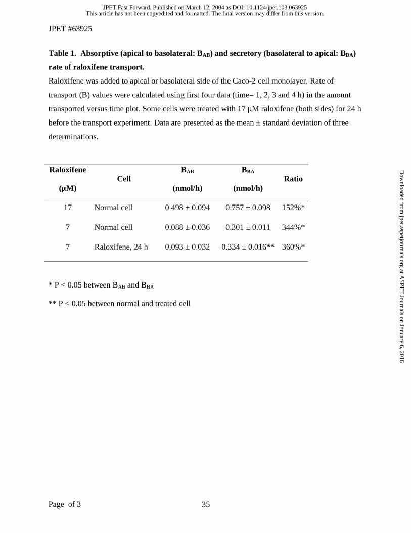
The rates of raloxifene transport were determined with respect to the direction of
transport (Table 1). Secretory (basolateral to apical) rates of transport (BBA) were always higher
than absorptive (apical to basolateral) rates of transport (BAB) (P < 0.05) (Table 1). As
concentration of raloxifene increased, the ratio of BBA to BAB decreased from 3.5 to 1.5. Pre-
treatment of Caco-2 cells with 17 µM raloxifene to saturate the conjugating enzymes increased
(p < 0.05) the secretory rate of transport, but the extent of increase was small (Table 1).
Effect of Raloxifene Concentration on the Transport of Raloxifene
Raloxifene has low absorptive permeability (PAB=0.39x10⁻⁶ ~ 4.1x10⁻⁶, 1.5~30µM) when
compared with genistein (PAB=31x10-6, 35 µM) and apigenin (PAB=17x10-6, 35 µM) (Table 2),
two compounds with extensive metabolism in the intestine (Chen et al., 2003; Hu et al., 2003).
Permeability of raloxifene increased linearly with rising raloxifene concentration (Fig. 3A). On
the other hand, the rate of transport increased with the square of concentration (Fig. 3B).
This article has not been copyedited and formatted. The final version may differ from this version.JPET Fast Forward. Published on March 12, 2004 as DOI: 10.1124/jpet.103.063925
at ASPE
T Journals on January 6, 2016
jpet.aspetjournals.orgD
ownloaded from
JPET #63925
12
Effect of Raloxifene Concentration on the Excretion of its Metabolites
Significant amounts of raloxifene were metabolized into glucuronidated and sulfated
metabolites during transport across the Caco-2 cell monolayers, with the sulfates being the major
metabolites (Fig. 2, Table 2). The extent of metabolism decreased as the concentration of
raloxifene increased (Table 2).
The extensive metabolism of raloxifene at lower concentration can be demonstrated by its
high clearance value. The clearance values for apical efflux of glucuronide and sulfate (at a
raloxifene concentration of 7 µM) were 10.04±1.37 µl/hr, and 26.57±1.12 µl/hr, respectively.
For basolateral efflux, they were roughly equal, or about 11 µl/hr. Taken together, the total
clearance values were 37.57 µl/hr for sulfates and 21 µl/hr for glucuronides, respectively. With a
cellular water volume of about 3.66 µl/mg protein, the total monolayer cellular water volume is
about 3.66 µl (Hu et al, 1994a). Therefore, the whole “cellular sulfate volume” is replaced about
10 times per hour or once every 6 min, whereas the whole “cellular glucuronide volume” is
replaced about 5 times per hour or once every 12 min.
Clearance of raloxifene glucuronide into both apical and basolateral sides were similar
and both decreased significantly (p < 0.05 according to one-way ANOVA) with an increase in
raloxifene concentration (Fig. 4A, Fig.4B).
Clearance of raloxifene sulfate, on the other hand, was polarized and mainly (up to 3.5
times more) to the apical side (p < 0.05). Similar to clearance of glucuronides, both apical and
This article has not been copyedited and formatted. The final version may differ from this version.JPET Fast Forward. Published on March 12, 2004 as DOI: 10.1124/jpet.103.063925
at ASPE
T Journals on January 6, 2016
jpet.aspetjournals.orgD
ownloaded from

JPET #63925
13
basolateral clearance also decreased significantly (p < 0.05 according to one-way ANOVA) with
an increase in raloxifene concentration (Fig. 4A, Fig.4B).
When comparing the clearance of these two conjugates from the same side of the cell
monolayers, apical clearance of raloxifene sulfate was higher (2.7 ~23 times) than that of
glucuronide at raloxifene concentration between 1.5 and 17 µM (p < 0.05) (Fig. 4A). However,
this difference in clearance decreased as raloxifene concentration increased, and eventually
became minimal at 30 µM. On the other hand, basolateral clearance of the two metabolites was
similar at all the concentrations measured (Fig. 4B).
Effect of Multidrug Resistance Related Protein (MRP) and p-Glycoprotein Inhibitors on
the Transport of Raloxifene and Excretion of its Metabolites
Multidrug resistance related protein (MRP) inhibitor MK-571 (50 µM) and leukotriene
C4 (leukotriene C4, 0.1 µM) (Hu et al., 2003; Rappa et al., 1999; Wheeler et al., 2000) or p-
glycoprotein inhibitors, cyclosporin A or verapamil, were added to both sides of Caco-2 cell
monolayer to determine if MRP or P-glycoprotein is involved in the transport of raloxifene and
the excretion of its metabolites. Previously, it was shown that leukotriene C4 was effective in
decreasing the efflux of apigenin sulfate and glucuronide (Hu et al, 2003), and MK 571 was
effective in decreasing the efflux of both apigenin and chrysin conjugates from the Caco-2 cells
(Walle et al, 1999; Hu et al, 2003). MRP inhibitors significantly decreased PBA of raloxifene (7
µM) (p < 0.05) (Fig.5A), but had no effect on PAB of raloxifene (Fig. 5A). In contrast, P-
glycoprotein inhibitors significantly increased PAB of raloxifene (7 µM) (p < 0.05) (Fig.5A), but
had no effect on PBA of raloxifene (Fig. 5A).
This article has not been copyedited and formatted. The final version may differ from this version.JPET Fast Forward. Published on March 12, 2004 as DOI: 10.1124/jpet.103.063925
at ASPE
T Journals on January 6, 2016
jpet.aspetjournals.orgD
ownloaded from

JPET #63925
14
In addition, MRP inhibitors also significantly decreased the clearance of glucuronide
from both basolateral and apical sides (p < 0.05) (Fig. 5B), and the clearance of sulfate from
apical side (Fig. 5C). However, they did not change the basolateral clearance of sulfate (Fig. 5C).
P-glycoprotein inhibitors had no significant effect on the clearance of glucuronides and sulfates
from both sides of Caco-2 cells (Fig. 5B and 5C) except for cyclosporin A, which increased the
basolateral clearance of sulfate when raloxifene was added to apical side (Fig 5C).
Effect of Organic Anion Transporter (OAT) Inhibitors on the Transport of Raloxifene and
Excretion of its Metabolites
Estrone sulfate (an organic anionic transporter or OAT substrate and inhibitor) was
shown to be effective in decreasing the efflux of apigenin sulfate from the Caco-2 cells (Hu et al,
2003). Therefore, OAT inhibitors estrone glucuronide (50 µM) or estrone sulfate (50 µM) was
used to study the possible involvement of OAT in the transport of raloxifene (7 µM) and in the
excretion of raloxifene metabolites. Both of them increased the rate of transport for raloxifene
(70 and 90% for estrone glucuronide and estrone sulfate, respectively, p < 0.05), when they were
added to both the apical and the basolateral media. There was no significant difference between
the effects of these two OAT inhibitors (Fig. 6A).
For raloxifene glucuronide, estrone sulfate significantly decreased (40%) the basolateral
clearance without affecting its apical clearance, whereas estrone glucuronide significantly
increased (75%) the apical clearance without affecting its basolateral clearance (Fig. 6B). For
raloxifene sulfate, these two inhibitors significantly decreased its clearance from both sides of
the Caco-2 cell monolayer (25 ~ 50%) (Fig. 6C).
This article has not been copyedited and formatted. The final version may differ from this version.JPET Fast Forward. Published on March 12, 2004 as DOI: 10.1124/jpet.103.063925
at ASPE
T Journals on January 6, 2016
jpet.aspetjournals.orgD
ownloaded from

JPET #63925
15
Effect of Estrone Sulfate on the Transport and Metabolism of Raloxifene as a Function of
Concentration
Increasing concentration of estrone sulfate (1~50 µM) significantly increased
(135~190%) the rate of transport of raloxifene (7 µM) through Caco-2 cells (p <0.05) (Fig. 7A)
with an apparent EC50 value of 16.4 µM. On the other hand, increasing concentration of estrone
sulfate significantly decreased the basolateral clearance of raloxifene glucuronide with a
maximum decrease of ≈40% but did not significantly affect the apical clearance of glucuronide
(Fig. 7B). Estrone sulfate inhibited both apical and basolateral clearance of raloxifene sulfate
with a maximum decrease of ≈50% (Fig. 7C).
Effect of Estrone Sulfate on the Metabolism of Raloxifene by Caco-2 Cell Lysate
Twenty µM of estrone sulfate decreased in vitro formation of raloxifene glucuronide and
sulfate (using Caco-2 cell lysate) by about 30 % (p < 0.05) (Fig. 8). Excretion rates (Bex) of both
metabolites from the Caco-2 cells monolayer were higher (3.4 and 1.3 times for glucuronide and
sulfate, respectively) than the rates of metabolism in the cell lysate (Bmet) (Fig. 8). Estrone
sulfate decreased the ratio of excretion rate over metabolism rate (Bex/ Bmet) for raloxifene sulfate
by 40% (p < 0.05) (Fig. 8B), but did not affect this ratio for raloxifene glucuronide (Fig. 8A).
Cellular Accumulation of Raloxifene and Its Metabolites in Caco-2 Cells.
Raloxifene was extensively bound to Caco-2 cells (up to 73% of dose after 24 h
incubation with 6 µM raloxifene). Among the conjugated metabolites, only sulfate was detected
in the cell whereas glucuronide was not detected. The amount of raloxifene taken up by or bound
to cells was significantly decreased after 24 h incubation compared to those after 4 h incubation,
This article has not been copyedited and formatted. The final version may differ from this version.JPET Fast Forward. Published on March 12, 2004 as DOI: 10.1124/jpet.103.063925
at ASPE
T Journals on January 6, 2016
jpet.aspetjournals.orgD
ownloaded from

JPET #63925
16
while the amount of sulfate found inside the cells was significantly increased after 24 h
incubation (data not shown). The amount of raloxifene taken up by the Caco-2 cell increased
significantly as concentrations increased (up to 7 µM), but did not increase further after that
concentration (Fig. 9A). The uptake trend of raloxifene sulfate was similar to raloxifene, but the
extent of uptake was 3.5 times smaller (Fig. 9B).
This article has not been copyedited and formatted. The final version may differ from this version.JPET Fast Forward. Published on March 12, 2004 as DOI: 10.1124/jpet.103.063925
at ASPE
T Journals on January 6, 2016
jpet.aspetjournals.orgD
ownloaded from

JPET #63925
17
DISCUSSION
Raloxifene has the potential to become the drug for breast cancer prevention. However,
raloxifene has poor bioavailability (Lindstrom et al., 1984). We hypothesized that extensive
intestinal metabolism, enteric and enterohepatic recycling of raloxifene conjugates could explain
why this drug has poor oral bioavailability but a long half-life (Fig. 10). We further
hypothesized that bioavailability of raloxifene could be enhanced via inhibition of phase II
metabolism and/or inhibition of efflux transporters responsible for pumping out the intracellular
phase II metabolites. In this study, we investigated the mechanisms responsible for the
absorption and excretion of raloxifene and its metabolites in the Caco-2 cell culture model.
Our results suggest that raloxifene is subjected to efflux because: 1) as concentration
increased, the permeability of raloxifene across the Caco-2 cell monolayer increased and the rate
of transport increased parabolically (Fig.3), consistent with saturation of efflux transporters; 2)
secretory transport of raloxifene was faster than absorptive transport (Table 1); and 3) the ratio of
secretory to absorptive transport rate (BBA/BAB) was decreased as the concentration of raloxifene
increased (Table 1). The efflux appeared to be mediated by MRP and P-glycoprotein since
secretory transport was diminished when MRP inhibitors were added to the media and absorptive
transport was augmented when P-glycoprotein inhibitors were added to the media (Fig.5). The
decrease by MRP inhibitors was achieved even though blockage of conjugate efflux has the
tendency to increase the amounts of intact compound transported. Therefore, our data showed
for the first time that one of the reasons why intestinal absorption of raloxifene is incomplete was
due to its efflux by MRP and P-glycoprotein. This represents an important discovery about the
This article has not been copyedited and formatted. The final version may differ from this version.JPET Fast Forward. Published on March 12, 2004 as DOI: 10.1124/jpet.103.063925
at ASPE
T Journals on January 6, 2016
jpet.aspetjournals.orgD
ownloaded from

JPET #63925
18
transport of raloxifene in the enterocytes. Previously, absorption mechanisms of raloxifene were
unknown. Usually, the passive diffusion process was implied as the mechanism of absorption
since there is no demonstrated nonlinear relationship between dose and absorption.
Our results showed that raloxifene undergoes extensive intestinal metabolism during
transport across the Caco-2 cells (Fig.1, Table 2). At low concentration of 7 µM raloxifene, the
total clearance value for both conjugates added together was about 59 µl/hr, which is enough to
clear the cellular metabolite volume 16 times per hour. The extensive metabolism is consistent
with reported extensive first-pass metabolism of raloxifene following oral administration
(Hochner-Celnikier, 1999). The major metabolites in the Caco-2 cells are mainly sulfates
followed by glucuronides. In addition, sulfates are rapidly and preferentially excreted into the
apical side, which diminishes their transport into the systemic circulation. Furthermore,
percentages of glucuronides increased as the concentration of raloxifene increased. Therefore,
our results are consistent with earlier observation that the main metabolite in human plasma is
the glucuronides including the primary metabolite raloxifene-4’-glucuronide (Hochner-Celnikier,
1999). They are also consistent with a recent discovery that intestinal glucuronidation is the
most important contributor to the presystemic clearance of raloxifene (Kemp et al., 2002).
We then proceeded to determine the mechanisms responsible for the efflux of raloxifene
conjugates since metabolic clearance decreased as concentration increased (Fig.4), suggesting
the involvement of a saturable process. The results indicated that excretion of raloxifene
glucuronide and sulfate was mediated by efflux transporters such as MRP and/or OAT, because
1) clearance of metabolites was polarized and saturable (Fig.4), 2) clearances into apical and
This article has not been copyedited and formatted. The final version may differ from this version.JPET Fast Forward. Published on March 12, 2004 as DOI: 10.1124/jpet.103.063925
at ASPE
T Journals on January 6, 2016
jpet.aspetjournals.orgD
ownloaded from

JPET #63925
19
basolateral media were changed in the presence of MK-571 and leukotriene C4, estrone
glucuronide, or estrone sulfate (Fig.5 and Fig.6). Taken together, these results have elucidated
for the first time the mechanisms responsible for efflux of a glucuronide and a sulfate metabolite
of a drug from the intestinal cells. Previously, we have reported the efflux mechanism
responsible for the efflux of apigenin sulfate and apigenin glucuronide (Hu et al., 2003), whereas
Walle and his co-worker reported efflux of chrysin conjugates were inhibited by MK-571, an
inhibitor of both glucuronidation and MRP (Walle et al, 1999; Hu et al, 2003).
We further analyzed the results of various chemical inhibitors on the excretion to
determine which transporter(s) may be mainly responsible for the excretion of raloxifene sulfate
and glucuronide. The main pathway for glucuronide (Fig.10) appears to be MRP since use of
two MRP inhibitors inhibited the excretion by more than 80% (Fig.5), with minor contribution
from basolateral OAT (Fig.6). The main pathway for sulfate is less well defined since its
excretion was never inhibited by more than 55%. Contributions from both apical MRP and OAT
and basolateral OAT are likely and additional contributions from other transporter(s) are possible
(Fig.10). In comparison to the efflux of apigenin conjugates (Hu et al, 2003), the efflux of
raloxifene glucuronide is much better defined than apigenin conjugate (about 60% attributable to
MRP and OAT), but the efflux of sulfate is less well defined than apigenin sulfate (>80%
attributable to MRP and OAT).
Further analysis of the inhibitor results suggests that MDR1 or p-glycoprotein is not
involved in limiting the efflux of conjugates, since neither cyclosporine A nor verapamil
decreased the efflux of any conjugates. However, it is not presently clear why cyclosporine A
This article has not been copyedited and formatted. The final version may differ from this version.JPET Fast Forward. Published on March 12, 2004 as DOI: 10.1124/jpet.103.063925
at ASPE
T Journals on January 6, 2016
jpet.aspetjournals.orgD
ownloaded from

JPET #63925
20
increased the basolateral efflux of sulfate. To probe the possible mechanisms, we determined if
cyclosporine A would increase sulfate formation in Caco-2 cell lysate. The result indicated a
slight decrease in the formation of raloxifene sulfate in the presence of 20 µM cyclosporine A
using Caco-2 cell lysate (27% decrease), which suggest that cyclosporine A did not enhance the
metabolite formation. The actual mechanism for sulfate efflux enhancement remains to be
determined.
Our discovery that raloxifene and its metabolites are excreted by the enterocyte efflux
transporters is novel. The excretion of raloxifene metabolites is the result of direct coupling of
efflux transporter and phase II metabolic enzymes, which has not been demonstrated for the
disposition of a drug in the intestine previously. The direct coupling is demonstrated by the fact
that inhibition of conjugate efflux could result in increase in raloxifene transport (Fig.6 and
Fig.7). The direct coupling is clearly the enabling process for enteric recycling, and may also be
used to explain why raloxifene has a low oral bioavailability but a long half-life.
The coupling of conjugating enzymes that produce hydrophilic metabolites and efflux
transporters is more complex than the coupling of CYP3A4 and p-glycoprotein (Cummins et al.,
2002) because these conjugates cannot get out of cells via passive diffusion. Hence, the action of
the efflux transporters can change cellular equilibrium between parent compound and its
conjugates. This change in equilibrium has at least two possible consequences, both of which
could lead to a higher than expected excretion of raloxifene conjugates (Fig.8). The first
consequence is less accumulation of conjugates in the cells because of rapid excretion of
conjugates that can inhibit phase II conjugation via product inhibition. In other words, when the
This article has not been copyedited and formatted. The final version may differ from this version.JPET Fast Forward. Published on March 12, 2004 as DOI: 10.1124/jpet.103.063925
at ASPE
T Journals on January 6, 2016
jpet.aspetjournals.orgD
ownloaded from
JPET #63925
21
efflux dominates the cellular excretion, very little conjugate is accumulated by the cells. In the
present study, the excretion rate of raloxifene glucuronide was much faster (3.4 times) than its
formation rate, resulting in no measurable cellular accumulation of this metabolite even after 24
h. On the other hand, sulfate excretion rate was comparable to sulfate formation rates, resulting
in significant accumulation of sulfate in cells after 24 h (Fig.9). The second consequence is less
hydrolysis of the conjugates to parent drug via the action of glucuronidases and sulftases as
proposed (Hochner-Celnikier, 1999), resulting in higher than expected excretion of conjugates
(Fig.8). Taken together, these two consequences will result in higher apparent excretion rate
than the formation rate when efflux inhibitor was absent (Fig. 8). When the efflux transporter
function was inhibited by estrone sulfate, the excretion of sulfates became slower than
metabolism of sulfate as expected. Taken together, these results support the coupling hypothesis
that predicts the modulation of cellular excretion of hydrophilic conjugates by the efflux
transporters.
The coupling of efflux transporters and conjugating enzymes also support our hypothesis
that enteric recycling (Liu and Hu, 2002) is important for the in vivo metabolism of raloxifene.
This coupling can explain why substantial amounts of raloxifene conjugates are produced and
excreted by the enterocytes. Because this coupling appears to function better at the apical side,
at least in the case of raloxifene, it increases the importance of enteric recycling in the
disposition of raloxifene. Because the apical side was the preferred side of excretion for the
raloxifene sulfate, it may be used to explain why smaller amounts of sulfated metabolites are
recovered in plasma since excreted metabolites are expected to be eliminated in feces or be
hydrolyzed by the microflora.
This article has not been copyedited and formatted. The final version may differ from this version.JPET Fast Forward. Published on March 12, 2004 as DOI: 10.1124/jpet.103.063925
at ASPE
T Journals on January 6, 2016
jpet.aspetjournals.orgD
ownloaded from

JPET #63925
22
Lastly, we also hypothesized that we could increase the bioavailability of raloxifene by
inhibiting the efflux transporters responsible for the excretion of raloxifene conjugates. OAT
inhibitors we used are not known to interact with MRP, even though they could slightly decrease
the formation rates of conjugates. Therefore, increase in the intestinal transport of intact
raloxifene (Fig.6) can be viewed mainly as the result of inhibiting the efflux of conjugated
metabolites, which in turn decreased the amounts of metabolites formed and allow more intact
raloxifene to permeate the Caco-2 cell monolayers intact. This is an important discovery since
we have shown that inhibition of an efflux transporter(s) that are tightly coupled to conjugating
enzymes can decrease the extent of metabolism and improve the transport of the intact
compound. If such an effect can be shown in vivo, it would provide a new mechanism to
improve the oral bioavailability of drugs and to explain drug interactions in the clinic.
In conclusion, raloxifene and its phase II conjugates are excreted by the intestinal cells
via the actions of MRP and/or OAT. Coupling of efflux transporters and conjugating enzymes
enhances intestinal excretion and renders the enteric recycling. On the other hand, the inhibition
of OAT transporters responsible for conjugate efflux can increase transport of intact raloxifene
and has the potential to increase its bioavailability pending the actual demonstration in vivo.
Taken together with enterohepatic recycling, intestinal disposition may be used to explain why
this drug has low bioavailability but a long half-life.
This article has not been copyedited and formatted. The final version may differ from this version.JPET Fast Forward. Published on March 12, 2004 as DOI: 10.1124/jpet.103.063925
at ASPE
T Journals on January 6, 2016
jpet.aspetjournals.orgD
ownloaded from

JPET #63925
23
REFERENCES
Bohets H, Annaert P, Mannens G, Van Beijsterveldt L, Anciaux K, Verboven P, Meuldermans
W, Lavrijsen K (2001) Strategies for absorption screening in drug discovery and
development. Curr Top Med Chem. 1: 367-83
Caro I, Boulenc X, Rousset M, Meunier V, Bourrie M, Julian B, Joyeux H, Roques C, Berger Y,
Zwebaum A and Fabre G (1995) Characterization of a newly isolated Caco-2 clone (TC-7),
as a model of transport processes and biotransformation of drugs. Int J Pharm. 116: 147-157
Chen J, Lin H and Hu M (2003) Metabolism of flavonoids via enteric recycling: role of intestinal
disposition. J Pharmacol Exp Ther. 304:1228-1235
Colacurci N, Manzella D, Fornaro F, Carbonella M and Paolisso G (2003) Endothelial function
and menopause: effects of raloxifene administration. J Clin Endocrinol Metab. 88:2135-40
Cummins CL, Jacobsen W and Benet LZ (2002) Unmasking the dynamic interplay between
intestinal P-glycoprotein and CYP3A4. J Pharmacol Exp Ther. 300:1036-1045
Delmas PD, Bjarnason NH, Mitlak BH, Ravoux AC, Shah AS, Huster WJ, Draper M and
Christiansen C (1997) Effect of raloxifene on bone mineral density, serum cholesterol
concentrations, and uterine endometrium in postmenopausal women. New Eng J Med.
337:1641-1647
This article has not been copyedited and formatted. The final version may differ from this version.JPET Fast Forward. Published on March 12, 2004 as DOI: 10.1124/jpet.103.063925
at ASPE
T Journals on January 6, 2016
jpet.aspetjournals.orgD
ownloaded from

JPET #63925
24
Duschek EJ, Stehouwer CD, de Valk-de Roo GW, Schalkwijk CG, Lambert J and Netelenbos C
(2003) Raloxifene, conjugated oestrogen and endothelial function in postmenopausal women.
J Intern Med. 254:85-94.
Eli Lilly (1998), Raloxifene package insert.
Gres MC, Julian B, Bourrie M, Meunier V, Roques C, Berger M, Boulenc X, Berger Y, Fabre G
(1998) Correlation between oral drug absorption in humans, and apparent drug permeability
in TC-7 cells, a human epithelial intestinal cell line: comparison with the parental Caco-2 cell
line. Pharm Res. 15: 726-733
Hochner-Celnikier D (1999) Pharmacokinetics of raloxifene and its clinical application. Eur J
Obstet Gynecol Reprod Biol. 85:23-29
Hu M, Chen J, Tran D, Zhu Y and Leonardo G (1994b) The Caco-2 Cell Monolayers as an
intestinal metabolism model: metabolism of dipeptide Phe-Pro. J Drug Target. 2:79-89
Hu M, Chen J, Zhu Y, Dantzig AH, Stratford RE and Kuhfeld MT Jr. (1994a) Mechanism and
kinetics of transcellular transport of a new β-lactam antibiotic loracarbef across a human
intestinal epithelial model system (Caco-2). Pharm Res. 11:1405-1413
This article has not been copyedited and formatted. The final version may differ from this version.JPET Fast Forward. Published on March 12, 2004 as DOI: 10.1124/jpet.103.063925
at ASPE
T Journals on January 6, 2016
jpet.aspetjournals.orgD
ownloaded from

JPET #63925
25
Hu M, Li Y, Davitt CM, Huang SM, Thummel K, Penman BW, Crespi CL (1999) Transport and
metabolic characterization of Caco-2 cells expressing CYP3A4 and CYP3A4 plus
oxidoreductase. Pharm Res. 16: 1352-1359
Hu M, Chen, J and Lin, H (2003) Disposition of Flavonoids via Recycling: Mechanistic Studies
of Disposition of Apigenin in the Caco-2 Cell Culture Model. J Pharmacol Exp Ther. 307:
314-321
Jordan VC and Morrow M (1999) Tamoxifen, raloxifene, and the prevention of breast cancer.
Endocr Rev. 20:253-278
Kemp DC, Fan PW and Stevens JC (2002) Characterization of raloxifene glucuronidation in
vitro: contribution of intestinal metabolism to presystemic clearance. Drug Metab Dispos.
30:694-700
Lindstrom TD, Whitaker NG and Whitaker GW (1984) Disposition and metabolism of a new
benzothiophene antiestrogen in rats, dogs and monkeys. Xenobiotica. 14:841-847
Liu Y, Hu M (2002) Absorption and metabolism of flavonoids in the caco-2 cell culture model
and a perused rat intestinal model. Drug Metab Dispos. 30:370-7
MacGregor JI and Jordan C (1998) Basic guide to the mechanisms of antiestrogen action.
Pharmacol Rev. 50:151-196
This article has not been copyedited and formatted. The final version may differ from this version.JPET Fast Forward. Published on March 12, 2004 as DOI: 10.1124/jpet.103.063925
at ASPE
T Journals on January 6, 2016
jpet.aspetjournals.orgD
ownloaded from
JPET #63925
26
Pfrunder A, Gutmann H, Beglinger C, Drewe J (2003) Gene expression of CYP3A4, ABC-
transporters (MDR1 and MRP1-MRP5) and hPXR in three different human colon carcinoma
cell lines. J Pharm Pharmacol. 55: 59-66
Pontier C, Pachot J, Botham R, Lenfant B, Arnaud P (2001) HT29-MTX and Caco-2/TC7
monolayers as predictive models for human intestinal absorption: role of the mucus layer. J
Pharm Sci. 90: 1608-1619
Ranaldi G, Consalvo R, Sambuy Y, Scarino ML (2003) Permeability characteristics of parental
and clonal human intestinal Caco-2 cell lines differentiated in serum-supplemented and
serum-free media. Toxicol In Vitro. 17: 761-767
Rappa G, Finch RA, Sartorelli AC and Lorico A (1999) New insights into the biology and
pharmacology of the multidrug resistance protein (MRP) from gene knockout models.
Biochem Pharmacol. 58:557-562
Scott JA, Da Camara CC and Early JE (1999) Raloxifene: a selective estrogen receptor
modulator. Am Fam Physician 60:1131-1138
Sekine Y, Cha SH and Endou H (2000) The multispecific organic anion transporter (OAT)
family. Pflugers Arch. 440:337-350
This article has not been copyedited and formatted. The final version may differ from this version.JPET Fast Forward. Published on March 12, 2004 as DOI: 10.1124/jpet.103.063925
at ASPE
T Journals on January 6, 2016
jpet.aspetjournals.orgD
ownloaded from

JPET #63925
27
Sabolovic N, Magdalou J, Netter P, Abid A (2000). Nonsteroidal anti-inflammatory drugs and
phenols glucuronidation in Caco-2 cells: identification of the UDP-glucuronosyltransferases
UGT1A6, 1A3 and 2B7. Life Sci. 67:185-196.
Walle UK, Galijatovic A, Walle T. (1999). Transport of the flavonoid chrysin and its conjugated
metabolites by the human intestinal cell line Caco-2. Biochem Pharmacol. 58:431-8.
Wheeler R, Neo SY, Chew J, Hladky SB and Barrand MA (2000). Use of membrane vesicles to
investigate drug interactions with transporter proteins, P-glycoprotein and multidrug
resistance-associated protein. Int J Clin Pharmacol Ther. 38:122-129
This article has not been copyedited and formatted. The final version may differ from this version.JPET Fast Forward. Published on March 12, 2004 as DOI: 10.1124/jpet.103.063925
at ASPE
T Journals on January 6, 2016
jpet.aspetjournals.orgD
ownloaded from

JPET #63925
28
FOOTNOTES: This study was supported by National Institutes of Health (NIH) Grant CA 87779.
This article has not been copyedited and formatted. The final version may differ from this version.JPET Fast Forward. Published on March 12, 2004 as DOI: 10.1124/jpet.103.063925
at ASPE
T Journals on January 6, 2016
jpet.aspetjournals.orgD
ownloaded from

JPET #63925
29
LEGENDS FOR FIGURES:
Fig. 1. Representative HPLC profiles of raloxifene and its metabolites. Panel A shows the peaks
of major metabolites (i.e., glucuronide and sulfate) after extracting raloxifene with methylene
chloride from a 24 h Caco-2 incubation sample. Panel B shows disappearance of glucuronide
peak and appearance of raloxifene peak after treatment with glucuronidase. Panel C shows
reduction of sulfate peak and appearance of raloxifene peak after treatment with sulfatase.
Fig 2. Transport and excretion of raloxifene and its metabolites in the Caco-2 model. Raloxifene
(17 µM) was added to apical (AP) or basolateral (BL) side of the Caco-2 cell monolayer and the
amounts of raloxifene (circles), raloxifene glucuronide (squares) and raloxifene sulfate
(triangles) in apical (open symbols and dotted lines) and basolateral (solid symbols and solid
lines) were determined at 1, 2, 3 and 4 h after incubation as described under Materials and
Methods. These symbols (e.g., solid for basolateral appearance, squares for glucuronide and
triangles for sulfate) are used throughout the paper. Each data point represents the mean of three
determinations and the error bars are standard deviations of the mean. Each line represents linear
regression of the data points. Panel A, R2=0.9000, 0.99421 and 0.9900 for raloxifene,
glucuronide and sulfate; panel B, R2=0.9812 and 0.9805 for glucuronide and sulfate; panel C,
R2=0.9827, 0.9929 and 0.9902 for raloxifene, glucuronide and sulfate; panel D, R2=0.9972 and
0.9955 for glucuronide and sulfate, respectively.
Fig. 3. Effect of concentration on the absorptive transport of raloxifene. Raloxifene (1.5 ~30
µM) was added to apical (AP) side of the Caco-2 cell monolayer and the amount of raloxifene in
This article has not been copyedited and formatted. The final version may differ from this version.JPET Fast Forward. Published on March 12, 2004 as DOI: 10.1124/jpet.103.063925
at ASPE
T Journals on January 6, 2016
jpet.aspetjournals.orgD
ownloaded from

JPET #63925
30
receiver (BL) side were determined at 1, 2, 3 and 4 h after incubation. Rates of transport (B) and
permeability (P) across a Caco-2 cellular membrane were obtained according to the equation [1]
and [2] as described under Materials and Methods. Each data point represents the mean of three
determinations and the error bars are standard deviations of the mean. The line and curve
represent linear and polynomial regression of the data points in panel A (R2=0.9790) and panel
B(R2=0.9985), respectively.
Fig. 4. Effect of raloxifene concentration on the clearance of raloxifene metabolites to apical
(AP) or basolateral (BL) side of the Caco-2 cell monolayer. Raloxifene (1.5 ~30 µM) was added
to apical side of the Caco-2 cell monolayer and the amount of raloxifene glucuronide and sulfate
in receiver (basolateral) and donor (apical) side were determined at 1, 2, 3 and 4 h after
incubation. Metabolic clearance (CL) of raloxifene was calculated according to the equation [3]
described under Materials and Methods. Each data point represents the mean of three
determinations and the error bars are standard deviations of the mean. Each curve represents
power regression of the data points using Microsoft Excel. Panel A, R2=0.8243 and 0.9563 for
glucuronide and sulfate; panel B, R2=0.8973 and 0.8915 for glucuronide and sulfate, respectively.
The star symbol “*” indicates statistically significant differences (p < 0.05) between glucuronide
and sulfate.
Fig. 5. Effect of multidrug resistance related protein (MRP) and P-glycoprotein inhibitors on the
permeability of raloxifene and clearance of its metabolites in Caco-2 cells. Raloxifene (7 µM)
was added to apical (AP) or basolateral (BL) side of the Caco-2 cell monolayer. Fifty µM MK-
571 plus 0.1 µM leukotriene C4, 20 µM cyclosporine A or 20 µM verapamil was added to both
This article has not been copyedited and formatted. The final version may differ from this version.JPET Fast Forward. Published on March 12, 2004 as DOI: 10.1124/jpet.103.063925
at ASPE
T Journals on January 6, 2016
jpet.aspetjournals.orgD
ownloaded from

JPET #63925
31
sides of the Caco-2 cell monolayer. Metabolic clearance (CL) of raloxifene was calculated using
the amounts of metabolites at 1, 2, 3 and 4 h after incubation according to the equation [3]
described under Materials and Methods. Each column represents the mean of three
determinations and the error bars are standard deviations of the mean. Blank, solid, dashed and
dotted columns represent the control, MK-571 plus leukotriene C4, cyclosporine A or verapamil
treated group, respectively. The star symbol “*” indicates a statistically significant difference (p
< 0.05) compared to the control (i.e., raloxifene only).
Fig. 6. Effect of organic anionic transporter (OAT) inhibitors on the transport of raloxifene and
clearance of its metabolites in Caco-2 cells. Raloxifene (7 µM) was added to apical side of the
Caco-2 cell monolayer. 50 µM estrone glucuronide (EG) or estrone sulfate (ES) was added to
both sides of the Caco-2 cell monolayer. Rate of transport (Bt) and metabolic clearance (CL) of
raloxifene were calculated using the amounts of raloxifene and metabolites at 1, 2, 3 and 4 h
after incubation according to the equation [1] and [3] described under Materials and Methods.
Each column represents the mean of three determinations and the error bars are standard
deviations of the mean. Blank, solid and dashed columns represent the control, EG-treated and
ES-treated groups, respectively. The mean control values (± S.D.) of Bt and CLs were 0.059±
0.013 nmol/h (panel A), 3.10 ± 0.10x10⁻⁶ ml/sec 11.16±0.36 x µl/hr (glucuronide, basolateral or
BL side, panel B), 10.04 ± 1.37 µl/hr (glucuronide, apical side or AP, panel B), 11.23 ± 0.47
µl/hr or (sulfate, basolateral side, panel C) and 26.57 ± 1.12 µl/hr (sulfate, apical side, panel C),
respectively. The star symbol “*” indicates a statistically significant difference (p < 0.05)
compared to the control.
This article has not been copyedited and formatted. The final version may differ from this version.JPET Fast Forward. Published on March 12, 2004 as DOI: 10.1124/jpet.103.063925
at ASPE
T Journals on January 6, 2016
jpet.aspetjournals.orgD
ownloaded from
JPET #63925
32
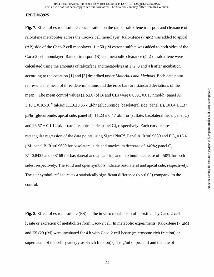
Fig. 7. Effect of estrone sulfate concentration on the rate of raloxifene transport and clearance of
raloxifene metabolites across the Caco-2 cell monolayer. Raloxifene (7 µM) was added to apical
(AP) side of the Caco-2 cell monolayer. 1 ~ 50 µM estrone sulfate was added to both sides of the
Caco-2 cell monolayer. Rate of transport (B) and metabolic clearance (CL) of raloxifene were
calculated using the amounts of raloxifene and metabolites at 1, 2, 3 and 4 h after incubation
according to the equation [1] and [3] described under Materials and Methods. Each data point
represents the mean of three determinations and the error bars are standard deviations of the
mean. . The mean control values (± S.D.) of Bt and CLs were 0.059± 0.013 nmol/h (panel A),
3.10 ± 0.10x10⁻⁶ ml/sec 11.16±0.36 x µl/hr (glucuronide, basolateral side, panel B), 10.04 ± 1.37
µl/hr (glucuronide, apical side, panel B), 11.23 ± 0.47 µl/hr or (sulfate, basolateral side, panel C)
and 26.57 ± 0.1.12 µl/hr (sulfate, apical side, panel C), respectively. Each curve represents
rectangular regression of the data points using SigmaPlot. Panel A, R2=0.9680 and EC50=16.4
µM; panel B, R2=0.9639 for basolateral side and maximum decrease of ≈40%; panel C,
R2=0.8435 and 0.8168 for basolateral and apical side and maximum decrease of ≈50% for both
sides, respectively. The solid and open symbols indicate basolateral and apical side, respectively.
The star symbol “*” indicates a statistically significant difference (p < 0.05) compared to the
control.
Fig. 8. Effect of estrone sulfate (ES) on the in vitro metabolism of raloxifene by Caco-2 cell
lysate or excretion of metabolites from Caco-2 cell. In metabolic experiments, Raloxifene (7 µM)
and ES (20 µM) were incubated for 4 h with Caco-2 cell lysate (microsome-rich fraction) or
supernatant of the cell lysate (cytosol-rich fraction) (≈1 mg/ml of protein) and the rate of
This article has not been copyedited and formatted. The final version may differ from this version.JPET Fast Forward. Published on March 12, 2004 as DOI: 10.1124/jpet.103.063925
at ASPE
T Journals on January 6, 2016
jpet.aspetjournals.orgD
ownloaded from

JPET #63925
33
metabolism (Bmet) was obtained as described under Materials and Methods. Equivalent of
amounts of organic solvent (0.5% in this case) were added instead of ES in the control
experiment. In transport experiment, raloxifene (7 µM) was added to apical (AP) side and
estrone sulfate (20 µM) was added to both sides of the Caco-2 cell monolayer. The sum of donor
(AP) side and receiver (basolateral, BL) side excretion rate (Bex) of each metabolite was
calculated using the amounts of metabolites at 1, 2, 3 and 4 hour after incubation as described
under Materials and Methods. Each column represents the mean of three determinations and the
error bars are standard deviations of the mean. Blank and solid columns represent rate of
metabolism and excretion, respectively. Panel A, Bex/Bmet = 3.4 and 3.7 for the control and ES-
treated group; panel B, Bex/Bmet = 1.3 and 0.8 for the control and ES-treated group, respectively.
The star symbol “*” and “**” indicate statistically significant differences (p < 0.05) between the
control and ES-treated group and between metabolism and excretion, respectively.
Fig. 9. Effect of raloxifene concentration on the accumulation of raloxifene and its sulfate by the
Caco-2 cells. Raloxifene (1.5 ~18 µM) was added to apical side of the Caco-2 cell monolayer
and the amounts of raloxifene and its metabolites inside the cell were determined at 24 h after
incubation as described under Materials and Methods. Each data point represents the mean of
three determinations and the error bars are standard deviations of the mean. Each curve
represents rectangular regression of the data points using SigmaPlot. Panel A, R2=0.9681;
panel B, R2=0.9720. The star symbol “*” indicates a statistically significant difference (p <
0.05) compared to the lower concentration.
This article has not been copyedited and formatted. The final version may differ from this version.JPET Fast Forward. Published on March 12, 2004 as DOI: 10.1124/jpet.103.063925
at ASPE
T Journals on January 6, 2016
jpet.aspetjournals.orgD
ownloaded from

JPET #63925
34
Fig. 10. Schematic representation of intestinal disposition of raloxifene. Absorbed raloxifene, in
free or conjugated form, will be effluxed to the lumen (AP) or be transported into the liver via
portal vein. After intact compounds are further metabolized in the liver, conjugates either enter
into the systemic circulation or are secreted to the intestinal lumen via bile. The excretion of the
phase II conjugates by the enterocytes and hepatocytes render enteric and enterohepatic recycling
of raloxifene, which in turn increases its apparent half-life. The present study focuses on
mechanisms (e.g., MDR-mediated, MRP-mediated or OAT-mediated efflux) responsible for
intestinal disposition of raloxifene and/or its conjugates.
This article has not been copyedited and formatted. The final version may differ from this version.JPET Fast Forward. Published on March 12, 2004 as DOI: 10.1124/jpet.103.063925
at ASPE
T Journals on January 6, 2016
jpet.aspetjournals.orgD
ownloaded from
JPET #63925
Page of 3 35
Table 1. Absorptive (apical to basolateral: BAB) and secretory (basolateral to apical: BBA)
rate of raloxifene transport.
Raloxifene was added to apical or basolateral side of the Caco-2 cell monolayer. Rate of
transport (B) values were calculated using first four data (time= 1, 2, 3 and 4 h) in the amount
transported versus time plot. Some cells were treated with 17 µM raloxifene (both sides) for 24 h
before the transport experiment. Data are presented as the mean ± standard deviation of three
determinations.
Raloxifene
(µM) Cell
BAB
(nmol/h)
BBA
(nmol/h) Ratio
17 Normal cell 0.498 ± 0.094 0.757 ± 0.098 152%*
7 Normal cell 0.088 ± 0.036 0.301 ± 0.011 344%*
7 Raloxifene, 24 h 0.093 ± 0.032 0.334 ± 0.016** 360%*
* P < 0.05 between BAB and BBA
** P < 0.05 between normal and treated cell
This article has not been copyedited and formatted. The final version may differ from this version.JPET Fast Forward. Published on March 12, 2004 as DOI: 10.1124/jpet.103.063925
at ASPE
T Journals on January 6, 2016
jpet.aspetjournals.orgD
ownloaded from

JPET #63925
Page of 3 36
Table 2. Absorptive permeability, amounts transported intact and amounts of raloxifene metabolites formed after 2 h
Drug was added to apical side of the Caco-2 monolayer. Permeability values were calculated using first four data (time= 1, 2, 3 and 4
h for raloxifene or time=0.5, 1, 1.5 and 2 h for apigenin and genistein) in the amount transported versus time plot. Amounts of
metabolites in the apical (AP) and basolateral (BL) media after 2 h-incubation were determined. Data are presented as the mean ±
standard deviation of three determinations.
Glucuronide (nmol) Sulfate (nmol) Total Amounts (nmol) % Metabolism Compound
Permeability
(10-7 cm/sec) AP side BL side AP side BL side Parent Conjugates Parent/(Total)
Raloxifene, 1.5 µM UDb UD UD 0.419 ± 0.052
UD - - -
Raloxifene, 3.4 µM 3.88 ± 0.83c 0.136 ± 0.007
0.159 ± 0.018
0.635 ± 0.040
0.172 ± 0.033
0.041 1.102 96.4
Raloxifene, 6 µM 7.87 ± 2.05 c 0.168 ± 0.025
0.321 ± 0.032
0.400 ± 0.025
0.232 ± 0.001
0.132 1.121 89.4
Raloxifene, 7 µM 8.66 ± 3.60 c 0.168 ± 0.020
0.190 ± 0.012
0.781 ± 0.047
0.296 ± 0.035
0.173 1.089 86.3
Raloxifene, 17 µM 17.8 ± 6.96 c 0.092± 0.008
0.240 ± 0.024
0.308 ± 0.014
0.338 ± 0.028
0.897 0.979 52.2
Raloxifene, 30 µM 41.4 ± 7.28 c 0.165 ± 0.165
0.307 ± 0.016
0.222 ± 0.011
0.392 ± 0.037
3.782 1.085 22.3
Apigenin, 35 µMa 169 ± 10 3.42 ± 0.35
4.09 ± 0.18
3.75 ± 0.22
3.21 ± 0.26
20.94 17.61 45.7
Genistein, 35 µMa 309 ± 90 0.867 ± 0.014
1.68 ± 0.04
0.314 ± 0.021
0.709 ± 0.017
33.39 3.9 10.5
a. Chen et al., 2003
This article has not been copyedited and form
atted. The final version m
ay differ from this version.
JPET
Fast Forward. Published on M
arch 12, 2004 as DO
I: 10.1124/jpet.103.063925 at ASPET Journals on January 6, 2016 jpet.aspetjournals.org Downloaded from
JPET #63925
Page of 3 37
b. Under detection limit
c. Significant difference with respect to raloxifene concentration according to one-way ANOVA (p < 0.05).
This article has not been copyedited and form
atted. The final version m
ay differ from this version.
JPET
Fast Forward. Published on M
arch 12, 2004 as DO
I: 10.1124/jpet.103.063925 at ASPET Journals on January 6, 2016 jpet.aspetjournals.org Downloaded from
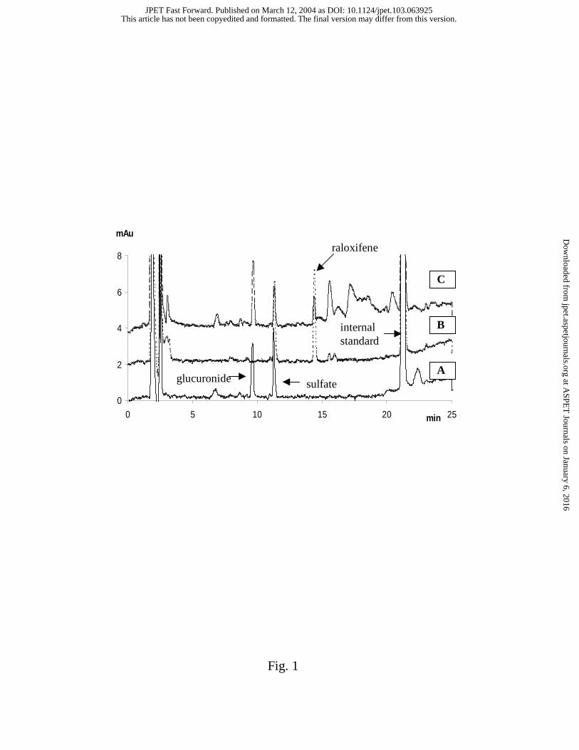
Fig. 1
0
2
4
6
8
0 5 10 15 20 25min
mAu
glucuronide sulfate
raloxifene
B
A
C
internal standard
This article has not been copyedited and formatted. The final version may differ from this version.JPET Fast Forward. Published on March 12, 2004 as DOI: 10.1124/jpet.103.063925
at ASPE
T Journals on January 6, 2016
jpet.aspetjournals.orgD
ownloaded from

Fig. 2
(A) A-->B Receiver (BL)
0
1
2
3
4
5
1 2 3 4Time (h)
ralo
xife
ne
or
met
abo
lites
(nm
ol)
(B) A-->B Donor (AP)
0
10
20
30
40
50
1 2 3 4
Time (h)
ralo
xife
ne
(nm
ol)
0
1
2
3
4
5
met
abo
lites
(nm
ol)
(C) B-->A Receiver (AP)
0
1
2
3
4
5
1 2 3 4Time (h)
ralo
xife
ne
or
met
abo
lites
(nm
ol)
(D) B-->A Donor (BL)
0
10
20
30
40
50
1 2 3 4
Time (h)
ralo
xife
ne
(nm
ol)
0
1
2
3
4
5
met
abo
lites
(nm
ol)
This article has not been copyedited and formatted. The final version may differ from this version.JPET Fast Forward. Published on March 12, 2004 as DOI: 10.1124/jpet.103.063925
at ASPE
T Journals on January 6, 2016
jpet.aspetjournals.orgD
ownloaded from

Fig. 3
A: Permeability
0
10
20
30
40
50
0 5 10 15 20 25 30 35Raloxifene (µM)
P (c
m/s
ec) X
107
B: Rate of Transport
0
1
2
3
0 5 10 15 20 25 30 35Raloxifene (µM)
B (n
mo
l/h)
This article has not been copyedited and formatted. The final version may differ from this version.JPET Fast Forward. Published on March 12, 2004 as DOI: 10.1124/jpet.103.063925
at ASPE
T Journals on January 6, 2016
jpet.aspetjournals.orgD
ownloaded from

Fig. 4
A: AP side
0
20
40
60
0 20 40 60 80 100Raloxifene (µM)
CL
(cm
3/s
ec)X
106 Glucuronide
Sulfate
*
*
*
**
*
B: BL side
0
2
46
8
10
12
0 20 40 60 80 100Raloxifene (µM)
CL
(cm
3 /sec
)X10
6 Glucuronide
Sulfate
This article has not been copyedited and formatted. The final version may differ from this version.JPET Fast Forward. Published on March 12, 2004 as DOI: 10.1124/jpet.103.063925
at ASPE
T Journals on January 6, 2016
jpet.aspetjournals.orgD
ownloaded from

Fig. 5
A: Raloxifene
0%
100%
200%
300%
PAB PBA
P (
% c
on
tro
l)
Raloxifene only
with LTC4 & MK-571
with cyclosporin A
with verapamil
*
*
*
B: Glucuronide
0%
100%
200%
300%
AP-->BL (BL side) AP-->BL (AP side) BL-->AP (AP side) BL-->AP (BL side)
CL
(%
co
ntr
ol)
** **
C: Sulfate
0%
100%
200%
300%
AP-->BL (BL side) AP-->BL (AP side) BL-->AP (AP side) BL-->AP (BL side)
CL
(%
co
ntr
ol)
* *
*
This article has not been copyedited and formatted. The final version may differ from this version.JPET Fast Forward. Published on March 12, 2004 as DOI: 10.1124/jpet.103.063925
at ASPE
T Journals on January 6, 2016
jpet.aspetjournals.orgD
ownloaded from

Fig. 6
A: Raloxifene
0%
100%
200%
300%
Control EG ES
B (
% c
on
tro
l)
Control EG ES
* *
B: Glucuronide
0%
100%
200%
300%
BL side AP side
CL (
% c
ontr
ol)
*
*
C. Sulfate
0%
50%
100%
150%
BL side AP side
CL (
% c
ontr
ol)
** *
*
This article has not been copyedited and formatted. The final version may differ from this version.JPET Fast Forward. Published on March 12, 2004 as DOI: 10.1124/jpet.103.063925
at ASPE
T Journals on January 6, 2016
jpet.aspetjournals.orgD
ownloaded from

Fig.7
B: Glucuronide
0 10 20 30 40 50
CL
(% c
on
tro
l)
0
50
100
150
200
C: Sulfate
Estrone Sulfate (µM)
0 10 20 30 40 50
CL
(%
co
ntr
ol)
0
20
40
60
80
100
120
*
** *
*
*
* * *
*
A: Raloxifene
0 10 20 30 40 50
B (
% c
on
tro
l)
0
50
100
150
200
250
**
**
* * * * *
B: Glucuronide
0 10 20 30 40 50
CL
(% c
on
tro
l)
0
50
100
150
200
C: Sulfate
Estrone Sulfate (µM)
0 10 20 30 40 50
CL
(%
co
ntr
ol)
0
20
40
60
80
100
120
*
** *
*
*
* * *
*
A: Raloxifene
0 10 20 30 40 50
B (
% c
on
tro
l)
0
50
100
150
200
250
**
**
B: Glucuronide
0 10 20 30 40 50
CL
(% c
on
tro
l)
0
50
100
150
200
C: Sulfate
Estrone Sulfate (µM)
0 10 20 30 40 50
CL
(%
co
ntr
ol)
0
20
40
60
80
100
120
*
** *
*
*
* * *
*
C: Sulfate
Estrone Sulfate (µM)
0 10 20 30 40 50
CL
(%
co
ntr
ol)
0
20
40
60
80
100
120
*
** *
*
*
* * *
*
*
** *
*
*
* * *
*
A: Raloxifene
0 10 20 30 40 50
B (
% c
on
tro
l)
0
50
100
150
200
250
**
**A: Raloxifene
0 10 20 30 40 50
B (
% c
on
tro
l)
0
50
100
150
200
250
**
**
**
**
* * * * ** * * * *
This article has not been copyedited and formatted. The final version may differ from this version.JPET Fast Forward. Published on March 12, 2004 as DOI: 10.1124/jpet.103.063925
at ASPE
T Journals on January 6, 2016
jpet.aspetjournals.orgD
ownloaded from

Fig. 8
A: Glucuronide
0
5
10
15
Control ES
pmol
/min
/mon
olay
erMetabolism
Excretion**
*
*
**
B: Sulfate
0
5
10
15
20
Control ES
pmol
/min
/mon
olay
er
**
**
**
This article has not been copyedited and formatted. The final version may differ from this version.JPET Fast Forward. Published on March 12, 2004 as DOI: 10.1124/jpet.103.063925
at ASPE
T Journals on January 6, 2016
jpet.aspetjournals.orgD
ownloaded from

Fig. 9
A: Raloxifene
0 10 20 30 40 50 60
nm
ol /
mo
no
laye
r
0
10
20
30
40
50
**
B: Sulfate
Raloxifene (µM)
0 10 20 30 40 50 60
nm
ol /
mo
no
laye
r
0
2
4
6
8
10
*
*
This article has not been copyedited and formatted. The final version may differ from this version.JPET Fast Forward. Published on March 12, 2004 as DOI: 10.1124/jpet.103.063925
at ASPE
T Journals on January 6, 2016
jpet.aspetjournals.orgD
ownloaded from
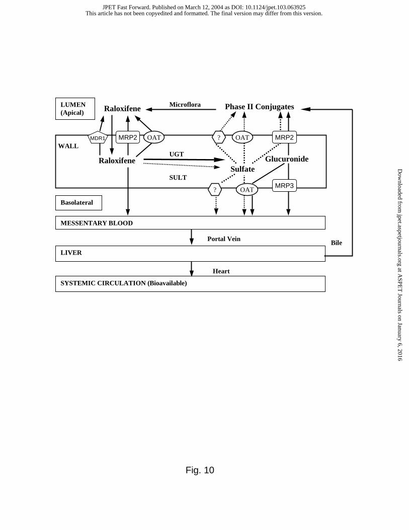
Fig. 10
LUMEN (Apical)
MESSENTARY BLOOD
LIVER
Portal Vein
Heart
Microflora
OAT
OAT
?
OAT
Phase II Conjugates
GlucuronideSulfate
SULT
UGTRaloxifene
Raloxifene
WALL
Basolateral
Bile
SYSTEMIC CIRCULATION (Bioavailable)
MRP2 MRP2
MRP3
MDR1 ?
This article has not been copyedited and formatted. The final version may differ from this version.JPET Fast Forward. Published on March 12, 2004 as DOI: 10.1124/jpet.103.063925
at ASPE
T Journals on January 6, 2016
jpet.aspetjournals.orgD
ownloaded from