detection and characterisation of respiratory pathogens
TRANSCRIPT

Detection and characterisation of respiratory pathogens among habituated, wild living chimpanzees (Pan troglodytes verus) of Taï National Park, Côte d’Ivoire
vorgelegt von Sophie Köndgen Diplom Biologin, geboren in Frankfurt/Main
Von der Fakultät III – Prozesswissenschaften der Technischen Universität Berlin
zur Erlangung des akademischen Grades
Doktor der Naturwissenschaften Dr. rer. nat.
Genehmigte Dissertation
Promotionsausschuss: Vorsitzender: Prof. Dr. L. Garbe 1. Gutachter: Prof. Dr. R. Lauster 2. Gutachter: Prof. Dr. G. Pauli Tag der wissenschaftlichen Aussprache: 10. Januar 2011
Berlin 2011 D83


I
Abstract
Respiratory diseases are one of the most important threats to wild great apes habituated to human
presence for research or tourism. However, the aetiological agents of such diseases have not been
documented so far. Between 1999 and 2006 five distinct respiratory disease outbreaks hit three
communities of habituated chimpanzees at our research site in Taï National Park, Côte d’Ivoire.
Three of the outbreaks resulted in high morbiditiy and mortality. Necropsies were performed on
seven individuals found shortly after death and histopathologic examination revealed the presence
of purulent bronchopneumonia. Based on these examinations, the main objective of the present
study was to identify and characterise the causative pathogens and determine possible sources of
infection.
Lung tissue samples were screened by PCR for a broad range of respiratory viruses. All samples
tested were positive for either of two paramyxoviruses, the human respiratory syncytial virus
(HRSV) or the human metapneumovirus (HMPV). To establish the origin of the viruses found,
phylogenetic analyses were performed and revealed that the strains were closely related to strains
circulating in contemporaneous, worldwide human epidemics. This represents the first direct
evidence of anthropozoonotic virus transmission to wild great apes, suggesting that the close
approach of humans to apes, which is central to both research and tourism programs, represents a
serious threat to these animals.
Furthermore, isolation of bacteria was performed and revealed that some of the deceased
individuals were co-infected with Pasteurella multocida. Isolates were subjected to a detailed pheno-
and genotypic characterisation providing the first description of P. multocida in wild living
chimpanzees. Two different strains were identified, both showing high similarity to previously
described strains from different host and geographical locations. This suggests that chimpanzees are
involved in the epidemiology of P. multocida. The question of whether this bacterium is carried
naturally by chimpanzees or was transmitted by other animals will be investigated in further studies.
To systematically evaluate the occurrence of respiratory pathogens without disturbing the
chimpanzees’ natural behaviour, the establishment of non-invasive diagnostics was another aim of
this work. Therefore, faecal samples which had been collected during and between outbreaks were
tested for HRSV and HMPV by PCR. Using this approach it was possible to identify the causative
agents of lethal as well as of non-lethal outbreaks, to evaluate the virus prevalence among a larger
study group, and to perform phylogenetic analyses of the viruses detected. This demonstrates that
the screening of faecal samples is a suitable tool for monitoring acute respiratory diseases in wild
living chimpanzees.
This is the first systematic study of respiratory diseases in wild great apes. The results presented are
of great relevance for future conservation strategies as a deeper knowledge of the involved
pathogens may help to prevent or mitigate future disease outbreaks.

II
Zusammenfassung
Respiratorische Erkrankungen sind eine der größten Bedrohungen für wildlebende Menschenaffen,
die für die Wissenschaft oder Tourismus an die Anwesenheit des Menschen gewöhnt (habituiert)
wurden. Trotz dieser Bedrohung fehlen bisher genaue Untersuchungen zu den dafür
verantwortlichen Erregern. Bei drei habituierten Schimpansengruppen des Taï Nationalparks (Côte
d’Ivoire) wurden zwischen 1999 und 2006 fünf verschiedene respiratorische Krankheitsausbrüche
dokumentiert von denen drei hohe Morbiditäts- und Mortalitätsraten aufwiesen. Insgesamt konnte
bei sieben verstorbenen Individuen eine Sektion durchgeführt werden, wobei in allen Fällen durch
nachfolgende histopathologische Untersuchungen eine eitrige Lungenentzündung festgestellt
wurde. Basierend auf diesen Voruntersuchungen waren die genaue Charakterisierung der
verantwortlichen Erreger sowie die Identifikation möglicher Infektionsquellen Hauptziele dieser
Arbeit.
Zum Nachweis ursächlicher Krankheitserreger wurden Lungengewebeproben mittels PCR auf ein
breites Spektrum respiratorischer Erreger untersucht. Alle untersuchten Proben waren positiv für
eines von zwei Paramyxoviren, dem humanen respiratorischem Synzytialvirus (HRSV) oder dem
humanen Metapneumovirus (HMPV). Phylogenetische Untersuchungen der in den Schimpansen
detektierten Virusstämme zeigten eine enge Verwandtschaft zu Stämmen, die zeitgleich weltweit in
der menschlichen Bevölkerung zirkulierten. Dies ist der erste Hinweis auf eine
anthropozoonotische Virusübertragung auf wildlebende Menschenaffen und legt nahe, dass der
enge Kontakt zwischen Menschen und Menschenaffen - der sowohl bei wissenschaftlichen als auch
touristischen Projekten gegeben ist - eine ernstzunehmende Bedrohung für diese Tiere darstellt.
In Voruntersuchungen zu möglichen bakteriellen Krankheitserregern ergaben sich Hinweise auf das
Vorhandensein von Pasteurella multocida. Der Keim wurde aus dem Lungengewebe einiger
Individuen angezüchtet und die verschiedenen Isolate einer breiten phäno- und genotypischen
Charakterisierung unterworfen. Dies stellt die erste Beschreibung von P. multocida bei wildlebenden
Schimpansen dar. Es wurden zwei unterschiedliche Stämme identifiziert, die beide eine große
Ähnlichkeit zu bisher beschriebenen Stämmen von Wirten unterschiedlichster Spezies und
Herkunft zeigten. Die lässt vermuten, dass Schimpansen in die Epidemiologie von P. multocida
involviert sind. Ob Schimpansen jedoch natürliche Träger dieses Bakteriums sind, oder ob dieses
von anderen Tieren übertragen wurde, ist Thema weiterer Studien.
Ein weiteres Ziel dieser Arbeit war die Etablierung nicht-invasiver diagnostischer Methoden,
welche die systematische Untersuchung respiratorischer Erreger ermöglichen, ohne dabei das
natürliche Verhalten der Schimpansen zu stören. Dafür wurden Faezes-Proben, die sowohl
während als auch zwischen den respiratorischen Krankheitsausbrüchen gesammelt wurden mittels
PCR auf HRSV und HMPV getestet. Hierdurch war es möglich, die verantwortlichen Erreger -
auch von nicht letalen Ausbrüchen - zu identifizieren, die Virusprävalenz innerhalb einer größeren
Studiengruppe zu evaluieren und die detektierten Viren phylogenetisch zu analysieren. Es konnte
gezeigt werden, dass die Untersuchung von Faezes-Proben eine geeignete Methode darstellt, um

III
ursächliche Krankheitserreger akuter respiratorischer Erkrankungen bei wildlebenden Schimpansen
zu identifizieren.
Nur ein tiefgehendes Verständnis der involvierten Erreger kann dazu beitragen, neue Strategien zur
Prävention und Kontrolle zukünftiger Krankheitsausbrüche zu entwickeln. Aus diesem Grund sind
die hier vorgestellten Untersuchungen für den Schutz von Menschenaffen von größter Relevanz.

IV
Table of contents
ABSTRACT ..............................................................................................................................................I
ZUSAMMENFASSUNG ....................................................................................................................... II
LIST OF ABBREVIATIONS ............................................................................................................. VII
1 INTRODUCTION..........................................................................................................................1
2 BACKGROUND............................................................................................................................. 3
2.1 RESPIRATORY DISEASE IN GREAT APES..................................................................................................... 3
2.1.1 Respiratory pathogens ................................................................................................................................... 3 2.1.1.1 Respiratory viruses ...........................................................................................................................................4 2.1.1.2 Respiratory bacteria..........................................................................................................................................8
2.2 PATHOGEN TRANSMISSION BETWEEN HUMANS AND NONHUMAN PRIMATES ..................................10
2.3 PATHOGEN TRANSMISSION BETWEEN CHIMPANZEES AND MONKEYS...............................................11
2.4 HEALTH MONITORING OF GREAT APES ..................................................................................................11
2.4.1 Non invasive diagnostic methods .................................................................................................................12
2.5 THE TAÏ CHIMPANZEE PROJECT...............................................................................................................12
2.5.1 Study location: Taï National Park.............................................................................................................12
2.5.2 Taï chimpanzees ........................................................................................................................................13
2.5.3 Taï chimpanzee health project .....................................................................................................................15
2.5.4 Hygiene measurements at the research camps of Taï National Park .............................................................15
2.5.5 Respiratory outbreaks in Taï chimpanzees ..................................................................................................16 2.5.5.1 Preliminary work ............................................................................................................................................16
2.6 AIMS .............................................................................................................................................................18
3 MATERIALS .................................................................................................................................19
3.1 CHEMICALS, MEDIA AND BUFFER.............................................................................................................19
3.1.1 Chemicals ..................................................................................................................................................19
3.1.2 Media........................................................................................................................................................21 3.1.2.1 Culture medium for Escherichia coli XL-1 blue ............................................................................................21 3.1.2.2 Cell culture media...........................................................................................................................................21 3.1.2.3 Culture media for P. multocida cultivation and characterisation ...............................................................22
3.1.3 Buffer and solutions for DNA analytics .....................................................................................................23 3.1.3.1 PCR buffer ......................................................................................................................................................23 3.1.3.2 Gel electrophoresis (PCR) ............................................................................................................................23 3.1.3.3 Pulsed field gel electrophoresis ....................................................................................................................23
3.1.4 Reagents ....................................................................................................................................................24
3.2 ENZYMES.....................................................................................................................................................25
3.3 CELL LINES..................................................................................................................................................25
3.4 BACTERIAL STRAINS...................................................................................................................................25
3.5 KITS..............................................................................................................................................................25

V
3.6 TECHNICAL EQUIPMENT ...........................................................................................................................26
3.7 CONSUMABLES............................................................................................................................................27
3.8 SOFTWARE...................................................................................................................................................28
4 METHODS................................................................................................................................... 29
4.1 DETECTION AND CHARACTERISATION OF RESPIRATORY PATHOGENS FROM CHIMPANZEES ........29
4.1.1 Sample collection ........................................................................................................................................29
4.1.2 Screening and phylogenetic analysis of respiratory viruses ..............................................................................29 4.1.2.1 Extraction of nucleic acids............................................................................................................................29 4.1.2.2 cDNA synthesis..............................................................................................................................................30 4.1.2.3 PCR assays.......................................................................................................................................................30 4.1.2.4 Generation of phylogenetically relevant DNA fragments........................................................................33 4.1.2.5 DNA purification ...........................................................................................................................................36 4.1.2.6 Sequencing.......................................................................................................................................................36 4.1.2.7 Phylogenetic analyis .......................................................................................................................................36
4.1.3 Virus isolation...........................................................................................................................................37 4.1.3.1 Cell lines and cultivation of cells ..................................................................................................................37 4.1.3.2 Preparation of tissue samples .......................................................................................................................38 4.1.3.3 Infection of cells .............................................................................................................................................38 4.1.3.4 Cell culture passage ........................................................................................................................................39
4.1.4 Characterisation of respiratory bacteria........................................................................................................39 4.1.4.1 Sources of isolates ..........................................................................................................................................39 4.1.4.2 Culture conditions and biochemical analyses.............................................................................................40 4.1.4.3 Antimicrobial susceptibility testing..............................................................................................................40 4.1.4.4 DNA preparations..........................................................................................................................................40 4.1.4.5 Phylogenetic Analysis ....................................................................................................................................43
4.2 ANALYSIS OF SAMPLES FROM POSSIBLE TRANSMITTERS........................................................................45
4.2.1 Humans: sample collection..........................................................................................................................45
4.2.2 Colobus monkeys: sample collection .............................................................................................................45 4.2.2.1 Extraction of nucleic acids from throat swabs ..........................................................................................45 4.2.2.2 Screening for respiratory pathogens ............................................................................................................45
4.3 NON INVASIVE DIAGNOSTICS: PCR SCREENING OF FAECAL SAMPLES...............................................46
4.3.1 Faecal sample collection ..............................................................................................................................46
4.3.2 Molecular analysis......................................................................................................................................47 4.3.2.1 Extraction of nucleic acids............................................................................................................................47 4.3.2.2 Screening for HMPV and HRSV.................................................................................................................47 4.3.2.3 Phylogenetic analysis of HMPV and HRSV RNA from faecal samples................................................48
4.3.3 Analysis of observational and molecular data ..............................................................................................48
5 RESULTS...................................................................................................................................... 49
5.1 DETECTION AND CHARACTERISATION OF RESPIRATORY PATHOGENS..............................................49
5.1.1 Screening for respiratory viruses...................................................................................................................49
5.1.2 Phylogenetic analysis of detected respiratory viruses .......................................................................................50

VI
5.1.3 Virus isolation...........................................................................................................................................51
5.1.4 Characterisation of P. multocida .................................................................................................................51 5.1.4.1 Biochemistry ...................................................................................................................................................51 5.1.4.2 Antibiotic profile ............................................................................................................................................52 5.1.4.3 Genotyping......................................................................................................................................................52 5.1.4.4 Identification of P. multocida and capsule typing ........................................................................................52 5.1.4.5 Virulence associated genes ............................................................................................................................53 5.1.4.6 MLST analysis.................................................................................................................................................54 5.1.4.7 Phylogenetic analysis......................................................................................................................................55
5.2 ANALYSIS OF FURTHER POTENTIAL TRANSMITTERS .............................................................................57
5.2.1 Human samples .........................................................................................................................................57
5.2.2 Colobus samples.........................................................................................................................................57
5.3 NONINVASIVE DIAGNOSTIC: EVALUATION OF THE INCIDENCE OF RESPIRATORY VIRUSES ...........57
5.3.1 Screening results .........................................................................................................................................57
5.3.2 Sequence analysis of HMPV and HRSV RNA from faecal samples.........................................................60
5.3.3 Prevalence of RSV and HMPV infections among the South Group of Taï chimpanzees ..............................61
5.3.4 Comparison of molecular and observational data over the time course of the outbreaks ...................................61
6 DISCUSSION ............................................................................................................................... 63
6.1 DETECTION AND CHARACTERISATION OF RESPIRATORY PATHOGENS FROM TISSUE SAMPLES......63
6.1.1 Paramyxoviruses: HMPV and HRSV.....................................................................................................63
6.1.2 Origin of detected paramyxoviruses..............................................................................................................63
6.1.3 Respiratory bacteria: characterisation of detected P. multocida strains............................................................65 6.1.3.1 The role of P. multocida in acute and chronic respiratory disease ............................................................67 6.1.3.2 P. multocida isolated from chimpanzees – natural carrier status versus transmission from other
animal species? ......................................................................................................................................................................68 6.2 MONITORING RESPIRATORY DISEASE BASED ON NONINVASIVE DIAGNOSTIC METHODS ..............69
6.2.1 Evaluation of the applicability of PCR based analysis of faecal samples........................................................69
6.3 MOLECULAR EPIDEMIOLOGY OF PARAMYXOVIRUSES BASED ON FAECAL SAMPLES ........................70
6.4 GENERAL DISCUSSION...............................................................................................................................73
6.5 OUTLOOK....................................................................................................................................................75
7 REFERENCES ............................................................................................................................ 77
8 APPENDIX .................................................................................................................................. 89
ACKNOWLEDGEMENTS.................................................................................................................. 93
CURRICULUM VITAE ....................................................................................................................... 95
SELBSTÄNDIGKEITSERKLÄRUNG................................................................................................ 97

VII
List of abbreviations
∞ infinity
AIDS Acquired immune deficiency syndrome
as antisense
BEAST Bayesian Ecological Analysis of Statistical Trends
BHI brain heart infusion
C Celsius
ddH20 double distilled water
DMEM Dulbecco´s Modified Eagle Medium
dNTP Deoxynucleoside triphosphate
DPZ Deutsches Primatenzentrum
DTT Dithiothreitol
dUTP Desoxyuridine triphosphate
E. coli Escherichia coli
EDTA Ethylenediaminetetraacetic acid
EM Electron microscopy
f feminine/female
FU Freie Universität, Berlin
g gram
GAHMU Great Ape Health Monituring Unit
GAHW Great Ape Health Workshop
h hour
H20 water
H2O2 Hydrogen peroxide
HCl Hydrochloric acid
HIV Human immunodeficiency virus
HMPV Human metapneumovirus
HPIV Human parainfluenza viruses
HRSV Human respiratory syncytial virus
IPTG Isopropyl-ß-D-thiogalactopyranoside
IUCN International Union for the Conservation of Nature and Natural Resources
K2HPO4 Dipotassium phosphate
kb kilo-base pairs
l liter
LB Lysogeny broth
m masculine/male

VIII
Mb mega-base pairs
MGB minor groove binder
MgCl2 Magnesium chloride
min minutes
ml millilitre
mM milliMol
MRCA most recent common ancestors
n nano
NaCl Sodium chloride
NCBI National Center for Biotechnology Information
NCTC National Collection of Type Cultures
ATCC American Type Culture Collection
neg negative
NHP Non human primates
nt nucleotide
OD Optical Density
PBS Phosphate buffered saline
PCR Polymerase chain reaction
PFGE Pulsed field gel electrophoresis
pH pondus Hydrogeni; measure of acidity/basicity
pmol picomol
pos positive
p-value probability
RKI Robert Koch-Institut
RNA Ribonucleic acid
s sense
SARS severe acute respiratory syndrome
sec seconds
SFV Simian Foamy Virus
SIV Simian immunodeficiency virus
ssp subspecies
STLV Simian T-cell Leukemia Virus
TAE buffer Tris-acetic acid-EDTA buffer
TBE buffer Tris-boric acid-EDTA buffer
TCP Taï chimpanzee project
TE buffer Tris-EDTA buffer
TM Taqman

IX
U Unit
UPGMA Unweighted Pair Group Method with Arithmetic mean
UV Ultraviolet
VAG Virulence associated gene
WHO World Health Organization

Introduction 1
1 Introduction
Since 2000, all six species of the wild great apes, gorillas (Gorilla gorilla and Gorilla beringei),
chimpanzees (Pan troglodytes), bonobos (Pan paniscus) and orang utans (Pongo pygmaeus and Pongo
abelii), have been listed on the IUCN Red List (Hilton-Taylor, 2000). Among habitat loss and
poaching, infectious diseases represent one of the major threats for endangered great apes.
However, the full impact of infectious disease on wild populations has been underestimated for a
long time (Ferber 2000; Woodford et al., 2002). Ebola virus infections, for example, have led to a
decline of about 50 % in the gorilla- and chimpanzee population in Central Africa (Walsh et al.,
2003; Leroy et al., 2004). Other diseases like respiratory diseases and anthrax have also caused
significant numbers of mortalities (Goodall 1986; Homsy, 1999; Woodford et al., 2002; Leendertz
et al., 2004a). Hence for the conservation of great apes it is highly relevant to evaluate their health
status and monitor emerging infectious diseases.
Due to their close phylogenetic relatedness, humans and great apes are vulnerable to a considerable
array of the same diseases. Mostly from studies on apes held in captivity it is known, that apes are
susceptible to the common cold, pneumonia, influenza, hepatitis, smallpox, chicken pox, bacterial
meningitis, tuberculosis, measles, rubella, mumps, yellow fever, paralytic poliomyelitis, encephalo-
myocarditis, and Ebola haemorrhagic fever. Parasitic diseases are also shared, including malaria,
schistosomiasis and giardiasis, to name just a few (Benirschke & Adams, 1980; Kalter, 1980, 1989;
McClure et al., 1986; Toft, 1986; Wolfe et al., 1998, 2001). In contrast, there is little knowledge
about existing diseases and pathogen transmission in wild living great apes, thus a baseline about
the pathogens that are “normal” in these animals is still missing.
During the last decade, several outbreaks of acute respiratory disease were observed among
habituated chimpanzees of Taï National Park, Côte d’Ivoire, resulting in high morbidity and
mortality (Formenty et al., 1999; Boesch & Boesch-Achermann, 2000; Leendertz et al., 2004a). Two
major questions arose from these observations: First, which pathogens are responsible for apparent
signs of disease and associated deaths and second, what is the origin of the pathogens that infect
the chimpanzees? With regard to the origin of the pathogens, two scenarios are possible: First, the
pathogens could be naturally circulating within chimpanzee population, in other animals within the
same habitat or in the environment. Second, the pathogens could be introduced directly by humans
or indirectly through human-induced habitat alterations or due to climatic changes.
The aims of this study were therefore i) to identify and characterise the pathogens inducing
respiratory diseases in wild chimpanzees, ii) to study the origin of the pathogens detected in the
chimpanzees and iii) to establish non-invasive diagnostic methods for systematic monitoring of
respiratory pathogens in wild living chimpanzees to generate epidemiological data on prevalence
and incidence of infections.

Introduction 2
Based on these data the anthropo-/zoonotic risk for nonhuman and human primates should be
assessed and incorporated into future prevention programmes. This is of great relevance not only
for the Taï chimpanzee project but also for other great ape projects where close contact between
humans and great apes exists (e.g. eco-tourism, research).

Background 3
2 Background
2.1 Respiratory disease in great apes
The first evidence of the importance of respiratory diseases for wild great ape populations came
from investigations in chimpanzee populations (Pan troglodytes). There are several descriptions of the
lethal consequences of respiratory diseases on the chimpanzees of the Gombe Stream National
Park, Tanzania, reporting many deaths (Goodall, 1983; Goodall, 1986). Additional outbreaks were
observed in 1987 and 1996, affecting numerous chimpanzees and killing many (Wallis & Lee, 1999).
Furthermore, in the Mahale Mountains National Park, Tanzania, 11 of 70 individuals were
suspected to have fallen victim to a flu-like epidemic in 1993 and 1994 (Nishida et., 2003).
Respiratory diseases also affected the health of mountain gorillas (Gorilla beringei) of the Virunga
Volcanoes in Rwanda, where 10.4% of the 356 observed cases of disease all affected the respiratory
tract (Foster, 1993). This gorilla population experienced an outbreak of respiratory disease in 1988
with symptoms such as sneezing and coughing during which six individuals died and 33 showed
severe symptoms (Sholley & Hastings, 1989). Also, among bonobos (Pan paniscus) at the field side in
Wamba, Democratic Republic of Congo, respiratory disease has been observed and resulted in
mortalities (Sakamaki et al., 2009).
Hence, among habituated African great ape research populations respiratory diseases play an
important, demographic role. Especially for chimpanzees this is of major concern: possibly as a
consequence of respiratory disease about half of the long-term chimpanzee research populations
have shown major declines (Hill et al., 2001; Woodford et al., 2002). However, these reports are
often based only on clinical signs and precise pathogen identification is mostly lacking, thus the
origins of these diseases remain speculative (Leendertz et al., 2006). The major limitation to
elucidate these questions is the fact that such indispensable investigations are limited to projects
with great apes that have been habituated to human observers. Unfortunately, these projects
represent only a small fraction of the total wild ape population and among the existing ones a
veterinary infrastructure is mostly lacking. In addition, the knowledge gained from animals
habituated to humans might be biased as we have to account for the possibility that pathogen
transmission is related to the presence of humans (Wallis & Lee 1999; Woodford et al. 2002;
Leendertz et al 2006; Goldberg et al. 2007).
2.1.1 Respiratory pathogens
There is a considerable number of pathogens that can induce or are involved in respiratory diseases.
The following reviews the most common viruses and bacteria which are known to cause respiratory
symptoms in both nonhuman primates and/or humans.

Background 4
2.1.1.1 Respiratory viruses
Human respiratory syncytial virus
Human respiratory syncytial virus (HRSV) is an enveloped virus of the family Paramyxoviridae with a
non-segmented, single-stranded, negative-sense RNA genome. It was originally recovered from a
colony of captive chimpanzees with coryza and designated chimpanzee coryza agent (Blount et al.,
1956; Channock et al., 1957). HRSV is the most common cause of acute lower respiratory tract
infections in children worldwide (Simoes, 1999) but is also recognized as an important pathogen in
adults. Immunity following primary exposure does not prevent secondary or subsequent infections
(Henderson, 1979a), and re-infections with HRSV have been recorded throughout life (Sullender,
1998). Older children and healthy adults usually develop only mild, cold-like symptoms. In younger
children and infants, HRSV can lead to severe infections including bronchiolitis, croup and
pneumonia.
Transmission occurs by direct inoculation of contagious secretions from the hands or by large-
particle aerosols into the eyes and nose, but rarely the mouth. Recently it has been shown, that
HRSV is shed not only in respiratory secretion but also has been detected in faeces or sweat (von
Linstow et al., 2006). The prolonged survival of HRSV on skin, cloth, and other objects emphasizes
the importance of fomites in pathogen spread (Hall & Douglas, 1981). The incubation time is
between 2 to 8 days (Hall, 2001) and the reported duration of shedding is 6.7 days with a range of
1-21 days (Hall & Douglas, 1975).
Several species of captive nonhuman primates (NHPs) have been infected experimentally with
HRSV, including Cebus spp., Saimiri spp., M. mulatta and P. troglodytes (Bennet et al., 1998). However,
only the chimpanzee developed clinical illness (Belshe et al., 1977). Captive chimpanzees are often
naturally infected and in one serosurvey, 100% of the chimpanzees tested had antibodies (Kalter,
1983). Other great apes (Gorilla gorilla gorilla, G. g. beringi, Pongo pygmaeus and Pan paniscus) are also
often seropositive, but the association with clinical disease is uncertain. The signs observed in
captive chimpanzees are coughing and sneezing, and in older individuals that have been previously
exposed, the infection is usually limited to the upper respiratory tract. In very young animals,
however, initial infections may lead to lower respiratory tract involvement (Bennet et al., 1998).
Human metapneumovirus
Human Metapneumovirus (HMPV) was first identified in 2001 in the Netherlands (van den
Hoogen et al., 2001). Soon after its discovery, HMPV was found in patients with respiratory disease
worldwide and serological studies showed that the virus has been circulating in humans for at least
50 years. It is a member of the Paramyxoviridae family and has been assigned to the Metapneumovirus
genus of the Pneumovirinae subfamily. HMPV is a leading cause of lower respiratory infection in very
young children, elderly individuals and immunocompromised patients (Peret et al, 2002; Boivin et

Background 5
al., 2003; Cane et al., 2003; Fouchier et al., 2005; Williams et al., 2005). It has been detected
worldwide and serological surveys have demonstrated greater than 90% of those over the age of 5
having antibodies to HMPV. However, re-infections have been shown to occur frequently
throughout life (van den Hoogen et al., 2001; Ebihara et al., 2004; Leung et al., 2005). The clinical
syndrome of HMPV is currently indistinguishable from that resulting from HRSV infection, with
some cases characterised by upper respiratory tract infection and others characterised by severe
bronchiolitis and pneumonia (Freymouth et al., 2003). The epidemiology and seasonality also
resembles that of HRSV (Easton et al., 2004). Transmission is likely to occur via direct contact and
droplets and incubation time ranges from 4-6 days. In chimpanzees, natural HMPV infection
occurs, thus captive chimpanzees (P. troglodytes) have been found to be seropositive for HMPV and
animal experiments revealed that they develop mild respiratory symptoms after infection
(Skiadopoulos et al., 2004).
Human parainfluenza virus
Human parainfluenza viruses (HPIVs) are negative-sense, single-stranded RNA viruses of the
family Paramyxoviridae. HPIVs are medically important respiratory pathogens and are a common
cause of lower respiratory tract illness in infants and young children (Gardner et al., 1973; Mufson
et al., 1973; Collins et al., 1996). Although re-infections in healthy older children and adults are
typically less severe, serious lower respiratory tract illness caused by HPIVs has been reported
among immunocompromised and elderly individuals (Jarvis et al., 1979; Apalsch et al., 1995; Arola
et al., 1995;). Of the four recognized serotypes of HPIV, HPIV3 is most commonly associated with
serious lower respiratory tract illness (Hendley, 1990).
Antibodies to HPIV3 are common in captive nonhuman primates, although for newly captured
monkeys it is uncommon to be seropositive for HPIV3 (Shah & Southwick, 1965). In chimpanzees,
it has been demonstrated that infection with HPIV3 can predispose invasive pneumococcal
infections (Jones et al., 1984).
Influenza A and B viruses
Influenza (flu) is a respiratory disease causing substantial morbidity and mortality worldwide.
Influenza viruses are negative stranded, segmented RNA viruses and belong to the family
Othomyxoviridae. Influenza A viruses are divided into three types, designated A, B and C. Influenza A
viruses infect a wide variety of mammals and birds and are the main pathogens associated with
human epidemics and pandemics. Influenza B infects mammals only and cause diseases, but
generally not as severe as influenza A types. Influenza C rarely cause disease and is genetically and
morphologically different from A and B types.

Background 6
Both influenza A and B viruses have been described in captive NHPs: experimental inoculations
have been carried out in New World Monkeys (Cebus spp., Saimiri sp., Aotus sp.), Old World
monkeys (M. mulatta, M. fascicularis, Papio spp.), and apes (Hylobates sp. and P. troglodytes) (Bennet et
al., 1998). Reports on clinical signs include fever, coryza, tachypnea, dyspnea, coughing or sneezing,
lethargy and anorexia. Although in many animals the signs are short-lived and self-limiting, illness
and death have occurred in both cynomolgus (M. fascicularis) and rhesus monkeys (M. mulatta)
(Saslaw & Carlisle, 1965). Further studies have also shown that chimpanzees and baboons follow
the serological patterns to influenza virus observed in humans (Kalter & Heberling, 1978). Animal
experiments with squirrel monkeys highlighted the significance of pneumococcal superinfection in
causing lethal outbreaks (Berendt et al., 1975). Little is known about the infection of wild living
nonhuman primates with influenza viruses. Antibodies to Influenza A virus were detected in sera of
wild Macaques (M. tonkeana) in Sulawesi, Indonesia (Jones-Engel et al., 2001). However, these
macaques lived in close proximity to human villages, thus transmission from humans or livestock
cannot be excluded. As described in chapter 2.1, respiratory disease related to influenza has been
assumed several times among wild apes, but was never confirmed virologically.
Adenoviruses
Members of the family Adenoviridae infect species throughout the vertebrates (Russell & Benkö,
1999), including many NHPs. Adenoviruses are non-enveloped, icosahedral viruses containing
double-stranded DNA. In humans, adenoviruses most commonly cause respiratory illness and
symptoms range from the common cold syndrome to pneumonia, croup and bronchitis. In
chimpanzees, adenoviruses seem to be latent and clinical disease is less common (Bennet et al.,
1998). Adenoviral pneumonia has been described in a captive, juvenile chimpanzee (Butchin et al.,
1992). It was assumed that adenoviral pneumonia may be secondary to recrudescence of latent
infection in the face of immunosuppression caused by retroviruses (King, 1993; Lowenstine, 1993).
Phylogenetically, chimpanzee adenoviruses are closely related to human adenoviruses, making
cross-species transmission highly possible (Davison et al., 2003). For example, neutralizing
antibodies to chimpanzee adenovirus are more often observed in human sera from sub-Saharan
Africa (compared to United States and Thailand), where hunting, butchering and consuming of
bush meat is common (Xiang et al., 2006).
Picornaviruses
Members of the family Picornaviridae are non-enveloped, single-stranded, positive sensed RNA
viruses that infect a number of mammals, including humans, NHPs and livestock. Picornaviruses
are separated into twelve genera in which two genera are known to cause respiratory symptoms in
humans: enteroviruses and rhinoviruses (Chonmaitree & Mann, 1995; Mäkelä et al., 1998). About
200 Picornaviridae serotypes have been identified, of which more than 100 belong to the genus

Background 7
Rhinovirus. Rhinoviruses are among the main causative agents of the common cold (Stanway, 1990).
The acquisition of immunity to the disease is, however, hampered by the existence of the numerous
antigenically distinct serotypes (Gwaltney, 1975). Rhinovirus infections are generally restricted to
the upper airways and induce usually mild symptoms. For elderly and very young individuals, it has
been reported that rhinoviruses can also cause lower respiratory tract infections that may result in
severe illness (Krilov et al., 1986; McMillan et al., 1993). Experimental infection with human
rhinovirus has been achieved in chimpanzees, but clinical symptoms after challenge were absent
(Dick & Dick, 1968; Huguenel et al., 1997).
About 66 enteroviruses serotypes are recognized of which most of them are human pathogens
(Melnick, 1996). Enteroviruses are spread through the faecal-oral route and cause illness including
poliomyelitis, meningitis, myocarditis and respiratory disease, but infections can also be mild or
even asymptomatic. Subgroups of enteroviruses (poliovirus and coxsackievirus) are clearly
associated with disease in nonhuman primates (Bennet et al., 1998). Eighteen different “simian”
enterovirus serotypes have also been identified; although their association with disease is less clear.
Coronaviruses
Coronaviruses are large, enveloped, positive-stranded RNA viruses that infect multiple species of
vertebrates. They are classified into three groups, which contain viruses pathogenic for mammals
(group 1 and 2) and poultry (group 3) (Cavanagh, 1997). The human viruses HCoV-229E, -NL63, -
OC43, and -HKU1 are endemic worldwide and cause mainly respiratory infections in children and
adults, but have occasionally been associated with other pathologies, such as pneumonia,
meningitis, and enteritis (Riski & Hovi, 1980; Resta et al., 1985). The severe acute respiratory
syndrome (SARS) coronavirus (SARS-CoV) is a novel zoonotic coronavirus causing severe
respiratory and enteric infections with high mortality. Animal experiments showed that NHPs can
be infected with SARS-CoV (Fouchier et al., 2003; McAuliffe et al., 2004). Using electron
microscopy (EM), coronavirus-like particles have been observed in faecal specimens of several
NHP species, including chimpanzees (Smith et al. 1982, Wang et al., 2007), but no further
characterisation had been done in these studies.
Measles
Measles virus infections are a public health problem worldwide and have therefore been designated
as a target for eradication by the WHO. Measles viruses are enveloped, single stranded, negative
stranded RNA viruses and belong to the genus Morbillivirus which is a member of the
Paramyxoviridae. The virus is highly contagious and spreads by aerosolisation mostly from respiratory
secretions. In humans the disease is systemic, inducing fever, cough, coryza, conjunctivitis, and a
maculopapular rash that begins on the face and spreads downward. Blue-white spots on the buccal
mucosa, called Koplik spots, are pathognomonic. Complications, including diarrhea, pneumonia,

Background 8
encephalitis, and abortion, are observed in 30% of cases (Centers for Disease Control and
Prevention, 2000). While measles is proven to be a potentially devastating disease of many Old
World monkey species, its effects on apes are less clear (Whittier et al., 2000). Signs include fever,
nasal discharge and in severe cases, pneumonia (Bennet et al., 1998). In apes, the characteristic skin
rash seen in humans and some monkeys has not been observed. Based on serological and
pathological evidence, an outbreak among the Virunga gorillas in 1988 has presumed to be caused
by measles (Sholley & Hastings, 1989). It has been assumed that the measles virus itself is not
particularly pathogenic but that its immunosuppressive properties make apes susceptible to other
infections (Bennet et al., 1998).
2.1.1.2 Respiratory bacteria
Streptococcus pneumoniae
Streptococcus pneumoniae, pneumococcus, is an important human bacterial pathogen that causes both
serious invasive infections, such as meningitis, sepsis, and pneumonia, as well as mild upper
respiratory infections. Pneumococci can colonize the nasopharynx and cause respiratory disease in
several animal species, including rodent species (Percy & Barthold, 2007), equine species (Benson &
Sweeney, 1984), rhesus monkeys (Fox & Soave, 1971), and chimpanzees (Solleveld et al., 1984). So
far, 90 different capsular serotypes have been identified (Henrichsen, 1995). They are grouped into
46 serotypes based on antigenic similarities and more than one serotype can be carried
simultaneously. Pneumococci spread through the respiratory route. Colonisation studies have
shown that humans acquire pneumococci at a young age and carry them for various periods of time
(Dowling et al., 1971; Gray et al., 1980). At the age of two, pneumococcal carriage is highest and
decreases over the years. It is important to note that pneumococcal disease will not occur without
preceding nasopharyngeal colonisation with the homologous strain (Gray et al., 1980; Faden et al.,
1990). In addition, pneumococcal carriage is believed to be an important source of horizontal
spread within the community, which is increased by crowding, as occurs in hospitals and day-care
centres (Hoge et al., 1994; de Galan et al., 1999; Principi et al., 1999). The reported rates of bacterial
acquisition and carriage depend on age, geographic area, genetic background and socioeconomic
conditions (Principi et al., 1999, Bogaert et al., 2001, 2004). The local host immune response plays
an important regulatory role in trafficking of pathogens in the upper respiratory tract (Faden et al.,
1997).
Findings in a primate rehabilitation unit demonstrated that viral upper respiratory tract infections
can predispose chimpanzees to invasive infections caused by S. pneumoniae (Jones et al., 1984).
Whether these S. pneumoniae strains were of human or chimpanzee origin was not investigated, but it
has been recently shown that virulent S. pneumoniae occurs in wild chimpanzees and cause infections
similar to those in humans (Chi et al. 2007).

Background 9
Pasteulla multocida
Pasteurella multocida is a gram negative coccobacillus which colonizes the nasopharynx and
gastrointestinal tract of many wild and domestic animals. Certain serotypes are known to cause
severe pasteurelloses, such as fowl cholera in poultry, atrophic rhinitis in swine, and hemorrhagic
septicaemia in cattle and buffalo.
In humans, P. multocida is usually absent from the normal flora (Weber et al., 1984). Infections
occur predominantly as a result of bites or scratches by dogs and cats (Holst et al., 1992; Talan et
al., 1999). P. multocida may also cause upper respiratory tract infections, including sinusitis, otitis
media, epiglottitis, pharyngitis. In rare cases, lower respiratory tract infections, including pneumonia
and tracheobronchitis can develop, usually in individuals with underlying pulmonary disease (Klein
& Cunha, 1997). P. multocida is a facultative pathogen. The manifestation of an infection depends on
an increase in the rate of colonisation which is in turn promoted by various predisposing factors
such as infections with other pathogens. The molecular basis of pathogencity and virulence are still
not fully understood, but it is known that various bacterial virulence factors are involved (Ewers et
al., 2004)
In nonhuman primates, respiratory infections due to P. multocida have been described for various
species held in captivity (Good & May, 1971; McClure et al., 1986; Kalter, 1989). Other than
respiratory disease, P. multocida has been reported to be associated with septicaemia in Cebus
monkeys (Cebus albifrons), septicaemia and meningitis in squirrel monkeys (Sarmiri sciureus) and
several systemic suppurative maladies in owl monkeys (Aotus trivergatus) (Greensteins et al, 1965;
Clarkson et al., 1968; Benjamin & Lang, 1971; Daniel et al., 1976). However, these reports refer to
captive animals whereas not data exist about pasteurellosis in wild living nonhuman primates.
Haemophilus influenzae
Haemophilus influenzae is a Gram-negative coccobacillus belonging to the Pasteurellaceae family. H.
influenzae exists as a commensal or as a pathogen and is an important etiological agent for
respiratory diseases including bronchitis, otitis media, sinusitis and pneumonia in mainly children,
but also adults. Of the six capsular antigenic types of H. influenzae (a-f), capsulate strain type b is
responsible for more than 90% of human infections, although many non-encapsulated strains also
cause disease. H. influenzae is primarily transmitted by respiratory droplets from the infected
individual or carriers (McChlery et al., 2009).
Several NHPs are susceptible for H. influenzae and have been frequently used for animal
experiments. Natural infection has been also observed in captive NHP colonies; H. influenzae (and
other Haemophilus species) is one of the organisms most frequently identified as a cause of
pneumonia (Good and May, 1971; McClure et al., 1986). H. influenzae has been also isolated from

Background 10
cases of airsacculitis (McClure et al., 1986; Strobert and Swenson, 1979). In captive chimpanzees,
H. influenzae has also been associated with meningitis (Solleveld et al., 1984).
2.2 Pathogen transmission between humans and nonhuman primates
Infectious diseases that cross species barriers pose an increasing threat to both human health and
the conservation of wildlife. Emergent pathogens such as SARS and avian influenza virus have
recently highlighted the critical importance of zoonotic diseases. One striking example for primate
associated zoonoses is the global HIV-1/AIDS pandemic, which has been linked to zoonotic
transmission of SIV (group M) from chimpanzees (Gao et al., 1999; Keele et al., 2006). Other
viruses, such as Simian T-cell Leukemia Virus (STLV) or Ebolavirus, pass frequently between
NHPs and humans (Leroy et al., 2004; Wolfe et al., 2004). Such transmission events have been
traced to the butchering and consumption of bushmeat (Wolfe et al., 2005).
Whereas the concern for disease transmission is typically regarding the risk to humans from
nonhumans, one should also consider the reverse relationship: the threat to NHPs by pathogens
indigenous to humans. Particularly considering the increasing human encroachment on NHPs
habitat, anthropozoonosis are of major concern (Wolfe et al. 1998).
Apart from direct transmission via body fluids (i.e. respiratory secretions), transmission can also
occur indirectly through vectors (i.e. arthropods) or environmental contamination. For example,
looking at parasite levels of chimpanzees around the Gombe National Park a strong correlation has
been found between parasite diversity and prevalence in regard to the proximity of chimpanzee
communities to humans (Wallis & Lee, 1999). Similarly, the exchange of E. coli between humans
and chimpanzees has been shown in known communities of chimpanzees living in Kibale National
Park, Uganda (Goldberg, 2007). In a study from Jones-Engel (2001) antibodies to influenza A and
parainfluenza-1 have been detected in samples from free-ranging macaques (M. tonkeana) living
close to a village in Indonesia.
From zoo and laboratory facilities it is known, that anthropozoonotic infections occur frequently
among NHPs (Kalter, 1989; Kalter et al. 1997; Bennet et al., 1998). In captive great apes, studies
have shown that the main route of transmission of human diseases to apes is respiratory (aerosol)
and that common human respiratory viruses are easily transmittable (for example see Dick et al.,
1968; Jones et al., 1984; Skiadopoulos et al., 2004). In this context, it was suspected that pathogen
transmission from humans could account for outbreaks observed in wild chimpanzees habituated
to the presence of humans (Bennett et al., 1998; Wolfe et al, 1998; Wallis & Lee, 1999; Woodford et
al., 2002). Due to the relatively close contact between humans and wild chimpanzees, as is the case
for researchers, field assistants, local hunters and tourist in National Parks, the introduction of

Background 11
human pathogens is of concern. Therefore, detailed knowledge on pathogens and pathogen
transmission is required to optimise prevention strategies.
2.3 Pathogen transmission between chimpanzees and monkeys
In chimpanzees, evidence exists that interspecies transmission of pathogens occurs. The most
prominent example is that of chimpanzee SIV (SIVcpz). It has been shown that SIVcpz arose
through successive cross-species transmission and recombination events of SIVs infecting several
monkey species belonging to the Cercopitthecinae family (Bailes et al., 2003). Similar, molecular data
suggested a transmission of Simian T-cell Leukemia Virus Type 1 (STLV-1) from red colobus
monkeys and sooty mangabeys to chimpanzees, both known to be prey species of the Taï
chimpanzees (Boesch & Boesch-Achermann, 2000; Leendertz et al., 2004b; Junglen et al., 2010). It
has also been demonstrated that Simian Foamy Virus (SFV) can be transmitted from red colobus
monkeys or respectively Cercopithecus species to chimpanzees (Leendertz et al., 2008; Liu et al.,
2008). It has been assumed that the chimpanzees acquired the monkey-associated virus strains in
the context of predation. For example, several studies have shown how such an exposure might
occur as saliva is a predominant route of SFV transmission (Brooks et al., 2003; Switzer et al., 2004;
Jones-Engel, 2005; Calattini et al., 2006); as most chimpanzees are frequent hunters, they may have
been bitten by their prey. Also, during consumption of their prey, chimpanzees have been reported
to chew entire bones (Boesch and Boesch-Achermann, 2000), which may cause lesions in the oral
cavity and increases the risk of pathogen transmission via body fluids.
Reports on the transmission of viruses from monkeys to chimpanzees are based on retroviruses
that induce persitent infections. Whether or not the transmission of viruses causing acute disease or
non-viral pathogens also occurs has not yet been studied systematically.
2.4 Health monitoring of great apes
The evaluation of the health status of wild primate populations is important to make evidence-
based recommendations regarding conservation strategies. However, monitoring the health of wild
great apes goes along with some difficulties. First, signs of disease are rarely observed in wild living
primates, as infected animals have a tendency to mask their weakness in order to maintain their
social position and avoid attacks by predators (Boesch & Boesch-Achermann, 2000; Krief et al.,
2005; Leendertz et al., 2006). In the cases of mortality, with the rapid decomposition of carcasses in
rainforests, time of necropsy is crucial as sample quality declines rapidly within a few hours. Thus,
close monitoring is of critical importance so that key observations and rapid necropsy can be
performed in the event of death. This is mainly possible with habituated animals (Leendertz et al.,

Background 12
2006). The fact that diagnostic samples primarily originate from necropsies of deceased animals has
its disadvantages, in that molecular diagnostic of an acute disease can only be performed if this
disease results in mortality. Furthermore, baseline data about pathogens carried normally is still
missing, as systematic and continuous sampling using invasive collection methods is not feasible.
Therefore, methods are needed which allow reliable pathogen detection in materials which can be
collected noninvasively (e.g., such as faeces, urine or saliva from food remains) with limited
disruption to the chimpanzees’ natural behaviour.
2.4.1 Non invasive diagnostic methods
In wild living great apes, non invasive methods have been used mainly for the detection of gastro-
intestinal parasites (Ashford et al., 1990, 2000; Lilly et al., 2002) or systemic chronic viruses such as
Simian Immunodeficiency Virus, Simian Foamy Virus and Hepatitis B Virus which can be found in
faeces (Keele et al., 2006; Santiago et al., 2002; Liu et al., 2008; Makuwa et al., 2005). In respect to
the detection of pathogens causing acute diseases, non invasive diagnostics of faecal samples are
well established for pathogens affecting the gastrointestinal tract (i.e., bacteria, viruses, parasites and
fungi). Recently it has been shown that the detection of respiratory viruses is also possible from
faeces samples. In a study by von Linstow, faecal samples from 48 human infants with a confirmed
HRSV infection were analysed for the presence of HRSV RNA and ten percent tested positive (von
Linstow et al., 2006). The same approach was applied in order to detect HMPV, but detection of
HMPV RNA from human stool samples was not possible. In contrast, HMPV RNA was detected
in faecal samples from two habituated chimpanzees with respiratory symptoms at a research site in
Mahale, Tanzania (Kaur et al., 2008). Both studies used PCR based methods for the screening of
faeces samples. However, in the human study the assay sensitivity was low and in the chimpanzee
study only a few samples were available. Thus, in order to examine the incidence of respiratory
viruses in wild great apes, the evaluation of the applicability of PCR-based analyses of faeces as a
screening tool would be of great advantage.
2.5 The Taï chimpanzee project
2.5.1 Study location: Taï National Park
The Taï National Park lies in the South of Côte d’Ivoire (see Figure 2.1 A). With an area of 4550
km2 it is the largest protected rainforest area in West Africa since its creation in 1972. The study site
is located in the westernmost part of the Park; it is an evergreen rain forest with an annual rainfall
of 1800 mm and an average temperature of 24-28°C. The climate at Taï National Park is
characterised by two dry season (major, Nov.-Feb.; minor, July-Aug.) and two rainy seasons (major,
Aug.-Oct.; minor Mar.-June) (Boesch & Boesch-Achermann, 2000). The fauna of the park is rich,

Background 13
including pygmy hippopotamus (Cheoropsis liberiensis), bushpig (Potamochoerus porcus), the giant forest
hog (Hylocheorus rneinertzhageni), six species of duikers (Cephalophus jentink, C. sylvicultur, C. ogilbyi C.
dorsalis, C. zebra and D. monticola), the honey badger (Mellivora capensis), the long-nosed mongoose
(Mungos obscurus), the giant pangolin (Manis gigantea) and some carnivores - the leopard (Panthera
pardus), the golden cat Profelis aurata), the pardine genet (Genetta pardina), and the civet cat (Viverra
civetta). Besides chimpanzees, ten species of primates live in the area: three colobus (P. badius, C.
polykomos, and C. verus), four cercopithecoids (Cercopithecus diana, C. petaurista, C. campbelli and C.
nictitans), the sooty mangabey (Cercocebus atys), the dwarf galago (Galago dernidovii), and the Bosman’s
potto (Perodicticus potto).
The human population of the region around the park increased strongly in the last decades. For
example, the human density in the Taï sous-préfecture, close to the study side, increased from 8
inhabitants per km2 in 1971 to 135 inhabitants per km2 in 1991 (Boesch & Boesch-Achermann,
2000). Due to this increasing demographic pressure the size of the Taï forest has been reduced
constantly. Both human encroachment and the increase of poaching that goes along with that are
tremendous problems for the fauna around and within the park.
2.5.2 Taï chimpanzees
Chimpanzees of Taï National Park belong to the subspecies Pan troglodytes verus. Since 1979
Christophe Boesch and co-workers have studied the chimpanzees of Taï National Park. Three
chimpanzee groups have been habituated: the “North Group”; “South Group”, and “Middle
Group1”. A further group named “East Group” is still under habituation. Chimpanzees were
habituated to human presence without artificial provisioning of food (Boesch & Boesch-
Achermann, 2000). At the time of writing, the communities included approximately 102 individuals
(North: 17 individuals; South: 38 individuals; East: approx. 47 individuals; Middle: 2 individuals),
living in the western part of the park, about 20 km from the village of Taï. Figure 2.1 B (Kouakou
et al., 2009) shows the chimpanzees’ territories except for the East Group, for which the territorial
borders have not been evaluated yet. The territories of the South-, Middle- and East Group are
slightly overlapping, but encounters with neighbouring groups rarely occurs (Boesch and Boesch-
Achermann, 2000; Boesch et al. 2008). The chimpanzees of the North Group previously shared
parts of their territory with the members of the Middle Group, but since the latter population one
has been in a steep decline the North Group is now isolated from other habituated groups.
1 Due to the strong decrease of the community size (13 individuals in 1998 versus 2 individuals in 2009), the Middle Group is not included in the present study.

Background 14
Chimpanzees live in large social groups and have a flexible social structure, so called fission–fusion
societies, where the community is regularly observed to split into several parties, thus only a small
number of group members are seen every day (Boesch & Boesch-Achermann, 2000). There is
seasonal variation in diet: during the dry season the Taï chimpanzee diet consists mainly of fruit,
leaves and nuts and hunting occurs only occasionally; during the wet season the chimpanzees hunt
more frequently (Boesch & Boesch, 1989). Their most common primate prey is the red colobus
monkey (80%) and black and white colobus (13%), though they also eat olive colobus, and very
rarely diana monkeys, spot nosed monkeys, mona monkeys and sooty mangabees (Boesch &
Boesch-Achermann, 2000).
Figure 2.1 Map of the study area showing survey design and chimpanzees territories between 2004 and 2006. A: Location of Taï National Park (marked as P.N.T.) in Côte d’Ivoire; B: Territories of three communities of habituated chimpanzee based only on nest sites for each community. The territory of the East Group is not included in the figure but is situated east of the South Group’s territory. Source: Kouakou et al., 2009.

Background 15
2.5.3 Taï chimpanzee health project
In the last 24 years, the group size among the Taï chimpanzees decreased considerably. Due to the
high number of unexplained deaths, the Taї Chimpanzee Health Project was established by the
Max-Planck-Institute for Evolutionary Anthropology (Leipzig, Germany) and the Robert Koch-
Institut (Berlin, Germany). Since 2001 a veterinary unit is permanently assigned to the Taï
chimpanzee project (TCP) in order to monitor disease and health in the chimpanzee population.
Whenever a chimpanzee shows signs of weakness or disease, it is specifically followed and detailed
data on the type and quantity of clinical symptoms, body condition, respiratory rates, food intake
rates and resting time is recorded. As shown in figure 2.2, both the North and South Group
experienced several multiple mortality events. In four of these events, the cause of death could be
identified, including Ebola (Formenty et al., 1999), anthrax (Leendertz et al., 2004a), and two
poaching incidents. Between 1999 and 2006 five outbreaks with a respiratory symptomatic had
been observed and some of them lead to animal deaths (Figures 2.2 A and B).
2.5.4 Hygiene measurements at the research camps of Taï National Park
Due to the high risk of anthropozoonotic disease transmission, strict hygiene protocols were
implemented at the research camps in the Taï National Park. These rules include a minimum
distance of 7m when observing chimpanzees, wearing of facemasks when in the field, disinfection
of hands and boots before and after observing chimpanzees, disposal of faeces and to refrain from
working in case of illness.
Figure 2.2 Multiple mortality events (consecutive months with a cumulative total of at least four deaths) in the North Group (A) and South Group (B). Causes of death are marked as follows: asterisk (*), poaching; plus symbol (+), Ebola virus infection; number symbol (#), respiratory disease; degree symbol (°), Anthrax infection. Grey shaded areas indicate the first 4 years after habituation in which chimpanzees allow increasingly closer approach by researchers. Data are pooled for north and South Groups. Source: Koendgen et al., 2008.

Background 16
2.5.5 Respiratory outbreaks in Taï chimpanzees
Between 1999 and 2006 five outbreaks of acute respiratory diseases were observed2 among the
habituated chimpanzee communities (details are given in Schenk 2007). Morbidity was high in the
outbreaks, with an average of 92.2% of individuals showing clinical symptoms, including elevated
breathing rate, conspicuous breathing sounds, breathing with mouth open, sneezing, and either dry
or moist coughing. Heavily affected animals showed a decrease in daily-food intake and signs of
weakness such as increased resting time and decreased ability to keep up with other animals or to
sustain physical activity. Recovery without medical intervention was not observed in such advanced
cases. Time from first visible symptoms to death ranged from 1 day for infants to 11 days for
adults. Three of the outbreaks resulted in mortalities, killing at least 6 of 32 (19%) individuals in the
North Group and 8 of 44 (18%) individuals in 2004 and 1 of 34 (3%) in 2006 in the South Group.
In between those severe outbreaks, two minor respiratory epidemics without fatalities occurred.
2.5.5.1 Preliminary work
Necropsy and pathological analyses on seven deceased chimpanzees found shortly after death were
performed by the veterinarians F. Leendertz, S. Schenk and S. Leendertz (for details see Leendertz,
2006; Schenk, 2007). All necropsies were conducted under high safety standards and precautions
such as protection suits, gloves, and face masks were used to avoid contamination of samples with
human pathogens and exposure of the persons to pathogens.
Tissue samples were sent to the Robert Koch-Institut (Berlin, Germany) for molecular analysis and
to the German Primate Centre (Göttingen, Germany) for histological examinations3: the main
pathologic and histopathologic changes were observed in lung tissue, with severe purulent
multifocal bronchopneumonia, lung oedema in all lobes, and involvement of the upper respiratory
tract.
First results from a bacterial screening showed the presence of Streptococcus pneumoniae in all samples
tested. In the outbreak in March 2004, first hints for Pasteurella multocida were also obtained (Chi et
al., 2007). An overview about the already obtained results of the bacterial analysis is given in table
2.1.
Additionally, during the respiratory outbreaks in 2004, 2005 and 2006 throat swabs were collected
from the field-assistants and researchers of the project and were tested for respiratory pathogens by
S. Schenk and M. Leider (Schenk, 2007; Chi et al., 2007).
2 Data on morbidity and mortality were recorded by: F. Leendertz; S. Leendertz, S. Schenk, A. Blankenburg
3 Histological examinations were performed by Dr. Mätz-Rensing

Background 17
Table 2.1 Bacteria detected by PCR in lung tissue
Date of death Group Individual S. pneumoniae P. multocia
(unclassified)
H. influenzae
North Loukoum + 2308a - - May 1999
North Lefkas + 2308a - -
South Orest + 2309a + -
South Ophelia + 2309a + -
March 2004
South Virunga + 2309a + -
Feb. 2006 South Isha’s Baby + 2309a - -
East Candy + 2308a - -
East Vasco + 2308a - -
a new S. pneumoniae strain 2308 and 2309 (see Chi et al., 2007)

Background 18
2.6 Aims
The aims of the present work are to
- Identify and characterise the causative agents of respiratory disease outbreaks among the
Taï chimpanzees:
o Screening of lung tissue samples for the presence of respiratory viruses. Isolation
and phylogenetic characterisation of the viruses detected.
o Isolation and characterisation of the P. multocida strains found in the lungs from
chimpanzees that died in the respiratory outbreak in March 2004.
- Study the origin of the pathogens detected in the chimpanzees:
o Analysing additional samples from possible transmitters like humans (human
villagers of the region) and animals sharing the same habitat (i.e., P. badius and C.
polykomus) for the pathogens detected in the chimpanzees.
o Performing phylogenetic analysis to trace potential transmission events.
- Estimate the incidence and prevalence of respiratory viruses among the study group
o Establishment of non invasive diagnostic methods
o Systematic testing of symptomatic and asymptomatic individuals using faecal
samples
On the basis of these aims, the chapters 4 (Methods) and 5 (Results) are divided into three parts:
the first part focuses on the detection and characterisation of respiratory viruses and bacteria from
Taï chimpanzees. In the second part, samples from possible transmitters are specifically screened
for the pathogens found in the chimpanzees to study their origin. The last part deals with the
evaluation of the applicability of non invasive diagnostic methods.

Materials 19
3 Materials
3.1 Chemicals, media and buffer
3.1.1 Chemicals
5-bromo-4-chloro-3-indolyl-b-D-galactopyranoside (X-Gal) Carl Roth GmbH, Karlsruhe
Ampicillin Sigma, Deisenhofen
Acetic acid Carl Roth GmbH, Karlsruhe
Amylalcohol Merck, Darmstadt
Bacto tryptone BD, Heidelberg
Bacto peptone BD, Heidelberg
Bacto yeast extract BD, Heidelberg
BBL™ Sensi-Disc™ Susceptibility Test Discs BD, Heidelberg
Boric acid Sigma, Deisenhofen
Bromphenol blue Merck, Darmstadt
p-Dimethylaminobenzaldehyde Merck, Darmstadt
Deoxynucleoside triphosphate (dNTP) InvitrogenTM
, Karlsruhe
Desoxyuridine triphosphate (dUTP) Fermentas GmbH, St. Leon-Rot
Dipotassium hydrogen phosphate Merck, Darmstadt
Dithiothreitol (DTT) InvitrogenTM
, Karlsruhe
Dulbecco´s Modified Eagle Medium (DMEM) Gibco, BRL®, Eggenstein
Casein-peptone Merck, Darmstadt
Ethylenediaminetetraacetic acid (EDTA) Sigma, Deisenhofen
Ethanol (RNase-free) Carl Roth GmbH, Karlsruhe
Ethidium bromide (10 mg/ml) Promega GmbH, Mannheim
Foetal calf serum (FCS) Gibco, BRL®, Eggenstein
GeneRulerTM 1 kb DNA Ladder Fermentas GmbH, St. Leon-Rot
GeneRulerTM 100 bp DNA Ladder Fermentas GmbH, St. Leon-Rot
Glucose Sigma, Deisenhofen

Materials 20
Hydrochloric acid Carl Roth GmbH, Karlsruhe
6xDNA Loading Dye Fermentas GmbH, St. Leon-Rot
Isopropyl-ß-D-thiogalactopyranoside (IPTG) Carl Roth GmbH, Karlsruhe
Difco meat extract BD, Heidelberg
Magnesium chloride (PCR) InvitrogenTM
, Karlsruhe
Monopotassium phosphate Merck, Darmstadt
Sodium chlorid Merck, Darmstadt
L-glutamine Biochrom KG, Berlin
L-Ornithine Monohydrochloride Carl Roth GmbH, Karlsruhe
Penicillin/streptomycin Biochrom KG, Berlin
Peq Gold Universal Agarose Peq Lab, Erlangen
1,4 Phenylendiamoniumdichloride Merck, Darmstadt
Primers and probes TIB Molbiol, Berlin
InvitrogenTM
, Karlsruhe
Phenol red Carl Roth GmbH, Karlsruhe
PeqGOLD pulsed-field certified agarose Peq Lab, Erlangen
Pyridoxolhydrochloride Carl Roth GmbH, Karlsruhe
MGB probes Applied Biosystems, UK
Random primer (Hexamer) Metabion, Martinsried
Urea agar base Oxoid, Wesel
Tris-HCl Sigma, Deisenhofen
Trypsin PAA, Pasching
Sarcosyl Sigma, Deisenhofen
Water (nuclease free) Applied Biosystems, Darmstadt

Materials 21
3.1.2 Media
3.1.2.1 Culture medium for Escherichia coli XL-1 blue
Luria-Bertani (LB) agar in house production RKI
(supplemented with 10 mg/ml ampicillin)
10 g Bacto-tryptone
5 g Bacto yeast extract
10 g NaCl, 15 g agar
Remaining filled with double distilled water to 1000 ml total volume, pH 7
X Gal 200 mg/ml
IPTG 0.1 M
3.1.2.2 Cell culture media
Phosphate-buffered saline (PBS) in house production RKI
8 g NaCl
0.2 g KCl
1.44 g Na2HPO4
0.24 g KH 2PO4
Remaining filled with double distilled water to 1000 ml total volume, pH 7.2
Trypsin-EDTA diluent
8 g NaCl
0.4 g KCl
0.06 g KH2PO4
0.06 g Na2HPO4
1 g glucose, C6H12O6
0.375 g NaHCO3
Remaining filled with double distilled water to 1000 ml total volume, pH 7
add 0.2 % EDTA

Materials 22
3.1.2.3 Culture media for P. multocida cultivation and characterisation
Transport Medium Amies (with and without charcoal) BD, Heidelberg
Blood agar Oxoid, Wesel
Test tubes for carbohydrate fermentation reaction in house production FU
Bromthymol blue bouillon
10 g pancreatic peptone
5 g NaCl
0.025 g Bromthymol blue
pH 7.2 ± 0.3
dissolve under heating, autoclave for 12 min at 121°C,
add the following carbohydrates:
0.5 % trehalose
0.5 % xylose
2 % arabinose
1 % sorbite
1 % maltose
Heat for 20 min at 100°C
Urea in house production FU
250 ml trypsin bouillon
(urea agar base according to Christensen; Oxoid)
2.5 g urea
Indole trypsin bouillon in house production FU
9 g Bacto-peptone (Difco)
1 g K2HPO4
20 ml NaCl (saturated solution)
Remaining filled with double distilled water to 1000 ml total volume

Materials 23
3.1.3 Buffer and solutions for DNA analytics
3.1.3.1 PCR buffer
10x Rxn-buffer
InvitrogenTM
, Karlsruhe
3.1.3.2 Gel electrophoresis (PCR)
TAE-buffer (50x)
242.28 g Tris
18.61 g EDTA
60 ml acetic acid
Remaining filled with double distilled water to 1000 ml total volume, pH 8
Ethidium bromide 10 mg/ml
3.1.3.3 Pulsed field gel electrophoresis
ES-Solution
18.62g EDTA
1 g Sarcosyl
Remaining filled with double distilled water to 100 ml total volume, pH 9.5
ESP Solution
0.9 mg Proteinase K
1 ml ES-Solution
TE-buffer
2.42 g Tris
7.45 g EDTA
Remaining filled with double distilled water to 2000 ml total volume, pH 7.5
EDTA-solution
EDTA (500mmol/l): 18.62 g in 80 ml double distilled water
Remaining filled with double distilled water to 100 ml total volume, pH 8
TBE-buffer (10x)
108 g Tris
Materials 24
55g boric acid
40 ml EDTA (0.5M)
Remaining filled with double distilled water to 1000 ml total volume, pH 8
3.1.4 Reagents
Cytochrome oxidase reagent in house production FU
0.5 g 1.4 Phenylendiamoniumdichloride
50 ml double distilled water
Use the following day.
Catalase reagent in house production FU
3 % H2O2-solution
Kovacs Indole reagent in house production FU
5 g p-Dimethylaminobenzaldehyde
75 ml Amyl alcohol (100 %)
Dissolve in amyl alcohol in a water bath at 50-60°
25 ml HCl (concentrated)
Add HCl to the cooled solution while stirring constantly
Ornithine-Decarboxylase bouillon (according to Rinka) in house production FU
5 g peptone
3 g yeast extract
3 g meat extract
1 g NaCl
0.5 g Monopotassium phosphate
1 g D-glucose
0.01 g Pyridoxolhydrochloride
0.03 g Phenol red
dissolve in 1l of double distilled water, pH 7.4
add 1.8 g L-Ornithinmonohydrochlorid on 250 ml

Materials 25
3.2 Enzymes
Platinum® Taq DNA- Polymerase InvitrogenTM
, Karlsruhe
Superscript Reverse Transkriptase InvitrogenTM
, Karlsruhe
SmaI Fermentas GmbH, St. Leon-Rot
ApaI Fermentas GmbH, St. Leon-Rot
Proteinase K Carl Roth GmbH, Karlsruhe
3.3 Cell lines
Hep-2 human epidermoid cancer cells, ECACC Number: 86030501
LLC-MK2 kidney rhesus monkey cells, ATTC CCL-7
3.4 Bacterial strains
Escherichia coli XL-1 blue Gibco, BRL®, Eggenstein
Pasteurella multocida (reference strains)
National Collection of Type Cultures (NCTC), UK: NCTC 10322 (capsular type A)
American Type Culture Collection, Manassas, USA: ATCC 12948 (capsular type D)
3.5 Kits
BigDye® Terminator v3.1 Cycle Sequencing Kit Applied Biosystems, Darmstadt
DNeasy Tissue Kit Qiagen, Hilden
ExoSAP-IT® For PCR Product Clean-Up USB Corporation, USA
Jet Quick Gel Extraction Spin Kit Genomed GmbH, Löhne
GenMatrix stool DNA purification Kit Roboklon, Berlin
Master Pure Genomic DNA Purification Kit Biozym, Hess. Oldendorf
QIAquick PCR purification Kit Qiagen, Hilden
QIAamp Viral RNA Mini Kit Qiagen, Hilden
RNeasy Tissue Kit Quiagen, Hilden

Materials 26
SuperScriptTM II Reverse Transcriptase Kit InvitrogenTM
, Karlsruhe
TOPO TA Cloning Kit InvitrogenTM
, Karlsruhe
3.6 Technical equipment
ABI Prism 3130xl Genetic Analyzer (sequencer) Applied Biosystems, Darmstadt
Bench Scale Sartorius, Göttingen
Centrifuges Labofuge 400e Heraeus, Hanau
Centrifuge Heraeus Sepatech Heraeus, Hanau
Centrifuge 5402 PA Eppendorf AG, Hamburg
CHEF DR® III (PFGE chamber) Bio-Rad Laboratories, Munich
Table centrifuge Eppendorf AG, Hamburg
FlexCycler Biozym Scientific GmbH,
Oldendorf
Incubator Heraeus B6030 Heraeus, Hanau
Incubator Heraeus B6060 Heraeus, Hanau
Incubator shaker InnovaTM 4200 New Brunswick Scientific,
Edison, USA
Power supply gel electrophoresis Neolab, Heidelberg
FastPrep® FP120, cell disruptor Qbiogene, Heidelberg
Gel electrophoresis chamber Neolab, Heidelberg
Light microscope Axiophot Zeiss, Oberkochen
Mastercycler epgradient Eppendorf AG, Hamburg
Microwaves SB-Großhandels GmbH, Quelle
Gruppe, Nürnberg
Neubauer counting chamber Carl Roth, Karlsruhe
Nanodrop ND-1000 Peq Lab, Erlangen
Pipetus®-akku Hirschmann Laborgeräte,
Eberstadt

Materials 27
Pipettes Gilson, Abimed Analysen
Technik, Langenfeld and
Eppendorf AG, Hamburg
Table-top scale Sartorius, Göttingen
Taqman Stratagene Mx3000P Agilent Technologies, USA
Biological Safety cabinet HeraSafe Thermo Fisher Scientific, USA
Scalpel Carl Roth, Karlsruhe
Dissecting set (Scissors and forceps) Carl Roth, Karlsruhe
Gel documentation system Phase, Lübeck
Vortexer (Labdancer) Carl Roth GmbH, Karlsruhe
Water bath P-D Group, Dresden
3.7 Consumables
24-, 12-, and 6 well plates Nunc, Wiesbaden
ABgene PCR Plates Thermo Fisher Scientific, USA
Cryotubes (1.2 ml) Carl Roth GmbH, Karlsruhe
Cell culture flasks (25, 75, 175 cm2) Nunc, Wiesbaden
Centrifuge tube (15 ml, 50 ml) Neolab, Heidelberg
Ceramic beads (Ø 1.4 mm) Precellys, USA
Clear Seal Diamond, ABgene Thermo Fisher Scientific, USA
Falcon tubes (15 ml) TPP, Swiss
Filters (0.22µm, 0.45µm) Millipore, USA
Micropipette tips nerbe plus GmbH, Winsen/Luhe
Micro tubes (2 ml) Sarstedt AG, Nümbrech
Parafilm American National Can, USA
Reaction tubes (0.2 ml) Peq Lab, Erlangen und
Thermo Fisher Scientific, USA
Reaction tubes (0.5 ml) Carl Roth GmbH, Karlsruhe
Reaction tubes (1.5 ml, 2 ml) Sarstedt AG, Nümbrecht

Materials 28
Toothpicks BTS Biotech, St. Leon-Rot
3.8 Software
BioEdit 7.0.9 Ibis Bioscience, USA
BLAST 2.2.18 NCBI, Bethesda, MD, USA
www.ncbi.nlm.nih.gov/BLAST/
Corel Draw 12 Corel Corporation, USA
DNASTAR Lasergene DNAStar Inc., USA
(PrimerSelect®, SeqMan® II, EditSeq®)
Microsoft Office Microsoft, USA
MxPro 4.0 Stratagene
PHYLIP Software-Paket (3.572) PHYLogeny Inference Package
Treeview 1.6 Gubusoft, USA
BioNumerics 4.6 Applied Maths, Belgium

Methods 29
4 Methods
4.1 Detection and characterisation of respiratory pathogens from chimpanzees
4.1.1 Sample collection
Necropsies and sample collection has been performed prior to the beginning of the present work
and is summarised in chapter 2.5.5. Lung tissue from deceased chimpanzees was preserved in liquid
nitrogen and sent to the Robert Koch-Institut (RKI) for further analysis. Table 4.1 provides an
overview of the individuals on which subsequent molecular analyses were performed.
Additionally, swab samples were obtained from a male group member that was immobilised for the
purpose of surgically treating of air-sacculitis in May 2009 (Lancester, in prep.). Here, swabs were
taken from purulent discharge of the air sac and transported in charcoal Amies Medium at 8°C.
Table 4.1 Samples used in this study
Date of death Group Individual Sex Material Symptoms
North Loukoum f Tissue Respiratory May 1999
North Lefkas m Tissue Respiratory
South Orest m Tissue Respiratory
South Ophelia f Tissue Respiratory
March 2004
South Virunga f Tissue Respiratory
South Ishas Baby m Tissue Respiratory February 2006
East Candy f Tissue Respiratory
May 2009 South Sagu m Pus Airsacculitis
f: feminine m: masculine
4.1.2 Screening and phylogenetic analysis of respiratory viruses
4.1.2.1 Extraction of nucleic acids
DNA and RNA were extracted from frozen lung tissue with DNAeasy and RNAeasy tissue kits. A
lentil-size piece of tissue was added to a Bead tube containing the lysis buffer. Samples were
homogenized with a FastPrep cell disruptor at and further processed according to the
manufacturer’s instructions. DNA was eluted with 200 µl and RNA with 60 µl RNAse free water.
DNA and RNA were stored at -20°C and -80°C respectively.

Methods 30
4.1.2.2 cDNA synthesis
cDNA was synthesized by using the Superscript Kit according to the manufacture’s instructions.
The maximum amount of RNA was added to the mix, primers were random hexamer primers.
4.1.2.3 PCR assays
In order to identify relevant pathogens, Polymerase Chain Reaction (PCR) has been used (Mullis et
al., 1986). Various established PCR approaches were applied, both conventional and real-time PCR.
Samples were screened for influenza virus A-H1, A-H3, B, adenovirus, measles virus,
coronaviruses, enterovirus, rhinovirus, parainfluenza virus types 1-3, HRSV and HMPV. The
respective protocols are given in table 4.2 – 4.6. PCR assays for Influenza A+B, Enterovirus,
Adenovirus and HRSV were adopted from B. Schweiger and co-workers and had been designed for
a rapid throughput of large sample amounts (Table 4.6). Positive and negative controls were
included in each run. For further information about the used primers and probes see table 4.9.
Table 4.2 PCR protocol and cycling conditions Coronavirus and Rhinovirus
Rxn-Puffer (10x)
2.5 µl
Cycling conditions: dNTPs (2.5 mM) 2 µl 94°C 10 min
MgCl2 (50mM) 1 µl 94°C 30 sec
Forward Primer (10 µM) 0.75 µ 50-60°C a 30 sec
Reverse Primer (10 µM) 0.75 µl 72°C 1 min
45x
Platinum Taq Polymerase (5U/µl) 0.1 µl 72°C 10 min
Template 2-5 µl
Double distilled water ad 25 µl
a Annealing Temperature for CoV PCR: 60°C; for Rhinovirus PCR: 50 °C
Table 4.3 PCR protocol and cycling conditions Measles virus (nested PCR)
Rxn-Puffer (10x)
2.5 µl
Cycling conditions:
dNTPs (2,5 mM) 2 µl 94°C 10 min
MgCl2 (50mM) 2 µl 94°C 30 sec
Forward Primer (25 µM) 0.3 µ 54°C 30 sec
Reverse Primer (25 µM) 0.3 µl 72°C 1 min
25x
Platinum Taq Polymerase (5U/µl) 0.2 µl 72°C 10 min
Template 2.5-5µl b
Double distilled water ad 25 µl
b first round: 5µl; nested round: 2.5µl

Methods 31
Table 4.4 Protocol and cycling conditions Parainfluenza 1-3 (nested PCR)
Rxn-Puffer (10x)
2.45 µl
Cycling conditions:
dNTPs (2.5 mM) 2 µl 94°C 10 min
MgCl2 (50mM) 1 µl 94°C 30 sec
Forward Primer 1+2 +3 (25 µM) 0.5 µ each 50/58°C d 30 sec
Reverse Primer 1+2+3 (25 µM) 0.5 µl each 72°C 1 min
35x
Platinum Taq Polymerase (5U/µl) 0.25 µl 72°C 10 min
Template 2.5/4.5µl c
Double distilled water ad 25 µl
c first round: 4.5µl; nested round: 2.5µl ; d first round: 50°C ; nested round: 58°C
Table 4.5 Real -time PCR protocol HMPV
Rxn-Puffer (10x)
2.5 µl
Cycling conditions:
dNT(U)Ps (2.5 mM) 25 µl
MgCl2 (50mM) 2 µl 94°C 10 min
Primer F4 (10µM) 0.7 µl 94°C 15 sec
Primer R9 (10µM) 0.7 µl 60°C 34 sec
Primer F4-I (10µM) 0.7 µl
Primer R9-I (10µM) 0.7 µl
Probe (10µM) 0.25
Platinum Taq Polymerase (5U/µl) 0.25 µl
Template 5 µl
Double distilled water ad 25 µl
40x

Methods 32
Table 4.6 Real-time PCR protocols, buffer mixes and primer mixes for HRSV, Influenza A+B, Enterovirus and Adenovirus
PCR PROTOCOL
Buffer Mix
8 µl Cycling conditions:
Primer Mix 3 µl 94°C 10 min
Platinum Taq Polymerase (5U/µl) 0,1 µl 94°C 15 sec
Template 3 µl 60°C 34 sec
40x
Double distilled water ad 25 µl
BUFFER MIX
for 10 samples for 100 samples
Rxn-Puffer (10x) 25 µl 250 µl
dNT(U)Ps (2.5 mM) 25 µl 250 µl
MgCl2 (50mM) 20 µl 200 µl
Double distilled water 10 µl 100 µl
PRIMER MIXES
HRSV + Adeno
Forward Primer (25 µM) 3 µl 30 µl
Reverse Primer (25 µM) 12 µl 120 µl
Probe (20µM) 1.875 µl 18.75 µl
Double distilled water 13.125 µl 131.25 µl
Enterovirus
Forward Primer (25 µM) 3 µl 30 µl
Reverse Primer (25 µM) 6 µl 60 µl
Probe (20µM) 1.25 µl 12.5 µl
Double distilled water 13.75 µl 137.5 µl
Influenza A+B
Forward Primer (25 µM) 3 µl 30 µl
Reverse Primer (25 µM) 12 µl 120 µl
Probe (20µM) 1.25 µl 12.5
Double distilled water 13.75 µl 137.5 µl

Methods 33
4.1.2.4 Generation of phylogenetically relevant DNA fragments
Samples tested positive in the screening PCR were further characterised with PCR assays targeting
phylogenetically relevant DNA fragments. For HMPV a 937 bp fragment of the P Gen was
amplified. For HRSV, first and heminested PCRs targeting the hypervariable region of the G
protein were performed. Protocols are given in table 4.7 and 4.8. PCR products were analysed by
electrophoresis in a 1.5 % agarose gel.
Table 4.7 PCR protocoll for HMPV P gene
Rxn-Puffer (10x)
2,5 µl
Cycling conditions:
dNTPs (2,5 mM) 2 µl 94°C 10 min
MgCl2 (50mM) 2 µl 94°C 30 sec
Forward Primer (10 µM) 0.75 µ 54°C 30 sec
Reverse Primer (10 µM) 0.75 µl 72°C 1 min
45x
Platinum Taq Polymerase (5U/µl) 0.25 µl 72°C 10 min
Template 5 µl
Double distilled water ad 25 µl
Table 4.8 PCR protocol HRSV G gene (semi nested)
Rxn-Puffer (10x)
2.5 µl
Cycling conditions:
dNTPs (2.5 mM) 2 µl 94°C 10 min
MgCl2 (50mM) 2 µl 94°C 30 sec
Forward Primer (10 µM) 0.75 µ 50°C 30 sec
Reverse Primer (10 µM) 0.75 µl 72°C 1 min
35x
Platinum Taq Polymerase (5U/µl) 0.25 µl 72°C 10 min
Template 3-5 µl e
Double distilled water ad 25 µl
e first round: 5µl; nested round: 3µl

Methods
34
Pathogen Name Sequence AT (°C)
Gene Reference
N15 s GATGGCTCTTAGCAAAGTCAAGTT 60N184 as CATCTTCWGTGATTAATARCATCRCACATA
RSV A+B
(Taqman) RSV MGB 6-FAM-ACAGGAGATARTATTDAYACTC
N Reiche et al.
2009
F4 s ACCTTGCTTAAGGAATCATCAGG 60R9 as GTCCCACTTCTATGGTTGATGCTAG
F4-Inosin s ACCTTGCTTAAGGAATCATCAGG
R9-Inosin as GTCCCACTTCTATGGTTGATGCTAG
HMPV
(Taqman; used for screening of tissue samples)
HMPV-MGB 6-Fam TCAGCACCAGACACACC
N Finsterbusch
in prep.
HMPV Fs GCTCCGTAATYTACATGGTGCAHMPV Fs 1 GAAGCTCYGTGATTTACATGGTYCA
HMPV Fas GACCCTGCARTCTGACAATACCA
HMPV Fas1 AGTKGATCCTGCATTTTTACAATACCA
HMPV TMG 6-Fam CCYTGCTGGATAGTAAAA
HMPV
(Taqman; used for screening of faecal samples)
HMPV TMGB1 6-Fam CCTTGTTGGATAATCAA
60 F Reiche in prep.
M+25 s AGATGAGTCTTCTAACCGAGGTCG 60M-124 as TGCAAAAACATCTTCAAGTCTCTG
Influenza A
(Taqman) probe +64 6-FAM-TCAGGCCCCCTCAAAGCCGA-TAMRA
M Schweiger
et al. 2000
B-MP-46 s CAATTGCCTACCTGCTTTCA 60B-MP-214 as TAGAGGCACCAATTAGTGCT
Influenza B
(Taqman) BS-MP-103 TET-CTGCTAGTTCTGCTTTGCCTTCTCCATCT-TAMRA
M Schweiger
et al. 2000
AD-024 s GACGCYTCGGAGTACCTGAG 60AD-024 as RGCCAGIGTRWAICGMRCYTTGTA
Adeno Virus
(Taqman) AD-024 probe 6-FAM-CTGGTGCAGTTYGCCCGC-TAMRA
DPol Chmielewicz
et al. 2005
E-TM1 s GCCCCTGAATGCGGCTAAT 60E-TM2 as RATTGTCACCATAAGCAGYCA
Entero Virus
(Taqman) Entero probe 6-FAM AACCGACTACTTTGGGTGTCCGTGTTTC-TAMRA
5’ UTR Push et al.
2005
Tab
le 4-9 Prim
ers and
probes u
sed in th
is stud
y
AT: annealing temperature; s: sense; as: antisense

Methods
35
Pathogen Name Sequence AT (°C) Gene Referenc
HPIV1 s CCTTAAATTCAGATATGTAT 50HPIV1 as GATAAATAATTATTGATACG
HPIV2 s AACAATCTGCTGCAGCATTT
HPIV2 as ATGTCAGACAATGGGCAAAT
HPIV3 s CTGTAAACTCAGACTTGGTA
PIV 1-3 (1st round)
HPIV3 as TTTAAGCCCTTGTCAACAAC
HPIV1 s CCGGTAATTTCTCATACCTATG 58
HPIV1 as CTTTGGAGCGGAGTTGTTAAG
HPIV2 s CCATTTACCTAAGTGATGGAAT
HPIV2 as GCCCTGTTGTATTTGGAAGAGA
ParaIII-1060 s CCTGGTCCAACAGATGGGT (modified*)
PIV 1-3 (nested)
ParaIII-1150 as ACACCCAGTTGTG TTGCAGATT (modified*)
HN Echevarría, et al. 1998
* modified by Schweiger et al.
MaN 1 s GGTYCGGATGGTTCGAGAACA 54Measles (1st round) MaN 2 as GRTTCATCAAGGACTCAAGTG
MaN 3 s TGAAGTGCAAGACCCTGAGGGMeasles (nested)
MaN 4 as TTC ATG CAG TCC AAG AGC AGG
54
N
Santibanez et al. 1999
CoVall s AARTTTTAYGGYGGBTGGVATRAYATGTT 60Corona CoVall as TGYTGDGARCARAAYTCRTGWGGTCC
pp1ab Nitsche in prep.
OL-26 s GCACTTCTGTTTCCCC 55OL-27 as CGGACACCCAAAGTAG
Picorna (seminested)
JWA-1b as CATTCAGGGGCCGGAGGA
5’UTR Jang et al. 2005
MPVP 01.6 s ATGTCATTCCCTGAAGGA 54HMPV MPVM 02.4 as GTCTACTAGGTAGGACTCCAT
P Mackay et al. 2004
GPA s TTGAAGTGTTCAACTTCGTTCC
F1 as CAACTCCATTGTTATTTGCC
RSV A (seminested)
nRSAG s TATGCAGCAACAATCCAACC
50
GPB s AAGATGATTACCATTTTGAAGT
F1 as see RSV A
RSV B (seminested)
nRSBG s GTGGCAACAATCAACTCTGC
50
G Sato et al. 2005
Tab
le 4.9 contin
ued
AT: annealing temperature; s: sense; as: antisense

Methods 36
4.1.2.5 DNA purification
PCR products of HRSV and HMPV were purified using the QIAquick PCR purification kit. When
multiple bands were present, the expected band (458bp for HRSV and 937bp for HMPV) was cut
out of the gel, purified using the Gel Extraction Spin Kit and then cloned with the Topo TA
Cloning Kit according to the manufacture’s instructions. Colonies were analysed by colony PCR
and PCR products of positive clones were purified with ExoSAP according to the manufacture’s
instructions.
4.1.2.6 Sequencing
Sequence analysis was performed according to Sanger (Sanger et al., 1977). For the sequencing
reaction the ABI Big Dye Termination Kit was used (Table 4.10). For sequences longer than 500bp
the “½ mix” was used. Primers were the same as used for DNA amplification (unless otherwise
stated). Samples were processed by an ABI Prism 3100 Genetic analyser.
Table 4.10 Sequence protocol
¼ Mix ½ Mix Cycling conditions:
Primer 0.5 µl 0.5 µl 96°C 2 min
Big Dye 1 µl 2 µl 96°C 10 sec
ABI Buffer 1.5 µl 1 µl 50-60°C 5 sec
Double distilled water Ad 10 µl Ad 10 µl 60°C 4 min
25x
DNA see below see below
PCR products: 100-200bp 1-3ng
200-500bp 3-10ng
500-1000bp 10-20ng
4.1.2.7 Phylogenetic analyis
The obtained sequence data was analysed with Seqman software. Sequences were aligned to human
sequences from the GenBAnk database using the program BioEdit.
Viral sequences generated from chimpanzee samples were compared to sequences amplified from
human patients that were available in GenBank (see appendix, table 8.1 and 8.2). Final data sets
contained 99 taxa for HRSV and 36 taxa for HMPV and were trimmed in length to 381 and 867 bp,
respectively, to avoid large end gaps. The following analyses were conducted in cooperation with
Dr. R. Biek (University of Glasgow): adequate substitution models were selected based on Akaike’s
Information Criterion from the set of models included in Modeltests (Posada & Crandall, 1998) as
well as several codon-position (CP) models (Shapiro et al., 2006). Model likelihoods were calculated

Methods 37
in Paup* v4.0b10 (Swofford, 2003) and in baseml which is part of the PAML package (Yang, 1997).
The selected models were GTR+G for HRSV and HKYuf112 + CP112 + G112 for HMPV (Biek et
al., 2007). Maximum Likelihood (ML) trees were found using heuristic searches in Treefinder (Jobb,
2007) based on the previously estimated model parameters. The same program was used to assess
the statistical support for individual nodes based on 1000 bootstrap replicates. Trees were rooted
using the two oldest sequences from 1960 and 1962 as an outgroup in the case of HRSV and by
midpoint rooting in the case of HMPV.
Evolutionary rates and the corresponding divergence dates associated with the human virus/
chimpanzee virus splits were estimated in BEAST v1.4.8 (Drummond & Rambaut, 2007). Only taxa
for which the year of sampling was available were included in the analysis (HRSV: n = 90, HMPV:
n = 25). Six HMPV sequences from 2003/2004 were randomly assigned in equal parts to each of
the two years. Two independent runs with 10 Million generations under a constant population size
model were performed with the first 1 Million generations being subsequently removed as burn-in.
Convergence between runs and effective sample sizes for parameters of interest were assessed using
program Tracer (Rambaut & Drummond, 2007). No date was available for a HRSV sequence from
Beijing which was the human-derived virus in the data set most closely related to the chimpanzee
viruses found during the 2006 outbreak. In this case the estimated genetic ML divergence between
the two groups was combined with the median evolutionary rate estimate to produce a divergence
date. Upper and lower bound of the 95% highest posterior density interval of the rate estimate were
used to provide a confidence interval.
4.1.3 Virus isolation
To isolate and further characterise the viruses found in the chimpanzees lung tissue from Virunga,
Ophelia, Orest, Candy and Isha’s Baby were selected4. Virus isolation was performed under high
safety (BSL3) conditions, as it was not known if human-pathogenic viruses were present in the
samples.
4.1.3.1 Cell lines and cultivation of cells
The following cell lines for isolation of HRSV and HMPV were used: Hep2 for RSV (Hall et al.,
1975) and LLC-MK2 for HMPV (Boivin et al., 2002). Both cell lines are adherent growing and were
incubated in cell culture flaks. Growth medium (D-MEM) was supplemented with 1% L-glutamine
and 5 % FCS for Hep2 and respectively 10 % FCS for LLC-MK2. For LLC-MK2, trypsin was also
4 Leonardo, Loukoum and Lefkas were not included due to scarcity of sample material

Methods 38
added to the medium (0.08 %). Cells were incubated at 37°C in a 5 % CO2 incubator and split every
3-4 days. Therefore, cells were detached using a trypsin-EDTA solution (1:2) and resuspended in
fresh medium in a proportion of 1:3 to 1:6.
4.1.3.2 Preparation of tissue samples
Based on the results from PCR screening, HRSV-positive samples were used for inoculation of
Hep2 cells and HMPV-positive samples were used for inoculation of LLC-MK2 cells. Medium
used for homogenization and inoculation was D-MEM. For HMPV, no supplements were added
while for RSV, D-MEM contained 1% FCS.
Samples were processed based on three approaches:
A piece of tissue (size of a lentil) was homogenized in 1 ml of D-MEM using a micro-homogeniser.
Homogenized tissue was then centrifuged for ten minutes at 3000 rpm. Both supernatant and pellet
were used for infection of the cells: The supernatant was filtered (4.5 nm filter) and then used for
infection of the cells (approach 1). The pellet was resuspended in 500 µl of D-MEM and the
suspension was used for infection (approach 2). Additionally, one small piece of tissue (Ø 3mm)
was placed directly on the cells and 0.5ml of D-MEM was added (approach 3).
4.1.3.3 Infection of cells
One day before infection, 2 x 10⁴/cm2 Hep2 cells and respectively 4 x 10⁴/cm2 LLC-MK2 were
seeded on 24-well plates to get 70-80 % cell layers on the day of inoculation. Cells were counted
with a Neubauer counting chamber. Negative controls were incubated separately. Medium was
removed and cells were then inoculated with sample material processed as described above. Cells
were incubated for 1h at 37 °C. After incubation, the supernatant was removed and 1ml of D-
MEM (for RSV: 3 % FCS; for HMPV: 5 % FCS and 0. 08 % Trypsin) was added. Attention was
paid to ensure that the piece of tissue (approach 3) was still placed on the cells. Except for the cells
infected with the filtrate, antibiotics (penicillin/streptomycin; 10x) was added to the medium. Cells
were cultivated for seven days at 37°C in an incubator (5 % CO2); daily, the cultures were observed
microscopically for the presence of a cytopathic effect (CPE).
Three days after infection, daily samples were taken for molecular analysis of viral growth; in this
process 140 µl of the supernatant were taken and replaced with fresh medium. The supernatant was
then used for RNA extraction using the QIAamp viral Kit according to the manufactures
recommendation. cDNA synthesis (see chapter 4.1.2.2 ) was performed and samples were tested
quantitatively by real time PCR (see chapter 4.1.2.3 ) for HMPV and HRSV, respectively.

Methods 39
4.1.3.4 Cell culture passage
As the incubation time of HMPV is longer than one week, supernatant from the infected cultures
was not passaged but cells and media were split after the first and the second week. Therefore, the
supernatant was removed and kept aside, the cells were washed and detached as described above.
To remove the trypsin, cells were resuspended in 10 ml D-MEM and centrifuged for 10 min at
1000 rpm. The supernatant was discarded and cells were resuspended in 3ml D-MEM (5 % FCS
and 0.08 % trypsin). Finally, the supernatant from the previous passage was added to the medium
and the suspension was split 1:3.
For cultivation of HRSV, 200µl of the supernatant was passaged on new cells, which had been
seeded on 24-well plates (2 x 10⁴/cm2) the day before. Cells were incubated for 1 h at 37 °C. Cells
from the previous passage were then harvested after one freeze-thaw cycle and stored at -80°C as a
backup sample. Virus passage was performed after 7 and 14 days.
In total, cells were incubated for 14 days and 21 days for HRSV and HMPV, respectively.
4.1.4 Characterisation of respiratory bacteria
Previous work on the detection of respiratory bacteria in the lung tissue revealed the presence of S.
pneumoniae and P. multocida; methods and results concerning their detection as well as the
characterisation of S. pneumoniae are described elsewhere (Chi et al., 2007). The present work
focuses on the isolation and characterisation of P. multocida.
4.1.4.1 Sources of isolates
Isolates originated from the lungs from two female chimpanzees from the South Group that died
due to respiratory disease in March 2004 and from the pus of a male group member that suffered
from airsacculitis (see chapter 4.1.1). An overview about the analysed material and the respective
isolates is given in table 4.11.
Table 4.11 Origin of isolates
Isolate Nr. Individual Material Date of sample collection
Symptoms
996 Sagu Pus May 09 Airsacculitis
114
121
122
127
Virunga Lung tissue March 04 Respiratory
420 Ophelia Lung tissue March 04 Respiratory

Methods 40
4.1.4.2 Culture conditions and biochemical analyses
Lung tissue was placed in brain heart infusion (BHI) and incubated overnight at 37°C.5
Consecutively, the BHI as well as the swabs from the pus were inoculated on tryptic soy yeast
extract (TSYE) agar supplemented by 5 % of defibrinated sheep blood and were identified as P.
multocida using standard biochemical procedures, including production of catalase, oxidase, and
indol, urease activity, as well as production of ornithine. Biochemical analyses were done according
to the Manual of Clinical Microbiology (Murray et al., 1999). Isolates were further characterised by
fermentation reactions to eight carbohydrate substrates as has been described by Mutters (Mutters
et al., 1989). A total of six isolates from three different individuals were selected for further
investigation (see table 4.11). Of these, five were isolated from two deceased individuals from the
respiratory outbreak in 2004 and one was from the chimpanzee which underwent surgery of the air
sacs in 2009.
4.1.4.3 Antimicrobial susceptibility testing
Antimicrobial susceptibility was tested by agar diffusion test according to the standards given by the
Clinical and Laboratory Standards Institute (CLSI, 2004). Therefore, isolates were grown overnight
in BHI at 37 °C. A 0.5 McFarland suspension of each isolate was prepared in sterile distilled water
and inoculated on TSYE agar supplemented by 5 % of defibrinated sheep blood.
The antimicrobial compounds tested included amoxicillin amoxicillin with clavulanic acid (20/10
µg), amikacin (30 µg), ampicillin (10 µg), cefalexin (30 µg), cefazolin (30 µg), cefovecin (30 µg),
chloramphenicol (30 µg), clindamycin (2 µg), doxycyclin (30 µg), enrofloxacin (5 µg), gentamycin
(10 µg), marbofloxacin (5 µg), penicillin (10 U), polymyxin B (300 IU), sulfamethoxazole with
trimethoprim (23,75/1, 25 µg), and tetracycline (30 µg). Plates were incubated at 37 °C for 16-24 h
and resistance to the drugs was interpreted based on the measurements of the zone diameters.
4.1.4.4 DNA preparations
DNA Extraction
Bacterial DNA was extracted by a boiling procedure. Colony material was suspended in 50 µl of
ddH2O, boiled for 10 minutes and centrifuged. Two microliters of the supernatant served as a
template for PCR reactions. For the sodA PCR as well as for MLST analysis, total DNA was
5 Primary cultivation from lung tissue was performed by M. Leider. All further biochemical and molecular analysis were performed by the author.

Methods 41
extracted using the Master PureTM Genomic DNA Purification Kit according to the
manufacturer’s recommendations.
Capsular typing and detection of virulence associated genes
P. multocida strains were analysed by two Multiplex PCRs for the presence of capsule biosynthesis
genes capA, D and F and several virulence associated genes (VAG) as has been described before
(Ewers, 2006; Townsend et al., 2001).
In the first Multiplex PCR, isolates were tested for the the P. multocida specific kmt gene, the toxA
gene and the genes capA and capD. For further characterisation a second Multiplex PCR targeting
the VAG ptfA/fim4, exbB/tonB, oma87, nanB, nanH, pfhAB, hgbB, hgbA was performed. For tbpA,
sodC, sodA and ompH single PCRs were performed. PCR protocols are given in table 4.12 and 4.13.
DNA-sequences of oligonucleotide primers are shown in table 4.14. P. multocida strains NCTC
10322 (capsular type A) and ATCC 12948 (capsular type D) served as positive controls.
Amplification products were analysed by gel electrophoresis on a 1.5% agarose gel, stained with
ethidium bromide, and photographed under UV exposure.
Table 4.12 PCR protocol for Multiplex 1 and 2
Multiplex 1 Multiplex 2
Rxn-Puffer (10x) 2.5 µl 2.5 µl Cycling conditions:
dNTPs (2,5 mM) 2 µl 0.6 µl 94°C 5 min
MgCl2 (50mM) 2 µl 2 µl 94°C 30 sec
Each pair of primers (100 µM) 0.1 µ 0.1 µ 58°C 45 sec
Platinum Taq Polymerase (5U/µl) 0.2 µl 0.3 µl 68°C 210 sec
25x
Template (heat boiled DNA) 2 µl 2 µl 72°C 10 min
Double distilled water ad 25 µl ad 25 µl 4°C ∞
Table 4.13 PCR protocol for single PCRs
Rxn-Puffer (10x) 2.5 µl Cycling conditions:
dNTPs (2,5 mM) 2 µl 94°C 5 min
MgCl2 (50mM) 1 µl 94°C 30 sec
Forward primer (10 µM) 0.75 µ 52-58°Cf 45 sec
Reverse primer (10 µM) 0.75 µl 68°C 90 sec
25x
Platinum Taq Polymerase (5U/µl) 0.1 µl 72°C 10 min
Template (heat boiled DNA) 2-5 µl 4°C ∞
Double distilled water ad 25 µl
f respective annealing temperatures are listed in table 4.14

Methods 42
Multi Locus Sequence Typing
Multilocus sequence typing (MLST) is an established technique for characterising isolates of
bacterial species using the sequences of internal fragments of seven house-keeping genes. For each
house-keeping gene, the different sequences present within a bacterial species are assigned as
distinct alleles and, for each isolate, the alleles at each of the seven loci define the allelic profile or
sequence type (ST). Hence, to investigate the relationships among the chimpanzees and other
isolates of P. multocida, MLST analysis was performed and PCR fragments of all seven housekeeping
genes were obtained for all isolates. Standard primers and protocols were used as described at
http://pubmlst.org/pmultocida_rirdc/info/primers.shtml.
Pulsed-field gel electrophoresis
Pulsed field gel electrophoresis (PFGE) has been first described in 1984 (Schwartz and Cantor
1984) and is since then considered as the gold standard for bacterial genotyping. PFGE enables the
separation of extremely large DNA, raising the upper size limit of DNA separation in agarose from
30-50 kb to well over 10 Mb.
The protocol was as follows: P. multocida isolates were grown overnight in BHI at 37 °C and
adjusted with PBS to an optimal density (OD)600 of approximately 0.7. Strain NCTC 10322 (P.
multocida, capsular type A) served as experimental control. One and a half milliliters of culture was
used for DNA preparation and centrifuged to obtain a bacterial pellet (8000rpm for 5min). The
supernatant was discarded and the Pellet resuspended in 250 µl of PBS by vortexing. The bacterial
suspension was warmed to 37 °C. The 1.2 % PFGE agarose was melted and equilibrated to 60 °C.
Two-hundred and fifty millilitres of agarose were added to the bacterial suspension and mixed
thoroughly. The mixture was transferred to plug moulds and the agarose was allowed to solidify at
4 °C for 20 min. The solidified plugs were incubated in 0.5 ml of ESP solution (containing
proteinase K) at 56 °C overnight. The plugs were washed four times in 13 ml TE buffer for 30
minutes each at 4 °C with gentle agitation. The plug were halved and rinsed with 1 ml of fresh TE
buffer. Prior to restriction, each plug was incubated with 0.2 ml 1x restriction buffer for 30 °C at
room temperature for equilibration. One half of the plug was digested with ApaI restriction
enzyme, the other with SmaI restriction enzyme. For ApaI, plugs were incubated overnight in 150
µl of 1x restriction buffer containing 20 units of the restriction enzyme at 37 °C. For SmaI, plugs
were incubated overnight in 200 µl of 1x restriction buffer containing 10 units of the restriction
enzyme at 30 °C. After digestion, the buffer was removed and plugs were then equilibrated in 0.5
ml TE buffer. The fragments were separated in a 1.2 % agarose gel in 0.5 x TBE buffer by using a
CHEF-DR III system. The electrophoresis condition for ApaI were 6V/cm at 14 °C for 22 h, the
ramping times were 1-30 s. The separation of the SmaI fragments was conducted at 5.6 V/cm and
pulse time was ramped from 2-5 s for 11 h and 20-40 s for 13 h. Gels were stained with ethidium

Methods 43
bromide, visualized under UV illumination and recorded. PFGE profiles were compared digitally
using BioNumerics software (version 4.6). Cluster analysis of Dice similarity indices based on the
unweighted pair group method with arithmetic mean (UPGMA) was exerted to generate
dendrograms depicting the relationships among PFGE profiles.
4.1.4.5 Phylogenetic Analysis
Sequences from the Mn-dependent superoxide dismutase (sodA) and seven housekeeping genes
(adk, pgi, mdh, gdh, est, pmi, zwf) were generated for phylogenetic and/or MLST analysis. Therefore,
PCR products were purified using ExoSAP according to the manufactures recommendation and
sequenced as described in chapter 4.1.2.6
The phylogenetic tree for sodA was calculated with sequences collected from P. multocida strains
from the public database NCBI (http://www.ncbi.nlmnih.gov/) and included all available
sequences from strains that had been previously typed to the subspecies level. Alignments were
constructed using the ClustalW software program in BioEdit software version 7.0.9 (Hall 1999) and
sequences were collapsed into unique haplotypes using FaBox (Villesen 2007). The final data set
contained 12 taxa and 464 positions. Accession numbers of all sequences used are listed in the
appendix, table 8.3. Furthermore, phylogenetic analysis was performed using concatenated MLST
allele sequences of all available STs from the MLST database (72 taxa; 3696 positions
(http://pubmlst.org/pmultocida_rirdc/)).
To determine the appropriate nucleotide substitution model, alignments were then exported into
jModeltest v0.1.1 (Guindon & Gascuel, 2003; Posada 2008). According to the Akaike information
criterion (AIC), comparisons of model likelihoods were most favourable to GTR+G (sodA) and
GTR+I+G (MLST). Phylogenetic trees were built using the PhyML webserver (http://www.atgc-
montpellier.fr/phyml/; (Guindon & Gascuel 2003; Guindon et al., 2005). Equilibrium frequencies,
topology and branch lengths were optimised, the starting tree was determined using BioNJ and
both nearest neighbour interchange (NNI) and subtree pruning and regrafting (SPR) algorithms of
tree search were used (keeping the best outcome). Branch robustness was assessed by performing
nonparametric bootstrapping with 500 replicates (sodA) or 100 replicates (MLST).

Methods 44
Table 4.14 Primers used in this study
Name Sequence (5´- 3´) Gene AT (C°)
Reference
KMT_T7 s ATCCGTATTTACCCAGTGG
KMT_SP6 as GCTGTAAACGAACTCGCCAC Kmt
ToxA s CTTAGATGAGCGACAAGGTT
ToxA as GGAATGCCACACCTCTATA toxA
CapA s TGCCAAAATCGCAGTCAG
CapA as TTGCCATCATTGTCAGTG hyaD
CapD s TTACAAAAGAAAGACTAGGAGCCC
CapD as CATCTACCCACTCAACCATATCAG dcbF
CapF s AATCGGAGAACGCAGAAATCAG
CapF as TTCCGCCGTCAATTACTCTG fcbD
58
Townsend et al. (2001)
Oma87 s ATGAAAAAACTTTTAATTGCGAGC
Oma87-1 as TGACTTGCGCAGTTGCATAAC oma87 Ewers (2006)
Fim4 s TGTGGAATTCAGCATTTTAGTGTGTC
Fim4 as TCATGAATTCTTATGCGCAAAATCCTGCTGG ptfA Doughty et al.
(2000)
Pm TbPA s TTGGTTGGAAACGGTAAAGC
Pm TbPA as TAACGTGTACGGAAAAGCCC tbPA
Pm NanB1 s GTCCTATAAAGTGACGCCGA
Pm NanB1 as ACAGCAAAGGAAGACTGTCC nanB
Pm NanH1 s GAATATTTGGGCGGCAACA
Pm NanH1 as TTCTCGCCCTGTCATCACT nanH
60
Ewers (2006)
MhPm ExbB s GGTGGTGATATTGATGCGGC
Pm TonB as GCATCATGCGTGCACGGTT
exbBD
tonB 60
Past sodA s ACTGCAGAGAAATAATGATC
Past sodA as GTATAGATTGTGATCTCTCTsodA 55
Pm SodC s AGTTAGTAGCGGGGTTGGCA
Pm SodC as TGGTGCTGGGTGATCATCATGsodC 55
Pfha1 s AGCTGATCAAGTGGTGAAC
Pfha1 as TGGTACATTGGTGAATGCTGpfhB2 60
HgbA s TGGCGGATAGTCATCAAG
HgbA as CCAAAGAACCACTACCCAhgbA 60
HgbB s ACCGCGTTGGAATTATGATTG
HgbB as CATTGAGTACGGCTTGACAThgbB 60
OmpHs_ 318 ATGAAAAAGACAATCGTAGCATTAGC
OmpHas _ 317 TTAGAAGTGTACGCGTAAACComph PS 52
Ewers (2006)
AT: annealing temperature; s : sense; as: antisense

Methods 45
4.2 Analysis of samples from possible transmitters
4.2.1 Humans: sample collection
In cooperation with the Institute Pasteur, Côte d’Ivoire (IPCI), a study on zoonotic diseases in the
region of the Taï-National Park was started. People from the surrounding villages were asked about
their contact with bushmeat, i.e. hunting and meat consumption, but also about past and recent
diseases6. During the examination, a blood sample and a throat swab has been taken and stored in
liquid nitrogen. All together 779 humans had been sampled. Out of these, 71 swab samples
obtained from humans with acute and chronic signs of respiratory disease were selected for the
present study. Time of swab sample collection was May 2006 and January 2007.
4.2.2 Colobus monkeys: sample collection
Further samples from other potential pathogen transmitters were collected. Ten red colobus
monkeys (P. badius) and ten black and white colobus monkeys (C. polykomus) were narcotised and
blood samples as well as throat and nose swabs were obtained and stored in liquid nitrogen.
Anaesthesia and sample collection have been done by F. Leendertz and S. Leendertz.
4.2.2.1 Extraction of nucleic acids from throat swabs
Throats swabs from both human and colobus samples were vortexed for 30 s in 1 ml of DMEM7.
RNA and DNA were extracted simultaneously using the QIAamp viral Kit according to the
manufactures recommendation, except that columns were loaded twice for higher RNA/DNA
concentrations. Therefore, the volume of starting material, AVL buffer and ethanol was doubled.
4.2.2.2 Screening for respiratory pathogens
cDNA was synthesized as described in chapter 4.1.2.2. Both human and colobus samples were
screened for the presence of the respiratory viruses found in the chimpanzees using the HRSV and
HMPV real-time PCR assays described above (see table 4.5 and 4.6). Additionally, DNA from
colobus monkeys was screened for the P. multocida specific ktm gene (see table 4.13).
6 Sample collection has been done by Dr. E. Adjogoua, IPCI
7 DMEM was used to allow subsequent cell culture experiments using the same sample (data not shown).

Methods 46
4.3 Non invasive diagnostics: PCR screening of faecal samples
The following study design is build on the results obtained from the analysis of the lung tissue from
the chimpanzees that died during the respiratory outbreaks, where two paramyxoviruses, HMPV
and HRSV were found (see chapter 5.1.1).
4.3.1 Faecal sample collection
Faecal samples are collected continuously from symptomatic and asymptomatic chimpanzees
immediately after defecation and are assigned to the respective individual; thus samples can be
linked to age, sex and the presence of symptoms. Samples are then transported on ice to the field
camp and preserved in liquid nitrogen and thereafter sent to the Robert Koch-Institut for
molecular analyses.
Faecal samples analysed in this study originate from four respiratory disease outbreaks, for two of
these outbreaks (March 2004 and February 2006) the causative pathogens have been described
through analyses of necropsy samples (see chapter 5.1.1), for the other two (October 2004 and
August 2005) the causative pathogens were not known.
The duration of each outbreak was defined by the first and last observed clinical symptoms in the
group. Sample size varied between 29-65 depending on the duration of the outbreaks with 1-12
samples collected per day and between 1-7 samples collected from individual chimpanzees over the
course of an outbreak. As a control, two sets of samples were tested: First, samples were selected
from a 4-week period in June/July 2005, when the whole group appeared healthy and showed no
respiratory symptoms. Samples were chosen from the same individuals that had been tested during
outbreak times, resulting in a total of 21 individuals that were tested for the presence of HMPV and
HRSV (one sample each). Second, in order to determine if HMPV and HRSV were persistent in
affected individuals, additional samples which had been collected in the time between consecutive
outbreaks were tested (33 samples from 10 individuals) (see figure 4.1).
Figure 4.1 Schematic overview about the outbreak times and sample collection
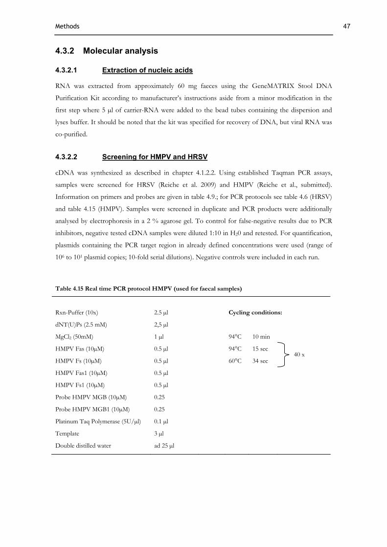
Methods 47
4.3.2 Molecular analysis
4.3.2.1 Extraction of nucleic acids
RNA was extracted from approximately 60 mg faeces using the GeneMATRIX Stool DNA
Purification Kit according to manufacturer’s instructions aside from a minor modification in the
first step where 5 µl of carrier-RNA were added to the bead tubes containing the dispersion and
lyses buffer. It should be noted that the kit was specified for recovery of DNA, but viral RNA was
co-purified.
4.3.2.2 Screening for HMPV and HRSV
cDNA was synthesized as described in chapter 4.1.2.2. Using established Taqman PCR assays,
samples were screened for HRSV (Reiche et al. 2009) and HMPV (Reiche et al., submitted).
Information on primers and probes are given in table 4.9.; for PCR protocols see table 4.6 (HRSV)
and table 4.15 (HMPV). Samples were screened in duplicate and PCR products were additionally
analysed by electrophoresis in a 2 % agarose gel. To control for false-negative results due to PCR
inhibitors, negative tested cDNA samples were diluted 1:10 in H20 and retested. For quantification,
plasmids containing the PCR target region in already defined concentrations were used (range of
106 to 101 plasmid copies; 10-fold serial dilutions). Negative controls were included in each run.
Table 4.15 Real time PCR protocol HMPV (used for faecal samples)
Rxn-Puffer (10x)
2.5 µl
Cycling conditions:
dNT(U)Ps (2.5 mM) 2,5 µl
MgCl2 (50mM) 1 µl 94°C 10 min
HMPV Fas (10µM) 0.5 µl 94°C 15 sec
HMPV Fs (10µM) 0.5 µl 60°C 34 sec
HMPV Fas1 (10µM) 0.5 µl
HMPV Fs1 (10µM) 0.5 µl
Probe HMPV MGB (10µM) 0.25
Probe HMPV MGB1 (10µM) 0.25
Platinum Taq Polymerase (5U/µl) 0.1 µl
Template 3 µl
Double distilled water ad 25 µl
40 x

Methods 48
4.3.2.3 Phylogenetic analysis of HMPV and HRSV RNA from faecal samples
Randomly, positive samples with a viral load > 10 copies/μl cDNA (determined by Taqman PCR)
were selected for further characterisation by PCR assays targeting phylogenetically relevant DNA
fragments (4/21 for the 2004, 6/22 for the 2005 and 3/6 for the 2006 outbreak). Concerning the
HRSV outbreaks, we included samples from individuals that had been tested positive in both 2005
and 2006 (n=3). Protocols are given in chapter 4.1.2.4.
Viral sequences generated from faeces were then compared to sequences previously generated from
tissue samples using BioEdit.
4.3.3 Analysis of observational and molecular data
The prevalence of respiratory disease was calculated for every outbreak based on both molecular
data (percentage of positive tested individuals) and observational data (percentage of individuals
observed with symptoms). Differences in the number of positive tested individuals between
outbreaks caused by the same virus were calculated using the McNemar test and based on repeated
observations of the same respective individuals. We used exact test since the sample sizes were
small (Siegel & Castellan, 1988; Mundry & Fischer, 1998). A p-value < 0.05 was considered
significant.
To compare molecular and observational data, the percentages of observed symptomatic
individuals and PCR positive faecal samples were plotted against the time course of the respective
outbreaks including the mean viral load of the samples. For graphic illustration, the viral load was
represented in terms of categories that differentiate between a low, medium and high viral load.
Therefore, all copy numbers were logarithmised to the base of 10 and the according range was
divided in an upper, middle and lower third (separately for HMPV and HRSV). Since faecal sample
size ranged from 1-12 samples per day, data from every 2 days was pooled to avoid small sample
sizes. Observational data was pooled in the same way to fit the molecular data. Between 2 and 35
chimpanzees were observed per day during the outbreaks.

Results 49
5 Results
5.1 Detection and characterisation of respiratory pathogens
5.1.1 Screening for respiratory viruses
Necropsy samples were screened for influenza virus A and B, HRSV, HMPV, measles virus,
adenovirus, enterovirus, rhinovirus and coronavirus by using different generic PCR methods (see
chapter 4.1.2.3). All available samples tested positive for one of two paramyxoviruses: HRSV was
diagnosed in two individuals (“Lefkas”and “Loukoum”) that died in the 1999 North Group
outbreak and in one adult female (East Group, “Candy”) and one infant (South Group, “Isha's
Baby”) who died in the 2006 outbreak, which occurred simultaneously in both groups. The second
virus identified was HMPV, detected in three animals (“Ophelia”, “Orest”, “Virunga”) that died in
the 2004 South Group outbreak. PCR screening results are given in table 5.1, together with an
overview about mortality, morbidity and the outcomes from the bacterial PCR screening
(performed by M. Leider).
Table 5.1 Characteristics of three respiratory epidemics observed in the Taï chimpanzees
May 1999 March 2004 February 2006
Group
Group size
North
n = 32
South
n = 44
South
n = 34
East
n.d.b
Pathogens identified HRSV
S. pneu. (2308)f
HMPV
S. pneu. (2309),
P. multo. f
HRSV
S. pneu. (2309) f
HRSV
S. pneu. (2308) f
Morbiditya 100% 100% 92% n.d.b
Mortalityd 6c / 32 8 / 44 1 / 34 2
- adult/adolescente 5 / 15 0 / 22 0 / 19 2
- juvenilee 1 / 7 3 / 10 0 / 5 0
- infante 0 / 10 5 / 12 1 / 10 0
a Morbidity rates refer to the total number of weaned individuals observed with respiratory symptoms during an epidemic. b nd, not determined because this group is not fully habituated and the number of individuals unknown. c After this outbreak three infants died of starvation after their mothers died of disease. d Mortality refers to total group size and the number of cases of death due to respiratory disease excluding indirect cases (c). e Age classes for Taï chimpanzees: infant, 0–5 years; juvenile, 5–10 years; adolescent, 10–15 years; adult, >15 years. f For completeness, results from the bacterial screening (performed by M. Leider) are also shown here.

Results 50
5.1.2 Phylogenetic analysis of detected respiratory viruses
To establish the origin of the chimpanzee disease outbreaks, phylogenetic analyses on HRSV and
HMPV were conducted. Both HMPV and two strains of HRSV clustered firmly within known
human clades (figures 5.1 A and B). The 1999 chimpanzee HRSV contained a specific insert of 60
base pairs that were first found in human respiratory outbreaks in Buenos Aires in 1999 (Trento et
al., 2003). The HRSV strain found in the chimpanzees in 2006 grouped most closely with a strain
reported recently from Asia. Both strains belonged to the HRSV subgroup B. For HMPV, fewer
sequences were available, but the chimpanzees’ strains were closely related to strains circulating in
North America and Asia from 1997 to 2000 (Figure5.1 B) and clustered within subgroup B2.
Figure 5.1 Phylogenetic position of HRSV and HMPV amplified from chimpanzees relative to human viruses sampled worldwide. Shown are the phylogenetic trees of HRSV (A) and HMPV (B). Trees were generated under the Maximum Likelihood criterion. Percent bootstrap support for relevant internal nodes is shown above branches. Names of infected chimpanzees are boxed. Stars next to taxa symbols indicate multiple identical sequences from the same locality. Dates associated with the most recent common ancestors (MRCA) of chimpanzee and human viruses were estimated using a Bayesian molecular clock technique; Dates next to ancestral nodes are the estimated year and the 95% posterior density interval. Grey box in A) signifies sequences that share a 60 base pair (bp) insert. Branches in most basal position in B) are not drawn to scale, actual branch lengths are shown below branches. Rooting of the tree was accomplished for HRSV by using the two oldest sequences (1960 and 1962) as outgroups and for HMPV by the midpoint method.

Results 51
Dates associated with the most recent common ancestors (MRCA) of chimpanzee and human
viruses were estimated using a Bayesian molecular clock technique: viruses amplified from
chimpanzees and humans shared a common ancestor within 3–6 years for HRSV and 8 years for
HMPV (figures 5.1.A and B).
5.1.3 Virus isolation
Lung tissue from the HMPV-positive individuals (“Ophelia”, “Orest” and “Virunga”) and the RSV-
positive individuals (“Candy” and “Isha’s Baby”) were used for inoculation of LLC-MK2 or Hep2
cells. No CPE was observed after multiple passages and parallel quantitative PCR analysis of the
supernatant did not show an increase of viral copy numbers.
5.1.4 Characterisation of P. multocida
Six isolates from three individuals were analysed using biochemical and molecular methods. On a
cellular level, small coccoid rods (gram negative) were observed. Colonies were grey-white and
mucoid. No morphological differences were observed between the isolates.
5.1.4.1 Biochemistry
All strains were oxidase and catalase positive. The results concerning ornithine decarboxylation,
formation of indole, splitting of urea and the fermentation reactions of different carbohydrates are
shown in table 5.2. The isolates 114/122/996 differed from the isolates 121/127/420 in the ability to
ferment xylose. Fermentation reactions were also considered for the typing of the subspecies; here,
variations in sorbitol, dulcit and arabinose have been reported to be of taxonomic relevance
(Mutters et al., 1985). Results of the reactions were read off after 24 and 48 hours. Ambiguous
results were performed repeatedly.
Table 5.2 Biochemical profiles of P. multocida isolates from chimpanzees
Oxi
dase
Orn
ithin
e
Indo
l
Ure
ase
Tre
halo
se
Mal
tose
Sacc
haro
se
Xyl
ose
Ara
bino
se
Man
nit
Sorb
itol
Dul
cito
l
Isolates 114/122/996 + + + - +/- - + - + + + -
Isolates 121/127/420 + + + - +/- - + + + + + -
+: positive -: negative +/-: ambigous result

Results 52
5.1.4.2 Antibiotic profile
Antimicrobial susceptibility testing of the isolates against 16 antimicrobials was performed by agar
diffusion test. The panel of antibiotics included β-lactam antimicrobials (amoxicillin, ampicillin,
penicillin, cefalexin, cefazolin, cefovecin), aminoglycosides (amikacin, gentamycin), fluorchinolone
(enrofloxacin, marbofloxacin), tetracycline (doxycycline, tetracycline), sulfadimidine
(sulfamethoxazole with trimethoprim), lincosamide (clindamycin), polymyxine (polymyxin B) and a
broad spectrum antibiotic (chloramphenicol). Isolates were intermediate susceptible to tetracycline,
resistant to clindamycin and sensitive to all other antibiotics tested.
5.1.4.3 Genotyping
PFGE revealed the presence of two different clones: using SmaI, two patterns were distinguished
(see figure 5.2), differentiating between the isolates 114/122/996 (referred to as clone 1 in the
following text) and 121/127/420 (referred to as clone 2). Four of the isolates originated from the
same individual (“Virunga”: isolate 114, 121, 122 and 127), two of which turned out to be identical
(114=122 and 121=127). From “Ophelia” (isolate 420) and Sagu (isolate 996) one clone each had
been isolated; “Virunga” had been infected with both clones. Data on restriction with ApaI are not
shown, since the isolates belonging to clone 2 repeatedly could not be digested with that enzyme.
Figure 5.2 Dendrogramm (% similiarity) showing DNA restriction pattern after digestion with SmaI for all isolates. NCTC 1032 served as a positive control. Analysis was conducted with BioNumerics using UPGMA. Dice coefficient: 1 % tolerance and 0.5 % optimisation.
5.1.4.4 Identification of P. multocida and capsule typing
Using PCR methods, isolates were investigated for the kmt gene and the presence of three capsule
biosynthesis genes. All analysed isolates were of capsular type A and harboured the species specific
gene sequence kmt (see figure 5.3 A).
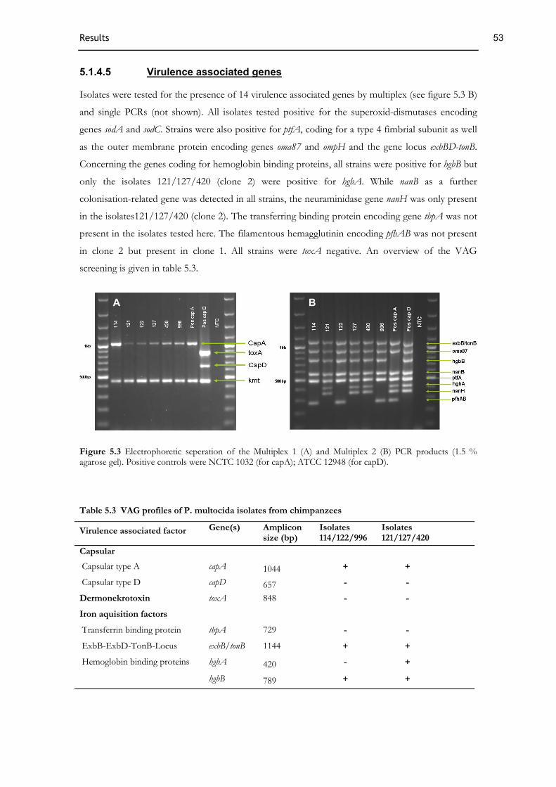
Results 53
5.1.4.5 Virulence associated genes
Isolates were tested for the presence of 14 virulence associated genes by multiplex (see figure 5.3 B)
and single PCRs (not shown). All isolates tested positive for the superoxid-dismutases encoding
genes sodA and sodC. Strains were also positive for ptfA, coding for a type 4 fimbrial subunit as well
as the outer membrane protein encoding genes oma87 and ompH and the gene locus exbBD-tonB.
Concerning the genes coding for hemoglobin binding proteins, all strains were positive for hgbB but
only the isolates 121/127/420 (clone 2) were positive for hgbA. While nanB as a further
colonisation-related gene was detected in all strains, the neuraminidase gene nanH was only present
in the isolates121/127/420 (clone 2). The transferring binding protein encoding gene tbpA was not
present in the isolates tested here. The filamentous hemagglutinin encoding pfhAB was not present
in clone 2 but present in clone 1. All strains were toxA negative. An overview of the VAG
screening is given in table 5.3.
Figure 5.3 Electrophoretic seperation of the Multiplex 1 (A) and Multiplex 2 (B) PCR products (1.5 % agarose gel). Positive controls were NCTC 1032 (for capA); ATCC 12948 (for capD).
Table 5.3 VAG profiles of P. multocida isolates from chimpanzees
Virulence associated factor Gene(s) Ampliconsize (bp)
Isolates114/122/996
Isolates 121/127/420
Capsular
Capsular type A capA 1044 + +
Capsular type D capD 657 - -
Dermonekrotoxin toxA 848 - -
Iron aquisition factors
Transferrin binding protein tbpA 729 - -
ExbB-ExbD-TonB-Locus exbB/tonB 1144 + +
hgbA 420 - + Hemoglobin binding proteins
hgbB 789 + +
A B
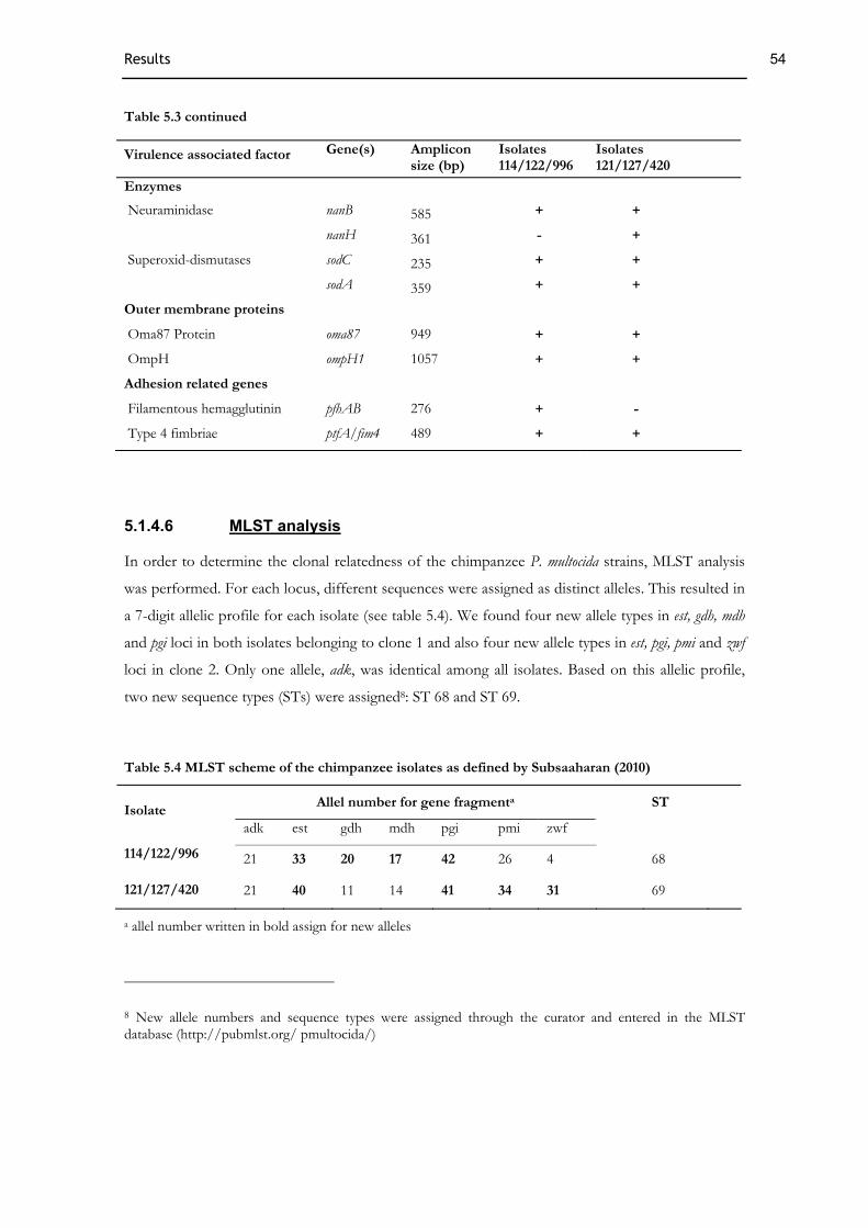
Results 54
Table 5.3 continued
Virulence associated factor Gene(s) Ampliconsize (bp)
Isolates114/122/996 ( l 1)
Isolates 121/127/420 ( l 2)
Enzymes
nanB 585 + + Neuraminidase
nanH 361 - +
sodC 235 + + Superoxid-dismutases
sodA 359 + +
Outer membrane proteins
Oma87 Protein oma87 949 + +
OmpH ompH1 1057 + +
Adhesion related genes
Filamentous hemagglutinin pfhAB 276 + -
Type 4 fimbriae ptfA/fim4 489 + +
5.1.4.6 MLST analysis
In order to determine the clonal relatedness of the chimpanzee P. multocida strains, MLST analysis
was performed. For each locus, different sequences were assigned as distinct alleles. This resulted in
a 7-digit allelic profile for each isolate (see table 5.4). We found four new allele types in est, gdh, mdh
and pgi loci in both isolates belonging to clone 1 and also four new allele types in est, pgi, pmi and zwf
loci in clone 2. Only one allele, adk, was identical among all isolates. Based on this allelic profile,
two new sequence types (STs) were assigned8: ST 68 and ST 69.
Table 5.4 MLST scheme of the chimpanzee isolates as defined by Subsaaharan (2010)
Allel number for gene fragmenta ST Isolate
adk est gdh mdh pgi pmi zwf
114/122/996 21 33 20 17 42 26 4 68
121/127/420 21 40 11 14 41 34 31 69
a allel number written in bold assign for new alleles
8 New allele numbers and sequence types were assigned through the curator and entered in the MLST database (http://pubmlst.org/ pmultocida/)

Results 55
5.1.4.7 Phylogenetic analysis
For molecular taxonomy, a phylogenetic tree of the sodA gene had been created which is shown in
figure 5.4. The topology of the sodA tree shows that taxa representing different P. multocida
subspecies are separated, however, statistical support was only given for the branching of P.
multocida ssp. septica and P. multica ssp. multocida/P. multocida ssp. gallida. The chimpanzee isolates
group closest with strains of the subspecies P. multica ssp. multocida or, in the case of isolates
114/122/996, sequences were even identical with strains that had been classified as subspecies
multocida (strain CNP 927 and CNP 954 (Gautier et al., 2005)).
Figure 5.4 Phylogenetic analysis of the sodA gene. The tree was built using the maximum-likelihood method from an analysis of sodA sequences (452bp) from the chimpanzee’s isolates and sequences obtained from GenBank. Isolates belonging to the species P. multocida are boxed grey; the chimpanzees isolates are written in bold. Taxon labels indicate species and strain number, including strains with identical sodA sequences (species and subspecies assignments are according to Gautier (2005)). P. langaensis and P. bettyae were used as outgroup. Bootstrap values were calculated with 500 replicates and are given in percent.
To further analyse the genetic relationships between the chimpanzees and other isolates,
concatenated gene sequences from both STs were compared to the available P. multocida STs from
the MLST database. A ML tree was constructed using the concatenated sequences of 73 STs (see
figure 5.5) and taxa were colour-coded according to the host they had been isolated from. Due to a
lack of a suitable outgroup, the data is presented as an unrooted radial tree. Although not supported
by a bootstrap value > 70%, the resulting tree topology depicted two groups; this division was in

Results 56
general agreement with the population structure recognised by previous studies using MLST
(Subsahaaran et al., 2010), ribotyping and MLEE (Blackall et al., 1998): one group consists of
isolates originating exclusively from birds and cats9 and includes the subspecies multocida and
septica as well as the type strain of the subspecies septica. The other group (shown as a subtree; see
figure 5.5.A) includes strains from various host (mainly birds, cattle, and pigs) and include the
typing strains of the subspecies multocida and gallicida. The STs found in the chimpanzees group
firmly together with strains of the latter group.
Figure 5.5 Radial Maximum Likelyhood trees constructed with concatenated MLST allele sequences. Shown left is the complete MLST tree constructed with 73 STs and the position of the type strains for the P. multocida subspecies. The position of the chimpanzee isolates (boxed grey) is displayed in the subtree (A). Taxa are labelled with coloured dots indicating the isolation sources and ST numbers . Bold branches indicate for bootstrap values > 70 %.
9 Although the typing strain for P. multocida ssp. septica had been isolated from a human this might be misleading. Humans are not considered to carry this pathogen naturally and infections are mostly inflicted by scratches or bites from cats or dogs.

Results 57
5.2 Analysis of further potential transmitters
5.2.1 Human samples
Samples from humans with acute signs of respiratory disease were analysed. Therefore, cDNA
from 71 throat swabs were screened for the respiratory viruses which had been detected in the
chimpanzees as described above (chapter 5.1.1). All analysed samples tested HRSV- and HMPV
negative.
5.2.2 Colobus samples
Samples from ten P. badius and ten C. polykomus were analysed. cDNA from throat swabs was
screened for the presence of HRSV and HMPV and tested negative. Furthermore, DNA was
screened for the P. multocida specific kmt gene. All analysed samples were negative in PCR for this
genomic region.
5.3 Noninvasive diagnostic: evaluation of the incidence of respiratory viruses
5.3.1 Screening results
Faecal samples that had been collected during four distinct outbreaks; two with known aetiology
(March 2004 and February 2006) and two with unknown aetiology (October 2004 and August
2005) were screened for the presence of HMPV and HRSV RNA.
For the respiratory outbreak in March 2004, with known involvement of HMPV, faecal samples
were tested for HMPV by real time PCR. Sixty-five faecal samples collected from 29 chimpanzees
were analyzed and 72 % of the individuals tested positive. For the two outbreaks in October 2004
and August 2005, from which no necropsy samples are available, 29 and 60 faecal samples from 15
and 24 individuals, respectively, were screened for HMPV and HRSV. From the outbreak in
October 2004, only one individual tested weakly positive for HMPV, and in the August 2005
outbreak 92 % of the individuals were positive for HRSV. From the outbreak in 2006 with known
involvement of HRSV, 42 faecal samples from 24 individuals were analysed and 25 % of indivduals
tested positive for HRSV. All faecal samples collected during non-outbreak times were negative for
HMPV and HRSV. The results from the faecal sample screening are summarized in table 5.5.
From real time PCR assays, the amount of viral cDNA ranged from 9x101 to 1.1 x10⁵ copies/g
faeces for the HMPV assay and from 1.1x10² to 6.7x10⁵ copies/g faeces for the HRSV assay. PCR
products were additionally analysed by gel electrophoresis. Negative samples were re-tested using a
1:10 dilution, but no inhibitory effect was observed using this approach. Samples were considered

Results 58
negative when neither a Ct-value nor a band on the gel was present. On the individual level (in case
multiple samples were available), copy numbers varied over the time course of the outbreak and
were usually lower or negative in the beginning and the end of the outbreak. Rarely, individuals
tested positive, then negative and then positive again. Details on the viral loads related to individual
samples are shown for the HMPV outbreak in March 2004 and both HRSV outbreaks in 2005 and
2006 (see table 5.6, 5.7 and 5.8).
Table 5.5 Characteristics of respiratory epidemics observed in the South Group of Taï chimpanzees and molecular results of faeces samples including healthy control group.
March
2004
October
2004
August
2005
February
2006
Control b
Observed symptoms respiratory respiratory respiratory respiratory no symptoms
Number of observed animals
n=28 n=22 n=35 n=34 n=35
Morbidity 100% 64% 100% 92% 0
Viruses identified from tissue samples
HMPV no tissue samples available
no tissue samples available
HRSV no tissue samples available
Pathogens identified from faecal samples
HMPV HMPV HRSV HRSV None
Positive tested faecal samplesa
37/65 3/29 43/60 8/42 0/54 b
Positive tested individuals
21/29 (72%) 1/15 (7%) 22/24 (92%) 6/24 (25%) 0/21b
a Based on all available faecal samples which had been collected during the respiratory disease outbreaks. Individuals were sampled 1-7 times except for the control group were only one sample from each individual was tested.
b Control samples were selected from a period when the whole group appeared healthy and showed no respiratory symptoms: in June/July 2005 one sampled from each of 21 individuals was tested for the presence of HMPV and HRSV. To determine if these viruses were persistent in affected individuals additional samples which had been collected in the time between consecutive outbreaks of HMPV or HRSV were tested: 13 samples from 5 individuals which had been observed with symptoms in both the HMPV outbreak in March and October 2004 were tested for HMPV. In addition, samples from 6 individuals that tested HRSV positive in both the RSV outbreak in August 2005 and February 2006 were tested for HRSV (n=20).

Results 59
Table 5.6 Screening results for all tested faecal samples from the HMPV outbreak in March 2004
IndividualsSettut neg
Sumatra Feces neg
Isha Feces 4,5 2,9
Inousha 3
Zyon 3,4 2,6
Besar 3,7 neg
Utan neg
Louise Feces 5,1 3,5 2,4
Zora Feces 2,9 neg
Kaos 4,2 4,2
Sagu Feces 3,8 3,9* neg neg neg
Kinshasa 4,1 neg
Mustapha neg
Gogol Feces 4,9 neg neg
Kuba neg
Yucca 3,6
Ibrahim neg
Duna 2,8 neg
Coco 4,2 3,2 4 neg 2,7 2,6 neg neg
Woodstock 2,9
Celine neg neg neg neg
Julia neg neg neg 2,8 4*
Romario 4,1 2,8 neg
Rubra 3,6 3,2 neg
Taboo Feces neg
Wapi 2,9
Jacobo 2,2 2,4
Rebecca 2,4
Caramel neg
3 4 5 6 7 8 9 10 11 12 13 14 15 16 17 18 19 20 21 22 23 24 25 26 27 28 29 30
Days after first symptoms observed
Viral load log10 (copies/gram) > 4 < 4 < 3 neg negative * sampled twice the same day (mean)
Table 5.7 Screening results for all tested faecal samples from the HRSV outbreak in August 2005
IndividualsAtena 3,6 neg
Kinshasa neg 5
Taboo 4,1 5,1 neg
Coco 5,1
Gogoll neg 5,7 5,4 2,9
Ibrahim 4,7 neg
Kuba 4,9 5,4
Utan 4,8 neg 2,6
Kaos 1,5 5,1 2,4 3,1
Celine 4,8 3,9 neg
Louise 5,3*
Zora 5,2 neg neg
Sumatra 5,8 5* 5
Zyon 5,2 4,9 3,3
Woodstock 4,4 4
Shogun 3,7* 2,6
Sagu 4,2
Yucca 4,8
Jacobo 5 2,9
Wapi 4
Caramel neg
Olivia 2 neg 3
Romario neg
Rubra 2,6 2,6**
1 2 3 4 5 6 7 8 9 10 11 12 13 14 15 16 17 18 19 20 21 22 23 24 25 26 27
Days after first symptoms observed
Viral load log10(copies/gram) > 4,7 < 4,7 < 3,4 neg negative
* sampled twice the same day (mean) ** sampled twice the same day and tested one time positive, one time negative (mean)

Results 60
Table 5.8 Screening results for all tested faecal samples from the HRSV outbreak in February 2006
IndividualsAthena neg
Caramel neg neg
Celine 2,6
Coco neg
Gogol neg
Ibrahim neg
Isha neg
Jacobo neg
Java neg
Julia neg
Kaos 3,5 2,6
Kuba 2,3 2,2 neg
Louise 2,6 neg neg neg
Lula 2,1 neg neg
Olivia neg neg
Sagu neg neg neg
Shogun 2,7
Sumatra neg neg neg
Taboo neg neg
Wapi neg
Woodstock neg
Zora neg neg
Zyon neg
Kinshasa neg neg neg
2 3 4 5 6 7 8 9 10 11 12 13 14 15 16 17 18 19 20 21 22 23 24 25 26 27
Days after first symptoms observed
Viral load log10(copies/gram) < 4,7 < 3,4 neg negative
5.3.2 Sequence analysis of HMPV and HRSV RNA from faecal samples
In order to determine which strains of each virus infected the chimpanzees, we generated sequence
information. For HMPV, a 867 bp fragment of the P gene and for HRSV a 309 bp fragment of the
G gene were compared to sequences we had obtained from lung tissue (Gen Bank accession
numbers: EU240452 to EU240455) of chimpanzees that had died during these outbreaks. The virus
sequences obtained from faecal samples shared >99.5% nucleotide identity with those obtained
from tissue samples. Sequences from the HRSV outbreak in 2006 and the outbreak in 2005 of
unknown aetiology were identical. Unfortunately, it was not feasible to generate sequences for
phylogenetic analyses from the weakly positive samples from the one positive individual from the
HMPV outbreak in October 2004, where the analysed samples contained less than 10 copies/µl
cDNA (determined by Taqman PCR). Here, only sequences from the PCR products from the real
time PCR were generated, which confirmed the presence of HMPV. Thus we were not able to
compare faeces-derived, phylogenetically relevant sequences from the known HMPV in the March
2004 outbreak and the consecutive outbreak in the same year.

Results 61
5.3.3 Prevalence of RSV and HMPV infections among the South Group of Taï chimpanzees
The virus prevalence for the respective outbreaks is shown in figure 5.6 together with the
percentages of individuals observed with symptoms. In March 2004, 21 of 29 (72 %) individuals
tested positive for HMPV whereas only one individual tested positive seven months later in
October 2004. For HRSV 92 % of individuals tested positive during the outbreak in 2005, and
25 % of individuals tested positive during the second outbreak in 2006. Virus prevalence was always
higher in the first HMPV (March 2004) and respectively HRSV (August 2005) outbreak than in the
consecutives ones. In contrast, only slight differences were observed for the number of
symptomatic individuals. For both the HMPV and the HRSV epidemics, there is a significant
difference in the number of infected individuals between the first and consecutive outbreak (exact
Mc Nemar Test, both p <0.001).
Figure 5.6 Percentages of infected individuals in A) the HMPV outbreaks in 2004 and B) the HRSV outbreaks in 2005 and 2006. Observed morbidity (striped) was high in all outbreaks, while the percentages of positive tested individuals (dark grey) decreased in the consecutive outbreaks, both for HMPV and HRSV.
5.3.4 Comparison of molecular and observational data over the time course of the outbreaks
Except for the HMPV outbreak in October 2004, all outbreaks were documented in detail, making
it possible to compare laboratory based and observational data over the course of the outbreaks.
Therefore, percentages of symptomatic individuals, PCR positive samples and respective mean viral
load were plotted against the duration of the outbreaks (Figure 5.7). Both, the diagnostic and the
observational data sets reflect the course of the disease, with fewer symptomatic cases, positive-
tested samples and lower viral load early on; with a progression toward a higher viral load and more

Results 62
symptomatic and positive-tested individuals, leading to a peak where almost all individuals were
highly infected prior to a decline in disease symptoms and viral load.
For both, HMPV and HRSV, the first epidemic lasted longer then the consecutive epidemics
(HMPV: 31 days (March 2004) vs. 9 days (October 2004); HRSV: 22 days (August 2005) vs. 15 days
(February 2006).
Figure 5.7 Time trends of the HMPV outbreak in March 2004 and the HRSV outbreak in 2005 and 2006. Data is based on the percentage of individuals seen with signs of respiratory symptoms (striped) and samples tested positive for HMPV or HRSV by molecular analysis (shaded coloured). Data from every two days were pooled to avoid small sample sizes. The viral load is colour-coded differentiating between low (yellow), medium (orange) and high (red) copy numbers (categories were defined as described in chapter 4.3.3) Number symbols (#) indicate that no faecal samples had been collected during these days.

Discussion 63
6 Discussion
6.1 Detection and characterisation of respiratory pathogens from tissue samples
6.1.1 Paramyxoviruses: HMPV and HRSV
Tissue samples from seven deceased chimpanzees were analysed for the presence for respiratory
viruses. Using PCR, all available samples tested positive for one of two paramyxoviruses: human
respiratory syncytial virus (HRSV) was diagnosed in two individuals that died in the 1999 North
Group outbreak and in one adult female (East Group) and one infant (South Group) who died in
the 2006 outbreak, which occurred simultaneously in both groups. The second virus identified was
human metapneumovirus (HMPV), detected in three animals that died in the 2004 South Group
outbreak. Additionally, an effort has been made to cultivate these viruses, but no viral growth could
be observed. This could be due to the rather poor sample quality, as the time span between death
and necropsy of the animal was at least 10 hours.
HRSV and HMPV are common causes of respiratory disease in humans and are the leading causes
of lower respiratory disease in children and, in developing countries, a major source of infant
mortality (Weber et al., 1998; Boivin et al., 2003). In adults, HRSV and HMPV usually cause mild
upper-respiratory-tract infections but can lead to pneumonia and bronchiolitis. Both viruses are
shed in respiratory secretions but also have been detected in faeces or sweat from infants (von
Linstow et al., 2006). Transmission of HRSV occurs through droplets of respiratory secretions or
through direct contact with contaminated fomites: HRSV in fresh secretions survives 20 min on
hands and up to 7 hr on plain surfaces (Hall & Douglas, 1981). HRSV and HMPV also are known
to cause respiratory symptoms in captive chimpanzees (Clarke et al., 1994; Skiadopoulos et al.,
2004). Although paramyxoviruses can cause severe respiratory symptoms in their own right, they
also predispose captive chimpanzees to secondary bacterial infection (Jones et al., 1984). As has
been shown before, S. pneumoniae (all outbreaks) and P. multocida (outbreak March 2004) were also
found in the lung tissue of the chimpanzees (Chi et al., 2007). Thus, though the outbreaks among
Taï chimpanzees may have been initially triggered by HRSV and HMPV, secondary infection with
S. pneumoniae or P. multocida were likely to be the proximate cause of death.
6.1.2 Origin of detected paramyxoviruses
Viral sequences generated from chimpanzees were compared to sequences from human patients
that were available in GenBank. Both HMPV and two strains of HRSV clustered firmly within
known human clades (Figure5.1 A and B). HRSV strains are known to circulate globally and tend to
form temporal, rather than regional, clusters (Cane & Pringle, 1995). This is also evident from the

Discussion 64
HRSV tree (Figure 5.1 A), where closely related strains were often distributed worldwide.
Intriguingly, the 1999 chimpanzee HRSV contained a specific insert of 60 base pairs that were
found in human respiratory outbreaks in Buenos Aires in 1999 (Trento et al., 2003). The HRSV
strain found in the chimpanzees in 2006 grouped most closely with a strain reported recently from
Asia. For HMPV, fewer sequences were available in the data banks, but the chimpanzee’s strains
were closely related to strains circulating in North America and Asia from 1997 to 2000 (Figure 5.1
B).
Unfortunately, HRSV and HMPV sequences from humans of West or Central Africa were not
available for the outbreak years. Hence, in order to investigate the origin of the chimpanzee
outbreaks, throat swab samples from humans with close contact to the Taï chimpanzees were
examined for the presence of HRSV and HMPV. Humans with close contact include a) the reseach
assistants of the TCP and b) villagers from the region who enter the forest for poaching or who
have contact to assistants or poachers. In a previous work by Schenk (2007), throat swab samples
from the assistants were collected during outbreak times and analysed for the presence of
respiratory viruses, but HMPV and HRSV were detectable in none of the samples. In the present
work, samples from villagers with clinical signs of respiratory disease were screened for respiratory
viruses. Time of sample collection was 3-11months after the last outbreak among the chimpanzees,
but subtypes of both HRSV and HMPV can circulate for more than one season (Peret et al., 1998;
Venter et al., 2001; Sloots et al., 2006).
However, although we could not detect HRSV and/or HMPV in human samples with close
contact to the chimpanzees, the analysis here (conducted in BEAST) indicates that in all three cases
viruses amplified from chimpanzees and published virus sequences from humans shared a common
ancestor within 3–6 years (HRSV) and 8 years (HMPV). As humans are the only known reservoir
host for both viruses, these results strongly suggest that humans introduced the two viruses directly
and repeatedly into wild chimpanzee populations in the recent past.
The chimpanzee groups do not range outside the park, and there are currently no villages or
plantations inside the park. Thus, either research personnel or poachers are the most plausible
sources of infection. However, potential transmission foci, such as poaching camps, have been
detected in the study-group territories on only a few occasions over the last 24 years, and poachers
only occasionally enter the study-group core areas where most chimpanzee activity is concentrated.
In contrast, an average of about one research personnel spends 8 hr a day within 15 m of
chimpanzee parties typically containing 5–10 individuals. Therefore, it is highly likely that personnel
have been responsible for virus transmission.
Another possibility would be that transmission of the paramyxoviruses occurred through bridge
hosts. However, contact with respiratory pathogens deposited by humans on the forest floor is

Discussion 65
much less likely for monkeys, which are largely arboreal, than it is for chimpanzees, which are
regularly terrestrial.
6.1.3 Respiratory bacteria: characterisation of detected P. multocida strains
Previous work on the involved bacteria revealed that beside two new strains of S. pneumoniae, P.
multocida played a role in the outbreak in March 2004 (see Chi et al., 2007). In the present work P.
multocida isolated from chimpanzee samples was characterised with molecular and biochemical
methods.
P. multocida has a wide disease and host spectrum, ranging from haemorrhagic septicaemia in cattle
and fowl cholera in birds, respectively, where it is considered a primary pathogen, through
secondary invaders of pneumonic lesions (Carter, 1984). Research on P. multocida has been mostly
limited to strains from livestock, poultry or companion animals, where it exists as a commensal in
the upper respiratory tracts. Furthermore, there is few data on human strains, however, P. multocida
infections are considered as a zoonosis and cases are mainly associated with animal bites or
scratches. In NHP, respiratory infections due to P. multocida have been described for various species
held in captivity (Good & May, 1971; McClure et al., 1986; Kalter 1989). For example, analysing
respiratory disease among a dynamic laboratory colony including > 8000 animals and 10 different
species of NHPs P. multocida was among the major bacterial pathogens isolated (Good & May,
1971). Among newly imported macaques that died with pneumonia, P. multocida or S. pneumoniae
were the most common bacterial isolates from the respiratory system (Lewis, 1975) and in the
South American owl monkey P. multocida was found as the principle pathogen (Good and May,
1971). P. multocida has also been found to be involved in air sacculitis among several captive NHP
species, including great apes (Gross, 1978; McClure et al., 1986; Kalter, 1989). Whether
pasteurellosis in NHPs arises from commensal bacteria or from bacteria transmitted from other
animals has been barely investigated. There is only one study where P. multocida was found to be
part of the pharyngeal flora in healthy wild-born baboons (Brondson & DiGiacomo, 1993). So far,
the relevance of P. multocida for wild living chimpanzees is unclear.
This is the first detailed description of P. multocida isolated from wild chimpanzees. Isolates found in
the lungs of deceased chimpanzees living in Taï National Park were compared to an isolate that
originated from the pus from a chimpanzee that underwent surgery due to air sacculitis. Based on
the fermentation reactions of different carbohydrates, all isolates matched the properties of
previous described strains of the taxon P. multocida (Mutters 1989; Blackall et al., 1994; Fegan et al.,
1995; Ekundayo et al., 2008). Two different clones were identified using pulsed field gel
electrophoresis. Both clone 1 and clone 2 were isolated from the lung samples collected during the
respiratory outbreak in 2004. A clone 1 type bacterium was also recovered from a pus sample from

Discussion 66
“Sagu” who suffered from air sacculitis. PCR analysis showed that all isolates were of capsular type
A. The capsular type in P. multocida is usually host and disease associated and is assumed to play a
role in host and disease specificity. Capsular type A strains are the predominant type associated with
fowl cholera and respiratory disease/pneumonia in ruminants (Chanter & Rutter, 1989; Choi et al.,
2001; Ewers et al., 2006; Ross, 2006), the latter corresponds perfectly to the clinical picture we
observed in the deceased chimpanzees.
P. multocida constitutes a heterogeneous species and virulence features are divergent. However,
efforts have been made to correlate several VAG pattern to distinct hosts and various diseases
(Ewers et al., 2004, 2006; Bethe et al., 2009). Hence, to learn more about the characteristics of the
strains found in the chimpanzees, the respective VAG patterns were examined. Differences in
between the isolates were in agreement with the results obtained from pheno- and genotyping.
Most of the regularly distributed VAGs could be also detected in the chimpanzees’ strains: for
example, genes coding for outer membrane proteins (ompH and oma87), type 4 fimbriae (ptfA),
superoxide dismutases (sodA, sodC), and iron acquisition related factors (exbB/tonB) were present in
all of the isolates. Concerning the VAGs associated with high pathogenic potential (toxA, pfhAB
and tbpA), only clone 1 showed to be pfhAB-positive.
For taxonomic assignment at the subspecies level, both biochemical and phylogenetic analysis were
taken into account. Sequence analysis of the sodA gene has been proposed as an accurate tool to
type Pasteurella species and subspecies (Gautier et al., 2005). Therefore, sodA sequences from the
chimpanzee isolates were generated and compared to sequences from Genbank. The chimpanzee’s
sodA sequences group firmly within strains belonging to the subspecies P.multocida ssp. multocida and
P. multocida ssp. gallicida (Figure 5.4). However, based on the analysis performed here, a further
identification to the subspecies level was not possible. This might be due to the fact that only few
sodA sequences are available but also because the sodA variability within these subspecies is too low.
Results from the fermentation reactions were also ambigous: for example, e.g. a key characteristic
for the subspecies P. multocida ssp. multocida includes dulcitol-negative, sorbitol-positive isolates
(Mutters et al., 1985), which was complied by all of our isolates. In contrast, both clones were
positive for the fermentation of arabinose, a feature normally observed in the subspecies P. multocida
ssp. gallicida (Mutters et al., 1985; Fegan et al., 1995; Blackall et al., 1997). However, conflicting
results from both molecular and phenotypic data have been described in other investigations
(Gerardo & Goldstein, 1999; Petersen et al., 2001; Davies et al., 2004), suggesting that the precise
typing of the subspecies is complex and has yet to be satisfactorily resolved.
When MLST analysis was performed, four new alleles were identified in each clone. Based on these
allelic profiles two new STs could be assigned (ST 68 and ST 69). MLST is a highly specific,
sensitive and stable tool and is one of the “gold standards” used for typing of bacteria. The MLST
scheme used here has only recently been developed (Subaaharan et al., 2010) and at the time of

Discussion 67
writing consisted of 177 isolates representing 73 STs which build six clonal complexes (with a
clonal complex being defined as STs that shared 6 or more loci). Isolates are mostly of avian
(72.5 %), but also of porcine (13.2 %), bovine (2.8 %), feline (1.7 %), rodent (1.1 %) and human
origin (1.1 %). Isolates were collected in 14 countries, mainly in Australia and followed by Germany
and Denmark. To further analyse the genetic relationships, concatenated gene sequences from the
chimpanzees STs were compared to STs from the MLST database (Figure 5.5). Both STs group
together with strains originating from various hosts, clustering closely with the typing strains of the
subspecies P. multocida ssp. multocida and gallicida. However, it should be noted that this database is
still in development. So far it consists mostly of isolates obtained from domesticated animals and
from a limited number of geographical locations; furthermore, i.e. strains of the subspecies
multocida/gallicida originated from humans have yet not been added. Based on the present data, it
can be stated that although the chimpanzees strains show phylogenetic differences they are closely
involved in the epizootiology of P. multocida.
This is the first description of P. multocida involved in diseases of wild living chimpanzees. Up to
now, there is no detailed molecular data on African or NHP-associated strains, hence it was
interesting that the strains analysed here show such a high similarity to known strains of P. multocida.
However, additional data from African countries and other primates would be useful to clarify the
positioning of the chimpanzees P. multocida.
6.1.3.1 The role of P. multocida in acute and chronic respiratory disease
Pasteurellosis in captive NHPs often occurs when local and systemic defence mechanisms are
impaired. There are a variety of predisposing factors resulting in increased levels of stress:
transportation, crowding or by the damaging effects of respiratory viral infections (Bennet, 1989).
In addition, NHPs have developed P. multocida infections secondary to surgical procedures, chair
restraint, or chronic catheterization (Brondson & DiGiacomo, 1993). As already mentioned above,
the respiratory outbreak in March 2004 was caused by a multifactorial infection, where HMPV as
well as S. pneumoniae and P. multocida were found in the lung tissue. It is thus difficult to determine
the actual role played by P. multocida in the causation of disease, but it can be assumed that the
underlying viral infection has determined the secondary infection by the bacteria which might have
then caused the lethal outcome. One strain of the P. multocida involved in the respiratory disease
(isolate 114/122/996 or respectively ST 68) was recovered in the purulent discharge of the air sacs
from a chimpanzee sampled five years after the outbreak. First signs of its airsacculitis were already
observed in 2004, but it was not until 2009 that the size of the swellings of the airsacs became life-
threatening and required surgical intervention. Apart from P. multocida, Enterobacter sp. and Prevotella
sp. were also isolated from the air sacs (data not shown), showing again that P. multocida was
probably not the single cause. This is in agreement with the literature, where airsacculitis has been
often associated with mixed infections of enteric organism (Hill et al., 2001). Furthermore, this

Discussion 68
individual was also infected by HMPV during the respiratory outbreak in 2004, as determined by
screening faecal samples collected during that time (see figure 5.6). It might be possible that the
respiratory disease in 2004 was the initial cause for the development of the airsacculitis. As an
example, this has been shown in a study on airsacculitis in orang-utans, where a significant
proportion of affected animals had a history of recent upper respiratory tract infection (Strobert
and Swenson, 1979).
Hence, the chimpanzee strains were involved in acute and/or chronic respiratory disease. Since
both infections were caused by a mix of viral and/or bacterial pathogens, it might be assumed that
P. multocida found here acted as an opportunistic pathogen. The fact that the chimpanzee’s strains
were lacking the major VAGs associated with primary disease is in favour of this assumption.
However, further isolates from healthy and diseased chimpanzees are needed to evaluate their
potential carrier status and their possible role in disease development.
6.1.3.2 P. multocida isolated from chimpanzees – natural carrier status versus transmission from other animal species?
Another question is, whether the chimpanzees are naturally carriers of P. multocida or if this
pathogen has been transmitted to the chimpanzees from other species. As stated above (chapter 2.2
and 2.3), evidence exists that interspecies transmission may play a substantial role, and transmission
cycles have been described between both humans, chimpanzees and other monkeys (Wolfe et al.,
2004; Goldberg et al., 2007; Leendertz et al., 2008). On the one hand, chimpanzees share a
considerable array of the same pathogens with humans due to our close genetic relationship. Both
zoonotic (Gao et al., 1999; Wolfe et al., 2004; Keele et al., 2006; Leendertz et al., 2008) and
anthropozoonotic disease transmission have been described (Kalter, 1989; Wallis & Lee, 1999;
Kaur et al., 2008; Köndgen et al., 2008). On the other hand, virus transmission from other primate
species (i.e. colobus monkeys) to chimpanzees has been described in the context of predation
(Leendertz et al., 2004b; Leendertz et al., 2008). Concerning the respiratory outbreak in 2004, there
is strong evidence that the viruses involved were transmitted from humans (see chapter 6.1.2). In
contrast, the found S. pneumoniae was assumed to be harboured naturally by the chimpanzees, as
their MLST profile differed from that of strains isolated from humans working with the
chimpanzees (Chi et al., 2007). P. multocida is considered a strict animal pathogen and in humans,
infections are mostly inflicted by animal contact (bite and scratch injuries). However, among animal
exposed humans (i.e. animal handlers, farm workers) 2-5% seem to harbour P. multocida in the
throat or oropharynx (Smith, 1959; Jones & Smull, 1973; Boivin & Leterme, 1974; Avril et al.,
1990). Hence, although the knowledge about P. multocida in healthy people is scarce, animal contact
can lead to a human carrier state. Furthermore, evidence exists that strain transmission between
cattle, pigs, and poultry occurs (Davies et al., 2004). This is also shown in the MLST tree, where the
same STs have been isolated from different host (see figure 5.5). Therefore, it might be possible

Discussion 69
that the chimpanzees got infected through contact with other animals harbouring P. multocida. In
most animals, P. multocida is primarily transmitted by the respiratory route, including direct contact
to infectious secretions, inhalation of aerosols or intake of contaminated water or food (Backstrand
& Botzler, 1986; Botzler, 1991; Thomson et al., 1992). The Taï chimpanzees share their habitat with
many different mammals, including ruminants, rodents and also carnivores most of them known as
carriers of P. multocida (Schipper 1947; DiGiacamo et al., 1983; Burdge et al., 1985; Jaworksi et al.,
1998; Talan et al., 1999; Dunbar et al., 2000; Pedersen et al., 2003; Dabo et al., 2007). As food
sources like fruit trees are shared jointly it is possible that chimpanzees get in contact with
infectious secretion from infected animals and that oral ingestion of P. multocida resulted in
oropharyngeal colonisation. Alternatively, P. multocida could have been transmitted from colobus
monkeys, which are known to be prey species of the Taï chimpanzees (Boesch & Boesch-
Achermann, 2000). Therefore, throat swabs from 20 apparently healthy colobus monkeys were
analysed for the presence of P. multocida, but all tested negative. Further samples and species need to
be analysed to answer to the question whether chimpanzees are natural carriers of P. multocida
undergoing an endogenous infection, or if they got infected through horizontal transmission from
other animals.
6.2 Monitoring respiratory disease based on noninvasive diagnostic methods
6.2.1 Evaluation of the applicability of PCR based analysis of faecal samples
As described in chapter 6.1.1, chimpanzees which died due to respiratory disease were infected with
HMPV or HRSV. However, from these investigations it was not possible to obtain detailed
information on the epidemiology of the infection with these respiratory viruses due to the fact that
diagnostics were only based on tissue samples collected from deceased individuals. More detailed
data on pathogen prevalence could not be obtained from survivors of these outbreaks or from
symptomatic individuals from other, non-lethal outbreaks.
To address this issue, methods were established for the detection of both HRSV and HMPV RNA
from faecal samples. Using this non-invasive approach, it was possible to calculate the minimum
prevalence of HMPV and HRSV among the South Group of Taï chimpanzees during respiratory
disease outbreaks and to identify the viral causative agents of two respiratory outbreaks with
unknown aetiology: in October 2004 HMPV was detected in faecal samples from one individual
while in 2005, 92 % of individuals tested positive for HRSV.
Observational data collected during the outbreaks in March 2004, August 2005, and February 2006
were compared to the results of the molecular diagnostic. The PCR results correlate strongly with

Discussion 70
the observational data, and reveal that there is a relatively broad time frame where the viruses can
be detected with non-invasive methods (Figure 5.7). Viral RNA could be found in faeces collected
3-4 days after the first symptoms were seen and was detectable until day 15 (HRSV outbreak 2005),
day 11 (HRSV outbreak 2006) and day 26 (HMPV outbreak March 2004), respectively. The
quantification of the viral load should be considered an approximation, since faecal samples are
heterogenous and may also contain inhibitors. Since no internal control was used, this might
present a limiting factor and would explain why in some cases, an individual was tested first
positive, then negative and then positive again (see see table 5.6 and 5.7) However, looking at the
mean viral load over time, the viral load is generally in line with the course of the outbreaks,
therefore we see a lower viral load in the beginning and end and a high viral load during the peak of
the outbreaks (Figure 5.7). These data are helpful if phylogenetic analyses are planned to be
performed, because conventional PCR assays (which are used for amplification and sequencing of
genomes) are often less sensitive wherefore the identification of samples from the peak times (with
a high viral load) is useful. This in turn implies that continous group monitoring and sample
collection should be done.
Both HMPV and HRSV are known to replicate in the respiratory epithelium and tropism of the
gastric or gut mucosa has not been described so far. Hence, an explanation for the finding of
Paramyxovirus RNA in faeces might be that the chimpanzees have swallowed respiratory
secretions. It has been shown that paramyxovirus-like particles can be found in stool samples by
transmission electron microscopy (Kaur et al. 2008), which could explain why viral RNA is still
detectable and not degraded after being transported through the entire gastrointestinal tract.
However, it cannot be excluded that these viruses also replicate in the chimpanzee’s gastrointestinal
tract. In the avian system infectious avian MPV is shed in the faeces of non-vaccinated hens (Hess
et al. 2004). Whether excretion via faeces in the chimpanzees presents a possible route of
transmission remains to be investigated. However, epidemiologically, this will be a minor factor
since chimpanzees of a given group live in close social contact and transmission via smear infection
or aerosols is more likely.
6.3 Molecular epidemiology of paramyxoviruses based on faecal samples
As shown above, HRSV obtained from deceased chimpanzees in 2006 was closely related to
published human strains, suggesting that humans had introduced the virus to the chimpanzee
population (Köndgen et al., 2008). Phylogenetic analysis of the virus sequences from faecal samples
collected during the outbreak in August 2005 revealed identical sequences to those obtained from
tissue/faecal samples from the outbreaks of February 2006.

Discussion 71
There are two major groups of HRSV strains, A and B, which are distinguished mainly by variations
within the G protein and can be further subdivided into genotypes. Circulation patterns are
complex, with several genotypes co-circulating as well as a variety of distinct sequences within each
genotype (Cane 2001). Certain genotypes become dominant and then decline before being replaced
by a different dominant genotype. Typically the predominant strain is replaced each year (Peret et
al., 2000), but it has also been demonstrated that the same genotype can remain dominant for more
than one season (Venter et al., 2001). Even looking at the nucleotide level, HRSV isolates with
identical G gene sequences could be seen over a 3-year period in one human community in the
United States (Peret et al., 1998) or even for longer periods of 6-7 seasons in Germany (Reiche &
Schweiger, 2009). This suggests that successful variants may be able to persist and remain dominant
for more than one season. In our study we also found HRSV isolates with identical G gene
sequences in two distinct epidemics (2005 and 2006). This suggests that HRSV had already been
introduced in 2005 and that multiple transmission events occurred. Another possibility is that the
virus circulated within the different chimpanzee communities of the area and was spread by
chimpanzee-to-chimpanzee contact after the initial introduction of the virus into the population in
2005. However, contacts between neighbouring groups are rare (Boesch and Boesch-Achermann,
2000; Boesch et al., 2008) and when chimpanzees are sick, they will not travel far or seek conflicts
with neighbours. Within habituated chimpanzee groups, signs of respiratory disease have rarely
been observed between outbreaks and if so, only in one or few individuals. These observations are
not in favour of the assumption that the virus circulated in the community for longer time periods
and lead occasionally to disease outbreaks.
It might also be possible that chimpanzees may not have been reinfected, but rather had very
prolonged viral persistence/shedding, as persistent HRSV or HMPV infections without the
presence of symptoms have been documented for immuno-compromised humans (Debiaggi et al.,
2006; Sikkel et al., 2008). Therefore, we tested faecal samples collected between the two HRSV and
HMPV outbreaks, but all were negative, making it unlikely that the virus had persisted in the
chimpanzees. But all in all, we cannot exclude the possibility of persistence as the viral load may be
too low to be detected in faeces with the detection systems used in this study.
In humans, re-infections of HRSV with both the homologous and heterologous group are common
and occur throughout life (Mufson et al., 1987; Hall et al., 1990; Sullender et al., 1998). In older
children and adults, re-infections are usually associated with milder disease, indicating that HRSV
infections induce an incomplete immunity (Henderson et al., 1979b). It has been assumed, that this
is due to the variability between HRSV strains (Hall et al., 1990; Peret et al., 2000). In our study, the
chimpanzees became re-infected with the same subtype (based on the sequence data). Re-infection
with the same virus strain has also been observed in humans: for example, within two month in

Discussion 72
adults (Hall et al., 1991) and within 7-9 months in infants (Scott et al., 2006). These data raise
questions about the strength of homologous protection by the immune response.
Comparing the total number of HRSV positive individuals in 2005 and 2006, there is a significant
decrease in the number of positive samples in 2006, although observed morbidity was equally high
in both outbreaks (Figure 5.6). This is similar for the two consecutive HMPV outbreaks. The
second epidemics were milder and resulted in lower and thus undetectable virus shedding,
suggesting that chimpanzees had acquired a certain level of immunity, although not strong enough
to prevent the re-infection. This is in contrast to a study where captive-bred chimpanzees were
analysed for seroprevalence and susceptibility to HMPV, which showed that previous infection by
HMPV completely protected chimpanzees from re-infection, both for the homologous and for the
heterologous group (Skiadopoulos et al., 2004).
The HMPV outbreak in 2004 had the highest mortality; in this outbreak, eight infants and juveniles
died. As mentioned above, the second epidemics, both for HMPV and HRSV, showed a rather
mild course of disease, probably due to acquired immunity. But the question arises: why was
mortality in the first HRSV epidemic (2005) so low? Especially considering that the first HMPV
outbreak showed such a high degree of mortality. Previous studies with captive-bred chimpanzees
showed that the pathogenicity of HRSV and HMPV is similar, if not even stronger for HRSV
(Belshe et al., 1977; Skiadopoulos et al., 2004). Indeed, we already observed a severe HRSV
outbreak in another community (North group) of Taï chimpanzees in 1999, which killed many
chimpanzees (Köndgen et al., 2008). In humans, clinically severe HRSV infections occur
predominantly in the course of the first infection, thus perhaps the chimpanzees of the South
Group were exposed to HRSV in earlier times. However, no hints for severe respiratory outbreaks
have been obtained through analyses of long-term observational data of the chimpanzees (Boesch
& Boesch-Achermann, 2000). Unfortunately, no faecal samples were collected before 2001, thus
molecular identification of earlier infections is not possible. However, there could be many reasons
for the different mortality rates between epidemics. In humans, particularly among very young
children, both HRSV and HMPV infections are associated with significant morbidity and mortality.
Thus another possibility for the higher mortality in 2004 might be a higher abundance of
susceptible infants. However, the number of infants and juveniles in 2005 and 2006 was not too far
out of line from that in 2004 (22 in 2004 versus 16 in 2005 versus 15 in 2006), but the mortality rate
was much lower in 2005 and 2006 (see table 5.4), suggesting that the abundance of infants and
juveniles had no strong effect on mortality. Although environmental factors might have played a
role, in a recent study infant mortality rates of Taï chimpanzees were correlated to the average
monthly values of either rainfall or an index of the availability of fruit and no significant
relationship was found (Kuehl et al., 2008).

Discussion 73
In addition, differences in the severity of the secondary bacterial infection might have caused the
higher mortality in 2004. During this outbreak, not only S. pneumoniae but also P. multocida was
found in the lung tissue of the chimpanzees (Chi et al., 2007) and might have influenced the lethal
course of pneumonia. As has been mentioned above, it is likely that it is not the viral infection itself
which triggers the severity of the outbreaks but bacterial secondary infections.
6.4 General discussion
It has long been recognized that respiratory disease is the most important cause of morbidity and
mortality among wild great apes habituated to human presence for research or tourism (Goodall
1986; Homsy, 1999; Nishida et al., 2003; Hanamura et al., 2008). Possibly as a consequence of
respiratory disease, about half of the long-term chimpanzee research populations have shown major
declines (Woodford et al., 2002; Hill et al., 2001). However, the etiological agents of such disease
have not been documented. Therefore, the aim of the present study was the identification of the
causative pathogens leading to multiple deaths among Taï chimpanzees. Here it could be
demonstrated, that the chimpanzees suffered from a multifactorial disease where both respiratory
viruses and bacteria played roles. It is assumed that virus infection triggered the secondary bacterial
infection which resulted in lethal pneumonia.
Two new strains of S. pneumoniae have been recently described using the same set of clinical samples
(Chi et al., 2007). In the present work, we succeeded to cultivate and fully characterise two strains
of P. multocida and provide the first finding of P. multocida in wild living chimpanzees. Whereas the
bacteria found here seem to be harboured naturally by the chimpanzees, there is strong evidence
that the detected paramyxoviruses were of human origin. In addition, based on the findings from
the screening of non-invasive samples it might be speculated that these viruses were introduced
repeatedly into the chimpanzee population.
The present work implies that the combination of a human-transmitted paramyxovirus together
with S. pneumoniae (in one case in addition with P. multocida) caused multiple lethal outbreaks among
the Taï chimpanzees. This pathogen combination seems not to be a regionally based occurrence but
rather a widespread problem: analysis of lung tissue of a deceased chimpanzee from a zoo in
Münster, Germany, revealed that the animal was infected with HRSV and S. pneumoniae.
Furthermore, analysis of samples from the keepers of the animal gave strong evidence, that the
HRSV strain found in the chimpanzee had been transmitted by its keeper (Szentiks et al., 2009).
Similarly, lung tissue from five chimpanzees that died in the course of respiratory disease in a
sanctuary in Cameroon had been screened for respiratory pathogens (Lancester & Koendgen, in
prep.). Again, a combination of a human associated paramyxovirus (four individuals were HMPV-
and one individual was RSV-positive) together with S. pneumoniae was detected in the samples.

Discussion 74
Faeces samples from (two) sick chimpanzees from the Mahale field side, Tanzania, were screened
for respiratory viruses and tested positive for HMPV (Kaur et al., 2008).
Our results suggest that the close approach of humans to apes, which is central to both research
and tourism programs, represents a serious threat to wild apes. This represents a dilemma because
both activities have clear benefits for ape conservation. For instance, ape tourism constitutes an
important source of income in some countries (Butinski & Kalina, 1998). Likewise, the presence of
both research and tourism projects in Taï National Park has suppressed poaching, resulting in a
strong positive correlation between proximity to chimpanzee habituation sites and the density of
chimpanzees (see Köndgen et al., 2008). In this case, the ape conservation benefits of research and
tourism seem to have outweighed the costs.
In order to reduce the negative effects of research and tourism, strict hygiene protocols should be
implemented at all field sites were close contact between humans and great apes exists (Wallis &
Lee, 1999; The Mountain Gorilla Veterinary Project 2002 Employee Health Group, 2004; Lonsdorf
et al., 2006). These protocols should include vaccination requirements for tourists, tourism
personnel, park staff, and research personnel against all potentially dangerous diseases for which
vaccines are available (e.g., measles, mumps, and rubella). Human populations living around the
parks and reserves should also be vaccinated, thereby decreasing the chances of human-pathogen
introduction into chimpanzee populations. Only non-symptomatic visitors and staff should have
access to habituated apes. Faeces, vomit, and other human debris or wastes should be removed
from areas where chimpanzees may come in contact with it or buried at a depth where other
animals will not uncover it (Leendertz et al., 2006). Because carriers of human respiratory
pathogens are often non-symptomatic, wearing of masks should be mandatory.
However, we still do not know how effective these hygiene measurements actually are. In Taï
National Park, for example, the mentioned hygiene measures had already been implemented after
the severe outbreak in 2004, but chimpanzees became re-infected by HRSV and HMPV. Since 2006
compliance of masks wearing has improved and additional hygiene rules like hand washing before
entering the forest have been implemented in the project (Boesch, 2008). These measures are
known to be effective in public health and animal husbandry sectors and given the high infection
pressure through growing human populations around protected areas with high disease burdens,
the implementation of such measures is ethically “required”. However, compliance to the required
hygiene measures can not always be guaranteed: for example, testing of swab samples from
assistants of the Taï chimpanzee project revealed, that one assistant tested positive for influenza
virus without showing any symptoms. This substantiates the concern, that even clinically healthy
people can carry and shed pathogens (Schenk, 2007). Further data and long term documentation
are needed to evaluate reduction of transmission by implemented hygiene measures.

Discussion 75
Another point to mention is the question of medical intervention in great ape populations, which is
discussed controversially. However, considering the strong decline of the Taï chimpanzee
population and the yet remaining risk of further outbreaks, interventions seem to be necessary
under certain conditions. Therefore, the diagnostic of infections and the characterisation of the
pathogens, as shown in this investigation for P. multocida, are of great relevance and might enable a
targeted therapy. For example, data on antimicrobial susceptibility may help to provide adequate
treatment and thus mitigate the impact of future disease outbreaks.
The true impact of anthropozoonotic transmission events is still unclear. Whereas numerous great
ape projects regularly report outbreaks of respiratory disease, there remain others where almost no
(lethal) outbreaks have been observed for a number of years or where the communities experience
even an increase in group size since the time of habituation (Hill et al., 2001). This leads to some
open questions: Why are some great ape communities more affected by respiratory disease than
others? Which factors trigger infection by human pathogens and the outcome of lethal outbreaks?
There might be several factors: 1) The immunologic status of the population. Here, the time of
habituation is an important factor, although of conflicting influence. On the one hand longer
contact to humans might have accustomed the animals to human pathogens and they are thus no
longer immunologically naïve for human pathogens. On the other hand humans’ presence might
stress the animals, which is known to have a negative effect on the animals immune system. 2) A
genetic predisposition which might make several species or subspecies more susceptible for human
pathogens than others. 3) Underlying chronic infections might weaken the immune system
(different parasite levels, different STLV- or SIV-prevalence). 4) Environmental factors: climate
changes and human encroachment: these changes are often accompanied by a disturbance of the
ecological balance of the habitat which might influence the spread and transmission of pathogens.
And 5) a higher exposure to human viruses: the number of humans with access to populations of
habituated great apes varies considerably between field sites. This factor strongly influences the
probability of anthropozoonotic transmission events. Finally, it might be a combination of all or at
least some of these factors which influences the course of diseases caused by human-transmitted
pathogens.
6.5 Outlook
The transmission of diseases from humans to wild great apes has raised a broad discussion within
the scientific and ecotourism communities. In order to find solutions to protect great apes from
transmission risks, to gather different ideas, protocols and suggestions and to exchange experiences,
the Great Ape Health Workshop (GAHW) took place in Entebbe, Uganda, in 2009. Various health
problems at a variety of field sides had been discussed during this workshop with the final aim to
establish baselines on great ape health management. This kind of interdisciplinary collaboration

Discussion 76
between primatologist, veterinarians, biologists, tourism managers and government representatives
is an important step forward for the improvement of conservation programs as it enforces the
implementation of demographic, clinical, and diagnostic monitoring systems. This in turn will make
further studies comparing various sites and species possible and thus would enable to test for the
hypothesis mentioned above. Non-invasive diagnostic methods, such as those described in this
work, allow comprehensive long-term studies and may help to assess the influence of human
pathogens and serve to better understand diseases in wild great apes.

References 77
7 References
Apalsch, A. M., Green, M., Ledesma-Medina, J., Nour, B. & Wald, E. R. 1995. Parainfluenza and influenza virus infections in pediatric organ transplant recipients. Clin Infect Dis, 20, 394-399.
Arola, M., Ruuskanen, O., Ziegler, T. & Salmi, T. T. 1995. Respiratory virus infections during anticancer treatment in children. Pediatr Infect Dis J, 14, 690-694.
Ashford, R. W., Reid, G. D. & Butynski, T. M. 1990. The intestinal faunas of man and mountain gorillas in a shared habitat. Ann Trop Med Parasitol, 84, 337-340.
Ashford, R. W., Reid, G. D. & Wrangham, R. W. 2000. Intestinal parasites of the chimpanzee Pan troglodytes in Kibale Forest, Uganda. Ann Trop Med Parasitol, 94, 173-179.
Avril, J. L., Donnio, P. Y. & Pouedras, P. 1990. Selective medium for Pasteurella multocida and its use to detect oropharyngeal carriage in pig breeders. J Clin Microbiol, 28, 1438-1440.
Backstrand, J. M. & Botzler, R. G. 1986. Survival of Pasteurella multocida in soil and water in an area where avian cholera is enzootic. J Wildl Dis, 22, 257-259.
Bailes, E., Gao, F., Bibollet-Ruche, F., Courgnaud, V., Peeters, M., Marx, P. A., Hahn, B. H. & Sharp, P. M. 2003. Hybrid origin of SIV in chimpanzees. Science, 300, 1713.
Basnight, M., Jr., Rogers, N. G., Gibbs, C. J., Jr. & Gajdusek, D. C. 1971. Characterization of four new adenovirus serotypes isolated from chimpanzee tissue explants. Am J Epidemiol, 94, 166-171.
Belshe, R. B., Richardson, L. S., London, W. T., Sly, D. L., Lorfeld, J. H., Camargo, E., Prevar, D. A. & Chanock, R. M. 1977. Experimental respiratory syncytial virus infection of four species of primates. J Med Virol, 1, 157-162.
Benirschke, K. & Adams, F. D. 1980. Gorilla diseases and causes of death. J Reprod Fertil Suppl, Suppl 28, 139-148.
Benjamin, S. A. & Lang, C. M. 1971. Acute pasteurellosis in owl monkeys (Aotus trivirgatus). Lab Anim Sci, 21, 258-262.
Bennet, B. T., Abee, C. R. & Henrickson, R. 1998. Nonhuman Primates in Biomedical Research. Diseases. Academic Press. San Diego, CA.
Benson, C. E. & Sweeney, C. R. 1984. Isolation of Streptococcus pneumoniae type 3 from equine species. J Clin Microbiol, 20, 1028-1030.
Berendt, R. F., Long, G. G. & Walker, J. S. 1975. Influenza alone and in sequence with pneumonia due to Streptococcus pneumoniae in the squirrel monkey. J Infect Dis, 132, 689-693.
Bethe, A., Wieler, L. H., Selbitz, H. J. & Ewers, C. 2009. Genetic diversity of porcine Pasteurella multocida strains from the respiratory tract of healthy and diseased swine. Vet Microbiol, 139, 97-105.
Biek, R., Henderson, J. C., Waller, L. A., Rupprecht, C. E. & Real, L. A. 2007. A high-resolution genetic signature of demographic and spatial expansion in epizootic rabies virus. Proc Natl Acad Sci USA, 104, 7993-7998.
Bisgaard, M. 1993. Ecology and significance of Pasteurellaceae in animals. Zentralbl Bakteriol, 279, 7-26.
Blackall, P. J., Pahoff, J. L. & Bowles, R. 1997. Phenotypic characterisation of Pasteurella multocida isolates from Australian pigs. Vet Microbiol, 57, 355-360.
Blackall, P.J., Fegan, N., Chew, G.T. & Hampson, D.J. 1998. Population structure and diversity of avian isolates of Pasteurella multocida from Australia. Microbiology 144, 279–289.
Blount, R. E., Jr., Morris, J. A. & Savage, R. E. 1956. Recovery of cytopathogenic agent from chimpanzees with coryza. Proc Soc Exp Biol Med, 92, 544-549.
Boesch, C. & Boesch, H. 1989. Hunting behavior of wild chimpanzees in the Taï National Park. Am J Phys Anthropol, 78, 547-573.
Boesch, C. & Boesch-Achermann, H. 2000. The chimpanzees of the Taї Forest: Behavioural Ecology and Evolution. Oxford University Press, Oxford/New York.

References 78
Boesch, C., Crockford, C., Herbinger, I., Wittig, R., Moebius, Y. & Normand, I. 2008. Intergroup conflicts among chimpanzees in Taï National Park: lethal violence and the female perspective. Am J Primatol, 70, 519-532
Boesch, C. 2008. Why do chimpanzees die in the forest? The challenges of understanding and controlling for wild ape health. Am J Primatol, 70, 722-726.
Bogaert, D., Engelen, M. N., Timmers-Reker, A. J., Elzenaar, K. P., Peerbooms, P. G., Coutinho, R. A., de Groot, R. & Hermans, P. W. 2001. Pneumococcal carriage in children in The Netherlands: a molecular epidemiological study. J Clin Microbiol, 39, 3316-3320.
Bogaert, D., De Groot, R. & Hermans, P. W. 2004. Streptococcus pneumoniae colonisation: the key to pneumococcal disease. Lancet Infect Dis, 4, 144-154.
Boivin, G., Abed, Y., Pelletier, G., Ruel, L., Moisan, D., Cote, S., Peret, T. C., Erdman, D. D. & Anderson, L. J. 2002. Virological features and clinical manifestations associated with human metapneumovirus: a new paramyxovirus responsible for acute respiratory-tract infections in all age groups. J Infect Dis, 186, 1330-1334.
Boivin, G., De Serres, G., Cote, S., Gilca, R., Abed, Y., Rochette, L., Bergeron, M. G. & Dery, P. 2003. Human metapneumovirus infections in hospitalized children. Emerg Infect Dis, 9, 634-640.
Boivin, M. & Leterme, L. 1974. Etude de cinq cas de pasteurelloses broncho-pulmonaires et enquête dans les élevages contaminés. Med Mal Infect, 4, 37-40.
Botzler, R. G. 1991. Epizootiology of avian cholera in wildfowl. J Wildl Dis, 27, 367-395.
Bronsdon, M. & DiGiacomo, R. 1993. Pasteurella multocida infections in baboons (Papio cynocephalus). Primates, 34, 205-209.
Brooks, J. I., Rud, E. W., Pilon, R. G., Smith, J. M., Switzer, W. M. & Sandstrom, P. A. 2002. Cross-species retroviral transmission from macaques to human beings. Lancet, 360, 387-388.
Burdge, D. R., Scheifele, D. & Speert, D. P. 1985. Serious Pasteurella multocida infections from lion and tiger bites. JAMA, 253, 3296-3297.
Butchin, R., Letcher, J., Weisenberg, E. & Snook, S. 1992. Presumptive adenoviral pneumonia in a juvenile chimpanzee (Pan troglodytes). Proc Annu Meet., Am Assoc Zoo Vet, 386-387.
Butinski, T. M. & Kalina, J. 1998. Gorilla tourism: A critical review. In: Conservation of Biological Resources. Blackwell Science, Oxford, UK.
Calattini, S., Wanert, F., Thierry, B., Schmitt, C., Bassot, S., Saib, A., Herrenschmidt, N. & Gessain, A. 2006. Modes of transmissionand genetic diversity of foamy viruses in a Macaca tonkeana colony. Retrovirology, 3, 23.
Cane, P. A. & Pringle, C. R. 1995. Molecular epidemiology of respiratory syncytial virus: a review of the use of reverse transcription-polymerase chain reaction in the analysis of genetic variability. Electrophoresis, 16, 329-333.
Cane, P. A. 2001. Molecular epidemiology of respiratory syncytial virus. Rev Med Virol, 11, 103-116.
Cane, P. A., van den Hoogen, B. G., Chakrabarti, S., Fegan, C. D. & Osterhaus, A. D. 2003. Human metapneumovirus in a haematopoietic stem cell transplant recipient with fatal lower respiratory tract disease. Bone Marrow Transplant, 31, 309-310.
Carter, G. R. 1984. Genus I. Pasteurella. Williams & Wilkins, Baltimore, MD.
Cavanagh, D. 1997. Nidovirales: a new order comprising Coronaviridae and Arteriviridae. Arch Virol, 142, 629-633.
Chanock, R., Roizman, B. & Myers, R. 1957. Recovery from infants with respiratory illness of a virus related to chimpanzee coryza agent (CCA). Isolation, properties and characterization. Am J Hyg, 66, 281-290.
Chanter, N., Goodwin, R. F. & Rutter, J. M. 1989. Comparison of methods for the sampling and isolation of toxigenic Pasteurella multocida from the nasal cavity of pigs. Res Vet Sci, 47, 355-358.
Chi, F., Leider, M., Leendertz, F., Bergmann, C., Boesch, C., Schenk, S., Pauli, G., Ellerbrok, H. & Hakenbeck, R. 2007. New Streptococcus pneumoniae clones in deceased wild chimpanzees. J Bacteriol, 189, 6085-6088.

References 79
Choi, C., Kim, B., Cho, W. S., Kim, J., Kwon, D., Cheon, D. S. & Chae, C. 2001. Capsular serotype, toxA gene, and antimicrobial susceptibility profiles of Pasteurella multocida isolated from pigs with pneumonia in Korea. Vet Rec, 149, 210-212.
Chonmaitree, T., Mann, L. 1995. Respiratory infections. In: H.A. Rotbart (ed), Human Enterovirus Infections. ASM Press, Washington, DC
Clarke, C. J., Watt, N. J., Meredith, A., McIntyre, N. & Burns, S. M. 1994. Respiratory syncytial virus-associated bronchopneumonia in a young chimpanzee. J Comp Pathol, 110, 207-212.
Clarkson, T. B., Bullock, B. G., Lehner, N. D. M. & Feldner, M. A. 1968. The squirrel monkey as a laboratory animal. In: Animal models for biomedical research, Inst Lab Anim Resources Natl Acad Sci, Washington, DC.
Clinical and Laboratory Standards Institute 2004. CLSI document M31-S1. In Performance standards for antimicrobial disk and dilution susceptibility tests for bacteria isolated from animals. Wayne, PA.
Collins, P. L., Chanock, R. M. & McIntosh, K. 1996. Parainfluenza viruses. Lippincott-Raven Publications, Philadelphia, PA.
Daniel, M. D., Fraser, C. E., Barahona, H. H., Hajema, E. M. & Melendez, L. V. 1976. Microbial agents of the owl monkey (Aotus trivirgatus). Lab Anim Sci, 26, 1073-1078.
Dabo, S. M., Taylor, J. D. & Confer, A. W. 2007. Pasteurella multocida and bovine respiratory disease. Anim Health Res Rev, 8, 129-150.
Davies, R. L., MacCorquodale, R. & Caffrey, B. 2003. Diversity of avian Pasteurella multocida strains based on capsular PCR typing and variation of the OmpA and OmpH outer membrane proteins. Vet Microbiol, 91, 169-182.
Davies, R. L., MacCorquodale, R. & Reilly, S. 2004. Characterisation of bovine strains of Pasteurella multocida and comparison with isolates of avian, ovine and porcine origin. Vet Microbiol, 99, 145-158.
Davison, A. J., Benko, M. & Harrach, B. 2003. Genetic content and evolution of adenoviruses. J Gen Virol, 84, 2895-2908.
de Galan, B. E., van Tilburg, P. M., Sluijter, M., Mol, S. J., de Groot, R., Hermans, P. W. & Jansz, A. R. 1999. Hospital-related outbreak of infection with multidrug-resistant Streptococcus pneumoniae in the Netherlands. J Hosp Infect, 42, 185-192.
Debiaggi, M., Canducci, F., Sampaolo, M., Marinozzi, M. C., Parea, M., Terulla, C., Colombo, A. A., Alessandrino, E. P., Bragotti, L. Z., Arghittu, M., Goglio, A., Migliavacca, R., Romero, E. & Clementi, M. 2006. Persistent symptomless human metapneumovirus infection in hematopoietic stem cell transplant recipients. J Infect Dis, 194, 474-478.
Dick, E. C. & Dick, C. R. 1968. A subclinical outbreak of human rhinovirus 31 infection in chimpanzees. Am J Epidemiol, 88, 267-272.
DiGiacomo, R. F., Garlinghouse, L. E., Jr. & Van Hoosier, G. L., Jr. 1983. Natural history of infection with Pasteurella multocida in rabbits. J Am Vet Med Assoc, 183, 1172-1175.
Doughty, S. W., Ruffolo, C. G. & Adler, B. 2000. The type 4 fimbrial subunit gene of Pasteurella multocida. Vet Microbiol, 72, 79-90.
Dowling, J. N., Sheehe, P. R. & Feldman, H. A. 1971. Pharyngeal pneumococcal acquisitions in "normal" families: a longitudinal study. J Infect Dis, 124, 9-17.
Drummond, A. J. & Rambaut, A. 2007. BEAST: Bayesian evolutionary analysis by sampling trees. BMC Evol. Biol., 7, 214.
Dunbar, M. R., Wolcott, M. J., Rimler, R. B. & Berlowski, B. M. 2000. Septicemic pasteurellosis in free-ranging neonatal pronghorn in Oregon. J Wildl Dis, 36, 383-388.
Easton, A. J., Domachowske, J. B. & Rosenberg, H. F. 2004. Animal pneumoviruses: molecular genetics and pathogenesis. Clin Microbiol Rev, 17, 390-412.
Ebihara, T., Endo, R., Ishiguro, N., Nakayama, T., Sawada, H. & Kikuta, H. 2004. Early reinfection with human metapneumovirus in an infant. J Clin Microbiol, 42, 5944-5946.
References 80
Echevarria, J. E., Erdman, D. D., Swierkosz, E. M., Holloway, B. P. & Anderson, L. J. 1998. Simultaneous detection and identification of human parainfluenza viruses 1, 2, and 3 from clinical samples by multiplex PCR. J Clin Microbiol, 36, 1388-1391.
Ekundayo, S., Odugbo, M., Olabode, A. & Okewole, P. 2008. Phenotypic variability among strains of Pasteurella multocida isolated from avian, bovine, caprine, leporine and ovine origin. Afr J Biotechnol, 7 1347-1350.
Ewers, C., Lubke-Becker, A. & Wieler, L. H. 2004. Pasteurella: insights into the virulence determinants of a heterogenous bacterial type. Berl Munch Tierarztl Wochenschr, 117, 367-386.
Ewers, C., Lubke-Becker, A., Bethe, A., Kiebling, S., Filter, M. & Wieler, L. H. 2006. Virulence genotype of Pasteurella multocida strains isolated from different hosts with various disease status. Vet Microbiol, 114, 304-317.
Ewers, C 2006. Molecular epidemiologic analyses of bacteria of the genera Pasteurella and Mannheimia to establish valid diagnostic tools based on Multiplex polymerase chain reactions. Dissertation, Freie Universität Berlin.
Faden, H., Stanievich, J., Brodsky, L., Bernstein, J. & Ogra, P. L. 1990. Changes in nasopharyngeal flora during otitis media of childhood. Pediatr Infect Dis J, 9, 623-626.
Faden, H., Duffy, L., Wasielewski, R., Wolf, J., Krystofik, D. & Tung, Y. 1997. Relationship between nasopharyngeal colonization and the development of otitis media in children. Tonawanda/Williamsville Pediatrics. J Infect Dis, 175, 1440-1445.
Fegan, N., Blackall, P. J. & Pahoff, J. L. 1995. Phenotypic characterisation of Pasteurella multocida isolates from Australian poultry. Vet Microbiol, 47, 281-286.
Ferber, D. 2000. Primatology. Human diseases threaten great apes. Science, 289, 1277-1278.
Formenty, P., Boesch, C., Wyers, M., Steiner, C., Donati, F., Dind, F., Walker, F. & Le Guenno, B. 1999. Ebola virus outbreak among wild chimpanzees living in a rain forest of Côte d'Ivoire. J Infect Dis, 179 Suppl 1, S120-126.
Foster, J. W. 1993. Health plan for the mountain gorillas of Rwanda. Saunders, Philadelphia.
Fouchier, R. A., Kuiken, T., Schutten, M., van Amerongen, G., van Doornum, G. J., van den Hoogen, B. G., Peiris, M., Lim, W., Stohr, K. & Osterhaus, A. D. 2003. Aetiology: Koch's postulates fulfilled for SARS virus. Nature, 423, 240.
Fouchier, R. A., Rimmelzwaan, G. F., Kuiken, T. & Osterhaus, A. D. 2005. Newer respiratory virus infections: human metapneumovirus, avian influenza virus, and human coronaviruses. Curr Opin Infect Dis, 18, 141-146.
Fox, J. G. & Soave, O. A. 1971. Pneumococcic meningoencephalitis in a rhesus monkey. J Am Vet Med Assoc, 159, 1595-1597.
Freymouth, F., Vabret, A., Legrand, L., Eterradossi, N., Lafay-Delaire, F., Brouard, J. & Guillois, B. 2003. Presence of the new human metapneumovirus in French children with bronchiolitis. Pediatr Infect Dis J, 22, 92-94.
Gao, F., Bailes, E., Robertson, D. L., Chen, Y., Rodenburg, C. M., Michael, S. F., Cummins, L. B., Arthur, L. O., Peeters, M., Shaw, G. M., Sharp, P. M. & Hahn, B. H. 1999. Origin of HIV-1 in the chimpanzee Pan troglodytes troglodytes. Nature, 397, 436-441.
Garcia-Rodriguez, J. A. & Fresnadillo Martinez, M. J. 2002. Dynamics of nasopharyngeal colonization by potential respiratory pathogens. J Antimicrob Chemother, 50 Suppl S2, 59-73.
Gautier, A. L., Dubois, D., Escande, F., Avril, J. L., Trieu-Cuot, P. & Gaillot, O. 2005. Rapid and accurate identification of human isolates of Pasteurella and related species by sequencing the sodA gene. J Clin Microbiol, 43, 2307-2314.
Gardner, P. S., Court, S. D., Brocklebank, J. T., Downham, M. A. & Weightman, D. 1973. Virus cross-infection in paediatric wards. Br Med J, 2, 571-575.
Gerardo, H. S. & Goldstein, E. J. C. 1999. Pasteurella multocida and other Pasteurella species. Williams and Wilkins, Baltimore, Md.

References 81
Gillespie, T. R., Nunn, C. L. & Leendertz, F. H. 2008. Integrative approaches to the study of primate infectious disease: Implications for biodiversity conservation and global health. Am J Phys Anthropol, 137, 53-69.
Goldberg, T. L., Gillespie, T. R., Rwego, I. B., Wheeler, E., Estoff, E. L. & Chapman, C. A. 2007. Patterns of gastrointestinal bacterial exchange between chimpanzees and humans involved in research and tourism in western Uganda. Biol Cons, 135, 511-517.
Goldberg, T. L., Gillespie, T. R., Rwego, I. B., Estoff, E. L. & Chapman, C. A. 2008. Forest fragmentation as cause of bacterial transmission among nonhuman primates, humans, and livestock, Uganda. Emerg Infect Dis, 14, 1375-1382.
Good, R. C. & May, B. D. 1971. Respiratory Pathogens in Monkeys. Infect Immun, 3, 87-93.
Goodall, J. 1983. Population dynamics during a 15 year period in one community of free-living chimpanzees in the Gombe National Park, Tanzania. Z. Tierpsycholo., 61, 1-60.
Goodall, J. 1986. The Chimpanzees of Gombe: Patterns of Behaviour. Harvard University Press, Cambridge, MA.
Gray, B. M., Converse, G. M., & Dillon, H. C., Jr. 1980. Epidemiologic studies of Streptococcus pneumoniae in infants: acquisition, carriage, and infection during the first 24 months of life. J Infect Dis, 142, 923-933.
Greenstein, E. T., Doty, R. W. & Lowy, K. 1965. An Outbreak of a Fulminating Infectious Disease in the Squirrel Monkey, Saimiri Sciureus. Lab Anim Care, 15, 74-80.
Gross, G. S. 1978. Medical and surgical approach to laryngeal air sacculitis in a baboon caused by Pasteurella multocida. Lab Anim Sci, 28, 737-741.
Guindon, S. & Gascuel, O. 2003. A simple, fast, and accurate algorithm to estimate large phylogenies by maximum likelihood. Syst Biol, 52, 696-704.
Guindon, S., Lethiec, F., Duroux, P. & Gascuel, O. 2005. PHYML Online -a web server for fast maximum likelihood-based phylogenetic inference. Nucleic Acids Res, 33, W557-9.
Gwaltney, J. M., Jr. 1975. Rhinoviruses. Yale J Biol Med, 48, 17-45.
Hall, C. B. & Douglas, R. G. 1975. Clinically useful method for the isolation of respiratory syncytial virus. J Infect Dis, 131, 1-5.
Hall, C. B. & Douglas, R. G. 1981. Modes of transmission of respiratory syncytial virus. J Pediatr, 99, 100-103.
Hall, C. B., Walsh, E. E., Schnabel, K. C., Long, C. E., McConnochie, K. M., Hildreth, S. W. & Anderson, L. J. 1990. Occurrence of groups A and B of respiratory syncytial virus over 15 years: associated epidemiologic and clinical characteristics in hospitalized and ambulatory children. J Infect Dis, 162, 1283-1290.
Hall, C. B., Walsh, E. E., Long, C. E. & Schnabel, K. C. 1991. Immunity to and frequency of reinfection with respiratory syncytial virus. J Infect Dis, 163, 693-698.
Hall, C. B. 2001. Respiratory syncytial virus and parainfluenza virus. N Engl J Med, 344, 1917-1928.
Hall, T. 1999. BioEdit: a user-friendly biological sequence alignment editor and analysis program for Window 95/98/NT. Nucleic Acids Symp Ser, 41, 95-98.
Hanamura, S., Kiyono, M., Lukasik-Braum, M., Mlengeya, T., Fujimoto, M., Nakamura, M. & Nishida, T. 2008. Chimpanzee deaths at Mahale caused by a flu-like disease. Primates, 49, 77-80.
Hector, J., Hänichen, T., Henke, J., Deinert, M. & Wiesner T. 1998. Three rare diseases in primates - case reports. Proceedings of the 2. Scientific Meeting of the European Association of Zoo- and Wildlife Veterinarians, 121-126.
Henderson, F. W., Collier, A. M., Clyde, W. A., Jr. & Denny, F. W. 1979a. Respiratory-syncytial-virus infections, reinfections and immunity. A prospective, longitudinal study in young children. N Engl J Med, 300, 530-534.
Henderson, F. W., Clyde, W. A., Jr., Collier, A. M., Denny, F. W., Senior, R. J., Sheaffer, C. I., Conley, W. G., 3rd & Christian, R. M. 1979b. The etiologic and epidemiologic spectrum of bronchiolitis in pediatric practice. J Pediatr, 95, 183-190.
Hendley, J. O. 1990. Parainfluenza viruses, 3rd edn., Churchill Livingstone, New York.
References 82
Henrichsen, J. 1995. Six newly recognized types of Streptococcus pneumoniae. J Clin Microbiol, 33, 2759-2762.
Herbinger, I., Boesch, C., Rothe, H. 2001. Territory characteristics among three neighboring
chimpanzee communities in the Taï National Park, Côte d’Ivoire. Int J Primatol, 22, 143-167.
Hess, M., Huggins, M. B., Mudzamiri, R. & Heincz, U. 2004. Avian metapneumovirus excretion in vaccinated and non-vaccinated specified pathogen free laying chickens. Avian Pathol, 33, 35-40.
Hill, K., Boesch, C., Goodall, J., Pusey, A., Williams, J. & Wrangham, R. 2001. Mortality rates among wild chimpanzees. J Hum Evol, 40, 437-450.
Hill, L. R., Lee, D. R. & Keeling, M. E. 2001. Surgical technique for ambulatory management of airsacculitis in a chimpanzee (Pan troglodytes). Comp Med, 51, 80-84.
Hilton-Taylor, C. (compiler). 2000. 2000 IUCN Red List of Threatened Species. IUCN, Gland, Switzerland and Cambridge, UK.
Hoge, C. W., Reichler, M. R., Dominguez, E. A., Bremer, J. C., Mastro, T. D., Hendricks, K. A., Musher, D. M., Elliott, J. A., Facklam, R. R. & Breiman, R. F. 1994. An epidemic of pneumococcal disease in an overcrowded, inadequately ventilated jail. N Engl J Med, 331, 643-648.
Holst, E., Rollof, J., Larsson, L. & Nielsen, J. P. 1992. Characterization and distribution of Pasteurella species recovered from infected humans. J Clin Microbiol, 30, 2984-2987.
Homsy, J. 1999. Ape tourism and human diseases: How close should we get? A critical Review of Rules and Regulations Governing Park Management and Tourism for the Wild Mountain Gorilla, Gorilla gorilla beringei. (http://www.igcp.org/pdf/homsy_rev.pdf).
Huguenel, E. D., Cohn, D., Dockum, D. P., Greve, J. M., Fournel, M. A., Hammond, L., Irwin, R., Mahoney, J., McClelland, A., Muchmore, E., Ohlin, A. C. & Scuderi, P. 1997. Prevention of rhinovirus infection in chimpanzees by soluble intercellular adhesion molecule-1. Am J Respir Crit Care Med, 155, 1206-1210.
Hunt Gerardo, S., Citron, D. M., Claros, M. C., Fernandez, H. T. & Goldstein, E. J. 2001. Pasteurella multocida subsp. multocida and P. multocida subsp. septica differentiation by PCR fingerprinting and alpha-glucosidase activity. J Clin Microbiol, 39, 2558-2564.
Jarvis, W. R., Middleton, P. J. & Gelfand, E. W. 1979. Parainfluenza pneumonia in severe combined immunodeficiency disease. J Pediatr, 94, 423-425.
Jaworski, M. D., Hunter, D. L. & Ward, A. C. 1998. Biovariants of isolates of Pasteurella from domestic and wild ruminants. J Vet Diagn Invest, 10, 49-55.
Jobb, G. 2007. TREEFINDER version of June 2007 (computer program). Munich, Germany. Distributed by the author (www.treefinder.de).
Jones, E. E., Alford, P. L., Reingold, A. L., Russell, H., Keeling, M. E. & Broome, C. V. 1984. Predisposition to invasive pneumococcal illness following parainfluenza type 3 virus infection in chimpanzees. J Am Vet Med Assoc, 185, 1351-1353.
Jones, F. L., Jr. & Smull, C. E. 1973. Infections in man due to Pasteurella multocida. Pa Med, 76, 41-44 passim.
Jones-Engel, L., Engel, G. A., Schillaci, M. A., Babo, R. & Froehlich, J. 2001. Detection of antibodies to selected human pathogens among wild and pet macaques (Macaca tonkeana) in Sulawesi, Indonesia. Am J Primatol, 54, 171-178.
Jones-Engel, L., Engel, G. A., Schillaci, M. A., Rompis, A., Putra, A., Suaryana, K. G., Fuentes, A., Beer, B., Hicks, S., White, R., Wilson, B. & Allan, J. S. 2005. Primate-to-human retroviral transmission in Asia. Emerg Infect Dis, 11, 1028-1035.
Junglen, S., Hedemann, C., Ellerbrok, H., Pauli, G., Boesch, C. & Leendertz, F. H. 2010. Diversity of STLV-1 strains in wild chimpanzees (Pan troglodytes verus) from Côte d'Ivoire. Virus Res, 150, 143-147.
Kalter, S. S. 1980. Infectious diseases of the great apes of Africa. J Reprod Fertil Suppl, Suppl 28, 149-159.
Kalter, S. S. 1983. Primate viruses- their significance. In: S.S. Kalter (ed), Viral and immunological diseases in nonhuman primates. Alan R. Liss, New York..

References 83
Kalter, S. S. 1986. Overview of simian viruses and recognised virus diseases and laboratory support for the diagnosis of viral infections. Springer-Verlag, New York.
Kalter, S. S. 1989. Infectious diseases of nonhuman primates in a zoo setting. Zoo Biology, 8, 61-76.
Kalter, S. S. & Heberling, R. L. 1978. Serologic response of primates to influenza viruses. Proc Soc Exp Biol Med, 159, 414-417.
Kalter, S. S., Heberling, R. L., Cooke, A. W., Barry, J. D., Tian, P. Y. & Northam, W. J. 1997. Viral infections of nonhuman primates. Lab Anim Sci, 47, 461-467.
Kaur, T., Singh, J., Tong, S., Humphrey, C., Clevenger, D., Tan, W., Szekely, B., Wang, Y., Li, Y., Muse, E. A., Kiyono, M., Hanamura, S., Inoue, E., Nakamura, M., Huffman, M. A., Jiang, B. & Nishida, T. 2008. Descriptive epidemiology of fatal respiratory outbreaks and detection of a human-related metapneumovirus in wild chimpanzees (Pan troglodytes) at Mahale Mountains National Park, Western Tanzania. Am J Primatol, 70, 755-765.
Keele, B. F., Van Heuverswyn, F., Li, Y., Bailes, E., Takehisa, J., Santiago, M. L., Bibollet-Ruche, F., Chen, Y., Wain, L. V., Liegeois, F., Loul, S., Ngole, E. M., Bienvenue, Y., Delaporte, E., Brookfield, J. F., Sharp, P. M., Shaw, G. M., Peeters, M. & Hahn, B. H. 2006. Chimpanzee reservoirs of pandemic and nonpandemic HIV-1. Science, 313, 523-526.
King, N. W. 1993. Simian immunodeficiency virus infection. Springer Verlag, New York.
Klein, N. C. & Cunha, B. A. 1997. Pasteurella multocida pneumonia. Semin Respir Infect, 12, 54-56.
Köndgen, S., Kühl, H., N'Goran, P. K., Walsh, P. D., Schenk, S., Ernst, N., Biek, R., Formenty, P., Mätz-Rensing, K., Schweiger, B., Junglen, S., Ellerbrok, H., Nitsche, A., Briese, T., Lipkin, W. I., Pauli, G., Boesch, C. & Leendertz, F. H. 2008. Pandemic Human Viruses Cause Decline of Endangered Great Apes. Curr Biol, 18, 260-264.
Koralnik, I. J., Boeri, E., Saxinger, W. C., Monico, A. L., Fullen, J., Gessain, A., Guo, H. G., Gallo, R. C., Markham, P. & Kalyanaraman, V. 1994. Phylogenetic associations of human and simian T-cell leukemia/lymphotropic virus type I strains: evidence for interspecies transmission. J Virol, 68, 2693-2707.
Krief, S., Huffman, M., Sévenet, T., Guillot, J., Bories, C., Hladik, C. & Wrangham, R. 2005. Noninvasive monitoring of the health of Pan troglodytes schweinfurthii in the Kibale National Park, Uganda Int J Primatol, 26, 467-490.
Krilov, L., Pierik, L., Keller, E., Mahan, K., Watson, D., Hirsch, M., Hamparian, V. & McIntosh, K. 1986. The association of rhinoviruses with lower respiratory tract disease in hospitalized patients. J Med Virol, 19, 345-352.
Kuehl, H. S., Elzner, C., Moebius, Y., Boesch, C. & Walsh, P. D. 2008. The price of play: self-organized infant mortality cycles in chimpanzees. PLoS One, 3, e2440.
Kuhnert, P., Boerlin, P., Emler, S., Krawinkler, M. & Frey, J. 2000. Phylogenetic analysis of Pasteurella multocida subspecies and molecular identification of feline P. multocida subsp. septica by 16S rRNA gene sequencing. Int J Med Microbiol, 290, 599-604.
Leendertz, F. H., Ellerbrok, H., Boesch, C., Couacy-Hymann, E., Matz-Rensing, K., Hakenbeck, R., Bergmann, C., Abaza, P., Junglen, S., Moebius, Y., Vigilant, L., Formenty, P. & Pauli, G. 2004a. Anthrax kills wild chimpanzees in a tropical rainforest. Nature, 430, 451-452.
Leendertz, F. H., Junglen, S., Boesch, C., Formenty, P., Couacy-Hymann, E., Courgnaud, V., Pauli, G. & Ellerbrok, H. 2004b. High variety of different simian T-cell leukemia virus type 1 strains in chimpanzees (Pan troglodytes verus) of the Taï National Park, Côte d'Ivoire. J Virol, 78, 4352-4356.
Leendertz, F. H., Pauli, G., Maetz-Rensing, K., Boardman, W., Nunn, C., Ellerbrok, H., Jensen, S. A., Junglen, S. & Christophe, B. 2006. Pathogens as drivers of population declines: The importance of systematic monitoring in great apes and other threatened mammals. Biol Cons, 131, 325-337.
Leendertz, F. H., Zirkel, F., Couacy-Hymann, E., Ellerbrok, H., Morozov, V. A., Pauli, G., Hedemann, C., Formenty, P., Jensen, S. A., Boesch, C. & Junglen, S. 2008. Interspecies transmission of simian foamy virus in a natural predator-prey system. J Virol, 82, 7741-7744.
Leroy, E. M., Rouquet, P., Formenty, P., Souquiere, S., Kilbourne, A., Froment, J. M., Bermejo, M., Smit, S., Karesh, W., Swanepoel, R., Zaki, S. R. & Rollin, P. E. 2004. Multiple Ebola virus transmission events and rapid decline of central African wildlife. Science, 303, 387-390.
References 84
Leung, J., Esper, F., Weibel, C. & Kahn, J. S. 2005. Seroepidemiology of human metapneumovirus (hMPV) on the basis of a novel enzyme-linked immunosorbent assay utilizing hMPV fusion protein expressed in recombinant vesicular stomatitis virus. J Clin Microbiol, 43, 1213-1219.
Lewis, P. 1975. Contemporary Primatology. Proc 5th Internatl Cong Primatol, 487-492 .
Lilly, A. A., Mehlman, P. T. & Doran, D. 2002. Intestinal Parasites in Gorillas, Chimpanzees, and Humans at Mondika Research Site, Dzanga-Ndoki National Park, Central African Republic Int J Primatol, 23, 555-573.
Liu, W., Worobey, M., Li, Y., Keele, B. F., Bibollet-Ruche, F., Guo, Y., Goepfert, P. A., Santiago, M. L., Ndjango, J.-B. N., Neel, C., Clifford, S. L., Sanz, C., Kamenya, S., Wilson, M. L., Pusey, A. E., Gross-Camp, N., Boesch, C., Smith, V., Zamma, K., Huffman, M. A., Mitani, J. C., Watts, D. P., Peeters, M., Shaw, G. M., Switzer, W. M., Sharp, P. M. & Hahn, B. H. 2008. Molecular Ecology and Natural History of Simian Foamy Virus Infection in Wild-Living Chimpanzees. PLoS Pathog, 4, e1000097.
Lonsdorf, E. V., Travis, D., Pusey, A. E. & Goodall, J. 2006. Using retrospective health data from the Gombe chimpanzee study to inform future monitoring efforts. Am J Primatol, 68, 897-908.
Lowenstine, L. J. 1993. Type D retrovirus infection, macaques. Springer Verlag, New York.
Luo, Y., Glisson, J., Jackwood, M., Hancock, R., Bains, M., Cheng, I. & Wang, C. 1997. Cloning and characterization of the major outer membrane protein gene (ompH) of Pasteurella multocida X-73. J. Bacteriol., 179, 7856-7864.
Mackay, Ian M., Bialasiewicz, S., Waliuzzaman, Z., Chidlow, Glenys R., Fegredo, David C., Laingam, S., Adamson, P., Harnett, Gerald B., Rawlinson, W., Nissen, Michael D. & Sloots, Theo P. 2004. Use of the P Gene to Genotype Human Metapneumovirus Identifies 4 Viral Subtypes. J Infect Dis, 190, 1913-1918.
Mäkelä, M. J., Puhakka, T., Ruuskanen, O., Leinonen, M., Saikku, P., Kimpimaki, M., Blomqvist, S., Hyypia, T. & Arstila, P. 1998. Viruses and Bacteria in the Etiology of the Common Cold. J Clin Microbiol, 36, 539-542.
Makuwa, M., Souquière, S., Clifford, S. L., Mouinga-Ondeme, A., Bawe-Johnson, M., Wickings, E. J., Latour, S., Simon, F. & Roques, P. 2005. Identification of hepatitis B virus genome in faecal sample from wild living chimpanzee (Pan troglodytes troglodytes) in Gabon. J Clin Virol, 34, S83-S88.
McAuliffe, J., Vogel, L., Roberts, A., Fahle, G., Fischer, S., Shieh, W. J., Butler, E., Zaki, S., St Claire, M., Murphy, B. & Subbarao, K. 2004. Replication of SARS coronavirus administered into the respiratory tract of African Green, rhesus and cynomolgus monkeys. Virology, 330, 8-15.
McChlery, S., Ramage, G., & Bagg, J. 2009. Respiratory tract infections and pneumonia. Periodontology 2000, 49, 151–165
McClure, H. M., Brodie, A. R., Anderson, D. C. & Svenson, R. B. 1986. Bacterial infections of non-human primates. Springer Verlag, New York.
McMillan, J. A., Weiner, L. B., Higgins, A. M. & Macknight, K. 1993. Rhinovirus infection associated with serious illness among pediatric patients. Pediatr Infect Dis J, 12, 321-325.
Melnick, J. L. 1996. Enteroviruses: polioviruses, coxsackieviruses, echoviruses,and newer enteroviruses, 3rd edn, Lippincott-Raven Publishers, Philadelphia, PA.
Mufson, M. A., Mocega, H. E. & Krause, H. E. 1973. Acquisition of parainfluenza 3 virus infection by hospitalized children. I. Frequencies, rates, and temporal data. J Infect Dis, 128, 141-147.
Mufson, M. A., Belshe, R. B., Orvell, C. & Norrby, E. 1987. Subgroup characteristics of respiratory syncytial virus strains recovered from children with two consecutive infections. J Clin Microbiol, 25, 1535-1539.
Mullis, K., Faloona, F., Scharf, S., Saiki, R., Horn, G. & Erlich, H. 1986. Specific enzymatic amplification of DNA in vitro: the polymerase chain reaction. Cold Spring Harb Symp Quant Biol, 51 Pt 1, 263-273.
Mundry, R. & Fischer, J. 1998. Use of statistical programs for nonparametric tests of small samples often leads to incorrect P values: examples from Animal Behaviour. Anim Behav, 56, 256-259.
Murray, P. R., Baron, E. J., Pfaller, M. A., Tenover, F. C. & R.H. Yolken, A. P. 1999. Manual of Clinical Microbiology, 7th edn, ASM Press, Washington.

References 85
Mutters, R., Ihm, P., Pohl, S., Frederiksen, W., & Mannheim, W. 1985.Reclassification of the genus Pasteurella Trevisan 1887 on the basis of deoxyribonucleic acid homology, with proposals for the new species Pasteurella dagmatis, Pasteurella canis, Pasteurella stomatis, Pasteurella anatis, and Pasteurella langaa. Int J Syst Bacteriol, 35, 309–322.
Mutters, R., Mannheim, W. & Bisgaard, M. 1989. Taxonomy of the group. In: C. Adlam and J.M. Rutter (eds), Pasteurella and Pasteurellosis. Academic Press, London.
Nishida, T., Corp, N., Hamai, M., Hasegawa, T., Hiraiwa-Hasegawa, M., Hosaka, K., Hunt, K. D., Itoh, N., Kawanaka, K., Matsumoto-Oda, A., Mitani, J. C., Nakamura, M., Norikoshi, K., Sakamaki, T., Turner, L., Uehara, S. & Zamma, K. 2003. Demography, female life history, and reproductive profiles among the chimpanzees of Mahale. Am J Primatol, 59, 99-121.
Pedersen, K., Dietz, H. H., Jorgensen, J. C., Christensen, T. K., Bregnballe, T. & Andersen, T. H. 2003. Pasteurella multocida from outbreaks of avian cholera in wild and captive birds in Denmark. J Wildl Dis, 39, 808-816
Percy, D. H. & Barthold, S. W. 2007. Pathology of Laboratory Rodents and Rabbits, 3rd edn, Iowa State University Press, Ames, IA.
Peret, T. C., Hall, C. B., Schnabel, K. C., Golub, J. A. & Anderson, L. J. 1998. Circulation patterns of genetically distinct group A and B strains of human respiratory syncytial virus in a community. J Gen Virol, 79 ( Pt 9), 2221-2229.
Peret, T. C., Hall, C. B., Hammond, G. W., Piedra, P. A., Storch, G. A., Sullender, W. M., Tsou, C. & Anderson, L. J. 2000. Circulation patterns of group A and B human respiratory syncytial virus genotypes in 5 communities in North America. J Infect Dis, 181, 1891-1896.
Peret, T. C., Boivin, G., Li, Y., Couillard, M., Humphrey, C., Osterhaus, A. D., Erdman, D. D. & Anderson, L. J. 2002. Characterization of human metapneumoviruses isolated from patients in North America. J Infect Dis, 185, 1660-1663.
Petersen, K. D., Christensen, H., Bisgaard, M. & Olsen, J. E. 2001. Genetic diversity of Pasteurella multocida fowl cholera isolates as demonstrated by ribotyping and 16S rRNA and partial atpD sequence comparisons. Microbiology, 147, 2739-2748.
Posada, D. & Crandall, K. A. 1998. MODELTEST: testing the model of DNA substitution. Bioinformatics, 14, 817-818.
Posada, D. 2008. jModelTest: phylogenetic model averaging. Mol Biol Evol, 25, 1253-1256.
Principi, N., Marchisio, P., Schito, G. C. & Mannelli, S. 1999. Risk factors for carriage of respiratory pathogens in the nasopharynx of healthy children. Ascanius Project Collaborative Group. Pediatr Infect Dis J, 18, 517-523.
Rambaut, A. & Drummond., A. J. 2007. Tracer v1.4. Available from http://beast.bio.ed.ac.uk/Tracer
Reiche, J. & Schweiger, B. 2009. Genetic Variability of Group A Human Respiratory Syncytial Virus Strains Circulating in Germany from 1998 to 2007. J Clin Microbiol, 47, 1800-1810.
Reiche, J., Neubauer, K., Hafemann, S., Milde, J., Schweiger, B. (submitted). Molecular Epidemiology of Human Metapneumovirus in Germany: A Ten-Year Study.
Resta, S., Luby, J. P., Rosenfeld, C. R. & Siegel, J. D. 1985. Isolation and propagation of a human enteric coronavirus. Science, 229, 978-981.
Riski, H. & Hovi, T. 1980. Coronavirus infections of man associated with diseases other than the common cold. Journal of Medical Virology, 6, 259-265.
Roper, R. L. & Rehm, K. E. 2009. SARS vaccines: where are we? Expert Rev Vaccines, 8, 887-898.
Ross, R. F. 2006. Pasteurella multocida and its role in porcine pneumonia. Anim Health Res Rev, 7, 13-29.
Russell, W. C. & Benkö, M. 1999. Animal adenoviruses. Academic Press, New York.
Sakamaki, T., Mulavwa, M., Furuichi T. 2009. Flu-like epidemics in wild bonobos (Pan paniscus) at
Wamba, the Luo Scientific Reserve, Democratic Republic of Congo. Pan Africa News, 16, 1-4.
Sanger, F., Nicklen, S. & Coulson, A. R. 1977. DNA sequencing with chain-terminating inhibitors. Proc Natl Acad Sci U S A, 74, 5463-5467.
References 86
Santiago, M. L., Rodenburg, C. M., Kamenya, S., Bibollet-Ruche, F., Gao, F., Bailes, E., Meleth, S., Soong, S.-J., Kilby, J. M., Moldoveanu, Z., Fahey, B., Muller, M. N., Ayouba, A., Nerrienet, E., McClure, H. M., Heeney, J. L., Pusey, A. E., Collins, D. A., Boesch, C., Wrangham, R. W., Goodall, J., Sharp, P. M., Shaw, G. M. & Hahn, B. H. 2002. SIVcpz in Wild Chimpanzees. Science, 295, 465.
Saslaw, S. & Carlisle, H. N. 1965. Aerosol Exposure of Monkeys to Influenza Virus. Proc Soc Exp Biol Med, 119, 838-843.
Sato, M., Saito, R., Sakai, T., Sano, Y., Nishikawa, M., Sasaki, A., Shobugawa, Y., Gejyo, F. & Suzuki, H. 2005. Molecular Epidemiology of Respiratory Syncytial Virus Infections among Children with Acute Respiratory Symptoms in a Community over Three Seasons. J Clin Microbiol, 43, 36-40.
Schenk, S. 2007. Respiratorische Erkrankungen bei wildlebenden Schimpansen im Taï-Nationalpark, Côte d'Ivoire. Dissertation, Freie Universität Berlin.
Schipper, G. J. 1947. Unusual pathogenicity of Pasteurella multocida isolated from the throats of common wild rats. Bull Johns Hopkins Hosp, 81, 333-356.
Schwartz, D. C. & Cantor, C. R. 1984. Separation of yeast chromosome-sized DNAs by pulsed field gradient gel electrophoresis. Cell, 37, 67-75.
Schweiger, B., Zadow, I., Heckler, R., Timm, H. & Pauli, G. 2000. Application of a Fluorogenic PCR Assay for Typing and Subtyping of Influenza Viruses in Respiratory Samples. J Clin Microbiol, 38, 1552-1558.
Scott, P. D., Ochola, R., Ngama, M., Okiro, E. A., James Nokes, D., Medley, G. F. & Cane, P. A. 2006. Molecular analysis of respiratory syncytial virus reinfections in infants from coastal Kenya. J Infect Dis, 193, 59-67.
Shah, K. V. & Southwick, C. H. 1965. Prevalence of Antibodies to Certain Viruses in Sera of Free-Living Rhesus and of Captive Monkeys. Indian J Med Res, 53, 488-500.
Shapiro, B., Rambaut, A. & Drummond, A. J. 2006. Choosing appropriate substitution models for the phylogenetic analysis of protein-coding sequences. Mol Biol Evol, 23, 7-9.
Sholley, C. & Hastings, B. 1989. Outbreak of illness among Rwanda’s gorillas. Gorilla Conservation News, 3, 7.
Siegel, S. & Castellan, N. J. 1988. Nonparametric Statistics for the Behavioral Sciences. McGraw-Hill, New York.
Sikkel, M. B., Quint, J. K., Mallia, P., Wedzicha, J. A. & Johnston, S. L. 2008. Respiratory syncytial virus persistence in chronic obstructive pulmonary disease. Pediatr Infect Dis J, 27, S63-70.
Simoes, E. A. 1999. Respiratory syncytial virus infection. Lancet, 354, 847-852.
Skiadopoulos, M. H., Biacchesi, S., Buchholz, U. J., Riggs, J. M., Surman, S. R., Amaro-Carambot, E., McAuliffe, J. M., Elkins, W. R., St. Claire, M., Collins, P. L. & Murphy, B. R. 2004. The Two Major Human Metapneumovirus Genetic Lineages Are Highly Related Antigenically, and the Fusion (F) Protein Is a Major Contributor to This Antigenic Relatedness. J Virol, 78, 6927-6937.
Sloots, T.P., Mackay, I.M., Bialasiewicz, S., Jacob, K.C., McQueen, E., Harnett. G.B., Siebert, D.J., Masters, B.I., Young, P.R. & Nissen, M.D. 2006. Human metapneumovirus, Australia, 2001-2004. Emerg Infect Dis, 12, 1263-1266
Smith, G. C., Lester, T. L., Heberling, R. L. & Kalter, S. S. 1982. Coronavirus-like particles in nonhuman primate feces. Arch Virol, 72, 105-111.
Smith, J. E. 1959. Studies on Pasteurella septica. III. Strains from human beings. J Comp Pathol, 69, 231-235.
Solleveld, H. A., van Zwieten, M. J., Heidt, P. J. & van Eerd, P. M. 1984. Clinicopathologic study of six cases of meningitis and meningoencephalitis in chimpanzees (Pan troglodytes). Lab Anim Sci, 34, 86-90.
Stanway, G. 1990. Structure, function and evolution of picornaviruses. J Gen Virol, 71 ( Pt 11), 2483-2501.
Strobert, E. A. & Swenson, R. B. 1979. Treatment regimen for air sacculitis in the chimpanzee (Pan troglodytes). Lab Anim Sci, 29, 387-388.
Subaaharan, S., Blackall, L. L. & Blackall, P. J. Development of a multi-locus sequence typing scheme for avian isolates of Pasteurella multocida. Vet Microbiol, 141, 354-361.

References 87
Sullender, W. M., Mufson, M. A., Prince, G. A., Anderson, L. J. & Wertz, G. W. 1998. Antigenic and genetic diversity among the attachment proteins of group A respiratory syncytial viruses that have caused repeat infections in children. J Infect Dis, 178, 925-932.
Switzer, W. M., Bhullar, V., Shanmugam, V., Cong, M. E., Parekh, B., Lerche, N. W., Yee, J. L., Ely, J. J., Boneva, R., Chapman, L. E., Folks, T. M. & Heneine, W. 2004. Frequent simian foamy virus infection in persons occupationally exposed to nonhuman primates. J Virol, 78, 2780-2789.
Swofford, D. L. 2003. Paup*4.0b10. Sinauer Associates, Sunderland, MA.
Szentiks, C. A., Köndgen, S., Silinski, S., Speck, S. & Leendertz, F. H. 2009. Lethal pneumonia in a captive juvenile chimpanzee (Pan troglodytes) due to human-transmitted human respiratory syncytial virus (HRSV) and infection with Streptococcus pneumoniae. J Med Primatol, 38, 236-240.
Talan, D. A., Citron, D. M., Abrahamian, F. M., Moran, G. J. & Goldstein, E. J. 1999. Bacteriologic analysis of infected dog and cat bites. Emergency Medicine Animal Bite Infection Study Group. N Engl J Med, 340, 85-92.
The Mountain Gorilla Veterinary Project 2002 Employee Health Group. 2004. Risk of disease transmission between conservation personnel and the mountain gorillas: Results from an employee health programin Rwanda. EcoHealth 1, 351–361.
Thomson, C. M., Chanter, N. & Wathes, C. M. 1992. Survival of toxigenic Pasteurella multocida in aerosols and aqueous liquids. Appl Environ Microbiol, 58, 932-936.
Toft, J. D. 1986. The pathoparasitology of nonhuman primates a review. Springer-Verlag, New York.
Townsend, K. M., Boyce, J. D., Chung, J. Y., Frost, A. J. & Adler, B. 2001. Genetic organization of Pasteurella multocida cap Loci and development of a multiplex capsular PCR typing system. J Clin Microbiol, 39, 924-929.
Trento, A., Galiano, M., Videla, C., Carballal, G., Garcia-Barreno, B., Melero, J. A. & Palomo, C. 2003. Major changes in the G protein of human respiratory syncytial virus isolates introduced by a duplication of 60 nucleotides. J Gen Virol, 84, 3115-3120.
van den Hoogen, B. G., de Jong, J. C., Groen, J., Kuiken, T., de Groot, R., Fouchier, R. A. & Osterhaus, A. D. 2001. A newly discovered human pneumovirus isolated from young children with respiratory tract disease. Nat Med, 7, 719-724.
Venter, M., Madhi, S. A., Tiemessen, C. T. & Schoub, B. D. 2001. Genetic diversity and molecular epidemiology of respiratory syncytial virus over four consecutive seasons in South Africa: identification of new subgroup A and B genotypes. J Gen Virol, 82, 2117-2124.
von Linstow, M. L., Eugen-Olsen, J., Koch, A., Winther, T. N., Westh, H. & Hogh, B. 2006. Excretion patterns of human metapneumovirus and respiratory syncytial virus among young children. Eur J Med Res, 11, 329-335.
Wallis, J. & Lee, D. R. 1999. Primate Conservation: The Prevention of Disease Transmission. Int J Primatol, 20, 803-826.
Walsh, P. D., Abernethy, K. A., Bermejo, M., Beyers, R., De Wachter, P., Akou, M. E., Huijbregts, B., Mambounga, D. I., Toham, A. K., Kilbourn, A. M., Lahm, S. A., Latour, S., Maisels, F., Mbina, C., Mihindou, Y., Obiang, S. N., Effa, E. N., Starkey, M. P., Telfer, P., Thibault, M., Tutin, C. E., White, L. J. & Wilkie, D. S. 2003. Catastrophic ape decline in western equatorial Africa. Nature, 422, 611-614.
Wang, Y., Tu, X., Humphrey, C., McClure, H., Jiang, X., Qin, C., Glass, R. I. & Jiang, B. 2007. Detection of viral agents in fecal specimens of monkeys with diarrhea. J Med Primatol, 36, 101-107.
Weber, D. J., Wolfson, J. S., Swartz, M. N. & Hooper, D. C. 1984. Pasteurella multocida infections. Report of 34 cases and review of the literature. Medicine (Baltimore), 63, 133-154.
Weber, M. W., Mulholland, E. K. & Greenwood, B. M. 1998. Respiratory syncytial virus infection in tropical and developing countries. Trop Med Int Health, 3, 268-280.
Whittier, C. A. , Nutter, F.B. & Stoskopf, M. K. 2000. Zoonotic disease concerns in primate field settings. In: The Apes - Challenges for the 21st Century, Conference proceedings; Brookfield, IL

References 88
Williams, J. V., Martino, R., Rabella, N., Otegui, M., Parody, R., Heck, J. M. & Crowe, J. E., Jr. 2005. A prospective study comparing human metapneumovirus with other respiratory viruses in adults with hematologic malignancies and respiratory tract infections. J Infect Dis, 192, 1061-1065.
Wolfe, N. D., Escalante, A. A., Karesh, W. B., Kilbourn, A., Spielman, A. & Lal, A. A. 1998. Wild primate populations in emerging infectious disease research: the missing link? Emerg Infect Dis, 4, 149-158.
Wolfe, N. D., Kilbourn, A. M., Karesh, W. B., Rahman, H. A., Bosi, E. J., Cropp, B. C., Andau, M., Spielman, A. & Gubler, D. J. 2001. Sylvatic transmission of arboviruses among Bornean orangutans. Am J Trop Med Hyg, 64, 310-316.
Wolfe, N. D., Switzer, W. M., Carr, J. K., Bhullar, V. B., Shanmugam, V., Tamoufe, U., Prosser, A. T., Torimiro, J. N., Wright, A., Mpoudi-Ngole, E., McCutchan, F. E., Birx, D. L., Folks, T. M., Burke, D. S. & Heneine, W. 2004. Naturally acquired simian retrovirus infections in central African hunters. Lancet, 363, 932-937.
Wolfe, N. D. 2005. Bushmeat Hunting, Deforestation, and Prediction of Zoonotic Disease Emergence. Emerg Infect Dis, 11, 1822-1827.
Woodford, M. H., Butynski, T. M. & Karesh, W. B. 2002. Habituating the great apes: the disease risks. Oryx, 36, 153-160.
Xiang, Z., Li, Y., Cun, A., Yang, W., Ellenberg, S., Switzer, W. M., Kalish, M. L. & Ertl, H. C. 2006. Chimpanzee adenovirus antibodies in humans, sub-Saharan Africa. Emerg Infect Dis, 12, 1596-1599.
Yang, Z. 1997. PAML: a program package for phylogenetic analysis by maximum likelihood. Comput Appl Biosci, 13, 555-556.

Appendix 89
8 Appendix
Table 8.1 Sequences of HRSV G gene used for phylogentic analysis
Origin Year of isolation Strain/isolate
New Zealand 1984 NZB_84_05 1985 NZB_85_03 1985 NZB_85_01 1988 NZB_88_01 1988 NZB_88_02 1989 NZB_89_01 1989 NZB_89_02 1989 NZB_89_03 1989 NZB_89_04 1990 NZB_90_01 1990 NZB_90_02 1990 NZB_90_03 1990 NZB_90_05 1991 NZB_91_01 1992 NZB_92_03 1992 NZB_92_05 1993 NZB_93_05 1994 NZB_94_01 1995 NZB_95_01 1995 NZB_95_02 2004 NZB_04_01 2004 NZB_04_02 1984 NZB_84_01 1984 NZB_84_02 1984 NZB_84_06 Europe Belgium 1991 BE/154/91 1991 BE/23/91 1995 BE/14273/95 1996 BE/12015/96 2001 BE/12595/01 2002 BE/1162/02 2002 BE/1613/02 2001 BE/12670/01 2001 BE/12370/01 2001 BE/12670/01 UK 12/1995 70870/12/95 11/1995 70207/11/95 12/1995 70739/12/95 12/1995 70319/12/95 1/1996 70003/01/96 1960 SW/8/60

Appendix 90
Table 8.1 continued
Origin Year of isolation Strain/isolate
North America USA 1994/1995 NY01 1994/1995 MO30 1994/1995 AL19734-4 1990-1995 CH93-18b 1990-1995 CH93-18b 1985 WV15291 1994/1995 TX69208 1994/1995 MO53 1962 CH/18537/62 1980 WV4843 1983 WV10010 1985 WV/B1/85 1985 WV/15291/85 1989 NM/1355/89 1990 CH10b 1992 CH9b 1993 CH53b Canada 1994/1995 CN1839 South America Argentina 1999 BA/3997/99 1999 BA/1370/99 1999 BA/802/99 1999 BA4128/99B 1999 BA3859/99B 1999 BA3833/99B 1999 BA/3931/99 1999 BA/1326/99 2002 BA/1214/02 2002 BA/770/02 2002 BA/1461/02 2004 BA/1526/04 2004 BA/493/04 2003 BA/5021/03 2003 BA/4909/03 2002 BA/1445/02 1999 BA/3859/99 Uruguay 1999 mon/1/99 1999 mon/7/99 1994 strain 41605 1994 strain 40745 1990 MON/15/90 Asia China recent years Beijing H1123 Japan 2003 NG153/03

Appendix 91
Table 8.1 continued
Origin Year of isolation Strain/isolate
2000 Sap/4/00-01 India 2003 DEL/609/03/B Africa Kenia 2003 Ken/29/03 2003 Ken/23/03 2002 Ken/109/02 Mozambique 1999 Moz 11/99 1999 Moz/205/99 1999 Moz/198/99 South Africa 1998 SA98D1656 1999 SA99V800 1997 SA97D934 1999 SA99V429 Ivory Coast 1999 Loukoum_99 2006 Candy_06 2006 Ishas Baby_06 1999 Lefkas_99
Table 8.2 Sequences of HMPV P gene used for phylogentic analysis
Origin Year of isolation Strain/isolate
Europe The 2001 00-1 unknown NL/1/99 North Canada 1998 CAN98-78 1998 CAN98-77 2000 CAN00-15 1998 CAN98-73 1998 CAN98-74 1998 CAN98-75 1998 CAN98-76 1998 CAN98-79 2000 CAN00-13 1997 CAN97-82 1999 CAN99-81 2000 CAN00-16 1999 CAN99-81 Asia China unknown CS113 unknown CS088 unknown CS058 unknown CS099 unknown CS105 unknown BJ1816 unknown BJ1887
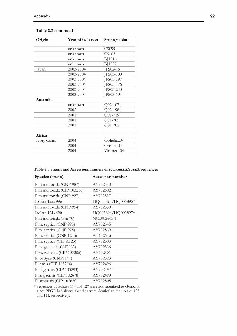
Appendix 92
Table 8.2 continued
Origin Year of isolation Strain/isolate
unknown CS099 unknown CS105 unknown BJ1816 unknown BJ1887 Japan 2003-2004 JPS02-76 2003-2004 JPS03-180 2003-2004 JPS03-187 2003-2004 JPS03-176 2003-2004 JPS03-240 2003-2004 JPS03-194 Australia unknown Q02-1071 2002 Q02-1981 2001 Q01-719 2001 Q01-705 2001 Q01-702 Africa Ivory Coast 2004 Ophelia_04 2004 Oreste_04 2004 Virunga_04
Table 8.3 Strains and Accessionnummers of P. multocida sodA sequences
Species (strain) Accession number
P.m multocida (CNP 987) AY702540 P.m multocida (CIP 103286) AY702502 P.m multocida (CNP 927) AY702537 Isolate 122/996 HQ003894/HQ003895* P.m multocida (CNP 954) AY702538 Isolate 121/420 HQ003896/HQ003897* P.m multocida (Pm 70) NC_002663.1 P.m. septica (CNP 993) AY702545 P.m. septica (CNP 978) AY702539 P.m. septica (CNP 1246) AY702546 P.m. septica (CIP A125) AY702503 P.m. gallicida (CNP982) AY702536 P.m. gallicida (CIP 103285) AY702501 P. bettyae (CNP1147) AY702523 P. canis (CIP 103294) AY702496 P. dagmatis (CIP 103293) AY702497 P.langaensis (CIP 102678) AY702499 P. stomatis (CIP 102680) AY702505
* Sequences of isolates 114 and 127 were not submitted to Genbank since PFGE had shown that they were identical to the isolates 122 and 121, respectively.

93
Acknowledgements
First of all I would like to thank my supervisor Dr. F. Leendertz for giving me the opportunity to
work on this important and interesting topic, for helpful discussions regarding the planning and
interpretation of my work and for his encouragement and support. I want to express my special
gratitude to Prof. Dr. G. Pauli for the supervision of this thesis and for his scientific advice. My
thanks go also to Prof. Dr. R. Lauster for his evaluation of my thesis.
I am very grateful to Prof. C. Boesch for his support of this work; without his collaboration and the
help of the Tai Chimpanzee Project staff this work would not have been possible. Thanks also to
all the students and assistants who were engaged in field observations and sample collection.
Many thanks to Christa Ewers who supervised the bacterial study and who allowed me to benefit
from her impressive knowledge of Pasteurella multocida. I also want to thank Antina Luebke Becker
and colleagues for giving me a fantastic introduction to bacterial diagnostics. I am grateful to Prof.
L. Wieler for giving me the opportunity to work in the facilities of the Freie Universität, Berlin.
I would also like to thank Kerstin Mätz Rensing from the Deutsche Primaten Zentrum for
performing the histopathological analysis. Furthermore, I am much obliged to Dr. R Biek for his
spontaneous help in phylogenetic analysis. Roger Mundry, I want to thank for statistical advice.
I thank all my colleagues from the RKI who contributed to this work, in particular Brunhilde
Schweiger and co-workers for providing diagnostic assays and her great helpfulness. I would also
like to thank Heinz Ellerbrock for his methodical advice, especially at the beginning of this work.
Many thanks to Andreas Nitsche who opened the doors of the RKI to me four years ago, for his
help regarding PCR assay design as well as pleasant conversations. Frau Sim-Brandenburg I thank
for her tremendous support regarding any aspect of cell culture and virus cultivation. Also, I would
like to thank Jule Tesch and the rest of the team of the sequencing lab.
Great thanks to all my colleagues from NG2 for their fantastic support whenever needed, and for
creating such a friendly atmosphere. Many thanks to Sandra Junglen for all her methodical advice,
proof reading of my thesis and last but not least for providing a fun time in the office, lab and field!
I am also very grateful to Siv Aina Jensen for helping me with data analysis. Adeelia Goffe I thank
for the thorough proof reading of this thesis and further manuscripts and her constructive
criticisms.

94
I thank the Ivorian authorities for long term support of the Tai chimpanzee Project, especially the
Ministry of the Environment and Forests as well as the Ministry of Research, the directorship of
the Taï National Park, and the Swiss Research Centre in Abidjan. This work was financially
supported by the Robert Koch-Institute and the Max-Planck-Society.
Finally I want to thank all my friends for helping, encouraging and diverting me from the very start
to the very end! Andrew, thanks for keeping me grounded in these last, hectic weeks prior to
submission. My parents I thank for supporting me during my studies and all circumstances.

95
Curriculum vitae
Personal information
Name Sophie Köndgen
Date of Birth 12.01.1979
Place of Birth Frankfurt/Main, Germany
Nationality German
Parents Elisabeth Köndgen and Günter Gretz
Education and Qualification
Since 10/2006 Dissertation at Robert Koch Institute, Berlin
10/2005 Diploma in Biology
2002-2005 Humboldt University, Berlin, Germany
2001-2002 Universidad Autónoma de Madrid, Spain
1999-2001 Albert Ludwig University, Freiburg, Germany
06/ 1998 A levels („Abitur“)
1989-1998 Secondary school: Bettina Gymnasium, Frankfurt/Main
1985-1989 Primary school: Ebelfelddschule, Frankfurt/Main
Languages
Englisch (fluently), Spanish (fluently), French (intermediate)
Stipends
Phd stipend, Max Planck Institute for Evolutionary Anthropology, Leipzig
DAAD stipend for participating at the “International Primatological Society 13th Congress” in Kyoto, September 2010
Publications
Köndgen S, Schenk S, Pauli G, Boesch C, Leendertz FH (September 2010). Non-invasive monitoring of respiratory viruses in wild chimpanzees. EcoHealth
Köndgen S, Kühl H, N’Goran PK, Walsh PD, Schenk S, Ernst N, Biek R, Formenty P, Mätz-Rensing K, Schweiger B, Junglen J, Ellerbrok H, Nitsche A, Briese T, Lipkin WI, Pauli G, Boesch C, Leendertz FH (February 2008). Pandemic human viruses cause decline of endangered great apes. Current Biology
Briese T, Renwick N, Venter M, Jarman RG, Ghosh D, Köndgen S, Shrestha SK, Hoegh AM, Casas I, Adjogoua EV, Akoua-Koffi C, Myint KS, Williams DT, Chidlow G, van den Berg R, Calvo C, Koch O, Palacios G, Kapoor V, Villari J, Dominguez SR, Holmes KV, Harnett G, Smith D, Mackenzie JS, Ellerbrok H, Schweiger B, Schønning K, Chadha MS, Leendertz FH, Mishra AC, Gibbons RV, Holmes EC, Lipkin WI (June 2008). Global distribution of novel rhinovirus genotype. Emerging Infectious Diseases

96
Szentiks CA, Köndgen S, Silinski S, Speck S, Leendertz FH (August 2009). Lethal pneumonia in a captive juvenile chimpanzee (Pan troglodytes) due to human-transmitted human respiratory syncytial virus (HRSV) and infection with Streptococcus pneumoniae. Journal of Medical Primatology
Mätz-Rensing K, Winkelmann J, Becker T, Burckhardt I, van der Linden M, Köndgen S, Leendertz F, Kaup FJ (October 2009). Outbreak of Streptococcus equi subsp. zooepidemicus infection in a group of rhesus monkeys (Macaca mulatta). Journal of Medical Primatology
Koch SP, Koendgen S, Bourayou R, Steinbrink J, Obrig H (June 2008). Individual alpha-frequency correlates with amplitude of visual evoked potential and hemodynamic response. Neuroimage
Oral and poster presentations at scientific meetings
09/ 2010 International Primatological Society 13th Congress, Kyoto, Japan. Oral Presentation
08/ 2009 Great Ape Health Workshop, Entebbe, Uganda. Oral Presentation
03/ 2009 19th Annual Meeting of the Society for Virology, Leipzig, Germany. Poster Presentation

97
Selbständigkeitserklärung
Ich bestätige, dass die vorliegende Dissertation in allen Teilen von mir selbstständig angefertigt
wurde und die benutzten Hilfsmittel vollständig angegeben worden sind. Veröffentlichungen von
Teilen der vorliegenden Dissertation sind angegeben. Weiter erkläre ich, dass ich nicht schon
anderweitig einmal die Promtionsabsicht angemeldet oder ein Promotionsverfahren beantragt habe.
Berlin, den 05.10.2010
Sophie Köndgen