cigarette smoke-promoted acquisition of bacterial pathogens in the upper respiratory tract leads to...
TRANSCRIPT

Voss et al. Respiratory Research (2015) 16:41 DOI 10.1186/s12931-015-0204-8
RESEARCH Open Access
Cigarette smoke-promoted acquisition of bacterialpathogens in the upper respiratory tract leads toenhanced inflammation in miceMeike Voss1, Bodo Wonnenberg1, Anja Honecker1, Andreas Kamyschnikow1, Christian Herr1, Markus Bischoff2,Thomas Tschernig3, Robert Bals1 and Christoph Beisswenger1*
Abstract
Background: Bacterial colonization and recurrent infections of the respiratory tract contribute to the progression ofchronic obstructive pulmonary disease (COPD). There is evidence that exacerbations of COPD are provoked by newbacterial strains acquired from the environment. Using a murine model of colonization, we examined whetherchronic exposure to cigarette smoke (CS) promotes nasopharyngeal colonization with typical lung pathogens andwhether colonization is linked to inflammation in the respiratory tract.
Methods: C57BL/6 N mice were chronically exposed to CS. The upper airways of mice were colonized withnontypeable Haemophilus influenzae (NTHi) or Streptococcus pneumoniae. Bacterial colonization was determined inthe upper respiratory tract and lung tissue. Inflammatory cells and cytokines were determined in lavage fluids.RT-PCR was performed for inflammatory mediators.
Results: Chronic CS exposure resulted in significantly increased numbers of viable NTHi in the upper airways,whereas NTHi only marginally colonized air-exposed mice. Colonization with S. pneumoniae was enhanced in theupper respiratory tract of CS-exposed mice and was accompanied by increased translocation of S. pneumoniae intothe lung. Bacterial colonization levels were associated with increased concentrations of inflammatory mediators andthe number of immune cells in lavage fluids of the upper respiratory tract and the lung. Phagocytosis activity wasreduced in whole blood granulocytes and monocytes of CS-exposed mice.
Conclusions: These findings demonstrate that exposure to CS impacts the ability of the host to control bacterialcolonization of the upper airways, resulting in enhanced inflammation and susceptibility of the host to pathogensmigrating into the lung.
Keywords: Cigarette smoke, Bacterial colonization, Inflammation, COPD
IntroductionRespiratory diseases associated with cigarette smoke(CS) are a leading cause of morbidity and mortalityworldwide. The most prominent respiratory diseasecaused by CS is chronic obstructive pulmonary disease(COPD) and it is most likely that it will constitute thefourth most common cause of death by the year 2030[1]. A hallmark of COPD is chronic inflammation of the
* Correspondence: [email protected] of Internal Medicine V – Pulmonology, Allergology andRespiratory Critical Care Medicine, Saarland University, 66421 Homburg/Saar,GermanyFull list of author information is available at the end of the article
© 2015 Voss et al.; licensee BioMed Central. ThCommons Attribution License (http://creativecreproduction in any medium, provided the orDedication waiver (http://creativecommons.orunless otherwise stated.
lung, leading to tissue destruction. The ongoing tissuedestruction leads to emphysema and loss of pulmonaryfunction [2,3].Stable COPD patients are frequently colonized by bac-
terial pathogens, such as nontypeable H. influaenzae,Pseudomonas aeruginosa, and S. pneumoniae [2,4,5].Moreover, recurrent infections of the respiratory tractand chronic bronchitis are common in COPD [2-5]. It issuggested that acute bacterial lung infections as well aschronic bacterial colonization of the lung contribute tothe progression of COPD by amplifying inflammationwhich further promotes lung damage and impacts innatelung defence [2]. In addition, pathogenic bacteria cause
is is an Open Access article distributed under the terms of the Creativeommons.org/licenses/by/4.0), which permits unrestricted use, distribution, andiginal work is properly credited. The Creative Commons Public Domaing/publicdomain/zero/1.0/) applies to the data made available in this article,

Voss et al. Respiratory Research (2015) 16:41 Page 2 of 11
direct damage to the epithelial surfaces of the lung andthereby contribute to impaired mucociliary clearance,elevated mucus secretion and subsequent mucus reten-tion, disrupted ciliary activity, and finally airflow ob-struction [3,6,7].There is evidence that exacerbations are provoked by
the acquisition of new bacterial strains from the environ-ment by the host [2,3]. It is hypothesized that newly-acquired bacterial pathogens trigger infections of thelung and evoke harmful inflammatory responses, con-tributing to clinical deterioration of the patient duringexacerbation [2,3,8]. Among the most frequent bac-terial pathogens isolated from patients suffering fromexacerbation are H. influenzae (20–30% of patients)and S. pneumoniae (10–15% of patients) [2].Several studies have shown that CS has an impact on
the immune system of the respiratory tract, even inhealthy individuals [9,10]. Thus, smoking and passivesmoking are associated with disordered microbial com-munities of the upper airways, impairment of com-mensal airway flora, and an increased risk of bacterialinfections in healthy individuals [4,11-14]. Brook andGoober demonstrated that the nasopharyngeal flora ofsmokers contains fewer aerobic and anaerobic organismswith interfering capabilities and more potential patho-gens, such as H. influenzae and S. pneumoniae, com-pared with those of non-smokers [12]. It is alsosuggested that smoking enhances colonization of theoral cavity with periodontal pathogens, affects the sub-gingival microflora in periodontitis, and is a risk factorfor periodontal disease [15,16]. Moreover, cigarettesmoking increases the risk of community-acquired pneu-monia [17,18] and is a strong independent risk factor forinvasive pneumococcal disease among immunocompe-tent, non-elderly adults [19]. Exposure to passive smokefavors bacterial colonization and infection in infants[12-14,20]. Greenberg et al. reported that children ofsmoking parents have a significantly higher rate ofS. pneumoniae carriage than children of non-smokers[14]. Exposure to passive smoke also correlates witha significantly increased risk of infants developing in-fections of the lower respiratory tract in the first twoyears of life [20].It was the aim of the present study to investigate
whether chronic exposure to CS promotes nasopharyn-geal colonization with the leading gram-positive andgram-negative bacterial lung pathogens H. influenzaeand S. pneumoniae by combining a clinically relevantmurine model of colonization of the upper respira-tory tract with a model of chronic CS exposure [21].It was further investigated whether CS-promotedcolonization of the upper-respiratory tract results inincreased inflammation and bacterial translocationinto the lung.
MethodsBacterial strains and productsA patient isolate of H. influenzae (ATCC® 49247™) wasgrown overnight at 37°C and 5% CO2 on chocolate agar(Becton Dickinson, Germany). Bacteria were taken fromthe plate and re-suspended in 7 ml 1 × PBS to obtain anoptical density of 1 at 600 nm. Bacteria were spun at4000 rpm and 4°C for 10 minutes and re-suspended in350 μl 1 × PBS for the infection. A type 6A clinical iso-late of S. pneumoniae (a gift from Dr. Jeffrey Weiser,University of Pennsylvania) [22,23] was cultured in ToddHewitt broth at 37°C. Mid-log-phase S. pneumoniaewere washed with PBS and re-suspenended in 450 μl1 × PBS.
CS exposure and nasopharyngeal colonizationAll animal experiments were approved by the Landesamtfür Soziales, Gesundheit und Verbraucherschutz of theState of Saarland following the national guidelines foranimal treatment. Mice were maintained under apathogen-free condition. 7 to 9 week-old female wild-type C57BL/6 N mice were exposed to a combinationof sidestream and mainstream CS (3R4F, College ofAgriculture, Reference Cigarette Program, University ofKentucky, Lexington, Kentucky, USA) in a TE-10 smok-ing machine (Teague Enterprises, Woodland, California,USA) for a total of 261 minutes/day, 5 days per week.The smoking time was 87 minutes with 40 minutesair exposure in between CS exposures, with 3 CSexposures/day. Mice were exposed to CS or air for13 (3 months) or 30 weeks (7 months) as indicatedin the figures. The CS concentration was 120 mg/m3
total suspended particles. 24 hours after the final CSexposure the upper airways of room air- and CS-exposedmice were colonized using an established model of naso-pharyngeal colonization [22-24]. Mice were inoculatedintranasally with 10 μl of mid-log phase NTHi (5.5 × 10⁶
to 4.5 × 10⁷ CFU) or S. pneumoniae 6A (1.5 × 10⁷ to2.5 × 107 CFU) applied atraumatically to the nares with-out anesthesia.
Lavage of the upper airways and bronchoalveolar lavageand phagocytosisLavage of the upper airways and bronchoalveolar lavage(BAL) were performed as described before [22,23].Briefly, the animals were sacrificed and the trachea werecannulated. Lavage of the upper airways was performedwith 300 μl of PBS. To obtain BAL fluids from the lungBAL was performed with 1 ml of PBS flushed threetimes into the lungs. Lavage fluids were plated in serialdilutions and CFUs were determined. Lavage fluids werecentrifuged to obtain immune cells and supernatants.Cell numbers were determined. Inflammatory cells weredifferentiated on cytospins. Serum was obtained by

Voss et al. Respiratory Research (2015) 16:41 Page 3 of 11
cardiac puncture. Phagocytosis activity of whole bloodmonocytes and granulocytes obtained from mice ex-posed to air or CS for 3 months was determined using aflow cytometry-based assays (Phagotest, Orpegen Pharma,Germany), according to the instruction. Samples wereanalyzed in a FACS Calibur flow cytometer (BecktonDickinson, Heidelberg, Germany). Gates (forward/sidescatter) were set on granulocytes and monocytes. For de-termination of the phagocytosis activity in each sample themean fluorescence intensity was calculated according tothe instruction of the manufacturer (MFI).
Determination of cytokine concentrations andimmunohistochemistryConcentrations of inflammatory mediators were assessedby cytometric bead array (CBA) using a FACSCanto II(BD Bioscience, Germany). For immunohistochemistryof the upper respiratory tract, the heads of the micewere fixed in 4%-PBS-buffered formaldehyde and decal-cified [22]. Immunohistochemistry on paraffin sectionswas performed as described before, using anti-H. influ-enzae (abcam, USA) antibody [25,26].
Figure 1 Colonization levels in the upper airways of mice inoculatedintranasally inoculated with NTHi. Colonization densities were determined bpostinoculation. Data are shown as mean ± SEM. Bars indicate significant dtissue by immunohistochemistry (red staining). Scale bar, 100 μm.
Quantitative real-time PCRTotal RNA from the upper airways was isolated afterflushing the upper airways to obtain cells [23]. Theupper airways were flushed with 300 μl RLT Lysis buffer(Qiagen, Germany). RNA was isolated and reverse tran-scription and real-time PCR were performed as describedbefore [27]. Primers were: mouse β-Actin: 5′-AGC CATGTA CGT AGC CAT CC-3′ and 5′-CTC TCA GCTGTG GTG GTG AA-3′, mouse Lysozyme M: 5′-CTGGCT ACT ATG GAG TCA GC-3′and 5′-TTG ATC CCACAG GCA TTC AC-3′, mouse MIP-2: 5′-AAG TTTGCC TTG ACC CTG AA-3′ and 5′-AGG CAC ATCAGG TAC GAT CC-3′, and mouse mBD-1: 5′-GGC TGCCAC CAC TAT GAA AAC TC-3′ and 5′-GAG ACAGAATCC TCC ATG TTG AA-3′.
Statistical analysisValues are displayed as mean ± SEM. The Mann–Whitneytest was used to compare the groups. Results were consid-ered statistically significant for p < 0.05. All statistical testswere performed using the software Prism (GraphPadSoftware, San Diego, CA, USA).
with NTHi. (A) Mice were exposed to CS or air as indicated andy quantitative cultures of upper airway lavages 24 hoursifferences of *p < 0.05, n≥ 5 per group. (B) NTHi are shown in nasal
Voss et al. Respiratory Research (2015) 16:41 Page 4 of 11
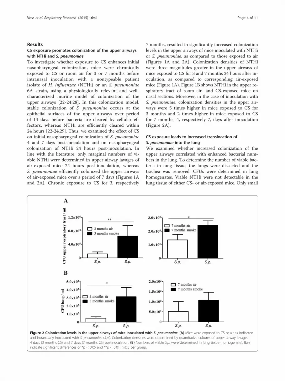
ResultsCS exposure promotes colonization of the upper airwayswith NTHi and S. pneumoniaeTo investigate whether exposure to CS enhances initialnasopharyngeal colonization, mice were chronicallyexposed to CS or room air for 3 or 7 months beforeintranasal inoculation with a nontypeable patientisolate of H. influenzae (NTHi) or an S. pneumoniae6A strain, using a physiologically relevant and well-characterized murine model of colonization of theupper airways [22-24,28]. In this colonization model,stable colonization of S. pneumoniae occurs at theepithelial surfaces of the upper airways over periodof 14 days before bacteria are cleared by cellular ef-fectors, whereas NTHi are efficiently cleared within24 hours [22-24,29]. Thus, we examined the effect of CSon initial nasopharyngeal colonization of S. pneumoniae4 and 7 days post-inoculation and on nasopharyngealcolonization of NTHi 24 hours post-inoculation. Inline with the literature, only marginal numbers of vi-able NTHi were determined in upper airway lavages ofair-exposed mice 24 hours post-inoculation, whereasS. pneumoniae efficiently colonized the upper airwaysof air-exposed mice over a period of 7 days (Figures 1Aand 2A). Chronic exposure to CS for 3, respectively
Figure 2 Colonization levels in the upper airways of mice inoculatedand intranasally inoculated with S. pneumoniae (S.p.). Colonization densities4 days (3 months CS) and 7 days (7 months CS) postinoculation. (B) Numbindicate significant differences of *p < 0.05 and **p < 0.01, n≥ 5 per group
7 months, resulted in significantly increased colonizationlevels in the upper airways of mice inoculated with NTHior S. pneumoniae, as compared to those exposed to air(Figures 1A and 2A). Colonization densities of NTHiwere three magnitudes greater in the upper airways ofmice exposed to CS for 3 and 7 months 24 hours after in-oculation, as compared to corresponding air-exposedmice (Figure 1A). Figure 1B shows NTHi in the upper re-spiratory tract of room air- and CS-exposed mice onnasal sections. Moreover, in the case of inoculation withS. pneumoniae, colonization densities in the upper air-ways were 5 times higher in mice exposed to CS for3 months and 2 times higher in mice exposed to CSfor 7 months, 4, respectively 7, days after inoculation(Figure 2A).
CS exposure leads to increased translocation ofS. pneumoniae into the lungWe examined whether increased colonization of theupper airways correlated with enhanced bacterial num-bers in the lung. To determine the number of viable bac-teria in lung tissue, the lungs were dissected and thetrachea was removed. CFUs were determined in lunghomogenates. Viable NTHi were not detectable in thelung tissue of either CS- or air-exposed mice. Only small
with S. pneumoniae. (A) Mice were exposed to CS or air as indicatedwere determined by quantitative cultures of upper airway lavagesers of viable S.p. were determined in lung tissue (homogenate). Bars.

Voss et al. Respiratory Research (2015) 16:41 Page 5 of 11
numbers of viable S. pneumoniae were determinedin the lung tissue of both air- and CS-exposed mice(Figure 2B). The numbers of S. pneumoniae in the lungtissue were, however, enhanced in mice which had beenchronically exposed to CS prior to bacterial colonization.Significantly increased numbers of viable S. pneumoniaewere determined in the lung tissue of mice exposed toCS for 3 months, as compared to corresponding air-exposed mice 4 days after inoculation (Figure 2B).
Figure 3 Concentrations of inflammatory mediators in upper airway lNTHi or S. pneumoniae. Mice were exposed to CS or air for 3 months and iof KC (A), RANTES (B), and IL-6 (C) were measured in upper airway lavages ofshown as mean ± SEM. Bars indicate significant differences of *p < 0.05 and **
CS exposure increases the expression of inflammatorycytokines in the upper airways of mice colonizedwith NTHiWe further examined whether chronic exposure toCS exacerbates upper airway inflammation inducedby colonizing bacteria. Figure 3 shows that concen-trations of inflammatory mediators were increased inupper airway lavages of mice chronically exposed toCS and colonized with NTHi or S. pneumoniae, as
avages of mice chronically exposed to CS and colonized withntranasally inoculated with NTHi or S. pneumoniae (S.p.). Concentrationsmice by CBA 24 hours (NTHi) and 4 days (S.p.) postinoculation. Data arep < 0.01, n≥ 5 per group.

Voss et al. Respiratory Research (2015) 16:41 Page 6 of 11
compared to colonized air-exposed mice. Concentra-tions of KC, a major murine chemoattractant forneutrophils, were significantly increased in upper air-way lavages of mice exposed to CS and colonizedwith NTHi, as compared to air-exposed mice colo-nized with NTHi and non-infected mice (Figure 3A).CS exposure and colonization with NTHi alone didnot result in significantly increased concentrations ofKC as compared to air-exposed control mice. Expos-ure to CS slightly - however not significantly (p = 0.072) -increased concentrations of KC in upper airway lavagesof mice colonized with S. pneumoniae (Figure 3A).Similar to KC, concentrations of RANTES (also namedCCL5), a major chemoattractant for monocytes, weresignificantly increased in upper airway lavages of miceexposed to CS and colonized with NTHi, as comparedto air-exposed mice colonized with NTHi and non-infected mice (Figure 3B). However, exposure to CSalone also led to significantly increased concentrationsof RANTES in upper airway lavages, as compared tonon-infected control mice, whereas colonization of air-exposed mice with NTHi or S. pneumoniae did not leadto increased concentrations of RANTES. Colonization ofair-exposed mice with NTHi or S. pneumonaie led to in-creased concentrations of IL-6 in upper airway lavages,which was not further increased in animals exposed toCS (Figure 3C). We further examined the expression oflysozyme, the antimicrobial peptide mBD-1 and the che-mokine MMP-2 by qRT-PCR with RNA obtained fromthe upper airways of mice colonized with NTHi. It hasbeen shown before that mBD-1 deficient mice are moresusceptible to H. influenzae lung infection [30]. Chronicexposure to CS suppressed the expression of the anti-microbial peptide mBD-1 and increased the expression ofthe chemokine MIP-2 whereas the expression of lyso-zyme was not affected by CS (Figure 4).
Figure 4 Expression of inflammatory mediators in the upper respiratoryinoculated with NTHi. Total RNA was obtained by flushing the upper airways(C) was determined by qRT-PCR. Data are shown as mean ± SEM. n = 4 per gr
CS exposure does not inhibit the recruitment ofinflammatory cells in the upper airways of colonized miceWe determined the total numbers of inflammatory cellsin upper airway lavages and differentiated macrophagesand neutrophils on cytospins. Exposure to CS andbacterial colonization led to a significantly increasedinflux of total immune cells (Figure 5A), neutrophils(Figure 5B), and macrophages (Figure 5C) into the upperairways. The number of neutrophils in upper airwaylavages was approximately one magnitude higher than thenumber of macrophages. Numbers of total immune cells,neutrophils, and macrophages were significantly increasedin upper airway lavages of mice exposed to CS and colo-nized with NTHi, as compared to non-infected air- andCS-exposed mice (Figure 5). The number of total immunecells, neutrophils, and macrophages was increased inupper airway lavages of CS-exposed NTHi-colonized micecompared to those of air-exposed NTHi-colonizedmice. However, the differences did not reach statisticalsignificance.
The phagocytosis activity is reduced in whole bloodgranulocytes and monocytesHaving shown that the influx of immune cells into theupper airways of colonized mice is not inhibited by CS,we determined whether the phagocytosis activity ofgranulocytes and monocytes was affected by exposure toCS. As there are only marginal numbers of granulocytesand monocytes present in the upper airways of non-colonized animals, we determined the ability of granulo-cytes and monocytes obtained from anticoagulatedwhole blood to phagocytose FITC-conjugated Escheri-chia coli. The phagocytosis activity was reduced ingranulocytes and monocytes of mice exposed to CS for3 months compared to those of air-exposed mice(Figure 6).
tract. Mice were exposed to CS or air for 7 months and intranasallywith lysis buffer. The expression of mBD-1 (A), lysozyme (B) and MIP-2oup.

Figure 5 Influx of inflammatory cells in the upper airways. Mice were exposed to CS or air for 3 months and intranasally inoculated withNTHi or S. pneumoniae (S.p.). Numbers of total immune cells (A), neutrophils (B) and macrophages (C) were determined in upper airway lavages24 hours (NTHi) and 4 days (S.p.) postinoculation. Data are shown as mean ± SEM. Bars indicate significant differences of *p < 0.05 and **p < 0.01,n≥ 5 per group.
Voss et al. Respiratory Research (2015) 16:41 Page 7 of 11
Bacterial colonization is linked with an increasedrecruitment of immune cells into the lungTo examine whether chronic exposure to CS and bacter-ial colonization affects recruitment of immune cells intothe lung we determined the total number of immunecells, macrophages, and neutrophils in bronchoalveolarlavage (BAL) fluids. The total number of immune cellsand macrophages was significantly increased in BAL
fluids of CS-exposed mice and there was an increase inmacrophages in colonized air-exposed mice, as com-pared to air-exposed control mice (Figure 7A and B).Interestingly, there was a significant increase in thenumber of total immune cells and macrophages in micechronically exposed to CS and colonized with NTHi orS. pneumoniae, as compared to CS-exposed control miceand colonized air-exposed mice (Figure 7A and B). In

Figure 6 Effect of CS on E. coli-induced phagocytosis. Wholeblood samples were obtained from mice exposed to CS or air for3 months and incubated with FITC-conjugated E. coli. Phagocytosisactivity is shown as mean fluorescence intensity (MFI). Data areshown as mean ± SEM. Bars indicate significant differences of**p < 0.01, n = 3 per group in duplicates.
Voss et al. Respiratory Research (2015) 16:41 Page 8 of 11
addition, we stimulated alveolar macrophages of air andCS-exposed mice ex vivo with NTHi or S. pneumoniaefor 4 hours. There was no difference between the releaseof KC in alveolar macrophages obtained from CS-exposed mice and those of air-exposed mice after bacter-ial stimulation (data not shown).
DiscussionWe combined a murine model of colonization ofthe upper respiratory tract with a model of COPD to in-vestigate whether CS-induced inflammation promotesacquisition of bacterial lung pathogens and whether col-onizing of the upper airways contributes to increasedinflammation in the respiratory tract. We focused onNTHi as this bacterium frequently colonizes lungs ofCOPD patients and is linked to exacerbations of COPD[2,5] and on S. pneumoniae as colonization of the nasalspace by pneumococci occasionally causes pneumoniaand invasive disease, especially in subjects exposed to CSor passive smoke [17-19]. Our data show that chronicexposure to CS has an impact on the ability of the hostto contain bacterial colonization of the upper airwayswith bacterial pathogens and that CS-induced colonizationis accompanied by increased inflammation of the respira-tory tract and susceptibility of the host to pathogensmigrating into the lung.It is hypothesized that exacerbations in COPD are pro-
voked by bacterial strains newly acquired from the envir-onment [2,3]. In line with this hypothesis, our studyproves that COPD-like inflammation promotes the
acquisition of typical COPD pathogens, such as NTHi,into the upper airways. Increased colonization of theupper airways of CS-exposed mice with NTHi is alsoassociated with an enhanced inflammation in the respira-tory tract characterized by an increased release of chemo-kines and influx of immune cells into the upper airwaysand into the lung. Thus, our data suggest that CS promotesthe acquisition and colonization of the upper airways withrespiratory pathogens and that these newly- acquired path-ogens contribute to the progression of COPD by enhan-cing and perpetuating lung inflammation [2,3,8].For many bacterial pathogens colonization of the
upper airways is often a first step in the process leadingto infectious diseases [22,23]. Frequent acquisition ofbacterial pathogens in the upper airways may also pro-mote infectious lung diseases and invasive diseases inindividuals exposed to mainstream or passive smoke.Our data indicate that the increased risk of smokers forcommunity-acquired pneumonia and invasive pneumo-coccal disease [17-19] and the increased risk of infantsexposed to passive smoke of developing infections of thelower respiratory tract [20] are a direct consequence ofCS-induced acquisition of bacterial pathogens into theupper airways. Our results are further in line with find-ings that children of smoking parents have a significantlyhigher rate of S. pneumoniae carriage than those of non-smokers [14].Several studies have examined how CS affects immune
mechanisms at the mucosal surfaces of the respiratorytract [9,27,31-36]. In vitro studies have shown that ex-posure of respiratory epithelial cells to CS results in thesuppression of cellular signaling cascades (e.g. AP-1- andNF-κB-dependent signaling cascades) that are of keyimportance in the activation of innate immune mecha-nisms in the case of microbial infections [31,35,36].Thus, in vitro, CS exposure results in a reduced expres-sion and release of inflammatory mediators and anti-microbial peptides by respiratory epithelial cells infectedwith bacterial pathogens [9,27,31,35,36]. Moreover, wehave previously shown that smokers with community-acquired pneumonia have reduced levels of antimicrobialpeptides in their sputum and pharyngeal washings [9].In this study, we found that chronic CS exposure re-sulted in reduced expression levels of the antimicrobialpeptide mBD-1 in the upper airways of NTHi-colonizedmice, even though the colonization levels were increasedin CS-exposed mice, as compared to air-exposed mice.It has been shown before that mBD-1 deficient mice aremore susceptible to H. influenzae lung infection [30].Furthermore, it has been shown that CS increasesadhesion of bacteria to epithelial cells [37-40]. Grigget al. showed, for instance, that CS extract stimulatesplatelet-activating factor receptor- dependent adhesion ofS. pneumonaie to respiratory epithelial cells [39]. Thus,

Figure 7 Influx of inflammatory cells in the lung. Mice were exposed to CS or air for 3 months and intranasally inoculated with NTHi orS. pneumoniae (S.p.). Numbers of total immune cells (A), macrophages (B) and neutrophils (C) were determined in BAL fluids 24 hours (NTHi) and4 days (S.p.) postinoculation. Bars indicate significant differences of *p < 0.05 and **p < 0.01, n ≥ 5 per group.
Voss et al. Respiratory Research (2015) 16:41 Page 9 of 11
the suppression of innate immune functions of respiratoryepithelial cells, including the expression of epithelial anti-microbial peptides as well as increased bacterial adherenceto epithelial cells, are potential mechanisms responsiblefor the increased levels of colonization observed in micechronically exposed to CS.
Studies have also shown that CS impairs the responseof immune cells such as alveolar macrophages and neu-trophils to bacterial pathogens, which results in aderegulated release of inflammatory mediators and im-paired phagocytic activity [41-44]. Alveolar macrophagesfrom ex-smokers with COPD, for instance, showed a
Voss et al. Respiratory Research (2015) 16:41 Page 10 of 11
reduced expression of inflammatory mediators (e.g.TNF-α) in response to antigens of H. influaenzae, com-pared to macrophages obtained from ex-smokers with-out COPD and non-smokers [41]. Moreover, ex vivostudies with alveolar macrophages obtained from CS-exposed mice showed that CS exposure enhances therelease of the chemokines IL-1α and MCP-1 and in-hibits the release of TNF-α by alveolar macrophagesstimulated with NTHi [42,43]. Berenson et al. showedthat the phagocytosis of NTHi is impaired in alveolarmacrophages obtained from donors with COPD, ascompared to that from donors without COPD [45].Taylor et al. found that monocyte-derived macrophagesfrom COPD patients showed reduced phagocytic re-sponses to H. influenza and S. pneumoniae, comparedwith smokers and non-smokers, which was not ob-served by Berenson et al. [46]. Exposure to CS extractalso resulted in a reduced uptake of NTHi by murineand human cell line-derived macrophages [44]. Expos-ure to CS extracts also reduced the complement-mediated phagocytosis of S. pneumoniae by alveolarmacrophages ex vivo [33] and the phagocytosis ofE. coli by human neutrophils [34]. In our model,chronic exposure to CS did not result in a defect inthe recruitment of phagocytes into the upper airwaysin response to colonizing bacteria. Chronic CS exposureresulted in increased numbers of neutrophil and macro-phages in the upper airways of mice colonized withNTHi and S. pneumoniae, which was associated with in-creased levels of chemokines in upper airway lavages.However, the phagocytosis activity of whole blood granu-locytes and monocytes was inhibited in CS-exposed mice.This was a surprising and not suspected finding whichmight have impact on extra pulmonal research on to-bacco smoke, too. Thus, it is conceivable that impairedphagocytosis of NTHi and S. pneumoniae by neutrophilsand macrophages is another potential cause of theincreased colonization levels in the upper airways ofCS-exposed mice.In summary, our data indicate that continuous ex-
posure to CS opens a niche for typical COPD patho-gens in the upper airways, which results in theacquisition of bacterial pathogens. Our data suggestthat bacterial pathogens efficiently colonizing the mu-cosal surfaces of the upper respiratory tract contributeto increased lung colonization and are a cause of pul-monary inflammation. Frequent acquisition of bacterialpathogens into the upper airways may also promotepneumonia and invasive diseases in individuals ex-posed to mainstream or passive smoke. Our modelsystem provides a tool for future mechanistic studiesto examine mechanisms and potential therapeutic in-terventions regarding bacterial colonization and COPDexacerbations.
Competing interestsThe authors declare that they have no competing interests.
Authors’ contributionsMV, CB: designed the study, collected data, analyzed data, and wrote themanuscript. AH, AK, BW, CH: collected data. TT, MB: analyzed data, wrote themanuscript. RB: designed the study, analyzed data, and wrote the manuscript.All authors read and approved the final manuscript.
AcknowledgmentsThe authors thank Ms Soether for correction of the language. This study wassupported by grants from DFG BE 4813/1-1 to CB and Ba 1641/12 to RB.
Author details1Department of Internal Medicine V – Pulmonology, Allergology andRespiratory Critical Care Medicine, Saarland University, 66421 Homburg/Saar,Germany. 2Institute of Medical Microbiology and Hygiene, SaarlandUniversity, 66421 Homburg/Saar, Germany. 3Institute of Anatomy and CellBiology, Saarland University, 66421 Homburg/Saar, Germany.
Received: 6 November 2014 Accepted: 9 March 2015
References1. Mathers CD, Loncar D. Projections of global mortality and burden of disease
from 2002 to 2030. PLoS Med. 2006;3:e442.2. Sethi S. Infection as a comorbidity of COPD. Eur Respir J. 2010;35:1209–15.3. Sethi S, Murphy TF. Infection in the pathogenesis and course of chronic
obstructive pulmonary disease. N Engl JMed. 2008;359:2355–65.4. Garmendia J, Morey P, Bengoechea JA. Impact of cigarette smoke exposure
on host-bacterial pathogen interactions. Eur Respir J. 2012;39:467–77.5. Moghaddam SJ, Ochoa CE, Sethi S, Dickey BF. Nontypeable Haemophilus
influenzae in chronic obstructive pulmonary disease and lung cancer. Int JChron Obstruct Pulmon Dis. 2011;6:113–23.
6. Sethi S, Murphy TF. Bacterial infection in chronic obstructive pulmonarydisease in 2000: a state-of-the-art review. Clin Microbiol Rev. 2001;14:336–63.
7. Shuto T, Xu H, Wang B, Han J, Kai H, Gu XX, et al. Activation of NF-kappa Bby nontypeable Hemophilus influenzae is mediated by toll-like receptor2-TAK1-dependent NIK-IKK alpha /beta-I kappa B alpha and MKK3/6-p38MAP kinase signaling pathways in epithelial cells. Proc Natl Acad Sci USA.2001;98:8774–9.
8. Veeramachaneni SB, Sethi S. Pathogenesis of bacterial exacerbations ofCOPD. COPD. 2006;3:109–15.
9. Herr C, Beisswenger C, Hess C, Kandler K, Suttorp N, Welte T, et al.Suppression of pulmonary innate host defence in smokers. Thorax.2009;64:144–9.
10. Mehta H, Nazzal K, Sadikot RT. Cigarette smoking and innate immunity.Inflamm Res. 2008;57:497–503.
11. Charlson ES, Chen J, Custers-Allen R, Bittinger K, Li H, Sinha R, et al. Disorderedmicrobial communities in the upper respiratory tract of cigarette smokers. PLoSOne. 2010;5:e15216.
12. Brook I, Gober AE. Recovery of potential pathogens and interfering bacteriain the nasopharynx of smokers and nonsmokers. Chest. 2005;127:2072–5.
13. Brook I, Gober AE. Recovery of potential pathogens and interfering bacteriain the nasopharynx of otitis media-prone children and their smoking andnonsmoking parents. Arch Otolaryngol Head Neck Surg. 2005;131:509–12.
14. Greenberg D, Givon-Lavi N, Broides A, Blancovich I, Peled N, Dagan R. Thecontribution of smoking and exposure to tobacco smoke to Streptococcuspneumoniae and Haemophilus influenzae carriage in children and theirmothers. Clin Infect Dis. 2006;42:897–903.
15. Zambon JJ, Grossi SG, Machtei EE, Ho AW, Dunford R, Genco RJ. Cigarettesmoking increases the risk for subgingival infection with periodontalpathogens. J Periodontol. 1996;67:1050–4.
16. van Winkelhoff AJ, Bosch-Tijhof CJ, Winkel EG, van der Reijden WA. Smokingaffects the subgingival microflora in periodontitis. J Periodontol.2001;72:666–71.
17. Almirall J, Bolibar I, Balanzo X, Gonzalez CA. Risk factors for community-acquired pneumonia in adults: a population-based case–control study.Eur Respir J. 1999;13:349–55.

Voss et al. Respiratory Research (2015) 16:41 Page 11 of 11
18. Almirall J, Gonzalez CA, Balanzo X, Bolibar I. Proportion of community-acquired pneumonia cases attributable to tobacco smoking. Chest.1999;116:375–9.
19. Nuorti JP, Butler JC, Farley MM, Harrison LH, McGeer A, Kolczak MS, et al.Cigarette smoking and invasive pneumococcal disease. Active Bacterial CoreSurveillance Team. N Engl J Med. 2000;342:681–9.
20. Jones LL, Hashim A, McKeever T, Cook DG, Britton J, Leonardi-Bee J. Parentaland household smoking and the increased risk of bronchitis, bronchiolitisand other lower respiratory infections in infancy: systematic review andmeta-analysis. Respir Res. 2011;12:5.
21. Herr C, Han G, Li D, Tschernig T, Dinh QT, Beisswenger C, et al. Combinedexposure to bacteria and cigarette smoke resembles characteristicphenotypes of human COPD in a murine disease model. Exp ToxicolPathol. 2015;67:261–9.
22. Nelson AL, Roche AM, Gould JM, Chim K, Ratner AJ, Weiser JN. Capsuleenhances pneumococcal colonization by limiting mucus-mediatedclearance. Infect Immun. 2007;75:83–90.
23. Beisswenger C, Lysenko ES, Weiser JN. Early bacterial colonization inducestoll-like receptor-dependent transforming growth factor beta signaling inthe epithelium. Infect Immun. 2009;77:2212–20.
24. Zola TA, Lysenko ES, Weiser JN. Mucosal clearance of capsule-expressingbacteria requires both TLR and nucleotide-binding oligomerization domain1 signaling. J Immunol. 2008;181:7909–16.
25. Li D, Beisswenger C, Herr C, Hellberg J, Han G, Zakharkina T, et al.Myeloid cell RelA/p65 promotes lung cancer proliferation throughWnt/beta-catenin signaling in murine and human tumor cells. Oncogene.2014;33:1239–48.
26. Seiler F, Hellberg J, Lepper PM, Kamyschnikow A, Herr C, Bischoff M, et al.FOXO Transcription Factors Regulate Innate Immune Mechanisms inRespiratory Epithelial Cells. J Immunol. 2013;190:1603–13.
27. Pfeifer P, Voss M, Wonnenberg B, Hellberg J, Seiler F, Lepper PM, et al.IL-17C is a mediator of respiratory epithelial innate immune response. Am JRespir Cell Mol Biol. 2013;48:415–21.
28. McCool TL, Weiser JN. Limited role of antibody in clearance ofStreptococcus pneumoniae in a murine model of colonization. InfectImmun. 2004;72:5807–13.
29. Zhang Z, Clarke TB, Weiser JN. Cellular effectors mediating Th17-dependentclearance of pneumococcal colonization in mice. J Clin Invest.2009;119:1899–909.
30. Moser C, Weiner DJ, Lysenko E, Bals R, Weiser JN, Wilson JM. beta-Defensin1 contributes to pulmonary innate immunity in mice. Infect Immun.2002;70:3068–72.
31. Kulkarni R, Rampersaud R, Aguilar JL, Randis TM, Kreindler JL, Ratner AJ.Cigarette smoke inhibits airway epithelial cell innate immune responses tobacteria. Infect Immun. 2010;78:2146–52.
32. Chen H, Cowan MJ, Hasday JD, Vogel SN, Medvedev AE. Tobacco smokinginhibits expression of proinflammatory cytokines and activation of IL-1R-associated kinase, p38, and NF-kappaB in alveolar macrophages stimulatedwith TLR2 and TLR4 agonists. J Immunol. 2007;179:6097–106.
33. Phipps JC, Aronoff DM, Curtis JL, Goel D, O’Brien E, Mancuso P. Cigarettesmoke exposure impairs pulmonary bacterial clearance and alveolarmacrophage complement-mediated phagocytosis of Streptococcuspneumoniae. Infect Immun. 2010;78:1214–20.
34. Stringer KA, Tobias M, O’Neill HC, Franklin CC. Cigarette smoke extract-induced suppression of caspase-3-like activity impairs human neutrophilphagocytosis. Am J Physiol Lung Cell Mol Physiol. 2007;292:L1572–9.
35. Manzel LJ, Shi L, O'Shaughnessy PT, Thorne PS, Look DC. Inhibition bycigarette smoke of nuclear factor-kappaB-dependent response to bacteria inthe airway. Am J Respir Cell Mol Biol. 2011;44:155–65.
36. Laan M, Bozinovski S, Anderson GP. Cigarette smoke inhibitslipopolysaccharide-induced production of inflammatory cytokines bysuppressing the activation of activator protein-1 in bronchial epithelialcells. J Immunol. 2004;173:4164–70.
37. Fainstein V, Musher DM, Cate TR. Bacterial adherence to pharyngeal cellsduring viral infection. J Infect Dis. 1980;141:172–6.
38. El Ahmer OR, Essery SD, Saadi AT, Raza MW, Ogilvie MM, Weir DM, et al. Theeffect of cigarette smoke on adherence of respiratory pathogens to buccalepithelial cells. FEMS Immunol Med Microbiol. 1999;23:27–36.
39. Grigg J, Walters H, Sohal SS, Wood-Baker R, Reid DW, Xu CB, et al. Cigarettesmoke and platelet-activating factor receptor dependent adhesion ofStreptococcus pneumoniae to lower airway cells. Thorax. 2012;67:908–13.
40. Raman AS, Swinburne AJ, Fedullo AJ. Pneumococcal adherence to thebuccal epithelial cells of cigarette smokers. Chest. 1983;83:23–7.
41. Berenson CS, Wrona CT, Grove LJ, Maloney J, Garlipp MA, Wallace PK, et al.Impaired alveolar macrophage response to Haemophilus antigens inchronic obstructive lung disease. Am J Respir Crit Care Med.2006;174:31–40.
42. Nikota JK, Shen P, Morissette MC, Fernandes K, Roos A, Chu DK, et al.Cigarette smoke primes the pulmonary environment to IL-1alpha/CXCR-2-Dependent Nontypeable Haemophilus influenzae-Exacerbated Neutrophiliain mice. J Immunol. 2014;193:3134–45.
43. Gaschler GJ, Skrtic M, Zavitz CC, Lindahl M, Onnervik PO, Murphy TF, et al.Bacteria challenge in smoke-exposed mice exacerbates inflammation andskews the inflammatory profile. Am J Respir Crit Care Med. 2009;179:666–75.
44. Marti-Lliteras P, Regueiro V, Morey P, Hood DW, Saus C, Sauleda J, et al.Nontypeable Haemophilus influenzae clearance by alveolar macrophages isimpaired by exposure to cigarette smoke. Infect Immun. 2009;77:4232–42.
45. Berenson CS, Garlipp MA, Grove LJ, Maloney J, Sethi S. Impairedphagocytosis of nontypeable Haemophilus influenzae by humanalveolar macrophages in chronic obstructive pulmonary disease.J Infect Dis. 2006;194:1375–84.
46. Taylor AE, Finney-Hayward TK, Quint JK, Thomas CM, Tudhope SJ,Wedzicha JA, et al. Defective macrophage phagocytosis of bacteria inCOPD. Eur Respir J. 2010;35:1039–47.
Submit your next manuscript to BioMed Centraland take full advantage of:
• Convenient online submission
• Thorough peer review
• No space constraints or color figure charges
• Immediate publication on acceptance
• Inclusion in PubMed, CAS, Scopus and Google Scholar
• Research which is freely available for redistribution
Submit your manuscript at www.biomedcentral.com/submit