biocontrol of pathogens in the meat chain
TRANSCRIPT

Meat Biotechnology

Fidel ToldraEditor
Meat Biotechnology
123
EditorFidel ToldraDepartment of Food ScienceInstituto de Agroquımica y Tecnologıa
de Alimentos (CSIC)46100 Burjassot (Valencia), [email protected]
ISBN: 978-0-387-79381-8 e-ISBN: 978-0-387-79382-5
Library of Congress Control Number: 2008935902
c© 2008 Springer Science+Business Media, LLCAll rights reserved. This work may not be translated or copied in whole or in part without the writtenpermission of the publisher (Springer Science+Business Media, LLC, 233 Spring Street, New York,NY 10013, USA), except for brief excerpts in connection with reviews or scholarly analysis. Use inconnection with any form of information storage and retrieval, electronic adaptation, computer software,or by similar or dissimilar methodology now known or hereafter developed is forbidden.The use in this publication of trade names, trademarks, service marks, and similar terms, even if they arenot identified as such, is not to be taken as an expression of opinion as to whether or not they are subject toproprietary rights.
Printed on acid-free paper
springer.com
Preface
The main goal of this book is to provide the reader with the recent developmentsin biotechnology for its application in the meat processing chain. To achieve thisgoal, the book is divided into four sections. The first part deals with the productionsystems towards an improved meat quality through the use of modern biotechnol-ogy applied to farm animals. This section includes chapters dealing with trans-genic farm animals, genetic control of quality traits and traceability based on DNA.The second part is focused on the recent biotechnological developments in startercultures to improve meat fermentation. The chapters cover the molecular iden-tification of microorganisms, its characterization and the genetics of lactic acidbacteria, yeasts and molds. The third part presents the current approaches employedto improve the quality and nutritional properties of meat. This section includeschapters on flavor generation, probiotics and bioactive compounds. The final partdeals with latest advances for the protection against foodborne pathogens and otherrecent trends in the field. The 9 chapters of this section cover biotechnological-basedmethods for the control of spoilage and detection of pathogens, GMOs, veterinarydrugs, as well as recent developments in bioprotective cultures, bacteriocins, smartpackaging and safety and regulatory aspects.
This book, which is written by distinguished international contributors with solidexperience and reputation, brings together all the advances in such varied and dif-ferent biotechnological topics related with meat. I thank the production team atSpringer and wish to express my gratitude to Susan Safren (Editor) and DavidParsons (Editorial assistant) for their kind assistance in this book.
Fidel Toldra, Ph.D.Editor
v
Contents
Part I Animal Biotechnology for the Enhancement of Meat Quality
1 Transgenic Farm Animals . . . . . . . . . . . . . . . . . . . . . . . . . . . . . . . . . . . . . . . 3Morse B. Solomon, Janet S. Eastridge, and Ernest W. Paroczay
2 Genetic Control of Meat Quality Traits . . . . . . . . . . . . . . . . . . . . . . . . . . . 21John L. Williams
3 DNA-Based Traceability of Meat . . . . . . . . . . . . . . . . . . . . . . . . . . . . . . . . . 61G.H. Shackell and K.G. Dodds
Part II Biotechnology of Starter Cultures for Meat Fermentation
4 Molecular Methods for Identification of Microorganismsin Traditional Meat Products . . . . . . . . . . . . . . . . . . . . . . . . . . . . . . . . . . . . 91Luca Cocolin, Paola Dolci, and Kalliopi Rantsiou
5 Characteristics and Applications of Microbial Starters in MeatFermentations . . . . . . . . . . . . . . . . . . . . . . . . . . . . . . . . . . . . . . . . . . . . . . . . . 129Pier Sandro Cocconcelli and Cecilia Fontana
6 Genetics of Lactic Acid Bacteria . . . . . . . . . . . . . . . . . . . . . . . . . . . . . . . . . 149Monique Zagorec, Jamila Anba-Mondoloni, Anne-Marie Crutz-Le Coq,and Marie-Christine Champomier-Verges
7 Genetics of Yeasts . . . . . . . . . . . . . . . . . . . . . . . . . . . . . . . . . . . . . . . . . . . . . . 167Amparo Querol, Ma Teresa Fernandez-Espinar, and Carmela Belloch
8 Characteristics and Applications of Molds . . . . . . . . . . . . . . . . . . . . . . . . . 181Elisabetta Spotti, Elettra Berni, and Cristina Cacchioli
vii
viii Contents
Part III Biotechnology for Better Quality and NutritionalProperties of Meat Products
9 Biotechnology of Flavor Generation in Fermented Meats . . . . . . . . . . . . 199Fidel Toldra
10 Latest Developments in Probiotics . . . . . . . . . . . . . . . . . . . . . . . . . . . . . . . . 217Frederic Leroy, Gwen Falony, and Luc de Vuyst
11 Bioactive Compounds in Meat . . . . . . . . . . . . . . . . . . . . . . . . . . . . . . . . . . . 231Keizo Arihara and Motoko Ohata
Part IV Biotechnology for Safer Meat and Meat Products
12 Biocontrol of Pathogens in the Meat Chain . . . . . . . . . . . . . . . . . . . . . . . . 253Catherine M. Burgess, Lucia Rivas, Mary J. McDonnell,and Geraldine Duffy
13 At-Line Methods for Controlling Microbial Growth and Spoilagein Meat Processing Abattoirs . . . . . . . . . . . . . . . . . . . . . . . . . . . . . . . . . . . . 289Daniel Y.C. Fung, Jessica R. Edwards, and Beth Ann Crozier-Dodson
14 The Detection of Genetically Modified Organisms: An Overview . . . . . 319Jaroslava Ovesna, Katerina Demnerova, and Vladimıra Pouchova
15 Biosensors for Detecting Pathogenic Bacteria in the Meat Industry . . . 335Evangelyn C. Alocilja
16 Immunology-Based Techniques for the Detection of VeterinaryDrug Residues in Foods . . . . . . . . . . . . . . . . . . . . . . . . . . . . . . . . . . . . . . . . . 361Milagro Reig and Fidel Toldra
17 Antimicrobial Activity of Bacteriocins and Their Applications . . . . . . . 375Eleftherios H. Drosinos, Marios Mataragas, and Spiros Paramithiotis
18 Bioprotective Cultures . . . . . . . . . . . . . . . . . . . . . . . . . . . . . . . . . . . . . . . . . . 399Graciela Vignolo, Silvina Fadda, and Patricia Castellano
19 Smart Packaging Technologies and Their Applicationin Conventional Meat Packaging Systems . . . . . . . . . . . . . . . . . . . . . . . . . 425Michael N. O’Grady and Joseph P. Kerry
20 Meat Safety and Regulatory Aspectsin the European Union . . . . . . . . . . . . . . . . . . . . . . . . . . . . . . . . . . . . . . . . . . 453Ron H. Dwinger, Thomas E. Golden, Maija Hatakka, and Thierry Chalus
Contributors
Evangelyn C. Alocilja
Department of Biosystems and Agricultural Engineering, MichiganState University, 213 Farrall Hall, East Lansing, MI 48824-1323, USA,e-mail: [email protected]
Jamila Anba-Mondoloni
Unite Flore Lactique et Environment Carne, UR309, INRA, Domaine de Vilvert,78 350 Jouy-en-Josas, France
Keizo Arihara
Department of Animal Science, Kitasato University, 35-1 Higashi-23-Bancho,Towada-shi, 034-8628, Japan, e-mail: [email protected]
Carmela Belloch
Department of Biotechnology, Instituto de Agroquımica y Tecnologıa deAlimentos (CSIC), PO Box 73, 46100 Burjassot (Valencia), Spain, e-mail:[email protected]
Elettra Berni
Stazione Spermentale per lı Industria delle Conserv Alimentari, Viale F. Tanara31/A, Parma 43100, Italy
Catherine M. Burgess
Ashtown Food Research Centre, Teagasc, Ashtown, Dublin 15, Ireland
Cristina Cacchioli
Stazione Spermentale per lı Industria delle Conserv Alimentari, Viale F. Tanara31/A, Parma 43100, Italy
Patricia Castellano
Centro de Referencia para Lactobacillos, CERELA, CONICET, Chacabuco 145,San Miguel de Tucuman, T 4000 ILC Tucuman, Argentina
ix
x Contributors
Thierry ChalusUnit of Hygiene and control measures, Health and Consumer Protection DG,European Commission, Office: Room 4/10, Rue Belliard 232, B-1049 Brussels,Belgium, e-mail: [email protected]
Marie Champomier-VergesUnite Flore Lactique et Environment Carne UR309, INRA, Domaine deVilvert, 78350 Jouy-en-Josas, France, e-mail: [email protected]
Luca CocolinDipartimento di Valorizzazione e Protezione delle Risorse Agroforestali, Universityof Turin, Faculty of Agriculture, via Leonardo da Vinci 44, 10095 Grugliasco –Turin, Italy, e-mail: [email protected]
Pier Sandro CocconcelliIstituto di Microbiologia, Centro Ricerche Biotecnologiche, Universita Cattolicadel Sacro Cuore, via Emilia Parmense 84, 29 100 Piacenza-Cremona, Italy, e-mail:[email protected]
Beth Ann Crozier-DodsonDepartment of Animal Sciences and Industry, Kansas State University, Manhattan,KS 66506, USA, e-mail: [email protected]
Anne-Marie Crutz-Le CoqUnite Flore Lactique et Environment Carne, UR309, INRA, Domaine de Vilvert,78350 Jouy-en-Josas, France, e-mail: [email protected]
Katerina DemnerovaInstitute of Chemical Technology, Technicka, 16000 Prague 6, Czech Republic,e-mail: [email protected]
Luc de VuystResearch Group of Industrial Microbiology and Food Biotechnology, Departmentof Applied Biological Sciences and Engineering, Vrije Universiteit Brussel,Pleinlaan 2, B-1050 Brussels, Belgium, e-mail: [email protected]
Ken G. DoddsInvermay Agr Center, Private Bag 50034, AgResearch, Mosgiel 9053, New Zealand,e-mail: [email protected]
Paola DolciDipartimento di Valorizzazione e Protezione delle Risorse Agroforestali, Universityof Turin, Faculty of Agriculture, via Leonardo da Vinci 44, 10095 Grugliasco –Turin, Italy, e-mail: [email protected]
Eleftherios H. DrosinosLaboratory of Food Quality Control and Hygiene, Department of Food Science andTechnology, Agricultural University of Athens, Iera Odos 75, GR-11855 Athens,Greece, e-mail: [email protected]
Contributors xi
Geraldine Duffy
Ashtown Food Research Centre, Teagasc, Ashtown, Dublin 15, Ireland, e-mail:[email protected]
Ronald H. Dwinger
Food and Consumer Product Safety Authority (VWA), Room 9A12, PrinsesBeatrixlaan 2, P. O. Box 19506, 2500 CM The Hague, The Netherlands, e-mail:[email protected]
Janet S. Eastridge
Food Technology and Safety Lab, USDA-ARS, Bldg 201, BARC-East, 10300Baltimore Ave., Beltsville, MD 20705, USA
Jessica R. Edwards
Department of Animal Sciences and Industry, Kansas State University, Manhattan,KS 66506, USA
Silvina Fadda
Centro de Referencia para Lactobacillos, CERELA, CONICET, Chacabuco145, San Miguel de Tucuman, T 4000 ILC Tucuman, Argentina, e-mail:[email protected]
Gwen Falony
Research Group of Industrial Microbiology and Food Biotechnology, Departmentof Applied Biological Sciences and Engineering, Vrije Universiteit Brussel,Pleinlaan 2, B-1050 Brussels, Belgium, e-mail: [email protected]
Ma Teresa Fernandez-Espinar
Department of Biotechnology, Instituto de Agroquımica y Tecnologıa de Alimentos(CSIC), PO Box 73, 46100 Burjassot (Valencia), Spain, e-mail: [email protected]
Cecilia Fontana
Istituto di Microbiologia, Centro Ricerche Biotecnologiche, Universita Cattolicadel Sacro Cuore, via Emilia Parmense 84, 29 100 Piacenza-Cremona, Italy, e-mail:[email protected]
Daniel Y.C. Fung
Department of Animal Sciences and Industry, Kansas State University, Manhattan,KS 66506, USA, e-mail: [email protected]
Thomas E. Golden
Unit of Hygiene and control measures, Health and Consumer Protection DG,European Commission, Office: Room 4/10Rue Belliard 232, B-1049 Brussels,Belgium, e-mail: [email protected]
Maija Hatakka
Evira. Mustialankatu 3, 00790, Helsinki, Finland, e-mail: [email protected]
xii Contributors
Joseph P. Kerry
Department of Food & Nutrition Science, University College Cork, NationalUniversity of Ireland, Cork, Ireland, e-mail: [email protected]
Frederic Leroy
Research Group of Industrial Microbiology and Food Biotechnology, Departmentof Applied Biological Sciences and Engineering, Vrije Universiteit BrusselPleinlaan 2, B-1050 Brussels, Belgium, e-mail: [email protected]
Marios Mataragas
Laboratory of Food Quality Control and Hygiene, Department of Food Science andTechnology, Agricultural University of Athens, Iera Odos 75, GR-11855 Athens,Greece
Mary J. McDonnell
Ashtown Food Research Centre, Teagasc, Ashtown, Dublin 15, Ireland
Motoko Ohata
Department of Animal Science, Kitasato University, 35-1 Higashi-23-Bancho,Towada-shi, 034-8628, Japan, e-mail: [email protected]
Michael N. O’Grady
Department of Food & Nutrition Science, University College Cork, NationalUniversity of Ireland, Cork, Ireland, e-mail: [email protected]
Jaroslava Ovesna
Crop Research Institute, Drnovska 507, 16106 Prague 6-Ruzyne, Czech Republic,e-mail: [email protected]
Spiros Paramithiotis
Laboratory of Food Quality Control and Hygiene, Department of Food Science andTechnology, Agricultural University of Athens, Iera Odos 75, GR-11855 Athens,Greece
Ernest W. Paroczay
Food Technology and Safety Lab, USDA-ARS, Bldg 201, BARC-East, 10300Baltimore Ave., Beltsville, MD 20705, USA
Vladimıra Pouchova
Crop Research Institute, Drnovska 507, 16106 Prague 6-Ruzyne, Czech Republic,e-mail: [email protected]
Amparo Querol
Department of Biotechnology, Instituto de Agroquımica y Tecnologıa deAlimentos (CSIC), PO Box 73, 46100 Burjassot (Valencia), Spain, e-mail:[email protected]
Contributors xiii
Kalliopi RantsiouDipartimento di Valorizzazione e Protezione delle Risorse Agroforestali, Universityof Turin, Faculty of Agriculture, via Leonardo da Vinci 44, 10095 Grugliasco –Turin, Italy, e-mail: [email protected]
Milagro ReigInstitute of Engineering for Food Development, Polytechnical University ofValencia, Ciudad Politecnica de la Innovacion, edif 8E, Camino de Vera s/n, 46022Valencia, Spain, e-mail: [email protected]
Lucıa RivasAshtown Food Research Centre, Teagasc, Ashtown, Dublin 15, Ireland, e-mail:[email protected]
Grant H. ShackellInvermay Agr Center, Private Bag 50034, AgResearch, Mosgiel 9053, New Zealand,e-mail: [email protected]
Morse SolomonFood Technology and Safety Lab, USDA-ARS, Bldg 201, BARC-East, 10300 Bal-timore Ave., Beltsville, MD 20705, USA, e-mail: [email protected]
Elisabetta SpottiStazione Spermentale per lı Industria delle Conserv Alimentari, Viale F. Tanara31/A, Parma 43100, Italy, e-mail: [email protected]
Fidel ToldraDepartment of Food Science, Instituto de Agroquımica y Tecnologıa de Alimentos(CSIC), PO Box 73, 46100 Burjassot (Valencia), Spain, e-mail: [email protected]
Graciela VignoloCentro de Referencia para Lactobacillos, CERELA, CONICET, Chacabuco145, San Miguel de Tucuman, T 4000 ILC Tucuman, Argentina, e-mail:[email protected]
John L. WilliamsParco Tecnologico Padano, Via Einstein, Polo Universitario Lodi 26900, Italy,e-mail: [email protected]
Monique ZagorecUnite Flore Lactique et Environment Carne, UR309, INRA, Domaine de Vilvert,78 350 Jouy-en-Josas, France

Part IAnimal Biotechnology for the Enhancement
of Meat Quality
Chapter 1Transgenic Farm Animals
Morse B. Solomon, Janet S. Eastridge, and Ernest W. Paroczay
Introduction
Conventional science to improve muscle and meat parameters has involved breedingstrategies, such as selection of dominant traits or selection of preferred traits bycross breeding, and the use of endogenous and exogenous hormones. Improvementsin the quality of food products that enter the market have largely been the resultof postharvest intervention strategies. Biotechnology is a more extreme scientificmethod that offers the potential to improve the quality, yield, and safety of foodproducts by direct genetic manipulation. In the December 13, 2007 issue of theSoutheast Farm Press, an article by Roy Roberson pointed out that biotechnology isdriving most segments of U.S. farm growth. He indicated that nationwide, the agri-culture industry is booming and much of that growth is the result of biotechnologyadvancements. For example, the United States produces over half the worldwideacreage of bio-engineered crops (GMO), and this growth is expected to continueworldwide. With respect to livestock, biotechnology is a more novel approach tothe original methods of genetic selection and crossbreeding, or administration andmanipulation of various hormones (i.e., growth).
Biotechnology in animals is primarily achieved by cloning, transgenesis, ortransgenesis followed by cloning. Animal cloning is a method used to producegenetically identical copies of a selected animal (i.e., one which possesses highbreeding value), while transgenesis is the process of altering an animal’s genome byintroducing (via gene transfer) a new or foreign gene (i.e., DNA) not found in therecipient species, or deleting or modifying an endogenous gene with the ultimategoal of producing an animal expressing a beneficial function or a superior attribute(e.g., adding a gene that promotes increased muscle growth). The gene or genesthat are transferred or modified is called the transgene (TG). A combination of thetwo methods, i.e., transgenic cloning, is the process of producing a clone whose
Mention of brand or firm names does not constitute an endorsement by the United StatesDepartment of Agriculture over others of a similar nature not mentioned.
M.B. SolomonUSDA, ARS, BARC-East, Bldg. 201, Beltsville, MD 20705, USA
F. Toldra (ed.), Meat Biotechnology, 3C© Springer Science+Business Media, LLC 2008
4 M.B. Solomon et al.
donor cells contain heritable DNA inserted by a molecular biology technique, asused in a transgenic event. The first to report on creating cloned animals was HansDreisch in the late 1800s. Dreisch’s intent, however, was not to create identicalanimals but rather to prove that genetic material is not lost during cell division.His research experiments involved sea urchins, which he intentionally chose, sincesea urchins have large embryo cells and grow independently of their mothers. Apioneering report by Palmiter et al. (1982) on the accelerated growth of transgenicmice that developed from eggs microinjected with a growth hormone (GH) fusiongene started the revolution in biotechnology of animals. Based on this research,many novel uses for biotechnology in animals were envisioned, beginning with theenhancement of production-related traits (yield and composition) and expandinginto disease-resistance strategies and production of biological products (i.e., phar-maceuticals). The primary goal of transgenesis is to establish a new genetic lineof animals, in which the trait is stably transmitted to succeeding generations. Thepast several years involving transgenic research has primarily focused on alteringcarcass composition, increasing milk production, enhancing disease resistance, andreducing excretion of phosphate by pigs. A significant amount of progress has beenachieved. However, the success of this research is dependent upon improving theefficiency of the nuclear transfer technology, which will in turn reduce the cost ofproducing transgenic animals.
Early methods of cloning involved a technology called embryo splitting, but thetraits of the resulting clone were unpredictable. Today’s method of cloning, i.e.,somatic (adult) cell nuclear transfer, became established in 1996 with the produc-tion of the world’s first cloned farm animal, “Dolly” the sheep (Wilmut, Schnieke,McWhir, Kind, & Campbell, 1997), at the Roslin Institute in Scotland, and hassince been used for cattle, goats, mice, and pigs. Cloning could be a promisingmethod of restoring endangered, or nearly extinct, species and populations. Produc-tion of transgenic animals is carried out by a technique called pronuclear microin-jection, reported first in mice (Gordon, Scangos, Plotkin, Barbosa, & Ruddle, 1980),and later adapted to rabbits, sheep, and pigs (Hammer et al., 1985). An excel-lent review on genome modification techniques and applications was published byWells 2000).
Before 1980, applications for patents on living organisms were denied by theU.S. Patent and Trademark Office (USPTO), because anything found in naturewas considered non-patentable subject matter. However, the U.S. scientist AmandaChakrabarty, who wanted to obtain a patent for a genetically engineered bacteriumthat consumes oil spills, challenged the USPTO in a case that landed in the U.S.Supreme Court, which in 1980 ruled that patents could be awarded on anythingthat was human-made. Since then, some 436 transgenic or bio-engineered animalshave been patented, including 362 mice, 26 rats, 19 rabbits, 17 sheep, 24 pigs,20 cows, 2 chickens, and 3 dogs (Kittredge, 2005). Due to the steps specific totransgenic procedures, for instance the DNA construct, its insertion site, and thesubsequent expression of the gene construct, animals derived from transgenesishave more potential risks than cloned animals. Based on a National Academy ofSciences, National Research Council (NRC) 2002 report, “Animal Biotechnology:
1 Transgenic Farm Animals 5
Science-Based Concerns,” the U.S. FDA in 2003 announced that meat or dairy prod-ucts from cloned animals are likely to be safe to eat, but to date has not yet approvedthese products for human consumption. More recently (2007 and 2008), the U.S.FDA has reported that meat and meat from cloned animals is as safe as those fromtheir counterparts bred the old-fashioned way. However, progress in this area is veryslow and has a long way to go before having an impact at a commercial usagelevel. It still will be years before many foods from cloned or transgenic animalsreach the shelves in stores, mainly for economic reasons. At an estimated cost of$10,000–$20,000 for each bio-engineered animal, these technologically engineeredanimals are a lot more expensive than their ordinary bred counterpart. Thus, pro-ducers will be more inclined to use the bio-engineered offspring for meat and notthe cloned or transgenic animal itself. The U.S. Department of Agriculture (USDA),however, recommended that the U.S. farmers should keep their cloned animals outof the market place indefinitely, even as FDA officials claim that food from clonedlivestock is safe to eat.
Bio-engineered foods are regulated by three agencies: USDA, Food and DrugAdministration (FDA), and the Environmental Protection Agency (EPA). TheUSDA has an oversight for meat and poultry, whereas seafood regulation falls underthe FDA. The FDA Center for Veterinary Medicine (CVM) also regulates transgenicanimals because any drug or biological material created through transgenesis isconsidered a drug and will have to undergo the same scrutiny to demonstrate safetyand effectiveness (Lewis, 2001). The EPA has a responsibility for pesticides thatare genetically engineered into plants. In the mid-1980s, federal policy declaredthat biotechnologically derived products would be evaluated under the same lawsand regulatory authorities used to review comparable products produced withoutbiotechnology. As stated on the FDA website, the CVM has asked companies notto introduce animal clones, their progeny, or their food products into the human oranimal food supply until there is sufficient scientific information available on thedirect evaluation of safety.
Characterization of Candidate Genes/Genetic Markersfor Carcass and Meat Quality Traits
Animals vary widely in their genetic merit and commercial value. Classical selectiontechniques have been utilized, over the years, with great success for improving ani-mal production traits, but the underlying genetic changes were elusive to researchersin the past. Technological advances in molecular biology in the early 1990s openedup a whole new area of investigations into the DNA genome. Presently, there isa lot of attention being paid to the identification and sequencing of chromosomalregions representing quantitative trait loci (QTL) influencing carcass traits, growth,and meat quality factors. Research aimed at elucidating potential candidate genesand characterizing their role on these important traits is an essential preliminarystep to incorporate genetic manipulation into future biotechnology projects.

6 M.B. Solomon et al.
There are two proposed models for the genetic control of complex traits: theinfinitesimal model and the major gene model. The infinitesimal model assumesthat complex traits are controlled by large numbers of unlinked genes, of whicheach has only an infinitesimal effect on the trait. In contrast, the major gene modelassumes that a small number of major genes contribute a substantial proportion ofthe genetic variation in the expressed trait. The results from QTL mapping reportssuggest that modest numbers of QTL can explain some, but not all of the geneticvariation in the complex traits.
In August of 2007, A Johns Hopkins University scientist (Se-Jin Lee) illustratedthat the absence of the protein myostatin (MSTN) leads to oversized muscles inmice and reported that a second protein, follistatin, when triggered to overproducein mice lacking the protein MSTN in turn quadruples the muscle mass (Lee, 2007).Transgenic mice expressing the MSTN pro-domain (Yang et al., 2001; Mitchelland Wall, 2004) also showed significantly increased muscle mass resulting in 22–44% heavier carcasses compared to the controls. They concluded that the lowerpercentage of fat in those mice was due to a higher proportion of lean mass, becausethe epididymal fat pad weight was not reduced. The dramatic muscular phenotype,observed throughout the whole carcass, was attributed to muscle hypertrophy sinceno change in fiber numbers between controls and transgenic mice were detected.Fast-twitch fibers were larger in transgenic mice. Thus, overexpression of the MSTNpro-domain could also be an alternative to MSTN knockout as a means of increasingmuscle mass. Researchers at Adelaide University in Australia have identified a genethat they claim explains a large increase in the retail beef yield of edible tissue.While the gene, called MSTN F94L, is not the only gene that influence retail yield,they indicate that it has a tremendous effect on the retail yield.
Bovine
Information in this area is very limited and highly desired by federal agencies thatregulate food safety issues. There have been some studies evaluating the meat ofanimals cloned from embryonic cells (Gerken, Tatum, Morgan, & Smith, 1995;Diles et al., 1996; Harris et al., 1997). Those results, however, do not correspondwith the products from animals cloned from adult somatic cells. This is becauseembryonic animal clones are produced from blastomeres of fertilized embryos ata very early stage of development, and thus embryonic clones may undergo littlegene reprogramming during their development. Consequently, they would not servewell as scientific evidence for assessing the food safety risks of somatically clonedfood animals. A few reports which provide data on the composition of meat anddairy products derived from adult somatic cell clones indicate that these products areequivalent to those of normal animals. The first report on the chemical compositionof bovine meat arising from genetic engineering was in cloned cattle (Takahashi &Ito, 2004). In the meat samples derived from cloned and non-cloned Japanese Blackcattle, at the age of 27–28 months, data were collected for proximate analysis (water,
1 Transgenic Farm Animals 7
protein, lipids, and ash) as well as fatty acids, amino acids, and cholesterol. Theresults of this study showed that the nutritional properties of meat from cloned cattleare similar to those of non-cloned animals, and were within the recommended valuesof the Japanese Dietetic Information guidelines. Also, based on the marbling score,the meat quality score of the cloned cattle in this study graded high (Class 4) accord-ing to the Japanese Meat Grading Standard (Class 1, poor to Class 5, premium). Noother carcass characteristics were discussed in this report.
A comprehensive study designed specifically to provide the scientific datadesired by U.S. regulatory agencies on the safety issue of the composition of meatand milk from animal cloning was recently published (Tian et al., 2005). All animalswere subjected to the same diet and management protocols. They analyzed over100 parameters that compare the composition of meat and milk from beef and dairycattle derived from cloning, to those of genetic- and breed-matched control animalsfrom conventional reproduction. The beef cattle, in this study, were slaughtered at26 months of age and also examined for meat quality and carcass composition. Across section between the sixth and seventh rib of the left side dressed carcass wasinspected according to the Japan Meat Grading Association guidelines. Additionalparameters of the carcass analyzed were organ or body part weights and the totalproportion of muscle and fat tissue to carcass weight. The histopathology of sevenorgans was examined for appearance of abnormalities. Six muscles (infraspinatus(IS), longissimus thoracis, latissimus dorsi, adductor, biceps femoris (BF), andsemitendinosus) were removed from the carcass and measured for the percentagesof moisture, crude protein, and crude fat. Samples from these muscles for musclefiber type profiling, however, were not performed. The fatty acid profile of fivemajor fat tissues (subcutaneous fat, intra- and inter-muscular fats, celom fat, andkidney leaf fat) and the amino acid composition of the longissimus thoracis musclewas also determined. Out of more than 100 parameters examined, a significantdifference was observed in 12 parameters for the paired comparisons (clone vsgenetic comparator and clone vs breed comparator). Among these 12 parameters,8 were related to the amount of fat or fatty acids in the meat/fat. The other fourparameters that were found different between clones and comparators include yieldscore, the proportion of longissimus thoracis muscle to body weight, the musclemoisture, and the amount of crude protein in the semitendinosus muscle, all fallwithin the normal range of industry standards. Therefore, none of these parameterswould be a cause for concern to product safety.
The mechanisms of regulation of muscle development, differentiation, andgrowth are numerous and complex. Meeting the challenge of optimizing the effi-ciency of muscle growth and meat quality requires a thorough understanding ofthese processes in the different meat-producing species. Application of biotech-nology for livestock and meat production potentially will improve the economicsof production, reduce environmental impact of production, improve pathogenresistance, improve meat quality and nutritional content, and allow production ofnovel products for food, agricultural, and biomedical industries.
In a recent article by Wall et al. (2005), the authors reported the success of genet-ically enhanced cows with lysostaphin to resist intra-mammary Staphylococcus
8 M.B. Solomon et al.
aureus (mastitis) infection. Mastitis is the most consequential disease in dairy cattleand costs the U.S. dairy industry billions of dollars annually. Their findings indi-cated that genetic engineering of animals can provide a viable tool for enhancingresistance to the disease and thus improving the well-being of the livestock.
Ovine
Although the first mammalian species to be cloned using a differentiated cell(Wilmut et al., 1997) was ovine, continued development of cloning technology inthis species has been in support of conserving endangered species (Loi et al., 2001;Ryder, 2002). About 5–10% of cloned sheep embryos result in offspring, but notall are healthy. Several groups have attempted transgenic introduction of growthhormone (GH) genes in sheep, but none have resulted in commercially useful trans-genic animals. Growth promoting TG in sheep was first accomplished by Hammeret al. (1985) followed by Rexroad et al. (1989, 1991) where gene constructs insertedinto the sheep produced a 10–20 times elevation of plasma GH level. Growth rateswere similar to the control sheep early in life, but after 15–17 weeks of life, theover expression of GH was cited by Ward et al. (1989) and Rexroad et al. (1989) tobe responsible for reduced growth rate and shortened life span. Ward et al. (1990)summarized their studies with transgenic sheep, noting reduced carcass fat, elevatedmetabolic rate and heat production, skeletal abnormalities, and impaired survivaldue to the unregulated production of GH in the transgenic sheep unless an all ovineconstruct was used.
The pattern of expression of the various growth hormones and growth-hormonereleasing factor (GRF) TG in sheep could not be predicted (Murray and Rexroad,1991), since circulating levels of GH and insulin-like growth factor I (IGF-I) lev-els did not correlate to expression of the TG. Transgenic sheep that were non-expressing had transgenic progeny that also failed to express the TG (Murrayand Rexroad, 1991). Transgenic lambs which expressed either GH or GRF hadgrowth rates similar to non-transgenic controls, even though the transgenic lambshad elevated plasma levels of IGF-I and insulin. Early literature on transgenicsheep expressing GH indicated similar growth rates and feed efficiency (Rexroadet al., 1989) as non-transgenic controls; however, all transgenic sheep displayedpathologies and shortened life span. Further, transgenic sheep expressing GH,were noted to have significantly reduced amounts of body and perirenal fat (Wardet al., 1990; Nancarrow et al., 1991), and were also susceptible to developing chron-ically elevated glucose and insulin levels of diabetic conditions.
Progress in overcoming the health problems of GH transgenic sheep was madeby switching to an ovine GH gene with an ovine metallothionein promoter (Wardand Brown, 1998). They encountered no health problems through, at least, the firstfour years of life; although Ward and Brown (1998) noted increased organ sizes andnoticeably reduced carcass fat in the G1 generation. Twenty transgenic lambs of theG2 generation (Ward and Brown, 1998) grew significantly faster than the controls,
1 Transgenic Farm Animals 9
with differences detected between rams and ewes. Growth rate of transgenic ramswas greater than controls from birth onwards; whereas, increased growth rate intransgenic ewes were not noted until 4 months of age. No difference in feed conver-sion from 4–7 months of age was observed between control and transgenic lambs(Ward and Brown, 1998). In the G3 generation, Brown and Ward (2000) reportedthe average difference in body weight between transgenic and controls at 12 monthsof age was 8 and 19% heavier for rams and ewes, respectively. Their results wereconsistent with the increased circulating levels of GH in the transgenics comparedto controls.
Piper, Bell, Ward, and Brown (2001) evaluated the effects of an ovine GH TGon lamb growth and the wool production performance using 62 transgenic Merinosheep. The G4 transgenic lambs were from a single transgenic founder ram andwere compared to 46 sibling controls. Pre-weaning body weights were similar fortransgenics and controls, but began to diverge and were significantly different from7 months of age onward. Transgenic lambs were about 15% larger than the controlsat 12 months of age and had a very low amount of subcutaneous fat. Major woolproduction traits, greasy fleece weight and mean fiber diameter, were not differentfrom the controls.
Adams, Briegel, and Ward (2002) also examined the effects of a TG encodingovine GH and an ovine metallothionein promoter, in the progeny of 69 Merino and49 Poll Dorset lambs from ewes inseminated by G4 transgenic rams heterozygousfor the gene construct. As seen in earlier research using mouse-derived GH trans-genes, the effects of the ovine construct varied according to the active expression ofthe TG. The TG failed to be expressed in some progeny (Adams et al., 2002) despitea positive status for the TG. The ovine GH produced negligible health problems,similar to that reported by Ward and Brown (1998). Among the progeny with activeTG expression, plasma GH levels were twice those of the controls. Those sheepalso grew faster to heavier weights and were leaner, but had higher parasite fecalegg counts compared to the non-transgenic sheep. Females at 18 months of age haddecreased longissimus muscle depth compared to males. Adams et al. (2006) con-cluded that phenotypic effects of genetic manipulation of sheep may depend on age,breed, and sex of the animal and that modification to the fusion genes is requiredto meet the species-specific requirements to enhance expression in the transgenicsheep while maintaining the long-term health status.
Callipyge sheep have muscle fiber hypertrophy determined by a paternallyinherited polar overdominance allele (Cockett et al., 1994), which is a result ofa single base change (Freking et al., 2002; Freking, Smith, & Leymaster, 2004).This naturally occurring mutation that alters the muscle phenotype in sheep wasdescribed by Jackson and Green (1993) and Cockett et al. (1994), and since hasbeen subject of much research. The callipyge phenotype is a post-translational effect(Charlier et al., 2001), in which the dam’s normal allele suppresses the synthesisof at least four proteins that form muscle tissue. The phenotype is characterizedby hypertrophy in certain muscles, vis., longissimus thoracis et lumborum (LTL),gluteus medius, semimembranosus, semitendinosus, adductor, quadriceps femoris,BF, and triceps brachii, while other muscles, such as IS, and supraspinatus (SS),
10 M.B. Solomon et al.
are unaffected. The hypertrophy is caused by increased size of the fast-twitch fibersrather than increased fiber numbers (Carpenter, Rice, Cockett, & Snowder, 1996).Lorenzen et al. (1997) measured the elevated protein/ DNA ratio in callipyge LTLand BF but not in IS and SS muscles. Fractional protein accretion rate did not differamong those muscles, and protein synthesis rate was decreased by 22% in callipygeLTL and by 16% in callipyge BF muscles. Since the protein degradation rate wasalso decreased by 35% in callipyge compared to the controls, Lorenzen et al. (1997)concluded that callipyge-induced muscle hypertrophy was due to decreased muscleprotein degradation. Reduced tenderness in callipyge was also related to higher cal-pastatin (CAST) (Koohmaraie, Shackelford, Wheeler, Lonergan, & Doumit, 1995;Freking et al., 1999; Goodson, Miller, & Savell, 2001) and m-calpain activities(Koohmaraie et al., 1995) compared to the control sheep. Otani et al. (2004)presented an evidence in mice that overexpression of CAST contributes to musclehypertrophy, although this has not been investigated in relation to the callipygephenotype.
Busboom et al. (1994) indicated that callipyge lambs had less monounsaturatedand more polyunsaturated fatty acids (PUFA) than the controls. Muscle hypertrophyin callipyge sheep was also at the expense of adipose tissue (Rule, Moss, Snowder, &Cockett, 2002), possibly from a decrease in differentiation of the adipocytes. Ruleet al. (2002) measured lower lipogenic enzyme activities in adipose tissues of het-erozygous callipyge lambs compared to the controls, but were unable to relate thesedifferences to insulin or IGF-I levels. The callipyge locus has been mapped to achromosome segment that carries four genes that are preferentially expressed inthe skeletal muscle and are subject to parental imprinting, namely, Delta-like 1(DLK1), gene-trap locus 2 (GTL2), paternal expressed gene 11 (PEG11), and mater-nal expressed gene 8 (MEG8). The same conserved order was found on human andmouse chromosomes. The causative mutation for callipyge is a single base tran-sition from A to G in the inter-gene region between DLK1 and GLT2 (Bidwellet al., 2004). Charlier et al. (2001) demonstrated the unique and very abundantexpression of DLK1 (involved in adipogenesis) and PEG11 (unknown function) incallipyge sheep; however, the authors were not able to explain how the over expres-sion of these genes were related to muscle hypertrophy. They suggested that thecallipyge mutation does not alter the imprinting of DLK1 or PEG11, but modifiesthe activity of a common regulatory element which could be an enhancer or silencer.Bidwell et al. (2004) similarly detected elevated DLK1 and PEG11 in the musclesof lambs with the callipyge allele and named them as candidate genes responsiblefor the skeletal muscle hypertrophy. PEG11 was 200 times higher in heterozygousand 13 times higher in homozygous callipyge sheep than in the controls. Frekinget al. (2004) discussed expression profiles and imprint status of genes near themutated region of the callipyge locus. Markers for polymorphic genes that controlfatness and leaness, such as, thyroglobulin, or the callipyge gene, could be used formaking genetic selection improvements in animals (Sillence, 2004).
The apparent advantages of higher carcass yield, increased lean and reduced fatcontent of callipyge sheep would benefit the meat industry except for the asso-ciated toughness in the hypertrophied muscles. In contrast to minimal tenderness
1 Transgenic Farm Animals 11
improvement using ante-mortem techniques to control growth rate, size, or fatnesslevel (Duckett, Snowder, & Cockett, 2000) or treatment with dietary vitamin D3
(Wiegand, Parrish, Morrical, & Huff-Lonergan, 2001), some success at improvingthe tenderness of meat from callipyge has been accomplished by various post-mortem treatments. Tenderness was improved slightly by electrical stimulation(Kerth, Cain, Jackson, Ramsey, & Miller, 1999). Other post-mortem treatmentseffective for improving the tenderness in callipyge include prerigor freezing priorto aging (Duckett, Klein, Dodson, & Snowder, 1998), calcium chloride injec-tion (Koohmaraie, Shackelford, & Wheeler, 1998), hydrodynamic pressure treat-ment (Solomon, 1999), and extended aging to 48 days (Kuber et al., 2003). Thehigher CAST level responsible for the hypertrophy of callipyge lambs (Koohmaraieet al., 1995; Freking et al., 1999; Goodson et al., 2001) is often cited as contribut-ing to the lower tenderness of the meat because CAST interferes with the normalpost-mortem proteolysis during aging, particularly the breakdown of troponin-T(Wiegand et al., 2001). The lack of tenderness associated with the callipyge genemust be addressed before the economic advantages can be realized.
Porcine
Among major livestock species, the pig was last to be cloned (Onishi et al., 2000;Polejaeva et al., 2000; Betthauser et al., 2000). There appears to be more interestin transgenesis and cloning of pigs as a model for studying human diseases, suchas osteoporosis and diabetes, and for donor organs for xeno-transplantation ratherthan for improving meat production. Pigs, due to their vast numbers and similarorgan size and function like that of humans, are desirable for xeno-transplantation.Hyperacute rejection of xeno-transplanted organs was a major concern until Prather,Hawley, Carter, Lai, and Greenstein (2003) accomplished genetic modification ofthe (1,3)-galactosyltransferase gene prior to nuclear transfer cloning. Nuclear trans-fer cloning efficiency rates for swine averages between 1 and 6% of embryos. Thisand other issues need to be solved with this technology. Cloned pigs appear tohave inadequate immune systems (Carroll, Korte, Dowd, & Prather, 2004), dis-play behavioral variations (Archer, Friend, Piedrahita, Nevill, & Walker, 2003), andcould transmit viruses (van der Laan et al., 2000). In contrast, Carter et al. (2002)used green fluorescent protein TG and then cloned pigs to evaluate the pheno-type and health status. They declared that cloned pigs can be normal and withoutimpaired immune system.
Approximately 40% of the red meat consumed worldwide comes from pigs(FAO, 2004), and pork consumption has increased consistently with increasingworld population. Continued improvements in pork production, therefore, areneeded to meet future demands for red meat. Research in genomics is one avenueto increase production efficiency. Selection of pigs based on the ranodyne receptor(RyR) gene, muscle regulatory factor (MRF) gene family, hormones, or otherpotential candidate genes affecting growth and fattening traits are needed to increase
12 M.B. Solomon et al.
production. QTL evaluation of factors associated with meat quality and growth areunderway; however, in pigs, some quality traits are polygenic (Krzecio et al., 2004b)requiring evaluation of their interactions.
In pigs, halothane sensitivity is associated with malignant hyperthermia syn-drome and reduced meat quality. Kortz et al. (2004) evaluated meat quality parame-ters like pH, water binding capacity, water-soluble protein content, and meat color,among other traits to determine the frequency of occurrence of normal vs PSE(pale, soft, exudative) meat quality. Pigs that were recessively homozygous (nn) forhalothane sensitivity had higher amount of carcass lean and had higher frequenciesof PSE than the dominant homozygous (NN) pigs. The heterozygous genotype (Nn)pigs had the leanest and a lower proportion of carcasses with partial or fully PSEmeat. The NN genotype did not guarantee PSE free meat as PSE was also observedin NN carcasses. Milan et al. (2000) related the Rendement Napole (RN) allele,which originated in Hampshire breed of pigs, to 70% increased glycogen contentin the muscle and poor water binding quality. Hedegaard et al. (2004) characterizedproteome patterns related to the porcine RN– genotype and showed changes in theexpression and activity of the key enzymes of glycolysis as well as down-regulationof an intracellular antioxidant enzyme. The RN– mutation likely leads to a lossof function resulting in the reduced degradation of glycogen, based on adenosinemonophosphate-activated protein kinase (AMPK) activity which is approximatelythree times lower in RN– than in normal rn+ pigs (Hedegaard et al., 2004). TheRN– allele is of interest to pig breeders because it is also associated with increasedgrowth rate and lean content in the carcass. The negative outcome of this mutation,however, is lower 24 hours post-mortem muscle pH, reduced water binding capac-ity, and reduced cooked ham yields. The RN– was mapped to a mutation, coinedPRKAG3, which is the third isoform identified of a mammalian AMPK. AMPKplays a central role in regulating energy metabolism through glucose transport intothe cell and in fatty acid synthesis and oxidation. The muscle-specific expressionof PRKAG3 is consistent with the fact that RN– pigs have high glycogen con-tent in their muscles but not in the liver. The PRKAG3 mutation was identifiedby seven nucleotide differences between rn+/rn+ and RN–/RN– pigs. Analysis ofthe single nucleotide polymorphisms further identified the 200 codon region to bethe causative polymorphism. This 200Q substitution was found in RN– pigs but notin any rn+ pigs. Functional characterization of the RN– mutation is complicatedby its location in a regulatory subunit of AMPK and by the expression of severalisoforms of AMPK in skeletal muscle. Completion of the porcine genome sequencewill increase the identification of genes and interactions with other genes associatedwith controlling muscle and fat. Transgenesis to inhibit or increase the action ofthese genes may prove useful in increasing pork production.
QTL analysis of factors affecting tenderness and juiciness of the pork weremapped to chromosome 2, and based on that location the CAST gene was consid-ered (Ciobanu et al., 2004) a likely candidate. Meat quality traits in pigs negative forthe halothane sensitivity ryanodyne receptor (RyR1) and RN– alleles were evaluatedfor interactions with CAST (Krzecio, Kury, Kocwin-Podsiada, & Monin, 2004a).For stress-resistant RyR1 pigs, CAST polymorphisms using the Rsa1 restriction
1 Transgenic Farm Animals 13
enzyme (CAST/Rsa1) were identified as AA, AB, and BB genotypes. These werefound to affect water holding capacity (WHC), drip loss, and water and proteincontent of the muscle. CAST/Rsa1 AA genotype pigs had lower WHC, lower driploss at 96 hours, less moisture and higher protein content in muscle compared toBB genotype. Stress resistant pigs (homozygous and heterozygous RyR1 resis-tant genotype) had highly significant lactate level, pH at 35 and 45 minutes post-mortem and on reflectance values. Homozygous stress resistant pigs produced themost desirable quality traits. The interaction of CAST/Rsa1 and RyR1 was sig-nificant for the longissimus lumborum muscle pH at 45 minutes post-mortem anddrip loss at 48 h; however, no interactions were detected for carcass lean (Krzecioet al., 2004a, 2004b) or cooking yield. That CAST and RyR1 would interact isnot surprising since CAST is an endogenous inhibitor of calcium-dependent cys-teine proteases, the calpains, and a mutation in RyR1 is partly responsible for thedisturbed regulation of intracellular Ca2+ in pig skeletal muscle (Kuryl, Krzecio,Kocwin-Podsiada, & Monin, 2004). These studies indicate that the quality of meatshould be considered not only by each individual genotype, but also by the interac-tions with other genes.
Polymorphisms of the CAST gene and their association between genotypes at theporcine loci MSTN growth differentiation factor 8 were considered by Klosowskaet al. (2005). Mutations in the MSTN gene are responsible for extreme musclehypertrophy, or double muscling, in several breeds of cattle. MSTN is importantfor controlling the development of muscle fibers and is considered to be a nega-tive regulator of muscle growth (McPherron, Lawler, & Lee, 1997). Since calpainactivity is required for myoblast fusion, cell proliferation and growth, it may alsoaffect the number of skeletal muscle fibers. The fusion of myoblasts to form fibersis accompanied by a dramatic change in the calpain/CAST ratio. Over expressionof CAST, an endogenous calpain inhibitor in transgenic mice resulted in substan-tially increased muscle tissue (Otani et al., 2004). Klosowska et al. (2005) analyzedthe interaction of MSTN and CAST in Pietrain × (Polish Large White × PolishLandrace) cross-bred pigs and the Stamboek line of Dutch Large White × DutchLandrace pigs. The MSTN genotypes identified using the Taq1 restriction enzymewere CC or CT, and CAST/Rsa1 genotypes were identified as EE, EF, or FF. Theyreported that 79.5% of the Stamboek line was characterized as MSTN/Taq1 CCgenotype. Interestingly, the FF genotype of CAST/Rsa1 was not detected in thePietrain cross-bred pigs. Muscle fiber size and type distributions were not affectedby the MSTN genotypes although there were breed differences. Pietrain crosseshad larger mean fiber diameters in all the fiber types compared to Stamboek pigs.Proportion of fiber types in a bundle was higher for slow-twitch oxidative (SO) andlower for fast-twitch glycolytic (FG) fibers in Pietrain cross-bred pigs compared toStamboek pigs. Of the multiple deletions or substitutions identified for MSTN, onlyone results in muscle hypertrophy seen in double muscle cattle and in mice. The Cto T replacement in the MSTN gene does not result in an amino acid substitution(Stratil and Kopecny, 1999), thus it is probable that this genotype has no effect onthe MSTN function in pigs. Muscle fiber diameters and the number of fibers perunit area were not different for CAST genotypes in Pietrain cross pigs, whereas,
14 M.B. Solomon et al.
the CAST genotype had an effect in the Stamboek line. In all the fiber types, fiberdiameters were larger in the CAST EE and EF genotypes and smallest in FF. Loineye area of EE genotype also was significantly larger than for EF or FF genotypes.Because of the missing FF genotype in Pietrain cross pigs, the interaction of CASTand MSTN could not be assessed.
Transgenic pigs expressing a plant gene, spinach desaturase, for the synthesisof the essential PUFAs, linoleic and linolenic acids, have been produced (Saekiet al., 2004), marking the first time that a plant gene has been functionally expressedin mammalian tissue. This transgenesis could result in a significant improvement inpork quality beneficial to human health. They detected levels of linoleic acid inadipocytes that was about ten times higher in transgenic than in the control pigs.Niemann (2004) suggested that modifying the fatty acid composition of productsfrom domestic animals may make this technology more appealing to the public.High levels of dietary PUFA were shown to improve processing and increasedPUFA in pork muscle. Earlier work with transgenic pigs and with injected porcinesomatotropin also led to reduced levels of saturated fatty acids in pork (Pursel andSolomon, 1993; Solomon, Pursel, & Mitchell, 2002).
Many reports have documented the effects on growth of pigs receiving addi-tional GH by exogenous administration or endogenously through transgenesis(Vize et al., 1988; Wieghart et al.,1988; Pursel et al., 1988; Pursel and Rexroad,1993; Pursel and Solomon, 1993; Pursel et al., 1997; Solomon, Pursel, Paroczay,& Bolt, 1994). Transgenic pigs expressing IGF-I, a regulator of GH, have beendescribed in detail (Solomon et al., 2002; Mitchell and Pursel, 2003; Purselet al., 2001a, 2001b, 2004). Pursel et al. (2004) summarized the advances madein pigs expressing a skeletal α-actinin-hIGF-I TG, namely, the expression of IGF-Iin skeletal muscles gradually improved body composition in transgenic pigs withoutmajor effects on growth performance. Lean tissue accretion rates were significantlyhigher (30.3 and 31.6%), and fat accretion rates were 20.7 and 23.7% lower in trans-genic gilts and boars, respectively, compared to controls. Body fat, bone, and leantissue measurements by dual-energy X-ray absorptiometry confirmed that trans-genic pigs had less fat and bone but higher lean tissue amount than the control pigs.
Dietary conjugated linolenic acid (CLA) and IGF-I TG had little or no effecton pork quality (Eastridge, Solomon,Pursel, Mitchell, & Arguello, 2001; Solomonet al., 2002). Carcass weight of IGF-I TG pigs was less than non-TG controls;however, TG pigs had a 16% larger loin eye area, 26–28% reduced back fat thick-ness, and 21% less carcass fat. Dietary CLA acted synergistically with the IGF-ITG in reducing back fat thickness. Muscle pH at 45 minutes (pH45) was lower(p < 0.01) in TG than non-TG (6.0 vs 6.1), while dietary CLA resulted in sig-nificantly higher pH45 than for pigs fed with control diets (pH45 6.1 vs 6.0). At24 hours, muscle pH was not different, averaging pH 5.6, for all carcasses. Nei-ther the gene status nor dietary CLA affected drip/purge loss during the 21 daysrefrigerated storage in a vacuum package, pork chop cooking yield, or thiobarbituricreactive substances measured in vacuum packaged loins stored for 5 and 21 daysfresh and 6 months frozen. In pigs receiving the control diet, pork chop tendernesswas improved significantly,i.e., lower shear force values, in IGF-I TG compared
1 Transgenic Farm Animals 15
to non-TG (5.3 vs 7.0 kgf). Dietary CLA improved the tenderness in non-TG pigsequivalent to the tenderness of TG. Wiegand et al. (2001) detected no effects ofCLA supplementation of swine diets on sensory attributes; although, it improvedmeat color, marbling, and firmness. Bee (2001) detected no effect of CLA on piggrowth performance, carcass lean, or fat deposition, but there was a marked effecton fatty acid profiles. Saturated fatty acids, palmitic and stearic, were increasedsignificantly while monounsaturated linoleic and polyunsaturated arachidonic acidswere reduced. Activity of lipogenic enzymes in vitro was not altered by the dietaryCLA suggesting that lipogenesis was not affected by CLA (Bee, 2001).
Directing IGF-I expression specifically to skeletal muscle appeared to overcomethe problems encountered with GH transgenics or with daily injections of exogenousIGF-I (Pursel et al., 2004) and clearly had a major impact on carcass composition.Pietrain pigs have 5–10% more meat than comparable pigs of other breeds (Houbaand te Pas, 2004), although the muscle hypertrophy phenotype in Pietrain pigs isnot as strongly expressed as the double-muscle condition in cattle or callipyge insheep. The mechanism of Pietrain pig hypertrophy is still unknown; however, itmay be associated with changes to the CAST gene. Klosowska et al. (2005) did notdetect a CAST polymorphism FF genotype in Pietrain cross-bred pigs. Pigs withthe FF CAST genotype had smaller muscle fiber diameters compared to the EE andEF phenotypes. Linking the CAST genotype with phenotype to meat quality wouldbenefit the meat industry, especially in pigs. The relationship between the genotypeat the CAST and MSTN loci to phenotype remains to be elucidated.
Conclusions
The development of recombinant DNA technology has enabled scientists to iso-late single genes, analyze and modify their nucleotide structure(s), make copies ofthese isolated genes, and insert copies of these genes into the genome of plants andanimals. The transgenic technology of adding genes to livestock species has beenwidely adopted because it is technically straightforward, although it is not efficient.The primary goal of transgenesis is to establish a new genetic line of animals, inwhich the trait(s) of concern are stably transmitted to succeeding generations. Notall injected eggs will develop into transgenic animals and not all transgenic animalswill express the TG in the desired manner. Eating quality and food safety must notbe compromised as meat animals are designed and developed using these biotech-nological approaches.
References
Adams, N. R., Briegel, J. R., Pethick, D. W., & Cake, M. A. (2006). Carcass and meat charac-teristics of sheep with an additional growth hormone gene. Australian Journal of AgriculturalResearch, 57, 1321–1325.
Adams, N. R., Briegel, J. R., & Ward, K. A. (2002). The impact of a transgene for ovinegrowth hormone on the performance of two breeds of sheep. Journal of Animal Science, 80,2325–2333.
16 M.B. Solomon et al.
Archer, G. S., Friend, T. H., Piedrahita, J., Nevill, C. H., & Walker, S. (2003). Behavioral variationamong cloned pigs. Applied Animal Behaviour Science, 82, 151–161.
Bee, G. (2001). Dietary conjugated linoleic acids affect tissue lipid composition but not de novolipogenesis in finishing pigs. Animal Research, 50, 383–399.
Betthauser, J., Forsberg, E., Augenstein, M., Childs, L., Eilertsen, K., Enos, J., et al. (2000). Pro-duction of cloned pigs from in vitro systems. Nature Biotechnology, 18, 1055–1059.
Bidwell, C. A., Kramer, L. N., Perkins, A. C., Hadfield, T. S., Moody, D. E., & Cockett, N. E.(2004). Expression of PEG11 and PEG11AS transcripts in normal and callipyge sheep. BMCBiology, 2, 17–27.
Brown, B. W., & Ward, K. A. (2000). 14th International Congress on Animal Reproduction, 14,250 (Abstract No. 19:22).
Busboom, J. R., Hendrix, W. F., Gaskins, C. T., Cronrath, J. D., Jeremiah, L. E., & Gibson, L. L.(1994). Cutability, fatty acid profiles and palatability of callipyge and normal lambs. Journal ofAnimal Science, 72 (Suppl. 1), 60.
Carpenter, C. E., Rice, O. D., Cockett, N. E., & Snowder, G. D. (1996). Histology and compositionof muscles from normal and callipyge lambs. Journal of Animal Science, 74, 388–393.
Carroll, J. A., Carter, D. B., Korte, S., Dowd, S. E., & Prather, R. (2004). The acute-phaseresponse of cloned pigs following an immune challenge. Retrieved from http://www.ars.usda.gov/research/publications/publications.htm?SEQ NO 115=170690.
Carter, D. B., Lai, L., Park, K. W., Samuel, M., Lattimer, J. C., Jordan, K. R., et al. (2002). Pheno-typing of transgenic cloned piglets. Cloning and Stem Cells, 4, 131–145.
Charlier, C., Segers, K., Karim, L., Shay, T., Gyapay, G., Cockett, N., et al. (2001). The callipygemutation enhances the expression of coregulated imprinted genes in cis without affecting theirimprinting status. Nature Genetics, 27, 367–369.
Ciobanu, D. C., Bastiaanseni, J. W. M., Lonergan, S. M., Thomsen, H., Dekkers, J. C. M., Plastow,G. S., et al. (2004). New alleles in calpastatin gene are associated with meat quality traits inpigs. Journal of Animal Science, 82, 2829–2839.
Cockett, N. E., Jackson, S. P., Shay, T. L., Nielsen, D. M., Moore, S. S., Steele, M. R., et al. (1994).Chromosomal localization of the callipyge gene in sheep (Ovis aries) using bovine DNA mark-ers. Proceedings of the National Academy of Sciences of the United States of America, 91,3019–3023.
Diles, J. J. B., Green, R. D., Shepard, H. H., Mathiews, G. L., Hughes, L. J., & Miller, M. F. (1996).Relationships between body measurements obtained on yearling brangus bulls and measures ofcarcass merit obtained from their steer clone-mates. The Professional Animal Scientist, 12,244–249.
Duckett, S. K., Klein, T. A., Dodson, M. V., & Snowder, G. D. (1998). Tenderness of normal andcallipyge lamb aged fresh or after freezing. Meat Science, 49, 19–26.
Duckett, S. K., Snowder, G. D., & Cockett, N. E. (2000). Effect of the callipyge gene on musclegrowth, calpastatin activity, and tenderness of three muscles across the growth curve. Journalof Animal Science, 78, 2836–2841.
Eastridge, J. S., Solomon, M. B., Pursel, V. G., Mitchell, A. D., & Arguello, A. (2001). Dietaryconjugated linoleic acid and IGF-I transgene effects on pork quality. Journal of Animal Sci-ence, 79 (Suppl. 1). Proceedings of the Reciprocal Meat Conference, 54 (Vol. II), 20 (AbstractNo. 85).
FAO (2004). The state of agricultural commodity markets. Food and Agriculture Organization ofthe United Nations. ISBN: 925105133X.
FDA (2003). Executive summary of the assessment of safety of animal cloning. Food andDrug Administration. Retrieved October 31, 2003, from http://www.fda.gov/bbs/topics/news/2003/new00968.html.
Freking, B. A., Keele, J. W., Shackelford, S. D., Wheeler, T. L., Koohmaraie, M., Nielsen, M. K.,et al. (1999). Evaluation of the ovine callipyge locus: III. Genotypic effects on meat qualitytraits. Journal of Animal Science, 77, 2336–2344.
Freking, B. A., Murphy, S. K., Wylie, A. A., Rhodes, S. J., Keele, J. W., Leymaster, K. A.,et al. (2002). Identification of the single base change causing the callipyge muscle hypertrophy
1 Transgenic Farm Animals 17
phenotype, the only known example of polar overdominance in mammals. Genome Research,12, 1496–1506.
Freking, B. A., Smith, T. P. L., & Leymaster, K. A. (2004). The callipyge mutation for sheepmuscular hypertrophy – genetics, physiology and meat quality. In M. F. W. te Pas, M. E. Everts,& H. P. Haagsman (Eds.), Muscle development of livestock animals: Physiology, genetics andmeat quality (pp. 317–342). Wallingford, UK: CABI Publishing.
Gerken, C. L., Tatum, J. D., Morgan, J. B., & Smith. G. C. (1995). Use of genetically identical(clone) steers to determine the effects of estrogenic and androgenic implants on beef qualityand palatability characteristics. Journal of Animal Science, 73, 3317–3324.
Goodson, K. J., Miller, R. K., & Savell, J. W. (2001). Carcass traits, muscle characteristics, andpalatability attributes of lambs expressing the callipyge phenotype. Meat Science, 58, 381–387.
Gordon, J. W., Scangos, G. A., Plotkin, D. J., Barbosa, J. A., & Ruddle, F. H. (1980). Genetic trans-formation of mouse embryos by microinjection of purified DNA. Proceedings of the NationalAcademy of Sciences of the United States of America, 77, 7380–7384.
Hammer, R. E., Pursel, V. G., Rexroad, Jr., C. E., Wall, R. J., Bolt, D. J., Ebert, K. M., et al. (1985).Production of transgenic rabbits, sheep and pigs by microinjection. Nature, 315, 680–683.
Harris, J. J., Lunt, D. K., Smith, S. B., Mies, W. L., Hale, D. S., Koohmaraie, M., et al. (1997).Live animal performance, carcass traits, and meat palatability of calf- and yearling-fed clonedsteers. Journal of Animal Science, 75, 986–992.
Hedegaard, J., Horn, P., Lametsch, R., Møller, H. S., Roepstorff, P., Bendixen, C., et al. (2004).UDP-glucose pyrophosphorylase is upregulated in carriers of the porcine RN-mutation in theAMP-activated protein kinase. Proteomics, 4, 2448–2454.
Houba, P. H. J., & te Pas, M. F. W. (2004). The muscle regulatory factors gene family in relationto meat production. In M. F. W. te Pas & E. Haagsman (Eds.), Muscle development of livestockanimals: Physiology, genetics and meat quality (pp. 201–224). Wallingford, Oxfordshire, UK:Cambridge, MA.
Jackson, S. P., & Green, R. D. (1993). Muscle trait inheritance, growth performance and feedefficiency of sheep exhibiting a muscle hypertrophy phenotype. Journal of Animal Science 71(Suppl. 1), 14–18.
Kerth, C. R., Cain, T. L., Jackson, S. P., Ramsey, C. B., & Miller, M. F. (1999). Electrical stimu-lation effects on tenderness of five muscles from Hampshire × Rambouillet crossbred lambswith the callipyge phenotype. Journal of Animal Science, 77, 2951–2955.
Kittredge, C. (2005). A question of chimeras. The Scientist, 19, 54–55.Klosowska, D., Kury, J., Elminowska-Wenda, G., Kapelanski, W., Walasik, K., Pierzchaa, M., et al.
(2005). An association between genotypes at the porcine loci MSTN (GDF8) and CAST andmicrostructural characteristics of m. longissimus lumborum: A preliminary study. Archiv furTierzucht, 48, 50–59.
Koohmaraie, M., Shackelford, S. D., & Wheeler, T. L. (1998). Effects of prerigor freezing and cal-cium chloride injection on the tenderness of callipyge longissimus. Journal of Animal Science,76, 1427–1432.
Koohmaraie, M., Shackelford, S. D., Wheeler, T. L., Lonergan, S. M., & Doumit, M. E. (1995). Amuscle hypertrophy condition in lamb (callipyge): Characterization of effects on muscle growthand meat quality traits. Journal of Animal Science, 73, 3596–3607.
Kortz, J., Rybarczyk, A., Pietruszka, A., Czarnecki, R., Jakubowska, M., & Karamucki, T. (2004).Effect of HAL genotype on normal and faulty meat frequency in hybrid fatteners. Polish Jour-nal of Food & Nutrition Science, 13, 387–390.
Krzecio, E., Kocwin-Podsiada, M., Kury, J., Antosik, K., Zybert, A., Sieczkowska, H., et al.(2004b). An association between genotype at the CAST locus (calpastatin) and meat qual-ity traits in porkers free of RYR1 SUP T allele. Animal Science Papers Reports, 22,489–496.
Krzecio, E., Kury, J., Kocwin-Podsiada, M., & Monin, G. (2004a). The influence of CAST/RsaIand RYR1 genotypes and their interactions on selected meat quality parameters in three groupsof four-breed fatteners with different meat content of carcass. Animal Science Papers Report,22, 469–478.
18 M.B. Solomon et al.
Kuber, P. S., Duckett, S. K., Busboom, J. R., Snowder, G. D., Dodson, M. V., Vierck, J. L., et al.(2003). Measuring the effects of phenotype and mechanical restraint on proteolytic degradationand rigor shortening in callipyge lamb longissimus dorsi muscle during extended aging. MeatScience, 63, 325–331.
Kuryl, J., Krzecio, E., Kocwin-Podsiada, M., & Monin, G. (2004). The influence of CAST andRYR1 genes polymorphism and their interactions on selected quality parameters in four-breedfatteners. Animal Science Papers Report, 22, 479–488.
van der Laan, L. J. W., Lockey, C., Griffeth, B. C., Frasler, F.S., Wilson, C.A., Onlons, D. E.,et al. (2000). Infection by porcine endogenous retrovirus after islet xenotransplantation in SCIDmice. Nature, 407, 90–94.
Lee, S.J. (2007). Quadrupling muscle mass in mice by targeting TGF-β signaling pathways.PLoSONE, 2, 1–7.
Lewis, C. (2001). A new kind of fish story: The coming of biotechnology animals. FDA ConsumerJanuary–February. Retrieved from http://www.cfsan.fda.gov/∼dms/fdbiofish.html.
Loi, P., Ptak, G., Barboni, B., Fulka, Jr., J., Cappai, P., & Clinton, M. (2001). Genetic rescue of anendangered mammal by cross-species nuclear transfer using postmortem somatic cells. NatureBiotechnology, 19, 962–964.
Lorenzen, C. L., Fiorotto, M. L., Jahoor, F., Freetly, H. C., Shackelford, S. D., Wheeler, T. L., et al.(1997). Determination of the relative roles of muscle protein synthesis and protein degradationin callipyge-induced muscle hypertrophy. Proceedings 50th Reciprocal Meat Conference (p.175) June 29–July 2, Ames, IA. Savoy, IL: American Meat Science Association.
McPherron, A. C., Lawler, A. M., & Lee, S. J. (1997). Regulation of skeletal muscle mass in miceby a new TGF-β super family member. Nature, 387, 83–90.
Milan, D., Jeon, J.-T., Looft, C., Amarger, V., Robic, A., Thelander, M., et al. (2000). A mutationin PRKAG3 associated with excess glycogen content in pig skeletal muscle. Science, 288,1248–1251.
Mitchell, A. D., & Pursel, V. G. (2003). Efficiency of energy deposition and body compositionof control and IGF-I transgenic pigs. In W. B. Souffrant & C. C. Metges (Eds.), Progress inresearch on energy and protein metabolism, EAAP scientific series (pp. 61–64), 109.
Mitchell, A. D., & Wall, R. J. (2004). In vivo evaluation of changes in body composition of trans-genic mice expressing the myostatin pro domain using dual energy x-ray absorptiometry FASEBJournal 18, A210.
Murray, J. D., & Rexroad Jr., C. E. (1991). The development of sheep expressing growth promotingtransgenes. NABC Report, 3, 251–263.
Nancarrow, C. D., Marshall, J. T., Clarkson, J. L., Murray, J. D., Millard, R. M., Shanahan, C. M.,et al. (1991). Expression and physiology of performance regulating genes in transgenic sheep.Journal of Reproduction and Fertility, 43 (Suppl.), 277–291.
Niemann, H. (2004). Transgenic pigs expressing plant genes. Proceedings of the National Academyof Sciences of the United States of America, 101, 7211–7212.
NRC (2002). Animal biotechnology: Science-based concerns. National research council commit-tee on defining science-based concerns associated with products of animal biotechnology, com-mittee on agricultural biotechnology, health, and the environment, board on agriculture andnatural resources, board life sciences, division on earth and life studies (p. 181). Washington,DC: National Academies Press.
Onishi, A., Iwamoto, M., Akita, T., Mikawa, S., Takeda, K., Awata, T., et al. (2000). Pig cloningby microinjection of fetal fibroblast nuclei. Science, 289, 1188–1190.
Otani, K., Han, D.-H., Ford, E. L., Garcia-Roves, P. M., Ye, H., Horikawa, Y., Bell, G. I., et al.(2004). Calpain system regulates muscle mass and glucose transporter GLUT4 turnover. Jour-nal of Biological Chemistry, 278, 20915–20920.
Palmiter, R. D., Brinster, R. L., Hammer, R. E., Trumbauer, M. E., Rosenfeld, M. G., Brinbert,N. C., et al. (1982). Dramatic growth of mice that develop from eggs microinjected withmetallothionein-growth hormone fusion genes. Nature, 300, 611–615.
Piper, L. R., Bell, A. M., Ward, K. A., & Brown, B. W. (2001). Effect of ovine growth hormonetransgenesis on performance of merino sheep at pasture. 1. Growth and wool traits to 12 months
1 Transgenic Farm Animals 19
of age. Proceedings of the Association for the Advancement of Animal Breeding and Genetics,14, 257–260.
Polejaeva, I. A., Chen, S.-H., Vaught, T. D., Page, R. L., Mullins, J., Ball, S., et al. (2000). Clonedpigs produced by nuclear transfer from adult somatic cells. Nature, 407, 86–90.
Prather, R. S., Hawley, R. J., Carter, D. B., Lai, L., & Greenstein, J. L. (2003). Transgenic swinefor biomedicine and agriculture. Theriogen, 59, 115–123.
Pursel, V. G., Campbell, R. G., Miller, K. F., Behringer, R. R., Palmiter, R. D., & Brinster, R. L.(1988). Growth potential of transgenic pigs expressing a bovine growth hormone gene. Journalof Animal Science, 66 (Suppl. 1), 267.
Pursel, V. G., Mitchell, A. D., Bee, G., Elsasser, T. H., McMurtry, J. P., Wall, R. J., et al. (2004).Growth and tissue accretion rates of swine expressing an insulin-like growth factor I transgene.Animal Biotechnology, 15, 33–45.
Pursel, V. G., Mitchell, A. D., Wall, R. J., Coleman, M. E., & Schwartz, R. J. (2001a) Effect of anIGF-I transgene on tissue accretion rates in pigs. Journal of Animal Science, 79 (Suppl. 1), 29(Abstract No. 121).
Pursel, V. G., Mitchell, A. D., Wall, R. J., Solomon, M. B., Coleman, M. E., & Schwartz, R. J.(2001b). Transgenic research to enhance growth and lean carcass composition in swine. In J.P. Toutant & E. Balazs (Eds.), Molecular farming proceedings OECD conference on molecularfarming (pp. 77–86). Paris: INRA Editions.
Pursel, V. G., & Rexroad, Jr., C. E. (1993). Status of research with transgenic farm animals. Journalof Animal Science, 71 (Suppl. 3), 10–19.
Pursel, V. G., & Solomon, M. B. (1993). Alteration of carcass composition in transgenic swine.Food Reviews International, 9, 423–439.
Pursel, V. G., Wall, R. J., Solomon, M. B., Bolt, D. J., Murray, J. D., & Ward, K. A. (1997). Transferof an ovine metallothionein-ovine growth hormone fusion gene into swine. Journal of AnimalScience, 75, 2208–2214.
Rexroad, Jr., C. E., Hammer, R. E., Boh, D. J., Mayo, K. E., Frohman, L. A., Palmiter, R. D.,et al. (1989). Production of transgenic sheep with growth-regulating genes. Molecular andReproductive Development, 1, 164–169.
Rexroad, Jr., C. E., Mayo, K., Bolt, D. J., Elsasser, T. H., Miller, K. F., Behringer, R. R., et al.(1991). Transferrin- and albumin-directed expression of growth-related peptides in transgenicsheep. Journal of Animal Science, 69, 2995–3004.
Rule, D. C., Moss, G. E., Snowder, G. D., & Cockett, N. E. (2002). Adipose tissue lipogenicenzyme activity, serum IGF-I, and IGF-binding proteins in the callipyge lamb. Sheep GoatResearch Journal, 17, 39–46.
Ryder, O. A. (2002). Cloning advances and challenges for conservation. Trends in Biotechnology,20, 231–232.
Saeki, K., Matsumoto, K., Kinoshita, M., Suzuki, I., Tasaka, Y., Kano, K., et al. (2004). Functionalexpression of a 12 fatty acid desaturase gene from spinach in transgenic pigs. Proceedings ofthe National Academy of Sciences of the United States of America, 101, 6361–6366.
Sillence, M. N. (2004). Technologies for the control of fat and lean deposition in livestock. Veteri-nary Journal, 167, 242–257.
Solomon, M. B. (1999). The callipyge phenomenon: Tenderness intervention methods. Journal ofAnimal Science, 77 (Suppl. 2), 238–242; Journal of Dairy Science, 82 (Suppl. 2), 238–242.
Solomon, M. B., Pursel, V. G., & Mitchell, A. D. (2002). Biotechnology for meat qualityenhancement. In F. Toldra (Ed.), Research advances in the quality of meat and meat products(pp. 17–31). Kerala, India: Research Signpost.
Solomon, M. B., Pursel, V. G., Paroczay, E. W., & Bolt, D. J. (1994). Lipid composition of car-cass tissue from transgenic pigs expressing a bovine growth hormone gene. Journal of AnimalScience, 72, 1242–1246.
Stratil, A., & Kopecny, M. (1999). Genomic organization, sequence and polymorphism of theporcine myostatin (GDF8; MSTN) gene. Animal Genetics, 30, 468–469.
Takahashi, S., & Ito, Y. (2004). Evaluation of meat products from cloned cattle: Biological andbiochemical properties. Cloning and Stem Cells, 6, 165–171.
20 M.B. Solomon et al.
Tian, X. C., Kubota, C., Sakashita, K., Izaike, Y., Okano, R., Tabara, N., et al. (2005). Meat andmilk compositions of bovine clones. Proceedings of the National Academy of Sciences of theUnited States of America, 102, 6261–6266.
Vize, P. D., Michalska, A. E., Ashman, R., Lloyd, B., Stone, B. A., Quinn, P., et al. (1998). Intro-duction of a porcine growth hormone fusion gene into transgenic pigs promotes growth. Journalof Cell Science, 90, 295–300.
Wall, R. J., Powell, A. M., Paape, M. J., Kerr, D. E., Bannerman, D. D., Pursel, V. G., et al.(2005). Genetically enhanced cows resist intramammary Staphylococcus aureus infection.Nature Biotechnology, 23, 445–451.
Ward, K. A., & Brown, B. W. (1998). The production of transgenic domestic livestock: Successes,failures and the need for nuclear transfer. Reproductive and Fertility Development, 10, 659–665.
Ward, K. A., Nancarrow, C. D., Byrne, C. R., Shanahan, C. M., Murray, J. D., Leish, Z., et al.(1990). The potential of transgenic animals for improved agricultural productivity. OIE RevueScientifique et Technique, 9, 847–864.
Ward, K. A., Nancarrow, C. D., Murray, J. D., Wynn, P. C., Speck, P., & Hales, J. R. S. (1989). Thephysiological consequences of growth hormone fusion gene expression in transgenic sheep.Journal of Cellular Biochemistry, Suppl. 13b, 164, Abstract F006.
Wells, K. D. (2000). Genome modification for meat science: Techniques and applications. Pro-ceedings of 53rd Annual Reciprocal Meat Conference (pp. 87–93). Ohio State University.
Wieghart, M., Hoover, J., Choe, S. H., McGrane, M. M., Rottman, F. M., Hanson, R. W., et al.(1988). Genetic engineering of livestock – transgenic pigs containing a chimeric bovine growthhormone (PEPCK/bGH) gene. Journal of Animal Science, 66 (Suppl. 1), 266.
Wiegand, B. R., Parrish Jr., F. C., Morrical, D. G., & Huff-Lonergan, E. (2001). Feeding highlevels of vitamin D3 does not improve tenderness of callipyge lamb loin chops. Journal ofAnimal Science, 79, 2086–2091.
Wiegand, B. R., Parrish Jr., F. C., Swan, J. E., Larsen, S. T., & Baas, T. J. (2001). Conjugatedlinoleic acid improves feed efficiency, decreases subcutaneous fat, and improves certain aspectsof meat quality in stress-genotype pigs. Journal of Animal Science, 79, 2187–2195.
Wilmut, I., Schnieke, A. E., McWhir, J., Kind, A. J., & Campbell, K. H. S. (1997). Viable offspringderived from fetal and adult mammalian cells. Nature, 385, 810–813.
Yang, J., Ratovitski, T., Brady, J. P., Solomon, M. B., Wells, K. D., & Wall, R. J. (2001). Expressionof myostatin pro domain results in muscular transgenic mice. Molecular and ReproductiveDevelopment, 60, 351–361.
Chapter 2Genetic Control of Meat Quality Traits
John L. Williams
Introduction
Meat was originally produced from non-specialized animals that were used fora variety of purposes, in addition to being a source of food. However, selectivebreeding has resulted in “improved” breeds of cattle that are now used to produceeither milk or beef, and specialized chicken lines that produce eggs or meat. Theseimproved breeds are very productive under appropriate management systems. Theselection methods used to create these specialized breeds were based on easilymeasured phenotypic variations, such as growth rate or physical size. Improve-ment in the desired trait was achieved by breeding directly from animals displayingthe desired phenotype. However, more recently sophisticated genetic models havebeen developed using statistical approaches that consider phenotypic informationcollected, not only from individual animals but also from their parents, sibs, andprogeny. This combined information allows the genetic potential of individuals to bethe better predicted. The predicted potential for several traits can then be combinedinto an index which provides a measure of the overall genetic merit of the individual.Using these statistical approaches animals are selected for breeding using the indexof their estimated breeding value (EBV), rather than directly on their phenotype.
The results of these phenotype focused selection approaches have been highlysuccessful, with dramatic improvements in the traits under selection. Modern broilerchickens used for meat production are eight times larger and grow much morerapidly than layer types that have been selected for egg production. Specializedbeef cattle grow rapidly reaching a mature size in less than a year compared withthe 24–36 months required to “finish” traditional breeds. Milk production from spe-cialized dairy cows has also increased dramatically over the past 20 years underselection. However, these past selection choices have resulted in new problems,
J.L. WilliamsParco Tecnologico Padano, Via Einstein, Polo Universitario, Lodi 26900, Italy
F. Toldra (ed.), Meat Biotechnology, 21C© Springer Science+Business Media, LLC 2008
22 J.L. Williams
such as a decrease in fertility, which in some breeds now threatens the viabilityof production. Fertility is currently a major cause for concern for dairy cattle andbroiler poultry producers. Dairy farmers are also faced with increasing lameness intheir herds while poultry breeders have to cope with birds that have brittle bones.These are major welfare problems as well as threatening productivity. In addition,the inadvertent selection for genetic defects linked to desirable production charac-teristics is a potential risk, especially when selection programes focus on a limitednumber of breeding individuals.
The traits that are routinely recorded are, through necessity, simple and focusedon the commercially important traits. There has been little opportunity, and in somecases desire, to select other traits, many of which in the past were considered lessrelevant. However, for the livestock production to remain sustainable, it will be nec-essary to consider a wide range of traits in selection programs, particularly thosethat have an impact on the health and welfare of the animals. While there hasbeen an increase in the quantity of production, arguably, until recently there hasbeen little or no attention paid to the quality or composition of the products. Withthe growing awareness of the consumers with respect to choices, there is now anincreasing demand for better quality, as well as lower cost products. Consumers areincreasingly more conscious of their own health and also the welfare of animals inagricultural production systems. Over many years, there have been welfare concernsregarding, for example, battery farming for egg production. The frequent focus ofthe mass media on both human and animal health issues has drawn the public atten-tion to health and welfare problems in, or arising from, agricultural production.This has resulted in increased pressures to change the associated production prac-tices. Following on from the devastating BSE outbreak in the UK in the late 1980s,the public has seen Salmonella contamination in egg production, Escherichia colicontamination of beef products, and Foot and Mouth disease in cattle and sheep toname a few examples, all of which have had a major impact on the credibility andfinancial sustainability of the livestock industry. In addition to the public concernsover the risks from pathogens, there is now the desire to have food with healthycomposition, in particular, meat products with lower fat content. In response to thesedemands the industry has turned its’ attention to the quality as well as quantity ofproduction. The major problem to date has been that improvement can only be madein traits where there are reliable measurements recoding variations in the phenotype.This information is required to develop selection strategies. For many health andquality related traits, this data is difficult and expensive to obtain. Additionally, theinformation can be difficult to apply in breeding programs: e.g., measurements ofmeat texture, composition or flavor are complicated to carry out, require specificsamples, and can only be made after an animal is slaughtered. It is therefore diffi-cult to collect these data in a routine way, and obviously post-slaughter there is noopportunity to breed from animals with superior characteristics. A further compli-cation is that a large part of the variation, particularly in meat quality traits, resultsfrom differences in environmental conditions, in particular differences in feed andhandling.
2 Genetic Control of Meat Quality Traits 23
Approaches and Tools for Genetic Selection
Although the traits that are important for beef production are influenced by theenvironment, e.g., management conditions, nutritional status and handling pre- andpost-slaughter, the genetically controlled variation (heritability) of the traits impor-tant for production, and product quality is relatively high, between 0.15 and 0.35(e.g., Wheeler, Cundiff, Shackelford, & Koohmaraie, 2004). This suggests that anappreciable proportion of the variation is under genetic control, and hence couldbe improved by selection. Knowledge of the genes controlling the variation in atrait would open the opportunity to use genomic information in selection programs.By choosing to breed from the animals with the most favorable alleles at importantgenes, the rate of animal improvement could be significantly increased. Importantly,these gene-based methods have the potential to facilitate the improvement of traitsthat are difficult to select for by the traditional phenotype based methods. Infor-mation on polymorphisms in genes controlling particular traits, and understandingthe biological effects of these polymorphisms, will allow genetic information to beused effectively in animal improvement programs. However, progress can be madeeven before these “trait genes” have been identified. Markers that are geneticallylinked to the traits genes can potentially be used in marker-assisted selection (MAS)programs (Dekkers, 2004).
As recombination events are relatively rare, large regions of chromosomes arepassed intact from one generation to the next. Thus, polymorphisms in the DNAsequence that are close to the trait genes, if used with care, can be used to predictthe alleles present at the trait loci. The MAS approach has received considerableattention as the results of numerous genome mapping studies have been publishedwhich identify markers linked to the genes controlling important production traits.The majority of traits that are important in livestock production are “complex”, thatis, they are under the control of several genes, each contributing to a part of theobserved phenotypic variation. Therefore, these have been called quantitative traitsand hence the genetic loci controlling them quantitative trait loci (QTL). By usinga relatively small number or markers, it has been possible to crudely identify thechromosomal locations of QTL containing some of the major the genes controllinga number of important production traits in livestock species. Markers for these QTLcould be used for MAS. However, the major drawback is that it is necessary todetermine the alleles at the linked markers that are predictive of the favorable alleleat the trait locus, which is known as “phase”. Phase has to be determined in everypopulation and family in which the MAS will be undertaken, as recombination atthe population level means that the association between markers and the trait genecannot be assumed. In addition, the phase may change from one generation to thenext, because of recombination; thus the linked markers can only be used with confi-dence for a limited number of generations. The likelihood of recombination betweenthe trait gene and the markers is dependent on the genetic distance separating them.Having markers either very close to the trait gene or ideally knowing the functionalvariation within the gene means that there is a very low probability of recombination
24 J.L. Williams
between the markers and the gene. In such cases, the markers can be used directlywithout first having to determine the phase.
Over the last two decades there has been a considerable effort to develop sev-eral different types of maps that cover the whole genomes of many species, includ-ing all the major livestock species. Genetic maps (e.g., pig: Archibald et al., 1995;Rohrer et al., 1996; and cattle: Barendse et al., 1997; Kappes et al., 1997) havebeen produced using recombination to determine the relative position of polymorphicmarkers to each other based on the frequency of recombination between them. Inaddition, several types of physical maps have been produced, ranging from largefragment clone maps (Snelling, Chiu, Schein, & The International Bovine BACMapping Consortium, 2007) and maps produced by in situ hybridization (Hayeset al., 1995; Solinas-Toldo, Lengauer, & Fries, 1995; Chowdhary, Fronicke, Gus-tavsson, & Scherthan, 1996) to maps produced based on the probability of chromoso-mal breaks occurring between loci following irradiation (McCarthy, 1996). Togetherthese genome wide maps have led to rapid advances in understanding the structureof genomes and have provided the markers required for genetic mapping studies tolocalize and identify the trait genes. Following the publication of the human genomesequence (Lander et al., 2001), several projects were initiated to sequence the genomesof a large number of other species. For livestock, the first sequence to be published wasthat of the chicken (International Chicken Genome Sequencing Consortium, 2004); adraftof thebovinesequencewasalsomadeavailable in2004andamorecompletedraftis soontobepublishedbytheBovineGenomeConsortium(2008).Workisprogressingrapidly on sequencing the pig genome, with a full draft sequence expected early in2009. Work is also underway on the sequence of the sheep genome. Availability of thewhole genome sequence provides information on the number and location of genes,and on gene regulation and genetic variations. Availability of the genome sequencealso allows tools to be developed which can be used to identify the genes controllingtarget traits more rapidly.
This chapter will describe the approaches that have been followed to investigatethe genetic control of meat production traits and provide examples of the identifica-tion of trait genes controlling variation in meat quality related traits.
Definition of Meat Quality
Meat quality can be defined in a number of ways, but the focus is on those fac-tors that affect consumer appreciation of the product. The main sensory factorswhich influence purchase are color and visible fat, and the primary factors affectingthe enjoyment of consuming meat are texture and flavor. However, consumers areincreasingly concerned with food safety from the point of view of health implica-tions, e.g., the composition of poly-unsaturated vs saturated fat, and microbiologicalcontamination. The management of animals can influence these factors, for exam-ple, feed can affect fat composition and flavor, while the rate of growth and henceage at slaughter can affect the texture. Texture and color of meat are strongly influ-enced by the way an animal is handed prior to slaughter, and then the treatment and
2 Genetic Control of Meat Quality Traits 25
processing of the carcass post-slaughter. The genetics of the individual may have asmaller influence on many quality characteristics than these management aspects,but nevertheless, genetic variation can make a difference. Indeed, genetic variationmay control the way different management practices affect meat quality, such aspropensity for and animal convert food into fat vs muscle. Genetics also affectsmuscle composition and hence texture. In principle, molecular genetic approachescould be applied to defining the response of an individual to environmental factors,and help to establish the correct management conditions to optimize meat composi-tion and physical characteristics. In addition to contributing to the control of micro-biological infections, the genetic control of immune response and susceptibility todisease is important for improving animal health and reducing the use of antibioticsand other veterinary products that may contaminate food products. The reduction ofpathogens through improved genetic resistance would also reduce the potential forbacterial contamination on carcasses and processed meat.
Traditional Genetic Selection
Genetic improvement of livestock has been achieved by selective breeding, whichhas been highly successful in improving some traits. However, to establish breedingprograms it is necessary to have performance records for the traits that will beselected for. In a commercial context, only simple measurements and recording pro-cedures are possible, without interfering with the management practices and henceadding significantly to the costs of production. Hence the traits routinely recordedare easily and quickly measured. At slaughter, basic information is routinely col-lected on carcass quality. Some more complex measurements are undertaken bysome producers, such as the use of ultra-sound to measure back-fat or muscle depth.However, in general these measurements are not systematically recorded in a cen-tralized way that would allow the data to be used effectively is selection programs.Measurement and routine recording of the more difficult-to-measure traits has notbeen attempted at a commercial level for obvious reasons: cost or because themeasurements are imprecise. Many of the traits associated with, e.g., meat quality,or health, are subjective, dependent on the criteria set and the person carrying out themeasurements. Without establishing standardized and detailed protocols for record-ing traits it is impossible to compare measurements taken in different places and atdifferent times. To standardize trait recording it is often necessary to carry out com-plex measurements, which are difficult to apply in large populations, and certainlyin a commercial setting. There is an increased interest by producers in developingselection criteria that are aimed at improving quality, efficiency, and health traits.In some countries, the recording of more complex traits has been centralized at anational level, e.g., centralized health recording was pioneered by the Scandina-vian countries and more recently, Ireland has introduced a national animal healthrecording system. However, to be effective, the recording protocols should besimple and standardized, preferably at an international level. To achieve this, the
26 J.L. Williams
international organization responsible for animal recording, ICAR, can play animportant role.
For some species, especially cattle, artificial insemination (AI) has contributedsignificantly to breed improvement by allowing individual elite sires to producelarge numbers of progeny. When improvement is required in a trait that is sex lim-ited, such as milk-related traits, or that which can only be determined post-slaughter,such as meat quality or composition, testing of the progeny of a sire allows hisgenetic quality for target traits to be estimated. The genetic index of an animal cal-culated from the performance of daughters or sons can be used to select the highestmerit sires for breeding. Sophisticated statistical methods have been developed toanalyze this “progeny test” data and identify sires that are above average for thedesired trait. Commercial progeny test schemes have maximized the genetic gainin traits such as milk yield and composition, which are easy to measure in a com-mercial setting compared with meat quality traits. Even with easily measured traits,the progeny test schemes are very expensive; especially considering that a largenumber of the sires tested will not be used for breeding. To recover the costs oftesting, a large number of semen doses have to be sold for each elite bull. Unlesscarefully managed, this breeding strategy can risk high levels of inbreeding andthe associated loss of vigor, and the concentration of deleterious recessive alleles.Progeny testing and artificial reproductive approaches have been used extensively indairy cattle breeding, where AI is now used ubiquitously, and as a result milk yieldsof the Holstein breed have more than trebled over the past 25 years. However, thishighly focused selection strategy has lead to an alarming reduction in the effectivepopulation size of the breed. The occurrence of bovine leukocyte adhesion defi-ciency (BLAD) was a dramatic example of the potential problems associated withusing a limited pool of elite sires. BLAD is a result of a mutation in one gene (CD18)that originated in a single bull, probably about 60 years ago. The effects of the muta-tion are recessive; therefore while the mutated allele remained at low frequency theadverse affects were not observed. However, a carrier bull turned out to be highlyproductive and large numbers of his sons were used extensively for breeding aselite AI sires, and in their turn their sons were used to breed further elite sires. By1990 the frequency of the mutated allele had reached 15% in some countries, andanimals homozygous for the mutations started appearing in the Holstein population.The effects were observed as a disease of young Holstein calves characterized bypneumonia, delayed healing of wounds and death (Gilbert et al., 1993). The mutatedallele has now been effectively removed from the Holstein population by genetictesting. This example illustrates why selection programs should be coupled withgood breeding management to maintain the effective population size and hence thegenetic diversity present in the population: to avoid inbreeding and the accumulationof recessive defects.
For progeny selection to be effective, a large number of sires have to be tested,which is very expensive. Therefore, approaches to identify potentially superior ani-mals at an early age would help to reduce cost and would mean that a higherproportion of bulls selected for testing were of high quality. For slow-growing orlate-maturing species, juvenile predictors of adult performance can be used to speed
2 Genetic Control of Meat Quality Traits 27
up selection and reduce costs (Meuwissen, 1998). Juvenile predictors would alsoallow animals with high potential to be selected before many of the rearing costshad been incurred. However, up to now few reliable juvenile predictors have beenidentified. DNA markers offer the potential to select breeding animals at a veryearly age, indeed as embryos, and to enhance the reliability in predicting the maturephenotype of the individual.
New Opportunities
There are undoubtedly genetic factors that affect meat quality, such as fatty acidcomposition, fat distribution, muscle fiber type, etc. Up to 35% of the variation seenin muscle composition is under genetic control (Wheeler et al., 2004). In addition,significant differences have been found in the organo-leptic properties and composi-tion of meat produced from different breeds of cattle and pigs, which also suggeststhat genetics plays an important role in controlling variations in meat quality. Anal-ysis of the genetic make-up of breeds has shown that, although there is geneticvariation within breeds, this is small compared with the variation found betweenbreeds (e.g., Blott, Williams, & Haley, 1999). Thus, genetic selection could be usedto improve meat quality, and would be most effectively achieved using DNA mark-ers. However, up to now few of the genes controlling variability in meat qualityand composition have been identified, and specifically, few functional variationswithin the genes that control the phenotypic differences are known. With recenttechnological advances this situation may be about to change.
The application of simple phenotype-guided selection will inevitably be influ-enced by conflicting choices when considering the diverse range of traits that areimportant at different levels of the production chain. Some traits appear to beobligatorily in conflict: i.e., when alleles of a particular gene are beneficial forone trait but have negative effects on another. Molecular genetic approaches canbe used to aid breeding decisions and may allow selection for a wide variety oftraits. Another problem occurs when the genes controlling different traits are closetogether on a chromosome. In this case, it may appear that there is only one locushaving an effect on both traits, as alleles at the closely linked genetic loci willgenerally be inherited together. However, even for very closely linked genes, byexamining sufficient individuals (meiosis), some chromosomes will be identifiedwhere recombination has occurred, even between very closely linked loci. Knowl-edge of the alleles at particular genetic loci and their genetic effects will allowdirect selection choices to identify individuals with the most beneficial combina-tion of alleles. Therefore, in theory at least, a strategy to simultaneously select forimproved performance in a number of traits could be devised using genetic markers,even when at the phenotypic level the traits may seem to be in conflict. If appliedwith care, the use of molecular information in selection programs has the potentialto increase productivity, enhance environmental adaptation, and maintain geneticdiversity.
28 J.L. Williams
Advances in Knowledge
Over the past 20 years there have been rapid advances in the development of molec-ular biological techniques, which have been applied to understanding the regulationof gene expression and function. The application of these techniques to the field ofgenetics has advanced the knowledge of the structure of the genome and the iden-tification of sequence variations between individuals, some of which have knowneffects on gene function and phenotypic variation. The most significant advance inthe past few years has been the completion of the human genome sequencing project(Lander et al., 2001). This project spurred the development of new technologies thatare allowing biological problems to be addressed on a large scale. With these newtechnologies, it is now possible to analyze many thousands of DNA sequences ina single day. And instead of studying the expression of individual genes, it is nowpossible to examine the expression of all the genes in the genome simultaneouslyand to address the interactions between genes. The resources developed to sequencethe human genome were subsequently used to sequence the genomes of many otherspecies. The first draft of the bovine sequence was released in October 2004, and amore complete sequence including the annotation of the genes was made available in2007 (http://www.hgsc.bcm.tmc.edu/projects/bovine/ and http://www.ensembl.org).A genome-sequencing project for pigs is currently underway, and the entire pigsequence is likely to be available in 2009. The sequences of these genomes, togetherwith the information on genetic variations, gene structure, expression and regula-tion, together with the new technologies for rapidly sequencing the genomes ofindividuals, will facilitate the identification of the genes controlling variations incommercially relevant traits. Information on polymorphisms, within these genes,could then be used to enhance selection programs, or to develop improved manage-ment strategies. Information on large numbers of genetic polymorphisms togetherwith the highthroughput methods to genotype them opens the possibility of genome-wide selection, rather than focusing on a limited number of loci.
For genetic studies, the most important developments arising from the genomesequencing projects has been the identification of large numbers of differences(polymorphisms) in the DNA sequence between individuals. Some of these poly-morphisms may be functional, in so far as they alter levels of gene expression orthe activity of the protein encoded by the sequence, e.g., changing the affinity of areceptor for its ligand, or the activity of an enzyme. Other variations may be neutralif they occur in inter-genic regions outwith regulatory regions, or within codingregions of genes but are conservative, i.e., do not change the amino acid in theprotein. The functional polymorphisms may be involved in controlling variationsin phenotypes including those relevant to meat quality, such as muscle compositionor structure. The number of DNA polymorphisms known, and the way they aredetected, is rapidly changing the way the identification of the genes controllingparticular traits is carried out. Before discussing these advances, some examplesof different genetic markers will be described.
2 Genetic Control of Meat Quality Traits 29
Genetic Markers
The earliest form of DNA marker used to construct the first true genomic mapswas the Restriction Fragment Length Polymorphism (RFLP). Bacterial “restriction”enzymes bind and cut DNA molecules at highly specific recognition sequences.Variation in the target restriction enzyme binding site results in differences in thesize of the fragments generated, following digestion with a restriction enzyme. Ini-tially, RFLPs at specific positions in the genome had to be identified individually.This was a laborious process that could only investigate one gene at a time. A spe-cial form of RFLP allowed variations in loci that are present in multiple copiesthroughout the genome to be investigated at the same time. These “Variable Num-ber Tandem Repeat” VNTR markers were successfully used to identify familialrelationships between individuals in wild populations by creating “genetic finger-prints” and were also used in genetic mapping studies (e.g., Jeffreys, Wilson, &Thein, 1985; Georges et al., 1990). A major breakthrough came with the identifica-tion of microsatellite sequences. These are loci in the genome that contain typically5–20 copies of a short sequence motif 2 and 4 bp in length, repeated in tandem(e.g., CGCGCGCG). These sequences have a relatively high mutation rate, resultingfrom DNA replication errors, and so at a population level the number of repeat unitsat a locus can be highly variable, providing a large number of alleles that can beused as markers in genetic analyses. The number of alleles at these “microsatellitesequences” is approximately proportional to the number of repeat units present.
This high allele number and amenability of microsatellite loci to polymerasechain reaction (PCR) amplification make them excellent markers for use in geneticstudies, and indeed most of the gene mapping studies carried out in the past 10 yearshave used this type of marker. Genotyping the microsatellite locus was initiallyachieved by PCR using primers that flanked the microsatellite repeat region. ThePCR products were labeled by the incorporation of radioactive nucleotides in thereaction, and alleles identified by determining the sizes of PCR product by gel elec-trophoresis. More recently, the use of fluorescent dyes and automated DNA analyz-ers allowed the simultaneous analysis of, typically, 5–10 different microsatellite locisimultaneously (multiplexing). Nevertheless, the gel electrophoresis-based methodsrequired to genotype this type of marker mean that it is difficult to automate theprocedures and the cost of genotyping remains high.
Genetic variations fall into two classes: insertions or deletions of DNA sequence(indels), of which the microsatellite loci are a special type, or changes to thenucleotide sequence, often at individual bases. These single nucleotide polymor-phisms (SNPs) are much more frequent than indels in the genome and occur inboth coding and non-coding regions. Estimates from genome sequencing projectsin different species suggest that SNPs occur at a frequency of one in every 200 bp,on average. Thus, there are potentially many millions of SNPs in the genome.SNPs within coding regions may have no effect on the protein coded by the gene(silent polymorphisms) or may result in a change in an amino acid. The latter
30 J.L. Williams
are more likely to be the functional polymorphisms responsible for variations intraits, although in some cases the functional polymorphism may occur in inter-genic regions (see, e.g., IGF2 below). There have been considerable technologicaladvances to facilitate the high throughput genotyping of SNP variations, and currentand future whole genome genetic studies are likely to use this type of marker.
Identifying the Genes Controlling Phenotypic Variations
There are several approaches that can be used to identify the genes controlling phe-notypic variations. For simple monogenic traits, knowledge of the physiology of thetrait may allow the biochemical pathways involved to be analyzed to identify thegene(s) likely to be responsible for the observed differences in the phenotype. If thepattern of expression of these genes among tissues is known, this information canbe used to clone the gene(s). Once cloned, the genes from individuals displayingdifferent phenotypes can be sequenced and compared to reveal if there are poly-morphisms present that are responsible for the phenotypic variation. Some of thefirst genes controlling variations in monogenic traits were identified in this way.However, to be successful, a good a priori knowledge of the trait and the underlyingphysiology is clearly required. Another approach is to use comparative informationbetween species; in some cases knowledge of the gene that controls a phenotype inone species may suggest a candidate gene that could be tested in another. Findingpolymorphisms within the candidate genes that co-segregate with the phenotypicvariation in the trait will indicate if the gene is responsible for the variation. Thiscomparative approach requires a good knowledge of the phenotype, as even subtledifferences may be the result of the action of different genes. Even with a goodknowledge of the physiology of the trait, other genes, that are not obviously part ofthe biochemical pathways involved, may contribute to the variation and so wouldnot be considered as candidates. Therefore for complex traits, it may be better tostart with no prior assumptions regarding the physiology and to use a genetic, orlinkage, mapping approach.
There are two requirements for identifying genes controlling particular traits bylinkage mapping. The first are the genetic markers that are used to track inheri-tance of chromosomal regions in families segregating for the trait. The second arethe families that are segregating for the target traits, for which both phenotype andpedigree information is available. Using statistical methods to correlate inheritanceof the phenotype with inheritance of the genetic markers in the families, it is pos-sible to localize the gene(s) controlling the trait to broad regions of chromosomes.The chromosomal location is then used as the starting point to identify the traitgenes themselves. In practice, the identification of the “trait genes” starting froma chromosomal location is not easy, and the successful identification of trait geneshas usually been achieved using a combination of genetic mapping, to localize theQTL region on a chromosome, followed by a candidate gene or positional cloningapproaches to identify the trait gene within the QTL region.
Following an initial low resolution linkage mapping study, it is usual that severalputative QTLs will be identified. The likely position of these QTL may span quite
2 Genetic Control of Meat Quality Traits 31
large chromosomal regions, typically a quarter or possibly half of a chromosome,which may contain between 200 and 400 genes. It is therefore necessary to refinethe QTL localization by fine mapping before assessing putative candidate genes inthe region. The precision of the localization of the QTL is dependent on the numberof recombination events in the region, which will reduce the amount of flankingchromosomal region that is inherited with the QTL. To increase the possibility ofrecombination in the target region, additional individuals in the segregating familieshave to be studied, and additional markers will be required within the region todetect the recombination events. The fine mapping of a trait gene can be achievedmost efficiently by examining the target chromosomal region inherited by differentbranches of a family arising from a common ancestor. The more generations thathave separated the different branches of a family from the common ancestor, thegreater the reduction in the ancestral genome inherited with the trait gene: i.e., theamount of the genome in linkage disequilibrium (LD). The genomic regions comingfrom the common ancestor are identical by descent (IBD) and can refine the loca-tion of a QTL to small chromosomal intervals (see Anderson and Georges, 2004).Once the QTL region has been fine mapped, the next step is to examine the regioncarefully for any genes, which from their known function, may be involved in con-trolling the trait (a candidate gene). Likely “candidate” genes will then be examinedfor variations that may alter their function and hence may be responsible for thephenotypic variation observed. In the absence of any good candidate genes it will benecessary to clone and sequence the refined QTL region from individuals carryingdifferent QTL alleles in order to identify specific genetic polymorphisms that areassociated with the variation in the trait.
Analysis of gene expression patterns may also indicate specific genes or bio-chemical pathways that are involved in the regulation of phenotypic variation:genes that have an increased or decreased level of expression associated with aspecific phenotype may be directly responsible for the observed variation, or maybe involved in the regulation of other genes that are. For many years, the assay forgene expression has been cumbersome and has focused on individual genes. Thedevelopment of methods to create arrays of thousands of different DNA probes hasfacilitated the examination of changes in the expression of large numbers of genessimultaneously. The explosion in the amount of genomic information available andthe development of these microarrays have resulted in large amounts of data ongene expression from different tissues and individuals with different genotypes tobe gathered very rapidly. Analysis of such information may suggest those genesresponsible for variations in phenotypes.
Application of the Techniques to Locating the Genes ControllingMeat Quality Traits
In this section, the different approaches to identify genes controlling meat qual-ity traits are described and examples given for the successful application in eachapproach.
32 J.L. Williams
Candidate Genes
The most straightforward approach for identifying the genes controlling a partic-ular trait is to use knowledge of the physiology of the trait to identify the bio-chemical pathways involved. This will then suggest genes that may be importantfor controlling key processing in the development of the phenotype. These genesare “candidate” genes that can be tested by identifying polymorphisms within thegenes and observing whether the occurrence of the polymorphisms can account forsome or all of the variation observed in the trait. In order to select the candidategenes, it is necessary to have a good a priori knowledge of the trait and the under-lying physiology. However, even with a good knowledge of the trait, not all of theimportant genes will be identified and important candidates may be missed if theyare not obviously involved in the known physiology. An additional drawback oftesting candidate genes is that most traits that are important for livestock produc-tion, such as feed efficiency, disease resistance, growth rate, or muscle composition,are not under the control of a single gene, but are controlled by several genes thathave an additive effect. For most of these “complex” traits, a very large numberof genes could be considered as potential candidates. Testing all of these potentialcandidate genes is not practical; thus the candidate gene approach is better used inconjunction with other approaches that can refine the choice. A two-step approach,in which the chromosomal location of the gene is identified using a conventionallinkage mapping approach, then this information used as the starting point to selectpositional candidate genes within the chromosomal location has proved successful.The application of these approaches is discussed below with reference to examples.
Examples of Genes and QTL Mapping in Livestock
Meat quality
In man, some individuals are know to have an adverse response to halothane anes-thetic, which results in muscle spasm and a dangerous drop in muscle temperature.This condition is called malignant hypothermia and is a genetic disease controlledby a single major gene (HAL). A similar condition has been described in pigs. Inaddition to the adverse reaction to anesthetic, pigs carrying the halothane sensi-tivity gene (HAL) also respond badly to stressful situations, such as handling andtransport. The physiological response can result in sudden death and is referred toas the porcine stress syndrome (PSS). Meat from pigs that carry the HAL gene ischaracterized by a pale color and a soft texture with a very high drip loss, which isreferred to as pale, soft, exudative meat (PSE). The appearance of PSE meat is unac-ceptable to the consumer, and is associated with low yields of cooked and dry-curedham. However, carcass traits of halothane sensitive pigs are heavier, shorter, andleaner than halothane negative pigs. HAL sensitive carcasses have better lean con-tent, but are worse for pH, color, drip loss, intramuscular fat tenderness, and juici-ness. Response to halothane anesthetic was used to identify pigs carrying the HAL
2 Genetic Control of Meat Quality Traits 33
mutation, and breeding programs have used this information to select boars andsows with different HAL carrier status. By crossing between the carriers and non-carriers, heterozygous production animals could be produced, thus taking advantageof the improved lean growth, without the problems of PSE associated with homozy-gosity. The gene responsible for the halothane response was initially identified inman as the muscle ryanodine receptor, which is a calcium release channel of thesarcoplasmic reticulum in the skeletal muscle (MacLennan et al., 1989). The HALsensitivity locus was first mapped chromosome 6 in pigs (Fujii et al., 1991), andlater the gene responsible was also shown to be porcine ryanodine receptor (RYR1),which in addition to the halothane sensitivity, causes PSS. After initial breeding pro-grams to produce heterozygous animals, pig breeders decided that the detrimentaleffects of the HAL gene outweighed the positive effects and therefore the markerinformation has been used to eradicate the HAL-sensitive allele.
Fat, Obesity, and Feed Intake
The obese strain of mouse (Ob) has been used for many years as a model for humanobesity. The obesity of the Ob strain was known to be the result if a single geneticmutation, and that the obesity of these mice was the result of a very high feedin take. The gene responsible was identified as leptin, using a positional cloningapproach (Zhang et al., 1994). The LEP gene encodes a protein composed of 146amino acids which is expressed in adipose tissue, but is released into the blood, andthus has a general endocrine effect. The protein acts as a hormone that inducessatiety and hence regulates food intake and energy balance (Barb, Hausman, &Hoseknechtm, 2001). Studies in livestock have investigated leptin as a candidategene that may affect carcass fat. Several polymorphisms have been reported in thebovine LEP gene, some of which have been associated to carcass fat levels and,feed intake in cattle (Buchanan et al., 2002; Lagonigro, Wiener, Pilla, Woolliams,& Williams, 2003; Barendse, Bunch, & Harrison, 2005; Liefers et al., 2005). TheLEP gene has also been investigated in pigs and seven polymorphisms have beenreported in the porcine leptin gene although none of these have been conclusivelyassociated with fat levels in the carcass (Jiang & Gibson, 1999; Kennes, Murphy,Pothier, & Palin, 2001).
Some forms of genetically associated obesity in humans have been associatedwith non-functional (frame-shift) mutations in the melanocyte receptor hormone(MC4R). This gene is associated with pigmentation, and more than 30 amino acidvariations have been found in different ethnic groups, some of which have beenassociated obesity and impaired melanocyte cell functions (Yeo et al., 2003; Kim,Reecy, Hsu, Anderson, & Rothschild, 2004). In pigs, a mutation in the MCR4 generesults in an amino acid substitution in the coded protein (Asp298Asn) that hasbeen associated with growth, fatness, and feed intake traits (Kim, Larsen, Short,Plastow, & Rothschild, 2000; Huston, Cameron, & Rance, 2004). Different expres-sion levels of MC4R and LEP genes have been reported in two pig breeds withsignificantly different levels of intra muscular fat (D’Andrea, Fidotti & Pilla, 2005).In cattle, several mutations have been reported in the MC4R gene and also in the
34 J.L. Williams
pro-opiomelanocortin (POMC) gene that is the precursor of alpha-melanocyte stim-ulating hormone (αMSH), which is an agonist of MC4R (Buchanan, Thue, Yu, &Winkelman-Sim, 2005). But up to now these have not been associated with variationin fat traits. Thus, candidate genes selected because they show a major effect in onespecies may have less obvious effects in another species.
Meat Tenderization
The overriding factor that affects the liking of meat when consumed is the tex-ture, or specifically tenderness. The structure of muscle has a major effect on meattenderness, but post-slaughter aging and maturation of meat plays a major partin the quality achieved in the final meat product. This aging process includes theproteolytic degradation of proteins in the meat by calcium dependent proteases.Hence, the tenderization process is affected by the activity of the proteases andthe availability of calcium. A family of photolytic enzymes, called calpains, isinvolved in the proteolysis of muscular proteins during meat aging; the Calpain1 or μ-Calpain enzyme is encoded by the CAPN1 gene (Smith, Casas, RexroadIII, Kappes, & Keele, 2000). As its name implies, this protease in activated in thepresence of micromolar amounts of calcium and has been associated with degra-dation of myofibrillar protein in living muscle. A large number of polymorphismshave been described in the bovine CAPN1 gene (e.g., Page et al., 2002), and twopolymorphisms in particular, within exons 9 and 14 of this gene, result in amino-acid substitutions (A316G and I530V), and have been associated with variations intenderness determined by the Warner Brazler shear force test.
The level of calpain proteolytic activity is regulated by the availability of cal-cium, and calpains are inhibited by calpastatin (CAST). The CAST gene codesfor calpastatin, and several polymorphisms, have been found in the bovine CASTgene, some of which have been associated beef tenderness measured by the shearforce test (Barendse, 2002a; Koohmaraie et al., 1995). There is a strong correla-tion between calpastatin activity and tenderness measured by Warner-Bratzler shearforce (Shackelford et al., 1994). Two SNPs, located in the 3’UTR region of thegene, have been associated with variations in tenderness in several studies (e.g.,Nonneman, Kappes, & Koohmaraie, 1999). However, among the different combi-nations of these polymorphisms (haplotypes), only two have been associated withimproved tenderness. Therefore, specific polymorphisms do not have a consistenteffect, which suggests that the known polymorphisms are not the functional varia-tions but are linked to it. The use of CAST polymorphisms for assessing animals thatmay produce tender meat has been patent protected, but it should be noted that noQTL for tenderness have been reported on chromosome 7 in cattle, in the genomicregion containing the CAST gene.
A QTL associated with pork tenderness has been reported on pig chromosome 2,close to the CAST locus (Malek, Dekkers, Lee, Baas, & Prusa, 2001). Several poly-morphisms have been reported in the porcine CAST gene (Rothschild, Ciobanu, &Daniel, 2004), some of which affected the protein structure and had a large effecton tenderness. Lysyl oxidase, an enzyme involved in cross-linking collagen fibers,
2 Genetic Control of Meat Quality Traits 35
is encoded by a gene (LOX), which is located close to CAST. Two alleles at the LOXlocus have been associated with variation in meat tenderness in cattle. It is thereforedifficult to separate the activities of these two neighboring genes to determine whichis responsible for the observed variation in meat texture.
Intramuscular Fat and Marbling
The second major factor involved in the appreciation of meat quality, defined by sen-sory panel testing, is intramuscular fat, which affects both flavor and juiciness. Thelevel of intramuscular fat may vary because of the number of fat cells, or because ofvariations in fat synthesis. Two genes located on chromosome 14 potentially affectboth of these factors. The first Thyroglobulin (TG) is proteolytically cleaved to thy-roid hormones involved in the regulation of adipocyte development. This gene wasinitially implicated in fat associated traits as it is close to a microsatellite marker,CSSM66, which was associated with carcass fat in QTL mapping studies. Subse-quently, polymorphisms within the TG gene were directly associated with variationin intramuscular fat (Barendse, 2002b). A second gene located close to TG encodesdiacylglycerol-O-acyltransferase (DGAT1), which is involved in the last stages of fatsynthesis and has been identified as the gene underlying a QTL for milk fat synthesis(see below). The polymorphism of the DGAT1 gene, responsible for the Ala232Lysvariation in the protein, is associated with variations in milk fat and also seems toaffect intramuscular fat (Thaller et al., 2003). A QTL for beef marbling has beenreported on bovine chromosome 14, where both the TG gene and the DGAT1 geneare located, but both the genes, TG and DGAT1, seem to have independent effects,because the alleles of the two genes are not in linkage disequilibrium.
QTL Mapping
Most of the traits associated with variations in meat quality are likely to be complexin-so-far as several genes will to contribute to the observed variation. Each of thesegenes will be responsible for a different proportion if the variation, some beingresponsible for a large amount of the variation, some with only a minor effect. Forsome traits, it may be possible to postulate candidate genes that contribute to thevariation in these quantitative traits, but even with a good knowledge of the physi-ology of the trait, other genes not obviously part of relevant biochemical pathwaysmay contribute to the variation and so would not be considered as candidates. Con-versely, many apparently obvious candidate genes will not contribute significantly tothe observed variation, e.g., because there are no functional or regulatory variationswithin the gene. Therefore, for complex quantitative traits, it may be better to startwith no prior assumptions regarding candidate genes, and to use a genetic mappingapproach. This approach first identifies the chromosomal location of the quantitativetrait locus (QTL) and then uses this information as the starting point for identifyingthe gene and the functional variation within that gene – the QTN. The genetic, orlinkage, mapping approach has two requirements: families which are segregating
36 J.L. Williams
for the trait of interest and DNA markers to track the inheritance of chromosomalregions segregating in the families. The DNA marker information is then correlatedwith measurements characterizing the variations in the trait, and statistical methodsare used to localize the trait genes to broad chromosomal regions.
Families and Data for QTL Mapping
Families segregating traits of interest, in which the traits have been recorded, are aprimary requirement for locating the genes controlling the traits. Unfortunately, therange of traits that are routinely recorded in commercial populations is very limited,and through necessity focused on simple traits, such as growth rates and milk yields.An additional consideration is that traits are often sex specific and while selection isapplied most strongly in the males, because of artificial insemination (AI), the traitis often expressed in the females, e.g., fertility. In the case of meat quality, accuratemeasurements of the traits can only be measured post-slaughter and thus cannot bemade in the animals intended for breeding.
The structure of the commercial dairy population includes many large half-sibcows produced by AI from a limited number of elite bulls. This population struc-ture is particularly appropriate for mapping QTL. The genetic contribution of bullsto milk production traits can be determined with high accuracy by measuring thephenotypes of their daughters. Bulls with high breeding value are then used exten-sively through AI to improve the dairy cow population. The sons of bulls with highbreeding values are in turn used as AI sires to produce a large number of daugh-ters. Georges et al. (1995) used the US Holstein population to map several QTLinvolved in milk yield. However, the way bulls are selected in the beef industry issomewhat different. Beef is produced from a large number of breeds, which havenot been under the same intense selection as the dairy breeds, and therefore thebeef population structure is different. In some countries, there is some systematicrecording of beef production related traits in the live animal, such as growth, fat,and conformation traits in order to select the better bulls for breeding. This dataprovides some opportunities for mapping QTL for simple beef production traits.However, up to now, there has been little direct recording of meat quality traits.Thus, the majority of information published on QTL controlling beef quality traitscomes from specifically bred “resource” herds in which the animals used and matingschemes are carefully controlled. These resource herds are usually managed understandardized protocols, which provide the opportunity to record the more difficulttraits, which would be impossible using commercial herds (MacNeil, Miller, &Grosz, 2002). Several of the studies investigating meat quality have used resourceherds created by cross-breeding between Bos taurus x Bos. indicus types of cat-tle. There is known to be very large differences in meat quality traits, particularlytoughness between these two cattle types. QTL for several beef associated traitshave been localized using these extreme cross-bred herds, e.g., for intra-muscularfat or marbling, muscle mass, meat texture, etc. (e.g. Stone, Keele, Shackelford,Kappes, & Koohmaraie, 1999; Casas et al., 2003). However, the value of these QTLin pure-bred populations has yet to be demonstrated.
2 Genetic Control of Meat Quality Traits 37
Genetic Maps
To carry out QTL studies the second requirement is genetic markers to track theinheritance of chromosomal regions. Thus, to be useful, the location of the mark-ers in the genome and their relationship to each other (genetic distances) must beknown. The location, or map position, allows markers to be selected that covered thewhole genome, or for fine mapping to be concentrated in targeted regions. The chro-mosomal location of the genetic markers is also used to place the QTL on chromo-somes. Over the past decade, genetic and physical maps have been developed for thegenomes of all the major domestic species. Two types of genome maps exist: geneticand physical maps. Genetic maps are created by determining the linkage betweenmarkers, estimated from their inheritance in families (Snelling et al., 2005; Barendseet al., 1997; Georges et al., 1995; Bishop et al., 1994; Archibald et al., 1995;Rohrer et al., 1996). Recombination will occur between markers that are far aparton chromosomes more frequently than those that are close together. Thus, therecombination frequency between markers can be used to calculate the geneticdistance between them. The genetic maps of the livestock species were initiallycomposed predominantly of “anonymous” markers such as microsatellite loci, butmore recently genes and expressed sequence tags (ESTs) have been added to themaps. Genetic maps of the cow (Ihara et al., 2004) and other species now containaround 4000 markers. Genetic maps, however, have limited utility for identifyingpositional candidate genes near QTL; as in general, there are a few markers withingenes included within the linkage maps. The resolution of the available genetic mapsis also fairly low, as the order and genetic distance between markers can only bedetermined when there are recombination events between them. For closely linkedmarkers, the probability of a recombination between them is low, and so to deter-mine their order, a large number of individuals would have to be genotyped.
To overcome the deficiencies of the genetic maps three types of physical mapshave been constructed. The first examines the chromosomes directly, by in situhybridization of gene probes to chromosomes and direct visualization of the locationof the probe by microscopy (e.g., Hayes et al., 1995; Solinas-Toldo et al., 1995). Thesecond type of physical map is created by observing the retention of chromosomefragments in hybrid cells created by fusing together cells of the target species andimmortalized cells of another species, typically hamster. Such somatic hybrid cellsretain only a fraction of the chromosomes coming from the target species, and there-fore the frequency of co-retention of two marker loci can be used to estimate howclose together they are on the genome. A refinement of somatic cell hybrid mappingis the radiation hybrid map. These maps are constructed based on the probability of aradiation induced break between the loci in the genome of the target species. To cre-ate a radiation hybrid map, cells from the target species are irradiated before fusionto the immortalized cells. As with somatic hybrid cells, the presence or absence ofmarkers in a series of the hybrid cells can then used to determine how close they arein the genome (Itoh et al., 2005; Jann et al., 2006; Everts-van der Wind et al., 2005).High resolution radiation hybrid maps are achieved by increasing the radiation dose,and hence causing a greater fragmentation of the target genome. The third type
38 J.L. Williams
of physical map is composed of an ordered set of DNA clones built from largefragment DNA libraries. Currently, the most widely used large fragment cloningvector is the bacterial artificial chromosome (BAC, Zhu et al., 1999). The recoveryof DNA from particular genomic regions can be achieved rapidly by selecting theappropriate BAC clone. Using sequence information obtained from clones such asBAC end sequences, clone-based maps can also be used to enhance connectionsbetween annotated genome sequences of different species (Gregory et al., 2002).The drawback of physical maps compared with genetic maps is that they lack thepolymorphic markers that are needed to refine the locations of the QTL. Therefore,to locate the QTL on a chromosomal region, then fine map that region and ultimatelyidentify the gene(s) controlling the trait, it is often necessary to use the informationfrom both physical and genetic maps.
QTL Discovery by Linkage Mapping
The basic principle underpinning genetic mapping studies and to identify QTL islinkage mapping. This concept is very simple, and is that the likelihood of a recom-bination event between locations on a chromosome is proportional to the distancethey are apart. Therefore, alleles at loci that are on different chromosomes or whichare far apart on the same chromosome will be randomly assorted in the successivegenerations, whereas the closer together genes are, on a chromosome, the morelikely they are to be inherited together. By examining the patterns of inheritanceof markers and phenotypes, the markers close to the genes controlling the trait areidentified as those that show the same, or a very similar, pattern of inheritance.Hence, from knowing the position of the markers in relation to each other in thegenetic map, the position of the genes controlling the trait being studied can bededuced. However, the first problem is that each QTL explains only a small fractionof the phenotypic variation, and hence the correlation between the phenotype andthe genotype is low. The second problem is that not all of the variation seen inthe phenotype is genetic, and environmental factors can have a large impact on theobserved variation. The ease with which a particular QTL can be detected is propor-tional to the amount of the variation in the trait that it explains. A QTL that explainsa large proportion of the phenotypic variance, in the extreme case a Mendelian genewhich explains all the genetically controlled phenotypic variation, will be easier todetect than one that explains only a small proportion of the variation. The amountof variation in a trait, and the accuracy with which the trait can be measured, is alsovery important. For traits that show a large variation in relation to the accuracy ofmeasurement, detection of QTL will be easier than a QTL with a smaller effect.Meat quality traits are in general poorly defined, and hence not measured withgreat precision. Sensory panels, for example, are subjective, and so the definitionof a trait by different people will vary. The use of objective measures as surrogatesfor human taste panels, such as mechanical shear force to define toughness, arepoorly correlated with the appreciation of toughness while eating. With such poorlydefined traits, only QTL with major effect, i.e., accounting for a large proportion
2 Genetic Control of Meat Quality Traits 39
of the phenotypic variation, will be identified with confidence. To identify a QTLwith a reasonable certainty, a large number of individuals will be required to makestatistically robust associations between the markers and the traits. As the size ofa study increases, and in particular by increasing the number of loci (or number oftraits) tested, the likelihood of detecting a chance association between marker andphenotypes increases. It is then necessary to set high thresholds for accepting theassociations detected, which risks rejecting some real associations.
The methods suggested to detect QTLs in livestock populations were initiallydeveloped to use either two or three generation pedigrees to analyze linkage betweena single marker and a QTL (Neimann-Sorensen & Robertson, 1961; Weller, Kashi, &Soller, 1990). The drawback of these methods is that they use information from asingle marker at a time. If information from multiple markers is used simultane-ously, the confidence with which QTL can be detected is increased. Interval map-ping methods, which use information from all markers within a defined geneticinterval, have been developed and successfully used to identify QTL using half-sibfamilies (Georges et al., 1995; Knott, Elsen, & Haley, 1996). A web-based soft-ware package “QTL Express” (Seaton, Haley, Knott, Kearsey, & Visscher, 2002)implements a multi-marker linear regression method (Knott et al., 1996). This soft-ware is publicly available and has been used to analyze the data from many QTLstudies in livestock. Alleles at different, independent QTL potentially interact andmay influence the same trait, with positive or negative effects, depending on thealleles present at each locus. By considering the interactions of multiple chromo-somal regions simultaneously in the analyses, it is possible to increase the powerand the precision of QTL mapping in out-bred populations (de Koning et al., 2001).
The results of QTL studies should only be taken as preliminary evidence forthe role of a particular genomic region in the control of a trait. Before using QTLinformation, either in selection programs, or as the starting point to identify the traitgene, it is wise to obtain independent data that supports the existence of the QTLand the size of its’ effect. Supporting evidence may include the identification ofthe QTL in independent studies. Examples of QTL studies, their confirmation, andidentifying trait genes from the QTL are discussed below.
Intramuscular Fat
Intramuscular fat, seen as marbling fat in meat, is important for eating quality, as itaffects flavor, juiciness, and possibly toughness. The accumulation of subcutaneousfat and intramuscular fat are affected by management, and selection for one is highlycorrelated with the other. While intramuscular fat is desirable, subcutaneous fat islargely unwanted and is trimmed and discarded when meat is dressed. It would beadvantageous for producers if animals could be selected that maintained the accu-mulation of intramuscular but had a reduced accumulation subcutaneous fat.
A QTL for marbling as an indicator of the amount of intramuscular fat wasidentified by Casas et al. (1998) on bovine chromosome 2. The QTL effects wereinitially attributed to myostatin as a candidate gene in the region. Four further
40 J.L. Williams
studies also identified a marbling QTL on chromosome 2 (Stone et al., 1999; Casas,Keele, Shackelford, Koohmaraie, & Stone, 2004; Schimpf et al., 2000; MacNeil &Grosz, 2002), which provided some evidence to confirm the first study; however,the QTL peak in each study seemed to be at a different chromosomal position. Thisuncertainty is because each study used different sets of markers, and genetic maps,in addition to different breeds and measurements of the trait. Thus, it is not certainif the same or different QTL is being detected in each case. QTL with an effecton muscle marbling have be reported on other chromosomes, with evidence for theQTL provided by more than one study; fat associated QTL have been detected onchromosome 3 in three independent studies (Casas et al., 2004; Stone et al., 1999;Casas et al., 2003) and on chromosome 27 in two separate studies (Casas et al., 2000;Casas et al., 2003), although again the positions of the QTL differed between eachstudy. Other QTL affecting fat traits have been reported on chromosome 5 (Stoneet al., 1999), 8, 10 (Casas et al., 2001), 9, 14, 23 (Casas et al., 2003) 16 (Casaset al., 2004), 17 (Casas et al., 2000), and 29 (MacNeil & Grosz, 2002) have not yetbeen detected in other studies.
Tenderness
Tenderness is a primary factor influencing the consumers’ reaction to meat. How-ever, tenderness is difficult to define objectively. The appreciation of tendernesswhen eating is not explained by the force required to cut through a piece of meat,but is affected by the way the muscle fibers breakdown and the release of juiceand flavor while chewing. Tenderness has been measured experimentally by threedifferent approaches: either as shear force required for artificial jaws to pass througha piece of meat, the most common mechanical shear method for defining tendernessis Warner-Bratzler Shear Force (WBSF), by taste panel analysis, and by determiningthe myofibriler fragmentation index (MFI), which is related to the way that meatfibers are broken down. Several QTLs have been reported for WBSF including QTLregions on chromosomes 5, 9, 15, 20 (Keele, Shackelford, Kappes, Koohmaraie, &Stone, 1999, Casas et al., 2001, Casas et al., 2003, Casas et al., 2004), but noneof these have been confirmed in independent studies of the same measurement.Gutierrez-Gil et al. (2007), identified a suggestive QTL for myofibriler fragmen-tation index (MFI) on chromosome 15, but did not identify a shear force QTL atthe same position. Several independent studies have identified a locus on bovinechromosome 29 with effect on tenderness (Casas et al., 2003; Casas et al., 2001;Casas et al., 2000). The calpain 1 (CAPN1) gene that codes for a calcium dependentprotease involved in meat tenderization post-mortem (see above) is located withinthis QTL region and is a strong candidate gene for this QTL for meat tenderness.
Unexpected Discoveries
Globally, consumer requirements and trends in preferences of meat quality vary.Currently, the European market demands meat that is low in fat, while the Asian
2 Genetic Control of Meat Quality Traits 41
market requires meat with a high fat content, particularly intramuscular fat. Toaddress local markets selection has resulted in breeds with very different meatcharacteristics in different global regions. In Europe, cattle breeds, such as theBelgian Blue, Charolais, Limousin, etc., have been selected for rapid, lean growthand good feed conversion efficiency. Traditional European breeds that tended toproduce meat with high levels of fat, such as the Hereford and Angus have recentlyundergone selection to change the breed characteristics and reduce the levels offat in their meat. In contrast, e.g., in Japan, selection of the Wagu cattle has beenstrongly focused on developing cattle that produce exceptionally high levels ofintra-muscular fat, which is sold at high value in the home market. Similar diver-gent selection criteria have been applied in pig production. In response to consumerdemand, pig breeds used extensively in Europe have been selected for lean growth,particularly in the last 20 years, whereas Meishan pigs from China, have large quan-tities of fat.
Studies designed to identify QTL controlling fertility and carcass composition inpigs have been carried out using specifically designed cross-bred resource popula-tions from founder breeds with different phenotypes, these include crosses betweenwild boar, commercial landraces, and local breeds. QTL for carcass and fertilitytraits have been identified in studies on such extreme breed crosses (e.g., Rattinet al., 2000; Nagamine, Haley, Sewalem, & Visscher, 2003). A QTL that is of par-ticular interest was identified on pig chromosome 7 in a cross between the leanLarge White and the fat Meishan pig breeds. This QTL had a particularly largeeffect, explaining about 30% of the difference in back fat thickness between the twobreeds (de Koning et al., 1999). The surprising finding, however, was that the alleleassociated with lean growth originated from the phenotypically fat Meishan breed.
This example demonstrates that the most beneficial allele for a particular traitmay not be found in population showing a desirable phenotype for the trait. Thismay be because of founder effects, i.e., that the most favorable allele was not presentin the individuals initially used to create the population, or breed, or that the mostfavorable gene may have been lost over time, e.g., because of genetic drift. Alter-natively, the favorable allele may have a deleterious effect on another trait and sothe benefit is either not seen or the association with undesirable characteristics hasplaced the gene under negative selection.
Finding the Trait Genes Starting from a QTL
Using a linkage mapping approach for localizing a QTL will define the chromoso-mal location through flanking DNA markers. These linked markers can be used toenhance selection programs by identifying animals that carry the favorable allele atthe QTL, which can then be used for breeding. This process is called marker-assistedselection (MAS, Kashi, Hallerman, & Soller, 1990). However, these markers arelikely to be at a significant genetic distance from the gene controlling the trait;therefore recombination is likely to occur between the marker and the trait gene. As
42 J.L. Williams
a result, at a population level, the alleles present at the marker loci will not predictthe alleles present at the trait locus. Therefore, before the linked markers can beuse for MAS, it is first necessary to determine the phase of the markers, i.e., whichalleles at each of the marker loci are linked to the favorable or unfavorable alleles atthe trait gene. Determining the phase of markers has to be done within a family byrecording the phenotype of individuals in the family and relating this information tothe genotype at the linked markers. The phase of allele at the flanking markers andthe trait gene may not be the same in different families. Furthermore, recombinationcan occur at each generation and so the phase of allele at the marker loci and the traitgene has to be frequently reconfirmed. For these reasons, marker assisted selection isnot particularly efficient if implemented simply using flanking markers. In contrast,identifying the trait gene, or better, knowing the functional polymorphism that isresponsible for the variation in the trait provides markers that can be used directlyin the population, without first having to determine their phase. Using informationon the gene in selection programs is called gene assisted selection (GAS) and ismore effective for enhancing selection than linked markers. Nevertheless, the firststep for identifying the trait gene is currently a linkage mapping approach.
There are now a large number of QTLs identified for production traits in livestock(e.g., see http://bovineqtl.tamu.edu/ and http://www.animalgenome.org/QTLdb/).However, so far, few trait genes, and specifically the functional mutations withinthese genes, have been identified. Identification of the trait genes starting fromthe chromosomal location is not an easy task. Genetic mapping studies generallylocalize QTL at low-resolution. Typically studies using microsatellite markers haveplaced the QTL within a 20–40 centi-Morgan (cM) interval, which roughly equatesto 20–40 Mb DNA or a quarter of a chromosome. A QTL region of this size couldcontain 200 or more genes. Thus, it is necessary to either refine the map positionbefore trying to identify the specific gene that controls the trait, or use other infor-mation to select genes within the region for which there is good evidence that theyhave an effect on the trait.
Linkage-mapping relies on recombination to determine the order of the markersin relation to the trait gene(s) on the chromosome. The unrecombined region of achromosome that is inherited together with the trait gene is in linkage disequilibriumwith it. In order to fine map a QTL, it is necessary to examine additional meioseswhich provide the opportunity for addition recombination events within the QTLregion closer to trait gene, and hence reduce the region flanking the gene that is inlinkage disequilibrium (LD). In practice, a large number of individuals are requiredto find those with recombination occurring within the region in LD flanking a QTL.Therefore, fine mapping a QTL region requires a large number of individuals thathave been recorded for a trait, within families that are segregating for the QTL.Once the QTL region has been fine mapped, two approaches can then be adoptedfor identifying the trait gene. The most popular and successful approach so far hasbeen a refinement of the candidate gene approach described earlier, using both theinformation on a biological function of the gene and its’ chromosomal position toselect a “positional candidate” gene. The identification of positional candidate genesis assisted using comparative genomics to examine the equivalent chromosomal
2 Genetic Control of Meat Quality Traits 43
regions across species, together with information on known functions of the genesin controlling phenotypes, e.g., from comparative studies in mice. In the absenceof a candidate gene, or when the candidate genes identified prove not to includethe trait gene, it is then necessary to adopt alternative strategies. To date this hasrequired the cloning of the QTL region followed by sequencing in order to expandinformation available on the genes and variations present within the QTL region.Sequences of individuals showing differences in their phenotype is then comparedto identify animals in which recombination has occurred within the QTL regionto reveal specific genetic variations that are associated with differences observed inthe trait. Depending on the positions of recombinations, the position of the causativemutation may be refined, or possibly a specific functional polymorphism identifiedthat is responsible for the observed phenotypic variation.
Identification of the trait genes and functional variations
There are now several examples of studies that have identified trait genes usingboth position candidate gene and positional cloning approaches. Illustrative exam-ples of both approaches and a combination of these approaches are discussedbelow.
Myostatin
Double muscling is a distinctive phenotype that has been recognized by breeders ofseveral European beef breeds. It is characterized by pronounced muscular develop-ment, resulting from both muscular hypertrophy and hyperplasia, and is associatedwith reduced intra-muscular fat (Menissier, 1982). Hence, double muscled animalsproduce carcasses that have an increased yield of the expensive cuts of meat, andare exceptionally lean. The most extreme form of double muscling is found in theBelgian Blue breed where the trait behaves as if it is controlled by a single majorgene. To localize the gene responsible, a research population of cattle was createdby crossing double muscled Belgian Blue cattle to a non-carrier breed. Using thispopulation and a conventional linkage mapping approach, the double muscling genewas localized to a region on bovine chromosome 2 (Charlier et al., 1995). Exami-nation of the genes within this region identified no strong candidate genes for thetrait. A collagen gene that was a possible candidate was ruled out, and so a finemapping and positional cloning strategy was started. However, at the same timework in mice on the transforming growth factor (TGFβ) family of genes identified anew member of the gene family, GDF8, that had an effect on muscle development.Transgenic mice in which expression of GFF8 gene was “knocked-out” developedhyper-muscularity similar to the double muscling phenotype in cattle (McPherron,Lawler, & Lee, 1997). The GDF-8 gene product was found to be a negative reg-ulator of muscle growth and was therefore called myostatin. Myostatin acts in thedevelopmental pathway which regulates the differentiation of progenitor cells intomuscle and fat tissues. In the absence of myostatin, the cells preferentially develop

44 J.L. Williams
into muscle instead of fat. Belgian Blue cattle that showed the double musclingphenotype were found to have an 11 bp deletion within the coding region of theGDF8 gene (Grobet et al., 1997; Kambadur, Sharma, Smith, & Bass, 1997). Cattleof other breeds with the double muscled phenotype were subsequently found to havemutations within the coding region of their myostatin gene, e.g., the Piedmontasebreed has a single base mutation within the coding region of the myostatin gene(McPherron et al., 1997). Individuals of the Marchigiana beef breed that carry asingle nucleotide polymorphism in the 5’ promoter region of the gene have a mus-cularity index 25% higher than individuals without the variation (Crisa, Marchitelli,Savarese, & Valentini, 2003).
Interestingly, the same variation as found in the Marchigiana had also been iden-tified in pigs, where it is associated with higher average daily gain, as well asimproved muscling (Jiang, Li, Du, & Wu, 2002). Muscular hypertrophy in Texelsheep is also associated with a variation in the GDF8 gene. In this case, a poly-morphism in the 3’UTR in some individuals in this breed creates a target site formicroRNAs that destabilize the myostatin gene product. The result is a reductionin myostatin and hence the muscular hypertrophy seen in some Texel sheep (Clopet al., 2006).
Calypyge
In sheep excessive muscular development was observed in the progeny of a sin-gle ram, Solid Gold, in the USA in the early 1990s. The trait showed an unusualmode of inheritance, with the expression of the phenotype dependent on whetherthe “Calypyge” allele was inherited from the sire or the dam, a phenomenon knownas imprinting. Linkage mapping studies localized the gene controlling the CLPG tosheep chromosome 18 (Fahrenkrug et al., 2000; Berghmans et al., 2001). The regioncontaining the QTL was then sequenced and the polymorphisms identified used torefine the locations of recombination events and reduce the candidate interval toapproximately 400 Kb. This interval contained a good candidate gene, DLK1. Theregion of the human chromosome 14 and mouse chromosome 12 that correspondto this region of sheep chromosome 18 have been intensively studied because theyshow parental imprinting, i.e., allelic expression is dependent on the parent of origin(Schmidt, Matteson, Jones, Xiao-Juan, & Tilghman, 2000). In fact, two genes withinthis chromosomal region both in mouse and human are imprinted, with paternalallele of DLK1, but the maternal allele of MEG3 is expressed. This region was fullysequenced from sheep carrying the CLPG mutation, and an A to G polymorphismwas identified in an inter-genic regulatory region between MEG3 and DLK1 thatsegregated with the trait. The mutation seems to affect a site that regulates the epi-genetic modifications involved in the imprinting of the DLK1-GTL2 region (Frekinget al., 2002). While the increase in muscularity associated with CLPG is a positivefeature which was initially selected for, subsequently the trait was associated withpoor meat quality.
2 Genetic Control of Meat Quality Traits 45
Insulin-Like Growth Factor (IGF2)
Resource populations, in which genetically divergent founders with distinct phe-notypes are crossed, have been used extensively as a tool to identify QTL, partic-ularly QTL involved in variations in carcass traits. A QTL on chromosome 2 inpigs controlling variation in muscle traits and fat depth was mapped, initially inseveral resource populations, some created by crossing European with Asian pigbreeds. Using the combined data from several different studies, in a meta-analysis,the position of this QTL was refined to a small region including the insulin-likegrowth factor 2 (IGF2) gene (Nezer et al., 1999). Sequencing across the IGF2 locusrevealed 258 polymorphisms in and around the gene (van Laere et al., 2003). Thesepolymorphisms could be assembled into two haplotypes, one defining a chromoso-mal region of European origin and one from the Chinese breeds. Examination ofthese haplotype blocks within individual animals in the crossbred resource popu-lations identified several individuals with recombinations between the two haplo-types that further refined the chromosomal location of the causative variation andidentified a single SNP, a G to A transition within intron 3 that appeared to be thecausative mutation, or “quantitative trait nucleotide” (QTN). Interestingly, intron 3,is not within the region of the gene coding for the IGF2 protein. One mechanismfor the regulation of gene expression through imprinting involves the methylation ofcytosine bases to inactivate the gene. The IGF2 QTN occurs in a region of the DNAthat is imprinted as a result of cytosine methylation. The expression pattern of IGF2is known to show imprinting, and the specific SNP seems to be within a binding sitefor a protein that regulates gene expression.
Both the IGF2 and the Callipyge example discussed above (Freking et al., 2002),are traits associated with muscle composition that are controlled by genetic varia-tions outside the protein coding region of the genes involved, and within regionsthat show imprinting.
Hints from, Fat in Bovine Milk (DGAT1)
Many studies performed on dairy cattle using commercially collected data haveidentified QTL for milk yield and composition (e.g., Georges et al., 1995). Oneof these QTL, associated with variations in milk fat content, was mapped tochromosome 14 then fine mapped using new markers produced from a sequencingcontiguous set of BAC clones spanning the QTL region (Coppieters et al., 1998).This region of bovine chromosome 14 is equivalent to a region of human chromo-some 8 which contain the same set of genes, i.e., shares conserved synteny. Studiesin mice have shown that a gene acyiCoA:diacylglycerol acyltransferase 1 (DGAT1)with this region on human chromosome 8 affects milk synthesis and mice in whichthere is no expression of the gene fail to establish lactation (Casas et al., 1998).This DGAT1 gene was found within a contig of BAC clones spanning the QTL milkregion on bovine chromosome 14 which made it a strong candidate gene for the milkfat QTL. Sequencing the DGAT1 gene from bulls carrying different alleles at theQTL revealed a number of polymorphisms within the gene. These polymorphismsincluded one in exon 12 of the gene that resulted in an amino acid change (alanine
46 J.L. Williams
to lysine K232A) in the mature protein (Grisart, Farnir, & Karim, 2002). Extensivestudies of Holstein cattle have shown that cows with the lysine allele have consis-tently higher milk fat than those with the alanine allele and that the lysine allele isassociated with decreased protein content and milk yield.
Knowledge that DGAT1 is involved in the biochemistry of fat synthesis, andthe discovery that variations in this gene present in cattle had an effect on milk fat,prompted studies to investigate the possible effect of this gene on muscle fat. A studyof the K232A substitution polymorphism within DGAT 1 in a crossbred Holstein vsCharolais experimental herd suggested that the lysine allele was associated withincreased enzyme activity and higher lipid content of different tissues, includingmuscle (Thaller et al., 2003).
Applications
The information now available on the association between specific variation in genesand traits such as the examples given here can, and in some cases, are being imple-mented in selection programs to improve quality, quantity, and efficiency of meatproduction. The advances in genomic information and the technology available forgenomic studies in livestock species should make the discovery of the trait genesand causative variations within the genes more rapid in the future. In addition, thenew technologies that are now available for large volume genomic studies, open thepossibility to apply novel strategies to enhancing selection programs, using wholegenome assisted selection, rather than applying information locus by locus. How-ever, to achieve this it is necessary to have markers at sufficiently high density toidentify linkage disequilibrium block in the population.
New Opportunities
Initially, studies to identify the genes controlling commercially important traitsused either a candidate gene or gene mapping approach, as discussed above. Thesestrategies were appropriate to the knowledge and technology available at that time.However, information on the role of a gene could also come from other sources,e.g., the level of expression of a gene, or by extension the variation in expressionof groups of genes involved in particular metabolic pathways. The study of geneexpression has traditionally been carried out gene by gene. The first methods usedgel electrophoresis and hybridization of radioactive probes to reveal the expres-sion and crudely quantify the levels of particular RNAs (Northern blotting). Morerecently, techniques using the polymerase chain reaction (reverse transcribed PCR:RT PCR), and derivatives of this technique (e.g., TaqMan. Applied Biosystems Inc),have facilitated a more accurate quantification of the amounts of a particular RNAmolecule present in a sample. Nevertheless, the use of these techniques followsfrom the identification of a specific gene, or at least a small number of genes thatare believed to play an important role in controlling variations in the target trait.To identify candidate genes, when little prior information is available, it would be
2 Genetic Control of Meat Quality Traits 47
necessary to examine many genes, preferably simultaneously, instead of individualsgenes.
Several approaches have been developed to examine the expression profiles ofseveral genes, starting from samples obtained from appropriate tissues taken fromindividuals with divergent phenotypes in the trait of interest. These approachesbegin with no prior hypothesis regarding the genes or gene products that play a rolein controlling the observed variation. Differential Display-Reverse Transcribed-PCR (DDRT-PCR) uses a “quasi-random” set of oligonucleotide primers andreverse transcriptase to create cDNA fragments representing a large proportion ofthe expressed genes in a sample. These are then compared with cDNA fragmentscreated from other samples. The fragments are separated by polyacrylamide gelelectrophoresis and side-by-side comparisons allow the identification of transcriptsthat are differentially expressed between the two samples (Liang & Pardee, 1992).This approach has been applied to examine the variations expression associatedwith muscle fat by comparing the expression in muscle from Charolais vs Holstienbreeds which are phenotypically very different for muscle properties (Dorrochet al., 2001). The DDRT-PCR approach identified 277 differentially genes betweenthe breeds. Sequencing followed by mapping of these differentially transcribedgenes to assign them to chromosomal regions (Goldammer et al., 2002) provideda number of candidate genes for muscle related traits. Comparing the genomelocation with the QTL mapping data for intramuscular fat identified one clusterof differentially expressed genes located on bovine chromosome 3 within a QTLfor marbling traits. Other studies have used this technique to ask more focusedquestions, e.g., to associate variation of gene expression with variations in hormonelevels (Bellmann, Wegner, Teuscher, Schneider, & Ender, 2004).
The DDRT-PCR allows differences in expression patterns to be observed fromPCR fragments produced from expressed genes. However, identifying the genefrom which the fragments originate then requires cloning and sequencing of theDNA fragment. A major technological advance that has opened the way for moreextensive studies of gene expression patterns, e.g., between cells from animals withdifferent phenotypes or following particular treatments, has been the developmentof expression-arrays. By creating cDNA copies of RNA coding for particular genesand binding this cDNA to a solid matrix in an organized array, so that the locationof each RNA type is known, the expression of that RNA species, e.g., from differentcell types can be investigated by hybridizing RNA from cells to the array. The firstarrays, macroarrays, were created by spotting the target cDNA onto a nylon matrixand hybridizing with RNA labeled with radioactive probes. Such arrays could inves-tigate the expression of several hundreds to thousands of genes of simultaneously,but quantifying small difference in the expression from the radioactive signal wasnot very accurate. More recently, microarrays have been developed by printing athigh density many thousands of either cDNAs coding for specific genes, or oligonu-cleotide probes representing fragments of the cDNA sequences, onto glass slidesor other matrices. These arrays are then probed using fluorescently labeled probeswhich can more precisely measure the radioactively labeled probes. In addition, thelabeling of the samples to be compared with two different colored fluors, allows
48 J.L. Williams
the samples to be hybridized simultaneously to the same array and differences inexpression directly compared.
Initial studies used cross species array probes, from humans and mice, as insuffi-cient information was available from the livestock species. A dual color fluorescenceapproach, using a human array, for example, was used to compare expression pat-terns in muscle samples from cattle selected for high and low growth potential. Fromthe 1300 human gene probes on the array, 34 genes were identified with differentlevels of expression between the genetic types (Casser-Malek et al., 2003). Manyof the genes identified with differential expression were associated with musclestructure (e.g., titin) or cell regulation (e.g., thyroid hormone receptor). Data fromstudies using cross species probes can indicate genes with differential expressionpatterns, but should be treated with caution as the genes identified may not bethe equivalents between species, especially when the genes are members of genefamilies.
Currently, sophisticated technologies are being used to create arrays with hun-dreds of thousands of “gene features” which can be used to investigate the relativeexpression of genes with high precision. Using this array technology, the expres-sion of a very large number of genes can be compared between samples, e.g., oftissues from animals with different phenotypes or in different physiological states.With the increase in genomic information available for livestock in recent years,arrays are now available with probes from the target species. Many arrays havebeen developed for specific tissues, or biochemical pathways, and arrays are nowavailable for cattle and pigs that contain probes for a major part of the expressedgenome. Using a microarray approach to investigate fat deposition in JapaneseBlack (JB) vs Holstein cattle many genes expressed at a higher level in the JBwere found that are associated with unsaturated fatty acid synthesis, fat deposition,and the thyroid hormone pathway (Wang et al., 2005). These array data are consis-tent with the increased monounsaturated fatty acids observed in beef from JapaneseBlack Cattle.
The microarray approach has also been used to compare gene transcrip-tion profiles between pig breeds with different muscle characteristics (Lin &Hsu, 2005). This study identified several genes involved in, e.g., muscle structure(myosin light and heavy chains, and troponin) and energy metabolic enzymes(electron-transferring flavo-protein dehydrogenase, NADH dehydrogenase, malatedehydrogenase, and ATP synthases) associated with lean growth. However, theselection of the tissue for gene profiling studies requires careful consideration.Liver is an organ involved in energy metabolism; therefore, variations in liverfunction may influence carcass size and quality. Studies carried out on liver samplescomparing expression in German Landrace, which has a relatively high fat carcasscompared with the Pietrain that is very lean (Ponsuksili, Murani, Walz, Schwerin, &Wimmers, 2007), showed that animals with fatter carcasses have higher expressionof genes associated with lipid metabolism pathways (FASN, ACSS2, ACACA)while lean growth was associated with expression of genes for cell growth and/ormaintenance, protein syntheses, and cell proliferation pathways (PPARD, POU1F1,IGF2R).
2 Genetic Control of Meat Quality Traits 49
eQTL
The information obtained from mapping and expression studies is different but com-plimentary. Genes containing polymorphisms identified from mapping studies arenot necessarily those that will be differentially expressed, and conversely genes withdifferential expression will not necessarily contain genetic variations. The polymor-phic gene may, e.g., be a receptor that regulates another gene which is involvedin the development of the phenotype. Specific polymorphisms in the receptor maynot affect the expression of the receptor gene itself, but may be within the ligandbinding site, and hence genes which are regulated by the receptor may show differ-ences in expression. Such a case is referred to as trans-acting effects: i.e., when thelocation of genetic polymorphism and gene with affected expression are at differentlocations. Alternatively, a variation within a gene may directly affect the expressionof that gene, e.g., variation is in a promoter region necessary for expression. Suchvariations act in “cis”, i.e., the differentially expressed gene, and the gene with thegenetic variation are at the same location.
In order to locate the gene regulating the variations in expression, an “expressionQTL” (eQTL) approach is used. If the expression data is considered as a pheno-type, the location of the genes responsible for the variation can be mapped using aconventional genetic mapping approach. If the eQTL(s) maps to the location of thegene(s) with differential expression, i.e., are in cis, it is likely that the differentialexpression is the result of a polymorphism within that gene. If not, a trans-eQTL,implies that the gene with differential expression is being regulated by another geneat a different location on the genome. If the expression eQTL and QTL data for thetrait under investigation are combined, together they may suggest the gene(s) andregulatory pathways important controlling variations in the trait. To date, there havebeen no eQTL studies completed for livestock species, partly because obtaining theexpression data is expensive, and partly because the designs of such experimentsare not simple. For meat associated traits suitable tissues should be obtained fromanimals within families that differ in the target trait. However, the choice of tissue isnot always obvious, as a trait displayed in one tissue may be the result of a variationin the activity in another. As discussed above, variation in muscle composition mayarise from the muscle, liver, or adipose tissues, or indeed, from another tissue, e.g.,pituitary.
Proteomic Analysis
The study of gene regulation at the level of the transcript (RNA) may be misleading asamount of RNA coding for a specific protein may not correlate very accurately withthe concentration of that protein in the cell, or e.g. in the case of a hormone in theserum. There are many mechanisms in the cell that can alter the rate of translation ofan RNA molecule into a protein. Therefore, it may be better to examine the level ofa protein directly, instead of inferring the level of a protein from the amount of RNAthat codes for it. Additionally, the protein may be post-translationally modified, either
50 J.L. Williams
by processing of a precursor, or chemically e.g., by addition of sialic-acid or phos-phate, before the protein is functional. By taking a proteomic approach it is possible toconsider the levels of a protein and also post-translational modifications, and so relatemore accurately the level of active protein with phenotypic effect.
The basic technique for proteomic analysis has not changed significantly over thepast 20 years and involves analysis of proteins by two-dimensional gel electrophore-sis, where the proteins are separated using two different physical properties, typi-cally electrical charge, using iso-electric focusing electrophoresis, and then by size,on denaturing SDS gel electrophoresis. The patterns produced from the differentsamples are then compared and where differences observed, the protein extractedfrom the gel for analysis. The protein is then identified by analyzing the fragmentsproduced following digestion, typically, by trypsin. The resent advances to improveproteomic analysis have not been in the basic technique, but with improvementsin the reproducibility of the electrophoresis and the image analysis to compare thepatterns. Analysis of tryptic digests is now carried out by mass spectroscopy andlarge libraries with information on the tryptic digests of many proteins are availableto interpret the data (http://www.expasy.ch/tools/). To date, there have been fewreports of proteomic studies in livestock associated with meat quality traits. Oneexample investigated the proteins expressed in muscle from double muscled andnormal cattle. The data identified that muscles from animals with the 11 bp deletionin the myostatin gene have a protein pattern consistent with an increase in fast-twitchglycolytic fiber number, which is inhibited when myostatin is expressed normally(Bouley et al., 2005).
Genome Sequence
The ultimate map of the genome of any species is the genome sequence. Followingthe publication of human genome sequence (Lander et al., 2001), the sequencingcapacity that had been assembled was deployed to sequence the genomes of otherspecies. A project to sequence the bovine genome started in 2003 and the firstdraft sequence with a threefold coverage of the genome was made publicly avail-able in November 2004. Continued work has now produced a full draft sequenceconstructed from a sixfold genome shotgun (random) sequencing of DNA from aninbred Hereford cow combined with a 1.1 fold sequence coverage from a minimumtiling path of BAC clones from a of a Hereford bull. The assembly of the shot-gun sequence was assisted using a composite genome map of the bovine genomethat incorporated all the available genome mapping data (Snelling et al., 2007). Aproject to sequence the pig genome is currently underway and is using a well orderedgenome-wide set of ordered BAC clones to create the whole genome sequence.
One of the important outputs of the human genome sequencing project has beena large number of SNP markers, that are being used in genetic studies. Currently,more than 4 million human SNPs have been validated (International HapMap Con-sortium, 2007, http://www.genome.gov/10001688). The cattle genome sequencingproject identified over 2 million putative SNPs, but many of these are artefacts from
2 Genetic Control of Meat Quality Traits 51
sequencing errors. Additional genome sequencing from genetically divergent breedsin the bovine Hapmap project identified 30K SNPs, that have been tested and con-firmed across many breeds. A similar approach to SNP discovery has been adoptedfor pig.
Genotyping Technologies
SNP polymorphisms have advantages over other marker types, in-so-far as they canbe detected by methods other than electrophoresis, which is slow and difficult toautomate. Following the discovery of many hundreds of thousands of SNPs fromthe human sequencing project, automated assays have been developed using, e.g.,fluorescence or Mass-spectroscopy, to genotype SNPs. It is now possible to rapidlygenotype hundreds to thousands of individuals for tens of thousands of SNP markersin a few hours.
A panel of about 50,000 SNPs was recently created for the bovine genome bya consortium of researchers in the USA. The power of this panel of markers wasrecently demonstrated using DGAT1 (see above) as an example. Using this markerpanel with Holstein cattle families, a QTL with an exceptionally high LOD scoreand a 3 Kb 90% confidence interval was identified centred on the DGAT1 gene(Schnabel et al., 2008). This panel is now commercially available (Illumina Inc, SanDiego USA). This high density of SNP markers will allow genes to be identifieddirectly by the association of regions of DNA that are inherited in linkage disequi-librium with the trait gene. To date, the relatively low density of markers (typicallyaround 150–200) used in QTL mapping studies has required using families.
Application of the Data in Breeding
As discussed above, breed improvement, up to now, has been achieved throughphenotypic selection focused on easily measured traits. Over the last four decades,the approaches to selection have been refined and trait measurements, made on theindividual, have been replaced by calculated “breeding values” that make use ofall the available information on the genetic merit of the individual, including infor-mation from relatives, parents, progeny, and siblings. However, many of the eco-nomically important traits, and certainly those involved in variation in meat qualityare difficult to measure and are “quantitative” in nature. The phenotypic variationsin these traits were originally thought to result from the interactions between themany genes, each having a small effect on the phenotype - the infinitesimal model(see review by Flint & Mott, 2001). If this were the case, it was thought that iden-tifying the genes controlling a quantitative trait would be impossible. Fortunately,as demonstrated by the QTL examples given above, for at least some economi-cally important traits it seems that, although there may be many genes involvedin controlling the variation in the trait, there are usually a small number of majorgenes that control a reasonable proportion of the observed variation. Informationon a few genes can be readily incorporated in to selection programs, by genotyping
52 J.L. Williams
individuals and adding the information into the calculation of the breeding valuecurrently estimated from phenotypic measurements.
The use of genetic markers to improve estimated breeding values was suggestedover 15 years ago (Fernando & Grossman, 1989), but in general the use of markerto increase the accuracy of selection has been restricted by a lack of knowledgeof QTLs with large effects. There are now several examples where the genes con-trolling important meat production traits have been identified, as discussed above,and a few pilot programs are including this information among the selection crite-ria. However, for most traits the information available is a QTL position based onloosely linked markers, and not the gene, or variation within the gene. The QTLinformation is specific to the study population in so far as different alleles at themarker loci can be associated with favorable or unfavorable alleles in different pop-ulations, thus linkage phase between a marker and QTL had to be established forevery family in which the markers are used. The inclusion of neighboring markerinformation, to create haplotypes containing the QTL, increases the confidence forcorrectly identifying genomic regions containing favorable alleles compared withusing markers individually. Using a high density of markers to identify haplotypesspanning relatively short genetic distance regions that are identical by descent (IBD)and which are conserved at the population level allows recombination events closeto the trait gene to be recognized. This information can identify possible errors incorrectly assigning alleles at the trait locus. And, as discussed before, knowingthe trait gene and variations in that gene allows the information to be used withcertainly.
The rate of genetic improvement that can be achieved in selection for meat qual-ity traits, is dependent on the amount of the variation that is genetically controlled.The improvement that is possible using markers in the selection program is pro-portional to the amount of variation that is explained by the genes included in theselection criteria. For both beef and pork, meat quality traits between 10 and 30% ofthe variation is genetically controlled (e.g., Burrow, Moore, Johnston, Barendse, &Bindon, 2001), but individual genes may explain only a few percent of this vari-ation. However, some traits that have a well defined biological basis, and whichaffect specific aspects of meat quality, have a much higher genetic contribution, e.g.,the size and number of fibers in particular muscles, which will affect lean muscledevelopment (Rehfeldt, Fiedler, Dietl, & Ender, 2000). Indeed the myostatin gene,which is associated with double muscling in several breeds of cattle, has been shownin mice to have a major influence in regulating muscle fiber size, type, and number(Rehfeldt et al., 2006). However, although the mutation in the myostatin gene hasbeen shown to control a major part of the double muscling phenotype in the BelgianBlue breed, in other breeds, e.g., the South Devon, the same mutation has a morelimited effect (Wiener, Smith, Lewis, Woolliams, & Williams, 2002). Therefore,even when the trait gene is known and the effect has been characterized in onepopulation, care should be exercised in extrapolating information for use in another.It is likely that even for a gene that is responsible for a large proportion of the geneticvariation the affect on the phenotype may be dependent on interactions with othergenes (epistasis).
2 Genetic Control of Meat Quality Traits 53
Eventually, sufficient information will be accumulated to define the biochemicalpathways that control particular traits and phenotypes. It will then be possible toselect for improvement on several criteria and multiple genetic loci, each of whichare involved in the development of the desired phenotype. To identify these path-ways, QTL mapping and individual trait gene identification is just the first step.Several approaches will be required to improve the factors involved in regulatingmeat quality parameters. One route to identifying particular biochemical or devel-opmental pathways that are involved in the meat quality traits, will be to examine theexpression patterns of genes and identify those that are co-regulated during particu-lar developmental processes and are associated with, e.g., specific nutritional statusor with particular phenotypes. This information along with the genetic informationcan be used to optimize the selection strategies.
Genome Selection
The use of genome wide marker information has been proposed by several authors(Meuwissen, Hayes, & Goddar, 2001; Gianola et al., 2004), to select the best ani-mals based on their whole genome, rather than on one, or a few markers. Thegenome selection approach is used to estimate the genetic value of an individ-ual based on all the available genetic marker information, rather than detection ofquantitative trait loci. The main challenge with this approach, not withstanding thecollection of suitable data, is to develop a functional statistical model that simulta-neously relates phenotypes to SNP genotypes taking into account additive geneticeffects between different loci and other nuisance effects, such as sex or age of anindividual, environment etc. Standard quantitative genetics theory gives a mechanis-tic basis to the mixed-effects linear model, treated either from classical (Sorensen &Kennedy, 1983; Henderson, 1984) or from Bayesian (Gianola & Fernando, 1986)perspectives. With the availability of a 50,000 SNP genotyping panel for cattle, ithas been possible to assess the feasibility of the genome selection approach. Usingthe data from Holstein cattle, van Tassel et al. (2008) demonstrated the value of ahybrid approach in which genome-wide marker data combined with the phenotypedata to calculate a genome assisted breeding value (GAEBV). GAEBV calculatedusing data from bulls in selection programs between 1995 and 2000 was used totest the accuracy of predictions compared with the standard EBV by examining theactual performance estimated for bulls progeny tested in 2000–2003. For milk yield,protein, fat content and SCC, the GAEBV were considerably more accurate than thestandard EBVs.
Conclusions
Use of genetic markers will facilitate more effective genetic selection for improve-ment in production associated traits in livestock. In the first instance, before thegenes controlling the traits under selection have been identified and characterized,
54 J.L. Williams
marker-assisted selection based on markers that are linked to the gene controllingvariation in the traits can be applied. However, this has to be done within familieswhere the allelic associations between markers and traits (phase) had been deter-mined. Once identified, the functional allelic variation within the trait gene canbe used to select more effectively for the best alleles at particular loci. With thediscovery of tens of thousands of markers genome-wide and the development oftechniques to genotype large numbers of markers efficiently, it has become possibleto use genomic selection methods. Using a sufficiently high density of markers, it ispossible to use those in markers in strong disequilibrium with trait genes to selectfor chromosomal regions with the best alleles for a trait, even before the trait geneis identified. This is similar to marker assisted selection, but instead of selecting forindividual loci, the technique is applied across the whole genome.
There are several advantages of using markers in selection programs, rather thanrelying on phenotype-based selection. These include a more rapid and accurate pre-diction of the phenotype and hence earlier selection of breeding stock. Using thegenome selection approach, the estimates of breeding values are increased allow-ing for a more accurate selection and increased confidence in the choice of thebest animals. Such selection methods will also help to optimize improvements intraits controlled by loci with pleiotropic effects, where current selection suggeststhat progress in one trait may have a negative impact on another important trait.Knowledge of the genetic effects of each region of the genome may suggest waysof improving apparently confounded traits simultaneously.
Although there has been rapid advances in knowledge of the genome sequence,in techniques for assaying variations and in the statistical methods for using thisinformation effectively in selection programs, the major barrier to implementing thetechniques remains the lack of populations in which phenotypic traits are system-atically recorded. Such information is essential to estimate the proportion of thephenotypic variation that is under environmental and genetic control. Phenotypicinformation is also necessary to calculate the breeding value of each chromosomalsegment for a given trait. Genes with a major effect on a phenotype in one populationmay be associated with little phenotypic variation in another, depending on thegenetic background. It is therefore important to collect phenotypic information fromdifferent genetic types, and in different environments. Further information on geneinteractions may come from gene expression studies. Gene expression microarrayshave now been produced for the majority of livestock species, allowing the expres-sion patterns of many thousands of genes to be assayed simultaneously. Buildingup information on patterns of gene expression in different tissues and species couldreveal co-regulated physiological pathways that are currently unknown. This datawill add to the information derived from the analysis of the genome and contributeto our understanding of variations in meat quality and other traits.
The increasing availability of genomic sequence from a wide range of specieswill allow comparison of coding and non-coding regions across species, to identifyfunctional domains and regulatory sequences. When this information is combinedwith gene expression studies it will be possible to identify those regions of thegenome that regulate expression of metabolic pathways involved responsible for
2 Genetic Control of Meat Quality Traits 55
complex traits. In some, possibly many, cases the genetic polymorphism responsiblefor phenotypic variation in traits may not be obvious if it lies in a regulatory regionrather than within the coding part of a gene, as found for fat deposition controlledby IGF2 in pigs or the Callipyge muscling phenotype in sheep. And, as with theseexamples, the expression of these genes may not be simple, with the expression ofspecific functional allele dependent on the parent of origin (imprinting). Improvedknowledge of genetic and epigenetic effects and interactions between genes (epis-tasis and pleioptropy) and genes with the environment will allow information tobe effectively incorporated into selection and management strategies. These ever-increasing refinements will provide breeders with better tools to rapidly respond tochanging market demands for meat products with different qualities.
References
Anderson, L., & Georges, M. (2004). Domestic animal genomic: deciphering the genetics of com-plex traits. Nature Reviews Genetics, 5, 202–212.
Archibald, A. L., Haley, C. S., Brown, J. F., Couperwhite, S., McQueen, H. A., Nicholson, D.,et al. (1995). The PiGMaP consortium linkage map of the pig (Sus scrofa). Mamm Genome, 6,157–175.
Barb, C. R., Hausman, J. H., & Hoseknechtm, K. L. (2001). Biology of leptin in the pig. DomesticAnimal Endocrinology, 21, 297–317.
Barendse, W. (2002a). DNA markers for meat tenderness. Patent WO02064820.Barendse, W. (2002b). Assessing lipid metabolism. Patent WO9923248.Barendse, W., Bunch, R. J., & Harrison, B. E. (2005). The leptin C73T missense mutation is not
associated with marbling and fatness traits in a large gene mapping experiment in Australiancattle. Animal Genetics, 36, 71–93.
Barendse, W., Vaiman, D., Kemp, S., Sugimoto, Y., Armitage, S., Williams, J. L., et al. (1997). Amedium density genetic linkage map of the bovine genome. Mammalian Genome, 8, 21–28.
Bellmann, O., Wegner, J., Teuscher, F., Schneider, F., & Ender, K. (2004). Muscle characteristicsand corresponding hormone concentrations in different types of cattle. Livestock ProductionScience, 85, 45–57.
Berghmans, S., Segers, K., Shay, T., Georges, M., Cockett, N., & Charlier, C. (2001). Breakpointmapping positions the callipyge gene within a 285 kilobase chromosome segment containingthe GTL-2 gene. Mammalian Genome, 12, 183–185.
Bishop, M. D., Kappes, S. M., Keele, J. W., Stone, R. T., Sunden, S. L. F, Hawkins, G. A., et al.(1994). A genetic linkage map for cattle. Genetics, 136, 619–639.
Blott, S. C., Williams, J. L., & Haley, C. S. (1999). Discriminating among between cattle breedsusing genetic markers. Heredity, 6, 613–619.
Bouley, J., Meunier, B., Chambon, C., DeSmet, S., Hocquette, J. F., & Picard, B. (2005). Proteomicanalysis of bovine skeletal muscle hypertrophy. Proteomics, 5, 490–500.
Buchanan, F. C., Fitzsimmons, C. J., Van Kessel, A. G., Thue, T. D., Winkelman-Sim, D. C., &Schmutz, S. M. (2002). Association of a missense mutation in the bovine leptin gene withcarcass fat content and leptin mRNA levels. Genetic Selection Evolution, 34, 105–116.
Buchanan, F. C., Thue, T. D., Yu, P., & Winkelman-Sim, D. C. (2005). Single nucleotide polymor-phisms in the corticotrophin-releasing hormone and pro-opiomelancortin genes are associatedwith growth and carcass yield in beef cattle. Animal Genetics, 36, 127–131.
Burrow, H. M., Moore, S. S., Johnston, D. J., Barendse, W., & Bindon, B. M. (2001). AustralianJournal of Experimental Agriculture, 41, 893–919.
56 J.L. Williams
Casas, E., Keele, J. W., Shackelford, S. D., Koohmaraie, M., & Stone, R. T. (2004). Identifica-tion of quantitative trait loci for growth and carcass composition in cattle. Animal Genetics,35, 2–6.
Casas, E., Shackelford, S. D., Keele, J. W., Koohmaraie, M., Smith, T. P. L., & Stone, R. T. (2003).Detection of quantitative trait loci for growth and carcass composition in cattle. Journal ofAnimal Science,81, 2976–2983.
Casas, E., Shackelford, S. D., Keele, J. W., Stone, R. T., Kappes, S. M., & Koohmaraie, M. (2000).Quantitative trait loci affecting growth and carcass composition of cattle segregating alternateforms of myostatin. Journal of Animal Science, 78, 560–569.
Casas, E., Stone, R. T., Keele, J. W., Shackelford, S. D., Kappes, S. M., & Koohmaraie, M. (2001).A comprehensive search for quantitative trait loci affecting growth and carcass compositionof cattle segregating alternative forms of the myostatin gene. Journal of Animal Science, 79,854–860.
Casas, S., Smith, S. J., Zheng, Y.-W., Myers, H. M., Lear, S. R., Sande, E.,et al.. (1998). Iden-tification of a gene encoding an acyl CoA:diacylglycerol acyltransferase, a key enzyme intriacylglycerol synthesis. Proceedings of the National Academy of Sciences of the United Statesof America, 95, 13018–13023.
Casser-Malek, I., Sundre, K., Listrat, A., Ueda, Y., Jurie, C., Briand, Y., et al. (2003). Integratedapproach combining genetics genomics and muscle biology to manage beef quality. BritishSociety of Animal Science York.
Charlier, C., Coppieters, W., Farnir, F., Grobet, L., Leroy, P. L., Michaux, C.,et al. (1995). The mhgene causing double-muscling in cattle maps to bovine Chromosome 2. Mammalian Genome,6, 788–792.
Chowdhary, B. P., Fronicke, L., Gustavsson, I., & Scherthan, H, (1996). Comparative analysis ofthe cattle and human genomes: detection of ZOO-FISH and gene mapping-based chromosomalhomologies. Mammalian Genome, 7, 297–302.
Clop, A., Marcq, F., Takeda, H., Pirottin, D., Tordoir, X., Bibe, B., et al. (2006). A mutation creatinga potential illegitimate microRNA target site in the myostatin gene affects muscularity in sheep.Nature Genetics, 38, 813–818.
Coppieters, W., Riquet, J., Arranz, J.-J., Berzi, P., Cambisano, N., Grisart, B.,et al. (1998). A QTLwith major effect on milk yield and composition maps to bovine Chromosome 14. MammalianGenome, 9, 540–544.
Crisa, A., Marchitelli, C., Savarese, M. C., & Valentini, A. (2003). Sequence analysis of myostatinpromoter in cattle. Cytogenetics Genome Research, 102, 48–52.
D’Andrea, M., Fidotti, M., & Pilla, F. (2005). Differences in MC4R mRNA levels between Caser-tana and large white pig breeds. Italian Journal of Animal Science, 4 (Suppl. 2), 94–96.
de Koning, D. J., Janss, L. L. G., Rattink, A. P., van Oers, P. A. M., de Vries, B. J., Groenen, M. A.M.,et al. (1999). Detection of quantitative trait loci for backfat thickness and intramuscular fatcontent in pigs (sus scrofa). Genetics, 152, 1679–1690.
de Koning, D. J., Schulman, N. F., Elo, K., Moisio, S., Kinos, R., et al. (2001). Mapping of multiplequantitative trait loci by simple regression in half-sib designs. Journal of Animal Science, 79,616–622.
Dekkers, J. C. M. (2004). Commercial application of marker- and gene-assisted selection in live-stock: Strategies and lessons. Journal of Animal Science, 82 (E. Suppl.), E313–E328.
Dorroch, U., Goldammer, T., Brunner, R. M., Kata, S. R., Kuhn, C.,Womack, J. E., et al. (2001).Isolation and characterization of hepatic and intestinal expressed sequence tags potentiallyinvolved in trait differentiation between cows of different metabolic type. Mammalian Genome,12, 528–537.
Everts-van der Wind, A., Larkin, D. M., Green, C. A., Elliott, J. S., Olmstead, C. A., Chiu, R.,Schein, J. E., Marra, M. A., Womack, J. E. & Lewin, H. A. (2005). A high resolution whole-genome cattle-human comparative map reveals details of mammalian chromosome evolution.Proceedings of the National Academy of Sciences USA, 102, 18526–18531.
Fahrenkrug, S. C., Freking, B. A., Rexroad III, C. A., Leymaster, K. A., Kappes, S. M., & Smith,T. P. L. (2000). Comparative mapping of the CLPG locus. Mammalian Genome, 11, 871–876.
2 Genetic Control of Meat Quality Traits 57
Fernando, R. L., & Grossman, M. (1989). Marker-assisted selection using best linear unbiasedprediction. Genetics, Selection, Evolution, 21, 467–477.
Flint, J., & Mott, R. (2001). Finding the molecular basis of quantitative traits: Successes and pit-falls. Nature Reviews Genetics, 2, 437–445.
Freking, B. A., Murphy, S. K., Wylie, A. A., Rhodes, S. J., Keele, J. W., Leymaster, et al. (2002).Identification of the single base change causing the callipyge muscular hypertrophy pheno-type, the only known example of polar over dominance in mammals. Genome Research, 12,1496–1506.
Fujii, J., Otsu, K., Zorzato, F., De Leon, S., Khanna, V. K., Weiler, J. E., et al. (1991). Identificationof a mutation in the porcine ryanodine receptor that is associated with malignant hypertemia.Science, 253, 448–451.
Georges, M., Lathrop, M., Hilbert, P., Marcotte, A., Schwers, A., Swillens, S.,et al. 1990. On theuse of DNA fingerprints for linkage studies in cattle. Genomics, 6, 461–474.
Georges, M., Nielsen, D., Mackinnon, M., Mishra, A., Okimoto, R., Pasquino, A. T.et al. (1995).Mapping quantitative trait loci controlling milk production in dairy cattle by exploiting progenytesting. Genetics, 139, 907–920.
Gianola, D., & Fernando, R. L. (1986). Journal Animal Science, 63, 217–244.Gianola, D., Ødegard, J., Heringstad, B., Klemetsdal, G., Sorensen, D., Madsen, P.,et al. (2004).
Mixture model for inferring susceptibility to mastitis in dairy cattle: A procedure for likelihood-based inference. Genetics, Selection, Evolution. 36, 3–27.
Gilbert, R. O., Rebhun, C. A., Kim, C. A., Kehrli, M. E. Jr., Shuster, D. E., & Achermann, M. R.(1993). Clinical manifestation of leukocyte adhesion deficiency in cattle: 14 cases (1977–1991).Journal of American Veterinary Medical Association, 202, 445–449.
Goldammer, T., Dorroch, U., Brunner, R. M., Kata, S. R., Womack, J. E., & Schwerin, M. (2002).Identification and chromosome assignment of 23 genes expressed in meat and dairy cattle.Chromosome Research, 10, 411–418.
Gregory, S. G., Sekhon, M., Schein, J., Zhao, S., Osoegawa, K., Scott, C. E., et al. (2002). Aphysical map of the mouse genome. Nature, 418, 743–750.
Grisart, B., Farnir, F., Karim, L., Cambisano, N., Kim, J.-J., Kvasz, A., et al. (2004). Genetic andfunctional confirmation of the causality of the DGAT1 K232A quantitative trait nucleotide inaffecting milk yield and composition. Proceedings of the National Academy of Sciences of theUnited States of America, 101, 2398–2403.
Grobet, L., Martin, L. J. R., Poncelet, D., Pirottin, D., Brouwers, B., Riquet, J.,et al. (1997). Adeletion in the bovine myostatin gene causes the double-muscled phenotype in cattle. NatureGenetics, 17, 71–74.
Gutierrez-Gil, B., Wiener, P., Nute, G. R., Gill, J. L., Wood, J. D. & Williams, J. L., (2007).Detection of Quantitative Trait Loci for Meat Quality Traits in Cattle. Animal Genetics, 39,51–61.
Hayes, H. (1995). Chromosome painting with human chromosome-specific DNA libraries revealsthe extent and distribution of conserved segments in bovine chromosomes. Cytogenetics andCell Genetics, 71, 168–174.
Henderson, C. R. (1984). Applications of linear models in animal breeding. Ontario, ON, Canada:University of Guelph.
Huston, R. D., Cameron, N. D., & Rance, K. A. (2004). A melanocortin-4 receptor (MC4R)polymorphism is associated with performance traits in divergently selected large white pigpopulations. Animal Genetics, 35, 386–390.
Ihara, N., Takasuga, A., Mizoshita, K., Takeda, H., Sugimoto, M., Mizoguchi, Y.,et al. (2004).A comprehensive genetic map of the cattle genome based on 3802 microsatellites. GenomeResearch, 14, 1987–1998.
International Chicken Genome Sequencing Consortium. (2004). Sequence and comparative anal-ysis of the chicken genome provide unique perspectives on vertebrate evolution, Nature, 432,695–716.
International HapMap Consortium. (2007). A second generation human haplotype map of over 3.1million SNPs. Nature, 449, 851–861.
58 J.L. Williams
Itoh, T., Watanabe, T., Ihara, N., Mariani, P., Beattie, C. W., Sugimoto, Y., et al. (2005). A com-prehensive radiation hybrid map of the bovine genome comprising 5593 loci. Genomics, 85,413–424.
Jann, O. C., Aerts, J., Jones, M., Hastings, N., Law, A., McKay, S., et al. (2006). A second genera-tion radiation hybrid map to aid the assembly of the bovine genome sequence. BMC Genomics,7, 283.
Jeffreys, A. J., Wilson, V., & Thein, S. L. (1985). Hypervariable ’minisatellite’ regions in humanDNA. Nature, 314, 67–73.
Jiang, Y. L., Li, N., Du, L. X., & Wu, C. X. (2002). Relationship of T–>A mutation in the promoterregion of myostatin gene with growth traits in swine. Yi Chuan Xue Bao, 29, 413–416.
Jiang, Z.-H., & Gibson, J. P. (1999). Genetics polymorphism in the leptin gene and their associationwith fatness in four pig breeds. Mammalian Genome, 10, 191–193.
Kambadur, R., Sharma, M., Smith, T. P. L., & Bass, J. J. (1997). Mutations in myostatin(GDF-8) in double muscled Belgian Blue and Piedmontese cattle. Genome Research, 7,910–915.
Kappes, S. S., Keele, J. W., Stone, R. T., McGraw, R. A., Sonstegard, T. S., Smith, T. P.,et al.(1997). A second-generation linkage map of the bovine genome. Genome Research, 7,235–249.
Kashi, Y., Hallerman, E., & Soller, M. (1990). Marker-assisted selection of candidate bulls forprogeny testing programs. Animal Production, 51, 63–74.
Keele, J. W., Shackelford, S. D., Kappes, S. M., Koohmaraie, M., & Stone, R. T. (1999). A regionon bovine chromosome 15 influences beef longissimus tenderness in steers. Journal of AnimalScience, 77, 1364–1371.
Kennes, Y. M., Murphy, B. D., Pothier, F., & Palin, M.-F. (2001). Characterization of swine lep-tin (LEP) polymorphisms and their association with production traits. Animal Genetics, 32,215–218.
Kim, K. S., Larsen, N., Short, T., Plastow, G., & Rothschild, M. F. (2000). A missense variantof porcine melanocortin-4 receptor (MC4R) gene is associated with fatness, growth, and feedintake traits. Mammalian Genome, 11, 131–135.
Kim, K. S., Reecy, J. M., Hsu, W. H., Anderson, L. L., & Rothschild. (2004). Functional and phy-logenetic analyses of a melanocortin-4 receptor mutation in domestic pigs. Domestic AnimalEndocrinology, 26, 75–86.
Knott, S. A., Elsen, J. M., & Haley, C. S. (1996). Methods for multiple-marker mapping of quan-titative trait loci in half-sib populations. Theoretical Applied Genetics, 93, 71–80.
Koohmaraie, M., Killefer, J., Bishop, M. D., Shackelford, S. D., Wheeler, T. L., & Arbona, J. R.(1995). Calpastatin-based method for predicting meat tenderness. In A. Ouali, D. Demeyer, &F. Smulders (Eds.), Expression of tissue proteinases and regulation of protein degradation asrelated to meat quality (pp. 395–410). Utrecht, The Netherlands: ECCEAMST.
Lagonigro, R., Wiener, P., Pilla, F., Woolliams, J. A., & Williams, J. L. (2003). A mutation incoding region of the bovine leptin gene associated with feed intake. Animal Genetics, 34,371–374.
Lander, E. S., Linton, L. M., Birren, B., Nusbaum, C., Zody, M. C., Baldwin, J., et al. (2001).Initial sequencing and analysis of the human genome. Nature, 40, 9860–9921.
Liang, P., & Pardee, A. B. (1992). Differential display of eukaryotic messenger RNA by means ofthe polymerase chain reaction. Science, 257, 967–971.
Liefers, S. C., Veerkamp, R. F., Te Pas, M. F., Chilliard, Y., & Van der Lende, T. (2005). Geneticsand physiology of leptin in periparturient dairy cows. Domestic Animal Endocrinology, 29,227–238.
Lin, C. S., & Hsu, C. W. (2005). Differentially transcribed genes in skeletal muscle of Duroc andTaoyuan pigs. Journal of Animal Science, 83, 2075–2086.
MacLennan, D. H., Zorzato, F., Fujii, J., Otsu, K., Phillips, M., Lai, F. A.,et al. (1989). Cloningand localization of the human calcium release channel (ryanodine receptor) gene to the prox-imal long arm (cen-q13.2) of human chromosome 19. (Abstract) American Journal of HumanGenetics, 45 (Suppl.), A205.
2 Genetic Control of Meat Quality Traits 59
MacNeil, M. D., & Grosz, M. D. (2002). Genome-wide scans for QTL affecting carcass traitsin Hereford x composite double backcross populations. Journal of Animal Science, 80,2316–2324.
MacNeil, M. D., Miller, R. K., & Grosz, M. D. (2003). Genome-wide scan for quantitative traitsloci affecting palatability traits of beef. Plant and Animal Genomes XI Conference, San Diego,USA.
Malek, M., Dekkers, J. C. M., Lee, H. K., Baas, T. J., Prusa, K., Huff-Lonergan, E., et al. (2001). Amolecular genome scan analysis to identify chromosomal regions influencing economic traitsin the pig. II. Meat and muscle composition. Mammalian Genome, 12, 637–645.
McCarthy, L. C. (1996). Whole genome radiation hybrid mapping. Trend in Genetics, 12,491–493.
McPherron, A. C., Lawler, A. M., & Lee, S.-J. (1997). Regulation of skeletal muscle mass in miceby a new TGF-b superfamily member. Nature, 387, 83–90.
Menissier, F. 1982. General survey of the effect of double muscling on cattle performance. In J.W. B. King & F. Menissier (Eds.), Muscle hypertrophy of genetic origin and its use to improvebeef production (pp. 437–449). London: Martinus Nijhoff Publishers.
Meuwissen, T. H. E. (1998). Optimizing pure line breeding strategies utilizing reproductive tech-nologies. Journal of Dairy Science, 81 (Suppl. 2), 47–54.
Meuwissen, T. H. E., Hayes, B. J., & Goddar, M. E. (2001). Prediction of total genetic value usinggenome-wide dense marker maps. Genetics, 157, 1819–1829.
Nagamine, Y., Haley, C. S., Sewalem, A., & Visscher, P. M. (2003). Quantitative trait loci vari-ation for growth and obesity between and within lines of pigs (sus scrofa). Genetics, 164,629–635.
Neimann-Sorensen, A., & Robertson, A. (1961). The association between blood groups and severalproduction characteristics in three Danish cattle breeds. Acta Agricultural Scandinavia, 11,163–196.
Nezer, C., Moreau, L., Brouwers, B., Coppieters, A., Detilleux, J., Hanset, R.,et al. (1999). Animprinted QTL with major effect on muscle mass and fat deposition maps to the IGF2 locus inpigs. Nature Genetics, 21, 155–156.
Nonneman, D., Kappes, S. M., & Koohmaraie, M. (1999). Rapid communication: A polymorphicmicrosatellite in the promoter region of the bovine calpastatin gene. Journal of Animal Science,77, 3114–3115.
Page, B. T., Casas, E., Heaton, M. P., Cullen, N. G., Hyndman, D. L., Morris, C. A., et al. (2002).Evaluation of single-nucleotide polymorphisms in CAPN1 for association with meat tendernessin cattle. Journal of Animal Science, 80, 3077–3085.
Ponsuksili, S., Murani, E., Walz, C., Schwerin, M., & Wimmers, K. (2007). Pre- and postnatalhepatic gene expression profiles of two pig breeds differing in body composition: Insight intopathways of metabolic regulation. Physiol Genomics, 29, 267–279.
Rattink, A. P., De Koning, D. J., Faivre, M., Harlizius, B., van Arendonk, J. A. M., & Groenen, A.M. (2000). Fine mapping and imprinting analysis for fatness trait QTLs in pigs. MammalianGenome, 11, 656–661.
Rehfeldt, C., Fiedler, I., Dietl, G., & Ender, K. (2000). Myogenesis and postnatal skeletal musclecell growth as influenced by selection. Livestock Production Science, 66, 177–188.
Rehfeldt, C., Ott, G., Gerrard, D. E., Varga, L., Schlote, W., Williams, J. L., Renne, U. & Bunger L.(2006). Effects of the Compact mutant myostatin allele Mstn (Cmpt-dl1Abc) introgressed intoa high growth mouse line on skeletal muscle cellularity. Journal of Muscle Research and CellMotility, 26, 103–112.
Rohrer, G. A., Alexander, L. J., Hu, Z., Smith, T. P., Keel, J. W., & Beattie, C. W. (1996). Acomprehensive map of the porcine genome. Genome Research, 6, 371–391.
Rothschild, M., Ciobanu. F., & Daniel, C. (2004). Novel calpastatin (CAST) alleles. United StatesPatent Application 20040048267.
Schimpf, R. J., Winkelman-Sim, D. C., Buchanan, F. C., Aalhus, J. L., Plante, Y., & Schmutz, S. M.(2000). QTL for marbling maps to cattle chromosome 2. 27th International Conference onAnimal Genetics, Minneapolis, USA.
60 J.L. Williams
Schmidt, J. V., Matteson, P. G., Jones, B. K., Xiao-Juan, G., & Tilghman, S. M. (2000). The Dlk1and Gtl2 genes are linked and reciprocally imprinted. Genes Development, 14, 1997–2002.
Schnabell, R. D., Van Tassell, C. O. P., Matukumalli, L. K., Sonstegard, T. S., Smith, T. P.,Moore, S. S., et al. Application of the BovineSNP50 assay for QTL mapping and prediction ofgenetic merit in holstein cattle. Plant & Animal Genomes XVI Conference (p. 521).
Seaton, G., Haley, C. S., Knott, S. A., Kearsey, M., & Visscher, P. M. (2002). QTL Express:User-friendly software to map quantitative trait loci in outbred populations. Bioinformatics, 18,339–340.
Shackelford, S. D., Koohmaraie, M., Cundiff, L. V., Gregory, K. E., Rohrer, G. A., & Savell, J. W.(1994). Heritabilities and phenotypic and genetic correlations for bovine postrigor calpastatinactivity, intramuscular fat content, Warner-Bratzler shear force, retail product yield, and growthrate. Journal of Animal Science, 72, 857–863.
Smith, T. P. L., Casas, E., Rexroad III, C. E., Kappes, S. M., & Keele, J. W. (2000). Bovine CAPN1maps to a region of BTA29 containing a quantitative trait locus for meat tenderness. Journal ofAnimal Science, 78, 2589–2594.
Snelling, W. M., Casas, E., Stone, R. T., Keele, J. W., Harhay, G. P., Bennett, G. L.,et al. (2005).Linkage mapping bovine EST-based SNP. BMC Genomics, 6, 74–78.
Snelling, W. M., Chiu, R., Schein, J. E., & The International Bovine BAC Mapping Consortium.(2007). A physical map of the bovine genome. Genome Biology, 8, R165 doi:10.1 186/gb-2007-8-8-r165.
Solinas-Toldo, S., Lengauer, C., & Fries, R. (1995). Comparative genome map of human and cattle.Genomics, 27, 489–596.
Sorensen, D. A., & Kennedy, B. W. (1983). Estimation of response to selection using least-squaresand mixed model methodology. Journal of Animal Science, 58, 1097–1106.
Stone, R. T., Keele, J. W., Shackelford, S. D., Kappes, S. M., & Koohmaraie, M. (1999). A pri-mary screen of the bovine genome for quantitative trait loci affecting carcass and growth traits.Journal of Animal Science, 77, 1379–1384.
Thaller, G., Kuhn, C., Winter, A., Ewald, G., Bellmann, O., Wegner, J., et al. (2003). DGAT1, anew positional and functional candidate gene for intramuscular fat deposition in cattle. AnimalGenetics, 34, 354–357.
Van Laere, S.-A., Nguyen, M., Braunschweig, M., Nezer, C., Collette, C., Moreau, L., et al. (2003).A regulatory mutation in IGF2 causes a major QTL effect on muscle growth in the pig. Nature,425, 832–836.
Wang, Y. H., Byrne, K. A., Reverter, A., Harper, G. S., Taniguchi, M., McWilliam, S. M., et al.(2005). Transcriptional profiling of skeletal muscle tissue from two breeds of cattle. Mam-malian Genome. 16, 201–210.
Weller, J. I., Kashi, Y., & Soller, M. (1990). Power of daughter and granddaughter designs fordetermining linkage between marker loci and quantitative trait loci in dairy cattle. Journal ofDairy Science, 73, 2525–2537.
Wheeler, T. L., Cundiff, L. V., Shackelford, S. D., & Koohmaraie, M. (2004). Characterization ofbiological types of cattle (Cycle VI): Carcass, yield, and longissimus palatability traits. Journalof Animal Science, 82, 1177–1189.
Wiener, P., Smith, J. A., Lewis, A. M., Woolliams, J. A., & Williams, J. L. (2002). Muscle-relatedtraits in cattle: The role of the myostatin gene in the South Devon breed. Genetic Selection andEvolution, 34, 221–232.
Yeo, G. S., Lank, E. J., Farooqi, I. S., Keogh, J., Challis, B. G., & O’Rahilly, S. (2003). Muta-tions in human melanocortin-4 receptor gene associated with severe familial obesity disruptsreceptor function through multiple molecular mechanism. Human Molecular Genetics, 12,561–574.
Zhang, Y., Proenca, R., Maffel, M., Barone, M., Leopold, L., & Friedman, J. M. (1994). Positionalcloning of the mouse obese gene and its human homologue. Nature, 372, 425–432.
Zhu, B., Smith, J., Tracey, S., Konfortov, B., Welzel, K., Schalkwyk, L., et al. (1999). A five foldcoverage BAC library: Production, characterisation and distribution. Mammalian Genome, 10,706–709.
Chapter 3DNA-Based Traceability of Meat
G.H. Shackell and K.G. Dodds
Introduction
Definitions of meat traceability are as varied as the people who write them. Inessence, they have been encapsulated by McKean (2001) who defined traceabilityof meat as “the ability to maintain a credible custody of identification for animalsor animal products through various steps within the food chain from the farm to theretailer”.
The principles of meat traceability adhere very closely to those of “chain of cus-tody” in forensic science. Chain of custody is a legal term that refers to the ability toguarantee the identity and integrity of a specimen by documenting its chronologicalhistory, from acquisition through analysis to storage, for future reference and/ordisposal (Tomlinson, Elliott-Smith, & Radosta, 2006). By referring to custody, thedefinition of traceability implies the same level of confidence in all the identificationand record keeping from when an animal is born, while it is being raised, duringprocessing and its transfer, as meat, into the marketplace and ultimately on to theconsumer’s plate.
When traceability follows a non-empirical pathway, surety demands anindependent verification system to confirm (or refute) the claims against producers,manufacturers, wholesalers, or retailers (Arana, Soret, Lasa, & Alfonso, 2002).DNA-based traceability systems offer that unequivocal verification tool.
From “Gate to Plate”
Throughout the pathway that meat follows before it reaches the consumer (seeFig. 3.1), there are a variety of risk factors that can mitigate against the value ofthe product. Furthermore, there are a number of stakeholders involved, with eachgroup having a vested interest at different stages of the pathway. For this reason,traceability is often viewed as a “blame-shifting” initiative, whereby stakeholders
G.H. ShackellAgResearch Invermay, Private Bag 50 034, Mosgiel 9053, New Zealand
F. Toldra (ed.), Meat Biotechnology, 61C© Springer Science+Business Media, LLC 2008
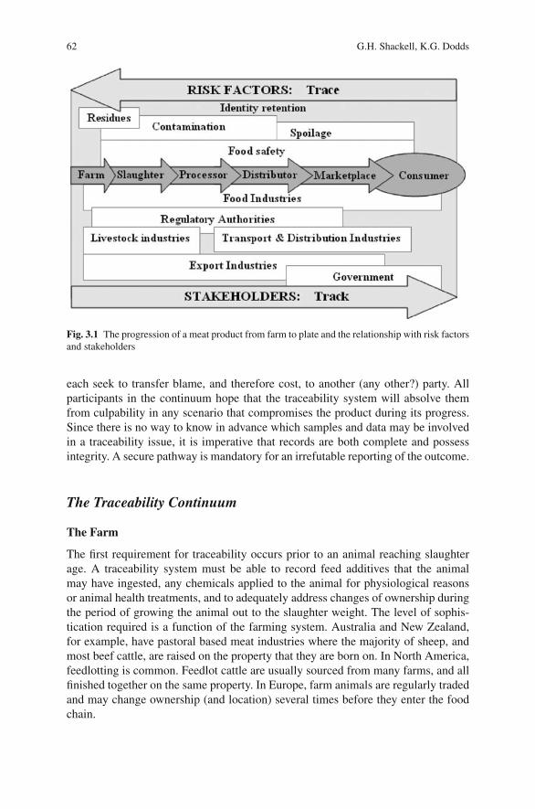
62 G.H. Shackell, K.G. Dodds
Fig. 3.1 The progression of a meat product from farm to plate and the relationship with risk factorsand stakeholders
each seek to transfer blame, and therefore cost, to another (any other?) party. Allparticipants in the continuum hope that the traceability system will absolve themfrom culpability in any scenario that compromises the product during its progress.Since there is no way to know in advance which samples and data may be involvedin a traceability issue, it is imperative that records are both complete and possessintegrity. A secure pathway is mandatory for an irrefutable reporting of the outcome.
The Traceability Continuum
The Farm
The first requirement for traceability occurs prior to an animal reaching slaughterage. A traceability system must be able to record feed additives that the animalmay have ingested, any chemicals applied to the animal for physiological reasonsor animal health treatments, and to adequately address changes of ownership duringthe period of growing the animal out to the slaughter weight. The level of sophis-tication required is a function of the farming system. Australia and New Zealand,for example, have pastoral based meat industries where the majority of sheep, andmost beef cattle, are raised on the property that they are born on. In North America,feedlotting is common. Feedlot cattle are usually sourced from many farms, and allfinished together on the same property. In Europe, farm animals are regularly tradedand may change ownership (and location) several times before they enter the foodchain.
3 DNA-Based Traceability of Meat 63
The Slaughterhouse
The first parts of the carcass to be removed and discarded are the head, the skin,and the viscera. Therefore, any physical identification cues are lost in relation to themeat. Carcasses may be re-identified as they move through the slaughter plant bythe use of barcodes or radio frequency identification devices (RFIDs) attached tothe body, and/or the hardware associated with each carcass. In order to maintain atracking/traceability chain, the original mark and the slaughterhouse identificationmust be linked in such a way as to be unambiguous.
The Processor
Meat may be further processed on premises associated with the slaughterhouse, orthe carcasses may be shipped to a third-party processor. The animal is dismantledduring processing for distribution either as primal cuts, which are further reducedprior to sale, or as pre-packaged individual cuts. The procedure is usually non-linear,so meat from different animals becomes jumbled during processing and packag-ing. For the process to allow full traceability, a label or identifier must be correctlyattached to each individual piece of meat as it is created, and remain with that pieceof meat at all times. In a system designed to meet USDA regulations, requiring thatthe carcasses and their parts can be identified “as being derived from a particular[live] animal”, Heaton et al. (2005), found that 9.5% of liver samples did not matchthe animal they purportedly came from.
The majority of ground meat products is made from off-cuts or trimmed meat.Processing times and costs may increase considerably if each animal or each pieceof trim meat is identified individually throughout the boning process.
The Distributor
Even if it has been possible to keep all of the components of a carcass togetherduring slaughter and processing, it becomes much harder to create a verifiable trailonce the meat is placed with a distributor. It is highly likely that a distributor willsupply to multiple retailers, and that this will be done on the basis of inventoryrequirements, with no reference to the origin of the meat.
The Marketplace
In the marketplace, it is likely that large retail chains may in fact further distributemeat within their own organization, thereby adding to the complication of trace-ability. The marketplace is also where substitution and/or fraudulent packaging (orre-packaging) is most likely to occur.
The Consumer
When a meat product reaches the consumer, the integrity of any traceability trail isalmost invariably lost. It is highly unlikely that the consumer will retain the packag-ing from a meat cut for traceability purposes. However, in the restaurant trade, where
64 G.H. Shackell, K.G. Dodds
meat is often purchased as primal cuts, a record of the supplier and any identificationof the meat are more likely to be kept. In some Japanese restaurants, the informationis made available for those diners who want to check the origin of the steak they areeating (Ozawa, Lopez-Villalobos, & Blair, 2005).
Risk
Identity Retention
The greatest risk in any system where the objective is to provide traceability is iden-tity retention. The most common animal identification system is printed tags (whichcan be changed), usually inserted in the ear. Less common, but still widely used,is branding the hide of the animal by burning an identification mark onto the skinusing heat or ultra-cold temperatures. Even less commonly used are tattoos. Theseare generally on the ear, but could be on the lip, or in hair/wool by free skin such asthe inside of the shoulder. Advances in electronics has resulted in the increasing useof RFIDs, which can be embedded in ear tags, implanted under the skin or encased ina protective layer and inserted into the digestive tract of the animal. More recently inlive animals, biometric technologies have also been developed, including scanningretinal and/or nose patterns, which have a similar ability to differentiate individualanimals as do fingerprints in the human.
Each of these methods offers a way of tracing animals through on-farm recordkeeping, changes of ownership, and during transport. Whichever method is used,most records are stored by transcribing the information into a computer file and/oronto a hard copy or PDA. Other than with RFID, an observer must read the tagor brand and then write the number(s) down. This provides potential error points,where the integrity of the traceability trail is only as reliable as the reading andtranscription of the data.
Food Safety
Food safety and quality, along with concomitant environmental and animal welfareissues, are increasingly important in the world marketplace. The meat industry glob-ally has responded to a series of crises such as Bovine Spongiform Encephalopathy(Smith, Cousens, Huillard D’Aignaux, Ward, & Will, 2004), labeling scandals(Castaldo, 2003), chemical contamination scares (e.g., Dioxin, Verbeke, 2001;ElAmin, 2006) and microbiological poisoning (Evans, Lane, Frost, & Nylen, 1998;Anonymous, 2002; Cagney et al., 2004). These issues, plus an increasing publicawareness of animal ethics (Hobbs, Hobbs, Isaac, & Kerr, 2002) and negativ-ity toward genetically modified food (Miraglia et al., 2004), have been citedas the drivers of increased consumer concern about the quality, origin, andintegrity of meat at all levels of the food chain (Arana et al., 2002; Hoban, 1997;Aumaıtre, 1999).
3 DNA-Based Traceability of Meat 65
Stakeholders
Bernues, Olaizola, and Corcoran (2003) found that consumers can be placed indemographic groups, based on age, family, disposable income, and meat consump-tion patterns, which have widely differing requirements for traceability. Interest-ingly, in another consumer survey, Latouche, Rainelli, and Vermersch (1998) foundthat 25–30% of the respondents who were unwilling to pay more for meat with atraceability guarantee held that view because they believed that the cost should notbe borne by the consumer. Verbeke, Frewer, Scholderer, and De Brabander (2007)suggested that some consumer behavior toward food safety and risk informationwas sometimes irrational because they did not necessarily equate behavior with riskinformation. Despite this, retailers, wholesalers, and in many cases legislators, insistthat the call for traceability is consumer driven. However, it is highly likely thatthe individual consumer desire for traceability varies with socio-economic status(Dickinson & Bailey, 2002; Hobbs, Bailey, Dickinson, & Haghiri, 2005).
Traceability is critical for the successful identification and recall of contaminatedproducts from the market, and as a support for the quality assurance processes(Caporale, Giovannini, Di Francesco, & Calistri, 2001). Microbial transfer can actu-ally occur during the slaughter process (Buncic, McKinstry, Reid, & Anil, 2002).
Some producers are interested in traceability for the protection and even value-enhancement of their brands, especially in niche markets or during a food crisis(Arana et al., 2002; Cavani and Petracci, 2004).
The industries that transport, process, and distribute animals and/or meat prod-ucts have an obligation to maintain records that will enhance traceability. In theinterests of food safety, it is now becoming increasingly common to have real-timemonitoring of location and conditions during transport.
Considerable effort has resulted in meat traceability schemes, which are nowrequired, by regulation, in some regions such as Japan (Clemens, 2003) and theEU (Cavani & Petracci, 2004), as governments have reacted to the growing publicconcern. The Japanese system, for example, is available at point-of-sale (Ozawaet al., 2005). In the Netherlands, a slaughterhouse may not receive a pig of unknownorigin (Madec, Geers, Vesseur, Kjeldsen, & Blaha, 2001).
What is DNA?
DNA is the blueprint that determines how species, breeds, strains, and ultimatelyindividual animals differ from each other. Within an individual, the DNA of alltissues is always the same. The only way two individuals can have the same DNAcode is if they are identical twins (or clones) or in some cases when they come frominbred lines or crosses between inbred lines. Therefore, DNA provides an in-builtproof of identity in a meat traceability system.
DNA is made up of four molecules (adenine, thiamine, guanine, and cyto-sine) called nucleotides. The way nucleotides are arranged is a sequence, and thecomplete sequence of nucleotides for an individual is its genome. A specific region
66 G.H. Shackell, K.G. Dodds
within the genomic DNA is referred to as a locus. Within that locus there are sub-setsof sequences called alleles. An allele may be comprised of many apparently randomnucleotides, a group of (usually di, tri, or tetra) repeated nucleotides or just a singlenucleotide. The underlying precept is that each locus has its own set of alleles.
Alleles are usually inherited by the Mendelian genetic principles; one allele ateach locus is inherited from each parent. If both parents contribute the same allele,the individual is said to be homozygous at that locus and if each parent contributesa different allele the individual is heterozygous.
The pattern of allele differences at a set of multiple loci can distinguish betweenindividuals and the frequency with which the components of those differences occur,can distinguish between breeds or species. These differences are referred to as DNAmarkers, and it is the variation between the markers that underpins DNA-basedtraceability technologies.
DNA Markers
The first genetic markers were for simple phenotype variants such as eye color, orprotein polymorphisms such as the hemoglobin blood groups. DNA markers havean advantage over protein markers in that they are not degraded by cooking and theyare present in all cells in the body. The advent of DNA technologies, in particular,the polymerase chain reaction (PCR) technique, which amplifies small amounts ofDNA into analyzable quantities, has allowed the rapid development of informative(variable) genetic markers.
Multi-locus Markers
Minisatellite or VNTR (variable number tandem repeat) markers: Minisatellites,discovered by Jeffreys, Wilson, and Thein (1985), were the first markers sufficientlyinformative that each individual had a unique genotype. They were mostly dominantmarkers with only one allele able to be identified. A few, highly informative, singleloci minisatellites identified in livestock (Georges et al., 1990) have been founduseful. However, with the advent of microsatellites (see below) they have fallen outof favor because they are technically demanding and require a large quantity of DNAfor analysis.
RAPD (random amplified polymorphic DNA fragments) markers: RAPD mark-ers were among the first PCR-based markers to be used (Williams, Kubelik, Livak,Rafalski, & Tingey, 1990). They are very easy to generate and require only smallquantities of DNA. They also have disadvantages. Their appearance (and disap-pearance) is very sensitive to slight changes in PCR conditions, so they are noteasily reproducible. More seriously however, they are not locus specific. The bandsgenerated by a particular primer in one pedigree may not bear any relation to bandsgenerated by the same primer in a second pedigree.
AFLP (amplified fragment length polymorphisms) markers: AFLPs are nowthe multi-locus markers of choice (Vos et al., 1995). Genomic DNA is cut withrestriction enzymes and linkers are attached to each end of the fragments. Subsets of
3 DNA-Based Traceability of Meat 67
the fragments are then selectively amplified from the mixture of genomic restrictionfragments and separated according to size. Bands that are present in some individu-als but absent in others can be used as genetic markers.
Single Locus Markers
Restriction Fragment Length Polymorphisms: RFLPs were the first DNA mark-ers developed and they pre-date the development of PCR methods. The RFLPmethodology utilizes restriction enzyme digestion of the genomic DNA, its sep-aration by size using agarose gel electrophoresis and detection and analysis ofthe DNA sequence by a technique called Southern blotting (Sambrook, Fritsch, &Maniatis, 1989). RFLP markers detect the presence or absence of a restriction site,including insertions and deletions of additional sequences, provided it contains anadditional restriction site or changes the band size seen on the Southern blot.
Microsatellites: Microsatellites, like minisatellites, are multi-allelic tandemrepeats. However, they are single locus, co-dominant, and spread throughout thegenome. They require only small amounts of template DNA and are relatively easyto find and characterize.
A microsatellite is a simple sequence that is repeated 10–50 times. Virtually allof the microsatellites that have been found for livestock have the sequence (AC/GT)as the repeat unit as it is the most abundant type within livestock genomes andtherefore much easier to find and characterize. Allelic variation of microsatellitesis due to different numbers of simple sequence repeats, which alters the size of theDNA fragment and allows them to be separated, usually by electrophoresis on aDNA sequencing gel.
Single Nucleotide Polymorphisms: The variation in sequence at a particularposition is called a single nucleotide polymorphism (SNP). It has been estimatedthat an SNP occurs about once in every kilo base in humans (Cooper, Smith, &Cooke, 1985). In other mammals, such as livestock, the figure is likely to be simi-lar giving potentially two to three million SNPs. SNPs represent by far the richestsource of genetic variation available for traceability applications. A disadvantageis that SNPs are almost always bi-allelic. Therefore, individually they are muchless informative than the multi-allelic markers and differentiation between indi-viduals requires many more SNPs to be analyzed than highly informative multi-allelic markers. For this reason, as they were first becoming popular, Vignal, Milan,SanCristobal, and Eggen (2002) referred to the increase in use of SNPs as an “appar-ent step backward”. The development of new technologies, such as “DNA chips”containing high density arrays of DNA (Chee et al., 1996), allows many SNPs to begenotyped at once, thus off-setting the disadvantage of requiring large numbers ofsuch markers by significantly decreasing the unit cost per marker.
DNA as a Traceability Tool
Traceability systems usually revolve around devices that are attached to an animalor a piece of meat. Because DNA is unique to an individual it is a useful toolfor proof of identity and/or audit in meat traceability systems (Shackell, 2005).
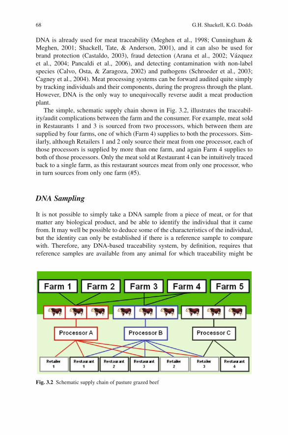
68 G.H. Shackell, K.G. Dodds
DNA is already used for meat traceability (Meghen et al., 1998; Cunningham &Meghen, 2001; Shackell, Tate, & Anderson, 2001), and it can also be used forbrand protection (Castaldo, 2003), fraud detection (Arana et al., 2002; Vazquezet al., 2004; Pancaldi et al., 2006), and detecting contamination with non-labelspecies (Calvo, Osta, & Zaragoza, 2002) and pathogens (Schroeder et al., 2003;Cagney et al., 2004). Meat processing systems can be forward audited quite simplyby tracking individuals and their components, during the progress through the plant.However, DNA is the only way to unequivocally reverse audit a meat productionplant.
The simple, schematic supply chain shown in Fig. 3.2, illustrates the traceabil-ity/audit complications between the farm and the consumer. For example, meat soldin Restaurants 1 and 3 is sourced from two processors, which between them aresupplied by four farms, one of which (Farm 4) supplies to both the processors. Sim-ilarly, although Retailers 1 and 2 only source their meat from one processor, each ofthose processors is supplied by more than one farm, and again Farm 4 supplies toboth of those processors. Only the meat sold at Restaurant 4 can be intuitively tracedback to a single farm, as this restaurant sources meat from only one processor, whoin turn sources from only one farm (#5).
DNA Sampling
It is not possible to simply take a DNA sample from a piece of meat, or for thatmatter any biological product, and be able to identify the individual that it camefrom. It may well be possible to deduce some of the characteristics of the individual,but the identity can only be established if there is a reference sample to comparewith. Therefore, any DNA-based traceability system, by definition, requires thatreference samples are available from any animal for which traceability might be
Fig. 3.2 Schematic supply chain of pasture grazed beef
3 DNA-Based Traceability of Meat 69
required, and that these reference samples are stored in a way that they can be easilyaccessed. Once the shelf life of the product has been exceeded, the reference samplescan be discarded.
Reference samples can be blood, semen, meat/tissue, tail hair follicles, or evensaliva or milk. When a DNA sample has been taken from an animal as a referencesample, it can later be used to verify that animal at any time simply by comparing itto the test sample. This makes the animal fully traceable. For example, the referenceDNA sample can be used to ensure that meat supposedly from a specific animal isin fact from that animal only and has not been substituted.
Although the primary focus of this chapter is meat, DNA traceability is not limitedtomeat fromdomesticatedanimals.Amongothers, the seafoodandaquaculture indus-tries are increasingly using DNA traceability (Hastein, Hill, Berthe, & Lightner, 2001;Hayes, Sonesson, & Gjerde, 2005), especially to detect species fraud (Aranishi,Okimoto, & Izumi, 2005; Maldini, Nonnis Marzano, Fortes, Papa, & Gandolfi,2006).
Using DNA Markers to Make Inferences About the Originof a Sample
When a DNA sample is obtained from a test item, traceability addresses the need tolearn something about its relationship to (one or more) reference items. The test andreference samples are assayed for a set of genetic markers (usually microsatellites orSNPs). The simplest relationship to test is that the samples are from the same indi-vidual i.e., a test for identity. Genetic marker results can also be used to make infer-ences about more distant types of relationships, for example, parent–offspring, fullor half-sibling, geographic region of origin, breed, or species. Identity and parentalrelationships lend themselves to exclusion tests – the hypothesized relationship isexcluded if the samples do not share at least two or one, respectively, alleles atevery locus. Other types of relationships cannot be tested in this way, for example,full-sibs may share any number of alleles (including zero) at a locus. In these caseswe are interested in how likely such results would be.
Notation and Assumptions
For a particular marker, let pi be the frequency of the i th allele, Ai , i = 1, 2, . . . , v.
Let St = ∑νi=1 pt
i
Unless stated otherwise, it is assumed that the markers are in Hardy–Weinbergequilibrium (Falconer & Mackay, 1996) in the source population, and that the allelefrequencies are known without error for this population. Further assumptions arethat the markers are autosomal (nuclear but not X-linked) and have alleles that aretransmitted in a Mendelian fashion, that there is no mutation and that genotypes
70 G.H. Shackell, K.G. Dodds
are determined without error. Different markers are assumed to be in linkage equi-librium and segregating independently. A final assumption is that individuals andmates are sampled at random from the population.
Identity
Probability of Identity
Identity testing using DNA markers is an extremely powerful technique that has ledto its application in forensics and traceability. The genotype results for an individualare often called its DNA “profile” or “fingerprint”. A test and a reference sample arecompared, and if their profiles are not the same, the samples are supposed to bederived from different individuals (assuming no errors are made in the testing pro-cess). This is a powerful technique because, with even a few markers, the probabilitythat two unrelated individuals share the same genotypes is extremely small.
The power of this test is often quantified using probability statements. There aretwo situations. The first is before the test sample has been genotyped, and we want toknow the usefulness of the marker set. The probability that two individuals, chosenat random from the population, have the same profile is known as the (average)probability of identity (PI) and is (e.g., Taberlet & Luikart, 1999)
PI =∑ν
i=1p4
i +∑
i
∑
j>i(2pi p j )
2
= 2S22 − S4
(see Dodds, Tate, McEwan, & Crawford, 1996). For multiple markers, the PI is theproduct of the individual marker PIs (assuming the markers are independent, i.e.,are in linkage equilibrium). The probability that the two individuals do not share thesame profile is known as the identity exclusion. Some values for 1-PI (i.e., identityexclusions) are shown in Figs. 3.3–3.5.
Once a test individual has been profiled, we would like to know what the prob-ability is of that particular profile being seen in another individual. This is oftenreferred to as the match probability (for unrelated individuals). The match probabil-ity for each marker is the population frequency of that genotype:
P(Ai Ai ) = p2i
P(Ai A j ) = 2pi p j , i �= j
Once again, the product of these for each marker is the match probability of theprofile. Sometimes both of the probabilities described above are referred to as matchprobabilities or identity probabilities.
These calculations make a number of assumptions, but refinements are availablewhich allow some of these to be relaxed. The reference samples may containindividuals which are more related than randomly chosen individuals from the

3 DNA-Based Traceability of Meat 71
0
0.2
0.4
0.6
0.8
1
Minor allele frequency
Pro
bab
ility
of
excl
usi
on
Q3, Progeny-Parent
Q2, Single Parent
1-PI, Identity
0 0.1 0.2 0.3 0.4 0.5
Fig. 3.3 Exclusion probabilities for a single locus with two alleles
0
0.2
0.4
0.6
0.8
1
Number of alleles
Pro
bab
ilit
y o
f ex
clu
sio
n
Q3, Progeny-Parent
Q2, Single Parent
1-PI, Identity
2 4 6 8 10
Fig. 3.4 Exclusion probabilities for a single locus with multiple alleles with equal frequencies
population. Animals processed at the same location and on the same date, are likelyto contain cohorts including half- and full-siblings. The probability of identity canbe modified to account for expected relationships, or expected population substruc-ture (Ayres & Overall, 2004). Similar adjustments are available for match proba-bilities (Evett & Weir, 1998). Vazquez et al. (2004) used a simulation procedureto accommodate their population structure consisting of two breeds and the crossbetween them. The assumption of non-independent loci is not considered a serious
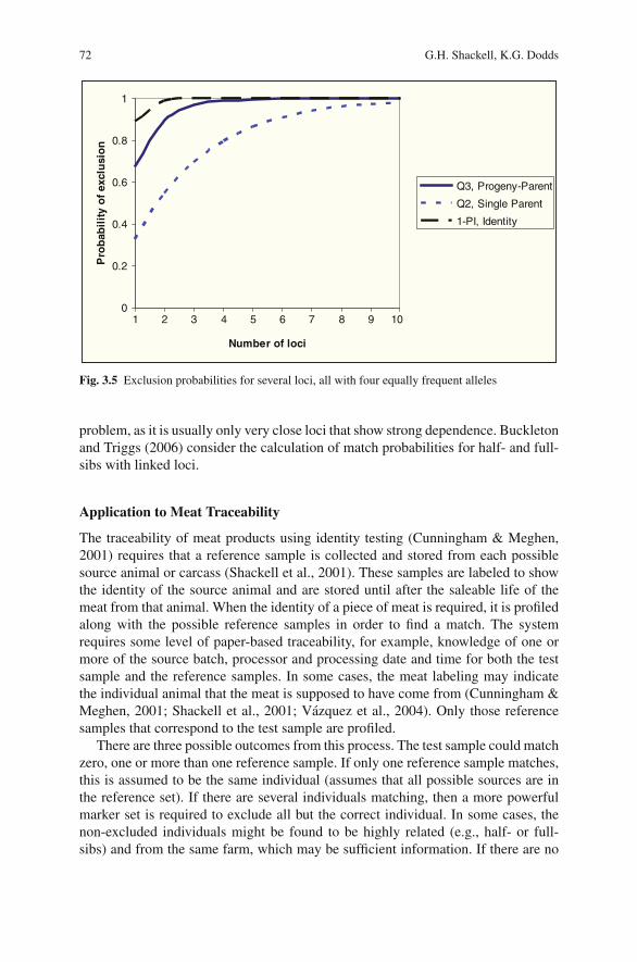
72 G.H. Shackell, K.G. Dodds
0
0.2
0.4
0.6
0.8
1
Number of loci
Pro
bab
ilit
y o
f ex
clu
sio
n
Q3, Progeny-Parent
Q2, Single Parent
1-PI, Identity
1 2 3 4 5 6 7 8 9 10
Fig. 3.5 Exclusion probabilities for several loci, all with four equally frequent alleles
problem, as it is usually only very close loci that show strong dependence. Buckletonand Triggs (2006) consider the calculation of match probabilities for half- and full-sibs with linked loci.
Application to Meat Traceability
The traceability of meat products using identity testing (Cunningham & Meghen,2001) requires that a reference sample is collected and stored from each possiblesource animal or carcass (Shackell et al., 2001). These samples are labeled to showthe identity of the source animal and are stored until after the saleable life of themeat from that animal. When the identity of a piece of meat is required, it is profiledalong with the possible reference samples in order to find a match. The systemrequires some level of paper-based traceability, for example, knowledge of one ormore of the source batch, processor and processing date and time for both the testsample and the reference samples. In some cases, the meat labeling may indicatethe individual animal that the meat is supposed to have come from (Cunningham &Meghen, 2001; Shackell et al., 2001; Vazquez et al., 2004). Only those referencesamples that correspond to the test sample are profiled.
There are three possible outcomes from this process. The test sample could matchzero, one or more than one reference sample. If only one reference sample matches,this is assumed to be the same individual (assumes that all possible sources are inthe reference set). If there are several individuals matching, then a more powerfulmarker set is required to exclude all but the correct individual. In some cases, thenon-excluded individuals might be found to be highly related (e.g., half- or full-sibs) and from the same farm, which may be sufficient information. If there are no
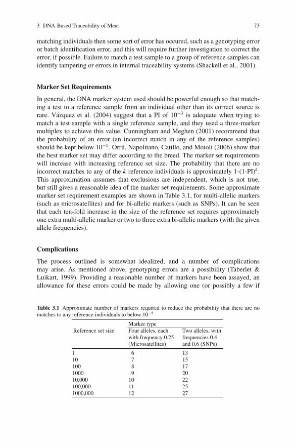
3 DNA-Based Traceability of Meat 73
matching individuals then some sort of error has occured, such as a genotyping erroror batch identification error, and this will require further investigation to correct theerror, if possible. Failure to match a test sample to a group of reference samples canidentify tampering or errors in internal traceability systems (Shackell et al., 2001).
Marker Set Requirements
In general, the DNA marker system used should be powerful enough so that match-ing a test to a reference sample from an individual other than its correct source israre. Vazquez et al. (2004) suggest that a PI of 10−3 is adequate when trying tomatch a test sample with a single reference sample, and they used a three-markermultiplex to achieve this value. Cunningham and Meghen (2001) recommend thatthe probability of an error (an incorrect match in any of the reference samples)should be kept below 10−5. Orru, Napolitano, Catillo, and Moioli (2006) show thatthe best marker set may differ according to the breed. The marker set requirementswill increase with increasing reference set size. The probability that there are noincorrect matches to any of the k reference individuals is approximately 1-(1-PI)k .This approximation assumes that exclusions are independent, which is not true,but still gives a reasonable idea of the marker set requirements. Some approximatemarker set requirement examples are shown in Table 3.1, for multi-allelic markers(such as microsatellites) and for bi-allelic markers (such as SNPs). It can be seenthat each ten-fold increase in the size of the reference set requires approximatelyone extra multi-allelic marker or two to three extra bi-allelic markers (with the givenallele frequencies).
Complications
The process outlined is somewhat idealized, and a number of complicationsmay arise. As mentioned above, genotyping errors are a possibility (Taberlet &Luikart, 1999). Providing a reasonable number of markers have been assayed, anallowance for these errors could be made by allowing one (or possibly a few if
Table 3.1 Approximate number of markers required to reduce the probability that there are nomatches to any reference individuals to below 10−5
Marker typeReference set size Four alleles, each
with frequency 0.25(Microsatellites)
Two alleles, withfrequencies 0.4and 0.6 (SNPs)
1 6 1310 7 15100 8 171000 9 2010,000 10 22100,000 11 251000,000 12 27
74 G.H. Shackell, K.G. Dodds
very many markers are assayed) mismatching genotype in the profiles. Dependingon the application, the test sample and reference individuals with few mismatchinggenotypes may have their profiles rechecked or re-assayed. A possible strategy isto duplicate the genotyping of all samples as a matter of course, but this doublesthe genotyping costs. Another approach would be to allow for genotyping errors ina likelihood model. The reference sample with the highest likelihood is declaredthe source individual, provided no other samples have a similar likelihood. Sim-ilar approaches have been used in parentage testing applications (Sancristobal &Chevalet, 1997; Marshall, Slate, Kruuk, & Pemberton, 1998).
There can also be errors associated with the labeling of samples and products. Insuch cases, it might be possible to find a match by widening the group of referencesamples tested. This would be successful, for example, if the error was to use a labelfrom the previous batch.
There may be reasons (apart from genotyping error) for why two samples fromthe same individual do not match, although these cases are rare. Sometimes, indi-viduals are chimeric (e.g., arising from the fusion of two early embryos) or mosaic(e.g., resulting from somatic cell mutations during early development), with differ-ing genotypes in different parts of their body. The most common livestock exampleof chimerism is when cattle litter mates share blood in utero, resulting in non-fertilefreemartin females in the case of mixed-sex twins. This can be seen by finding morethan two alleles for an individual (with multi-allelic markers).
There may also be reasons why two different individuals have matching pro-files (even with highly powerful marker sets). The individuals may have the samegenotypes, because they are monozygous twins, or because they are clones. Again,such cases are rare with production livestock, but may occur more frequently in thebreeding stock generated by using in vitro technologies. Currently, many countrieshave a voluntary moratorium on allowing cloned animals or their offspring to beused for human consumption. Most are waiting for an FDA decision (Rudenko &Matheson, 2007).
Parentage
Traceability by Parent Matching
One approach to DNA traceability is to match the test sample to its family (Hasteinet al., 2001). This could be achieved by collecting reference samples of parents(male, female, or both). With this approach, it is only possible to trace back to theset of progeny of the parent(s). For example, if a sire is known to have progeny onlyon one farm (with no stock movements to other farms), then a match to that sirewould narrow the source of the meat to animals from that farm. The main advantageof the approach is that possibly many fewer reference samples are required (althoughthey would need to be kept until after the retail life of the meat from any of theirprogeny). This approach might be particularly useful for species that are small insize but have large half- or full-sibling families, such as in aquaculture.
3 DNA-Based Traceability of Meat 75
Parentage Exclusion Probabilities
The power of DNA markers to test for parentage has usually been presented as anexclusion probability. For example, a paternity exclusion probability is the prob-ability that a randomly chosen unrelated male from the population has a geno-type incompatible with the paternity for the given genotypes of the individualand its dam. Parentage exclusion probabilities are given in, for example, Doddset al. (1996). Those that are most likely to be of relevance to traceability are theparent-offspring exclusion (i.e., without knowledge of the other parent’s genotype):
Q2 = 1 − 4S2 + 4S3 − 3S4 + 2S22
and the parent-pair (offspring) exclusion probability:
Q3 = 1 + 4S4 − 4S5 − 3S6 − 8S22 + 2S2
3 + 8S3S2
For multiple independent markers inclusion probabilities are found by multiply-ing together the inclusion probabilities for each marker. Exclusion probabilities areone minus the inclusion probability. Some values of these exclusion probabilitiesare shown in Figs. 3.3–3.5.
Relatedness
It is also possible to make inferences about relationships other than identity andparentage. In general, it is more difficult to make definitive statements, as it is notpossible to use exclusion-based methods. Some types of relationship that may be ofuse in traceability are an individual with its grandparent(s), full-sib(s) or half-sib(s).If these relationships can be inferred, then this might help narrow the source of ameat product.
Relationships are inferred by estimating relatedness (r ), which is twice the prob-ability that random alleles from each of the individuals are identical by descent(IBD). Some types of relationships with the same or similar values of r can bedistinguished by considering the kinship coefficients, i.e., the probability of sharing0, 1, or 2 alleles that are IBD (k0, k1, and k2), where r = k1/2 + k2. Table 3.2 showsthese values for some common relationships.
There are several methods for estimating relatedness or kinship coefficients.These methods have been reviewed by Blouin (2003) and Weir, Anderson, &Hepler (2006). The estimators tend to have a high sampling variance (Van DeCasteele, Galbusera, & Matthysen, 2001), and many markers are required to provideacceptable standard errors (Blouin, 2003; Weir et al., 2006). An alternative approachis to conduct hypothesis tests using likelihood ratios (Goring & Ott, 1997), but thesealso require many markers, especially if there is a need to distinguish between rea-sonably close relationships such as half- and full-sibs. These might be used, forexample, when trying to match an individual to a reference set thought to contain
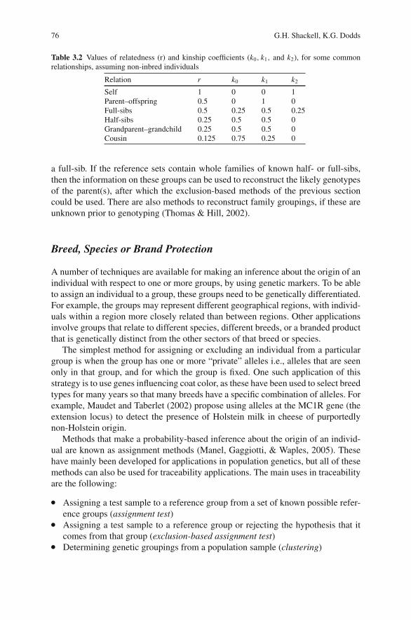
76 G.H. Shackell, K.G. Dodds
Table 3.2 Values of relatedness (r) and kinship coefficients (k0, k1, and k2), for some commonrelationships, assuming non-inbred individuals
Relation r k0 k1 k2
Self 1 0 0 1Parent–offspring 0.5 0 1 0Full-sibs 0.5 0.25 0.5 0.25Half-sibs 0.25 0.5 0.5 0Grandparent–grandchild 0.25 0.5 0.5 0Cousin 0.125 0.75 0.25 0
a full-sib. If the reference sets contain whole families of known half- or full-sibs,then the information on these groups can be used to reconstruct the likely genotypesof the parent(s), after which the exclusion-based methods of the previous sectioncould be used. There are also methods to reconstruct family groupings, if these areunknown prior to genotyping (Thomas & Hill, 2002).
Breed, Species or Brand Protection
A number of techniques are available for making an inference about the origin of anindividual with respect to one or more groups, by using genetic markers. To be ableto assign an individual to a group, these groups need to be genetically differentiated.For example, the groups may represent different geographical regions, with individ-uals within a region more closely related than between regions. Other applicationsinvolve groups that relate to different species, different breeds, or a branded productthat is genetically distinct from the other sectors of that breed or species.
The simplest method for assigning or excluding an individual from a particulargroup is when the group has one or more “private” alleles i.e., alleles that are seenonly in that group, and for which the group is fixed. One such application of thisstrategy is to use genes influencing coat color, as these have been used to select breedtypes for many years so that many breeds have a specific combination of alleles. Forexample, Maudet and Taberlet (2002) propose using alleles at the MC1R gene (theextension locus) to detect the presence of Holstein milk in cheese of purportedlynon-Holstein origin.
Methods that make a probability-based inference about the origin of an individ-ual are known as assignment methods (Manel, Gaggiotti, & Waples, 2005). Thesehave mainly been developed for applications in population genetics, but all of thesemethods can also be used for traceability applications. The main uses in traceabilityare the following:
� Assigning a test sample to a reference group from a set of known possible refer-ence groups (assignment test)
� Assigning a test sample to a reference group or rejecting the hypothesis that itcomes from that group (exclusion-based assignment test)
� Determining genetic groupings from a population sample (clustering)
3 DNA-Based Traceability of Meat 77
The main difference between the first two applications is that in the first, all pos-sible reference groups are known (and characterized for the markers being used)while in the second application only the group of prime interest is characterized. Anexample of the first is when meat is claimed to come from an animal of a particularbreed, but it could be from one of several breeds. An example of the second is whenmeat is claimed to be of a particular brand, but if not it could come from any othergroup within the species. The third application (finding genetic groupings) might beundertaken to help create a genetically distinct brand.
Assignment Test
An assignment test endeavors to classify an unknown into its group of origin, usingDNA marker information (Manel et al., 2005). The methods rely on all possiblesource groups being known and having genotype information (for the same markers)(i.e., they are reference groups). They generally assume that the reference groups arerandomly sampled and are in Hardy–Weinberg and linkage equilibrium. In its sim-plest form, the assignment test calculates the likelihood of the test sample comingfrom each of the reference groups (i.e., the probability of the test genotypes giventhe allele frequencies for the reference group). The test sample is then assignedto the reference group that yields the highest likelihood (Manel et al., 2005). Analternative method is to calculate a genetic distance between the test sample and thereference groups, and assign the closest reference group. This method relaxes theassumptions of Hardy–Weinberg and linkage equilibrium (Cornuet, Piry, Luikart,Estoup, & Solignac, 1999).
One difficulty with the method is that if the test sample contains an allele not seenin a particular reference sample, then the likelihood will be zero. This is unsatisfac-tory as it may be due to insufficient sampling of the reference population. A numberof solutions have been suggested (Ciampolini et al., 2006). Another difficulty is thatthe test may be assigned to one reference group, but its likelihood for another groupis similar, that is, an assignment is made even though there is little discriminationbetween two groups. This is less of a problem in population genetics, where thetests were developed, than for traceability applications. In population genetics, it islikely that many individuals are being assigned to populations and an error in oneis not critical. In traceability applications, it is usually a single individual that is thefocus of the investigation. A possible solution is to use an exclusion-based test (seebelow) as a follow up, or to establish a statistical confidence level using simulationprocedures, similar to those used in parentage studies by Marshall et al. (1998).
Exclusion-Based Assignment Test
An exclusion test can be constructed using either likelihood or distance-basedapproaches. A large number of individuals are simulated for a particular referencepopulation, by drawing alleles from the population (using estimated frequencies) atrandom. The assignment test value is calculated for each simulated individual givinga distribution of values for members from that population, which allows a confidence
78 G.H. Shackell, K.G. Dodds
interval to be constructed (Cornuet, 1999). A modification of this procedure allowsfor linkage disequilibrium (created, e.g., by population admixture) by sampling eachset of an individual’s alleles from a randomly chosen individual (rather than frommany different individuals) in the reference population (Paetkau, Slade, Burden, &Estoup, 2004).
Clustering
A number of methods exist for genetic clustering, for example, phylogenetic meth-ods. These assume that there is no interchange of genetic material between groups,and a number of methods without this assumption have been developed, includ-ing the one used by STRUCTURE (Pritchard, Stephens, & Donnelly, 2000). Thismethod allows individuals to be partially assigned to several groups.
Application to Food Safety
DNA can also be used for traceability related to food safety issues. In this case, itis the extrinsic DNA of pathogens and microbes that is tested, as opposed to theintrinsic DNA of the individuals that are the source of the meat or meat products.
As living organisms, the same principles of DNA apply. In fact, because of thevery short generation interval, mutations can quickly identify specific strains andpopulations. In some instances, specific geographic locations develop “signature”microbes, which can be used to isolate the origin of contamination. DNA tech-nologies can, much more rapidly than the traditional culture methods, identify thepresence and the species of microbes (Kim et al., 2007), and/or strains of microbes(O’Hanlon, Catarame, Blair, McDowell, & Duffy, 2005; Villani, Russo, Blaiotta,Moschetti, & Ercolini, 2005).
Application to Audit
By definition, a paper/electronic based traceability system can only be forwardaudited. If traceability through the supply chain is to an animal level, the only wayto effectively audit a paper trail, is to follow an animal right through the chain,from the Farm in a forward direction via the Processor to the Retailer or Restaurant.What an auditor cannot do is enter the paper/e-trail pathway and work in reverse.It is possible to follow the trail, but the integrity of any record keeping cannot beguaranteed. DNA identification technologies offer the potential to effectively iden-tify meat from individual animals, by providing the ability to augment and ver-ify a traceability paper trail (Meghen et al., 1998; Cunningham & Meghen, 2001;Shackell et al., 2001; Tate, 2001), or to detect fraud (Arana et al., 2002; Vazquezet al., 2004). For example, DNA reference samples can be used as a random audit
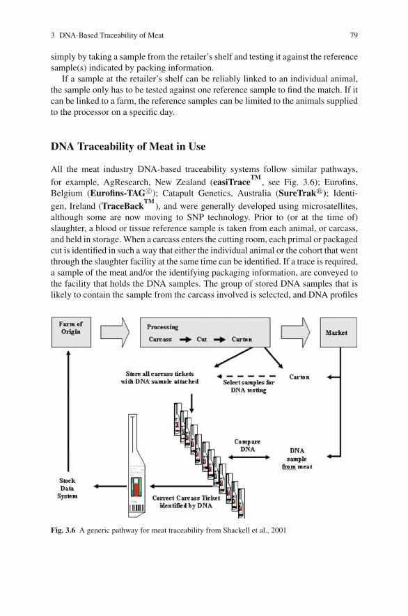
3 DNA-Based Traceability of Meat 79
simply by taking a sample from the retailer’s shelf and testing it against the referencesample(s) indicated by packing information.
If a sample at the retailer’s shelf can be reliably linked to an individual animal,the sample only has to be tested against one reference sample to find the match. If itcan be linked to a farm, the reference samples can be limited to the animals suppliedto the processor on a specific day.
DNA Traceability of Meat in Use
All the meat industry DNA-based traceability systems follow similar pathways,
for example, AgResearch, New Zealand (easiTraceTM
, see Fig. 3.6); Eurofins,Belgium (Eurofins-TAG c©); Catapult Genetics, Australia (SureTrak�); Identi-
gen, Ireland (TraceBackTM
), and were generally developed using microsatellites,although some are now moving to SNP technology. Prior to (or at the time of)slaughter, a blood or tissue reference sample is taken from each animal, or carcass,and held in storage. When a carcass enters the cutting room, each primal or packagedcut is identified in such a way that either the individual animal or the cohort that wentthrough the slaughter facility at the same time can be identified. If a trace is required,a sample of the meat and/or the identifying packaging information, are conveyed tothe facility that holds the DNA samples. The group of stored DNA samples that islikely to contain the sample from the carcass involved is selected, and DNA profiles
Fig. 3.6 A generic pathway for meat traceability from Shackell et al., 2001
80 G.H. Shackell, K.G. Dodds
are used to unequivocally match the sample to the carcass or animal that it origi-nated from. For any DNA-based system to be effective an auditable production trailthrough the processing facility is essential and the processor must follow standardoperating procedures to avoid contamination among reference samples (Shackellet al., 2001). Provided these conditions are met, a standardized and validated ana-lytical procedure will match the DNA profiles of a carcass at slaughter to a meatsample in a packing carton.
There are other systems (e.g., Maple leaf Foods in Canada and Kyodo Inter-national Inc. in Japan) that use mitochondrial SNP technologies. In these cases,samples are taken from the parent stock and the Genotypes stored in databases.Test samples are analyzed and the resultant genotypes are compared to the databaseinformation to determine the farm of origin.
The philosophy of traceability and audit is to reduce the reference samplesrequired to match a test sample to the smallest number possible. This increases thecost-effectiveness of the testing.
Compound Meat Products
Compound or blended meat products are not easily traceable since the origin ofthe source material is difficult to establish (McKean, 2001), and ground beef andprocessed products are generally exempt from traceability legislation (e.g., Japan,Clemens, 2003; European Union, Regulation (EC) No 1760/2000).
Individual DNA profiles show one or a maximum of two alleles at each DNAmarker (see Fig. 3.7). Because mixed samples contain many individuals, and eachhas its own unique alleles, mixed samples have a distinctive DNA “profile”, wherebymany of the potential alleles at each marker are present. Therefore, while conven-tional DNA traceability schemes work well for tracing primal and/or sub-primalmeat cuts, they have not been so readily adopted for compound meat products,which use ground meat from an unknown number of (usually) unidentified animals.For DNA traceability to be applicable to sausages and ground beef, methodologiesmust be developed to allow genotyping of DNA mixtures.
Determining the number of contributors to a mixture of randomly selected ani-mals is not feasible when the mixture contains DNA from more than five or sixindividuals (Dodds & Shackell, 2004). However, if the product can be separatedinto its constituents, the principle of identifying individuals by their DNA profilecan be applied (Shackell & Mathias, unpublished). Furthermore, given the nature ofmost mixed products, it is unlikely that the trim meat from a set of animals will go totwo different manufacturers. Therefore, if at least some of the contributors to mixedproducts are known, it is not necessary to identify every individual in the mixtureto know that the mixture is in fact the one being tested (Vetharaniam, Shackell &Upsdell, unpublished).
By using microsatellite markers as a tool for traceability of ground beef prod-uct, it is possible to match anonymous ground beef samples to the correct batch of
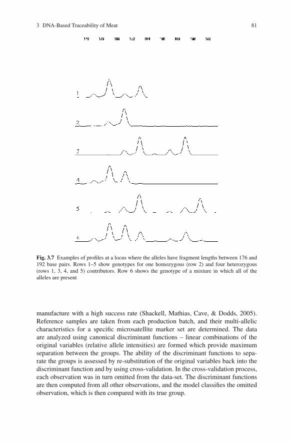
3 DNA-Based Traceability of Meat 81
Fig. 3.7 Examples of profiles at a locus where the alleles have fragment lengths between 176 and192 base pairs. Rows 1–5 show genotypes for one homozygous (row 2) and four heterozygous(rows 1, 3, 4, and 5) contributors. Row 6 shows the genotype of a mixture in which all of thealleles are present
manufacture with a high success rate (Shackell, Mathias, Cave, & Dodds, 2005).Reference samples are taken from each production batch, and their multi-alleliccharacteristics for a specific microsatellite marker set are determined. The dataare analyzed using canonical discriminant functions – linear combinations of theoriginal variables (relative allele intensities) are formed which provide maximumseparation between the groups. The ability of the discriminant functions to sepa-rate the groups is assessed by re-substitution of the original variables back into thediscriminant function and by using cross-validation. In the cross-validation process,each observation was in turn omitted from the data-set. The discriminant functionsare then computed from all other observations, and the model classifies the omittedobservation, which is then compared with its true group.
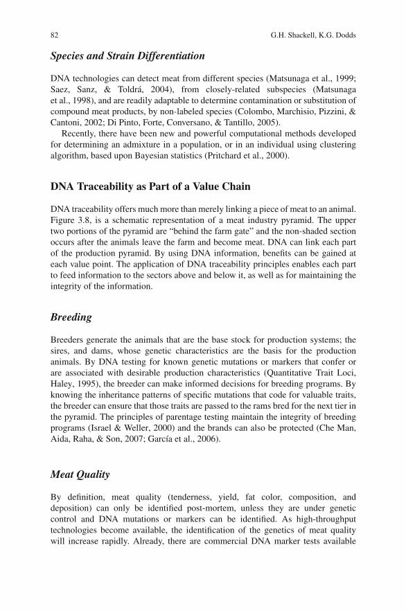
82 G.H. Shackell, K.G. Dodds
Species and Strain Differentiation
DNA technologies can detect meat from different species (Matsunaga et al., 1999;Saez, Sanz, & Toldra, 2004), from closely-related subspecies (Matsunagaet al., 1998), and are readily adaptable to determine contamination or substitution ofcompound meat products, by non-labeled species (Colombo, Marchisio, Pizzini, &Cantoni, 2002; Di Pinto, Forte, Conversano, & Tantillo, 2005).
Recently, there have been new and powerful computational methods developedfor determining an admixture in a population, or in an individual using clusteringalgorithm, based upon Bayesian statistics (Pritchard et al., 2000).
DNA Traceability as Part of a Value Chain
DNA traceability offers much more than merely linking a piece of meat to an animal.Figure 3.8, is a schematic representation of a meat industry pyramid. The uppertwo portions of the pyramid are “behind the farm gate” and the non-shaded sectionoccurs after the animals leave the farm and become meat. DNA can link each partof the production pyramid. By using DNA information, benefits can be gained ateach value point. The application of DNA traceability principles enables each partto feed information to the sectors above and below it, as well as for maintaining theintegrity of the information.
Breeding
Breeders generate the animals that are the base stock for production systems; thesires, and dams, whose genetic characteristics are the basis for the productionanimals. By DNA testing for known genetic mutations or markers that confer orare associated with desirable production characteristics (Quantitative Trait Loci,Haley, 1995), the breeder can make informed decisions for breeding programs. Byknowing the inheritance patterns of specific mutations that code for valuable traits,the breeder can ensure that those traits are passed to the rams bred for the next tier inthe pyramid. The principles of parentage testing maintain the integrity of breedingprograms (Israel & Weller, 2000) and the brands can also be protected (Che Man,Aida, Raha, & Son, 2007; Garcıa et al., 2006).
Meat Quality
By definition, meat quality (tenderness, yield, fat color, composition, anddeposition) can only be identified post-mortem, unless they are under geneticcontrol and DNA mutations or markers can be identified. As high-throughputtechnologies become available, the identification of the genetics of meat qualitywill increase rapidly. Already, there are commercial DNA marker tests available
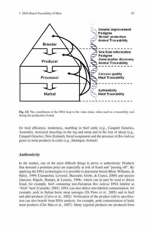
3 DNA-Based Traceability of Meat 83
Fig. 3.8 The contribution of the DNA loop to the value chain, when used as a traceability toolduring the production of meat
for feed efficiency, tenderness, marbling in beef cattle (e.g., Catapult Genetics,Australia), increased muscling on the leg and rump and in the loin of sheep (e.g.,Catapult Genetics, New Zealand), breed assignment and the presence of Bos indicusgenes in meat products in cattle (e.g., Identigen, Ireland).
Authenticity
In the market, one of the most difficult things to prove is authenticity. Productsthat demand a premium price are especially at risk of fraud and “passing off”. Byapplying the DNA technologies it is possible to determine breed (Blott, Williams, &Haley, 1999; Ciampolini, Leveziel, Mazzanti, Grohs, & Cianci, 2000) and species(Janssen, Hagele, Buntjer, & Lenstra, 1998), which can in turn be used to detectfraud, for example, beef containing non-European Bos indicus DNA labeled as“Irish” beef (Castaldo, 2003). DNA can also detect non-labeled contamination, forexample, pork in Italian horse meat sausages (Di Pinto et al., 2005) and in beefand pate products (Calvo et al., 2002). Verification of the product sold to specifica-tion can also benefit from DNA analysis, for example, pork contamination of halalmeat products (Che Man et al., 2007). Many regional products are produced from
84 G.H. Shackell, K.G. Dodds
specific breeds or strains and DNA can detect fraudulent substitution or breeding,for example, Iberian ham (Garcıa et al., 2006).
Summary
DNA offers an in-built traceability verification tool that cannot be removed froma piece of meat and is not destroyed by cooking. The only criterion is as follows:for traceability to be possible, reference samples are required. For an entire meatindustry, DNA offers much more than a simple traceability option by:
� providing information that allows genetic improvement of production animals� validating that improvement by verifying of pedigree� verifying the origin of meat� protecting the brand of niche market meats� identifying and enabling selection for meat quality� detecting fraud
References
Anonymous. (2002). Source of deadly US listeria outbreak unknown. Retrieved October 21, 2002,from http://www.foodnavigator.com.
Arana, A., Soret, B., Lasa, I., & Alfonso, L. (2002). Meat traceability using DNA markers: Appli-cation to the beef industry. Meat Science, 61, 367–373.
Aranishi, F., Okimoto, T., & Izumi, S. (2005). Identification of gadoid species (Pisces, Gadidae)by PCR-RFLP analysis. Journal of Applied Genetics, 46(1), 69–73.
Aumaıtre, A. (1999). Quality and safety of animal products. Livestock Production Science,59(2–3), 113–124.
Ayres, K. L., & Overall, A. D. J. (2004). api-calc 1.0: A computer program for calculating theaverage probability of identity allowing for substructure, inbreeding and the presence of closerelatives. Molecular Ecology Notes, 4(2), 315–318.
Bernues, A., Olaizola, A., & Corcoran, K. (2003). Labelling information demanded by Europeanconsumers and relationships with purchasing motives, quality and safety of meat. Meat Science,65(3), 1095–1106.
Blott, S. C., Williams, J. L., & Haley, C. S. (1999). Discriminating among cattle breeds usinggenetic markers. Heredity, 82(Part 6), 613–619.
Blouin, M. S. (2003). DNA-based methods for pedigree reconstruction and kinship analysis innatural populations. Trends in Ecology & Evolution, 18(10), 503–511.
Buckleton, J., & Triggs, C. (2006). The effect of linkage on the calculation of DNA match proba-bilities for siblings and half siblings. Forensic Science International, 160(2–3), 193–199.
Buncic, S., McKinstry, J., Reid, C. A., & Anil, M. H. (2002). Spread of microbial contamina-tion associated with penetrative captive bolt stunning of food animals. Food Control, 13(6–7),425–430.
Cagney, C., Crowley, H., Duffy, G., Sheridan, J. J., O’Brien, S., Carney, E., et al. (2004). Prevalenceand numbers of Escherichia coli O157:H7 in minced beef and beef burgers from butcher shopsand supermarkets in the Republic of Ireland. Food Microbiology, 21(2), 203–212.
Calvo, J. H., Osta, R., & Zaragoza, P. (2002). Quantitative PCR detection of pork in raw and heatedground beef and pate. Journal of Agricultural and Food Chemistry, 50(19), 5265–5267.
3 DNA-Based Traceability of Meat 85
Caporale, V., Giovannini, A., Di Francesco, C., & Calistri, P. (2001). Importance of the traceabilityof animals and animal products in epidemiology. OIE Revue Scientifique et Technique, 20(2),372–378.
Castaldo, D. (2003). Brazilian beef labeled Irish. Retrieved September 26, 2003, from Meat-News.com.
Cavani, C., & Petracci, M. (2004). Rabbit meat processing and traceability. Proceedings of the 8thWorld Rabbit Congress, 1318–1336.
Che Man, Y. B., Aida, A. A., Raha, A. R., & Son, R. (2007). Identification of pork derivatives infood products by species-specific polymerase chain reaction (PCR) for halal verification. FoodControl, 18(7), 885–889.
Chee, M., Yang, R., Hubbell, E., Berno, A., Huang, X. C., Stern, D., et al. (1996). Accessinggenetic information with high-density DNA arrays. Science, 274(5287), 610–614.
Ciampolini, R., Cetica, V., Ciani, E., Mazzanti, E., Fosella, X., Marroni, F., et al. (2006). Statisticalanalysis of individual assignment tests among four cattle breeds using fifteen STR loci. Journalof Animal Science, 84(1), 11–19.
Ciampolini, R., Leveziel, H., Mazzanti, E., Grohs, C., & Cianci, D. (2000). Genomic identificationof the breed of an individual or its tissue. Meat Science, 54(1), 35–40.
Clemens, R. (2003). Meat traceability in Japan. Iowa Ag Review, 9(4), 4–5.Colombo, F., Marchisio, E., Pizzini, A., & Cantoni, C. (2002). Identification of the goose species
(Anser anser) in Italian “Mortara” salami by DNA sequencing and a Polymerase Chain Reac-tion with an original primer pair. Meat Science, 61(3), 291–294.
Cooper, D. N., Smith, B. A., & Cooke, H. J. (1985). An estimate of unique DNA sequence het-erozygosity in the human genome. Human Genetics, 69(3), 201–205.
Cornuet, J. M., Piry, S., Luikart, G., Estoup, A., & Solignac, M. (1999). New methods employ-ing multilocus genotypes to select or exclude populations as origins of individuals. Genetics,153(4), 1989–2000.
Cunningham, E. P., & Meghen, C. M. (2001). Biological identification systems: genetic markers.Scientific and Technical Review of the Office International des Epizooties, 20(2), 491–499.
Di Pinto, A., Forte, V. T., Conversano, M. C., & Tantillo, G. M. (2005). Duplex polymerase chainreaction for detection of pork meat in horse meat fresh sausages from Italian retail sources.Food Control, 16(5), 391–394.
Dickinson, D. L., & Bailey, D. (2002). Meat traceability: Are U.S. consumers willing to pay for it?Journal of Agricultural and Resource Economics, 27(2), 348–364.
Dodds, K. G., & Shackell, G. H. (2004). The number of alleles seen in a mixture. Abstract MTU42.Paper presented at the XXIInd International Biometric Conference, Cairns, Australia.
Dodds, K. G., Tate, M. L., McEwan, J. C., & Crawford, A. M. (1996). Exclusion probabilities forpedigree testing farm animals. Theoretical and Applied Genetics, 92, 966–975.
ElAmin, A. (2006). Belgium, Netherlands meat sectors face dioxin crisis. Retrieved January 31,2006, from http://www.foodproductiondaily.com.
Evans, M. R., Lane, W., Frost, J. A., & Nylen, G. (1998). A campylobacter outbreak associatedwith stir-fried food. Epidemiology and Infection, 121(2), 275–279.
Evett, I. W., & Weir, B. S. (1998). Interpreting DNA evidence—statistical genetics for forensicscientists. Sunderland, MA: Sinauer Associates.
Falconer, D. S., & Mackay, T. F. C. (1996). Introduction to Quantitative Genetics (4th ed.). Harlow,Essex: Addison Wesley Longman.
Garcıa, D., Martınez, A., Dunner, S., Vega-Pla, J. L., Fernandez, C., Delgado, J. V., & Canon,J. (2006). Estimation of the genetic admixture composition of Iberian dry-cured ham samplesusing DNA multilocus genotypes. Meat Science, 72(3), 560–566.
Georges, M., Lathrop, M., Hilbert, P., Marcotte, A., Schwers, A., Swillens, S., et al. (1990). On theuse of DNA fingerprints for linkage studies in cattle. Genomics, 6(3), 461–474.
Goring, H., & Ott, J. (1997). Relationship estimation in affected sib pair analysis of late-onsetdiseases. European Journal of Human Genetics, 5(2), 69–77.
Haley, C. S. (1995). Livestock QTLs – bringing home the bacon. Trends in Genetics, 11(12),488–492.
86 G.H. Shackell, K.G. Dodds
Hastein, T., Hill, B. J., Berthe, F., & Lightner, D. V. (2001). Traceability of aquatic animals. Scien-tific and Technical Review of the Office International des Epizooties, 20(2), 564–583.
Hayes, B., Sonesson, A. K., & Gjerde, B. (2005). Evaluation of three strategies using DNA markersfor traceability in aquaculture species. Aquaculture, 250(1–2), 70.
Heaton, M. P., Keen, J. E., Clawson, M. L., Harhay, G. P., Bauer, N., Shultz, C., et al. (2005). Use ofbovine single nucleotide polymorphism markers to verify sample tracking in beef processing.Journal of the American Veterinary Medical Association, 226(8), 1311–1314.
Hoban, T. J. (1997). Consumer acceptance of biotechnology: An international perspective. NatureBiotechnology, 15(3), 232–234.
Hobbs, A. L., Hobbs, J. E., Isaac, G. E., & Kerr, W. A. (2002). Ethics, domestic food policy and tradelaw: Assessing the EU animal welfare proposal to the WTO. Food Policy, 27(5–6), 437–454.
Hobbs, J. E., Bailey, D., Dickinson, D. L., & Haghiri, M. (2005). Traceability in the Canadian redmeat sector: Do consumers care? Canadian Journal of Agricultural Economics/Revue canadi-enne d’agroeconomie, 53(1), 47–65.
Israel, C., & Weller, J. I. (2000). Effect of misidentification on genetic gain and estimation ofbreeding value in dairy cattle populations. Journal of Dairy Science, 83(1), 181–187.
Janssen, F. W., Hagele, G. H., Buntjer, J. B., & Lenstra, J. A. (1998). Species identification in meatby using PCR-generated satellite probes. Journal of Industrial Microbiology and Biotechnol-ogy, 21 (3), 115–120.
Jeffreys, A. J., Wilson, V., & Thein, S. L. (1985). Individual-specific ’fingerprints’ of human DNA.Nature, 316(6023), 76–79.
Kim, J. S., Lee, G. G., Park, J. S., Jung, Y. H., Kwak, H. S., Kim, S. B., et al. (2007). A novelmultiplex PCR assay for rapid and simultaneous detection of five pathogenic bacteria: Escheriacoli O157:H7, Salmonella, Staphylococcus aureus, Listeria monocytogenes and Vibrio para-haemolyticus. Journal of Food Protection, 70(7), 1656–1662.
Latouche, K., Rainelli, P., & Vermersch, D. (1998). Food safety issues and the BSE scare: Somelessons from the French case. Food Policy, 23(5), 347–356.
Madec, F., Geers, R., Vesseur, P., Kjeldsen, N., & Blaha, T. (2001). Traceability in the pig produc-tion chain. OIE Revue Scientifique et Technique, 20(2), 523–537.
Maldini, M., Nonnis Marzano, F., Fortes, G. G., Papa, R., & Gandolfi, G. (2006). Fish and seafoodtraceability based on AFLP markers: Elaboration of a species database. Aquaculture, 261(2),487–494.
Manel, S., Gaggiotti, O. E., & Waples, R. S. (2005). Assignment methods: matching biologicalquestions with appropriate techniques. Trends in Ecology & Evolution, 20(3), 136–142.
Marshall, T. C., Slate, J., Kruuk, L. E. B., & Pemberton, J. M. (1998). Statistical confidence forlikelihood-based paternity inference in natural populations. Molecular Ecology, 7, 639–655.
Matsunaga, T., Chikuni, K., Tanabe, R., Muroya, S., Nakai, H., Shibata, K., et al. (1998). Deter-mination of mitochondrial cytochrome B gene sequence for red deer (Cervus Elaphus) and thedifferentiation of closely related deer meats. Meat Science, 49(4), 379–385.
Matsunaga, T., Chikuni, K., Tanabe, R., Muroya, S., Shibata, K., Yamada, J., & Shinmura, Y.(1999). A quick and simple method for the identification of meat species and meat products byPCR assay. Meat Science, 51(2), 143–148.
Maudet, C., & Taberlet, P. (2002). Holstein’s milk detection in cheeses inferred from melanocortinreceptor 1 (MC1R) gene polymorphism. Journal of Dairy Science, 85(4), 707–715.
McKean, J. D. (2001). The importance of traceability for public health and consumer protection.OIE Revue Scientifique et Technique, 20(2), 363–371.
Meghen, C. N., Scott, C. S., Bradley, D. G., MacHugh, D. E., Loftus, R. T., & Cunningham, E. P.(1998). DNA based traceability techniques for the beef industry. Animal Genetics, 29S1, 48–49.
Miraglia, M., Berdal, K. G., Brera, C., Corbisier, P., Holst-Jensen, A., Kok, E. J., et al. (2004).Detection and traceability of genetically modified organisms in the food production chain. Foodand Chemical Toxicology, 42(7), 1157–1180.
O’Hanlon, K. A., Catarame, T. M. G., Blair, I. S., McDowell, D. A., & Duffy, G. (2005). Compar-ison of a real-time PCR and an IMS/culture method to detect Escherichia coli O26 and O111in minced beef in the Republic of Ireland. Food Microbiology, 22(6), 553–560.
3 DNA-Based Traceability of Meat 87
Orru, L., Napolitano, F., Catillo, G., & Moioli, B. (2006). Meat molecular traceability: How tochoose the best set of microsatellites? Meat Science, 72(2), 312–317.
Ozawa, T., Lopez-Villalobos, N., & Blair, H. T. (2005). An update on beef traceability regulationsin Japan. Proceedings of the New Zealand Society of Animal Production, 65, 80–84.
Paetkau, D., Slade, R., Burden, M., & Estoup, A. (2004). Genetic assignment methods for thedirect, real-time estimation of migration rate: a simulation-based exploration of accuracy andpower. Molecular Ecology, 13(1), 55–65.
Pancaldi, M., Carboni, E., Paganelli, A., Righini, G., Salvi, A., Fontanesi, L., et al. (2006). Use of anatural tracer combined with the analysis of its DNA to guarantee the authenticity of agro-foodproducts: The Authentifood system applied to typical dry-cured hams. Industrie Alimentari,45(463), 1147–1155.
Pritchard, J. K., Stephens, M., & Donnelly, P. (2000). Inference of population structure using mul-tilocus genotype data. Genetics, 155(2), 945–959.
Regulation (EC) No 1760/2000 of the European Parliament and of the Council of July 17, 2000establishing a system for the identification and registration of bovine animals regarding thelabelling of beef products and repealing Council Regulation (EC), No. 820/97.
Rudenko, L., & Matheson, J. C. (2007). The US FDA and animal cloning: Risk and regulatoryapproach. Theriogenology, 67(1), 198–206.
Saez, R., Sanz, Y., & Toldra, F. (2004). PCR-based fingerprinting techniques for rapid detection ofanimal species in meat products. Meat Science, 66(3), 659–665.
Sambrook, J., Fritsch, E. F., & Maniatis, T. (1989). Molecular cloning: a Laboratory Manual (2nded.). Cold Spring Harbor, New York: Cold Spring Harbor Laboratory Press.
Sancristobal, M., & Chevalet, C. (1997). Error tolerant parent identification from a finite set ofindividuals. Genetical Research, 70(1), 53–62.
Schroeder, C. M., White, D. G., Ge, B., Zhang, Y., McDermott, P. F., Ayers, S., Zhao, S., &Meng, J. (2003). Isolation of antimicrobial-resistant Escherichia coli from retail meats pur-chased in Greater Washington, DC, USA. International Journal of Food Microbiology, 85(1–2),197–202.
Shackell, G. H. (2005). Traceability systems in the meat industry. Proceedings of the New ZealandSociety of Animal Production, 65, 97–101.
Shackell, G. H., Mathias, H. C., Cave, V. M., & Dodds, K. G. (2005). Evaluation of microsatel-lites as a potential tool for product tracing of ground beef mixtures. Meat Science, 70(2),337–345.
Shackell, G. H., Tate, M. L., & Anderson, R. M. (2001). Installing a DNA-based traceability systemin the meat industry. Proceedings of the Association for the Advancement of Animal Breedingand Genetics, 14, 533–536.
Smith, P. G., Cousens, S. N., Huillard D’Aignaux, J. N., Ward, H. J. T., & Will, R. G. (2004).The Epidemiology of Variant Creutzfeldt-Jakob Disease. Current Topics in Microbiology andImmunology, 284, 161–191.
Taberlet, P., & Luikart, G. (1999). Non-invasive genetic sampling and individual identification.Biological Journal of the Linnean Society, 68, 41–55.
Tate, M. L. (2001). Traceability of meat products – application of DNA technology. Proceedingsof the New Zealand Grasslands Association, 63, 255–257.
Thomas, S. C., & Hill, W. G. (2002). Sibship reconstruction in hierarchical population structuresusing Markov chain Monte Carlo techniques. Genetical Research, 79(03), 227–234.
Tomlinson, J. J., Elliott-Smith, W., & Radosta, T. (2006). Laboratory information management sys-tem chain of custody: Reliability and security. Journal of Automated Methods and Managementin Chemistry, Article id 74907 4 pages doi: 10.1155 JAMMC/2006/74907.
Van De Casteele, T., Galbusera, P., & Matthysen, E. (2001). A comparison of microsatellite-basedpairwise relatedness estimators. Molecular Ecology, 10(6), 1539–1549.
Vazquez, J. F., Perez, T., Urena, F., Gudın, E., Albornoz, J., & Domınguez, A. (2004). PracticalApplication of DNA Fingerprinting To Trace Beef. Journal of Food Protection, 67, 972–979.
Verbeke, W. (2001). Beliefs, attitude and behaviour towards fresh meat revisited after the Belgiandioxin crisis. Food Quality and Preference, 12(8), 489–498.
88 G.H. Shackell, K.G. Dodds
Verbeke, W., Frewer, L. J., Scholderer, J., & De Brabander, H. F. (2007). Why consumers behaveas they do with respect to food safety and risk information. Analytica Chimica Acta, 586(1–2SPEC. ISS.), 2–7.
Vignal, A., Milan, D., SanCristobal, M., & Eggen, A. (2002). A review on SNP and other typesof molecular markers and their use in animal genetics. Genetics Selection Evolution, 34(3),275–305.
Villani, F., Russo, F., Blaiotta, G., Moschetti, G., & Ercolini, D. (2005). Presence and characteri-sation of verotoxin producing E. coli in fresh Italian pork sausages, and preparation and use ofan antibiotic-resistant strain for challenge studies. Meat Science, 70(1), 181–188.
Vos, P., Hogers, R., Bleeker, M., Reijans, M., Van de Lee, T., Hornes, M., et al. (1995). AFLP: Anew technique for DNA fingerprinting. Nucleic Acids Research, 23(21), 4407–4414.
Weir, B. S., Anderson, A. D., & Hepler, A. B. (2006). Genetic relatedness analysis: modern dataand new challenges. Nature Reviews Genetics, 7(10), 771–780.
Williams, J. G. K., Kubelik, A. R., Livak, K. J., Rafalski, J. A., & Tingey, S. V. (1990). DNApolymorphisms amplified by arbitrary primers are useful as genetic markers. Nucleic AcidsResearch, 18(22), 6531–6535.
Useful Websites:
http://www.aic.gov.au/publicationshttp://www.catapultgenetics.comhttp://archives.foodsafetynetwork.cahttp://www.epa.govhttp://www.eurofins.comhttp://www.fda.govhttp://www.foodnavigator.comhttp://www.foodproductiondaily.comhttp://www.geneticsolutions.com.auhttp://www.hypor.comhttp://www.identigen.comhttp://www.kyodo-inc.co.jphttp://www.mapleleaf.comhttp://www.species-dna-detection.comhttp://www.toxlab.co.uk

Part IIBiotechnology of Starter Cultures
for Meat Fermentation
Chapter 4Molecular Methods for Identificationof Microorganisms in Traditional Meat Products
Luca Cocolin, Paola Dolci, and Kalliopi Rantsiou
Introduction
Traditional fermentations are those that have been used for centuries and evenpre-date written historical records. Fermentation processes have been developedto upgrade plant and animal materials, to yield a more acceptable food, to addflavor, to prevent the growth of pathogenic and spoilage microorganisms, and topreserve food without refrigeration (Hesseltine & Wang, 1980). Among fermentedfoods, sausages are the meat products with a longer history and tradition. It is oftenassumed that sausages were invented by the Sumerians, in what is Iraq today, around3000 BC. Chinese sausage lachang, which consisted of goat and lamb meat, was firstmentioned in 589 BC. Homer, the poet of The Ancient Greece, mentioned a kind ofblood sausage in the Odyssey (book 20, verse 25), and Epicharmus (ca. 550 BC–ca.460 BC) wrote a comedy entitled “The Sausage”. Evidence suggests that sausageswere already popular both among the ancient Greeks and Romans (Lucke, 1974).
Today, there is a great variety and diversity of fermented sausages in the marketworld-wide, as a consequence of different formulations used in their production.The ingredients, such as raw materials and spices, together with manufacturing prac-tices and fermentation techniques are the main factors that lead to the production offermented sausages with specific organoleptic profiles and physico-chemical char-acteristics. However, a general distinction can be made between fermented productsthat are produced in the Northern countries and the ones manufactured in areas withtemperate climate, mainly the Mediterranean countries. Northern products have apH below 5, while Mediterranean products have a pH of 5.3–6.2 and are highlydesiccated (Talon, Leroy, & Lebert, 2007).
The fermentation of sausages is a microbial process that has been investigatedsince 1960 (Lerche & Reuter, 1960; Lucke, 1974; Reuter, 1972), and these stud-ies highlighted that the main microorganisms responsible for the transformationare lactic acid bacteria (LAB, Lactobacillus spp.) and coagulase-negative cocci
L. CocolinDipartimento di Valorizzazione e Protezione delle Risorse Agroforestali, University of Turin,Faculty of Agriculture, via Leonardo da Vinci 44, 10095 Grugliasco – Turin, Italy
F. Toldra (ed.), Meat Biotechnology, 91C© Springer Science+Business Media, LLC 2008
92 L. Cocolin et al.
(CNC, Staphylococcus and Kocuria spp.). Moreover, in some fermented sausages,especially those produced in France, Italy, and Spain, the characteristics of the finalproducts are influenced by the activity of molds and yeasts that are developing onthe surface of the product (Lucke, 2000).
Microbial dynamics during fermentation have a tremendous impact on the sen-sory properties of the product. Sausages with lower pH are characterized by ashort ripening time and have more lactobacilli from the early stages of fermen-tation. An acid flavor predominates in the product and commercialization takesplace after less than two weeks of ripening. Long ripening times allow for morediverse microbial activities. LAB are mainly responsible for acidification. They areable to reduce the pH of the sausages by production of lactic acid from carbohy-drates (Hammes, Bantleon, & Min, 1990; Hammes & Knauf, 1994). Moreover, theyinfluence the sensory characteristics of the fermented sausages by the productionof small amounts of acetic acid, ethanol, acetoin, pyruvic acid, and carbon diox-ide (Bacus, 1986; Demeyer, 1982), and they are able to initiate the production ofaromatic substances thanks to the proteolytic activity of muscle sarcoplasmatic pro-teins (Fadda et al., 1999a, 1999b). Apart from LAB, also CNC and yeasts con-tribute to the final characteristics, due to their capability to produce higher lev-els of volatile compounds with low sensory thresholds (Demeyer, Verplaetse, &Gistelink, 1986). CNC also have a fundamental role in the development and stabil-ity of the red color through the formation of nitrosomyoglobin by nitrate reductaseactivity, possibly involved in the limitation of the lipid oxidation, as well (Talon,Walter, Chartier, Barriere, & Montel, 1999). Furthermore, CNC are able to pro-duce proteolytic and lipolytic enzymes responsible for the release of low molecularweight compounds, such as peptides, amino acids, aldehydes, amines, and free fattyacids, that are influencing the aromatic profile of the final product (Demeyer et al.,1986; Schleifer, 1986).
In the last 10 years, the approach to study microbial biodiversity has changeddramatically. With the advancement of molecular biology and the invention of thepolymerase chain reaction (PCR), a new range of techniques were developed thatcan help in the understanding of the microbial complexity in natural ecosystems.As a consequence, the traditional microbiological techniques, based on plating, iso-lation, and biochemical identification, are now supported by new methods that relyon the analysis of the nucleic acids for detection, identification, and characterizationof the microorganisms. In this context, several groups of food scientists started toapply molecular methods to various food fermentations. The aim of this chapter isto report on the molecular methods used so far to identify, characterize, and profilemicrobial diversity during the fermentation of sausages.
Approaches Used to Study the Microbial Diversity
The scientific community recognizes that the use of methods that are relying on thecultivation of the microorganisms (culture-dependent techniques), do not properlyprofile the microbial diversity present in a specific ecosystem (Hugenholtz, Goebel,
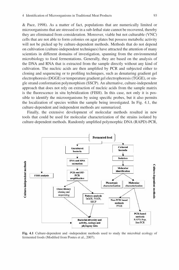
4 Identification of Microorganisms in Traditional Meat Products 93
& Pace, 1998). As a matter of fact, populations that are numerically limited ormicroorganisms that are stressed or in a sub-lethal state cannot be recovered, therebythey are eliminated from consideration. Moreover, viable but not culturable (VNC)cells that are not able to form colonies on agar plates but possess metabolic activitywill not be picked up by culture-dependent methods. Methods that do not dependon cultivation (culture-independent techniques) have attracted the attention of manyscientists in different domains of investigation, spanning from the environmentalmicrobiology to food fermentations. Generally, they are based on the analysis ofthe DNA and RNA that is extracted from the sample directly without any kind ofcultivation. The nucleic acids are then amplified by PCR and subjected either tocloning and sequencing or to profiling techniques, such as denaturing gradient gelelectrophoresis (DGGE) or temperature gradient gel electrophoresis (TGGE), or sin-gle strand conformation polymorphism (SSCP). An alternative, culture-independentapproach that does not rely on extraction of nucleic acids from the sample matrixis the fluorescence in situ hybridization (FISH). In this case, not only it is pos-sible to identify the microorganisms by using specific probes, but it also permitsthe localization of species within the sample being investigated. In Fig. 4.1, theculture-dependent and independent methods are summarized.
Finally, the extensive development of molecular methods resulted in newtools that could be used for molecular characterization of the strains isolated byculture-dependent methods. Randomly amplified polymorphic DNA (RAPD)-PCR,
Fig. 4.1 Culture-dependent and -independent methods used to study the microbial ecology offermented foods (Modified from Pontes et al., 2007).
94 L. Cocolin et al.
repetitive bacterial DNA elements (Rep)-PCR, and enterobacterial repetitiveintergenic consensus (ERIC)-PCR are common practices in almost all the laborato-ries that deal with microbial ecology and diversity. At present, it is widely acceptedthat different methods for the identification and characterization of microbial speciesin a specific ecosystem are necessary. The combination of different methods to studythe microbial ecology of a system is defined as a “polyphasic approach” (Pontes,Lima-Bitterncourt, Chartone-Souza, & Amaral Nascimiento, 2007).
Molecular Methods Used to Investigate the Diversityof Microorganisms in Fermented Sausages
As mentioned above, an important distinction that has to be made is betweenculture-dependent and independent methods. While the culture-dependent methodsare commonly used to identify and molecularly characterize microbial isolates, theculture-independent methods are used to profile directly the microbial populationsduring fermentation of sausages (Rantsiou & Cocolin, 2006).
Culture-Dependent Techniques
Strain Identification
The identification of strains isolated from sausage fermentation is most commonlyachieved by targeting the ribosomal RNA operon (rRNA). It possesses some req-uisites and attributes that are very important for microbial identification. Apartfrom being present in all microbial species, it is characterized by the presenceof conserved and variable regions, that become very important for differentiationpurposes, and it functions as an evolutionary clock, giving the possibility to drawconclusions regarding evolution and phylogeny. In the case of the bacteria, the 16SrRNA gene is the most used target for molecular applications (Collins et al., 1991),while the D1-D2 loop of the 26S RNA gene became a generally accepted targetfor yeast identification (Kurtzman & Robnett, 1997). The process of identificationof microbial strains by targeting the rRNA usually consists of a first step in whichthe specific gene (16S or 26S rRNA), or portion of it, is amplified by PCR thatsubsequently is subjected to DNA sequencing. By alignment of the sequence in theGeneBank, using available software programs online (Blast, Altschul et al., 1997),it is possible to achieve the identification. This experimental tactic is not applicablefor ecology studies because of the high number of isolates; it is not feasible and eco-nomically convenient to sequence hundreds of strains. For this reason, researchersdeveloped species-specific PCR and ribosomal RNA probes as fastest ways to obtainspecies identification. Ribosomal RNA probes (Nissen & Dainty, 1995), species-specific PCR primers (Berthier & Ehrlich, 1998; Blaiotta, Pennacchia, Parente, &Villani, 2003; Morot-Bizot, Talon, & Leroy-Setrin, 2003; Rossi, Tofalo, Torriani,& Suzzi, 2001), and multiplex PCR (Corbiere Morot-Bizot, Talon, & Leroy, 2004)
4 Identification of Microorganisms in Traditional Meat Products 95
have been used in the field of fermented sausages for the identification of LAB andCNC isolated during fermentation.
Alternatively, the PCR-sequencing methodology is coupled with techniques thatare able to differentiate strains based on fingerprinting profiles, allowing grouping ofthe isolates and reducing the number of strains requiring sequencing. The methodsused to group strains and reduce the number of isolates to sequence are once morebased on an amplification step. The differences in the strains to identify can bedetected either by exploiting primers that are annealing in various regions of thegenome of the strain to identify, thereby producing a band pattern that is represen-tative for a species, or by detecting differences in the DNA sequences specificallyamplified by PCR. The first group of methods is mainly represented by RAPD-PCRand Rep-PCR. RAPD-PCR is characterized by an amplification process, using asingle random primer that will anneal in several portions of the genomic DNAand give amplification products when the forward and reverse annealing sites areallowing extension by the DNA polymerase (Caetono-Anolles, 1993; Power, 1996).The main drawback of this technique is its low reproducibility. The annealing steptakes place at very low temperature (37–40◦C), and for this reason, the same strainsubjected to RAPD-PCR in different moments can produce different profiles. Whileit is possible to reach a good intra-laboratory reproducibility, if all the experimen-tal steps are optimized and standardized, it is practically impossible to be able tocompare the results obtained in different laboratories. Concerning Rep-PCR, theprimers used are specific to the repetitive elements spread around the bacterialgenomes (Gilson, Clement, Brutlag, & Hofnung, 1984), and for this reason theprofiles obtained are highly specific for a species and they are highly reproducible,as well. RAPD-PCR and Rep-PCR are methods that were first applied for identifica-tion purposes (Andrighetto, Zampese, & Lombardi, 2001; Berthier & Ehrlich, 1999;Gevers, Huys, & Swings, 2001; Rebecchi, Crivori, Sarra, & Cocconcelli, 1998), andonly recently they have been used for molecular characterization of LAB and CNC(Cocolin et al., 2004; Comi et al., 2005; Iacumin, Comi, Cantoni, & Cocolin, 2006a;Rantsiou, Drosinos, Gialitaki, Urso, et al., 2005, Rantsiou, Iacumin, Cantoni, Comi,& Cocolin, 2005; Rantsiou, Drosinos, Gialitaki, Metaxopoulos, et al., 2006; Urso,Comi, & Cocolin, 2006), as well as yeasts (Cocolin, Urso, Rantsiou, Cantoni, &Comi, 2006a) isolated from fermented sausages.
The techniques that are allowing strain grouping based on differences in a DNAsequence and that which have been applied in the field of fermented sausages arethe restriction fragment length polymorphism (RFLP) analysis of the 16S rRNAgene (Lee et al., 2004; Sanz, Hernandez, Ferrus, & Hernandez, 1998) and the dena-turing/temperature gradient gel electrophoresis (D/TGGE) (Blaiotta, Pennacchia,Ercolini, Moschetti, & Villani, 2003; Cocolin, Manzano, Cantoni, & Comi, 2000;Cocolin, Manzano, Aggio, Cantoni, & Comi, 2001; Cocolin, Manzano, Cantoni,& Comi, 2001; Ercolini, Moschetti, Blaiotta, & Coppola, 2001a). With the RFLPapproach, the differences in the DNA sequence are identified by using a restric-tion endonuclease that is cutting the DNA in specific restriction sites, giving a spe-cific pattern for a given species. The D/TGGE methods are detecting differences inthe sequences by analyzing their denaturation behavior. More specifically, as the
96 L. Cocolin et al.
DNA molecule encounters an appropriate denaturant concentration, a sequence-dependent partial denaturation of the double strand occurs. The change in theDNA conformation determines a reduction of the migration rate of the molecule,and thereby DNA with different nucleotide sequences will show differential elec-trophoretic patterns. This approach was first used in environmental ecosystems(Muyzer, de Waal, & Uitterlinden, 1993), and at the beginning of the 21st centuryan impressive number of papers applying D/TGGE in the field of food microbiologyhave been published (for review see Ercolini, 2004).
Strain Characterization
Strains isolated during the fermentation of sausages can be also subjected to molec-ular characterization, in order to understand the dynamics and diversity within thesame species. Usually, these approaches are applied to isolates that have previouslybeen identified with other molecular techniques (see above) and intra-species dif-ferences are highlighted. Only the strains belonging to the same species can besubjected to this kind of characterization. Also in this case, the methods that areused are divided into methods that are exploiting a PCR amplification and non-PCR methods. In the case of PCR-based methods, once more the RAPD-PCRand Rep-PCR are well-established techniques to look into the molecular diversitywithin selected species of bacteria or yeasts. RAPD-PCR has been applied to exam-ine the diversity of Lactobacillus sakei in naturally fermented Italian sausages, inorder to understand the RAPD-types that were able to take over the fermentation(Urso et al., 2006), and to differentiate Debaryomyces hansenii strains from thesausages fermented at low temperature (Cocolin et al., 2006a). Rep-PCR was usedin order to characterize Staphylococcus xylosus strains isolated from three fermen-tation processes in Northern Italy (Iacumin et al., 2006a). In addition, in this study,a new technique, named Sau-PCR, developed for the characterization of dairy-related microorganims (Corich, Mattiazzi, Soldati, Carraro, & Giacomini, 2005),was applied for the first time to differentiate the strains of meat origin. This methodis based on DNA digestion with the restriction endonuclease Sau3A, followedby amplification, using one primer that contains the sequence of the restrictionsite of the enzyme, and thereby is able to anneal to the ends of the fragmentsobtained. The result is a pattern of amplified bands that represents the restrictionfragments obtained by digestion with the specific endonuclease. Using the samerational, several enzymes can be used in order to obtain a more precise molecularcharacterization.
Regarding the molecular methods that do not exploit an amplification step, theonly one used in the field of fermented meats is represented by the pulsed-field gelelectrophoresis (PFGE). Currently, this technique is considered the “gold standard”for the characterization of strains, since it is very precise, reproducible, and reli-able. In the field of food borne pathogens, a database of PFGE profiles was created(PulseNet, http://www.cdc.gov/pulsenet/), thereby allowing epidemiologic analysisof food borne disease outbreaks. This is possible because the results obtained byPFGE are comparable between different laboratories thanks to the reproducibility
4 Identification of Microorganisms in Traditional Meat Products 97
of the method. PFGE is an electrophoretic method that allows separation of thechromosomes for the yeasts and of macro-restriction fragments obtained from thegenomic DNA for bacteria (Tenover et al., 1995). The first step is the extraction ofthe DNA from the microorganism that is embedded in agarose gel and subjected tolysis treatments, in order to digest the cell wall. Once the DNA has been freed, itcan be subjected directly to PFGE or it is digested with a restriction endonucleasethat generally has the characteristic to be rare-cutting. In this way, a relatively smallset of restriction fragments is obtained that are simply resolved in an agarose gel.The electrophoresis is carried out in specific chambers, in which the electric fieldis changing orientation. In this way, the DNA molecules are separated more effi-ciently than in conventional electrophoresis. The result obtained is a band patternthat is specific to the strain that was subjected to the PFGE analysis. Despite thefact that PFGE has been applied several times to characterize strains of dairy-origin,it was applied rarely to LAB and CNC isolated from fermented sausages. PFGEwas used to monitor S. xylosus starter cultures, both in Southern Italy (Di Maria,Basso, Santoro, Grazia, & Coppola, 2002) and in two French processing plants(Corbiere Morot-Bizot, Leroy, & Talon, 2007), and to study the diversity of Lacto-bacillus strains isolated from fermented sausages to be used as potential probiotics(Pennacchia, Vaughan, & Villani, 2006).
Culture-Independent Techniques
As mentioned above, the culture-independent methods are able to profile the micro-bial populations in complex microbial ecosystems without any cultivation. Thestrategy based on which these methods are working is the analysis of the nucleicacids extracted directly from the matrix. Once the DNA and RNA are available,they can be subjected to several types of analysis that can be preceded by a PCRstep or not. The culture-independent method that has been more extensively appliedto sausage fermentation is the DGGE. As already reported above, the method isable to differentiate DNA molecules based on their denaturation behaviors. Whenthe method is used for microbial population profiling, the PCR is carried out withuniversal primers, able to prime amplification for all the microbes present in thesample. After this step, a complex mixture of DNA molecules will be obtained, thatcan be differentiated if separated in gels with denaturant gradients. Every singleband that is visible in D/TGGE gels represents a component of the micro-biota. Themore bands are visible, the more complex is the ecosystem. By using these meth-ods, it is possible not only to profile the microbial populations, but also to followtheir dynamics during time. However, these methods are not quantitative. DGGEanalysis has been applied mainly to Italian fermented sausages (Cocolin, Manzano,Cantoni, et al., 2001; Rantsiou, Urso, et al., 2005; Silvestri et al., 2007; Villaniet al., 2007), but studies on the fermentation dynamics of Argentinean sausagesare available as well (Fontana, Cocconcelli, & Vignolo, 2005; Fontana, Vignolo, &Cocconcelli, 2005).
98 L. Cocolin et al.
The use of species-specific primers in the PCR amplification, analyzing theDNA extracted directly from the sample, can be considered to be another culture-independent method. This approach was so far used to identify LAB and CNCspecies in Spanish fermented sausages (Aymerich, Martin, Garriga, & Hugas, 2003).Moreover, with the last technological improvements that allowed the PCR tobecome a quantitative method, direct enumeration of technologically importantspecies during fermentation of sausages can be achieved. This was described forthe first time by Martin, Jofre, Garriga, Pla, and Aymerich, (2006), who optimizeda quantitative PCR (qPCR) protocol for the rapid quantitative detection of Lb. sakeiin fermented sausages.
Lastly, a promising culture-independent method that, unfortunately, has not yetbeen efficiently exploited to study the microbial diversity in fermented sausages,is the FISH. In this technique a set of specific probes are used to target differentmicroorganisms directly in the sample. The probes are labeled with different fluo-rophores, thereby allowing the detection of several species simultaneously. Since theprobes are generally designed on the ribosomal RNA, only alive cells are detectedby FISH (Bottari, Ercolini, Gatti, & Neviani, 2006). One of the most fascinating fea-tures of FISH is the possibility to localize the microorganisms directly into the foodmatrix. This has been applied to dairy products (Ercolini, Hill, & Dodd, 2003), butnever to fermented sausages. The only application of FISH to fermented and freshsausages to profile the microbial populations in food products has been describedby Cocolin et al. (2007).
The Microbial Ecology of Fermented Sausages as Determinedby Culture-Dependent Methods
The experimental approach used by researchers in order to describe the microbialecology of fermented sausages, using culture-dependent methods, is summarized inFig. 4.2, and the list of studies published using these techniques for the identificationand characterization of isolates are reported in Table 4.1. After homogenization, thesample is analyzed by plating onto a specific media to allow the selective growth ofthe microorganisms of interest. After incubation, counts and random isolations areperformed. The isolates are subsequently subjected to molecular identification andcharacterization.
Molecular Methods for the Identification of Isolated Strains
In the nineties, molecular techniques for the identification of microorganisms iso-lated from fermented meat products, started to place side by side or to substitutebiochemical assays. Although the two different approaches, in most of the cases,arrived at similar results, molecular techniques immediately showed a higher levelof reproducibility, automatism, and fastness.
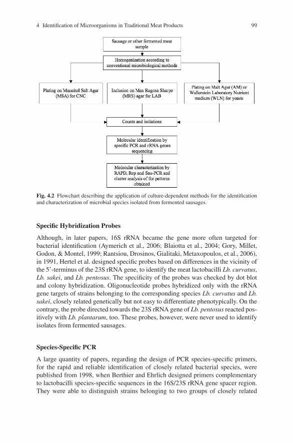
4 Identification of Microorganisms in Traditional Meat Products 99
Fig. 4.2 Flowchart describing the application of culture-dependent methods for the identificationand characterization of microbial species isolated from fermented sausages.
Specific Hybridization Probes
Although, in later papers, 16S rRNA became the gene more often targeted forbacterial identification (Aymerich et al., 2006; Blaiotta et al., 2004; Gory, Millet,Godon, & Montel, 1999; Rantsiou, Drosinos, Gialitaki, Metaxopoulos, et al., 2006),in 1991, Hertel et al. designed specific probes based on differences in the vicinity ofthe 5’-terminus of the 23S rRNA gene, to identify the meat lactobacilli Lb. curvatus,Lb. sakei, and Lb. pentosus. The specificity of the probes was checked by dot blotand colony hybridization. Oligonucleotide probes hybridized only with the rRNAgene targets of strains belonging to the corresponding species Lb. curvatus and Lb.sakei, closely related genetically but not easy to differentiate phenotypically. On thecontrary, the probe directed towards the 23S rRNA gene of Lb. pentosus reacted pos-itively with Lb. plantarum, too. These probes, however, were never used to identifyisolates from fermented sausages.
Species-Specific PCR
A large quantity of papers, regarding the design of PCR species-specific primers,for the rapid and reliable identification of closely related bacterial species, werepublished from 1998, when Berthier and Ehrlich designed primers complementaryto lactobacilli species-specific sequences in the 16S/23S rRNA gene spacer region.They were able to distinguish strains belonging to two groups of closely related
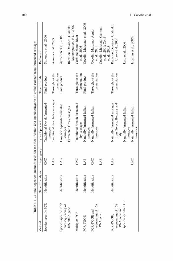
100 L. Cocolin et al.
Tabl
e4.
1C
ultu
re-d
epen
dent
met
hods
used
for
the
iden
tifica
tion
and
char
acte
riza
tion
ofst
rain
sis
olat
edfr
omfe
rmen
ted
saus
ages
Met
hod
Type
ofan
alys
isTa
rget
grou
pTy
peof
prod
uct
Type
ofst
udy
Ref
eren
ce
Spec
ies-
spec
ific
PCR
Iden
tifica
tion
CN
CT
radi
tiona
lSlo
vak
ferm
ente
dsa
usag
esFi
nalp
rodu
ctSi
mon
ova
etal
.,20
06
LA
BT
radi
tiona
lFre
nch
dry
saus
ages
Thr
ough
outt
hefe
rmen
tatio
nA
mm
oret
al.,
2005
Spec
ies-
spec
ific
PCR
and
sequ
enci
ngof
16S
rRN
Age
ne
Iden
tifica
tion
LA
BL
owac
idSp
anis
hfe
rmen
ted
saus
ages
Fina
lpro
duct
Aym
eric
het
al.,
2006
Ferm
ente
dG
reek
saus
ages
Ran
tsio
u,D
rosi
nos,
Gia
litak
i,M
etax
opou
los,
etal
.,20
06M
ultip
lex
PCR
Iden
tifica
tion
CN
CT
radi
tiona
lFre
nch
ferm
ente
ddr
ysa
usag
esT
hrou
ghou
tthe
ferm
enta
tion
Cor
bier
eM
orot
-Biz
otet
al.,
2006
PCR
-TG
GE
Iden
tifica
tion
LA
BN
atur
ally
ferm
ente
dIt
alia
nsa
usag
esFi
nalp
rodu
ctC
ocol
in,M
anza
no,e
tal.,
2000
PCR
-DG
GE
and
sequ
enci
ngof
16S
rRN
Age
ne
Iden
tifica
tion
CN
CN
atur
ally
ferm
ente
dIt
alia
nsa
usag
esT
hrou
ghou
tthe
ferm
enta
tion
Coc
olin
,Man
zano
,Agg
io,
etal
.,20
01L
AB
Coc
olin
,Man
zano
,Can
toni
,et
al.,
2001
;Com
iet
al.,
2005
PCR
-DG
GE
,se
quen
cing
of16
SrR
NA
gene
and
spec
ies-
spec
ific
PCR
Iden
tifica
tion
LA
BN
atur
ally
ferm
ente
dsa
usag
esfr
omG
reec
e,H
unga
ryan
dIt
aly
Thr
ough
outt
hefe
rmen
tatio
nR
ants
iou,
Dro
sino
s,G
ialit
aki,
Urs
o,et
al.,
2005
Nat
ural
lyfe
rmen
ted
Ital
ian
saus
ages
Urs
oet
al.,
2006
CN
CN
atur
ally
ferm
ente
dIt
alia
nsa
usag
esIa
cum
inet
al.,
2006
b
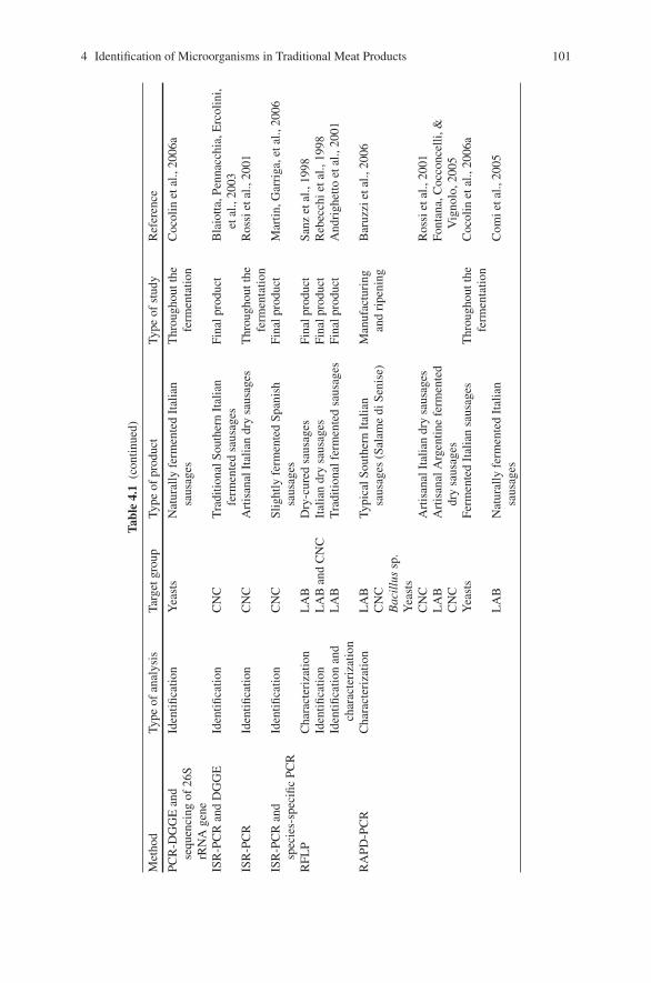
4 Identification of Microorganisms in Traditional Meat Products 101
Tabl
e4.
1(c
ontin
ued)
Met
hod
Type
ofan
alys
isTa
rget
grou
pTy
peof
prod
uct
Type
ofst
udy
Ref
eren
ce
PCR
-DG
GE
and
sequ
enci
ngof
26S
rRN
Age
ne
Iden
tifica
tion
Yea
sts
Nat
ural
lyfe
rmen
ted
Ital
ian
saus
ages
Thr
ough
outt
hefe
rmen
tatio
nC
ocol
inet
al.,
2006
a
ISR
-PC
Ran
dD
GG
EId
entifi
catio
nC
NC
Tra
ditio
nalS
outh
ern
Ital
ian
ferm
ente
dsa
usag
esFi
nalp
rodu
ctB
laio
tta,P
enna
cchi
a,E
rcol
ini,
etal
.,20
03IS
R-P
CR
Iden
tifica
tion
CN
CA
rtis
anal
Ital
ian
dry
saus
ages
Thr
ough
outt
hefe
rmen
tatio
nR
ossi
etal
.,20
01
ISR
-PC
Ran
dsp
ecie
s-sp
ecifi
cPC
RId
entifi
catio
nC
NC
Slig
htly
ferm
ente
dSp
anis
hsa
usag
esFi
nalp
rodu
ctM
artin
,Gar
riga
,eta
l.,20
06
RFL
PC
hara
cter
izat
ion
LA
BD
ry-c
ured
saus
ages
Fina
lpro
duct
Sanz
etal
.,19
98Id
entifi
catio
nL
AB
and
CN
CIt
alia
ndr
ysa
usag
esFi
nalp
rodu
ctR
ebec
chie
tal.,
1998
Iden
tifica
tion
and
char
acte
riza
tion
LA
BT
radi
tiona
lfer
men
ted
saus
ages
Fina
lpro
duct
And
righ
etto
etal
.,20
01
RA
PD-P
CR
Cha
ract
eriz
atio
nL
AB
CN
CB
acil
lus
sp.
Yea
sts
Typi
calS
outh
ern
Ital
ian
saus
ages
(Sal
ame
diSe
nise
)M
anuf
actu
ring
and
ripe
ning
Bar
uzzi
etal
.,20
06
CN
CA
rtis
anal
Ital
ian
dry
saus
ages
Ros
siet
al.,
2001
LA
BC
NC
Art
isan
alA
rgen
tine
ferm
ente
ddr
ysa
usag
esFo
ntan
a,C
occo
ncel
li,&
Vig
nolo
,200
5Y
east
sFe
rmen
ted
Ital
ian
saus
ages
Thr
ough
outt
hefe
rmen
tatio
nC
ocol
inet
al.,
2006
a
LA
BN
atur
ally
ferm
ente
dIt
alia
nsa
usag
esC
omie
tal.,
2005
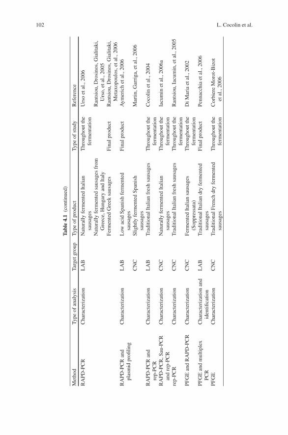
102 L. Cocolin et al.
Tabl
e4.
1(c
ontin
ued)
Met
hod
Type
ofan
alys
isTa
rget
grou
pTy
peof
prod
uct
Type
ofst
udy
Ref
eren
ce
RA
PD-P
CR
Cha
ract
eriz
atio
nL
AB
Nat
ural
lyfe
rmen
ted
Ital
ian
saus
ages
Thr
ough
outt
hefe
rmen
tatio
nU
rso
etal
.,20
06
Nat
ural
lyfe
rmen
ted
saus
ages
from
Gre
ece,
Hun
gary
and
Ital
yR
ants
iou,
Dro
sino
s,G
ialit
aki,
Urs
o,et
al.,
2005
Ferm
ente
dG
reek
saus
ages
Fina
lpro
duct
Ran
tsio
u,D
rosi
nos,
Gia
litak
i,M
etax
opou
los,
etal
.,20
06R
APD
-PC
Ran
dpl
asm
idpr
ofilin
gC
hara
cter
izat
ion
LA
BL
owac
idSp
anis
hfe
rmen
ted
saus
ages
Fina
lpro
duct
Aym
eric
het
al.,
2006
CN
CSl
ight
lyfe
rmen
ted
Span
ish
saus
ages
Mar
tin,G
arri
ga,e
tal.,
2006
RA
PD-P
CR
and
rep-
PCR
Cha
ract
eriz
atio
nL
AB
Tra
ditio
nalI
talia
nfr
esh
saus
ages
Thr
ough
outt
hefe
rmen
tatio
nC
ocol
inet
al.,
2004
RA
PD-P
CR
,Sau
-PC
Ran
dre
p-PC
RC
hara
cter
izat
ion
CN
CN
atur
ally
ferm
ente
dIt
alia
nsa
usag
esT
hrou
ghou
tthe
ferm
enta
tion
Iacu
min
etal
.,20
06a
rep-
PCR
Cha
ract
eriz
atio
nC
NC
Tra
ditio
nalI
talia
nfr
esh
saus
ages
Thr
ough
outt
hefe
rmen
tatio
nR
ants
iou,
Iacu
min
,eta
l.,20
05
PFG
Ean
dR
APD
-PC
RC
hara
cter
izat
ion
CN
CFe
rmen
ted
Ital
ian
saus
ages
(Sop
pres
sata
)T
hrou
ghou
tthe
ferm
enta
tion
DiM
aria
etal
.,20
02
PFG
Ean
dm
ultip
lex
PCR
Cha
ract
eriz
atio
nan
did
entifi
catio
nL
AB
Tra
ditio
nalI
talia
ndr
yfe
rmen
ted
saus
ages
Fina
lpro
duct
Penn
acch
iaet
al.,
2006
PFG
EC
hara
cter
izat
ion
CN
CT
radi
tiona
lFre
nch
dry
ferm
ente
dsa
usag
esT
hrou
ghou
tthe
ferm
enta
tion
Cor
bier
eM
orot
-Biz
otet
al.,
2006

4 Identification of Microorganisms in Traditional Meat Products 103
Lactobacillus species: one composed of Lb. curvatus, Lb. graminis, and Lb. sakeiand the other of Lb. paraplantarum, Lb. pentosus, and Lb. plantarum. The nextyear the same authors (Berthier & Ehrlich, 1999) developed pairs of PCR primersthat could be used to specifically detect Lb. curvatus and Lb. sakei. They evaluatedthe phenotypic and genotypic diversity among 165 isolates, previously assigned toLb. curvatus and Lb. sakei species, by comparing the results of biochemical assaysand RAPD data. This allowed them to consider the presence of species-specificRAPD bands that were cloned and sequenced. On the basis of differences in thesesequences, species-specific primers were designed. Likewise, a pair of primers spe-cific for S. xylosus were designed by Morot-Bizot et al. (2003) on a RAPD productof 539 bp, shared by S. xylosus strains from international collections. The validityof the method was confirmed by comparing the results obtained with the designedprimers and by PFGE.
Since among the CNC species, S. xylosus, S. equorum, and S. carnosus arefrequently involved in the fermentation of several typologies of meat products, anumber of papers regarding the design of primers specific for these microorganismswere published. Blaiotta, Pennacchia, Parente, et al. (2003) used xylulokinase and a60 kDa heat-shock protein coding genes as targets for the design of S. xylosus spe-cific primers. They successfully evaluated the specificity of two sets of primers on27 references strains of the Deutsche Sammlung von Mikroorganismen and Zellkul-turen GmbH (DSM) (Germany) collection. Later on, Blaiotta, Ercolini, Mauriello,Salzano, & Villani, (2004) developed primers specific for S. equorum that targetedthe manganese-dependent superoxide dismutase (sodA) gene and were successfulin differentiating S. xylosus and S. equorum that are otherwise difficult to separateby biochemical or physiological traits. The reliability of the method was tested on atotal of 112 strains, representing 26 different species of the genus Staphylococcus, 3species of the genus Kocuria, and different strains of the Macrococcus caseolyticusspecies. Similarly, Blaiotta, Casaburi, and Villani (2005) designed two different setsof primers, targeting the sodA genes of S. carnosus and S. simulans and were ableto differentiate the two species.
Simonova et al. (2006) employed primers previously described by other authors(Aymerich et al., 2003) to detect S. xylosus and S. carnosus strains in differenttypes of Slovak traditional sausages, for the selection of potential starter culturesto be used in sausage processing. As in many Italian and Spanish fermented meatproducts (Blaiotta et al., 2004; Coppola, Iorizzo, Saotta, Sorrentino, & Grazia, 1997;Garcia-Varona, Santos, Jaime, & Rovira, 2000), S. xylosus was the dominating CNC(63.6%), while S. carnosus showed 10.7% frequency.
By using species-specific primers, Ammor et al. (2005) carried out a survey onLAB isolated in a small-scale facility producing traditional dry sausage in France.While Enterococcus faecium, Vagococcus carniphilus, and Lactococcus garvieaewere the predominant species isolated from processing equipment and raw material,Lb. sakei became the predominant species during the fermentation of the product.Lb. sakei strains prevailed (43.3%) also among homo-fermentative mesophilic rodsfound by Greco, Mazzette, De Santis, Corona, & Cosseddu (2005) in the productionand ripening of a typical dry fermented sausage from Sardinia (Italy). The authors used
104 L. Cocolin et al.
species-specific primers to detect also Lb. plantarum and Lb. curvatus strains, whichwere isolated from the product in the percentages of 16.6 and 13.3%, respectively.
The biodiversity of lactobacilli was also evaluated in low acid Spanish fermentedsausages and in naturally fermented Greek sausages, produced in three different pro-cessing plants, by Aymerich et al. (2006) and Rantsiou, Drosinos, Gialitaki, Metax-opoulos, et al. (2006), respectively. In both cases, isolates that could not be identifiedby species-specific PCR were identified by the partial sequencing of the 16S rRNAgene that allowed overcoming the limit of species-specific PCR method. In fact, inmicrobial ecology studies, it is unlikely to reach the identification of all isolates byspecies-specific PCR, because of the lack of primers or the presence of unexpectedspecies. Lb. sakei was the predominant species (74%) in the Spanish fermentedsausages (Aymerich et al., 2006), followed by Lb. curvatus (21.2%) and Leuconos-toc mesenteroides (4.8%). The results obtained from Rantsiou, Drosinos, Gialitaki,Metaxopoulos, et al. (2006) highlighted that the main populations involved in thefermentations analyzed, belonged to the species Lb. sakei, Lb. plantarum, and Lb.curvatus, differently predominating depending on the plants considered.
Corbiere Morot-Bizot et al. (2004) developed a multiplex PCR for the iden-tification of bacteria belonging to the Staphylococcus genus and in particular tothe species S. xylosus, S. saprophyticus, S. epidermidis, and S. aureus. Later, thesame authors (Corbiere Morot-Bizot et al., 2007) used this approach to investigatethe diversity of the staphylococcal community occurring in both the environmentand meat products of a small French unit, manufacturing traditional dry fermentedsausages. The staphylococcal flora of the environment and meat products of thissmall unit was dominated by S. equorum and S. succinus species, rarely described inmeat products and never in the environment. They represented the 49% and 33% ofisolates, respectively. Other staphylococci belonged to the species S. saprophyticus,S. xylosus, S. carnosus, S. simulans, and S. warneri.
PCR-DGGE
In the last years, the single or combined use of sets of species-specific primers,started to be used with or be substituted by new, versatile approaches such asD/TGGE. The first paper, applying uniquely this technique for the identificationof microorganisms involved in meat fermentations, was published by Cocolin,Manzano, et al. (2000). This work describes the use of PCR-TGGE to identifyLactobacillus spp. strains from naturally fermented Italian sausages. This method,based on the amplification of a small fragment from the 16S rRNA gene, followedby TGGE, allowed the differentiation of the strains of Lb. sakei, Lb. curvatus,Lb. alimentarius, Lb. casei, Lb. plantarum, and Lb. brevis.
The same authors (Cocolin, Manzano, Aggio, et al., 2001) used CNC controlstrains from international collections to optimize a PCR-DGGE method for their dif-ferentiation that was subsequently used to identify, based on co-migration with con-trol strains, 90 isolates from natural fermented Italian sausages. The results showedthat the strains of S. xylosus, S. cohnii, S. intermedius, S. carnosus, K. varians, andK. kristinae could be reliably identified on the basis of the DGGE mobility of the
4 Identification of Microorganisms in Traditional Meat Products 105
16S rRNA gene V3 region; once again, S. xylosus was the main bacterium involvedin fermented sausage production, representing, from the tenth day of ripening, theonly CNC species isolated.
In the next years, PCR-DGGE, more than PCR-TGGE, found wide applicationsin studies of microbial ecology. These techniques showed the possibility to processhigh number of isolates in a shorter time; in fact, as already mentioned in the previ-ous sections, these methods allowed grouping of strains and reduced the number ofisolates to sequence.
Using these methods, Cocolin, Manzano, Cantoni, et al., (2001) monitored LABdynamics in fermented sausages, at 3, 10, 20, 30, and 45 days of ripening, preparedin a local meat factory in Italy by traditional techniques. One-hundred and ninetytwo LAB strains were identified by PCR with the primers targeting the V1 regionof the 16S rRNA gene and DGGE. Only two DGGE profiles were detected, belong-ing to Lb. sakei and Lb. curvatus. In the first stages of fermentation, Lb. sakei wasthe main LAB present, and only at the end of maturation Lb. curvatus became thepredominant microorganism.
The same approach was applied to study the LAB and CNC ecology of freshsausages stored at 4◦C at days 0, 3, 6, and 10 (Cocolin et al., 2004; Rantsiou,Iacumin, et al., 2005). Strains with the same DGGE profile were grouped, and rep-resentatives of each group were amplified with primers targeting the V1–V3 regionof the 16S rRNA gene and sequenced. A total of 80 LAB were analyzed, amongwhich 69 isolates were identified as Lb. sakei. To study the Staphylococcus spp.ecology, PCR-DGGE was coupled with S. xylosus-specific primers because of thevery similar DGGE migration patterns shown by strains belonging to S. xylosus andS. haemolyticus. Primers based on the gehM gene, coding for the lipase of S. xylosus,gave a definitive and sure identification of this species. Almost 50% of isolates wererecognized as S. xylosus. Interesting population dynamics were observed, character-ized by a succession of S. pasteuri and S. warneri at day 0, with S. xylosus at 3 daysand S. equorum at 6 and 10 days.
Comi et al. (2005), studied three productions of a traditional fermented sausageof Northeast Italy. One-hundred and fifty LAB were isolated and identified by meansof PCR-DGGE, as described above (Cocolin, Manzano, Cantoni, et al., 2001). Theonly species that were isolated in all three fermentations were Lb. curvatus and Lb.sakei. A low number of strains belonged to other species, namely E. pseudoavium,L. lactis, Lb. brevis, Lc. mesenteroides, Lb. plantarum, Lb. paraplantarum/pentosus,Lc. citreum and W. paramesenteroides/hellenica.
Rantsiou, Drosinos, Gialitaki, Urso, et al. (2005), using both species-specificPCR and PCR-DGGE, defined the geographic distribution of LAB populationsresponsible for the fermentation and maturation of naturally fermented sausagesproduced in three European countries: Greece, Hungary, and Italy. Three-hundredand fifty eight strains belonging to the LAB group were identified. Three specieswere common to all the three countries studied and were also the most numer-ous; Lb. curvatus, Lb. plantarum, and Lb. sakei. These three species were identi-fied by species-specific PCR. Isolates that did not belong to any of three speciesabove were identified by PCR-DGGE grouping and 16S rRNA gene sequencing.
106 L. Cocolin et al.
Country specific-species: E. faecium/durans, Lb. alimentarius, Lb. casei/paracasei,were only found in sausages from Greece, E. pseudoavium, L. lactis subsp.lactis and Lb. paraplantarum/pentosus were only found in sausages from Italy,while Lc. mesenteroides and W. viridescens were specific to the sausages fromHungary. Lb. paraplantarum was isolated from both Italian and Greek products, andLb. paraplantarum/plantarum was from both Greek and Hungarian sausages. Lc.citreum and W. paramesenteroides/hellenica were found in Italian and Hungarianproducts. A similar approach, coupling species-specific PCR with PCR-DGGE, wasdescribed by Urso et al. (2006), who investigated the ecology of the LAB microfloraof three naturally fermented sausages produced in Northeast Italy. Four-hundredand sixty five strains were identified. The only species that were isolated in all threefermentations were Lb. sakei and Lb. curvatus (353 and 67 isolates, respectively). Alow number of other LAB species were found as well, Lb. plantarum, Lb. casei, Lb.paraplantarum, L. lactis, L. garvieae, Lc. mesenteroides, Lc. carnosum, Weissellahellenica, and W. paramesenteroides.
Likewise, Iacumin, Comi, Cantoni, and Cocolin (2006b) used Six couples ofspecies-specific primers (for S. xylosus, S. epidermidis, S. simulan, S. carnosus, S.warneri, and K. varians) to identify 617 CNC strains isolated from naturally fer-mented sausages in three different plants of the Northeast of Italy. Strains that didnot give any PCR amplification with the species-specific primers were subjected toPCR-DGGE and sequencing of the V3 region of the 16S rRNA gene, as describedby Rantsiou, Iacumin, et al. (2005). The same species of CNC were found in allthe three processing plants, but their contribution to the fermentations was different.In two plants, S. xylosus was the main species involved in the fermentation process,while in the third the maturation was carried out equally by three species: S. xylosus,S. warneri, and S. pasteuri.
Cocolin, Urso, Rantsiou, Cantoni, & Comi (2006b) evaluated the ability of acommercial starter culture to perform a 28 day sausage fermentation, combiningspecies-specific PCR and DGGE analysis. As was declared on the starter culturelabel, LAB isolated from the starter belonged to Lb. plantarum species. CNC strainsof S. xylosus, together with S. carnosus, were also isolated and identified in disagree-ment with what was declared from the factory producing the starter.
Recently, PCR-DGGE was also used to investigate the yeast populations inItalian fermented sausages (Cocolin et al., 2006a). Yeast isolates were subjectedto PCR-DGGE according to Cocolin, Bisson, & Mills (2000), and strains givingidentical migration patterns were grouped together; at least two representativeswere amplified with the primers NL1 and NL4 (Kurtzman, & Robnett, 1998) andsequenced. The work highlighted the dominance of D. hansenii, which was alreadypresent, as the major species, at the beginning of the fermentation. This species wasusually accompanied by Candida zeylanoides, and only at the end of the fermenta-tion, by Metschnikowia pulcherrima. At the start of the fermentation, a high biodi-versity was observed, as five species could be identified; apart from D. hansenii, thespecies Pichia triangularis, Candida parapsilosis, Saccharomyces cerevisiae, andSterigmatomyces elviae were found.
4 Identification of Microorganisms in Traditional Meat Products 107
ISR-PCR
The polymorphism of the inter-gene spacer regions (ISR), was also used in combi-nation with DGGE for the identification of the CNC strains isolated from fermentedsausages. Blaiotta, Pennacchia, Ercolini, et al., (2003), applied a polyphasic molec-ular approach to describe staphylococcal population occurring in traditional fer-mented sausages of Southern Italy by combining DGGE and ISR-PCR. The resultsobtained by ISR-PCR and PCR-DGGE analysis allowed a clear diffentiation of thereference strains analyzed, with the exception of the pairs S. cohnii-S. equorum andS. schleiferi-S. carnosus. The strains that displayed the same ISR-PCR pattern couldshow a different PCR-DGGE profile and vice versa. Among the isolates, the mostabundant species of CNC was S. xylosus. The strains of S. saprophyticus, S. lentus,S. warneri, S. cohnii-S. equorum, S. epidermidis, S. haemolyticus, S. succinus, S.vitulus, S. pasteuri, S. aureus, M. caseolyticus, and Kocuria spp. were also isolated.
In a previous research (Rossi et al., 2001), ISR-PCR permitted to successfullyidentify strains belonging to S. xylosus, S. simulans, and S. carnosus and otherclosely-related species, and Kocuria species, isolated from an Italian artisanal drysausage. Fifty one CNC strains were genotypically identified by amplification withuniversal primers (Jensen, Webster, & Strauss, 1993), and once more, S. xylosusprevailed over the other CNC.
Recently, 240 CNC strains, isolated from the Spanish slightly fermentedsausages, were subjected to ISR-PCR with universal primers (Martin, Garriga,et al., 2006). The results obtained by amplification of the intergenic regions wereconfirmed by species-specific PCR, as previously described (Aymerich et al., 2003).S. xylosus was the predominant species (80.8%), followed by S. warneri (8.3%),S. epidermidis (5.8%) S. carnosus (4.6%), and K. varians (0.4%).
Concerning ISR-PCR, discrepancy in the results is often found. While Forsman,Tilsala-Timisijarvi, & Alatossava, (1997), Mendoza, Meugnier, Bes, Etienne, &Freney, (1998), and Villard, Kodjo, Borges, Maurin, & Richard, (2000) reporteda high polymorphism, Blaiotta, Pennacchia, Ercolini, et al., (2003), in agreementwith Couto, Pereira, Miragaia, Sanches, & de Lancastre, (2001), found a lower dis-crimination potential of this analysis for staphylococci.
Molecular Methods for the Characterization of Isolated Strains
The growing interest in the studies of microbial ecology of fermented products andin monitoring of specific strains carrying out, spontaneously or after inoculationas starters, fermentation processes, pushed for the availability of methods for sub-species characterization of the isolates.
In 1998, RFLP analysis had been already used by Sanz et al. to characterizeLb. sakei strains isolated from dry-cured sausages, and previously identified byDNA-DNA hybridization. RFLP studies, by using the restriction enzymes EcoRI
108 L. Cocolin et al.
and HindIII, and, as probes, cDNA from Escherichia coli 16S and 23S rRNA genesor from Lb. sakei 16S rRNA gene from Lb. sakei strains, showed distinct poly-morphism levels. EcoRI-digested DNA, probed with the cDNA from E. coli, high-lighted the presence of a unique cluster for the meat Lb. sakei isolates tested. WhenHindIII-digested DNA was hybridized with the E. coli cDNA probe, strain specificpatterns were obtained, showing a higher discrimination power. Considerable straindifferentiation was also observed when EcoRI and HindIII digests were hybridizedwith Lb. sakei 16S rRNA gene probes.
In the following years, RAPD-PCR became the most used technique to study theintra-specific variability even if, initially, it was applied for identification purposes(Andrighetto et al., 2001; Baruzzi, Matarante, Caputo, & Morea, 2006; Berthier &Ehrlich, 1999; Rebecchi et al., 1998). Rebecchi et al. (1998) studied the microbialcommunity in the production of a 2-month-ripened Italian dry sausage. Their results,obtained by RAPD-PCR fingerprints, highlighted that environmental parametersinteracted to select a limited number of strains during the fermentation process.The dominant strains belonged to the species S. xylosus and S. sciuri, among CNC,and Lb. sakei and Lb. plantarum, among LAB.
RAPD-PCR with primers M13 and D8635 was applied to identify 53 lactobacilliisolates originating from traditional fermented sausages and artisanal meat plantsof Veneto region in Italy (Andrighetto et al., 2001). RAPD-PCR assigned mostof the isolates to the species Lb. sakei and Lb. curvatus and, at the same time, anintra-specific variability was detected; in some cases, RAPD subgroups reflected theorigin of the isolates; in others, different strains were found in the same environment.
In fermented products, the possibility to detect strains that were able to carry outthe fermentation processes became extremely important in the selection of microbialstrains to be used as starters. Rossi et al. (2001), compared and combined RAPDprofiles, obtained with the primers OPL-01, OPL-02, OPL-05, and Hpy 1, from theS. xylosus strains isolated from dry sausages. In this paper, the authors underlinedthe suitability of the RAPD-PCR analysis to discriminate strains with technologi-cally relevant activities; in fact, strains showing nitrate reduction and amino aciddecarboxylase activities clustered separately.
Later, Baruzzi et al. (2006) correlated acidification, proteolytic, lipolytic, andnitrate reduction activities to strains isolated from “Salame di Senise”,, a typicalsausage produced in the South of Italy. The application of RAPD-PCR to more than 90isolates made it possible to define 18 bacterial and 2 yeast bio-types, mainly belongingto three species of Bacillus (Bacillus subtilis, B. pumilus and B. amyloliquefaciens),three species of Lactobacillus (Lb. curvatus, Lb. sakei and Lb. casei), three species ofStaphylococcus (S. succinus, S saprophyticus, and S. equorum), and D. hansenii.
The microbial community responsible for the artisanal fermentation of drysausages produced in Argentina, was studied by Fontana et al. (2005). The authorscarried out RAPD analysis with the primers M13, XD9, RAPD1, and RAPD2, todifferentiate and characterize 100 strains of lactobacilli and CNC, identified by16S rRNA gene sequencing. This approach allowed them to demonstrate that theripening process of the Argentinean artisanal fermented sausages was driven bya limited number of Lactobacillus and Staphylococcus strains selected from the
4 Identification of Microorganisms in Traditional Meat Products 109
environmental micro-biota for the ability to best compete under the prevailing con-ditions of the ecological niche. The authors concluded that these well adapted strainsshould have been eventually considered for the selection of starter cultures.
Cocolin et al. (2006b) used an identical approach to follow the development,in fermented sausages, of an inoculated commercial starter. RAPD characterizationwas carried out on 15 LAB and 15 CNC strains from a starter used in a sausageproduction and on 70 LAB and 70 CNC strains isolated during the same production.RAPD analysis with primer M13 revealed three Lb. plantarum RAPD bio-types, butonly one of them was able to conduct the fermentation. S. xylosus strains from thestarter culture, which clustered mainly with strains isolated at 14 and 28 days ofripening, were able to predominate only in the latter stages of fermentation. Thedifferent behavior of Lb. plantarum and S. xylosus could be explained consideringthe fact that the starter culture was dissolved in white wine, thus inhibiting the initialdevelopment of S. xylosus, not able to overcome the ethanol stress.
Recently, some authors evaluated possible correlations between microbial strainsclustered on the basis of RAPD profiles and their manufacturing origin or geograph-ical provenience. Comi et al. (2005), in the study of three fermentations of a North-east Italy traditional fermented sausage, highlighted, by RAPD-PCR with primerM13, that lactobacilli population was distributed in a fermentation-specific way;the authors supposed that strains grouped in fermentation-specific clusters camefrom the ingredients used in the productions. Lb. sakei showed a higher degree ofheterogeneity than Lb. curvatus.
The same approach was followed by Urso et al. (2006) who reached similarresults. They isolated and identified, from three naturally fermented sausagesproduced in the Friuli-Venezia-Giulia region (Italy), 353 Lb. sakei and 67 Lb.curvatus strains. RAPD-PCR analysis by using primer M13 detected clustersformed by strains isolated from specific fermentations, confirming that ingredientcomposition, fermentation, and maturation parameters could play an important rolein the selection of populations adapted to a specific environment. However, clusterscontaining strains isolated from different plants were also observed, underlining ahomogeneous population distribution in the three different fermentations.
Rantsiou, Drosinos, Gialitaki, Metaxopoulos, et al., (2006) characterized, byRAPD-PCR with primer M13, Lb. sakei, Lb. plantarum, and Lb. curvatus strainsisolated from naturally fermented sausages, produced in three different processingplants in continental Greece. In agreement with the results mentioned above, somestrains were plant-specific, whereas others shared a degree of homology indepen-dently of the provenience.
In a similar study, Lb. curvatus, Lb. plantarum, and Lb. sakei strains, isolatedfrom naturally fermented sausages produced in three European countries (Greece,Italy, and Hungary), were subjected to RAPD-PCR with primer M13 (Rantsiou,Drosinos, Gialitaki, Urso, et al., 2005). The distribution of the lactobacilli strainsreflected, in almost all cases, the provenience of the same. It was thereby possibleto distinguish the Italian, Greek, and Hungarian LAB populations.
In order to improve the characterization and differentiation among microbialstrains, some authors considered both chromosomal and plasmidic characteristics.
110 L. Cocolin et al.
Thus, 250 LAB isolated from low acid Spanish fermented sausages were analyzed(Aymerich et al., 2006) by combining RAPD-PCR and plasmid profiling. One-hundred and forty four different strains could be differentiated, 112 belonging toLb. sakei, 23 to Lb. curvatus, and 9 to Lc. mesenteroides. The isolates identified asthe same strain showed common phenotypic properties, in terms of biogenic amineproduction and antibiotic susceptibility, although isolates with specific traits couldnot be grouped in single clusters, indicating a probable horizontal exchange of DNAbetween bacteria.
Martin, Garriga, et al., (2006) also compared and combined RAPD-PCR andplasmid profiling techniques in the characterization of CNC strains isolated fromSpanish slightly fermented sausages. In particular, the authors could differentiate169 profiles out of the 194 S. xylosus isolates, indicating a great genetic intra-specific variability. It should be underlined that, in this study, the plasmid pat-tern analysis was more discriminatory than RAPD-PCR analysis; in fact, the firstdetected 140 profiles against the 118 of the latter. However, the number of differentprofiles obtained in the other CNC species analyzed at the strain level (S. warneri,S. epidermidis, and S. carnosus), was similar with both methods. Moreover, plasmidtyping has two inconveniences: strains without plasmid cannot be typed, and manyfactors could influence the final plasmid patterns. For these reasons this analysis isconsidered not reliable when used alone.
In 2004, Cocolin et al. compared RAPD-PCR to rep-PCR technique, in order tocharacterize 69 Lb. sakei isolates, obtained from fresh sausages prepared by tradi-tional techniques, and to detect different populations that eventually developed ondifferent days of storage. Rep-PCR had been previously applied, in 2001, by Gevers,Huys, and Swings, to differentiate a wide range of lactobacilli recovered from thedifferent types of fermented dry sausages, and it showed to be a promising tool forrapid and reliable speciation and typing of lactobacilli. In their research, Cocolinand his colleagues found that the Lb. sakei isolates were grouped in four RAPDclusters, whereas rep-PCR led to the identification of five different clusters. Theauthors underlined that most strains isolated at day 10 of ripening were grouped ina unique cluster, likely due to selection during storage at low temperature.
Once more RAPD-PCR and rep-PCR, together with Sau-PCR, were comparedin a paper by Iacumin et al. (2006a). The purpose of this study was to characterizethe S. xylosus strains isolated from naturally fermented sausages, produced in threedifferent processing plants in the Friuli Venezia Giulia region (Italy), and eventuallyto detect a possible strain differentiation depending on their specific provenience.By applying the rep-PCR method, 10 clusters among a total of 13, were formedsolely by strains coming from one plant, and some of these clusters contained alarge number of isolates. Similar results were obtained with Sau-PCR, while theRAPD technique produced only 8 clusters, out of the 17, formed by strains comingfrom one plant. The authors confirmed the theory that, depending on temperature,humidity, and ingredients of specific production plants, there is always a specificselection of microorganisms which influences the characteristics of the final prod-ucts. In this paper, the only correlation found between technological characterizationand molecular typing was that all the strains isolated from one of the plants, grew at
4 Identification of Microorganisms in Traditional Meat Products 111
a temperature of 10◦C. The specific plant was the only one using low temperaturematuration, thereby selecting a population of CNC that is able to grow under theseenvironmental conditions.
Due to the efficiency and reliability of the technique, some authors used rep-PCR, exclusively. For example, Rantsiou, Iacumin, et al. (2005) used rep-PCR tocharacterize the S. xylosus strains isolated from fresh sausages, at 0, 3, 6, and 10days. Molecular characterization and cluster analysis highlighted the presence ofsix main populations, but no correlation was obtained between the day of isolationand the grouping into clusters. This fact underlined a homogeneous distribution ofthe S. xylosus strains throughout the storage of the fresh sausages.
Lastly, RAPD-PCR analysis with primer M13 was used by Cocolin et al. (2006a)to characterize the D. hansenii strains, from Italian fermented sausages. The authorsnoticed a shift in the D. hansenii population from the beginning to the end of sausagematuration. In fact, strains present during the early stages of the fermentation weregrouped in clusters that differed from those defined in the final phases of thematuration.
The last technique for molecular characterization that we take into account isPFGE. As reported in the previous sections, nowadays PFGE is considered the “goldstandard” for the characterization of microbial strains, since it is very precise, repro-ducible, and reliable. However, few studies are reported where PFGE is applied tocharacterize LAB and CNC strains isolated from meat products, probably becauseit is a labor intensive and time-consuming technique. For this reason, PFGE is com-monly applied in combination with other, PCR-based techniques, for strain typing.Di Maria et al. (2002) used RAPD-PCR analysis to monitor a S. xylosus starterculture throughout the ripening of “Soppressata molisana”, a fermented sausageproduced in Southern Italy. RAPD-PCR was successfully employed for the discrim-ination of the added strains from those naturally present during the ripening of theproduct. PFGE was then applied to confirm the RAPD results.
Pennacchia et al. (2006), coupled a species-specific multiplex PCR assay withPFGE analysis to identify and characterize 25 potential probiotic Lactobacillusstrains isolated in a previous study, from fermented sausages.
In a study of the staphylococcal community occurring in the environment andin traditional dry fermented sausages produced in a small French unit, CorbiereMorot-Bizot, Leroy, & Talon, (2006) defined 17 distinct PFGE patterns namedpulsotypes. Twelve of the pulsotypes (A to H, J, L, M, N) were found inde-pendently of the period of sampling (winter or spring), whereas the pulsotypeP was sporadic in winter and the pulsotypes I, K, O, and Q were sporadic inspring. The 201 isolates of S. equorum, which dominated both in the environmentand in the meat products, together with S. succinus, belonged to eight distinctunrelated pulsotypes (A to H), showing a wide diversity among this species.However, the dominant pulsotype C, with 77 clones, was found both in theenvironment of the small processing unit and in the meat products. The pres-ence of a dominant pulsotype indicated that some strains could be capable foradaptation to food plant environment and processing. Recently, the same authors(Corbiere Morot-Bizot et al., 2007) monitored, by PFGE, the growth and survival
112 L. Cocolin et al.
of S. xylosus and S. carnosus starters during sausage manufacture in two Frenchprocessing plants. The PFGE analysis revealed that all S. xylosus and S. carnosusstrains isolated corresponded to the starter strains inoculated. In particular,S. xylosus starter strain dominated the staphylococcal micro-biota, whereas theS. carnosus starter strain survived during the process. Since successful implan-tation of the starter cultures is obviously a pre-requisite for their contribution tosensorial qualities, this work highlighted both the efficiency and ineffectivenessof S. xylosus and S. carnosus, respectively, as starter cultures. It is noteworthythat neither of them were able to colonize the environment of the two processingplants.
The Microbial Ecology of Fermented Sausages as Determinedby Culture-Independent Methods
In recent years, the study of the microbial ecology of fermented foods has beenenhanced due to the introduction in this field of direct, culture-independentmethods. These methods are based on the extraction of total nucleic acids fromany given sample, and description of the microbial groups present in the foods(with identification of their individual members), based on DNA and/or RNAsequences.
The analysis of the nucleic acids can be carried out by hybridization with specificprobes, by species-specific PCR or by universal PCR and sequence-based separationand identification of the PCR products. The disadvantage of the species-specificPCR is that there is a limit to the number of species one can detect/identify in asample. Moreover, one has to know which microorganisms to look for in a sample.Alternatively, with the use of universal primers, theoretically all the species of largegroups are amplified. Then, the sequence-based separation is achieved by D/TGGE.D/TGGE was first developed for the study of the microbial ecology in environmentalsamples (Muyzer & Smalla 1998), but soon found application in food microbiology(Ercolini, 2004).
The main advantages of the direct approaches are: (i) no cultivation takes placeand therefore, the bias associated with the use of conventional microbiologicalmedia for enumeration/isolation, is negated, (ii) compared to the classic approachused so far in microbiology, that is based on isolation of strains from the food matrixand identification, either by physiological/phenotypical tests or by molecular meth-ods, they are less time-consuming and require less effort, (iii) they allow a paral-lel description of the populations of different microbial groups. In contrast, thesetechniques generally require specialized personnel and relatively costly equipment.Furthermore, it has been determined that the detection limit for the most commonmethod used in direct analyzes, that is the D/TGGE, is in order of 103–104 colonyforming units (cfu)/g or ml (Cocolin, Manzano, Cantoni, et al., 2001). As a conse-quence, microbial groups that are present and active, but their population is lowerthan 103–104 cfu/g, will not be taken into consideration.
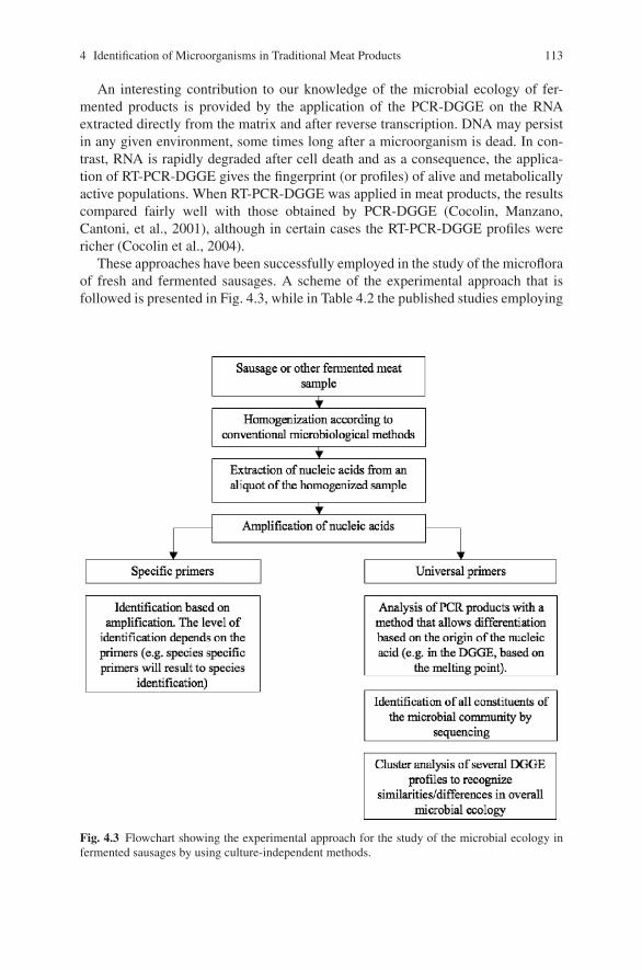
4 Identification of Microorganisms in Traditional Meat Products 113
An interesting contribution to our knowledge of the microbial ecology of fer-mented products is provided by the application of the PCR-DGGE on the RNAextracted directly from the matrix and after reverse transcription. DNA may persistin any given environment, some times long after a microorganism is dead. In con-trast, RNA is rapidly degraded after cell death and as a consequence, the applica-tion of RT-PCR-DGGE gives the fingerprint (or profiles) of alive and metabolicallyactive populations. When RT-PCR-DGGE was applied in meat products, the resultscompared fairly well with those obtained by PCR-DGGE (Cocolin, Manzano,Cantoni, et al., 2001), although in certain cases the RT-PCR-DGGE profiles werericher (Cocolin et al., 2004).
These approaches have been successfully employed in the study of the microfloraof fresh and fermented sausages. A scheme of the experimental approach that isfollowed is presented in Fig. 4.3, while in Table 4.2 the published studies employing
Fig. 4.3 Flowchart showing the experimental approach for the study of the microbial ecology infermented sausages by using culture-independent methods.
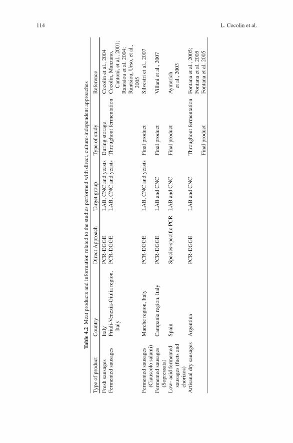
114 L. Cocolin et al.
Tabl
e4.
2M
eatp
rodu
cts
and
info
rmat
ion
rela
ted
toth
est
udie
spe
rfor
med
with
dire
ct,c
ultu
re-i
ndep
ende
ntap
proa
ches
Type
ofpr
oduc
tC
ount
ryD
irec
tApp
roac
hTa
rget
grou
pTy
peof
stud
yR
efer
ence
Fres
hsa
usag
esIt
aly
PCR
-DG
GE
LA
B,C
NC
and
yeas
tsD
urin
gst
orag
eC
ocol
inet
al.,
2004
Ferm
ente
dsa
usag
esFr
iuli-
Ven
ezia
-Giu
liare
gion
,It
aly
PCR
-DG
GE
LA
B,C
NC
and
yeas
tsT
hrou
ghou
tfer
men
tatio
nC
ocol
in,M
anza
no,
Can
toni
,eta
l.,20
01;
Ran
tsio
uet
al.2
004;
Ran
tsio
u,U
rso,
etal
.,20
05Fe
rmen
ted
saus
ages
(Cia
usco
losa
lam
i)M
arch
ere
gion
,Ita
lyPC
R-D
GG
EL
AB
,CN
Can
dye
asts
Fina
lpro
duct
Silv
estr
ieta
l.,20
07
Ferm
ente
dsa
usag
es(S
opre
ssat
a)C
ampa
nia
regi
on,I
taly
PCR
-DG
GE
LA
Ban
dC
NC
Fina
lpro
duct
Vill
anie
tal.,
2007
Low
-ac
idfe
rmen
ted
saus
ages
(fue
tsan
dch
oriz
os)
Spai
nSp
ecie
s-sp
ecifi
cPC
RL
AB
and
CN
CFi
nalp
rodu
ctA
ymer
ich
etal
.,20
03
Art
isan
aldr
ysa
usag
esA
rgen
tina
PCR
-DG
GE
LA
Ban
dC
NC
Thr
ough
outf
erm
enta
tion
Font
ana
etal
.,20
05;
Font
ana
etal
.200
5Fi
nalp
rodu
ctFo
ntan
aet
al.2
005
4 Identification of Microorganisms in Traditional Meat Products 115
culture-independent methods to study the microbial ecology of these products arereported.
The Choice of PCR-DGGE Primers
When applying PCR-DGGE to study complex microbial populations, such as theones present in food samples, an important parameter, that will influence the resultsobtained, is the choice of the amplification target gene prior to DGGE.
The target gene has to have two basic characteristics: (i) it should be present in allthe members of the microbial group that is under consideration, (ii) it should haveconserved regions, where universal primers can be designed, and variable regions,based on which separation is possible. In addition, if the RNA is going to be ana-lyzed, the gene should be characterized by constitutive expression. Genes that fulfillthese requirements are those that are involved in important and universal cell func-tions. Commonly, genes encoding for rRNA fall within this category. In bacteria,various regions of the 16S rRNA coding gene have been used in PCR-DGGE, whilein yeasts, the 26S rRNA coding gene is commonly the target.
One important advantage of the 16S and 26S rRNA coding genes is the factthat, for both genes, databases of sequences from a large number of representativespecies is available. This is important since it allows identification of DGGE bandsby sequencing and comparison with the database. A drawback associated with theuse of rRNA coding genes is the inherent sequence heterogeneity, within the samespecies, that is the result of multi-copies of the genes with small differences in thesequence. The multi-copies often result in multi-bands in the DGGE profiles thatcomplicate the analysis.
An alternative gene has been proposed for use in PCR-DGGE. It is the rpoB gene,encoding for the β-subunit of the RNA polymerase, which is usually present as asingle copy in each genome. The limitation of using the rpoB gene is the restrictednumber of sequences available in the databases that hinders the identification ofunknown DGGE bands.
In the DGGE studies performed so far on meat products, several primers havebeen used, and the relevant information is summarized in Table 4.3. The qualityof information produced by PCR-DGGE is dependent on both the number and res-olution of the amplicons in denaturing gradient gels (Yu & Morrison, 2004). Infingerprinting the microbial communities of a food product, it is important thatthe method allows differentiation of individual species that are associated with aspecific food. For this reason, the primers used and the conditions employed inPCR-DGGE need to be carefully considered and if necessary, optimized, prior toapplication in real food samples (Cocolin, Manzano, Cantoni, et al., 2001; Yu &Morrison, 2004).
As shown in Table 4.2, for the profiling of bacterial populations in fresh andfermented sausages, several regions of the 16S rRNA coding genes have beenemployed.
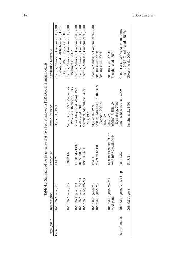
116 L. Cocolin et al.
Tabl
e4.
3Su
mm
ary
ofth
eta
rget
gene
sth
atha
vebe
enem
ploy
edin
PCR
-DG
GE
ofm
eatp
rodu
cts
Targ
etgr
oup
Targ
etre
gion
Prim
erse
tPr
imer
Ref
eren
ceA
pplic
atio
nre
fere
nce
Bac
teri
a16
SrR
NA
gene
,V1
P1/P
2K
lijn
etal
.,19
91C
ocol
in,M
anza
no,C
anto
ni,e
tal.,
2001
;C
ocol
inet
al.,
2004
;Ran
tsio
u,U
rso,
etal
.,20
05;S
ilves
trie
tal.,
2007
16S
rRN
Age
ne,V
333
8f/5
18r
Am
peet
al.,
1999
,Muy
zer,
deW
aal,
&U
itter
linde
n,19
93C
ocol
in,M
anza
no,C
anto
ni,e
tal.,
2001
;V
illan
ieta
l.,20
0716
SrR
NA
gene
,V9
Ec1
055/
Ec1
392
Ferr
is,M
uyze
r,&
War
d,19
96C
ocol
in,M
anza
no,C
anto
ni,e
tal.,
2001
16S
rRN
Age
ne,V
2-V
3H
DA
1/H
DA
2W
alte
ret
al.,
2000
Coc
olin
,Man
zano
,Can
toni
,eta
l.,20
0116
SrR
NA
gene
,V6-
V8
U96
8/L
1401
Zoe
tend
al,A
kker
man
s,&
deV
os,1
998
Coc
olin
,Man
zano
,Can
toni
,eta
l.,20
01
16S
rRN
Age
ne,V
3P3
/P4
Klij
net
al.,
1991
Coc
olin
,Man
zano
,Can
toni
,eta
l.,20
0116
SrR
NA
gene
,V3
V3f
/Uni
-051
5rE
rcol
ini,
Mos
chet
ti,B
laio
tta,&
Cop
pola
,200
1bL
ane,
1991
Font
ana
etal
.,20
05;
Font
ana
etal
.,20
05
16S
rRN
Age
ne,V
2-V
3B
act-
0124
f/U
niv-
0515
rL
ane,
1991
Font
ana
etal
.,20
05rp
oBge
nerp
oB16
98f/
rpoB
2014
rD
ahllo
f,B
ailli
e,&
Kje
llebe
rg,2
000
Ran
tsio
uet
al.,
2004
Yea
sts/
mou
lds
26S
rRN
Age
ne,D
1-D
2lo
opN
L1-
LS2
Coc
olin
,Bis
son,
etal
.,20
00C
ocol
inet
al.,
2004
;Ran
tsio
u,U
rso,
etal
.,20
05;C
ocol
inet
al.,
2006
a26
SrR
NA
gene
U1-
U2
Sand
huet
al.,
1995
Silv
estr
ieta
l.,20
07
4 Identification of Microorganisms in Traditional Meat Products 117
In an early study, conducted by Cocolin, Manzano, Cantoni, et al., (2001) usingcontrol strains from culture collections that belonged to bacterial species commonlyassociated with meat products, it was concluded that region V1 of the 16S rRNAallowed the best differentiation in DGGE. In the DGGE optimization process withthe P1/P2 set of primers, it was determined that the 40–60% denaturant gradientallowed differentiation of members of CNC, distinguishing Kocuria strains fromStaphylococcus strains, while the 30–50% denaturant gradient allowed differentia-tion of all the Lactobacillus spp. tested. This set of primers has been subsequentlyused in DGGE profiling of bacterial populations in different meat products (Cocolinet al., 2004; Rantsiou, Urso, et al., 2005; Silvestri et al., 2007).
Fontana et al. (2005) applied a nested PCR approach in their study of fermentedsausages. With primers P0/P4 (Klijn, Weerkamp, & de Vos, 1991), fragments ofabout 700 bp from the 5’ region of the 16S rRNA coding gene were amplified andwere used for nested PCR with two different sets of primers targeting the V2–V3 andthe V3 regions. They concluded that the best DGGE patterns of the microbial com-munity of fermented sausages were obtained with a 40–60% denaturant gradient forboth primer combinations. In a subsequent study by Fontana et al. (2005), three setsof primers were tested, targeting the V1, V2–V3, and V3 regions, and the authorsreport that the best results from the direct analysis of the microbial community byDGGE were obtained by amplifying the V3 region.
Microbial Ecology of Meat Products Determined by DirectApproaches
Bacterial Ecology by PCR-DGGE
In the study of the microbial ecology of meat products, researchers have followedtwo approaches: they looked into the evolution of the different microbial populationsthroughout the production process and until the product was ready for marketing,or they considered the more static picture obtained from the final product. From thestudies of the first type, a general observation is that a great diversity, both withinthe LAB and CNC groups, and complex DGGE profiles characterize the freshlyprepared meat mix, just prior to the initiation of the fermentation or storage period,in the case of non-fermented meat products. This biodiversity and the associatedcomplexity in the DGGE profiles, is sharply reduced from the first days of fermen-tation and just a few species appear to predominate and are commonly detectedthroughout the rest of the process. On the other hand, when a final product is takeninto consideration, then the microorganisms that have managed to persist and mostlikely were responsible for the transformation process are detected. Of course, in thisapproach, microorganisms that may play a role at earlier stages but do not surviveuntil the end, cannot be seen.
In the first work published (Cocolin, Manzano, Cantoni, et al., 2001) regardingdirect DGGE analysis, traditional fermented sausages produced by local laboratoriesin the Friuli-Venezia-Giulia region of Italy were analyzed throughout fermentation
118 L. Cocolin et al.
and ripening that lasted in total for 45 days. In the DNA and RNA-DGGE gels,multiple bands were visible for the first 3 days of fermentation, when differentspecies, most of which were related to Staphylococcus spp. were identified. Fromthe tenth day of ripening only the LAB bands were present. The LAB population wascharacterized by Lb. sakei and Lb. curvatus throughout the process. Lb. plantarumwas only detected on the first day in the DNA gel and the band could have beengenerated from dead cells. Staphylococcus species were found only in the meatmixture before the sausages were filled and after 3 days. The only Staphylococcusspecies represented in the DGGE gel after 3 days was S. xylosus, which produceda specific band in the gel until the end of fermentation. The corresponding band inthe RNA gel was only present at day 0 and day 3 and then disappeared. This couldbe explained by the large quantity of LAB RNA that restricted amplification of theS. xylosus RNA.
In a later study focusing on traditional fermented sausages from the same regionof Italy, the goal was to follow the fermentation in three different plants, in whichno starters are being used. PCR-DGGE was employed and the bands were identifiedby sequencing. Furthermore, cluster analysis of the DGGE profiles, obtained fromthe three fermentations and at different sampling points, was conducted (Rantsiou,Urso, et al., 2005). A general consideration that resulted from this study is thatthe main differences detected in the ecology, between the three sausages, were notrepresented by the species of microorganisms identified by band sequencing but bytheir relative distribution between the fermentations. In all the three fermentations,a stable signal, from the beginning of the period studied was visible for Lb. curvatusand Lb. sakei, which remained constant throughout the transformation. In one of thefermentation, a band that corresponded to Lb. paracasei was present for part of theperiod, while L. garviae was detected in two of the three sausages. No Lb. plan-tarum was detected in these sausages. As seen from the DGGE profiles, importantcontribution to the microbial ecology was given by the Staphylococcus species. S.equorum and S. succinus were present in all the fermentations, while S. xylosus wasmainly present in one of them.
When the rpoB gene was targeted in PCR-DGGE of the same type of fermentedsausages (Rantsiou, Comi, & Cocolin, 2004), significantly simpler profiles, allthrough the transformation process, were obtained. At the beginning of the fermen-tation, S. xylosus was detected while at the later stages, Lb. curvatus, Lb. sakei, andLb. plantarum comprised the bacterial population.
In a study conducted by Fontana et al. (2005), PCR-DGGE has been applied toprofile the bacterial community of the artisanal Argentinean sausages. Targeting theV3 region of the 16S rRNA coding gene, a highly complex fingerprint was obtainedat day 0 of the fermentation that was characterized by the presence of Lb. plantarum,P. acidilactici, Lb. sakei, Lb. curvatus, S. equorum (identified by co-migration withcontrol strains), and Corynebacterium variabilis (identified by sequencing). A morebasic fingerprint was obtained at day 5 and day 14 (last day of ripening), with thepresence of Lb. plantarum, Lb. sakei, and Lb. curvatus, while S. saprophyticus rep-resented the Staphylococcus group. In a similar study by the same authors (Fontanaet al., 2005), the fermentation of a sausage from the Tucuman region of Argentina
4 Identification of Microorganisms in Traditional Meat Products 119
was followed during the 14 day production and the same sausage was compared to aripened sausage from the Cordoba region. A high microbial diversity was observedat day 0 with the presence of Lb. plantarum, Lb. sakei, S. saprophyticus, C. vari-abilis and that with varying intensities were also present in the other two samplingpoints (day 5 and day 14). When the two sausages were compared, a common band,associated with Lb. sakei was detected. Furthermore, the bacterial community of theTucuman sausage contained S. saprophyticus and an uncultured bacterium while inthe Cordoba sausage Brochothrix thermosphacta was present.
The bacterial ecology of the traditional Italian salami “ciauscolo” was recentlysurveyed by PCR-DGGE (Silvestri et al., 2007). The V1 region was amplified from22 samples of 14 artisan and 8 industrial production plants from different geo-graphical areas of the Marche region, in Central Italy. Overall, five LAB specieswere detected: Lb. plantarum, Lb. curvatus, Lb. sakei, P. acidilactici, and L. lactis.No Staphylococcus bands were observed in the DGGE profiles, either because thepopulations were below the detection limit or due to the higher number of LAB thatmay create a masking effect. The most frequently detected species were, once again,Lb. curvatus and Lb. sakei.
Finally, in a recent study of the ecology of a fermented sausage from SouthernItaly, the “soppressata”, targeting the V3 and V1 regions, it was possible to identifyS. xylosus, S. succinus, and S. equorum among the staphylococci and Lb. sakei andLb. curvatus within the lactobacilli (Villani et al., 2007).
A slightly different picture of bacterial ecology was seen when fresh sausagesfrom Italy were considered (Cocolin et al., 2004). These products are highly perish-able, since no fermentation takes place. After mixing of the ingredients and casing,they are stored at 4◦C. They have a very short shelf life (about 10 days) and arecooked before consumption. Bacterial DGGE profiles were characterized, for bothDNA and RNA, by the presence of a constant band throughout the process thatwas B. thermosphacta, whereas the Lactobacillus spp., visible at the DNA level,were only marginally present at the RNA level, mainly represented by Lb. sakei. Lc.mesenteroides was present at both DNA and RNA the last day of sampling. On thefresh mix, at the RNA level, a Bacillus sp. band was present, while Staphylococcusspp. was also detected during storage of the fresh sausages.
An alternative application of the PCR-DGGE directly in a food matrix was pre-sented by Cocolin et al. (2006b). The authors followed the microbial dynamics ofsausage fermentation, with particular interest for Lb. plantarum and S. carnosusthat were inoculated as a starter. PCR-DGGE allowed the detection of a stable band,identified by sequencing as Lb. curvatus, that was only marginally detected by theconventional microbiological techniques. This application shows the potential of themethod in monitoring, in a culture-independent way, specific microbial groups.
Yeast Ecology by PCR-DGGE
The yeast ecology has been described by targeting the 26S rRNA coding genefor Italian fermented sausages of the Friuli-Venezia-Giulia region (Rantsiou, Urso,
120 L. Cocolin et al.
et al., 2005; Cocolin et al., 2006a) and of the Marche region (Silvestri et al., 2007)and for fresh sausages (Cocolin et al., 2004).
Overall, the DGGE profiles are less complex than those obtained for the bac-teria and oftentimes, they are characteristic of the products. In particular, whena cluster analysis of the yeast DGGE profiles was carried out, from the samplescollected during production of three fermented sausages, a fermentation-specificdistribution was obtained, with the clusters containing samples from the same fer-mentation (Rantsiou, Urso, et al., 2005). In this study, the yeast ecology results werecharacterized by members of the genera Candida, Debaryomyces, and Willopsis.D. hansenii, a proteolytic yeast that is associated with fermented sausage produc-tion, was dominant in one of the three fermentations but was not seen in the othertwo productions. C. krisii and Willopsis saturnus and C. sake or austromarina char-acterized, respectively, the other two fermentations. Finally, one band was commonin all the three fermentations that after sequencing showed a 86.5% homology toMayaca fluviatilis. This homology score is very low and most likely, the DGGEband corresponds to a yeast species that has not yet been described or for which no26S rRNA gene sequence is available.
The predominance of D. hansenii was also confirmed by a later study in Italiansausages fermented at low temperatures, probably due to its physiological character-istics, and it should therefore be considered well adapted to the specific environmentyeast species (Flores, Dura, Marco, & Toldra, 2004). Throughout the 60 days of fer-mentation a stable band, at both the DNA and RNA level, identified as D. hansenii,was visible in the DGGE gels (Cocolin et al., 2006a).
In the “ciauscolo” salami of the Marche region, the most frequently detectedspecies was D. hansenii, while C. physchrophila and Saccharomyces barnettii werealso occasionally found (Silvestri et al., 2007).
D. hansenii was the yeast, characterizing the DNA and RNA profiles of the freshsausages. It was present during the whole period followed. The only other yeastspecies detected was Capronia mansonii, at day 0 and only in the RNA profile(Cocolin et al., 2004).
Bacterial Ecology by Species-Specific PCR and qPCR
Aymerich et al. (2003) studied the bacterial ecology of 17 commercially availablelow acid fermented sausages from Spain. Ten of them were fuets (cold-ripenedfermented sausages with black pepper) and seven where chorizos (cold-ripened fer-mented sausages with red pepper). They used 12 sets of primers for 12 differentspecies, six LAB (Lb. sakei, Lb. curvatus, E. faecium, Lb. plantarum, L. lactis,and P. acidilactici) and six CNC (S. carnosus, S. warneri, K. varians, S. xylosus,S. simulans, and S. epidermidis). They screened for the 12 species in the fermentedsausage homogenate, before and after a 24-hour enrichment period (in MRS for theLAB and in mannitol salt broth [MSB] for the CNC).
Without enrichment, Lb. sakei and Lb. curvatus were detected in 11.8% of thesamples, and Lb. plantarum and S. xylosus were detected in 17.6%. The low per-centages of detection for the species of importance in the fermentation is probably
4 Identification of Microorganisms in Traditional Meat Products 121
related with a low sensitivity limit of the method, that needed an enrichment step,in order to obtain some information for the species present. However, it should benoted that enrichment can significantly alter the picture of the microbial ecology,since different microbial groups or even species within a microbial group, havedifferent abilities to grow in enrichment media. After the 24h enrichment, it waspossible to detect Lb. sakei and S. xylosus in all the samples, a result that highlightsthat these two species are predominant in slightly fermented sausages. Regardingother LAB species, Lb. curvatus was detected in 71% of the samples (in 80% ofthe fuets and 57% of the chorizos), while Lb. plantarum was present in all of thechorizos and in 50% of the fuets (62% of the samples in total). P. acidilactici andL. lactis were not detected while E. faecium was present in 11.8% of the samples.With regard to the other CNC species, S. carnosus was only detected in chorizos(in 14% of them) and S. epidermidis in the fuets (in 20% of them). No S. simulansor S. warneri were found. Overall, the ecology of the low-acid fermented sausagesfrom Spain, as determined after the enrichment, was characterized by the presenceof Lb. sakei and S. xylosus. Lb. curvatus and Lb. plantarum were also detected butwith different frequencies for the two types of sausages. In addition, the presence ofS. epidermidis and S. carnosus characterized the fuets and the chorizos respectively,although they were not detected in all the respective samples.
Recently, a protocol based on qPCR amplification of the 16S-23S rRNA inter-genic transcribed region (ITS) of Lb. sakei, has been optimized (Martin et al., 2006).The goal of the work was to develop a tool that would allow the quantification ofLb. sakei, without cultivation, in the fermented sausages. The protocol was testedin 11 commercial samples of sausages and meat batter; and Lb. sakei was detectedin the range of 3 to 9 log10 cfu/g. Of the 11 samples, Lb. sakei was not detected inthree while in a fourth, it was below the quantification limit, which was determinedto be in the order of 105 cfu/g. The method can be considered as a valid alternativeto conventional microbiological testing of fermented sausages that allows rapid andaccurate quantification of Lb. sakei.
Conclusions
Often at times, when both the culture-dependent and independent methods areapplied in parallel to study the microbial ecology of fermented foods, interestinginformation becomes available. From the experience gained so far in this field, wecan conclude that the two approaches complement each other, and their concurrentuse highlights important aspects that could otherwise pass un-noticed.
Indisputably, the main breakthrough of the direct methods is that, the inherentlimitations of microbial cultivation, related to non-culturable organisms, either dueto injury or stress, or due to the selectivity of the media, are circumvented. Fur-thermore, for the first time, we are able to see in situ which microorganisms aremetabolically active and derive information regarding the relative importance ofdifferent microbial groups in the transformation process.
122 L. Cocolin et al.
The results of the application of molecular approaches to study the microflora offermented sausages that have been presented here, highlight the unequivocal dom-inance of Lb. sakei and Lb. curvatus in these types of products. To a lesser extend,Lb. plantarum, in a more product-specific manner, was also shown to be important.The presence and activity of the different Staphylococcus members appears to beproduct-specific as well. Finally, regarding yeasts, D. hansenii was detected with ahigh frequency (Rantsiou & Cocolin, 2006).
Moreover, the use of molecular techniques, able to differentiate strains withinthe same species, helps in the understanding of the intra-species diversity thatmay characterize fermented products produced in different countries or in differ-ent plants within the same country. Different strains, together with the differentingredients used and the technological processes during production, are responsi-ble for the development of specific sensory profiles. To us, this is an importantaspect to be considered in our understanding of the fermentation process, and itneeds to be investigated further. Defining and understanding microbial dynamics,as determined by species successions, as well as microbial ecology, as determinedby species interactions at each time point and throughout fermentation, is crucial,since these are the parameters that will have a great impact on the organoleptic andsensorial characteristics of the final product.
References
Altschul, S. F., Madden, T. L., Shaffer, A. A., Zhang, J., Zhang, Z., Miller, W., et al. (1997). GappedBLAST and PSI-BLAST: A new generation of protein database search programs. Nucleic AcidResearch, 25, 3389–3402.
Ammor, S., Rachman, C., Chaillou, S., Prevost, H., Dousset, X., Zagorec, M., et al. (2005). Phe-notypic and genotypic identification of lactic acid bacteria isolated from a small-scale facilityproducing traditional dry sausages. Food Microbiology, 22, 373–382.
Ampe, F., Ben Omar, N., Moizan, C., Wacher, C., & Guyot, J. P. (1999). Polyphasic study of thespatial distribution of microorganisms in Mexican pozol, a fermented maize dough, demon-strates the need for cultivation-independent methods to investigate traditional fermentations.Applied and Environmental Microbiology, 65, 5464–5473.
Andrighetto, C., Zampese, L., & Lombardi, A. (2001). RAPD-PCR characterization of lactobacilliisolated from artisanal meat plants and traditional fermented sausages of Veneto region (Italy).Letter in Applied Microbiology, 33, 26–30.
Aymerich, T., Martin, B., Garriga, M., & Hugas, M. (2003). Microbial quality and direct PCRidentification of lactic acid bacteria and nonpathogenic staphylococci from artisanal low-acidsausages. Applied and Environmental Microbiology, 69, 4583–4594.
Aymerich, T., Martin, B., Garrica, M., Vidal-Carou, M. C., Bover-Cid, S., & Hugas, M. (2006).Safety properties and molecular strain typing of lactic acid bacteria from slightly fermentedsausages. Journal of Applied Microbiology, 100, 40–49.
Bacus, J. N. (1986). Fermented meat and poultry products. In A. M. D. Pearson (Ed.), Advances inmeat and poultry microbiology (pp. 123–164). London: Macmillan.
Baruzzi, F., Matarante, A., Caputo, L., & Morea, M. (2006). Molecular and physiological char-acterization of natural microbial communities isolated from a traditional Southern Italian pro-cessed sausages. Meat Science, 72, 261–269.
Berthier, F., & Ehrlich, S. D. (1998). Rapid species identification within two groups of closelyrelated lactobacilli using PCR primers that target the 16S/23S rRNA spacer region. FEMSMicrobiology Letters, 161, 97–106.
4 Identification of Microorganisms in Traditional Meat Products 123
Berthier, F., & Ehrlich, S. D. (1999). Genetic diversity within Lactobacillus sakei and Lactobacilluscurvatus and design of PCR primers for its detection using randomly amplified polymorphicDNA. International Journal of Systematic Bacteriology, 49, 997–1007.
Blaiotta, G., Casaburi, A., & Villani, F. (2005). Identification and differentiation of Staphylococcuscarnosus and Staphylococcus simulans by species-specific PCR assays of sodA genes. System-atic and Applied Microbiology, 28, 519–526.
Blaiotta, G., Ercolini, D., Mauriello, G., Salzano, G., & Villani, F. (2004). Rapid and reliableidentification of Staphylococcus equorum by a species-specific PCR assay targeting the sodAgene. Systematic and Applied Microbiology, 27, 696–702.
Blaiotta, G., Pennacchia, C., Ercolini, D., Moschetti, G., & Villani, F. (2003). Combining dena-turing gradient gel electrophoresis of 16S rDNA V3 region and 16S-23S rDNA spacer regionpolymorphism analyses for the identification of staphylococci from Italian fermented sausages.Systematic and Applied Microbiology, 26, 423–433.
Blaiotta, G., Pennacchia, C., Parente, E., & Villani, F. (2003). Design and evaluation of specificPCR primers for rapid and reliable identification of Staphylococcus xylosus strains isolatedfrom dry fermented sausages. Systematic and Applied Microbiology, 26, 601–610.
Blaiotta, G., Pennacchia, C., Villani, F., Ricciardi, A., Tofalo, R., & Parente, E. (2004). Diversityand dynamics of communities of coagulase-negative staphylococci in traditional fermentedsausages. Journal of Applied Microbiology, 97, 271–284.
Bottari, B., Ercolini, D., Gatti, M., & Neviani, E. (2006). Application of FISH technology formicrobiological analysis: Current state and prospects. Applied Microbiology and Biotechnol-ogy, 73, 485–494.
Caetono-Anolles, G. (1993). Amplifying DNA with arbitrary oligonucleotide primers. PCR Meth-ods and Applications, 3, 85–94.
Cocolin, L., Bisson, L. F., & Mills, D. A. (2000). Direct profiling of the yeast dynamics in winefermentations. FEMS Microbiology Letters, 189, 81–87.
Cocolin, L., Diez, A., Urso, R., Rantsiou, K., Comi, G., Bergmaier, I., et al. (2007). Optimiza-tion of conditions for profiling bacterial populations in food by culture-independent methods.International Journal of Food Microbiology, 20, 400–409.
Cocolin, L., Manzano, M., Aggio, D., Cantoni, C., & Comi, G. (2001). A novel polymerasechain reaction (PCR)—denaturing gradient gel electrophoresis (DGGE) for the identification ofMicrococcaceae strains involved in meat fermentations. Its application to naturally fermentedItalian sausages. Meat Science, 57, 59–64.
Cocolin, L., Manzano, M., Cantoni, C., & Comi, G. (2000). Development of a rapid method forthe identification of Lactobacillus spp. isolated from naturally fermented Italian sausage using apolymerase chain reaction—temperature gradient gel electrophoresis. Letters in Applied Micro-biology, 30, 126–129.
Cocolin, L., Manzano, M., Cantoni, C., & Comi, G. (2001). Denaturing gradient gel electrophore-sis analysis of the 16S rRNA gene V1 region to monitor dynamic changes in the bacterialpopulation during fermentation of Italian sausages. Applied and Environmental Microbiology,67, 5113–5121.
Cocolin, L., Rantsiou, K., Iacumin, L., Urso, R., Cantoni, C., & Comi, G. (2004). Study of the ecol-ogy of fresh sausages and characterization of populations of lactic acid bacteria by molecularmethods. Applied and Environmental Microbiology, 70, 1883–1894.
Cocolin, L., Urso, R., Rantsiou, K., Cantoni, C., & Comi, G. (2006a). Dynamics and character-ization of yeasts during natural fermentation of Italian sausages. FEMS Yeast Research, 6,692–701.
Cocolin, L., Urso, R., Rantsiou, K., Cantoni, C., & Comi, G. (2006b). Multiphasic approach tostudy the bacterial ecology of fermented sausages inoculated with a commercial starter culture.Applied and Environmental Microbiology, 72, 942–945.
Collins, M. D., Rodriguez, U., Ash, C., Aguirre, M., Farrow, J. E., Martinezmurcia, A., et al.(1991). Phylogenetic analysis of the genus Lactobacillus and related lactic acid bacteria asdetermined by reverse-transcriptase sequencing of 16S ribosomal-RNA. FEMS MicrobiologyLetters, 77, 5–12.
124 L. Cocolin et al.
Comi, G., Urso, R., Iacumin, L., Rantsiou, K., Cattaneo, P., Cantoni, C., et al. (2005). Character-ization of naturally fermented sausages produced in the North East of Italy. Meat Science, 69,381–392.
Coppola, R., Iorizzo, M., Saotta, R., Sorrentino, E., & Grazia, L. (1997). Characterization ofmicrococci and staphylococci isolated from soppressata molisana, a Southern Italy fermentedsausage. Food Microbiology, 14, 47–53.
Corbiere Morot-Bizot, S., Leroy, S., & Talon, R. (2006). Staphylococcal community of a small unitmanufacturing traditional dry fermented sausages. International Journal of Food Microbiology,108, 210–217.
Corbiere Morot-Bizot, S., Leroy, S., & Talon, R. (2007). Monitoring of staphylococcal startersin two French processing plants manufacturing dry fermented sausages. Journal of AppliedMicrobiology, 102, 238–244.
Corbiere Morot-Bizot, S., Talon, R., & Leroy, S. (2004). Development of a multiplex PCR forthe identification of Staphylococcus genus and four staphylococcal species isolated from food.Journal of Applied Microbiology, 97, 1087–1094.
Corich, V., Mattiazzi, A., Soldati, E., Carraro, A., & Giacomini, A. (2005). Sau-PCR, a novelamplification technique for genetic fingerprinting of microorganisms. Applied and Environ-mental Microbiology, 71, 6401–6406.
Couto, I., Pereira, S., Miragaia, M., Sanches, I. S., & de Lancastre, H. (2001). Identification ofclinical staphylococcal isolates from humans by internal transcribed spacer PCR. Journal ofClinical Microbiology, 39, 3099–3103.
Dahllof, I., Baillie, H., & Kjelleberg, S. (2000). rpoB-based microbial community analysis avoidslimitations inherent in 16S rRNA gene intraspecies heterogeneity. Applied and EnvironmentalMicrobiology, 66, 3376–3380.
Demeyer, D. I. (1982). Stoichiometry of dry sausage fermentation. Antonie Leeuwenhoek, 48,414–416.
Demeyer, D. I., Verplaetse, A., & Gistelink, M. (1986). Fermentation of meat: An integrated pro-cess. Belgian Journal of Food Chemistry and Biotechnology, 41, 131–140.
Di Maria, S., Basso, A. L., Santoro, E., Grazia, L., & Coppola, R. (2002). Monitoring of Staphy-lococcus xylosus DSM 20266 added as starter during fermentation and ripening of soppressatamolisana, a typical Italian sausage. Journal of Applied Microbiology, 92, 158–164.
Ercolini, D. (2004). PCR-DGGE fingerprinting: Novel strategies for detection of microbes in food.Journal of Microbiological Methods, 56, 297–314.
Ercolini, D., Hill, P. J., & Dodd, E. R. (2003). Bacterial community structure and location in Stiltoncheese. Applied and Environmental Microbiology, 69, 3540–548.
Ercolini, D., Moschetti, G., Blaiotta, G., & Coppola, S. (2001a). Behavior of variable V3 regionfrom 16S rDNA of important lactic acid bacteria in denaturing gradient gel electrophoresis.Current Microbiology, 42, 199–202.
Ercolini, D., Moschetti, G., Blaiotta, G., & Coppola, S. (2001b). The potential of a polyphasicPCR-DGGE approach in evaluating microbial diversity of natural whey cultures from water-buffalo Mozzarella cheese production: Bias of “culture dependent” and “culture independent”approaches. Systematic and Applied Microbiology, 24, 610–617.
Fadda, S., Sanz, Y., Vignolo, G., Aristoy, M.-C., Oliver, G., & Toldra, F. (1999a). Hydrolysis ofpork muscle sarcoplasmatic proteins by Lactobacillus curvatus and Lactobacillus sake. Appliedand Environmental Microbiology, 65, 578–584.
Fadda, S., Sanz, Y., Vignolo, G., Aristoy, M.-C., Oliver, G., & Toldra, F. (1999b). Characterizationof muscle sarcoplasmatic and myofibrillar protein hydrolysis caused by Lactobacillus plan-tarum. Applied and Environmental Microbiology, 65, 3540–3546.
Ferris, M. J., Muyzer, G., & Ward, D. M. (1996). Denaturing gradient gel electrophoresis profilesof 16S rRNA-defined populations inhabiting a hot spring microbial mat community. Appliedand Environmental Microbiology, 62, 340–346.
Flores, M., Dura, M.-A., Marco, A., & Toldra, F. (2004). Effect of Debaryomyces spp. on aromaformation and sensory quality of dry-fermented sausages. Meat Science, 68, 439–446.
4 Identification of Microorganisms in Traditional Meat Products 125
Fontana, C., Cocconcelli, P. S., & Vignolo, G. (2005). Monitoring the bacterial population dynam-ics during fermentation of artisanal Argentinean sausages. International Journal of Food Micro-biology, 103, 131–142.
Fontana, C., Vignolo, G., & Cocconcelli, P. S. (2005). PCR-DGGE analysis for the identificationof microbial populations from Argentinean dry fermented sausages. Journal of MicrobiologicalMethods, 63, 254–263.
Forsman, P., Tilsala-Timisijarvi, A., & Alatossava, T. (1997). Identification of the staphylococcaland streptococcal causes of the bovine mastitis using 16S–23S rRNA spacer regions. Microbi-ology, 143, 3491–3500.
Garcia-Varona, M., Santos, E. M., Jaime, I., & Rovira, J. (2000). Characterization of Micrococ-caceae isolated from different varieties of chorizo. International Journal of Food Microbiology,54, 189–195.
Gevers, D., Huys, G., & Swings, J. (2001). Applicability of rep-PCR fingerprinting for identifica-tion of Lactobacillus species. FEMS Microbiology Letters, 205, 31–36.
Gilson, E., Clement, J. M., Brutlag, D., & Hofnung, M. (1984). A family of dispersed repetitiveextragenic palindromic DNA sequences in E . coli. EMBO Journal, 3, 1417–1421.
Gory, L., Millet, L., Godon, J. J., & Montel, M. C. (1999). Identification of Staphylococcuscarnosus and Staphylococcus warneri isolated from meat by florescent in situ hybridizationwith 16S-targeted oligonucleotide probes. Systematic and Applied Microbiology, 22, 225–228.
Greco, M., Mazzette, R., De Santis, E. P. L., Corona, A., & Cosseddu, A. M. (2005). Evolution andidentification of lactic acid bacteria isolated during the ripening of Sardinian sausages. MeatScience, 69, 733–739.
Hammes, W. P., Bantleon, A., & Min, S. (1990). Lactic acid bacteria in meat fermentation. FEMSMicrobiology Reviews, 87, 165–174.
Hammes, W. P., & Knauf, H. J. (1994). Starters in processing of meat products. Meat Science, 36,155–168.
Hertel, C., Ludwig, W., Obst, M., Vogel, R. F., Hammes, W. P., & Schleifer, K. H. (1991). 23SrRNA-targeted oligonucleotide probes for the rapid identification of meat lactobacilli. System-atic and Applied Microbiology, 14, 173–177.
Hesseltine, C. W., & Wang, H. L. (1980). The importance of traditional fermented foods. Bio-science, 30, 402–404.
Hugenholtz, P., Goebel, B. M., & Pace, N. R. (1998). Impact of culture-independent studies on theemerging phylogenetic view of bacterial diversity. Journal of Bacteriology, 180, 4765–4774.
Iacumin, L., Comi, G., Cantoni, C., & Cocolin, L. (2006a). Molecular and technological character-ization of Staphylococcus xylosus isolated from Italian naturally fermented sausages by RAPD,Rep-PCR and Sau-PCR analysis. Meat Science, 74, 281–288.
Iacumin, L., Comi, G., Cantoni, C., & Cocolin, L. (2006b). Ecology and dynamics of coagulase-negative cocci isolated from naturally fermented Italian sausages. Systematic and AppliedMicrobiology, 29, 480–486.
Jensen, M. A., Webster, J. A., & Strauss, N. (1993). Rapid identification of bacteria on the basisof polymerase chain reaction-amplified ribosomal DNA spacer region polymorphisms. Appliedand Environmental Microbiology, 59, 945–952.
Klijn, N., Weerkamp, A. H., & de Vos, W. M. (1991). Identification of mesophilic lactic acid bac-teria by using polymerase chain reaction-amplified variable regions of 16S rRNA and specificDNA probes. Applied and Environmental Microbiology, 57, 3390–3393.
Kurtzman, C. P., & Robnett, C. J. (1997). Identification of clinically important ascomycetous yeastbased on nucleotide divergence in the 5’ and of the large-subunit (26S) ribosomal DNA gene.Journal of Clinical Microbiology, 35, 1216–1223.
Kurtzman, C. P.., & Robnett, C. J. (1998). Identification and phylogeny of ascomycetous yeastsfrom analysis of nuclear large subunit (26S) ribosomal DNA partial sequence. Antonie vanLeeuwenhoek 73, 331–371.
Lane, D. J. (1991). 16S/23S sequencing. In E. Stackebrandt, & M. Goodfellow (Eds.), Nucleic acidtechniques in bacterial systematics (pp. 115–175). Chichester: John Wiley & Sons.
126 L. Cocolin et al.
Lee, J., Jang, J., Kim, B., Kim, J., Jeong, G., & Han, H. (2004). Identification of Lactobacillussakei and Lactobacillus curvatus by multiplex PCR-based restriction enzyme analysis. Journalof Microbiological Methods, 59, 1–6.
Lerche, M., & Reuter, G. (1960). A contribution to the method of isolation and differentation ofaerobic “lactobacilli” (Genus Lactobacillus Beijerinck). Archiv fur Hygiene und Bakteriologie,179, 354–370.
Lucke, F. K. (1974). Fermented sausages. In B. J. B. Wood (Ed.), Microbiology of fermented foods(pp. 41–49). London: Applied Science Publishers.
Lucke, F. K. (2000). Utilization of microbes to process and preserve meat. Meat Science, 56,105–115.
Martin, B., Garriga, M., Hugas, M., Bover-Cid, S., Veciana-Nogues, M. T, & Aymerich, T. (2006).Molecular, technological and safety characterization of Gram-positive catalase-positive coccifrom slightly fermented sausages. International Journal of Food Microbiology, 107, 148–158.
Martin, B., Jofre, A., Garriga, M., Pla, M., & Aymerich, T. (2006). Rapid quantitative detection ofLactobacillus sakei in meat and fermented sausages by real-time PCR. Applied and Environ-mental Microbiology, 72, 6040–6048.
Mendoza, M., Meugnier, H., Bes, M., Etienne, J., & Freney, J. (1998). Identification of Staphy-lococcus species by 16S–23S rDNA intergenic spacer PCR analysis. International Journal ofSystematic Bacteriology, 48, 1049–105.
Morot-Bizot, S., Talon, R., & Leroy-Setrin, S. (2003). Development of specific primers for a rapidand accurate identification of Staphylococcus xylosus, a species used in food fermentation.Journal of Microbiological Methods, 55, 279–286.
Muyzer, G., De Waal, E. D., & Uitterlinden, A. G. (1993). Profiling of complex microbial pop-ulations by denaturing gradient gel electrophoresis analysis of polymerase chain reaction-amplified genes coding for 16S rRNA. Applied and Environmental Microbiology, 59, 695–700.
Muyzer, G., & Smalla, K. (1998). Application of denaturing gradient gel electrophoresis (DGGE)and temperature gradient gel electrophoresis (TGGE) in microbial ecology. Antonie VanLeeuwenhoek, 73, 127–141.
Nissen, H., & Dainty, R. (1995). Comparsion of the use of rRNA probes and conventional methodsin identifying strains of Lactobacillus sake and L. curvatus isolated from meat. InternationalJournal of Food microbiology 25, 311–315.
Pennacchia, C., Vaughan, E. E., & Villani, F. (2006). Potential probiotic Lactobacillus strainsfrom fermented sausages: Further investigations on their probiotic properties. Meat Science, 73,90–101.
Pontes, S. D., Lima-Bitterncourt, I. C., Chartone-Souza, E., & Amaral Nascimiento, A. M. (2007).Molecular approaches: Advantages and artifacts in assessing bacterial diversity. Journal ofIndustrial Microbiology and Biotechnology, 34, 463–473.
Power, E. G. (1996). RAPD typing in microbiology—a technical review. Journal of Hospital Infec-tion, 34, 247–265.
Rantsiou, K., & Cocolin, L. (2006). New developments in the study of the microbiota of naturallyfermented sausages as determined by molecular methods: A review. International Journal ofFood Microbiology, 108, 255–267.
Rantsiou, K., Comi, G., & Cocolin, L. (2004). The rpoB gene as a target for PCR-DGGE analysis tofollow lactic acid bacteria population dynamics during food fermentations. Food Microbiology,21, 481–487.
Rantsiou, K., Drosinos, E. H., Gialitaki, M., Metaxopoulos, I., Comi, G., & Cocolin, L. (2006). Useof molecular tools to characterize Lactobacillus spp. isolated from Greek traditional fermentedsausages. International Journal of Food Microbiology, 112, 215–222.
Rantsiou, K., Drosinos, E. H., Gialitaki, M., Urso, R., Krommer, J., Gasparik-Reichardt, J., et al.(2005). Molecular characterization of Lactobacillus species isolated from naturally fermentedsausages produced in Greece, Hungary and Italy. Food Microbiology, 22, 19–28.
Rantsiou, K., Iacumin, L., Cantoni, C., Comi, G., & Cocolin, L. (2005). Ecology and characteri-zation by molecular methods of Staphylococcus species isolated from fresh sausages. Interna-tional Journal of Food Microbiology, 97, 277–284.
4 Identification of Microorganisms in Traditional Meat Products 127
Rantsiou, K., Urso, R., Iacumin, L., Cantoni, C., Cattaneo, P., Comi, G., et al. (2005). Culturedependent and independent methods to investigate the microbial ecology of Italian fermentedsausages. Applied and Environmental Microbiology, 71, 1977–1986.
Rebecchi, A., Crivori, S., Sarra, P. G., & Cocconcelli, P. S. (1998). Physiological and moleculartechniques for the study of bacterial community development in sausage fermentation. Journalof Applied Microbiology, 84, 1043–1049.
Reuter, G. (1972). Experimental ripening of dry sausages using lactobacilli and micrococci startercultures. Fleischwirtschaft, 52, 465–468, 471–473.
Rossi, F., Tofalo, R., Torriani, S., & Suzzi, G. (2001). Identification by 16S-23S rDNA intergenicregion amplification, genotypic and phenotypic clustering of Staphylococcus xylosus strainsfrom dry sausages. Journal of Applied Microbiology, 90, 365–371.
Sandhu, G. S., Kline, B. C., Stockman, L., & Roberts, G. D. (1995). Molecular probes for diagnosisof fungal infections. Journal of Clinical Microbiology, 33, 2913–2919.
Sanz, Y., Hernandez, M., Ferrus, M. A., & Hernandez, J. (1998). Characterization of Lactobacillussake isolates from dry-cured sausage by restriction fragment length polymorphism analysis ofthe 16S rRNA gene. Journal of Applied Microbiology, 84, 600–6006.
Schleifer, K. H. (1986). Gram positive cocci. In P. H. A Sneath, N. S. Mair, & Holt, J. G. (Eds.),Bergey’s manual of systematic bacteriology (Vol. 2, pp. 999–1003). Baltimore: Williams &Wilkins.
Silvestri, G., Santarelli, S., Aquilanti, L., Beccaceci, A., Osimani, A., Tonucci, F., et al. (2007).Investigation of the microbial ecology of Ciauscolo, a traditional Italian salami, by culture-dependent techniques and PCR-DGGE. Meat Science, 77, 413–423.
Simonova, M., Strompfova, V., Marcinakova, M., Laukova, A., Vetserlund, S., Moratella, et al.(2006). Characterization of Staphylococcus xylosus and Staphylococcus carnosus isolated fromSlovak meat products. Meat Science, 73, 559–564.
Talon, R., Leroy, S., & Lebert, I. (2007). Microbial ecosystems of traditional fermented meat prod-ucts: The importance of indigenous starters. Meat Science, 77, 55–62.
Talon, R., Walter, D., Chartier, S., Barriere, C., & Montel, M. C. (1999). Effect of nitrate andincubation conditions on the production of catalase and nitrate reductase by staphylococci.International Journal of Food Microbiology, 52, 47–56.
Tenover, F. C., Arbeit, R. D., Goering, R. V., Mickelsen, P. A., Murray, B. E., Persing, D. H.,et al. (1995). Interpreting chromosomal DNA restriction patterns produced by pulsed-fieldgel electrophoresis: Criteria for bacterial strain typing. Journal of Clinical Microbiology, 33,2233–2239.
Urso, R., Comi, G., & Cocolin, L. (2006). Ecology of lactic acid bacteria in Italian fermentedsausages: Isolation, identification and molecular characterization. Systematic and AppliedMicrobiology, 29, 671–680.
Villani, F., Casaburi, A., Pennacchia, C., Filosa, L., Russo, F., & Ercolini, D. (2007). The micro-bial ecology of the Soppressata of Vallo di Diano, a traditional dry fermented sausage fromsouthern Italy, and in vitro and in situ selection of autochthonous starter cultures. Applied andEnvironmental Microbiology, 73, 5453–5463.
Villard, L., Kodjo, A., Borges, E., Maurin, F., & Richard, Y. (2000). Rybotiping and rapid iden-tification of Staphylococcus xylosus by 16S–23S spacer amplification. FEMS MicrobiologyLetters, 185, 83–87.
Walter, J., Tannock, G. W., Tilsala-Timisjarvi, A., Rodtong, S., Loach, D. M., Munro, K., et al.(2000). Detection and identification of gastrointestinal Lactobacillus species by using denatur-ing gradient gel electrophoresis and species-specific PCR primers. Applied and EnvironmentalMicrobiology, 66, 297–303.
Yu, Z., & Morrison, M. (2004). Comparison of different hypervariable regions of rrs genes for usein fingerprinting of microbial communities by PCR-denaturing gradient gel electrophoresis.Applied and Environmental Microbiology, 70, 4800–4806.
Zoetendal, E. G., Akkermans, A. D. L., & de Vos, W. M. (1998). Temperature gradient gel elec-trophoresis analysis of 16S rRNA from human fecal samples reveals stable and host-specificcommunities of active bacteria. Applied and Environmental Microbiology, 64, 3854–3859.
Chapter 5Characteristics and Applications of MicrobialStarters in Meat Fermentations
Pier Sandro Cocconcelli and Cecilia Fontana
Introduction
Fermentation and drying are among the most ancient food preservation techniquesused by man. Developed through the years, these processes prolonged the storagetime of meats (and meat products) and brought favorable changes to their organo-leptic properties, with respect to the original substrate.
The microbiology of fermented sausages is varied and complex. The type ofmicroflora that develops is often closely related to the ripening technique utilized.Several investigations established two groups of microorganisms as being the mainorganisms responsible for the transformations involved during fermentation andripening of sausages. The spontaneous fermentation of dry sausages involves theparticipation of the Lactic Acid Bacteria (LAB), Gram-positive Catalase-positiveCocci (GCC+, mostly Staphylococcus and Kocuria species), and, less importantly,yeasts and moulds. Most of the commercially available meat starter cultures containmixtures of LAB and GCC+. These bacterial groups are responsible for the basicmicrobial reactions that occur simultaneously during fermentation; the decrease ofpH values via glycolysis by LAB, the reduction of nitrate, and the development ofaroma by GCC+.
Research on the use of starter cultures in meat products began in the USA inthe 1940s, inoculating the raw material for fermented sausages using lactobacilli,with the aim to govern and accelerate the fermentation. In the late fifties, the Niini-vaara (1955) helped to launch this idea in Europe developing mixed cultures of theMicrococcus sp. and Pediococcus cerevisiae.
According to the definitions of Hammes, Bantleon, and Min (1990), meat startercultures are preparations that contain active or dormant microorganisms that developthe desired metabolic activity in the meat. They are, by definition, used to drivethe fermentation process in the desired direction, reducing the variability in thequality of the product, limiting the growth of spoilage bacteria by accelerating
P.S. CocconcelliIstituto di Microbiologia, Centro Ricerche Biotecnologiche, Universita Cattolica del Sacro Cuore,via Emilia Parmense 84, 29100 Piacenza-Cremona, Italy
F. Toldra (ed.), Meat Biotechnology, 129C© Springer Science+Business Media, LLC 2008
130 P.S. Cocconcelli, C. Fontana
fermentation, and improving the sensory properties of fermented meat products.Appropriate cultures have to be selected according to the specific formulation ofthe batter and technology of fermentation, since environmental factors will interactto select a limited number of strains that are competitive enough to dominate theprocess.
In 1994, Buckenhuskes divided the meat starter cultures into two categories. Thefirst generation of starter bacteria, generally derived from cultures for vegetablefermentation, such as L. plantarum and pediococci, were mainly selected for theiracidification properties. A second generation developed isolating strains of meatorigin, such as L. sakei and coagulase negative staphylococci (CNS), that harboredphenotypic traits of technological relevance. This second generation is now widelyused in the industrial processes of fermented meat production.
Development of a third generation of functional starter cultures with increaseddiversity, stability, and industrial performance is under way. Using comparativegenomics, microarray analysis, transcriptomics, proteomics, and metabolomics, thenatural diversity of wild strains that occur in traditional artisan foods is beingexplored. This approach permits rapid, high-throughput screening of promising wildstrains with interesting functional properties that lack negative characteristics, andit will enable the construction of genetically modified starter cultures with a tailoredfunctionality. The most promising bacteria for starter cultures are those that areisolated from the indigenous micro-biota of traditional products, because they arewell adapted to the meat environment and are capable of controlling the micro-biotaof products.
The aim of this chapter is to provide the most recent information on bacterialinoculants and their potential functionalities, explaining how they (can) improve thequality of fermented meat products.
Starter Cultures for Fermented Meats
Taxonomy
The genus Lactobacillus is of utmost importance in meat fermentation and thatis why its members are often used as starter cultures in dry fermented sausageproduction. This genus encompasses more than 100 different species with a largevariety of phenotypic, biochemical, and physiological properties (Axelsson, 2004),but only a few of them have been isolated from meat fermentations and used asstarter cultures. L. sakei, L. curvatus, and L. plantarum, belonging to the sub-groupof facultative hetero-fermentative lactobacilli, are generally used for this purpose.The main energetic metabolism of these bacteria is the dissimilation of sugar toorganic acid, by means of the glycolysis and phosphoketolase pathways. Whenhexoses are the energy source, lactic acid is the major fermentation end product.L. sakei is the predominant species in fermented meat products, and its use as astarter culture for sausage production is widespread. Although L. plantarum hasbeen identified as part of the meat micro-biota and is used as starter cultures formeat fermentation, this species lacks the specific adaptation to meat environment
5 Characteristics and Applications of Microbial Starters in Meat Fermentations 131
found in L. sakei. L. plantarum is a versatile bacterium encountered in a varietyof different environments; its genome size, 3.3 Mb (Kleerebezem et al., 2003), isthe largest of the Lactobacillus genus, reflecting its metabolic and environmentalflexibility. Much less information is available on the physiology and genetics ofL. curvatus, other than for the production of antimicrobial peptides, discussed inChap. 18.
Pediococci are not often isolated from European fermented sausages but occa-sionally occur in small percentages (Papamanoli, Tzanetakis, Litopoulou-Tzanetaki,and Kotzekidou, 2003). They are more common in fermented sausages from theUnited States, where they are deliberately added as starter cultures to acceler-ate the acidification of the meat batter. Pediococci are Gram-positive, coccus-shaped, LAB, showing the distinctive characteristic of tetrads formation via celldivision in two perpendicular directions in a single plane. Pediococci presenta homo-fermentative metabolism, where lactic acid is the major metabolic endproduct (Axelsson, 2004). Phylogenetically, Pediococcus species belong to theL. casei – Pediococcus sub-cluster of the Lactobacillus cluster. The genus consistscurrently of nine species, but only P. pentosaceus is generally used as a starterculture for meat fermentation. The species P. cerevisiae, frequently mentionedas a starter culture, has now been reclassified as P. pentosaceus. The genomesequencing project of P. pentosaceus ATCC 25745 is complete (http://genome.jgi-psf.org/draft microbes/pedpe/pedpe.info.html).
Micrococcaceae are frequently mentioned as components of meat starter cul-tures, but this term generally refers to the members of the Staphylococcus genus(that belongs to the family Staphylococcaceae). Staphylococcus was originallygrouped with other Gram-positive cocci, such as Micrococcus genus, because thesetwo genera often co-habit the same habitats. However, molecular taxonomy hasrevealed that these genera are phylogenetically separate and distinct. The genusStaphylococcus belongs to the Clostridium subdivision of Gram-positive bacteria,while Micrococcus is part of the Actinomycetales.
The genus Staphylococcus comprises 42 validly described species and sub-species of Gram-positive, catalase-positive cocci (Ghebremedhin, Layer, Konig,& Konig, 2008; Bannerman, 2003; Kwok, & Chow, 2003; Spergser et al., 2003),10 of which contain subdivisions with subspecies designations (Garrity, Bell, &Lilburn, 2004; Spergser et al., 2003; Place, Hiestand, Gallmann, & Teuber, 2003).The staphylococci present a spherical shape and the cells are often grouped to formclusters. The staphylococci are widespread in nature; their major habitats are skin,skin glands and mucous membranes of mammals and birds. Some species, mainlycoagulase-positive such as S. aureus, are pathogens responsible for infections andfood born intoxications. Staphylococci are facultative anaerobes capable of metab-olizing a number of different sugars. Under anaerobic conditions the major endproduct is lactic acid, but acetate and pyruvate and acetoin are also formed. Theseorganisms show the ability to survive under environmental stress, such as high saltand low temperatures, as encountered during meat fermentation. Moreover, theyreduce nitrate to nitrite, a technologically relevant physiological feature. Many CNSsuch as Staphylococcus xylosus, S. carnosus, S. equorum, and S. saprophyticus havebeen isolated from dry fermented sausages, but other species occur too.
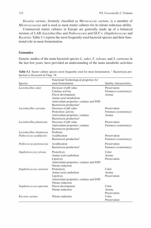
132 P.S. Cocconcelli, C. Fontana
Kocuria varians, formerly classified as Micrococcus varians, is a member ofMicrococcaceae and is used as meat starter cultures for its nitrate reductase ability.
Commercial starter cultures in Europe are generally made up of a balancedmixture of LAB (Lactobacillus and Pediococcus) and GCC+ (Staphylococcus andKocuria). Table 5.1 reports the most frequently used bacterial species and their func-tional role in meat fermentation.
Genomics
Genetic studies of the main bacterial species L. sakei, S. xylosus, and S. carnosus inthe last few years, have provided an understanding of the main metabolic activities
Table 5.1 Starter culture species most frequently used for meat fermentation. ∗ Bacteriocin pro-duction is discussed in Chap. 18.
Functional Technological properties forSpecies meat fermentation Quality characteristics
Lactobacillus sakei Decrease of pH valueCatalase activityFlavor developmentAmino acid metabolismAntioxidant properties: catalase and SODBacteriocin production∗
PreservationFirmness (consistency)Aroma
Lactobacillus curvatus Decrease of pH valueProteolytic activityAntioxidant properties: catalaseBacteriocin production∗
PreservationFirmness (consistency)Aroma
Lactobacillus plantarum Decrease of pH valueAntioxidant properties: catalaseBacteriocin production∗
PreservationFirmness (consistency)
Lactobacillus rhamnosus ProbioticPediococcus acidilactici Acidification
Bacteriocin production∗PreservationFirmness (consistency)
Pediococcus pentosaceus AcidificationBacteriocin production∗
PreservationFirmness (consistency)
Staphylococcus xylosus ProteolysisAmino acid catabolismLipolysisAntioxidant properties: catalase and SODNitrate reduction
ColorAromaPreservation
Staphylococcus carnosus ProteolysisAmino acid catabolismLipolysisAntioxidant properties: catalase and SODNitrate reduction
ColorAromaPreservation
Staphylococcus equorum Flavor developmentNitrate reduction
ColorAromaPreservation
Kocuria varians Nitrate reduction ColorPreservation
5 Characteristics and Applications of Microbial Starters in Meat Fermentations 133
and stress resistance functions. The maximum information was obtained from therecent analysis of the 1.8 Mb genome sequence from L. sakei 23K (Chaillouet al., 2005). This organism has evolved to adapt itself to the meat environment,harboring the genetic function that confers it the ability to grow and survive in themeat environment. L. sakei seems to be very well suited to derive energy from othercompounds that are more abundant in meat. Its adaptation to the meat, an envi-ronment rich in amino acids because of the activity of endogenous proteases, hascaused it to lose biosynthetic pathways for amino acid synthesis. L. sakei is thereforeauxotrophic for all amino acids except aspartic and glutamic acid (Champomier-Verges, Marceau, Mera, & Zagorec, 2002). Amino acid metabolism can provide analternative energy source for L. sakei when glucose is exhausted and this affects thesensory properties of the sausage, as discussed later. The genome shows a particu-larly well-developed potential for amino acid catabolism, and, in addition, L. sakeihas the ability to use purine nucleosides for energy production (a unique propertyamong LAB).
The most common CNS species used as starter cultures are S. carnosus andS. xylosus. The genome sequencing of S. xylosus is ongoing and information canbe obtained at www.cns.fr/externe/English/Projets/Projet NN/NN.html. The size ofS. xylosus strains C2a was estimated to be 2868 kb (Dordet-Frisoni, Talon &.Leroy, 2007) and S. carnosus TM300 strain was estimated by PFGE to be 2590 kb(Wagner, Doskar, & Gotz, 1998). The complete genome sequence of these strainsenabled the exact size of the chromosome to be determined at 2.56 Mb (Rosenstein,Nerz, Resch, & Gotz, 2005).
The chromosome of S. carnosus TM300 comprises 2454 ORFs and the annota-tion of the complete sequence is under way. The first comparative studies showed ahigh conservation in gene order between S. carnosus and other sequenced staphylo-cocci. However, comparison of the gene products revealed that about 20% of themhave no staphylococcal homologue (Rosenstein et al., 2005).
Knowledge of the whole chromosome sequence of S. xylosus will provide fora better understanding of the physiology of this species. A proteomics approachto study cell-envelope proteins of S. xylosus has been developed (Planchon, 2006;Planchon et al., 2007), in which a significant set of cell enveloped proteins canbe recovered. When such information is integrated with future analyses of thetranscripts, a more integrated and comprehensive knowledge of the mechanism bywhich L. sakei and S. xylosus contribute to the fermentation of meat can be obtained,as can how these bacteria interact with each other.
Functional Features
Competitiveness in the Meat Environment
One of the fundamental properties of bacterial starter cultures is their ability tocompete with the adventitious micro-biota of meat, to colonize this environment,and to dominate the microbial community of the fermented products. The starter
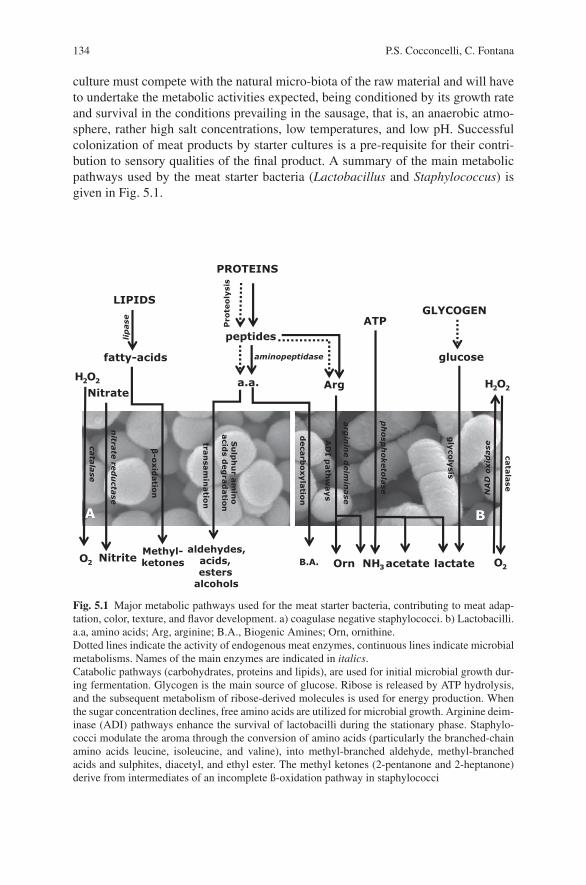
134 P.S. Cocconcelli, C. Fontana
culture must compete with the natural micro-biota of the raw material and will haveto undertake the metabolic activities expected, being conditioned by its growth rateand survival in the conditions prevailing in the sausage, that is, an anaerobic atmo-sphere, rather high salt concentrations, low temperatures, and low pH. Successfulcolonization of meat products by starter cultures is a pre-requisite for their contri-bution to sensory qualities of the final product. A summary of the main metabolicpathways used by the meat starter bacteria (Lactobacillus and Staphylococcus) isgiven in Fig. 5.1.
PROTEINS
LIPIDS
glucose
GLYCOGEN
lactate
ATP
acetate
Arg
Orn NH3B.A.
fatty-acids
A B
peptides
a.a. H2O2
O2
Nitrate
Nitrite
H2O2
O2
cata
lase
nitra
tere
du
ctase
β-o
xid
atio
n
Pro
teo
lysi
s
lip
ase
arg
inin
e d
eim
inase
aminopeptidase
deca
rbo
xyla
tion
tran
sam
inatio
n
Methyl-ketones
aldehydes, acids,esters
alcohols
Su
lph
ur
am
ino
acid
sd
eg
rad
atio
n
gly
coly
sis
ph
osp
ho
keto
lase
cata
lase
NA
D o
xid
ase
AD
I path
ways
Fig. 5.1 Major metabolic pathways used for the meat starter bacteria, contributing to meat adap-tation, color, texture, and flavor development. a) coagulase negative staphylococci. b) Lactobacilli.a.a, amino acids; Arg, arginine; B.A., Biogenic Amines; Orn, ornithine.Dotted lines indicate the activity of endogenous meat enzymes, continuous lines indicate microbialmetabolisms. Names of the main enzymes are indicated in italics.Catabolic pathways (carbohydrates, proteins and lipids), are used for initial microbial growth dur-ing fermentation. Glycogen is the main source of glucose. Ribose is released by ATP hydrolysis,and the subsequent metabolism of ribose-derived molecules is used for energy production. Whenthe sugar concentration declines, free amino acids are utilized for microbial growth. Arginine deim-inase (ADI) pathways enhance the survival of lactobacilli during the stationary phase. Staphylo-cocci modulate the aroma through the conversion of amino acids (particularly the branched-chainamino acids leucine, isoleucine, and valine), into methyl-branched aldehyde, methyl-branchedacids and sulphites, diacetyl, and ethyl ester. The methyl ketones (2-pentanone and 2-heptanone)derive from intermediates of an incomplete ß-oxidation pathway in staphylococci
5 Characteristics and Applications of Microbial Starters in Meat Fermentations 135
Two of the most common preservative conditions employed in meat process-ing are low temperatures and high salt concentrations. L. sakei is remarkably wellequipped to cope with these conditions. It contains several transporters for cryo-and osmoprotective substances and has more cold stress proteins than other lacto-bacilli. L. sakei has psychotropic and osmo-tolerant properties, being able to growat low temperatures and in the presence of up to 10% of sodium chloride (NaCl).These physiological features are associated with the presence, in its genome, of ahigher number of genes coding for stress response proteins, such as cold shock andosmo-tolerance proteins, than found in other lactobacilli.
The genome contains genes encoding a heme-dependent catalase, a superoxidedismutase, and a NADH oxidase, to cope with reactive oxygen species, and there areseveral systems to cope with changes in the redox potential. L. sakei lacks proteinsinvolved in adhesion to the intestinal mucous, but its genome codes for numerousproteins that may be involved in adhesion to the meat surface (e.g., to collagen),aggregation and biofilm formation. Thus, the bacterium seems well equipped toadhere to and spread on a meat surface (Eijsink & Axelsson, 2005).
Sanz and Toldra (2002) reported an arginine-specific aminopeptidase activity inL. sakei that is important for the release of the free amino acid, since it could befurther channeled into the arginine deiminase pathway. The genes encoding theproteins required for arginine catabolism in L. sakei are organized in a cluster(Zurıga, Miralles, & Perez- Martınez, 2002), and their transcription is repressedby glucose and induced by arginine. Arginine, in particular, is an essential aminoacid for L. sakei and it specifically promotes its growth in meat, being used as anenergy source in the absence of glucose (Champomier-Verges et al., 1999). Theconcentration of free arginine in raw meat is low, although it is relatively abundant inmuscle myofibrillar proteins. Moreover, the genome analysis has shown that L. sakeiharbors a second putative arginine deiminase pathway, containing two peptydil-arginine deaminases, enzymes that can contribute to the metabolism of arginineresidues not released from the peptides (Chaillou et al., 2005), thus increasing thecompetitiveness of L . sakei in a meat environment.
Acid Production in Meat Products
Sugars (glucose and occasionally lactose or saccharose) are usually included forthe industrial manufacture of fermented meat products, though in Spain, chorizo istraditionally manufactured with little or no added sugar. During fermentation andripening, LAB converts glucose (their primary energy source) to lactic acid, whichis the main component responsible for the pH decrease. This acidification has apreservative effect due to inhibition of pathogenic and spoilage bacteria that arelittle resistant to low pH, and it contributes to the development of the typical organo-leptic characteristics of the fermented sausages (Bover-Cid, Izquierdo-Pulido, &Vidal-Carou, 2001). Although it is well established that fermentable carbohydrateshave an influence on flavor, texture and yield of the fermented sausages, carbohy-drates for use in dry sausages formulations are generally chosen to ensure an ade-quate initial drop in meat pH (Bacus, 1984; Lucke, 1985) for preservation reasons,
136 P.S. Cocconcelli, C. Fontana
and less importance is given to the product texture. The level of acidification useddepends, however, on the desired sensory properties of the product; lactic acid tasteis preferred in northern European sausages, but not in typical southern Europeanfermented meats. The pH value during ripening is influenced by the meat fermen-tation technology, as are the growth dynamics of Lactobacillus and Staphylococcus(Fig. 5.2). Figure 5.2a shows the growth kinetics of Lactobacillus and Staphylo-coccus and the pH during fermentation of an artisanal sausage without addition ofstarter cultures (Fontana, Cocconcelli, & Vignolo, 2005). A more acidic product canbe obtained using a Lactobacillus starter culture by adding more carbohydrates tothe sausage matrix (Fig. 5.2b), while less acidic products are obtained using a lowerconcentration of glucose and by adding starter cultures were Staphylococcus arepredominant (Fig. 5.2c). The increase of pH in the latter stages of fermentation isrelated to ATP and amino acid metabolism.
Acidification could also be the result of alternative pathways. In L. sakei, thepresence of genes involved in the energetic catabolism of nucleosides, such asadenosine and inosine, is an example of the adaptation of this organism to the meatenvironment. Glucose, the favorite carbon source of L. sakei, is rapidly consumedin meat, while adenosine and inosine are abundant, reaching twice the concentra-tion of glucose. In addition (as shown in Fig. 5.1), L. sakei harbors genes codingfor adenosine deaminase, inosine hydrolase, and nucleoside phosphorylase, all ofwhich enable the release of a ribose moiety from the nucleoside (adenosine andinosine) and its subsequent metabolism (Chaillou et al., 2005). Moreover, the pres-ence of methylglyoxal synthase, a novel genetic trait in LAB, has been proposedas a pathway to counteract frequent glucose starvation and modulate metabolism ofalternative carbon sources (Chaillou et al., 2005).
Catalase Activity
The metabolism of most LAB, such as the adventitious lactobacilli that contam-inate raw meat, could lead to the formation of hydrogen peroxide, a compoundthat interferes with the sensory properties of meat products, being involved in dis-coloration of nitroso- heme-pigments and lipid oxidation. Bacterial strains used inmeat cultures can produce catalase, an antioxidant enzyme that disproportionateshydrogen peroxide to oxygen and water, preventing the risk of a reduced quality ofthe fermented meat. Thus, catalase production is considered a relevant technologicalproperty of the starter cultures for fermented meat products (Leroy, Verluyten, & DeVuyst, 2006). Production of this antioxidant enzyme is a common trait in aerobicbacteria, such as CNS. The characterization of catalase and superoxide dismutasesin S. carnosus and S. xylosus have been reported. The catalase gene, katA, of S.xylosus has been studied in detail (Barriere et al., 2001a; Barriere, Leroy-Setrin, &Talon, 2001b; Barriere, Bruckner, Centeno, & Talon, 2002). Transcriptional activityof this gene is activated and induced upon entry into the stationary phase, by oxy-gen and hydrogen peroxide. Moreover, a second gene coding for heme-dependentcatalase has been detected in S. xylosus.
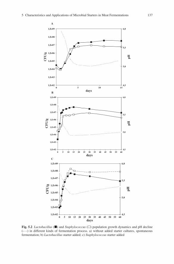
5 Characteristics and Applications of Microbial Starters in Meat Fermentations 137
1,E+02
1,E+03
1,E+04
1,E+05
1,E+06
1,E+07
1,E+08
1,E+09
0 5 10 15
days
CF
U/g
4,5
5,0
5,5
6,0
pH
1,E+02
1,E+03
1,E+04
1,E+05
1,E+06
1,E+07
1,E+08
1,E+09
0 5 10 15 20 25 30 35 40 45 50 55 60
days
CF
U/g
4,5
5,0
5,5
6,0
pH
1,E+02
1,E+03
1,E+04
1,E+05
1,E+06
1,E+07
1,E+08
1,E+09
0 5 10 15 20 25 30 35 40 45 50 55 60days
CF
U/g
4,5
5,0
5,5
6,0
pH
A
B
C
Fig. 5.2 Lactobacillus (�) and Staphylococcus (�) population growth dynamics and pH decline(- - -) in different kinds of fermentation process. a) without added starter cultures, spontaneousfermentation; b) Lactobacillus starter added; c) Staphylococcus starter added
138 P.S. Cocconcelli, C. Fontana
Although LAB have long been considered as catalase negative microorganisms,in the last decade, two groups of catalase activity have been reported in the generaLactobacillus, Pediococcus, and Leuconostoc. The first group is defined as hemecatalase and the second group as non-heme Mn-containing catalase. The presenceof a heme-dependent catalase has been demonstrated in L. plantarum (Igarashi,Kono, & Tanaka, 1996) and L. sakei (Noonpakdeea, Pucharoen, Valyasevi, &Panyim, 1996); it can be active in meat products because these substrates con-tain abundant heme sources (Hertel, Schmidt, Fischer, Oellers, & Hammes, 1998).Moreover, the analysis of the genome of L. sakei revealed that this meat organ-ism harbors systems for the protection against reactive oxygen species, such as Mndependent SOD and heme-dependent catalase (Chaillou et al., 2005).
Nitrate Reduction
Nitrate is added to fermented sausages for its capacity to fix and obtain the typicalcolor of cured products, rather than for its antimicrobial properties. To be effec-tive, the added nitrate must be reduced to nitrite. Besides contributing to flavor,Staphylocuccus and Kocuria also have a role for their nitrate reductase and antioxi-dant activities (Talon, Walter, Chartier, Barriere, & Montel, 1999; Talon, Deliere, &Bertrand, 2002). These microorganisms reduce nitrate to nitrite, which is importantfor the formation of nitrosylmyoglobin, the compound responsible for the charac-teristic red color of fermented meats. The nitrate reductase activity is widespread inCNS; it has been detected in S. xylosus, S. carnosus, S. epidermidis, S. equorum,S. lentus, and S. simulans (Talon et al., 1999; Mauriello, Casaburi, Blaiotta, &Villani, 2004). In S. carnosus, the molecular genetic determinants for nitrogen regu-lation, the nreABC genes, were identified and shown to link the nitrate reductaseoperon (narGHJI) and the putative nitrate transporter gene narT. The data pro-vide evidence for a global regulatory system with oxygen as the effector molecule(Fedtke, Kamps, Krismer, & Gotz, 2002).
Flavor Formation
The flavor and aroma of fermented meats is a combination of several elements,such as batter ingredients, manufacture technology, activity of tissue enzymes, pro-teases and lipases, and microbial metabolism. The bacterial community contributesto flavor development through carbohydrate dissimilation, peptide metabolism, andlipolysis. LAB produce lactic acid and small amounts of acetic acid, ethanol andacetoin, and together with staphylococci are involved in peptide and amino acidmetabolism, while only CNS show an effective metabolism of lipids.
Even though microbial proteolytic activity is generally low in the conditionsfound in fermented sausages (Kenneally, Fransen, Grau, O’Neill, & Arendt, 1999),a minor, strain-dependent activity may still partly contribute to the initial proteinbreakdown (Molly et al., 1997; Sanz et al., 1999; Fadda et al., 1999a, 1999b; Fadda,Oliver, & Vignolo, 2002). It has been shown that several Lactobacillus spp. exhibit
5 Characteristics and Applications of Microbial Starters in Meat Fermentations 139
proteolytic activity on porcine muscle myofibrillar and sarcoplasmic proteins. Faddaet al. (2001a) reported the contribution of curing conditions to the generation ofhydrophilic peptides and free amino acids by the proteolytic activity of L. curvatusCRL 705. Moreover, it has been demonstrated that L. sakei plays an important rolein amino acid generation (Fadda et al., 1999a, 1999b; Sanz et al., 1999).
To ensure the sensory quality of fermented sausages, the contribution of theproteolytic and lipolytic activities of staphylococci is fundamental. The volatilesso far recognized as being produced by staphylococci are primarily amino acidcatabolites, pyruvate metabolites, and methylketones from incomplete β-oxidationof fatty acids (Stahnke, Holck, Jensen, Nilsen, & Zanardi, 2002). In particular,S. xylosus and S. carnosus modulate the aroma through the conversion of aminoacids, particularly the branched-chain amino acids (BCAA: leucine, isoleucine, andvaline). The BCAA can be degraded into methyl-branched aldehydes, alcohols, andacids by S. xylosus and S. carnosus (Beck, Hansen, & Lauritsen, 2002; Larrouture,Ardaillon, Pepin, & Montel, 2000; Vergnais, Masson, Montel, Berdague’, &Talon, 1998). Furthermore, addition of the S. carnosus starter culture has beenshown to decrease the maturation time of Italian dried sausages by more than2 weeks (Stahnke et al., 2002). Olesen, Strunge Meyer, and Stahnke (2004)reported that curing conditions had a considerable influence on the development ofvolatile compounds in sausages. In addition, major differences were observed in thedevelopment of volatile compounds depending on whether S. xylosus or S. carnosuswere used as starter culture.
Bacteriocin and Biopreservation
In recent years, there has been a considerable increase in studies of naturalantimicrobial compounds on and in the food produced by LAB, referred to asbio-protective cultures. Bio-protective cultures may act as starter cultures in foodfermentation processes, such as dry sausage manufacturing, or they may protectfoods from any detrimental organo-leptic changes.
The ability to produce different antimicrobial compounds, such as bacteriocinsand/or low-molecular mass antimicrobial compounds may be one of the criticalcharacteristics for effective competitive exclusion. As mentioned above, one of themain roles of meat LAB starter cultures is the rapid production of organics acids;this inhibits the growth of unwanted biota and enhances product safety and shelf-life. The production of bacteriocins, one of the most promising technological fea-tures of starter cultures, is discussed in Chap. 18.
Probiotics
Foods which have health benefits beyond their nutritional content (functional foods),and particularly foods containing probiotics, are products that are growing in pop-ularity. Probiotics are available as dietary supplements or they may be incorporateddirectly into foods. They are live microorganisms that when administered in ade-quate amounts confer a health benefit on the host (FAO, 2006); they are added to
140 P.S. Cocconcelli, C. Fontana
a variety of foods. Recently, attention has been directed to the use of fermentedsausages as a food carrier because these products could contain high numbersof viable LAB. To use probiotics as starter cultures for fermented sausages, inaddition to the demonstrated probiotic features (FAO, 2006), other properties aredemanded.
Although dairy products are the most commonly used food vehicles for deliveryof probiotics, the future of dry fermented sausages in this field has been termed“promising” (Incze, 1998). The probiotic culture should be well-adapted to the con-ditions of the fermented sausage, in order to dominate in the final product, com-peting with other bacterial populations from meat and from the starter culture. Inaddition, the culture should not develop off-flavors in the final product.
The potential for dry fermented sausages to serve as a vehicle for probioticorganisms has been comprehensively reviewed by Tyopponen, Petaja, & Mattila-Sandholm (2003). Most of the studies discussed in this review relied on thefermentative abilities of the probiotic organisms used, so the selection of probi-otics was limited to organisms that were capable of fermenting carbohydrate inmeat.
Various studies have shown that probiotic organisms have poor survival infermented foods such as yoghurt, fermented milks, and dry fermented sausages(Kailasapathy & Rybka, 1997; Shah 2000; Shah & Ravula, 2000; Lucke, 2000;Erkkila, Suihko, Eerola, Petaja, & Mattila-Sandholm, et al., 2001). Dry fermentedsausages with their low aw and pH, plus curing salts and competing organisms,would seem to present a challenging environment for the survival of probioticsduring processing. Kearney, Upton, and McLoughlin (1990) was the first to reportthe use of micro-encapsulation in alginate to protect the starter cultures duringmeat fermentation. Recently, Muthukumarasamy and Holley (2006) used micro-encapsulation technology as a means to protect a recognized probiotic organism(L. reuteri), from the harsh environment during sausage processing. Based on theirresults the authors suggest that micro-encapsulation may be an option for formu-lation of fermented meat products with viable heath-promoting bacteria. Anotherapproach for selecting bio-protective and probiotic cultures for use in dry fermentedsausages involves the isolation of LAB which possesses acid and bile resistancefrom finished products (Papamanoli et al., 2003; Pennacchia et al., 2004). Thisapproach requires an extensive study of the isolates for other beneficial properties,such as intestinal colonization potential and inhibitory activity against pathogenicbacteria.
Commercial probiotic cultures, such as strains L. rhamnosus GG, L. rhamnosusLC-705, L. rhamnosus E-97800, and L. plantarum E-98098, have been tested asfunctional starter culture strains in northern European sausage fermentation withoutnegatively affecting the technological or sensory properties, with the exception ofL. rhamnosus LC- 705 (Erkkila et al., 2001). Klingberg, Axelsson, Naterstad, Elsser,and Budde (2005) identified L. plantarum and L. pentosus strains, originating fromthe dominant NSLAB of fermented meat products, as promising candidates of pro-biotic meat starter cultures, suitable for the manufacture of the Scandinavian-typefermented sausage.
5 Characteristics and Applications of Microbial Starters in Meat Fermentations 141
Safety of Selected Bacterial Starter Cultures
Biogenic Amines
The presence in food of biogenic amines (BA), such as cadaverine, putrescine,spermidine, histamine, phenethylamine, agmatine, and tyramine, is of concernfor health because their biological effect can lead to toxicological symptoms,such as pseudo-allergic reactions, histaminic intoxication, and interaction withdrugs (Shalaby, 1996). Excessive consumption of these amines could cause ner-vous, gastric, intestinal, and blood pressure problems (Suzzi & Gardini, 2003).Nowadays, increasing attention is given to BA because of the growing number ofconsumers who are sensitive to them; in such people the action of amine oxidases,the enzymes involved in the detoxification of these substances, is deficient (Suzzi &Gardini, 2003). High levels of BA, especially tyramine, and also histamine and thediamines putrescine and cadaverine, have been described in fermented sausages(Hernandez-Jover, Izquierdo-Pulido, Veciana-Noguez, Marine-Font, & Vidal-Carou, 1997a, 1997b; Bover-Cid, Hugas, Izquierdo-Pulido, & Vidal-Carou, 2000a;Bover-Cid, Izquierdo-Pulido, Vidal-Carou, 2000b).
The accumulation of BA in foods requires the presence of amino acids precur-sors, microorganisms with amino acid decarboxylase activity, and favorable condi-tions for growth and decarboxylation. The large quantities of protein present and theproteolytic activity found during ripening of meat products provide the precursorsfor later decarboxylase reactions performed by both the starter cultures and wildmicro-biota (Suzzi & Gardini, 2003; Komprda et al., 2004). Many LAB from meatand meat products can decarboxylate amino acids (Bover-Cid & Holzapfel, 1999).Many studies have reported a significant correlation between pH and BA con-tents, the lowest pH generally being characterized by the highest amine levels(Vandekerckove, 1977; Eitenmiller, Koehler, & Reagan, 1978; Halasz, BaraAth,Simon-Sarkadi, & Holzapfel, 1994; Bover-Cid, Schoppen, Izquierdo-Pulido, &Vidal-Carou, 1999; Parente et al., 2001), according to the hypothesis that BA pro-duction could be a protective mechanism for micro-organisms against acidic envi-ronmental conditions.
The final BA contents in fermented sausages depend on the microbial composi-tion of meat used as raw material, but also on the type and activity of the inocu-lated starter culture. Most strains of L. curvatus, one of the main species used as astarter in sausage production, are associated with high BA production (Bover-Cid &Holzapfel, 1999; Pereira, Crespo, & Romao, 2001).
The use of starter cultures with negative decarboxylase activity was shownto prevent the growth of BA producers and lead to end-products nearly free ofBA, as long as the raw material was of sufficient quality. Several papers havereported on the ability of a selected starter culture (L. sakei CTC494) to greatlyreduce BA accumulation in fermented sausages (Bover-Cid et al., 2001; Gonzalez-Fernandez, Santos, Jaime, & Rovira, 2003). This negative-decarboxylate strain,can decrease the pH quickly during the fermentation step and be predominantthroughout the process, thus preventing the growth of bacteria that can produce BA.
142 P.S. Cocconcelli, C. Fontana
The introduction of starter strains that possess amine oxidase activity might bea way of further decreasing the amount of BA produced during meat fermenta-tion (Martuscelli, Crudele, Gardini, & Suzzi, 2000; Fadda et al., 2001b; Gardini,Matruscelli, Crudele, Paparella, & Suzzi, 2002; Suzzi & Gardini, 2003).
Antibiotic Resistance
The safety of bacterial strains intentionally added to foods, such as starter culturesused for meat products, is becoming an issue for their application in food. Althoughmeat starter cultures have a long history of apparent safe use, safety concerns can beassociated with LAB and, more frequently, with CNS. A risk factor potentially asso-ciated with all bacterial groups used as starter cultures for sausage is the presenceof acquired genes for antimicrobial resistance.
The food chain has been recognized as one of the main routes for the trans-mission of antibiotic resistant bacteria between animal and human populations(Witte, 2000). The European Food Safety Authority has recently concluded thatbacteria used for or in feed production might pose a risk to human and animal healthbecause of carrying acquired resistance genes (EFSA, 2007). Fermented meats thatare not heat-treated before consumption provide a vehicle for such bacteria, andcan act as a direct link between the indigenous micro-flora of animals and thehuman GIT.
Several studies have reported antibiotic resistance in LAB from meats andmeat products; a few strains involved in sausage fermentation such as L. sakei,L. curvatus, and L. plantarum have been found to show such resistance (Geverset al., 2003; Holley & Blaszyk, 1997; Teuber & Perreten, 2000). Some geneticdeterminants such as chloramphenicol acetyltransferase (cat-TC), erythromycinermB, and tetracycline, tetL, tetM, tetS resistance genes have been identified, sug-gesting that horizontal gene transfer may have occurred (Ahn, Collins-Thompson,Duncan, & Stiles, 1992; Gevers et al., 2003; Lin, Fung, Wu, & Chung, 1996; Tan-nock et al., 1994; Ammor et al., 2008)
Antimicrobial resistance in CNS has been studied in detail due to its clinicalrelevance. These bacteria display a high prevalence of antibiotic resistance (Agvald-Ohman, Lund, & Edlun, 2004), and can constitute reservoirs of antibiotic resistancegenes that can be transferred to other staphylococci (Wielders et al., 2001). Antibi-otic resistant strains were found in food (Gardini, Tofalo, & Suzzi, 2003; Martinet al., 2006) and genes for antimicrobial resistance to tetracycline, tet(M), tet(K),to erythromycin, ermB and ermC, and to β-lactams (blaZ and mecA), has beendetected in the CNS isolated from fermented meat. Moreover, S. xylosus strains,isolated from poultry infections, were found to be resistant to streptogramins, har-boring the vatB and the vgaB genes.
An additional concern is that, even in the absence of selective pressure, mobilegenetic elements carrying antibiotic resistance can be transferred at a high fre-quency among the microbial community during sausage fermentation (Vogel,Becke-Schmid, Entgens, Gaier, & Hammes, 1992; Cocconcelli, Cattivelli, &Gazzola, 2003).
5 Characteristics and Applications of Microbial Starters in Meat Fermentations 143
Toxigenic Potential
Some members of the CNS group, primarily S. epidermidis, are common nosocomialpathogens and the presence of regulatory elements, involved in the control of the viru-lence factor synthesis, have been recently identified. Remarkably, strains of S. xylosuswere isolated from patients, who had an underlying disease. The same species hasbeen reported to be involved in infections of poultry (Aarestrup et al., 2000).
Although CNS of food origin have not been found to produce nosocomial infec-tions, some strains that produced enterotoxins have been described (Balaban andRasooly, 2000). Vernozy-Rozand et al. (1996) reported enterotoxin E as the mostfrequent enterotoxin found in S. equorum and S. xylosus, though Martin et al.(2006), reported that the occurrence of staphylococcal enterotoxin genes in CNSfrom slightly fermented sausages was rare, detecting only entC in S. epidermidis.The coagulase positive species, S. intermedius, and S. aureus, have been more fre-quently related to staphylococcal intoxication till date. Moreover, S. intermedius areopportunistic pathogens in implant infections, due to their ability to form a bio-filmon prosthetic materials.
The S. xylosus and S, carnosus strains, currently used as starter cultures or iso-lated from fermented meat products, generally lack of toxic genes. Absence of genescoding for staphylococcal enterotoxins or enterotoxin like superantigens is a quali-fication required for the CNS strains selected for use as starter culture.
Conclusion
The performance of a selected starter culture should be seen in the context of itsapplication, since functionality will depend on the type of sausage, the technologyapplied, the ripening time, and the ingredients and raw materials used. Besides, thefocus on functional characteristics of potential new starter cultures, negative aspectssuch as antibiotic resistance, virulence genes, and undesirable metabolite formationshould not be overlooked. The control of antibiotic resistance, for instance, is a topicof importance because the high load of endogenous bacteria in raw meat materialand the inoculation with starters may represent a problem with regard to the spread-ing of antibiotic resistance.
References
Aarestrup, F. M., Agersø, Y., Ahrens, P., Østergaard Jørgensen, J. C., Madsen, M., & BogøJensen, L. (2000). Antimicrobial susceptibility and presence of resistance genes in staphylo-cocci from poultry. Veterinary Microbiology, 74, 353–364
Agvald-Ohman, C., Lund, B., & Edlun, C. (2004). Multiresistant coagulase-negative staphylococcidisseminate frequently between intubated patient into a multidisciplinary intensive care unit.Critical Care, 8, 42–47.
Ahn, C., Collins-Thompson, D., Duncan, C., & Stiles, M. E. (1992). Mobilization and location ofthe genetic determinant of chloramphenicol resistance from Lactobacillus plantarum caTC2R.Plasmid, 27, 169–176.

144 P.S. Cocconcelli, C. Fontana
Ammor, M. S., Gueimonde, M., Danielsen, M., Zagorec, M., van Hoek, A. H., de Los Reyes-Gavilan, C. G., et al. (2008). Two different tetracycline resistance mechanisms, plasmid-carried tet(L) and chromosomally-encoded transposon-associated tet(M), coexist in Lactobacil-lus sakei Rits 9. Applied Environmental Microbiology (Epub ahead of print).
Axelsson, L. (2004). Lactic acid bacteria: Classification and physiology. In S. Salminen, A. Ouwe-hand, & A. Von Wright (Eds.), Lactic acid bacteria: Microbiology and functional aspects (3rded.). New York: Marcel Dekker, Inc.
Bacus, J. (1984). Utilization of microorganisms in meat processing: A handbook for meat plantoperators. Research Studies, Letchworth (UK).
Balaban, N., & Rasooly, A. (2000). Staphylococcal enterotoxins. International Journal of FoodMicrobiology, 61, 1–10.
Bannerman, T. L. (2003). Staphylococcus, Micrococcus, and other catalase-positive cocci thatgrow aerobically. In P. R. Murray, E. J. Baron, J. H. Jorgensen, M. A. Pfaller, R. H. Yolken(Eds.), Manual of Clinical Microbiology (Vol. 5, pp. 384–404). Washington: American SocietyMicrobiology.
Barriere, C., Bruckner, R., Centeno, D., & Talon, R. (2002). Characterization of the katA geneencoding a catalase and evidence for at least a second catalase activity in Staphylococcus xylo-sus, bacteria used in food fermentation. FEMS Microbiology Letters, 216(2), 277–283.
Barriere, C., Centeno, D., Lebert, A., Leroy-Setrin, S., Berdague’, J., & Talon, R. (2001a). Roles ofsuperoxide dismutase and catalase of Staphylococcus xylosus in the inhibition of linoleic acidoxidation. FEMS Microbiology Letters, 201, 181–185.
Barriere, C., Leroy-Setrin, S., & Talon, R. (2001b). Characterization of catalase and superox-ide dismutase in Staphylococcus carnosus 833 strain. Journal of Applied Microbiology, 91,514–519.
Beck, H., Hansen, A., & Lauritsen, F. (2002). Metabolite production and kinetics of branched-chainaldehyde oxidation in Staphylococcus xylosus. Enzyme and Microbial Technology, 31, 94–101.
Bover-Cid, S., Izquierdo-Pulido, M., & Vidal-Carou, M. (2001). Effectiveness of a Lactobacillussakei starter culture in the reduction of biogenic amine accumulation as a function of the rawmaterial quality. Journal of Food Protection, 64, 367–373.
Bover-Cid, S., Hugas, M., Izquierdo-Pulido, M., & Vidal-Carou, M. (2000a). Reduction of bio-genic amine formation using a negative amino acid-decarboxylase starter culture for fermenta-tion of “Fuet” sausages. Journal of Food Protection, 63, 237–243.
Bover-Cid, S., Izquierdo-Pulido, M., & Vidal-Carou, M. (2000b). Mixed starter cultures to con-trol biogenic amine production in dry fermented sausages. Journal of Food Protection, 63,1556–1562.
Bover-Cid, S., & Holzapfel, W. H. (1999). Improved screening procedure for biogenic amine pro-duction by lactic acid bacteria. International Journal of Food Microbiology, 53, 33–41.
Bover-Cid, S., Schoppen, S., Izquierdo-Pulido, M., & Vidal-Carou, M. C. (1999). Relationshipbetween biogenic amine contents and the size of dry fermented sausages. Meat Science 51,305–311.
Buckenhuskes, H. J. (1994). Bacterial starter cultures for fermented sausages. Meat Focus Interna-tional, 12, 497–500.
Chaillou, S., Champomier-Verges, M. C., Cornet, M., Crutz-Le Coq, A. M., Dudez, A. M., Mar-tin, V., et al. (2005). The complete genome sequence of the meat-borne lactic acid bacteriumLactobacillus sakei 23K. Nature Biotechnology, 23, 1527–1533.
Champomier-Verges, M. C., Marceau, A., Mera, T., & Zagorec, M. (2002). The pepR gene ofLactobacillus sakei is positively regulated by anaerobiosis at the transcriptional level. Appliedand Environmental Microbiology, 68(8), 3873–3877.
Champomier-Verges, M. C., Zuniga, M, Morel-Deville, F., Perez-Martınez, G., Zagorec, M., &Ehrlich, S. D. (1999). Relationships between arginine degradation, pH and survival in Lacto-bacillus sakei. FEMS Microbiology Letters, 180, 297–304.
Cocconcelli, P. S., Cattivelli, D., & Gazzola, S. (2003). Gene transfer of vancomycin and tetracy-cline resistances among Enterococcus faecalis during cheese and sausage fermentation. Inter-national Journal of Food Microbiology, 88, 315–323.
5 Characteristics and Applications of Microbial Starters in Meat Fermentations 145
Dordet-Frisoni, E., Talon R., &. Leroy, S. (2007). Physical and genetic map of the Staphylococcusxylosus C2a chromosome. FEMS Microbiology Letters, 266(2), 184–193.
EFSA The EFSA Journal. (2007). 587, 1–16 c© European Food Safety Authority, 2007 Introduc-tion of a Qualified Presumption of Safety (QPS) approach for assessment of selected microor-ganisms referred to EFSA.
Eijsink, V., & Axelsson, L. (2005). Bacterial lessons in sausages making. Nature Biotechnology,23(12), 1494–1495.
Eitenmiller, R. R., Koehler, P. E., & Reagan, J. O. (1978). Tyramine in fermented sausages: fac-tors affecting formation of tyramine and tyrosine decarboxylase. Journal of Food Science, 43,689–693.
Erkkila, S., Suihko, M. L., Eerola, S., Petaja, E., & Mattila-Sandholm, T. (2001). Dry sausagefermented by Lactobacillus rhamnosus strains. International Journal of Food Microbiology,64, 205–210.
Fadda, S., Oliver, G., & Vignolo, G. (2002). Protein degradation by Lactobacillus plantarum andLactobacillus casei in a sausage model system. Journal of Food Science, 67, 1179–1183.
Fadda, S., Vignolo, G., Aristoy, M. C., Oliver, G., & Toldra, F. (2001a). Effect of curing condi-tions and Lactobacillus casei CRL705 on the hydrolysis of meat proteins. Journal of AppliedMicrobiology, 91(3), 478–487.
Fadda, S., Vignolo, G., & Oliver, G. (2001b). Tyramine degradation and tyramine/histamine pro-duction by lactic acid bacteria and Kocuria strains. Biotechnology Letters, 23, 2015–2019.
Fadda, S., Sanz, Y., Vignolo, G., Aristoy, M. C., Oliver, G., & Toldra, F. (1999a). Hydrolysis ofpork muscle sarcoplasmic proteins by Lactobacillus curvatus and Lactobacillus sakei. Appliedand Environmental Microbiology, 65, 578–584.
Fadda, S., Sanz, Y., Vignolo, G., Aristoy, M. C., Oliver, G., & Toldra, F. (1999b). Characterizationof pork muscle protein hydrolysis caused by Lactobacillus plantarum. Applied and Environ-mental Microbiology, 65, 3540–3546.
FAO. (2006). Probiotics in Food. Health and nutritional properties and guidelines for evaluation.FAO food and nutrition paper. 85.
Fedtke, I., Kamps, A., Krismer, B., & Gotz, F. (2002). The nitrate reductase and nitrite reductaseoperons and the narT gene of Staphylococcus carnosus are positively controlled by the noveltwo-component system NreBC. Journal of Bacteriology, 184(23), 6624–6634.
Fontana, C., Cocconcelli, P. S., & Vignolo, G. (2005). Monitoring the bacterial population dynam-ics during fermentation of artisanal Argentinean sausages. International Journal of Food Micro-biology, 103, 131–142.
Gardini, F., Tofalo, R., & Suzzi, G. (2003). A survey of antibiotic resistance in Micrococcaceaeisolated from Italian dry fermented sausages. Journal of Food Protection, 66, 937–945.
Gardini, F., Matruscelli, M., Crudele, M. A., Paparella, A., & Suzzi, G. (2002). Use of Staphylo-coccus xylosus as a starter culture in dried sausages: effect on biogenic amine content. MeatScience, 61, 275– 283.
Garrity, G. M., Bell, J. A. & Lilburn, T. G. (2004). Taxonomic Outline of the Prokaryotes.InBergey’s Manual of Systematic Bacteriology, 2nd edn. Release 5.0, May 2004. Springer-Verlag,NY.
Gevers, D., Masco, L., Baert, L., Huys, G., Debevere, J., & Swings, J. (2003). Prevalence anddiversity of tetracycline resistant lactic acid bacteria and their tet genes along the process lineof fermented dry sausages. Systematic and Applied Microbiology, 26(2), 277–283.
Ghebremedhin, B., Layer, F., Konig, W. & Konig, B. (2008). Genetic classification and distin-guishing of Staphylococcus species based on different partial gene sequences: gap, 16S rRNA,hsp60, rpoB, sodA, and tuf gene. Journal of Clinical Microbiology (In press).
Gonzalez-Fernandez, C., Santos, E., Jaime, I., & Rovira, J. (2003). Influence of starter cultures andsugar concentrations on biogenic amine contents in chorizo dry sausage. Food Microbiology,20, 275–284.
Halasz, A., BaraAth, A., Simon-Sarkadi, L. & Holzapfel, W. (1994). Biogenic amines and theirproduction by micro-organisms in food. Trends in Food Science and Technology 5, 42±48.
146 P.S. Cocconcelli, C. Fontana
Hammes, W. P., Bantleon, A., & Min, S. (1990). Lactic acid bacteria in meat fermentation. FEMSMicrobiology Letters, 87, 165–173.
Hernandez-Jover, T., Izquierdo-Pulido, M., Veciana-Noguez, M., Marine-Font, A., & Vidal-Carou,M. (1997a). Biogenic amine and polyamine contents in meat and meat products. Journal ofAgricultural Food Chemistry, 45, 2098–2102.
Hernandez-Jover, T., Izquierdo-Pulido, M., Veciana-Noguez, M., Marine-Font, A., & Vidal-Carou,M. (1997b). Effect of starters cultures on biogenic amine formation during sausages production.Journal of Food Protection, 60, 825–830.
Hertel, C., Schmidt, G., Fischer, M., Oellers, K., & Hammes, W. P. (1998). Oxygen-dependentregulation of the expression of the catalase gene katA of Lactobacillus sakei LTH677 Appliedand Environmental Microbiology, 64(4), 1359–1365.
Holley, R. A., & Blaszyk, M. (1997). Antibiotic challenge of meat starter cultures and effects uponfermentations. Food Research International, 30, 513–522.
Igarashi, T., Kono, Y., & Tanaka, K. (1996). Molecular cloning of manganese catalase from Lac-tobacillus plantarum. Journal of Biological Chemistry, 271, 29521–29524.
Incze, K. (1998). Dry fermented sausages. Meat Science 49 (Suppl 1) S169-S177.Kailasapathy, K., & Rybka, S. (1997). L. acidophilus and Bifidobacterium spp.—their therapeutic
potential and survival in yogurt. The Australian Journal of Dairy Technology, 52, 28–33.Kearney, L., Upton, M., & McLoughlin, A. (1990). Meat fermentations with immobilized lactic
acid bacteria. Applied Microbiology and Biotechnology, 33, 648–651.Kenneally, M., Fransen, G., Grau, H., O’Neill, E., & Arendt, K. (1999). Effects of environmen-
tal conditions on microbial proteolysis in a pork myofibril model system. Journal of AppliedMicrobiology, 87, 794–803.
Kleerebezem, M., Boekhorst, J., van Kranenburg, R., Molenaar, D., Kuipers, O. P., Leer, R., et al.(2003). Complete genome sequence of Lactobacillus plantarum WCFS1. Proceedings of theNational Academy of Sciences, U S A. 100, 1990–1995.
Klingberg, D., Axelsson, L., Naterstad, K., Elsser, D., & Budde, B. (2005). Identification of poten-tial probiotic starter cultures for Scandinavian-type fermented sausages. International Journalof Food Microbiology, 105, 419–431.
Komprda, T., Smela, D., Pechova’, P., Kalhotka, L., Stencl, J., & Klejdus, B. (2004). Effect ofstarter culture, spice mix and storage time and temperature on biogenic amine content of dryfermented sausages. Meat Science 67, 607– 616.
Kuipers, P., Buist, G., & Kok, J. (2000). Current strategies for improving food bacteria. Researchin Microbiology, 151, 815–822.
Kwok, A. Y., & Chow, A. W. (2003). Phylogenetic study of Staphylococcus and Macrococcusspecies based on partial hsp60 gene sequences. International Journal of Systematic Evolution-ary Microbiology, 53, 87–92.
Larrouture, C., Ardaillon, V., Pepin, M., & Montel, C. (2000). Ability of meat starter cultures tocatabolize leucine and evaluation of the degradation products by using an HPLC method. FoodMicrobiology, 17, 563–570.
Leroy, F., Verluyten, J., & De Vuyst, L. (2006). Functional meat starter cultures for improvedsausage fermentation. International Journal of Food Microbiology, 106, 270–285.
Lin, C. F., Fung, Z. F., Wu, C. L., & Chung, T. C. (1996). Molecular characterization of a plasmid-borne (pTC82) chloramphenicol resistance determinant (cat-TC) from Lactobacillus reuteriG4. Plasmid, 36, 116–124.
Lucke, K. (2000). Utilization of microbes to process and preserve meat. Meat Science, 56,105–115.
Lucke, K. (1985). Fermented sausages. In B. J. Wood (Ed.), Microbiology of fermented foods(pp. 41–83). London: Elsevier.
Martuscelli, M., Crudele, A., Gardini, F., & Suzzi, G. (2000). Biogenic amine formation andoxidation by Staphylococcus xylosus from artisanal fermented sausages. Letters and AppliedMicrobiology, 31, 228–232.
Mauriello, G., Casaburi, A., Blaiotta, G., & Villani, F. (2004). Isolation and technological prop-erties of coagulase negative staphylococci from fermented sausages of Southern Italy. MeatScience, 67, 49–158.
5 Characteristics and Applications of Microbial Starters in Meat Fermentations 147
Molly, K., Demeyer, D., Johansson, G., Raemaekers, M., Ghistelinck, M., & Geenen, I. (1997).The importance of meat enzymes in ripening and flavour generation in dry fermented sausages.First results of a European project Food Chemistry, 59(4), 539–545.
Muthukumarasamy, P., & Holley, R. (2006). Microbiological and sensory quality of dry fermentedsausages containing alginate-microencapsulated Lactobacillus reuteri. International Journal ofFood Microbiology, 111, 164–169
Niinivaara, F. (1955). Uber den Einfluss von Bacterienreinkulturen auf die Reifung und Umrotungder Rohwurst. In Acta Agralia Fennica, 84, 1–128.
Noonpakdeea, W., Pucharoen, K., Valyasevi, R., & Panyim, S. (1996). Molecular cloning, DNAsequencing and expression of catalase gene of Lactobacillus sake SR911. Journal of MolecularBiology and Biotechnology, 4, 229–235.
Olesen, P., Strunge Meyer, A., & Stahnke, L. (2004). Generation of flavour compounds in fer-mented sausages—the influence of curing ingredients, Staphylococcus starter culture and ripen-ing time. Meat Science, 66, 675–687.
Papamanoli, E., Tzanetakis, N., Litopoulou-Tzanetaki, E., & Kotzekidou, P. (2003). Characteri-zation of lactic acid bacteria isolated from a Greek dry fermented sausage in respect of theirtechnological and probiotic properties. Meat Science, 65, 859–867.
Parente, E., Martuscelli, M., Gardini, F., Grieco, S., Crudele, M. A., & Suzzi, G. (2001). Evolutionof microbial populations and biogenic amine production in dry sausages produced in SouthernItaly. Journal of Applied Microbiology, 90 (6), 882–891.
Pennacchia, C., Ercolini, D., Blaiotta, G., Pepe, O., Mauriello, G., & Villani, F. (2004). Selectionof Lactobacillus strains from fermented sausages for their potential use as probiotics. MeatScience, 67, 309–317.
Pereira, C. I., Barreto Crespo, M. T., & San Romao, M. V. (2001). Evidence for proteolytic activityand biogenic amines production in Lactobacillus curvatus and L. homohiochii. InternationalJournal of Food Microbiology 68(3), 211–216.
Place, R., Hiestand, D., Gallmann, H. R., & Teuber, M. (2003). Staphylococcus equorum subsp.linens, subsp. nov., a starter culture component for surface ripened semi-hard cheese. System-atic Applied Microbiology, 26, 30–37.
Planchon, S., Chambon, C., Desvaux, M., Chafsey, I., Leroy, S., Talon, R., et al. (2007). Proteomicanalysis of cell envelope proteins from Staphylococcus xylosus C2a. Journal of ProteomicsResearch, Submitted.
Planchon, S. (2006). Aptitude de Staphylococcus carnosus et Staphylococcus xylosus a former desbiofilms- Etude d’une souche biofilm positif par une approuche proteomique. These de Docteurde l’Universite Blaise Pascal, Clemont Ferrand II.
Rosenstein, R., Nerz, C., Resch, A., & Gotz, F. (2005). Comparative genome analysis of staphylo-coccal species. 2nd European Conference on prokaryotic genomes. Prokagen.
Sanz, Y., & Toldra, F. (2002). Purification and characterization of an arginine aminopeptidase fromLactobacillus sakei. Applied and Environmental Microbiology, 68(4), 1980–1987.
Sanz, Y., Fadda, S., Vignolo, G., Aristoy, C., Oliver, G., & Toldra, F. (1999). Hydrolysis of musclemyofibrillar proteins by Lactobacillus curvatus and Lactobacillus sakei. International Journalof Food Microbiology, 53, 115–125.
Shah, N. P. (2000). Probiotic bacteria: Selective enumeration and survival in dairy foods. Journalof Dairy Science, 83(4), 894–907.
Shah, N. P., & Ravula, R. (2000). Microencapsulation of probiotic bacteria and their survival infrozen fermented dairy desserts. Australian Journal of Dairy Technology, 55(3), 139–144.
Shalaby, A. (1996). Significance of biogenic amines to food safety and human health. FoodResearch International, 29(7), 675–690.
Spergser, J., Wieser, M., Taubel, M., Rossello-Mora, R. A., Rosengarten, R., & Busse, H. J. (2003).Staphylococcus nepalensis sp. nov., isolated from goats of the Himalayan region. InternationalJournal of Systematic Evolutionary Microbiology, 53, 2007–2011.
Stahnke, H., Holck, A., Jensen, A., Nilsen, A., & Zanardi,. E. (2002). Maturity acceleration ofItalian dried sausage by Staphylococcus carnosus—relationship between maturity and flavorcompounds. Journal of Food Science, 67, 1914–1921.
148 P.S. Cocconcelli, C. Fontana
Suzzi, G., & Gardini, F. (2003). Biogenic amines in dry fermented sausages: a review. InternationalJournal of Food Microbiology, 88, 41–54.
Talon, D., Deliere, E., & Bertrand, X. (2002). Characterization of methicillin-resistant Staphylo-coccus aureus strains susceptible to tobramycin. International Journal of Antimicrobial Agents,20(3), 174–179.
Talon, R., Walter, D., Chartier, S., Barriere, C., & Montel, M. (1999). Effect of nitrate and incuba-tion conditions on the production of catalase and nitrate reductase by staphylococci. Interna-tional Journal of Food Microbiology, 52, 47–50.
Tannock, G. W., Luchansky, J. B., Miller, L., Connell, H., Thode-Andersen, S., & Mercer, A.(1994). Molecular characterization of a plasmid-borne (pGT633) erythromycin resistancedeterminant (ermGT) from Lactobacillus reuteri 100–63. Plasmid, 31, 60–71.
Teuber, M., & Perreten, V. (2000). Role of milk and meat products as vehicles for antibiotic-resistant bacteria. Acta Veterinaria Scandinavica, (Suppl. 93), discussion 111–7, 75–87.
Tyopponen, S., Petaja, E., & Mattila-Sandholm, T. (2003). Bioprotectives and probiotics for drysausages. International Journal of Food Microbiology, 83, 233–244.
Vandekerckove, P. (1977). Amines in dry fermented sausage: a research note. Journal of FoodScience, 42, 283–285.
Vergnais, L., Masson, F., Montel, M. C., Berdague’, J. L., & Talon, R. (1998). Evaluation of solid-phase microextraction for analysis of volatile metabolites produced by staphylococci. Journalof Agricultural and Food Chemistry, 46, 228–234.
Vernozy-Rozand, C., Mazuy, C., Prevost, G., Lapeyre, C., Bes, M., Brun, Y., et al. (1996). Entero-toxin production by coagulase-negative staphylococci isolated from goat’s milk and cheese.International Journal of Food Microbiology, 30(3), 271–280.
Vogel, R. F., Becke-Schmid, M., Entgens, P., Gaier, W., & Hammes, W. (1992). Plasmid transferand segregation in Lactobacillus curvatus LTH1432 in vitro and during sausage fermentations.Systematic and Applied Microbiology, 15, 129–136.
Wagner, E., Doskar, J., & Gotz, F. (1998). Physical and genetic map of the genome of Staphylo-coccus carnosus TM300. Microbiology, 144, 509–517.
Wielders, C., Vriens, M., Brisse, S., de Graaf-Miltenburg, L., Troelstra, A., Fleer, A., Schmitz, F.,Verhoof, J., & Fluit, A. (2001). Evidence for in vivo transfer of mecA DNA between strains ofStaphylococcus aureus. Lancet, 357, 1674–1675.
Witte, W. (2000). Selective pressure by antibiotic use in livestock. International Journal of Antimi-crobial Agents, 16, S19–S24.
Zurıga, M., Miralles, M., & Perez- Martınez, G. (2002). The product of arcR, the sixth gene of thearc peron of Lactobacillus sakei, is essential for expression of the arginine deiminase pathway.Applied Environmental Microbiology, 68(12), 6051–6058.
Chapter 6Genetics of Lactic Acid Bacteria
Monique Zagorec, Jamila Anba-Mondoloni, Anne-Marie Crutz-Le Coq,and Marie-Christine Champomier-Verges
Introduction
Many meat (or fish) products, obtained by the fermentation of meat originating fromvarious animals by the flora that naturally contaminates it, are part of the humandiet since millenaries. Historically, the use of bacteria as starters for the fermenta-tion of meat, to produce dry sausages, was thus performed empirically through theendogenous micro-biota, then, by a volunteer addition of starters, often performedby back-slopping, without knowing precisely the microbial species involved. It isonly since about 50 years that well defined bacterial cultures have been used asstarters for the fermentation of dry sausages. Nowadays, the indigenous micro-biotaof fermented meat products is well identified, and the literature is rich of reportson the identification of lactic acid bacteria (LAB) present in many traditional fer-mented products from various geographical origin, obtained without the addition ofcommercial starters (See Talon, Leroy, & Lebert, 2007, and references therein). TheLAB species that are naturally present in those products and become dominant inthe final processing steps essentially belong to Lactobacillus sakei, Lactobacilluscurvatus, and Lactobacillus plantarum. These are also the three main species thatare sold as starters for the fermentation of dry sausages, essentially in Europe, towhich should be added the two other pediococci species Pediococcus pentosaceusand Pediococcus acidilactici (Hammes & Hertel, 1998).
Since the last 20 years, many microbiologists have investigated the physiologyof these LAB, in order to understand the mechanisms by which they contributeto the quality of the final product, and to improve their use. Molecular tools weretherefore developed, leading to an increase of the knowledge about their genetics.More recently, the genomes of L. plantarum WCFS1 (Kleerebezem, et al, 2003),L. sakei 23K (Chaillou, et al., 2005), and P. pentosaceus ATCC25745 (Makarova,et al., 2006) were entirely sequenced, giving a general overview on the wholegenetic repertoire of those bacteria. However, the description and analysis of all
M. ZagorecUnite Flore Lactique et Environnement Carne, UR309, INRA, Domaine de Vilvert, F-78350 Jouyen Josas, France
F. Toldra (ed.), Meat Biotechnology, 149C© Springer Science+Business Media, LLC 2008
150 M. Zagorec et al.
the genes composing a genome is a hard and tricky task, and authors usually focuson emblematic characteristics of a species when they aim to describe what are themost important genetic traits encoded by a genome. For instance, the genome ofL. plantarum, a ubiquitous species found in many fermented products but alsopresent in the gastrointestinal tract of mammals and even described as a putativeprobiotic, was searched mostly for the functions explaining its ubiquity. The genomeof P. pentosaceus was compared with that of many other LAB, in order to under-stand evolution and phylogeny of this vast taxonomic group, and finally the L. sakeigenome was analyzed in order to understand its adaptation to the meat environment.
In this chapter, we will present an overview of the genetic elements known forthese species and we will describe, from the three genomes available, some traitsthat are required for the development of fermented meat products and that are com-mon to L. sakei, L. plantarum, and P. pentosaceus. We will consider P. pentosaceusas belonging to lactobacilli, since its genome analysis confirmed that the pediococcishould be included in the lactobacilli genera (Makarova et al., 2006).
Mobile Elements and Plasmids in Meat LAB Starters
These genetic elements can be transferred intra- and inter-species and may largelycontribute to bacterial genetic evolution and to the acquisition of new functions thatwill then influence the ability of the host to adapt or to become more competitive toan ecological niche.
Insertion Sequences and Transposons
Transposable genetic elements are ubiquitous, their presence or absence within agenome can vary between individual strains. Transposable elements have impacts onthe host genomes by altering gene expression, providing genomic rearrangements,causing insertional mutations and are sources of phenotypic variation. Moreover,numerous systems of bacterial transferable elements such as plasmids, transposons,integrative and conjugative or mobilizable elements, insertion sequences (IS), andprophages are known to promote horizontal gene transfer events and DNA rear-rangements. Many of these elements carry accessory genes that encode variousfunctions such as resistance to antibiotics, virulence, and degradation of xenobi-otics (Frost, Leplae, Summers, & Toussaint, 2005). Conjugative elements harbormodules involved in their own conjugative transfer, while mobilizable elementscarry a module which includes an origin of transfer and one or two mobilizationgenes, and require the mating machinery provided by the conjugative elements tobe transferred. IS contain genes involved in transposition usually edged by twoinverted repeat sequences, that allow insertion and excision processes through site-specific recombination. Transposition, performed by transposons or by bacterialIS may lead to the random mutation of chromosomal genes. Twelve IS are found
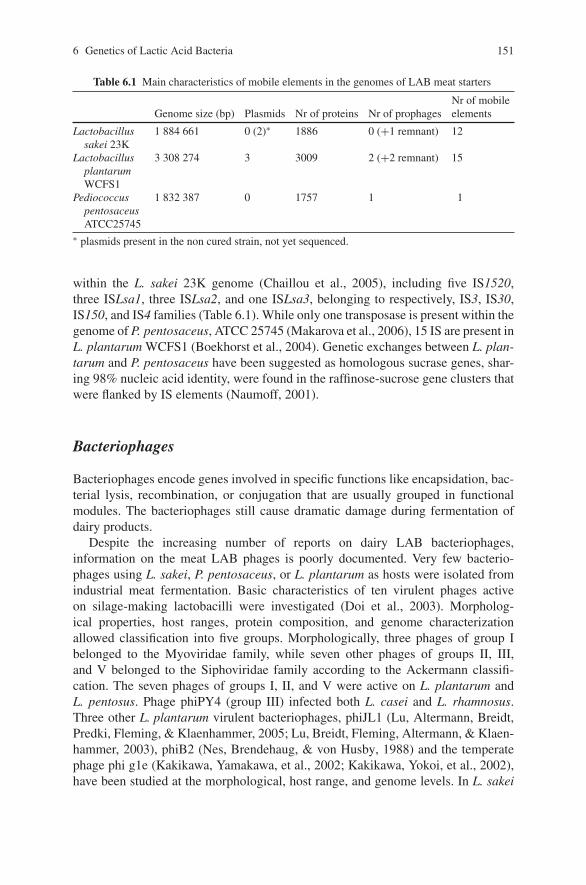
6 Genetics of Lactic Acid Bacteria 151
Table 6.1 Main characteristics of mobile elements in the genomes of LAB meat starters
Genome size (bp) Plasmids Nr of proteins Nr of prophagesNr of mobileelements
Lactobacillussakei 23K
1 884 661 0 (2)∗ 1886 0 (+1 remnant) 12
LactobacillusplantarumWCFS1
3 308 274 3 3009 2 (+2 remnant) 15
PediococcuspentosaceusATCC25745
1 832 387 0 1757 1 1
∗ plasmids present in the non cured strain, not yet sequenced.
within the L. sakei 23K genome (Chaillou et al., 2005), including five IS1520,three ISLsa1, three ISLsa2, and one ISLsa3, belonging to respectively, IS3, IS30,IS150, and IS4 families (Table 6.1). While only one transposase is present within thegenome of P. pentosaceus, ATCC 25745 (Makarova et al., 2006), 15 IS are present inL. plantarum WCFS1 (Boekhorst et al., 2004). Genetic exchanges between L. plan-tarum and P. pentosaceus have been suggested as homologous sucrase genes, shar-ing 98% nucleic acid identity, were found in the raffinose-sucrose gene clusters thatwere flanked by IS elements (Naumoff, 2001).
Bacteriophages
Bacteriophages encode genes involved in specific functions like encapsidation, bac-terial lysis, recombination, or conjugation that are usually grouped in functionalmodules. The bacteriophages still cause dramatic damage during fermentation ofdairy products.
Despite the increasing number of reports on dairy LAB bacteriophages,information on the meat LAB phages is poorly documented. Very few bacterio-phages using L. sakei, P. pentosaceus, or L. plantarum as hosts were isolated fromindustrial meat fermentation. Basic characteristics of ten virulent phages activeon silage-making lactobacilli were investigated (Doi et al., 2003). Morpholog-ical properties, host ranges, protein composition, and genome characterizationallowed classification into five groups. Morphologically, three phages of group Ibelonged to the Myoviridae family, while seven other phages of groups II, III,and V belonged to the Siphoviridae family according to the Ackermann classifi-cation. The seven phages of groups I, II, and V were active on L. plantarum andL. pentosus. Phage phiPY4 (group III) infected both L. casei and L. rhamnosus.Three other L. plantarum virulent bacteriophages, phiJL1 (Lu, Altermann, Breidt,Predki, Fleming, & Klaenhammer, 2005; Lu, Breidt, Fleming, Altermann, & Klaen-hammer, 2003), phiB2 (Nes, Brendehaug, & von Husby, 1988) and the temperatephage phi g1e (Kakikawa, Yamakawa, et al., 2002; Kakikawa, Yokoi, et al., 2002),have been studied at the morphological, host range, and genome levels. In L. sakei
152 M. Zagorec et al.
23K, only one remnant prophage is found in the complete genome (Chaillouet al., 2005). One virulent L. sakei bacteriophage, PWH2, has been isolated fromfermented sausage. Fourteen L . sakei strains and five L. curvatus strains were testedas potential hosts and only strain L. sakei Ls2 was sensitive (Leuschner, Arendt,& Hammes, 1993). These results may explain why no direct proof has ever beenshown for a detrimental role of phage attack in meat fermentations.
The PWH2 bacteriophage belongs to the family of Siphoviridae according tothe Ackermann classification (Ackermann, 1987). In the genome of L. plantarumWCFS1, two entire prophages and two remnants are present (Ventura, et al., 2003).Two prophages were observed in the P. pentosaceus ATCC25745 genome(Table 6.1). Little is known about the mechanism of induction or on the effectof their presence in these genomes except for the three prophages of P. acidilactici,which can be induced by classical addition of mitomycin C (Caldwell, McMahon,Oberg, & Broadbent, 1999). The new bacteriophages, named pa97, pa40, and pa42were characterized morphologically and belong to the family Siphoviridae.
Plasmid Content of the Strains
With the rise of molecular biology of the lactobacilli in the late 1980s, their plasmidshave become a subject of study, as a means to characterize strains, to search forplasmid-borne functions, or to characterize replicons for potential use as cloningvehicles. Although many plasmids have been isolated, the global plasmid content oflactobacilli has been the object of relatively few and ancient systematic studies (for areview see Wang & Lee, 1997). As other species of lactobacilli, L. curvatus, L. plan-tarum, and L. sakei have been reported to commonly contain one to about ten plas-mids, whatever the (i.e. isolated from/material) vegetable, meat, or fermented originof the strains (Liu, Kondo, Barnes, & Bartholomew, 1988; Nes, 1984; Ruiz-Barba,Piard, & Jimenez-Diaz, 1991; Vogel, Lohmann, Weller, Hugas, & Hammes, 1991;West & Warner, 1985). P. pentosaceus and P. acidilactici have also been reportedto commonly harbor plasmids (Graham & McKay, 1985; Perez Pulido et al., 2006).These plasmids are usually circular and their size may range from less than to 2 kb tomore than 60 kb. Mega plasmids, larger than 100 kb have been found in L. plantarum(Wang & Lee, 1997), although it rather seems to be a specific trait of L. salivariusand a few other species of lactobacilli (Li et al., 2007). Linear plasmids among LABhave been described only in L. salivarius to date (Li et al., 2007). Some strains haveno naturally resident plasmid.
Circular plasmids can be classified according to their mode of replication(rolling-circle replication characterized by an intermediate single-stranded stateor theta-type replication) and the need for both host factors and plasmid-bornedeterminants. The latter comprise at least a segment of DNA called the originof replication at which replication initiates and may comprise an additionalprotein called initiator or Rep protein. Usually, newly sequenced plasmids canbe assigned to families by sequence homology. A classification of plasmids has
6 Genetics of Lactic Acid Bacteria 153
been tentatively proposed at the website of the Database of Plasmid Replicon(http://www.essex.ac.uk/bs/staff/osborn/DPR home.htm).
Several plasmids from the five species mentioned above have been isolated andcharacterized. Among those, the two main modes of replication, rolling-circle,and theta replication exist. At the time of writing, the Genbank database containsthe complete sequence for one plasmid of L. curvatus, one plasmid of L. sakei,15 plasmids of L. plantarum, two plasmids of P. pentosaceus, and two plasmidsof P. acidilactici. Their characteristics are given in Table 6.2. One obvious conclu-sion is that plasmids belonging to the same family are distributed across species. Apicture of plasmid distribution can also be brought by hybridization experiments,which detects homology relationships. For example, it is known that L. sakei andL. curvatus strains harbor plasmids related to the pLP1 rolling-circle plasmid, origi-nally isolated from L. plantarum (Bringel, Frey, & Hubert, 1989) or that pLC2-typeplasmids, originally described in L. curvatus, are also detected in L. sakei (Vogelet al., 1991).
Another characteristic which was revealed by sequence data is the compositenature of plasmids, made of mosaics of segments or clusters of genes from differentorigins. Typical examples are given by pMD5057 (Danielsen, 2002) and pRV500(Alpert, Crutz-Le Coq, Malleret, & Zagorec, 2003).
Plasmid-Encoded Functions
So far, only a few functions, other than those involved in replication or transfer,have been identified on these plasmids. Many cryptic plasmids with no characterizedphenotype are known in lactobacilli. Among the functions for which the presenceof plasmids correlated with a particular phenotype, we can mention cystein trans-port in L. sakei (Shay, Egan, Wright, & Rogers, 1988), β-galactosidase activity inL. plantarum (Mayo, Gonzalez, Arca, & Suarez, 1994), maltose utilization in L.plantarum and Lactobacillus ssp. isolated from fresh meat (Liu et al., 1988) andutilization of various sugars like raffinose, melibiose, maltose, or sucrose in P. pen-tosaceus and P. acidilactici (Gonzalez & Kunka, 1986; Gonzalez & Kunka, 1987;Halami, Ramesh, & Chandrashekar, 2000). Bacteriocin production, that can be chro-mosomally encoded, has also been reported to be encoded by plasmids in severalspecies (Giacomini, Squartini, & Nuti, 2000; Gonzalez & Kunka, 1987; Kanatani &Oshimura, 1994; Kantor, Montville, Mett, & Shapira, 1997; Miller, Ray, Steinmetz,Hanekamp, & Ray, 2005; Motlagh, Bukhtiyarova, & Ray, 1994; Osmanagaoglu,Beyatli, & Gunduz, 2000; Simon, Fremaux, Cenatiempo, & Berjeaud, 2002; Skau-gen, Abildgaard, & Nes, 1997; Van Reenen, Van Zyl, & Dicks, 2006; Vaughan,Eijsink, & Van Sinderen, 2003).
As seen in Table 6.2, conjugative or mobilizable elements are also commonlyencoded by plasmids (Gevers, Huys, & Swings, 2003). They can, in addition,encode antibiotic resistance genes and metabolic functions which can be transferredbetween strains present in the bacterial population sharing the same ecological
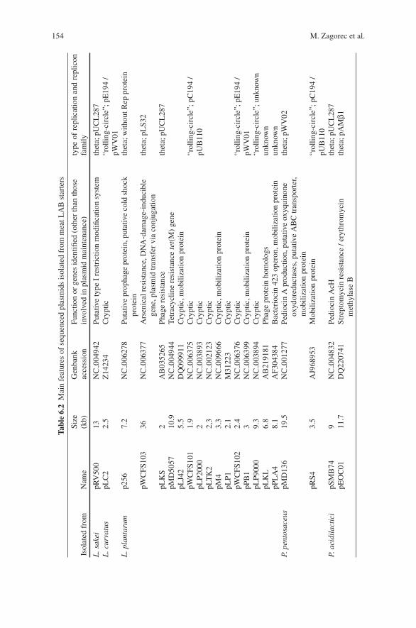
154 M. Zagorec et al.
Tabl
e6.
2M
ain
feat
ures
ofse
quen
ced
plas
mid
sis
olat
edfr
omm
eatL
AB
star
ters
Isol
ated
from
Nam
eSi
ze(k
b)G
enba
nkac
cess
ion
Func
tion
orge
nes
iden
tified
(oth
erth
anth
ose
invo
lved
inpl
asm
idm
aint
enan
ce)
type
ofre
plic
atio
nan
dre
plic
onfa
mily
L.s
akei
pRV
500
13N
C00
4942
Puta
tive
type
Ire
stri
ctio
nm
odifi
catio
nsy
stem
thet
a;pU
CL
287
L.c
urva
tus
pLC
22.
5Z
1423
4C
rypt
ic“r
ollin
g-ci
rcle
”;pE
194
/pW
V01
L.p
lant
arum
p256
7.2
NC
0062
78Pu
tativ
epr
opha
gepr
otei
n,pu
tativ
eco
ldsh
ock
prot
ein
thet
a;w
ithou
tRep
prot
ein
pWC
FS10
336
NC
0063
77A
rsen
ical
resi
stan
ce,D
NA
-dam
age-
indu
cibl
ege
ne,p
lasm
idtr
ansf
ervi
aco
njug
atio
nth
eta;
pLS3
2
pLK
SpM
D50
57pL
J42
2 10.9
5.5
AB
0352
65N
C00
4944
DQ
0999
11
Phag
ere
sist
ance
Tetr
acyc
line
resi
stan
cete
t(M
)ge
neC
rypt
ic,m
obili
zatio
npr
otei
n
thet
a;pU
CL
287
pWC
FS10
1pL
P200
0pL
TK
2pM
4pL
P1
1.9
2 2,3
3.3
2.1
NC
0063
75N
C00
3893
NC
0021
23N
C00
9666
M31
223
Cry
ptic
Cry
ptic
Cry
ptic
Cry
ptic
,mob
iliza
tion
prot
ein
Cry
ptic
“rol
ling-
circ
le”;
pC19
4/
pUB
110
pWC
FS10
2pP
B1
2.4
3N
C00
6376
NC
0063
99C
rypt
icC
rypt
ic,m
obili
zatio
npr
otei
n“r
ollin
g-ci
rcle
”;pE
194
/pW
V01
pLP9
000
9.3
NC
0038
94C
rypt
ic“r
ollin
g-ci
rcle
”;un
know
npL
KL
6.8
AB
2191
81Ph
age
prot
ein
hom
olog
sun
know
npP
LA
48.
1A
F304
384
Bac
teri
ocin
423
oper
on,m
obili
zatio
npr
otei
nun
know
nP.
pent
osac
eus
pMD
136
19.5
NC
0012
77Pe
dioc
inA
prod
uctio
n,pu
tativ
eox
yqui
none
oxyd
ored
ucta
ses,
puta
tive
AB
Ctr
ansp
orte
r,m
obili
zatio
npr
otei
n
thet
a;pW
V02
pRS4
3.5
AJ9
6895
3M
obili
zatio
npr
otei
n“r
ollin
g-ci
rcle
”;pC
194
/pU
B11
0P.
acid
ilac
tici
pSM
B74
9N
C00
4832
Pedi
ocin
AcH
thet
a;pU
CL
287
pEO
C01
11.7
DQ
2207
41St
rept
omyc
inre
sist
ance
/ery
thro
myc
inm
ethy
lase
Bth
eta;
pAM
β1
6 Genetics of Lactic Acid Bacteria 155
niche. The possibility of the dissemination of antibiotic resistance by meat lacto-bacilli has been addressed by Gevers, Danielsen, Huys, and Swings, (2003), whoshowed that tetracycline resistance plasmids could be transferred from L. sakei orL. plantarum to other Gram-positive bacteria such as Enterococcus faecalis andLactococcus lactis by in vitro conjugation (Gevers, Danielsen, et al., 2003; Gevers,Huys, et al., 2003). This suggests that meat lactobacilli might be the reservoir micro-organisms for acquired resistance genes that can be spread among other bacteria.
A number of other plasmid-encoded functions were assigned after plasmidsequence determination, such as putative restriction modification systems or puta-tive stress proteins (see Table 6.2). Thus, plasmids may clearly carry a source ofgenetic diversity and provide an extension of metabolic properties or additionalresistance to environmental conditions.
Vectors and Genetic Tools
Geneticists developed various genetic tools in order to construct mutants, eitherrandomly or in targeted genes, to express foreign genes, or to monitor their expres-sion. Such approaches rely on the capability to transfer DNA molecules, thereforeefficient transformation protocols have been developed in parallel.
Electroporation, based on transient pore formation in membranes after apply-ing an electrical field, is the method of choice for gene transfer and protocolshave been adapted for each species. Depending on the recipient strains used, theygive different amount of transformants, the most efficient ones reaching up to104–106 transformants per μg of plasmid DNA (Alegre, Rodriguez, & Mesas, 2004;Aukrust & Blom, 1992; Berthier, Zagorec, Champomier-Verges, Ehrlich, & Morel-Deville, 1996; Luchansky, Muriana, & Klaenhammer, 1988; Rodriguez, Alegre, &Mesas, 2007).
Conjugation is known to naturally occur in lactobacilli but requires conjugativeplasmids. It can be made through the use of broad host-range conjugative plasmidsisolated from other bacteria such as pAMβ1 or pIP501 (Gonzalez & Kunka, 1983;Langella, Zagorec, Ehrlich, & Morel-Deville, 1996) either alone or as helperswith mobilizable plasmids. Conjugation has proven useful for plasmid transfer intostrains that are poorly transformable by electroporation.
Natural competence and transformation were never reported in LAB, althoughcomplete genome sequences revealed the presence of genes homologous to bonafide competence genes involved in DNA transport, in other bacterial groups. Thequestion of naturally occurring gene transfer is of particular interest with food-borneorganisms. Plasticity of the chromosomes and plasmids has revealed the relativeimportance of gene transfer especially among bacteria sharing a same biotope. Thismay then occur by natural, but not yet characterized competence, stress-inducedpermeation, plasmid mobilization/conjugation, or phage infection. Indeed, conjuga-tive transfers were detected to occur during sausage fermentation with L. curvatus(Vogel, Becke-Schmid, Entgens, Gaier, & Hammes, 1992).
156 M. Zagorec et al.
Plasmid vectors have been developed for Lactobacillus and to a lesser extent forPediococcus, whose molecular biology is still at its beginning. A non-exhaustive listof vectors and methods used in “meat species” is highlighted here. A comprehensivereview of the natural plasmids from LAB and derived vectors was recently publishedby Shareck, Choi, Lee, & Miguez (2004).
Integrative vectors (also called delivery or suicide vectors), that is those that areincapable of self-replication in the recipient host, are used for the construction ofchromosomal mutants. The vector part often comes from widely used plasmidsof Escherichia coli of the colE1-type, like pRV300 (Leloup, Ehrlich, Zagorec, &Morel-Deville, 1997) or pJDC9 (Chen & Morrison, 1988). Chromosomal integra-tion relies on homologous recombination and allows different kinds of mutationsto be obtained: (i) inactivation of genes after the simple insertion of the recombi-nant vector, or (ii) point mutations (Stentz & Zagorec, 1999) and deletion mutants(Ferain et al., 1996; Leer et al., 1993; Malleret, Lauret, Ehrlich, Morel-Deville, &Zagorec, 1998; Stentz, Loizel, Malleret, & Zagorec, 2000), for which no exogenousDNA persists. The latter strategy of gene replacement usually involves two succes-sive recombination steps; the first one involves selecting for integration of the entireplasmid, the second one consisting of an excision of plasmid sequences betweenregions duplicated after integration. In the case of high recombination frequency andprovided it can be selected or screened for, a double-recombination event may beobtained in one step (Malleret et al., 1998). Conditionally replicative vectors (repli-cating only in so-called permissive conditions) are considered as the most effectivedelivery vectors, especially for low transformable strains, because they allow tem-poral separation of the transformation and integration steps. Some plasmids havebeen found to be naturally thermo-sensitive at 42◦C, and thermo-sensitive vectorshave also been developed (see Shareck et al., 2004). However, it should be noted thatthis character is strongly dependent on the host and that no thermo-sensitive plasmidhas been found effective in mesophilic species, such as a number of L. sakei strains,which do not grow over 37◦C (Gory, Montel, & Zagorec, 2001).
Replicative vectors can be used to easily bring new functions in the cell or toexpress genes at a high level (expression vectors). Inducible promoters have beenused for over-expression (Sørvig, Mathiesen, Naterstad, Eijsink, & Axelsson, 2005)or to modulate functions (Stentz et al., 2000).
Reporter genes are also interesting tools in the molecular biologist’s arsenal andcomprise lacZ (Stentz et al., 2000), gusA (Hertel, Schmidt, Fischer, Oellers, &Hammes, 1998) and the Green Fluorescent Protein (GFP) gene (Gory et al., 2001),the activity of the latter being detected by fluorescence, thereby allowing to followthe strains in complex environments. It should be noted that the use of lacZ may behampered by the presence of β-galactosidase activity often existing in a number oflactobacilli.
In a view of food-grade applications, plasmids should not contain DNAoriginating out of LAB species and in particular should not harbor genes forresistance to antibiotics. In this context, appropriate selective cassettes have beendeveloped; nisin immunity conferred by nisI has been used in L. plantarum (Takala& Saris, 2002), and selection based on specific metabolic properties could be used
6 Genetics of Lactic Acid Bacteria 157
provided the encoding genes can complement a deficient strain as described inBron, et al., (2002); and in Takala, Saris, and Tynkkynen, (2003).
Random mutagenesis via transposition is still to be developed in meat starterspecies, this approach also requires thermo-sensitive delivery vectors. Genetic toolshave been developed in LAB using IS, such as IS946 or ISAS1, in combination withnon-replicative vectors to efficiently perform site-directed or random mutagenesisby transposition (Maguin, Prevost, Ehrlich, & Gruss, 1996; Romero & Klaenham-mer, 1992). No system based on the IS elements present in their genome have yetbeen developed for L. sakei, P. pentosaceus, and L. plantarum. Attempts for obtain-ing random transposition were performed in L. plantarum with variable success.Whereas Tn917 delivered by pTV1Ts essentially targeted the naturally harboredplasmids (Cosby, Axelsson, & Dobrogosz, 1989), pGhost9:ISS1 was successfullyused at 42◦C to obtain a set of random transposition mutants (Gury, Barthelmebs, &Cavin, 2004).
Chromosomal Elements
In addition to the genetic elements mentioned above, that largely participate in thegenetic repertoire and biodiversity of bacteria, the chromosomally encoded func-tions represent the large majority of the genetic elements that characterize a bacterialspecies and its properties.
The chromosomes of L. plantarum WCFS1, L. sakei 23K, and P. pentosaceusATCC27745 encode respectively 3009, 1886, and 1757 coding sequences(Table 6.1), showing a large difference between those species. Besides house-keeping functions, the genetic repertoire of those bacteria shows specific featuresthat are common to the three species, and absent from other closely related bacteria(lactobacilli) which do not usually develop in meat products. One can thus hypoth-esize that these specific features are important for the adaptation to the fermentedmeat environment. They mostly allow bacteria to grow in meat as a substrate, andto resist the stressing environment that the steps of meat fermentation represent,for instance the presence of high amount of salt, redox potential variations, ortemperature changes during the process.
Functions Involved in Growth and Fermentation
The main role of LAB starters during meat fermentation is to degrade sugars toproduce mainly lactic acid. Some traditional sausages can be manufactured withoutthe addition of any sugar. In this case, LAB performs fermentation through the uti-lization of the carbon sources that are naturally present in meat, mainly ribose andglucose. We previously noticed that in L. sakei, the gene cluster involved in riboseutilization had a characteristic organization (Stentz & Zagorec, 1999). Indeed, ribosetransport was not performed by an ABC transporter as usually observed in other
158 M. Zagorec et al.
bacteria. Instead of the rbsABCD genes encoding the ribose specific ABC trans-porter, we found a gene, named rbsU that was similar to glucose transporters medi-ating sugar transport by a facilitated diffusion mechanism. Interestingly, a similarsituation is observed in the genomes of L. plantarum and P. pentosaceus, but also inthe genome of Enterococcus faecalis V583, a bacterial species that can contaminatemeat products. Such a situation is not encountered in other LAB species, the genomeof which has been sequenced. It is therefore plausible that such a property, shared bymeat-borne bacteria confers them an advantage to compete for carbon sources, use-ful for energy production and growth. As a comparison, the gene syntheny observedaround the ldhL gene of L. sakei, encoding the unique lactate dehydrogenase in thisbacterium, responsible for L-lactate production, is conserved in L. plantarum and P.pentosaceus but also in other LAB genomes such as those of L. casei ATCC334and L. brevis ATCC367, which are not used for meat fermentation. Concerningthe capability to ferment sugars, it was previously noticed that L. plantarum hasthe widest range of sugar utilization due to the presence of many sugar transportsystems, a characteristic trait of this species that is certainly linked to the manyecosystems it can colonize (Kleerebezem et al., 2003).
Meat is an iron rich medium, especially regarding complexed iron likehaemoglobin or myoglobin. Iron metabolism seems to be emblematic of meatLAB species. More particularly, the ability to transport complex iron seems to berestricted to meat lactobacilli; only two other LAB species (L. casei and Lacto-coccus lactis) possess such an equipment (Table 6.3). L. plantarum, is also theonly LAB species possessing a ironIII ABC transporter (AfuA). Moreover, irondependent transcriptional regulators (Fur family) are also well represented in thesemeat LAB species. In fact, three are present in the L. sakei genome and two in bothL. plantarum and P. pentosaceus. It seems thus likely that the ability to use ironcompounds and especially iron complexes could be considered as a kind of meatsignature for meat LAB species.
Functions Involved in Resistance to the Stressing Environmentof Fermented Meat Products
Addition of NaCl to fermented meat products, as well as the drying process induce ahigh osmolarity environment which the bacterial should resist in order to survive inthe processed meat. We previously noticed in the genome of L. sakei 23K the pres-ence of three gene clusters encoding putative ABC transporters that may be involvedin the uptake of osmoprotectants, such as glycine betaine, carnitine, or choline(Chaillou et al., 2005). Interestingly, Listeria monocytogenes, a pathogenic Gram-positive bacterium that can contaminate meat products shares the same equipment.However, among those three systems, only the cluster lsa0616-0619 was also con-served in L. plantarum WCFS1 (corresponding to lpl1607-1610) and P. pentosaceusATCC25745 (genes pepe1655-1652). The two other osmoprotectant transporters ofL. sakei, encoded by lsa1694-1695 and lsa1869-1870 may result from a duplication
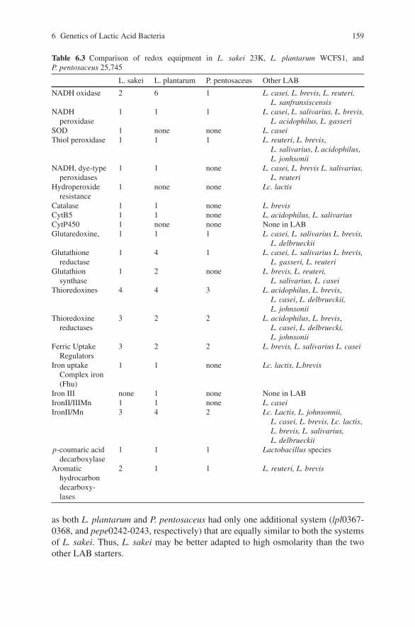
6 Genetics of Lactic Acid Bacteria 159
Table 6.3 Comparison of redox equipment in L. sakei 23K, L. plantarum WCFS1, andP. pentosaceus 25,745
L. sakei L. plantarum P. pentosaceus Other LAB
NADH oxidase 2 6 1 L. casei, L. brevis, L. reuteri,L. sanfransiscensis
NADHperoxidase
1 1 1 L. casei, L. salivarius, L. brevis,L. acidophilus, L. gasseri
SOD 1 none none L. caseiThiol peroxidase 1 1 1 L. reuteri, L. brevis,
L. salivarius, L acidophilus,L. jonhsonii
NADH, dye-typeperoxidases
1 1 none L. casei, L. brevis L. salivarius,L. reuteri
Hydroperoxideresistance
1 none none Lc. lactis
Catalase 1 1 none L. brevisCytB5 1 1 none L. acidophilus, L. salivariusCytP450 1 none none None in LABGlutaredoxine, 1 1 1 L. casei, L. salivarius L. brevis,
L. delbrueckiiGlutathione
reductase1 4 1 L. casei, L. salivarius L. brevis,
L. gasseri, L. reuteriGlutathion
synthase1 2 none L. brevis, L. reuteri,
L. salivarius, L. caseiThioredoxines 4 4 3 L. acidophilus, L. brevis,
L. casei, L. delbrueckii,L. johnsonii
Thioredoxinereductases
3 2 2 L. acidophilus, L. brevis,L. casei, L. delbruecki,L. johnsonii
Ferric UptakeRegulators
3 2 2 L. brevis, L. salivarius L. casei
Iron uptakeComplex iron(Fhu)
1 1 none Lc. lactis, L.brevis
Iron III none 1 none None in LABIronII/IIIMn 1 1 none L. caseiIronII/Mn 3 4 2 Lc. Lactis, L. johnsonnii,
L. casei, L. brevis, Lc. lactis,L. brevis, L. salivarius,L. delbrueckii
p-coumaric aciddecarboxylase
1 1 1 Lactobacillus species
Aromatichydrocarbondecarboxy-lases
2 1 1 L. reuteri, L. brevis
as both L. plantarum and P. pentosaceus had only one additional system (lpl0367-0368, and pepe0242-0243, respectively) that are equally similar to both the systemsof L. sakei. Thus, L. sakei may be better adapted to high osmolarity than the twoother LAB starters.
160 M. Zagorec et al.
Meat storage and processing are characterized by numerous changes in oxygenlevels, including the use of reducing agents such as nitrate and nitrite in fermentedmeat products. All the bacterial species present in the meat ecosystem thus have tocope with this main stressing environmental factor in order to grow and survive infermented meat products. Protection against oxidative damage implies both defensesystems against Reactive Oxygen Species (ROS) and repair systems to restore pro-tein functionality or DNA damage. Iron can also enhance oxygen toxicity due toa possible arising of chemical reactions and the consecutive generation of ROS bythe Fenton reactions. The emblematic meat species L. sakei has previously beenreported as a bacterium with complete genetic equipment dedicated to defenseagainst oxidative stress (Chaillou et al., 2005). These features have been searched inthe two other meat LAB species for which whole genome sequence was available(Table 6.3). This genome comparison revealed some common features together withspecies specific traits that could be considered specific for meat LAB starters.
Analysis of the genome content of L. plantarum WCFS1, L. sakei 23K, andP. pentosaceus ATCC25745 shows that the three species harbor a genetic equipmentdedicated to protection against oxidative damages. However, this equipment seemsto be more similar between L. plantarum and L. sakei, P. pentosaceus being devoidof the main functions related to peroxide and hydroperoxide resistance. L. sakeiharbors the most complete equipment for fighting against oxygen toxicity, includ-ing, in particular, one superoxide dismutase, a hydroperoxide resistance protein andan uncharacterized NADH oxidase that is unique in LAB. L. plantarum is clearlycharacterized by a high redundancy in glutathion reductases and glutathion syn-thase, a compound involved in protection against oxidative stress. It appears thenthat P. pentosaceus might be less resistant, than the two other species, to oxidativedamage conditions encountered during meat fermentation.
Aromatic compounds can arise from exposure to spices and smoke, that canbe used as meat bio-preservation methods, in addition to the fermentation process.Detoxification of such compounds can be achieved by a p-coumaric acid decarboxy-lase (PdcA) and aromatic hydrocarbon decarboxylases. PdcA homologs are largelyfound in Lactobacillus species. Homologs of the aromatic hydrocarbon decarboxy-lases are present in meat LAB and less represented in other LAB (only L. brevis andL. reuteri). Remarkably, L. sakei is unique among LAB, since it is the only speciespossessing a specific aromatic hydrocarbon hydroxylase for which no homologousprotein is found in other LAB. Moreover, a gene encoding a putative cytochromeP450, whose function is yet unknown, is present only in L. sakei.
Conclusion
Genetic studies on LAB emerged a few decades ago, as a mean for microbiolo-gists to better understand the mechanisms of food fermentation, in order to controland improve it or to select the better starter strains. Most of the literature was, fora long time, almost exclusively dedicated to the dairy LAB Lc. lactis. However,
6 Genetics of Lactic Acid Bacteria 161
although with a certain delay, the genetics of other species, including that of meatLAB starters, gained the interest of the scientific community. The study of the plas-midic genetic elements was relatively poorly developed, mainly aimed as a tool forspecies characterization or for studies on systematics or classification. The searchfor bacteriocins, to improve meat safety, led to the isolation of several plasmids but,unfortunately, only the genes responsible for bacteriocin production were charac-terized, plasmids by themselves being poorly considered. The global conclusion ofthe genetic knowledge gained from the study of plasmids in LAB in general is thatthose genetic elements, as other mobile elements, are largely shared and exchangedbetween strains, species, and genera.
In the 1990s, molecular biologists started to develop tools and methods to genet-ically modify strains, as a basis to gain knowledge on the metabolism of LAB, andwith the dream to control and orientate metabolic fluxes for improving fermentationprocesses. However, the use of genetically modified organisms in food, especiallyin fermented food that are eaten raw, i. e., with living microorganisms, such as fer-mented meat is not acceptable for the consumer. The use of genetics for LAB thusstayed just as a tool for laboratory studies to generate information.
In the mean time, genomics emerged and an increasing number of bacterialgenomes were sequenced and analyzed each year during the last decade. After thesequencing of bacterial species, which were considered as models by the microbi-ologists, many pathogenic species were also sequenced, and finally, the genomesof most of the LAB used for food fermentation have been sequenced in the lastyears. The genome sequence of L. plantarum was published in 2003, that of L. sakeiin 2005, and that of P. pentosaceus in 2006. Consequently, the delay traditionallyobserved with the knowledge of meat LAB compared to dairy LAB stopped.
Whole genome analysis revealed specific features of the various species. Thecomparison between several species and genera of LAB confirmed the status ofP. pentosaceus as belonging to the Lactobacillus genus (Makarova et al., 2006).A careful analysis of the genome features common to meat LAB has still to beperformed. However, some characteristic traits have already been detected: (i) allspecifically show a metabolic capability oriented to the meat environment, whichis not present in other LAB. This is exemplified by the use of ribose or iron, twonutrients present in meat and absent or substituted by other components in othermaterials that are also commonly fermented such as milk or vegetables; (ii) all show,with some species differences, the ability to resist the stressing environment thatcharacterizes meat fermentation, such as oxidative or salt stresses or detoxificationof molecules issued from spices or smoking.
References
Ackermann, J. W. (1987). Bacteriophage taxonomy in 1987. Microbiology Sciences, 4, 214–218.Alegre, M. T., Rodriguez, M. C., & Mesas, J. M. (2004). Transformation of Lactobacillus plan-
tarum by electroporation with in vitro modified plasmid DNA. FEMS Microbioogy Letters,241, 73–77.
162 M. Zagorec et al.
Alpert, C. A., Crutz-Le Coq, A.-M., Malleret, C., & Zagorec, M. (2003). Characterization of atheta-type plasmid from Lactobacillus sakei: A potential basis for low-copy vectors in lacto-bacilli. Applied and Environmental Microbiology, 69, 5574–5584.
Aukrust, T., & Blom, H. (1992). Transformation of Lactobacillus strains used in meat and veg-etable fermentation. Food Research International, 25, 253–261.
Berthier, F., Zagorec, M., Champomier-Verges, M., Ehrlich, S. D., & Morel-Deville, F. (1996).High-frequency transformation of Lactobacillus sake by electroporation. Microbiology, 142,1273–1279.
Boekhorst, J., Siezen, R. J., Zwahlen, M. C., Vilanova, D., Pridmore, R. D., Mercenier, A.,et al. (2004). The complete genomes of Lactobacillus plantarum and Lactobacillus johnsoniireveal extensive differences in chromosome organization and gene content. Microbiology, 150,3601–3611.
Bringel, F., Frey, L., & Hubert, J. C. (1989). Characterization, cloning, curing, and distribution inlactic acid bacteria of pLP1, a plasmid from Lactobacillus plantarum CCM1904 and its use inshuttle vector construction. Plasmid, 22, 193–202.
Bron, P. A., Benchimol, M. G., Lambert, J., Palumbo, E., Deghorain, M., Delcour, J., et al. (2002).Use of the alr gene as a food-grade selection marker in lactic acid bacteria. Applied and Envi-ronmental Microbiology, 68, 5663–5670.
Caldwell, S. L., McMahon, D. J., Oberg, C. J., & Broadbent, J. R. (1999). Induction and charac-terization of Pediococcus acidilactici temperate bacteriophage. Systematic and Applied Micro-biology, 22, 514–519.
Chaillou, S., Champomier-Verges, M. C., Cornet, M., Crutz Le Coq, A.-M., Dudez, A.-M., et al.(2005). Complete genome sequence of the meat-borne lactic acid bacterium Lactobacillus sakei23K. Nature Biotechnology, 23, 1527–1533.
Chen, J. D., & Morrison, D. A. (1988). Construction and properties of a new insertion vector,pJDC9, that is protected by transcriptional terminators and useful for cloning of DNA fromStreptococcus pneumoniae. Gene, 64, 155–164.
Cosby, W. M., Axelsson, L. T., & Dobrogosz, W. J. (1989). Tn917 transposition in Lactobacillusplantarum using the highly temperature-sensitive plasmid pTV1Ts as a vector. Plasmid, 22,236–243.
Danielsen, M. (2002). Characterization of the tetracycline resistance plasmid pMD5057 from Lac-tobacillus plantarum 5057 reveals a composite structure. Plasmid, 48, 98–103.
Doi, K., Zhang, Y., Nishizaki, Y., Umeda, A., Ohmomo, S., & Ogata, S. (2003). A comparativestudy and phage typing of silage-making Lactobacillus bacteriophages. Journal of Bioscienceand Bioengineering, 95, 518–525.
Ferain, T., Hobbs, J. N., Jr., Richardson, J., Bernard, N., Garmyn, D., Hols, P., et al. (1996).Knockout of the two ldh genes has a major impact on peptidoglycan precursor synthesis inLactobacillus plantarum. Journal of Bacteriology, 178, 5431–5437.
Frost, L. S., Leplae, R., Summers, A. O., & Toussaint, A. (2005). Mobile genetic elements: Theagents of open source evolution. Nature Reviews Microbiology, 3, 722–732.
Gevers, D., Danielsen, M., Huys, G., & Swings, J. (2003). Molecular characterization of tet(M)genes in Lactobacillus isolates from different types of fermented dry sausage. Applied andEnvironmental Microbiology, 69, 1270–1275
Gevers, D., Huys, G., & Swings, J. (2003). In vitro conjugal transfer of tetracycline resistancefrom Lactobacillus isolates to other Gram-positive bacteria. FEMS Microbiology Letters, 225,125–130.
Giacomini, A., Squartini, A., & Nuti, M. P. (2000). Nucleotide sequence and analysis of plasmidpMD136 from Pediococcus pentosaceus FBB61 (ATCC43200) involved in pediocin A produc-tion. Plasmid, 43, 111–122.
Gonzalez, C. F., & Kunka, B. S. (1983). Plasmid transfer in Pediococcus spp.: Intergeneric andintrageneric transfer of pIP501. Applied and Environmental Microbiology, 46, 81–89.
Gonzalez, C. F., & Kunka, B. S. (1986). Evidence for plasmid linkage of raffinose utilizationand associated alpha-galactosidase and sucrose hydrolase activity in Pediococcus pentosaceus.Applied and Environmental Microbiology, 51, 105–109.
6 Genetics of Lactic Acid Bacteria 163
Gonzalez, C. F., & Kunka, B. S. (1987). Plasmid-associated bacteriocin production and sucrosefermentation in Pediococcus acidilactici. Applied and Environmental Microbiology, 53,2534–2538.
Gory, L., Montel, M. C., & Zagorec, M. (2001) Use of Green Fluorescent Protein to monitorLactobacillus sakei in fermented meat products. FEMS Microbiology Letters, 194, 127–133.
Graham, D. C., & McKay, L. L. (1985). Plasmid DNA in strains of Pediococcus cerevisiae andPediococcus pentosaceus. Applied and Environmental Microbiology, 50, 532–534.
Gury, J., Barthelmebs, L., & Cavin, J. F. (2004). Random transposon mutagenesis of Lactobacillusplantarum by using the pGh9:ISS1 vector to clone genes involved in the regulation of phenolicacid metabolism. Archives in Microbiology, 182, 337–345.
Halami, P. M., Ramesh, A., & Chandrashekar, A. (2000). Megaplasmid encoding novel sugar uti-lizing phenotypes, pediocin production and immunity in Pediococcus acidilactici C20. FoodMicrobiology, 17, 475–483.
Hammes, W. P., & Hertel, C. (1998). New development in meat starter culture. Meat Science, 49,S125–S128.
Hertel, C., Schmidt, G., Fischer, M., Oellers, K., & Hammes, W. P. (1998). Oxygen-dependentregulation of the expression of the catalase gene katA of Lactobacillus sakei LTH677. Appliedand Environmental Microbiology, 64, 1359–1365.
Kakikawa, M., Yamakawa, A., Yokoi, K. J., Nakamura, S., Taketo, A., & Kodaira K. (2002) Char-acterization of the major tail protein gpP encoded by Lactobacillus plantarum phage phi gle.Journal of Biochemistry, Molecular Biology, and Biophysics, 6, 185–191.
Kakikawa, M., Yokoi, K. J., Kimoto, H., Nakano, M., Kawasaki, K., Taketo, A., et al. (2002).Molecular analysis of the lysis protein Lys encoded by Lactobacillus plantarum phage phi g1e.Gene, 299, 227–234.
Kanatani, K., & Oshimura, M. (1994). Plasmid-associated bacteriocin production by a Lactobacil-lus plantarum strain. Bioscience Biotechnology Biochemistry, 58, 2084–2086.
Kantor, A., Montville, T. J., Mett, A., & Shapira, R. (1997). Molecular characterization of the repli-con of the Pediococcus pentosaceus 43200 pediocin A plasmid pMD136. FEMS MicrobiologyLetters, 151, 237–244.
Kleerebezem, M., Boekhorst, J., van Kranenburg, R., Molenaar, D., Kuipers, O. P., Leer, R., et al.(2003). Complete genome sequence of Lactobacillus plantarum WCFS1. Proceedings of theNational Academy of Science USA, 100, 1990–1995.
Langella, P., Zagorec, M., Ehrlich, S. D., & Morel-Deville, F. (1996). Intergeneric and intragenericconjugal transfer of plasmids pAMβ1, pIL205 and pIP501 in Lactobacillus sake. FEMS Micro-biology Letters, 139, 51–56.
Leer, R. J., Christiaens, H., Verstraete, W., Peters, L., Posno, M., & Pouwels, P. H. (1993). Genedisruption in Lactobacillus plantarum strain 80 by site-specific recombination: isolation ofa mutant strain deficient in conjugated bile salt hydrolase activity. Molecular and GeneralGenetics, 239, 269–272.
Leloup, L., Ehrlich, S. D., Zagorec, M., & Morel-Deville, F. (1997). Single cross-over integrationin the Lactobacillus sake chromosome and insertional inactivation of the ptsI and lacL genes.Applied and Environmental Microbiology, 63, 2127–2133.
Leuschner, R. G. K., Arendt, E. K., & Hammes, W. P. (1993). Characterization of a virulentLactobacillus sake phage PWH2. Applied Microbiology and Biotechnology, 39, 617–621.
Li, Y., Canchaya, C., Fang, F., Raftis, E., Ryan, K. A., van Pijkeren, J. P., et al. (2007). Distributionof megaplasmids in Lactobacillus salivarius and other lactobacilli. Journal of Bacteriology,189, 6128–6139.
Liu, M. L., Kondo, J. K., Barnes, M. B., & Bartholomew, D. T. (1988). Plasmid-linked maltoseutilization in Lactobacillus ssp. Biochimie, 70, 351–355.
Lu, Z., Altermann, E., Breidt, F., Predki, P., Fleming, H. P., & Klaenhammer, T. R. (2005).Sequence analysis of the Lactobacillus plantarum bacteriophage PhiJL-1. Gene, 348, 45–54.
Lu, Z., Breidt, F. Jr., Fleming, H. P., Altermann, E., & Klaenhammer, T. R. (2003). Isolationand characterization of a Lactobacillus plantarum bacteriophage, phiJL-1, from a cucumberfermentation. International Journal of Food Microbiology, 84, 225–235.
164 M. Zagorec et al.
Luchansky, J. B., Muriana, P. M., & Klaenhammer, T. R. (1988). Application of electroporation fortransfer of plasmid DNA to Lactobacillus, Lactococcus, Leuconostoc, Listeria, Pediococcus,Bacillus, Staphylococcus, Enterococcus and Propionibacterium. Molecular Microbiology, 2,637–646.
Maguin, E., Prevost, H., Ehrlich, S. D., & Gruss, A. (1996). Efficient insertional mutagenesis inlactococci and other Gram-positive bacteria. Journal of Bacteriology, 178, 931–935.
Makarova, K., Slesarev, A., Wolf, Y., Sorokin, A., Mirkin, B., Koonin, E., et al. (2006). Compara-tive genomics of the lactic acid bacteria. Proceedings of the National Academy of Science USA,103, 15611–15616.
Malleret, C., Lauret, R., Ehrlich, S. D., Morel-Deville, F., & Zagorec, M. (1998). Disruption ofthe sole ldhL gene in Lactobacillus sakei prevents the production of both L- and D-lactate.Microbiology, 144, 3327–3333.
Mayo, B., Gonzalez, B., Arca, P., & Suarez, J. E. (1994). Cloning and expression of the plas-mid encoded beta-D-galactosidase gene from a Lactobacillus plantarum strain of dairy origin.FEMS Microbiology Letters, 122, 145–151.
Miller, K. W., Ray, P., Steinmetz, T., Hanekamp, T., & Ray, B. (2005). Gene organization andsequences of pediocin AcH/PA-1 production operons in Pediococcus and Lactobacillus plas-mids. Letters in Applied Microbiology, 40, 56–62.
Motlagh, A., Bukhtiyarova, M., & Ray, B. (1994). Complete nucleotide sequence of pSMB74,a plasmid encoding the production of pediocin AcH in Pediococcus acidilactici. Letters inApplied Microbiology, 18, 305–312.
Naumoff, D. G. (2001). Beta-fructosidase superfamily: Homology with some alpha-L-arabinasesand beta-D-xylosidases. Proteins, 42, 66–76.
Nes, I.. F. (1984). Plasmid profiles of ten strains of Lactobacillus plantarum. FEMS MicrobiologyLetters, 21, 359–361.
Nes, I. F., Brendehaug, J., & von Husby, K. O. (1988). Characterization of the bacteriophage B2of Lactobacillus plantarum ATCC 8014. Biochimie, 70, 423–427.
Osmanagaoglu, O., Beyatli, Y., & Gunduz, U. (2000). Cloning and expression of a plasmid-linkedpediocin determinant trait of Pediococcus acidilactici F. Journal of Basic Microbiology, 40,41–49.
Perez Pulido, R., Abriouel, H., Ben Omar, N., Lucas Lopez, R., Martınez Canamero, M., & Galvez,A. (2006). Plasmid profile patterns and properties of pediococci isolated from caper fermenta-tions. Journal of Food Protection, 69, 1178–1182.
Rodrıguez, M. C., Alegre, M. T., & Mesas, J. M. (2007). Optimization of technical condi-tions for the transformation of Pediococcus acidilactici P60 by electroporation. Plasmid, 58,44–50.
Romero, D. A., & Klaenhammer, T. R. (1992). IS946-mediated integration of heterologous DNAinto the genome of Lactococcus lactis subsp. lactis. Applied and Environmental Microbiology,58, 699–702.
Ruiz-Barba, J. L., Piard, J. C., & Jimenez-Dıaz, R. (1991). Plasmid profiles and curing of plasmidsin Lactobacillus plantarum strains isolated from green olive fermentations. Journal of AppliedBacteriology, 71, 417–421.
Shareck, J., Choi, Y., Lee, B., & Miguez, C. B. (2004). Cloning vectors based on cryptic plasmidsisolated from lactic acid bacteria: Their characteristics and potential applications in biotechnol-ogy. Critical Reviews in Biotechnology, 24, 155–208.
Shay, B. J., Egan, A. F., Wright, M., & Rogers, P. J. (1988). Cystein metabolism in an isolate ofLactobacillus sake: Plasmid composition and cystein transport. FEMS Microbiology Letters,56, 183–188.
Simon, L., Fremaux, C., Cenatiempo, Y., & Berjeaud, J.-M. (2002). Sakacin G, a new type ofantilisterial bacteriocin. Applied and Environmental Microbiology, 68, 6416–6420.
Skaugen, M., Abildgaard, C. I., & Nes, I. F. (1997). Organization and expression of a gene clusterinvolved in the biosynthesis of the lantibiotic lactocin S. Molecular and General Genetics, 253,674–686.
6 Genetics of Lactic Acid Bacteria 165
Sørvig, E., Mathiesen, G., Naterstad, K., Eijsink, V. G., & Axelsson, L. (2005). High-level,inducible gene expression in Lactobacillus sakei and Lactobacillus plantarum using versatileexpression vectors. Microbiology, 151, 2439–2449.
Stentz, R., Loizel, C., Malleret, C., & Zagorec, M. (2000). Development of genetic tools for Lacto-bacillus sakei: Disruption of the β-galactosidase gene and use of lacZ as a reporter gene to studyregulation of the putative copper ATPase, AtkB. Applied and Environmental Microbiology, 66,4272–4278.
Stentz, R., & Zagorec, M. (1999). Ribose utilization in Lactobacillus sakei: Analysis of the regu-lation of the rbs operon and putative involvement of a new transporter. Journal of MolecularMicrobiology and Biotechnology, 1, 165–173.
Takala, T. M., & Saris, P. E. (2002). A food-grade cloning vector for lactic acid bacteria based onthe nisin immunity gene nisI. Applied Microbiology and Biotechnology, 59, 467–471.
Takala, T. M., Saris, P. E., & Tynkkynen, S. S. (2003). Food-grade host/vector expression sys-tem for Lactobacillus casei based on complementation of plasmid-associated phospho-beta-galactosidase gene lacG. Applied Microbiology and Biotechnology, 60, 564–570.
Talon, R., Leroy, S., & Lebert, I. (2007). Microbial ecosystems of traditional fermented meat prod-ucts: the importance of indigenous starters. Meat Science, 77, 55–62.
Van Reenen, C. A., Van Zyl, W. H., & Dicks, L. M. (2006). Expression of the immunity pro-tein of plantaricin 423, produced by Lactobacillus plantarum 423, and analysis of the plasmidencoding the bacteriocin. Applied and Environmental Microbiology, 72, 7644–7651.
Vaughan, A., Eijsink, V. G., & Van Sinderen, D. (2003). Functional characterization of a com-posite bacteriocin locus from malt isolate Lactobacillus sakei 5. Applied and EnvironmentalMicrobiology, 69, 7194–7203.
Ventura, M., Canchaya, C., Kleerebezem, M., de Vos, W. M., Siezen, R. J., & Brussow, H. (2003).The prophage sequences of Lactobacillus plantarum strain WCFS1. Virology, 316, 245–255.
Vogel, R. F., Becke-Schmid, M., Entgens, P., Gaier, W., & Hammes, W. P. (1992). Plasmid transferand segregation in Lactobacillus curvatus LTH1432 in vitro and during sausage fermentations.Systematics and Applied Microbiology, 15, 129–136.
Vogel, R. F., Lohmann, M., Weller, A. N., Hugas, M., & Hammes, W. P. (1991). Structural similar-ity and distribution of small cryptic plasmids of Lactobacillus curvatus and Lactobacillus sake.FEMS Microbiology Letters, 68, 183–190.
Wang, T. T., & Lee, B. H. (1997). Plasmids in Lactobacillus. Critical Reviews in Biotechnology,17, 227–272.
West, C. A., & Warner, P. J. (1985). Plasmid profiles and transfer of plasmid-encoded antibi-otic resistance in Lactobacillus plantarum. Applied and Environmental Microbiology, 50,1319–1321.
Chapter 7Genetics of Yeasts
Amparo Querol, Ma Teresa Fernandez-Espinar, and Carmela Belloch
Introduction
The use of yeasts in biotechnology processes dates back to ancient days. Before7000 BC, beer was produced in Sumeria. Wine was made in Assyria in 3500 BC, andancient Rome had over 250 bakeries, which were making leavened bread by 100BC. And milk has been made into Kefyr and Koumiss in Asia for many centuries(Demain, Phaff, & Kurtzman, 1999). However, the importance of yeast in the foodand beverage industries was only realized about 1860, when their role in food man-ufacturing became evident.
Yeasts are used in many industrial processes, as they grow on a wide range ofsubstrates and can tolerate extreme physico-chemical conditions. Today, the impactof yeasts on food and beverage production extends beyond the original and pop-ular notions of bread, beer, and wine fermentations by Saccharomyces cerevisiae(Fleet, 2006; Querol, Belloch, Fernandez-Espinar, & Barrio, 2003). In addition toS. cerevisiae, S. bayanus, and S. pastorianus, it is now well established that var-ious species of Hanseniaspora, Candida, Pichia, Metschnikowia, Kluyveromyces,Schizosaccharomyces, and Issatchenkia can make a positive contribution in the man-ufacture of fermented foods, dairy, meats, cereals, coffee, and sauces. In the caseof fermentation of meat sausages and maturation of hams, the most predominantand important yeasts are Debaryomyces hansenii (and the anamorph C. famata),Yarrowia lipolytica, and various Candida species.
Methods for Yeast Identification
The development of rapid and simple methods for the identification and characteri-sation of yeast strains is an essential tool for the meat industry. This is obvious, sinceyeasts in meat products can contribute to beneficial and detrimental aspects, and the
D. A. QuerolInstituto de Agroquımica y Tecnologıa de Alimentos, CSIC, P.O. Box 73, E-46100 Burjassot(Valencia) Spain
F. Toldra (ed.), Meat Biotechnology, 167C© Springer Science+Business Media, LLC 2008
168 A. Querol et al.
identification of individual strains is often required to improve the microbiologicalquality of these foods.
Classical taxonomy is based predominantly on phenotypic characteristics, suchas physiological traits and cell morphology. These approximations have beenused in the identification of yeasts isolated from different meat products (Gardiniet al., 2001; Nunez, Rodrıguez, Cordoba, Bermudez, & Asensio, 1996). However,nutritional characteristics have been shown to be highly variable as well as mutable,and genetic crosses have linked the characteristics to one or only a few genes,which in some cases could lead to an incorrect classification of a species or a falseidentification of strains (for a review see Boekhout & Robert, 2003; Fernandez-Espinar, Martorell, de Llanos, & Querol, 2006). The conventional methodologyfor yeast identification requires the evaluation of some 60–90 tests and the processis complex, laborious, and time consuming. Besides, in the case of one of themost relevant yeast isolates in meat products like Debaryomyces, many of theresults obtained with these tests are variable, thus making correct identificationof the Debaryomyces species difficult. As an example, serious difficulties wereexperienced in identifying isolates assigned to Debaryomyces, due to the erroneousassimilation of D-xylose, and to a lesser extent, raffinose and L-arabinose (Metax-opoulos, Stavropoulos, Kakouri, & Samelis, 1996). In recent years, rapid kits foryeast identification have been developed to improve the conventional methods.However, these were designed initially for clinical diagnosis and their application isrestricted to 40–60 yeast species of medical interest (Deak & Beuchat, 1996). Othermethods based on the analysis of total cell proteins and long-chain fatty acids usinggas chromatography have also been developed. However, the reproducibility ofthese techniques is questionable due to the fact that they depend on the physiologicalstate of the yeast cells (Golden, Beuchat, & Hitchcock, 1994).
Recent progress in molecular biology has led to the development of newtechniques for yeast identification (for a review see Boekhout & Robert, 2003;Fernandez-Espinar et al., 2006). In the next sections we review the new methodsthat have been developed and their application to the rapid identification of meatyeasts in the industrial practice. The specificity of nucleic acid sequences hasprompted the development of several methods for rapid species identification. Thecomparison of ribosomal RNA (rRNA) and its template ribosomal DNA (rDNA) hasbeen used extensively in recent years, to assess both close and distant relationshipsamong many kinds of organisms. In several hemiascomycetous yeasts, rRNA genesare located in a single genome region composed of 100–150 tandem repeats of afragment of 9 Kb. The ribosomal genes (5.8S, 18S, and 26S) are grouped in tandemforming transcription units that are repeated in the genome between 100 and 200times. In each transcription unit, two other regions exist, the internal transcribedspacers (ITS) and the external ones (ETS) that are transcribed but are not processed.In turn, the codifying units are separated by the IGS intergenic spacers, also calledNTS. The gene 5S is not included in the previously described transcription unit butis found adjacent to the same repetition unit, in tandem, in the case of yeast. Thesequences of the ribosomal genes 5.8S, 18S, and 26S, as well as the spacers ITS andNTS, represent powerful tools to establish phylogenetic relationships and identifyspecies (Kurtzman & Robnett, 1998), due to the conserved sequences found there, as
7 Genetics of Yeasts 169
well as their concerted evolution, i.e., the similarity between repeated transcriptionunits is greater within the species than between units belonging to different species,due to mechanisms like unequal crossing-over or genetic conversion (Li, 1997).
Using the information contained in these regions, different methods have beendeveloped to identify yeast species, as we will describe below.
Ribosomal RNA Gene Sequencing
One of these methods is based on the determination and comparison of thenucleotide sequences in these regions. The two most commonly used regionsare those corresponding to the domains D1 and D2, located at the 5′ end of thegene 26S (Kurtzman & Robnett, 1998) and the gene 18S (James, Cai, Roberts, &Collins, 1997). The availability of these sequences in databases, especially in thecase of the D1/D2 region of gene 26S, make this technique very useful to assignan unknown yeast to a specific species when the percentage of homology of itssequences is over or close to 99% (Kurtzman & Robnett, 1998).
Restriction Fragment Length Polymorphism of rDNA
With an industrial application in mind, other simpler identification methods basedon PCR amplification of the regions of the ribosomal DNA and later restrictionof the amplified fragment were developed. Guillamon, Sabate, Barrio, Cano, &Querol (1998) proposed a rapid and easy method for routine wine yeast identifi-cation, based on RFLPs of the 5.8S rRNA gene and the internal transcribed spacers(ITS1 and 2). Later on its use has been extended to a total of 191 yeast species (deLlanos, Fernandez-Espinar, & Querol, 2004; Esteve-zarzoso, Belloch, Uruburu, &Querol, 1999; Fernandez-Espinar, Esteve-Zarzoso, Querol, & Barrio, 2000) relatedwith beverages and food including meat yeasts isolates. The amplified fragmentsand restriction profiles of these species with the enzymes HaeIII, HinfI, CfoI, andDdeI are currently available “online” at the address http://yeast-id.com. The util-ity of the technique has been proved studying reference strains (Esteve-Zarzoso,Zorman, Belloch, & Querol, 2003; Fernandez-Espinar et al., 2000; Ramos, Valente,Hagler, & Leoncini, 1998) and has been applied by numerous authors for yeastisolate identification in different foods and beverages, such as isolates pertaining tothe genus Debaryomyces, Yarrowia, and Candida (Andrade, Rodrıguez, Sanchez,Aranda, & Cordoba, 2006; Martorell, Fernandez-Espinar, & Querol, 2005; Ramoset al., 1998).
PCR-DGGE Denaturing Gradient Gel Electrophoresis
Recently, a genetic fingerprinting technique based on PCR amplification and dena-turing gradient gel electrophoresis or DGGE has been introduced into microbial
170 A. Querol et al.
ecology (Muyzer, de Waal, & Uitterlinden, 1993). In PCR-DGGE, DNA fragmentsof the same length but with different sequences can be separated. The use of DGGEin microbial ecology is still in its infancy, but their future perspectives are promising(Muyzer & Smalla, 1998). Its application to yeast identification in foods and bev-erages is very recent (for a review see Fernandez-Espinar et al., 2006), and only afew studies related with meat products or the yeast ecology of fermented sausageshave been performed (Cocolin, Urso, Rantsiou, Canton, & Comi, 2006; Rantsiouet al., 2005).
Yeast Biodiversity in Meat Products
The most important yeasts in meat products belong to the ascomycetous genera,Debaryomyces, Pichia, Yarrowia, and Candida, and the basidiomycetous genera,Rhodotorula, Cryptococcus, and Trichosporon. Fresh meat processing and storagecauses the progressive replacement of basidiomycetous by ascomycetous yeasts.A predominance of Candida spp. occurs during spoilage of fresh meat, whilemeat salting, curing and fermentation are selective for Debaryomyces (Samelis &Sofos, 2003). Some important physiological and biochemical characteristics ofmeat yeasts and their interactions result in the selection of certain yeast speciesin specific meat products, like Y . lipolytica in fresh and spoiled poultry (Ismail,Deak, El-Rahman, Yassien, & Beuchat, 2000), and D. hansenii in cured dried andfermented meats (Dura, Flores, & Toldra, 2004a, 2004b; Guerzoni, Lanciotti, &Marchetti, 1993). Consequently, yeasts in meat products have both spoiling andbeneficial aspects.
Spoilage Yeasts in Meat
Yeasts have commonly been considered unimportant in meat spoilage because theirlow initial numbers and their slow growth rates at refrigeration temperatures preventthem from competing effectively with psychrophilic bacteria (Dillon, 1998; Nortjeet al., 1990). Most yeasts, however, are more resistant than bacteria to several foodrelated stresses, such as low water activity, low pH, high salinity, and chemicalpreservatives, while certain species are extremely psychrophilic (Fleet, 1990). Thus,opportunistic yeasts may become important spoilage agents in meat products whenbacterial growth is retarded due to the inhibitory effects of the above stress factors(Samelis & Sofos, 2003).
The increasing consumer demand for less processed, more “natural” foods hasled the food industries to commercialize new products, in which the use of lowerconcentration of preservatives, packaging in modified atmospheres, or new formu-lations occasionally permit the growth of yeasts. Yeast contributes a small, butpermanent part of the natural microflora on meat. The ability of some yeast togrow at low temperature, high salt concentration, and under reduced oxygen tension
7 Genetics of Yeasts 171
enables them to proliferate in refrigerated, cured and vacuum-packed meat and meatproducts (Deak & Beuchat, 1996). Meat spoilage caused by yeasts is mainly due totheir lipolytic and proteolytic activities, although their action on carbohydrates andassociated by-products of bacterial metabolism may also lead to the formation ofcompounds reducing the sensory quality of meat products, such as organic acids,alcohols, esters, and others (Dillon, 1998).
Numerous studies have been conducted in yeasts isolated from meat and meatproducts (for a review see Samelis & Sofos, 2003), and the yeasts most frequentlyisolated on spoiled meat are summarized in Table 7.1 (see Romano, Capece, &Jespersen, 2006; Samelis & Sofos, 2003; Selgas & Garcıa, 2007).
Beneficial Aspects of Yeast on Meats: Yeast Starter Cultures
Yeasts can also be considered habitual components of the micro-biota growing onfermented sausages and dry-cured hams, and their origin is mainly related to theenvironment and the meat used as raw material. In fermented meats, the lactic acidproduced by bacteria and the low water activity resulting from the presence of saltsor a dehydration process constitute a modified environmental factor that hinders thebacterial growth and favor the development of natural competitors. Thus, yeasts useall the nutrients and energy and grow quickly and easily (Dillon & Board, 1991).Yeasts can grow at pH, water activity, and temperature values usual in fermentedsausages (Hammes & Knauf, 1994; Monte, Villanueva, & Domınguez, 1986). Thepresence of yeasts in fermented raw sausages has been studied less than bacteria andmolds.
Sausages and dry-cured hams are traditional meat products obtained after sev-eral months of ripening. Proteolysis and lipolysis constitute the main biochemicalreactions in the generation of flavor precursors, where the endogenous enzymesplay the most important role (Toldra, 1998). Yeasts and molds traditionally play animportant role in sausages fermentation and in dry ham (Martın, Cordoba, Aranda,Cordoba, & Asensio, 2006; Martın, Cordoba, Benito, Aranda, & Asensio, 2003),and their contribution of the fungal population enzymes to proteolysis in minceddry-cured meat products, such as dry-cured sausages, is widely known.
The use of commercially available starters, mainly constituted of lactic acid bac-teria and micrococci, may also produce an impoverishment of flavor and aromaand a loss of peculiar organo-leptic characteristics found in naturally fermentedsausages. For this reason, in several European countries, artisanal sausages are stillpreferred by the consumer and are manufactured by relying on an unknown “factoryflora” (Samelis, Stavropoulos, Kakouri, & Metaxopoulos, 1994). Lactic acid bacte-ria, micrococci, and coagulase-negative staphylococci have the most relevant rolein the fermentative process and ripening, but also yeasts and molds can be involvedas we explained before. Though there are several reports on the yeast populationsin various meat products (Fung & Liang, 1990), studies on the yeast biodiversityin sausages are limited. The earliest studies on salami reported that D. hansenii was
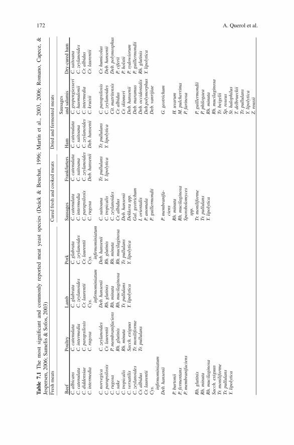
172 A. Querol et al.
Tabl
e7.
1T
hem
ost
sign
ifica
ntan
dco
mm
only
repo
rted
mea
tye
ast
spec
ies
(Dea
ck&
Beu
chat
,19
96;
Mar
tınet
al.,
2003
,20
06;
Rom
ano,
Cap
ece,
&Je
sper
sen,
2006
;Sam
elis
&So
fos,
2003
)Fr
esh
mea
tsC
ured
fres
han
dco
oked
mea
tsD
ried
and
ferm
ente
dm
eats
Bee
fPo
ultr
yL
amb
Pork
Saus
ages
Fran
kfur
ters
Ham
Saus
ages
and
sala
mis
Dry
-cur
edha
mC
.alb
ican
sC
.cat
enul
ata
C.g
labr
ata
C.g
labr
ata
C.c
aten
ulat
aC
.cat
enul
ata
C.c
aten
ulat
aC
.gro
peng
iess
eri
C.s
aito
ana
C.c
aten
ulat
aC
.int
erm
edia
C.z
eyla
noid
esC
.zey
lano
ides
C.i
nter
med
iaC
.sai
toan
aC
.sai
toan
aC
.hae
mul
onii
C.z
eyla
noid
esC
.did
dens
iae
C.p
arap
silo
sis
Cr.
laur
enti
iC
r.la
uren
tii
C.p
arap
silo
sis
C.z
eyla
noid
esC
.zey
lano
ides
C.i
nter
med
iaC
r.al
bidu
sC
.int
erm
edia
C.r
ugos
aC
ys. infir
mom
inia
tum
Cys
. infir
mom
inia
tum
C.r
ugos
aD
eb.h
anse
nii
Deb
.han
seni
iC
.kru
isii
Cr.
laur
enti
i
C.n
orve
gica
C.z
eyla
noid
esD
eb.h
anse
nii
Deb
.han
seni
iC
.sai
toan
aTr
.pul
lula
nsTr
.pul
lula
nsC
.par
apsi
losi
sC
r.hu
mic
olus
C.p
arap
silo
sis
Cr.
laur
enti
iR
h.gl
utin
isR
h.gl
utin
isC
.tro
pica
lis
Y.li
poly
tica
Y.li
poly
tica
C.z
eyla
noid
esD
eb.h
anse
nii
C.r
ugos
aP.
mem
bran
ifac
iens
Rh.
min
uta
Rh.
min
uta
C.z
eyla
noid
esC
it.m
atri
tens
isD
eb.p
olym
osph
usC
.sak
eR
h.gl
utin
isR
h.m
ucil
agin
osa
Rh.
muc
ilag
inos
aC
r.al
bidu
sC
r.al
bidu
sP.
cife
rii
C.t
ropi
cali
sR
h.m
inut
aTr
.pul
lula
nsTr
.pul
lula
nsD
eb.h
anse
nii
Cr.
skin
neri
P.ho
lsti
iC
.ver
sati
lis
Sacc
h.ex
iguu
sY.
lipo
lyti
caY.
lipo
lyti
caD
ekke
rasp
p.D
eb.h
anse
nii
P.sy
dow
ioru
mC
.zey
lano
ides
Tr.m
onil
ifor
me
Gal
.geo
tric
hum
Deb
.mar
amus
P.gu
ille
rmon
dii
Cr.
albi
dus
Tr.p
ullu
lans
I.or
ient
alis
Deb
.occ
iden
tali
sR
h.gl
utin
isC
r.la
uren
tii
P.an
omal
aD
eb.p
olym
orph
usY.
lipo
lyti
caC
ys. infir
mom
inia
tum
P.gu
ille
rmon
dii
Deb
.van
riji
ae
Deb
.han
seni
iP.
mem
bran
ifa-
cien
sG
.geo
tric
hum
P.bu
rton
iiR
h.m
inut
aH
.uva
rum
P.fe
rmen
tans
Rh.
muc
ilag
inos
aM
.pul
cher
rim
aP.
mem
bran
ifac
iens
Spor
obol
omyc
essp
p.P.
fari
nosa
Rh.
glut
inis
Tr.m
onil
ifor
me
P.gu
ille
rmon
dii
Rh.
min
uta
Tr.p
ullu
lans
P.ph
ilog
aea
Rh.
muc
ilag
inos
aY.
lipo
lyti
caR
h.m
inut
aSa
cch.
exig
uus
Rh.
muc
ilag
inos
aTr
.mon
ilif
orm
eTr
.bei
geli
iTr
.pul
lula
nsSp
.ros
eus
Y.li
poly
tica
St.h
alop
hilu
sT.
delb
ruec
kii
Tr.p
ullu
lans
Y.li
poly
tica
Z.r
ouxi
i
7 Genetics of Yeasts 173
the most commonly isolated yeast. Most recently, several researches confirmed theseresults, but other yeast genera were found, such as Candida, Pichia, Rhodotorula,Hansenula (synonym of Pichia), and Torulopsis (synonym of Candida) (Dalton,Board, & Davenport, 1984; Grazia, Suzzi, Romano, & Giudici, 1989). On this basis,D. hansenii was used as a starter with positive effects on the development of a char-acteristic yeast flavor and stabilization of the reddening reaction. D. hansenii strainsare presently used in starter preparations to be added to the sausages (Hammes &Knauf, 1994). The yeast Yarrowia lipolytica, the perfect form of Candida lipolyt-ica, has also frequently been isolated from fresh beef (Dalton et al., 1984; Fung &Liang, 1990) and sausages (Viljoen, Dykes, Collis, & von Holy, 1993). Due to itslipolytic and proteolytic activity, this species can have a high technological potential(Sinigaglia, Lanciotti, & Guerzoni, 1994).
We can conclude that D. hansenii is the most frequently isolated yeast infermented sausages and dry cured ham (Romano, Capece, & Jespersen, 2006;Samelis & Sofos, 2003; Selgas & Garcıa, 2007). The contribution of this yeast to thetypical aroma of the products is based on their primary and secondary metabolitesproduced by the activity of lipases and proteinases, which are the key enzymes (Duraet al., 2004a, 2004b). In the next section we will give a detailed description of thisyeast species.
Physiological and Genetic Characteristics of D.hansenii
According to the last revision, the genus Debaryomyces comprises 15 species(Nakase, Suzuki, Phaff, & Kurtzman, 1998). Many representatives can be foundin natural habitats such as air, soil, pollen, tree exudates, plants, fruits, insects,and faeces and gut of vertebrates (Barnett, Payne, & Yarrow, 2000). Nine of theDebaryomyces species, namely D. carsonii, D. etchellsii, D. hansenii, D. mara-mus, D. melissophilus, D. polymorphus, D. pseudopolymorphus, D. robertsiae, andD. vanrijiae have been found in a variety of processed foods, such as fruit juices andsoft drinks, wine, beer, sugary products, bakery products, dairy products, and meator processed meats. The presence of the Debaryomyces species in foods usuallyhas no detrimental effects and in some cases is beneficial to the food. However,D. hansenii as well as other Debaryomyces species may also be responsible forfood spoilage, causing off-odors and off-flavors. The best method for identificationof this species is the PCR-RFLP of the intergenic spacer region (IGS) region ofrDNA (Quiros et al., 2006).
Morpholgy and Physiology
Colonies on agar medium at 25◦C are grayish white to yellowish, dull to shiny,and smooth to wrinkle. The two varieties within D. hansenii are distinguished by
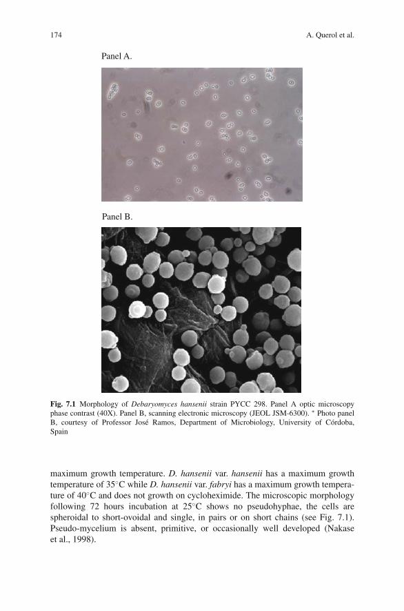
174 A. Querol et al.
Panel A.
Panel B.
Fig. 7.1 Morphology of Debaryomyces hansenii strain PYCC 298. Panel A optic microscopyphase contrast (40X). Panel B, scanning electronic microscopy (JEOL JSM-6300). ∗ Photo panelB, courtesy of Professor Jose Ramos, Department of Microbiology, University of Cordoba,Spain
maximum growth temperature. D. hansenii var. hansenii has a maximum growthtemperature of 35◦C while D. hansenii var. fabryi has a maximum growth tempera-ture of 40◦C and does not growth on cycloheximide. The microscopic morphologyfollowing 72 hours incubation at 25◦C shows no pseudohyphae, the cells arespheroidal to short-ovoidal and single, in pairs or on short chains (see Fig. 7.1).Pseudo-mycelium is absent, primitive, or occasionally well developed (Nakaseet al., 1998).
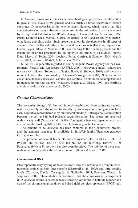
7 Genetics of Yeasts 175
D. hansenii shows some remarkable biotechnological properties like the abilityto grow at 10% NaCl or 5% glucose and assimilates a broad spectrum of carbonsubstrates. D. hansenii has a high chemo-stress tolerance, which means that highconcentrations of many substrates can be used in the cultivation. It is characterizedby its cryo and halo-tolerance (Prista, Almagro, Loureiro-Dias, & Ramos, 1997,Prista, Loureiro-Dias, Montiel, Garcıa, & Ramos, 2005), and its ability to metab-olize lactic and citric acids. Such properties allow its development in all types ofcheeses (Fleet, 1990) and different fermented meat products (Encinas, Lopez-Dıaz,Garcıa-Lopez, Otero, & Moreno, 2000) contributing to the ripening process and thegeneration of aroma precursors by the lipolytic and proteolytic activities (Flores,Dura, Marco, & Toldra, 2004; Leclercq-Perlat, Corrieu, & Spinnler, 2004; Martınet al., 2003; Petersen, Westall, & Jespersen, 2002).
D. hansenii is generally regarded as non-pathogenic (Swiss Agency for the Envi-ronment, Forests and Landscape [SAEFL], 2004); however, clinical isolates arenot rare (Nishikawa, Tomomatsu, Sugita, Ikeda, & Shinoda, 1996) and there arereports of bone infection caused by D. hansenii (Wong et al., 1982). D. hansenii cancause subcutaneous abscesses, osteitis, and keratitis in both immunocompetent andimmunocompromised patients (Mattsson, Haemig, & Olsen, 1999) and extrinsicallergic alveolitis (Yamamoto et al., 2002).
Genetic Characteristics
The molecular biology of D. hansenii is poorly established. Most strains are haploid,mate very rarely and diploidize transiently by somatogamous autogamy to formasci. Vegetative reproduction is by multilateral budding. Heterogamous conjugationbetween the cell and its bud precedes ascus formation. The spores are sphericalwith a warty wall (Nakase et al., 1998). Conjugation between separate cells mayalso occur, thus making difficult the use of classical genetic techniques.
The genome of D. hansenii has been explored in the Genolevures project,and the genome sequence is available in http://cbi.labri.fr/Genolevures/about/GL1 genome.php).
The presence of several linear plasmids designated pDHL1 (8.4 kB), pDHL2(9.2 kB) and pDHL3 (15.0 kB) [79] and pDH1A and B (Cong, Yarrow, Li, &Fukuhara, 1994) in D. hansenii has also been described. The stability of these plas-mids seems to depend on the osmotic pressure (Breuer & Harms, 2006).
Chromosomal DNA
Electrophoretic karyotyping of Debaryomyces strains showed very divergent chro-mosomal profiles at both inter-specific (Martorell et al., 2005) and intra-specificlevels (Corredor, Davila, Casaregola, & Gaillardin, 2003; Petersen, Westall, &Jespersen, 2002). These studies demonstrated that the chromosomal arrangementin D. hansenii strains is heterogeneous, showing variations in both the number andsize of the chromosomal bands on a Pulsed field gel electrophoresis (PFGE) gel.
176 A. Querol et al.
The number of chromosomal bands observed varies from five to ten, but the mostcommon chromosome number was found to be six. However no data about chromo-somal profiles in isolates of D. hansenii from meat products are available.
Tandem gene duplications are 5–10 times more frequent in D. hansenii than inother yeasts, but few duplicated blocks have been detected (Lepingle et al., 2000).Tandem gene duplications refer to head-to-tail repeats of directly adjacent homo-geneous genes or gene groups, whereas homologous blocks of genes (up to250 contiguous genes) on different chromosomes, which duplicated simultaneouslyas a result of a poly-ploidization event (Friedman & Hughes, 2001) are calledduplicated blocks.
Transformation Systems
Several transformation systems have been developed for Debaryomyces (for a revi-sion see Breuer & Harms, 2006). The first methods to be tested were designed forSchwanniomyces occidentalis cells. However, the halo-tolerance of Debaryomycesseemed too difficult for the transformation process, thus no specific and effectivesystem for inserting the genes into the genome of D. hansenii was developed.
A transformation system that did effectively transform D. hansenii was alsodeveloped based on a plasmid that is integrated into the chromosomal DNA. Theexpression of heterologous genes in Debaryomyces has been tested using a bacterialgene coding for a NADPH-dependent acetoacetyl-CoA reductase (phbB).
In another study, six heterologous yeast expression vectors were constructedfor D. hansenii, using heterologous promoters from S. cerevisiae. The expres-sion parameters of proteins in D. hansenii seemed to be similar to those inS. cerevisiae, with transcription being controllable by almost all of the S. cerevisiaeand D. hansenii inducible promoters tested, except for the ADH2 gene promotorfor alcohol dehydrogenase 2 from S. cerevisiae (Maggi & Govind, 2004).
Therefore, we can conclude that the genetic machinery and operational transfor-mation methods required for efficient expression of the heterologous genes or wholepathways in D. hansenii are available. However, further investigation on this subjectis recommended if secretory heterologous products are involved.
References
Andrade, M. J., Rodrıguez, M., Sanchez, B., Aranda, E., & Cordoba, J. J. (2006). DNA typingmethods for differentiation of yeasts related to dry-cured meat products. International Journalof Food Microbiology, 107, 48–58.
Barnett, J. A., Payne, R., & Yarrow, D. (2000). In J. A. Barnett, R. W. Payne, & D. Yarrow (Eds.),Yeasts: Characteristics and identification (3rd ed.). Cambridge: Cambridge University Press.
Boekhout, T., & Robert, V. (2003). Yeast in food. Beneficial and detrimental aspects. Cambridge:CRC Press.
Breuer, U., & Harms, H. (2006). Debaryomyces hansenii an extremophilic yeast with biotechno-logical potential. Yeast, 23(6), 415–437.
Cocolin, L., Urso, R., Rantsiou, K., Canton, C., & Comi, G. (2006). Dynamics and characterizationof yeasts during natural fermentation of Italian sausages. FEMS Yeast Research, 6, 692–701.
7 Genetics of Yeasts 177
Cong, Y. S., Yarrow, D., Li, Y. Y., & Fukuhara, H. (1994). Linear DNA plasmids from Pichiaetchellsii, Debaryomyces hansenii and Wingea robertsiae. Microbiology, 140, 1327–1335.
Corredor, M., Davila, A. M., Casaregola, S., & Gaillardin, C. (2003). Chromosomal polymorphismin the yeast species Debaryomyces hansenii. Antonie van Leeuwenhoek, 84(2), 81–88.
Dalton, H. K., Board, R. G., & Davenport, R. R. (1984). The yeasts of British fresh sausage andminced beef. Antonie van Leeuwenhoek, 50, 227–248.
Deak, T., & Beuchat, L. R. (1996). Food spoilage yeasts. CRC Boca Raton, FL: Press.de Llanos, R., Fernandez-Espinar, M. T., & Querol, A. (2004). Identification of species of the genus
Candida by RFLP analysis of the 5.8S rRNA gene and the two ribosomal internal transcribedspacers. Antonie Van Leewenhoek, 85, 175–185.
Demain, A. L., Phaff, H. J., & Kurtzman, C. P. (1999). In C. P. Kurtzman & J. W. Fell (Eds.), Theyeasts a taxonomic study. Amsterdam: Elsevier.
Dillon, V. M. (1998). Yeasts and moulds associated with meat and meat products. In A. Davies &R. Board (Eds.), The microbiology of meat and poultry (pp. 85–117). London: Blacie Academicand professional.
Dillon, V. M., & Board, R. G. (1991). Yeast associated with red meats. Journal of Applied Micro-biology, 71, 93–108.
Dura, M. A., Flores, M., & Toldra, F. (2004a). Effect of growth phase and dry-cured sausageprocessing conditions on Debaryomyces spp. generation of volatile compounds from branched-chain amino acids. Food Chemistry, 86, 391–399.
Dura, M. A., Flores, M., & Toldra, F. (2004b). Effect of Debaryomyces spp. on the proteolysis ofdry-fermented sausages. Meat Science, 68, 319–328.
Encinas, J. P., Lopez-Dıaz, T. M., Garcıa-Lopez, M. L., Otero, A., & Moreno, B. (2000). Yeastpopulations on Spanish fermented sausages. Meat Science, 54, 203–208.
Esteve-zarzoso, B., Belloch, C., Uruburu, F., & Querol, A. (1999). Identification of yeast by RFLPanalysis of the 5.8S rRNA gene and the two ribosomal internal transcribed spacers. Interna-tional Journal of Systematic Bacteriology, 49, 329–337.
Esteve-Zarzoso, B., Zorman, T., Belloch, C., & Querol, A. (2003). Molecular characterisation ofthe species of the genus Zygosaccharomyces. Systematic Applied Microbiology, 26, 404–411.
Fernandez-Espinar, M. T., Esteve-Zarzoso, B., Querol, A., & Barrio, E. (2000). RFLP analysis ofthe ribosomal internal transcribed spacer and the 5.8S rRNA gene region of the genus Saccha-romyces: A fast method for species identification and the differentiation of flor yeasts. AntonieVan Leeuwenhoek, 78, 87–97.
Fernandez-Espinar, M. T., Martorell, P., de Llanos, R., & Querol, A. (2006). Molecular methodsto identify and characterize yeast in foods and beverages. In A. Querol & G. H. Fleet (Eds.),Yeast in food and beverages. The yeast handbook (pp. 1–13). Berlin: Springer.
Fleet, G. H. (1990). Food spoilage yeasts. In F. F. T. Spencer & D. M. Spencer (Eds.), In yeasttechnology (pp. 124–166). Berlin, Heidelberg, Germany: Springer-Verlag.
Fleet, G. H. (2006). The commercial and community significance of yeasts in food and beverageproduction. In A. Querol & G. H. Fleet (Eds.). Yeast in Food and beverages. The Yeast Hand-book (pp. 1–13). Berlin: Springer.
Flores, M., Dura, M. A., Marco, A., & Toldra, F. (2004). Effect of Debaryomyces spp. on aromaformation and sensory quality of dry-fermented sausages. Meat Science, 68, 439–446.
Friedman, R., & Hughes, A. L. (2001). Gene duplication and the structure of eukaryotic genomes.Genome Research, 11, 373–381.
Fung, D. Y. C., & Liang, C. (1990). Critical Review of isolation, detection, and identification ofyeasts from meat products. CRC Critical Reviews in Food Science and Nutrition, 29, 341–379.
Gardini, F., Suzzi, G., Lombardi, A., Galgano, F., Crudele, M. A., Andrighetto, C., Schirone, M.& Tofalo, R. (2001). A survey of yeasts in tradicional sausages of southern Italy. FEMS YeastResearch, 161–167.
Golden, D. A., Beuchat, L. R., & Hitchcock, H. L. (1994). Changes in fatty acid composition ofZygosaccharomyces rouxii as influenced by solutes, potassium sorbate and incubation temper-ature. International Journal of Food Microbiology, 21, 293–303.
Grazia, L., Suzzi, G., Romano, P., & Giudici, P. (1989). The yeasts of meat products. Yeast, 5,495–499.
178 A. Querol et al.
Guerzoni, M. E., Lanciotti, R., & Marchetti, R. (1993). Survey of the physiological properties ofthe most frequent yeasts associated with commercial chilled foods. International Journal ofFood Microbiology, 17, 329–341.
Guillamon, J. M., Sabate, J., Barrio, E., Cano, J., & Querol, A. (1998). Rapid identification of wineyeast species based on RFLP analysis of the ribosomal internal transcribed spacer ITS region.Archives of Microbiology, 169, 387–392.
Hammes, W. P., & Knauf, H. J. (1994). Starters in the processing of meat products. Meat Science,36, 155–168.
Ismail, S. A., Deak, T., El-Rahman, H. A., Yassien, M. A., & Beuchat, L. R. (2000). Presence andchanges in populations of yeasts on raw and processed poultry products stored at refrigerationtemperature. International Journal of Food Microbiology, 62(1–2), 113–121.
James, S. A., Cai, J., Roberts, I. N., & Collins, M. D. (1997). Phylogenetic analysis of thegenus Saccharomyces based on 18S rRNA gene sequences: Description of Saccharomyceskunashirensis sp. nov. and Saccharomyces martiniae sp. nov. International Journal of System-atic Bacteriology, 47, 453–460.
Kurtzman, C. P., & Robnett, C. J. (1998). Identification and phylogeny of ascomycetous yeastsfrom analysis of nuclear large subunit 26S ribosomal DNA partial sequences. Antonie vanLeeuwenhoek, 73, 331–371.
Leclercq-Perlat, M. N., Corrieu, G., & Spinnler, H. E. (2004). Comparison of volatile compoundsproduced in model cheese medium deacidified by Debaryomyces hansenii and Kluyveromycesmarxianus. Journal of Dairy Science, 87, 1545–1550.
Lepingle, A., Casaregola, S., Neuveglise, C., Bon, E., Nguyen, H., Artiguenave, F., et al.(2000). Genomic exploration of the hemiascomycetous yeasts: 14. Debaryomyces hansenii var.hansenii. FEBS Lett, 487, 82–86.
Li, W. H. (1997). Molecular evolution. Sunderland, MA: Sinauer Associates.Maggi, R. G., & Govind, N. S. (2004). Regulated expression of green fluorescent protein in
Debaryomyces hansenii. Journal of Industrial Microbiology and Biotechnology, 31, 301–310.Martın, A., Cordoba, J. J., Aranda, E., Cordoba, M. G., & Asensio, M. A. (2006). Contribution of a
selected fungal population to the volatile compounds on dry-cured ham. International Journalof Food Microbiology, 110, 8–18.
Martın, A., Cordoba, J. J., Benito, M. J., Aranda, E., & Asensio, M. A. (2003). Effect of Penicil-lium chrysogenum and Debaryomyces hansenii on the volatile compounds during controlledripening of pork loins. International Journal of Food Microbiology, 84, 327–338.
Martorell, P., Fernandez-Espinar, M. T., & Querol, A. (2005). Sequence-based identification ofspecies belonging to the genus Debaryomyces. FEMS Yeast Research, 5, 1157–1165.
Mattsson, R., Haemig, P. D., & Olsen, B. (1999). Feral pigeons as carriers of Cryptococcus lauren-tii, Cryptococcus uniguttulatus and Debaryomyces hansenii. Medical Mycology, 37, 367–369.
Metaxopoulos, J., Stavropoulos, S., Kakouri, A., & Samelis, J. (1996). Yeasts isolated from tradi-tional Greek dry salami. Italian Journal of Food Science, 1, 25–32.
Monte, E., Villanueva, J. R., & Domınguez, A. (1986). Fungal profiles of Spanish country-curedhams. International Journal of Food Microbiology, 3, 355–359.
Muyzer, G., de Waal, E. C., & Uitterlinden, A. G. (1993). Profiling of complex microbialpopulations by denaturing gradient gel electrophoresis analysis of polymerase chain reaction-amplified genes encoding for 16S rRNA. Applied and Environmental Microbiology, 59,695–700.
Muyzer, G., & Smalla, K. (1998). Application of denaturing gradient gel electrophoresis (DGGE)and temperature gradient gel electrophoresis (TGGE) in microbial ecology. Antonie vanLeeuwenhoek, 73, 127–14.
Nakase, T., Suzuki, M., Phaff, H. J., & Kurtzman, C. P. (1998). Debaryomyces Lodder & Kreger-van Rij Nom. Cons. In C. P. Kurtzman & J. W. Fell (Eds.), The yeasts – A taxonomic study(pp. 157–173). Amsterdam: Elsevier.
Nishikawa, A., Tomomatsu, H., Sugita, T., Ikeda, R., & Shinoda, T. (1996). Taxonomic position ofclinical isolates of Candida famata. Journal of Medical and Veterinary Mycology, 34, 411–419.
7 Genetics of Yeasts 179
Nortje, G. L., Nel, L., Jordaan, E., Badenhorst, K., Goedhart, E., & Holzapfel, W. H. (1990).The aerobic psychrotrophic populations on meat and meat contact surfaces in a meat produc-tion system and on meat stored at chill temperature. The Journal of Applied Bacteriology, 68,335–344.
Nunez, F., Rodrıguez, M. M., Cordoba, J. J., Bermudez, M. E., & Asensio, M. A. (1996). Yeastpopulation during ripening of dry-cured Iberian ham. International Journal of Food Microbiol-ogy, 29, 271–280.
Petersen, K. M., Westall, S., & Jespersen, L. (2002). Microbial succession of Debaryomyceshansenii strains during the production of Danish surfaced-ripened cheeses. Journal of DairyScience, 85, 478–486.
Prista, C., Almagro, A., Loureiro-Dias, M. C., & Ramos, J. (1997). Physiological basis for thehigh salt tolerance of Debaryomyces hansenii. Applied and Environmental Microbiology, 63,4005–4009.
Prista, C., Loureiro-Dias, M. C., Montiel, V., Garcıa, R., & Ramos, J. (2005). Mechanisms under-lying the halotolerant way of Debaryomyces hansenii. FEMS Yeast Research, 5, 693–701.
Querol, A., Belloch, C., Fernandez-Espinar, M. T., & Barrio, E. (2003). Molecular evolution inyeast of biotechnological interest. International Microbiology, 6, 201–205.
Quiros, M., Martorell, P., Valderrama, M. J., Querol, A., Peinado, J. M., & de Siloniz, M. I. (2006).PCR-RFLP analysis of the IGS region of rDNA: A useful tool for the practical discriminationbetween species of the genus Debaryomyces. Antonie Van Leeuwenhoe, 90(3), 211–219.
Ramos, J. P., Valente, P., Hagler, A. N., & Leoncini, O. (1998). Restriction analysis of ITS regionfor characterization of the Debaryomyces species. The Journal of General and Applied Micro-biology, 44, 399–404.
Rantsiou, K., Iacumin, L., Cantoni, C., Cattaneo, P., Comi, G., & Cocolin. L. (2005). Applied andEnvironmental Microbiology, 71, 1977–1986.
Romano, P., Capece, A., & Jespersen, L. (2006). Taxonomic and ecological diversity of food andbeverages yeasts. In A. Querol & G. H. Fleet (Eds.), Yeast in food and beverages. The yeasthandbook (pp. 13–55). Berlin: Springer.
Samelis, J., & Sofos, J. (2003). Yeast in meat and meat products. In T. Boekhout & V. Robert.Yeasts in food, beneficial and detrimental aspects (pp. 234–265). Hamburg, Germany Behr’s:Verlag Gmbh & Co.
Samelis, J., Stavropoulos, S., Kakouri, A., & Metaxopoulos, J. (1994). Quantification and charac-terization of microbial population associated with naturally fermented Greek dry salami. FoodMicrobiology, 11, 447–460.
Selgas, M. D., & Garcıa, M. L. (2007). Starter cultures: Yeasts. In F. Toldra, Y. H. Hui, I. Asti-asaran, W. K. Nip, J. G. Sebranek, E. T. F. Silveira, L. H. Stahnke, & R. Talon (Eds.), Handbookof fermented meat and poultry (pp. 159–169), Ames, Iowa: Blackwell Publishing.
Sinigaglia, M., Lanciotti, R., & Guerzoni, M. E. (1994). Biological and physiological characteris-tics of Yarrowia lipolytica strains in relation to isolation source. Canadian Journal of Microbi-ology, 40, 54–59.
Swiss Agency for the Environment, Forests and Landscape (SAEFL). (2004). Guideline classifi-cations of organisms: Fungi. Berne, Switzerland: SAEFL.
Toldra, F. (1988). Proteolysis and lypolisis in flavour development of dry-cured meat products.Meat Science, 65, 935–948.
Viljoen, B. C., Dykes, G. A., Collis, M., & von Holy, A. (1993). Yeasts associated with Viennasausage packaging. International Journal of Food Microbiology, 18, 53–62.
Wong, B., Kiehn, T. E., Edwards, F., Bernard, E. M., Marcove, R. C., de Harven, E., et al. (1982).Bone infection caused by Debaryomyces hansenii in a normal host: A case report. Journal ofClinical Microbiology, 16, 545–548.
Yamamoto, Y., Osanai, S., Fujiuchi, S., Yamazaki, K., Nakano, H., Ohsaki, Y., et al. (2002). Extrin-sic allergic alveolitis induced by the yeast Debaryomyces hansenii. The European RespiratoryJournal, 20, 1351–1353.
Chapter 8Characteristics and Applications of Molds
Elisabetta Spotti, Elettra Berni, and Cristina Cacchioli
Introduction
Molds on Aged Meat Products
The ripening and maturing techniques used to process meat products quickly leadto a distinctive surface colonization by a great number of mycetes.
In matured products such as dry-cured hams, a typical microbial population cangrow on the surface; apart from bacteria such as micrococci and staphylococci,both yeasts and molds can colonize surface layers of dry-cured meats for mostof the seasoning time, which usually lasts 12–24 months (Lori, Grisenti, Parolari,& Barbuti, 2005; Martin, Cordoba, Nunez, & Asensio, 2004). In general, yeaststend to form a film on the whole ham (Comi & Cantoni, 1983), contributing tothe development of the final sensory characteristics due to their enzymatic activity(Simoncini, Rotelli, Virgili, & Quintavalla, 2007), while molds tend to develop theirmycelium after yeasts have grown. Uncontrolled fungal growth can lead to differ-ent detrimental effects: anomalous aspect; changes in technological properties andnutritive value of the product, such as the so-called “phenic acid defect” (Baldini &Spotti, 1995); production of toxic secondary metabolites such as mycotoxins (Pietri,Bertuzzi, Gualla, & Piva, 2006); formation of allergenic substances and spreading ofmycosis among workers employed in the meat industry (De Hoog, Guarro, Gene, &Figueras, 2000).
In any case, moulding is generally tolerated in aged products if these molds:
– have an antioxidative effect, contributing to keep the color;– prevent the surface to become sticky or slimy;– contribute to lipolysis and proteolysis, concurring at the development of charac-
teristic aromatic compounds at the end of the process (Martın, Cordoba, Aranda,Cordoba, & Asensio, 2006).
E. SpottiDepartment of Microbiology, Stazione Sperimentale per l’Industria delle Conserve Alimentari,SSICA (Experimental Station for the Food Preserving Industry), Viale Tanara 31/A, 43100PARMA, Italy
F. Toldra (ed.), Meat Biotechnology, 181C© Springer Science+Business Media, LLC 2008
182 E. Spotti, et al.
In ripened products such as salami, surface mycoflora includes both yeasts, whichplay a leading role in the fermentation process, and filamentous fungi (molds),which are considered fundamental to impart desirable appearance as well as sat-isfactory technological and sensory traits in some meats (Baldini et al., 2000;Samelis & Sofos, 2003). At the first step of the process, yeasts are the predominantmicro-organisms on casings, totalling about 95% of the surface microflora. Afterthe first 2 weeks, molds and yeasts are present in equal amounts. At the end of theripening process (from 2nd to 8th week for sausages; from 4th to 8th month forlarger-sized products) mycoflora is mainly represented by molds, while yeasts mayundergo one logarithmic decrement. On these products, molds usually prevail onyeasts; this is due to the progressive reduction of surface water activity in sausagesand due to the invasive way molds grow on the surfaces (by apical increase andlateral branching of cells called “hyphae”, and by substantial production of conidia,the reproductive units of molds).
In general, molding of ripened products is a favored practice in several Europeancountries. In Italy, Rumania, Bulgaria, France, Hungary, Switzerland, Germany,Spain, Austria, and Belgium selected molds can be used as starter cultures oncasings of meat products to improve their quality. This is due to the fact that themycelium:
– prevents excessive drying, allowing water loss and therefore homogeneousdehydration of the product;
– protects fat portions from oxidation because it metabolizes and consumes perox-ides, thus preventing rancidity;
– reduces the O2 levels on the surface of the product, thus avoiding oxidative pro-cesses and improving meat color;
– contributes to enhancing the flavor of the final product (especially when naturalcasing is used), because it breaks up fats, proteins, and lactic acid, thus favoringan increase in pH;
– makes sausage peeling easier, thanks to the differentiation of the fungal basalhyphae into a sort of root called “rhizoid”, which can penetrate the inner part ofthe mixture.
Parameters Affecting Mold Growth
The physico-chemical parameters recorded in industrial environments are directlyconnected with microbial growth (Baldini et al., 2000; Battilani et al., 2007)
In particular, surface molding of dry-cured meats (i.e., hams) is influenced byparameters such as levels of environmental contamination by fungal spores andlength of exposure of the products to air; RH and T values recorded in plants (air fluxis very important for keeping the correct thermohygrometric parameters in each partof the drying chamber as well as to get the correct mass transfer between productand air during the first 5 months of drying); fat and protein content of the exposedmuscle portion; presence of autochthonous microrganisms (i.e., yeasts) capable of
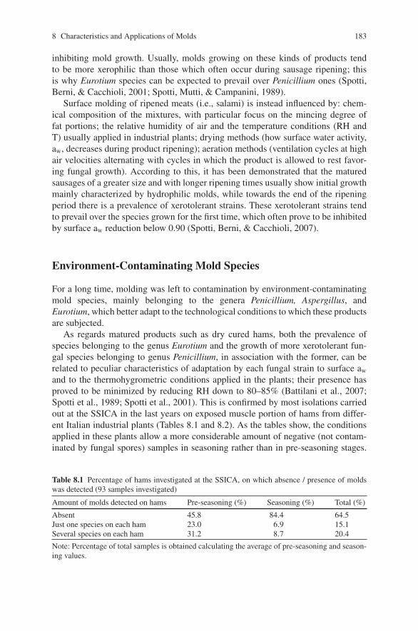
8 Characteristics and Applications of Molds 183
inhibiting mold growth. Usually, molds growing on these kinds of products tendto be more xerophilic than those which often occur during sausage ripening; thisis why Eurotium species can be expected to prevail over Penicillium ones (Spotti,Berni, & Cacchioli, 2001; Spotti, Mutti, & Campanini, 1989).
Surface molding of ripened meats (i.e., salami) is instead influenced by: chem-ical composition of the mixtures, with particular focus on the mincing degree offat portions; the relative humidity of air and the temperature conditions (RH andT) usually applied in industrial plants; drying methods (how surface water activity,aw, decreases during product ripening); aeration methods (ventilation cycles at highair velocities alternating with cycles in which the product is allowed to rest favor-ing fungal growth). According to this, it has been demonstrated that the maturedsausages of a greater size and with longer ripening times usually show initial growthmainly characterized by hydrophilic molds, while towards the end of the ripeningperiod there is a prevalence of xerotolerant strains. These xerotolerant strains tendto prevail over the species grown for the first time, which often prove to be inhibitedby surface aw reduction below 0.90 (Spotti, Berni, & Cacchioli, 2007).
Environment-Contaminating Mold Species
For a long time, molding was left to contamination by environment-contaminatingmold species, mainly belonging to the genera Penicillium, Aspergillus, andEurotium, which better adapt to the technological conditions to which these productsare subjected.
As regards matured products such as dry cured hams, both the prevalence ofspecies belonging to the genus Eurotium and the growth of more xerotolerant fun-gal species belonging to genus Penicillium, in association with the former, can berelated to peculiar characteristics of adaptation by each fungal strain to surface aw
and to the thermohygrometric conditions applied in the plants; their presence hasproved to be minimized by reducing RH down to 80–85% (Battilani et al., 2007;Spotti et al., 1989; Spotti et al., 2001). This is confirmed by most isolations carriedout at the SSICA in the last years on exposed muscle portion of hams from differ-ent Italian industrial plants (Tables 8.1 and 8.2). As the tables show, the conditionsapplied in these plants allow a more considerable amount of negative (not contam-inated by fungal spores) samples in seasoning rather than in pre-seasoning stages.
Table 8.1 Percentage of hams investigated at the SSICA, on which absence / presence of moldswas detected (93 samples investigated)
Amount of molds detected on hams Pre-seasoning (%) Seasoning (%) Total (%)
Absent 45.8 84.4 64.5Just one species on each ham 23.0 6.9 15.1Several species on each ham 31.2 8.7 20.4
Note: Percentage of total samples is obtained calculating the average of pre-seasoning and season-ing values.
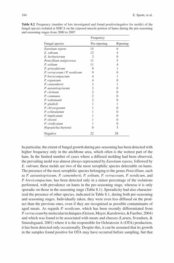
184 E. Spotti, et al.
Table 8.2 Frequency (number of lots investigated and found positive/negative for molds) of thefungal species isolated at SSICA on the exposed muscle portion of hams during the pre-seasoningand seasoning stages from 2000 to 2007
Frequency
Fungal species Pre-ripening Ripening
Eurotium repens 15 6E. rubrum 12 4E. herbariorum 2 0Penicillium nalgiovense 11 5P. solitum 11 4P. griseofulvum 9 1P. verrucosum / P. nordicum 9 0P. brevicompactum 4 1P. expansum 4 0P. camemberti 3 1P. aurantiogriseum 3 0P. citrinum 2 0P. commune 2 0P. waksmanii 2 0P. gladioli 1 1P. chrysogenum 1 0P. echinulatum 1 0P. implicatum 1 0P. olsonii 1 0P. viridicatum 1 0Hypopichia burtonii 0 5
Negative 22 38
In particular, the extent of fungal growth during pre-seasoning has been detected withhigher frequency only in the aitchbone area, which often is the wettest part of theham. In the limited number of cases where a diffused molding had been observed,the prevailing mold was almost always represented by Eurotium repens, followed byE. rubrum; these molds are two of the most xerophilic species detectable on hams.The presence of the most xerophilic species belonging to the genus Penicillium, suchas P. aurantiogriseum, P. camemberti, P. solitum, P. verrucosum, P. nordicum, andP. brevicompactum, has been detected only in a minor percentage of the isolationsperformed, with prevalence on hams in the pre-seasoning stage, whereas it is onlysporadic on those in the seasoning stage (Table 8.1). Sporadicity had also character-ized the presence of other species, indicated in Table 8.1, during both pre-seasoningand seasoning stages. Individually taken, they were even less diffused on the prod-uct than the previous ones, even if they are recognized as possible contaminants ofaged meats. As regards P. nordicum, which has been recently differentiated fromP.verrucosumbymolecular techniques (Geisen,Mayer,Karolewiez,&Faerber,2004)and which was found to be associated with meats and cheeses (Larsen, Svendsen, &Smeedsgaard, 2001) where it is the responsible for Ochratoxin A (OTA) production,it has been detected only occasionally. Despite this, it can be assumed that its growthin the samples found positive for OTA may have occurred before sampling, but that
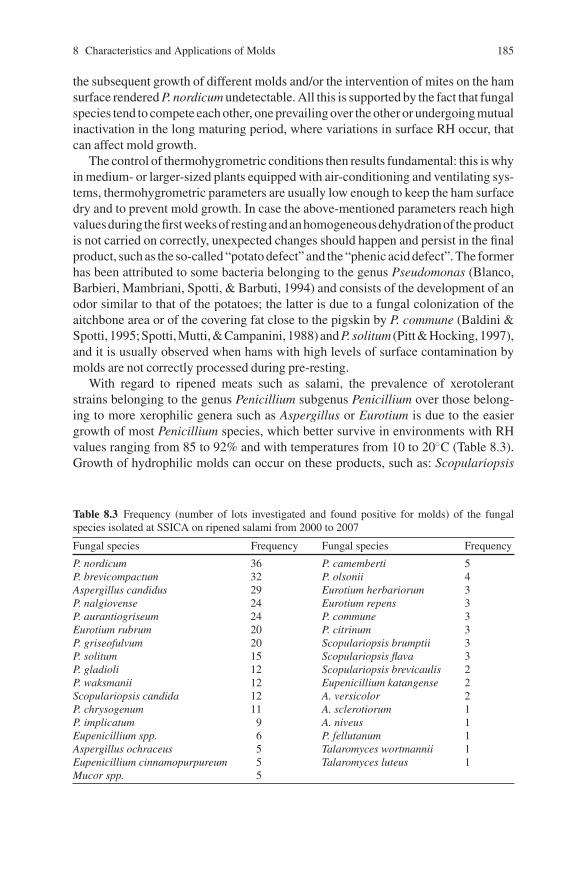
8 Characteristics and Applications of Molds 185
the subsequent growth of different molds and/or the intervention of mites on the hamsurface rendered P. nordicum undetectable. All this is supported by the fact that fungalspecies tend to compete each other, one prevailing over the other or undergoing mutualinactivation in the long maturing period, where variations in surface RH occur, thatcan affect mold growth.
The control of thermohygrometric conditions then results fundamental: this is whyin medium- or larger-sized plants equipped with air-conditioning and ventilating sys-tems, thermohygrometric parameters are usually low enough to keep the ham surfacedry and to prevent mold growth. In case the above-mentioned parameters reach highvaluesduring thefirstweeksof restingandanhomogeneousdehydrationof theproductis not carried on correctly, unexpected changes should happen and persist in the finalproduct, such as the so-called “potato defect” and the “phenic acid defect”. The formerhas been attributed to some bacteria belonging to the genus Pseudomonas (Blanco,Barbieri, Mambriani, Spotti, & Barbuti, 1994) and consists of the development of anodor similar to that of the potatoes; the latter is due to a fungal colonization of theaitchbone area or of the covering fat close to the pigskin by P. commune (Baldini &Spotti, 1995;Spotti,Mutti,&Campanini, 1988)andP. solitum (Pitt&Hocking,1997),and it is usually observed when hams with high levels of surface contamination bymolds are not correctly processed during pre-resting.
With regard to ripened meats such as salami, the prevalence of xerotolerantstrains belonging to the genus Penicillium subgenus Penicillium over those belong-ing to more xerophilic genera such as Aspergillus or Eurotium is due to the easiergrowth of most Penicillium species, which better survive in environments with RHvalues ranging from 85 to 92% and with temperatures from 10 to 20◦C (Table 8.3).Growth of hydrophilic molds can occur on these products, such as: Scopulariopsis
Table 8.3 Frequency (number of lots investigated and found positive for molds) of the fungalspecies isolated at SSICA on ripened salami from 2000 to 2007
Fungal species Frequency Fungal species Frequency
P. nordicum 36 P. camemberti 5P. brevicompactum 32 P. olsonii 4Aspergillus candidus 29 Eurotium herbariorum 3P. nalgiovense 24 Eurotium repens 3P. aurantiogriseum 24 P. commune 3Eurotium rubrum 20 P. citrinum 3P. griseofulvum 20 Scopulariopsis brumptii 3P. solitum 15 Scopulariopsis flava 3P. gladioli 12 Scopulariopsis brevicaulis 2P. waksmanii 12 Eupenicillium katangense 2Scopulariopsis candida 12 A. versicolor 2P. chrysogenum 11 A. sclerotiorum 1P. implicatum 9 A. niveus 1Eupenicillium spp. 6 P. fellutanum 1Aspergillus ochraceus 5 Talaromyces wortmannii 1Eupenicillium cinnamopurpureum 5 Talaromyces luteus 1Mucor spp. 5
186 E. Spotti, et al.
brevicaulis, S. brumptii, and Mucor spp., which tend to form darkish spots oncasings; S. flava, whose growth can impart ripened meats either desirable appear-ance as well as satisfactory technological and sensory traits or reddish undesirablespots, in case it produces fruiting bodies derived from sexual reproduction (Abbott,Lumley, & Sigler, 2002; Spotti et al., 2007); S. candida, which produces whitishmycelium and conidia, giving the sausages a good appearance. These species mustbe removed because the former do not allow homogeneous drying of the productin the first steps of the process and the latter can produce a nasty ammonia odorbecause of its strong proteolytic activity in the final step of the ripening.
Fungal Starter Cultures in Ripened Meats
In European countries where food industries aim to obtain traditional products, theripening techniques normally applied in the industrial plants usually allow for thegrowth of characteristic whitish molds which can also inhibit the multiplication ofother molds, especially those which proved to be potentially toxigenic and/or witha mycelium having an undesirable color (Baldini et al., 2000). In particular, recentfindings concerning the ability of many Penicillium and Aspergillus species, fre-quently found on the surface of dry sausages to produce mycotoxins, have stressedthe importance of studies on the possibility of controlling mold growth on rawsausages during ripening by using mold starters.
These starters consist of selected fungal isolates that proved to impart a desir-able appearance and good technological and sensory characteristics to the product,proved unable to synthesize antibiotics and/or toxic metabolites such as mycotox-ins and do not cause well-known allergies or mycoses. For the reasons mentionedabove, they can be used to inoculate fermented sausages by spraying or by immer-sion in a conidial suspension.
Among the starters, which can be considered both safe and effective,P. nalgiovense has been frequently isolated from the surface of ripened meatproducts as a “domesticated species” from P. chrysogenum (wild type) (Pitt &Hocking, 1997); at present it is routinely used as a starter culture in many traditionalproductions and so it is often present in the environmental air.
Even P. chrysogenum has been mentioned as a suitable starter for mold-fermented products (Hammes & Knauf, 1994; Ministero della Sanita, 1995),since some non-toxic strains proved to produce proteases which contributed to thegeneration of characteristic flavored compounds in dry fermented sausages (Martınet al., 2002; Rodrıguez, Nunez, Cordoba, Bermudez, & Asensio, 1998). However,at present P. chrysogenum is not always considered acceptable in the industrialpractice because the marketed isolates with a lightly pigmented conidiation mayagain start producing green conidia, and most of the strains tested may produceantibiotics and toxic substances such as roquefortine C (Pitt & Hocking, 1997).
P. camemberti had also been considered as a starter culture for the meat indus-try because of its ability to improve the sensory characteristics of dry fermentedsausages (Bruna et al., 2003). Nevertheless, all the studied isolates proved to be
8 Characteristics and Applications of Molds 187
cyclopiazonic-acid producers, so it has not been taken into account as a commercialstarter (Samson & Frisvad, 2004). Other strains belonging to the non-toxigenic Peni-cillium species were also excluded by the industrial practice because they producedarkish-green conidia which may impair the outward appearance of the sausage.
In order to safeguard typical productions, in case both the appearance and aromaimparted to ripened meats by autochthonous molds come up to the expectations ofa peculiar quality, molding should be favored by means of preventive and specificstudies. These studies should be at first focused on the recognition of the genusand the species to which the most occurring (on that specific meat product) strainsbelong; then, on the selection of the strain which has been considered safer and moresuitable because of its biochemical and physiological characteristics.
At the beginning, the identification of the species will be entrusted to expertmycologists, who are usually able to carry out any morphological, biochemical,and, when possible, molecular assays in order to identify molds. In the industrialpractice, the dominance of the above-mentioned autochthonous strains over theenvironmental contaminating fungal species will be then favored by means ofthe most appropriate technological interventions. Regular inspections concerningthe real presence of the autochthonous starter culture inoculated are part of anyQuality Control system, and they must be carried on throughout the ripeningprocess.
In the last years, several experiments have been carried out at the SSICA, makinguse of two isolates of P. gladioli: one from Professor Grazia collection (Grazia,Romano, Bagni, Roggiani, & Guglielmi, 1986), the other from the SSICA myco-logical collection. At present, both strains, isolated on meat products from EmiliaRomagna (Northern Italy), can be considered as “real autochthonous starters”. Theyhave proved to be non-mycotoxin producers and impart to ripened meats a desir-able appearance, due to a pale-grayish conidiation (Baldini et al., 2005; Graziaet al., 1986; Samson & Frisvad, 2004).
S. flava, for a long time now used as starter culture in some French industries pro-ducing dry-smoked sausages (Dragoni, Cantoni, Papa, & Vallone, 1997), has beenisolated from salami and used too as “autochthonous starter” in recent experimentscarried out at the SSICA. This mold morphologically resembles S. brevicaulis but,unlike it, S. flava can impart on ripened meats a desirable appearance, due to agrayish-beige conidiation. Only if a high relative humidity is present, S. flava mustbe avoided, because it develops nasty reddish spots and a strong ammonia odor.
Lipolytic and Proteolytic Activity of Molds
In the last years, a lot of studies concerning the lipolytic and proteolytic activ-ity of the molds involved in the ripening process, including P. nalgiovense,P. chrysogenum, and P. camemberti, were carried out throughout the world (Brunaet al., 2003; Geisen, Lucke, & Krockel, 1992; Ockerman, Cespedes Sanchez, OrtegaMariscal, & Leon-Crespo, 2001; Rodriguez et al., 1998).
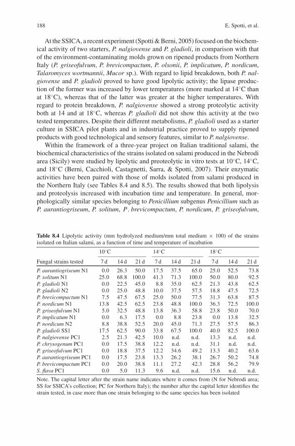
188 E. Spotti, et al.
At the SSICA, a recent experiment (Spotti & Berni, 2005) focused on the biochem-ical activity of two starters, P. nalgiovense and P. gladioli, in comparison with thatof the environment-contaminating molds grown on ripened products from NorthernItaly (P. griseofulvum, P. brevicompactum, P. olsonii, P. implicatum, P. nordicum,Talaromyces wortmannii, Mucor sp.). With regard to lipid breakdown, both P. nal-giovense and P. gladioli proved to have good lipolytic activity; the lipase produc-tion of the former was increased by lower temperatures (more marked at 14◦C thanat 18◦C), whereas that of the latter was greater at the higher temperatures. Withregard to protein breakdown, P. nalgiovense showed a strong proteolytic activityboth at 14 and at 18◦C, whereas P. gladioli did not show this activity at the twotested temperatures. Despite their different metabolisms, P. gladioli used as a starterculture in SSICA pilot plants and in industrial practice proved to supply ripenedproducts with good technological and sensory features, similar to P. nalgiovense.
Within the framework of a three-year project on Italian traditional salami, thebiochemical characteristics of the strains isolated on salami produced in the Nebrodiarea (Sicily) were studied by lipolytic and preoteolytic in vitro tests at 10◦C, 14◦C,and 18◦C (Berni, Cacchioli, Castagnetti, Sarra, & Spotti, 2007). Their enzymaticactivities have been paired with those of molds isolated from salami produced inthe Northern Italy (see Tables 8.4 and 8.5). The results showed that both lipolysisand proteolysis increased with incubation time and temperature. In general, mor-phologically similar species belonging to Penicillium subgenus Penicillium such asP. aurantiogriseum, P. solitum, P . brevicompactum, P. nordicum, P. griseofulvum,
Table 8.4 Lipolytic activity (mm hydrolyzed medium/mm total medium × 100) of the strainsisolated on Italian salami, as a function of time and temperature of incubation
10◦C 14◦C 18◦C
Fungal strains tested 7 d 14 d 21 d 7 d 14 d 21 d 7 d 14 d 21 d
P. aurantiogriseum N1 0.0 26.3 50.0 17.5 37.5 65.0 25.0 52.5 73.8P. solitum N1 25.0 68.8 100.0 41.3 71.3 100.0 50.0 80.0 92.5P. gladioli N1 0.0 22.5 45.0 8.8 35.0 62.5 21.3 43.8 62.5P. gladioli N2 0.0 25.0 48.8 10.0 37.5 57.5 18.8 47.5 72.5P. brevicompactum N1 7.5 47.5 67.5 25.0 50.0 77.5 31.3 63.8 87.5P. nordicum N1 13.8 42.5 62.5 23.8 48.8 100.0 36.3 72.5 100.0P. griseofulvum N1 5.0 32.5 48.8 13.8 36.3 58.8 23.8 50.0 70.0P. implicatum N1 0.0 6.3 17.5 0.0 8.8 23.8 0.0 13.8 32.5P. nordicum N2 8.8 38.8 52.5 20.0 45.0 71.3 27.5 57.5 86.3P. gladioli SS1 17.5 62.5 90.0 33.8 67.5 100.0 40.0 82.5 100.0P. nalgiovense PC1 2.5 21.3 42.5 10.0 n.d. n.d. 13.3 n.d. n.d.P. chrysogenum PC1 0.0 17.5 38.8 12.2 n.d. n.d. 31.1 n.d. n.d.P. griseofulvum PC1 0.0 18.8 37.5 12.2 34.6 49.2 13.3 40.2 63.6P. aurantiogriseum PC1 0.0 17.5 23.8 13.3 26.2 38.1 26.7 50.2 74.8P. brevicompactum PC1 0.0 20.0 38.8 11.1 27.2 42.3 28.8 56.2 79.9S. flava PC1 0.0 5.0 11.3 9.6 n.d. n.d. 15.6 n.d. n.d.
Note. The capital letter after the strain name indicates where it comes from (N for Nebrodi area;SS for SSICA’s collection; PC for Northern Italy); the number after the capital letter identifies thestrain tested, in case more than one strain belonging to the same species has been isolated
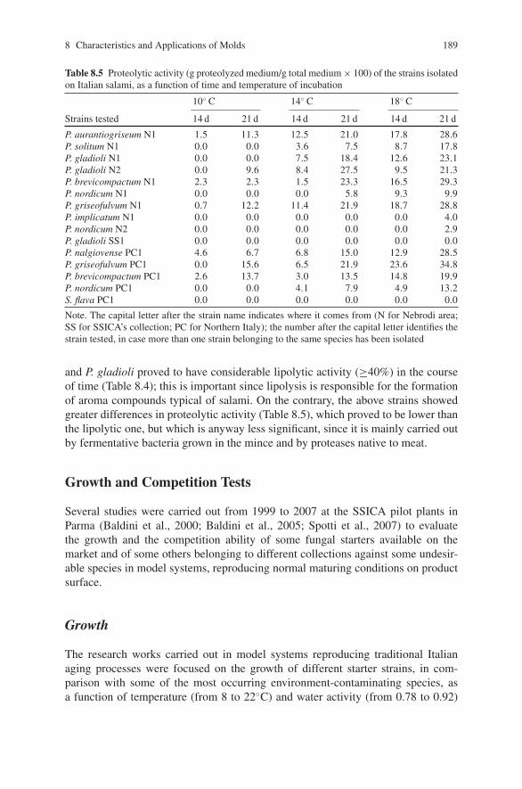
8 Characteristics and Applications of Molds 189
Table 8.5 Proteolytic activity (g proteolyzed medium/g total medium × 100) of the strains isolatedon Italian salami, as a function of time and temperature of incubation
10◦ C 14◦ C 18◦ C
Strains tested 14 d 21 d 14 d 21 d 14 d 21 d
P. aurantiogriseum N1 1.5 11.3 12.5 21.0 17.8 28.6P. solitum N1 0.0 0.0 3.6 7.5 8.7 17.8P. gladioli N1 0.0 0.0 7.5 18.4 12.6 23.1P. gladioli N2 0.0 9.6 8.4 27.5 9.5 21.3P. brevicompactum N1 2.3 2.3 1.5 23.3 16.5 29.3P. nordicum N1 0.0 0.0 0.0 5.8 9.3 9.9P. griseofulvum N1 0.7 12.2 11.4 21.9 18.7 28.8P. implicatum N1 0.0 0.0 0.0 0.0 0.0 4.0P. nordicum N2 0.0 0.0 0.0 0.0 0.0 2.9P. gladioli SS1 0.0 0.0 0.0 0.0 0.0 0.0P. nalgiovense PC1 4.6 6.7 6.8 15.0 12.9 28.5P. griseofulvum PC1 0.0 15.6 6.5 21.9 23.6 34.8P. brevicompactum PC1 2.6 13.7 3.0 13.5 14.8 19.9P. nordicum PC1 0.0 0.0 4.1 7.9 4.9 13.2S. flava PC1 0.0 0.0 0.0 0.0 0.0 0.0
Note. The capital letter after the strain name indicates where it comes from (N for Nebrodi area;SS for SSICA’s collection; PC for Northern Italy); the number after the capital letter identifies thestrain tested, in case more than one strain belonging to the same species has been isolated
and P. gladioli proved to have considerable lipolytic activity (≥40%) in the courseof time (Table 8.4); this is important since lipolysis is responsible for the formationof aroma compounds typical of salami. On the contrary, the above strains showedgreater differences in proteolytic activity (Table 8.5), which proved to be lower thanthe lipolytic one, but which is anyway less significant, since it is mainly carried outby fermentative bacteria grown in the mince and by proteases native to meat.
Growth and Competition Tests
Several studies were carried out from 1999 to 2007 at the SSICA pilot plants inParma (Baldini et al., 2000; Baldini et al., 2005; Spotti et al., 2007) to evaluatethe growth and the competition ability of some fungal starters available on themarket and of some others belonging to different collections against some undesir-able species in model systems, reproducing normal maturing conditions on productsurface.
Growth
The research works carried out in model systems reproducing traditional Italianaging processes were focused on the growth of different starter strains, in com-parison with some of the most occurring environment-contaminating species, asa function of temperature (from 8 to 22◦C) and water activity (from 0.78 to 0.92)
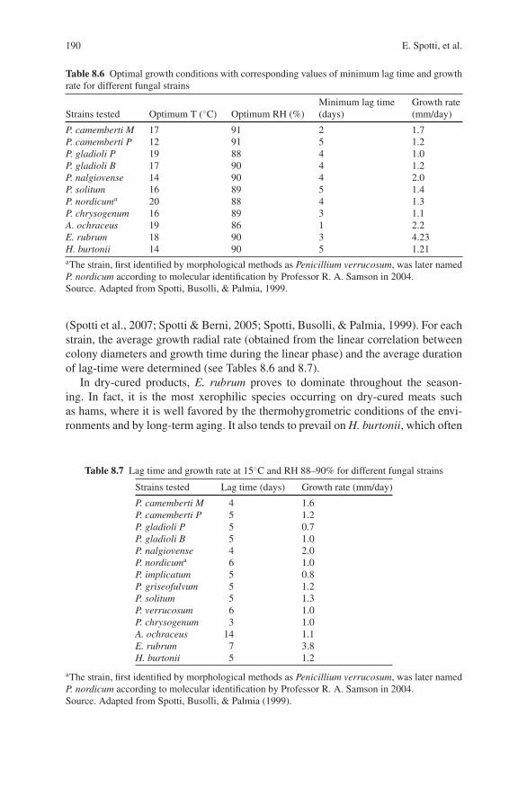
190 E. Spotti, et al.
Table 8.6 Optimal growth conditions with corresponding values of minimum lag time and growthrate for different fungal strains
Strains tested Optimum T (◦C) Optimum RH (%)Minimum lag time(days)
Growth rate(mm/day)
P. camemberti M 17 91 2 1.7P. camemberti P 12 91 5 1.2P. gladioli P 19 88 4 1.0P. gladioli B 17 90 4 1.2P. nalgiovense 14 90 4 2.0P. solitum 16 89 5 1.4P. nordicuma 20 88 4 1.3P. chrysogenum 16 89 3 1.1A. ochraceus 19 86 1 2.2E. rubrum 18 90 3 4.23H. burtonii 14 90 5 1.21aThe strain, first identified by morphological methods as Penicillium verrucosum, was later namedP. nordicum according to molecular identification by Professor R. A. Samson in 2004.Source. Adapted from Spotti, Busolli, & Palmia, 1999.
(Spotti et al., 2007; Spotti & Berni, 2005; Spotti, Busolli, & Palmia, 1999). For eachstrain, the average growth radial rate (obtained from the linear correlation betweencolony diameters and growth time during the linear phase) and the average durationof lag-time were determined (see Tables 8.6 and 8.7).
In dry-cured products, E. rubrum proves to dominate throughout the season-ing. In fact, it is the most xerophilic species occurring on dry-cured meats suchas hams, where it is well favored by the thermohygrometric conditions of the envi-ronments and by long-term aging. It also tends to prevail on H. burtonii, which often
Table 8.7 Lag time and growth rate at 15◦C and RH 88–90% for different fungal strains
Strains tested Lag time (days) Growth rate (mm/day)
P. camemberti M 4 1.6P. camemberti P 5 1.2P. gladioli P 5 0.7P. gladioli B 5 1.0P. nalgiovense 4 2.0P. nordicuma 6 1.0P. implicatum 5 0.8P. griseofulvum 5 1.2P. solitum 5 1.3P. verrucosum 6 1.0P. chrysogenum 3 1.0A. ochraceus 14 1.1E. rubrum 7 3.8H. burtonii 5 1.2
aThe strain, first identified by morphological methods as Penicillium verrucosum, was later namedP. nordicum according to molecular identification by Professor R. A. Samson in 2004.Source. Adapted from Spotti, Busolli, & Palmia (1999).
8 Characteristics and Applications of Molds 191
derives from the spreadable fat mince (“sugna”) after the sixth month of seasoningand which is now beginning to be studied (Simoncini et al., 2007).
On the contrary, the molds most likely to grow during drying in ripened salamiare P. gladioli, P. solitum, P. nordicum, and P. chrysogenum, since they are favoredby the short-term ripening and by the thermohygrometric conditions for optimumgrowth (Table 8.6). However, it is also possible to find P. camemberti, A. ochraceus(Mutti, Previdi, Quintavalla, & Spotti, 1988), and P. griseofulvum (Spotti &Berni, 2005). Under these conditions, the suitability of initial treatment of theproducts with starter cultures proves recommendable. It has also been concludedthat P. nalgiovense and P. camemberti are particularly able to proliferate duringripening, on the basis of the temperature for optimum growth as reported inTable 8.6. However, for a better comparison of the various strains, lag time andgrowth rate values were determined at 15◦C keeping the relative humidity at theoptimal value for each individual strain (Table 8.7).
Tables 8.6 and 8.7 illustrate different dominance over “undesired molds”. Inripened products, the strain for which dominance over the “undesired species” ispredictable is P. nalgiovense, since it has the highest growth rate under the tem-perature conditions indicated. Even though P. chrysogenum has a shorter lag time(3 days) as compared to P. nalgiovense (4 days), its growth rate is half that ofP. nalgiovense and its mycelium is therefore less invasive. On the contrary, togetherwith P. camemberti and P. gladioli strains, assuming equal contamination levels,also P. chrysogenum, P. solitum, P. griseofulvum, and P. nordicum may be presenton the same product.
Tables 8.6 and 8.7 also illustrate that RH proves to have a greater influence thantemperature for almost all strains; this accounts for the lag time at optimum RHin Table 8.7 not being too different from those in Table 8.6. The only exception isA. ochraceus, which is much more sensitive to temperature changes than to humid-ity, as it is shown by its long lag time at 15◦C (14 days), whereas under optimumconditions it is only 1 day.
Recently, the course of growth rate of the fungal species that more frequentlyoccur on the surface of molded meat products during ripening has also been inves-tigated (Spotti & Berni, 2005). Table 8.8 clearly shows how the growth of startercultures such as P. nalgiovense and P. gladioli is favored by a higher relative humid-ity in the first days of ripening.
Competition
According to the techniques tested by Wheeler & Hocking (1993), several compe-tition trials were carried out, where each starter was inoculated in combination withthe undesired strain, each at a time (Spotti et al., 1999; Spotti & Berni, 2005; Spotti,Mutti, & Scalari, 1994). The results of these experiments showed that P. nalgiovensemaybeovergrownbyA.ochraceusduringdrying (T ≥ 15◦C),whereasP. nalgiovensewill dominate this undesired mold during ripening (T ≤ 15◦C) as previously referred.
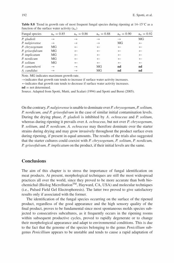
192 E. Spotti, et al.
Table 8.8 Trend in growth rate of most frequent fungal species during ripening at 14–15◦C as afunction of the surface water activity (aw)
Fungal species aw = 0.85 aw = 0.86 aw = 0.88 aw = 0.90 aw = 0.92
P. gladioliP. nalgiovenseP. chrysogenumP. griseofulvumP. implicatumP. nordicumP. solitumP. camembertiA. candidus
→→MGMGMGMGMG→→
→→←←←←←→→
→→←←←←←MGMG
→MG←←←←←ndnd
MG←←←←←←ndnd
Note. MG indicates maximum growth rate.→indicates that growth rate tends to increase if surface water activity increases.←indicates that growth rate tends to decrease if surface water activity increases.nd = not determined.Source. Adapted from Spotti, Mutti, and Scalari (1994) and Spotti and Berni (2005).
On the contrary, P. nalgiovense is unable to dominate over P. chrysogenum, P. solitum,P. nordicum, and P. griseofulvum in the case of similar initial contamination levels.During the drying phase, P. gladioli is inhibited by A. ochraceus and P. solitum,whereas during ripening it prevails over A. ochraceus, but not over P. chrysogenum,P. solitum, and P. nordicum. A. ochraceus may therefore dominate over the starterstrains during drying and may grow invasively throughout the product surface evenduring ripening, if present in equal amounts. The results of the trials also suggestedthat the starter cultures could coexist with P. chrysogenum, P. solitum, P. nordicum,P. griseofulvum, P. implicatum on the product, if their initial levels are the same.
Conclusions
The aim of this chapter is to stress the importance of fungal identification onmeat products. At present, morphological techniques are still the most widespreadpractices all over the world, since they proved to be more accurate than both bio-chemichal (Biolog MicroStationTM, Hayward, CA, USA) and molecular techniques(i.e., Pulsed Field Gel Electrophoresis). The latter two proved to give satisfactoryresults only if associated with the former.
The identification of the fungal species occurring on the surface of the ripenedproduct, regardless of the good appearance and the high sensory quality of thefinal product, proves to be fundamental since most spontaneous molds species sub-jected to consecutives subcultures, as it frequently occurs in the ripening roomswithin subsequent productive cycles, proved to rapidly degenerate or to changetheir morphological appearance and adapt to environmental conditions. This is dueto the fact that the genome of the species belonging to the genus Penicillium sub-genus Penicillium appears to be unstable and tends to cause a rapid adaptation of
8 Characteristics and Applications of Molds 193
the species to the nutritional niches available (Williams, Pitt, & Hocking, 1985).The above-mentioned variations cannot always be considered beneficial to the finalproduct.
In case it is decided for the growth of a selected fungal strain, the choice mustfirst be addressed to that species which proved not to impair the outward appearanceof the sausages, in each step of the ripening process because of its heavy-coloredconidiation (this is what frequently happens when P. solitum, P. brevicompactum,and P . chrysogenum grow on surface of meat products), and which proved not toproduce toxic metabolites (as for P. nordicum or P. verrucosum, whose isolates inmost cases result to be OTA-producers). Lipolytic and proteolytic activities of moldscan be taken into account when selecting a proper starter culture; these two char-acteristics become important when choosing the strain to inoculate within the samespecies, in order to obtain the best quality standards.
The poor competitiveness of superficial starter cultures compared with othercontaminants requires a low rate of environmental contamination; therefore startermolds should be used at high inoculation levels on sausages to be ripened in orderto avoid possible development of undesirable species.
The control on the growth of the selected species and their actual predominanceover undesirable molds should be periodically planned on the basis of routine labo-ratory tests (i.e., isolation and identification of the species employed in the industrialprocess) in order to avoid the unexpected setting up of environment-contaminatingmold species, which are morphologically similar to the selected ones.
References
Abbott, S. P., Lumley, T. C., & Sigler, L. (2002). Use of holomorph characters to delimit Microas-cus nidicola and M. sopii sp. nov., with notes on the genus Pithoascus. Mycologia, 92(2),362–369.
Baldini, P., Berni, E., Diaferia, C., Follini, A., Palmisano, S., Rossi, A., et al. (2005). Materia primaflora microbica nella produzione dei salumi piacentini. Rivista di Suinicoltura, 4, 183–187.
Baldini, P., Cantoni, E., Colla, F., Diaferia, C., Gabba, L., Spotti, E., et al. (2000). Drysausages ripening: Influence of thermohygrometric conditions on microbiological, chemicaland physico-chemical characteristics. Food Research International, 33, 161–170.
Baldini, P., & Spotti, E. (1995). Importanza della fase di preriposo sulla probabilita di ritrovareodori estranei (acido fenico) in prosciutti stagionati. Industria Conserve, 70, 418–422.
Battilani, P., Pietri, A., Giorni, P., Formenti, S., Bertuzzi, T., Toscani, T., et al. (2007). Penicil-lium populations in dry-cured ham manufacturing plants. Journal of Food Protection, 70(4),975–980.
Berni, E., Cacchioli, C., Castagnetti, G., Sarra, P. G., & Spotti, E. (2007). Microbial surface col-onization in Nebrodi salami. Proceedings of the 6th International Symposium on the Mediter-ranean Pig, October 11–13, 2007 – Capo d’Orlando & Messina (Italy)
Blanco, D., Barbieri, G., Mambriani, P., Spotti, E., & Barbuti, S. (1994). Studio sul “difetto dipatata” nel prosciutto crudo stagionato. Industria Conserve, 69, 230–236.
Bruna, J. M., Hierro, E. M., De la Hoz, L., Mottram, D. S., Fernandez, M., & Ordonez, J. A.(2003). Changes in selected biochemical and sensory parameters as affected by the superficialinoculation of Penicillium camemberti on dry fermented sausages. International Journal ofFood Microbiology, 85, 111–125.
194 E. Spotti, et al.
Comi, G., & Cantoni, C. (1983). Yeasts in dry Parma hams. Industrie Alimentari, 22, 102–104.De Hoog, G. S., Guarro, J., Gene, J., & Figueras, M. J. (2000). In G. S. de Hoog, J. Guardo, J.
Gene, & M. J. Figueras (Eds.), Atlas of clinical fungi (pp. 21–29). Utrecht: CBS.Dragoni, I., Cantoni, C., Papa, A., & Vallone, L. (1997). In I. Dragoni (Ed.), Muffe alimenti e
micotossicosi. Milano: UTET, CittaStudiEdizioni.Geisen, R., Lucke, F. K., & Krockel, L. (1992). Starter and protective cultures for meat and meat
products. Fleischwirtschaft, 72(6), 894–898.Geisen, R., Mayer, Z., Karolewiez, A., & Faerber, P. (2004). Development of a real time PCR
system for detection of Penicillium nordicum and for monitoring of ochratoxin A productionin foods by targeting the ochratoxin polyketide synthase gene. Systematic and Applied Micro-biology, 27(4), 501–507.
Grazia, L., Romano, P., Bagni, A., Roggiani, D., & Guglielmi, D. (1986). The role of moulds inthe ripening process of salami. Food Microbiology, 3, 19–25.
Hammes, W. P., & Knauf, H. J. (1994). Starter in the processing of meat products. Meat Science,36, 155–168.
Larsen, T. O., Svendsen, A., & Smeedsgaard, J. (2001). Biochemical characterization of Ochra-toxin A producing strains of the genus Penicillium. Applied and Environmental Microbiology,67, 3630–3635.
Lori, D., Grisenti, M. S., Parolari, G., & Barbuti, S. (2005). Microbiology of dry-cured ham. Indus-tria Conserve, 80, 23–32.
Martın, A., Asensio, M. A., Bermudez, M. E., Cordoba, M. G., Aranda, E., & Cordoba, J. J. (2002).Proteolytic activity of Penicillium chrysogenum and Debaryomyces hansenii during controlledripening of pork loins. Meat Science, 62, 129–137.
Martin, A., Cordoba, J. J., Nunez, F., & Asensio, M. A. (2004). Contribution of a selected fungalpopulation to proteolysis on dry cured ham. International Journal of Food Microbiology, 94(1),55–56.
Martın, A., Cordoba, J. J., Aranda, E., Cordoba, M. G., & Asensio, M. A. (2006). Contribution of aselected fungal population to the volatile compounds on dry-cured ham. International Journalof Food Microbiology, 110(1), 8–18.
Ministero della Sanita. (1994). Decreto 28 Dicembre 1994. Gazzetta Ufficiale Repubblica Italiana.Serie generale n. 89 (pp. 4–5), 15.04.1995.
Mutti, P., Previdi, M. P., Quintavalla, S., & Spotti, E. (1988). Toxigenity of mould strains isolatedfrom salami as a function of culture medium. Industria Conserve, 63, 142–145.
Ockerman, H., W., Cespedes Sanchez, F. J., Ortega Mariscal, M. A., & Leon-Crespo, F. (2001).The lipolytic activity of some indigenous Spanish moulds isolated from meat products. Journalof Muscle Foods, 12, 275–284.
Pietri, A., Bertuzzi, T., Gualla, A., & Piva, G. (2006). Occurrence of ochratoxin A in raw hammuscles and in pork products from northern Italy. Italian Journal Food Science, 18(1), 99–106.
Pitt, J. I., & Hocking, A. D. (1997). Fungi and Food Spoilage (2nd ed.). Cambridge: UniversityPress. Blackie Academic & Professional.
Rodrıguez, M., Nunez, F., Cordoba, J. J., Bermudez, M. E., & Asensio, M. A. (1998). Evaluationof proteolytic activity of micro-organisms isolated from dry cured ham. Journal of AppliedMicrobiology, 85, 905–912.
Samelis, J., & Sofos, J. N. (2003). In T. Boekhout, & V. Robert (Eds.) Yeasts in Foods (pp. 239–265). Cambridge: Woodhead Publishing Limited.
Samson, R. A., & Frisvad, J. C. (2004). Penicillium subgenus Penicillium: New taxonomic schemes,mycotoxins and other extrolites. Uthrecht: CBS.
Simoncini, N., Rotelli, D., Virgili, R., & Quintavalla, S. (2007). Dynamic and characterization ofyeasts during ripening of typical Italian dry-cured ham. Food Microbiology, 24(6), 577–584.
Spotti, E., & Berni, E. (2005). Variazione della flora microbica superficiale nei salumi oggettodella ricerca. Oral Presentation of 2d Annual Conference on the Research Project “Salumipiacentini”. October 25, 2005. Parma, Italy: SSICA.
Spotti, E., Berni, E., & Cacchioli, C. (2007). Le muffe sugli insaccati stagionati. Rassegna e aggior-namento sui progressi della ricerca SSICA Industria Conserve, 82(3), 243–250.

8 Characteristics and Applications of Molds 195
Spotti, E., Busolli, C., & Palmia, F. (1999). Growth of mould cultures of primary importance in themeat product industry. Industria Conserve, 74, 23–33.
Spotti, E., Chiavaro, E., Lepiani, A., & Colla, F. (2001). Mould and ochratoxin A contaminationof pre-ripened and fully ripened hams. Industria Conserve, 76, 341–354.
Spotti, E., Mutti, P., & Campanini, M. (1988). Indagine microbiologica sul “difetto dell’acidofenico” sul prosciutto durante la stagionatura. Industria Conserve, 63, 343–346.
Spotti, E., Mutti, P., & Campanini, M. (1989). Presenza di muffe sui prosciutti durante la presta-gionatura e la stagionatura: Contaminazione degli ambienti e sviluppo sulla porzione musco-lare. Industria Conserve, 64, 110–113.
Spotti, E., Mutti, P., & Scalari, F. (1994). P. nalgiovense, P. gladioli, P. candidum e A. candidus:Possibilita d’impiego quali colture starter. Industria Conserve, 69, 237–241.
Wheeler, K. A., & Hocking, A. D. (1993). Interactions among xerophilic fungi associated withdried salted fish. Journal of Applied Bacteriology, 74, 164–169.
Williams, A. P., Pitt, J. I., & Hocking, A. D. (1985). The closely related species of subgenusPenicillium – A phylogenic exploration. In R. A. Samson, & J. I. Pitt (Eds.), Advances inAspergillus and Penicillium Systematics (pp. 121–128). New York: Plenum Press.

Part IIIBiotechnology for Better Quality
and Nutritional Properties of Meat Products
Chapter 9Biotechnology of Flavor Generationin Fermented Meats
Fidel Toldra
Introduction
Traditionally, meat fermentation was based on the use of natural flora, including the“back-slopping”, or addition of a previous successful fermented sausage. However,these practices gave a great variability in the developed flora and affected thesafety and quality of the sausages (Toldra, 2002; Toldra & Flores, 2007). Thenatural flora of fermented meat has been studied for many years (Leistner, 1992;Toldra, 2006a), and more recently, these micro-organisms have been isolated andbiochemically identified through molecular methods applied to extracted DNAand RNA (Cocolin, Manzano, Aggio, Cantoni, & Comi, 2001; Cocolin, Manzano,Cantoni, & Comi, 2001; Comi, Urso, Lacumin, Rantsiou, Cattaneo & Cantoni,2005). Today, most of the fermented sausages are produced by using microbialstarters, usually from both Lactobacilli and Micrococcaceae because this com-bination ensures rapid acidulation and optimal flavor development (Demeyer &Toldra, 2004; Toldra, Nip, & Hui, 2007). The main groups of micro-organisms usedas starters are briefly described below.
Main Groups of Microbial Starters
Lactic Acid Bacteria (LAB)
Lactobacilli are the most competitive micro-organisms usually found in fermentedmeat products and are also essential components of starter cultures. These bacte-ria are able to metabolize glucose or any other available carbohydrate to generatelactic acid through either homo or heterofermentative pathways. The accumulationof lactic acid produces a pH drop towards acidic values. The homofermentativepathway only produces lactic acid but in the case of heterofermentative pathway,some undesirable secondary products (i.e.,- acetic acid, hydrogen peroxide, acetoin,
F. ToldraInstituto de Agroquımica y Tecnologıa de Alimentos (CSIC), PO Box 73, 46100 Burjassot(Valencia), Spain
F. Toldra (ed.), Meat Biotechnology, 199C© Springer Science+Business Media, LLC 2008
200 F. Toldra
etc.) may also be produced (Demeyer, 1992). Lactobacillus sakei and Lactobacilluscurvatus grow well at mild temperatures (20–25◦C), and this is the main reasonwhy they are mainly used in European sausages (mild fermentation temperatures).On the other hand, L. plantarum and Pediococcus acidilactici are used in the U.Sbecause they grow better at higher temperatures (30–35◦C) (Toldra, 2007a). LABhave a proteolytic system, with a good number of peptidases, which may partlycontribute to the degradation of muscle proteins. Most important proteases foundin L. sakei are reported in Table 9.1. Further description on the genetics of LAB isgiven in Chap. 6.
Micrococcacceae
Staphylococcus and Kocuria (formerly Micrococcus) are the main representatives ofthis family. These micro-organisms have important enzymes like some proteases andlipases that contribute to flavor generation as will be later described. Other interest-ing enzymes are nitrate reductase, that reduces nitrate to nitrite and contributes to theformation of nitrosylmyoglobin giving rise to the typical color formation and safetyof the sausage and catalase that helps to prevent lipid oxidation and improve flavorformation and color stability (Talon, Walter, Chartier, Barriere, & Montel, 1999).
Yeasts
Debaryomyces hansenii has been found as the predominant yeast in fermentedsausages. This yeast grows preferentially in the outer area of the sausage due toits aerobic metabolism. D. hansenii has a good proteolytic activity (Bolumar, Sanz,Aristoy, & Toldra, 2003a, 2003b, 2005) as shown in Table 9.1, and its lipolyticactivity is able to generate flavored volatile compounds from branched-chain aminoacids (Dura, Flores, & Toldra, 2004a). Another important contribution to sausageflavor is through its deaminase/deamidase activity on certain free amino acids thatgenerate ammonia as a by-product which rises the pH in the sausage (Dura, Flo-res, & Toldra, 2002). Further description on the genetics of yeasts is given inChap. 7.
Molds
A white coating on the surface of dry fermented sausages is typical in certain areaslike the Mediterranean. The most usual molds identified are Penicillium nalgiovenseand P. chrysogenum (Sunesen & Stahnke, 2003). They contribute to the flavorthrough their proteolytic and lipolytic activity as well as to the ammonia generatedby deamination and deamidation. Further extensive description on molds is given inChap. 8.
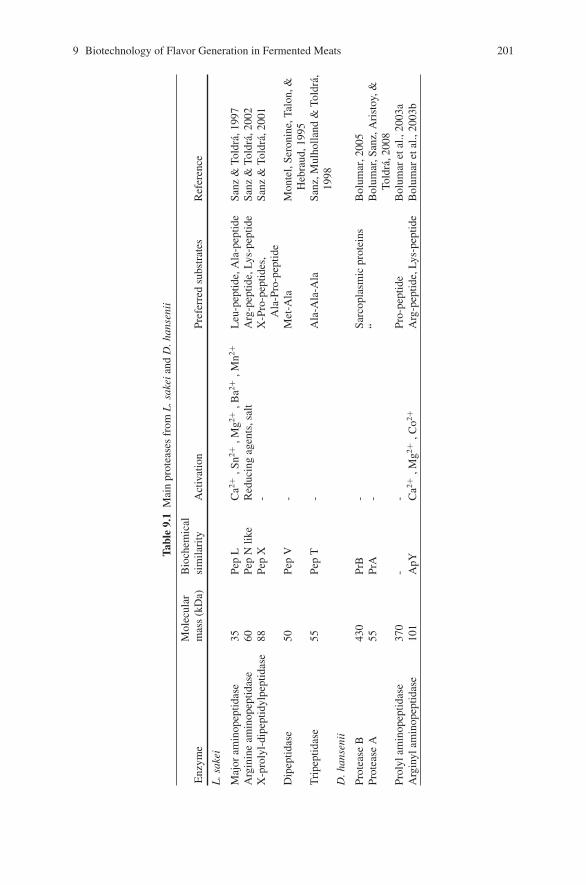
9 Biotechnology of Flavor Generation in Fermented Meats 201
Tabl
e9.
1M
ain
prot
ease
sfr
omL
.sak
eian
dD
.han
seni
i
Enz
yme
Mol
ecul
arm
ass
(kD
a)B
ioch
emic
alsi
mila
rity
Act
ivat
ion
Pref
erre
dsu
bstr
ates
Ref
eren
ce
L.s
akei
Maj
oram
inop
eptid
ase
35Pe
pL
Ca2+
,Sn2+
,Mg2+
,Ba2+
,Mn2+
Leu
-pep
tide,
Ala
-pep
tide
Sanz
&To
ldra
,199
7A
rgin
ine
amin
opep
tidas
e60
Pep
Nlik
eR
educ
ing
agen
ts,s
alt
Arg
-pep
tide,
Lys-
pept
ide
Sanz
&To
ldra
,200
2X
-pro
lyl-
dipe
ptid
ylpe
ptid
ase
88Pe
pX
-X
-Pro
-pep
tides
,A
la-P
ro-p
eptid
eSa
nz&
Told
ra,2
001
Dip
eptid
ase
50Pe
pV
-M
et-A
laM
onte
l,Se
roni
ne,T
alon
,&H
ebra
ud,1
995
Tri
pept
idas
e55
Pep
T-
Ala
-Ala
-Ala
Sanz
,Mul
holla
nd&
Told
ra,
1998
D.h
anse
nii
Prot
ease
B43
0Pr
B-
Sarc
opla
smic
prot
eins
Bol
umar
,200
5Pr
otea
seA
55Pr
A-
“B
olum
ar,S
anz,
Ari
stoy
,&To
ldra
,200
8Pr
olyl
amin
opep
tidas
e37
0-
-Pr
o-pe
ptid
eB
olum
aret
al.,
2003
aA
rgin
ylam
inop
eptid
ase
101
ApY
Ca2+
,Mg2+
,Co2+
Arg
-pep
tide,
Lys-
pept
ide
Bol
umar
etal
.,20
03b
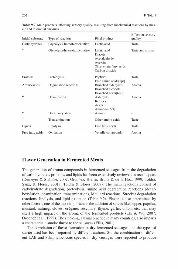
202 F. Toldra
Table 9.2 Main products, affecting sensory quality, resulting from biochemical reactions by mus-cle and microbial enzymes
Initial substrate Type of reaction Final productEffect on sensoryquality
Carbohydrates Glycolysis-homofermentative Lactic acid Taste
“ Glycolysis-heterofermentative Lactic acidDiacetylAcetaldehydeAcetoinShort chain fatty acidsCarbon dioxide
Taste and aroma
Proteins Proteolysis PeptidesFree amino acids[6pt]
Taste
Amino acids Degradation reactions Branched aldehydesBranched alcoholsBranched acids[6pt]
Aroma
“ Deamination AldehydesKetonesAcidsAmmonia[6pt]
Aroma
“ Decarboxylation Amines -
“ Transamination Other amino acids Taste
Lipids Lipolysis Free fatty acids Taste
Free fatty acids Oxidation Volatile compounds Aroma
Flavor Generation in Fermented Meats
The generation of aroma compounds in fermented sausages from the degradationof carbohydrates, proteins, and lipids has been extensively reviewed in recent years(Demeyer & Stahnke, 2002; Ordonez, Hierro, Bruna & de la Hoz, 1999; Toldra,Sanz, & Flores, 2001a; Toldra & Flores, 2007). The main reactions consist ofcarbohydrate degradation, proteolysis, amino acid degradation reactions (decar-boxylation, deamination, transamination), Maillard reactions, Strecker degradationreactions, lipolysis, and lipid oxidation (Table 9.2). Flavor is also determined byother factors; one of the most important is the addition of spices like pepper, paprika,mustard, nutmeg, cloves, oregano, rosemary, thyme, garlic, onion, etc. that mayexert a high impact on the aroma of the fermented products (Chi & Wu, 2007;Ordonez et al., 1999). The smoking, a usual practice in many countries, also impartsa characteristic smoke flavor to the sausages (Ellis, 2001).
The correlation of flavor formation in dry fermented sausages and the types ofstarter used has been reported by different authors. So, the combination of differ-ent LAB and Sthaphylococcus species in dry sausages were reported to produce
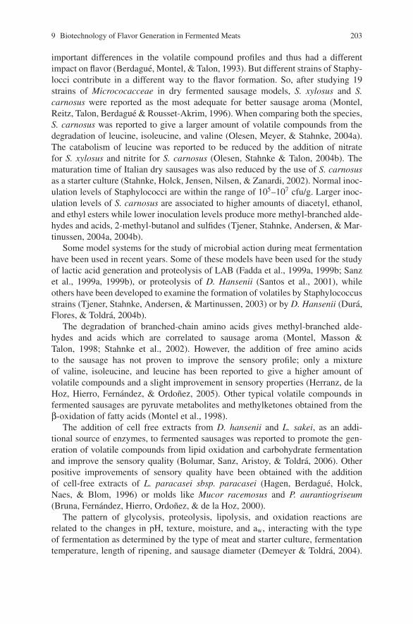
9 Biotechnology of Flavor Generation in Fermented Meats 203
important differences in the volatile compound profiles and thus had a differentimpact on flavor (Berdague, Montel, & Talon, 1993). But different strains of Staphy-locci contribute in a different way to the flavor formation. So, after studying 19strains of Micrococacceae in dry fermented sausage models, S. xylosus and S.carnosus were reported as the most adequate for better sausage aroma (Montel,Reitz, Talon, Berdague & Rousset-Akrim, 1996). When comparing both the species,S. carnosus was reported to give a larger amount of volatile compounds from thedegradation of leucine, isoleucine, and valine (Olesen, Meyer, & Stahnke, 2004a).The catabolism of leucine was reported to be reduced by the addition of nitratefor S. xylosus and nitrite for S. carnosus (Olesen, Stahnke & Talon, 2004b). Thematuration time of Italian dry sausages was also reduced by the use of S. carnosusas a starter culture (Stahnke, Holck, Jensen, Nilsen, & Zanardi, 2002). Normal inoc-ulation levels of Staphylococci are within the range of 105–107 cfu/g. Larger inoc-ulation levels of S. carnosus are associated to higher amounts of diacetyl, ethanol,and ethyl esters while lower inoculation levels produce more methyl-branched alde-hydes and acids, 2-methyl-butanol and sulfides (Tjener, Stahnke, Andersen, & Mar-tinussen, 2004a, 2004b).
Some model systems for the study of microbial action during meat fermentationhave been used in recent years. Some of these models have been used for the studyof lactic acid generation and proteolysis of LAB (Fadda et al., 1999a, 1999b; Sanzet al., 1999a, 1999b), or proteolysis of D. Hansenii (Santos et al., 2001), whileothers have been developed to examine the formation of volatiles by Staphylococcusstrains (Tjener, Stahnke, Andersen, & Martinussen, 2003) or by D. Hansenii (Dura,Flores, & Toldra, 2004b).
The degradation of branched-chain amino acids gives methyl-branched alde-hydes and acids which are correlated to sausage aroma (Montel, Masson &Talon, 1998; Stahnke et al., 2002). However, the addition of free amino acidsto the sausage has not proven to improve the sensory profile; only a mixtureof valine, isoleucine, and leucine has been reported to give a higher amount ofvolatile compounds and a slight improvement in sensory properties (Herranz, de laHoz, Hierro, Fernandez, & Ordonez, 2005). Other typical volatile compounds infermented sausages are pyruvate metabolites and methylketones obtained from theβ-oxidation of fatty acids (Montel et al., 1998).
The addition of cell free extracts from D. hansenii and L. sakei, as an addi-tional source of enzymes, to fermented sausages was reported to promote the gen-eration of volatile compounds from lipid oxidation and carbohydrate fermentationand improve the sensory quality (Bolumar, Sanz, Aristoy, & Toldra, 2006). Otherpositive improvements of sensory quality have been obtained with the additionof cell-free extracts of L. paracasei sbsp. paracasei (Hagen, Berdague, Holck,Naes, & Blom, 1996) or molds like Mucor racemosus and P. aurantiogriseum(Bruna, Fernandez, Hierro, Ordonez, & de la Hoz, 2000).
The pattern of glycolysis, proteolysis, lipolysis, and oxidation reactions arerelated to the changes in pH, texture, moisture, and aw, interacting with the typeof fermentation as determined by the type of meat and starter culture, fermentationtemperature, length of ripening, and sausage diameter (Demeyer & Toldra, 2004).
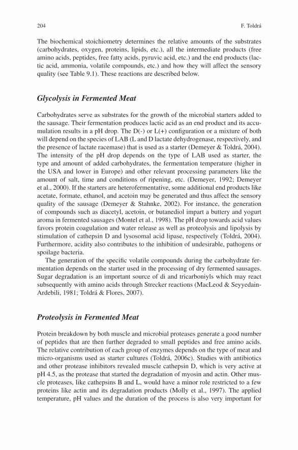
204 F. Toldra
The biochemical stoichiometry determines the relative amounts of the substrates(carbohydrates, oxygen, proteins, lipids, etc.), all the intermediate products (freeamino acids, peptides, free fatty acids, pyruvic acid, etc.) and the end products (lac-tic acid, ammonia, volatile compounds, etc.) and how they will affect the sensoryquality (see Table 9.1). These reactions are described below.
Glycolysis in Fermented Meat
Carbohydrates serve as substrates for the growth of the microbial starters added tothe sausage. Their fermentation produces lactic acid as an end product and its accu-mulation results in a pH drop. The D(-) or L(+) configuration or a mixture of bothwill depend on the species of LAB (L and D lactate dehydrogenase, respectively, andthe presence of lactate racemase) that is used as a starter (Demeyer & Toldra, 2004).The intensity of the pH drop depends on the type of LAB used as starter, thetype and amount of added carbohydrates, the fermentation temperature (higher inthe USA and lower in Europe) and other relevant processing parameters like theamount of salt, time and conditions of ripening, etc. (Demeyer, 1992; Demeyeret al., 2000). If the starters are heterofermentative, some additional end products likeacetate, formate, ethanol, and acetoin may be generated and thus affect the sensoryquality of the sausage (Demeyer & Stahnke, 2002). For instance, the generationof compounds such as diacetyl, acetoin, or butanediol impart a buttery and yogurtaroma in fermented sausages (Montel et al., 1998). The pH drop towards acid valuesfavors protein coagulation and water release as well as proteolysis and lipolysis bystimulation of cathepsin D and lysosomal acid lipase, respectively (Toldra, 2004).Furthermore, acidity also contributes to the inhibition of undesirable, pathogens orspoilage bacteria.
The generation of the specific volatile compounds during the carbohydrate fer-mentation depends on the starter used in the processing of dry fermented sausages.Sugar degradation is an important source of di and tricarboniyls which may reactsubsequently with amino acids through Strecker reactions (MacLeod & Seyyedain-Ardebili, 1981; Toldra & Flores, 2007).
Proteolysis in Fermented Meat
Protein breakdown by both muscle and microbial proteases generate a good numberof peptides that are then further degraded to small peptides and free amino acids.The relative contribution of each group of enzymes depends on the type of meat andmicro-organisms used as starter cultures (Toldra, 2006c). Studies with antibioticsand other protease inhibitors revealed muscle cathepsin D, which is very active atpH 4.5, as the protease that started the degradation of myosin and actin. Other mus-cle proteases, like cathepsins B and L, would have a minor role restricted to a fewproteins like actin and its degradation products (Molly et al., 1997). The appliedtemperature, pH values and the duration of the process is also very important for
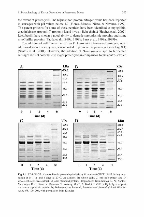
9 Biotechnology of Flavor Generation in Fermented Meats 205
the extent of proteolysis. The highest non-protein nitrogen value has been reportedin sausages with pH values below 4.7 (Flores, Marcus, Nieto, & Navarro, 1997).The parent proteins for some of these peptides have been identified as myoglobin,creatin kinase, troponin T, troponin I, and myosin light chain 2 (Hughes et al., 2002).Lactobacilli have shown a good ability to degrade sarcoplasmic proteins and somemyofibrillar proteins (Fadda et al., 1999a, 1999b; Sanz et al., 1999a, 1999b).
The addition of cell free extracts from D. hansenii to fermented sausages, as anadditional source of enzymes, was reported to promote the proteolysis (see Fig. 9.1)(Santos et al., 2001). However, the addition of Debaryomices spp. to fermentedsausages did not contribute to major proteolysis in comparison to the controls which
Fig. 9.1 SDS–PAGE of sarcoplasmic protein hydrolysis by D. hansenii CECT 12487 during incu-bation at 0, 1, 2, and 4 days at 27◦C. A: Control, B: whole cells, C: cell-free extract and D:whole cells cell-free extract. St lane: Standard proteins. Reproduced from Santos, N. N., Santos-Mendonca, R. C., Sanz, Y., Bolumar, T., Aristoy, M.-C., & Toldra, F. (2001). Hydrolysis of porkmuscle sarcoplasmic proteins by Debaryomyces hansenii, International Journal of Food Microbi-ology, 68, 199–206, with permission from Elsevier
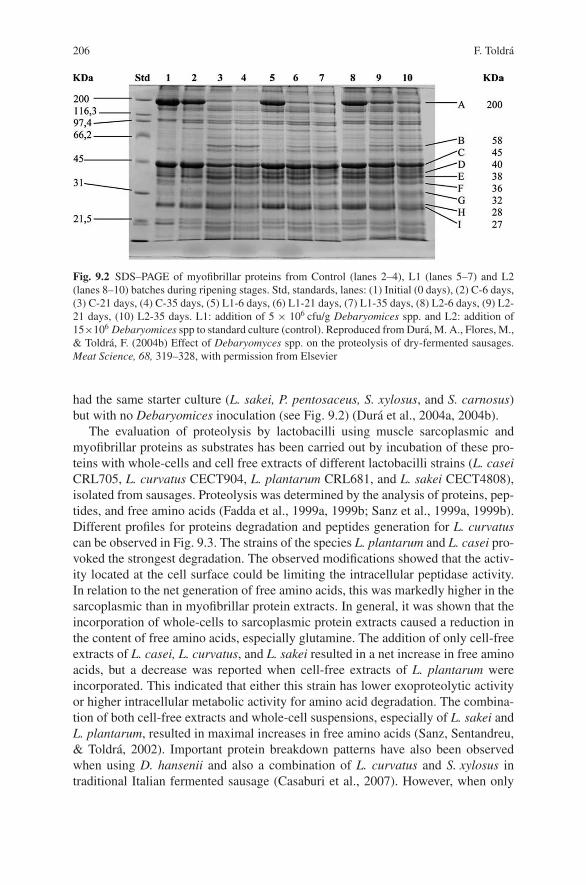
206 F. Toldra
Fig. 9.2 SDS–PAGE of myofibrillar proteins from Control (lanes 2–4), L1 (lanes 5–7) and L2(lanes 8–10) batches during ripening stages. Std, standards, lanes: (1) Initial (0 days), (2) C-6 days,(3) C-21 days, (4) C-35 days, (5) L1-6 days, (6) L1-21 days, (7) L1-35 days, (8) L2-6 days, (9) L2-21 days, (10) L2-35 days. L1: addition of 5 × 10⁶ cfu/g Debaryomices spp. and L2: addition of15×106 Debaryomices spp to standard culture (control). Reproduced from Dura, M. A., Flores, M.,& Toldra, F. (2004b) Effect of Debaryomyces spp. on the proteolysis of dry-fermented sausages.Meat Science, 68, 319–328, with permission from Elsevier
had the same starter culture (L. sakei, P. pentosaceus, S. xylosus, and S. carnosus)but with no Debaryomices inoculation (see Fig. 9.2) (Dura et al., 2004a, 2004b).
The evaluation of proteolysis by lactobacilli using muscle sarcoplasmic andmyofibrillar proteins as substrates has been carried out by incubation of these pro-teins with whole-cells and cell free extracts of different lactobacilli strains (L. caseiCRL705, L. curvatus CECT904, L. plantarum CRL681, and L. sakei CECT4808),isolated from sausages. Proteolysis was determined by the analysis of proteins, pep-tides, and free amino acids (Fadda et al., 1999a, 1999b; Sanz et al., 1999a, 1999b).Different profiles for proteins degradation and peptides generation for L. curvatuscan be observed in Fig. 9.3. The strains of the species L. plantarum and L. casei pro-voked the strongest degradation. The observed modifications showed that the activ-ity located at the cell surface could be limiting the intracellular peptidase activity.In relation to the net generation of free amino acids, this was markedly higher in thesarcoplasmic than in myofibrillar protein extracts. In general, it was shown that theincorporation of whole-cells to sarcoplasmic protein extracts caused a reduction inthe content of free amino acids, especially glutamine. The addition of only cell-freeextracts of L. casei, L. curvatus, and L. sakei resulted in a net increase in free aminoacids, but a decrease was reported when cell-free extracts of L. plantarum wereincorporated. This indicated that either this strain has lower exoproteolytic activityor higher intracellular metabolic activity for amino acid degradation. The combina-tion of both cell-free extracts and whole-cell suspensions, especially of L. sakei andL. plantarum, resulted in maximal increases in free amino acids (Sanz, Sentandreu,& Toldra, 2002). Important protein breakdown patterns have also been observedwhen using D. hansenii and also a combination of L. curvatus and S. xylosus intraditional Italian fermented sausage (Casaburi et al., 2007). However, when only
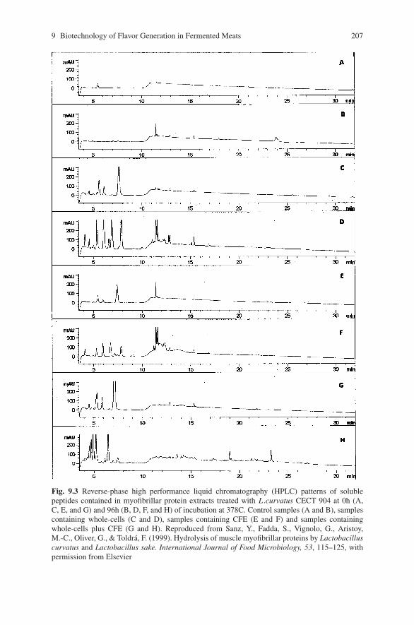
9 Biotechnology of Flavor Generation in Fermented Meats 207
Fig. 9.3 Reverse-phase high performance liquid chromatography (HPLC) patterns of solublepeptides contained in myofibrillar protein extracts treated with L .curvatus CECT 904 at 0h (A,C, E, and G) and 96h (B, D, F, and H) of incubation at 378C. Control samples (A and B), samplescontaining whole-cells (C and D), samples containing CFE (E and F) and samples containingwhole-cells plus CFE (G and H). Reproduced from Sanz, Y., Fadda, S., Vignolo, G., Aristoy,M.-C., Oliver, G., & Toldra, F. (1999). Hydrolysis of muscle myofibrillar proteins by Lactobacilluscurvatus and Lactobacillus sake. International Journal of Food Microbiology, 53, 115–125, withpermission from Elsevier
208 F. Toldra
S. xylosus was used as starter without lactobacilli, the observed proteolysis wasidentical to that in non-inoculated control sausages, reinforcing the importance ofproteolysis contribution by lactobacilli (Casaburi et al., 2008).
Tri- and di-peptides are generated as a result of the action of muscle and micro-bial tri and dipeptidylpeptidases, respectively. These enzymes are activated or inhib-ited depending on the pH and the level of salt (Sentandreu & Toldra, 2001). Infact, the generation of small peptides can be reduced by increasing the salt content,a known inhibitor of muscle peptidases (Sanz et al., 2002; Sanz & Toldra, 1999;Toldra, 2004). This is probably an important reason for wide variations (between2 and 12 fold) in the increase in non-protein-nitrogen. The final step of proteolysisconsists of the generation of free amino acids by muscle and microbial aminopep-tidases. Muscle alanyl and methionyl aminopeptidases have important activity insausages and also have a wide scope. Something similar has been reported forthe major aminopeptidase from L. sakei (Sanz & Toldra, 1997) and an arginylaminopeptidase from D. hansenii (Bolumar et al., 2003b) (see Table 9.1). Recently,an important generation of free amino acids has been reported in fermented sausagesinoculated with L. curvatus and S. xylosus (Casaburi et al., 2007, 2008).
Transformation of Amino Acids
Amino acids have important contribution to taste (Talon, Leroy-Setrin, &Fadda, 2004) and also to aroma by the generation of branched-chain aldehydesand its secondary products like acids, alcohols, and esters through microbialmetabolism of leucine, valine, & isoleucine (Toldra, 2006b). The Micrococcaceaefamily (Staphylococcus and Kocuria) and D. hansenii have good ability for suchtype of metabolism (Demeyer et al., 2000). The generation of compounds like3-methylbutanoic acid and α-hydroxy isocaproic acid have been reported afterincubation of leucine with S. xylosus and S. carnosus (Olesen et al., 2004b) althoughthese reactions are somehow affected by curing salts (Olesen et al., 2004a). Otherauthors (Herranz, Fernandez, de la Hoz, & Ordonez, 2006) have studied the additionof intracellular cell-free extracts of L. sakei and B. pumilis to enhance amino acidcatabolism, specially of valine, leucine, and isoleucine even though the sensoryquality of the final dry fermented sausage was not really improved.
Other enzymes with activity against amino acids are of interest. For instance, aglutaminase from D. hansenii was purified and characterized (Dura et al., 2002).This glutaminase was able to act on L-glutamine and generate ammonia that neu-tralized the sour taste and L-glutamate that is a flavor enhancer.
Lipolysis in Fermented Meat
Lipids constitute the major fraction of dry fermented sausages that are subject tolipolytic and oxidative reactions. Lipolysis consists of the enzymatic breakdown
9 Biotechnology of Flavor Generation in Fermented Meats 209
of tri-, di- and mono-acylglycerols and phospholipids to release free fatty acids.Lipases and phospholipases, respectively, are the responsible enzymes. Oncereleased, free fatty acids exert a direct effect on taste (slight contribution tosourness) and an indirect effect on aroma through the generation of aroma volatilecompounds by oxidative reactions. Lipolysis in meat is the result of the actionof muscle and adipose tissue lipases as well as muscle phospholipases (Toldra &Navarro, 2002), while in fermented meat it is the combined result of muscle andmicrobial enzymes (Hierro, de la Hoz, & Ordonez, 1997). The main muscle lipasesin the muscle are the lysosomal acid lipase and acid phospholipase, while thehormone sensitive lipase and the monoacylglycerol lipase are the most importantin adipose tissue (Toldra, 1992, 1998). These enzymes show good stability indrying processes (Toldra, 2006b), but their activity vary depending on the finalpH, salt content, ripening temperature, and water activity (Toldra, 2007b). Therewas some controversy in the literature about the percentage of contribution of bothmuscle and microbial lipolytic enzymes to total fat hydrolysis. It was estimatedthat muscle lipases contribution was around 60 to 80% and the rest being due tomicrobial lipases (Molly, Demeyer, Civera, & Verplaetse, 1996; Molly, Demeyer,Civera, & Verplaetse, 1997). In general, a noticeable generation of free fatty acidsis usually observed along the ripening of the sausages (Casaburi et al., 2007; Mollyet al., 1996).
Starter cultures have a definitive effect on the formation of flavor in dry sausages(Berdague et al., 1993) since lipolytic activity depends on the type of microorgan-ism. So, Micrococcaceae are considered responsible for lipid breakdown (Ordonezet al., 1999), even though molds and yeasts can also contribute due to their highlipolytic activity (Hammes & Knauf, 1994). Staphylococcus have been reported tohydrolyze mono-, di- and tri-acylglycerols and short-chain fatty tri-acylglycerols(Casaburi et al., 2007) while lactobacilli strains can only hydrolyze them at a lowerrate (Sanz, Selgas, Parejo, & Ordonez, 1998). Staphylococci have also shown a highesterase activity in whole cell suspensions and cell-free extracts, being lower in theextracellular medium (Casaburi, Villani, Toldra, & Sanz, 2006).
Oxidation in Fermented Meat
Fatty acids with double bonds (mono and polyunsaturated fatty acids) aresusceptible to oxidative reactions with the production of peroxides and carbonylcompounds and final generation of volatile compounds. Oxidation can be initiatedthrough external catalyzers like light, heat, presence of moisture and/or metalliccations or internal muscle oxidative enzymes, like peroxidases and cyclooxygenases(Toldra et al., 2001). Primary oxidation products are formed (hydroxyperoxides) butare very reactive and generate secondary oxidation products that contribute to flavor.Off-flavor development is typical of excessive oxidation (Skibsted, Mikkelsen, &Bertelsen, 1998). Staphylococcus species contribute to oxygen consumption andcatalase activity that reduces the rancidity and improves color stability (Berdagueet al., 1993). Production of catalase by Staphylococci has been reported to be
210 F. Toldra
maximal at the end of the exponential growth phase (Talon et al., 1999) butdepends on the type of strain. So, S. warneri was found to be low in catalaseactivity and thus caused rancidity in the sausages while S. carnosus had a highercatalase activity which is better suited for ideal fermented sausage production(Talon et al., 1999).
The main products from lipid oxidation are aliphatic hydrocarbons, alcohols,aldehydes, and ketones. Esters may be produced in the absence of nitrite by thereaction of alcohols with free carboxylic fatty acids (Toldra, 2006b).
Generation of Volatile Compounds
Usual starter cultures used for meat fermentation are known to contribute to thegeneration of a large amount of volatile compounds. Depending on the type, amountand balance of volatile compounds, pleasant or unpleasant aroma will be generated(Stahnke, 1994, 1995, 1999). A good example is the production of large amountsof hexanal that is associated to rancidity (Montel et al., 1998). In addition, sausagesproduced with different starter cultures show different volatile patterns associatedto different sensory profiles (Montel et al., 1996). Staphylococci have been reportedto contribute to the typical sausage flavor and enhance its perception as well asreduce the maturation time (Stahnke et al., 2002). In fact, S. xylosus and S. carnosusare able to produce large amounts of branched aldehydes, methyl ketones, andethyl esters. The latter compounds contribute to a fruity note to the flavor eventhough it has a low sensory threshold (Demeyer et al., 2000). Both micro-organismscan degrade the branched-chain amino acids leucine, isoleucine and valine intomethyl-branched aldehydes, alcohols and acids (Beck, Hansen, & Lauritsen, 2002a,2002b). For instance, the degradation of leucine produces not only 3-methyl butanal(Demeyer et al., 2000) as the main product but also 3-methylbutanoic acid aswell as some amounts of α-hydroxy isocaproic acid (Olesen et al., 2004b). In anycase, the volatile profile depends on whether S. xylsous or S. carnosus are usedas starter cultures (Olesen et al., 2004a). Since major volatile compounds proceedfrom amino acid degradation, it has been suggested that flavor quality is closelyrelated to these compounds (Stahnke, 1999). The levels of most of these volatilecompounds increase with the inoculation level and over the time of ripening (Tjener,Stahnke, Andersen, & Martinussen, 2004a). Staphylococci and Kocuria are knownto improve the color and flavor of fermented meats and influence the compositionof volatile compounds by inhibition of the unsaturated fatty acids oxidation (Talon,Leroy-Setrin, & Fadda, 2002). In fact, various staphylococci and LAB have beenstudied for its effects on unsaturated fatty acids. Staphylococci limited the oxidationof linoleic acid but LAB was unable when manganesum was lacking in the media(Talon, Walter, & Montel, 2000).
Debaryomices spp. has been used in combination with starter cultures (LAB andstaphylocci) and its effects on dry-fermented sausages have been studied. Debary-omices spp showed an important effect on the generation of volatile compounds
9 Biotechnology of Flavor Generation in Fermented Meats 211
by inhibition of the generation of lipid oxidation products and by generation ofethyl esters that contributed to an appropriate flavor (Flores, Dura, Marco, &Toldra, 2004). In the same study, it was reported that an excess of Debaryomicesspp produced large amounts of acids that masked the positive effect so that it isimportant to use the right amount of this yeast.
Conclusion
There are important interactions between micro-organisms, meat and fat enzymesand processing conditions, all of them determining the sensory quality of fermentedsausages. The use of starter cultures contribute to an improvement in the develop-ment of color, texture, and flavor of the final dry-fermented sausage but all thesesensory characteristics strongly depend on the type and amount of strains used inthe starter culture and the processing conditions.
References
Beck, H. C., Hansen, A. M., & Lauritsen, F. R. (2002a). Metabolite production and kinetics ofbranched chain acid formation in Staphylococcus xylosus. Enzyme and Microbial Technology,31, 94–101.
Beck, H. C., Hansen, A. M., & Lauritsen, F. R. (2002b). Catabolism of leucine to branched chainacids in Staphylococcus xylosus. Journal of Applied Microbiology, 91, 514–519.
Berdague, J. L., Monteil, P., Montel, M. C., & Talon, R. (1993). Effects of starter cultures on theformation of flavour compounds in dry sausage. Meat Science, 35, 275–287.
Bolumar, T., Sanz, Y., Aristoy, M. C., Flores, M., & Toldra, F. (2006). Sensory improvement ofdry-fermented sausages by the addition of cell-free extracts from Debaryomices hansenii andLactobacillus sakei. Meat Science, 72, 457–466.
Bolumar, T., Sanz, Y., Aristoy, M. C., & Toldra, F. (2003a). Purification and characterization ofa prolyl aminopeptidase from Debaryomyces hansenii. Applied and Environmental Microbiol-ogy, 69, 227–232.
Bolumar, T., Sanz, Y., Aristoy, M. C., & Toldra, F. (2003b). Purification and properties of an arginylaminopeptidase from Debaryomyces hansenii. International Journal of Food Microbiology, 86,141–151.
Bolumar, T., Sanz, Y., Aristoy, M. C., & Toldra, F. (2005). Protease B from Debaryomyceshansenii. Purification and biochemical properties. International Journal of Food Microbiology,98, 167–177.
Bolumar, T., Sanz, Y., Aristoy, M. C., & Toldra, F. (2008). Purification and characterization ofproteases A and D from Debaryomyces hansenii. International Journal of Food Microbiology.Manuscript submitted for publication.
Bruna, J. M., Fernandez, M., Hierro, E. M., Ordonez, J. A., & de la Hoz, L. (2000). Improvementof the sensory properties of dry fermented sausages by the superficial inoculation and/or theaddition of intracellular extracts of Mucor racemosus. Journal of Food Science, 65, 731–738.
Casaburi, A., Aristoy, M. C., Cavella, S., Di Monaco, R., Ercolini, D., Toldra, F., et al. (2007).Biochemical and sensory characteristics of traditional fermented sausages of Vallo di Diano(Southern Italy) as affected by use of starter cultures. Meat Science, 76, 295–307.
Casaburi, A., Di Monaco, R., Cavella, S., Toldra, F., Ercolini, D., & Villani, F. (2008). Proteolyticand lipolytic starter cultures and their effect on traditional fermented sausages ripening andsensory traits. Food Microbiology, 25, 335–347.
212 F. Toldra
Casaburi, A., Villani, F., Toldra, F., & Sanz, Y. (2006). Protease and esterase activity of Staphylo-cocci. International Journal of Food Microbiology, 112, 223–229.
Chi, S-P., & Wu, Y-C. (2007). Spices and seasonings. In F. Toldra, Y. H. Hui, I. Astiasaran, W. K.Nip, J. G. Sebranek, E. T. F. Silveira, L. H. Stahnke, & R. Talon (Eds.), Handbook of fermentedmeat and poultry (pp. 87–100). Ames, IA: Blackwell Publishing.
Cocolin, L., Manzano, M., Aggio, D., Cantoni, C., & Comi, G. (2001). A novel polymerase chainreaction (PCR)-denaturing gradient gel electrophoresis (DGGE) for the identification of Micro-cocacceae strains involved in meat fermentations. Its application to naturally fermented Italiansausages. Meat Science, 58, 59–64.
Cocolin, L., Manzano, M., Cantoni, C., & Comi, G. (2001). Denaturing gradient gel electrophore-sis analysis of the 16S rRNA gene VI region to monitor dynamic changes in the bacterialpopulation during fermentation of Italian sausages. Applied and Environmental Microbiology,67, 5113–5121.
Comi, G., Urso, R., Iacumin, L., Rantsiou, K., Cattaneo, P., Cantoni, C., et al. (2005). Character-isation of naturally fermented sausages produced in the North East of Italy. Meat Science, 69,381–392.
Demeyer, D. (1992). Meat fermentation as an integrated process. In F. J. M. Smulders, F. Toldra,J. Flores, & M. Prieto (Eds.), New Technologies for Meat and Meat Products (pp. 21–36).Nijmegen (The Netherlands): Audet.
Demeyer, D. I., & Toldra, F. (2004). Fermentation. In W. Jensen, C. Devine, & M. Dikemann(Eds.), Encyclopedia of Meat Sciences (pp. 467–474). London, UK: Elsevier Science Ltd.
Demeyer, D., Raemaekers, M., Rizzo, A., Holck, A., De Smedt, A., ten Brink, B., et al. (2000).Control of bioflavour and safety in fermented sausages: first results of a European project. FoodResearch International, 33, 171–180.
Demeyer, D., & Stahnke, L. (2002). Quality control of fermented meat products. In J. Kerry& D. Ledward (Eds.), Meat processing: Improving quality (pp. 359–393).Cambridge (UK):Woodhead Pub. Co.
Dura, M. A., Flores, M., & Toldra, F. (2002). Purification and characterisation of a glutaminasefrom Debaryomices spp. International Journal of Food Microbiology, 76, 117–126.
Dura, M. A., Flores, M., & Toldra, F. (2004a). Effect of growth phase and dry-cured sausageprocessing conditions on Debaryomyces spp. generation of volatile compounds from branched-chain amino acids. Food Chemistry, 86, 391–399.
Dura, M. A., Flores, M., & Toldra, F. (2004b). Effect of Debaryomyces spp. on the proteolysis ofdry-fermented sausages. Meat Science, 68, 319–328.
Dura, M. A., Flores, M., & Toldra, F. (2004c). Effect of Debaryomyces spp on aroma formationand sensory quality of dry-fermented sausages. Meat Science, 68, 439–446.
Ellis, D. F. (2001). Meat smoking technology. In Y. H. Hui, H. Nip, R. W. Rogers & O. Young(Eds.), Meat Science and Applications. (pp. 509–519). New York: Marcel Dekker Inc.
Fadda, S., Sanz, Y., Vignolo, G., Aristoy, M. C., Oliver, G., & Toldra, F. (1999a). Hydrolysis ofpork muscle sarcoplasmic proteins by Lactobacillus curvatus and Lactobacillus sake. Appliedand Environmental Microbiology, 65, 578–584.
Fadda, S., Sanz, Y., Vignolo, G., Aristoy, M. C., Oliver, G., & Toldra, F. (1999b). Characterizationof muscle sarcoplasmic and myofibrillar protein hydrolysis caused by Lactobacillus plantarum.Applied and Environmental Microbiology, 65, 3540–3546.
Flores, J., Marcus, J. R., Nieto, P., & Navarro, J. L. (1997). Effect of processing conditions on prote-olysis and taste of dry-cured sausages. Zetiscrift furLebensmittel Untersuchung und ForschungA, 204, 168–172.
Flores, M., Dura, M. A., Marco, A., & Toldra, F. (2004). Effect of Debaryomyces spp. on aromaformation and sensory quality of dry-fermented sausages. Meat Science, 68, 439–446.
Hagen, B. F., Berdague, J. L., Holck, A. L., Naes, H., & Blom, H. (1996). Bacterial proteinasereduces maturation time of dry fermented sausages. Journal of Food Science, 61, 1024–1029.
Hammes, W. P., & Knauf, H. J. (1994). Starters in the processing of meat products. Meat Science,36, 155–168.
9 Biotechnology of Flavor Generation in Fermented Meats 213
Herranz, B., de la Hoz, L., Hierro, E., Fernandez, M., & Ordonez, J. A. (2005). Improvement ofthe sensory properties of dry-fermented sausages by the addition of free amino acids. FoodChemistry, 91, 673–682.
Herranz, B., Fernandez, M., de la Hoz, L., & Ordonez, J. A. (2006). Use of bacterial extracts toenhance amino acid breakdown in dry fermented sausages. Meat Science, 72, 318–325.
Hierro, E., De la Hoz, L., Ordonez, J. A. (1997). Contribution of microbial and meat endogenousenzymes to the lipolysis of dry fermented sausages. Journal of Agricultural and Food Chemistry45, 2989–2995.
Hughes, M. C., Kerry, J. P., Arendt, E. K., Kenneally, P. M., McSweeney, P. L. H., & O’Neill, E. E.(2002). Characterization of proteolysis during the ripening of semidry fermented sausages.Meat Science, 62, 205–216.
Leistner, F. (1992). The essentials of producing stable and safe raw fermented sausages. InF. J. M. Smulders, F. Toldra, J. Flores & M. Prieto (Eds.), New Technologies for Meat andMeat Products (pp. 1–19). Nijmegen (The Netherlands): Audet.
MacLeod, G., & Seyyedain-Ardebili, M. (1981). Natural and simulated meat flavors (with partic-ular reference to beef). CRC Critical Reviews in Food Science and Nutrition, 14, 309–437.
Molly, K., Demeyer, D. I., Civera, T., & Verplaetse, A. (1996). Lipolysis in Belgian sausages:Relative importance of endogenous and bacterial enzymes. Meat Science, 43, 235–244.
Molly, K., Demeyer, D. I., Johansson, G., Raemaekers, M., Ghistelinck, M., & Geenen, I. (1997).The importance of meat enzymes in ripening and flavor generation in dry fermented sausages.First results of a European project. Food Chemistry, 54, 539–545.
Montel, M. C., Masson, F., & Talon, R. (1998). Bacterial role in flavour development, Meat Sci-ence, 49, S111–S123.
Montel, M. C., Reitz, J., Talon, R., Berdague, J. L., & Rousset-Akrim, S. (1996). Biochemicalactivities of Micrococcaceae and their effects on the aromatic profiles and odours of a drysausage model. Food Microbiology, 13, 489–499.
Montel, M. C., Seronine, M. P., Talon, R., & Hebraud, M. (1995). Purification and characteriza-tion of a dipeptidase from Lactobacillus sake. Applied and Environmental Microbiology, 61,837–839.
Olesen, P. T., Meyer, A. S., & Stahnke, L. H. (2004b). Generation of flavour compounds in fer-mented sausages-the influence of curing ingredients, Staphylococcus starter culture and ripen-ing time. Meat Science, 66, 675–687.
Olesen, P. T., Stahnke, L.,H., & Talon, R. (2004a). Effect of ascorbate, nitrate and nitrite on theamount of flavour compounds produced from leucine by Staphylococcus xylosus and Staphylo-cocuus carnosus. Meat Science, 68, 193–200.
Ordonez, J. A., Hierro, E. M., Bruna, J. M., & de la Hoz, L. (1999). Changes in the components ofdry-fermented sausages during ripening. Critical Reviews in Food Science and Nutrition, 39,329–367.
Santos, N. N., Santos, R. C., Sanz, Y., Bolumar, T., Aristoy, M-C., & Toldra, F. (2001). Hydrolysisof muscle sarcoplasmic proteins by Debaryomyces hansenii. International Journal of FoodMicrobiology, 68, 199–206.
Sanz, B., Selgas, D., Parejo, I., & Ordonez, J. A. (1998). Characteristics of lactobacilli isolatedfrom dry fermented sausages. International Journal of Food Microbiology, 6, 199–205.
Sanz, Y., Fadda, S., Vignolo, G., Aristoy, M. C., Oliver, G., & Toldra, F. (1999a). Hydrolytic actionof Lactobacillus casei CRL 705 on pork muscle sarcoplasmic and myofibrillar proteins. Journalof Agricultural and Food Chemistry, 47, 3441–3448.
Sanz, Y., Fadda, S., Vignolo, G., Aristoy, M. C., Oliver, G., & Toldra, F. (1999b). Hydrolysis ofmuscle myofibrillar proteins by Lactobacillus curvatus and Lactobacillus sake. InternationalJournal of Food Microbiology, 53, 115–125.
Sanz, Y., Mulholland, F., & Toldra, F. (1998). Purification and characterization of a tripeptidasefrom Lactobacillus sake. Journal of Agricultural and Food Chemistry, 46, 349–353.
Sanz, Y., Sentandreu, M. A., & Toldra, F. (2002). Role of muscle and bacterial exopeptidasesin meat fermentation. In F. Toldra (Ed.), Research advances in the quality of meat and meatproducts (pp. 143–155). Trivandrum, India: Research Signpost.
214 F. Toldra
Sanz, Y., & Toldra, F. (1997). Purification and characterization of an aminopeptidase from Lacto-bacillus sake. Journal of Agricultural and Food Chemistry, 45, 1552–1558.
Sanz, Y., & Toldra, F. (1999). The role of exopeptidases from Lactobacillus sake in dry fermentedsausages. Recent Research Developments in Agricultural & Food Chemistry, 3, 11–21.
Sanz, Y., & Toldra, F. (2001). Purification and characterization of an X-prolyl-dipeptidyl peptidasefrom Lactobacillus sake. Applied and Environmental Microbiology, 67, 1815–1820.
Sanz, Y., & Toldra, F. (2002). Purification and characterization of an arginine aminopeptidase fromLactobacillus sakei. Applied and Environmental Microbiology, 68, 1980–1987.
Sentandreu, M. A., & Toldra, F. (2001). Dipeptidylpeptidase activities along the processing ofSerrano dry-cured ham. European Food Research Technology, 213, 83–87.
Skibsted, L. H., Mikkelsen, A., & Bertelsen, G. (1998). Lipid-derived off-flavours in meat. InF. Shahidi (Ed.), Flavor of meat, meat products and seafoods (pp. 216–256). London, UK:Blackie Academic & Professional.
Stahnke, L. H. (1994). Aroma components from dried sausages fermented with Staphylococcus-xylosus. Meat Science, 38, 39–53.
Stahnke, L. H. (1995). Dried Sausages Fermented with Staphylococcus xylosus at Different Tem-peratures and with Different Ingredient Levels .2. Volatile Components. Meat Science, 41,193–209.
Stahnke, L. H. (1999). Volatiles produced by Staphylococcus xylosus and Staphylococcus carnosusduring growth in sausage minces - Part I. Collection and identification. Food Science and Tech-nology. Lebensmittel-Wissenschaft & Technologie, 32, 357–364.
Stahnke, L. H., Holck, A., Jensen, A., Nilsen, A., & Zanardi, E. (2002). Maturity acceleration ofItalian dried sausage by Staphylococcus carnosus - Relationship between maturity and flavorcompounds. Journal of Food Science, 67, 1914–1921.
Sunesen, L. O., Stahnke, L. H. (2003). Mould starter cultures for dry sausages-selection, applica-tion and effects. Meat Science, 65, 935–948.
Talon, R., Leroy-Setrin, S., & Fadda, S. (2002). Bacterial starters involved in the quality of fer-mented meat products. In F. Toldra (Ed.), Research advances in the quality of meat and meatproducts (pp. 175–191). Trivandrum (India): Research Signpost.
Talon, R., Leroy-Setrin, S., & Fadda, S. (2004). Dry fermented sausages. In Y. H. Hui, L. M.Goddik, J. Josephsen, P. S. Stanfield, A. S. Hansen, W. K. Nip & F Toldra (Eds.), Handbook offood and beverage fermentation technology (pp. 397–416). New York: Marcel-Dekker.
Talon, R., Walter, D., Chartier, S., Barriere, C., & Montel, M. C. (1999). Effect of nitrate andincubation conditions on the production of catalase and nitrate reductase by staphylococci.International Journal of Food Microbiology, 52, 47–56.
Talon, R., Walter, D., & Montel, M. C. (2000). Growth and effect of staphylococci and lactic acidbacteria on unsaturated free fatty acids. Meat Science, 54, 41–47.
Tjener, K., Stahnke, L. H., Andersen, L., & Martinussen, J. (2003). A fermented meat model systemfor studies of microbial aroma formation. Meat Science, 66, 211–218.
Tjener, K., Stahnke, L. H., Andersen, L., & Martinussen, J. (2004a). Growth and production ofvolatiles by Staphylococcus carnosus in dry sausages: Influence of inoculation level and ripen-ing time. Meat Science, 67, 447–452.
Tjener, K., Stahnke, L. H., Andersen, L., & Martinussen, J. (2004b). Addition of alpha-ketoglutarate enhances formation of volatiles by Staphylococcus carnosus during sausage fer-mentation. Meat Science, 67, 711–719.
Toldra F. (1992). The enzymology of dry-curing of meat products. In F. J. M. smulders, F. Toldra,J. Flores & M. Prieto (Eds.) New Technologies for Meat and Meat Products (pp. 209–231).Nijmegen (The Netherlands): Audet.
Toldra, F. (1998). Proteolysis and lipolysis in flavour development of dry-cured meat products.Meat Science, 49, s101–s110.
Toldra, F. (2002). Dry-cured meat products (pp. 1–38). Trumbull, CT: Food & Nutrition Press.Toldra, F. (2004). Fermented meats. In Y. H. Hui, J. S. Smith (Eds.), Food processing: Principles
and applications (pp. 399–415). Ames, IA: Blackwell Publishing.
9 Biotechnology of Flavor Generation in Fermented Meats 215
Toldra, F. (2006a). Meat fermentation. In Y. H. Hui, E. Castell-Perez, L. M. Cunha, I. Guerrero-Legarreta, H. H. Liang, Y. M. Lo, D. L. Marshall, W. K. Nip, F. Shahidi, F. Sherkat, R. J. Winger& K. L. Yam (Eds.), Handbook of Food Science, Technology and Engineering (Vol. 4, pp. 181-1to 181-12). Boca Raton, FL: CRC Press.
Toldra, F. (2006b). Biochemistry of fermented meat. In Y. H. Hui, W. K. Nip, M. L. Nollet,G. Paliyath & B. K. Simpson (Eds.), Food biochemistry and food processing (pp. 641–658).Ames, IA: Blackwell Publishing.
Toldra, F. (2006c). Biochemical proteolysis basis for improved processing of dry-cured meats. InL. M. L. Nollet & F. Toldra (Eds.), Advanced technologies for meat processing (pp. 329–351).Boca Raton, FL: CRC Press.
Toldra, F. (2007a). Fermented meat production. In Y. H. Hui, R. Chandan, S. Clark, N. Cross, J.Dobbs, W. J. Hurst, L. M. L. Nollet, E. Shimoni, N. Sinha, E. B. Smith, S. Surapat, A. Titchenal& F. Toldra (Eds.), Handbook of Food Product Manufacturing (Vol. 2, pp. 263–277). Hoboken,NJ: John Wiley Interscience of NY.
Toldra, F. (2007b). Biochemistry of muscle and fat. In F. Toldra, Y. H. Hui, I. Astiasaran, W. K.Nip, J. G. Sebranek, E. T. F. Silveira, L. H. Stahnke, & R. Talon (Eds.), Handbook of fermentedmeat and poultry (pp. 51–58). Ames, IA: Blackwell Publishing
Toldra, F., & Flores, M. (2007). Processed pork meat flavors. In Y. H. Hui, R. Chandan, S. Clark, N.Cross, J. Dobbs, W. J. Hurst, L. M. L. Nollet, E. Shimoni, N. Sinha, E. B. Smith, S. Surapat, A.Titchenal & F. Toldra (Eds), Handbook of Food Product Manufacturing (Vol. 2, pp. 279–299).Hoboken, NJ: John Wiley Interscience of NY.
Toldra, F., Flores, M., & Sanz, Y. (2001). Meat fermentation technology. In Y. H. Hui, W. K. Nip,R. W. Rogers, & O. A. Young (Eds.), Meat Science and applications (pp. 537–561). New York:Marcel Dekker Inc.
Toldra, F., & Navarro, J. L. (2002). Action of muscle lipases during the processing of dry-curedham. In F. Toldra (Ed.), Research advances in the quality of meat and meat products (pp. 249–254). Trivandrum, India: Research Signpost.
Toldra, F., Nip, W. K., & Hui, Y. H. (2007). Dry fermented sausages: An overview. In F. Toldra,Y. H. Hui, I. Astiasaran, W. K. Nip, J. G. Sebranek, E. T. F. Silveira, L. H. Stahnke, & R.Talon (Eds.), Handbook of fermented meat and poultry (pp. 321–332). Ames, IA: BlackwellPublishing.
Toldra, F., & Reig, M. (2007). Sausages. In Y. H. Hui, R. Chandan, S. Clark, N. Cross, J. Dobbs,W. J. Hurst, L. M. L. Nollet, E. Shimoni, N. Sinha, E. B. Smith, S. Surapat, A. Titchenal, & F.Toldra (Eds.), Handbook of Food Product Manufacturing (Vol. 2, pp. 249–262). Hoboken, NJ:John Wiley Interscience of NY.

Chapter 10Latest Developments in Probiotics
Frederic Leroy, Gwen Falony, and Luc de Vuyst
Introduction
Probiotic foods are a group of health-promoting, so-called functional foods, withlarge commercial interest and growing market shares (Arvanitoyannis & vanHouwelingen-Koukaliaroglou, 2005). In general, their health benefits are basedon the presence of selected strains of lactic acid bacteria (LAB), that, when takenup in adequate amounts, confer a health benefit on the host. They are adminis-tered mostly through the consumption of fermented milks or yoghurts (Mercenier,Pavan, & Pot, 2003). In addition to their common use in the dairy industry, probioticLAB strains may be used in other food products too, including fermented meats(Hammes & Hertel, 1998; Incze, 1998; Krockel, 2006; Tyopponen, Petaja, & Mattila-Sandholm, 2003). Although the concept is not new, only a few manufacturers offerfermented sausages with probiotic LAB. This is probably due to the more artisanorientation of sausage manufacturers as compared to the dairy industry, a largervariety of products, as well as a number of uncertainties concerning technological,microbiological, and regulatory aspects (Krockel, 2006). The application of probioticLAB must in all cases be based on a careful selection procedure, if any health claimsare to be taken into account. The present chapter gives an overview of researchactivities that have previously explored the potential of probiotic LAB strains infermented meats and aims at giving a critical interpretation of the results obtained.
Probiotics
History and Definitions
Although probiotics are usually linked with gut health, the first suggestion of abeneficial association between microorganisms and the human host can probably
Luc de VuystResearch Group of Industrial Microbiology and Food Biotechnology (IMDO), Vrije UniversiteitBrussel (VUB), Pleinlaan 2, B-1050 Brussels, Belgium
F. Toldra (ed.), Meat Biotechnology, 217C© Springer Science+Business Media, LLC 2008
218 F. Leroy et al.
be attributed to Albert Doderlein, who proposed in 1892 that lactic acid produc-tion by vaginal bacteria prevented or inhibited the growth of pathogenic bacte-ria (Doderlein, 1892). Most cited as the founding father of the probiotic concept,however, is Ilya Metchnikoff. In his work, “The Prolongation of Life – OptimisticStudies”, published in 1908, he implicated a lactic acid bacterium found in Bul-garian yoghurts as the agent responsible for deterring intestinal putrefaction andageing (Metchnikoff, 1908). Hence, he became the first to speculate on the potentialhealth-promoting and even life-lengthening properties of LAB. Another milestonein the history of probiotics is undoubtedly the work of Minoru Shirota, who wasthe first to actually cultivate a beneficial intestinal bacterium, Lactobacillus caseiShirota, and distribute it in a dairy drink that was introduced to the market in 1935(Yakult Central Institute for Microbiological Research, 1999).
The word “probiotic” stems from the Greek π�o β�oς (pro bios, “for life”)and was originally proposed to describe growth-promoting substances producedby one protozoan for the benefit of another (Lilly & Stillwell, 1965). In 1974, itwas first linked with the intestinal microbial balance by Parker (1974) in his workon animal feed supplements with beneficial effect on the host. In the followingdecades, the concept of probiotics was regularly defined and redefined. Nowa-days, a widely accepted definition is a rather broad one that was proposed by aJoint Expert Consultation of the Food and Agricultural Organization of the UnitedNations (FAO) and the World Health Organization (WHO). It classifies probiotics as“life microogranisms that, when consumed in an adequate amount, confer a healthbenefit on the host” (FAO/WHO, 2001). This definition does not emphasize on thenature of the host (animal or human), the origin of the microorganisms (humanor non-human), or the ability to adhere to body surfaces. Also, although stressedelsewhere in the FAO/WHO report, the viability at the target site is not considereda restriction, thus leaving the door open for the acceptance of the yoghurt bacte-ria Streptococcus thermophilus and Lactobacillus delbrueckii subsp. bulgaricus asprobiotic microorganisms as well as of the health effects ascribed to certain cellcomponents. As no site of action is precise, probiotic preparations can not only betargeted towards the benefit of the gut, but, for instance, also towards that of the oralcavity, the nasopharynx, the respiratory tract, the stomach, the vagina, the bladder,and the skin (Reid, 2005). Of course, no definition is final, and, amongst others, theviability of the probiotics, or better, their effectiveness after food processing andstorage, their site of activity, the numbers necessary to exert a beneficial effect, theformat of intake, and the nature of the carrier remain critical issues in a vivid discus-sion (Makras, Avonts, & De Vuyst, 2004; Mercenier et al., 2003; Senok, Ismaeel, &Botta, 2005).
As probiotics are mainly directed to alter the composition and/or metabolic activ-ity of the gut microbiota towards what is generally believed to be a healthy or bal-anced one, that is, being predominantly saccharolytic and comprising significantnumbers of bifidobacteria and lactobacilli (Picard et al., 2005), pre- and synbi-otics need to be mentioned as alternative or complementary strategies to achievethis goal. Prebiotics are selectively fermented, non-digestible, food ingredients thatallow specific changes, both in the composition and/or activity of the gastrointestinal
10 Latest Developments in Probiotics 219
microbiota, that confer benefits upon host well-being and health (Gibson, Probert,Van Loo, Rastall, & Roberfroid, 2004; Gibson & Roberfroid, 1995). To this day,sufficient scientific evidence exists to recognise three types of carbohydrates asprebiotics, namely inulin-type fructans, transgalacto-oligosaccharides, and lactulose(Gibson et al., 2004), although the latter should be considered as a laxative drug(Bass & Dennis, 1981). For some other candidate prebiotics, promising data havebeen published but further investigation is required (Gibson et al., 2004). Finally,synbiotics are mixtures of pro- and prebiotics, wherein, the latter is thought toimprove survival and implantation of the former, either by stimulating growth orby metabolically activating the health-promoting bacteria, thereby taking advantageof the individual and possibly the synergistic health effects of both components(Gibson & Roberfroid, 1995; Rastall & Maitin, 2002).
Probiotic Microorganisms
Different types of food products or food supplements containing viable probioticmicroorganisms with health-promoting properties are commercially available, eitheras fermented or non-fermented food commodities, or as specific food supplementsand pharmaceutical preparations in the form of powders, tablets, or capsules. Thefirst step in the development of such products is the selection of an appropriatemicrobial strain. Throughout the years, several selection criteria for probiotics havebeen formulated, including human origin, non-pathogenic behaviour, safety, resis-tance to gastric acidity and bile toxicity, adhesion to or interaction with the gutepithelial tissue, ability to persist within the gastrointestinal tract, production ofantimicrobial substances and nutraceuticals, evidence of beneficial health effects,ability to influence metabolic activities, and resistance to technological processes(Dunne et al., 2001; Maldonado Galdeano, de Moreno de LeBlanc, Vinderola, BibasBonet, & Perdigon, 2007; Ross, Desmond, Fitzgerald, & Stanton, 2005). Althoughsome of the present criteria appear obvious, others are directly related with the def-inition of probiotics handled by the authors. It should, however, be stressed thatcharacteristics such as survival of the passage through the upper gastrointestinaltract, thus resisting the action of gastric juice, bile salts, and proteolytic enzymes,are not sufficient to call a certain microbial strain as probiotic. According to theWHO/FAO definition, the main criterion for a probiotic strain should be the fact thatit confers a health benefit on the host (FAO/WHO, 2001). This benefit can only bedemonstrated through well-designed, randomized, double blind, placebo-controlled,multi-centre human trials, the results of which are published in peer-reviewed inter-national scientific journals (Guarner & Schaafsma, 1998; Salminen et al., 1998).Furthermore, it should be stressed that probiotic effects are strain-dependent, andextrapolation of the existing data from closely related microorganisms is not suf-ficient to identify a strain as probiotic (Mercenier et al., 2003). Exact identifica-tion and characterization of the potential probiotic strain used at the genus, species,and even strain level, using internationally accepted methodologies, should be the
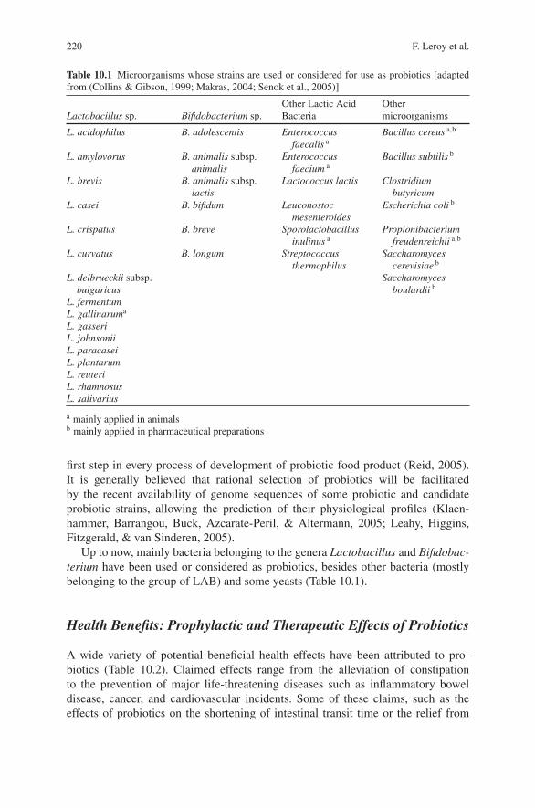
220 F. Leroy et al.
Table 10.1 Microorganisms whose strains are used or considered for use as probiotics [adaptedfrom (Collins & Gibson, 1999; Makras, 2004; Senok et al., 2005)]
Other Lactic Acid OtherLactobacillus sp. Bifidobacterium sp. Bacteria microorganisms
L. acidophilus B. adolescentis Enterococcusfaecalis a
Bacillus cereus a,b
L. amylovorus B. animalis subsp.animalis
Enterococcusfaecium a
Bacillus subtilis b
L. brevis B. animalis subsp.lactis
Lactococcus lactis Clostridiumbutyricum
L. casei B. bifidum Leuconostocmesenteroides
Escherichia coli b
L. crispatus B. breve Sporolactobacillusinulinus a
Propionibacteriumfreudenreichii a,b
L. curvatus B. longum Streptococcusthermophilus
Saccharomycescerevisiae b
L. delbrueckii subsp.bulgaricus
Saccharomycesboulardii b
L. fermentumL. gallinaruma
L. gasseriL. johnsoniiL. paracaseiL. plantarumL. reuteriL. rhamnosusL. salivarius
a mainly applied in animalsb mainly applied in pharmaceutical preparations
first step in every process of development of probiotic food product (Reid, 2005).It is generally believed that rational selection of probiotics will be facilitatedby the recent availability of genome sequences of some probiotic and candidateprobiotic strains, allowing the prediction of their physiological profiles (Klaen-hammer, Barrangou, Buck, Azcarate-Peril, & Altermann, 2005; Leahy, Higgins,Fitzgerald, & van Sinderen, 2005).
Up to now, mainly bacteria belonging to the genera Lactobacillus and Bifidobac-terium have been used or considered as probiotics, besides other bacteria (mostlybelonging to the group of LAB) and some yeasts (Table 10.1).
Health Benefits: Prophylactic and Therapeutic Effects of Probiotics
A wide variety of potential beneficial health effects have been attributed to pro-biotics (Table 10.2). Claimed effects range from the alleviation of constipationto the prevention of major life-threatening diseases such as inflammatory boweldisease, cancer, and cardiovascular incidents. Some of these claims, such as theeffects of probiotics on the shortening of intestinal transit time or the relief from
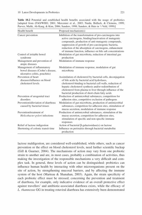
10 Latest Developments in Probiotics 221
Table 10.2 Potential and established health benefits associated with the usage of probiotics[adapted from (FAO/WHO, 2001; Mercenier et al., 2003; Naidu, Bidlack, & Clemens, 1999;Parvez, Malik, Ah Kang, & Kim, 2006; Sanders, 1998; Sanders, & Huis in ’t Veld, 1999)]
Health benefit Proposed mechanism(s)
Cancer prevention Inhibition of the transformation of pro-carcinogens intoactive carcinogens, binding/inactivation of mutageniccompounds, production of anti-mutagenic compounds,suppression of growth of pro-carcinogenic bacteria,reduction of the absorption of carcinogens, enhancmentof immune function, influence on bile salt concentrations
Control of irritable bowelsyndrome
Modulation of gut microbiota, reduction of intestinal gasproduction
Management and prevention ofatopic diseases
Modulation of immune response
Management of inflammatorybowel diseases (Crohn’s disease,ulcerative colitis, pouchitis)
Modulation of immune response, modulation of gutmicrobiota
Prevention of heartdiseases/influence on bloodcholesterol levels
Assimilation of cholesterol by bacterial cells, deconjugationof bile acids by bacterial acid hydrolases,cholesterol-binding to bacterial cell walls, reduction ofhepatic cholesterol synthesis and/or redistribution ofcholesterol from plasma to liver through influence of thebacterial production of short-chain fatty acids
Prevention of urogenital tractdisorders
Production of antimicrobial substances, competition foradhesion sites, competitive exclusion of pathogens
Prevention/alleviation of diarrhoeacaused by bacteria/viruses
Modulation of gut microbiota, production of antimicrobialsubstances, competition for adhesion sites, stimulation ofmucus secretion, modulation of immune response
Prevention/treatment ofHelicobacter pylori infections
Production of antimicrobial substances, stimulation of themucus secretion, competition for adhesion sites,stimulation of specific and non-specific immuneresponses
Relief of lactose indigestion Action of bacterial β-galactosidase(s) on lactoseShortening of colonic transit time Influence on peristalsis through bacterial metabolite
production
lactose maldigestion, are considered well-established, while others, such as cancerprevention or the effect on blood cholesterol levels, need further scientific backup(Gill & Guarner, 2004). The mechanisms of action may vary from one probioticstrain to another and are, in most cases, probably a combination of activities, thusmaking the investigation of the responsible mechanisms a very difficult and com-plex task. In general, three levels of action can be distinguished: probiotics caninfluence human health by interacting with other microorganisms present on thesite of action, by strengthening mucosal barriers, and by affecting the immunesystem of the host (Marteau & Shanahan, 2003). Again, the strain specificity ofeach probiotic effect must be stressed; concerning the prevention and treatmentof diarrhoea, for example, only indicative evidence of an overall protective effectagainst travellers’ and antibiotic-associated diarrhoea exists, while the efficacy ofL. rhamnosus GG in treating rotaviral diarrhoea has extensively been demonstrated
222 F. Leroy et al.
(Gill & Guarner, 2004; Santosa, Farnworth, & Jones, 2006). Further research tosupport the health claims attributed to probiotics and to unravell the mechanismsbehind them is needed.
Safety Considerations
As viable probiotic bacteria have to be consumed in large quantities, over anextended period of time, to exert beneficial effects, the issue of the safety of thesemicroorganisms is of primary concern (Senok et al., 2005). Historical data indicatethat lactobacilli and bifidobacteria are safe for human use (Reid, 2005). It has beensuggested that the human origin of a strain confirms its normal commensal nature,and, therefore, its safety. However, it remains difficult to establish the origin of abacterial species, and the fact that infants are born with sterile intestines raisesthe question whether “human origin” is an appropriate classification for bacteria(Reid, 2005).
Although minor side effects of the use of probiotics have been reported,infections with probiotic bacteria rarely occur and invariably only in immuno-compromised patients or those with intestinal bleeding (Gueimonde, Frias, &Ouwehand, 2006; Marteau, 2002; Reid, 2005).
An issue of concern regarding the use of probiotics is the presence of chro-mosomal, transposon-, or plasmid-located antibiotic resistance genes amongst theprobiotic microorganisms. At this moment, insufficient information is available onsituations in which these genetic elements could be mobilised, and it is not knownif situations could arise where this would become a clinical problem. When dealingwith the selection of probiotic strains, the FAO/WHO Consultancy recommends thatprobiotic microorganisms should not harbour transmissible drug resistance genesencoding resistance to clinically used drugs (FAO/WHO, 2001).
For the assessment of the safety of probiotic microorganisms and products,FAO/WHO has formulated guidelines, recommending that probiotic strains shouldbe evaluated for a number of parameters, including antibiotic susceptibility patterns,toxin production, metabolic and haemolytic activities, and infectivity in immuno-compromised animals (FAO/WHO, 2002; Reid, 2005; Senok et al., 2005).
Application of Probiotics in Fermented Meat Products
Fermented Meat as a Carrier for Probiotic Bacteria
Dry fermented meat products are usually not or only mildly heated, which is ade-quate for the carriage of probiotic bacteria (Ammor & Mayo, 2007; Arihara, 2006).Although there are in principle no major reasons preventing application of probioticLAB strains in meat, several points have to be carefully addressed.
10 Latest Developments in Probiotics 223
Although meat is a food with high nutritional value, some consumers may per-ceive meat products as unhealthy (Arihara, 2006). This can be ascribed to the imageof meat as such, in combination with the presence of nitrite, salt, and fat. Meat prod-ucts are seldomly perceived as “healthy foods”, which may compromise their mar-keting potential (Lucke, 2000). However, adding nutritional assets to meat productscould be a strategy to promote them as valuable elements of a high quality diet andto meet the trend for healthier meat products (Arihara, 2006; Jimenez-Colmenero,Carballo, & Cofrades, 2001).
In addition, the impact of the meat environment, with its high content in curingsalt and its low water activity and pH, and of meat fermentation technology, basedon acidification and drying, on the viability of the cells must be taken into account.
The approaches followed up till now can be summarized as follows: 1) screeningfor probiotic properties among bacteria that are naturally present in the meat orthat originate from meat starter cultures, 2) application of existing probiotic LABin meat products, 3) evaluating the impact of probiotic sausages on humans dur-ing clinical studies, and 4) assessment of the technological suitability of probioticLAB during sausage-making, in particular with respect to sensory deviations (Leroy,Verluyten, & De Vuyst, 2006).
Screening for Probiotic Properties AmongMeat-Associated Bacteria
A promising strategy for the development of probiotic fermented sausages con-sists of using bacteria that are commonly associated with the meat environmentand that possess probiotic properties. In this way, sausage isolates (Klingberg,Axelsson, Naterstad, Elsser, & Budde, 2005; Papamanoli, Tzanetakis, Litopoulou-Tzanetaki, & Kotzekidou, 2003; Pennacchia et al., 2004; Pennacchia, Vaughan, &Villani, 2006; Rebucci et al., 2007) or existing commercial meat starter cultures(Erkkila & Petaja, 2000) are screened for probiotic properties.
Frequently, the following characteristics are mentioned as indicators for probi-otic activity: tolerance to the low pH of gastric juice, resistance to the detergent-likeaction of bile salts, adhesion to the intestinal mucosa for temporary ileum coloni-sation, growth capability in the presence of prebiotic carbohydrates, antimicrobialactivity towards intestinal pathogens, and nutraceutical properties such as the produc-tion of vitamins and conjugated linoleic acid (Ammor & Mayo, 2007; Pennacchiaet al., 2006). However, the relevance of at least some of these properties can be ques-tioned and conclusions about true probiotic qualities require caution (see below).
Following this approach, the commercial meat starter strains L. sakei Lb3 andPediococcus acidilactici PA-2 have been proposed as potential probiotic starter cul-tures because of their survival capacities under simulated gastrointestinal condi-tions (Erkkila & Petaja, 2000). Also, isolates of L. casei/paracasei from sausagesfermented with L. casei, L. paracasei, L. rhamnosus, and L. sakei were screenedfor viability in artificial gastric juice, artificial intestinal fluid, in vitro adhesion
224 F. Leroy et al.
to human intestinal cell lines, organic acid production, and pathogen inactiva-tion (Rebucci et al., 2007). Several L. plantarum sausage isolates were found tohave appreciable adhesion rates towards Caco-2 cell lines and were consideredas better adhesive bacteria than L. brevis and L. paracasei–group sausage isolates(Pennacchia et al., 2006).
It is important to note that the obtained results of the latter studies are preliminaryand that further research is needed to prove the true probiotic health nature of thecandidate strains obtained.
Use/Application of Known Probiotic Strains
As an alternative to the approach mentioned above, it may be investigated if strainswith (presumed) probiotic properties perform well in a fermented meat environ-ment. Such strains are usually human intestinal isolates and hence not from meatorigin. Therefore, they should be able to compete with the natural meat microbiotain an environment which is not their natural habitat, be able to survive the fermen-tation and drying process, and, preferably, be able to grow to numbers that dis-play health-promoting effects. Alternatively, micro-encapsulation in alginate beadsmay be used to increase survival (Muthukumarasamy, & Holley, 2006, 2007). Inthis way, several lactobacilli of human intestinal origin have been shown to sur-vive the sausage manufacturing process and can be detected in high numbers inthe end-product (Arihara et al. 1998; Erkkila, Petaja, et al., 2001; Erkkila, Suihko,Eerola, Petaja, & Mattila-Sandholm, 2001; Pidcock, Heard, & Henriksson, 2002;Sameshima et al., 1998).
It is certainly an asset if these new meat starter cultures also contribute tofood safety. Probiotic strains, with additional food safety assets, could contributea high added value to healthy fermented meat products. For instance, L. reuteriATCC 55730 and Bifidobacterium longum ATCC 15708 increased the inactivationof Escherichia coli O157:H7 during sausage manufacturing (Muthukumarasamy &Holley, 2007). L. rhamnosus FERM P-15120 and L. paracasei subsp. paracaseiFERM P-15121 inhibited the growth and enterotoxin production of Staphylococ-cus aureus to the same extent as a commercial L. sakei starter culture (Sameshimaet al., 1998). On the other hand, L. acidophilus FERM P-15119 could not satisfac-torily decrease Staph. aureus numbers, indicating the importance of careful strainselection with respect to both probiotic and food safety properties.
Human Studies
Ultimately, human studies should confirm the functionality of probiotic fermentedsausages. In contrast to the dairy industry, such studies are very scarce till date andresults have been moderately successful. One study deals with the effect of probioticsausages on immunity and blood serum lipids. The daily consumption of 50 g of
10 Latest Developments in Probiotics 225
probiotic sausage by healthy volunteers, containing L. paracasei LTH 2579, duringseveral weeks has been shown to modulate various aspects of host immunity butthere was no significant influence on the serum concentration of different cholesterolfractions and triacylglycerides (Jahreis et al., 2002). In faecal samples, there was astatistically significant increase in the numbers of L. paracasei LTH 2579, but notin the faeces of all volunteers. It is interesting to mention that the sausage matrixseems to protect the survival of probiotic lactobacilli through the gastrointestinaltract (Klingberg & Budde, 2006).
Technological Suitability
In all cases, it should be checked that the sensory properties of the end-products arenot negatively affected, especially when strains from non-meat origin are used. The(potential) probiotic strains L. rhamnosus GG, L. rhamnosus LC-705, L. rhamnosusE-97800 and L. plantarum E-98098 have been tested as functional starter culturestrains in Northern European sausage fermentation without negatively affectingthe technological or sensory properties, with a (minor) exception for L. rhamno-sus LC-705 (Erkkila, Petaja, et al., 2001; Erkkila, Suihko, et al., 2001). Similarly,the intestinal isolates L. paracasei L26 and B. lactis B94 had no negative impacton the sensory properties of the product when applied in conjunction with a tra-ditional meat starter culture (Pidcock et al., 2002). Also, the use of alginate-microencapsulation of L. reuteri was not resulting in differences concerning sensoryquality (Muthukumarasamy, & Holley, 2006).
Conclusions and Critical Remarks
Although meat products containing probiotic LAB are already being marketedsince 1998 by German and Japanese producers (Arihara, 2006), most scientificresults obtained until now are rather preliminary and mostly based on incompleteapproaches, not permitting full assessment of the probiotic effects of fermentedsausages on human health.
In vitro studies are valuable tools to asses the survival of potential probioticstrains in the human gastrointestinal tract. Furthermore, they can provide insightinto the abilities of a strain to adhere to surfaces and to inhibit growth or adhesion ofpathogens. However, the mere ability of a strain to survive the passage through thehuman intestinal tract does not qualify a microorganism as a probiotic. Inhibitionstudies might help to elucidate the mechanisms behind a probiotic effect, but invitro inhibition of pathogens by a potential probiotic by no means guarantees thatthe same will occur in the complexity of the colon ecosystem. When only based onresults of in vitro studies, the use of the term “potential” probiotic is questionable.Research regarding the launch of new probiotic strains should rigorously follow theguidelines formulated by the FAO/WHO (2002), including detailed identification ofthe strain and an approved beneficial health effect.
226 F. Leroy et al.
Nevertheless, the addition of microorganisms with known probiotic characteris-tics to a meat fermentation process seems an elegant solution for the development ofprobiotic meat products. Most research concerning this strategy focuses on the sur-vival of the added species in the meat matrix and its influence on the technologicaland sensory characteristics of the final product. However, the influence of the carrier(meat matrix) and its interactions with the microbial cells on the beneficial effectsexerted by a probiotic strain must be assessed. It is recommended that the function-ality of each probiotic strain is documented independently in each final formulation(Mercenier et al., 2003).
Finally, one must not overlook that meat, in particular, cured meat, might notbe the most obvious carrier for probiotic microorganisms, as compared to dairyproducts, because of its negative connotations and potential health implications inthe Western diet.
References
Ammor, M. S., & Mayo, B. (2007). Selection criteria for lactic acid bacteria to be used as functionalstarter cultures in dry sausage production: An update. Meat Science, 76, 138–146.
Arihara, K. (2006). Strategies for designing novel functional meat products. Meat Science, 74,219–229.
Arihara, K., Ota, H., Itoh, M., Kondo, Y., Sameshima, T., Yamanaka, H., et al. (1998). Lacto-bacillus acidophilus group lactic acid bacteria applied to meat fermentation. Journal of FoodScience, 63, 544–547.
Arvanitoyannis, I. S., & van Houwelingen-Koukaliaroglou, M. (2005). Functional foods: A surveyof health claims, pros and cons, and current legislation. Critical Reviews in Food Science andNutrition, 45, 385–404.
Bass, P., & Dennis, S. (1981). The laxative effects of lactulose in normal and constipated subjects.Journal of Clinical Gastroenterology, 3, 23–28.
Collins, M. D., & Gibson, G. R. (1999). Probiotics, prebiotics, and synbiotics: Approaches formodulating the microbial ecology of the gut. American Journal of Clinical Nutrition, 69,1052S–1057S.
Doderlein, A. (1892). Das Scheidensecret und seine Bedeutung fur das Puerperalfieber. Central-blatt fur Bacteriologie, 11, 699–700.
Dunne, C., O’Mahony, L., Murphy, L., Thornton, G., Morrissey, D., O’Halloran, S., et al. (2001). Invitro selection criteria for probiotic bacteria of human origin: Correlation with in vivo findings.American Journal of Clinical Nutrition, 73, 386S–392S.
Erkkila, S., & Petaja, E. (2000). Screening of commercial meat starter cultures at low pH and inthe presence of bile salts for potential probiotic use. Meat Science, 55, 297–300.
Erkkila, S., Petaja, E., Eerola, S., Lilleberg, L., Mattila-Sandholm, T., & Suihko, M. L. (2001).Flavour profiles of dry sausages fermented by selected novel meat starter cultures. MeatScience, 58, 111–116.
Erkkila, S., Suihko, M. L., Eerola, S., Petaja, E., & Mattila-Sandholm, T. (2001). Dry sausagefermented by Lactobacillus rhamnosus strains. International Journal of Food Microbiology,64, 205–210.
FAO/WHO. (2001). Health and nutritional properties of probiotics in food including powdermilk with live lactic acid bacteria – Joint Food and Agricultural Organization of the UnitedNations and World Health Organization Expert Consultation Report. Cordoba, Argentina:http://www.who.int/foodsafety/publications/fs management/probiotics/ en/index.html.
10 Latest Developments in Probiotics 227
FAO/WHO. (2002). Guidelines for the evaluation of probiotics in food – Joint Food andAgricultural Organization of the United Nations and World Health Organization WorkingGroup Meeting Report. London Ontario, Canada: http://www.who.int/foodsafety/ publica-tions/fs management/probiotics2/en/index.html.
Gibson, G. R., Probert, H. M., Van Loo, J. A. E., Rastall, R. A., & Roberfroid, M. B. (2004). Dietarymodulation of the human colonic microbiota: Updating the concept of prebiotics. NutritionResearch Reviews, 17, 259–275.
Gibson, G. R., & Roberfroid, M. B. (1995). Dietary modulation of the human colonic microbiota –Introducing the concept of prebiotics. Journal of Nutrition, 125, 1401–1412.
Gill, H. S., & Guarner, F. (2004). Probiotics and human health: A clinical perspective. PostgraduateMedical Journal, 80, 516–526.
Guarner, F., & Schaafsma, G. J. (1998). Probiotics. International Journal of Food Microbiology,39, 237–238.
Gueimonde, M., Frias, R., & Ouwehand, A. C. (2006). Assuring the continued safety of lactic acidbacteria used as probiotics. Biologia, 61, 755–760.
Hammes, W. P., & Hertel, C. (1998). New developments in meat starter cultures. Meat Science, 49,S125–S138.
Incze, K. (1998). Dry fermented sausages. Meat Science, 49, S169–S177.Jahreis, G., Vogelsang, H., Kiessling, G., Schubert, R., Bunte, C., & Hammes, W. P. (2002).
Influence of probiotic sausage (Lactobacillus paracasei) on blood lipids and immunologicalparameters of healthy volunteers. Food Research International, 35, 133–138.
Jimenez-Colmenero, F., Carballo, J., & Cofrades, S. (2001). Healthier meat and meat products:Their role as functional foods. Meat Science, 59, 5–13.
Klaenhammer, T. R., Barrangou, R., Buck, B. L., Azcarate-Peril, M. A., & Altermann, E. (2005).Genomic features of lactic acid bacteria effecting bioprocessing and health. FEMS Microbiol-ogy Reviews, 29, 393–409.
Klingberg, T. D., Axelsson, L., Naterstad, K., Elsser, D., & Budde, B. B. (2005). Identificationof potential probiotic starter cultures for Scandinavian-type fermented sausages. InternationalJournal of Food Microbiology, 105, 419–431.
Klingberg, T. D., & Budde, B. B. (2006). The survival and persistence in the human gastrointestinaltract of five potential probiotic lactobacilli consumed as freeze-dried cultures or as probioticsausage. International Journal of Food Microbiology, 109, 157–159.
Krockel, L. (2006). Use of probiotic bacteria in meat products. Fleischwirtschaft, 86,109–113.
Leahy, S. C., Higgins, D. G., Fitzgerald, G. F., & van Sinderen, D. (2005). Getting better withbifidobacteria. Journal of Applied Microbiology, 98, 1303–1315.
Leroy, F., Verluyten, J., & De Vuyst, L. (2006). Functional meat starter cultures for improvedsausage fermentation. International Journal of Food Microbiology, 106, 270–285.
Lilly, D. M., & Stillwell, R. H. (1965). Probiotics: Growth-promoting factors produced by microor-ganisms. Science, 147, 747–748.
Lucke, F. K. (2000). Utilization of microbes to process and preserve meat. Meat Science, 56,105–115.
Makras, L., Avonts, L., & De Vuyst, L. (2004). Probiotics, prebiotics, and gut health. InC. Remacle & B. Reusens (Eds.), Functional foods: Ageing and degenerative disease(pp. 416–482). Cambridge, United Kingdom: Woodhead Publishing Ltd.
Maldonado Galdeano, C., de Moreno de LeBlanc, A., Vinderola, G., Bibas Bonet, M. E., &Perdigon, G. (2007). Proposed model: Mechanisms of immunomodulation induced by probioticbacteria. Clinical and Vaccine Immunology, 14, 485–492.
Marteau, P. (2002). Probiotics in clinical conditions. Clinical Reviews in Allergy and Immunology,22, 255–273.
Marteau, P. & Shanahan, F. (2003). Basic aspects and pharmacology of probiotics: An overviewof pharmacokinetics, mechanisms of action and side-effects. Best Practice and Research inClinical Gastroenterology, 17, 725–740.
228 F. Leroy et al.
Mercenier, A., Pavan, S., & Pot, B. (2003). Probiotics as biotherapeutic agents: Present knowledgeand future prospects. Current Pharmaceutical Design, 9, 175–191.
Metchnikoff, E. (1908). The prolongation of life – Optimistic studies. London, United Kingdom:Butterworth-Heinemann.
Muthukumarasamy, P., & Holley, R. A. (2006). Microbiological and sensory quality of dry fer-mented sausages containing alginate-microencapsulated Lactobacillus reuteri. InternationalJournal of Food Microbiology, 111, 164–169.
Muthukumarasamy, P., & Holley, R. A. (2007). Survival of Escherichia coli O157:H7 in dry fer-mented sausages containing micro-encapsulated probiotic lactic acid bacteria. Food Microbiol-ogy, 24, 82–88.
Naidu, A. S., Bidlack, W. R., & Clemens, R. A. (1999). Probiotic spectra of lactic acid bacteria.Critical Reviews in Food Science and Nutrition, 39, 13–126.
Papamanoli, E., Tzanetakis, N., Litopoulou-Tzanetaki, E., & Kotzekidou, P. (2003). Characteri-zation of lactic acid bacteria isolated from a Greek dry-fermented sausage in respect of theirtechnological and probiotic properties. Meat Science, 65, 859–867.
Parker, R. B. (1974). Probiotics: The other half of the antibiotics story. Animal Nutrition andHealth, 29, 4–8.
Parvez, S., Malik, K. A., Ah Kang, S., & Kim, H. Y. (2006). Probiotics and their fermented foodproducts are beneficial for health. Journal of Applied Microbiology, 100, 1171–1185.
Pennacchia, C., Ercolini, D., Blaiotta, G., Pepe, O., Mauriello, G., & Villani, F. (2004). Selectionof Lactobacillus strains from fermented sausages for their potential use as probiotics. MeatScience, 67, 309–317.
Pennacchia, C., Vaughan, E. E., & Villani, F. (2006). Potential probiotic Lactobacillus strainsfrom fermented sausages: Further investigations on their probiotic properties. Meat Science, 73,90–101.
Picard, C., Fioramonti, J., Francois, A., Robinson, T., Neant, F., & Matuchansky, C. (2005). Bifi-dobacteria as probiotic agents – Physiological effects and clinical benefits. Alimentary Phar-macology and Therapeutics, 22, 495–512.
Pidcock, K., Heard, G. M., & Henriksson, A. (2002). Application of nontraditional meat startercultures in production of Hungarian salami. International Journal of Food Microbiology, 76,75–81.
Rastall, R. A., & Maitin, V. (2002). Prebiotics and synbiotics: Towards the next generation. CurrentOpinion in Biotechnology, 13, 490–496.
Rebucci, R., Sangalli, L., Fava, M., Bersani, C., Cantoni, C., & Baldi, A. (2007). Evaluation offunctional aspects in Lactobacillus strains isolated from dry fermented sausages. Journal ofFood Quality, 30, 187–201.
Reid, G. (2005). The importance of guidelines in the development and application of probiotics.Current Pharmaceutical Design, 11, 11–16.
Ross, R. P., Desmond, C., Fitzgerald, G. F., & Stanton, C. (2005). Overcoming the technolog-ical hurdles in the development of probiotic foods. Journal of Applied Microbiology, 98,1410–1417.
Salminen, S., Bouley, C., Boutron-Ruault, M. C., Cummings, J. H., Franck, A., Gibson, G. R., et al.(1998). Functional food science and gastrointestinal physiology and function. British Journalof Nutrition, 80, S147–S171.
Sameshima, T., Magome, C., Takeshita, K., Arihara, K., Itoh, M., & Kondo, Y. (1998). Effect ofintestinal Lactobacillus starter cultures on the behaviour of Staphylococcus aureus in fermentedsausage. International Journal of Food Microbiology, 41, 1–7.
Sanders, M. E. (1998). Overview of functional foods: Emphasis on probiotic bacteria. InternationalDairy Journal, 8, 341–347.
Sanders, M. E., & Huis in ’t Veld, J. (1999). Bringing a probiotic-containing functional food to themarket: Microbiological, product, regulatory and labeling issues. Antonie Van LeeuwenhoekInternational Journal of General and Molecular Microbiology, 76, 293–315.
Santosa, S., Farnworth, E., & Jones, P. J. H. (2006). Probiotics and their potential health claims.Nutrition Reviews, 64, 265–274.

10 Latest Developments in Probiotics 229
Senok, A. C., Ismaeel, A. Y., & Botta, G. A. (2005). Probiotics: Facts and myths. Clinical Micro-biology and Infection, 11, 958–966.
Tyopponen, S., Petaja, E., & Mattila-Sandholm, T. (2003). Bioprotectives and probiotics for drysausages. International Journal of Food Microbiology, 83, 233–244.
Yakult Central Institute for Microbiological Research. (1999). Lactobacillus casei Shirota –Intestinal flora and human health. Tokyo, Japan: Yakult Honsha Co., Ltd.
Chapter 11Bioactive Compounds in Meat
Keizo Arihara and Motoko Ohata
Introduction
Since health-conscious consumers have made functional foods the leading trendsin the food industry, efforts have been taken in many countries to develop newfunctional foods and to establish regulations for functional foods (Arihara, 2004;Dentali, 2002; Eve, 2000; Hutt, 2000). For example, in 1991, the concept of foodsfor specified health use (FOSHU) was established by the Japanese Ministry ofHealth and Welfare (Arihara, 2004, 2006b). FOSHU are foods that, based on theknowledge of the relationship between foods or food components and health, areexpected to have certain health benefits and have been licensed to bear the labelclaiming that a person using them may expect to obtain that health use throughthe consumption of these foods. As of June 2008, 786 FOSHU products have beenapproved in Japan. Also, in the United States and European countries, markets forfunctional foods have been expanding rapidly.
Chemicals found as natural components of foods that have been determinedto be beneficial to the human body in preventing or treating one or morediseases or improving physiological performance are known as nutraceuticals(Wildman, 2000a, 2000b). Numerous food components with such physiologicalfunctions have been isolated and characterized (Hasler, 1998). Many vegetables, forexample, have been shown to contain a variety of biologically active phytochemicals(Lindsay, 2000). There has been an accumulation of scientific findings regardingthe roles of such components in the prevention of diseases. Rapid progress has beenmade in the development of functional foods based on the results of studies onfood components that have positive health benefits other than the normal nutritionalbenefits (Arihara, 2004; Heasman & Mellentin, 2001).
In addition to various nutraceutical compounds found in vegetables (Lindsay,2000) and milk (Chandan, 2007; Chandan & Shah, 2007), several attractive meat-based bioactive substances have been studied for their physiological properties(Arihara, 2004, 2006b; Williams, 2007). Such substances include conjugated
K. AriharaDepartment of Animal Science, Kitasato University, Towada-shi, Aomori 034-8628, Japan
F. Toldra (ed.), Meat Biotechnology, 231C© Springer Science+Business Media, LLC 2008
232 K. Arihara and M. Ohata
linoleic acid (CLA), carnosine, anserine, L-carnitine, glutathione, taurine, coenzymeQ10 and creatine. Utilizing or emphasizing these physiological activities originatingfrom meat is one possible approach for designing healthier meat and meat products,including functional foods. The composition of animal products could be improvedthrough manipulation of animal feed. Several studies have shown that the feedingconditions of animals affect the contents of bioactive components, such as CLA andL-carnitine, in animal products (Krajcovicova-Kudlackova, Simoncic, Bederova,Babinska, & Beder, 2000; Mir et al., 2004). Various aspects of product process-ing, such as modification of constituents and incorporation of ingredients, are alsoimportant for developing functional meat products. Such efforts could lead to thecreation of differentiated meat and meat products.
This chapter provides a brief overview of the potential benefits of representa-tive meat-based bioactive compounds (e.g., CLA, carnosine, L-carnitine) on humanhealth. Along with these compounds, this chapter focuses on the properties of meatprotein-derived bioactive peptides (e.g., antihypertensive peptides), which have apotential for the development of functional meat products.
Meat-Based Bioactive Compounds
In the food guide pyramid, meat is categorized as a protein food group along withpoultry, fish, and eggs (Lachance & Fisher, 2005). Since meat contains an abundanceof proteins with high biological value, regarded nutritional, meat is a fundamentalsource of essential amino acids. Meat is also an excellent source of some valuableminerals and vitamins (Biesalski, 2005; Mulvihill, 2004). Some of these nutrientsare either not present or have inferior bioavailability in other foods. In addition tothese basic nutrients, much attention has recently been paid to meat-based bioactivecompounds, such as conjugated linoleic acid.
Minerals and Vitamins
Meat plays an important role in supplying iron, zinc, selenium, and B vitamins to thediet. The contributions of meat and meat products to total dietary intakes of selectedmicronutrients are: 14% iron, 30% zinc, 14% vitamin B2, 21% vitamin B6, 22%vitamin B12, 19% vitamin D, and 37% niacin (Mulvihill, 2004). Red meat is rich iniron (e.g., 2.1 mg iron per 100 g of fillet steak). A large proportion of iron in meatis haem, which is a high absorbable form of iron, and meat proteins enhance theabsorption of iron. Also, zinc in meat is highly bioavailable. For a detailed infor-mation on minerals and vitamins in meat, refer to other articles (Biesalski, 2005;Higgs, 2000; Mulvihill, 2004).
Fat and Fatty Acids
Unfortunately, consumers often associate meat and meat products with a negativehealth image. The regretable image of meat is mainly due to the content of fat,
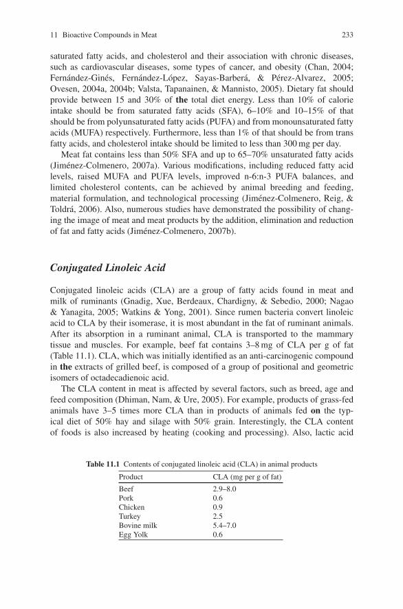
11 Bioactive Compounds in Meat 233
saturated fatty acids, and cholesterol and their association with chronic diseases,such as cardiovascular diseases, some types of cancer, and obesity (Chan, 2004;Fernandez-Gines, Fernandez-Lopez, Sayas-Barbera, & Perez-Alvarez, 2005;Ovesen, 2004a, 2004b; Valsta, Tapanainen, & Mannisto, 2005). Dietary fat shouldprovide between 15 and 30% of the total diet energy. Less than 10% of calorieintake should be from saturated fatty acids (SFA), 6–10% and 10–15% of thatshould be from polyunsaturated fatty acids (PUFA) and from monounsaturated fattyacids (MUFA) respectively. Furthermore, less than 1% of that should be from transfatty acids, and cholesterol intake should be limited to less than 300 mg per day.
Meat fat contains less than 50% SFA and up to 65–70% unsaturated fatty acids(Jimenez-Colmenero, 2007a). Various modifications, including reduced fatty acidlevels, raised MUFA and PUFA levels, improved n-6:n-3 PUFA balances, andlimited cholesterol contents, can be achieved by animal breeding and feeding,material formulation, and technological processing (Jimenez-Colmenero, Reig, &Toldra, 2006). Also, numerous studies have demonstrated the possibility of chang-ing the image of meat and meat products by the addition, elimination and reductionof fat and fatty acids (Jimenez-Colmenero, 2007b).
Conjugated Linoleic Acid
Conjugated linoleic acids (CLA) are a group of fatty acids found in meat andmilk of ruminants (Gnadig, Xue, Berdeaux, Chardigny, & Sebedio, 2000; Nagao& Yanagita, 2005; Watkins & Yong, 2001). Since rumen bacteria convert linoleicacid to CLA by their isomerase, it is most abundant in the fat of ruminant animals.After its absorption in a ruminant animal, CLA is transported to the mammarytissue and muscles. For example, beef fat contains 3–8 mg of CLA per g of fat(Table 11.1). CLA, which was initially identified as an anti-carcinogenic compoundin the extracts of grilled beef, is composed of a group of positional and geometricisomers of octadecadienoic acid.
The CLA content in meat is affected by several factors, such as breed, age andfeed composition (Dhiman, Nam, & Ure, 2005). For example, products of grass-fedanimals have 3–5 times more CLA than in products of animals fed on the typ-ical diet of 50% hay and silage with 50% grain. Interestingly, the CLA contentof foods is also increased by heating (cooking and processing). Also, lactic acid
Table 11.1 Contents of conjugated linoleic acid (CLA) in animal products
Product CLA (mg per g of fat)
Beef 2.9–8.0Pork 0.6Chicken 0.9Turkey 2.5Bovine milk 5.4–7.0Egg Yolk 0.6
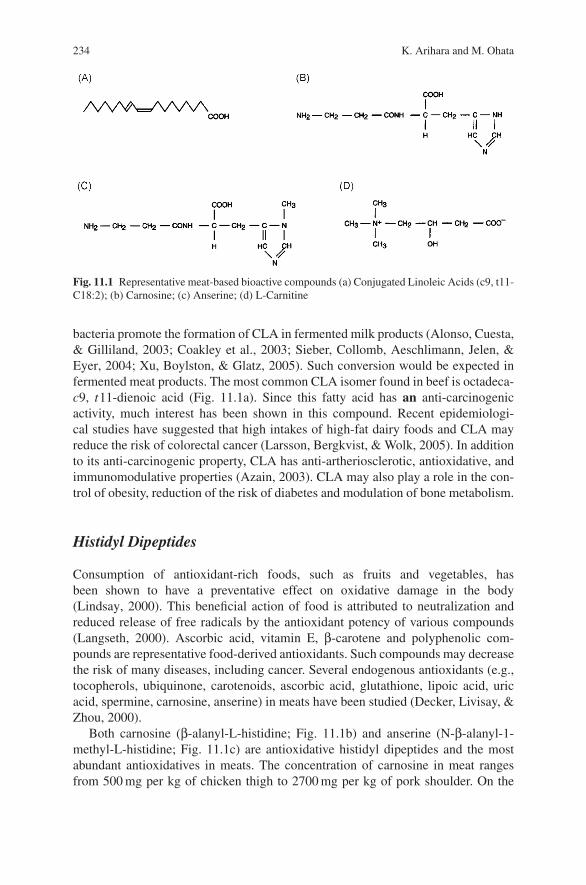
234 K. Arihara and M. Ohata
Fig. 11.1 Representative meat-based bioactive compounds (a) Conjugated Linoleic Acids (c9, t11-C18:2); (b) Carnosine; (c) Anserine; (d) L-Carnitine
bacteria promote the formation of CLA in fermented milk products (Alonso, Cuesta,& Gilliland, 2003; Coakley et al., 2003; Sieber, Collomb, Aeschlimann, Jelen, &Eyer, 2004; Xu, Boylston, & Glatz, 2005). Such conversion would be expected infermented meat products. The most common CLA isomer found in beef is octadeca-c9, t11-dienoic acid (Fig. 11.1a). Since this fatty acid has an anti-carcinogenicactivity, much interest has been shown in this compound. Recent epidemiologi-cal studies have suggested that high intakes of high-fat dairy foods and CLA mayreduce the risk of colorectal cancer (Larsson, Bergkvist, & Wolk, 2005). In additionto its anti-carcinogenic property, CLA has anti-artheriosclerotic, antioxidative, andimmunomodulative properties (Azain, 2003). CLA may also play a role in the con-trol of obesity, reduction of the risk of diabetes and modulation of bone metabolism.
Histidyl Dipeptides
Consumption of antioxidant-rich foods, such as fruits and vegetables, hasbeen shown to have a preventative effect on oxidative damage in the body(Lindsay, 2000). This beneficial action of food is attributed to neutralization andreduced release of free radicals by the antioxidant potency of various compounds(Langseth, 2000). Ascorbic acid, vitamin E, β-carotene and polyphenolic com-pounds are representative food-derived antioxidants. Such compounds may decreasethe risk of many diseases, including cancer. Several endogenous antioxidants (e.g.,tocopherols, ubiquinone, carotenoids, ascorbic acid, glutathione, lipoic acid, uricacid, spermine, carnosine, anserine) in meats have been studied (Decker, Livisay, &Zhou, 2000).
Both carnosine (β-alanyl-L-histidine; Fig. 11.1b) and anserine (N-β-alanyl-1-methyl-L-histidine; Fig. 11.1c) are antioxidative histidyl dipeptides and the mostabundant antioxidatives in meats. The concentration of carnosine in meat rangesfrom 500 mg per kg of chicken thigh to 2700 mg per kg of pork shoulder. On the
11 Bioactive Compounds in Meat 235
other hand, anserine is especially abundant in chicken muscle. Their antioxidantactivities may result from their ability to chelate transition metals such as copper(Brown, 1981) These antioxidative peptides have been reported to play a role inwound healing, recovery from fatigue and prevention of diseases related to stress. Arecent study demonstrated the bioavailability of carnosine by determining its con-centration in human plasma after ingestion of beef (Park, Volpe, & Decker, 2005).Increasing attention to these meat-based bioactive compounds has resulted in thedevelopment of a new sensitive procedure for determining these compounds includ-ing carnosine (Mora, Sentandreu, & Toldra, 2007).
L-Carnitine
L-Carnitine, β-hydroxy-gamma-trimethyl amino butyric acid (Fig. 11.1d), isdetected in the skeletal muscle of various animals (Shimada et al., 2005). L-Carnitine is especially abundant in beef (e.g., 1300 mg per kg of the thigh). It assiststhe human body in producing energy and in lowering the levels of cholesterol. Also,it helps the body to absorb calcium to improve skeletal strength and chromiumpicolinate to help build lean muscle mass. A recent study demonstrated that L-carnitine blocked apotosis and prevented skeletal muscle myopathy in heart failure(Vescovo et al., 2002). A drink product containing L-carnitine, which is marketedin the United States, is advertised as having several beneficial effects, such asmaintenance of stamina and fast recovery from fatigue. Also, a product containinga good amout of L-carnitine and carnosine, which is used as a functional foodingredient, has been marketed in Japan. This product is made from a by-product ofcorned beef.
Other Bioactive Components
Glutahione is an important antioxidative compound providing cellular defenseagainst toxicological and pathological processes. Red meat is a good source ofglutathione (12–26 mg per 100 g of beef; Jones et al., 1992). Taurine is a condi-tionally essential amino acid during lactation and at times of immune challenge(Bouckenooghe, Remacle, & Reusens, 2006). Also, taurine may protect our bodyfrom oxidative stress. Meat is the most excellent dietary source of taurine (77 mg per100 g of beef; Purchas, Rutherfurd, Pearce, Vather, & Wilkinson, 2004). CoenzymeQ10 (ubiquinone) shows antioxidative activity and its content in beef is 2 mg per100 g (Purchas & Busboom, 2005). Creatine and creatine phosphate have a criticalrole in muscle energy metabolism. Beef contains 350 mg of creatine per 100 g (Pur-chas & Busboom, 2005). Other components such as choline, balenine, creatinine,lipoic acid, putrescine, spermidine, and spermine should also be listed as meat-basedbioactive compounds.

236 K. Arihara and M. Ohata
Meat Protein-Derived Bioactive Peptides
In addition to the meat-based bioactive compounds described above, meat protein-derived peptides are another group of promising bioactive components of meat(Arihara, 2004, 2006a, 2006b). Several bioactive peptides from hydrolyzates ofmuscle proteins have been found. Although the activities of these peptides in thesequences of the parent proteins are latent, they are released by proteolytic enzymes.In this aspect, meat proteins have possible bioactivities beyond a nutritional sourceof amino acids alone.
Bioactive Peptides Derived from Food Proteins
Information on bioactive peptides generated from meat proteins is still limited.However, various physiologically functional peptides have been found from enzy-matic hydrolyzates of food proteins, such as milk and soy proteins (Arihara, 2006a;Korhonen & Pihlanto, 2007; Meisel, 1998; Mine & Shahidi, 2005; Pihlanto &Korhonen, 2003). Thus, bioactive peptides generated from food proteins are coveredbriefly at the beginning. Bioactive peptides from food proteins were first reportedby Mellander (1950). He described the effect of casein-derived phospho-relatedpeptides on vitamin D-independent bone calcification of rachitic infants. Sincethen, there have been numerous studies on bioactive peptides generated from foodproteins.
Angiotensin I-Converting Enzyme Inhibitory Peptides
Angiotensin I-converting enzyme (ACE) inhibitory peptides generated from foodproteins have been studied most extensively (Meisel, Walsh, Murry, & FitzGer-ald, 2005; Vermeirssen, Camp, & Verstraete, 2004). ACE inhibitory peptides havebeen shown to have antihypertensive effects and have been utilized for pharmaceu-ticals and physiologically functional foods. ACE plays an important physiologicalrole in the regulation of blood pressure (Fig. 11.2). ACE is a dipeptidyl carboxypep-tidase that converts an inactive form of the decapeptide, angiotensin I, to a potentvasoconstrictor, octapeptide angiotensin II, and inactivates bradykinin, which hasa depressor action (Li, Le, Shi, & Shrestha, 2004). Therefore, by inhibiting thecatalytic action of ACE, the elevation of blood pressure can be suppressed.
ACE inhibitory peptides from food protein were first identified in the hydrolyzateof gelatin by Oshima, Shimabukuro, and Nagasawa (1979). Since then, ACEinhibitory peptides have been found in the hydrolyzates of many proteins frommilk, fish, meat, eggs, soybean, corn, wheat, seaweed, and others (Arihara, 2006a;Vermeirssen et al., 2004). Some of these peptides have been reported to show antihy-pertensive effects in spontaneously hypertensive rats (SHR) by oral administration.ACE inhibitory peptides isolated from meat proteins are described later.
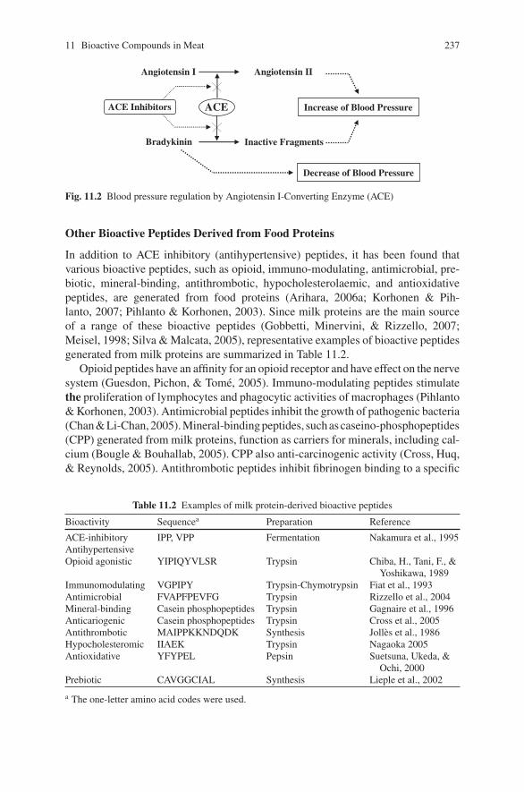
11 Bioactive Compounds in Meat 237
Angiotensin I Angiotensin II
ACEACE Inhibitors
Inactive Fragments
Decrease of Blood Pressure
Increase of Blood Pressure
Bradykinin
Fig. 11.2 Blood pressure regulation by Angiotensin I-Converting Enzyme (ACE)
Other Bioactive Peptides Derived from Food Proteins
In addition to ACE inhibitory (antihypertensive) peptides, it has been found thatvarious bioactive peptides, such as opioid, immuno-modulating, antimicrobial, pre-biotic, mineral-binding, antithrombotic, hypocholesterolaemic, and antioxidativepeptides, are generated from food proteins (Arihara, 2006a; Korhonen & Pih-lanto, 2007; Pihlanto & Korhonen, 2003). Since milk proteins are the main sourceof a range of these bioactive peptides (Gobbetti, Minervini, & Rizzello, 2007;Meisel, 1998; Silva & Malcata, 2005), representative examples of bioactive peptidesgenerated from milk proteins are summarized in Table 11.2.
Opioid peptides have an affinity for an opioid receptor and have effect on the nervesystem (Guesdon, Pichon, & Tome, 2005). Immuno-modulating peptides stimulatethe proliferation of lymphocytes and phagocytic activities of macrophages (Pihlanto& Korhonen, 2003). Antimicrobial peptides inhibit the growth of pathogenic bacteria(Chan & Li-Chan, 2005). Mineral-binding peptides, such as caseino-phosphopeptides(CPP) generated from milk proteins, function as carriers for minerals, including cal-cium (Bougle & Bouhallab, 2005). CPP also anti-carcinogenic activity (Cross, Huq,& Reynolds, 2005). Antithrombotic peptides inhibit fibrinogen binding to a specific
Table 11.2 Examples of milk protein-derived bioactive peptides
Bioactivity Sequencea Preparation Reference
ACE-inhibitoryAntihypertensive
IPP, VPP Fermentation Nakamura et al., 1995
Opioid agonistic YIPIQYVLSR Trypsin Chiba, H., Tani, F., &Yoshikawa, 1989
Immunomodulating VGPIPY Trypsin-Chymotrypsin Fiat et al., 1993Antimicrobial FVAPFPEVFG Trypsin Rizzello et al., 2004Mineral-binding Casein phosphopeptides Trypsin Gagnaire et al., 1996Anticariogenic Casein phosphopeptides Trypsin Cross et al., 2005Antithrombotic MAIPPKKNDQDK Synthesis Jolles et al., 1986Hypocholesteromic IIAEK Trypsin Nagaoka 2005Antioxidative YFYPEL Pepsin Suetsuna, Ukeda, &
Ochi, 2000Prebiotic CAVGGCIAL Synthesis Lieple et al., 2002
a The one-letter amino acid codes were used.
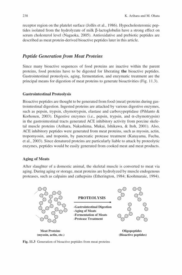
238 K. Arihara and M. Ohata
receptor region on the platelet surface (Jolles et al., 1986). Hypocholesteromic pep-tides isolated from the hydrolyzate of milk β-lactoglobulin have a strong effect onserum cholesterol level (Nagaoka, 2005). Antioxidative and prebiotic peptides aredescribed as meat protein-derived bioactive peptides later in this article.
Peptide Generation from Meat Proteins
Since many bioactive sequences of food proteins are inactive within the parentproteins, food proteins have to be digested for liberating the bioactive peptides.Gastrointestinal proteolysis, aging, fermentation, and enzymatic treatment are theprincipal means for digestion of meat proteins to generate bioactivities (Fig. 11.3).
Gastrointestinal Proteolysis
Bioactive peptides are thought to be generated from food (meat) proteins during gas-trointestinal digestion. Ingested proteins are attacked by various digestive enzymes,such as pepsin, trypsin, chymotrypsin, elastase and carboxypeptidase (Pihlanto &Korhonen, 2003). Digestive enzymes (i.e., pepsin, trypsin, and α-chymotrypsin)in the gastrointestinal tracts generated ACE inhibitory activity from porcine skele-tal muscle proteins (Arihara, Nakashima, Mukai, Ishikawa, & Itoh, 2001). Also,ACE inhibitory peptides were generated from meat proteins, such as myosin, actin,tropomyosin, and troponin, by pancreatic protease treatment (Katayama, Fuchu,et al., 2003). Since denatured proteins are particularly liable to attack by proteolyticenzymes, peptides would be easily generated from cooked meat and meat products.
Aging of Meats
After slaughter of a domestic animal, the skeletal muscle is converted to meat viaaging. During aging or storage, meat proteins are hydrolyzed by muscle endogenousproteases, such as calpains and cathepsins (Etherington, 1984; Koohmaraie, 1994).
-Gastrointestinal Digestion-Aging of Meats-Fermentation of Meats-Protease Treatment
PROTEOLYSIS
Meat Proteins(myosin, actin, etc.)
Oligopeptides(Bioactive peptides)
Fig. 11.3 Generation of bioactive peptides from meat proteins
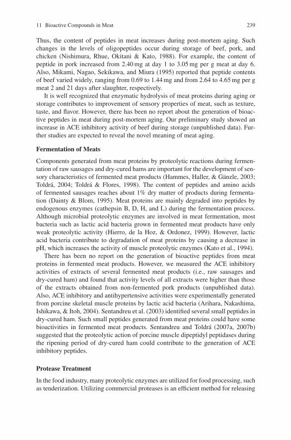
11 Bioactive Compounds in Meat 239
Thus, the content of peptides in meat increases during post-mortem aging. Suchchanges in the levels of oligopeptides occur during storage of beef, pork, andchicken (Nishimura, Rhue, Okitani & Kato, 1988). For example, the content ofpeptide in pork increased from 2.40 mg at day 1 to 3.05 mg per g meat at day 6.Also, Mikami, Nagao, Sekikawa, and Miura (1995) reported that peptide contentsof beef varied widely, ranging from 0.69 to 1.44 mg and from 2.64 to 4.65 mg per gmeat 2 and 21 days after slaughter, respectively.
It is well recognized that enzymatic hydrolysis of meat proteins during aging orstorage contributes to improvement of sensory properties of meat, such as texture,taste, and flavor. However, there has been no report about the generation of bioac-tive peptides in meat during post-mortem aging. Our preliminary study showed anincrease in ACE inhibitory activity of beef during storage (unpublished data). Fur-ther studies are expected to reveal the novel meaning of meat aging.
Fermentation of Meats
Components generated from meat proteins by proteolytic reactions during fermen-tation of raw sausages and dry-cured hams are important for the development of sen-sory characteristics of fermented meat products (Hammes, Haller, & Ganzle, 2003;Toldra, 2004; Toldra & Flores, 1998). The content of peptides and amino acidsof fermented sausages reaches about 1% dry matter of products during fermenta-tion (Dainty & Blom, 1995). Meat proteins are mainly degraded into peptides byendogenous enzymes (cathepsin B, D, H, and L) during the fermentation process.Although microbial proteolytic enzymes are involved in meat fermentation, mostbacteria such as lactic acid bacteria grown in fermented meat products have onlyweak proteolytic activity (Hierro, de la Hoz, & Ordonez, 1999). However, lacticacid bacteria contribute to degradation of meat proteins by causing a decrease inpH, which increases the activity of muscle proteolytic enzymes (Kato et al., 1994).
There has been no report on the generation of bioactive peptides from meatproteins in fermented meat products. However, we measured the ACE inhibitoryactivities of extracts of several fermented meat products (i.e., raw sausages anddry-cured ham) and found that activity levels of all extracts were higher than thoseof the extracts obtained from non-fermented pork products (unpublished data).Also, ACE inhibitory and antihypertensive activities were experimentally generatedfrom porcine skeletal muscle proteins by lactic acid bacteria (Arihara, Nakashima,Ishikawa, & Itoh, 2004). Sentandreu et al. (2003) identified several small peptides indry-cured ham. Such small peptides generated from meat proteins could have somebioactivities in fermented meat products. Sentandreu and Toldra (2007a, 2007b)suggested that the proteolytic action of porcine muscle dipeptidyl peptidases duringthe ripening period of dry-cured ham could contribute to the generation of ACEinhibitory peptides.
Protease Treatment
In the food industry, many proteolytic enzymes are utilized for food processing, suchas tenderization. Utilizing commercial proteases is an efficient method for releasing
240 K. Arihara and M. Ohata
bioactive peptides from food proteins. Many bioactive peptides have been experi-mentally generated by commercial proteases (Korhonen & Pihlanto, 2003; Pihlanto& Korhonen, 2003). Proteases from animal, plant, and microbial origins have beenused for the digestion of food proteins. In the meat industry, proteolytic enzymeshave been used for meat tenderization (Dransfield & Etherington, 1981). The mostcommonly used enzymes for meat tenderization are the plant enzymes papain,bromelain, and ficin. In meat treated with enzymatic tenderization, peptides havingbioactivities could be generated. On the other hand, effects of commercial proteaseson protein breakdown and sensory characteristics of dry fermented sausages havebeen investigated (Bruna, Fernandez, Hierro, Ordonez, & de la Hoz, 2000). Suchtreatment would also generate bioactive peptides in meat products.
ACE Inhibitory Peptides from Meat Proteins
As stated above, the most extensively studied bioactive peptides derived fromfood proteins are ACE inhibitory peptides. Also, among the bioactive peptidesderived from meat proteins, ACE inhibitory peptides have been studied extensively(Arihara, 2006a, 2006b; Vercruysse, Van Camp, & Smagghe, 2005). ACE inhibitorypeptides generated from meat proteins are summarized in Table 11.3.
Table 11.3 ACE inhibitory peptides derived from meat proteins
Sequencea Source IC50b (μM) SHRc References
IKW Chicken muscle 0.2 + Fujita et al., 2000LKA Chicken muscle (creatine kinase) 8.5 nt Fujita et al., 2000LKP Chicken muscle (aldolase) 0.3 + Fujita et al., 2000LAP Chicken muscle 3.5 + Fujita et al., 2000VWI Porcine muscle (actin) 1.1 + Arihara et al., 2005ITTNP Porcine myosin 549.0 + Arihara et al., 2001
Nakashima et al., 2002MNPPK Porcine myosin 945.5 + Arihara et al., 2001
Nakashima et al., 2002Fujita et al., 2000
FQKPKR Chicken muscle (myosin) 14.0 nt Jang & Lee, 2005VLAQYK Bovine muscle 23.2 + Fujita et al., 2000FKGRYYP Chicken muscle (creatine kinase) 0.6 - Arihara et al., 2004VFPMNPPK Fermented pork (myosin) 66.0 nt Fujita et al., 2000IVGRPRHQG Chicken muscle (actin) 2.4 - Katayama, Tomatsu
et al., 2003RMLGQTPTK Porcine troponin C 34.0 nt Katayama et al., 2004
Saiga, Okumuraet al., 2003
GFXGTXGLXGF Chicken muscle (collagen) 42.4 nta The one-letter amino acid codes were used.b The concentration of peptide needed to inhibit 50% of the ACE activity.c Antihypertensive activities in spontaneously hypertensive rats (+, positive activity; -, noactivity; nt, not tested).
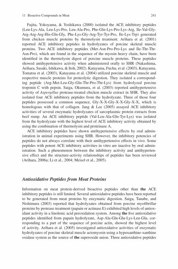
11 Bioactive Compounds in Meat 241
Fujita, Yokoyama, & Yoshikawa (2000) isolated the ACE inhibitory peptides(Leu-Lys-Ala, Leu-Lys-Pro, Leu-Ala-Pro, Phe-Gln-Lys-Pro-Lys-Arg, Ile-Val-Gly-Arg-Arg-Arg-His-Gln-Gly, Phe-Lys-Gly-Arg-Tyr-Tyr-Pro, Ile-Lys-Trp) generatedfrom chicken muscle proteins by thermolysin treatment. Arihara et al. (2001)reported ACE inhibitory peptides in hydrolyzates of porcine skeletal muscleproteins. Two ACE inhibitory peptides (Met-Asn-Pro-Pro-Lys and Ile-Thr-Thr-Asn-Pro), which are found in the sequence of the myosin heavy chain, have beenidentified in the thermolysin digest of porcine muscle proteins. These peptidesshowed antihypertensive activity when administered orally to SHR (Nakashima,Arihara, Sasaki, Ishikawa, & Itoh, 2002). Katayama, Fuchu, et al. (2003), Katayama,Tomatsu et al. (2003), Katayama et al. (2004) utilized porcine skeletal muscle andrespective muscle proteins for proteolytic digestion. They isolated a correspond-ing peptide (Arg-Met-Leu-Gly-Gln-Thr-Pro-Thr-Lys) from hydrolyzed porcinetroponin C with pepsin. Saiga, Okumura, et al. (2003) reported antihypertensiveactivity of Aspergillus protease-treated chicken muscle extract in SHR. They alsoisolated four ACE inhibitory peptides from the hydrolyzate. Three of those fourpeptides possessed a common sequence, Gly-X-X-Gly-X-X-Gly-X-X, which ishomologous with that of collagen. Jang & Lee (2005) assayed ACE inhibitoryactivities of several enzymatic hydrolyzates of sarcoplasmic protein extracts frombeef rump. An ACE inhibitory peptide (Val-Leu-Ala-Gln-Tyr-Lys) was isolatedfrom the hydrolyzate with the highest level of ACE inhibitory activity obtained byusing the combination of thermolysin and proteinase A.
ACE inhibitory peptides have shown antihypertensive effects by oral admin-istration in animal experiments using SHR. However, the inhibitory potencies ofpeptides do not always correlate with their antihypertensive effects in vivo. Somepeptides with potent ACE inhibitory activities in vitro are inactive by oral admin-istration. Such a phenomenon between the inhibitory activity and antihyperten-sive effect and the structure-activity relationships of peptides has been reviewed(Arihara, 2006a; Li et al., 2004; Meisel et al., 2005).
Antioxidative Peptides from Meat Proteins
Information on meat protein-derived bioactive peptides other than the ACEinhibitory peptides is still limited. Several antioxidative peptides have been reportedto be generated from meat proteins by enzymatic digestion. Saiga, Tanabe, andNishimura (2003) reported that hydrolyzates obtained from porcine myofibrillarproteins by protease treatment (papain or actinase E) exhibited high levels of antiox-idant activity in a linolenic acid peroxidation system. Among the five antioxidativepeptides identified from papain hydrolyzate, Asp-Ala-Gln-Glu-Lys-Leu-Glu, cor-responding to a part of the sequence of porcine actin, showed the highest levelof activity. Arihara et al. (2005) investigated antioxidative activities of enzymatichydrolyzates of porcine skeletal muscle actomyosin using a hypoxanthine-xanthineoxidase system as the source of the superoxide anion. Three antioxidative peptides
242 K. Arihara and M. Ohata
were isolated from a papain-treated hydrolyzate of pork actomyosin and they weresequenced as Asp-Leu-Tyr-Ala, Ser-Leu-Tyr-Ala, and Val-Trp. In addition to theantioxidative activity in vitro, these peptides showed physiological activity in vivo.Each of these peptides had an anti-fatigue effect when orally administered to micein an experiment using a treadmill.
Prebiotic Peptides from Meat Proteins
Prebiotics are defined as “non-digestible food ingredients that beneficially affectthe host by selectively stimulating the growth and/or activity of one or a limitednumber of bacteria in the colon and thus improve the health of the host” (Gibson, &Roberfroid, 1995). Oligosaccharides are representative prebiotic substances, whichare known to enhance the activity of probiotic bacteria (i.e., intestinal lactobacilliand bifidobacteria). The presence of prebiotic peptides has been suggested. Liepleet al. (2002) first reported nonglycosylated peptides that selectively stimulate thegrowth of bifidobacteria. Recently, we found that the hydrolyzate of porcine skeletalmuscle actomyosin, digested by papain, enhanced the growth of Bifidobacteriumstrains (Arihara, Ishikawa, & Itoh, 2006). One corresponding prebiotic peptide wasidentified as Glu-Leu-Met.
Other Promising Peptides from Meat Proteins
Opioid peptides are defined as peptides that have an affinity for an opiate recep-tor as well as opiate-like effects (Pihlanto & Korhonen, 2003). Opioid peptideshave effects on the nerve system and gastrointestinal functions. Typical opioid pep-tides (e.g., endorphins, enkephalin, and prodynorphin) have the same N-terminalsequence, Tyr-Gly-Gly-Phe. Several opioid peptides derived from food proteinshave been reported. The N-terminal sequence of most of these peptides is Tyr-X-Pheor Tyr-X1-X2-Phe. The N-terminal tyrosine residue and the presence of an aromaticamino acid at the third or fourth position form an important structure that fits withthe binding site of opioid receptors (Pihlanto-Leppala, 2001). There has been noreport on the generation of opioid peptides from meat proteins. However, sinceopioid sequences are thought to be present in the sequences of muscle proteins,it should be possible to find opioid peptides in meat proteins by proteolytic treat-ment. Although bovine blood hemoglobin is regarded as a minor component of meatand meat products, in some meat products, such as blood sausage, hemoglobin is amajor component. Investigation of hemoglobin peptic hydrolyzate has revealed thepresence of biologically active peptides with affinity for opioid receptors (Nyberg,Sanderson, & Glamsta, 1997; Zhao, Garreau, Sannier, & Piot, 1997).
Morimatsu et al. (1996) demonstrated hypocholesterolemic effects of papain-hydrolyzed pork meat. However, the corresponding peptides were not identified.Immuno-modulating peptides have been discovered in enzymatic hydrolyzates ofproteins from various foods, such as milk, eggs, soybeans, and rice. However, to
11 Bioactive Compounds in Meat 243
date, meat protein-derived immuno-modulating peptides have not been reported.In addition to bioactive peptides described here, several bioactive peptides (e.g.,antimicrobial, mineral-binding, antithrombotic, and hypocholesterolemic peptides)have been found from enzymatic hydrolyzates of various food proteins (Mine &Shahidi, 2005). It is expected that interest will be directed to research aimed atfinding such meat protein-derived peptides.
Apart from bioactivities, meat protein-derived peptides also contribute toorgano-leptic properties of meat (Nishimura & Kato, 1988; Nishimura et al., 1988;Arihara, 2006a). Meat products can be modified by adding functional ingredientsbeneficial for health or by eliminating or reducing components that are harmful.However, these modifications could result in products with inferior sensory prop-erties. Therefore, generation of peptides from meat proteins has a great potentialto produce novel functional foods with good organo-leptic properties. Also, fromthe aspect of food allergies, enzymatic treatment of food proteins, including meatproteins, warrants attention. Protein hydrolyzate based products have been producedand considered suitable for the diet of allergic patients. Although meat is lessallergic than common allergy-inducing foods, such as milk, eggs, and soya, meatproteins (e.g., serum albumin, gamma globulin, actin, myoglobin and tropomyosin)sometimes cause allergic reactions (Tanabe & Nishimura, 2005).
Utilization of Peptides for Functional Meat Products
Although bioactive peptides have not yet been utilized in the meat industry, suchpeptides are promising candidates for ingredients of functional foods. Several foodproducts containing ACE inhibitory peptides have been successfully marketed forhypertensives (Arihara, 2006b). There are two commercial dairy products contain-ing Ile-Pro-Pro and Val-Pro-Pro, which are generated from milk protein by fermen-tation (Nakamura, Yamamoto, Sakai, & Takano, 1995; Seppo, Jauhiaine, Poussa,& Korpela, 2003). Calpis Amiel-S drink has been approved as a FOSHU in Japan.The Finnish fermented milk drink Evolus, developed by Valio Ltd., contains thesame tripeptides as those in Amiel-S. The enzymatic digest of dried bonito con-taining antihypertensive peptides has also been used in a soup product in Japan. AFOSHU sour milk product utilizing casein-phosphopeptide (CPP) has been devel-oped in Japan. CPP milk protein-derived peptides act as mineral trappers resultingin enhancement of the absorption efficiency of calcium (Gagnaire, Pierre, Molle,& Leonil, 1996). Hydrolyzates of meat proteins and their corresponding bioactivepeptides would be utilized for functional foods.
Accumulation of bioactive peptides in meat products by fermentation is a goodstrategy for developing functional meat products. Bioactive peptides would be gen-erated in fermented meat products, since meat proteins are hydrolyzed by proteolyticenzymes during fermentation and storage. Rediscovery of traditional fermentedmeats as functional foods is also an interesting direction. In the dairy industry, manytraditional fermented foods, such as fermented dairy products, have been redis-covered as functional foods (Farnworth, 2003). Numerous physiologically active
244 K. Arihara and M. Ohata
components, including bioactive peptides, have been discovered in these traditionalfermented foods. Thus, traditionally fermented meats are attractive targets for find-ing new functional meat products.
Concluding Remarks
Although there has been extensive research and development of functionalfoods in the dairy industry (Chandan, 2007; Chandan & Shah, 2007; Mattila-Sandholm & Saarela, 2000; Playne, Bennett, & Smithers, 2003), little attentionhas been paid to functional meat products until recently. However, efforts havebeen directed in recent years to research and development of functional meatproducts (Arihara, 2004, 2006a, 2006b; Fernandez-Gines et al., 2005; Jimenez-Colmenero, 2007a, 2007b; Jimenez-Colmenero, Carballo, & Cofrades, 2001;Jimenez-Colmenero et al., 2006). Since meat and meat products are importantin the diet in most developed countries, healthier meat and meat products wouldcontribute to human health. Utilizing or emphasizing meat-based bioactive com-pounds, including bioactive peptides generated from meat proteins, is a promisingmeans for developing attractive functional meat products. Such efforts would alsocontribute to the demonstration for consumers of the scientific benefits of meat andmeat components for human health.
References
Alonso, L., Cuesta, E. P., & Gilliland, S. E. (2003). Production of free linoleic acid by Lactobacillusacidophilus and Lactobacillus casei of human intestinal origin. Journal of Dairy Science, 86,1941–1946.
Arihara, K. (2004). Functional foods. In W. K. Jensen, C. Devine, & M. Dikeman (Eds.), Encyclo-pedia of meat sciences (pp. 492–499). Oxford: Elsevier.
Arihara, K. (2006a). Functional properties of bioactive peptides derived from meat proteins. InN. M. L. Nollet, & F. Toldra (Eds.), Advanced technologies for meat processing (pp. 245–274).Boca Raton, FL: CRC Press.
Arihara, K. (2006b). Strategies for designing novel functional meat products. Meat Science, 74,219–229.
Arihara, K., Ishikawa, S., & Itoh, M. (2006). Bifidobacterium growth promoting peptides derivedfrom meat proteins. Japan patent (submitted to government).
Arihara, K., Nakashima, Y., Mukai, T., Ishikawa, S., & Itoh, M. (2001). Peptide inhibitors forangiotensin I-converting enzyme from enzymatic hydrolysates of porcine skeletal muscle pro-teins. Meat Science, 57, 319–324.
Arihara, K., Nakashima, Y., Ishikawa, S., & Itoh, M. (2004). Antihypertensive activities generatedfrom porcine skeletal muscle proteins by lactic acid bacteria. Abstracts of 50th InternationalCongress of Meat Science and Technology (p. 236), 8–13 August 2004, Helsinki, Finland.
Arihara, K., Tomita, K., Ishikawa, S., Itoh, M., Akimoto, M., & Sameshima, T. (2005). Anti-fatiguepeptides derived from meat proteins. Japan patent (submitted to government).
Azain, M. J. (2003). Conjugated linoleic acid and its effects on animal products and health insingle-stomached animals. Proceedings of the Nutrition Society, 62, 319–328.
11 Bioactive Compounds in Meat 245
Biesalski, H.-K. (2005). Meat as a component of a healthy diet – are there any risks or benefits ifmeat is avoided in the diet? Meat Science, 70, 509–524.
Bouckenooghe, T., Remacle, C., & Reusens, B. (2006). Is taurine a functional nutrient? CurrentOpinion in Clinical Nutrition and Metabolic Care, 9, 728–733.
Bougle, D., & Bouhallab, S. (2005). Mineral-binding proteins and peptides and bioavailability oftrace elements. In Y. Mine, & F. Shahidi (Eds.), Nutraceutical proteins and peptides in healthand disease (pp. 29–40). Boca Raton, FL: CRC Press.
Brown, C. E. (1981). Interactions among carnosine, anserine, ophidine and copper in biochemicaladaptation. Journal of Theoretical Biology, 88, 245–256.
Bruna, J. M., Fernandez, M., Hierro, E. M., Ordonez, J. A., & de la Hoz, L. (2000). Combineduse of pronase E and a fungal extract (Penicillium aurantiogriseum) to potentiate the sensorycharacteristics of dry fermented sausages. Meat Science, 54, 135–145.
Chan, J. C. K., & Li-Chan, E. C. Y. (2005). Antimicrobial peptides. In Y. Mine, & F. Shahidi (Eds.),Nutraceutical proteins and peptides in health and disease (pp. 99–136). Boca Raton, FL: CRCPress.
Chan, W. (2004). Macronutrients in meat. In W. K. Jensen, C. Devine, & M. Dikeman (Eds.),Encyclopedia of meat sciences (pp. 614–618). Oxford: Elsevier.
Chandan, R. C. (2007). Functional properties of milk constituents. In Y. H. Hui (Ed.), Handbookof food products manufacturing – Principles, bakery, beverages, cereals, cheese, confectionary,fats, fruits, and functional foods (pp. 971–987). Hoboken, NJ: John Wiley & Sons.
Chandan, R. C., & Shah, N. P. (2007). Functional foods based on dairy ingredients. InY. H. Hui (Ed.), Handbook of food products manufacturing – Principles, bakery, beverages,cereals, cheese, confectionary, fats, fruits, and functional foods (pp. 957–970). Hoboken,NJ: John Wiley & Sons.
Chiba, H., Tani, F., & Yoshikawa, M. (1989). Opioid antagonist peptides derived from �-casein.Journal of Dairy Research, 56, 363–366.
Coakley, M., Ross, R. P., Nordgren, M., Fitzerald, G., Devery, R., & Stanton, C. (2003). Conju-gated linoleic acid biosynthesis by human-derived Bifidobacterium species. Journal of AppliedMicrobiology, 94, 138–145.
Cross, K. J., Huq, N. L., & Reynolds, E. C. (2005). In Y. Mine, & F. Shahidi (Eds.), Nutraceuticalproteins and peptides in health and disease (pp. 335–351). Boca Raton, FL: CRC Press.
Dainty, R., & Blom, H. (1995). Flavor chemistry of fermented sausages. In G. Campbell-Plattand,& P. E. Cook (Eds.), Fermented meats (pp. 176–193). Glasgow, Scotland: Blackie Academic &Professional.
Decker, E. A., Livisay, S. A., & Zhou, S. (2000). Mechanisms of endogenous skeletal muscleantioxidants: Chemical and physical aspects. In E. A. Decker, C. Faustman, & C. J. Lopez-Bote(Eds.), Antioxidants in muscle foods (pp. 25–60). New York: Wiley-Interscience.
Dentali, S. (2002). Regulation of functional foods and dietary supplements. Food Technology,56(6), 89–94.
Dhiman, T. R., Nam, S. H., & Ure, A. L. (2005). Factors affecting conjugated linoleic acid contentin milk and meat. Critical Reviews of Food Science and Nutrition, 45, 463–482.
Dransfield, E., & Etherington, D. (1981). Enzymes in the tenderization of meat. In G. G. Birch,N. Blakebrough, & K. J. Parker (Eds.), Enzymes and food processing (pp. 177–194). London:Applied Science Publishers.
Etherington, D. J. (1984). The contribution of proteolytic enzymes to postmortem changes in mus-cle. Journal of Animal Science, 59, 1644–1650.
Eve, L. (2000). Regulatory issues: Europe and Japan. In M. K. Schmidl, & T. P. Labuza (Eds.),Essentials of functional foods (pp. 363–384). Gaithersburg, MD: Aspen Publication.
Farnworth, E. R. (2003). Handbook of fermented functional foods. Boca Raton, FL: CRC Press.Fernandez-Gines, J. M., Fernandez-Lopez, J., Sayas-Barbera, E., & Perez-Alvarez, J. A. (2005).
Meat products as functional foods: A review. Journal of Food Science, 70, R37–R43.Fiat, A. M., Migliore-Samour, D., Jolles, P., Drouet, L., Collier, C., & Caen, J. (1993). Biologically
active peptides from milk proteins with emphasis on two example concerning antithromboticand immuno-modulating activities. Journal of Dairy Science, 76, 301–310.
246 K. Arihara and M. Ohata
Fujita, H., Yokoyama, K., & Yoshikawa, M. (2000). Classification and antihypertensive activityof angiotensin I-converting enzyme inhibitory peptides derived from food proteins. Journal ofFood Science, 65, 564–569.
Gagnaire, V., Pierre, A., Molle, D., & Leonil, J. (1996). Phosphopeptides interacting with colloidalcalcium phosphate isolated by tryptic hydrolysis of bovine casein micelles. Journal of DairyResearch, 63, 405–422.
Gibson, G. R., & Roberfroid, M. B. (1995). Dietary modulation of the human colonic microbiota:Introducing the concept of prebiotics. Journal of Nutrition, 125, 1401–1412.
Gnadig, S., Xue, Y., Berdeaux, O., Chardigny, J. M., & Sebedio, J.-L. (2000). Conjugated linoleicacid (CLA) as a functional ingredient. In T. Mattila-Sandholm, & M. Saarela (Eds.), Functionaldairy products (pp. 263–298). Boca Raton, FL: CRC Press.
Gobbetti, M., Minervini, F., & Rizzello, C. G. (2007). Bioactive peptides in dairy products. InY. H. Hui (Ed.), Handbook of food products manufacturing – Health, meat, milk, poultry,seafood, and vegetables (pp. 489–517). Hoboken, NJ: John Wiley & Sons.
Guesdon, B., Pichon, L, & Tome, D. (2005). Opioid peptides. In Y. Mine, & F. Shahidi (Eds.),Nutraceutical proteins and peptides in health and disease (pp. 367–376). Boca Raton, FL:CRC Press.
Hammes, W. P., Haller, D., & Ganzle, M. G. (2003). Fermented meat. In E. R. Farnworth (Ed.),Handbook of fermented functional foods (pp. 251–275). Boca Raton, FL: CRC Press.
Hasler, C. M. (1988). Functional foods: Their role in disease prevention and health promotion.Food Technology, 52(10), 63–70.
Heasman, M., & Mellentin, J. (2001). The functional foods revolution. London: Earthscan Publi-cations.
Hierro, E., de la Hoz, L., & Ordonez, J. A. (1999). Contribution of the microbial and meat endoge-nous enzymes to the free amino acid and amine contents of dry fermented sausages. Journal ofAgricultural and Food Chemistry, 47, 1156–1161.
Higgs, J. D. (2000). The changing nature of red meat: 20 years of improving nutritional quality.Trends in Food Science and Technology, 11, 85–95.
Hutt, P. B. (2000). U.S. Government regulation of food with claims for special physiological value.In M. K. Schmidl, & T. P. Labuza (Eds.), Essentials of functional foods (pp. 339–352). Gaithers-burg, MD: Aspen Publication.
Jang, A., & Lee, M. (2005). Purification and identification of angiotensin converting enzymeinhibitory peptides from beef hydrolysates. Meat Science, 69, 653–661.
Jimenez-Colmenero, F. (2007a). Functional foods based on meat products. In Y. H. Hui (Ed.),Handbook of food products manufacturing – Principles, bakery, beverages, cereals, cheese,confectionary, fats, fruits, and functional foods (pp. 989–1015). Hoboken, NJ: John Wiley &Sons.
Jimenez-Colmenero, F. (2007b). Healthier lipid formulation approaches in meat-based functionalfoods. Technological options for replacement of meat fats by non-meat fats. Trends in FoodScience and Technology, 18, 567–578.
Jimenez-Colmenero, F., Carballo, J., & Cofrades, S. (2001). Healthier meat and meat products:Their role as functional foods. Meat Science, 59, 5–13.
Jimenez-Colmenero, F., Reig, M., & Toldra, F. (2006). New approaches for the development offunctional meat products. In L. M. L. Nollet, & F. Toldra (Eds.), Advanced technologies formeat processing (pp. 275–308). Boca Raton, FL: CRC Press.
Jolles, P. S., Levy-Toledano, S., Fiat, A. M., Soria, C., Gillessen, D., Thomaidis, A., et al. (1986).Analogy between fibrinogen and casein. European Journal of Biochemistry, 158, 379–384.
Jones, D. P., Coates, R. J., Flagg, E. W., Eley, J. W., Block, G., Greenberg, R. S., et al. (1992).Glutathione in foods listed in the National Cancer Institute’s Health Habits and History FoodFrequency Questionnaire. Nutrition and Cancer, 17, 57–75.
Katayama, K., Fuchu, H., Sakata, A., Kawahara, S., Yamauchi, K., Kawahara, Y., et al. (2003).Angiotensin I-converting enzyme inhibitory activities of porcine skeletal muscle proteins fol-lowing enzyme digestion. Asian-Australian Journal of Animal Science, 16, 417–424.
11 Bioactive Compounds in Meat 247
Katayama, K., Tomatsu, M., Fuchu, H., Sugiyama, M., Kawahara, S., Yamauchi, K., et al. (2003).Purification and characterization of an angiotensin I-converting enzyme inhibitory peptidederived from porcine troponin C. Animal Science Journal, 74, 53–58.
Katayama, K., Tomatsu, M., Kawahara, S., Yamauchi, K., Fuchu, H., Kodama, Y., et al.(2004). Inhibitory profile of nonapeptide derived from porcine troponin C against angiotensinI-converting enzyme. Journal of Agricultural and Food Chemistry, 52, 771–775.
Kato, T., Matsuda, T., Tahara, T., Sugimoto, M., Sato, Y., & Nakamura, R. (1994). Effects of meatconditioning and lactic fermentation on pork muscle protein degradation. Bioscience, Biotech-nology and Biochemistry, 58, 408–410.
Koohmaraie, M. (1994). Muscle proteinases and meat aging. Meat Science, 36, 93–104.Korhonen, H., & Pihlanto, A. (2003). Food-derived bioactive peptides: Opportunities for designing
future foods. Current Pharmaceutical Design, 9, 1297–1308.Korhonen, H., & Pihlanto, A. (2007). Bioactive peptides from food proteins. In Y. H. Hui (Ed.),
Handbook of food products manufacturing – Health, meat, milk, poultry, seafood, and vegeta-bles (pp. 5–37). Hoboken, NJ: John Wiley & Sons.
Krajcovicova-Kudlackova, M., Simoncic, R., Bederova, A., Babinska, K., & Beder, I. (2000).Correlation of carnitine levels to methionine and lysine intake. Physiological Research, 49,399–402.
Lachance, P. A., & Fisher, M. C. (2005). Reinvention of the food guide pyramid to promote health.Advances in Food and Nutrition Research, 49, 1–39.
Langseth, L. (2000). Antioxidants and their effect on health. In M. K. Schmidl, & T. P. Labuza(Eds.), Essentials of functional foods (pp. 303–317). Gaithersburg, MD: Aspen Publication.
Larsson, S. C., Bergkvist, L., & Wolk, A. (2005). High-fat dairy food and conjugated linoleicacid intakes in relation to colorectal cancer incidence in the Swedish Mammography Cohort.American Journal of Clinical Nutrition, 82, 894–900.
Li, G. H., Le, G. W., Shi, Y. H., & Shrestha, S. (2004). Angiotensin I-converting enzyme inhibitorypeptides derived from food proteins and their physiological and pharmacological effects. Nutri-tion Research, 24, 469–486.
Lieple, C., Adermann, K., Raida, M., Magert, H.-J., Forssman, W.-G., & Zucht, H.-D. (2002).Human milk provides peptides highly stimulating the growth of bifidobacteria. European Jour-nal of Biochemistry, 269, 712–718.
Lindsay, D. G. (2000). Maximizing the functional benefits of plants foods. In G. R. Gibson, &C. M. Williams (Eds.), Functional foods (pp. 183–208). Boca Raton, FL: CRC Press.
Mattila-Sandholm, T., & Saarela, M. (2000). Functional dairy products. Boca Raton, FL: CRCPress.
Meisel, H. (1998). Overview on milk protein-derived peptides. International Dairy Journal, 8,363–373.
Meisel, H., Walsh, D. J., Murry, B., & FitzGerald, R. J. (2005). ACE inhibitory peptides. InY. Mine, & F. Shahidi (Eds.), Nutraceutical proteins and peptides in health and disease(pp. 269–315). Boca Raton, FL: CRC Press.
Mellander, O. (1950). The physiological importance of the casein phosphopeptide calcium salts II:Peroral calcium dosage of infants. Acta Society Medicine Uppsala, 55, 247–255.
Mikami, M., Nagao, M., Sekikawa, M., & Miura, H. (1995). Changes in peptide and free aminoacid contents of different bovine muscle homogenate during storage. Animal Science and Tech-nology (Japan), 66, 630–638.
Mine, Y., & Shahidi, F. (2005). Nutraceutical proteins and peptides in health and disease. BocaRaton, FL: CRC Press
Mir, P. S., McAllister, T. A., Scott, S., Aalhus, J., Baron, V., McCartney, D., et al. (2004). Con-jugated linoleic acid-enriched beef production. American Journal of Clinical Nutrition, 79,1207S–1211S.
Mora, L., Sentandreu, M. A., & Toldra, F. (2007). Hydrophilic chromatographic determination ofcarnosine, anserine, balenine, creatine, and creatinine. Journal of Agricultural Food Chemistry,55, 4664–4669.
248 K. Arihara and M. Ohata
Morimatsu, F., Ito, M., Budijanto, S., Watanabe, I., Furukawa, Y., & Kimura, S. (1996). Plasmacholesterol-suppressing effect of papain-hydrolyzed pork meat in rats fed hypercholesterolemicdiet. Journal of Nutritional Science and Vitaminology, 42, 145–153.
Mulvihill, B. (2004). Micronutrients in meat. In W. K. Jensen, C. Devine, & M. Dikeman (Eds.),Encyclopedia of meat sciences (pp. 618–623). Oxford: Elsevier.
Nagao, K., & Yanagita, T. (2005). Conjugated fatty acids in food and their health benefits. Journalof Bioscience and Bioengineering, 100, 152–157.
Nagaoka, S. (2005). Cholesterol-lowering proteins and peptides. In Y. Mine, & F. Shahidi (Eds.),Nutraceutical proteins and peptides in health and disease (pp. 29–40). Boca Raton, FL: CRCPress.
Nakamura, Y., Yamamoto, N., Sakai, K., & Takano, T. (1995). Antihypertensive effect of sour milkand peptides isolated from it that are inhibitors to angiotensin I-converting enzyme. Journal ofDairy Science, 78, 1253–1257.
Nakashima, Y., Arihara, K., Sasaki, A., Ishikawa, S., & Itoh, M. (2002). Antihypertensive activitiesof peptides derived from porcine skeletal muscle myosin in spontaneously hypertensive rats.Journal of Food Science, 67, 434–437.
Nishimura, T., & Kato, H. (1988). Mechanisms involved in the improvement of meat taste duringpostmortem aging. Food Science and Technology International Tokyo, 4, 241–249.
Nishimura, T., Rhue, M. R., Okitani, A., & Kato, H. (1988). Components contributing to the improve-ment of meat taste during storage. Agricultural and Biological Chemistry, 52, 2323–2330.
Nyberg, F., Sanderson, K., & Glamsta, E.-L. (1997). The hemorphins: A new class of opioid pep-tides derived from the blood protein hemoglobin. Biopolymers, 43, 147–156.
Oshima, G., Shimabukuro, H., & Nagasawa, K. (1979). Peptide inhibitors of angiotensinI-converting enzyme in digests of gelatin by bacterial collagenase. Biochimica et BiophysicaActa, 566, 128–137.
Ovesen, L. (2004a). Cardiovascular and obesity health concerns. In W. K. Jensen, C. Devine, &M. Dikeman (Eds.), Encyclopedia of meat sciences (pp. 623–628). Oxford: Elsevier.
Ovesen, L. (2004b). Cancer health concerns. In W. K. Jensen, C. Devine, & M. Dikeman (Eds.),Encyclopedia of meat sciences (pp. 628–633). Oxford: Elsevier.
Park, Y. J., Volpe, S. L., & Decker, E. A. (2005). Quantitation of carnosine in humans plasma afterdietary consumption of beef. Journal of Agricultural and Food Chemistry, 53, 4736–4739.
Pihlanto-Leppala, A. (2001). Bioactive peptides derived from bovine whey proteins: Opioid andACE inhibitory peptides. Trends in Food Science and Technology, 11, 347–356.
Pihlanto, A., & Korhonen, H. (2003). Bioactive peptides and proteins. Advances in Food and Nutri-tion Research, 47, 175–276,
Playne, M. J., Bennett, L. E., & Smithers, G. W. (2003). Functional dairy foods and ingredients.Australian Journal of Dairy Technology, 58, 242–263.
Purchas, R. W., Rutherfurd, S. M., Pearce, P. D., Vather, R., & Wilkinson, B. H. P. (2004). Con-centrations in beef and lamb of taurine, carnosine, coenzyme Q10, and creatine. Meat Science,66, 629–637.
Purchas, R. W., & Busboom, J. R. (2005). The effect of production system and age on levels ofiron, taurine, carnosine, coenzyme Q10, and creatine in beef muscles and liver. Meat Science,70, 589–596.
Rizzello, C. G., Losito, I., Gobbetti, M., Carbonara, T., De Bari, M. D., & Zambonin, P. G. (2005).Antibacterial activities of peptides from the water-soluble extracts of Italian cheese varieties.Journal of Dairy Science, 88, 2348–2360.
Saiga, A., Okumura, T., Makihara, T., Katsuta, S., Shimizu, T., Yamada, R., et al. (2003).Angiotensin I-converting enzyme inhibitory peptides in a hydrolyzed chicken breast muscleextract. Journal of Agricultural and Food Chemistry, 51, 1741–1745.
Saiga, A., Tanabe, S., & Nishimura, T. (2003). Antioxidant activity of peptides obtained fromporcine myofibrillar proteins by protease treatment. Journal of Agricultural and Food Chem-istry, 51, 3661–3667.
Sentandreu, M. A., & Toldra, F. (2007a). Oligopeptides hydrolysed by muscle dipeptidyl pep-tidases can generate angiotensin-I converting enzyme inhibitory dipeptides. European FoodResearch and Technology, 224, 785–790.
11 Bioactive Compounds in Meat 249
Sentandreu, M. A., & Toldra, F. (2007b). Evaluation of ACE inhibitory activity of dipep-tides generated by the action of porcine muscle dipeptidyl peptidases. Food Chemistry, 102,511–515.
Sentandreu, M. A., Stoeva, S., Aristoy, M. C., Laib, K., Voelter, W., & Toldra, F. (2003). Identi-fication of small peptides generated in Spanish dry-cured ham. Journal of Food Science, 68,64–69.
Seppo, L., Jauhiaine, T., Poussa, T., & Korpela, R. (2003). A fermented milk high in bioactivepeptides has a blood-pressure lowering effect in hypertensive subjects. American Journal ofClinical Nutrition, 77, 326–330.
Shimada, K., Sakura, Y., Fukushima, M., Sekikawa, M., Kuchida, K., Mikami, M., et al. (2005).Species and muscle differences in L-carnitine levels in skeletal muscles based on a new simpleassay. Meat Science, 68, 357–362.
Sieber, R., Collomb, M., Aeschlimann, A., Jelen, P., & Eyer, H. (2004). Impact of microbial cul-tures on conjugated linoleic acid in dairy products – a review. International Dairy Journal, 14,1–15.
Silva, S. V., & Malcata, F. X. (2005). Caseins as source of bioactive peptides. International DairyJournal, 15, 1–15.
Suetsuna, K., Ukeda, H., & Ochi, H. (2000). Isolation and characterization of free radicalscavenging activities peptides derived from casein. Journal of Nutritional Biochemistry, 11,128–131.
Tanabe, S., & Nishimura, T. (2005). Meat allergy. In Y. Mine, & F. Shahidi (Eds.), Nutraceuticalproteins and peptides in health and disease (pp. 481–491). Boca Raton, FL: CRC Press.
Toldra, F. (2004). Dry. In W. K. Jensen, C. Devine, & M. Dikeman (Eds.), Encyclopedia of meatsciences (pp. 360–365). Oxford: Elsevier.
Toldra, F., & Flores, M. (1998). The role of muscle proteases and lipases in flavor developmentduring the processing of dry-cured ham. Critical Reviews of Food Science and Nutrition, 38,331–352.
Valsta, L. M., Tapanainen, H., & Mannisto, S. (2005). Meat fats in nutrition. Meat Science, 70,525–530.
Vercruysse, L., Van Camp, J., & Smagghe, G. (2005). ACE inhibitory peptides derived from enzy-matic hydrolysates of animal protein: A review. Journal of Agricultural and Food Chemistry,53, 8106-8115.
Vermeirssen, V., Camp, J. V., & Verstraete, W. (2004). Bioavailability of angiotensin I convertingenzyme inhibitory peptide (review article). British Journal of Nutrition, 92, 357–366.
Vescovo, G., Ravara, B., Gobbo, V., Sandri, M., Angelini, A., Dalla Libera, L., et al. (2002). L-Carnitine: A potential treatment for blocking apotosis and preventing skeletal muscle myopathyin heart failure. American Journal of Physiology, 283, C802–C810.
Watkins, B. A., & Yong, L. (2001). Conjugated linoleic acid: The present state of knowledge. InWildman, R. E. C. (Ed.), Handbook of nutraceuticals and functional foods (pp. 445–476). BocaRaton, FL: CRC Press.
Wildman, R. E. C. (2000a). Nutraceuticals: A brief review of historical and teleological aspects.In R. E. C. Wildman (Ed.), Handbook of nutraceuticals and functional foods (pp. 1–12). BocaRaton, FL: CRC Press.
Wildman, R. E. C. (2000b). Classifying nutraceuticals. In R. E. C. Wildman (Ed.), Handbook ofnutraceuticals and functional foods (pp. 13–30). Boca Raton, FL: CRC Press.
Williams, P. G. (2007). Nutritional composition of red meat. Nutrition and Dietetics, 64(Suppl. 4),S113–S119.
Xu, S., Boylston, T. D., & Glatz, B. A. (2005). Conjugated linoleic acid content and organolepticattributes of fermented milk products produced with probiotic bacteria. Journal of Agriculturaland Food Chemistry, 53, 9064–9072.
Zhao, Q., Garreau, I., Sannier, F., & Piot, J. M. (1997). Opioid peptides derived from hemoglobin:Hemorphins. Biopolymers, 43, 75–98.

Part IVBiotechnology for Safer Meat
and Meat Products
Chapter 12Biocontrol of Pathogens in the Meat Chain
Catherine M. Burgess, Lucia Rivas, Mary J. McDonnell, and Geraldine Duffy
Introduction
Bacterial foodborne zoonotic diseases are of major concern, impacting public healthand causing economic losses for the agricultural-food sector and the wider soci-ety. In the United States (US) alone foodborne illness from pathogens is respon-sible for 76 million cases of illnesses each year (Mead et al., 1999). Salmonella,Campylobacter jejuni and Enterohaemorraghic Escherichia coli (EHEC; predom-inately serotype O157:H7) and Listeria monocytogenes are the most predomi-nant foodborne bacterial pathogens reported in the developed world (United StatesDepartment of Agriculture, 2001). The importance of meat and meat productsas a vehicle of foodborne zoonotic pathogens cannot be underestimated (Centerfor Disease Control, 2006; Gillespie, O’Brien, Adak, Cheasty, & Willshaw, 2005;Mazick, Ethelberg, Nielsen, Molbak, & Lisby, 2006; Mead et al., 2006). Pathogencarriage in food animals, such as livestock and poultry can lead to both directand indirect contamination of raw and processed meats. Hide contamination andfecal pathogen shedding contribute to the contamination of the beef carcass (Elderet al., 2000; Koohmaraie et al., 2005), while skin and feathers contaminated withfeces serve as major sources of poultry contamination (Doyle & Erickson, 2006).Processing of meat can further spread microbial contamination, while inadequatetemperature control can allow pathogens to increase in numbers. Eradication ofpathogens from farm livestock and the environment is not yet an achievable goal.However, risk reduction measures can be implemented on the farm to minimize therisk of infection. During meat slaughter and processing, methods to ensure foodsafety and preservation may include a range of chemical preservative agents and/orphysical processing intervention strategies. However, increased consumer demandfor healthier and minimally processed food with lower amounts of additives aswell as concerns regarding antibiotic resistance in foodborne bacteria has led toa greater interest and demand for natural, biological methods of food preservationand safety.
G. DuffyAshtown Food Research Centre, Teagasc, Ashtown, Dublin 15, Ireland
F. Toldra (ed.), Meat Biotechnology, 253C© Springer Science+Business Media, LLC 2008
254 C.M. Burgess et al.
Extensive research on alternative biological methods (biocontrol) for biopreser-vation and reduction of foodborne pathogens is an active area of research. Numerousmethods have been reported for the reduction of food pathogens at the pre-harveststage (in the animal) and post-harvest and processing stages of the meat chain. Theseapproaches include the use of organic compounds, vaccines and bacteriophagesas well as the use of antagonistic bacteria (Callaway et al., 2004; Joerger, 2003;Koohmaraie et al., 2005; LeJeune & Wetzel, 2007). Antagonism refers to theinhibition of other (e.g., undesired or pathogenic) microorganisms through eithercompetitive exclusion or by the production of one or more antimicrobial activemetabolites such as organic (lactic and acetic) acids, hydrogen peroxide, or bacteri-ocins (Holzapfel, Geisen, & Schillinger, 1995; Kostrzynska & Bachand, 2006). Thecriteria for biocontrol agents is that they must be (1) efficacious, (2) practical, and (3)safe and not interfere with animal growth or development (Doyle & Erickson, 2006).This chapter reviews various biocontrol agents and their applications to reduce thecarriage of key food pathogens Salmonella, EHEC, Listeria and Campylobacter inanimals or levels of these pathogens on carcasses or processed meats.
Organic Compounds
Organic Acids
Organic acid solutions are the most frequently used decontaminants in meat pro-cessing. Organic acids including acetic and lactic acid are widely used in the USAto decontaminate carcasses. The use of these organic acid solutions at concentra-tions of up to 2.5% has been approved by the US Department of Agriculture’sFood Safety and Inspection service (USDA-FSIS) (1996). Such solutions are usuallyapplied to a carcass following hide removal when the carcass is still warm. Numer-ous studies have reported the effects of various organic acid solutions on generalmicroflora and pathogenic organisms on meat products (Table 12.1). The effec-tiveness of organic acid solutions in inactivating or removing bacterial pathogenson a carcass and meat product varies considerably (Table 12.1) (Dorsa, 1997;Huffman, 2002; Smulders & Greer, 1998). Hardin et al. (1995) reported that abeef carcass wash followed by a 2% acid spray was more effective than eithertrimming or washing with water alone in the reduction of E. coli O157:H7 andS. Typhimurium. In many studies, however, sanitizing rinses were either ineffec-tive in reducing the level of E. coli O157:H7 on beef tissues or only reducedthe bacterial counts by 1 to 2 log CFU cm−2 (Brackett, Hao, & Doyle, 1994;Fratamico, Schultz, Benedict, & Buchanan, 1996).
Recently, concerns have been expressed about whether acidic decontaminationmay induce acid resistance in pathogens (Samelis, Sofos, Kendall, & Smith, 2002).The efficiency of such compounds can also be dependent on a number of environ-mental factors which can influence application in various food products. For exam-ple, the antimicrobial effect of organic acids can be influenced by pH, tissue type and
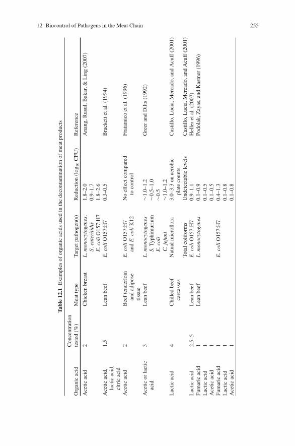
12 Biocontrol of Pathogens in the Meat Chain 255
Tabl
e12
.1E
xam
ples
ofor
gani
cac
ids
used
inth
ede
cont
amin
atio
nof
mea
tpro
duct
s
Org
anic
acid
Con
cent
ratio
nte
sted
(%)
Mea
ttyp
eTa
rget
path
ogen
(s)
Red
uctio
n(l
og10
CFU
)R
efer
ence
Ace
ticac
id2
Chi
cken
brea
stL
.mon
ocyt
ogen
es,
S.en
teri
tidi
sE
.col
iO15
7:H
7
1.8–
2.0
0.9–
1.7
1.8–
2.6
Ana
ng,R
usul
,Bak
ar,&
Lin
g(2
007)
Ace
ticac
id,
lact
icac
id,
citr
icac
id
1.5
Lea
nbe
efE
.col
iO15
7:H
70.
3–0.
5B
rack
ette
tal.
(199
4)
Ace
ticac
id2
Bee
fte
nder
loin
and
adip
ose
tissu
e
E.c
oliO
157:
H7
and
E.c
oliK
12N
oef
fect
com
pare
dto
cont
rol
Frat
amic
oet
al.(
1996
)
Ace
ticor
lact
icac
id3
Lea
nbe
efL
.mon
ocyt
ogen
esS.
Typh
imur
ium
E.c
oli
C.j
ejun
i
∼1.0
–1.2
∼0.5
–1.0
∼0.5
∼1.0
–1.2
Gre
eran
dD
ilts
(199
2)
Lac
ticac
id4
Chi
lled
beef
carc
asse
sN
atua
lmic
roflo
ra3.
0–3.
3on
aero
bic
plat
eco
unts
.C
astil
lo,L
ucia
,Mer
cado
,and
Acu
ff(2
001)
Tota
lcol
ifor
ms
Und
etec
tabl
ele
vels
Cas
tillo
,Luc
ia,M
erca
do,a
ndA
cuff
(200
1)L
actic
acid
2.5–
5L
ean
beef
E.c
oliO
157:
H7
0.9–
1.1
Hel
ler
etal
.(20
07)
Fum
aric
acid
1L
ean
beef
L.m
onoc
ytog
enes
0.1–
0.9
Podo
lak,
Zay
as,a
ndK
astn
er(1
996)
Lac
ticac
id1
0.1–
0.5
Ace
ticac
id1
0.1–
0.5
Fum
aric
acid
1E
.col
iO15
7:H
70.
4–1.
3L
actic
acid
10.
1–0.
8A
cetic
acid
10.
1–0.
8
256 C.M. Burgess et al.
bacterial microorganisms (Smulders & Greer, 1998). Sensory effects such as color,flavor or odor can also be a major concern. Dilute organic acid solutions of up to 3%,generally, have no effect on the desirable sensory properties of meat when used as acarcass decontaminant. However, some treatment conditions using lactic and aceticacid can produce adverse sensory and appearance changes when applied directly tomeat cuts or products (Smulders & Greer, 1998). Further evaluation of organic acidsand their effects in meat processing and their wider impact on pathogens continuesto be researched and improved.
Essential Oils
Essential oils (EOs) are aromatic oily liquids obtained from plant materials. Thegreatest use of EOs is as flavorings in food; however, their antimicrobial poten-tial has attracted increased attention and research. EOs can comprise more than 60individual components, where the major components make up 85% of the EO andother components are present as trace (Burt, 2004). The mode of action of EOsis relatively unknown (Burt, 2004; Lambert, Skandamis, Coote, & Nychas, 2001).However, the extensive range of EOs and components suggest that there is mostlikely to be many different modes of action (Burt, 2004). The phenolic compo-nents of EOs are chiefly responsible for the antibacterial properties of the EOsand usually EOs containing a high percentage of phenolic compounds have thestrongest activity (Lambert et al., 2001). Many studies have reported the antimi-crobial effect of EOs or their components against foodborne pathogens (Burt, 2004;Burt & Reinders, 2003; Hammer, Carson, & Riley, 1999; Lambert et al., 2001; Siet al., 2006). EOs have a broad spectrum of activity, but are slightly more activeagainst Gram-positive than Gram-negative bacteria (Lambert et al., 2001). Appli-cation of EOs and their components have been reported in many foods, includingmeat products (Table 12.2). The physical structure and composition of the foodmatrix as well as the environmental conditions of the food (e.g. temperature, vac-uum/gas/air packaging) can affect the efficiency of the EO against pathogens infoods (Tassou, Drosinos, & Nychas, 1995; Tsigarida, Skandamis, & Nychas, 2000).For example, high levels of fat and/or protein in foods such as meat products canprotect the bacteria from the antimicrobial activity of the EO, whereby the EO candissolve in the lipid phase of the food and will therefore be less available againstbacteria (Gill, Delaquis, Russo, & Holley, 2002; Tassou et al., 1995). Many stud-ies have attempted to improve microbial quality of meats by combining the use ofEOs with other preservation techniques, such as different packaging environments,radiosensitization, bacteriocins, and incorporation of EOs into packaging films(Brashears, Reilly, & Gilliland, 1998; Chiasson, Borsa, Ouattara, & Lacroix, 2004;Ghalfi, Benkerroum, Doguiet, Bensaid, & Thonart, 2007; Gill et al., 2002; Tsigaridaet al., 2000). An important aspect to consider in the application of EOs to foodsis the effect on the organo-leptic properties of the food product. Low concentra-tions of EOs in meat products have been reported to be acceptable after storage and
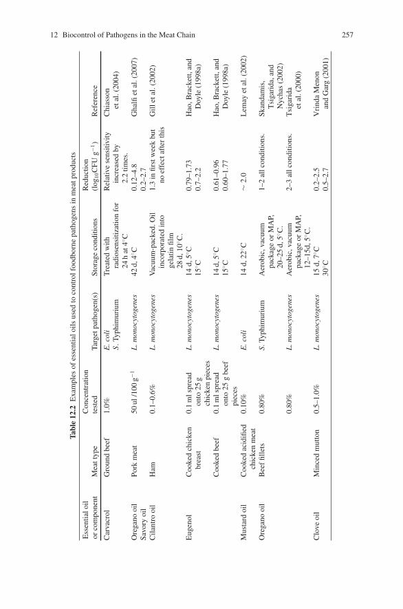
12 Biocontrol of Pathogens in the Meat Chain 257
Tabl
e12
.2E
xam
ples
ofes
sent
ialo
ilsus
edto
cont
rolf
oodb
orne
path
ogen
sin
mea
tpro
duct
s
Ess
entia
loil
orco
mpo
nent
Mea
ttyp
eC
once
ntra
tion
test
edTa
rget
path
ogen
(s)
Stor
age
cond
ition
sR
educ
tion
(log
10C
FUg−1
)R
efer
ence
Car
vacr
olG
roun
dbe
ef1.
0%E
.col
iS.
Typh
imur
ium
Tre
ated
with
radi
osen
sitiz
atio
nfo
r24
hat
4◦ C
Rel
ativ
ese
nsiti
vity
incr
ease
dby
2.2
times
.
Chi
asso
net
al.(
2004
)
Ore
gano
oil
Savo
ryoi
lPo
rkm
eat
50ul
/100
g−1L
.mon
ocyt
ogen
es42
d,4◦ C
0.12
–4.8
0.2–
2.7
Gha
lfiet
al.(
2007
)
Cila
ntro
oil
Ham
0.1–
0.6%
L.m
onoc
ytog
enes
Vac
uum
-pac
ked.
Oil
inco
rpor
ated
into
gela
tinfil
m28
d,10
◦ C.
1.3
infir
stw
eek
but
noef
fect
afte
rth
isG
illet
al.(
2002
)
Eug
enol
Coo
ked
chic
ken
brea
st0.
1m
lspr
ead
onto
25g
chic
ken
piec
es
L.m
onoc
ytog
enes
14d,
5◦ C15
◦ C0.
79–1
.73
0.7–
2.2
Hao
,Bra
cket
t,an
dD
oyle
(199
8a)
Coo
ked
beef
0.1
mls
prea
don
to25
gbe
efpi
eces
L.m
onoc
ytog
enes
14d,
5◦ C15
◦ C0.
61–0
.96
0.60
–1.7
7H
ao,B
rack
ett,
and
Doy
le(1
998a
)
Mus
tard
oil
Coo
ked
acid
ified
chic
ken
mea
t0.
10%
E.c
oli
14d,
22◦ C
∼2.
0L
emay
etal
.(20
02)
Ore
gano
oil
Bee
ffil
lets
0.80
%S.
Typh
imur
ium
Aer
obic
,vac
uum
pack
age
orM
AP,
20–2
5d,
5◦ C.
1–2
allc
ondi
tions
.Sk
anda
mis
,T
siga
rida
,and
Nyc
has
(200
2)0.
80%
L.m
onoc
ytog
enes
Aer
obic
,vac
uum
pack
age
orM
AP,
12–1
5d,5
◦ C.
2–3
allc
ondi
tions
.T
siga
rida
etal
.(20
00)
Clo
veoi
lM
ince
dm
utto
n0.
5–1.
0%L
.mon
ocyt
ogen
es15
d,7◦ C
30◦ C
0.2–
2.5
0.5–
2.7
Vri
nda
Men
onan
dG
arg
(200
1)
258 C.M. Burgess et al.
cooking (Tsigarida et al., 2000). Oregano oil (1% v/w) was also found to improvethe flavor and quality of minced meat following storage in modified atmospheres(Skandamis & Nychas, 2001). Further studies with respect to the interactions ofEOs and their components with food constituents is required to improve the efficacyof EOs against food spoilage and pathogenic organisms whilst minimizing impacton the organo-leptic properties of the product.
Antagonistic Bacteria
Bacterial Metabolites
The production of one or more antimicrobial active metabolites is part of thecomplex mechanisms by which a culture becomes established in the presenceof other competing organisms (Holzapfel et al., 1995). Along with bacteriocins,bacteria can produce many types of substances or metabolites that are inhibitoryto other bacteria. These can include clinical or therapeutic low-molecular weightantibiotics, lytic agents, toxins, bacteriolytic enzymes, bacteriophages and othermetabolic products such as hydrogen peroxide and diacetyl (Holzapfel et al., 1995;Kostrzynska & Bachand, 2006). These substances can act as bio-preservativesby inhibiting spoilage or pathogenic microorganisms (Deegan, Cotter, Hill, &Ross, 2006). The main mechanism by which lactic acid bacteria (LAB) inhibitmicroorganisms is through the production of organic acids such as lactic acid andacetic acid. Organic acids produced by LAB, including lactic, acetic and propi-onic acid, exert antimicrobial effects due to their action on the bacterial cyto-plasmic membrane, which interferes with the maintenance of membrane potentialand inhibits active transport (Kostrzynska & Bachand, 2006). The use of LAB iscommon in sausage fermentations, in which accumulating lactic acid levels inhibitmeat-borne pathogenic bacteria (Lucke, 2000). In addition, the inhibitory prop-erty of hydrogen peroxide has also been reported. Lactobacillus lactis can pro-duce hydrogen peroxide which in effect can reduce the numbers of E. coli O157(0.37–1.09 log10 CFU ml−1 lower counts compared to controls) on refrigeratedraw chicken (Brashears et al., 1998). Senne & Gilliland (2003) reported that theapplication of Lb. delbrueckii subsp. lactis (also found to produce hydrogen perox-ide) could reduce the numbers of E. coli O157:H7 (0.8–1.3 log10 CFU cm−2) andS. Typhimurium (0.8–1.5 log10 CFU cm−2) on pork and beef carcasses kept in refrig-erated storage. Reuterin, a broad-spectrum low-molecular weight antimicrobial sub-stance produced by Lb. reuteri during glycerol conversion has also been reported asa potential biopreservative for food. One study found that Lb. reuteri in the presenceof glycerol was highly effective against inocula of E. coli O157:H7 of 3 log10 CFUg−1 and 6 log10 CFU g−1 levels in ground beef during refrigerated storage in modi-fied atmosphere packages (Muthukumarasamy, Han, & Holley, 2003). Reuterin wasalso reported to inhibit the growth of L. monocytogenes but not Salmonella spp. onthe surface of sausages (Kuleasan & Cakmakci, 2002).
12 Biocontrol of Pathogens in the Meat Chain 259
Limitations of Using Metabolites
Although some metabolites of LAB have been shown to inhibit Gram-negative bac-teria their use in maintaining the safety and stability of meat products is not ideal assome metabolites can interfere with the sensory properties (e.g. hydrogen peroxide)of the food or may not be produced in sufficient amounts (e.g. reuterin). Researchmay provide methods to engineer metabolic pathways to give better control of therate and extent of formation of lactic and acetic acids, and to eliminate unwantedproperties such as formation of biogenic amines. Depending on the product andprocessing situation, one or more of these metabolites may constitute a basis forthe selection of a protective culture (Holzapfel et al., (1995). It must be emphasizedthat LAB used to reduce pathogens in foods should not affect the sensory charac-teristic of the foods, ensuring that foods are palatable to consumers (Kostrzynska &Bachand, 2006).
Competitive Exclusion Technology
Competitive exclusion (CE) as a technology, involves the addition of a non-pathogenicbacterial culture to the intestinal tract of food animals in order to reduce colonizationor decrease populations of pathogenic bacteria in the gastrointestinal tract (Callawayet al., 2004). A CE culture may be composed of one or many strains or species ofbacteria, but should ideally be composed of species normally resident in the animalintestinal microflora. The use of CE is similar to using probiotics which are defined as‘a preparation of or a product containing viable, defined microorganisms in sufficientnumbers which alter the microflora of the host and exert beneficial health effects inthis host’ (de Vrese, & Schrezenmeir, 2002). In contrast to CE, probiotic preparationsgenerally consist of individual species or mixtures of LAB or yeasts that are not nec-essarily of animal origin (Callaway et al., 2004). The use of probiotics in food animalshave been extensively reviewed (Callaway et al., 2004; Nava, Bielke, Callaway, &Castaneda, 2005; Wagner, 2006). This section will only focus on the use of CE culturesas a food safety strategy in food animals.
The precise mechanism by which CE microorganisms reduce pathogens in theanimal intestine is unclear, however, the main role of CE cultures is to attach to thesurface of the intestinal epithelium and establish itself within the gut. This directbinding of the CE culture to the intestinal wall prevents potentially pathogenicstrains from attaching to it. Some of the bacteria may produce antimicrobial com-pounds such as acids or bacteriocins to eliminate species competing within thesame niche (Callaway et al., 2004). Some of these antimicrobial compounds havebeen specifically investigated for use as biopreservatives and for food safety appli-cations in meat and their products and will be discussed later in this chapter. CEcultures may be composed of defined microbial strains (known and characterized)or undefined (incompletely characterized or unknown) microbial strains. The useof CE cultures has been used extensively in poultry to reduce Salmonella andCampylobacter carriage (Chen & Stern, 2001; La Ragione & Woodward, 2003;
260 C.M. Burgess et al.
Wagner, 2006; Zhang, Ma, & Doyle, 2007a, 2007b). The use of CE cultures, includ-ing commercial products in poultry has been reported to reduce the colonizationof poultry with Salmonella spp. by up to 70% or by 7–9 log10 cycles (Davies &Breslin, 2003; Hoszowski & Truszczynski, 1997; Schneitz & Hakkinen, 1998).Reductions of between 3–100% of Campylobacter spp. colonization on poultry hasalso been reported (Schoeni & Wong, 1994). The use of CE cultures in cattle andpigs to eliminate E. coli O157:H7 and/or Salmonella from rumen and gastrointesti-nal tract have also shown potential for further commercial development (Brashears,Jaroni, & Trimble, 2003; Genovese et al., 2003; Zhao et al., 2003).
Limitations of Using CE Cultures
The most important property of a CE culture is the establishment of a complexintestinal microbiota that resists colonization by human and animal pathogens.While CE has been shown to work in several animal species, the benefits have notbeen consistent. The differing results between studies involving CE in animals maybe due to the difference between host animals, cultures, or experimental designs.Although the benefit of using CE to reduce human pathogen carriage in animals hasenormous potential, many of the effective and commercially available CE productshave their disadvantages. Firstly, the use of CE culture in which the microbial iso-lates are unknown requires regulation as an animal drug due to the cultures’ effectson animal health and the risk of transfer of undesirable bacteria to humans. Thereis also the potential of these component bacteria of CE to transfer virulence genesonto the animal or human microbiota resulting in enhanced antimicrobial resistance(Wagner, 2006). Ongoing research in this area aims to characterize the bacteriapresent in CE products which will lead to safe assurance of use of the product andwill allow optimization of their efficacy.
Bacteriocin-Producing Bacteria and Bacteriocins
Bacteriocins are ribosomally synthesized antimicrobial peptides or proteins pro-duced by bacteria that kill or inhibit the growth of other bacteria, either in thesame species (narrow spectrum), or across genera (broad spectrum) (Cotter, Hill, &Ross, 2005). The use of bacteriocins in biopreservation is a growing area of research.Bacteriocins are a heterogeneous group, characteristically selected for evaluationand used as specific antagonists against problematic bacteria. However, their effec-tiveness in foods can become limited for various reasons, and cost remains an issueimpeding the broader use of bacteriocins as food additives. Evaluation of methods toimprove the use of bacteriocins in various food products is continually being inves-tigated and new developments are frequently reported. Although, Gram-negativeand Gram-positive bacteria can produce bacteriocins, LAB continues to be thepreferred source of food-use bacteriocins as they are generally regarded as safe(GRAS) bacteria (Chen & Hoover, 2003). The structure, biosynthesis, and appli-cation of LAB bacteriocins in many different food products have been reviewed
12 Biocontrol of Pathogens in the Meat Chain 261
by others (Cleveland, Montville, Nes, & Chikindas, 2001; Cotter et al., 2005;Deegan et al., 2006; Galvez, Abriouel, Lopez, & Omar, 2007; Kostrzynska &Bachand, 2006; O’Sullivan, Ross, & Hill, 2002). The use of bacteriocin-producingbacteria (BPB) or the derived bacteriocins in food safety strategies in meat applica-tions will be further discussed below.
Bacteriocins generally act by creating pores in the membrane of their target cellsand may be cytotoxic to the target cell as a result of disturbance of the bacterial inneror outer membranes. Alternatively, bacteriocins may pass through the membrane toreach a target inside the cell causing major disruptions in cell functions. Differentmechanisms of action by bacteriocins have been described but interaction with thebacterial membrane is an important requirement for most, if not all, bacteriocins(Cleveland et al., 2001).
Application of BPB and Bacteriocins in Animals
Rather than applying the bacteriocin directly as a biocontrol, the bacteriocin-producing bacteria (BPB) may be applied. BPB have been isolated from rumenenvironments and some have been applied to manipulate the rumen environmentin poultry and cattle (Cole, Farnell, Donoghue, Stern, & Evetoch, 2006; Diez-Gonzalez, 2007; Etcheverria, Arroyo, Perdigon, & Parma, 2005; Nava et al., 2005;Russell & Mantovan, 2002; Svetoch et al., 2005; Zhao et al., 1998). BPB can beadministered to animals by mixing dried or wet cultures with feed or drinking water,and depending on the ability of the bacterial strain to colonize the gastrointesti-nal tract may be fed sporadically or continuously. The feeding of BPB can have adirect effect on reducing the existing populations of foodborne pathogens such asSalmonella and E. coli O157:H7 and long-term colonization with BPB would pre-vent further re-introduction of the pathogenic bacteria. Few studies have addressedthe fate of bacteriocins in the intestinal tract, but some data suggests that some ofthe low molecular weight bacteriocins can survive at least some of the intestinalenvironments and possibly could be administered through feed (Ganzle, Hertel, vander Vossen, & Hammes, 1999).
The use of colicins (bacteriocin produced by E. coli), as a pre-harvest control,has been actively evaluated to reduce pathogenic E. coli in cattle populations (Diez-Gonzalez, 2007; Schamberger, Phillips, Jacobs, Diez-Gonzalez, 2004). Calves fedwith a colicin-producing E. coli yielded an overall reduction of 1.1 log10 CFUg−1 ofE. coli O157:H7 and a maximum decrease of 1.8 log10 CFUg−1 of E. coli O157:H7over 24 days (Schamberger et al., 2004). Colicins specific for E. coli are partic-ularly advantageous in the rumen intestine in that it will only inhibit one type ofbacterial strain or species while not disrupting the other microbial populations inthe intestine (Diez-Gonzalez, 2007). Studies continue to investigate whether expres-sion of colicins could be incorporated into bacteria which are normally present inthe animal rumen, which can decrease the chances of transfer of colicin genes toother potentially pathogenic E. coli strains, such as E. coli O157:H7 (McCormick,Klaenhammer, & Stiles, 1999). In poultry, a few studies have investigated the use
262 C.M. Burgess et al.
of BPB to control foodborne pathogens. Bacteriocin-like compounds were shownto have direct antimicrobial activity, in vitro against Campylobacter (Morency,Mota-Meira, LaPointe, Lacroix, & Lavoie, 2001; Schoenis & Doyle, 1992; Svetochet al., 2005). A purified bacteriocin produced by Paenibacillus polymyxa microen-capsulated and administered via feed was reported to reduce cecal Campylobac-ter colonization in young broiler chickens and turkeys (Cole et al., 2006; Sternet al., 2005). Treatment with bacteriocin eliminated detectable Campylobacter con-centrations in turkey cecal contents (detection limit, 1 × 10² CFU g−1) compared tocontrols (1.0 × 10⁶ CFU g−1 of cacal contents) (Cole et al., 2006). In chicken cacelsamples, use of bacteriocin resulted in significant reductions in colonization byC. jejuni for C. jejuni. These studies reported significant reductions of both intesti-nal levels and frequency of chicken and turkey colonization by C. jejuni, suggestingthat this on-farm application would be an alternative to chemical disinfection ofcontaminated carcasses (Stern et al., 2005). In order for BPB and their bacteriocinsto be used successfully in animal production, more research is required to improvetheir efficacy in different animal systems.
Application of Bacteriocin-Producing Bacteria and Bacteriocinsin Meat Products
Bacteriocins are potentially valuable biological tools to improve food preservationand food safety by reducing the prevalence of undesirable spoilage microorganismsor foodborne pathogens in a food product (Deegan et al., 2006). Although the use ofLAB is common in many food processes, the use of these microorganisms in differ-ent meat products and storage conditions continues to be evaluated (Kostrzynska &Bachand, 2006). The application of bacteriocins is not recommended as a primaryprocessing step or barrier to prevent the growth or survival of pathogens, but shouldform part of a system with multiple hurdles. Bacteriocins can be incorporated intofood as an ingredient in the form of a purified/semi-purified bacteriocin prepara-tion (Table 12.3). Alternatively, a BPB can be introduced as a ‘protective culture’in the form of a live culture which produces the bacteriocins in situ in the food(Table 12.4). In this case, the BPB is either substituted for all or part of the starter oris subsequently applied to the food to improve the safety of the culture (O’Sullivanet al., 2002). A major criterion for a protective culture in meat products is to inhibitpathogens and/or prolong shelf life, while not changing the sensory properties ofthe product (Lucke, 2000). The use of purified bacteriocins is not always attractiveas some can be inactivated in meats and may also required regulatory approval orbe labeled as an additive on the meat product (Aasen et al., 2003). The use of BPBas a protective culture is a more practical approach as it does not require regulatoryapproval or preservative label declarations and can be substituted into the productas a starter culture (Deegan et al., 2006; Jacobsen, Budde, & Koch, 2003; Katlaet al., 2002).
Numerous studies have attempted to isolate BPB, particularly LAB from meatproducts (Albano et al., 2007; Arlindo et al., 2006; Budde, Hornbaek, Jacobsen,
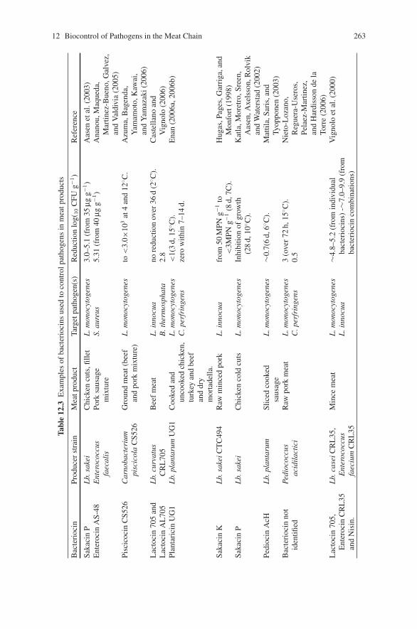
12 Biocontrol of Pathogens in the Meat Chain 263
Tabl
e12
.3E
xam
ples
ofba
cter
ioci
nsus
edto
cont
rolp
atho
gens
inm
eatp
rodu
cts
Bac
teri
ocin
Prod
ucer
stra
inM
eatp
rodu
ctTa
rget
path
ogen
(s)
Red
uctio
nlo
g(10
CFU
g−1)
Ref
eren
ce
Saka
cin
PL
b.sa
kei
Chi
cken
cuts
,fille
tL
.mon
ocyt
ogen
es3.
0–5.
1(f
rom
35μg
g−1)
Aas
enet
al.(
2003
)E
nter
ocin
AS-
48E
nter
ococ
cus
faec
alis
Pork
saus
age
mix
ture
S.au
reus
5.31
(fro
m40
μgg−1
)A
nano
u,M
aque
da,
Mar
tinez
-Bue
no,G
alve
z,an
dV
aldi
via
(200
5)Pi
scic
ocin
CS5
26C
arno
bact
eriu
mpi
scic
ola
CS5
26G
roun
dm
eat(
beef
and
pork
mix
ture
)L
.mon
ocyt
ogen
esto
<3.
0×10
3at
4an
d12
◦ C.
Azu
ma,
Bag
enda
,Y
amam
oto,
Kaw
ai,
and
Yam
azak
i(20
06)
Lac
toci
n70
5an
dL
acto
cin
AL
705
Lb.
curv
atus
CR
L70
5B
eef
mea
tL
.inn
ocua
B.t
herm
osph
ata
nore
duct
ion
over
36d
(2◦ C
).2.
8C
aste
llano
and
Vig
nolo
(200
6)Pl
anta
rici
nU
G1
Lb.
plan
taru
mU
G1
Coo
ked
and
unco
oked
chic
ken,
turk
eyan
dbe
efan
ddr
ym
orta
della
.
L.m
onoc
ytog
enes
C.p
erfr
inge
ns<
1(3
d,15
◦ C).
zero
with
in7–
14d.
Ena
n(2
006a
,200
6b)
Saka
cin
KL
b.sa
keiC
TC
494
Raw
min
ced
pork
L.i
nnoc
uafr
om50
MPN
g−1to
<3M
PNg−1
(8d,
7C).
Hug
as,P
ages
,Gar
riga
,and
Mon
fort
(199
8)Sa
kaci
nP
Lb.
sake
iC
hick
enco
ldcu
tsL
.mon
ocyt
ogen
esIn
hibi
tion
ofgr
owth
(28
d,10
◦ C).
Kat
la,M
oret
ro,S
reen
,A
asen
,Axe
lsso
n,R
olvi
kan
dW
ater
stad
(200
2)Pe
dioc
inA
cHL
b.pl
anta
rum
Slic
edco
oked
saus
age
L.m
onoc
ytog
enes
∼0.7
(6d,
6◦ C).
Mat
tila,
Sari
s,an
dTy
oppo
nen
(200
3)B
acte
rioc
inno
tid
entifi
edPe
dioc
occu
sac
idil
acti
ciR
awpo
rkm
eat
L.m
onoc
ytog
enes
C.p
erfr
inge
ns3
(ove
r72
h,15
◦ C).
0.5
Nie
to-L
ozan
o,R
egue
ra-U
sero
s,Pe
laez
-Mar
tinez
,an
dH
ardi
sson
dela
Torr
e(2
006)
Lac
toci
n70
5,E
nter
ocin
CR
L35
and
Nis
in.
Lb.
case
iCR
L35
,E
nter
ococ
cus
faec
ium
CR
L35
Min
cem
eat
L.m
onoc
ytog
enes
L.i
nnoc
ua∼4
.8–5
.2(f
rom
indi
vidu
alba
cter
ioci
ns)
-∼7.
0–9.
9(f
rom
bact
erio
cin
com
bina
tions
)
Vig
nolo
etal
.(20
00)
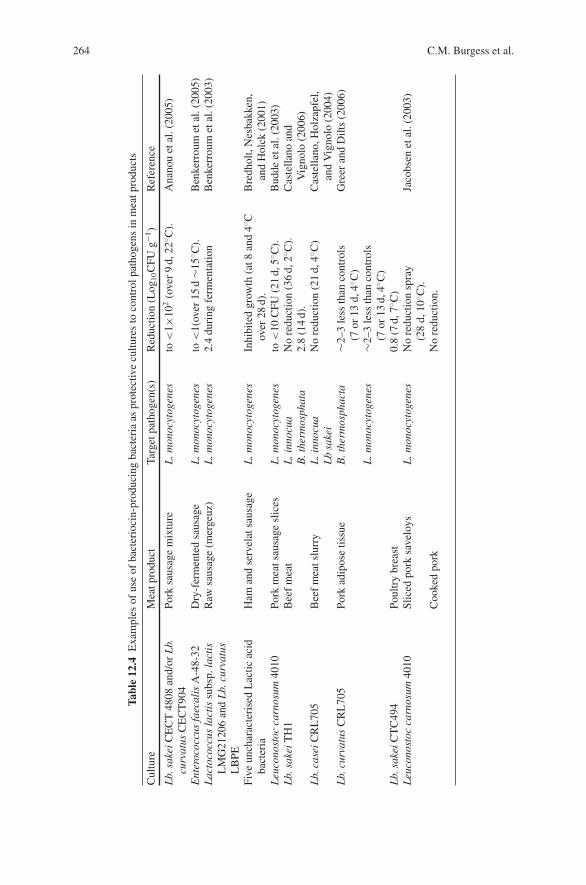
264 C.M. Burgess et al.
Tabl
e12
.4E
xam
ples
ofus
eof
bact
erio
cin-
prod
ucin
gba
cter
iaas
prot
ectiv
ecu
lture
sto
cont
rolp
atho
gens
inm
eatp
rodu
cts
Cul
ture
Mea
tpro
duct
Targ
etpa
thog
en(s
)R
educ
tion
(Log
10C
FUg−1
)R
efer
ence
Lb.
sake
iCE
CT
4808
and/
orL
b.cu
rvat
usC
EC
T90
4Po
rksa
usag
em
ixtu
reL
.mon
ocyt
ogen
esto
<1×
102
(ove
r9
d,22
◦ C).
Ana
nou
etal
.(20
05)
Ent
eroc
occu
sfa
ecal
isA
-48-
32D
ry-f
erm
ente
dsa
usag
eL
.mon
ocyt
ogen
esto
<1(
over
15d
∼15◦ C
).B
enke
rrou
met
al.(
2005
)L
acto
cocc
usla
ctis
subs
p.la
ctis
LM
G21
206
and
Lb.
curv
atus
LB
PE
Raw
saus
age
(mer
geuz
)L
.mon
ocyt
ogen
es2.
4du
ring
ferm
enta
tion
Ben
kerr
oum
etal
.(20
03)
Five
unch
arac
teri
sed
Lac
ticac
idba
cter
iaH
aman
dse
rvel
atsa
usag
eL
.mon
ocyt
ogen
esIn
hibi
ted
grow
th(a
t8an
d4◦ C
over
28d)
.B
redh
olt,
Nes
bakk
en,
and
Hol
ck(2
001)
Leu
cono
stoc
carn
osum
4010
Pork
mea
tsau
sage
slic
esL
.mon
ocyt
ogen
esto
<10
CFU
(21
d,5◦ C
).B
udde
etal
.(20
03)
Lb.
sake
iTH
1B
eef
mea
tL
.inn
ocua
B.t
herm
osph
ata
No
redu
ctio
n(3
6d,
2◦ C).
2.8
(14
d).
Cas
tella
noan
dV
igno
lo(2
006)
Lb.
case
iCR
L70
5B
eef
mea
tslu
rry
L.i
nnoc
uaL
bsa
kei
No
redu
ctio
n(2
1d,
4◦ C)
Cas
tella
no,H
olza
pfel
,an
dV
igno
lo(2
004)
Lb.
curv
atus
CR
L70
5Po
rkad
ipos
etis
sue
B.t
herm
osph
acta
∼2–3
less
than
cont
rols
(7or
13d,
4◦ C)
Gre
eran
dD
ilts
(200
6)
L.m
onoc
ytog
enes
∼2–3
less
than
cont
rols
(7or
13d,
4◦ C)
Lb.
sake
iCT
C49
4Po
ultr
ybr
east
0.8
(7d,
7◦ C)
Leu
cono
stoc
carn
osum
4010
Slic
edpo
rksa
velo
ysL
.mon
ocyt
ogen
esN
ore
duct
ion
spra
y(2
8d,
10◦ C
).Ja
cobs
enet
al.(
2003
)
Coo
ked
pork
No
redu
ctio
n.
12 Biocontrol of Pathogens in the Meat Chain 265
Barkholt, & Koch, 2003; Noonpakdee, Santivarangkna, Jumriangrit, Sonomoto, &Panyim, 2003; Prema, Bharathy, Palavesam, Sivasubramanian, & Immanuel, 2006;Schneider et al., 2006; Yin, Wu, & Jiang, 2003), with the aim of identifying a poten-tial protective culture that can not only be used as a starter culture but also possessbacteriocin activity to eliminate or reduce foodborne pathogens (Benkerroumet al., 2005; Benkerroum, Daoudi, & Kamal, 2003; Leroy & De Vuyst, 2005). ManyLABs are used to combat other spoilage organisms in aerobic or vacuum packedmeat products (Barakat, Griffiths, & Harris, 2000; Lucke, 2000). With respect tomeat safety, majority of the studies investigating the use of bacteriocins in meatproducts have targeted the pathogen L. monocytogenes due to its ability to grow atrefrigeration temperature and survive in fermented foods (Tables 12.3 and 12.4).The disadvantage of many LAB bacteriocins is that although they are active againstGram-positive organisms they are not as effective against Gram-negative foodbornepathogens such as E. coli (particularly E. coli O157:H7) or Salmonella spp. whichare also a concern in meat products (Lucke, 2000). This is due to the fact thatGram-negative bacteria are protected by their outer membrane, which prevents bac-teriocins from reaching the cytoplasmic membrane (Abee, Krockel, & Hill, 1995).Studies have therefore attempted to find alternative bacteriocins with broaderspectrums or have used other combinations of strategies to improve bacteriocinactivity.
The most commonly used bacteriocin in foods, including meat products isNisin (produced by Lactococcus lactis subsp. lactis). Nisin is one of the com-mercially available bacteriocins with US Food and Drug Administration (FDA)approval. It inhibits the growth of a wide range of Gram-positive bacteria includingL. monocytogenes. Nisin has been shown to be effective in a number of food sys-tems but is predominantly used in canned foods and dairy products, particularly incheese production, where it protects against heat-resistant, spore forming organismssuch as Bacillus and Clostridium spp (Cotter et al., 2005; Deegan et al., 2006).Nisin has been used as a food safety agent in beef (Ariyapitipun, Mustapha, &Clarke, 2000; Barboza De Martinez, Ferrer, & Marquez Salas, 2002; Zhang &Mustapha, 1999), sausages (Patel, Sanglay, Sharma, & Solomon, 2007), groundmince (Castillo, Meszaros, & Kiss, 2004) and poultry (Yuste, Pla, Capellas andMor-Mur, 2002; Zuckerman & Abraham, 2002), but is not as effective in the preser-vation of meat as it is in dairy products. The inhibitory activity of Nisin is reduced byinterference from meat components such as phospholipids, especially where theremay be a high fat content (Leroy & De Vuyst, 2005). In addition, Nisin has a lowsolubility at normal meat pH and its interaction with phospholipids results in theuneven distribution of the bacteriocin in the meat (De Martinis, Publio, Santarosa,& Freitas, 2001; Stergiou, Thomas, & Adams, 2006).
Substantial research has been done to evaluate and improve the effectivenessof Nisin and other bacteriocins activity against Gram-negative pathogens and itsapplicability in different food products. Several bacteriocins show additive or syn-ergistic effects when used in combination with other antimicrobial agents includ-ing, organic acids, other antimicrobials or sublethal treatments such as mild heator high pressure (Ananou, Galvez, Martinez-Bueno, Maqueda, & Valdivia, 2005;
266 C.M. Burgess et al.
Ariyapitipun et al., 2000; Arques et al., 2004; Ganzle, Weber, & Hammes, 1999;Rodriguez, Nunez, Gaya, & Medina, 2005).
An alternative use of bacteriocins is as the agent incorporated into packagingmaterials (Cooksey, 2005; Quintavalla & Vicini, 2002). Combining the bacteriocindirectly into a plastic material provides a number of advantages for delivery of thebacteriocin to the food product. Firstly, only the necessary amount of bacteriocinwould be required. Secondly, the agent would not be a direct additive to the foodproduct and would therefore avoid labeling and regulatory approval. Thirdly, ifthe plastic material were made from an edible and/or biodegradable plastic ben-efits for the environment would be apparent (Siragusa, Cutter, & Willett, 1999).Bacteriocins such as Nisin and Pediocin among others have been incorporated intodifferent films and have been successful in inhibiting spoilage microorganisms, suchas LAB and Brochothrix thermosphacta on beef carcass tissue, as well as Listeriaspp. on meat and poultry samples (Marcos, Aymerich, Monfort, & Garriga, 2007;Mauriello, Ercolini, La Storia, Casaburi, & Villani, 2004; Ming, Weber, Ayres, &Sandine, 1997; Scannell et al., 2000; Siragusa et al., 1999). Further development ofpackaging technologies may prove to be an effective way of delivering bacteriocinsto the surface of food to improve food safety and preservation.
Limitations of Using Bacteriocins and Future Prospects
The extensive use of bacteriocins in foods is hindered by a number of factors.One of the main limitations is the narrow spectrum of activity of most bacte-riocins, though the specificity of a bacteriocin can be advantageous for appli-cations in which a single bacterial strain of species is targeted without disrupt-ing other microbial populations. Research continues to identify alternative bac-teriocins which may extend the bacteriocin applications either through their usealone or in combination with other antimicrobials or hurdle technologies. Con-siderable studies are also required to investigate the factors influencing the appli-cability of certain bacteriocins in various food systems. Bacteriocin activity isdifficult to maintain in a range of foods particularly in meat products. Anotherpotential problem associated with using bacteriocins in foods is the developmentof resistant populations of problematic bacteria. Resistance can occur naturallyand it has been reported especially with regard to Class IIa bacteriocins (Nagh-mouchi, Kheadr, Lacroix, & Fliss, 2007). Consequently, studies have examinedthe possibility of generating multiple bacteriocin producers to limit the potentialof bacteriocin-resistant populations (O’Sullivan, Ryan, Ross, & Hill, 2003). SomeBPB strains may also spontaneously loss their ability to produce bacteriocin(s)due to genetic instability (Holzapfel et al., 1995; Riley & Wertz, 2002) and canalso become ineffective if the cell membrane of a target organism changes inresponse to a particular environmental condition. Although there are some limita-tions for the use of bacteriocins in meat applications, the ongoing study of existingbacteriocins as well as the identification of new bacteriocins and improvementsof application will only optimize the potential of these agents in many different
12 Biocontrol of Pathogens in the Meat Chain 267
food applications which will lead to further improvements of food safety andquality of meat products.
Antimicrobial Peptides
Antimicrobial peptides (AMPs) are generally short cationic peptides produced byboth animals and plants that have potent killing activity against a range of bac-teria, fungi, viruses, and protozoa (Higgs et al., 2007). They are ubiquitous innature and they play an important role in host defence and microbial control. Theirexact mode of action is not completely understood, but it is thought that cationicAMPs are attracted to negatively charged phospholipids in the cell membrane andinteract with the membrane, displacing lipids and altering the membrane structure.A number of hypotheses have been put forward as to how AMPs kill microbesand these are discussed elsewhere (Zasloff, 2002). Over 850 AMPs have beenidentified from a host of species and a catalogue of these can be found online(http://www.bbcm.univ.trieste.it/∼tossi/amsdb.html). AMPs can also be designedand chemically synthesized and have been shown to possess antimicrobial activity(Anzai et al., 1991; Appendini & Hotchkiss, 1999; Haynie, Crum, & Doele, 1995).
Many studies have demonstrated the activity of AMPs and derivatives from avariety of species, including host species, against pathogens including Salmonella,E. coli O157, and Listeria. A selection of these studies is shown in Table 12.5.AMPs are now emerging as a solution to the development of antibiotic resistanceby bacterial pathogens, and therefore, the focus at present is on understanding theirmode of action and potential application from a clinical point of view (Hancock &Sahl, 2006). Investigations of AMPs in food systems to date are limited. Some stud-ies have been performed in food products or suggest the potential of the AMP ina food product (Table 12.5). Yaron, Rydlo, Shachar, and Mor (2003) studied theantimicrobial activity of dermaseptin S4 and its derivative K4-S4 which come fromtree frogs. It was found that K4-S4 reduced the population of E. coli O157:H7 inapple juice by more than 7 log units in less than 2 hours, indicating its potentialusefulness as a biocontrol agent. The authors put forward potential advantages forthe use of animal derived AMPs in food safety and preservation measures, such astheir activity over a wide range of conditions. Another study in apple juice showedthat a synthetic AMP could reduce the E. coli O157:H7 numbers by 3.5 log units in8 hours. However, the same peptide had no effect on E. coli O157:H7 when grownin skim milk indicating that components from the milk could be interacting with thepeptide causing it to lose its antibacterial activity (Appendini & Hotchkiss, 1999).A different study showed that peptides produced by Lb. acidophilus fermenta-tion of sodium caseinate had bacteriocidal activity against Enterobacter sakaza-kii, L. innocua and E. coli, and it was suggested that such peptides could be usedas a protection mechanism against E. sakazakii in infant formula by producinga casein-cased milk ingredient by fermentation (Hayes, Ross, Fitzgerald, Hill, &Stanton, 2006).

268 C.M. Burgess et al.
Tabl
e12
.5E
xam
ples
ofan
timic
robi
alpe
ptid
es(A
MPs
)w
hich
have
antim
icro
bial
activ
ityag
ains
tpat
hoge
ns
AM
PSo
urce
Ant
imic
robi
alac
tivity
Ref
eren
ce
Synt
hetic
cath
elic
idin
sov
ine
cath
elic
idin
sE
.col
iO15
7:H
7A
nder
son
(200
5)Sy
nthe
ticPR
-26
porc
ine
neut
roph
ilsA
MP
E.c
oliO
157:
H7
and
L.m
onoc
ytog
enes
Ann
amal
ai,V
enki
tana
raya
nan,
Hoa
glan
d,an
dK
han
(200
1)6K
8LSy
nthe
ticB
.sub
tili
s,E
.col
iO15
7:H
7,L
.mon
ocyt
ogen
es,
Pse
udom
onas
fluor
esce
ns,S
.Typ
him
uriu
m,
Serr
atia
liqu
efas
cien
s,St
aphy
loco
ccus
aure
us,
Klu
yver
omyc
esm
arxi
anus
App
endi
ni&
Hot
chki
ss(1
999)
L.a
cido
phil
usfe
rmen
tatio
nof
sodi
umca
sein
ate
Bov
ine
αs1
-cas
ein
Ent
erob
acte
rsa
kaza
kaii
,L.i
nnoc
ua,E
.col
iH
ayes
etal
.(20
06)
Synt
hetic
mod
ified
AvB
D8
chic
ken
avia
nβ-
defe
nsin
-8E
.col
i,S.
Typh
imur
ium
and
L.m
onoc
ytog
enes
Hig
gset
al.(
2007
)Pe
psin
dige
stof
case
inB
ovin
eka
ppa-
case
inE
.col
i,L
.inn
ocua
Lop
ez-E
xpos
ito,M
iner
vini
,Am
igo,
and
Rec
io(2
006)
Rec
ombi
nant
galli
naci
nsC
hick
enS.
Typh
imur
ium
,S.e
nter
idit
isM
ilona
,Tow
nes,
Bev
an,a
ndH
all(
2007
)Pu
rifie
dau
relin
Aur
elia
auri
taje
llyfis
hE
.col
i,L
.mon
ocyt
ogen
es(2
006)
Synt
hetic
cath
elic
idin
Bac
7an
dde
riva
tives
Gra
nulo
cyte
sof
mam
mal
ian
spec
ies
S.Ty
phim
uriu
m,E
.col
iPo
dda
etal
.(20
06)
Synt
hetic
cath
elic
idin
K9C
AT
HC
anin
eL
.mon
ocyt
ogen
es,S
.aur
eus,
E.c
oli,
Kle
bsie
lla
pneu
mon
iae,
S.Ty
phim
uriu
m,P
s.ae
rogi
nosa
,P
rote
usm
irab
ilis
,S.e
nter
idit
is,N
eiss
eria
gono
rrho
eae
Sang
etal
.(20
07)
Lac
tofe
rrin
,pep
sin
hydr
olys
edla
ctof
erri
n,L
acto
ferr
icin
�
Bov
ine
E.c
oliO
157:
H7
Shin
etal
.(19
98)
Synt
hetic
derm
asep
tinS4
and
deri
vativ
eK
4-S
4T
ree
frog
sof
the
Phy
llom
edus
asp
ecie
sE
.col
i,E
.col
iO15
7:H
7,S.
Typh
imur
ium
,S.
Stan
ley,
L.m
onoc
ytog
enes
,L.i
nnoc
uaY
aron
etal
.(20
03)
12 Biocontrol of Pathogens in the Meat Chain 269
Very little research has focused on the use of AMPs in the meat industry. Onestudy looked at the effect of a synthetic peptide on the microflora in meat exudates,but aerobic and anaerobic counts were reduced by less than one log indicating asmall degree of inhibition (Appendini & Hotchkiss, 1999). Although studies in thisarea are limited at the moment, the potential advantages provided by AMPs, such astheir wide spectrum of activity and lack of bacterial resistance to AMPs, suggeststhat the interest in their use as biocontrol agents in foods can only grow in the yearsto come.
Bacteriophage
Bacteriophages are viruses that infect and kill bacterial cells by reproducing withinthe bacteria and disrupting the host metabolic pathways, causing the bacteriumto lyse. Bacteriophage are ubiquitous in the environment; they specifically tar-get bacterial cells and do not infect mammalian cells, hence the reason whythey are proposed as biocontrol agents in human, animal, clinical and industrialapplications.
Classification and Mode of Action
Most bacteriophages range in size from 24–200 nm in length and are classified into13 families. The bacteriophage structure consists of a head or capsid; composedof protein that acts as a protective barrier in which either DNA or RNA is stored.Some bacteriophages possess a tail through which the nucleic acid is delivered tothe bacterial cell during infection.
Bacteriophages are classified as either lytic or lysogenic. Lytic bacteriophagesbring about rapid lysis and death of the bacterial cell, whereas a lysogenic bacte-riophage does not result in immediate lysis, but instead enters a quiescent state andis known as a prophage during this period (Hanlon, 2007). A lysogenic bacterio-phage integrates into the genome of the host bacterial cell, it undergoes replicationwith the host chromosome and the viral DNA is passed onto the daughter cells.Lysogenic bacteriophages have the ability to transfer genes for toxin productionor pathogenicity factors between bacterial communities (Wagner & Waldor, 2002).Therefore, lytic bacteriophages are preferred for the purpose of bacteriophagetherapy.
Bacteriophage first come in contact with bacterial host cells during Brownianmotion. Infection of a bacterial cell by a bacteriophage involves attachment of thebacteriophage to the bacterial membrane. This is accomplished by the tail fibers oran equivalent structure, attaching to specific receptors on the surface of the bacterialcell. These receptors may be protein, peptidoglycan, teichoic acid, lipopolysaccha-ride and oligosaccharide (Lenski, 1988).
270 C.M. Burgess et al.
Application of Bacteriophage
There has been a vast amount of research into the application of bacteriophage tocontrol foodborne pathogens such as Campylobacter, E. coli O157:H7, Listeriaand Salmonella in animal food products, and food processing environments.Bacteriophage has also been applied to fruit and vegetables to control againstbacterial pathogens pre-harvest, (Balogh et al., 2003; Pao, Randolph, Westbrook, &Shen, 2004). Another aspect of bacteriophage biocontrol is the use of bacterio-phages as indicators for detection of pathogens in foods and fecal contaminationof animal feeds (Goodridge, Chen, & Griffiths, 1999; Hsu, Shieh, & Sobsey, 2002;Maciorowski, Pillai, & Ricke, 2001). Detection of bacterial contamination canbe achieved quickly by using bacteriophage rather than using bacteria (Hsuet al., 2002). An area of interest is the use of bacteriophage enzymes in foodsand food grade bacteria (LAB). Endolysins are bacteriophage enzymes that aresynthesized late during virus replication. They target bacterial peptidoglycan whichresults in the release of progeny virions (Gaeng, Scherer, Neve, & Loessner, 2000;Loessner, 2005). Gaeng et al. (2000) applied endolysin encoding genes fromListeria bacteriophage to lactococcal starter organisms to obtain organisms withbiopreservation properties against L. monocytogenes.
Application of Bacteriophage in Animals: Preharvest Control
Wagenaar, Van Bergen, Mueller, Wassenaar, and Carlton (2005) applied a bacterio-phage (bacteriophage strain 71 and 69) with a wide host range against C. jejunistrains to control C. jejuni colonization in broiler chickens. They observed a 3log10 CFUg−1 reduction in C. jejuni counts initially in the therapeutic group; how-ever, counts stabilized after 5 days to 1 log10 CFUg−1 lower than the controlgroup. They concluded that this bacteriophage treatment could be an alternativemethod for reducing C. jejuni colonisation in broiler chickens. Loc Carrillo et al.(2005) reported a reduction in C. jejuni counts of between 0.5 and 5 log10 CFUg−1
when broiler chickens were orally administered bacteriophage CP8 and CP34 in anantacid suspension to reduce C. jejuni colonization in broiler chickens.
The literature, till date, suggests that application of a cocktail of bacterio-phage yields more successful results in biocontrol of E. coli than bacteriophageadministered singly (Bach, McAllister, Viera, Gannon, & Holley, 2002). Waddellet al. (2000) administered a cocktail of bacteriophage to control shedding of E. coliO157:H7 in calves and showed that shedding of the pathogen was observed for 6 to 8days in bacteriophage treated calves, compared to 6 to 14 days in the control calves.Kudva, Jelacic, Tarr, Youderian, and Hovde (1999) isolated three coliphages (KH1,KH4, and KH5) and applied them to a number of O157 and non-O157 strains todetermine their ability to lyse laboratory cultures. The three coliphages were capableof lysing the O157 serotype, and did not have any effect on the non-O157 strains. Ahigh multiplicity of infection (MOI) of 103 plaque forming units (PFU) and aeration
12 Biocontrol of Pathogens in the Meat Chain 271
were required for successful control of E. coli O157:H7. Multiplicity of infectionis the ratio of infectious agents to infection targets. A difference between the threecoliphages in their ability to kill host cells was observed. KH1 was the most effectivecoliphage in reducing host cell numbers; however, the three coliphages were unableto eliminate E. coli O157:H7.
Sheng, Knecht, Kudva, & Hovde (2006) applied bacteriophage to determine theirability to control intestinal E. coli 0157:H7 in ruminants. Bacteriophage KH1 andSH1 were applied orally and rectally to sheep and cattle respectively. No reductionin intestinal carriage of E. coli O157:H7 in sheep was observed when KH1 wasadministered orally. An equal mixture of KH1 and SH1 were administered rectallyto cattle. Combination of the two bacteriophages reduced the numbers of E. coliO157:H7; however, they were unable to clear the E. coli O157:H7 infection fromthe cattle.
Raya et al. (2006) demonstrated that a single oral dose of E. coli O157:H7 spe-cific bacteriophage (CEV1) applied to sheep reduced shedding of the pathogen by2 log10 CFUg−1. Recently, the US Department of Agriculture approved a bacterio-phage for hide washing. The product produced by OmniLytics is administered onthe hides of live animals prior to slaughter to minimize contamination of E. coliO157:H7 onto beef carcasses.
Application of Bacteriophage in Meat and Meat Products
The application of bacteriophage has been reported for a range of pathogens onpoultry and meat. Atterbury, Connerton, Dodd, Rees, and Connerton (2003) showedthat when bacteriophage � 02 was applied to chicken skin inoculated with C. jejuniand stored at 4 and −20◦C a reduction of approximately 1 log10 CFU ml−1 wasobtained. They also observed a reduction in Campylobacteraceae on the frozensamples by 2 log10 CFU ml−1 after day 1, and remained at similar levels thereafter.Goode, Allen, and Barrow (2003), also showed that three bacteriophages, specificfor C. jejuni and Salmonella gave a 1 log10 CFU ml−1 reduction of both pathogens,at a MOI of 1. When other bacteriophages were applied at a MOI of 100 to 1000, areduction of 2 log10 CFU ml−1 in S. Enteritidis was observed over 48 hours.
The effectiveness of a three bacteriophage cocktail in reducing E. coli O157:H7 oninoculated meat samples has been demonstrated by O’Flynn et al. (2004). The threebacteriophages (e11/2, pp01 e41c) reduced E. coli O157:H7 from initial numbers ofapproximately 3 log10 CFU ml−1 to undetectable levels during a 2 hour enrichmentprocess. The effectiveness may be due to lysis from outside the cell as the MOI usedwas 106 – fold, the bacterial cells may have been overwhelmed by the number ofbacteriophage attaching to the cell surface causing the bacterial cell to lyse.
Dykes and Moorhead (2002), applied listeriophage LH7 to beef inoculated withL. monocytogenes, which was vacuum packed and stored at 4◦C. They also appliedLH7 to mixed population of L. monocytogenes stored in PBS. A combination of liste-riophage and nisin had no effect when applied to vacuum packed beef. Listeriophage
272 C.M. Burgess et al.
alone had no effect when applied to broth, however, when nisin was combined withLH7 and applied to broth a decrease in L. monocytogenes counts was observed. In2006, the US Food and Drug Administration approved a bacteriophage preparation tobeappliedonReady-To-Eat (RTE)meatandpoultryproductsasanantimicrobialagentagainst L. monocytogenes (Federal Register, 2006). The bacteriophage preparationconsists of six Listeria specific bacteriophages, which were combined to reduce thepossibility of L. monocytogenes developing resistance to the agent. Once L. mono-cytogenes is no longer present in the product the bacteriophage remains dormant.The regulation specifies that the cocktail of bacteriophage must be negative for L.monocytogenes and listeriolysin O, a toxin produced by L. monocytogenes. Anothercommercially available product is Listex TMP100, produced by EBI Food Safety. It is abacteriophage used for the control of L . monocytogenes in meat and cheese products.It is recognized as GRAS by the FDA and has a wide host range against Listeria strains(Carlton, Noordman, Biswas, de Meester, & Loessner, 2005).
Whichard, Sriranganathan, & Pierson (2003) investigated the ability of bacterio-phage Felix 01 (wild type) and a variant of Felix 01, to control Salmonella growth onchicken frankfurters. A 1.8 and 1.2 log10 CFU ml−1 reduction of S. Typhimurium inartificially contaminated chicken frankfurters was reported for the wild type Felix 01and the variant respectively. Another study artificially inoculated broiler carcasseswith S. Enteritidis and stored them at 4◦C for 2 hours. Carcasses were then sprayedwith 5.5 ml of saline containing bacteriophage PHL 4 at various concentrations.This resulted in a 93% reduction in recovery of S. Enteritidis when inoculated with108 or 1010 PFU. They also applied PHL 4 to carcass rinse water at concentrationsof 106 and 1010 PFU. When bacteriophage was applied at 1010 PFU recovery ofS. Enteritidis was reduced to between 50 and 100% (Higgins et al., 2005).
Limitations of Bacteriophage Use
Adsorption of a bacteriophage to receptors on a bacterial cell occurs during Brow-nian motion. This process may be obstructed by the existence of considerable num-bers of non host bacterial cells. This initial interaction may also be hindered bya viscous environment, for example, the rumen environment (Joerger, 2003). Thistype of environment may protect the bacterial cell from bacteriophage infection.Host bacterial cells may also be protected by biofilms present in a food environmentas bacteriophage would be incapable of penetrating and accessing the bacterial cellwithin the biofilm. Many meat products are distributed and stored at refrigerationtemperatures, conditions under which many pathogens may not grow. This poses aproblem with bacteriophage use as replication can not occur under conditions wherethe host is not dividing. In addition, other food conditions can also affect bacterio-phage use, these include visible and UV light, osmotic shock and pressure and ther-motolerence. Bacteriophages may be destroyed in food processing environments asthey are effectively cleaned and sanitized. There are reports of dairy bacteriophagesbeing destroyed by sodium hypochlorite (100 ppm free chlorine) and peracetic acid(Quiberoni, Suarez, & Reinheimer, 1999). Some pathogens can survive the low pH
12 Biocontrol of Pathogens in the Meat Chain 273
environments while bacteriophages would be instable at such low acidity. Smith,Huggins, and Shaw (1987) reported the stability of bacteriophage over a pH rangeof 3.5 to 6.8, however a decrease in bacteriophage titer was observed at pH 3, and alarge decrease was observed below pH 3.
The need for high bacterial populations for phage replication to occur and lysethe bacterial cells has been reported in the literature. Berchieri, Lovell, and Barrow(1991), stated that approximately 104 CFU ml−1 of the host cell was required forthe bacteriophage to have an effect on the host cell.
Bacteriophages also have the capability to transfer unfavorable genes from onebacterium to another; these could be virulence genes or antibiotic resistance genes(Alisky, Iczkowski, Rapoport, & Troitsky, 1998; Figueroa-Bossi & Bossi, 1999;Mirold, Rabsch, Tschape, & Hardt, 2001; Schmieger & Schicklmaier, 1999). Thisis a major concern and much effort must be applied to characterize and determinethe potential of a bacteriophage to transfer virulence factors and antibiotic resistancegenes to commensal or pathogenic bacteria before it is commercialised.
The emergence of host resistance to bacteriophages due to DNA mutations hasbeen reported in pathogens in pre-harvest (Sklar & Joerger, 2001) and post- harvestenvironments (Greer & Dilts, 2002). A cocktail of bacteriophage may settle theissues of host resistance (Barrow & Soothill, 1997; Leverentz et al., 2003; Tanjiet al., 2004). Desirably, bacteriophages with a broad host spectrum are favourablefor biocontrol as there are many subtypes in each pathogen species that may possessdifferent cell surface receptors. Finally, if bacteriophages are to be used in food pro-ducts, consumer acceptance will be a major factor. The marketing strategy involvedfor bacteriophage products will be a critical area to ensure consumer acceptance.
Vaccines
An option which has been investigated as a potential biocontrol agent at the pre-harvest stage is vaccination of food animals. In this way the animal’s own immunesystem is used to reduce pathogen loads by producing antigens against particularpathogens. A successful vaccine would prevent colonization of the host by thepathogen; i.e. when the animal ingests the pathogen it can not colonize and multiplyand therefore less of the pathogen would be present at slaughter or in the feces thusreducing the likelihood of the pathogen entering the food chain. There are obstaclesto be overcome; however, a major challenge being the ability to prime the mucosalimmune response of animals to mount a protective response against an otherwisecommensal organism (LeJeune & Wetzel, 2007). Vaccinations have been developedwith varying degrees of success for a number of zoonotic pathogens. The followingis by no means an exhaustive list but provides an overview of the different types ofvaccine, currently either in development or in use.
Intimin from E. coli O157:H7 has been identified as a potential vaccine candi-date. Intimin is an outer membrane protein encoded by the eae gene that is requiredfor intestinal colonization and attaching and effacing activity of E. coli O157:H7 in
274 C.M. Burgess et al.
piglets and calves (Dean-Nystrom, Bosworth, Moon, & O’Brien, 1998). A vaccinecontaining intiminO157 was tested using neonatal piglets as a challenge model and itwas demonstrated that piglets that ingested maternal antibodies against intiminO157
were protected from colonization with an intimin producing E. coli O157:H7 strain(Dean-Nystrom, Gansheroff, Mills, Moon, & O’Brien, 2002). This provides evi-dence that this may be a viable candidate for an anti-E. coli O157:H7 vaccine incattle, the main reservoir of enterohaemorrhagic E. coli. Another study looked atthe use of the cell-binding domain of intimin or a truncated EHEC factor for adher-ence (Efa-1) as potential vaccines. Both were found to induce humoral immunityin calves but did not protect against intestinal colonization by E. coli O157:H7and O26:H- upon subsequent challenge (van Diemen et al., 2007). Similarly, thesame study showed that an inactivated vaccine comprising of formalin-killed E. coliO157:H7 was ineffective, despite IgG responses. Nonetheless, the authors con-cluded that it may be possible to use these antigens, provided appropriate exposureto the intestinal immune system can be achieved.
Potter et al. (2004) looked at the use of proteins involved in intestinal colonizationas possible vaccine targets. In this case, the authors used the proteins Tir, EspA andEspB, which are part of a type III secretion system involved in bovine intestinalcolonization. Cattle were immunised with supernatant proteins containing Esps andTir and subsequently challenged with E. coli O157:H7 and the fecal shedding wasmonitored. It was found that fecal shedding of E. coli O157:H7 was significantlyreduced following vaccination, and the number of animals in the group sheddingthe pathogen and the duration of shedding were also reduced. It was also suggestedthat this vaccine could be used as a vaccine for non-O157 serotypes as the type IIIsecreted antigens are relatively conserved among non-O157 serotypes. However, asubsequent field trial of the vaccine in nine feedlots showed no significant asso-ciation between vaccination and pen prevalence of fecal E. coli O157:H7 (VanDonkersgoed, Hancock, Rogan, & Potter, 2005). A number of possible reasons weresuggested for this, including different preparation of the vaccine, different vacci-nation strategies, different pen sizes and different timeframes. Another study alsoinvestigated the use of highly purified recombinant EspA as a vaccine for calves, andwhile this induced antigen specific IgG and salivary IgA responses, these responsesdid not protect against intestinal colonization upon subsequent challenge with E. coliO157:H7 (Dziva et al., 2007).
Salmonella is another zoonotic pathogen where substantial efforts have beeninvested to develop an effective vaccine. The success of this is dependent on under-standing how Salmonella infect their hosts and the host response. However, a majorobstacle is that Salmonella pathogenicity is both serotype-dependent and host-dependent and the factors influencing serotype-host specificity are not well known(Barrow, 2007).
Salenvac� is a commercially available, killed iron-restricted S. enteriditis PT4vaccine which has been used as part of control programmes to reduce the burdenof S. enteriditis infection of poultry flocks. A laboratory trial showed that the vac-cine was successful in decreasing egg contamination (5.4–7.4% had culture posi-tive shells in comparison to 16.7% for unvaccinated birds) and tissue colonization
12 Biocontrol of Pathogens in the Meat Chain 275
subsequent to intravenous S. enteriditis challenge (Woodward, Gettinby, Breslin,Corkish, & Houghton, 2002). However, it has been suggested that oral or respiratorychallenge would have been more relevant (Barrow, 2007). Another study evaluatedthe efficacy of Salenvac� T which is made up of inactivated S. Typhimurium andS. enteriditis which have been grown under iron restriction. In this case the chickenswere orally challenged with S. Typhimurium. Vaccination resulted in a significantreduction in the shedding of S. Typhimurium (Clifton-Hadley et al., 2002). Liu,Yang, Chung, & Kwang (2001) used formalin-inactivated S. enteriditis encapsu-lated in biodegradable microspheres as a vaccine and dosed chickens either orallyor via an intramuscular route and also challenged them by these routes. It was foundthat shedding and colonization by S. enteriditis was significantly decreased in thevaccinated birds in comparison to the control birds. When challenged intramus-cularly vaccinated birds were 27.9% feces positive and 18.7% organ positive forS. enteriditis in comparison to 59.3% feces positive and 44% organ positive fornonvaccinated chickens.
A number of live Salmonella vaccines have also been developed, some of whichare commercially available. TAD Salmonella vac� E and TAD Salmonella vac� Tare metabolic drift mutants of S. enteriditis and S. Typhimurium respectively, whichwere produced by chemical mutagenesis (Linde, Beer, & Bondarenko, 1990). Itwas shown in laboratory studies that these vaccines used either singly or in combi-nation reduced organ and reproductive tract colonization and internal egg contami-nation, in comparison to control birds. This indicates cross protection provided bythe TAD Salmonella vac� T vaccine (Gantois et al., 2006). Another example of alive Salmonella vaccine for use in chickens was described by Cerquetti and Gher-ardi (2000). Trials showed that in the case of S. enteriditis and S. Gallinarum therewas a significant decrease in colonization of the cecum and also reduced coloniza-tion by S. Typhimurium but not to a significant degree. A recent study has lookedat using strains harbouring mutants in the Salmonella pathogenicity islands 1 and2 (either hilA, sipA, or ssrA) as protective vaccines against S. enteriditis challengein newly hatched chicks. While the sipA and ssrA mutants were found to protectagainst challenge strain colonization of the cecum and internal organs they were notdeemed to be useful due to the vaccine strains’ persistent colonization throughoutthe study. However, the hlyA mutant strain was found to confer protection againstcolonization by the challenge strain and the vaccine strain could not be detectedin the cecum four weeks post inoculation. The authors proposed that the longerthe vaccine strain colonized the intestine, the longer the protection against virulentSalmonella. Therefore, the challenge is to provide a balance between colonizationof the vaccine and clearance of the vaccine before slaughter to exploit colonizationinhibition as a protection mechanism (Bohez et al., 2007).
The investigation of Salmonella vaccines has not just been limited to poultrywith numerous studies being carried out in other animal models. One such exampleis the use of an attenuated S. Typhimurium strain which has a mutation in DNAadenine methylase in calves. It was found that it provided protection against sub-sequent virulent S. Typhimurium challenge via adaptive immunity and competitiveexclusion (Dueger, House, Heithoff, & Mahan, 2003a). This strain was also found
276 C.M. Burgess et al.
to provide protection in avian models (Dueger, House, Heithoff, & Mahan, 2001,2003b). Pigs usually do not develop clinical salmonellosis, but as they can be car-riers and shedders they can be a reservoir for the disease in humans. A metabolicdrift mutant of S. Typhimurium was used as an oral vaccine for piglets that weresubsequently challenged with a highly virulent S. Typhimurium strain. It was foundthat vaccinated animals shed substantially smaller amounts of the challenge strainand for a shorter period of time (Roesler et al., 2004). A similar trend was seen whenthe same strain was tested in poultry (Linde, Hahn, & Vielitz, 1996).
Subunit vaccines, i.e., vaccines which only contain individual proteins whichwill act as antigens, have received much less attention as a control agent forSalmonella and appear to have focused on using outer membrane proteins. Twostudies of such vaccines were shown to either reduce colonization of S. enteriditisto the chicken intestinal mucosa following challenge (Khan, Fadl, & Venkita-narayanan, 2003) or had significantly reduced shedding of the challenge organism(Meenakshi et al., 1999).
There are presently no commercially available vaccines against Campylobacterin poultry. The development of such vaccines is hampered by a number of factorsincluding the antigenic variety of strains and the lack of knowledge of antigenswhich induce a protective immune response. Also, there is a need to provide pro-tection in the very early days of life as Campylobacter infection can occur at a veryearly stage (Wagenaar, Mevius, & Havelaar, 2006). However, a number of strategieshave been or are being investigated and are reviewed by de Zoete, van Putten, &Wagenaar (2007). Killed whole cell vaccines have been examined and have shownmixed results. In one study, formalin-inactivated C. jejuni was administered withand without E. coli heat-labile toxin and after challenge by the homologous strainit was found that lower numbers of C. jejuni were isolated from the cecum (Rice,Rollins, Mallinson, Carr, & Joseph, 1997). However, another study which also usedformalin-inactivated C. jejuni did not result in reduced cecal colonization uponsubsequent homologous strain challenge (Cawthraw, Ayling, Nuijten, Wassenaar, &Newell, 1998). Subunit vaccines have also been looked at, and again have had vari-able levels of success. A number of studies have looked at using flagellin, which isinvolved in colonization of Campylobacter in the chicken gut. One such study byWidders et al. (1998) found that birds immunized twice intraperitoneally with killedC. jejuni and purified flagellin showed a significant reduction in cecal coloniza-tion, whereas, birds that received a second immunization orally, or birds immunizedtwice with flagellin alone did not show significant reductions in cecal colonizationof C. jejuni. Another study created a fusion protein where flagellin was fused tothe B-subunit of the labile toxin of E. coli and was administered as a vaccine tochickens that were subsequently challenged with C. jejuni. It was found that therewas significantly less colonization in comparison to the control birds (Khoury &Meinersmann, 1995). A cocktail of attenuated live C. jejuni strains has also beenused, but subsequent challenge with the parent strain did not result in reducedcolonization compared to control birds (Ziprin, Hume, Young, & Harvey, 2002).These studies clearly indicate that there is a long way to go in designing an efficientCampylobacter vaccine.
12 Biocontrol of Pathogens in the Meat Chain 277
A major advantage of vaccines as a biocontrol agent is that they can be usedas a pre-harvest intervention. Their use has the potential to reduce pathogen car-riage, thereby lessening the likelihood of contamination and occurrence of horizon-tal transfer which allows the pathogen into the food chain. However, the overallresults from vaccine trials to reduce the pathogen load in food animals have beenmixed and results appear to be very specific to the host used. Arguments also existabout what type of vaccine is best to use-live, killed or subunit vaccines. In the caseof Salmonella, live vaccines are seen to be more efficacious but concerns such aspublic acceptability and safety do exist. Killed vaccines are considered safer butappear be less efficient at prompting a protective immune response. Subunit vac-cines require an indepth understanding of the pathogen’s interaction with its host, inorder to choose an effective antigen. Overall, the development of effective vaccinesagainst foodborne pathogens for animals still faces many hurdles.
Concluding Remarks
The key role which meat and meat products play as a vehicle of foodborne zoonoticpathogens is in no doubt. Microbial contamination can occur at all areas of the farmto fork chain. Numerous chemical methods can be employed to remove microbialcontamination but more and more there is an increased consumer demand for foodswith less additives and an interest in the use of alternative biological agents. It hasbeen shown in this review that numerous potential options are available which canbe applied at various points in the food chain and there is substantial research beinginvested in the area of biocontrol agents. Many of these are in the early stages ofdevelopment, but some have been employed in food products or in animal trials,with varying degrees of success. What is very clear is that the success of a particularbiocontrol agent depends very much on the food matrix, the target pathogen and theconditions used, and a biocontrol which works successfully in one food or animalenvironment cannot necessarily be extrapolated to another food product type. Froma meat perspective, the potential for the use of biocontrol agents is huge as has beenexemplified in this review. However, much work remains to be done to tailor theseagents for particular products and pathogens and will no doubt be the focus of muchresearch in the years to come.
References
Aasen, I. M., Markussen, S., Moretro, T., Katla, T., Axelsson, L., & Naterstad, K. (2003). Inter-actions of the bacteriocins sakacin P and nisin with food constituents. International Journal ofFood Microbiology, 87, 35–43.
Abee, T., Krockel, L., & Hill, C. (1995). Bacteriocins: Modes of action and potentials in foodpreservation and control of food poisoning. International Journal of Food Microbiology, 28,169–185.
Albano, H., Todorov, S. D., van Reenen, C. A., Hogg, T., Dicks, T. M. T., & Teixeira, P.(2007). Characterization of two bacteriocins produced by Pediococcus acidilactici isolated
278 C.M. Burgess et al.
from “Alheira”, a fermented sausage traditionally produced in Portugal. International Journalof Food Microbiology, 116, 239–247.
Alisky, J., Iczkowski, K., Rapoport, A., & Troitsky, N. (1998). Bacteriophages show promise asantimicrobial agents. The Journal of Infection, 36, 5–15.
Alves, V. F., Martinez, R. C. R., Lavrador, M. A. S., & De Martinis, E. C. P. (2006). Antilisterialactivity of lactic acid bacteria inoculated on cooked ham. Meat Science, 74, 623–627.
Anang, D. M., Rusul, G., Bakar, J., & Ling, F. H. (2007). Effects of lactic acid and lauricidin onthe survival of Listeria monocytogenes, Salmonella enteritidis and Escherichia coli O157:H7in chicken breast stored at 4C. Food Control, 18, 961–969.
Ananou, S., Galvez, A., Martinez-Bueno, M., Maqueda, M., & Valdivia, E. (2005). Synergis-tic effect of enterocin AS-48 in combination with outer membrane permeabilizing againstEscherichia coli O157:H7. Journal Applied Microbiology, 99, 1364–1372.
Ananou, S., Garriga, M., Hugas, M., Maqueda, M., Martinez-Bueno, M., Galvez, A., et al. (2005).Control of Listeria monocytogenes in model sausages by enterocin AS-48. International Jour-nal of Food Microbiology, 103, 179–190.
Ananou, S., Maqueda, M., Martinez-Bueno, M., Galvez, A., & Valdivia, E. (2005). Control ofStaphylococcus aureus in sausages by enterocin AS-48. Meat Science, 71, 549–556.
Anderson, R. C., & Yu, P.-L. (2005). Factors affecting the antimicrobial activity of ovine-derivedcathelicidins against E. coli 0157:H7. International Journal of Antimicrobial Agents, 25,205–210.
Annamalai, T., Venkitanarayanan, K. S., Hoagland, T. A., & Khan, M. I. (2001). Inactivation ofEscherichia coli O157: H7 and Listeria monocytogenes by PR-26, a synthetic antibacterialpeptide. Journal of Food Protection, 64, 1929–1934.
Anzai, K., Hamasuna, M., Kadono, H., Lee, S., Aoyagi, H., & Kirino, Y. (1991). Formation of ionchannels in planar lipid bilayer membranes by synthetic basic peptides. Biochimica et Biophys-ica Acta, 1064, 256–266.
Appendini, P., & Hotchkiss, J. H. (1999). Antimicrobial activity of a 14-residue peptide againstEscherichia coli O157:H7. Journal of Applied Microbiology, 87, 750–756.
Ariyapitipun, T., Mustapha, A., & Clarke, A. D. (2000). Survival of Listeria monocytogenes ScottA on vacuum-packaged raw beef treated with polylactic acid, lactic acid and nisin. Journal ofFood Protection, 63, 131–136.
Arlindo, S., Calo, P., Franco, C., Prado, M., Cepeda, A., & Barros-Velazquez, J. (2006). Singlenucleotide polymorphism analysis of the enterocin P structural gene of Enterococcus faeciumstrains isolated from nonfermented animal foods. Molecular Nutrition and Food Research, 50,1229–1238.
Arques, J. L., Fernandez, J., Gaya, P., Nunez, M., Rodrigues, E., & Medina, M. (2004). Antimicro-bial activity of reuterin in combination with nisin against food-borne pathogens. InternationalJournal of Food Microbiology, 95, 225–229.
Atterbury, R. J., Connerton, P. L., Dodd, C. E., Rees, C. E., & Connerton, I. F. (2003). Applicationof host-specific bacteriophages to the surface of chicken skin leads to a reduction in recoveryof Campylobacter jejuni. Applied and Environmental Microbiology, 69, 6302–6306.
Azuma, T., Bagenda, D. K., Yamamoto, T., Kawai, Y., & Yamazaki, K. (2006). Inhibition of Lis-teria monocytogenes by freeze-dried Piscicocin CS526 fermentate in food. Letters in AppliedMicrobiology, 44, 138–144.
Bach, S. J., McAllister, T. A., Viera, D. M., Gannon, V. P. J., & Holley, R. A. (2002). Transmissionand control of Escherichia coli O157:H7. A review. Canadian Journal of Animal Science, 82,475–490.
Balogh, B., Jones, J. B., Momol, M. T., Olson, S. M., Obradovic, A., King, P., et al. (2003).Improved efficacy of newly formulated bacteriophages for management of bacterial spot ontomato. Plant Disease, 87, 949–954.
Barakat, R. K., Griffiths, M. W., & Harris, L. J. (2000). Isolation and characterization of Carnobac-terium, Lactococcus and Enterococcus spp. from cooked, modified atmosphere packaged,refrigerated, poultry meat. International Journal of Food Microbiology, 62, 83–94.
12 Biocontrol of Pathogens in the Meat Chain 279
Barboza De Martinez, Y., Ferrer, K., & Marquez Salas, E. (2002). Combined effects of lactic acidand nisin solution in reducing levels of microbiological contamination in red meat carcasses.Journal of Food Protection, 65, 1780–1783.
Barrow, P. A. (2007). Salmonella infections: Immune and non-immune protection with vaccines.Avian Pathology, 36, 1–13.
Barrow, P. A., & Soothill, J. S. (1997). Bacteriophage therapy and prophylaxis: Rediscovery andrenewed assessment of potential. Trends in Microbiology, 5, 268–271.
Benkerroum, N., Daoudi, A., & Kamal, M. (2003). Behaviour of Listeria monocytogenes in rawsausages (merguez) in presence of a bacteriocin-producing lactococcal strain as a protectiveculture. Meat Science, 63, 479–484.
Benkerroum, N., Daoudi, A., Thamraoui, T., Ghalfi, H., Thiry, G., Duroy, M., et al. (2005).Lyophilized preparation of bacteriocinogenic Lactobacillus curvatus and Lactococcus lac-tis subsp. lactis as potential protective adjuncts to control Listeria monocytogenes in dry-fermented sausages. Journal of Applied Microbiology, 98, 56–63.
Berchieri, A., Jr., Lovell, M. A., & Barrow, P. A. (1991). The activity in the chicken alimen-tary tract of bacteriophages lytic for Salmonella typhimurium. Research in Microbiology, 142,541–549.
Bohez, L., Ducatelle, R., Pasmans, F., Haesebrouck, F., & Van Immerseel, F. (2007). Long-termcolonisation-inhibition studies to protect broilers against colonisation with Salmonella Enteri-tidis, using Salmonella Pathogenicity Island 1 and 2 mutants. Vaccine, 25, 4235–4243.
Brackett, R. E., Hao, Y. Y., & Doyle, M. P. (1994). Ineffectiveness of hot acid sprays to decontam-inate Escherichia coli O157:H7 on beef. Journal of Food Protection, 57, 198–203.
Brashears, M. M., Jaroni, D., & Trimble, J. (2003). Isolation, selection, and characterization ofLactic acid bacteria for a competitive exclusion product to reduce shedding of Escherichia coliO157:H7 in cattle. Journal of Food Protection, 66, 355–363.
Brashears, M. M., Reilly, S. S., & Gilliland, S. E. (1998). Antagonistic action of cells of Lacto-bacillus lactis toward Escherchia coli O157:H7 on refrigerated raw chicken meat. Journal ofFood Protection, 61, 166–170.
Bredholt, S., Nesbakken, T., & Holck, A. (1999). Protective cultures inhibit growth of Listeriamonocytogenes and Escherichia coli O157:H7 in cooked, sliced, vacuum- and gas- packagedmeat. International Journal of Food Microbiology, 53, 43–52.
Bredholt, S., Nesbakken, T., & Holck, A. (2001). Industrial application of an antilisterial strain ofLactobacillus sakei as a protective culture and its effect on the sensory acceptibilty of cooked,sliced, vacuum-packaged meats. International Journal of Food Microbiology, 66, 191–196.
Budde, B. B., Hornbaek, T., Jacobsen, T., Barkholt, V., & Koch, A. G. (2003). Leuconostoccarnosum 4010 has the potential for use as a protective culture for vacuum-packed meats:Culture isolation, bacteriocin identification and meat application experiments. InternationalJournal of Food Microbiology, 83, 171–184.
Burt, S. (2004). Essential oils: Their antibacterial properties and potential applications in foods –A review. International Journal of Food Microbiology, 94, 223–253.
Burt, S. A., & Reinders, R. D. (2003). Antibacterial activity of selected plant essential oils againstEscherichia coli O157:H7. Letters in Applied Microbiology, 36, 162–167.
Callaway, T. R., Anderson, R. C., Edrington, T. S., Genovese, K. J., Harvey, R. B., & Poole, T. L.,et al. (2004). Recent pre-harvest supplementation strategies to reduce carriage and sheddingof zoonotic enteric bacterial pathogens in food animals. Animal Health Research Reviews, 5,35–47.
Carlton, R. M., Noordman, W. H., Biswas, B., de Meester, E. D., & Loessner, M. J. (2005). Bac-teriophage P100 for control of Listeria monocytogenes in foods: Genome sequence, bioinfor-matic analyses, oral toxicity study, and application. Regulatory Toxicology and Pharmacology,43, 301–312.
Castellano, P. H., Holzapfel, W. H., & Vignolo, G. (2004). The control of Listeria innocua andLactobacillus sakei in broth and meat slurry with the bacteriocinogenic strain Lactobacilluscasei CRL705. Food Microbiology, 21, 291–298.
280 C.M. Burgess et al.
Castellano, P., & Vignolo, G. (2006). Inhibition of Listeria innocua and Brochothrix thermosphactain vacuum-pakced meat by addition of bacteriocinogenic Lactobacillus curvatus CRL705 andits bacteriocins. Letters in Applied Microbiology, 43, 194–199.
Castillo, A., Lucia, L. M., Mercado, I., & Acuff, G. R. (2001). In-plant evaluation of a lactic acidtreatment for reduction of bacteria on chilled beef carcasses. Journal of Food Protection, 64,738–740.
Castillo, L. A., Meszaros, L., & Kiss, I. F. (2004). Effect of high hydrostatic pressure and nisin onmicroorganisms in minced meats. Acta Alimentaria, 33, 183–190.
Cawthraw, S., Ayling, R., Nuijten, P., Wassenaar, T., & Newell, D. G. (1998). Prior infec-tion, but not a killed vaccine, reduces colonization of chickens by Campylobacter jejuni. InA. J. Lastovica, D. G. Newell, and E. E. Lastovica (Eds.), Campylobacter, Helicobacter andrelated organisms (pp. 364–372). Cape Town: Institute of Child Health, University of CapeTown.
Center for Disease Control and Prevention (2006). Multistate outbreak of Salmonella typhimuriuminfections associated with eating ground beef–United States, 2004. MMWR. Morbidity andMortality Weekly Report, 55, 180–182.
Cerquetti, M. C., & Gherardi, M. M. (2000). Orally administered attenuated Salmonella enteritidisreduces chicken cecal carriage of virulent Salmonella challenge organisms. Veterinary Micro-biology, 76, 185–192.
Chen, H.-C., & Stern, N. J. (2001). Competitive exclusion of heterologous Campylobacter spp. inchicks. Applied And Environmental Microbiology, 67, 848–851.
Chen, H., & Hoover, D. G. (2003). Bacteriocins and their food applications. ComprehensiveReviews in Food Science and Food Safety, 2, 82–100.
Chiasson, F., Borsa, J., Ouattara, B., & Lacroix, M. (2004). Radiosensitization of Escherichia coliand Salmonella Typhi in ground beef. Journal of Food Protection, 67, 1157–1162.
Cleveland, J., Montville, T. J., Nes, I. F., & Chikindas, M. L. (2001). Bacteriocins: Safe, naturalantimicrobials for food preservation. International Journal of Food Microbiology, 71, 1–20.
Clifton-Hadley, F. A., Breslin, M., Venables, L. M., Sprigings, K. A., Cooles, S. W., Houghton, S.,et al. (2002). A laboratory study of an inactivated bivalent iron restricted Salmonella entericaserovars Enteritidis and Typhimurium dual vaccine against Typhimurium challenge in chickens.Veterinary Microbiology, 89, 167–179.
Cole, K., Farnell, M. B., Donoghue, A. M., Stern, N. J., Svetoch, E. A., Eruslanov, B. N.,Volodina, L. I., Kovalev, N., Perelygin V. V., Mitsevich, E. V., Mitserich, P., Levchuk, V.P., Pokhilenkov, D., Borzenkov, V. N., Svetoch, O. E., Kudrgavtseva, T. Y., Reyes-Herreia,I., Blore, P. J., Solis De Los Santos, F., & Donoghue, D. J. (2006). Bacteriocins reduceCampylobacter colonization and alter gut morphology in turkey poults. Poultry Science, 85,1570–1575.
Cooksey, K. (2005). Effectiveness of antimicrobial food packaging materials. Food Additives andContaminants, 22, 980–987.
Cotter, P. D., Hill, C., & Ross, R. P. (2005). Bacteriocins: Developing innate immunity for food.Nature Reviews. Microbiology, 3, 777–788.
Davies, R. H., & Breslin, M. F. (2003). Observations on the distribution and persistence ofSalmonella enterica serovar Enteritidis phage type 29 on a cage layer farm before and afterthe use of competitive exclusion treatment. British Poultry Science, 44, 551–557.
De Martinis, E. C. P., Publio, M. R. P., Santarosa, P. R., & Freitas, F. Z. (2001). Antilisterial activ-ity of lactic acid bacteria isolated from vacuum-packaged Brazilian meat and meat products.Brazilian Journal of Microbiology, 32, 32–37.
de Vrese, M., & Schrezenmeir, J. (2002). Probiotics and non-intestinal infectious conditions. TheBritish Journal of Nutrition, 88, S59–S66.
de Zoete, M. R., van Putten, J. P. M., & Wagenaar, J. A. (2007). Vaccination of chickens againstCampylobacter. Vaccine, 25, 5548–5557.
Dean-Nystrom, E. A., Bosworth, B. T., Moon, H. W., & O’Brien, A. D. (1998). Escherichiacoli O157:H7 requires intimin for enteropathogenicity in calves. Infection and Immunity, 66,4560–4563.
12 Biocontrol of Pathogens in the Meat Chain 281
Dean-Nystrom, E. A., Gansheroff, L. J., Mills, M., Moon, H. W., & O’Brien, A. D. (2002). Vac-cination of pregnant dams with intimin (O157) protects suckling piglets from Escherichia coliO157:H7 infection. Infection and Immunity, 70, 2414–2418.
Deegan, L. H., Cotter, P. D., Hill, C., & Ross, R. P. (2006). Bacteriocins: Biological tools forbio-preservation and shelf-life extension. International Dairy Journal, 16, 1058–1071.
Diez-Gonzalez, F. (2007). Applications of bacteriocins in livestock. Current Issues in IntestinalMicrobiology, 8, 15–24.
Dorsa, W. J. (1997). New and established carcass decontamination procedures commonly used inthe beef-processing industry. Journal of Food Protection, 60, 1146–1151.
Doyle, M. P., & Erickson, M. C. (2006). Reducing the carriage of foodborne pathogens in livestockand poultry. Poultry Science, 85, 960–973.
Dueger, E. L., House, J. K., Heithoff, D. M., & Mahan, M. J. (2001). Salmonella DNA ade-nine methylase mutants elicit protective immune responses to homologous and heterologousserovars in chickens. Infection and Immunity, 69, 7950–7954.
Dueger, E. L., House, J. K., Heithoff, D. M., & Mahan, M. J. (2003a). Salmonella DNA adeninemethylase mutants elicit early and late onset protective immune responses in calves. Vaccine,21, 3249–3258.
Dueger, E. L., House, J. K., Heithoff, D. M., & Mahan, M. J. (2003b). Salmonella DNA ade-nine methylase mutants prevent colonization of newly hatched chickens by homologous andheterologous serovars. International Journal of Food Microbiology, 80, 153–159.
Dykes, G. A., & Moorhead, S. M. (2002). Combined antimicrobial effect of nisin and a listerio-phage against Listeria monocytogenes in broth but not in buffer or on raw beef. InternationalJournal of Food Microbiology, 73, 71–81.
Dziva, F., Vlisidou, I., Crepin, V. F., Wallis, T. S., Frankel, G., & Stevens, M. P. (2007). Vacci-nation of calves with EspA, a key colonisation factor of Escherichia coli O157:H7, inducesantigen-specific humoral responses but does not confer protection against intestinal colonisa-tion. Veterinary Microbiology, 123, 254–261.
Elder, R. O., Keen, J. E., Siragusa, G. R., Brarkocy-Gallagher, G. A., Koohmaraie, M., & Lagreid,W. W. (2000). Correlation of enterohemorrhagic Escherichia coli O157 prevalance in feces,hides and carcasses of beef cattle during processing. Proceedings of the National Academy ofSciences of the United States of America, 97, 2999–3003.
Enan, G. (2006a). Inhibition of Clostridium perfringens LMG 11264 in meat samples of chicken,turkey and beef by the bacteriocin Plantarcin UG1. International Journal OF Poultry Science,5, 195–200.
Enan, G. (2006b). Behaviour of Listeria monocytogenes LMG 10470 in poultry meat and itscontrol by the bacteriocin Plantaricin UG 1. International Journal of Poultry Science,5,355–359.
Etcheverria, A. I., Arroyo, G., Perdigon, G., & Parma, A. E. (2005). Escherichia coli withanti-O157:H7 activity isolated from bovine colon. Journal of Applied Microbiology, 100,384–389.
Federal Register (2006). Food additives Permitted for Direct Addition to Food for Human Con-sumption; Bacteriophage Preparation. Federal Register, 71, 47729–47732.
Figueroa-Bossi, N., & Bossi, L. (1999). Inducible prophages contribute to Salmonella virulence inmice. Molecular Microbiology, 33, 167–176.
Fratamico, P. M., Schultz, F. J., Benedict, R. C., & Buchanan, R. L. (1996). Factors influencingattachment of Escherichia coli O157:H7 to beef tissues and removal using selected sanitizingrinses. Journal of Food Protection, 59, 453–459.
Gaeng, S., Scherer, S., Neve, H., & Loessner, M. J. (2000). Gene cloning and expression and secre-tion of Listeria monocytogenes bacteriophage-lytic enzymes in Lactococcus lactis. AppliedEnvironmental Microbiology, 66, 2951–2958.
Galvez, A., Abriouel, H., Lopez, R. L., & Omar, N. B. (2007). Bacteriocin-based strategies forfood biopreservation. International Journal of Food Microbiology, In press.
Gantois, I., Ducatelle, R., Timbermont, L., Boyen, F., Bohez, L., Haesebrouck, F., et al. (2006).Oral immunisation of laying hens with the live vaccine strains of TAD Salmonella vac E and
282 C.M. Burgess et al.
TAD Salmonella vac T reduces internal egg contamination with Salmonella Enteritidis. Vac-cine, 24, 6250–6255.
Ganzle, M. G., Hertel, C., van der Vossen, J. M. B. M., & Hammes, W. P. (1999). Effect of bacteriocin-producing lactobacilli on the survival of Escherichia coli and Listeria in a dynamic model of thestomach and the small intestine. . International Journal of Food Microbiology, 48, 21–35.
Ganzle, M. G., Weber, S., & Hammes, W. P. (1999). Effect of ecological factors on theinhibtory spectrum and activity of bacteriocins. International Journal of Food Microbiology,46, 207–217.
Genovese, K. J., Anderson, R. C., Harvey, R. B., Callaway, T. R., Poole, T. L., Edrington, T. S.,et al. (2003). Competitive exclusion of Salmonella from the cut of neonatal and weaned pigs.Journal of Food Protection, 66, 1353–1359.
Ghalfi, H., Benkerroum, N., Doguiet, D. D. K., Bensaid, M., & Thonart, P. (2007). Effectiveness ofcell-adsorbed bacteriocin produced by Lactobacillus curvatus CWBI-B28 and selected essen-tial oils to control Listeria monocytogenes in pork meat during cold storage. Letters in AppliedMicrobiology, 44, 268–273.
Gill, A. O., Delaquis, P. J., Russo, P., & Holley, R. A. (2002). Evaluation of antilisterial action ofcilantro oil on vacuum packed ham. International Journal of Food Microbiology, 73, 83–92.
Gillespie, I. A., O’Brien, S. J., Adak, G. K., Cheasty, T., & Willshaw, G. (2005). Foodborne generaloutbreaks of Shiga toxin-producing Escherichia coli O157 in England and Wales 1992–2002:where are the risks? Epidemiology and Infection, 133, 803–808.
Goode, D., Allen, V. M., & Barrow, P. A. (2003). Reduction of experimental Salmonella andCampylobacter contamination of chicken skin by application of lytic bacteriophages. AppliedEnvironmental Microbiology, 69, 5032–5036.
Goodridge, L., Chen, J., & Griffiths, M. (1999). The use of a fluorescent bacteriophage assay fordetection of Escherichia coli O157:H7 in inoculated ground beef and raw milk. InternationalJournal of Food Microbiology, 47, 43–50.
Greer, G. G., & Dilts, B. D. (1992). Factors affecting the susceptibilitiy of meatborne pathogensand spoilage bacteria to organic acids. Food Research International, 25, 355–364.
Greer, G. G., & Dilts, B. D. (2002). Control of Brochothrix thermosphacta spoilage of pork adiposetissue using bacteriophages. Journal of Food Protection, 65, 861–863.
Greer, G. G., & Dilts, B. D. (2006). Control of meatborne Listeria monocytogenes and Brochothrixthermosphacta by a bacteriocinogenic Brochothrix campestris ATCC 43754. Food Microbiol,23, 785–790.
Hammer, K. A., Carson, C. F., & Riley, T. V. (1999). Antimicrobial activity of essential oils andother plant extracts. Journal of Applied Microbiology, 86, 985–990.
Hancock, R. E., & Sahl, H. G. (2006). Antimicrobial and host-defense peptides as new anti-infective therapeutic strategies. Nature Biotechnology, 24, 1551–1557.
Hanlon, G. W. (2007). Bacteriophages: An appraisal of their role in the treatment of bacterialinfections. International Journal of Antimicrobial Agents, 30, 118–128.
Hao, Y. Y., Brackett, R. E., & Doyle, M. P. (1998a). Efficacy of plants extracts in inhibit-ing Aeromonas hydrophilia and Listeria monocytogenes in refrigerated cooked poultry. FoodMicrobiol, 15, 367–378.
Hao, Y. Y., Brackett, R. E., & Doyle, M. P. (1998b). Inhibition of Listeria monocytogenes andAeromonas hydrophila by plant extracts in refrigerated cooked beef. Journal of Food Protec-tion, 61, 307–312.
Hardin, M. D., Acuff, G. R., Lucia, L. M., Oman, J. S., & Savell, J. W. (1995). Comparisonof methods for decontamination from beef carcass surfaces. Journal of Food Protection, 58,368–374.
Hayes, M., Ross, R. P., Fitzgerald, G. F., Hill, C., & Stanton, C. (2006). Casein-derived antimicro-bial peptides generated by Lactobacillus acidophilus DPC6026. Applied and EnvironmentalMicrobiology, 72, 2260–2264.
Haynie, S. L., Crum, G. A., & Doele, B. A. (1995). Antimicrobial activities of amphiphilic pep-tides covalently bonded to a water-insoluble resin. Antimicrobial Agents and Chemotherapy,39, 301–307.
12 Biocontrol of Pathogens in the Meat Chain 283
Heller, C. E., Scanga, J. A., Sofos, J. N., Belk, K. E., Warren-Serna, W., Bellinger, G. R., et al.(2007). Decontamination of beef subprimal cuts intended for blade tenderization or moistureenhancement. . Journal of Food Protection, 70, 1174–1180.
Higgins, J. P., Higgins, S. E., Guenther, K. L., Huff, W., Donoghue, A. M., Donoghue, D. J.,et al. (2005). Use of specific bacteriophage treatment to reduce Salmonella in poultry products.Poultry Science, 84, 1141–1145.
Higgs, R., Lynn, D., Cahalane, S., Alana, I., Hewage, C., James, T., et al. (2007). Modification ofchicken avian β-defensin-8 at positively selected amino acid sites enhances specific antimicro-bial activity. Immunogenetics, 59, 573–580.
Holzapfel, W. H., Geisen, R., & Schillinger, U. (1995). Biological preservation of foods with ref-erence to protective cultures, bacteriocins and food-grade enzymes. International Journal ofFood Microbiology, 24, 343–362.
Hoszowski, A., & Truszczynski, M. (1997). Prevention of Salmonella typhimurium caecal coloni-sation by different preparations for competitive exclusion. Comparative Immunology, Microbi-ology and Infectious Diseases, 20, 111–117.
Hsu, F. C., Shieh, Y. S., & Sobsey, M. D. (2002). Enteric bacteriophages as potential fecal indica-tors in ground beef and poultry meat. Journal of Food Protection, 65, 93–99.
Huffman, R. D. (2002). Current and future technologies for the decontamination of carcasses andfresh meat. Meat Science, 62, 285–294.
Hugas, M., Pages, F., Garriga, M., & Monfort, J. M. (1998). Application of the bacteriocinogenicLactobacillus sakei CTC494 to prevent growth of Listeria in fresh and cooked meat productspacked with different atmospheres. Food Microbiology, 15, 639–650.
Jacobsen, T., Budde, B. B., & Koch, A. G. (2003). Application of Leuconostoc carnosum forbiopreservation of cooked meat products. Journal of Applied Microbiology, 95, 242–249.
Joerger, R. D. (2003). Alternatives to antibiotics: Bacteriocins, antimicrobial peptides and bacte-riophages. Poultry Science, 82, 640–647.
Katla, T., Moretro, T., Sveen, I., Assen, I. M., Axelsson, L., Rorvik, L. M., & Naterstad, K. (2002).Inhibition of Listeria monocytogenes by addition of Sakacin P and Sakacin P-producing Lac-tobacillus sakei. Journal of Applied Microbiology, 83, 191–196.
Khan, M. I., Fadl, A. A., & Venkitanarayanan, K. S. (2003). Reducing colonization of SalmonellaEnteritidis in chicken by targeting outer membrane proteins. Journal of Applied Microbiology,95, 142–145.
Khoury, C. A., & Meinersmann, R. J. (1995). A genetic hybrid of the Campylobacter jejuni flaAgene with LT-B of Escherichia coli and assessment of the efficacy of the hybrid protein as anoral chicken vaccine. Avian Diseases, 39, 812–820.
Koohmaraie, M., Arthur, T. M., Bosilevac, J. M., Guerini, M., Shackelford, S. D., & Wheeler, T. L.(2005). Post-harvest interventins to reduce/eliminate pathogens in beef. Meat Science, 71, 79–91.
Kostrzynska, M., & Bachand, A. (2006). Use of microbial antagonism to reduce pathogen levelson produce and meat products: A review. Canadian Journal of Microbiology, 52, 1017–1026.
Kudva, I. T., Jelacic, S., Tarr, P. I., Youderian, P., & Hovde, C. J. (1999). Biocontrol ofEscherichia coli O157 with O157-specific bacteriophages. Applied Environmental Microbiol-ogy, 65, 3767–3773.
Kuleasan, H., & Cakmakci, M. L. (2002). Effect of reuterin produced by Lactobacillus reuteri onthe surface of sausages to inhibit the growth of Listeria monocytogenes and Salmonella spp.Nahrung/Food, 46, 408–410.
La Ragione, R. M., & Woodward, M. J. (2003). Competitive exclusion by Bacillus subtilis sporesof Salmonella enterica serotype Enteritidis and Clostridium perfringens in young chickens.Veterinary Microbiology, 94, 245–256.
Lambert, R. J. W., Skandamis, P., Coote, P. J., & Nychas, G.-J. E. (2001). A study of the minimuminhibitory concentration and mode of action of oregano essential oil, thymol and carvacrol.Journal of Applied Microbiology, 91, 453–462.
LeJeune, J. T., & Wetzel, A. N. (2007). Preharvest control of Escherichia coli O157 in cattle.Journal of Animal Science, 85, E73–80.
284 C.M. Burgess et al.
Lemay, M. J., Choquette, J., Delaquis, P. J., Claude, G., Rodrigue, N., & Saucier, L. (2002).Antimicrobial effect of natural preservatives in a cooked and acidified chicken meat model.International Journal of Food Microbiology, 78, 217–226.
Lenski, R. E. (1988). Dynamics of interactions between bacteria and virulent bacteriophageAdvances in Microbial Ecology, 10, 1–44.
Leroy, F., & De Vuyst, L. (2005). Simulation of the effect of sausage ingredients and technologyon the functionality of the bacteriocin-producing Lactobacillus sakei CTC 494 strain. Interna-tional Journal of Food Microbiology, 100, 141–152.
Leverentz, B., Conway, W. S., Camp, M. J., Janisiewicz, W. J., Abuladze, T., Yang, M., et al.(2003). Biocontrol of Listeria monocytogenes on fresh-cut produce by treatment with lyticbacteriophages and a bacteriocin. Applied Environmental Microbiolgy, 69, 4519–4526.
Linde, K., Beer, J., & Bondarenko, V. (1990). Stable Salmonella live vaccine strains with two ormore attenuating mutations and any desired level of attenuation. Vaccine, 8, 278–282.
Linde, K., Hahn, I., & Vielitz, E. (1996). Entwicklung von optimal fur das Huhn attenuiertenSalmonella-Lebendimpfstoffen. Tierarztliche Umschau, 51, 23–31.
Liu, W., Yang, Y., Chung, N., & Kwang, J. (2001). Induction of humoral immune response andprotective immunity in chickens against Salmonella Enteritidis after a single dose of killedbacterium-loaded microspheres. Avian Diseases, 45, 797–806.
Loc Carrillo, C., Atterbury, R. J., El-Shibiny, A., Connerton, P. L., Dillon, E., Scott, A., et al.(2005). Bacteriophage Therapy To Reduce Campylobacter jejuni Colonization of BroilerChickens. Applied Environmental Microbiology, 66: 220–225.
Loessner, M. J. (2005). Bacteriophage endolysins – current state of research and applications.Current Opinion in Microbiology, 8, 480–487.
Lopez-Exposito, I., Minervini, F., Amigo, L., & Recio, I. (2006). Identification of antibacterialpeptides from bovine kappa-casein. Journal of Food Protection, 69, 2992–2997.
Lucke, F. K. (2000). Utilization of microbes to process and preserve meat. Meat Science, 56,105–115.
Maciorowski, K. G., Pillai, S. D., & Ricke, S. C. (2001). Presence of bacteriophages in animalfeed as indicators of fecal contamination. Journal of Environmental Science and Health, 36,699–708.
Marcos, B., Aymerich, T., Monfort, J. M., & Garriga, M. (2007). Use of antimicrobial biodegrad-able packaging to control Listeria monocytogenes during storage of cooked ham. InternationalJournal of Food Microbiology, In press.
Mattila, K., Saris, P., & Tyopponen, S. (2003). Survival of Listeria monocytogenes on sliced cookedsausage after treatment with pediocin AcH. International Journal of Food Microbiology, 89,281–286.
Mauriello, G., Ercolini, D., La Storia, A., Casaburi, A., & Villani, F. (2004). Development of poly-thene films for food packaging activated with an antilisterial bacteriocin from Lactobacilluscurvatus 32Y. Journal of Applied Microbiology, 97, 314–322.
Mazick, A., Ethelberg, S., Nielsen, E. M., Molbak, K., & Lisby, M. (2006). An outbreak of Campy-lobacter jejuni associated with consumption of chicken, Copenhagen, 2005. Euro Surveillance,11, 137–139.
McCormick, J. K., Klaenhammer, T. R., & Stiles, M. E. (1999). Colicin V can be produced bylactic acid bacteria. Letters in Applied Microbiology, 29, 37–41.
Mead, P. S., Dunne, E. F., Graves, L., Wiedmann, M., Patrick, M., Hunter, S., et al. (2006).Nationwide outbreak of listeriosis due to contaminated meat. Epidemiology and Infection, 134,744–751.
Mead, P. S., Slutsker, L., Dietz, V., McCaig, L. F., Bresee, J. S., Shapiro, C., et al. (1999). Food-related illness and death in the United States. Emerging Infectious Diseases, 5, 607–625.
Meenakshi, M., Bakshi, C. S., Butchaiah, G., Bansal, M. P., Siddiqui, M. Z., & Singh, V. P. (1999).Adjuvanted outer membrane protein vaccine protects poultry against infection with SalmonellaEnteritidis. Veterinary Research Communications, 23, 81–90.
Milona, P., Townes, C. L., Bevan, R. M., & Hall, J. (2007). The chicken host peptides, gallinacins4, 7, and 9 have antimicrobial activity against Salmonella serovars. Biochemical and Biophysi-cal Research Communications, 356, 169–174.
12 Biocontrol of Pathogens in the Meat Chain 285
Ming, X., Weber, G. H., Ayres, J. W., & Sandine, W. E. (1997). Bacteriocins applied to foodpackaging material to inhibit Listeria monocytogenes on meats. Journal of Food Science, 62,413–415.
Mirold, S., Rabsch, W., Tschape, H., & Hardt, W. D. (2001). Transfer of the Salmonella type IIIeffector sopE between unrelated phage families. Journal of Molecular Biology, 312, 7–16.
Morency, H., Mota-Meira, M., LaPointe, G., Lacroix, C., & Lavoie, M. C. (2001). Comparison ofthe activity spectra against pathogens of bacterial strains producing a mutacin or a lantibiotic.Canadian Journal of Microbiology, 47, 322–331.
Muthukumarasamy, P., Han, J. H., & Holley, R. A. (2003). Bactericidal effects of Lactobacillusreuteri and allyl isothiocyanate on Escherichia coli O157:H7 in refrigerated ground beef. Jour-nal of Food Protection, 66, 2038–2044.
Naghmouchi, K., Kheadr, E., Lacroix, C., & Fliss, I. (2007). Class I/IIa bacteriocin cross-resistancephenomenon in Listeria monocytogenes. Food Microbiology, 24, 718–727.
Nava, G. M., Bielke, L. R., Callaway, T. R., & Castaneda, M. P. (2005). Probiotic alternatives toreduce gastrointestinal infections: The poultry experience. Animal Health Research Reviews, 6,105–118.
Nieto-Lozano, J. C., Reguera-Useros, J. I., Pelaez-Martinez, M. C., & Hardisson de la Torre, A.(2006). Effect of a bacteriocin produced by Pediococcus acidilactici against Listeria monocy-togenes and Clostridium perfringenes on Spanish raw meat. Meat Science, 72, 57–61.
Noonpakdee, W., Santivarangkna, C., Jumriangrit, P., Sonomoto, K., & Panyim, S. (2003). Isola-tion of nisin-producing Lactococcus lactis WNC20 strain from nham, a traditional Thai fer-mented sausage. International Journal of Food Microbiology, 81, 137–145.
O’Flynn, G., R. P.Ross, Fitzgerald, G. F., & Coffey, A. (2004). Evaluation of a Cocktail of ThreeBacteriophages for Biocontrol of Escherichia coli O157:H7. Applied Environmental Microbi-ology, 70, 3417–3424.
O’Sullivan, L., Ross, R. P., & Hill, C. (2002). Potential of bacteriocin-producing lactic acid bacteriaform improvements in food safety and quality. Biochimie, 84, 593–604.
O’Sullivan, L., Ryan, M. P., Ross, R. P., & Hill, C. (2003). Generation of food-grade Lactococcalstarters which produce the Lantibiotics Lacticin 3147 and Lacticin 481. Applied EnvironmentalMicrobiology, 68, 3681–3685.
Ovchinnikova, T. V., Balandin, S. V., Aleshina, G. M., Tagaev, A. A., Leonova, Y. F., Krasnodem-bsky, E. D., et al. (2006). Aurelin, a novel antimicrobial peptide from jellyfish Aurelia auritawith structural features of defensins and channel-blocking toxins. Biochemical and BiophysicalResearch Communications, 348, 514–523.
Pao, S., Randolph, S. P., Westbrook, W., & Shen, H. (2004). Use of bacteriophages to controlSalmonella in experimentally contaminated sprout seeds. Journal of Food Science, 69, 127–130.
Patel, J. R., Sanglay, G. C., Sharma, M., & Solomon, M. B. (2007). Combining antimicrobialsand hydrodynamic pressure processing for control of Listeria monocytogenes in frankfurters.Journal of Muscle Foods, 18, 1—18.
Podda, E., Benincasa, M., Pacor, S., Micali, F., Mattiuzzo, M., Gennaro, R., et al. (2006). Dualmode of action of Bac7, a proline-rich antibacterial peptide. Biochimica Et Biophysica Acta,1760, 1732–1740.
Podolak, R. K., Zayas, J. F., Kastner, C. L., & Fung, D. Y. C. (1996). Inhibition of Listeria mono-cytogenes and Escherichia coli O157:H7 on beef by application of organic acids. Journal ofFood Protection, 59, 370–373.
Potter, A. A., Klashinsky, S., Li, Y., Frey, E., Townsend, H., Rogan, D., et al. (2004). Decreasedshedding of Escherichia coli O157:H7 by cattle following vaccination with type III secretedproteins. Vaccine, 22, 362–369.
Prema, P., Bharathy, S., Palavesam, A., Sivasubramanian, M., & Immanuel, G. (2006). Detection,purification and efficacy of warnerin produced by Staphylococcus warneri. World Journal ofMicrobiology & Biotechnology, 22, 865–872.
Quiberoni, A., Suarez, V. B., & Reinheimer, J. A. (1999). Inactivation of Lactobacillus helveticusbacteriophages by thermal and chemical treatments. Journal of Food Protection, 62, 894–898.
Quintavalla, S., & Vicini, L. (2002). Antimicrobial food packaging in meat industry. Meat Science,62, 373–380.
286 C.M. Burgess et al.
Raya, R. R., Varey, P., Oot, R. A., Dyen, M. R., Callaway, T. R., Edrington, T. S., et al. (2006).Isolation and characterization of a new T-even bacteriophage, CEV1, and determination of itspotential to reduce Escherichia coli O157:H7 levels in sheep. Applied Environmental Microbi-ology, 72, 6405–6410.
Rice, B. E., Rollins, D. M., Mallinson, E. T., Carr, L., & Joseph, S. W. (1997). Campylobacterjejuni in broiler chickens: Colonization and humoral immunity following oral vaccination andexperimental infection. Vaccine, 15, 1922–1932.
Riley, M. A., & Wertz, J. E. (2002). Bacteriocins: Evolution, ecology and application. AnnualReview of Microbiology, 56, 117–137.
Rodriguez, E. J. L. A., Nunez, M., Gaya, P., & Medina, M. (2005). Combined effect ofhigh-pressure treatments and bacteriocin-producing lactic acid bacteria on inactivation ofEscherichia coli O157:H7 in raw-milk cheese. Applied And Environmental Microbiology, 71,3399–3404.
Roesler, U., Marg, H., Schroder, I., Mauer, S., Arnold, T., Lehmann, J., et al. (2004). Oral vacci-nation of pigs with an invasive gyrA-cpxA-rpoB Salmonella Typhimurium mutant. Vaccine, 23,595–603.
Russell, J. B., and Mantovan, H. C. (2002). The bacteriocins of ruminal bacteria and their poten-tial as an alternative to antibiotics. Journal of Molecular Microbiology and Biotechnology, 4,347–355.
Samelis, J., Sofos, J. N., Kendall, P. A., 7 Smith, G. C. (2002). Effect of acid adaptation on survivalof Escherichia coli O157:H7 in meat decontamination washing fluids and potential effects oforganic acid interventions on the microbial ecology of the meat plant environment. Journal offood protection, 65, 33–40.
Sang, Y., Teresa Ortega, M., Rune, K., Xiau, W., Zhang, G., Soulages, J. L., et al. (2007). Caninecathelicidin (K9CATH): Gene cloning, expression, and biochemical activity of a novel pro-myeloid antimicrobial peptide. Developmental And Comparative Immunology, In Press, Cor-rected Proof.
Scannell, A. G. M., Hill, C., Ross, R. P., Marx, S., Hartmeier, W., & Arendt, E. K. (2000). Devel-opment of bioactive food packaging materials using immobilised bacteriocins Lacticin 3147and Nisaplin. International journal of food microbiology, 60, 241–249.
Schamberger, G. P., Phillips, R. L., Jacobs, J. L., & Diez-Gonzalez, F. (2004). Reduction ofEscherichia coli O157:H7 populations in cattle by feeding colicin E7-producing E. coli.Applied and Environmental Microbiology, i, 6053–6060.
Schmieger, H., & Schicklmaier, P. (1999). Transduction of multiple drug resistance of Salmonellaenterica serovar Typhimurium DT104. FEMS Microbiology Letters, 170, 251–256.
Schneider, R., Fernandez, F. J., Aguilar, M. B., Guerrero-Legerreta, I., Alpuche-Solis, A., & Ponce-Alquicira, E. (2006). Partial characterization of a class IIa pediocin produced by Pediococcusparvulus 133 strain isolated from meat (Mexican ’chorizo’). Food Control, 17, 909–915.
Schneitz, C., & Hakkinen, M. (1998). Comparison of two different types of competitive exclusionproducts. Letters in Applied Microbiology, 26, 338–341.
Schoeni, J. L., & Wong, A. C. L. (1994). Inhibition of Campylobacter jejuni colonization inchicks by defined competitive exclusion bacteria. Applied And Environmental Microbiology,60, 1191–1197.
Schoenis, J. L., & Doyle, M. P. (1992). Reduction of Campylobacter jejuni colonization of chicksby cecum-colonizing bacteria producing anti-C. jejuni metabolites. Applied And EnvironmentalMicrobiology, 58, 664–670.
Senne, M. M., & Gilliland, S. E. (2003). Antagonistic action of cells of Lactobacillus delbrueckiisubsp. lactis against pathogenic and spoilage microorganisms in fresh meat systems. Journalof food protection,: 418–425.
Sheng, H., Knecht, H. J., Kudva, I. T., & Hovde, C. J. (2006). Application of bacteriophages tocontrol intestinal Escherichia coli O157:H7 levels in ruminants. Applied And EnvironmentalMicrobiology,: 5359–5366.
Shin, K., Yamauchi, K., Teraguchi, S., Hayasawa, H., Tomita, M., Otsuka, Y., et al. (1998).Antibacterial activity of bovine lactoferrin and its peptides against enterohaemorrhagicEscherichia coli O157:H7. Letters in Applied Microbiology, 407–411.
12 Biocontrol of Pathogens in the Meat Chain 287
Si, W., Gong, J., Tsao, R., Zhou, E., Yu, H., Poppe, C., et al. (2006). Antimicrobial activity ofessential oils and structurally related synthetic food additives towards selected pathogenic andbeneficial gut bacteria. Journal of Applied Microbiology, 296–305.
Siragusa, G. R., Cutter, C. N., & Willett, J. L. (1999). Incorporation of bacteriocin in plastic retainsactivity and inhibits surface growth of bacteria on meat. Food Microbiology, 16, 229–235.
Skandamis, P. N., & Nychas, G.-J. E. (2001). Effect of oregano essential oil on the microbiolog-ical and physico-chemical attributes of minced meat stored in air and modified atmospheres. .Journal of Applied Microbiology, 91, 1011.
Skandamis, P., Tsigarida, E., & Nychas, G.-J. E. (2002). The effect of oregano essential oil onsurvival/death of Salmonella typhimurium in meat stored at 5C under aerobic, VP/MAP condi-tions. Food Microbiology, 19, 97–103.
Sklar, I. B., & Joerger, R. D. (2001). Attempts to utilize bacteriophage to combat Salmonellaenterica serovar Enteritidis infection in chickens. J Food Safety, 21, 15–29.
Smith, H. W., Huggins, M. B., & Shaw, K. M. (1987). Factors influencing the survival and multipli-cation of bacteriophages in calves and in their environment. Journal of General Microbiology,133, 1127–1135.
Smulders, F. J., & Greer, G. G. (1998). Integrating microbial decontamination with organic acidsin HACCP programmes for muscle foods: Prospects and controversies. International Journalof Food Microbiology, 44, 149–169.
Stergiou, V. A., Thomas, L. V., & Adams, M. R. (2006). Interactions of nisin with glutathione in amodel protein system and meat. Journal of Food Protection, 69, 951–956.
Stern, N. J., Svetoch, E. A., Eruslanov, B. V., Kovalev, Y. N., Volodina, L. I., Perelygin, V. V.,et al. (2005). Paenibacillus polymyxa purified bacteriocin to control Campylobacter jejuni inchickens. Journal of Food Protection, 68, 1450–1453.
Svetoch, E. A., Stern, N. J., Eruslanov, B. V., Kovalev, Y. N., Volodina, L. I., Perelygin, V. V., et al.(2005). Isolation of Bacillus circulans and Paenibacillus polymyxa strains inhibitory to Campy-lobacter jejuni and characterization of associated bacteriocins. Journal of Food Protection, 68,11–17.
Tanji, Y., Shimada, T., Yoichi, M., Miyanaga, K., Hori, K., & Unno, H. (2004). Toward rationalcontrol of Escherichia coli O157:H7 by a phage cocktail. Applied Microbiology and Biotech-nology, 64, 270–274.
Tassou, C. C., Drosinos, E. H., & Nychas, G. J. (1995). Effects of essential oil from mint (Menthapiperita) on Salmonella enteritidis and Listeria monocytogenes in model food systems at 4degrees and 10 degrees C. The Journal of Applied Bacteriology, 78, 593–600.
Tsigarida, E., Skandamis, P., & Nychas, G.-J. E. (2000). Behaviour of Listeria monocytogenes andautochthonous flora on meat stored under aerobic, vacuum and modified atmosphere packagingconditions with or without the presence of oregano essential oil at 5C. Journal of AppliedMicrobiology, 89, 901–909.
United States Department of Agriculture, Economic Resource Service (2001). Economics of food-borne disease: Estimating the benefits of reducing foodborne disease. United States Departmentof Agriculture, E.R.S. (ed.) Washington DC.
United States Department of Agriculture-Food Safety and Inspection Service (1996). Notice ofpolicy change; achieving the zero tolerance performances stard for beef carcasses by knifetrimming and vacuuming with hot water or steam; use of acceptable carcass interventions forreducing carcass contamination without prior agency approval. In Fed. Reg. 61:15024–15027.United States Department of Agriculture, F.S.a.I.S. (ed.).
van Diemen, P. M., Dziva, F., Abu-Median, A., Wallis, T. S., van den Bosch, H., Dougan, G., et al.(2007). Subunit vaccines based on intimin and Efa-1 polypeptides induce humoral immunity incattle but do not protect against intestinal colonisation by enterohaemorrhagic Escherichia coliO157:H7 or O26:H. Veterinary Immunology and Immunopathology, 116, 47–58.
Van Donkersgoed, J., Hancock, D., Rogan, D., & Potter, A. A. (2005). Escherichia coli O157:H7vaccine field trial in 9 feedlots in Alberta and Saskatchewan. The Canadian Veterinary Journal,46, 724–728.
288 C.M. Burgess et al.
Vignolo, G., Palacios, J., Farias, M. E., Sesma, F., Schillinger, U., Holzapfel, W. H., et al. (2000).Combined effect of bacteriocins on the survival of various Listeria species in broth and meatsystem. Current Microbiology, 41, 410–416.
Vrinda Menon, K., & Garg, S. R. (2001). Inhibitory effect of clove oil on Listeria monocytogenesin meat and cheese. Food Microbiology, 18, 647–650.
Waddell, T., Mazzocco, A., Johnson, R. P., Pacan, J., Campbell, S., Perets, A., et al. (2000). Controlof Escherichia coli O157:H7 infection in calves by bacteriophages. Presentation, 4th Interna-tional Symposium and Workshop on Shiga toxin (Verocytotoxin)- Producing Escherichia coliInfections, Kyoto, Japan.
Wagenaar, J. A., Mevius, D. J., & Havelaar, A. H. (2006). Campylobacter in primary animalproduction and control strategies to reduce the burden of human campylobacteriosis. RevueScience et Technique, 25, 581–594.
Wagenaar, J. A., Van Bergen, M. A., Mueller, M. A., Wassenaar, T. M., & Carlton, R. M. (2005).Phage therapy reduces Campylobacter jejuni colonization in broilers. Veterinary Microbiology,109, 275–283.
Wagner, P. L., and Waldor, M. K. (2002). Bacteriophage control of bacterial virulence. Infectionand Immunity, 70, 3985–3993.
Wagner, R. D. (2006). Efficacy and food safety consideration of poultry competitive exclusionproducts. Molecular Nutrition & Food Research, 50, 1061–1071.
Whichard, J. M., Sriranganathan, N., & Pierson, F. W. (2003). Suppression of Salmonella growthby wild-type and large-plaque variants of bacteriophage Felix O1 in liquid culture and onchicken frankfurters. Journal of Food Protection, 220–225.
Widders, P. R., Thomas, L. M., Long, K. A., Tokhi, M. A., Panaccio, M., & Apos, E. (1998). Thespecificity of antibody in chickens immunised to reduce intestinal colonisation with Campy-lobacter jejuni. Veterinary Microbiology, 64, 39–50.
Woodward, M. J., Gettinby, G., Breslin, M. F., Corkish, J. D., & Houghton, S. (2002). The efficacyof Salenvac, a Salmonella enterica subsp. Enterica serotype Enteritidis iron-restricted bacterinvaccine, in laying chickens. Avian Pathology, 31, 383–392.
Yaron, S., Rydlo, T., Shachar, D., & Mor, A. (2003). Activity of dermaseptin K4-S4 against food-borne pathogens. Peptides, 24, 1815–1821.
Yin, L. J., Wu, C. W., & Jiang, S. T. (2003). Bacteriocins from Pediococcus pentosaceus L and Sfrom pork meat. Journal of Agricultural and Food Chemistry, 51, 1071–7076.
Yuste, J., Pla, R., Capellas, M., & Mor-Mur, M. (2002). Application of high-pressure processingand nisin to mechanically recovered poultry meat for microbial decontamination. Food Control,13, 451–455.
Zasloff, M. (2002). Antimicrobial peptides of multicellular organisms. Nature, 415, 389–395.Zhang, G., & Mustapha, A. (1999). Reduction of Listeria monocytogenes and Escherichia coli
O157:H7 numbers on vacuum-packaged fresh beef treated with nisin or nisin combined withEDTA. Journal of Food Protection, 62, 1123–1127.
Zhang, G., Ma, L., & Doyle, M. P. (2007a). Salmonellae reduction in poultry by competitive exclu-sion bacteria Lactobacillus salivarius and Streptococcus cristatus. Journal of Food Protection,70, 874–878.
Zhang, G., Ma, L., & Doyle, M. P. (2007b). Potential competitive exclusion bacteria from poultryinhibitory to Campylobacter jejuni and Salmonella. Journal of Food Protection, 70, 867–873.
Zhao, T., Doyle, M. P., Harmon, B. G., Brown, C. A., Mueller, P. O. E., & Parks, A. H. (1998).Reduction of carriage of Enterohemorrhagic Escherichia coli O157:H7 in cattle in inoculationwith probiotic bacteria. Journal of Clinical Microbiology, 36: 641–647.
Zhao, T., Tkalcic, S., Doyle, M. P., Harmon, B. G., Brown, C. A., & Zhao, P. (2003). Pathogenicityof Enterohaemorrhagic Escherichia coli in neonatal calves and evaluation of fecal shedding bytreatment with probiotic Escherichia coli. Journal of Food Protection, 66, 924–30.
Ziprin, R. L., Hume, M. E., Young, C. R., & Harvey, R. B. (2002). Inoculation of chicks with viablenon-colonizing strains of Campylobacter jejuni: Evaluation of protection against a colonizingstrain. Current microbiology. 44, 221–223.
Zuckerman, H., & Abraham, R. B. (2002). Quality improvement of kosher chilled poultry. Poultryscience, 81, 1751–1757.
Chapter 13At-Line Methods for Controlling MicrobialGrowth and Spoilage in Meat ProcessingAbattoirs
Daniel Y.C. Fung, Jessica R. Edwards, and Beth Ann Crozier-Dodson
Introduction
Many decontamination strategies are available to the meat industry for the controlof spoilage and disease causing microorganisms. Most of these strategies are spray-wash methods, and a variety of other methods are becoming increasingly popular inthe industry as new research and developments are made. Hide-on decontaminationhas been shown to be extremely effective for controlling pathogens and may becomemore commonly incorporated into the hazard analysis and critical control point(HACCP) plans. A comprehensive review of decontamination of Escherichia coliO157:H7 in meat processing was made by Edwards and Fung (2006). The currentchapter is for at-line consideration of all types of microorganisms related to meatprocessing with E. coli O157:H7 as the main model. Another detailed analysis ofthe entire topic of meat safety was made by Fung et al. (2001). It covered the top-ics of history of meat industry safety, microbiological, chemical, physical hazardsassociated with meat and meat processing, regulatory policies and inspection, andthe future in meat safety.
Beef Cattle Slaughter Process
The manner in which beef cattle are slaughtered has significant effects on the resul-tant quality of the meat produced from the carcasses. The Humane Slaughter Actof 1958 regulates the method in which animals may be treated at harvest. Animalsmust be kept calm, equipment must be properly functioning, the personnel must befully trained in the process and animals must be rendered insensitive to pain. Thereare three approved methods for the stunning of an animal: mechanical, electrical, orchemical (Code of Federal Regulations [CFR], 2005d). Figure 13.1a and b (Inter-national Meat and Poultry HACCP Alliance [IMPHA], 1996) shows a general flowdiagram typical of commercial beef slaughter facilities. After stunning the animal,
D.Y.C. Fung207 Call Hall, Kansas State University, Manhattan, Kansas 66506, USA
F. Toldra (ed.), Meat Biotechnology, 289C© Springer Science+Business Media, LLC 2008

290 D.Y.C. Fung et al.
(a)
(b)
Fig. 13.1 (a) Beef slaughter process (Adapted from IMPHA, 1996). (b) Beef slaughter process(Adapted from IMPHA, 1996)
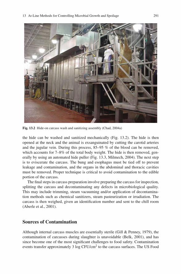
13 At-Line Methods for Controlling Microbial Growth and Spoilage 291
Fig. 13.2 Hide-on carcass wash and sanitizing assembly (Chad, 2004a)
the hide can be washed and sanitized mechanically (Fig. 13.2). The hide is thenopened at the neck and the animal is exsanguinated by cutting the carotid arteriesand the jugular vein. During this process, 85–95 % of the blood can be removed,which accounts for 7–8% of the total body weight. The hide is then removed, gen-erally by using an automated hide puller (Fig. 13.3, Milmech, 2004). The next stepis to eviscerate the carcass. The bung and esophagus must be tied off to preventleakage and contamination, and the organs in the abdominal and thoracic cavitiesmust be removed. Proper technique is critical to avoid contamination to the edibleportion of the carcass.
The final steps in carcass preparation involve preparing the carcass for inspection,splitting the carcass and decontaminating any defects in microbiological quality.This may include trimming, steam vacuuming and/or application of decontamina-tion methods such as chemical sanitizers, steam pasteurization or irradiation. Thecarcass is then weighed, given an identification number and sent to the chill room(Aberle et al., 2001).
Sources of Contamination
Although internal carcass muscles are essentially sterile (Gill & Penney, 1979), thecontamination of carcasses during slaughter is unavoidable (Belk, 2001), and hassince become one of the most significant challenges to food safety. Contaminationevents transfer approximately 3 log CFU/cm2 to the carcass surfaces. The US Food
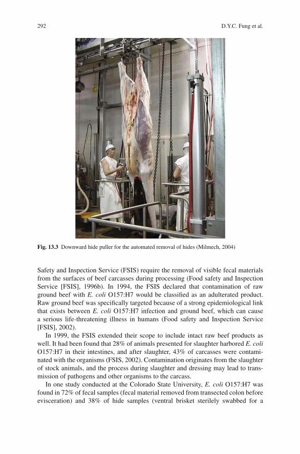
292 D.Y.C. Fung et al.
Fig. 13.3 Downward hide puller for the automated removal of hides (Milmech, 2004)
Safety and Inspection Service (FSIS) require the removal of visible fecal materialsfrom the surfaces of beef carcasses during processing (Food safety and InspectionService [FSIS], 1996b). In 1994, the FSIS declared that contamination of rawground beef with E. coli O157:H7 would be classified as an adulterated product.Raw ground beef was specifically targeted because of a strong epidemiological linkthat exists between E. coli O157:H7 infection and ground beef, which can causea serious life-threatening illness in humans (Food safety and Inspection Service[FSIS], 2002).
In 1999, the FSIS extended their scope to include intact raw beef products aswell. It had been found that 28% of animals presented for slaughter harbored E. coliO157:H7 in their intestines, and after slaughter, 43% of carcasses were contami-nated with the organisms (FSIS, 2002). Contamination originates from the slaughterof stock animals, and the process during slaughter and dressing may lead to trans-mission of pathogens and other organisms to the carcass.
In one study conducted at the Colorado State University, E. coli O157:H7 wasfound in 72% of fecal samples (fecal material removed from transected colon beforeevisceration) and 38% of hide samples (ventral brisket sterilely swabbed for a
13 At-Line Methods for Controlling Microbial Growth and Spoilage 293
total of 450 cm2) from cattle prepared for slaughter from a variety of facilities.Upon processing of these carcasses, 57% of post evisceration carcasses and 17% ofpost processing carcasses were positive for E. coli O157:H7 contamination (Elderet al., 2000).
Particular areas of the beef carcass are more prone to contamination with all kindsof microorganisms, with the brisket, fore-rib, flanks and round sites being the mostcommon (Johanson et al., 1983). The inner round is characterized by substantialamount of lean tissue surrounded by a collar of fat. This ridge allows fecal materialto become embedded in the junction and between muscle bundles, which resultsin difficult removal. This area is also the most difficult area to trim, because ofthe large collar of fat tissue, which also contributes to cross contamination (Hardinet al., 1995). Fecal contamination and pathogens are greater on areas of the beef car-cass covered by subcutaneous fat (Hardin et al., 1995). The butt, anal area, and rumpare areas of the beef carcass that remain moderately contaminated with organismsof fecal origin even after trimming and washing (Gill et al., 1995).
The abattoir contains many environments that can lead to cross contami-nation with pathogens (Belk, 2001; United States Department of Agriculture[USDA], 1993). Holding pens, slaughter and dressing processes, carcass skin-ning and evisceration have all been identified as points of entry for bacterialcontamination (USDA, 1993). Contamination is also possible from walls, floors, airpersonnel, knives and protective garments (Fung et al., 2001). Carcasses may evencontaminate each other if they make direct contact (Elder et al., 2000). The extent towhich carcasses are contaminated is directly influenced by plant design, the speedof slaughter and the overall skill of employees (Belk, 2001). The season, type ofanimal being slaughtered and the specific site on the carcass can dramatically affectthe level of contamination present on the carcass after slaughter (Sofos, Belk, &Smith, 1999). Despite the fact that processing methods and sanitation proceduresmay reduce the number of E coli O157:H7 positive carcasses, the amount ofcontamination after processing still poses a high food safety risk. Contamination ofbeef carcasses with E. coli O157:H7 designates a need for control on farms and infeedlots to reduce the risk of food-borne infection by this and other pathogens andeven non-pathogens (Elder et al., 2000).
Microbial Considerations in Meat and Foods, and FoodProcessing Environments
Before a discussion on the occurrence of microbes on meat and decontaminationstrategies, it is appropriate to define the meaning of microbial counts in or onmeat and food, air, food processing environments and food processing surfaces andutensils.
Table 13.1a and 13.1b is the Fung Applied Microbiology Scales listing the rangesof microorganisms for applied microbiology developed by the author after morethan 30 years of research experience in food and meat microbiology.
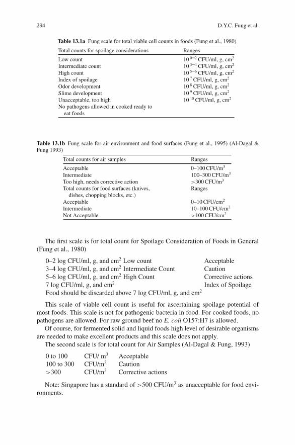
294 D.Y.C. Fung et al.
Table 13.1a Fung scale for total viable cell counts in foods (Fung et al., 1980)
Total counts for spoilage considerations Ranges
Low count 10 0–2 CFU/ml, g, cm2
Intermediate count 10 3–4 CFU/ml, g, cm2
High count 10 5–6 CFU/ml, g, cm2
Index of spoilage 10 7 CFU/ml, g, cm2
Odor development 10 8 CFU/ml, g, cm2
Slime development 10 9 CFU/ml, g, cm2
Unacceptable, too high 10 10 CFU/ml, g, cm2
No pathogens allowed in cooked ready toeat foods
Table 13.1b Fung scale for air environment and food surfaces (Fung et al., 1995) (Al-Dagal &Fung 1993)
Total counts for air samples Ranges
Acceptable 0–100 CFU/m3
Intermediate 100–300 CFU/m3
Too high, needs corrective action >300 CFU/m3
Total counts for food surfaces (knives,dishes, chopping blocks, etc.)
Ranges
Acceptable 0–10 CFU/cm2
Intermediate 10–100 CFU/cm2
Not Acceptable >100 CFU/cm2
The first scale is for total count for Spoilage Consideration of Foods in General(Fung et al., 1980)
0–2 log CFU/ml, g, and cm2 Low count Acceptable3–4 log CFU/ml, g, and cm2 Intermediate Count Caution5–6 log CFU/ml, g, and cm2 High Count Corrective actions7 log CFU/ml, g, and cm2 Index of SpoilageFood should be discarded above 7 log CFU/ml, g, and cm2
This scale of viable cell count is useful for ascertaining spoilage potential ofmost foods. This scale is not for pathogenic bacteria in food. For cooked foods, nopathogens are allowed. For raw ground beef no E. coli O157:H7 is allowed.
Of course, for fermented solid and liquid foods high level of desirable organismsare needed to make excellent products and this scale does not apply.
The second scale is for total count for Air Samples (Al-Dagal & Fung, 1993)
0 to 100 CFU/ m3 Acceptable100 to 300 CFU/m3 Caution>300 CFU/m3 Corrective actions
Note: Singapore has a standard of >500 CFU/m3 as unacceptable for food envi-ronments.
13 At-Line Methods for Controlling Microbial Growth and Spoilage 295
The third scale is for food contact surfaces (knives, spoons, forks, chop boards,table tops, etc. Fung et al., 1995)
0–10 CFU/cm2 Acceptable10–100 CFU/cm2 Caution>100 CFU/cm2 Corrective actions
Hide Removal
Meat becomes contaminated with microorganisms when feces or contaminatedhides contact the carcass during slaughter (Derfler, 2004; Elder et al., 2000; Gillet al., 1995). Elder et al. (2000) indicated that in cattle prepared for slaughter, 28%of carcasses had feces positive for E. coli O157:H7 and 11% of hides were positive.During processing, contamination spread to the carcass can range from 2 to 4 logCFU/cm2 (Anderson, Sebaugh, Marshall, & Stringer, 1980). The bulk of microbialcontamination occurs during hide removal (Bell, 1997; Buchanan & Doyle, 1997;Gill & Penney, 1979). When animals arrive at the abattoir, they carry a wide varietyof microorganisms on the hides and hooves, and in their intestinal tracts (Belk, 2001;Derfler, 2004; McEvoy et al., 2003).
Bung Tying
Bung tying is a possible source of contamination in the slaughter process, and greatcare must be taken to prevent transfer of bacteria from the anus of the animal ontothe edible adipose tissue (Gill et al., 1995). The bung tying process involves cuttingto loosen the anus, and then bagging the bung and securing with either a tie of a clip(Food Safety and Inspection Service [FSIS], 1994). The bung is then pushed throughto the abdominal cavity, where it can be removed during evisceration (Romans,Costello, Carlson, Greaser, & Jones, 2001). Tools or personnel that contact thebung may contribute to cross contamination (McEoy et al., 2003). Cross contam-ination, that is a direct result of manual bung tying, may be eliminated by using anautomated system. Such systems have reported lower total of E. coli and coliformcounts in the anal area than manual methods (Sheridan, 1998). Proper taping of theesophagus and enclosing the rectum to prevent leakage of the ingesta and feces, andensuring that the gastrointestinal tract is removed without incident are essential forcontrolling contamination (Bell, 1997). Figure 13.4 shows pre-evisceration carcasswashes.
Evisceration
The gastrointestinal tracts of cattle can carry a multitude of enteric pathogens.The evisceration process carries the potential of ingesta contamination to the
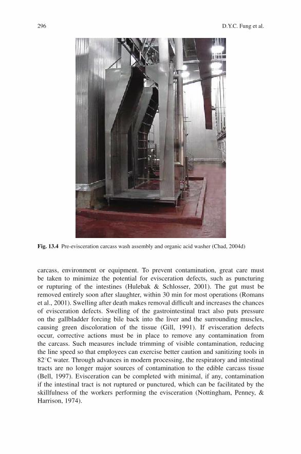
296 D.Y.C. Fung et al.
Fig. 13.4 Pre-evisceration carcass wash assembly and organic acid washer (Chad, 2004d)
carcass, environment or equipment. To prevent contamination, great care mustbe taken to minimize the potential for evisceration defects, such as puncturingor rupturing of the intestines (Hulebak & Schlosser, 2001). The gut must beremoved entirely soon after slaughter, within 30 min for most operations (Romanset al., 2001). Swelling after death makes removal difficult and increases the chancesof evisceration defects. Swelling of the gastrointestinal tract also puts pressureon the gallbladder forcing bile back into the liver and the surrounding muscles,causing green discoloration of the tissue (Gill, 1991). If evisceration defectsoccur, corrective actions must be in place to remove any contamination fromthe carcass. Such measures include trimming of visible contamination, reducingthe line speed so that employees can exercise better caution and sanitizing tools in82◦C water. Through advances in modern processing, the respiratory and intestinaltracts are no longer major sources of contamination to the edible carcass tissue(Bell, 1997). Evisceration can be completed with minimal, if any, contaminationif the intestinal tract is not ruptured or punctured, which can be facilitated by theskillfulness of the workers performing the evisceration (Nottingham, Penney, &Harrison, 1974).
13 At-Line Methods for Controlling Microbial Growth and Spoilage 297
Carcass-to-Carcass Transfer
McEoy et al. (2003) indicated that in a typical Irish beef abattoir, researchers exam-ined the persistence of E. coli O157:H7 on beef carcasses, as well as surveyed theprevalence of the bacterium in post slaughter and post dressing carcasses. In thefacilities chosen for the study, carcasses were slaughtered at a speed of 40–80 ani-mals per hour. Carcasses were processed using an automatic hide puller and werewashed with potable water at 35◦C before chilling (McEvoy et al. 2003). Carcasssampling revealed that cross contamination does occur during chilling. Samplingof two carcasses before chilling resulted in one positive and one negative samplefor E. coli O157:H7. However, after chilling both carcasses tested positive for theorganism. These carcasses were not together on the slaughter line, but were sideby side during chilling. Fecal rumen and carcass sampling were performed on 250carcasses. Of those 2.4% of fecal samples, 0.8% of rumen samples and 3.2% ofcarcass samples were positive for E. coli O157:H7 (McEvoy et al., 2003).
Decontamination of Beef Carcasses
Governmental Regulations and HACCP
It is impossible to guarantee bacteria and pathogen-free meat in large abattoirs,therefore, decontamination step in the form of washing and sanitizing during theslaughter process is important. Decontamination methods can improve the microbi-ological safety and increase shelf life, and should be an integral part of the slaughterprocess (Dickson & Anderson, 1992). Prompt chilling of carcasses after slaughterto below optimal bacterial growth temperatures is a critical measure, and chilling(Abdul-Raouf, Beuchat, & Ammur, 1993). Romans et al. (2001) recommend thatcarcasses should be chilled 16–20 h at –1 to –2C. Chilling can reduce the preva-lence of many cells and will stress cells. By closely adhering to the clean cattlepolicy and employing hygienic dressing and evisceration, contamination of car-casses can be minimized. In addition, ensuring that only clean, healthy animalsare presented for slaughter and are processed correctly will reduce the incidenceof contamination.
Governmental Regulations and HACCP
There has been great interests within the meat industry to develop slaughteringand dressing operations that reduce the incidence of contamination (Sofos, 1993).The first federal legislation concerning meat inspection dates back to 1890. In1891, US Congress assigned responsibility for meeting European meat require-ments to the USDA. The USDA then began conducting livestock inspections bothbefore and after slaughter of meat animals intended for US consumers (Hulebak &
298 D.Y.C. Fung et al.
Schlosser, 2001). The well known story of Upton Sinclair’s The Jungle whichdescribed the sickening and unsanitary conditions of the meat packing indus-try outraged the American public (Centers for Disease Control and Prevention[CDC], 1999) and resulted in the passing of the Federal Meat Inspection Act byUS Congress. This act established sanitation standards for slaughter and process-ing and authorized ante-mortem inspection of animals and post-mortem inspectionof every carcass for interstate commerce (Hulebak & Schlosser, 2001). Carcassinspection was intended to keep meat from diseased animals out of the food sup-ply, with Veterinarians and food inspectors checking live animals and carcasses forsigns of disease. The 1906 Meat Inspection Act allowed for continuous inspectionof processing operations by the USDA. It was not until the 1950s that concerngrew about the unseen threats to food safety, those that visual inspection couldnot identify. The wholesome Meat act of 1967 required that adequate inspectionof all meat products was performed. In 1977, the Food Safety and Quality Ser-vice (FSQS) was established and given the responsibility of inspecting meat andpoultry products. In 1981 the name was changed to Food Safety and InspectionService (FSIS). The Hazard Analysis Critical Control Point concept, started in1960, began as a joint effort between the National Aeronautics and Space Admin-istration, the United States Army Natick Laboratories and the Pillsbury Company.Now it is being applied to many industries including the meat industry. Becauseof HACCP the food supply nationally and internationally are much safer thanbefore.
Hide-on Decontamination
One method to reduce microbe and pathogen transfer during hide removal is toreduce the levels of bacteria present on animal hides before these animals enterthe slaughterhouse. A hide-on decontamination cabinet has been designed and usedin pilot plants for the removal of fecal contamination and debris before the dehid-ing process. The double-shackled cattle proceed into the cabinet after stunning andexsanguination (Fig. 13.4). Cattle are then vacuumed to remove excess liquid. Hide-on decontamination cabinets require an enormous amount of water, and thereforerequire recirculating systems to make such systems cost-effective (Bosilevac, Nou,Osborn, Allen, & Koohmaraie, 2005). Hide-on decontamination was studied usingwater and tri-sodium phosphate with and without vacuuming steps. Whole pulledhides were draped over barrels to simulate the hide-on carcass. Wash treatmentswere applied using an electric pressurized sprayer at a pressure of 1200 psi and ata distance of 65 cm from the hide. The treatments were applied for 20 s at 60◦C.Half of the treated hides were then vacuumed using a steam vacuum without steamto remove residual liquid and visible contamination. Hide-on decontamination prac-tices can reduce the bacteria on hides by about 5 log CFU/100 cm2, and decrease thebacterial load of carcasses by 1 log CFU/100 cm2. This in turn dramatically reducesthe prevalence of E. coli O157:H7 on carcasses (Bosilevac et al., 2005).
13 At-Line Methods for Controlling Microbial Growth and Spoilage 299
Chemical Dehairing
Chemical dehairing is a process utilizing sodium sulfide to remove hair and debrisfrom cattle hides. The solution is then neutralized by a combination of hydrogenperoxide spray and water washing. The cleaning of cattle hides results in a drasticreduction in subsequent carcass contamination (Nou et al., 2003). The object ofchemical dehairing is to remove hair, dirt, and fecal materials from the hide beforeslaughtering in an attempt to reduce subsequent carcass contamination. In a studyconducted by Nou et al. (2003), chemical dehairing was evaluated as a means toreduce carcass contamination. Carcass contamination can be nearly eliminated if thequality of the hide is improved before dehiding. Chemical dehairing is extremelyeffective at virtually eliminating carcass decontamination from the dehiding pro-cess. There are some additional considerations regarding the efficacy of chemicaldehairing. Sodium sulfide wash solutions and dehairing waste create extensive envi-ronmental problems for the industry, as the chemical is damaging to the environment(Agricultural Marketing Service [AMS], 2000).
Cetylpyridinium Chloride (CPC)
CPC is a quaternary ammonium compound used as a common oral antimicrobial(Bosilevac, Wheeler, et al., 2004), which is currently approved for use on poultrycarcasses as an antimicrobial agent for surface contamination. It is applied as amist at room temperature prior to poultry chilling. Application cannot exceed 0.3 gCPC/lb of raw poultry (Code of Federal Regulations [CFR], 2005b). Many typesof pathogens are sensitive to CPC and the chemical does not cause undesirableeffects to flavor, texture, appearance or odor of foods. The antibacterial activityof CPC is facilitated by the cations it forms in solution which decrease surfacetension and allows the molecules to penetrate the bacterial membrane to act oncellular components. CPC can reduce E coli O157:H7 by 2.78 log after 14 daysof storage (Lim & Mustapha 2004). Since CPC is rapidly inactivated by organicmaterials, the degree to which a hide is soiled will directly impact the efficacyof CPC as a decontamination method. The most logical point at which to applyCPC treatment would be on stunned, shackled animals before dehiding (Bosilevac,Wheeler, et al., 2004). Currently, CPC is not an approved method of decontam-ination in the beef industry (Ransom et al., 2003). However, studies have shownthat CPC is effective for reducing pathogens on beef hides (Cutter et al., 2000).One percent CPC is the most effective concentration for reducing bacterial pop-ulation on hides. The greatest efficacy for CPC is as a high-pressure pre-washat a concentration of 1% followed by a second high-pressure wash after stun-ning (Bosilevac, Wheeler, et al., 2004). Bosilevac Arthur, et al., (2004) studiedCPC on live animals in holding pens and found that CPC significantly reducedE. coli O157:H7 on hides, resulting in a near-zero incidence on pre-evisceratedcarcasses.
300 D.Y.C. Fung et al.
Knife Trimming
The knife trimming of carcasses is now required for slaughter facilities in an effortto remove all visible contaminants. Knife trimming has been written into many san-itation standard operating procedures and HACCP plans (Belk, 2001). The standardcommercial practice among slaughter houses in the USA is to knife trim all vis-ible contamination on the carcass, followed by a water wash (Acuff, Castillo, &Savell, 1997; Prasai et al., 1995). Trimming is far superior to washing, in orderto remove visible contaminants, as washing may not remove attached organismsand may spread contamination over the surface of the carcass (Food Safety andInspection Service [FSIS], 1996a). Trimming is especially useful at removing vis-ible organic matters in areas that are difficult to decontaminate with water wash orchemical sprays (Hardin et al., 1995). Some problems exist in trimming of ediblemeat. Trimming is a highly variable process whose efficacy is related to the skillof the individual doing the trimming. The equipment used during trimming canpotentially spread contamination if not properly sanitized. In addition, holding car-casses in the warm slaughter house environment can allow for attachment of bacteriabefore washing and chilling (Reagan et al., 1996). Meat inspection and trimmingrely on removing visual contamination from the carcass, and may overlook low-level contamination. The knife may be used on multiple sites of a single carcassor on multiple carcasses. Because the use of sterile instruments is not practical, awashing procedure will always be part of the standard protocol (Prasai et al., 1995).There is always a chance that the knife may contaminate other parts of the meatwhen trimming is being done. Trimming must be employed to comply with thezero-tolerance guidelines for fecal contamination; however, FSIS recommends thata secondary decontamination step be employed at some point in the process.
Vacuuming
In 1993, following the outbreak of E. coli O157:H7 that left four children dead, theFSIS stressed that manual trimming was the only way to remove visible contami-nation. At that time, there were no firm data indicating that any other method wasmore efficacious (FSIS, 1996a). In 1995, the FSIS held a public meeting to considerother methods for removing visible contamination. Testing of steam vacuuming wasapproved as a possible alternative to achieve the zero-tolerance regulatory policyfor fecal contamination. Fifty plants tested the new vacuum system (FSIS, 1996a).Spot vacuuming of carcasses involves the use of hot water or steam to inactivatebacteria and remove visible fecal contamination (Sofos et al., 1999). Each planttested 120 samples, 60 were trimmed and 60 were vacuumed. All contaminationover 1 inch in diameter was trimmed regardless of the treatment group. A secondstudy was done taking 60 samples over 60 days to evaluate the consistency of thenew method (FSIS, 1996a). These studies revealed, that carcasses that were vacu-umed had, on the average, 0.69 log CFU/cm2 less contamination than the trimmedsamples. In addition, over the 60-day trial period, a 0.54 log CFU/cm2 lower level
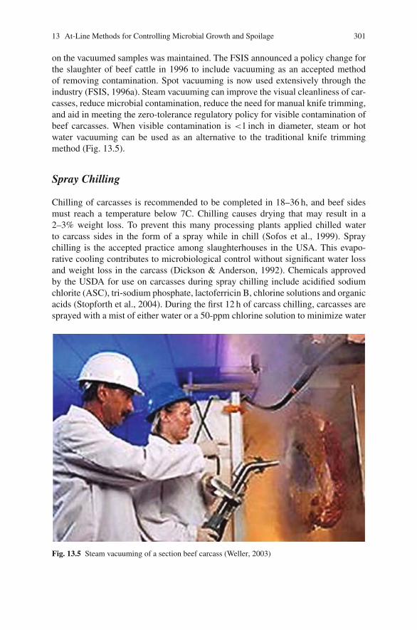
13 At-Line Methods for Controlling Microbial Growth and Spoilage 301
on the vacuumed samples was maintained. The FSIS announced a policy change forthe slaughter of beef cattle in 1996 to include vacuuming as an accepted methodof removing contamination. Spot vacuuming is now used extensively through theindustry (FSIS, 1996a). Steam vacuuming can improve the visual cleanliness of car-casses, reduce microbial contamination, reduce the need for manual knife trimming,and aid in meeting the zero-tolerance regulatory policy for visible contamination ofbeef carcasses. When visible contamination is <1 inch in diameter, steam or hotwater vacuuming can be used as an alternative to the traditional knife trimmingmethod (Fig. 13.5).
Spray Chilling
Chilling of carcasses is recommended to be completed in 18–36 h, and beef sidesmust reach a temperature below 7C. Chilling causes drying that may result in a2–3% weight loss. To prevent this many processing plants applied chilled waterto carcass sides in the form of a spray while in chill (Sofos et al., 1999). Spraychilling is the accepted practice among slaughterhouses in the USA. This evapo-rative cooling contributes to microbiological control without significant water lossand weight loss in the carcass (Dickson & Anderson, 1992). Chemicals approvedby the USDA for use on carcasses during spray chilling include acidified sodiumchlorite (ASC), tri-sodium phosphate, lactoferricin B, chlorine solutions and organicacids (Stopforth et al., 2004). During the first 12 h of carcass chilling, carcasses aresprayed with a mist of either water or a 50-ppm chlorine solution to minimize water
Fig. 13.5 Steam vacuuming of a section beef carcass (Weller, 2003)
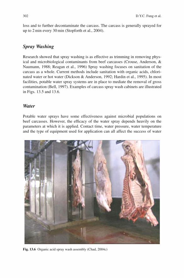
302 D.Y.C. Fung et al.
loss and to further decontaminate the carcass. The carcass is generally sprayed forup to 2 min every 30 min (Stopforth et al., 2004).
Spray Washing
Research showed that spray washing is as effective as trimming in removing phys-ical and microbiological contaminants from beef carcasses (Crouse, Anderson, &Naumann, 1988; Reagan et al., 1996) Spray washing focuses on sanitation of thecarcass as a whole. Current methods include sanitation with organic acids, chlori-nated water or hot water (Dickson & Anderson, 1992; Hardin et al., 1995). In mostfacilities, potable water spray systems are in place to mediate the removal of grosscontamination (Bell, 1997). Examples of carcass spray wash cabinets are illustratedin Figs. 13.5 and 13.6.
Water
Potable water sprays have some effectiveness against microbial populations onbeef carcasses. However, the efficacy of the water spray depends heavily on theparameters at which it is applied. Contact time, water pressure, water temperatureand the type of equipment used for application can all affect the success of water
Fig. 13.6 Organic acid spray wash assembly (Chad, 2004c)
13 At-Line Methods for Controlling Microbial Growth and Spoilage 303
sprays (Fung et al., 2001). Hot water has been shown to be much more effective thancold water at decontamination. Hot water washing is affected by the same parame-ters as cold water sprays (i.e. contact time, temperature, pressure, etc.). Cold watersprays still have some applications in meat processing plants for the removal ofdebris from the hide of the animal, although there is limited effect on the microbialsafety of the product (Fung et al., 2001). The use of hot water for decontaminationis highly regulated. The USDA-FSIS approves of hot water as a decontaminationmethod and recommends it to be used at 74–85◦C (Sofos et al., 1999). Using hotwater or steam creates the potential for sanitation problems by increased humid-ity and available moisture in the plant environment, thus creating an environmen-tal niche for microorganisms. The equipment for decontamination must meet therequirements set forth in 9 CFR 308.5. This method is only allowed at the process-ing steps where water spray is allowed. Automated and handheld equipments areavailable (FSIS, 1996b). In one study, spray washing of edible beef tissues showedthat spraying carcasses with hot pressurized water (74◦C and 20.68 bar) can reducethe initial microbial populations by 1-log unit and extend the shelf life by 5 days(Gorman, Morgan, Sofos, & Smith, 1995; Gorman, Sofos, Morgan, Schmidt, &Smith, 1995). In many instances temperature, pressure, chemicals combinations inwater spraying on carcasses have been studied throughout the world. Basically, a 2log CFU/cm2 reduction is considered a good method.
Acid Spray Washes
Organic acids are used in the beef industry as decontamination solutions duringslaughter and are the classic method for controlling the growth of microorganismson meat (Fang & Tsai, 2003). These spray washes reduce the microbial load andare intended to improve the safety of the meat, and some combinations resulted inreducing pathogenic contamination by up to 2 log CFU/cm2 (Berry & Cutter, 2000).The FSIS approves the use of a water rinse followed by a rinse with food gradeorganic acids on pre-eviscerated carcasses. The water rinse is intended to removeany hair, dirt or dust that has accumulated on the surface (FSIS, 1996a). Acetic,lactic and citric acids have been generally recognized as safe (GRAS) by the FDAfor multipurpose usage (Acuff et al., 1987; FSIS, 1996a). Two cabinet systems areapproved for use, and the acids are applied as a fine mist or fog. Organic acid treat-ment can be used in aqueous solutions of 1.5–2.5% and can be sprayed on carcassesat any stage in which water spray is allowed. The use of approved automated spraycabinets or hand-operated equipment is allowed. Acid washes have been shown toeffectively remove 2 log CFU/cm2 of the general microflora from beef carcasses(Fung et al., 2001).
Acetic Acid
Acetic acid has been an effective spray chilling sanitizer on beef carcasses in reduc-ing the number of pathogenic populations (Dickson, 1991). Anderson, Marshall,
304 D.Y.C. Fung et al.
Stringer, & Naumann, (1977) showed that acetic acid sprays (AASs) on beef car-casses could reduce aerobic bacteria by 99.9%. However, some research data showsthat spray washing with acetic acid has similar results (no significant difference) tohot water spray washing. For example, hot water (74◦C) reduced bacteria from 6 to4.36 log CFU/cm2 and acetic acid reduced E. coli to 4.31 log CFU/cm2. Short-chainfatty acids have been shown to discolor meat to a higher degree than other GRASacids. AASs have resulted in brown to yellow discoloration and offensive acid odors(Hamby, Savell, Acuff, Vanderzant, & Cross, 1987). In a study conducted by Brack-ett, Hao, & Doyle (1994), maximum reduction of E. coli O157:H7 on beef carcasseswas achieved when tissues were sprayed with 3% acetic acid at 20◦C. However,this treatment only resulted in a 0.61 log CFU/cm2 decrease of the pathogen load.These researchers suggest that organic acid sprays are of little value against E. coliO157:H7.
Lactic Acid
Aqueous solutions of lactic acid can be used to successfully decontaminate beefcarcasses (Kozempel et al., 2003). However, the acid concentration used on beefis limited because of the potential for adverse effects to the quality of the product.Lactic acid, specifically, can alter fresh meat color at blood spots, which can beprevented by removing blood spots with a water wash before treatment with lac-tic acid (Crozier-Dodson, 2000). Samelis, Sofos, Kendall, & Smith, (2002) recom-mended that lactic acid sprays be used, rather than AASs, for the decontamination ofbeef carcasses. It has been shown that acetic acid has a lower effectiveness againstE. coli O157:H7 and a higher effectiveness against the natural flora of meat car-casses. Lactic acid inactivates E. coli O157:H7 at a faster rate than acetic acid at thesame concentration in decontamination run-off fluids (Samelis, Sofos, Kendall, &Smith, 2001). Lactic acid is very effective for pathogen reduction as shown in astudy conducted by Ransom et al. (2003). Lactic acid sprays were able to achievereductions of pathogen numbers by 2.5 log CFU/cm2. From this study, it was shownthat lactic acid exceeds acetic acid reductions.
Fumaric Acid
Fumaric acid has been largely ignored by the processing industry as an organic acidcapable of increasing the microbiological safety of meats. It has several benefitsthat make it superior to the conventional acids used, such as acetic and lactic acids.Fumaric acid is readily available in nature and is present in fresh meat at low concen-trations. It is very cost-effective and more acidic than the other GRAS organic acids.It also has antioxidant properties that may make it beneficial in some industries.Research at the Kansas State University (Podolak, Zayas, Kastner, & Fung, 1996)compared the effectiveness of fumaric acid on the decontamination of lean beef
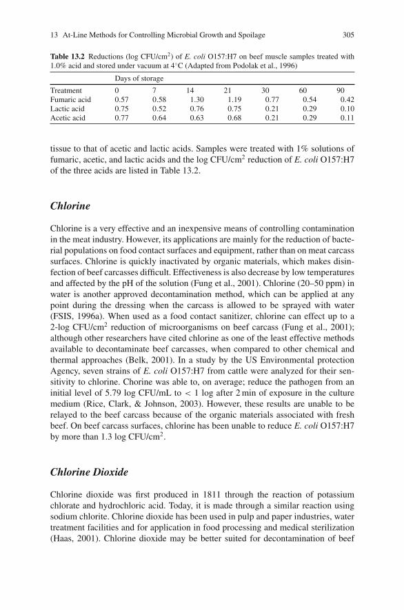
13 At-Line Methods for Controlling Microbial Growth and Spoilage 305
Table 13.2 Reductions (log CFU/cm2) of E. coli O157:H7 on beef muscle samples treated with1.0% acid and stored under vacuum at 4◦C (Adapted from Podolak et al., 1996)
Days of storage
Treatment 0 7 14 21 30 60 90Fumaric acid 0.57 0.58 1.30 1.19 0.77 0.54 0.42Lactic acid 0.75 0.52 0.76 0.75 0.21 0.29 0.10Acetic acid 0.77 0.64 0.63 0.68 0.21 0.29 0.11
tissue to that of acetic and lactic acids. Samples were treated with 1% solutions offumaric, acetic, and lactic acids and the log CFU/cm2 reduction of E. coli O157:H7of the three acids are listed in Table 13.2.
Chlorine
Chlorine is a very effective and an inexpensive means of controlling contaminationin the meat industry. However, its applications are mainly for the reduction of bacte-rial populations on food contact surfaces and equipment, rather than on meat carcasssurfaces. Chlorine is quickly inactivated by organic materials, which makes disin-fection of beef carcasses difficult. Effectiveness is also decrease by low temperaturesand affected by the pH of the solution (Fung et al., 2001). Chlorine (20–50 ppm) inwater is another approved decontamination method, which can be applied at anypoint during the dressing when the carcass is allowed to be sprayed with water(FSIS, 1996a). When used as a food contact sanitizer, chlorine can effect up to a2-log CFU/cm2 reduction of microorganisms on beef carcass (Fung et al., 2001);although other researchers have cited chlorine as one of the least effective methodsavailable to decontaminate beef carcasses, when compared to other chemical andthermal approaches (Belk, 2001). In a study by the US Environmental protectionAgency, seven strains of E. coli O157:H7 from cattle were analyzed for their sen-sitivity to chlorine. Chorine was able to, on average; reduce the pathogen from aninitial level of 5.79 log CFU/mL to < 1 log after 2 min of exposure in the culturemedium (Rice, Clark, & Johnson, 2003). However, these results are unable to berelayed to the beef carcass because of the organic materials associated with freshbeef. On beef carcass surfaces, chlorine has been unable to reduce E. coli O157:H7by more than 1.3 log CFU/cm2.
Chlorine Dioxide
Chlorine dioxide was first produced in 1811 through the reaction of potassiumchlorate and hydrochloric acid. Today, it is made through a similar reaction usingsodium chlorite. Chlorine dioxide has been used in pulp and paper industries, watertreatment facilities and for application in food processing and medical sterilization(Haas, 2001). Chlorine dioxide may be better suited for decontamination of beef
306 D.Y.C. Fung et al.
carcasses than aqueous chlorine. Chlorine dioxide does not react with nitrogenouscompounds as chlorine does (Berg, Roberts, & Matin, 1986), is less reactive withorganic compounds than chlorine (Shin, Chang, & Kang, 2004), has a greater oxi-dizing potential than chlorine, active at high pH (Berg et al., 1986), and has a higherantimicrobial activity than chlorine in food products (Shin et al., 2004). Chlorinedioxide is approved for use at 3 ppm as an antimicrobial agent for poultry chillwater (Code of Federal Regulations [CFR], 2005c) and approval is awaited for directfood contact, although approval for use on beef carcasses is not yet permitted (Shinet al., 2004). Chlorine dioxide inactivates bacteria by causing a loss of permeabilityof the outer cellular membrane, thus disrupting the ionic gradient across the mem-brane (Berg et al., 1986). Cutter & Dorsa (1995) found that chlorine dioxide reducedaerobic plate counts of <1 log CFU/cm2 at low pressure, and at high pressures withlonger contact times, reductions were increased from 0.93 to 1.52 CFU/cm2 andit was concluded that it is ineffective as a decontaminant of beef carcasses againstbacteria of fecal origin.
Tri-sodium Phosphate
Tri-sodium phosphate has been approved for use on beef carcasses and has shownto inhibit bacterial attachment to carcass surfaces (Sofos et al., 1999). Food gradetri-sodium phosphate may be applied to meat carcasses under very specific condi-tions: the temperature must be maintained at a range of 32–43◦C and be used atconcentrations between 8 and 12%. Treatment cannot be applied for more than 30 s.Tri-sodium phosphate can be applied at any point where carcasses can be sprayedwith water. Automated cabinets and hand-operated equipment are approved for use(FSIS, 1996a). The success of tri-sodium phosphate and other alkaline sanitizersin the poultry industry has lead to numerous studies for the application for beef.Dickson, Nettles Cutter, & Siragusa, (1994) investigated the effectiveness of tri-sodium phosphate as a sanitizer of beef tissue. Reduction of E. coli O157:H7 on beefwas similar to those found in studies for reducing Salmonella sp. contamination onpoultry carcasses. An average of 1.0–1.5 log of E. coli O157:H7 was successfullyachieved with the application of tri-sodium phosphate at 0.25 and 1.0 min; longerapplication times generally resulted in larger log reduction of E. coli O157:H7. Tem-perature had no significant effect on the results. Tri-sodium phosphate is unable toremove 100% of pathogenic contamination. However, the reductions show promisefor this sanitizer as part of a multi-hurdle system for increasing the safety of beefproducts (Dickson et al., 1994).
Acidified Sodium Chlorite (ASC)
The FDA approves the use of ASC for the decontamination of beef carcasses when itis combined with any organic acid with GRAS status. It can also be used in furtherprocessing of red meat as a dip. Sodium chlorite concentrations must be between
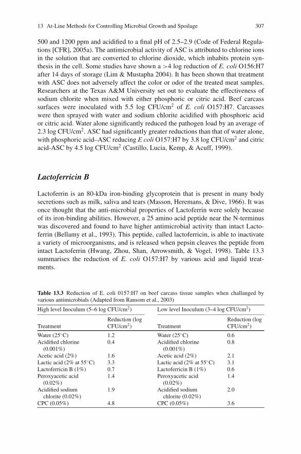
13 At-Line Methods for Controlling Microbial Growth and Spoilage 307
500 and 1200 ppm and acidified to a final pH of 2.5–2.9 (Code of Federal Regula-tions [CFR], 2005a). The antimicrobial activity of ASC is attributed to chlorine ionsin the solution that are converted to chlorine dioxide, which inhabits protein syn-thesis in the cell. Some studies have shown a >4 log reduction of E. coli O156:H7after 14 days of storage (Lim & Mustapha 2004). It has been shown that treatmentwith ASC does not adversely affect the color or odor of the treated meat samples.Researchers at the Texas A&M University set out to evaluate the effectiveness ofsodium chlorite when mixed with either phosphoric or citric acid. Beef carcasssurfaces were inoculated with 5.5 log CFU/cm2 of E. coli O157:H7. Carcasseswere then sprayed with water and sodium chlorite acidified with phosphoric acidor citric acid. Water alone significantly reduced the pathogen load by an average of2.3 log CFU/cm2. ASC had significantly greater reductions than that of water alone,with phosphoric acid–ASC reducing E coli O157:H7 by 3.8 log CFU/cm2 and citricacid-ASC by 4.5 log CFU/cm2 (Castillo, Lucia, Kemp, & Acuff, 1999).
Lactoferricin B
Lactoferrin is an 80-kDa iron-binding glycoprotein that is present in many bodysecretions such as milk, saliva and tears (Masson, Heremans, & Dive, 1966). It wasonce thought that the anti-microbial properties of Lactoferrin were solely becauseof its iron-binding abilities. However, a 25 amino acid peptide near the N-terminuswas discovered and found to have higher antimicrobial activity than intact Lacto-ferrin (Bellamy et al., 1993). This peptide, called lactoferricin, is able to inactivatea variety of microorganisms, and is released when pepsin cleaves the peptide fromintact Lactoferrin (Hwang, Zhou, Shan, Arrowsmith, & Vogel, 1998). Table 13.3summarises the reduction of E. coli O157:H7 by various acid and liquid treat-ments.
Table 13.3 Reduction of E. coli 0157:H7 on beef carcass tissue samples when challanged byvarious antimicrobials (Adapted from Ransom et al., 2003)
High level Inoculum (5–6 log CFU/cm2) Low level Inoculum (3–4 log CFU/cm2)
TreatmentReduction (logCFU/cm2) Treatment
Reduction (logCFU/cm2)
Water (25◦C) 1.2 Water (25◦C) 0.6Acidified chlorine
(0.001%)0.4 Acidified chlorine
(0.001%)0.8
Acetic acid (2%) 1.6 Acetic acid (2%) 2.1Lactic acid (2% at 55◦C) 3.3 Lactic acid (2% at 55◦C) 3.1Lactoferricin B (1%) 0.7 Lactoferricin B (1%) 0.6Peroxyacetic acid
(0.02%)1.4 Peroxyacetic acid
(0.02%)1.4
Acidified sodiumchlorite (0.02%)
1.9 Acidified sodiumchlorite (0.02%)
2.0
CPC (0.05%) 4.8 CPC (0.05%) 3.6
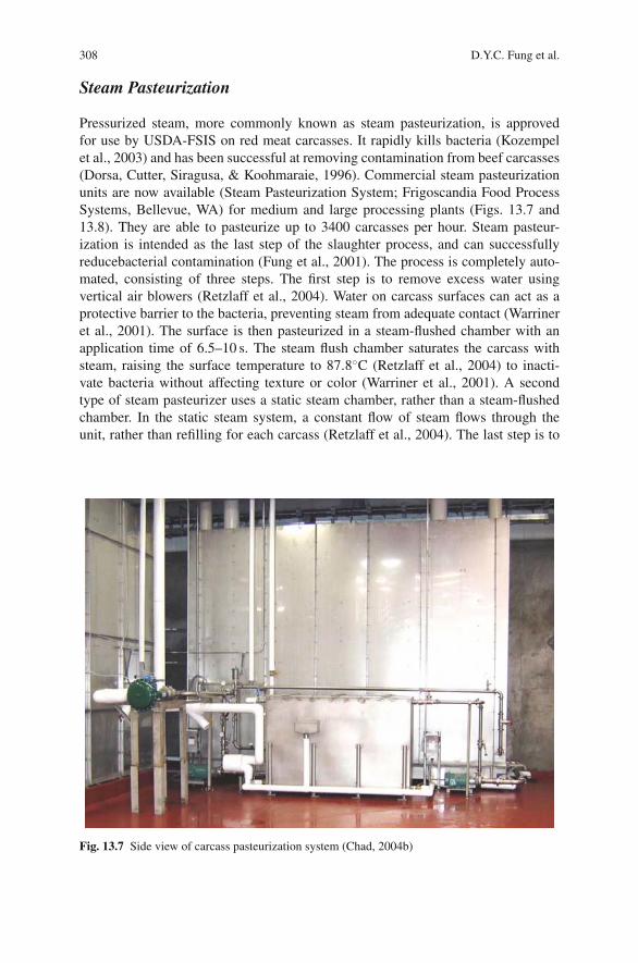
308 D.Y.C. Fung et al.
Steam Pasteurization
Pressurized steam, more commonly known as steam pasteurization, is approvedfor use by USDA-FSIS on red meat carcasses. It rapidly kills bacteria (Kozempelet al., 2003) and has been successful at removing contamination from beef carcasses(Dorsa, Cutter, Siragusa, & Koohmaraie, 1996). Commercial steam pasteurizationunits are now available (Steam Pasteurization System; Frigoscandia Food ProcessSystems, Bellevue, WA) for medium and large processing plants (Figs. 13.7 and13.8). They are able to pasteurize up to 3400 carcasses per hour. Steam pasteur-ization is intended as the last step of the slaughter process, and can successfullyreducebacterial contamination (Fung et al., 2001). The process is completely auto-mated, consisting of three steps. The first step is to remove excess water usingvertical air blowers (Retzlaff et al., 2004). Water on carcass surfaces can act as aprotective barrier to the bacteria, preventing steam from adequate contact (Warrineret al., 2001). The surface is then pasteurized in a steam-flushed chamber with anapplication time of 6.5–10 s. The steam flush chamber saturates the carcass withsteam, raising the surface temperature to 87.8◦C (Retzlaff et al., 2004) to inacti-vate bacteria without affecting texture or color (Warriner et al., 2001). A secondtype of steam pasteurizer uses a static steam chamber, rather than a steam-flushedchamber. In the static steam system, a constant flow of steam flows through theunit, rather than refilling for each carcass (Retzlaff et al., 2004). The last step is to
Fig. 13.7 Side view of carcass pasteurization system (Chad, 2004b)
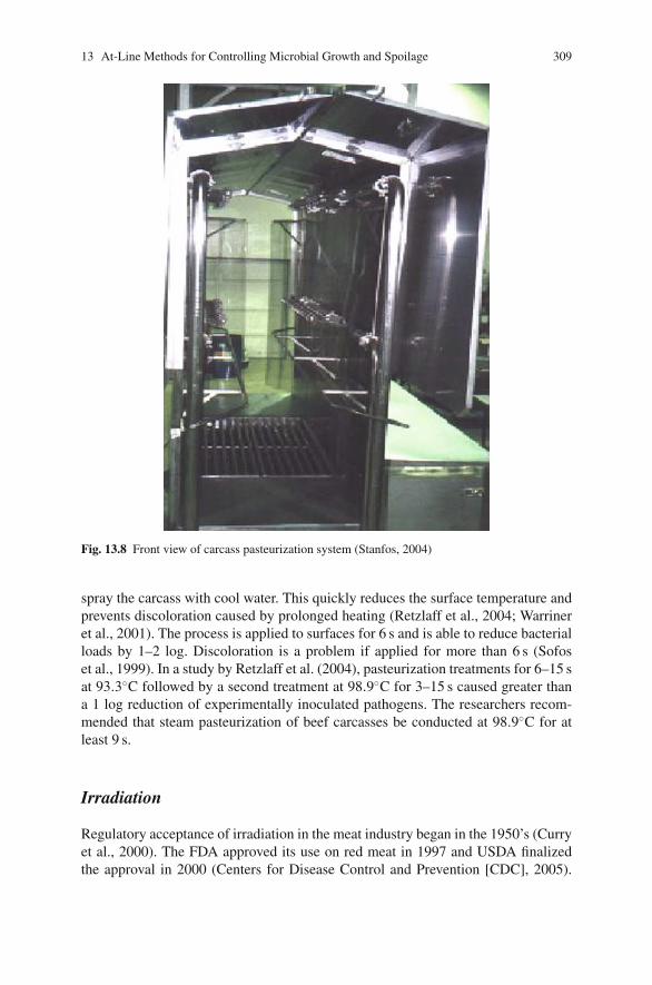
13 At-Line Methods for Controlling Microbial Growth and Spoilage 309
Fig. 13.8 Front view of carcass pasteurization system (Stanfos, 2004)
spray the carcass with cool water. This quickly reduces the surface temperature andprevents discoloration caused by prolonged heating (Retzlaff et al., 2004; Warrineret al., 2001). The process is applied to surfaces for 6 s and is able to reduce bacterialloads by 1–2 log. Discoloration is a problem if applied for more than 6 s (Sofoset al., 1999). In a study by Retzlaff et al. (2004), pasteurization treatments for 6–15 sat 93.3◦C followed by a second treatment at 98.9◦C for 3–15 s caused greater thana 1 log reduction of experimentally inoculated pathogens. The researchers recom-mended that steam pasteurization of beef carcasses be conducted at 98.9◦C for atleast 9 s.
Irradiation
Regulatory acceptance of irradiation in the meat industry began in the 1950’s (Curryet al., 2000). The FDA approved its use on red meat in 1997 and USDA finalizedthe approval in 2000 (Centers for Disease Control and Prevention [CDC], 2005).
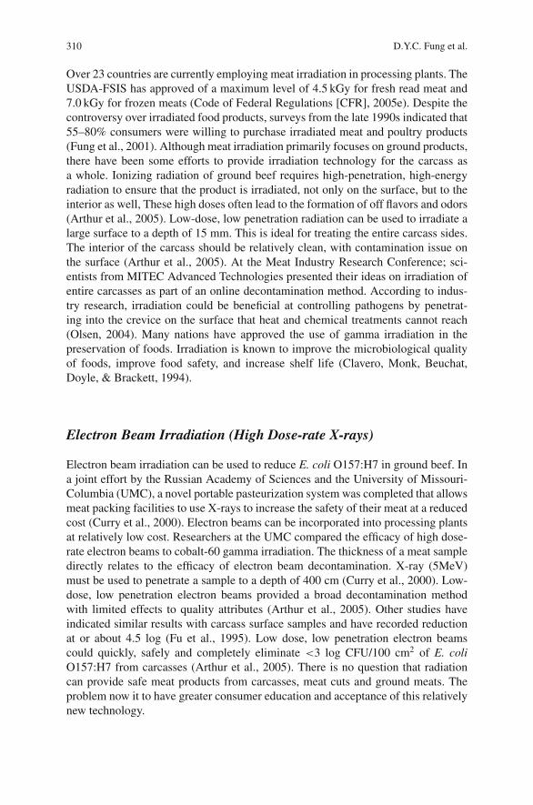
310 D.Y.C. Fung et al.
Over 23 countries are currently employing meat irradiation in processing plants. TheUSDA-FSIS has approved of a maximum level of 4.5 kGy for fresh read meat and7.0 kGy for frozen meats (Code of Federal Regulations [CFR], 2005e). Despite thecontroversy over irradiated food products, surveys from the late 1990s indicated that55–80% consumers were willing to purchase irradiated meat and poultry products(Fung et al., 2001). Although meat irradiation primarily focuses on ground products,there have been some efforts to provide irradiation technology for the carcass asa whole. Ionizing radiation of ground beef requires high-penetration, high-energyradiation to ensure that the product is irradiated, not only on the surface, but to theinterior as well, These high doses often lead to the formation of off flavors and odors(Arthur et al., 2005). Low-dose, low penetration radiation can be used to irradiate alarge surface to a depth of 15 mm. This is ideal for treating the entire carcass sides.The interior of the carcass should be relatively clean, with contamination issue onthe surface (Arthur et al., 2005). At the Meat Industry Research Conference; sci-entists from MITEC Advanced Technologies presented their ideas on irradiation ofentire carcasses as part of an online decontamination method. According to indus-try research, irradiation could be beneficial at controlling pathogens by penetrat-ing into the crevice on the surface that heat and chemical treatments cannot reach(Olsen, 2004). Many nations have approved the use of gamma irradiation in thepreservation of foods. Irradiation is known to improve the microbiological qualityof foods, improve food safety, and increase shelf life (Clavero, Monk, Beuchat,Doyle, & Brackett, 1994).
Electron Beam Irradiation (High Dose-rate X-rays)
Electron beam irradiation can be used to reduce E. coli O157:H7 in ground beef. Ina joint effort by the Russian Academy of Sciences and the University of Missouri-Columbia (UMC), a novel portable pasteurization system was completed that allowsmeat packing facilities to use X-rays to increase the safety of their meat at a reducedcost (Curry et al., 2000). Electron beams can be incorporated into processing plantsat relatively low cost. Researchers at the UMC compared the efficacy of high dose-rate electron beams to cobalt-60 gamma irradiation. The thickness of a meat sampledirectly relates to the efficacy of electron beam decontamination. X-ray (5MeV)must be used to penetrate a sample to a depth of 400 cm (Curry et al., 2000). Low-dose, low penetration electron beams provided a broad decontamination methodwith limited effects to quality attributes (Arthur et al., 2005). Other studies haveindicated similar results with carcass surface samples and have recorded reductionat or about 4.5 log (Fu et al., 1995). Low dose, low penetration electron beamscould quickly, safely and completely eliminate <3 log CFU/100 cm2 of E. coliO157:H7 from carcasses (Arthur et al., 2005). There is no question that radiationcan provide safe meat products from carcasses, meat cuts and ground meats. Theproblem now it to have greater consumer education and acceptance of this relativelynew technology.
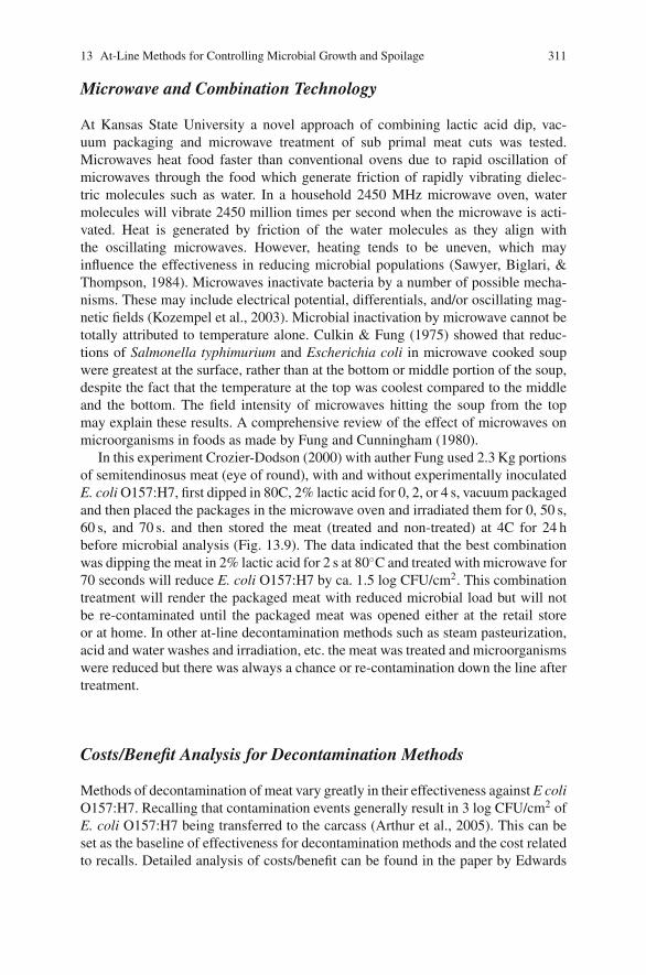
13 At-Line Methods for Controlling Microbial Growth and Spoilage 311
Microwave and Combination Technology
At Kansas State University a novel approach of combining lactic acid dip, vac-uum packaging and microwave treatment of sub primal meat cuts was tested.Microwaves heat food faster than conventional ovens due to rapid oscillation ofmicrowaves through the food which generate friction of rapidly vibrating dielec-tric molecules such as water. In a household 2450 MHz microwave oven, watermolecules will vibrate 2450 million times per second when the microwave is acti-vated. Heat is generated by friction of the water molecules as they align withthe oscillating microwaves. However, heating tends to be uneven, which mayinfluence the effectiveness in reducing microbial populations (Sawyer, Biglari, &Thompson, 1984). Microwaves inactivate bacteria by a number of possible mecha-nisms. These may include electrical potential, differentials, and/or oscillating mag-netic fields (Kozempel et al., 2003). Microbial inactivation by microwave cannot betotally attributed to temperature alone. Culkin & Fung (1975) showed that reduc-tions of Salmonella typhimurium and Escherichia coli in microwave cooked soupwere greatest at the surface, rather than at the bottom or middle portion of the soup,despite the fact that the temperature at the top was coolest compared to the middleand the bottom. The field intensity of microwaves hitting the soup from the topmay explain these results. A comprehensive review of the effect of microwaves onmicroorganisms in foods as made by Fung and Cunningham (1980).
In this experiment Crozier-Dodson (2000) with auther Fung used 2.3 Kg portionsof semitendinosus meat (eye of round), with and without experimentally inoculatedE. coli O157:H7, first dipped in 80C, 2% lactic acid for 0, 2, or 4 s, vacuum packagedand then placed the packages in the microwave oven and irradiated them for 0, 50 s,60 s, and 70 s. and then stored the meat (treated and non-treated) at 4C for 24 hbefore microbial analysis (Fig. 13.9). The data indicated that the best combinationwas dipping the meat in 2% lactic acid for 2 s at 80◦C and treated with microwave for70 seconds will reduce E. coli O157:H7 by ca. 1.5 log CFU/cm2. This combinationtreatment will render the packaged meat with reduced microbial load but will notbe re-contaminated until the packaged meat was opened either at the retail storeor at home. In other at-line decontamination methods such as steam pasteurization,acid and water washes and irradiation, etc. the meat was treated and microorganismswere reduced but there was always a chance or re-contamination down the line aftertreatment.
Costs/Benefit Analysis for Decontamination Methods
Methods of decontamination of meat vary greatly in their effectiveness against E coliO157:H7. Recalling that contamination events generally result in 3 log CFU/cm2 ofE. coli O157:H7 being transferred to the carcass (Arthur et al., 2005). This can beset as the baseline of effectiveness for decontamination methods and the cost relatedto recalls. Detailed analysis of costs/benefit can be found in the paper by Edwards
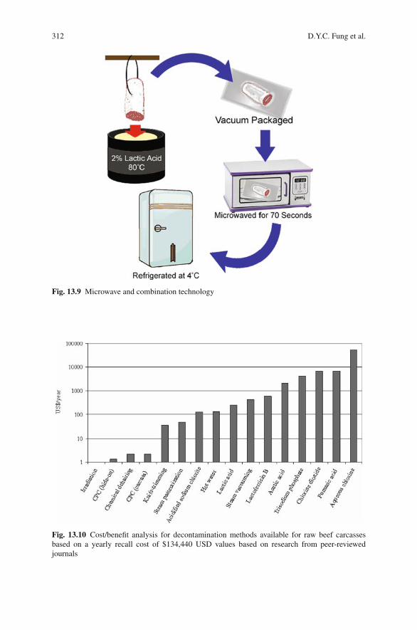
312 D.Y.C. Fung et al.
Fig. 13.9 Microwave and combination technology
Fig. 13.10 Cost/benefit analysis for decontamination methods available for raw beef carcassesbased on a yearly recall cost of $134,440 USD values based on research from peer-reviewedjournals
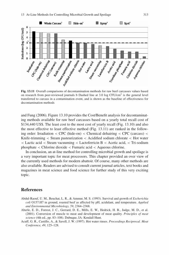
13 At-Line Methods for Controlling Microbial Growth and Spoilage 313
Fig. 13.11 Overall comparisons of decontamination methods for raw beef carcasses values basedon research from peer-reviewed journals b Dashed line at 3.0 log CFU/cm2 is the general leveltransferred to carcass in a contamination event, and is shown as the baseline of effectiveness fordecontamination methods
and Fung (2006). Figure 13.10 provides the Cost/Benefit analysis for decontaminat-ing methods available for raw beef carcasses based on a yearly total recall cost of$134,440 US$. The least cost to the most cost of yearly recall (Fig. 13.10) and alsothe most effective to least effective method (Fig. 13.11) are ranked in the follow-ing order: Irradiation < CPC (hide-on) < Chemical dehairing < CPC (carcass) <
Knife-trimming < Steam pasteurization < Acidified sodium chlorate < Hot water< Lactic acid < Steam vacuuming < Lactoferricin B < Acetic acid, < Tri-sodiumphosphate < Chlorine dioxide < Fumaric acid < Aqueous chlorine.
In conclusion, an at-line method for controlling microbial growth and spoilage isa very important topic for meat processors. This chapter provided an over view ofthe currently used methods for modern abattoir. Of course, many other methods arealso available. Readers are advised to consult current journal articles, text books andmagazines in meat science and food science for further study of this very excitingtopic.
References
Abdul-Raouf, U. M., Beuchat, L. R., & Ammur, M. S. (1993). Survival and growth of Escherichiacoli O157:H7 in ground, roasted beef as affected by pH, acidulant, and temperature. Appliedand Environmental Microbiology, 59, 2364–2368.
Aberle, E. D., Forrest, J. C., Gerrard, D. E., Mills, E. W., Hedrick, H. B., Judge, M. D., et al.(2001). Conversion of muscle to meat and development of meat quality. Principles of meatscience (4th ed., pp. 83–108). Dubuque, IA: Kendall Hunt.
Acuff, G. R., Castillo, A., & Savell, J. W. (1997). Hot water rinses. Proceedings Reciprocal. MeatConference, 49, 125–128.
314 D.Y.C. Fung et al.
Acuff, G. R., Vanderzant, C., Savell, J. W., Jones, D. K., Griffin, D. B., Ehlers, J. G., et al. (1987).Effect of acid decontamination of beef subprimal cuts on the microbiological and sensory char-acteristics of steaks. Meat Science, 19, 217–226.
Agricultural Marketing Service (AMS). (2000). Leather meal. AMS–USDA. http://www.ams.usda.gov/nop/NationalList/TAPReviews/LeatherMeal.pdf. Accessed 27 Oct 2005.
Al-Dagal, M., & Fung, D. Y. C. (1993). Aeromicrobiology: An assessment of a new meat researchcomplex. J. Environmental Health, 56, 7–14.
Anderson, M. E., Marshall, R. T., Stringer, W. C., & Naumann, H. D. (1977). Combined andindividual effects of washing and sanitizing on bacterial counts of meat – A model system.Journal of Food Protection, 40, 373–376.
Anderson, M. E., Sebaugh, J. L., Marshall, R. T., & Stringer, W. C. (1980). Amethod for decreasingsampling variance in bacteriological analyses of meat surfaces. Journal of Food Protection, 43,21–22.
Arthur, T. M., Wheeler, T. L., Shackelford, S. D., Bosilevac, J. M., Nou, X., & Koohmaraie, M.(2005). Effects of low-dose, low-penetration electron beam irradiation of chilled beef carcasssurface cuts on Escherichia coli O157:H7 and meat quality. Journal of Food Protection, 68,666–672.
Belk, K. E. (2001). Beef decontamination technologies. National Cattlemen’s Beef Association.http://www.beef.org/documents/ACFFC.pdf. Accessed 12 Feb 2005.
Bell, R. G. (1997). Distribution and sources of microbial contamination on beef carcasses. Journalof Applied Microbiology, 82, 292–300.
Bellamy, W., Wakabayashi, H., Takase, M., Kawase, K., Shimamura, S., & Tomita, M. (1993).Killing of Candida albicans by lactoferricin B, a potent antimicrobial peptide derived from theN-terminal region of bovine lactoferrin. Medical Microbiology Immunology, 182, 97–105.
Berg, J. D., Roberts, P. V., & Matin, A. (1986). Effect of chlorine dioxide on selected membranefunctions of Escherichia coli. The Journal of Applied Bacteriology, 60, 213–220.
Berry, E. D., & Cutter, C. N. (2000). Effects of acid adaptation of Escherichia coli O157:H7 on effi-cacy of acetic acid spray washes to decontaminate beef carcass tissue. Applied EnvironmentalMicrobiology, 66, 1493–1498.
Bosilevac, J. M., Arthur, T. M., Wheeler, T. L., Shackelford, S. D., Rossman, M., Reagan, J. O.,et al. (2004). Prevalence of Escherichia coli O157 and levels of aerobic bacteria and Enter-obacteriaceae are reduced when hides are washed and treated with cetylpyridinium chloride ata commercial beef processing plant. Journal of Food Protection, 67, 646–650.
Bosilevac, J. M., Nou, X., Osborn, M. S., Allen, D. M., & Koohmaraie, M. (2005). Developmentand evaluation of an on-line hide decontamination procedure for use in a commercial beefprocessing plant. Journal of Food Protection, 68, 265–272.
Bosilevac, J. M., Wheeler, T. L., Rivera-betancourt, M., Nou, X., Arthur, T. M., Shackelford, S. D.,et al. (2004). Protocol for evaluating the efficacy of cetylpyridinium chloride as a beef hideintervention. Journal of Food Protection, 67, 303–309.
Brackett, R. E., Hao, Y. Y., & Doyle, M. P. (1994). Ineffectiveness of hot acid sprays to decontam-inate Escherichia coli O157:H7 on beef. Journal of Food Protection, 57, 198–203.
Buchanan, R. L., & Doyle, M. P. (1997). Foodborne disease significance of Escherichia coliO157:H7 and other enterohemorrhagic E. coli. Food Technology, 51, 69–76.
Castillo, A., Lucia, L. M., Kemp, G. K., & Acuff, G. R. (1999). Reduction of Escherichia coliO157:H7 and Salmonella Typhimurium on beef carcass surfaces using acidified sodium chlo-rite. Journal of Food Protection, 62, 580–584.
Centers for Disease Control and Prevention (CDC). (1999). Safer and healthier foods – 1900–1999.Morbidity and Mortality Weekly Report, 48, 905–913.
Centers for Disease Control and Prevention (CDC). (2005). Food irradiation. CDC.http://www.cdc.gov/ncidod/dbmd/diseaseinfo/foodirradiation.htm. Accessed 27 Oct 2005.
Chad. (2004a). Hide-on carcass wash and sanitizing assembly. http://www.chadcompany.com/newproducts.html. Accessed 11 Oct 2005.
Chad. (2004b). Hot water pasteurization system. http://www.chadcompany.com/newproducts.html.Accessed 11 Oct 2005.
13 At-Line Methods for Controlling Microbial Growth and Spoilage 315
Chad. (2004c). Lactic acid spray assembly. http://www.chadcompany.com/ newproducts.html.Accessed 11 Oct 2005.
Chad. (2004d). Preevisceration carcass wash assembly and organic acid washer. http://www. chad-company.com/newproducts.html. Accessed 11 Oct 2005.
Clavero, M. R. S., Monk, J. D., Beuchat, L. R., Doyle, M. P., & Brackett, R. E. (1994). Inactivationof Escherichia coli O157:H7, salmonellae, and Campylobacter jejuni in raw ground beef bygamma irradiation. Applied Environmental of Microbiology, 60, 2069–2075.
Code of Federal Regulations (CFR). (2005a). Acidified sodium chlorite solutions. Code of FederalRegulations. 21 CFR 173, 325, 140–142.
Code of Federal Regulations (CFR). (2005b). Cetylpyridinium chloride. Code of Federal Regula-tions. 21 CFR 173, 375, 148–149.
Code of Federal Regulations (CFR). (2005c). Chlorine dioxide. Code of Federal Regulations. 21CFR 173, 300, 135–136.
Code of Federal Regulations (CFR). (2005d). Humane slaughter of livestock. Code of FederalRegulations. 9 CFR, 313, 145–151.
Code of Federal Regulations (CFR). (2005e). Ionizing radiation for the treatment of food. Code ofFederal Regulations. 21 CFR 179, 26, 451–453.
Crouse, J. D., Anderson, M. D., & Naumann, H. D. (1988). Microbial decontamination and weightof carcass beef as affected by automated washing pressure and length of time of spray. Journalof Food Protection, 51, 471–474.
Crozier-Dodson, B. (2000). Combined treatments of 2% lactic acid (80◦C), and microwaves forthe reduction of natural microflora and Escherichia coli O157:H7 on vacuum packaged beefsubprimals. MS Thesis, Hale Library, Kansas State University, Manhattan, KS.
Culkin, K. A., & Fung, D. Y. C. (1975). Destruction of Escherichia coli and SalmonellaTyphimurium in microwave-cooked soups. Journal of Milk and Food Technology, 38,8–15.
Curry, R. D., Unklesbay, K., Unklesbay, N., Clevenger, T. E., Brazos, B. J., Mesyats, G., et al.(2000). The effect of high-dose-rate X-rays on E. coli O157:H7 in ground beef. IEEE Transac-tions on Plasma Science, 28, 122–127.
Cutter, C. N., & Dorsa, W. J. (1995). Chlorine dioxide spray washes for reducing fecal contamina-tion on beef. Journal of Food Protection, 58, 1294–1296.
Cutter, C. N., Dorsa, W. J., Handie, A., Rodriguez-morales, S., Zhou, X., Breen, P. J., et al. (2000).Antimicrobial activity of cetylpyridinium chloride washes against pathogenic bacteria on beefsurfaces. Journal of Food Protection, 63, 593–600.
Derfler, P. S. (2004). FSIS directive: Verification of procedures for controlling fecal mate-rial, ingesta, and milk in slaughter operations. USDA FSIS. http://www.fsis.usda.gov/Frame/FrameRedirect.asp?main=/oppde/rdad/fsisdirectives/6420.2.pdf. Accessed 7 May 2005.
Dickson, J. S. (1991). Control of Salmonella Typhimurium, Listeria monocytogenes, andEscherichia coli O157:H7 on beef in a model spray chilling system. Journal of Food Science,56, 191–193.
Dickson, J. S., & Anderson, M. E. (1992). Microbiological decontamination of food animalcarcasses by washing and sanitizing systems: A review. Journal of Food Protection, 55,133–140.
Dickson, J. S., Nettles Cutter, C. G., & Siragusa, G. R. (1994). Antimicrobial effects of tri-sodiumphosphate against bacteria attached to beef tissue. Journal of Food Protection, 57, 952–955.
Dorsa, W. J., Cutter, C. N., Siragusa, G. R., & Koohmaraie, M. (1996). Microbial decontaminationof beef and sheep carcasses by steam, hot water spray washes and a steam vacuum sanitizer.Journal of Food Protection, 59, 127–135.
Edwards, J. R., & Fung, D. Y. C. (2006). Prevention and decontamination of Escherichia coliO157:H7 on raw beef carcasses in commercial beef abattoirs. Journal of Rapid Methods andAutomation in Microbiology, 14, 1–95.
Elder, R. O., Keen, J. E., Siragusa, G. R., Barkocy-Gallagher, G. A., Koohmaraie, M., & Laegreid,W. W. (2000). Correlation of enterohemorrhagic Escherichia coli O157:H7 prevalence in feces,hides, and carcasses of beef cattle during processing. PNSA 97, 2999–3003.
316 D.Y.C. Fung et al.
Fang, T. J., & Tsai, H. C. (2003). Growth patterns of Escherichia coli O157:H7 in ground beeftreated with nisin, chelators, organic acids and their combinations immobilized in calciumalginate gels. Food Microbiology, 20, 243–253.
Food Safety and Inspection Service (FSIS). (1994). Post-mortem inspection. http://www.fsis.usda.gov/PDF/PHVg-Post-mortem Inspection.pdf. Accessed 27 Oct 2005.
Food Safety and Inspection Service (FSIS). (1996a). Achieving the zero tolerance performancestandard for beef carcasses by knife trimming and vacuuming with hot water and steam; useof acceptable carcass interventions for reducing carcass contamination without prior agencyapproval: Notice of policy change. Federal Register, 61, 15024–15027.
Food Safety and Inspection Service (FSIS). (1996b). Pathogen reduction: Hazard analysis andcritical control point (HACCP) systems; final rule. Federal Register, 61, 38805–38989.
Food Safety and Inspection Service (FSIS). (2002). Backgrounder: New measures toaddress E. coli O157:H7 contamination. USDA FSIS. http://www.fsis.usda.gov/OA/ back-ground/ec0902.pdf. Accessed 27 March 2005.
Fu, A., Sarbanes, J. G., & Moreno, E. A. (1995). Survival of Listeria monocytogenes, Yersiniaenterocolitica, and Escherichia coli O157:H7 and quality changes after irradiation of beefsteaksand ground beef. Journal of Food Science, 60, 972–977.
Fung, D. Y. C and Cunningham, F. E. (1980). Effects of microwave cooking on microorganisms infoods. Journal of Food Protection, 43, 641–650.
Fung, D. Y. C., Hajmeer, M. N., Kastner, C. L., Kastner, J. J., Marsden, J. L., Penner, K. P., et al.(2001). Meat safety. In Y. H. Hui, W-K. Nip, R. W. Rogers, & O. W. Young (Eds.), Meat scienceapplications (pp. 171–205). New York, NY: Marcel Dekker.
Fung, D. Y. C., Kastner, C. L., Hunt, M. C., Dikeman, M. E., & Kropf, D. H. (1980). Mesophilicand psychrotrophic bacterial populations on hot-boned and conventionally processed beef.Journal of. Food Protection, 43, 547–550.
Fung, D. Y. C., Phebus, R. K., Kang, D. H., and Kastner, C. L. (1995). Effect of alcohol flam-ing on meat cutting knives. Journal of Rapid Methods and Automation in Microbiology, 3,237–243.
Gill, C. O. (1991). Microbial principles in meat processing. In J. B. Woolcock (Ed.), Microbiologyof animals and animal products (pp. 249–270). Amsterdam, The Netherlands: Elsevier.
Gill, C. O., McGinnis, J. C., & Badoni, M. (1995). Assessment of the hygienic characteristics of abeef carcass dressing process. Journal of Food Protection, 59, 136–140.
Gill, C. O., & Penney, N. (1979). Survival of bacteria in carcasses. Applied of EnvironmentalMicrobiology, 37, 667–669.
Gorman, B. M., Morgan, J. B., Sofos, J. N., & Smith, G. C. (1995). Microbiological and visualeffects of trimming and/or spray washing for removal of fecal material from beef. Journal ofFood Protection, 58, 984–989.
Gorman, B. M., Sofos, J. N., Morgan, J. B., Schmidt, G. R., & Smith, G. C. (1995). Evaluationof hand-trimming, various sanitizing agents, and hot water spray washing as decontaminationinterventions for beef brisket adipose tissue. Journal of Food Protection, 58, 899–907.
Haas, N. C. (2001). Decontamination using chlorine dioxide: Hearings on the decontaminationof anthrax and other biological agents. U.S. House of Representatives Committee on Science.http://www.house.gov/science/full/nov08/haas.htm. Accessed 1 Sep 2005.
Hamby, P. L., Savell, J. W., Acuff, G. R., Vanderzant, C., & Cross, H. R. (1987). Spray chillingand carcass decontamination systems using lactic and acetic acids. Meat Science, 21, 1–14.
Hardin, M. D., Acuff, G. R., Lucia, L. M., Oman, J. S., & Savell, J. W. (1995). Comparisonof methods for decontamination from beef carcass surfaces. Journal Food Protection, 58,368–374.
Hulebak, K. L., & Schlosser, W. S. (2001). HACCP history and conceptual overview. USDA FSIS.http://www.ce.ncsu.edu/risk/pdf/hulebak.pdf (accessed April 6, 2005).
Hwang, P. M., Zhou, N., Shan, X., Arrowsmith, C. H., & Vogel, H. J. (1998). Three-dimensionalsolution structure of lactoferricin B, and antimicrobial peptide derived from bovine lactoferrin.Biochemistry, 37, 4288–4298.
13 At-Line Methods for Controlling Microbial Growth and Spoilage 317
International Meat and Poultry HACCP Alliance (IMPHA). (1996). Generic HACCP model forbeef slaughter. USDA–FSIS. http://haccpalliance.org/alliance/haccpmodels/beefslaughter.pdf.Accessed 10 Oct 2005.
Johanson, L., Underdal, B., Grosland, K., Whelehan, O. P., & Roberts, T. A. (1983). A survey ofthe hygienic quality of beef and pork carcasses in Norway. Acta veterinaria Scandinavica, 25,1–13.
Kozempel, M., Goldberg, N., & Craig, J. J. C. (2003). The vacuum/steam/vacuum process. FoodTechnology, 57, 30–33.
Lim, K., & Mustapha, A. (2004). Effects of cetylpyridinium chloride, acidified sodium chlorite,and potassium sorbate on populations of Escherichia coli O157:H7, Listeria monocytogenes,and Staphylococcus aureus on fresh beef. Journal of Food Protection, 67, 310–315.
Masson, P. L., Heremans, J. F., & Dive, C. (1966). An iron-binding protein common to manyexternal secretions. Clinica Chimica Acta, 14, 735–739.
McEvoy, J. M., Doherty, A. M., Sheridan, J. J., Thomsoncarter, F. M., Garvey, P., McGuire, L.,et al. (2003). The prevalence and spread of Escherichia coli O157:H7 at a commercial beefabattoir. Journal of Applied Microbiology, 95, 256–266.
Milmech. (2004). Milmech Beef Downward Hide Puller. http://www.milmech.com/custom-slaughter.html. Accessed Oct 2005.
Nottingham, P. M., Penney, N., & Harrison, J. C. L. (1974). Microbiology of beef processing. NewZealand Journal of Agricultural Research, 17, 79–83.
Nou, X., Rivera-Betancourt, M., Bosilevac, J. M., Wheeler, T. L., Shackelford, S. D., Gwart-ney, B. L., et al. (2003). Effect of chemical dehairing on the prevalence of Escherichia coliO157:H7 and the levels of aerobic bacteria and Enterobacteriaceae on carcasses in a commer-cial beef processing plant. Journal of Food Protection, 66, 2005–2009.
Olsen, D. (2004). Food Irradiation update. Presented at the Meat Industry Research Conference(pp. 1–28).
Phebus, R. K., Nutsch, A. L., Schafer, D. E., Wilson, R. C., Riemann, M. J., Leising, J. D., et al.(1997). Comparisons of steam pasteurization and other methods for reduction of pathogens onsurfaces of freshly slaughtered beef. Journal of Food Protection, 60, 476–484.
Podolak, R. K., Zayas, J. F., Kastner, C. L., & Fung, D. Y. C. (1996). Inhibition of Listeria mono-cytogenes and Escherichia coli O157:H7 on beef by application of organic acids. Journal ofFood Protection, 59, 370–373.
Prasai, R. K., Phebus, R. K., Garcıa Zepeda, C. M., Kastner, C. L., Boyle, A. E., & Fung, D. Y. C.(1995). Effectiveness of trimming and/or washing on microbiological quality of beef carcasses.Journal of Food Protection, 58, 1114–1117.
Ransom, J. R., Belk, K. E., Sofos, J. N., Stopforth, J. D., Scanga, J. A., & Smith, G. C. (2003).Comparison of intervention technologies for reducing Escherichia coli O157:H7 on beef cutsand trimmings. Food Protection Trends, 23, 24–34.
Reagan, J. O., Acuff, G. R., Buege, D. R., Buych, M. J., Dickson, J. S., Kastner, C. L., et al. (1996).Trimming and washing of beef carcasses as a method of improving the microbiological qualityof meat. Journal of Food Protection, 59, 751–756.
Retzlaff, D., Phebus, R., Nutsch, A., Riemann, J., Kastner, C., & Marsden, J. (2004). Effectivenessof a laboratory-scale vertical tower static chamber steam pasteurization unit against Escherichiacoli O157:H7, Salmonella Typhimurium, and Listeria innocua on prerigor beef tissue. Journalof Food Protection, 67, 1630–1633.
Rice, E. W., Clark, R. M., & Johnson, C. H. (2003). Chlorine inactivation of Escherichiacoli O157:H7. CDC Emerging Infectious Diseases. http://www. cdc.gov/ncidod/EID/vol5no3/rice.htm. Accessed 12 Sep 2005.
Romans, J. R., Costello, W. J., Carlson, C. W., Greaser, M. L., & Jones, K. W. (2001). Cattleharvest. The Meat We Eat (14th ed., pp. 173–196). IL: Interstate Publishers.
Samelis, J., Sofos, J. N., Kendall, P. A., & Smith, G. C. (2001). Fate of Escherichia coli O157:H7,Salmonella Typhimurium DT 104, and Listeria monocytogenes in fresh meat decontaminationfluids at 4 and 10◦C. Journal of. Food Protection, 64, 950–957.
318 D.Y.C. Fung et al.
Samelis, J., Sofos, J. N., Kendall, P. A., & Smith, G. C. (2002). Effect of acid adaptation on survivalof Escherichia coli O157:H7 in meat decontamination washing fluids and potential effects oforganic acid interventions on the microbial ecology of the meat plant environment. Journal ofFood Protection, 65, 33–40.
Sawyer, C. A., Biglari, S. D., & Thompson, S. S. (1984). Internal end temperature and survivalof bacteria on meats with and without a polyvinyl chloride wrap during microwave cooking.Journal of Food Science, 49, 972–974.
Sheridan, J. J. (1998). Sources of contamination during slaughter and measures for control. Journalof Food Safety, 18, 321–339.
Shin, J., Chang, S., & Kang, D. (2004). Application of antimicrobial ice for reduction of foodbornepathogens (Escherichia coli O157:H7, Salmonella Typhimurium, Listeria monocytogenes) onthe surface of fish. Journal of Applied Microbiology, 97, 916–922.
Sofos, J. N. (1993). The HACCP system in meat processing and inspection in the United States.Meat Focus International, 2, 217–225.
Sofos, J. N., Belk, K. E., & Smith, G. C. (1999). Processes to reduce contamination with pathogenicmicroorganisms in meat. Congress of Meat Science and Technology, 45, 596–605.
Stanfos. (2004). Carcass pasteurizer. Stanfos, Inc. http://www.stanfos.com. Accessed 8 Oct 2005.Stopforth, J. D., Yoon, Y., Belk, K. E., Scanga, J. A., Kendall, P. A., Smith, G. C., et al. (2004).
Effect of simulated spray chilling with chemical solutions on acid habituated and non-acidhabituated Escherichia coli O157:H7 cells attached to beef carcass tissue. Journal of FoodProtection, 67, 2099–2106.
United States Department of Agriculture (USDA). (1993). National advisory committee of micro-biological criteria for foods, USDA – generic HACCP for raw beef. Food Microbiology, 10,449–488.
Warriner, K., Eveleigh, K., Goodman, J., Betts, G., Gonzales, M., & Waites, W. M. (2001). Attach-ment of bacteria to beef from steam pasteurized carcasses. Journal of Food Protection, 64,493–497.
Weller, K. (2003). ARS photo library. USDA Agriculture Research Service. http://www.ars.usda.gov/is/graphics/photos/oct00/k9082-14.htm. Accessed 8 Oct 2005.
Chapter 14The Detection of Genetically ModifiedOrganisms: An Overview
Jaroslava Ovesna, Katerina Demnerova, and Vladimıra Pouchova
Introduction
Genetically modified organisms (GMOs) are those whose genetic material has beenaltered by the insertion of a new gene or by the deletion of an existing one(s).Modern biotechnology, in particular, the rise of genetic engineering, has supportedthe development of GMOs suitable for research purposes and practical applications(Gepts, 2002; Novoselova, Meuwissen, & Huirne, 2007; Sakakibara & Saito, 2006).For over 20 years GM bacteria and other GM organisms have been used in laborato-ries for the study of gene functions (Maliga & Small, 2007; Ratledge & Kristiansen,2006). Agricultural plants were the first GMOs to be released into the environmentand placed on the market. Farmers around the world use GM soybeans, GM corn andGM cotton that are herbicide tolerant, or insect resistant, or combine several traitsthat reduce the costs associated with crop production (Corinne, Fernandez-Cornejo,& Goodhue, 2004). Biotech crop coverage increased globally by 13% (12 millionhectares) in 2005–06 (James, 2007), and, for example, in 2007 over 70% of allsoybean-producing areas were covered by GM varieties.
Although transgenesis of livestock began around 20 years ago, GM farm ani-mals, including fish, are still not as common as GM plants, the development ofwhich began somewhat earlier. Transgenic plants are most often developed by theinsertion of an alien (recombinant) gene using the soil bacteria, Agrobacteriumtumefaciens, which is able to transfer a piece of its own genetic information intoa plant cell. While GM plant development is at least partially based on naturallyoccurring mechanisms, the engineering of most transgenic livestock relies on highlytechnical approaches, such as pronuclear microinjection. However, newly developedtechniques [sperm mediated gene transfer (SGMT), somatic cell nuclear transfer(SCNT)] have been recently introduced that enable transgenic animals to be pro-duced more efficiently and more cheaply. These have been successfully appliedto the development of several types of GM animals including cattle, sheep, pigs,chicken and fish. The potential benefits of GM animals include accelerated animal
J. OvesnaCrop Research Institute, Drnovska 507, 161 06 Prague 6 – Ruzyne, Czech Republic
F. Toldra (ed.), Meat Biotechnology, 319C© Springer Science+Business Media, LLC 2008
320 J. Ovesna
growth, enhanced resistance to disease, and better meat quality (Niemann, Kues, &Carnwath, 2005).
The biomedical and agricultural communities have called for more research intothe use of viral vectors in the development of farmed GM animals in the hope thatmodified animals may prove more resistant to diseases (e.g. trypanosomiasis), aswell as to the cold. GM animals are able to produce human proteins in their milk oreggs, for e.g. GM chickens may produce as much as 0.1 g of human protein in eachegg. Apart from the potential pharmaceutical applications of such protein (e.g. USpatent 20060185029), it is also expected that GM animal products will follow GMplants and GM foodstuffs onto the food market in the near future (Wheeler, 2007).With GM animal products likely to appear on the consumer’s fork sometime soon,regulatory bodies have been set up to develop guidelines and discuss key issues(e.g. Codex Alimentarius) such as food safety, risk to the environment and ethicalconcerns. These discussions have resulted in regulations being developed for theprotection of consumer rights.
However, the situation is proving complicated to manage because GM technolo-gies and their derived products are perceived in different ways, both on a regionalbasis and by the various competing groups within a region. International organi-zations, such as WTO, OECD, FAO and Codex Alimentarius, are attempting toproduce a harmonized approach, but there are differences between the legislativeapproaches followed in North America, Asia and Europe.
To protect consumer rights in the EU, the placing of GM foods on the mar-ket is subject to “the precautionary principle” in EC food law (Just, Alston &Zilberman, 2006). Based on this principle, all GM products have to be proven safe inaccordance with the complex procedure prescribed by EC Regulation 1829/2003 ongenetically modified food and feed. The safety of each GM product is assessed bythe EFSA (European Food Safety Authority) GMO panel of experts, with infor-mation submitted by each applicant being subject to detailed risk analysis. Theavailability of validated detection and quantification methods is one of the pre-requisites for the acceptance of GM products. Applicants are required to submita specific method for the identification and quantification of the particular GMOthey wish to market. The method supplied must be verified by the CommunityReference Laboratory (CRL) at the Institute of Consumer Health and Protectionin Ispra, Italy (http://ihcp.jrc.ec.europa.eu), and validated in collaborative trialsconducted by laboratories affiliated to the European Network of GMO Laborato-ries (ENGL). Once the method has been validated it is published at http://gmocrl.jrc.it/statusofdoss.htm. Apart from the method, an applicant must also provide asample of the control material to the CRL, together with, on a confidential basis,information about the DNA sequence inserted into the genome of the modifiedorganism. Additional legislation covers GM handling procedures (Table 14.1); thewhole system being based on the traceability and appropriate labeling of approvedGMO products. (In the EU, all products containing more than 0.9% of GM materialas ‘unavoidable contamination of a constituent’ have to be labeled.)
The need to be able to trace GMOs and their derived products on the market hasgenerated a demand for analytical methods capable of reliably detecting, identifying
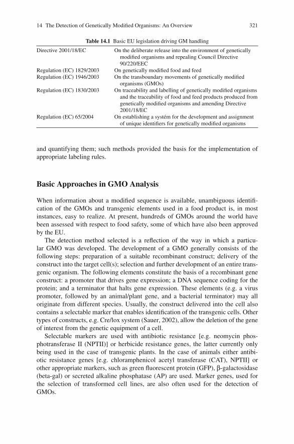
14 The Detection of Genetically Modified Organisms: An Overview 321
Table 14.1 Basic EU legislation driving GM handling
Directive 2001/18/EC On the deliberate release into the environment of geneticallymodified organisms and repealing Council Directive90/220/EEC
Regulation (EC) 1829/2003 On genetically modified food and feedRegulation (EC) 1946/2003 On the transboundary movements of genetically modified
organisms (GMOs)Regulation (EC) 1830/2003 On traceability and labelling of genetically modified organisms
and the traceability of food and feed products produced fromgenetically modified organisms and amending Directive2001/18/EC
Regulation (EC) 65/2004 On establishing a system for the development and assignmentof unique identifiers for genetically modified organisms
and quantifying them; such methods provided the basis for the implementation ofappropriate labeling rules.
Basic Approaches in GMO Analysis
When information about a modified sequence is available, unambiguous identifi-cation of the GMOs and transgenic elements used in a food product is, in mostinstances, easy to realize. At present, hundreds of GMOs around the world havebeen assessed with respect to food safety, some of which have also been approvedby the EU.
The detection method selected is a reflection of the way in which a particu-lar GMO was developed. The development of a GMO generally consists of thefollowing steps: preparation of a suitable recombinant construct; delivery of theconstruct into the target cell(s); selection and further development of an entire trans-genic organism. The following elements constitute the basis of a recombinant geneconstruct: a promoter that drives gene expression; a DNA sequence coding for theprotein; and a terminator that halts gene expression. These elements (e.g. a viruspromoter, followed by an animal/plant gene, and a bacterial terminator) may alloriginate from different species. Usually, the construct delivered into the cell alsocontains a selectable marker that enables identification of the transgenic cells. Othertypes of constructs, e.g. Cre/lox system (Sauer, 2002), allow the deletion of the geneof interest from the genetic equipment of a cell.
Selectable markers are used with antibiotic resistance [e.g. neomycin phos-photransferase II (NPTII)] or herbicide resistance genes, the latter currently onlybeing used in the case of transgenic plants. In the case of animals either antibi-otic resistance genes [e.g. chloramphenicol acetyl transferase (CAT), NPTII] orother appropriate markers, such as green fluorescent protein (GFP), β-galactosidase(beta-gal) or secreted alkaline phosphatase (AP) are used. Marker genes, used forthe selection of transformed cell lines, are also often used for the detection ofGMOs.
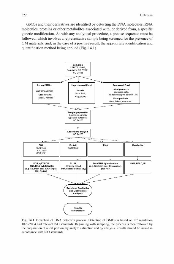
322 J. Ovesna
GMOs and their derivatives are identified by detecting the DNA molecules, RNAmolecules, proteins or other metabolites associated with, or derived from, a specificgenetic modification. As with any analytical procedure, a precise sequence must befollowed, which involves a representative sample being screened for the presence ofGM materials, and, in the case of a positive result, the appropriate identification andquantification method being applied (Fig. 14.1).
Fig. 14.1 Flowchart of DNA detection process. Detection of GMOs is based on EC regulation1829/2004 and relevant ISO standards. Beginning with sampling, the process is then followed bythe preparation of a test portion, by analyte extraction and by analysis. Results should be issued inaccordance with ISO standards
14 The Detection of Genetically Modified Organisms: An Overview 323
Sampling
Sampling and sample preparation are crucial steps in the process of GMO detection(Terry, Harris & Parkes, 2002). While comprehensive analytical procedures exist todeliver precise results, the ability of a sampling procedure in general, and a samplesize in particular, to erroneously influence final values should not be underestimated.The sampling procedure determines the representativeness of a result. In the case ofGM material, because the distribution of GM particles in a lot is usually randomand heterogeneous, a distribution-free statistical model has been suggested (Paolettiet al., 2006), and models have been developed that combine information about allstages of GMO event detection (Macarthur et al., 2007).
Being closely connected with statistical requirements, sample size has often beenthe subject of considerable discussion. It must be large enough to enable reliableGM detection at the desired sensitivity. A sample containing at least 3,500 particlesis necessary to ensure that a 1% concentration of GMO in a batch will be reliablydetected with a probability of p ≤ 0.05. To obtain this probability, in the case oflow level contamination, e.g. 0.1%, a test sample should contain 10 times moreparticles. Processed foods and complex matrices contain GM materials as one ofseveral ingredients, so strongly stratified variance distribution can be expected. Thisis why it not always possible to use existing sampling strategies for the detection ofother components in food commodities (Berger & von Holst, 2001).
General principles and rules for the sampling of GM foodstuffs are describedin more detail in ISO 21568, Foodstuffs - Methods of analysis for the detection ofgenetically modified organisms and derived products -Sampling. In accordance withthis procedure, samples delivered to laboratories must be stored under appropriateconditions, and further test portions prepared. A test portion should reflect the con-centration of GM material in the sample. A laboratory will analyze at least two testportions prepared from the identical sample. Analyte processing then follows.
Analysis of GM Material
The detection of GM materials is based on the identification of the DNA sequence,RNA sequence, protein sequence, or even metabolites, all of which result fromgenetic modification (Table 14.2). Methods differ with respect to their analyticalparameters, feasibility and cost-effectiveness. Only DNA- and protein-based meth-ods are currently used for control purposes.
Protein-Based Analysis
Typically, the production of specific proteins distinguishes GMOs from non-GMOs.Intact proteins are the required target for protein-based analysis; in the caseof GMOs, by using immunoassays. Immunoassays are analytical measurement
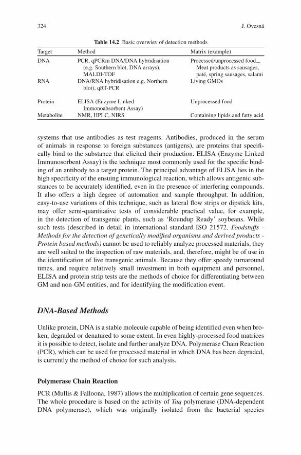
324 J. Ovesna
Table 14.2 Basic overwiev of detection methods
Target Method Matrix (example)
DNA PCR, qPCRm DNA/DNA hybridisation(e.g. Southern blot, DNA arrays),MALDI-TOF
Processed/unprocessed foodMeat products as sausages,pate, spring sausages, salami
RNA DNA/RNA hybridisation e.g. Northernblot), qRT-PCR
Living GMOs
Protein ELISA (Enzyme LinkedImmonoabsorbent Assay)
Unprocessed food
Metabolite NMR, HPLC, NIRS Containing lipids and fatty acid
systems that use antibodies as test reagents. Antibodies, produced in the serumof animals in response to foreign substances (antigens), are proteins that specifi-cally bind to the substance that elicited their production. ELISA (Enzyme LinkedImmunosorbent Assay) is the technique most commonly used for the specific bind-ing of an antibody to a target protein. The principal advantage of ELISA lies in thehigh specificity of the ensuing immunological reaction, which allows antigenic sub-stances to be accurately identified, even in the presence of interfering compounds.It also offers a high degree of automation and sample throughput. In addition,easy-to-use variations of this technique, such as lateral flow strips or dipstick kits,may offer semi-quantitative tests of considerable practical value, for example,in the detection of transgenic plants, such as ‘Roundup Ready’ soybeans. Whilesuch tests (described in detail in international standard ISO 21572, Foodstuffs -Methods for the detection of genetically modified organisms and derived products -Protein based methods) cannot be used to reliably analyze processed materials, theyare well suited to the inspection of raw materials, and, therefore, might be of use inthe identification of live transgenic animals. Because they offer speedy turnaroundtimes, and require relatively small investment in both equipment and personnel,ELISA and protein strip tests are the methods of choice for differentiating betweenGM and non-GM entities, and for identifying the modification event.
DNA-Based Methods
Unlike protein, DNA is a stable molecule capable of being identified even when bro-ken, degraded or denatured to some extent. In even highly-processed food matricesit is possible to detect, isolate and further analyze DNA. Polymerase Chain Reaction(PCR), which can be used for processed material in which DNA has been degraded,is currently the method of choice for such analysis.
Polymerase Chain Reaction
PCR (Mullis & Falloona, 1987) allows the multiplication of certain gene sequences.The whole procedure is based on the activity of Taq polymerase (DNA-dependentDNA polymerase), which was originally isolated from the bacterial species
14 The Detection of Genetically Modified Organisms: An Overview 325
Thermus aquaticus. This enzyme, which basically requires a DNA template (as shortas 200 pb is sufficient) and pieces of single-stranded DNA (primers) surroundingthe target sequence, is able to produce (amplify) millions of copies of the part ofthe genetic information that is to be analyzed. PCR is a cyclical process, each cycleconsisting of three successive steps running at different temperatures. The first stepin a cycle involves the separation of the two strands of the original DNA molecule.The second step involves the binding of the two primers to their complementarystrands. Using the complementary strands as a template, the third step involvesmaking two copies of the original double-stranded DNA molecule by adding theright nucleotides. Once a cycle is completed, it can be repeated, typically between30–50 times. With the number of copies doubling in each cycle, the number oftarget sequences grows exponentially according to the number of cycles undergone(Holst Jensen, 2004).
If an analyte contains a target sequence, for e.g. part of the transgene- or species-specific internal gene, amplification products can be subsequently visualized and/orquantified. The whole procedure consists of: (1) DNA extraction and purification,(2) amplification of the target sequence, (3) visualization by electrophoresis or fluo-rogenes detection, (4) quantification and (5) interpretation of results. The applicationof PCR to GMO analysis has been described in the literature (Lipp et al., 2005;Miraglia et al., 2004).
DNA Isolation
DNA quality, in particular its purity, and DNA quantity are critical parameters forGMO analysis. DNA can be isolated either as high molecular weight DNA obtainedfrom fresh material (e.g. blood, grains) or as fragmented DNA obtained from elderlyor processed matrices (e.g. bone parts, flour, sausages, pate).
The basic steps necessary to accomplish DNA isolation have been described byseveral authors (Somma 2004; Spoth & Strauss, 1999). DNA isolation begins withhomogenization of the test portion, grinding and cell disruption. Cell disruption,which results from homogenization followed by the addition of a buffer consisting,amongst other reagents, of a detergent (e.g. SDS or CTAB) and DNase inhibitors(β-mercaptoethanol and EDTA), enables the transfer of DNA from a cell into thebuffer. The mixture is further purified as cell debris, proteins and RNA from theextract are all removed. DNA is then precipitated. DNA can be further purified byappropriate procedures, such as the use of commercial kits based, for example, ongel filtration.
Several variations of this basic procedure exist. For example, PVP-based methodsare particularly suitable for DNA extraction from raw or boiled meat, sausages,chopped meat and processed meat products containing soybean protein. CTAB-based methods have been shown to be efficient for the isolation of DNA from gravy,hamburgers, fatty salami and ham. All of these methods have been validated in col-laborative trials, and are described in detail in ISO 21 571 “Foodstuffs - Methods ofanalysis for the detection of genetically modified organisms and derived products -Nucleic acid extraction”.
326 J. Ovesna
The resulting DNA should meet the quality criteria. Typically, its absorbance ismeasured in UV light, which enables the presence of any contaminating proteinsand/or phenolic compounds to be identified. Gel electrophoresis is used to detectthe presence of contaminating RNAs that may interfere with upstream reactions.
PCR Analysis
In order to use PCR, the precise nucleotide sequences flanking both ends of thetarget DNA region must be known. Any PCR-based detection strategy is depen-dent on a detailed knowledge of the transgenic DNA sequence, which, in the caseof authorized GMOs, can be found in the published databases (e.g. http://www.agbios.com/dbase.php). It also depends on the selection of the appropriate oligonu-cleotide primers. In addition to the well-documented issues concerning primer selec-tion (Burpo, 2001), the choice of primers depends on the objectives of PCR analysis.
Once the DNA is isolated, its amplificability must be ascertained. Usually, aspecies-specific sequence is used. Species-specific genes (e.g. lectin from soybeans,zein from corn) should be represented by one copy per haploid genome. Protocolsexist for the amplification of many species-specific sequences, and may be used forcontrol purposes (Von Holst, Baeten, Berben, & Brambilla, 2004)
If the isolated DNA is amplifiable, screening methods are usually applied toexclude negative samples from subsequent analysis. Screening tests for the commontransgenic are executed elements (e.g. promoters, terminators, selectable markers)present in some GMOs. In the case of GM plants, the 35S CaMV promoter (35Ssubunit of the cauliflower mosaic virus) and the T-NOS terminator (from the nopa-line strain of Agrobacterium tumefaciens) are used for screening. However, thismethod is not GMO-specific, and contamination of the commodities by either of thetwo organisms (CaMV and A. tumefaciens) may lead to false positives. Therefore,in case of a positive result, further specific tests must be performed to verify thefindings.
With the increasing number of GMOs in the market, not all kinds of GMOscan be reliably detected using existing screening methods. Some transgenic codingsequences might also occur across multiple GM varieties or species. If the primersare specific to a transgene occurring in multiple GMs it becomes difficult to locatethe original source of the GM material in complex matrices. Construct-specificmethods, involving, for example, one primer specific to the promoter and anotherspecific to the coding sequence, may help to identify GMOs more accurately. How-ever, event-specific methods, spanning transgene- and species-specific sequences,remain desirable for GM plant identification Fig. 14.2 (Yang et al., 2007).
Separation (either on agarose gel or by using PAGE) followed by visualizationby UV light enable the amplification products to be easily identified and evalu-ated (Fig. 14.3). Because each amplification product is characterized by its spe-cific size, it can be identified by checking it against a molecular ladder and byusing positive controls. The most respected control materials are the certified ref-erence materials produced by the Institute for Reference Material and Measurement(IRMM), an EU Joint Research Institution (JRC) located in Geel, Belgium. Also,
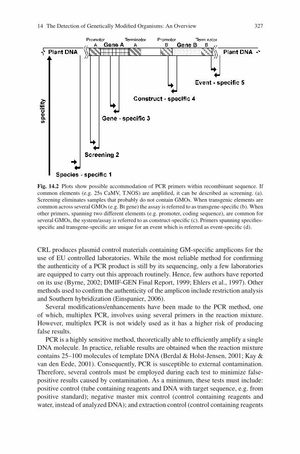
14 The Detection of Genetically Modified Organisms: An Overview 327
Fig. 14.2 Plots show possible accommodation of PCR primers within recombinant sequence. Ifcommon elements (e.g. 25s CaMV, T.NOS) are amplified, it can be described as screening. (a).Screening eliminates samples that probably do not contain GMOs. When transgenic elements arecommon across several GMOs (e.g. Bt gene) the assay is referred to as transgene-specific (b). Whenother primers, spanning two different elements (e.g. promoter, coding sequence), are common forseveral GMOs, the system/assay is referred to as construct-specific (c). Primers spanning specifies-specific and transgene-specific are unique for an event which is referred as event-specific (d).
CRL produces plasmid control materials containing GM-specific amplicons for theuse of EU controlled laboratories. While the most reliable method for confirmingthe authenticity of a PCR product is still by its sequencing, only a few laboratoriesare equipped to carry out this approach routinely. Hence, few authors have reportedon its use (Byrne, 2002; DMIF-GEN Final Report, 1999; Ehlers et al., 1997). Othermethods used to confirm the authenticity of the amplicon include restriction analysisand Southern hybridization (Einspanier, 2006).
Several modifications/enhancements have been made to the PCR method, oneof which, multiplex PCR, involves using several primers in the reaction mixture.However, multiplex PCR is not widely used as it has a higher risk of producingfalse results.
PCR is a highly sensitive method, theoretically able to efficiently amplify a singleDNA molecule. In practice, reliable results are obtained when the reaction mixturecontains 25–100 molecules of template DNA (Berdal & Holst-Jensen, 2001; Kay &van den Eede, 2001). Consequently, PCR is susceptible to external contamination.Therefore, several controls must be employed during each test to minimize false-positive results caused by contamination. As a minimum, these tests must include:positive control (tube containing reagents and DNA with target sequence, e.g. frompositive standard); negative master mix control (control containing reagents andwater, instead of analyzed DNA); and extraction control (control containing reagents
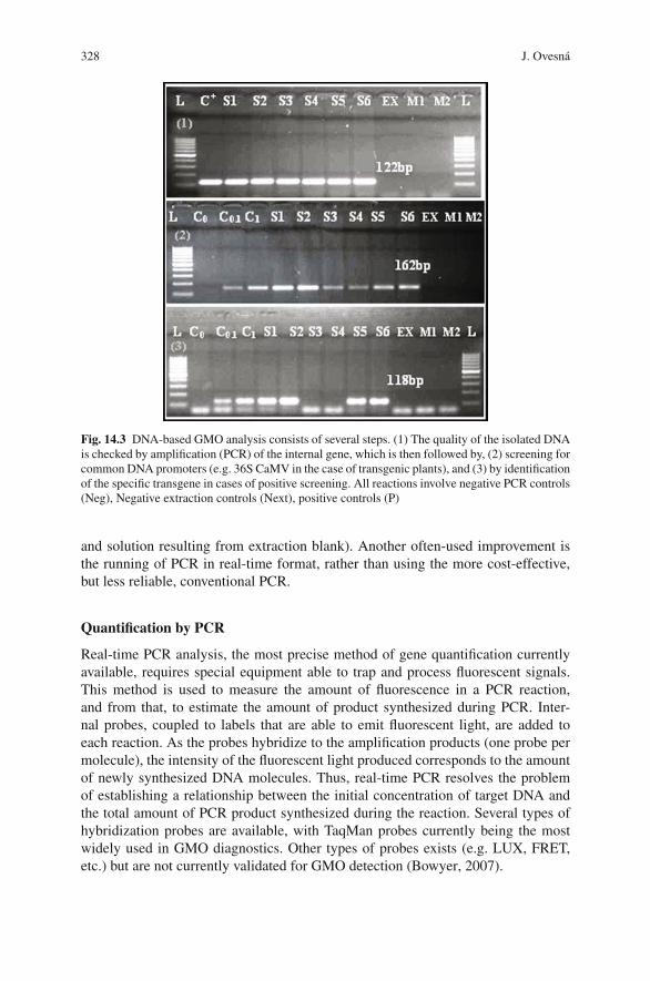
328 J. Ovesna
Fig. 14.3 DNA-based GMO analysis consists of several steps. (1) The quality of the isolated DNAis checked by amplification (PCR) of the internal gene, which is then followed by, (2) screening forcommon DNA promoters (e.g. 36S CaMV in the case of transgenic plants), and (3) by identificationof the specific transgene in cases of positive screening. All reactions involve negative PCR controls(Neg), Negative extraction controls (Next), positive controls (P)
and solution resulting from extraction blank). Another often-used improvement isthe running of PCR in real-time format, rather than using the more cost-effective,but less reliable, conventional PCR.
Quantification by PCR
Real-time PCR analysis, the most precise method of gene quantification currentlyavailable, requires special equipment able to trap and process fluorescent signals.This method is used to measure the amount of fluorescence in a PCR reaction,and from that, to estimate the amount of product synthesized during PCR. Inter-nal probes, coupled to labels that are able to emit fluorescent light, are added toeach reaction. As the probes hybridize to the amplification products (one probe permolecule), the intensity of the fluorescent light produced corresponds to the amountof newly synthesized DNA molecules. Thus, real-time PCR resolves the problemof establishing a relationship between the initial concentration of target DNA andthe total amount of PCR product synthesized during the reaction. Several types ofhybridization probes are available, with TaqMan probes currently being the mostwidely used in GMO diagnostics. Other types of probes exists (e.g. LUX, FRET,etc.) but are not currently validated for GMO detection (Bowyer, 2007).
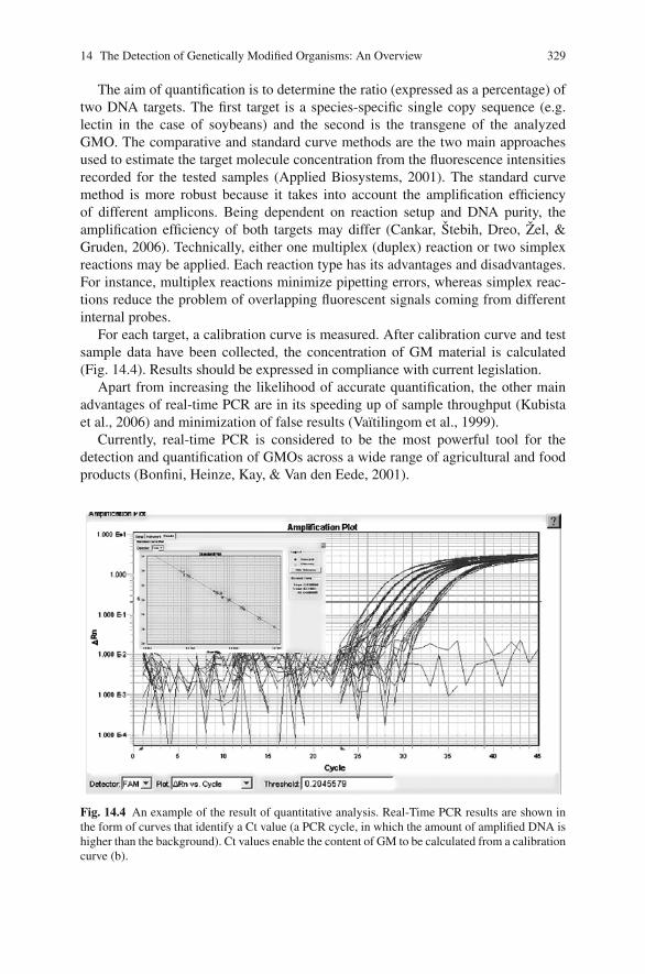
14 The Detection of Genetically Modified Organisms: An Overview 329
The aim of quantification is to determine the ratio (expressed as a percentage) oftwo DNA targets. The first target is a species-specific single copy sequence (e.g.lectin in the case of soybeans) and the second is the transgene of the analyzedGMO. The comparative and standard curve methods are the two main approachesused to estimate the target molecule concentration from the fluorescence intensitiesrecorded for the tested samples (Applied Biosystems, 2001). The standard curvemethod is more robust because it takes into account the amplification efficiencyof different amplicons. Being dependent on reaction setup and DNA purity, theamplification efficiency of both targets may differ (Cankar, Stebih, Dreo, Zel, &Gruden, 2006). Technically, either one multiplex (duplex) reaction or two simplexreactions may be applied. Each reaction type has its advantages and disadvantages.For instance, multiplex reactions minimize pipetting errors, whereas simplex reac-tions reduce the problem of overlapping fluorescent signals coming from differentinternal probes.
For each target, a calibration curve is measured. After calibration curve and testsample data have been collected, the concentration of GM material is calculated(Fig. 14.4). Results should be expressed in compliance with current legislation.
Apart from increasing the likelihood of accurate quantification, the other mainadvantages of real-time PCR are in its speeding up of sample throughput (Kubistaet al., 2006) and minimization of false results (Vaıtilingom et al., 1999).
Currently, real-time PCR is considered to be the most powerful tool for thedetection and quantification of GMOs across a wide range of agricultural and foodproducts (Bonfini, Heinze, Kay, & Van den Eede, 2001).
Fig. 14.4 An example of the result of quantitative analysis. Real-Time PCR results are shown inthe form of curves that identify a Ct value (a PCR cycle, in which the amount of amplified DNA ishigher than the background). Ct values enable the content of GM to be calculated from a calibrationcurve (b).
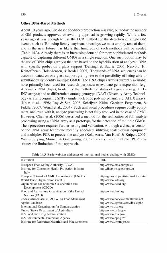
330 J. Ovesna
Other DNA-Based Methods
About 10 years ago, GM-based food/feed production was rare, but today the numberof GM products approved or awaiting approval is growing rapidly. While a fewyears ago it was enough to use the PCR method for the detection of single GMevents, such as ‘Roundup Ready’ soybean, nowadays we must employ tens of them,and in the near future it is likely that hundreds of such methods will be needed(Table 14.3). Already there is an increasing demand for more sophisticated methodscapable of capturing different GMOs in a single reaction. One such option may bethe use of DNA chips (arrays) that are based on the hybridization of analyzed DNAwith specific probes on a glass support (Deisingh & Badrie, 2005; Nesvold, H.,Kristoffersen, Holst-Jensen, & Berdal, 2005). Thousands of DNA sequences can beaccommodated on one glass support giving rise to the possibility of being able tosimultaneously identify multiple GMOs. The DNA chips (arrays) currently availablehave primarily been used for research purposes: to evaluate gene expression (e.g.Affymetix DNA chips); to identify the methylation status of a genome (e.g. TILL-ING arrays); and to differentiate among genotype [DArT (Diversity Array Technol-ogy) arrays recognizing SNPs (single nucleotide polymorphism), e.g. APEX arrays](Khan et al., 1998; Roy & Sen, 2006; Schrijver, Kulm, Gardner, Pergament, &Fiddler, 2007; Wenzl et al., 2004). Such analytical procedures require costly equip-ment, and even with it, analyte processing is not fully resolved in the case of GMO.However, Chen et al. (2006) described a method for the realization of full analyteprocessing using a cDNA array as a prototype for the detection of multiple GMOs.Their procedure requires further testing and validation. Although a cheaper versionof the DNA array technique recently appeared, utilizing scaled-down equipmentand multiplex PCR to process the analyte (Kok, Aarts, Van Hoef, & Kuiper, 2002;Wenijn, Siyang, Minnan, & Guangming, 2003), the very use of multiplex PCR con-stitutes the limitation of this approach.
Table 14.3 Basic websites addresses of international bodies dealing with GMOs
Institution URL
European Food Safety Authority (EFSA) http://www.efsa.europa.euInstitute for Consumer Health Protection in Ispra,
Italyhttp://ihcp.jrc.ec.europa.eu
Europen Network of GMO Laboratories. (ENGL) http://gmo-crl.jrc.it/statusofdoss.htmWorld Trade Organization (WTO) http://www.wto.orgOrganization for Eonomic Co-operation and
Development (OECD)http://www.oecd.org
Food and Agriculture Organization of the UnitedNations (FAO)
http://www.fao.org
Codex Alimentarius (FAO/WHO Food Standards) http://www.codexalimentarius.netAgbios database http://www.agbios.com/dbase.phpInternational Organization for Standardization http://www.iso.orgUnited States Department of Agriculture http://www.usda.govU.S.Food and Drug Administration http://www.fda.gov/U.S.Environmental Protection Agency http://www.epa.gov/Institute for Reference Materials and Measurements http://www.irmm.jrc.be
14 The Detection of Genetically Modified Organisms: An Overview 331
Another approach, based on Ligation Probe Amplification (LPA), has beenrecently introduced. LPA allows simultaneous event-specific detection and the rela-tive quantification of DNA from several GMOs in a single reaction. However, thisapproach requires careful primer setup and the optimization of reaction conditionsjust for the system to detect a mere 40–50 targets per reaction (Carrino, 1996;Mezzelani et al., 2002).
Novel approaches to DNA analysis based on micro and nanoparticles, as well ason mass spectrometry, may open new possibilities in DNA detection. The possibleadvantages of some of these approaches have been described by Sato, Hosokawa,and Maeda (2007); Einspanier (2006), amongst others.
Other Methods of GMO Detection
Where the composition of GMO, e.g. fatty acids or triglycerides, has been mod-ified, conventional chemical methods based on chromatography can be used todetect differences in the chemical profile. Metabolites, where they are the productof transgenic protein activity, can be detected by physical methods, such as NMR(Nuclear Magnetic Resonance), HPLC (High Performance Liquid Chromatography)or Near Infrared Spectroscopy (NIRS) (Heller, 2006; Roussel, Hardy, Hurburgh, &Rippke, 2001; Von Holst et al., 2004).
Data Quality and Interpretation
Any procedure used in GMO analysis for control purposes should be based on val-idated methods, some of which have been published by ISO (International Organi-zation for Standardization http://www.iso.org). All methods should be reproducible,repeatable, precise and accurate and should reflect the true value. These analyticalparameters are critical when applied to any method used for the detection of GMOsin foodstuffs. The limits of detection (LOD) and quantification (LOQ) should bedetermined in accordance with the method being used and the matrix of the sam-ple being analyzed (Berdal & Holst-Jensen, 2001). Full details of how to reportand issue results are described in ISO 24276: “Foodstuffs - Methods of analysisfor the detection of genetically modified organisms and derived products - Generalrequirements and definitions”. To ensure data quality, all control laboratories shouldbe accredited according to the ISO 17025:2005 (Zel, Cankar, Ravnikar, Camloh, &Gruden, 2006).
Despite the prescribed procedures, the Food Analysis Performance Scheme(FAPAS), which operates a proficiency testing scheme for GM food analyses inthe UK, has reported the following findings: a tendency for laboratories to overes-timate GM levels; significant differences between the results obtained using PCRand ELISA; data being skewed and not normally distributed until undergoing math-ematical treatment (Powell & Owen, 2002).
332 J. Ovesna
These findings indicate the need to better understand the processes underlyingGM detection methods, as well as the need for considerable investment in theresearch of more advanced techniques. Further development of methods used forthe detection and quantification of GMOs in agricultural commodities is essentialto monitor the implementation of regulatory compliance concerning the use of GMfood/feed. Such research should focus on improving the analytical parameters ofhigh-throughput methods so that they are fit for the purpose.
It is likely that new methods will not only be used to control GMOs and accom-modate consumer requirements, in some countries, but also to protect breeders’rights and intellectual properties worldwide.
Acknowledgements This work was supported by the project CZ0002700602 of the Czech Min-istry of Agriculture and by project the 1B44068 of the National Agency for Agricultural Research.
References
Applied Biosystems. (2001). Relative Quantification of Gene Expression. Applied Biosystems.User Bulletin 2.
Berdal, K. G., & Holst-Jensen, A. (2001). Effect of food components and processing parameterson DNA degradation in food. European Food Research and Technology, 213, 432–438.
Berger, B., & von Holst C. (2001). Pesticides residues in products of plant origin in the EuropeanUnion: Sampling strategy and results from the co-ordinated EU monitoring programme in 1996and 1997. Environmental Science and Pollution Research International, 8, 109–112.
Bonfini, L., Heinze, P., Kay, S., & Van den Eede, G. L. M. (2002). Review of GMO Detection andQuantification Techniques. EUR 20384/EN.
Bowyer, V. L. (2007). Real-time PCR. Forensic Science, Medicine, and Pathology, 3, 61–63.Burpo F. J. (2001). A critical review of PCR primer design algorithms and crosshybridiza-
tioncase study, Biochemistry 218 at Stanford University. Retrieved August 11, 2001, fromhttp://cmgm.stanford.edu/biochem218/Projects%202001/Burpo.pdf.
Byrne, D. (2002). Commission Recommendation of 25 January 2002 concerning a coordinatedprogramme for the official control of foodstuffs for 2002. Official Journal of the EuropeanCommunities 30.1.2002 L 26/8. Retrieved January 30, 2002, from http://www.sapidlife.org/documenti repository/scarica.php?file=en200612071039152002 66 CEEN.pdf.
Cankar, K., Stebih, D., Dreo, T., Zel, J., & Gruden, K. (2006). Critical points of DNA quantificationby real-time PCR – effects of DNA extraction method and sample matrix on quantification ofgenetically modified organisms. BMC Biotechnology, 6: 37.
Carrino, J. J. (1996). Multiplex ligations-dependent amplication. Patent aplication WO 96/15271.Chen, T. L., Sanjaya, V., Prasad, C. H., Lee, K. H., Lin, H. C., Chiueh,V., et al. (2006). Validation
of cDNA microarray as a prototype for throughput detection of GMOs. Botanical Studies, 47,1–11.
Corinne, A., Fernandez-Cornejo, J., & Goodhue, R. E. (2004). Farmers’ Adoption of Geneti-cally Modified Varieties with Input Traits. Giannini Foundation Research Report 347 (October2003). University of California: Division of Natural Resources. Retrieved October, 2003, fromhttp://giannini.ucop.edu/researchreports/347-goodhue.pdf.
Deisingh, A. K., & Badrie, N. (2005). Detection approaches for genetically modified organisms infoods. Food Research International, 38, 639–649.
DMIF-GEN Final Report. (1999). Retrieved February 1, 2000, from http://www.dmif-gen.bats.ch/dmif-gen/.
14 The Detection of Genetically Modified Organisms: An Overview 333
Ehlers, B., Strauch, E., Goltz, M., Kubsch, D., Wagner, H., Maidhof, H., et al. (1997). Nachweisgentechnischer Veranderungen in Mais mittels PCR. Bundesgesundhbl., 4, 118–121.
Einspanier, R. (2006). DNA based methods for detection of genetic modification. In K. J. Heller(Ed.), Genetically engineered food: Methods and detection (pp. 163–185, 2nd ed.). Wiley-VCHVerlag, GmbH & Co. KGaA., Weinheim, Germany
Gepts, P. (2002). A comparison between crop domestication, classical plant breeding, and geneticengineering. Crop Science, 42, 1780–1790.
Heller, K. J. (2006). Detection of Genetic Modifications-Some Basic Considerations Geneticallyengineered food methods and detection (pp. 155–162, 2nd ed.) Wiley-VCH Verlag GmbH.,Weinheim, Germany
Holst-Jensen, A. (2004). Detecting genetically modified food. New Food, 7, 16, 18–20, 22.James, C. (2007). Global Status of Commercialized Biotech. ISAAA Briefs No. 35-2006: /GM
Crops: 2006. Retrieved October 16, 2007, from http://www.isaaa.org/Resources/ Publica-tions/briefs/35/executivesummary/default.html.
Just, R. E., Alston J. M., & Zilberman. (2006). Regulating agricultural biotechnology: Economicsand policy. Natural Resource Management and Policy, 30, 392–402.
Kay, S., & Van Den Eede, G. (2001). The limits of GMO detection. National Biotechnology,19, 405.
Khan, et al. (1998). Gene expression profiling of alveolar rhabdomyosarcoma with cDNA microar-rays. Cancer Research, 58, 5009–5013.
Kok, E. J., Aarts, H. J., Van Hoef, A. M., & Kuiper, H. A. (2002). DNA methods: Critical reviewof innovative approaches. Journal of AOC International, 85, 797–800.
Kubista, M., Andrade, J. M., Bengtsson, M., Forootan, A., Jonak, J., Lind, K., et al. (2006). Thereal-time polymerase chain reaction. Molecular Aspects of Medicine, 27, 95–125.
Lipp, M., Shillito, R., Giroux, R., Spiegelhalter, F., Charlton, S., Pinero, D., et al. (2005). Poly-merase chain reaction technology as analytical tool in agricultural biotechnology. Journal ofAOC International, 88, 136–155.
Macarthur, R., Murray, A. W. A., Allnutt, T. R., Deppe, C., Hird, H. J., Kerins, G.M., et al. (2007).Model for tuning GMO detection in seed and grain. National Biotechnology, 25, 2, 169–170.
Maliga, P., & Small, I. (2007). Plant biotechnology: All three genomes make contributions toprogress. Current Opinion in Biotechnology, 18, 97–99.
Mezzelani, A., Bordoni, R., Consolandi, C., Rossi Bernardi, L., Rosini, A., Castiglioni, B., et al.(2002). Ligation detection reaction and universal array for detection and identification of genet-ically modified organisms (GMOs). Minerva Biotechnology, 14, 269–271.
Miraglia, M., Berdal, K. G., Brera, C., Corbisier, P., Holst-Jensen, A., Kok, E. J, et al. (2004).Detection and traceability of genetically modified organisms in the food production chain. FoodChemistry and Toxicology, 42, 1157–1180.
Mullis K. B., & Faloona, F. A. (1987). Specific synthesis of DNA in vitro via a polymerase-catalysed chain reaction. Methods in Enzymology, 155, 335–350.
Nesvold, H., Kristoffersen, A. B., Holst-Jensen, A., & Berdal, K. G. (2005). Design of a DNAchip for detection of unknown genetically modified organisms (GMOs). Bioinformatics, 21,1917–1926.
Niemann, H., Kues, W. A., & Carnwath, J. W. (2005). Transgenic farm animals: Present and future.Revue scientifique et technique (International Office of Epizootics), 24, 285–298.
Novoselova, T. A., Meuwissen, M. P. M., & Huirne, R. B. M. (2007). Adoption of GM technologyin livestock production chains: An integrating framework. Trends in Food Science & Technol-ogy, 18, 175–188.
Paoletti, C., Heissenberger, A., Mazzara, M., Larcher, S., Grazioli, E., Corbisier P., et al. (2006).Kernel lot distribution assessment (KeLDA): A study on the distribution of GMO in large soy-bean shipments, European Food Research and Technology, 224, 129–139.
Powell, J., & Owen, L. (2002). Reliability of food measurements: The application of proficiencytesting to GMO analysis. Accreditation and Quality Assurance, 10, 392–402.
Ratledge, C., & Kristiansen, B. (2006). Basic Biotechnology (3rd ed.) Cambridge: University.
334 J. Ovesna
Roussel, S. A., Hardy, C. L., Hurburgh, C. R., & Rippke, G. R. (2001). Detection of RoundupReadyTMSoybeans by Near-Infrared Spectroscopy. Applied Spectroscopy, 55, 1425–1430.
Roy, S., & Sen, C. K. (2006). CDNA microarray screening in food safety. Toxicology, 221,128–233.
Sakakibara, K., & Saito, K. (2006). Review: Genetically modified plants for the promotion ofhuman health. Biotechnology Letters, 28, 1983–1991.
Sato, K., Hosokawa, K., & Maeda, M. (2007). Biosensors based on DNA-nanoparticle conjugates.Analytical Sciences, 23, 17–23.
Sauer, B. (2002). Chromosome manipulation by Cre-lox recombination. In N. L. Craig, R. Craigie,M. Gellert, & A. M. Lambowitz, MobileDNA (2nd ed., pp. 38–58), ASM Press, Washington.
Schrijver, I., Kulm, M., Gardner, P. I., Pergament, E. P., & Fiddler, M. B. (2007). Comprehensivearrayed primer extension array for the detection of 59 Sequence Variants in 15 Conditions.Journal of Molecular Diagnostics, 9, 228–236.
Somma, M. (2004). Extraction and purification of DNA. In M. Querci, M. Jermini, & G. Vanden Eede (Ed.). The analysis of food samples for the presence of genetically, modifiedorganisms. European commission, Joint research centre. Retrieved October 16, 2007, fromhttp://gmotraining.jrc.it.
Spoth, B., & Strauss, E. (1999). Screening for genetically modified organisms in food usingPromega’s Wizard resin. Promega Notes Magazine, 73, 23–25.
Terry, C. F., Harris N., & Parkes, H. C. (2002). Detection of genetically modified crops and theirderivatives: Critical steps in sample preparation and extraction. Journal of AOAC International,85(3), 768–774.
US Patent 20060185029. Avians that produce eggs containing exogenous proteins. RetrievedAugust 17, 2006, from http://www.freepatentsonline.com/20060185029.html.
Vaıtilingom M., Pijnenburg, H., Gendre, F., Brignon, P. (1999). Real-time quantitative PCR detec-tion of genetically modified maximizer maize and roundup ready soybean in some representa-tive foods. Journal of Agricultural and Food Chemistry, 47, 5261–5266.
Von Holst, Ch., Baeten, Ch. V., Berben, G., & Brambilla, G. (2004). Overview of methods for thedetection of species specific proteins in feed intended for farmed animals. Retrieved September30, 2004, from http://ec.europa.eu/food/food/biosafety/bse/bse52 en.pdf.
Wenijn, S., Siyang, S., Minnan, L., & Guangming, L. (2003). Multiplex polymerase chain reac-tion/membrane hybridization assay for detection of genetically modified organisms. Journal ofBiotechnology, 105, 227–223.
Wenzl, P., Carling, J., Kudrna, D., Jaccoud, D., Huttner, E., Kleinhofs, A., & Kilian, A. (2004).Diversity Arrays Technology (DarT) for whole-genome profiling of barley Proc. Natl. Acad.Sci USA 101, 9915–9920.
Wheeler, M. B. (2007). Agricultural application for transgeniclivestock. Trends in Biotechnology,25, 204–210.
Yang, L., Guo, J., Pan, A., Zhang, H., Zhang, K., Wang, Z., et al. (2007). Event-specific quantitativedetection of nine genetically modified maizes using one novel standard reference molecule.Journal of Agricultural and Food Chemistry, 55, 15–24
Zel, J., Cankar, K., Ravnikar, M., Camloh, M., & Gruden, K. (2006). Accreditation of GMO detec-tion laboratories: Improving the reliability of GMO detection. Accred Quality Assurance, 10,531–536.
Chapter 15Biosensors for Detecting Pathogenic Bacteriain the Meat Industry
Evangelyn C. Alocilja
Introduction
Global meat production in 2006 increased 1.6% compared to 2005 (Food andAgriculture Organization [FAO], 2006). According to a September 2007 report bythe U.S. Meat Export Federation, the U.S. beef and beef variety meat exports world-wide increased 27% in value to $1.42 billion with a volume of 425,394 metric tons(mt) while U.S. pork and pork variety meat exports were up 5% in value to $1.7billion with a volume of 704,138 mt1. However, with increasing production alsocomes increasing product recalls, averaging 4,536 mt of meat and poultry everyyear since 1997 (Teratanavat & Hooker, 2004). For example, in September 2007, amajor meat processing company recalled up to 9,843 mt (21.7 million pounds) ofground beef due E. coli O157:H7 contamination, one of the largest meat recalls inU.S. history2. This contamination sickened 30 people in eight states. On October5, 2007, that company announced that it was closing its business3. Contaminationof meat products by foodborne pathogens is increasingly a major food safety andeconomic concerns. Billions of dollars are lost every year in medical costs, produc-tivity, product recalls, and jobs as a result of pathogen-contamination outbreaks. Inthe United States, there are up to 33 million cases of human illness each year frommicrobial pathogens in the food supply, with an associated cost of 2–4 billion dollarsin 20064.
Meat products can be contaminated from a number of sources, including theenvironment, the animal itself, fecal-contaminated water, or from human contactduring the conversion of animal meat into food products. Majority of bacterial con-tamination in meat is generally harmless to humans however some contaminants are
1 http://www.usmef.org/TradeLibrary/News07 0912a.asp2 http://www.cnn.com/2007/US/09/29/meat.recall/3 http://www.msnbc.msn.com/id/21149977/4 http://www.ers.usda.gov/Data/FoodborneIllness/
E.C. AlociljaDepartment of Biosystems and Agricultural Engineering, Michigan State University, 213 FarrallHall, East Lansing, MI 48824-1323
F. Toldra (ed.), Meat Biotechnology, 335C© Springer Science+Business Media, LLC 2008
336 E.C. Alocilja
human pathogens. Escherichia coli O157:H7 and Salmonella species are among thedeadliest of the foodborne pathogens in the United States.
Escherichia coli bacteria are part of the normal flora in the intestines of all ani-mals, including humans. Normally, E. coli serves a useful function in the bodyby suppressing the growth of harmful bacterial species and by synthesizing somevitamins. A minority of E. coli strains however are capable of causing human illnessby various mechanisms. E. coli O157:H7 produces toxins that damage the liningof the intestine, cause anemia, stomach cramps and bloody diarrhea, and seriouscomplications called hemolytic uremic syndrome (HUS) and thrombotic thrombo-cytopenic purpura (TTP) (Doyle, Zhao, Meng, & Zhao, 1997). In North America,HUS is the most common cause of acute kidney failure in children, who are par-ticularly susceptible to this complication. TTP has a mortality rate of as high as50% among the elderly (Food and Drug Administration [FDA], 2006). Recent foodsafety data indicates that cases of E. coli O157:H7 are rising in both the US andother industrialized nations (WHO, 2002).
Possible points of entry of this pathogen into the food supply chain include natu-rally occurring sources from wild animals and ecosystems, infected livestock, con-taminated processing operations, and unsanitary food preparation practices. E. coliO157:H7 may be introduced into food through meat grinders, knives, cutting blocksand storage containers. Human infections with E. coli O157:H7 have also beentraced back to individuals having direct contact with food in situations involvingfood handling or food preparation. E. coli O157:H7 has also been found in drinkingwater that has been contaminated by runoff from livestock farms as a result of heavyrains. Regardless of the source, E. coli O157:H7 has been traced to a number offood products including meat and meat products, apple juice or cider, milk, alfalfasprouts, unpasteurized fruit juices, dry-cured salami, lettuce, game meat, and cheesecurds (Doyle et al., 1997; Food and Drug Administration [FDA], 2005). The foodindustry is under increasing pressure to identify and control this organism on thefarm, in processing facilities, and in the food supply chain.
In 2000, E. coli O157:H7 was the etiological agent in 69 confirmed outbreaks(twice the number in 1999) involving 1564 people in 26 states (CDC, 2001a).Of the known transmission routes, 69% were attributed to food sources, 11% toanimal contact, 11% to water exposures, and 8% to person-to-person transmission(CDC, 2001a). In 2006, the associated cost of E. coli O157:H7 infection was $445.8million5.
All known strains of Salmonella are pathogenic with a very low infectious doseas observed in some of the foodborne outbreaks traced back to Salmonella contam-ination. Salmonella enterica serovar Typhimurium and Salmonella enterica serovarEnteritidis are the most common Salmonella serotypes found in the United States.According to the Centers for Disease Control and Prevention (CDC), salmonellosisis the most common foodborne illness (CDC, 2002b) with over 40,000 actual casesreported yearly in the U.S. (CDC, 2002a). Annually, approximately 1,000 deaths
5 http://www.ers.usda.gov/Data/FoodborneIllness/
15 Biosensors for Detecting Pathogenic Bacteria in the Meat Industry 337
are caused by Salmonella infections in the U.S. (CDC, 2001b; Mead et al., 1999).In 2006, Salmonella poisoning cost $2.5 billion5. Raw meats, poultry, eggs, milkand dairy products, fish, shrimp, frog legs, yeast, coconut, sauces and salad dress-ing, cake mixes, cream-filled desserts and toppings, dried gelatin, peanut butter,cocoa, and chocolate are some of the foods associated with Salmonella infection(D’Aoust, 1997).
Typhoid and paratyphoid strains of Salmonella are well adapted for invasionand survival within host tissues, causing enteric fever, a serious human disease.Non-typhoid Salmonella causes salmonellosis, which is manifested as gastroen-teritis with diarrhea, fever, and abdominal cramps. Severe infection could lead tosepticemia, urinary tract infection, and even death in at-risk populations. Newborns,infants, the elderly, and immuno-compromised individuals are more susceptible toSalmonella infections (D’Aoust, 1997). The developing immune system in new-borns and infants, the frequently weak and/or delayed immunological responses inthe elderly and debilitated persons, and low gastric acid production in infants andseniors facilitate the intestinal proliferation and systemic infection of salmonellaein this susceptible population (Blaser & Newman, 1982). Moreover, in recent years,some strains of Salmonella have become resistant to several of the antibiotics tra-ditionally used to treat it, in both animals and humans, making Salmonella infec-tions an important health concern in both developed and developing countries. Themajority of the increased incidence of resistance can be attributed to SalmonellaTyphimurium DT104. Treatment of this infectious disease is complicated by itsability to acquire resistance to multiple antibiotics (Carlson et al., 1999). Evidencesuggesting that ingestion of only a few Salmonella cells can develop a variety ofclinical conditions (including death) is a reminder for food producers, processors,and distributors that low levels of Salmonella in a finished food product can leadto serious public health consequences, and undermine the reputation and economicviability of the incriminated food manufacturer.
The USDA Food Safety and Inspection Service (FSIS) has issued a “zero-tolerance” policy for E. coli O157:H7 and Salmonella species. Thus, early and rapiddetection of these organisms are very important to the food industry so that appro-priate measures can be taken to eliminate infection through food. The detectionand identification of these foodborne pathogens in raw food materials, ready-to-eatfood products, restaurants, processing and assembly lines, hospitals, ports of entry,and drinking water supplies continue to rely on conventional culturing techniques.Conventional methods involve enriching the sample and performing various media-based metabolic tests (agar plates or slants). Although these methods are highlysensitive and specific, they are elaborate, laborious, and typically require 2–7 daysto obtain conclusive results (FDA, 2005).
An increased demand for rapid and high-throughput screening, especially inthe clinical and pharmaceutical industries, has produced several technologicaldevelopments for quickly detecting target biomolecules, including cells. Some ofthese emerging technologies include polymerase chain reaction (PCR), enzymelinked immunosorbent assay (ELISA), flow cytometry, chemiluminescence, molec-ular cantilevers, matrix-assisted laser desorption/ionization, immunomagnetic
338 E.C. Alocilja
separation, artificial membranes, spectroscopy, electronic noses, and biosensors.Pathogen detection utilizing ELISA methods for determining and quantifyingpathogens in food have been well established (Jiang et al., 2006; Cohn, 1998).The PCR method is extremely sensitive but requires pure sample preparation andhours of processing along with expertise in molecular biology (Meng, Zhao, Doyle,& Kresovich, 1996). Flow cytometry is another highly effective means for rapidanalysis of individual cells at rates up to 1000 cells/s (McClelland & Pinder, 1994),however, it has been used almost exclusively for eukaryotic cells. Electronic noseshave recently been developed for pathogen detection as well but they lack theneeded specificity for zero-tolerance policy (Alocilja, Ritchie, & Grooms, 2003;Younts et al., 2003; Younts, Alocilja, Osburn, Marquie, & Grooms, 2002). Thesedetection methods are mostly for laboratory use but cannot adequately serve theneeds of health practitioners and monitoring agencies in the field. Furthermore,these systems are costly, require specialized training, have complicated processingsteps in order to culture or extract the pathogen from food samples, and/or aretime consuming. In comparison, a field-ready biosensor is inexpensive, easy to use,portable, and provides results in minutes. A biosensor that can detect pathogenswithin minutes will allow food processors to take quick corrective action whenpathogens are detected, resulting in minimized product recalls and lawsuits for theindustry and savings from medical costs for consumers.
Biosensors are analytical instruments possessing a bio-molecule as a receptoror reactive surface in close proximity to a transducer, which converts the bindingof the target analyte with the capturing bio-molecule or receptor into a mea-surable signal (Muhammad-Tahir & Alocilja, 2003a; Turner & Newman, 1998).The capturing bio-molecule can be in the form of an antibody, enzyme, nucleicacid, cell, or aptamer. The transducer can be based on electrochemical (poten-tiometric, voltammetric, and conductometric), optical, thermal, or piezoelectricdevices, field effect transistors, or surface acoustic waves. Biosensors can pro-vide results in real-time and can be portable. These instruments often operatein a reagentless process (Muhammad-Tahir & Alocilja, 2003a, 2003b), makingthem user-friendly and field-ready devices. Some of the major attributes of abiosensor technology are its specificity, sensitivity, reliability, portability, realtime analysis, and simplicity of operation (D’Souza, 2001; Fratamico, Strobaugh,Medina, & Gehring, 1998; Muhammad-Tahir & Alocilja, 2003a). Market potentialfor biosensors is huge (Alocilja & Radke, 2003; Kalorama Information, 2000).Although majority of the applications are in the medical field, recent applicationsare in food safety, environmental integrity, and homeland security (Pal, Alocilja,& Downes, 2007; Radke & Alocilja, 2005a, 2005b). Of particular interest is themonitoring for biological and microbial contaminants in food and water (Corry,Uilk, & Crawley, 2002; D’Souza, 2001; Ko & Grant, 2006; Komarova, Aldissi,& Bogomolova, 2005; Lazcka, Campo, Javier, Munoz, & Xavier, 2007; Mathew& Alocilja, 2005; Pal et al., 2007). Optical (DeMarco & Lim, 2002; Fratamicoet al., 1998; Hoyle, 2001; Lazcka et al., 2007; Meeusen, Alocilja, & Osburn, 2005;Seo, Brackett, Hartman, & Campbell, 1999), piezoelectric (Bao, Deng, Nie, Yao &Wei, 1996), and electrochemical (Ghindilis, Atanasov, Wilkins & Wilkins, 1998;
15 Biosensors for Detecting Pathogenic Bacteria in the Meat Industry 339
Lazcka et al., 2007; Muhammad-Tahir & Alocilja, 2003a, 2003b; Pal et al., 2007)biosensors have been developed for the detection of pathogenic bacteria in foodand water. Electrochemical biosensors, often referred to as amperometric, con-ductometric or impedimetric, have the advantage of being highly sensitive, rapid,inexpensive and are highly amenable towards microfabrication (Gau, Lan, Dunn,Ho, & Woo, 2001; Ghindilis et al., 1998; Muhammad-Tahir & Alocilja, 2004; Palet al., 2007; Radke & Alocilja, 2004, 2005a, 2005b). They measure the change inelectrical properties of electrode structures as cells become entrapped or immo-bilized on or near the electrode. For example, an amperometric immunoassaywas developed for Staphyloccoccus aureus utilizing antibodies bound to a carbonelectrode and enzyme amplifiers to amplify the detection signal, with a lowerlimit of detection (LLD) at 103 CFU/mL in 30 min (Rishpon & Ivnitski, 1997).An impedimetric biosensor for E. coli O157:H7 was developed with a LLD of104 CFU/mL (Radke & Alocilja, 2005a, 2005b). Another impedimetric biosensorutilizing mircofluidics was able to detect 105 CFU/mL of Listeria monocytogenesin a few hours with respect to a sterile buffer (Gomez et al., 2001). A conduc-tometric biosensor was developed for E. coli strains, Salmonella species, andBacillus species in selected food matrices (Muhammad-Tahir & Alocilja, 2004;Pal et al., 2007). Using porous filter membranes, a flow-through conductomet-ric immuno-filtration biosensor was developed for the detection of Escherichiacoli O157:H7 in liquid media (Abdel-Hamid, Ivnitski, Atanasov, Wilkins, 1998;Muhammad-Tahir & Alocilja, 2003a). An enzyme-based amperometric biosen-sor for quantifying Escherichia coli had LLD of 103 CFU/mL in 60–75 min(Mittelmann, Ron, & Rishpon, 2002). An enzyme-based impedimetric biosen-sor was successfully used to detect the presence of glucose oxidase bindingto interdigitated electrodes deposited on silicon oxide surfaces (Van Gerwenet al., 1998). A similar architecture was used to detect urea concentrations as lowas 50 μM on interdigitated electrodes with immobilized urease (Sheppard, Mears,& Guiseppi-Elie, 1995). Enzyme-based chemiluminescence technique for pathogendetection also showed very good sensitivity (Mathew, Alagesan, & Alocilja, 2004;Mathew & Alocilja, 2004).
Immunosensors
Antibodies are frequently used in biosensor development due to their high speci-ficity attributes (Sadana, 2002). The specificity of antibodies is based on theimmunological reaction involving the unique structure recognition on the antigensurface and the antibody binding site (Barbour & George, 1997). The basic structureof all antibody or immunoglobulin molecules consists of four polypeptides–twoheavy chains and two light chains joined like a capital letter “Y”, and linked bydisulphide bonds (Sadana, 2002). The Fc fragment comprises the effector functions,such as complement activation and cell membrane receptor interaction. The Fabfragment, on the other hand, comprises the antigen binding sites. The amino acid
340 E.C. Alocilja
sequence on the tips of the “Y” (antigen binding sites) varies greatly among differentantibodies. This antigen binding site is composed of 110–130 amino acids, givingthe antibody its specificity for binding antigen (Barbour & George, 1997). Typically,antibodies are prepared as monoclonal or polyclonal. Polyclonal antibodies are pro-duced by multiple clones of antibody-producing molecules. Polyclonal antibodiesrecognize multiple epitopes or binding sites on the surface of the antigen, makingthem more tolerant to the variability in antigen structures. Monoclonal antibodieson the other hand, are those that are derived from a single clone antibody-producingmolecule and thus, react only to a specific epitope on the antigen (CHEMICONInternational, 2004). Because of their high specificity, monoclonal antibodies areexcellent to be used in immunoassay techniques. Studies also show that result frommonoclonal antibodies are highly reproducible between experiments (CHEMICONInternational, 2004). Monoclonal antibodies, however, are much more vulnerable toepitope lose due to chemical treatment compared to polyclonal antibodies.
Biosensors using antibodies as receptors are often referred to as immunosen-sors. In immunosensor development, antibody immobilization is a vital step. Theimmobilization method must preserve the biological activity of the antibody andenable efficient binding. These methods can be grouped into three main cate-gories: adsorption; immobilization via entrapment (Vikholm, 2005; Ye, Letcher, &Rand, 1997) or immobilization via cross-linking agents (Bhatia et al., 1989; Narang,Anderson, Ligler, & Burans, 1997; Nashat, Moronne, & Ferrari, 1998; Radke &Alocilja, 2005a, 2005b; Slavik, Homola, & Brynda, 2002); and self-assembledmonolayer (Chen et al., 2003; Su & Li, 2004; Vikholm, 2005).
Attachment of antibodies on quartz or glass can be achieved by simple adsorp-tion; however, the immobilized proteins suffer partial denaturation, and tend toleach or wash off the surface (Bhatia et al., 1989; Huang et al., 2004; Zhou &Muthuswamy, 2004). Also, this approach does not provide permanent attachmentbecause the complex is weakly bound to the solid support by adsorption.
Immobilization by entrapment on a pre-coated crystal with polyethylenimine(PEI) was also reported by Lin and Tsai (Lin & Tsai, 2003; Tsai & Lin, 2005).Immobilization through Protein A coupling (Boltovets et al., 2002; Su & Li, 2005)is another reported method. Protein A is a cell wall protein, produced by most strainsof Staphylococcus aureus. Protein A is a directed immobilization method due toits natural affinity towards the Fc region of IgG molecules. This does not blockthe active sites of the antibodies for analyte binding (Babacan, Pivarnik, Letcher &Rand, 2000). In the PEI–glutaraldehyde (GA) method, immobilization of antibodiesis achieved via surface aldehyde groups of GA on a quartz crystal pre-coated withPEI. Glutaraldehyde is a homobifunctional cross-linker. In this method, the antibod-ies are randomly oriented and bound to the active surface (Babacan et al., 2000).
The self-assembled monolayer (SAM) technique offers one of the simplest waysto provide a reproducible, ultra thin, and well-ordered layer suitable for furthermodification with antibodies, which has potential in improving detection sensitivity,speed, and reproducibility (Chen et al., 2003; Su & Li, 2004; Vikholm, 2005).
Covalent binding is a common method of attaching an antibody to a surface dueto the strong, stable linkage that is formed. Hydroxyl groups on the biosensor
15 Biosensors for Detecting Pathogenic Bacteria in the Meat Industry 341
platform surface provide sites for covalent attachment of organic molecules.Several investigators have modified surface hydroxyl groups to provide a func-tionality that would react directly with antibodies (Bhatia et al., 1989; Nashatet al., 1998; Shriver-Lake et al., 1997). Coating the biosensor surface witha silane film provides a method for modifying the reactive hydroxyl groupson the surface to attach cross-linking agents. Silanes reportedly used include4-aminobutyldimethylmethoxysilane, 4-aminobutyltriethoxysilane, 3-mercaptopro-pyltrimethoxysilane, mercaptomethyldimethylethoxysilane, and 3-aminopropyltrie-thoxysilane with cross-linkers such as glutaraldehyde, N-N-maleimidobutyryloxysuccinimide ester, N-succinimidyl-3-(2-pyridyldithio) propionate, N-succinimidyl-(4-iodoacetyl) aminobenzoate, and succinimidyl 4-(p-aleimidophenyl) butyrate(Bhatia et al., 1989; Shriver-Lake et al., 1997). Silanes utilized for the modificationof the biosensor surface (gold, silicon, glass) introduce amino groups on it, whichin turn provide reaction sites for covalently bonding to glutaraldehyde cross-linker.Antibodies are then immobilized through Schiff base to the cross-linker. Thesereactive glutaraldehyde polymers may bind many residues and form multi-proteincomplexes (Bhatia et al., 1989). These effects are likely to interfere with proteinfunction, lowering the antibody activity on the biosensor surface. To obviate theproblem, Sportsman and Wilson (Sportsman & Wilson, 1980) coated glass withglycidoxypropylsilane and oxidized the silane to produce aldehyde groups reactingdirectly with antibody. This method needs fewer functionalization steps and lessprocessing time, while providing a functionalized biosensor with a high activity ofimmobilized antibodies.
The optimization of bio-molecule immobilization however, is a critical issue inthe performance of a biosensor. Most current immobilization methodologies are notat 100% efficiency due to the limited immobilization capacity, partial loss of bioac-tivity of the immobilized molecules (Bunde, Jarvi, & Rosentrerer, 1998), cross-reactivity of enzymes (Cheung, Stockton, & Rubner, 1997), nonspecific binding(analyte binding occurs at places where it should not), and interference with thetransducer elements (Scheller et al., 1991). For instance, during the antibody immo-bilization process on the conducting polymer film, the antibody may bind at theFc region, on both Fc and Fb regions, or one of the antigen binding sites. Lu et al.emphasizes that if immobilization occurs on the antigen-binding sites, the abilityof the antibody to bind the antigen may be lost completely or at least to a highdegree, and thus affecting the overall performance of the biosensor (Lu, Smyth, &O’Kennedy, 1996).
Antibody-based biosensors have been developed for pathogen analysis in vari-ous matrices. A quartz crystal microbalance (QCM)-based biosensor was used todetect Salmonella species in milk samples with a detection limit of 106 CFU/ml(Park, Kim, & Kim, 2000). Listeria and Salmonella spp. were detected with asimilar detection limit by a surface plasmon resonance (SPR) biosensor (Koubovaet al., 2001). QCM and SPR biosensors for different bacterial targets, such as Pseu-domonas aeruginosa, Bacillus cereus, and E. coli O157:H7, were later developed byresearchers (Kim, Park, & Kim, 2004; Meeusen et al., 2005; Su & Li, 2004; Su &Li, 2005; Vaughan, Carter, O’Sullivan, & Guilbault, 2003), and showed no signifi-cant improvement in detection limits. In a dipstick-type assay, Park and Durst (2000)
342 E.C. Alocilja
were able to detect E. coli O157:H7 in food matrices at a low detection limit of about103 CFU/ml without any enrichment required.
An impedance biosensor chip for detection of E. coli O157:H7 was developedbased on the surface immobilization of affinity-purified antibodies onto indiumtin oxide (ITO) electrode chips, with a detection limit of 6 × 10³ cells/ml (Ruan,Yang, & Li, 2002). Shah, Chemburu, Wilkins, and Abdel-Hamid (2003) devel-oped an amperometric immunosensor with a graphite-coated nylon membrane serv-ing as a support for antibody immobilization and as a working electrode. Thisapproach was used for detection of E. coli with a low detection limit of 40 CFU/ml.An antibody-based conductometric biosensor developed by Muhammad-Tahir andAlocilja also showed a similar detection limit of 50 CFU/ml for bacteria and 103
cell culture infective dose (CCID)/ml for viruses within 10 min (Muhammad-Tahir& Alocilja, 2003a, 2003b; Muhammad-Tahir & Alocilja, 2004; Muhammad-Tahir,Alocilja, & Grooms, 2005a, 2005b, 2007).
An antibody-based fiber-optic biosensor, to detect low levels of L. monocyto-genes cells following an enrichment step, was developed using a cyanine 5-labeledantibody to generate a specific fluorescent signal. The sensitivity threshold wasabout 4.3 × 10³ CFU/ml for a pure culture of L. monocytogenes grown at 37◦C.Results could only be obtained after 2.5 h of sample processing. In less than 24 h,this method could detect L. monocytogenes in hot dog or bologna naturally con-taminated or artificially inoculated with 101 to 103 CFU/g after enrichment inbuffered Listeria enrichment broth (Geng, Morgan & Bhunia, 2004). In anotherstudy, a microcapillary flow injection liposome immunoanalysis system (mFILIA)was developed for the detection of heat-killed E. coli O157:H7. Liposomes taggedwith anti-E. coli O157:H7 and an encapsulating fluorescent dye were used to gen-erate fluorescence signals measured by a fluorometer. The mFILIA system success-fully detected as few as 360 cells/ml with a total assay time of 45 min (Ho, Hsu, &Huang, 2004).
The ability to detect small amounts of bacterial organisms was demonstratedusing micro electromechanical systems (MEMS) for the qualitative detection ofspecific Salmonella enterica strains with a functionalized silicon nitride micro-cantilever. Detection was achieved due to a change in the surface stress on thecantilever surface in situ upon binding of a small number of bacteria (Weekset al., 2003). A MEMS fabricated high-density microelectrode array biosensor,developed for the detection of E. coli O157:H7 using a change in impedancecaused by the bacteria measured over a frequency range of 100 Hz–10 MHz,was able to detect and discriminate 104–107 CFU/ml (Radke & Alocilja, 2005a,2005b). Nanotechnology-based biosensors coupled with novel signal processingarchitectures are also fast emerging (Gore, Chakrabartty, Pal & Alocilja, 2006;Yang, Chakrabartty, & Alocilja, 2007; Zuo, Chakrabartty, Muhammad-Tahir, Pal,& Alocilja, 2006).
The potential use of immunosensors is due to their general applicability, speci-ficity and selectivity of the antigen-antibody reaction, and the high sensitivity ofthe method. The antigen-antibody complex may be utilized in all types of sensors.However, immunosensors cannot be employed to specifically detect viable cells.
15 Biosensors for Detecting Pathogenic Bacteria in the Meat Industry 343
The antibodies used are selective to the epitope on the antigen. If the epitope ispresent on a living or dead microorganism, the antibody will capture the antigenand register a positive signal. In the case of bacterial foodborne pathogens that mustbe ingested by the host to cause disease, a positive result from non-viable cells mayraise false alarm.
DNA Sensors
A DNA probe is a segment of nucleic acid that specifically recognizes, andhybridizes to, a nucleic acid target. The recognition is dependent upon the formationof stable hydrogen bonds between the two nucleic acid strands. This contrasts withinteractions of antibody-antigen complex formation where hydrophobic, ionic, andhydrogen bonds play a role. The bonding between nucleic acids takes place atregular (nucleotide) intervals along the length of the nucleic acid duplex, whereasantibody-protein bonds occur only at a few specific sites (epitopes). The frequencyof bonding is reflected in the higher association constant for a nucleic acid duplexin comparison with an antibody-protein complex, and thus indicates that highlyspecific and sensitive detection systems can be developed using nucleic acid probes(McGown, Joseph, Pitner, Vonk, & Linn, 1995; Skuridin, Yevdokimov, Efimov,Hall, & Turner, 1996). The specificity of nucleic acid probes relies on the ability ofdifferent nucleotides to form bonds only with an appropriate counterpart. Since thenucleic acid recognition layers are very stable, an important advantage of nucleicacid ligands as immobilized sensors is that they can easily be denatured to reversebinding and then regenerated simply by controlling buffer-ion concentrations(Graham, Leslie, & Squirrell, 1992). Most nucleic acid biosensors are based onthis highly specific hybridization of complementary strands of DNA and also RNAmolecules.
Besides the immobilization techniques described for antibodies, DNA probesalso can be immobilized on a biosensor platform via avidin-biotin chemistry (Wang,Tombelli, Minunni, Spiriti & Mascini, 2004). Avidin is a basic glycoprotein withan isoelectric point (pI) of approximately 10, originally isolated from chicken eggwhite. Biotin is a naturally occurring vitamin found in every living cell (Savageet al., 1994). The most commonly used method in surface plasmon resonance(SPR)-based optical sensing is the immobilization of biotinylated DNA probes ontoa layer of streptavidin. Streptavidin is a biotin-binding protein isolated from culturebroth of Streptomyces avidinii. Streptavidin binds four moles of biotin per mole ofprotein, and has a pI of approximately 5–6 (Savage et al., 1994). Streptavidin iscovalently linked to carboxylated dextran fixed onto the gold surface of the SPRchip on a self-assembled monolayer of 11-mercaptoundecanol (Bianchi et al., 1997;Feriotto, Borgatti, Mischiati, Bianchi, & Gambari, 2002; Gao et al., 2004; Jordan,Frutos, Thiel, & Corn, 1997; Ruan, Zeng, Varghese, & Grimes, 2004; Silin &Plant, 1997; Tombelli, Mascini, Sacco, & Turner, 2000). This method is veryefficient in terms of sensitivity, selectivity, and stability of the realized sensor chip
344 E.C. Alocilja
for DNA hybridization detection (Kukanskis et al., 1999; Mariotti, Minunni, &Mascini, 2002; Silin & Plant, 1997).
The detection of bacteria and other pathogens in food, drinking water, and air,based on their nucleic acid sequences, has been well explored. Biosensors havebeen developed to detect DNA hybridization at sub-picomolar to micromolar lev-els using gravimetric detection systems (Sung Hoon, In Seon, Namsoo, & WooYeon, 2001). A quartz crystal nanobalance system could detect DNA hybridiza-tion at 0.3 nanogram levels using frequency shift nanogravimetric measurement(Nicolini et al., 1997). Tombelli et al. (2000) developed a DNA piezoelectric biosen-sor for the detection of bacterial toxicity based on the detection of PCR amplifiedaer gene of Aeromonas hydrophila. The biosensor was applied to vegetables, envi-ronmental water, and human specimens (Tombelli et al., 2000). The biosensor wasable to successfully distinguish between samples containing the pathogen and thosenot contaminated. Zhao et al. (2001) developed a QCM biosensor using 50 nm goldnanoparticles as the amplification probe for DNA detection in the order of 10 fM oftarget, which is higher than what has been ever reported using the same method. Thehigh sensitivity was explained by the weight of the larger particles, and the largerarea occupied by the larger particles that needed less target DNA for their binding(Zhao et al., 2001). Another QCM biosensor applied to the detection of E. coli inwater in combination with PCR amplification (of the lac gene) was able to detecta 10 fg of genomic E. coli DNA (few viable E. coli cells in 100 ml of water) (Mo,Zhou, Lei, & Deng, 2002). When used for detection of Hepatitis B virus, Zhou,Liu, Hu, Wang, and Hu (2002) observed that the QCM could detect frequency shiftsof DNA hybridization as a linear relationship in the range 0.02-0.14 μg/ml witha detection limit of 0.1 μg/ml, similar to the QCM biosensor developed for Pseu-domonas aeruginosa (He & Liu, 2004). An electrochemical DNA biosensor hasbeen developed for water quality monitoring of E. coli contaminants by Rodriguezand Alocilja (2005).
Optical biosensor systems developed for DNA detection exhibit higher sensitiv-ity compared to the QCM biosensors. An automated optical biosensor system basedon fluorescence excitation and detection in the evanescent field of a quartz fiber wasused to detect 16-mer oligonucleotides in DNA hybridization assays. The detec-tion limit for the hybridization with a complementary fluorescein-labeled oligonu-cleotide was 2 × 10⁻¹³ M (Abel, Weller, Duveneck, Ehrat, & Widmer, 1996).Another optical fiber evanescent wave DNA biosensor used a molecular beacon(MB) DNA probe that became fluorescent upon hybridization with target DNA. Thedetection limit of the evanescent wave biosensor with synthesized complementaryDNA was 1.1 nM although testing with environmental samples was not performed(Liu & Tan, 1999). Liu et al. later developed MB-DNA biosensors with microm-eter to submicrometer sizes for DNA/RNA analysis. The MB-DNA biosensor washighly selective with single base-pair mismatch identification capability, and coulddetect 0.3 nM and 10 nM of rat gamma-actin mRNA with a 105-μm biosensor anda submicrometer biosensor, respectively (Liu et al., 2000).
Optical biosensors targeting RNA as the analyte offer an added advantage overtraditional DNA-based detection methods, i.e., viable cell detection. Baeumner,Cohen, Miksic and Min (2003) detected as few as 40 E. coli cells/ml in samples
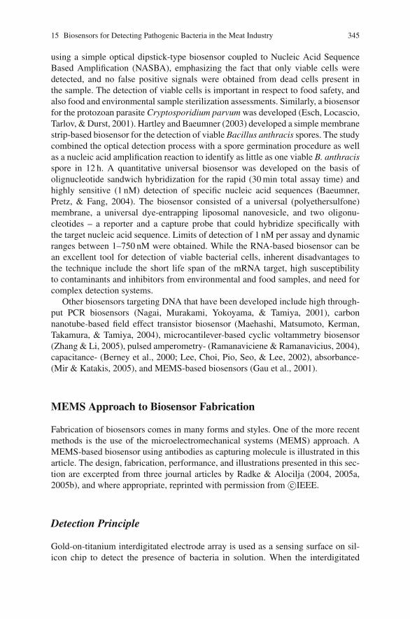
15 Biosensors for Detecting Pathogenic Bacteria in the Meat Industry 345
using a simple optical dipstick-type biosensor coupled to Nucleic Acid SequenceBased Amplification (NASBA), emphasizing the fact that only viable cells weredetected, and no false positive signals were obtained from dead cells present inthe sample. The detection of viable cells is important in respect to food safety, andalso food and environmental sample sterilization assessments. Similarly, a biosensorfor the protozoan parasite Cryptosporidium parvum was developed (Esch, Locascio,Tarlov, & Durst, 2001). Hartley and Baeumner (2003) developed a simple membranestrip-based biosensor for the detection of viable Bacillus anthracis spores. The studycombined the optical detection process with a spore germination procedure as wellas a nucleic acid amplification reaction to identify as little as one viable B. anthracisspore in 12 h. A quantitative universal biosensor was developed on the basis ofolignucleotide sandwich hybridization for the rapid (30 min total assay time) andhighly sensitive (1 nM) detection of specific nucleic acid sequences (Baeumner,Pretz, & Fang, 2004). The biosensor consisted of a universal (polyethersulfone)membrane, a universal dye-entrapping liposomal nanovesicle, and two oligonu-cleotides – a reporter and a capture probe that could hybridize specifically withthe target nucleic acid sequence. Limits of detection of 1 nM per assay and dynamicranges between 1–750 nM were obtained. While the RNA-based biosensor can bean excellent tool for detection of viable bacterial cells, inherent disadvantages tothe technique include the short life span of the mRNA target, high susceptibilityto contaminants and inhibitors from environmental and food samples, and need forcomplex detection systems.
Other biosensors targeting DNA that have been developed include high through-put PCR biosensors (Nagai, Murakami, Yokoyama, & Tamiya, 2001), carbonnanotube-based field effect transistor biosensor (Maehashi, Matsumoto, Kerman,Takamura, & Tamiya, 2004), microcantilever-based cyclic voltammetry biosensor(Zhang & Li, 2005), pulsed amperometry- (Ramanaviciene & Ramanavicius, 2004),capacitance- (Berney et al., 2000; Lee, Choi, Pio, Seo, & Lee, 2002), absorbance-(Mir & Katakis, 2005), and MEMS-based biosensors (Gau et al., 2001).
MEMS Approach to Biosensor Fabrication
Fabrication of biosensors comes in many forms and styles. One of the more recentmethods is the use of the microelectromechanical systems (MEMS) approach. AMEMS-based biosensor using antibodies as capturing molecule is illustrated in thisarticle. The design, fabrication, performance, and illustrations presented in this sec-tion are excerpted from three journal articles by Radke & Alocilja (2004, 2005a,2005b), and where appropriate, reprinted with permission from c©IEEE.
Detection Principle
Gold-on-titanium interdigitated electrode array is used as a sensing surface on sil-icon chip to detect the presence of bacteria in solution. When the interdigitated
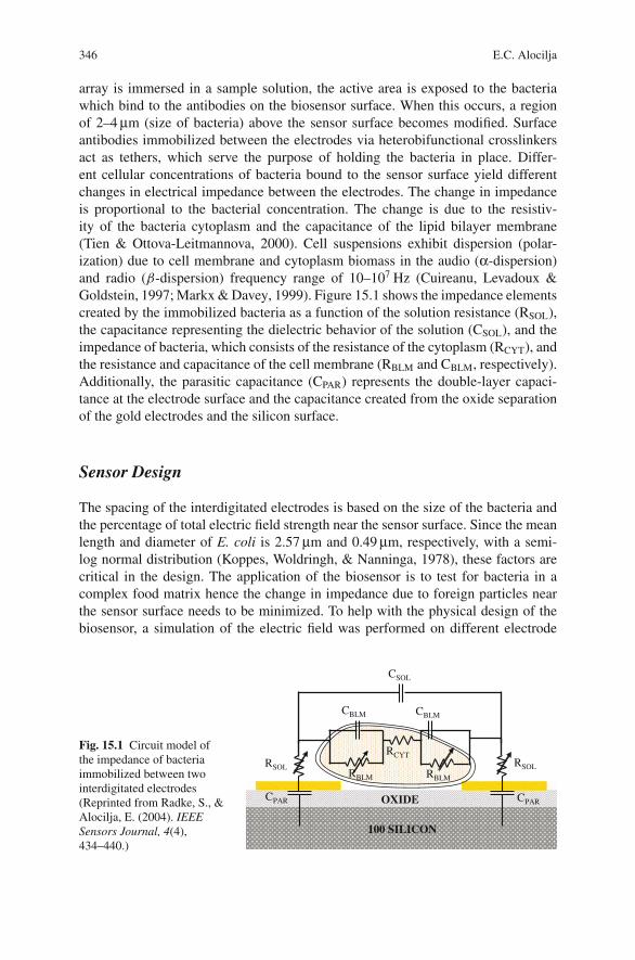
346 E.C. Alocilja
array is immersed in a sample solution, the active area is exposed to the bacteriawhich bind to the antibodies on the biosensor surface. When this occurs, a regionof 2–4 μm (size of bacteria) above the sensor surface becomes modified. Surfaceantibodies immobilized between the electrodes via heterobifunctional crosslinkersact as tethers, which serve the purpose of holding the bacteria in place. Differ-ent cellular concentrations of bacteria bound to the sensor surface yield differentchanges in electrical impedance between the electrodes. The change in impedanceis proportional to the bacterial concentration. The change is due to the resistiv-ity of the bacteria cytoplasm and the capacitance of the lipid bilayer membrane(Tien & Ottova-Leitmannova, 2000). Cell suspensions exhibit dispersion (polar-ization) due to cell membrane and cytoplasm biomass in the audio (α-dispersion)and radio (β-dispersion) frequency range of 10–107 Hz (Cuireanu, Levadoux &Goldstein, 1997; Markx & Davey, 1999). Figure 15.1 shows the impedance elementscreated by the immobilized bacteria as a function of the solution resistance (RSOL),the capacitance representing the dielectric behavior of the solution (CSOL), and theimpedance of bacteria, which consists of the resistance of the cytoplasm (RCYT), andthe resistance and capacitance of the cell membrane (RBLM and CBLM, respectively).Additionally, the parasitic capacitance (CPAR) represents the double-layer capaci-tance at the electrode surface and the capacitance created from the oxide separationof the gold electrodes and the silicon surface.
Sensor Design
The spacing of the interdigitated electrodes is based on the size of the bacteria andthe percentage of total electric field strength near the sensor surface. Since the meanlength and diameter of E. coli is 2.57 μm and 0.49 μm, respectively, with a semi-log normal distribution (Koppes, Woldringh, & Nanninga, 1978), these factors arecritical in the design. The application of the biosensor is to test for bacteria in acomplex food matrix hence the change in impedance due to foreign particles nearthe sensor surface needs to be minimized. To help with the physical design of thebiosensor, a simulation of the electric field was performed on different electrode
Fig. 15.1 Circuit model ofthe impedance of bacteriaimmobilized between twointerdigitated electrodes(Reprinted from Radke, S., &Alocilja, E. (2004). IEEESensors Journal, 4(4),434–440.)
RSOL
CPAR
RCYT
CBLM
CPAR
RSOL
CBLM
RBLM RBLM
100 SILICON
OXIDE
CSOL
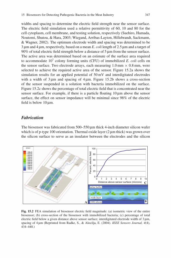
15 Biosensors for Detecting Pathogenic Bacteria in the Meat Industry 347
widths and spacing to determine the electric field strength near the sensor surface.The electric field simulation used a relative permittivity of 60, 10 and 80 for thecell cytoplasm, cell membrane, and testing solution, respectively (Suehiro, Hamada,Noutomi, Shutou, & Hara, 2003; Wiegand, Arribas-Layton, Hillebrandt, Sackmann,& Wagner, 2002). The optimum electrode width and spacing was determined to be3 μm and 4 μm, respectively, based on a mean E. coli length of 2.5 μm and a target of90% of total electric field strength below a distance of 5 μm from the sensor surface.The active area was determined based on an estimate of the surface area requiredto accommodate 107 colony forming units (CFU) of immobilized E. coli cells onthe sensor surface. Two electrode arrays, each measuring 1.0 mm × 0.8 mm, wereselected to achieve the required active area of the sensor. Figure 15.2a shows thesimulation results for an applied potential of 50 mV and interdigitated electrodeswith a width of 3 μm and spacing of 4 μm. Figure 15.2b shows a cross-sectionof the sensor suspended in a solution with bacteria immobilized on the surface.Figure 15.2c shows the percentage of total electric field that is concentrated near thesensor surface. For example, if there is a particle floating 10 μm above the sensorsurface, the effect on sensor impedance will be minimal since 98% of the electricfield is below 10 μm.
Fabrication
The biosensor was fabricated from 500–550 μm thick 4-inch diameter silicon waferwhich is of p-type 100 orientation. Thermal oxide layer (2 μm thick) was grown overthe silicon surface to serve as an insulator between the electrodes and the silicon
A (100) silicon
oxide layer
saline buffer
B
0
20
40
60
80
100
0 1 2 3 4 5 6 7 8 9 10Distance above sensor surface (microns)
Per
cent
, ele
ctric
fiel
d
C
Fig. 15.2 FEA simulation of biosensor electric field magnitude: (a) isometric view of the entirebiosensor; (b) cross-section of the biosensor with immobilized bacteria; (c) percentage of totalelectric field below a given distance above sensor surface; interdigitated electrode width of 3 μm,spacing of 4 μm (Reprinted from Radke, S., & Alocilja, E. (2004). IEEE Sensors Journal, 4(4),434–440.)
348 E.C. Alocilja
substrate. Micro-electrode arrays were fabricated by patterning S1805 photo resistand depositing 5 nm of titanium (for adhesion) and topped with 50 nm of gold. Usingultrasonic activation, a lift-off process developed the interdigitated electrode array.For ease of fabrication, two small electrode arrays were fabricated to reduce theaspect ratio as compared to one large array. The sensor included an interdigitatedelectrode array with circuit traces connecting to bond pads. Each electrode fingerhad a length of 750 μm, a width of 3 μm, and an in-between spacing of 4 μm. Eachsensor chip was diced to a dimension of 12 mm × 8 mm. Each chip had a total of1700 electrodes and a large active area totaling 9.6 mm2, as shown in Fig. 15.3.
After electrode array fabrication, antibodies were immobilized to the oxidesurface of the sensor. The antibodies were attached to the surface via heterobi-functional crosslinkers using an established process (Bhatia et al., 1989). Briefly,the biosensor was cleaned and activated by immersing the sensor in nitric acidfor 30 min followed by rinsing with distilled water. Silanization of the chip sur-face occurred after immersing the chip in a mixture of hydrochloric acid andmethanol for 30 min followed with immersion in sulfuric acid. The silanizing of3-Mercaptomethyldimethylethoxysilane (MDS) to the sensor surface occurred indry toluene. Crosslinkers, N -y-maleimidobutyryloxy succinimide ester (GMBS)dissolved in dimethylformamide (DMF) and ethanol, were attached to the chip sur-face after silanization. Antibodies were added to the sensor surface to attach to thecrosslinkers. The next step was to package the biosensor. Epoxy was used to bondthe backside of the device to a rectangular piece of PVC for support. The bond padswere connected to a zero insertion force (ZIF) socket with leads terminating to animpedance analyzer, completing the fabrication process.
The biosensor chips were inspected in order to validate the physical propertiesas shown in Fig. 15.4. A metallurgical microscope was used to validate the fabri-cation of the electrode array for complete deposition of metal and to ensure thatthe high aspect ratio electrodes (250:1) were within tolerance (Fig. 15.4a). Atomic
6 mm
1 mm
25 µm25 µm
Fig. 15.3 Interdigitated gold electrode arrays: (left) two high density, high aspect ratio electrodearrays; (right) end view of one electrode array (Reprinted from Radke, S., & Alocilja, E. (2004).IEEE Sensors Journal, 4(4), 434–440.)
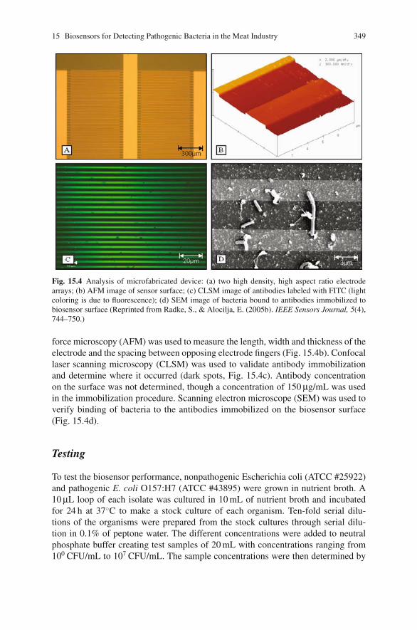
15 Biosensors for Detecting Pathogenic Bacteria in the Meat Industry 349
Fig. 15.4 Analysis of microfabricated device: (a) two high density, high aspect ratio electrodearrays; (b) AFM image of sensor surface; (c) CLSM image of antibodies labeled with FITC (lightcoloring is due to fluorescence); (d) SEM image of bacteria bound to antibodies immobilized tobiosensor surface (Reprinted from Radke, S., & Alocilja, E. (2005b). IEEE Sensors Journal, 5(4),744–750.)
force microscopy (AFM) was used to measure the length, width and thickness of theelectrode and the spacing between opposing electrode fingers (Fig. 15.4b). Confocallaser scanning microscopy (CLSM) was used to validate antibody immobilizationand determine where it occurred (dark spots, Fig. 15.4c). Antibody concentrationon the surface was not determined, though a concentration of 150 μg/mL was usedin the immobilization procedure. Scanning electron microscope (SEM) was used toverify binding of bacteria to the antibodies immobilized on the biosensor surface(Fig. 15.4d).
Testing
To test the biosensor performance, nonpathogenic Escherichia coli (ATCC #25922)and pathogenic E. coli O157:H7 (ATCC #43895) were grown in nutrient broth. A10 μL loop of each isolate was cultured in 10 mL of nutrient broth and incubatedfor 24 h at 37◦C to make a stock culture of each organism. Ten-fold serial dilu-tions of the organisms were prepared from the stock cultures through serial dilu-tion in 0.1% of peptone water. The different concentrations were added to neutralphosphate buffer creating test samples of 20 mL with concentrations ranging from100 CFU/mL to 107 CFU/mL. The sample concentrations were then determined by
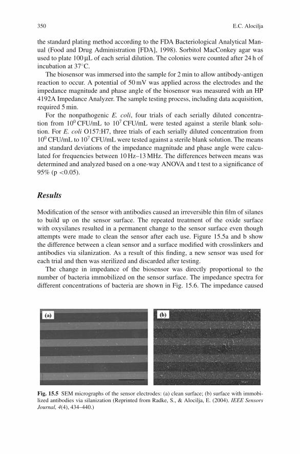
350 E.C. Alocilja
the standard plating method according to the FDA Bacteriological Analytical Man-ual (Food and Drug Administration [FDA], 1998). Sorbitol MacConkey agar wasused to plate 100 μL of each serial dilution. The colonies were counted after 24 h ofincubation at 37◦C.
The biosensor was immersed into the sample for 2 min to allow antibody-antigenreaction to occur. A potential of 50 mV was applied across the electrodes and theimpedance magnitude and phase angle of the biosensor was measured with an HP4192A Impedance Analyzer. The sample testing process, including data acquisition,required 5 min.
For the nonpathogenic E. coli, four trials of each serially diluted concentra-tion from 100 CFU/mL to 107 CFU/mL were tested against a sterile blank solu-tion. For E. coli O157:H7, three trials of each serially diluted concentration from100 CFU/mL to 107 CFU/mL were tested against a sterile blank solution. The meansand standard deviations of the impedance magnitude and phase angle were calcu-lated for frequencies between 10 Hz–13 MHz. The differences between means wasdetermined and analyzed based on a one-way ANOVA and t test to a significance of95% (p <0.05).
Results
Modification of the sensor with antibodies caused an irreversible thin film of silanesto build up on the sensor surface. The repeated treatment of the oxide surfacewith oxysilanes resulted in a permanent change to the sensor surface even thoughattempts were made to clean the sensor after each use. Figure 15.5a and b showthe difference between a clean sensor and a surface modified with crosslinkers andantibodies via silanization. As a result of this finding, a new sensor was used foreach trial and then was sterilized and discarded after testing.
The change in impedance of the biosensor was directly proportional to thenumber of bacteria immobilized on the sensor surface. The impedance spectra fordifferent concentrations of bacteria are shown in Fig. 15.6. The impedance caused
Fig. 15.5 SEM micrographs of the sensor electrodes: (a) clean surface; (b) surface with immobi-lized antibodies via silanization (Reprinted from Radke, S., & Alocilja, E. (2004). IEEE SensorsJournal, 4(4), 434–440.)
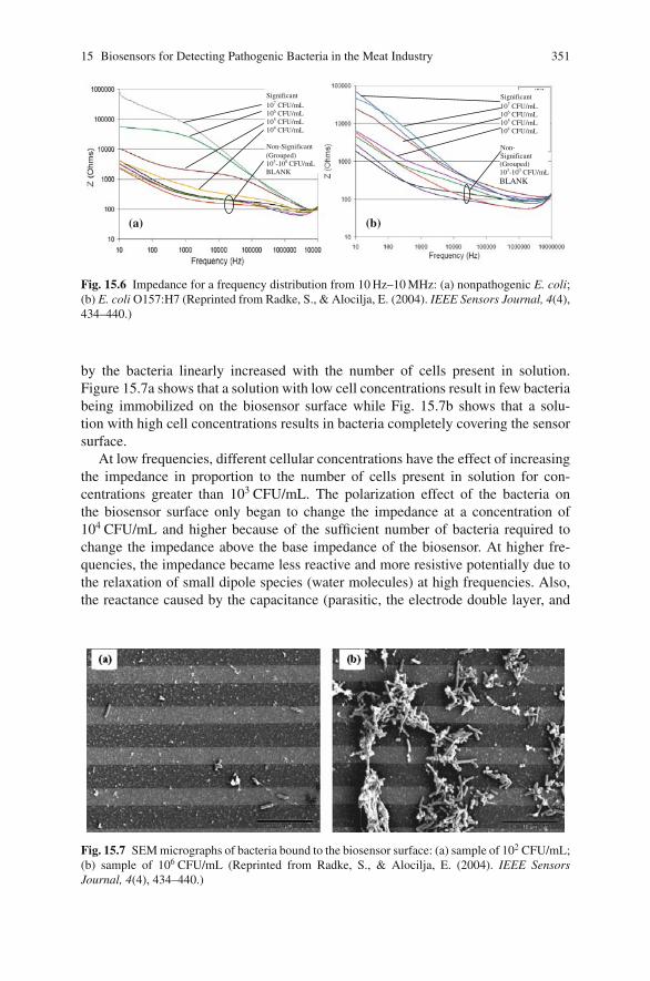
15 Biosensors for Detecting Pathogenic Bacteria in the Meat Industry 351
(a)
Significant107 CFU/mL 106 CFU/mL105 CFU/mL104 CFU/mL
Non-Significant(Grouped)103-100 CFU/mLBLANK
Significant107 CFU/mL 106 CFU/mL105 CFU/mL104 CFU/mL
Non-Significant(Grouped)103-100 CFU/mLBLANK
(b)
Fig. 15.6 Impedance for a frequency distribution from 10 Hz–10 MHz: (a) nonpathogenic E. coli;(b) E. coli O157:H7 (Reprinted from Radke, S., & Alocilja, E. (2004). IEEE Sensors Journal, 4(4),434–440.)
by the bacteria linearly increased with the number of cells present in solution.Figure 15.7a shows that a solution with low cell concentrations result in few bacteriabeing immobilized on the biosensor surface while Fig. 15.7b shows that a solu-tion with high cell concentrations results in bacteria completely covering the sensorsurface.
At low frequencies, different cellular concentrations have the effect of increasingthe impedance in proportion to the number of cells present in solution for con-centrations greater than 103 CFU/mL. The polarization effect of the bacteria onthe biosensor surface only began to change the impedance at a concentration of104 CFU/mL and higher because of the sufficient number of bacteria required tochange the impedance above the base impedance of the biosensor. At higher fre-quencies, the impedance became less reactive and more resistive potentially due tothe relaxation of small dipole species (water molecules) at high frequencies. Also,the reactance caused by the capacitance (parasitic, the electrode double layer, and
Fig. 15.7 SEM micrographs of bacteria bound to the biosensor surface: (a) sample of 102 CFU/mL;(b) sample of 106 CFU/mL (Reprinted from Radke, S., & Alocilja, E. (2004). IEEE SensorsJournal, 4(4), 434–440.)
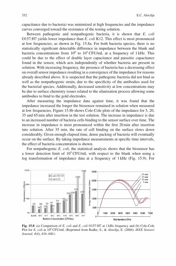
352 E.C. Alocilja
capacitance due to bacteria) was minimized at high frequencies and the impedancecurves converged toward the resistance of the testing solution.
Between pathogenic and nonpathogenic bacteria, it is shown that E. coliO157:H7 yields lower impedance than E. coli K12. This effect is most pronouncedat low frequencies, as shown in Fig. 15.8a. For both bacteria species, there is nostatistically significant detectable difference in impedance between the blank andbacteria concentrations from 100 to 103 CFU/mL at a frequency of 1 kHz. Thiscould be due to the effect of double layer capacitance and parasitic capacitancefound in the sensor, which acts independently of whether bacteria are present insolution. With increasing frequency, the presence of bacteria has a decreasing effecton overall sensor impedance resulting in a convergence of the impedance for reasonsalready described above. It is suspected that the pathogenic bacteria did not bind aswell as the nonpathogenic strain, due to the specificity of the antibodies used forthe bacterial species. Additionally, decreased sensitivity at low concentrations maybe due to surface chemistry issues related to the silanization process allowing someantibodies to bind to the gold electrodes.
After measuring the impedance data against time, it was found that theimpedance increased the longer the biosensor remained in solution when measuredat low frequencies. Figure 15.8b shows Cole-Cole plots of the impedance for 5, 20,35 and 65 min after insertion in the test solution. The increase in impedance is dueto an increased number of bacteria cells binding to the sensor surface over time. Theincrease in impedance is most pronounced within the first 20 min after insertioninto solution. After 35 min, the rate of cell binding on the surface slows downconsiderably. Given enough elapsed time, dense packing of bacteria will eventuallyoccur on the surface. By taking impedance measurements at specific time intervals,the effect of bacteria concentration is shown.
For nonpathogenic E. coli, the statistical analysis shows that the biosensor hasa lower detection limit of 105 CFU/mL with respect to the blank when using alog transformation of impedance data at a frequency of 1 kHz (Fig. 15.9). For
Fig. 15.8 (a) Comparison of E. coli and E. coli O157:H7 at 1 kHz frequency and (b) Cole-ColePlot for E. coli at 106 CFU/mL (Reprinted from Radke, S., & Alocilja, E. (2004). IEEE SensorsJournal, 4(4), 434–440.)
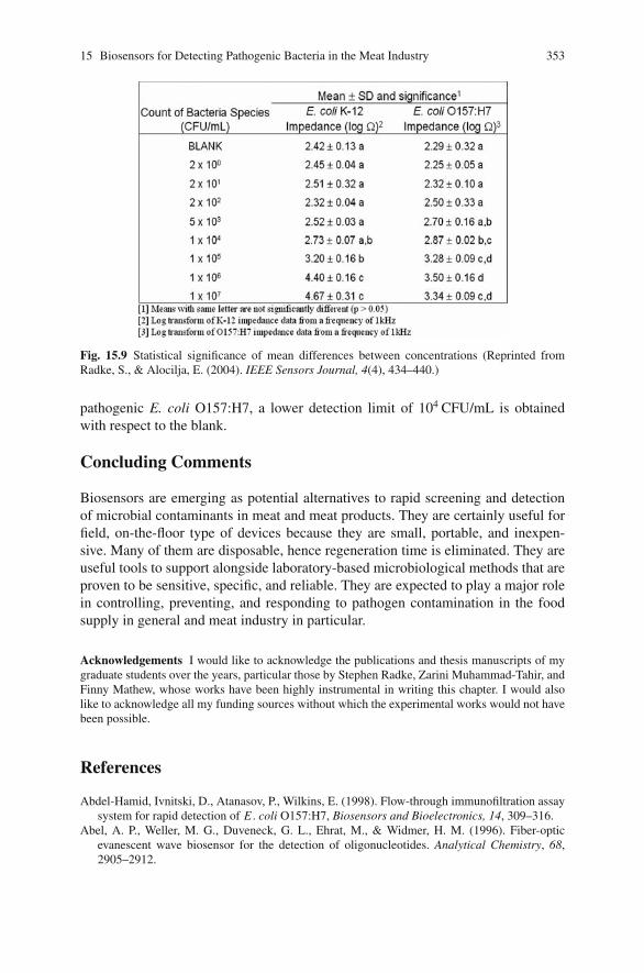
15 Biosensors for Detecting Pathogenic Bacteria in the Meat Industry 353
Fig. 15.9 Statistical significance of mean differences between concentrations (Reprinted fromRadke, S., & Alocilja, E. (2004). IEEE Sensors Journal, 4(4), 434–440.)
pathogenic E. coli O157:H7, a lower detection limit of 104 CFU/mL is obtainedwith respect to the blank.
Concluding Comments
Biosensors are emerging as potential alternatives to rapid screening and detectionof microbial contaminants in meat and meat products. They are certainly useful forfield, on-the-floor type of devices because they are small, portable, and inexpen-sive. Many of them are disposable, hence regeneration time is eliminated. They areuseful tools to support alongside laboratory-based microbiological methods that areproven to be sensitive, specific, and reliable. They are expected to play a major rolein controlling, preventing, and responding to pathogen contamination in the foodsupply in general and meat industry in particular.
Acknowledgements I would like to acknowledge the publications and thesis manuscripts of mygraduate students over the years, particular those by Stephen Radke, Zarini Muhammad-Tahir, andFinny Mathew, whose works have been highly instrumental in writing this chapter. I would alsolike to acknowledge all my funding sources without which the experimental works would not havebeen possible.
References
Abdel-Hamid, Ivnitski, D., Atanasov, P., Wilkins, E. (1998). Flow-through immunofiltration assaysystem for rapid detection of E . coli O157:H7, Biosensors and Bioelectronics, 14, 309–316.
Abel, A. P., Weller, M. G., Duveneck, G. L., Ehrat, M., & Widmer, H. M. (1996). Fiber-opticevanescent wave biosensor for the detection of oligonucleotides. Analytical Chemistry, 68,2905–2912.
354 E.C. Alocilja
Alocilja, E. C., & Radke, S. (2003). Market analysis of biosensors for food safety. Biosensors andBioelectronics Journal, 18(5–6), 841–846.
Alocilja, E. C., Ritchie, N., & Grooms, D. (2003). Protocol development using an electronic nosefor differentiating E. coli strains. IEEE Sensors Journal, 3(6), 801–805.
Babacan, S., Pivarnik, P., Letcher, S., & Rand, A. G. (2000). Evaluation of antibody immobilizationmethods for piezoelectric biosensor application. Biosensors and Bioelectronics, 15, 615–621.
Baeumner, A. J., Cohen, R. N., Miksic, V., & Min, J. (2003). RNA biosensor for the rapid detectionof viable Escherichia coli in drinking water. Biosensors and Bioelectronics, 18, 405–413.
Baeumner, A. J., Pretz, J., & Fang, S. (2004). A universal nucleic acid sequence biosensor withnanomolar detection limits. Analytical Chemistry, 76, 888–894.
Bao, L., Deng, L., Nie, L., Yao, S., & Wei, W. (1996). Determination of microorganisms with aquartz crystal microbalance sensor. Analytical Chemical Acta, 319, 97–101.
Barbour, W. M., & George, T. (1997). Genetic and immunologic techniques for detecting food-borne pathogens and toxins. In T. J. Montville (Ed.), Food microbiology: Fundamentals andfrontiers (pp. 30–65). Washington, DC: ASM Press.
Berney, H., West, J., Haefele, E., Alderman, J., Lane, W., & Collins, J. K. (2000). A DNAdiagnostic biosensor: Development, characterisation and performance. Sensors and ActuatorsB: Chemical, 68, 100–108.
Bhatia, S. K., Shriver-Lake, L. C., Prior, K. J., Georger, J. H., Calvert, J. M., Bredehorst, R., et al.(1989). Use of thiol-terminal silanes and heterobifunctional crosslinkers for immobilization ofantibodies on silica surfaces. Analytical Biochemistry, 178, 408–413.
Bianchi, N., Rutigliano, C., Tomassetti, M., Feriotto, G., Zorzato, F., & Gambari, R. (1997).Biosensor technology and surface plasmon resonance for real-time detection of HIV-1 genomicsequences amplified by polymerase chain reaction. Clinical and Diagnostic Virology, 8,199–208.
Blaser, M. J., & Newman, L. S. (1982). A review of human salmonellosis 1. Infective dose. Reviewsof Infectious Diseases, 4, 1096–1106.
Boltovets, P. M., Boyko, V. R., Kostikov, I. Y., Dyachenko, N. S., Snopok, B. A., & Shirshov, Y. M.(2002). Simple method for plant virus detection: Effect of antibody immobilization technique.Journal of Virological Methods, 105, 141–146.
Bunde, R. L., Jarvi, E. J., & Rosentrerer, J. J. (1998). Piezoelectric quartz crystal biosensor. Talanta,46, 1223–1229.
Carlson, S. A., Bolton, L. F., Briggs, C. E., Hurd, H. S., Sharma, V. K., Fedorka Cray, P. J., et al.(1999). Detection of multiresistant Salmonella typhimurium DT104 using multiplex and fluo-rogenic PCR. Molecular and Cellular Probes, 13, 213–222.
CDC. (2001a). Outbreaks caused by Shiga toxin-producing Escherichia coli-Summary of2000 Surveillance Data. Centers for Disease Control and Prevention. From http://www.cdc.gov/foodborneoutbreaks/ecoli/2000 summaryLetter.pdf.
CDC. (2001b). Salmonellosis. From http://www.cdc.gov/ncidod/dbmd/diseaseinfo/salmonellosisg.htm.
CDC. (2002a). Notice to readers: Final 2001 reports of notifiable diseases. Morbidity and MortalityWeekly Report, 51, 710.
CDC. (2002b). Preliminary FoodNet data on the incidence of foodborne illnesses – selected sites,United States, 2001. Morbidity and Mortality Weekly Report, 51, 325–329.
CDC. (2002c). Report on the decline of foodborne illness. Centers for Disease Control and Pre-vention. From http://www.cdc.gov/foodborne/publications/201-nelson 2004.pdf.
CHEMICON International. (2004). Introduction to antibodies. CHEMICON International, Inc.From http://www.chemicon.com/resource/ANT101/a1.asp.
Chen, Z. Z., Wang, K. M., Yang, X. H., Huang, S. S., Huang, H. M., Li, D., et al. (2003). Deter-mination of hepatitis B surface antigen by surface plasmon resonance biosensor. Acta ChimicaSinica, 61, 137–140.
Cheung, J. H., Stockton, W. B., & Rubner, M. F. (1997). Molecular-level processing of conjugatedpolymers: Layer-by-layer manipulation of polyaniline via electrostatic interactions. Macro-molecules, 30, 2712–2716.
15 Biosensors for Detecting Pathogenic Bacteria in the Meat Industry 355
Cohn, G. E. (1998). Systems and technologies for clinical diagnostics and drug discovery. SPIEProceedings, 3259, 11–17.
Corry, B., Uilk, J., & Crawley, C. (2003). Analytica Chemica Acta, 496, 103–116.Cuireanu, M., Levadoux, W., & Goldstein, S. (1997). Electrical impedance studies on a culture of
a newly discovered strain of Steptomyces. Enzyme and Microbial Technology, 21, 441–449.D’Aoust, J. Y. (1997). Salmonella species. In T. J. Montville (Ed.), Food microbiology: Funda-
mentals and frontiers (pp. 138–139). Washington, DC: ASM.DeMarco, D., & Lim, D. (2002). Detection of Escherichia coli O157:H7 in 10- and 25-gram ground
beef samples with an evanescent-wave biosensor with silica and polystyrene waveguides. Jour-nal of Food Protection, 65, 596–602.
Doyle, M. P., Zhao, T., Meng, J., & Zhao, S. (1997). Escherichia coli O157:H7. Food microbiologyfundamentals and frontiers. Washington, D.C: American Society for Microbiology.
D’Souza, S. F. (2001). Microbial biosensors (Review). Biosensors and Bioelectronics, 16,337–353.
Esch, M. B., Locascio, L. E., Tarlov, M. J., & Durst, R. A. (2001). Detection of viable Cryp-tosporidium parvum using DNA-modified liposomes in a microfluidic chip. Analytical Chem-istry, 73, 2952–2958.
FAO-Food and Agriculture Organization. (2006). Global market analysis. Food Outlook No. 1.Retrieved June 2006, from FTP://FTP.FAO.ORG/DOCREP/FAO/009/J7927E/J7927E00.PDF.
FDA-Food and Drug Administration. (1998). Bacteriological analytical manual.Food and Drug Administration, Gaithersburg, MD.
FDA-Food and Drug Administration. (2005). Bacteriological analytical manual. Rockville, MD,USA: Food and Drug Administration. From http://www.cfsan.fda.gov/∼ebam/bam-toc.html.
FDA-Food and Drug Administration. (2006). Foodborne pathogenic microorganisms andnatural toxins handbook: The “Bad Bug Book”. FDA-CFSAN. From http://www.cfsan.fda.gov/∼mow/intro.html.
Feriotto, G., Borgatti, M., Mischiati, C., Bianchi, N., & Gambari, R. (2002). Biosensor technologyand surface plasmon resonance for real-time detection of genetically modified roundup readysoybean gene sequences. Journal of Agricultural and Food Chemistry, 50, 955–962.
Fratamico, P. M., Strobaugh, T. P., Medina, M. B., & Gehring, A. G. (1998). Detection ofEscherichia coli O157:H7 using a surface plasmon resonance biosensor. Biotechnology Tech-niques, 12 (7), 571–576.
Gao, Z. X., Fang, Y. J., Ren, J., Ning, B., Zhu, H. Z., & He, Y. H. (2004). Studies on biotin-avidin indirect conjugated technology for a piezoelectric DNA sensor. International Journal ofEnvironmental Analytical Chemistry, 84, 599–606.
Gau, J. -J., Lan, E. H., Dunn, B., Ho, C. -M., & Woo, J. C. S. (2001). A MEMS based amperometricdetector for E. coli bacteria using self-assembled monolayers. Biosensors and Bioelectronics,16, 745–755.
Geng, T., Morgan, M. T., & Bhunia, A. K. (2004). Detection of low levels of Listeria monocyto-genes cells by using a fiber-optic immunosensor. Applied and Environmental Microbiology, 70,6138–6146.
Ghindilis, A., Atanasov, P., Wilkins, M., & Wilkins, E. (1998). Immunosensors: Electrochemicalsensing and other engineering approaches. Biosensors and Bioelectronics, 13, 113–131.
Gomez, R., Bashir, R., Sarikaya, A., Ladisch, M., Sturgis, J., Robinson, J., et al. (2001). Microflu-idic biochip for impedance spectroscopy of biological species. Biomedical Microdevices, 3,201–209.
Gore, A., Chakrabartty, S., Pal, S., & Alocilja, E. C. (2006). A multi-channel femtoampere-sensitivity potentiostat array for biosensing applications. IEEE Transactions on Circuits andSystems, 53(11), 2357–2363.
Graham, C. R., Leslie, D., & Squirrell, D. J. (1992). Gene probe assays on a fibre-optic evanescentwave biosensor. Biosensors and Bioelectronics, 7, 487–493.
Hartley, H. A., & Baeumner, A. J. (2003). Biosensor for the specific detection of a single viable B.anthracis spore. Analytical Bioanalytical Chemistry, 376, 319–327.
356 E.C. Alocilja
He, F. J., & Liu, S. Q. (2004). Detection of P. aeruginosa using nano-structured electrode-separatedpiezoelectric DNA biosensor. Talanta, 62, 271–277.
Ho, J. A., Hsu, H. W., & Huang, M. R. (2004). Liposome-based microcapillary immunosensor fordetection of Escherichia coli O157:H7. Analytical Biochemistry, 330, 342–349.
Hoyle, B. (2001). High-tech biosensor speeds bacteria detection. ASM News, 67, 434–435.Huang, T. S., Tzeng, Y., Liu, Y. K., Chen, Y. K., Walker, K. R., Guntupalli, R., et al. (2004).
Immobilization of antibodies and bacterial binding on nanodiamond and carbon nanotubes forbiosensor applications. Diamond and Related Materials, 13, 1098–1102.
Jiang, H., Adams, C., Graziano, N., Roberson, A., McGuire, M., & Khiari, D. (2006). Enzyme-linked immunosorbent analysis (ELISA) of atrazine in raw and finished drinking water. Envi-ronmental Engineering Science, 23 (2), 357–366.
Jordan, C. E., Frutos, A. G., Thiel, A. J., & Corn, R. M. (1997). Surface plasmon resonance imagingmeasurements of DNA hybridization adsorption and streptavidin/DNA multilayer formation atchemically modified gold surfaces. Analytical Chemistry, 69, 4939–4947.
Kalorama Information. (2000). MarketResearch.com, New York, p. 313.Kim, N., Park, I. S., & Kim, D. K. (2004). Characteristics of a label-free piezoelectric
immunosensor detecting Pseudomonas aeruginosa. Sensors and Actuators B-Chemical, 100,432–438.
Ko, S., & Grant, S. A. (2006). A novel FRET-based optical fiber biosensor for rapid detection ofSalmonella typhimurium. Biosensors and Bioelectronics, 21, 1283–1290.
Komarova, E., Aldissi, M., & Bogomolova, A. (2005). Direct electrochemical sensor for fastreagent-free DNA detection. Biosensors and Bioelectronics, 21, 182–189.
Koppes, L., Woldringh, C., & Nanninga, N. (1978). Size variations and correlation of different cellcycle events in slow-growing Escherichia coli. Journal of Bacteriology, 134, 423–433.
Koubova, V., Brynda, E., Karasova, L., Skvor, J., Homola, J., Dostalek, J., et al. (2001). Detectionof foodborne pathogens using surface plasmon resonance biosensors. Sensors and ActuatorsB-Chemical, 74, 100–105.
Kukanskis, K., Elkind, J., Melendez, J., Murphy, T., Miller, G., & Garner, H. (1999). Detectionof DNA hybridization using the TISPR-1 surface plasmon resonance biosensor. AnalyticalBiochemistry, 274, 7–17.
Lazcka, O., Campo, F., Javier, D., Munoz, F., & Xavier, F. (2007). Pathogen detection: A perspec-tive of traditional methods and biosensors. Biosensors and Bioelectronics, 22, 1205–1217.
Lee, J. S., Choi, Y.-K., Pio, M., Seo, J., & Lee, L. P. (2002). Nanogap capacitors for label free DNAanalysis. BioMEMS and Bionanotechnology, 729, 185–190.
Lin, H. C., & Tsai, W. C. (2003). Piezoelectric crystal immunosensor for the detection of staphy-lococcal enterotoxin B. Biosensors and Bioelectronics, 18, 1479–1483.
Liu, X., Farmerie, W., Schuster, S., & Tan, W. (2000). Molecular beacons for DNA biosensors withmicrometer to submicrometer dimensions. Analytical Biochemistry, 283, 56–63.
Liu, X., & Tan, W. (1999). A fiber-optic evanescent wave DNA biosensor based on novel molecularbeacons. Analytical Chemistry, 71, 5054–5059.
Lu, B., Smyth, M. R., and O’Kennedy, R. (1996). Oriented immobilization of antibodies and itsapplications in immunoassays and immunosensors. Analyst, 121, 29R–32R.
Maehashi, K., Matsumoto, K., Kerman, K., Takamura, Y., & Tamiya, E. (2004). Ultrasensi-tive detection of DNA hybridization using carbon nanotube field-effect transistors. JapaneseJournal of Applied Physics Part 2-Letters & Express Letters, 43, L1558–L1560.
Mariotti, E., Minunni, M., & Mascini, M. (2002). Surface plasmon resonance biosensor for genet-ically modified organisms detection. Analytica Chimica Acta, 453, 165–172.
Markx, G., & Davey, C. (1999). The dielectric properties of biological cells at radiofrequencies:Applications in biotechnology. Enzyme and Microbial Technology, 25, 161–171.
Mathew, F., Alagesan, D., & Alocilja, E. C. (2004). Chemiluminescence detection of Escherichiacoli in fresh produce obtained from different sources. Luminescence Journal, 19, 193–198.
Mathew, F., & Alocilja, E. C. (2004). Enzyme-based detection of Escherichia coli. Transactions ofthe ASAE, 47 (1), 357–362.
15 Biosensors for Detecting Pathogenic Bacteria in the Meat Industry 357
Mathew, F., & Alocilja, E. C. (2005). Porous silicon-based biosensor for pathogen detection.Biosensors and Bioelectronics Journal, 20 (8),1656–1661.
McClelland, R., & Pinder, A. (1994). Detection of Salmonella typhimurium in dairy productswith flow cytometry and monoclonal antibodies. Applied Environmental Microbiology A, 60,4255–4262.
McGown, L. B., Joseph, M. J., Pitner, J. B., Vonk, G. P., & Linn, C. P. (1995). The nucleic-acidligand – a new tool for molecular recognition. Analytical Chemistry, 67, A663–A668.
Mead, P. S., Slutsker, L., Dietz, V., McGaig, L., Bresee, J., Shapiro, C., et al. (1999). Food-relatedillnesses and death in the United States. Emerging Infectious Disease, 5, 607–625.
Meeusen, C., Alocilja, E. C., & Osburn, W. (2005). Detection of E. coli O157:H7 using a minia-turized surface plasmon resonance biosensor. Transactions of the ASAE, 48(6), 2409–2416.
Meng, J., Zhao, S., Doyle, M., & Kresovich, S. (1996). Polymerase chain reaction for detectionE. coli O157:H7. International Journal of Food Microbiology, 32, 103–113.
Mir, M., & Katakis, I. (2005). Towards a fast-responding, label-free electrochemical DNA biosen-sor. Analytical and Bioanalytical Chemistry, 381, 1033–1035.
Mittelmann, A. S., Ron, E. Z., & Rishpon, J. (2002). Amperometric quantification of total col-iforms and specific detection of Escherichia coli. Analytical Chemistry, 74(4), 903–907.
Mo, X. T., Zhou, Y. P., Lei, H., & Deng, L. (2002). Microbalance-DNA probe method for thedetection of specific bacteria in water. Enzyme and Microbial Technology, 30, 583–589.
Muhammad-Tahir, Z., & Alocilja, E. (2004). A disposable biosensor for pathogen detection infresh produce samples. Biosystems Engineering, 88, 145–151.
Muhammad-Tahir, Z., & Alocilja, E. C. (2003a). A conductimetric biosensor for biosecurity.Biosensors and Bioelectronics, 18, 813–819.
Muhammad-Tahir, Z., & Alocilja, E. C. (2003b). Fabrication of a disposable biosensor forEscherichia coli O157:H7 detection. IEEE Sensors Journal, 3, 345–351.
Muhammad-Tahir, Z., Alocilja, E. C., & Grooms, D. L. (2005a). Polyaniline synthesis and itsbiosensor application. Biosensors and Bioelectronics, 20, 1690–1695.
Muhammad-Tahir, Z., Alocilja, E. C., & Grooms, D. L. (2005b). Rapid detection of Bovine ViralDiarrhea Virus as surrogate of bioterrorism agents. IEEE Sensors Journal, 5, 757–762.
Muhammad-Tahir, Z., Alocilja, E. C., & Grooms, D. L. (2007). Indium tin oxide-polyanilinebiosensor: Fabrication and characterization. Sensors Journal, 7, 1123–1140.
Nagai, H., Murakami, Y., Yokoyama, K., & Tamiya, E. (2001). High-throughput PCR in siliconbased microchamber array. Biosensors and Bioelectronics, 16, 1015–1019.
Narang, U., Anderson, G. P., Ligler, F. S., & Burans, J. (1997). Fiber optic-based biosensor forricin. Biosensors and Bioelectronics, 12, 937–945.
Nashat, A. H., Moronne, M., & Ferrari, M. (1998). Detection of functional groups and antibodieson microfabricated surfaces by confocal microscopy. Biotechnology and Bioengineering, 60,137–146.
Nicolini, C., Erokhin, V., Facci, P., Guerzoni, S., Ross, A., & Paschkevitsch, P. (1997). Quartzbalance DNA sensor. Biosensors and Bioelectronics, 12, 613–618.
Pal, S., Alocilja, E. C., & Downes, F. P. (2007). Nanowire labeled direct-charge transfer biosensorfor detecting bacillus species. Biosensors & Bioelectronics Journal, 22, 2329–2336.
Park, I. S., Kim, W. Y., & Kim, N. (2000). Operational characteristics of an antibody-immobilizedQCM system detecting Salmonella spp. Biosensors and Bioelectronics, 15, 167–172.
Park, S., & Durst, R. A. (2000). Immunoliposome sandwich assay for the detection of Escherichiacoli O157:H7. Analytical Biochemistry, 280, 151–158.
Radke, S., & Alocilja, E. (2004). Design and fabrication of an impedimetric biosensor. IEEESensors Journal, 4, 434–440.
Radke, S., & Alocilja, E. C. (2005b). A microfabricated biosensor for detecting foodborne bioter-rorism agents. IEEE Sensors Journal, 5 (4), 744–750.
Radke, S. M., & Alocilja, E. C. (2005a). A high density microelectrode array biosensor for detec-tion of E. coli O157:H7. Biosensors and Bioelectronics SPEC ISS, 20, 1662–1667.
358 E.C. Alocilja
Ramanaviciene, A., & Ramanavicius, A. (2004). Pulsed amperometric detection of DNA withan ssDNA/polypyrrole-modified electrode. Analytical and Bioanalytical Chemistry, 379,287–293.
Rishpon, J., & Ivnitski, D. (1997). An amperometric enzyme-channeling immunosensor. Biosen-sors and Bioelectronics, 12, 195–204.
Rodriguez, M., & Alocilja, E. (2005). Embedded DNA-polypyrrole biosensor for rapid detectionof Escherichia coli. IEEE Sensors Journal, 5, 733–736.
Ruan, C. M., Yang, L. J., & Li, Y. B. (2002). Immunobiosensor chips for detection of Escherichiacoli O157: H7 using electrochemical impedance spectroscopy. Analytical Chemistry, 74,4814–4820.
Ruan, C. M., Zeng, K. F., Varghese, O. K., & Grimes, C. A. (2004). A staphylococcal enterotoxinB magnetoelastic immunosensor. Biosensors and Bioelectronics, 20, 585–591.
Sadana, A. 2002. Engineering biosensors: Kinetic and design application. London: AcademicPress.
Savage, M. D., Mattson, G., Desai, S., Nielander, G. W., Morgensen, S., & Conklin, E. J. (1994).Avidin-biotin chemistry: A handbook. Rockford, IL: Pierce Chemical Company.
Scheller, F. W., Hintscher, R., Pfeiffer, P., Schubert, F., Riedel, K., & Kindervater, R. (1991).Biosensors: Fundamentals, applications and trends. Sensors and Actuators B, 4, 197–206.
Seo, K. H., Brackett, R. E., Hartman, N. F., & Campbell, D. P. (1999). Development of a rapidresponse biosensor for detection of Salmonella Typhimurium. Journal of Food Protection, 62,431–437.
Shah, J., Chemburu, S., Wilkins, E., & Abdel-Hamid, I. (2003). Rapid amperometric immunoassayfor Escherichia coli based on graphite coated nylon membranes. Electroanalysis, 15, 1809–1814.
Sheppard, N. F., Mears, D. J., & Guiseppi-Elie, A. (1995). Model of an immobilized enzymeconductimetric urea biosensor. Biosensors and Bioelectronics, 11, 967–979.
Shriver-Lake, L. C., Donner, B., Edelstein, R., Breslin, K., Bhatia, S. K., & Ligler, F. S. (1997).Antibody immobilization using heterobifunctional crosslinkers. Biosensors and Bioelectronics,12, 1101–1106.
Silin, V., & Plant, A. (1997). Biotechnological applications of surface plasmon resonance. Trendsin Biotechnology, 15, 353–359.
Skuridin, S. G., Yevdokimov, Y. M., Efimov, V. S., Hall, J. M., & Turner, A. P. F. (1996). A newapproach for creating double-stranded DNA biosensors. Biosensors and Bioelectronics, 11,903–911.
Slavik, R., Homola, J., & Brynda, E. (2002). A miniature fiber optic surface plasmon resonancesensor for fast detection of Staphylococcal enterotoxin B. Biosensors and Bioelectronics, 17,591–595.
Sportsman, J. R., & Wilson, G. S. (1980). Chromatographic properties of silica-immobilized anti-bodies. Analytical Chemistry, 52, 2013–2018.
Su, X. L., & Li, Y. (2005). Surface plasmon resonance and quartz crystal microbalance immunosen-sors for detection of Escherichia coli O157 : H7. Transactions of the ASAE, 48, 405–413.
Su, X. L., & Li, Y. B. (2004). A self-assembled monolayer-based piezoelectric immunosensor forrapid detection of Escherichia coli O157 : H7. Biosensors and Bioelectronics, 19, 563–574.
Suehiro, J., Hamada, R., Noutomi, D., Shutou, M., & Hara, M. (2003). Selective detection of viablebacteria using dielectrophoretic impedance measurement method. Journal of Electrostatics, 57,157–168.
Sung Hoon, R., In Seon, P., Namsoo, K., & Woo Yeon, K. (2001). Hybridization of Salmonellaspp.-specific nucleic acids immobilized on a quartz crystal microbalance. Food Science andBiotechnology, 10, 663–667.
Teratanavat, R., & Hooker, N. H. (2004). Understanding the characteristics of US meat and poultryrecalls: 1994–2002. Food Control, 5, 359–367.
Tien, H., & Ottova-Leitmannova, A. (2000). Membrane biophysics as viewed from experimentalbilayer lipid membranes. Amsterdam: Elsevier.
15 Biosensors for Detecting Pathogenic Bacteria in the Meat Industry 359
Tombelli, S., Mascini, M., Sacco, C., & Turner, A. P. F. (2000). A DNA piezoelectric biosensorassay coupled with a polymerase chain reaction for bacterial toxicity determination in environ-mental samples. Analytica Chimica Acta, 418, 1–9.
Tsai, W. C., & Lin, I. C. (2005). Development of a piezoelectric immunosensor for the detectionof alpha-fetoprotein. Sensors and Actuators B-Chemical, 106, 455–460.
Turner, A. P., & Newman, J. D. (1998). An introduction to biosensor. In T. W. Gateshead (Ed.),Biosensor for Food Analysis (pp. 13–27). UK: Athaenaeum Press Ltd.
Van Gerwen, P., Laureyn, W., Laureys, W., Huyberechts, G., Op De Beeck, M., Baert, K., et al.(1998). Nanoscaled interdigitated electrode arrays for biochemical sensors. Sensors and Actu-ators B, 49, 73–80.
Vaughan, R. D., Carter, R. M., O’Sullivan, C. K., & Guilbault, G. G. (2003). A quartz crys-tal microbalance (QCM) sensor for the detection of Bacillus cereus. Analytical Letters, 36,731–747.
Vikholm, I. (2005). Self-assembly of antibody fragments and polymers onto gold for immunosens-ing. Sensors and Actuators B-Chemical, 106, 311–316.
Wang, R. H., Tombelli, S., Minunni, M., Spiriti, M. M., & Mascini, M. (2004). Immobilisationof DNA probes for the development of SPR-based sensing. Biosensors and Bioelectronics, 20,967–974.
Weeks, B. L., Camarero, J., Noy, A., Miller, A. E., Stanker, L., & De Yoreo, J. J. (2003).A microcantilever-based pathogen detector. Scanning, 25, 297–299.
WHO. (2002). Terrorist threats to food: Guidance for establishing and strengthening preventionand response systems. Geneva, Switzerland: World Health Organization Food Safety Dept.
Wiegand, G., Arribas-Layton, N., Hillebrandt, H., Sackmann, E., & Wagner, P. (2002). Electricalproperties of supported bilayer membranes. Journal of Physical Chemistry B, 106, 4245–4254.
Yang, L., Chakrabartty, S., & Alocilja, E. C. (2007). Fundamental building blocks formolecular bio-wire based forward-error correcting biosensors. Nanotechnology Journal, 18,424017 (6pp).
Ye, J. M., Letcher, S. V., & Rand, A. G. (1997). Piezoelectric biosensor for detection of SalmonellaTyphimurium. Journal of Food Science, 62, 1067.
Younts, S., Alocilja, E. C., Osburn, W. N., Marquie, S., & Grooms, D. L. (2002). Differentia-tion of Escherichia coli O157:H7 from non-O157:H7 E. coli serotypes using a sensor-based,computer-controlled detection system. Transactions of the ASAE, 44, 1681–1685.
Younts, S. Alocilja, E., Osburn, W., Marquie, S., Gray, J., & Grooms, D. (2003). Experimental useof a gas sensor-based instrument for differentiation of E. coli O157:H7 from non-O157:H7 E.coli field isolates. Journal of Food Protection, 66, 1455–1458.
Zhang, Z. X., & Li, M. Q. (2005). Electrostatic microcantilever array biosensor and its applicationin DNA detection. Progress in Biochemistry and Biophysics, 32, 314–317.
Zhao, H. Q., Lin, L., Li, J. R., Tang, J. A., Duan, M. X., & Jiang, L. (2001). DNA biosensor withhigh sensitivity amplified by gold nanoparticles. Journal of Nanoparticle Research, 3, 321–323.
Zhou, A. H., & Muthuswamy, J. (2004). Acoustic biosensor for monitoring antibody immobiliza-tion and neurotransmitter GABA in real-time. Sensors and Actuators B-Chemical, 101, 8–19.
Zhou, X. D., Liu, L. J., Hu, M., Wang, L. L., & Hu, J. M. (2002). Detection of Hepatitis B virus bypiezoelectric biosensor. Journal of Pharmaceutical and Biomedical Analysis, 27, 341–345.
Zuo, Y., Chakrabartty, S., Muhammad-Tahir, Z., Pal, S., & Alocilja, E. C. (2006). Spatio-temporalprocessing for multichannel biosensors using support vector machines. IEEE Sensors Journal,6, 1644–1651.
Chapter 16Immunology-Based Techniques for the Detectionof Veterinary Drug Residues in Foods
Milagro Reig and Fidel Toldra
Introduction
Veterinary drugs are used in farm animals, via the feed or the drinking water, toprevent the outbreak of diseases or even for the treatment of diseases. However, thegrowth of animals may be promoted through the use of hormones and antibiotics.Depending on the type of residue and the application and washing conditions, thesesubstances or its metabolites may remain in meat and other foods of animal originand may cause adverse effects on consumers’ health. This is the main reason whyits use is strictly regulated or even banned (case of the European Union) in differentcountries. Antibiotics typically used for growth promotion include chloramphenicol,nitrofurans, and enrofloxacin but others like sulphonamides, macrolides etc. mayalso be used (Reig & Toldra, 2007). An irreversible type of bone marrow depressionthat might lead to aplastic anaemia may be caused by chloramphenicol (Mottieret al., 2003), while some allergic reactions as well as emergence of drug-resistantbacteria may be caused by enrofloxacin (Cinquina, Longo, Anastasi, Giannetti, &Cozzani, 2003). One of the major metabolites of nitrofurans is furazolidone thathas been reported to have mutagenic and carcinogenic properties (Guo, Chou, &Liao, 2003). There is also some evidence on the toxicity of sulphonamides on thethyroid gland (Pecorelli, Bibi, Fioroni, & Galarini, 2004). Other important concernabout the presence of antibiotics residues in animal foodstuffs is related to theselection of resistant bacteria in the gastrointestinal tract and disruption of the col-onization barrier of the resident intestinal microflora (Cerniglia & Kotarski, 2005).Intestinal flora may vary depending on the diet and thus is subject to large vari-ations in the proportion of major bacterial species (Moore & Moore, 1995). Thepresence of antibiotics in meat may alter the intestinal microflora that is essen-tial for human physiology, food digestion, and metabolism of nutrients (Chadwick,George, & Claxton, 1992; Vollard & Clasener, 1994) contributing to the develop-ment of antibiotic resistance in the indigenous microflora and impairing colonization
F. ToldraInstituto de Agroquımica y Tecnologıa de Alimentos (CSIC), PO Box 73, 46100 Burjassot(Valencia), Spain
F. Toldra (ed.), Meat Biotechnology, 361C© Springer Science+Business Media, LLC 2008
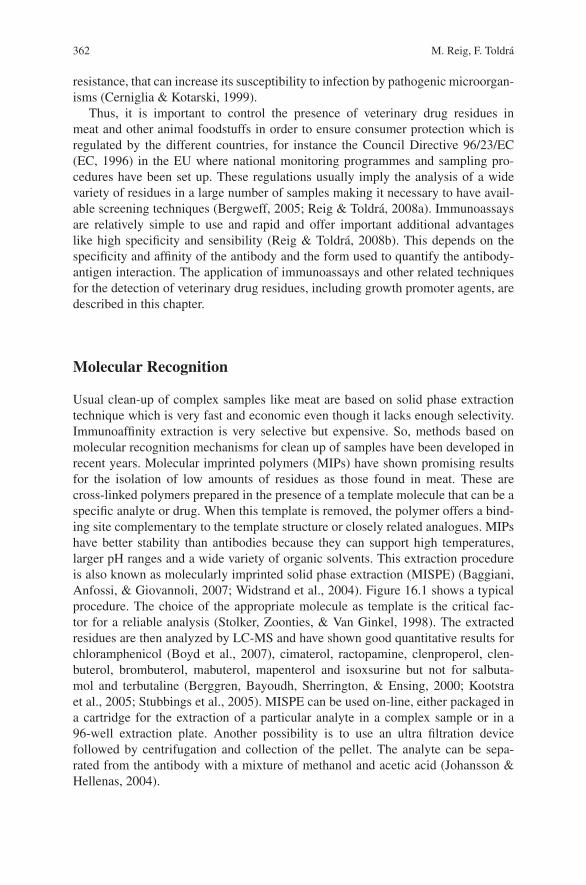
362 M. Reig, F. Toldra
resistance, that can increase its susceptibility to infection by pathogenic microorgan-isms (Cerniglia & Kotarski, 1999).
Thus, it is important to control the presence of veterinary drug residues inmeat and other animal foodstuffs in order to ensure consumer protection which isregulated by the different countries, for instance the Council Directive 96/23/EC(EC, 1996) in the EU where national monitoring programmes and sampling pro-cedures have been set up. These regulations usually imply the analysis of a widevariety of residues in a large number of samples making it necessary to have avail-able screening techniques (Bergweff, 2005; Reig & Toldra, 2008a). Immunoassaysare relatively simple to use and rapid and offer important additional advantageslike high specificity and sensibility (Reig & Toldra, 2008b). This depends on thespecificity and affinity of the antibody and the form used to quantify the antibody-antigen interaction. The application of immunoassays and other related techniquesfor the detection of veterinary drug residues, including growth promoter agents, aredescribed in this chapter.
Molecular Recognition
Usual clean-up of complex samples like meat are based on solid phase extractiontechnique which is very fast and economic even though it lacks enough selectivity.Immunoaffinity extraction is very selective but expensive. So, methods based onmolecular recognition mechanisms for clean up of samples have been developed inrecent years. Molecular imprinted polymers (MIPs) have shown promising resultsfor the isolation of low amounts of residues as those found in meat. These arecross-linked polymers prepared in the presence of a template molecule that can be aspecific analyte or drug. When this template is removed, the polymer offers a bind-ing site complementary to the template structure or closely related analogues. MIPshave better stability than antibodies because they can support high temperatures,larger pH ranges and a wide variety of organic solvents. This extraction procedureis also known as molecularly imprinted solid phase extraction (MISPE) (Baggiani,Anfossi, & Giovannoli, 2007; Widstrand et al., 2004). Figure 16.1 shows a typicalprocedure. The choice of the appropriate molecule as template is the critical fac-tor for a reliable analysis (Stolker, Zoonties, & Van Ginkel, 1998). The extractedresidues are then analyzed by LC-MS and have shown good quantitative results forchloramphenicol (Boyd et al., 2007), cimaterol, ractopamine, clenproperol, clen-buterol, brombuterol, mabuterol, mapenterol and isoxsurine but not for salbuta-mol and terbutaline (Berggren, Bayoudh, Sherrington, & Ensing, 2000; Kootstraet al., 2005; Stubbings et al., 2005). MISPE can be used on-line, either packaged ina cartridge for the extraction of a particular analyte in a complex sample or in a96-well extraction plate. Another possibility is to use an ultra filtration devicefollowed by centrifugation and collection of the pellet. The analyte can be sepa-rated from the antibody with a mixture of methanol and acetic acid (Johansson &Hellenas, 2004).
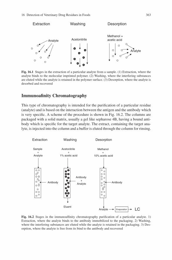
16 Detection of Veterinary Drug Residues in Foods 363
Extraction Washing Desorption
Analyte Acetonitrile
Analyte
Methanol + acetic acid
Fig. 16.1 Stages in the extraction of a particular analyte from a sample. (1) Extraction, where theanalyte binds to the molecular imprinted polymer. (2) Washing, where the interfering substancesare eluted while the analyte is retained in the polymer surface. (3) Desorption, where the analyte isdesorbed and recovered
Immunoafinity Chromatography
This type of chromatography is intended for the purification of a particular residue(analyte) and is based on the interaction between the antigen and the antibody whichis very specific. A scheme of the procedure is shown in Fig. 16.2. The columns arepackaged with a solid matrix, usually a gel like sepharose 4B, having a bound anti-body which is specific for the target analyte. The extract, containing the target ana-lyte, is injected into the column and a buffer is eluted through the column for rinsing.
Extraction Washing Desorption
Sample+
Analyte
Acetonitrile+
1% acetic acid
Analyte
Methanol+
10% acetic acid
EluentEvaporation LC
Antibody+
AnalyteAntibody Antibody
Fig. 16.2 Stages in the immunoaffinity chromatography purification of a particular analyte. 1)Extraction, where the analyte binds to the antibody immobilized to the packaging. 2) Washing,where the interfering substances are eluted while the analyte is retained in the packaging. 3) Des-orption, where the analyte is free from its bind to the antibody and recovered
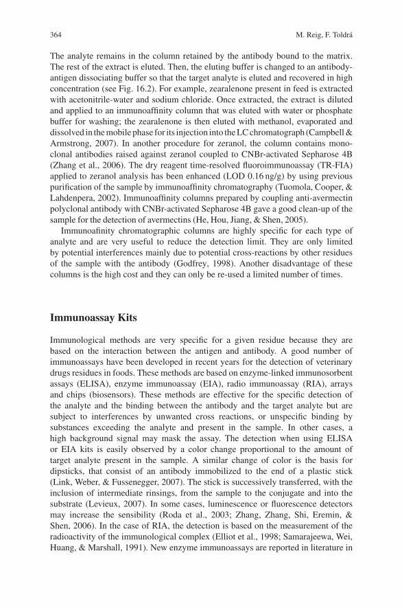
364 M. Reig, F. Toldra
The analyte remains in the column retained by the antibody bound to the matrix.The rest of the extract is eluted. Then, the eluting buffer is changed to an antibody-antigen dissociating buffer so that the target analyte is eluted and recovered in highconcentration (see Fig. 16.2). For example, zearalenone present in feed is extractedwith acetonitrile-water and sodium chloride. Once extracted, the extract is dilutedand applied to an immunoaffinity column that was eluted with water or phosphatebuffer for washing; the zearalenone is then eluted with methanol, evaporated anddissolved in the mobile phase for its injection into the LC chromatograph (Campbell &Armstrong, 2007). In another procedure for zeranol, the column contains mono-clonal antibodies raised against zeranol coupled to CNBr-activated Sepharose 4B(Zhang et al., 2006). The dry reagent time-resolved fluoroimmunoassay (TR-FIA)applied to zeranol analysis has been enhanced (LOD 0.16 ng/g) by using previouspurification of the sample by immunoaffinity chromatography (Tuomola, Cooper, &Lahdenpera, 2002). Immunoaffinity columns prepared by coupling anti-avermectinpolyclonal antibody with CNBr-activated Sepharose 4B gave a good clean-up of thesample for the detection of avermectins (He, Hou, Jiang, & Shen, 2005).
Immunoafinity chromatographic columns are highly specific for each type ofanalyte and are very useful to reduce the detection limit. They are only limitedby potential interferences mainly due to potential cross-reactions by other residuesof the sample with the antibody (Godfrey, 1998). Another disadvantage of thesecolumns is the high cost and they can only be re-used a limited number of times.
Immunoassay Kits
Immunological methods are very specific for a given residue because they arebased on the interaction between the antigen and antibody. A good number ofimmunoassays have been developed in recent years for the detection of veterinarydrugs residues in foods. These methods are based on enzyme-linked immunosorbentassays (ELISA), enzyme immunoassay (EIA), radio immunoassay (RIA), arraysand chips (biosensors). These methods are effective for the specific detection ofthe analyte and the binding between the antibody and the target analyte but aresubject to interferences by unwanted cross reactions, or unspecific binding bysubstances exceeding the analyte and present in the sample. In other cases, ahigh background signal may mask the assay. The detection when using ELISAor EIA kits is easily observed by a color change proportional to the amount oftarget analyte present in the sample. A similar change of color is the basis fordipsticks, that consist of an antibody immobilized to the end of a plastic stick(Link, Weber, & Fussenegger, 2007). The stick is successively transferred, with theinclusion of intermediate rinsings, from the sample to the conjugate and into thesubstrate (Levieux, 2007). In some cases, luminescence or fluorescence detectorsmay increase the sensibility (Roda et al., 2003; Zhang, Zhang, Shi, Eremin, &Shen, 2006). In the case of RIA, the detection is based on the measurement of theradioactivity of the immunological complex (Elliot et al., 1998; Samarajeewa, Wei,Huang, & Marshall, 1991). New enzyme immunoassays are reported in literature in
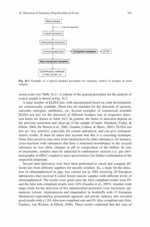
16 Detection of Veterinary Drug Residues in Foods 365
Meat sample
Liquid extraction
Solid phase extraction
Immunoassay test
Confirmatory methodsLC-MS, GC-MS, etc.
Non-compliant samples
Compliant samples STOP
+Internal standard
Fig. 16.3 Example of a typical standard procedure for routinary control of residues in meatsamples
recent years (see Table 16.1). A scheme of the general procedure for the analysis ofa meat sample is shown in Fig. 16.3.
A large number of ELISA kits, with measurement based on color development,are commercially available. These kits are intended for the detection of agonists,esteroids, estrogens, antibiotics, etc. Several examples of commercial availableELISA test kits for the detection of different residues and its respective detec-tion limits are shown in Table 16.2. In general, the limits of detection depend onthe previous extraction and clean-up of the sample (Cooper, Delahaut, Fodey, &Elliott, 2004; De Wasch et al., 2001; Gaudin, Cadieu, & Maris, 2003). ELISA testkits are very sensitive, especially for certain substances, and can give semiquan-titative results. It must be taken into account that this is a screening technique.Some false positives may arise from interferences by other substances, for instance,cross-reactions with substances that have a structural resemblance to the assayedsubstance or, less often, changes in pH or composition of the buffers. In caseof uncertainty, samples must be subjected to confirmatory analysis (i.e. gas chro-matography or HPLC coupled to mass spectrometry) for further confirmation of thesuspected compound.
Several inter-laboratory tests have been performed to check and compare dif-ferent kits from different suppliers for specific residues. So, a study for the detec-tion of chloramphenicol in pigs was carried out in 2002 involving 20 Europeanlaboratories that received 8 coded frozen muscle samples with different levels ofchloramphenicol. The results were good since the false compliant results were 0%and the false non-compliant results were 10% (Gaudin et al., 2003). Another widerange study for the detection of five antimicrobial promoters (zinc bacitracin, spi-ramycin, tylosin, virginiamycin and olaquindox) in feedstuffs with 15 Europeanlaboratories representing government agencies and private industry also revealedgood results with a 1.9% false non-compliant rate and 0% false compliant rate (Situ,Grutters, van Wichen, & Elliott, 2006). These results confirmed that this type of
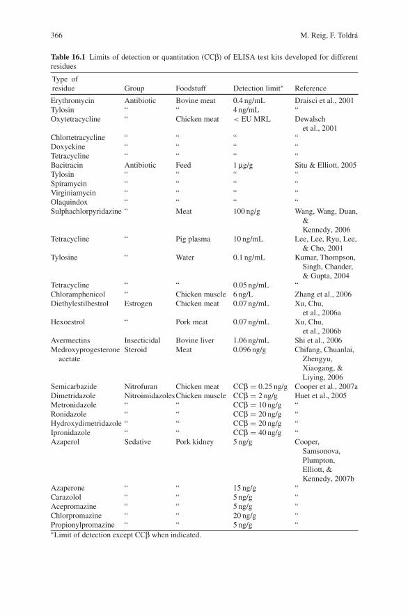
366 M. Reig, F. Toldra
Table 16.1 Limits of detection or quantitation (CCβ) of ELISA test kits developed for differentresidues
Type ofresidue Group Foodstuff Detection limit∗ Reference
Erythromycin Antibiotic Bovine meat 0.4 ng/mL Draisci et al., 2001Tylosin “ “ 4 ng/mL “Oxytetracycline “ Chicken meat < EU MRL Dewalsch
et al., 2001Chlortetracycline “ “ “ “Doxyckine “ “ “ “Tetracycline “ “ “ “Bacitracin Antibiotic Feed 1 μg/g Situ & Elliott, 2005Tylosin “ “ “ “Spiramycin “ “ “ “Virginiamycin “ “ “ “Olaquindox “ “ “ “Sulphachlorpyridazine “ Meat 100 ng/g Wang, Wang, Duan,
&Kennedy, 2006
Tetracycline “ Pig plasma 10 ng/mL Lee, Lee, Ryu, Lee,& Cho, 2001
Tylosine “ Water 0.1 ng/mL Kumar, Thompson,Singh, Chander,& Gupta, 2004
Tetracycline “ “ 0.05 ng/mL “Chloramphenicol “ Chicken muscle 6 ng/L Zhang et al., 2006Diethylestilbestrol Estrogen Chicken meat 0.07 ng/mL Xu, Chu,
et al., 2006aHexoestrol “ Pork meat 0.07 ng/mL Xu, Chu,
et al., 2006bAvermectins Insecticidal Bovine liver 1.06 ng/mL Shi et al., 2006Medroxyprogesterone
acetateSteroid Meat 0.096 ng/g Chifang, Chuanlai,
Zhengyu,Xiaogang, &Liying, 2006
Semicarbazide Nitrofuran Chicken meat CCβ = 0.25 ng/g Cooper et al., 2007aDimetridazole NitroimidazolesChicken muscle CCβ = 2 ng/g Huet et al., 2005Metronidazole “ “ CCβ = 10 ng/g “Ronidazole “ “ CCβ = 20 ng/g “Hydroxydimetridazole “ “ CCβ = 20 ng/g “Ipronidazole “ “ CCβ = 40 ng/g “Azaperol Sedative Pork kidney 5 ng/g Cooper,
Samsonova,Plumpton,Elliott, &Kennedy, 2007b
Azaperone “ “ 15 ng/g “Carazolol “ “ 5 ng/g “Acepromazine “ “ 5 ng/g “Chlorpromazine “ “ 20 ng/g “Propionylpromazine “ “ 5 ng/g “∗Limit of detection except CCβ when indicated.
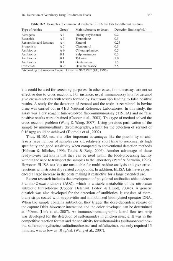
16 Detection of Veterinary Drug Residues in Foods 367
Table 16.2 Examples of commercial available ELISA test kits for different residues
Type of residue Group∗ Main substance to detect Detection limit (ng/mL)
Estrogens A 1 Diethylestylbestrol 0.2Esteroids A 3 Trenbolone 0.5Resorcylic acid lactones A 4 Zeranol 0.25B-agonists A 5 Clenbuterol 0.3Antibiotics A 6 Chloramphenicol 0.5Antibiotics B 1 Sulphonamides 0.5Antibiotics B 1 Tylosine 5.0Antibiotics B 1 Gentamicine 1.5Corticoids B 2f Dexamethasone 2.5∗According to European Council Directive 96/23/EC (EC, 1996).
kits could be used for screening purposes. In other cases, immunoassays are not soeffective due to cross reactions. For instance, usual immunoassay kits for zeranolgive cross-reactions with toxins formed by Fusarium spp leading to false positiveresults. A study for the detection of zeranol and the toxin α-zearalenol in bovineurine was carried out in 4 EU National Reference Laboratories. In this study, theassay was a dry reagent time-resolved fluoroimmunoassay (TR-FIA) and no falsepositive results were obtained (Cooper et al., 2003). This type of method solved thecross-reaction problem (Wang & Wang, 2007). Using previous purification of thesample by immunoaffinity chromatography, a limit for the detection of zeranol of0.16 ng/g could be achieved (Tuomola et al., 2002).
Thus, ELISA test kits offer important advantages like the possibility to ana-lyze a large number of samples per kit, relatively short time to response, its highspecificity and good sensitivity when compared to conventional detection methods(Hahnau & Julicher, 1996; Toldra & Reig, 2006). Another advantage of theseready-to-use test kits is that they can be used within the food-processing facilitywithout the need to transport the samples to the laboratory (Paraf & Sarradin, 1996).However, ELISA test kits are unsuitable for multi-residue analysis and give cross-reactions with structurally related compounds. In addition, ELISA kits have experi-enced a large increase in the costs making it restrictive for a large extended use.
Recent research includes the development of polyclonal antibodies able to detect3-amino-2-oxazolidinone (AOZ), which is a stable metabolite of the nitrofuranantibiotic furazolidone (Cooper, Delahaut, Fodey, & Elliott, 2004b). A genericdipstick was also developed for the detection of antibiotics. It consists of mem-brane strips coated with streptavidin and immobilized biotinylated operator DNA.When the sample contains antibiotics, they trigger the dose-dependent release ofthe capture DNA-biosensor interaction and the color developed can be determinedat 450 nm. (Link et al., 2007). An immunochromatographic lateral-flow test stripwas developed for the detection of sulfonamides in chicken muscle. It was in thecompetitive reaction format and the sensitivity for sulfonamides (sulfamonomethox-ine, sulfamethoxydiazine, sulfadimethoxine, and sulfadiazine), that only required 15minutes, was as low as 10 ng/mL (Wang et al., 2007).
368 M. Reig, F. Toldra
Biosensors
The rapid developments in biological and chemical technology have prompted theminiaturization for integrated, high-throughput and high-sensitivity analysis. Thesenew biosensors have been applied for the rapid detection of veterinary drugs in foodsof animal origin. The basis consists of the interaction of an immobilized antibody onthe surface of a transducer that interacts with the analyte in the sample, in most caseswith no need for sample clean-up (Wang, Wang, Duan, & Kennedy, 2006). Thechanges at the surface are converted into a measurable signal (optical or electronicaldetection) (De Wasch et al., 2001, Draisci et al., 2001). An advantage of biosensorsis the possibility for simultaneous detection, at a time, of multiple veterinary drugsresidues in a sample (Kumar, Thompson, Singh, Chander, & Gupta, 2004). The sur-face plasmon resonance (SPR) measures variations in the refractive index of thesolution adjacent to the metal surface (Cooper, 2004; Hauguey & Baxter, 2006). Theimmobilized antibody on the surface causes a shift in the resonance angle but whena high molecular antigen binds to the antibody, it causes a further change (Grundig,& Renneberg, 2002). This biosensor, which is commercially available as a fully
automated system with the name BiacoreTM
, allows real time quantification of theantibody-antigen interaction. Important advantages are simplicity, high precision,speed (few minutes per sample) and automation. Furthermore, they provide a widevariability of target analytes and sensibility depending on the signal to noise ratio.So, it has been successfully applied to the detection of different veterinary drugsresidues like ractopamine (Thompson et al., 2008), nitroimidazoles (Connollyet al., 2007; Cooper, Samsonova, Plumpton, Elliott, & Kennedy, 2007a; Situ &Elliott, 2005), clenbuterol in urine at a limit of detection of 0.27 ng/mL (Haugheyet al., 2001), flumequine in broiler muscle at a CCα of 500 ng/g (Haasnoot, Gercek,Cazemier, & Nielen, 2007), chloramphenicol in poultry with a CCα of 0.005 ng/g(Ferguson et al., 2005) and chloramphenicol glucuronide in kidney at CCα¡0.1 ng/g(Ashwin et al., 2005) and sulphonamides (sulfametazine and sulfadiazine) in porkmeat at a LOD of 0.1 ng/g (Crooks, Baxter, O’Connor, & Elliot, 1998). Twentysulfonamide residues were detected with a SPR biosensor in porcine muscle at alimit of detection of 16.9 ng/g (McGrath, Baxter, Ferguson, Haughey, & Bjurling,2005). Even a specific antibody mutant was developed for the SPR biosen-sor towards a multi-sulfonamide detection (Bienenmann-Ploum, Korpimaki,Haasnoot, & Kohen, 2005). SPR sensor system was also successfully tested ina slaughterhouse under cold and humid conditions (Baxter, O’Connor, Haughey,Crooks, & Elliott, 1999). Another SPR biosensor, where the extract is injectedover the surfaces of two chip flow cells, has been developed for the detection ofthiamphenicol, florenicol, florefenicol amine and chloramphenicol in shrimps. Thelimits of quantitation (CCβ) were 0.1, 0.2, 250 and 0.5 ppb, respectively (Dumont,Huet, Traynor, Elliott, & Delahaut, 2006). Different kits (Qflex� kit) with specificantibodies against a wide variety of residues are commercially available.
Other biosensors are based on the use of biochip arrays or microarrays. Smallmolecule microarrays use small molecules as probes immobilized on a variety ofsurfaces (Zuo and Ye, 2006). The target residues are conjugated with a carrier protein
16 Detection of Veterinary Drug Residues in Foods 369
and printed on modified glass slides. These residues are still able to interact withthe respective antibodies in solution or in the sample. The system is incubated forantigen-antibody reaction that is detected by using fluorescence labelled secondaryantibody. This biosensor has been successfully applied to the detection of clenbuterol,chloramphenicol and tylosin (Peng & Bang-Ce, 2006). These chips are specific fora certain number of residues like chloramphenicol in meat (Gaudin et al., 2003),nitroimidazoles in chicken (Huet et al., 2005) as well as clenbuterol and the hormoneanalogues ethinylestradiol and trenbolone (Johansson & Hellenas, 2001).
A broad spectrum protein biosensor was developed for the assay of tetracycline,streptogramin and macrolide antibiotics (Weber et al., 2005). This biosensor is basedon the interaction of protein sensor with specific DNA fragments chemically linkedto a solid surface. This interaction gives a particular response that is converted intoa colorimetric readout for each class of antibiotics and the color intensity is propor-tional to the antibiotic concentration.
References
Ashwin, H. M., Stead, S. L., Taylor, J. C., Startin, J. R., Richmond, S. F., Homer, V., et al.(2005). Development and validation of screening and confirmatory methods for the detec-tion of chloramphenicol and chloramphenicol glucuronide using SPR biosensor and liquidchromatography-tandem mass spectrometry. Analytica Chimica Acta, 529, 103–108.
Baggiani, C., Anfossi, L., & Giovannoli, C. (2007). Solid phase extraction of food contaminantsusing molecular imprinted polymers. Analytica Chimica Acta, 591, 29–39.
Baxter, G. A., O’Connor, M. C., Haughey, S. A., Crooks, S. R. H., & Elliott, C. T. (1999). Evalu-ation of an immunobiosensor for the on-site testing of veterinary drug residues at an abattoir.Screening for sulfamethazine in pigs. Analyst, 124, 1315–1318.
Berggren, C., Bayoudh, S., Sherrington, D., & Ensing, K. (2000). Use of molecularly imprintedsolid-phase extraction for the selective clean-up of clenbuterol from calf urine. Journal of Chro-matography A, 889, 105–110.
Bergweff, A. A. (2005). Rapid assays for detection of residues of veterinary drugs. In A. vanAmerongen, D. Barug, & M. Lauwars (Eds.), Rapid methods for biological and chemical con-taminants in food and feed (pp. 259–292). Wageningen: Academic Publishers.
Bienemann-Ploum, M., Korpimaki, T., Haasnoot, W., & Kohen, F. (2005). Comparison of multisulfonamide biosensor immunoassays. Analytica Chimica Acta, 529, 115–122.
Boyd, B., Bjork, H., Billing, J., Shimelis, O., Axelsson, S., Leonora, M., et al. (2007). Develop-ment of an improved method for trace analysis of chloramphenicol using molecularly imprintedpolymers. Journal of Chromatography A, 1174, 63–71.
Campbell, H. M., & Armstrong, J. F. (2007). Determination of zearalenone in cereal grains, ani-mal feed, and feed ingredients using immunoaffinity column chromatography and liquid chro-matography: Interlaboratory study. Journal of AOAC International, 90, 1610–1622.
Cerniglia, C. E., & Kotarski, S. (1999). Evaluation of veterinary drug residues in food for theirpotential to affect human intestinal microflora. Regulatory Toxicology and Pharmacology 29,238–261.
Cerniglia, C. E., & Kotarski, S. (2005). Approaches in the safety evaluations of veterinary antimi-crobial agents in food to determine the effects on the human intestinal microflora. Journal ofVeterinary Pharmacology and Therapy, 28, 3–20.
Chadwick, R. W., George, S. E., & Claxton, L. D. (1992). Role of gastrointestinal mucosa andmicroflora in the bioactivation of dietary and environmental mutagens or carcinogens. DrugMetabolism Reviews, 2, 425–492.
370 M. Reig, F. Toldra
Chifang, P., Chuanlai, X., Zhengyu, J., Xiaogang, C., & Liying, W. (2006). Determination ofanabolic steroid residues (medroxyprogesterone acetate) in pork by ELISA and comparisonwith liquid chromatography tandem mass spectrometry. Journal of Food Science, 71,C044–C050.
Cinquina, A. L. Longo, F., Anastasi, G., Giannetti, L., & Cozzani, R. (2003). Validation of ahigh-performance liquid chromatography method for the determination of oxytetracycline,tetracycline, chlortetracycline and doxycycline in bovine milk and muscle. Journal of Chro-matography A, 987, 227–233.
Connolly, L., Thompson, C. S., Haughey, S. A., Traynor, I. M., Tittlemeier, S., & Elliot, C. T.(2007). The development of a multi.nitorimidazole residue analysis assay by optical biosensorvia a proof of concept project to develop and assess a prototype test kit. Analytica ChimicaActa, 598, 155–161.
Cooper, J., Delahaut, P., Fodey, T. L., & Elliott, C. T. (2004). Development of a rapid screening testfor veterinary sedatives and the beta-blocker carazolol in porcine kidney by ELISA. Analyst,129, 169–174.
Cooper, K. M., Caddell, A., Elliott, C. T., & Kennedy, D. G. (2004). Production and characteri-sation of polyclonal antibodies to a derivative of 3-amino-2-oxazolidinone, a metabolite of thenitrofuran furazolidone. Analytica Chimica Acta, 520, 79–86.
Cooper, K. M., Ribeiro, L., Alves, P., Vozikis, V., Tsitsamis, S., Alfredssonk, G., et al. (2003).Interlaboratory ring test of time-resolved fluoroimmunoassays for zeranol and a-zearalenol andcomparison with zeranol test kits. Food Additives and Contaminants, 20, 804–812.
Cooper, K. M., Samsonova, J. V., Plumpton, L., Elliott, C. T., & Kennedy, D. G. (2007a). Enzymeimmunoassay for semicarbazide—The nitrofuran metabolite and food contaminant. AnalyticaChimica Acta, 592, 64–71.
Cooper, K. M., Samsonova, J. V., Plumpton, L., Elliott, C. T., & Kennedy, D. G. (2007b). Enzymeimmunoassay for semicarbazide—The nitrofuran metabolite and food contaminant. AnalyticaChimica Acta, 592, 64–71.
Crooks, S. R. H., Baxter, G. A., O’Connor, M. C., & Elliot, C. T. (1998). Immunobiosensor – analternative to enzyme immunoassay screening for residues of two sulfonamides in pigs. Analyst,123, 2755–2757.
De Wasch, K., Okerman, L., Croubels, S., De Brabander, H., Van Hoof, J., & De Backer, P. (2001).Detection of residues of tetracycline antibiotics in pork and chicken meat: Correlation betweenresults of screening and confirmatory tests. Analyst, 123, 2737–2741.
Draisci, R. delli Quadri, F., Achene, L., Volpe, G., Palleschi, L., & Palleschi, G. (2001). A newelectrochemical enzyme’linked immunosorbent assay for the screening of macrolide antibioticresidues in bovine meat. Analyst, 126, 1942–1946.
Dumont, V., Huet, A. C., Traynor, I., Elliott, C., & Delahaut, P. (2006). A surface plasmon res-onance biosensor assay for the simultaneous determination of thiamphenicol, florefenicol,florefenicol amine and chloramphenicol residues in shrimps. Analytica Chimica Acta, 567,179–183.
EC. (1996). Council Directive 96/23/EEC of 29 April 1996 on measures to monitor certain sub-stances and residues thereof in live animals and animal products. Official Journal of the Euro-pean Community L, 125, 10.
Elliott, C. T., Baxter, G. A., Hewitt, S. A., Arts, C. J. M., van Baak, M., Hellenas, K. E., et al.(1998). Use of biosensors for rapid drug residue analysis without sample deconjugation orclean-up: a possible way forward. Analyst, 123, 2469–2473.
Ferguson, J., Baxter, A., Young, P., Kennedy, G., Elliott, C., Weigel, S., et al. (2005). Detection ofchloramphenicol and chloramphenicol glucuronide residues in poultry muscle, honey, prawnand milk using a surface plasmon resonance biosensor and Qflex� kit chloramphenicol. Ana-lytica Chimica Acta, 529, 109–113.
Gaudin, V., Cadieu, N., & Maris, P. (2003). Inter-laboratory studies for the evaluation of ELISAkits for the detection of chloramphenicol residues in milk and muscle. Food and AgriculturalImmunology, 15, 143–157.
16 Detection of Veterinary Drug Residues in Foods 371
Godfrey, M. A. J. (1998). Immunoafinity extraction in veterinary residue analysis-a regulatoryviewpoint. Analyst, 123, 2501–2506.
Grundig, B., & Renneberg, R. (2002). Chemical and biochemical sensors. In A. Katerkamp, B.Grundig, & R. Renneberg (Eds.), Ullmann’s Encyclopedia of Industrial Chemistry (pp. 87–98).Verlag: Wiley-VCH.
Guo, J. J., Chou, H. N., & Liao, I. C. (2003). Disposition of 3-(4-cyano-2-oxobutylidene amino)-2-oxazolidone, a cyano-metabolite of furazolidone, in furazolidone-treated grouper. Food Addi-tives and Contaminants, 20, 229–236.
Haasnoot, W., Gercek, H., Cazemier, G., & Nielen, M. W. F. (2007). Biosensor immunoassay forflumequine in broiler serum and muscle. Analytica Chimica Acta, 586, 312–318.
Hahnau, S., & Julicher, B. (1996). Evaluation of commercially available ELISA test kits forthe detection of clenbuterol and other β-agonists. Food Additives and Contaminants, 13,259–274.
Haughey, S. A., & Baxter, C. A. (2006). Biosensor screening for veterinary drug residues in food-stuffs. Journal of AOAC International, 89, 862–867.
He, J. H., Hou, X. L., Jiang, H. Y., & Shen, J. Z. (2005). Multiresidue analysis of avermectinsin bovine liver by immunoaffinity column cleanup procedure and liquid chromatography withfluorescence detector. Journal of AOAC International, 88, 1099–1103.
Haughey, S. A., Baxter, G. A., Elliot, C. T., Persson, B., Jonson, C., & Bjurling, P. (2001). Deter-mination of clenbuterol residues in bovine urine by optical immunobiosensor assay. Journal ofAOAC International, 84, 1025–1030.
Huet, A. C. Mortier, L., Daeseleire, E., Fodey, T., Elliott, C. T., & Delahaut, P. (2005). Developmentof an ELISA screening test for nitroimidazoles in egg and chicken muscle Analytica ChimicaActa, 534, 157–162.
Johansson, M. A., & Hellenas, K. E. (2001). Sensor chip preparation and assay construction forimmunobiosensor determination of beta-agonists and hormones. Analyst, 126, 1721–1727.
Johansson, M. A., & Hellenas, K. E. (2004). Immunobiosensor determination of b-agonists inurine using integrated immunofiltration clean-up. International Journal of Food Science andTechnology, 39, 891–898.
Kootstra, P. R. Kuijpers, C. J. P. F., Wubs, K. L., van Doorn, D., Sterk, S. S., van Ginkel, L. A.,et al. (2005). The analysis of beta-agonists in bovine muscle using molecular imprinted poly-mers with ion trap LCMS screening. Anaytica Chimica Acta, 529, 75–81.
Kumar, K., Thompson, A., Singh, A. K., Chander, Y., & Gupta, S. C. (2004). Enzyme-linkedimmunosorbent assay for ultratrace determination of antibiotics in aqueous samples. Journalof Environmental Quality, 33, 250–256.
Lee, H. J., Lee, M. H., Ryu, P. D., Lee, H., & Cho, M. H. (2001). Enzyme-linked immunosorbentassay for screening the plasma residues of tetracycline antibiotics in pigs. Jounal of VeterinaryMedicine, 63, 553–556.
Levieux, D. (2007). Immunodiagnosctic technology and its applications. In L. M. L. Nollet, &F. Toldra (Eds.), Advances in Food Diagnostics (pp. 211–227). Ames, Iowa: BlackwellPublishing.
Link, N., Weber, W., & Fussenegger, M. (2007). A novel generic dipstick-based technology forrapid and precise detection of tetracycline, streptogramin and macrolide antibiotics in foodsamples. Journal of Biotechnology, 128, 668–680.
McGrath, T., Baxter, A., Ferguson, J., Haughey, S., & Bjurling, P. (2005). Multi sulfonamidescreening in porcine muscle using a surface plasmon resonance biosensor. Analytica ChimicaActa, 529, 123–127.
Moore, W. E. C. & Moore, L. H. (1995). Intestinal floras of populations that have risk of coloncancer. Applied and Environmental Microbiology, 61, 3202-3207.
Mottier, P., et al. (2003). Determination of the antibiotic chloramphenicol in meat and seafoodproducts by liquid chromatography–electrospray ionization tandem mass spectrometry. Journalof Chromatography A, 994, 75–84.
372 M. Reig, F. Toldra
Paraf, A., & Sarradin, P. (1996). Immunochemistry in food control. Recent Research Developmentsin Nutrition, 1, 99–114.
Pecorelli, I., Bibi, R., Fioroni, L., & Galarini, R. (2004). Validation of a confirmatory method forthe determination of sulphonamides in muscle according to the European Union regulation2002/657/EC. Journal of Chromatography A, 1032, 23–29.
Peng, Z., & Bang-Ce, Y. (2006). Small molecule microarrays for drug residue detection in food-stuffs. Journal of Agricultural and Food Chemistry, 54, 6978–6983.
Reig, M., & Toldra, F. (2007). Chemical origin toxic compounds. In F. Toldra, Y. H. Hui, I. Asti-asaran, W. K. Nip, J. G. Sebranek, E. T. F. Silveira, et al. (Eds.), Handbook of fermented meatand poultry (pp. 469–475). Ames, Iowa: Blackwell Publishing.
Reig, M., & Toldra, F. (2008a). Veterinary drug residues in meat: Concerns and rapid methods fordetection. Meat Science, 78, 60–67.
Reig, M., & Toldra, F. (2008b). Growth promoters. In L. M. L. Nollet, & F. Toldra (Eds.), Hand-book of muscle foods analysis. Boca Raton, Fl: CRC Press (in press).
Roda, A., Manetta, A. C., Portanti, O., Mirasoli, M., Guardigli, M., Pasini, P., et al. (2003). Arapid and sensitive 384-well microtitre format chemiluminiscent enzyme immunoassay for19.nortestosterone. Luminescence, 18, 72–78.
Samarajeewa, U., Wei, C. I., Huang, T. S., & Marshall, M. R. (1991). Application of immunoassayin the food industry. Critical Reviews in Food Science and Nutrition, 29, 403–434.
Shi, W. M., He, J. H., Jiang, H. Y., Hou, X. L., Yang, J. H., & Shen, J. Z. (2006). Determinationof multiresidue of avermectins in bovine liver by an indirect competitive ELISA. Journal ofAgricultural and Food Chemsitry, 54, 6143–6146.
Situ, C., & Elliott, C. T. (2005). Simultaneous and rapid detection of five banned antibiotic growthpromoters by immunoassay. Analytica Chimica Acta, 529, 89–96.
Situ, C., Grutters, E., van Wichen, P., & Elliott, C. T. (2006). A collaborative trial to evaluatethe performance of a multi-antibiotic enzyme-linked immunosorbent assay for screening fivebanned antimicrobial growth promoters in animal feedingstuffs. Analytica Chimica Acta, 561,62–68.
Stolker, A. A. M., Zoonties, P. W., & Van Ginkel, L. A. (1998). The use of supercritical fluidextraction for the determination of steroids in animal tissues. Analyst, 123, 2671–2676.
Stubbings, G., Tarbin, J., Cooper, A., Sharman, M., Bigwood, T., & Robb, P. (2005). A multi-residue cation-exchange clean up procedure for basic drugs in produce of animal origin. Ana-lyitica Chimica Acta, 547, 262–268.
Thompson, C. S., Haughey, S. A., Traynor, I. M., Fodey, T. L., Elliot, C. T., Antignac, J. P.,et al. (2008). Effective monitoring of ractopamine residues in samples of animal origin bySPR biosensor and mass spectrometry. Analytica Chimica Acta, 608, 217–225.
Toldra, F., & Reig, M. (2006). Methods for rapid detection of chemical and veterinary drug residuesin animal foods. Trends Food Science and Technology, 17, 482–489.
Tuomola, M., Cooper, K. M., & Lahdenpera, S. (2002). A specificity-emhanced time-resolvedfluoroimmunoassay for zeranol employing the dry reagent all-in-one-well principle. Analyst,127, 83–86.
Vollard, E. J., & Clasener, H. A. L. (1994). Colonization resistance. Antimicrobial Agents andChemotherapy, 38, 409–414.
Wang, S., Wang, Z. L., Duan, Z. J., & Kennedy, I. (2006). Analysis of sulphonamide residues inedible animal products: A review. Food Additives and Contaminants, 23, 362–384.
Wang, S., & Wang, X. H. (2007). Analytical methods for the determination of zeranol residues inanimal products: A review. Food Additives and Contaminants, 24, 573–582.
Wang, X. L., Li, K., Shi, D. S., Xiong, N., Jin, X., Yi, J. D., et al. (2007). Development of animmunochromatographic lateral-flow test strip for rapid detection of sulfonamides in eggs andchicken muscles. Journal of Agricultural and Food Chemistry, 55, 2072–2078.
Weber, C. C., Link, N., Fux, C., Zisch, A. H., Weber, W., & Fussenegger, M. (2005). Broad-spectrum protein biosensors for class-specific detection of antibiotics. Biotechnology and Bio-engineering, 89, 9–17.
16 Detection of Veterinary Drug Residues in Foods 373
Widstrand, C. Larsson, F., Fiori, M., Civitareale, C., Mirante, S., & Brambilla, G. (2004). Eval-uation of MISPE for the multi-residue extraction of β-agonists from calves urine. Journal ofChromatography B, 804, 85–91.
Xu, C. L., Chu, X. G., Peng, C. F., Liu, L. Q., Wang, L. Y., & Jin, Z. (2006). Comparison ofenzyme-linked immunosorbent assay with liquid chromatography-tandem mass spectrometryfor the determination of diethylstilbesterol residues in chicken and liver tissues. BiomedicalChromatography, 20, 1956–1064.
Xu, C. L., Peng, C. F., Liu, L. Q., Wang, L. Y., Jin, Z. Y., & Chu, X. G. (2006). Determination ofhexoestrol residues in animal tissues based on enzyme-linked immunosorbent assay and com-parison with liquid chromatography-tandem mass spectrometry. Journal of Pharmaceutical andBiomedical Analysis, 41, 1029–1036.
Zhang, S. X., Zhang, Z., Shi, W. M., Eremin, S. A., & Shen, J. Z. (2006). Development of achemiluminescent ELISA for determining chloramphenicol in chicken muscle. Journal of Agri-cultural and Food Chemistry, 54, 5718–5722.
Zhang, W., Wang, H. H., Wang, J. P., Li, X. W., Jiang, H. Y., & Shen, J. Z. (2006). Multiresiduedetermination of zeranol and related compounds in bovine muscle by gas chromatography/massspectrometry with immunoaffinity cleanup. Journal of AOAC Internationa,l 89, 1677–1681.
Zuo, P., & Ye, B. C. (2006). Small molecule microarrays for drug residue detection in foodstuffs.Journal of Agricultural and Food Chemistry, 54, 6978–6983.
Chapter 17Antimicrobial Activity of Bacteriocins and TheirApplications
Eleftherios H. Drosinos, Marios Mataragas, and Spiros Paramithiotis
Introduction
Bacteriocins are peptides or proteins that exert an antimicrobial action against arange of microorganisms. Their production can be related to the antagonism withina certain ecological niche, as the producer strain, being itself immune to its action,generally gains a competitive advantage. Many Gram-positive and Gram-negativemicroorganisms have been found to produce bacteriocins. The former, and espe-cially the ones produced by lactic acid bacteria, has been the field of intensiveresearch during the last decades mainly due to their properties that account for theirsuitability in food preservation and the benefits arising from that, and secondarilydue to the broader inhibitory spectrum compared to the ones produced by Gram-negative microorganisms. Bacteriocins are ribosomally synthesized peptides whoseresponsible genes are frequently associated with mobilisable elements on the chro-mosome such as in association with transposons or on plasmids (Deegan, Cotter,Hill, & Ross, 2006). This property is one of the main differences that distinguishthem from antibiotics (Cleveland, Montville, Nes, & Chikindas, 2001). The latterare considered being secondary metabolites and although many of them compriseamino acids, they are enzymatically rather than ribosomally synthesized.
Classification and Biochemical Properties
Bacteriocin classification has been a field of extensive debate. The original classifi-cation scheme has been proposed by Klaenhammer (1993); it was then modifiedby Nes et al. (1996), van Belkum and Stiles (2000) and Nes and Holo (2000)and new propositions were recently made by Cotter, Hill, and Ross (2005) andFranz, van Belkum, Holzapfel, Abriouel, and Galvez (2007), with the latter refer-ring mainly to enterococcal bacteriocins, since many of them appear as atypical
E.H. DrosinosLaboratory of Food Quality Control and Hygiene, Department of Food Science and Technology,Agricultural University of Athens, 75, Iera Odos Street, Votanicos, GR-118 55, Athens, Greece
F. Toldra (ed.), Meat Biotechnology, 375C© Springer Science+Business Media, LLC 2008
376 E.H. Drosinos et al.
and structurally distinct from the previously described general classes (Table 17.1).Moreover, the inclusion of a fifth class consisting of ribosomally synthesized non-modified head-to-tail-ligated cyclic antibacterial peptides has been proposed byKemperman et al. (2003), a suggestion that has met with controversial reactionssince these bacteriocins could fit into the second class (Cotter et al., 2005). It is,though, generally accepted that on the basis of their structure as well as biochemicalproperties, bacteriocins are divided into three clusters: lantibiotics, non-lantibioticsand non-lantibiotics heat-labile large molecules. Since the vast majority of the bac-teriocins produced by lactic acid bacteria belong to the first two classes, a hugeamount of data currently exists concerning their production and excresion, immu-nity, mode of action and factors that affect their production or activity in vitro orin situ.
Lantibiotics are characterized by the presence of thioether cross-links termed lan-thionines or methyl-lanthionines in addition to the amino acids 2,3-didehydroalanineand (Z)-2,3-didehydrobutyrine. The lantibiotic biosynthetic genes are clustered andlocated on the bacterial chromosome or on large plasmids. The products of thebiosynthetic genes have been classified into structural, modification, processing,transport, immunity and regulation proteins. The precursor peptide that is initiallyproduced contains an amino-terminal leader sequence of 23–59 amino acids anda structural region that is further modified in order to introduce several structuralmotifs. Several roles have been assigned to the leader peptide; among them is acontribution to the cell’s immunity by retaining the lantibiotic inactive within thecell, as a transport aid, and as a recognition sequence for the biosynthetic enzymesresponsible for production of the active lantibiotic (Chatterjee, Paul, Xie, & vander Donk, 2005). After the modification of the precursor peptide, the leader peptidemust be cleaved off and translocated across the cytoplasmic membrane. This isperformed either by a serine-type protease or an ATP-binding cassette protein thathas also been found to be responsible for its secretion. As far as the mode ofaction of lantibiotics is concerned, they seem to be far more complex than initiallysuspected. The antimicrobial activity involves one or more mechanisms that mayinclude disruption of cell wall biosynthesis, inhibition of spore outgrowth, andpore formation that may or may not be aided by prior docking on cellular targets(Chatterjee et al., 2005; Nagao et al., 2006; Patton & van der Donk, 2005; Xie &van der Donk, 2004).
Encoding of Class II bacteriocins’ protein set, share many similarities withrespect to lantibiotics. All genes are organised on plasmids, on the chromosomeor both and consist of a structural gene and a cognate immunity gene. It is not rarethat these two genes are clustered together with another group of genes encodingdedicated proteins for the export of the bacteriocin from the cell. Thus, class IIbacteriocins seem to require a minimum of four genes encoding for the bacteriocinprepeptide, cognate immunity protein, a dedicated cell-membrane-associated, ATP-binding cassette (ABC) transport protein and a membrane-bound accessory protein(van Belkum and Stiles, 2000). They are, thus, synthesized as precursor bacteriocinswith little or no antagonistic activity. The formation of the active peptide requiresthe cleavage of the N-terminus extension following the export from the cell. The
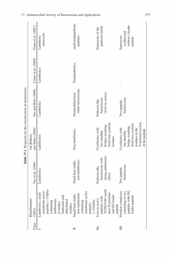
17 Antimicrobial Activity of Bacteriocins and Applications 377
Tabl
e17
.1Pr
opos
als
for
the
clas
sific
atio
nof
bact
erio
cins
Cla
ssK
laen
ham
mer
(199
3)N
eset
al.(
1996
)va
nB
elku
man
dSt
iles
(200
0)N
esan
dH
olo
(200
0)C
otte
ret
al.(
2005
)Fr
anz
etal
.(20
07)
IL
antib
iotic
s;sm
all
mem
bran
e-ac
tive
pept
ides
(<5
kDa)
cont
aini
ngla
nthi
onin
e,β
-met
hyl
lant
ioni
nean
dde
hydr
ated
resi
dues
Lan
tibio
tics
Lan
tibio
tics
Lan
tibio
tics
Lan
tibio
tics
Lan
tibio
ticen
tero
cins
IISm
allh
eat-
stab
le,
non-
lant
hion
ine
cont
aini
ngm
embr
ane-
activ
epe
ptid
es(<
10kD
a)
Smal
lhea
t-st
able
non-
lant
ibio
tics
Non
-lan
tibio
tics
Non
mod
ified
heat
stab
leba
cter
ioci
nsN
onla
ntib
iotic
ssm
alln
onla
ntib
iotic
pept
ides
IIa
Lis
teri
a-ac
tive
pept
ides
with
YG
NG
VX
Cm
otif
near
N-t
erm
inus
and
GG
lead
erpe
ptid
e
Pedi
ocin
-lik
eba
cter
ioci
nsw
ithst
rong
antil
iste
rial
effe
ct
Cys
tibio
tics
with
two
disu
lfite
brid
ges
resu
lting
from
four
cyst
eine
resi
dues
Pedi
ocin
-lik
eba
cter
ioci
ns(L
iste
ria-
activ
e)
-E
nter
ocin
sof
the
pedi
ocin
fam
ily
IIb
Pora
tion
com
plex
esco
nsis
ting
oftw
ope
ptid
esw
ithG
Gle
ader
pept
ide
Two
pept
ide
bact
erio
cins
Cys
tibio
tics
with
one
disu
lfite
brid
gere
sulti
ngfr
omtw
ocy
stei
nere
sidu
esin
the
N-t
erm
inal
sect
ion
ofth
epe
ptid
e
Two-
pept
ide
bact
erio
cins
-E
nter
ocin
ssy
nthe
size
dw
ithou
tale
ader
pept
ide
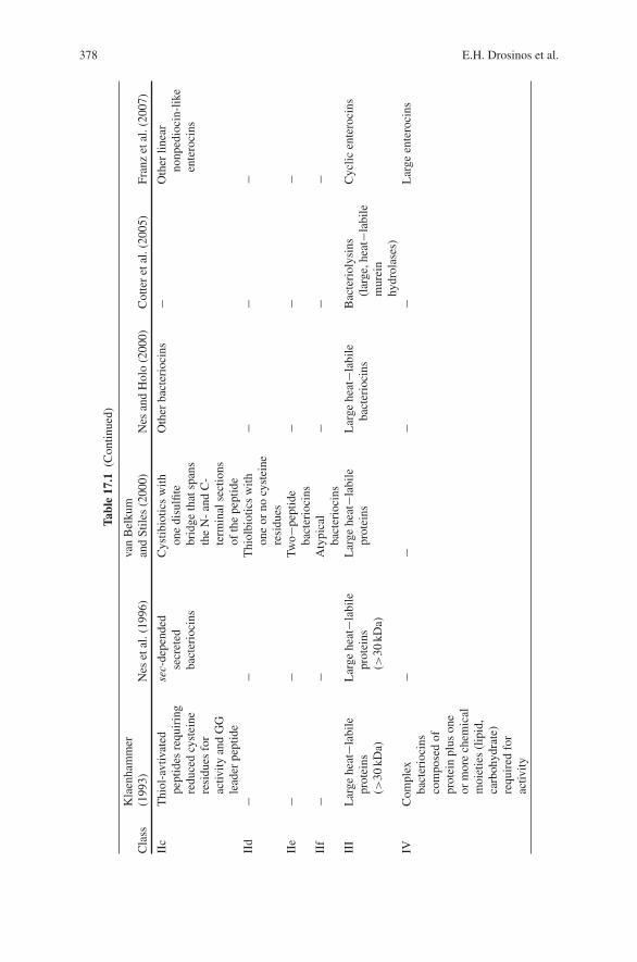
378 E.H. Drosinos et al.
Tabl
e17
.1(C
ontin
ued)
Cla
ssK
laen
ham
mer
(199
3)N
eset
al.(
1996
)va
nB
elku
man
dSt
iles
(200
0)N
esan
dH
olo
(200
0)C
otte
ret
al.(
2005
)Fr
anz
etal
.(20
07)
IIc
Thi
ol-a
vtiv
ated
pept
ides
requ
irin
gre
duce
dcy
stei
nere
sidu
esfo
rac
tivity
and
GG
lead
erpe
ptid
e
sec-
depe
nded
secr
eted
bact
erio
cins
Cys
tibio
tics
with
one
disu
lfite
brid
geth
atsp
ans
the
N-
and
C-
term
inal
sect
ions
ofth
epe
ptid
e
Oth
erba
cter
ioci
ns−
Oth
erlin
ear
nonp
edio
cin-
like
ente
roci
ns
IId
−−
Thi
olbi
otic
sw
ithon
eor
nocy
stei
nere
sidu
es
−−
−
IIe
−−
Two−
pept
ide
bact
erio
cins
−−
−
IIf
−−
Aty
pica
lba
cter
ioci
ns−
−−
III
Lar
gehe
at−l
abile
prot
eins
(>30
kDa)
Lar
gehe
at−l
abile
prot
eins
(>30
kDa)
Lar
gehe
at−l
abile
prot
eins
Lar
gehe
at−l
abile
bact
erio
cins
Bac
teri
olys
ins
(lar
ge,h
eat−
labi
lem
urei
nhy
drol
ases
)
Cyc
licen
tero
cins
IVC
ompl
exba
cter
ioci
nsco
mpo
sed
ofpr
otei
npl
uson
eor
mor
ech
emic
alm
oiet
ies
(lip
id,
carb
ohyd
rate
)re
quir
edfo
rac
tivity
−−
−−
Lar
geen
tero
cins
17 Antimicrobial Activity of Bacteriocins and Applications 379
principal target of these is the cytoplasmic membrane through permealization andconcomitant leakage, without, though, conclusive experimental evidence for theexistence or necessity of a bacteriocin receptor (Nes and Holo, 2000).
In Vitro and In Situ Bacteriocin Production
Typical spontaneous sausage fermentation is driven by lactic acid bacteria fol-lowed by members of the Micrococcaceae family and enterococci which are veryfrequently isolated in high numbers, suggesting a possible contribution to the fer-mentation process. Many researchers have studied their biochemical properties andmembers of nearly all genera have been found to produce bacteriocins (Table 17.2).The antimicrobial range varies from narrow to broad and from species withinthe same genus to a variety of Gram-positive and Gram-negative microorganisms,including several pathogens.
Table 17.2 Some bacteriocinogenic strains isolated from meat products
Species Reference
Lc. lactis (strains BB24, WNC 20) Rodriguez et al. (1995); Noonpakdee et al.(2003)
Lb. sakei (strains L45, 148, CTC494,CTC372, Lb706, Lb674)
Mortvedt et al. (1991); Sobrino et al. (1992);Aymerich et al. (2000b); Schillinger andLuecke (1989); Holck et al. (1994)
Pd. acidilactici (strains HA-6111-2,HA-5692-3, L50, H)
Albano et al. (2007a); Cintas et al. (1995);Motlagh et al. (1992)
Lb. curvatus (strains L442, LTH1174)
Mataragas et al. (2003b); Messens et al. (2003)
Cb. divergens (strain 750) Holck et al. (1996)Cb. piscicola (strains LV17, JG126) Ahn and Stiles (1990); Jack et al. (1996)Ent. faecium (strains L1, P13, L50,
T136, CTC492)Lyon et al. (1995); Cintas et al. (1997); Cintas
et al. (1998); Casaus et al. (1997); Aymerichet al. (1996)
Lb. plantarum (strains CTC305,UGl, 35d)
Aymerich et al. (2000b); Enan et al. (1996);Messi et al. (2001)
Leu. mesenteroides (strains E131,L124)
Drosinos et al. (2006); Mataragas et al. (2003b)
Leu. carnosum (strains OZ, 4010) Osmanagaoglu (2007); Budde et al. (2003)Leu. gelidum (strain A-UAL187) Harding and Shaw (1990)Pd. parvulus (strain 133) Schneider et al. (2006)Pd. pentosaceus Albano et al. (2007b)St. warneri (strains FM10, FM20
and FM30)Prema et al. (2006)
St. xylosus (strain 1E) Villani et al. (1997)
Lc.: Lactococcus; Lb.: Lactobacillus; Pd.: Pediococcus; Cb.: Carnobacterium; Ent.: Enterococcus;Leu.: Leuconostoc; St.: Staphylococcus
380 E.H. Drosinos et al.
Factors Affecting In Vitro Bacteriocin Production
Once a bacteriocinogenic strain is isolated and the genetic and phenotypic char-acterization of the bacteriocin has been completed, optimization of the bacteriocinproduction in synthetic media under strictly defined conditions and secondarily anin situ application consist the next step of research efforts. Since foods are com-plex systems with several factors influencing microbial growth and metabolism,these factors need to be tested on the performance of the bacteriocinogenic strains(Hugas, 1998). In order to evaluate the potential use of bacteriocinogenic LABstrains in the food systems, parameters known to influence growth and bacteriocinproduction should be considered. Such factors include the following:
Microbial Strain
A bacteriocin may be produced by more than one microbial strain and/or species.Yang and Ray (1994) found that nisin and leuconocin Lcm1 production substan-tially varied between the different strains producing these two bacteriocins, whereaspediocin AcH did not show such variation.
Physiological State of the Bacteriocinogenic Strain
Bacteriocins are considered primary metabolites, because their production startsearly (early exponential phase) and it is completed by the end of the exponentialgrowth phase or at the early stages of the stationary phase such as, in the case of,lactocin S, sakacin P, mesenterocin Y, curvaticin L442 and a bacteriocin producedby Leuconostoc mesenteroides L124 (Drosinos et al., 2005b; Drosinos, Mataragas,& Metaxopoulos, 2006; Mataragas et al., 2003b; Mataragas, Drosinos, Tsakalidou,& Metaxopoulos, 2004; Mortvedt-Abildgaard et al., 1995; Xiraphi et al., 2006).However, there are bacteriocins with maximum concentration at the early stages ofthe exponential growth phase e.g. a bacteriocin produced by Leu. gelidum UAL 187(Stiles & Hastings, 1991) or in the middle of the same phase (helveticin J) (Joerger &Klaenhammer, 1986).
Growth Medium
Type and level of nutrients (sugars, nitrogen, phosphorus, magnesium, calcium, etc.)influence the bacteriocin production. LAB possesses a limited number of catabolicpathways providing energy for cell synthesis and the energy demanding bacteri-ocin production. Therefore, conditions favoring cell growth will, probably, improvebacteriocin production (growth-associated process). However, an extremely richenvironment will not necessarily lead to a gain of bacteriocin production. Severalstudies have shown that the type and level of sugars (e.g. glucose, sucrose, fructose,etc.) used as carbon sources substantially affect the bacteriocin synthesis (Barcena,Sineriz, Gonzalez de Llano, Rodriguez, & Suarez, 1998; Biswas, Ray, Johnson, &
17 Antimicrobial Activity of Bacteriocins and Applications 381
Ray, 1991; Drosinos et al., 2005a, 2006; Mataragas et al., 2004; Matsusaki, Endo,Sonomoto, & Ishikazi, 1996). Bacteriocin production was stimulated at high glucoseconcentrations (Drosinos et al., 2005b), because sometimes the stress situation cre-ated by the high glucose content (higher osmotic pressures) causes better bacteri-ocin production (De Vuyst & Vandamme, 1994). Leu. mesenteroides E131 in thepresence of fructose, instead of glucose, displayed better bacteriocin production(Drosinos et al., 2005b, 2006). This happens because fructose, as an energy source,is utilized more effectively by leuconostocs under the same pH and temperatureconditions. Leuconostocs reduce some of the fructose to mannitol leading to energygain as the cells produce ATP during this process and accordingly more ATP pertime unit (Drosinos et al., 2005b). Nitrogen also plays a significant role. Kim, Hall,and Dunn (1997) observed that the maximum concentration of nisin is increased bythe increase of the contained nitrogen due to increased supply of essential aminoacids for cell formation and bacteriocin production. Type of nitrogen source has aneffect on bacteriocin production (Mataragas et al., 2004). Various nitrogen sourceswith different number of amino acids along the chain of the contained peptidesgave different results. Nitrogen sources (e.g. yeast extract) with a relatively largeproportion of free amino acids and short peptides (2 or 3 amino acids) favoredbacteriocin production than sources with long peptides (8 amino acids) (Mataragaset al., 2004) due to energy saving (Aasen, Moretro, Katla, Axelsson, & Storro, 2000;Benthin, Schulze, Nielsen, & Villadsen, 1994). However, an optimal ratio betweencarbon and nitrogen source is required to ensure satisfactory bacteriocin productionbecause when carbon or nitrogen is in excess the others become a limiting factor forbacteriocin synthesis (Mataragas et al., 2004). Presence of various elements suchas phosphorus, magnesium and calcium influence bacteriocin production but thisaction is strain specific (Parente & Ricciardi, 1999). Tween 80 (polyoxyethylenesorbitan mono-oleate) favors bacteriocin production in some cases (Daba, Lacroix,Huang, & Simard, 1993; Matsusaki et al., 1996; Parente & Hill, 1992); however,enhancement in bacteriocin activity, in the presence of Tween 80 seems to be relatedwith decreased adsorption of the bacteriocin molecules onto the cellular membrane(Joosten & Nunez, 1995).
Environmental Conditions: pH and Temperature
pH and temperature conditions constitute two of the most critical factors whichsignificantly affect bacteriocin production by bacteriocinogenic strains. Frequently,maximum bacteriocin concentration is achieved at pH (usually within the range5.5–6.0) and/or temperature values slightly lower than those optimal for growth(De Vuyst, Callewaert, & Crabbe, 1996; Drosinos et al., 2005a, 2006; Kaiser &Montville, 1993; Mataragas, Drosinos, & Metaxopoulos, 2003a; Meghrous, Huot,Quittelier, & Petitdemange, 1992; Parente & Ricciardi, 1994). Only some bacte-riocins display better production at lower pH values (5.0) (Barcena et al., 1998;Biswas et al., 1991; Mortvedt-Abildgaard et al., 1995; Yang and Ray, 1994). Bac-teriocin production is enhanced at lower values of environmental factors than thoseof optimum, probably because the whole bacteriocin synthesis process is favored
382 E.H. Drosinos et al.
by a relatively lower growth rates. Increased growth rates do not necessarily meanbetter bacteriocin production (De Vuyst et al., 1996; Mataragas et al., 2003a).Lower growth rates at sub-optimal pH or temperature values indicate potentiallybetter utilization of energy and essential metabolites (Aasen et al., 2000; Moretro,Aasen, Storro, & Axelsson, 2000). At higher temperatures or pH values bacte-riocin production is lower because energy needs for maintenance purposes arehigher when temperature or pH increases (Drosinos et al., 2005b; Leroy & DeVuyst, 1999). Maintenance operations such as turnover of macromolecules (DNA,RNA) and maintenance of the potential along the membrane of cells are growthdependant and faster growth rates mean more energy is required for maintenance(Nielsen, Nikolajsen, & Villadsen, 1991). However, there are cases of better bacte-riocin production at temperatures close to optimum for growth (Daba et al., 1993;Drosinos et al., 2005b, 2006; Matsusaki et al., 1996; Meghrous et al., 1992; Leje-une, Callewaert, Crabbe, & De Vuyst, 1998; Parente & Ricciardi, 1994). Finally,certain LAB such as Leu. mesenteroides subsp. mesenteroides FR52, produce morethan one bacteriocin requiring different environmental conditions. Maximum con-centration of mesenterocin 52A is observed at pH 5.5 and temperature 20◦C andof mesenterocin 52B at pH 5.0 and temperature 25◦C (Krier, Revol-Junelles, &Germain, 1998).
Predictive modeling has gained increased attention and mathematical mod-els have been developed to describe the growth and bacteriocin production ofthe bacteriocin-producing strains added to foods as starter or protective cultures(Leroy & De Vuyst, 2003; Messens, Neysens, Vasieleghem, Vanderhoeven, & DeVuyst, 2002; Messens et al., 2003). Modeling contributes to the determination ofhow environmental factors affect the growth and bacteriocin production (Leroy &De Vuyst, 2003; Leroy, Verluyten, Messens, & De Vuyst, 2002). Experimental andmathematical calculation of the various parameters of growth and bacteriocin pro-duction are displayed in Table 17.3. For more detailed information on the analyticalprocedure of the differential equations readers should refer to the literature listed inthe Table 17.3 and McKellar (1997).
One method that is usually followed for the optimization of bacteriocin produc-tion is that of varying one factor at a time. The other factors are kept constant.As it can be seen, this technique is laborious and requires a lot of time in case ofseveral factors under study. For this reason, statistical experimental designs havebeen developed to evaluate the influence of substrate composition on growth andbacteriocin production (Dominguez, Bimani, Caldera-Olivera, & Brandelli, 2007;Rollini & Manzoni, 2005).
Factors Related to Food Ingredients and Additives
Apart from the above mentioned parameters, the in vitro study of bacteriocin pro-duction and activity has often implicated various ingredients used for the productionof fermented sausages. Hugas, Garriga, Pascual, Aymerich, and Monfort (2002)studied the effect of various ingredients, used in the manufacture of fermentedsausages, on the antilisterial performance of the bacteriocinogenic Lactobacillus
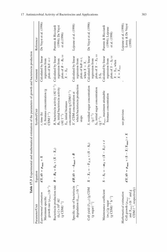
17 Antimicrobial Activity of Bacteriocins and Applications 383
Tabl
e17
.3E
xper
imen
tala
ndm
athe
mat
ical
estim
atio
nof
the
para
met
ers
ofgr
owth
and
bact
erio
cin
prod
uctio
n
Para
met
er/U
nit
Equ
atio
nSy
mbo
l/Uni
tC
omm
ent
Ref
eren
ce
Exp
erim
enta
lest
imat
ion
Max
imum
spec
ific
grow
thra
te(μ
ma
x)
(h−1
)
dX
/d
t=
μm
ax×
Xt,
time
(h)
X,b
iom
ass
conc
entr
atio
n(g
CD
Ml−1
)
Cal
cula
ted
bylin
ear
regr
essi
onfr
ompl
ots
ofln
Xvs
t
De
Vuy
stet
al.(
1996
)
Bac
teri
ocin
prod
uctio
nra
te(k
b)
[×10
6A
U(g
CD
M)−1
]
B−
B0
=k b
×(X
−X
0)
B,b
acte
rioc
inac
tivity
(AU
l−1)
B0,i
nitia
lbac
teri
ocin
activ
ity(A
Ul−1
)X
0,i
nitia
lbio
mas
sco
ncen
trat
ion
(gC
DM
l−1)
Cal
cula
ted
bylin
ear
regr
essi
onfr
ompl
ots
ofB
−B
0vs
X−
X0
Pare
nte
&R
icci
ardi
(199
4);D
eV
uyst
etal
.(19
96)
Spec
ific
rate
ofba
cter
ioci
nde
grad
atio
n(k
inac
t)(h
−1)
dB
/d
t=
−kin
act×
BX
′ ,C
DM
conc
entr
atio
nat
whi
chba
cter
ioci
npr
oduc
tion
stop
s
Cal
cula
ted
bylin
ear
regr
essi
onfr
ompl
ots
ofln
Bvs
t,w
hen
X>
X,
Lej
eune
etal
.(19
98)
Cel
lyie
ld(Y
X/
S)
[gC
DM
(gsu
gar)
−1]
X−
X0
=−Y
X/
S×
(S−
S 0)
S,re
sidu
alsu
gar
conc
entr
atio
n(g
l−1)
S 0,i
nitia
lsug
arco
ncen
trat
ion
(gl−1
)
Cal
cula
ted
bylin
ear
regr
essi
onfr
ompl
ots
ofX
−X
0vs
S-S 0
De
Vuy
stet
al.(
1996
)
Mai
nten
ance
coef
ficie
nt(m
S)
[gsu
gar
(gC
DM
)−1h−1
]
S−
S 0=
−mS
×(X
−X
0)×
tX
ma
x,m
axim
umat
tain
able
biom
ass
conc
entr
atio
nC
alcu
late
dby
linea
rre
gres
sion
from
plot
sof
S−
S 0vs
X−
X0,w
hen
X=
Xm
ax
Pare
nte
&R
icci
ardi
(199
4);L
ejeu
neet
al.(
1998
)
Mat
hem
atic
ales
tim
atio
nC
ellg
row
th(μ
ma
x
and
Xm
ax)
(h−1
and
gC
DM
l−1,r
espe
ctiv
ely)
dX
/d
t=
μm
ax×
(1−
X/
Xm
ax)×
Xse
epr
evio
us-
Lej
eune
etal
.(19
98);
Ler
oy&
De
Vuy
st(1
999)
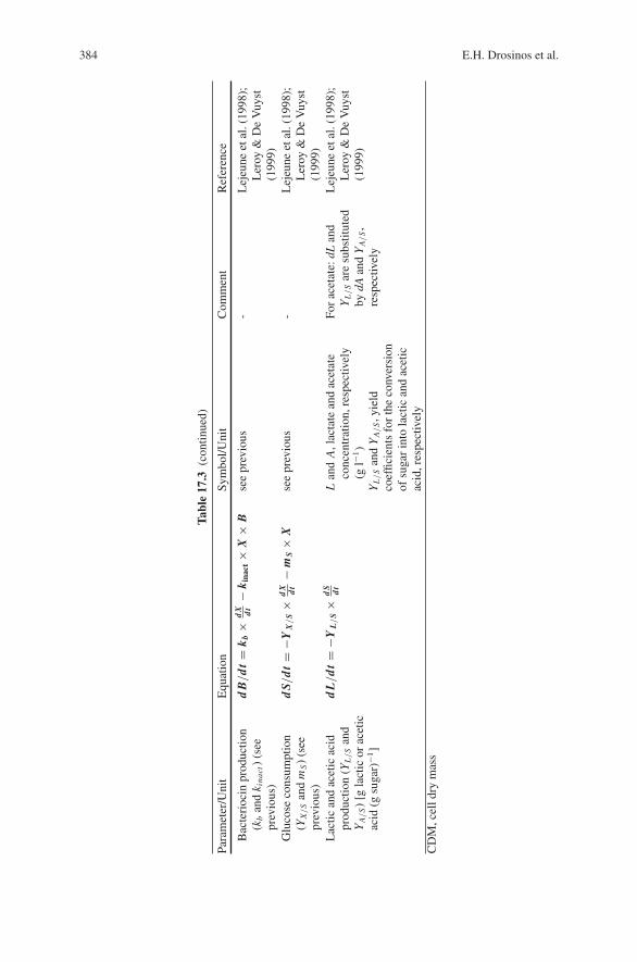
384 E.H. Drosinos et al.
Tabl
e17
.3(c
ontin
ued)
Para
met
er/U
nit
Equ
atio
nSy
mbo
l/Uni
tC
omm
ent
Ref
eren
ce
Bac
teri
ocin
prod
uctio
n(k
ban
dk i
nact
)(s
eepr
evio
us)
dB
/d
t=
k b×
dX dt
−k i
nact
×X
×B
see
prev
ious
-L
ejeu
neet
al.(
1998
);L
eroy
&D
eV
uyst
(199
9)G
luco
seco
nsum
ptio
n(Y
X/
San
dm
S)
(see
prev
ious
)
dS/
dt=
−YX
/S
×d
X dt
−m
S×
Xse
epr
evio
us-
Lej
eune
etal
.(19
98);
Ler
oy&
De
Vuy
st(1
999)
Lac
tican
dac
etic
acid
prod
uctio
n(Y
L/
San
dY
A/
S)
[gla
ctic
orac
etic
acid
(gsu
gar)
−1]
dL/
dt=
−YL/
S×
dS dt
Lan
dA
,lac
tate
and
acet
ate
conc
entr
atio
n,re
spec
tivel
y(g
l−1)
YL/
San
dY
A/
S,y
ield
coef
ficie
nts
for
the
conv
ersi
onof
suga
rin
tola
ctic
and
acet
icac
id,r
espe
ctiv
ely
For
acet
ate:
dLan
dY
L/
Sar
esu
bstit
uted
bydA
and
YA/
S,
resp
ectiv
ely
Lej
eune
etal
.(19
98);
Ler
oy&
De
Vuy
st(1
999)
CD
M,c
elld
rym
ass
17 Antimicrobial Activity of Bacteriocins and Applications 385
sakei CTC494 in vitro and in model fermented sausages. The influence of sausageingredients on sakacin K production was variable; sodium chloride mixed withnitrate and nitrite decreased total bacteriocin production, whereas sodium chlorideand pepper, while stimulating growth of the strain, did not affect bacteriocin pro-duction. Sodium chloride protected Listeria monocytogenes from the action of thebacteriocin, but black pepper and sodium nitrite enhanced sakacin K activity in vitroand in sausages, surpassing the protective effect of sodium chloride and furtherinhibiting the growth of L. monocytogenes. The same effect observed with blackpepper and nitrite was found when manganese was used as a sausage ingredientinstead of pepper, suggesting that one of the active factors in pepper was manganese.Verluyten et al. (Verluyten, Leroy, & de Vuyst, 2004; Verluyten, Messens, & DeVuyst, 2003, 2004) studied the effect of sodium chloride, sodium nitrite as wellas the effect of different spices relevant for the production of fermented sausageson curvacin A, a listericidal bacteriocin produced by Lb. curvatus LTH 1174, astrain isolated from fermented sausage. The strain was highly sensitive to nitrite;even a concentration of 10 ppm of sodium nitrite inhibited its growth and boththe volumetric and specific bacteriocin production. Both cell growth and bacteri-ocin activity were affected by changes in sodium chloride concentration. It clearlyslowed down the growth of Lb. curvatus LTH 1174, but more importantly, it hada detrimental effect on specific curvacin A production and hence on overall bac-teriocin activity. Even a low salt concentration (2%, wt/vol) decreased bacteriocinproduction, while growth was unaffected at this concentration. The inhibitory effectof NaCl was mainly due to its role as an aw-reducing agent. Further, it was clear thatsalt interfered with bacteriocin induction. Additionally, when 6% (wt/vol) sodiumchloride was added, the minimum biomass concentration necessary to start the pro-duction of curvacin A was 0.90 g (cell dry mass) per liter. Addition of a cell-freeculture supernatant or a protein solution as a source of induction factor resulted inan increase in the maximum attainable bacteriocin activity. As far as the effect of thespices were concerned, pepper, nutmeg, rosemary, mace, and garlic all decreased themaximum specific growth rate, while paprika was the only spice that increased it.The effect on the lag phase was minor except for nutmeg and especially for garlic,which increased it, yet garlic was stimulatory for biomass production. The maxi-mum attainable biomass concentration was severely decreased by the addition of0.40% (wt/vol) nutmeg, while 0.35% (wt/vol) garlic or 0.80% (wt/vol) white pepperincreased biomass concentration. Nutmeg decreased both growth and bacteriocinproduction considerably. Garlic was the only spice enhancing specific bacteriocinproduction, resulting in a higher bacteriocin activity in the cell-free culture super-natant. Finally, lactic acid production was stimulated by the addition of pepper,and this was not due to the manganese present. Addition of spices to the sausagemixture is clearly a factor that will influence the effectiveness of bacteriocinogenicstarter cultures in fermented-sausage manufacturing. Aymerich, Artigas, Garriga,Monfort, and Hugas (2000a) studied the effect of sausage ingredients and addi-tives on Enterocin A and B production by Enterococcus faecium strain CTC492,and concluded that it was significantly inhibited by sodium chloride, nitrate andpepper.
386 E.H. Drosinos et al.
In Situ Study of Bacteriocin Antimicrobial Activity
The in situ application of bacteriocins has been a field of intensive study. Its effec-tiveness against pathogens has been studied in raw, cooked or fermented meat prod-ucts. However, such an application might face several limitations occurring due tovarious reasons, the most common being the relatively narrow spectrum of action,the possible inactivation in food systems by proteases or as a result of their interac-tions with food constituents. Additionally, the emergence of resistant mutants, thathas been demonstrated (Rekhif, Atrih, & Lefebvre, 1994), might lead to a loss ofantimicrobial activity of bacteriocins against sensitive strains during food storage.
Currently, there are two major approaches that are commonly used. The firstone requires the inoculation of the commodity with a bacteriocinogenic strain. Thisstrain can either fulfill all the criteria in order to be used as a starter culture, or it canbe used as an adjunct culture provided that it can not only tolerate the action of thestarter cultures, but can proliferate and produce bacteriocin as well. Alternatively,the bacteriocin itself, after being purified to some degree, can be added as an ingre-dient in the food matrix or immobilized on a proper agent, such as silicon surfaces,soy- and corn- based films, cellulose, cellophane and several others. (Daeschel,Mcguire, & Almakhla, 1992; Guerra, Macias, Agrasar, & Castro 2005; Luchan-sky & Call, 2004; Natrajan & Sheldon, 2000; Siragusa, Cutter, & Willett, 1999).
Since the first attempts for meat and meat products biopreservation (Schillinger &Luecke, 1987) many interesting studies have taken place, referring to a wide rangeof products and thus extensive data currently exists. Both approaches are currentlyused in fermented meat products, although the addition of the bacteriocinogenicstrain is preferred due to legal and economic implications
Nisin, produced by Lactococcus lactis, was the first and most prominent mem-ber of lantibiotics, that was discovered just before penicillin, and has been usedextensively as a food preservative. Despite the promising results that are publishedregularly, concerning a variety of products such as minced raw buffalo meat (Pawar,Malik, Bhilegaonkar, & Barbuddhe, 2000), beef products (Cutter & Siragusa, 1994;Millette, Le Tien, Smoragiewicz, & Lacroix, 2007), raw and cooked pork (Fang &Lin, 1994; Murray & Richard, 1997), and sucuks (Hampikyan & Ugur, 2007), itseems to be a common belief that, compared to dairy products, its effectiveness isreduced due to its low solubility, uneven distribution and lack of stability. Moreoverthe required dose to be effective is uneconomical and exceeding the acceptable dailyintake (Hugas, 1998).
It has been previously shown that members of nearly all genera present in sponta-neous sausage fermentation have been found to produce bacteriocins. Concequently,a wide range of bacteriocinogenic microorganisms or their bacteriocins have beentested for their capacity to contribute to the safety of a variety of products.
The efficacy of Pediococcus acidilactici strains PAC 1.0, H and PO2 to controlgrowth of L. monocytogenes as well as Pd. acidilactici PA-2 against Escherichia coliO157:H7 and L. monocytogenes during the manufacture of dry sausage has beenstudied by Foegeding, Thomas, Pilkington, and Klaenhammer (1992), Luchanskyet al. (1992) and Lahti, Johansson, Honkanen-Buzalski, Hill, and Nurmi (2001).
17 Antimicrobial Activity of Bacteriocins and Applications 387
In the first case, the contribution of pediocin to the inactivation of L. monocytogenesstrains was confirmed, while in the second case, the incorporation of a bacterio-cinogenic strain of Pd. acidilactici (PA-2) as a starter culture had no effect onthe numbers of E. coli O157:H7, regardless of the inoculum level, and decreasedthe population of L. monocytogenes but only when it was present in high num-bers (5.05–5.41 log cfu g−1). Recently, the antilisterial activity of two strains ofPd. pentosaceus was verified during the production of ‘alheiras’ sausage (Albanoet al., 2007b), as well as the ability of a bacteriocin produced by a Pd. acidilac-tici strain to reduce the numbers of L. monocytogenes inoculated on the surface ofraw meat and stored at 4◦C or 15◦C, in addition to the bacteriostatic effect againstClostridium perfringens has been demonstrated (Nieto-Lozano, Reguera-Useros,del Pelaez-Martinez, & Hardisson de la Torr, 2006).
The effect of the bacteriocinogenic strain Staphylococcus xylosus 1E has beenstudied by Villani et al. (1997). It was able to reduce the viable counts of L. monocy-togenes in Naples-type sausages after 21 days of maturation. Moreover, no L. mono-cytogenes were recovered after 75 days in sausages inoculated with St. xylosus1E, while the pathogen was still present at this time in control sausages in whichL. monocytogenes was challenged.
As far as the Carnobacterium sp. are concerned, addition of piscicolin 126, abacteriocin produced by Carnobacterium piscicola JG126 to a devilled ham pastetest food system inhibited the growth of L. monocytogenes for at least 14 days (Jacket al., 1996).
Ananou, Maqueda, Martinez-Bueno, Galvez, and Valdivia (2005) studied theanti-staphylococcal activity of bacteriocin AS-48. The latter was added either asa semi-purified form or by in situ production via the incorporation of the producerstrain Ent. faecalis A-48-32 in the starter culture. The producer strain managed todevelop well and the best result was achieved with a bacteriocinogenic strain inocu-lum of 107 CFU/g. The effectiveness of the enterocin CCM 4231 in controllingL. monocytogenes contamination in dry fermented Hornad salami was verified byLaukova, Czikkova, Laczkova, and Turek (1999).
Campanini, Pedrazzoni, Barbuti, and Baidini (1993) observed that the additionof starter cultures prevented growth of L. monocytogenes and the pathogen wasdetected after enrichment. Listeria appeared to be absent only in salami inoculatedwith the bacteriocinogenic Lb. plantarum MCS strain. Vermeiren, Devlieghere,Vandekinderen, and Debevere (2006) studied two lactic acid bacteria, a non-bacteriocinogenic Lb. sakei subsp. carnosus strain 10A and lactocin S producingLb. sakei strain 148 for their usefulness as a protective culture in the biopreservationof cooked meat products using a cooked ham model inoculated with a cocktailof three L. monocytogenes strains. The application of the non-bacteriocinogenicstrain at 106 cfu/g was capable to limit the growth of L. monocytogenes to <1log10 cfu/g during 27 days at 7◦C, whilst an application level of 105 cfu/g failed toprevent the growth of the pathogen to acceptable levels. Lowering the temperatureto 4◦C or switching from vacuum packaging to modified atmosphere packagingdid not influence the ability of the strain to dominate. A combination of strain 10Aand 4◦C or a MAP containing 50% CO2 completely inhibited the growth of the
388 E.H. Drosinos et al.
pathogen. On the other hand the bacteriocinogenic strain failed to demonstrate anantagonistic effect towards L. monocytogenes. Mataragas et al. (2003b) studiedthe antagonistic activity of Lb. curvatus strain L442 and Leu. mesenteroides L124against different strains of L. monocytogenes and L. innocua in vacuum- or modifiedatmosphere-packaged sliced cooked cured pork shoulder at 4◦C. Inoculation of themeat products with both lactic acid bacteria resulted in a decrease of the listeriaepopulation, whereas addition of the bacteriocins seemed to be more effective as thelisteriae population was reduced to below enumeration limit regardless of the typeof packaging. Dicks, Mellett, and Hoffman (2004) applied two bacteriocinogenicstrains as starter cultures in the production of Ostrich meat salami. Lb. plantarumstrain 423 was abble to produce plantaricin 423 while Lb. curvatus strain DF126produced curvacin DF126. Neither of the two bacteriocins inhibited the growthof Micrococcus sp. MC50 and did not have any inhibitory effect on either of theproducer strains. Curvacin DF126 and plantaricin 423 inhibited the growth of L.monocytogenes in salami. However, after 15 h of fermentation, the viable countof L. monocytogenes LM1 increased, probably due to a decrease in activity of thebacteriocins and/or the development of resistant bacterial cells. Ghalfi, Benkerroum,Doguiet, Bensaid, & Thonart (2007) examined the effectiveness of a combinationof cell-adsorbed bacteriocin (CAB) of a Lactobacillus curvatus strain with oreganoor savory essential oil to control L. monocytogenes in pork meat. Addition oforegano or savory essential oil exhibited a synergistic effect with CAB to control L.monocytogenes in pork meat during storage at 4◦C.
Vignolo, Fadda, de Kairuz, Holgado, de, and Oliver (1998) inoculated meatslurry with L. monocytogenes and applied different levels of curing additives (NaCl,NaNO2, ascorbic acid, alginate meat binder, sodium lactate) in order to study theirinfluence on the inhibitory effect of lactocin 705 at 20◦C. The results obtained,indicated that the use of lactocin 705 to control L. monocytogenes was less effec-tive in the presence of curing ingredients. Benkerroum et al. (2005) combinedlyophilized bacteriocinogenic strains (Lc. lactis subsp. lactis LMG21206 and Lb.curvatus LBPE) with a non-inhibitory to L. monocytogenes commercial starter cul-ture in order to study their in vivo effectiveness to control L. monocytogenes indry-fermented sausages. The meat batter was experimentally contaminated witha mixture of four different strains of L. monocytogenes. The results showed thatL. monocytogenes did not grow in any of the contaminated batches, but no signifi-cant decrease was observed either in the positive control (no added starter culture)or in samples fermented with the bacteriocinogenic starter culture during the fer-mentation period and up to 15 days of drying.
Bacteriocins in the Context of the Hurdle Theory
The effectiveness of bacteriocins in various food commodities has been widely stud-ied and its contribution to their safety can hardly be questioned. It is though a factthat the food matrix along with several other specific processing parameters affects
17 Antimicrobial Activity of Bacteriocins and Applications 389
their efficacy on a variable degree, leading some times to a minimization of itsaction. Thus, in recent years, application of bacteriocins as part of hurdle-technologyhas gained attention. It has been shown that several bacteriocins might exhibit addi-tive or synergistic effects when used in combination with other antimicrobial agentsor processes, including chemicals, packaging, thermal or non-thermal treatments.
The combination of bacteriocins with several chemical substances such as curingagents, organic acids, chelating agents and the phenolic compounds of essential oils,has been studied both in vitro and in situ. Curing agents have a variable effect onbacteriocin activity. The negative effect of sodium chloride, that has already beenmentioned, can be attributed to ionic interactions between the bacteriocin moleculesand charged groups, involved in bacteriocin binding to target cells, induced confor-mational changes to bacteriocin molecules or even to changes in the cell envelopeof the target organisms (Bhunia, Johnson, Ray, & Kalchayanand, 1991; Jydegaard,Gravesen, & Knøchel, 2000; Lee, Iwata, & Oyagi, 1993). On the other hand, nitriteaddition enhanced bacteriocin activity against target microorganisms. For example,nisin activity was enhanced in presence of nitrite against clostridial endosporesoutgrowth (Taylor, Somer, & Kruger, 1985) and L. monocytogenes (Gill & Hol-ley, 2003). Similarly, the activity of enterocin EJ97, together with nitrite, wasamplified against L. monocytogenes, Bacillus coagulans, B. macroides/maroccanus,and AS-48 against B. cereus (Abriouel, Maqueda, Galvez, Martinez-Bueno, &Valdivia, 2002; Garcia et al., 2003; Garcia et al., 2004a, Garcia et al., 2004b. Organicacids and chelating agents also increase bacteriocin activity, the former via facili-tated translocation through the cell wall or through the increase of their solubilityand concomitantly facilitating their diffusion in the food matrix and the latter byallowing bacteriocins to easily reach the cytoplasmic membrane of Gram-negativebacteria. Concerning the synergistic effect with the active components of essentialoils that has been exhibited using a variety of combinations between bacteriocinsand the phenolic compounds (Grande et al., 2007), their impact on the organolepticproperties of the final product should be of concern when applying in a food matrix.
The combined effect of bacteriocins with vacuum or modified-atmosphere pack-aging has also been studied to some extent for various foodstuffs including a varietyof meat products. Apart from the individual action that these may exhibit, and theircombined effect, it had been shown that carbon dioxide act synergistically with nisinby enhancing its permealisation through the cytoplasmic membrane of L. monocy-togenes (Nilsson et al., 2000).
Thermal treatment that is quite often used in food processing might have pos-itive and negative effects, from a microbiological point of view. The former referto the reduction of the viable cell counts, whereas the surviving ones are sub-lethally injured, while the latter to the possible activation of bacterial endospores.Several studies have confirmed the synergistic action of bacteriocins with ther-mal treatment, in both a further reduction of viable cell counts and protectionagainst proliferation of the endospores that might even lead and in some cases toa reduction of the thermal treatment intensity (Ananou, Valdivia, Martınez Bueno,Galvez, & Maqueda, 2004; Grande et al., 2006). Moreover, due to the damageundergone by the heat treatment, an induced sensitivity to Gram-negative bacteria
390 E.H. Drosinos et al.
seemed to take place, against several bacteriocins (Ananou, Galvez, Martinez-Bueno, Maqueda, and Valdivia, 2005; Bakes, Kitis, Quattlebaum, & Barefoot, 2004;Boziaris, Humpheson, & Adams, 1998) that otherwise would have been ineffective.
During food processing, several treatments take place that do not necessaryinvolve heat exposure. Irradiated, pulsed electric or magnetic fields and high hydro-static pressure are processes that can be used in a variety of foodstuffs aiming atthe reduction of viable cell counts. Several reports on the combined effect of theseprocesses with bacteriocins are currently available and finely reviewed by Galvez,Abriouel, Lopez, and Ben Omar (2007)
The utilization of bacteriocins in meat preservation as natural antimicrobialshas attracted the interest in current research. The antimicrobial action of thatimplicit determinant in the meat microbial ecosystem, however, has to be quantifiedby the recent developments of predictive modelling in foods. In addition, multi-disciplinary efforts of researchers from the fields of molecular biology, microbialecology, food safety, meat and meat products technology should be applied, as aholistic approach to bacteriocin application in meat. The extent of the applicationof bacteriocins as a hurdle and the success to this end will be the outcome of thesecoordinated efforts.
References
Aasen, I. M., Moretro, T., Katla, T., Axelsson, L., & Storro, I. (2000). Influence of complexnutrients, temperature and pH on bacteriocin production by Lactobacillus sakei CCUG 42687.Applied Microbiology and Biotechnology, 53, 159–166.
Abriouel, H., Maqueda, M., Galvez, A., Martinez-Bueno, M., & Valdivia, E. (2002). Inhibition ofbacterial growth, enterotoxin production, and spore outgrowth in strains of Bacillus cereus bybacteriocin AS-48. Applied and Environmental Microbiology, 68, 1473–1477.
Ahn, C., & Stiles, M. E. (1990). Plasmid-associated bacteriocin production by a strain of Carnobac-terium piscicola from meat. Applied and Environmental Microbiology, 56, 2503–2510.
Albano, H., Todorov, S. D., van Reenen, C. A., Hogg, T., Dicks, L. M. T., & Texixeira, P.(2007a). Characterization of two bacteriocins produced by Pediococcus acidilactici isolatedfrom ‘Alheira’, a fermented sausage traditionally produced in Portugal. International Journalof Food Microbiology, 116, 239–247.
Albano, H., Oliveira, M., Aroso, R., Cubero, N., Hogg, T., & Teixeira, P. (2007b). Antiliste-rial activity of lactic acid bacteria isolated from ‘Alheiras’ (traditional Portuguese fermentedsausages): In situ assays. Meat Science, 76, 796–800.
Ananou, S., Galvez, A., Martinez-Bueno, M., Maqueda, M., & Valdivia, E. (2005). Synergisticeffect of enterocin AS-48 in combination with outer membrane permeabilizing treatmentsagainst Escherichia coli O157:H7. Journal of Applied Microbiology, 99, 1364–1372.
Ananou, S., Maqueda, M., Martinez-Bueno, M., Galvez, A., & Valdivia, E. (2005). Control ofStaphylococcus aureus in sausages by enterocin AS-48. Meat Science, 71, 549–556
Ananou, S., Valdivia, E., Martınez Bueno, M., Galvez, A., & Maqueda, M. (2004). Effect of com-bined physico-chemical preservatives on enterocin AS-48 activity against the enterotoxigenicStaphylococcus aureus CECT 976 strain. Journal of Applied Microbiology, 97, 48–56.
Aymerich, T., Holo, H., Havarstein, L.S., Hugas, M., Garriga, M., & Nes, I. F. (1996). Biochemicaland genetic characterization of enterocin A from Enterococcus faecium, a new antilisterialbacteriocin in the pediocin family of bacteriocins. Applied and Environmental Microbiology,62, 1676–1682.
17 Antimicrobial Activity of Bacteriocins and Applications 391
Aymerich, T., Artigas, M. G., Garriga, M., Monfort, J. M., & Hugas, M. (2000a). Effect of sausageingredients and additives on the production of enterocins A and B by Enterococcus faeciumCTC492.Optimization of in vitro production and anti-listerial effect in dry.fermented sausages.Journal of Applied Microbiology, 88, 686–694.
Aymerich, M. T., Garriga, M., Monford, J. M., Nes, I., & Hugas, M. (2000b). Bacteriocin-producing lactobacilli in Spanish-style fermented sausages: characterization of bacteriocins.Food Microbiology, 17, 33–45.
Bakes, S. H., Kitis, F. Y. E., Quattlebaum, R. G., & Barefoot, S. F. (2004). Sensitization of Gram-negative and Gram-positive bacteria to jenseniin G by sublethal injury. Journal of Food Protec-tion, 67, 1009–1013.
Barcena, B. J. M., Sineriz, F., Gonzalez de Llano, D., Rodriguez, A., & Suarez, J. E. (1998).Chemo-stat production of plantaricin C by Lactobacillus plantarum LL41. Applied and Envi-ronmental Microbiology, 57, 3512–3514.
Benkerroum, N., Daoudi, A., Hamraoui, T., Ghalfi, H., Thiry, C., Duroy, M., Evrart, P., Roblain,D., & Thonart, P. (2005). Lyophilized preparations of bacteriocinogenic Lactobacillus curvatusand Lactococcus lactis subsp. lactis as potential protective adjuncts to control Listeria mono-cytogenes in dry-fermented sausages. Journal of Applied Microbiology, 98, 56–63.
Benthin, S., Schulze, U., Nielsen, J., & Villadsen, J. (1994). Growth energetics of Lactococcuscremoris FD1 during energy-, carbon-, and nitrogen-limitation in steady state and transientcultures. Chemical Engineering Science, 49, 589–609.
Bhunia, A. K., Johnson, M. C., Ray, B., & Kalchayanand, N. (1991). Mode of action of pediocinAcH from Pediococcus acidilactici H on sensitive bacterial strains. Journal of Applied Bacte-riology, 70, 25–33.
Biswas, S. R., Ray, P., Johnson, M. C., & Ray, B. (1991). Influence of growth conditions on theproduction of a bacteriocin, pediocin AcH, by Pediococcus acidilactici H. Applied and Envi-ronmental Microbiology, 57, 1265–1267.
Boziaris, I. S., Humpheson, I., & Adams, M. R. (1998). Effect of nisin on heat injury and inactiva-tion of Salmonella enteritidis PT4. International Journal of Food Microbiology, 43, 7–13.
Budde, B. B., Hornbaek, T., Jacobsen, T., Barkholt, V., & Koch, A. G. (2003). Leuconostoccarnosum 4010 has the potential for use as a protective culture for vacuum-packed meats:Culture isolation, bacteriocin identification, and meat application experiments. InternationalJournal of Food Microbiology, 83, 171–184.
Campanini, M., Pedrazzoni, I., Barbuti, S., & Baidini, P. (1993). Behaviour of Listeria mono-cytogenes during the maturation of naturally and artificially contaminated salami: Effectof lactic-acid bacteria starter cultures lnternational Journal of Food Microbiology, 21,169–175.
Casaus, P., Nilsen, T., Cintas, L. M., Nes, I. F., Hernandez, P. E., & Holo, H. (1997). Enterocin B, anew bacteriocin from Enterococcus faecium T136 which can act synergistically with enterocinA. Microbiology, 143, 2287–2294.
Chatterjee, C., Paul, M., Xie, L., & van der Donk, W. A. (2005). Biosynthesis and mode of actionof Lantibiotics. Chemistry Reviews, 105, 633–683.
Cintas, L. M., Rodriguez, J. M., Fernandez, M. F., Sletten, K., Nes, I. F., Hernandez, P. E., & Holo,H. (1995). Isolation and Characterization of Pediocin L50, a new bacteriocin from Pediococcusacidilactici with a broad inhibitory spectrum. Applied and Environmental Microbiology, 61,263–2648.
Cintas, L. M., Casaus, P., Havarstein, L. S., Hernandez, P. E., & Nes, I. F. (1997). Biochemical andgenetic characterization of enterocin P, a novel sec-dependent bacteriocin from Enterococcusfaecium P13 with a broad antimicrobial spectrum. Applied and Environmental Microbiology,63, 4321–4330.
Cintas, L. M., Casaus, P., Holo, H., Hernandez, P. E., Nes, I. F., & Havarstein, L. S. (1998). Ente-rocins L50A and L50B, two novel bacteriocins from Enterococcus faecium L50, and related tostaphylococcal hemolysins. Journal of Bacteriology, 180, 1988–1994.
Cleveland, J., Montville, T. J., Nes, I. F., & Chikindas, M. L. (2001). Bacteriocins: Safe, naturalantimicrobials for food preservation. International Journal of Food Microbiology, 71, 1–20.
392 E.H. Drosinos et al.
Cotter, P. D., Hill, C., & Ross, R. P. (2005). Bacteriocins: Developing innate immunity for food.Nature Reviews Microbiology, 3, 777–788.
Cutter, C. N., & Siragusa, G. R. (1994). Decontamination of beef carcass tissue with nisin using apilot scale model carcass washer. Food Microbiology, 11, 481–489.
Daba, H., Lacroix, C., Huang, J., & Simard, R. (1993). Influence of growth conditions on produc-tion and activity of mesenterocin 5 by a strain of Leuconostoc mesenteroides. Applied Micro-biology and Biotechnology, 39, 166–173.
Daeschel, M. A., Mcguire, J., & Almakhla, H. (1992). Antimicrobial activity of nisin adsorbed tohydrophilic and hydrophobic silicon surfaces. Journal of Food Protection, 55, 731–735.
De Vuyst, L., Callewaert, R., & Crabbe, K. (1996). Primary metabolite kinetics of bacteriocinbiosynthesis by Lactobacillus amylovorus and evidence for stimulation of bacteriocin underunfavourable growth conditions. Microbiology, 142, 817–827.
De Vuyst, L., & Vandamme, E. J. (1994). Bacteriocins of lactic acid bacteria: Microbiology, genet-ics and applications. London: Blackie Academic and Professional.
Deegan, L. H., Cotter, P. D., Hill, C., & Ross, P. (2006). Bacteriocins: Biological tools for bio-preservation and shelf-life extension. International Dairy Journal, 16, 1058–1071.
Dicks, L. M. T., Mellett, F. D., & Hoffman, L. C. (2004). Use of bacteriocin-producing startercultures of Lactobacillus plantarum and Lactobacillus curvatus in production of ostrich meatsalami. Meat Science, 66, 703–708.
Dominguez, A. P. M., Bimani, D., Caldera-Olivera, F., & Brandelli, A. (2007). Cerein 8 productionin soybean protein using response surface methodology. Biochemical Engineering Journal, 35,238–243.
Drosinos, E. H., Mataragas, M., & Metaxopoulos, J. (2005a). Biopreservation: A new directiontowards food safety. In A. P. Riley (Ed.), New developments in food policy, control and research(pp. 31–64). New York: Nova Science Publishers, Inc.
Drosinos, E. H., Mataragas, M., & Metaxopoulos, J. (2006). Modeling of growth and bacteriocinproduction by Leuconostoc mesenteroides E131. Meat Science, 74, 690–696.
Drosinos, E. H., Mataragas, M., Nasis, P., Galiotou, M., & Metaxopoulos, J. (2005b). Growthand bacteriocin production kinetics of Leuconostoc mesenteroides E131. Journal of AppliedMicrobiology, 99, 1314–1323.
Enan, G., El-Essawy, A. A., Uyttendaele, M., & Debevere, J. (1996). Antibacterial activity ofLactobacillus plantarum UG1 isolated from dry sausage: Characterization production andbactericidal action of plantaricin UG1. International Journal of Food Microbiology, 30,189–215.
Fang, T. J., & Lin, L. W. (1994). Growth of Listeria monocytogenes and Pseudomonas fragi oncooked pork in a modified atmosphere packaging/nisin combination. Journal of Food Protec-tion, 57, 479–485.
Foegeding, P. M., Thomas, A. B., Pilkington, D. H., & Klaenhammer, T. R. (1992). Enhancedcontrol of Listeria monocytogenes by in situ-produced pediocin during dry fermented sausageproductiont. Applied and Environmental Microbiology, 58, 884–890.
Franz, C. M. A. P., van Belkum, M. J., Holzapfel, W. H., Abriouel, H., & Galvez, A. (2007).Diversity of enterococcal bacteriocins and their grouping in a new classification scheme. FEMSMicrobiology Reviews, 31, 293–310.
Galvez, A., Abriouel, H., Lopez, R. L., & Ben Omar, N. (2007). Bacteriocin-based strate-gies for food biopreservation. International Journal of Food Microbiology (in press)doi:10.1016/j.ijfoodmicro.2007.06.001.
Garcia, M. T., Ben Omar, N., Lucas, R., Perez-Pulido, R., Castro, A., Grande, M. J., Martinez-Canamero, M., & Galvez, A. (2003). Antimicrobial activity of enterocin EJ97 on Bacilluscoagulans CECT 12. Food Microbiology, 20, 533–536.
Garcia, M. T., Lucas, R., Abriouel, H., Ben Omar, N., Perez, R., Grande, M. J., Martinez-Canamero, M., & Galvez, A. (2004a). Antimicrobial activity of enterocin EJ97 against ‘Bacil-lus macroides/Bacillus maroccanus’ isolated from zucchini puree. Journal of Applied Microbi-ology, 97, 731–737.
17 Antimicrobial Activity of Bacteriocins and Applications 393
Garcia, M. T., Martinez Canamero, M., Lucas, R., Ben Omar, N., Perez Pulido, R., & Galvez, A.(2004b). Inhibition of Listeria monocytogenes by enterocin EJ97 produced by Enterococcusfaecalis EJ97. International Journal of Food Microbiology, 90, 161–170.
Ghalfi, H., Benkerroum, N., Doguiet, D. D. K., Bensaid, M., & Thonart, P. (2007). Effectiveness ofcell-adsorbed bacteriocin produced by Lactobacillus curvatus CWBI-B28 and selected essen-tial oils to control Listeria monocytogenes in pork meat during cold storage. Letters in AppliedMicrobiology, 44, 268–273.
Gill, A. O., & Holley, R. A. (2003). Interactive inhibition of meat spoilage and pathogenic bac-teria by lysozyme, nisin and EDTA in the presence of nitrite and sodium chloride at 24◦C.International Journal of Food Microbiology, 80, 251–259.
Grande, Ma. J., Lucas, R., Abriouel, H., Valdivia, E., Ben Omar, N., Maqueda, M., Martinez-Bueno,M., Martinez-Canamero, M., & Galvez, A. (2006). Inhibition of toxicogenic Bacillus cereus inrice-based foods by enterocin AS-48. International Journal of Food Microbiology, 106, 185–194.
Grande, Ma. J., Lucas, R., Abriouel, H., Valdivia, E., Ben Omar, N., Maqueda, M., Martinez-Canamero, M., & Galvez, A. (2007). Treatment of vegetable sauces with enterocin AS-48 aloneor in combination with phenolic compounds to inhibit proliferation of Staphylococcus aureus.Journal of Food Protection, 70, 405–411.
Guerra, N. P., Macias, C. L., Agrasar, A. T., & Castro, L. P. (2005). Development of a bioactivepackaging cellophane using Nisaplin as biopreservative agent. Letters in Applied Microbiology,40, 106–1610.
Hampikyan, H., & Ugur, M. (2007). The effect of nisin on L. monocytogenes in Turkish fermentedsausages (sucuks). Meat Science, 76, 327–332.
Harding, C. D., & Saw, B. G. (1990). Antimicrobial activity of Leuconostoc gelidum against closelyrelated species and Listeria monocytogenes. Journal of Applied Bacteriology, 69, 648–654.
Holck, A. L., Axelsson, L., Huhne, K., & Krockel, L. (1994). Purification and cloning of sakacin674, a bacteriocin from Lactobacillus sake Lb674. FEMS Microbiology Letters, 115, 143–150.
Holck, A., Axelsson, L., & Schillinger, U. (1996). Divergicin 750, a novel bacteriocin produced byCarnobacterium divergens 750. FEMS Microbiology Letters, 136, 163–168.
Hugas, M. (1998). Bacteriocinogenic lactic acid bacteria for the biopreservation of meat and meatproducts. Meat Science, 49, S139–S150.
Hugas, M., Garriga, M., Pascual, M., Aymerich, M. T., & Monfort, J. M. (2002). Enhancementof sakacin K activity against Listeria monocytogenes in fermented sausages with pepper ormanganese as ingredients. Food Microbiology, 19, 519–528.
Jack, R. W., Wan, J., Gordon, J., Harmark, K., Davidson, B. E., Hillier, A. J., Wettenhall, R. E. H.,Hickey, M. W., & Coventry, M. J. (1996). Characterization of the chemical and antimicrobialproperties of piscicolin 126, a bacteriocin produced by Carnobacterium piscicola JG126. Appliedand Environmental Microbiology, 62, 2897–2903.
Joerger, M. C., & Klaenhammer, T. R. (1986). Characterization and purification of helveticin J andevidence for a chromosomally determined, bacteriocin produced by Lactobacillus helveticus481. Journal of Bacteriology, 167, 439–446.
Joosten, H. M. L. J., & Nunez, M. (1995). Adsorption of nisin and enterocin 4 to polypropyleneand glass surface and its prevention by tween 80. Letters in Applied Microbiology, 21, 389–392.
Jydegaard, A.-M., Gravesen, A., & Knøchel, S. (2000). Growth condition-related response of Liste-ria monocytogenes 412 to bacteriocin inactivation. Letters in Applied Microbiology, 31, 68–72.
Kaiser, A. L., & Montville, T. J. (1993). The influence of pH and growth rate on the productionof the bacteriocin, bavaricin MN, in batch and continuous fermentations. Journal of AppliedBacteriology, 75, 536–540.
Kemperman, R., Kuipers, A., Karsens, H., Nauta, A., Kuipers, O., & Kok, J. (2003). Identifica-tion and characterization of two novel clostridial bacteriocins, Circularin A and Closticin 574.Applied and Environmental Microbiology, 69, 1589–1597.
Kim, W. S., Hall, R. J., & Dunn, N. W. (1997). The effect of nisin concentration and nutrientdepletion on nisin production of Lactobacillus lactis. Applied Microbiology and Biotechnology,50, 429–433.
394 E.H. Drosinos et al.
Klaenhammer, T. R. (1993). Genetics of bacteriocins produced by lactic acid bacteria. FEMSMicrobiology Reviews, 12, 39–86.
Krier, F., Revol-Junelles, A. M., & Germain, P. (1998). Influence of temperature and pH productionof two bacteriocins by Leuconostoc mesenteroides subsp. mesenteroides FR52 during batchfermentation. Applied Microbiology and Biotechnology, 50, 359–363.
Lahti, E., Johansson, T., Honkanen-Buzalski, T., Hill, P., & Nurmi, E. (2001). Survival and detec-tion of Escherichia coli O157:H7 and Listeria monocytogenes during the manufacture of drysausage using two different starter cultures. Food Microbiology, 18, 75–85.
Laukova, A., Czikkova, S., Laczkova, S., & Turek, P. (1999). Use of enterocin CCM 4231 tocontrol Listeria monocytogenes in experimentally contaminated dry fermented Hornad salami.International Journal of Food Microbiology, 52, 115–119.
Lee, S., Iwata, T., & Oyagi, H. (1993). Effects of salts on conformational change of basic amphi-pathic peptides from β-structure to α-helix in the presence of phospholipid liposomes and theirchannel-forming ability. Biochimica et Biophysica Acta, 1151, 75–82.
Lejeune, R., Callewaert, R., Crabbe, K., & De Vuyst, L. (1998). Modelling the growth and bacteri-ocin production by Lactobacillus amylovorus DCE 471 in batch cultivation. Journal of AppliedMicrobiology, 84, 159–168.
Leroy, F., & De Vuyst, L. (1999). Temperature and pH conditions that prevail during the fermenta-tion of sausages are optimal for the production of the antilisterial bacteriocin sakacin K. Appliedand Environmental Microbiology, 65, 974–981.
Leroy, F., & De Vuyst, L. (2003). A combined model to predict the functionality of the bacteriocin-producing Lactobacillus sakei strain CTC 494. Applied and Environmental Microbiology, 69,1093–1099.
Leroy, F., Verluyten, J., Messens, W., & De Vuyst, L. (2002). Modelling contributes to the under-standing of the different behaviour of bacteriocin-producing strains in a meat environment.International Dairy Journal, 12, 247–253.
Luchansky, J. B., & Call, J. E. (2004). Evaluation of nisin-coated cellulose casings for the controlof Listeria monocytogenes inoculated onto the surface of commercially prepared frankfurters.Journal of Food Protection, 67, 1017–1021.
Luchansky, J. B., Glass, K. A., Harsono, K. D., Degnan, A. J., Faith, N. G., Cauvin, B., Baccus-Taylor, G., Arihara, K., Bater, B., Maurer, A. J., & Cassens, R. G. (1992). Genomic analysis ofpediococcus starter cultures used to control Listeria monocytogenes in Turkey summer sausage.Applied and Environmental Microbiology, 58, 3053–3059.
Lyon, W. J., Olson, D. G., & Murano, E. A. (1995). Isolation and purification of enterocin EL1,a bacteriocin produced by a strain of Enterococcus faecium. Journal of Food Protection, 58,890–898.
Mataragas, M., Drosinos, E. H., & Metaxopoulos, J. (2003a). Antagonistic activity of lactic acidbacteria against Listeria monocytogenes in sliced cooked cured pork shoulder stored undervacuum or modified atmosphere at 4±2◦C. Food Microbiology, 20, 259–265.
Mataragas, M., Drosinos, E. H., Tsakalidou, E., & Metaxopoulos, J. (2004). Influence of nutrientson growth and bacteriocin production by Leuconostoc mesenteroides L124 and Lactobacilluscurvatus L442. [International Journal of General and Molecular Microbiology] Antonie vanLeeuwenhoek, 85, 191–198.
Mataragas, M., Metaxopoulos, J., Galiotou, M., & Drosinos, E. H. (2003b). Influence of pH andtemperature on growth and bacteriocin production by Leuconostoc mesenteroides L124 andLactobacillus curvatus L442. Meat Science, 64, 265–271.
Matsusaki, H., Endo, N., Sonomoto, K., & Ishikazi, A. (1996). Lantibiotic nisin Z fermentativeproduction by Lactobacillus lactis IO-1: Relationship between production of the lantibioticand lactate and cell growth. Applied Microbiology and Biotechnology, 45, 36–40.
McKellar, R. C. (1997). A heterogeneous population model for the analysis of bacterial growthkinetics. International Journal of Food Microbiology, 36, 179–186.
Meghrous, J., Huot, E., Quittelier, M., & Petitdemange, H. (1992). Regulation of nisin biosynthesisby continuous cultures and by resting cells of Lactococcus lactis subsp. lactis. Research inMicrobiology, 143, 879–890.
17 Antimicrobial Activity of Bacteriocins and Applications 395
Messens, W., Neysens, P., Vansieleghem, W., Vanderhoeven, J., & De Vuyst, L. (2002). Mod-eling growth and bacteriocin production by Lactobacillus amylovorus DCE 471 in responseto temperature and pH values used for sourdough fermentations. Applied and EnvironmentalMicrobiology, 68, 1431–1435.
Messens, W., Verluyten, J., Leroy, F., & De Vuyst, L. (2003). Modeling growth and bacteriocinproduction by Lactobacillus curvatus LTH 1174 in response to temperature and pH values usedfor European sausage fermentation processes. International Journal of Food Microbiology, 81,41–52.
Messi, P., Bondi, M., Sabia, C., Battini, R., & Manicardi G. (2001). Detection and preliminarycharacterization of a bacteriocin (plantaricin 35d) produced by a Lactobacillus plantarum strain.International Journal of Food Microbiology, 64, 193–198.
Millette, M., Le Tien, C., Smoragiewicz, W., & Lacroix, M. (2007). Inhibition of Staphylococ-cus aureus on beef by nisin-containing modified alginate films and beads. Food Control, 18,878–884.
Moretro, T., Aasen, I. M., Storro, I., & Axelsson, L. (2000). Production of sakacin P by Lacto-bacillus sakei in a completely defined medium. Journal of Applied Microbiology, 88, 536–545.
Mortvedt, C. I., Nissen-Meyer, J., Sletten, K., & Nes I. F. (1991). Purification and amino acidsequence of lactocin S, a bacteriocin produced by Lactobacillus sake L45. Applied and Envi-ronmental Microbiology, 57, 1829–1834.
Mortvedt-Abildgaard, C., Nissen-Meyer, J., Jelle, B., Grenov, B., Skaugen, M., & Nes, I. F. (1995).Production and pH-dependent bactericidal activity of lactocin S, a lantibiotic from Lactobacil-lus sake. Applied and Environmental Microbiology, 61, 175–179.
Motlagh, A. M., Bhunia, A. K., Szostek, F., Hansen, T. R., Johnson, M. C., & Ray B. (1992).Nucleotide and amino acid sequence of pap-gene (pediocin AcH production) in Pediococcusacidilactici H. Letters in Applied Microbiology, 15, 45–48.
Murray, M., & Richard, J. A. (1997). Comparative study of the antilisterial activity of nisin A andpediocin AcH in fresh ground pork stored aerobically at 5◦C. Journal of Food Protection, 60,1534–1540.
Nagao, J. -I., Asaduzzaman, S. M., Aso, Y., Okuda, K. -I., Nakayama, J., & Sonomoto, K. (2006).Lantibiotics: Insight and foresight for new paradigm. Journal of Bioscience and Bioengineer-ing, 102, 139–149.
Natrajan, N., & Sheldon, B. W. (2000). Efficacy of nisin-coated polymer films to inactivateSalmonella typhimurium on fresh broiler skin. Journal of Food Protection, 63, 1189–1196.
Nes, I. F., Diep, D. B., Havarstein, L. S., Brurberg, M. B., Eijsink, V., & Holo, H. (1996). Biosyn-thesis of bacteriocins in lactic acid bacteria. Antonie van Leeuwenhoek, 70, 113–128.
Nes, I. F., & Holo, H. (2000). Class II antimicrobial peptides from lactic acid bacteria. Biopolymers(Peptide Science), 55, 50–61.
Nielsen, J., Nikolajsen, K., & Villadsen, J. (1991). Structured modelling of a microbial system II.Experimental verification of a structured lactic acid fermentation model. Biotechnology andBioengineering, 38, 11–23.
Nieto-Lozano, J. C., Reguera-Useros, J. I., Pelaez-Martinez, M., del, C., & Hardisson de la Torr,A. (2006). Effect of a bacteriocin produced by Pediococcus acidilactici against Listeria mono-cytogenes and Clostridium perfringens on Spanish raw meat. Meat Science, 72, 57–61.
Nilsson, L., Chen, Y., Chikindas, M. L., Huss, H. H., Gram, L., & Montville, T. J. (2000). Carbondioxide and nisin act synergistically on Listeria monocytogenes. Applied and EnvironmentalMicrobiology, 66, 769–774.
Noonpakdee, W., Santivarngkna, C., Jumriangrit, P., Sonomoto, K., & Panyim, S. (2003). Isolationof nisin-producing Lactococcus lactis WNC 20 strain from nham, a traditional Thai fermentedsausage. International Journal of Food Microbiology, 81, 137–145.
Osmanagaoglu, O. (2007). Detection and characterization of Leucocin OZ, a new anti-listerial bac-teriocin produced by Leuconostoc carnosum with a broad spectrum of activity. Food Control,18, 118–123.
Parente, E., & Hill, C. (1992). A comparison of factors affecting the production of two bacteriocinsfrom lactic acid bacteria. Journal of Applied Bacteriology, 73, 290–298.
396 E.H. Drosinos et al.
Parente, E., & Ricciardi, A. (1994). Influence of pH on the production of enterocin 1146 duringbatch fermentation. Letters in Applied Microbiology, 19, 12–15.
Parente, E., & Ricciardi, A. (1999). Production, recovery and purification of bacteriocins fromlactic acid bacteria. Applied Microbiology and Biotechnology, 52, 628–638.
Patton, G. C., & van der Donk, W. A. (2005). New developments in lantibiotic biosynthesis andmode of action. Current Opinion in Microbiology, 8, 543–551.
Pawar, D. D., Malik, S. V. S., Bhilegaonkar, K. N., & Barbuddhe, S. B. (2000). Effect of nisin andits combination with sodium chloride on the survival of Listeria monocytogenes added to rawbuffalo meat mince. Meat Science, 56, 215–219.
Prema, P., Bharathy, S., Palavesam, A., Sivasubramanian, M., & Immanuel G. (2006). Detection,purification and efficacy of warnerin produced by Staphylococcus warneri. World Journal ofMicrobiology and Biotechnology, 22, 865–872.
Rekhif, N., Atrih, A., & Lefebvre, G. (1994). Selection and properties of spontaneous mutants ofListeria monocytogenes ATTC 15313 resistant to different bacteriocins produced by lactic acidbacteria strains. Current Microbiology, 28, 237–242.
Rodriguez, J. M., Cintas, L. M., Casaus, P., Horn, N., Dodd, H. M., Hernandez, P. E., & Gasson, M.J. (1995). Isolation of nisin-producing Lactococcus lactis strains from dry fermented sausages.Journal of Applied Bacteriology, 78, 109–115.
Rollini, M., & Manzoni, M. (2005). Influence of different fermentation parameters on glutathionevolumetric productivity by Saccharomyces cerevisiae. Process Biochemistry, 41, 1501–1505.
Schneider, R., Fernandez, F. J., Aquilar, M. B., Guerrero-Legarreta, I., Alpuche-Solis, A., & Ponce-Alquicira, E. (2006). Partial characterization of a class IIa pediocin produced by Pediococcusparvulus 133 strain isolated from meat (Mexical ‘chorizo’). Food Control, 17, 909–915.
Schillinger, U., & Luecke, F. K. (1987). Lactic acid bacteria on vacuum packaged meat and theirinfluence on shelf life. Fleischwirtschaft, 67, 1244–1248.
Schillinger, U., & Luecke, F. K. (1989). Antibacterial activity of Lactobacillus sake isolated frommeat. Applied and Environmental Microbiology, 55, 1901–1906.
Siragusa, G. R., Cutter, C. N., & Willett, J. L. (1999). Incorporation of bacteriocin in plastic retainsactivity and inhibits surface growth of bacteria on meat. Food Microbiology, 61, 229–235.
Sobrino, O. J., Rodriguez, J. M., Moreira, W. L., Cintas, L. M., Fernandez, M. F., Sanz, B., &Hernandez, P.E. (1992). Sakacin M, a bacteriocin-like substance from Lactobacillus sake 148.International Journal of Food Microbiology, 16, 215–225.
Stiles, M. E., & Hastings, J. W. (1991). Bacteriocin production by lactic acid bacteria: Potentialfor use in meat preservation. Trends in Food Science and Technology, 2, 247–251.
Taylor, J. I., Somer, E. B., & Kruger, L. A. (1985). Antibotulinal effectiveness of nisin-nitrite com-binations in culture medium and chicken frankfurter emulsions. Journal of Food Protection,48, 234–249.
van Belkum, M. J., & Stiles, M. E. (2000). Nonlantibiotic antimicrobial peptides from lactic acidbacteria. Natural Product Reports, 17, 323–365.
Verluyten, J., Leroy, F., & de Vuyst, L. (2004). Effects of different spices used in production offermented sausages on growth of and curvacin A production by Lactobacillus curvatus LTH1174. Applied and Environmental Microbiology, 70, 4807–4813.
Verluyten, J., Messens, W., & De Vuyst, L. (2003). The curing agent sodium nitrite, used in theproduction of fermented sausages, is less inhibiting to the bacteriocin-producing meat starterculture Lactobacillus curvatus LTH 1174 under anaerobic conditions. Applied and Environ-mental Microbiology, 69, 3833–3839.
Verluyten, J., Messens, W., & De Vuyst, L. (2004). Sodium chloride reduces production of cur-vacin A, a bacteriocin produced by Lactobacillus curvatus strain LTH 1174, originating fromfermented sausage. Applied and Environmental Microbiology, 70, 2271–2278.
Vermeiren, L., Devlieghere, F., Vandekinderen, I., & Debevere, J. (2006). The interaction of thenon-bacteriocinogenic Lactobacillus sakei 10A and lactocin S producing Lactobacillus sakei148 towards Listeria monocytogenes on a model cooked ham. Food Microbiology, 23, 511–518.

17 Antimicrobial Activity of Bacteriocins and Applications 397
Vignolo, G., Fadda, S., de Kairuz, M. N., Holgado, A. P., de, R., & Oliver, G. (1998). Effects ofcuring additives on the control of Listeria monocytogenes by lactocin 705 in meat slurry. FoodMicrobiology, 15, 259–264.
Villani, F., Sannino, L., Moschetti, G., Mauriello, G., Pepe, O., Amodio-Cocchieri R., & Coppola,S. (1997). Partial characterization of an antagonistic substance produced by Staphylococcusxylosus 1E and determination of the effectiveness of the producer strain to inhibit Listeriamonocytogenes in Italian sausages. Food Microbiology, 14, 555–566.
Xie, L., & van der Donk, W. A. (2004). Post-translational modifications during lantibiotic biosyn-thesis. Current Opinion in Chemical Biology, 8, 498–507.
Xiraphi, N., Georgalaki, M., Van Driessche, G., Devreese, B., Van Beeumen, J., Tsakalidou, E.,Metaxopoulos, J., & Drosinos, E. H. (2006). Purification and characterization of curvaticinL442, a bacteriocin produced by Lactobacillus curvatus L442. [International Journal of Generaland Molecular Microbiology] Antonie van Leeuwenhoek, 89, 19–26.
Yand, R., & Ray, B. (1994). Factors influencing production of bacteriocins by lactic acid bacteria.Food Microbiology, 11, 281–291.
Chapter 18Bioprotective Cultures
Graciela Vignolo, Silvina Fadda, and Patricia Castellano
Introduction
Although the globalization and liberalization of world food trade offers manybenefits and opportunities, it also presents new risks. As food is distributed fromthe original points of production, processing, and packaging to locations thousandsof kilometers away, there is an increased risk of cross-border transmission of infec-tious agents and exposure of consumers to new hazards. In the last few years, foodsafety concerns have increased their importance due to the dramatic impact that theyhave on public health. Despite the recent progress in food biotechnology, the meatindustry is still under scrutiny from consumers and media due to recent food safetycrises; a series of food scandals erupted involving meat and meat products, whichimpacted prominently on consumer confidence. The outbreaks of bovine spongi-form encephalitis (BSE), foot-and-mouth disease and the spread of avian influenza,among other factors, have triggered a sudden lack of consumer confidence in meatand meat products leading to a dramatic fall in demand. Outbreaks of gastroenteritishave been repeatedly associated with the consumption of raw meat and raw meatproducts. In 2003, over 120,000 disease episodes caused by foodborne pathogenswere reported in Germany, Salmonella being the most frequently involved pathogen(Bremer et al., 2005). Awareness of the consequences of the meat borne pathogenEscherichia coli O157:H7 has increased in the general public opinion making thisorganism a household name in the 21st century (Ransom et al., 2003). Recently, thepreliminary FooNet data on the incidence of infection with pathogens transmittedthrough food in the USA showed a decline in infections caused by Campylobacter,Listeria, Shigella and Yersinia; however, those caused by Shiga toxin-producingEscherichia coli 057 and Salmonella did not decrease significantly, while Vibrioinfections increased (Centers for Disease Control and Prevention [CDC], 2007).
Undoubtedly, the major threat to food safety is the emergence of “new”pathogens. The recent role of Listeria monocytogenes, E. coli O157:H7, Campy-lobacter jejuni, Y. enterocolitica and Vibrio parahemolyticus as foodborne
G. VignoloCentro de Referencia para Lactobacilos (CERELA), CONICET. Chacabuco 145, T4000ILCTucuman, Argentina
F. Toldra (ed.), Meat Biotechnology, 399C© Springer Science+Business Media, LLC 2008
400 G. Vignolo et al.
microorganisms has been related to the increase in outbreaks when comparedto traditional food pathogens (Church, 2004; Elmi, 2004). Modern life conditions,with some of them related to or being the result of globalization, ensure that factorsresponsible for disease emergence are more strongly prevalent than ever. Evenwhen the categorization of these factors is somewhat arbitrary, some of the mainones are ecological changes, intensive agriculture, anomalies in the climate, humandemographical changes and behavior, travel and commerce, technology and indus-try, microbial adaptation and change and the breakdown of public health measures.Further on, concerning pathogens, their most striking feature is their diversity andselection for drug resistance, suggesting that infections will continue to emerge andwill probably increase, which emphasizes the urgent need for effective surveillanceand control (Morse, 2004, 2007). Additionally, considering that most of the meatconsumed in developed countries is produced in developing countries, the existentgap between them in monitoring and surveillance systems for quality and safetyof foods can have a significant impact on foodborne illnesses (Cahill & Jouve,2004).
On these basis, the need for solutions regarding the hygienic quality of food isstated. Consumers are increasingly demanding food that is free from pathogens,with minimal processing and fewer preservatives and additives, but keeping its sen-sorial quality. As a response to these conflicting demands, current trends in thefood industry include the investigation of alternative inhibitors for use in foods.Biopreservation has gained increasing attention as a means of naturally control-ling the shelf life and safety of foods. The use of bioprotective cultures to ensurethe hygienic quality of food is a promising tool. Even though it was highlightedby Holzapfel, Geisen, & Schillinger, (1995), it should be considered only as anadditional measure to good manufacturing, processing, storage and distributionpractices. Some microorganisms, commonly associated with meats, have shownantagonism towards pathogenic and spoilage bacteria. Lactic acid bacteria (LAB)have a major potential for use in biopreservation because they are safe to consume(GRAS status) and during storage they naturally dominate the micro-biota in manyfoods. During the last 25 years knowledge of LAB antimicrobial peptides and bac-teriocins, has dramatically improved; however, their application has not met withequal success. Their somewhat limited host range application, the food compositionfactors affecting bacteriocin effectiveness and the restrictive legislation concerningfood additives are among the reasons that might explain the lack of industrial appli-cations of bacteriocins. From a regulatory perspective, nisin, a bacteriocin producedby Lactococcus lactis, is the only bacteriocin approved by the FDA for use in morethan 50 countries. It has gained widespread application in the food industry and itis also the most extensively studied bacteriocin. Thus, an alternative approach tointroduce bacteriocins in food is the use of live LAB cultures that produce bacte-riocins in situ in the food. In this case, the bacteriocin-producing strains are eithersubstituted for the whole starter culture, a part of it or are subsequently applied to thefood as a bioprotective culture to improve the safety of the product. In this contribu-tion, the use of antagonistic microorganisms to inhibit and/or inactivate pathogensand spoilage micro-biota in meat and meat products is discussed, with particular
18 Bioprotective Cultures 401
reference to the use of bacteriocin-producing LAB to kill pathogens and/or contam-inants as well as spoilage LAB present in these products.
Microbiology of Meat and Meat Products
The characteristic microbial populations that develop in meat and meat products arethe result of the effect of the prevailing environmental conditions on the growth ofthe types of microbes initially present in the raw materials or introduced by cross-contamination or processing. The intrinsic and extrinsic factors governing microbialgrowth will determine the type and number of bacteria present in meat, these fac-tors being predominantly chemical (concentration and availability of nutrients, pH,redox potential, buffering capacity, aw, meat structure) and related to storage andprocessing conditions, respectively. The extrinsic factors are often manipulated toextend the shelf life of meat products with temperature and oxygen availability beingthe major parameters. Hygiene during slaughter and dressing of carcasses togetherwith prompt and adequate cooling are of major importance for meat quality andsafety.
Raw Meat
The microbiology of meat carcasses is highly dependent on the conditions underwhich animals were reared, slaughtered and processed; the condition of animals atslaughter, contamination spread during the process and time-temperature of stor-age are important factors that will determine the microbiological quality of meat(Chang, Mills, & Cutter, 2003; Fegan, Vanderlinde, Higgs, & Desmarchelier, 2004;Savell, Mueller, & Baird, 2005). Regional and seasonal differences in the preva-lence of pathogens on the hides of cattle presented for harvest at commercial beefprocessing plants were also suggested; hide data reflecting the regional prevalencewhile carcass data being indicative of differences in slaughter practices (Rivera-Betancourt et al., 2004). After carcasses dressing, the micro-biota comprises amixture of mesophiles and psychrotrophs, which are gradually selected during chill-ing of meat; mesophiles growth will no longer occur and a psychrotrophic micro-biota will develop. Since most pathogens are mesophiles, meat obtained in goodhygienic conditions would not be expected to pose sanitary risk. Carcass decon-tamination strategies using sanitizing solutions have been successfully applied forpathogen reduction in beef carcass, especially when combined with pre-chill treat-ments (Castillo, Lucia, Mercado, & Acuff, 2001; Kanellos & Burriel, 2005; Ransomet al., 2003).
Salmonellae are typically intestinal pathogens and represent an important organ-ism of public health significance. High levels of Salmonella in the meat may arise foranimal production practices at the rearing stage as well as cross-contamination afterslaughter either at the abattoir or at the retail level (Beach, Murano, & Acuff, 2002).
402 G. Vignolo et al.
Salmonella prevalence in beef carcasses at different stages through the beef meatproduction as well as trimmings was reported to be in the range of 3–12% (McEvoy,Doherty, Sheridan, Blair, & McDowell, 2003), while a lower detection impact(0–0.2% of beef carcasses) was obtained after a recent national survey in Australia(Phillips, Jordan, Morris, Jenson, & Sumner, 2006). In pig slaughterhouses, theoccurrence of Salmonella showed high variability with average values between1.7% and 12% for pork carcasses while a higher incidence in environmental sam-ples was reported (Hald, Wingstrand, Swanenburg, von Altrock, & Thorberg, 2003;Pala & Sevilla, 2004; Rodriguez, Pangloli, Richards, Mount, & Draughton, 2006).Food-poisoning Staphylococci are also widely distributed, with meat contaminationbeing generally associated with highly manual handled foods and for the most partthe aetiologic agent is Staphylococcus aureus and its related heat stable enterotox-ins (Balaban & Rasooly, 2000). Although the microbiological quality of beef ishighly dependent on slaughterhouses, dressing operations and hygiene of processinglines, the hands of workers and environmental sites associated with the eviscera-tion process were the principal sources (Desmarchelier, Higgs, Mills, Sullivan, &Vanderlinde, 1999; Shale, Jacoby, & Plaatjies, 2006). The presence of Staphylococ-cus in bio-aerosols from red-meat abattoirs as well as bovine mastitis in dairy cowsmay also constitute a risk of foodborne pathogen contamination (Pitkala, Haveri,Pyorala, Myllys, & Honkanen-Buzalski, 2004; Shale, Lues, Venter, & Buys, 2006).Pig carcasses are an important source of contamination with S. aureus mainly dueto the sequential steps of slaughter (Pala & Sevilla, 2004; Spescha, Stephan, &Zweifel, 2006). Time and temperature abuse of a food product contaminated withenterotoxigenic S. aureus can result in enterotoxin formation; its occurrence in foodsof animal origin was reported to be extremely variable (Holeckova et al., 2002).
Enterohemorrhagic shiga toxin-producing E. coli (STEC) emerged as a food-borne pathogen more significantly than other well-known ones because of the severeconsequences of infection, its low infection dose, its unusual acid tolerance andits apparently special but inexplicable association with ruminants used for food.This pathogen causes serious complications in humans such as hemorrhagic colitisand hemolytic uremic syndrome (Griffin & Tauxe, 1991). The increased preva-lence of E. coli O157:H7 in foods may be associated with the consolidation ofthe beef industry into fewer but larger production and processing units. Reportsfrom France, the UK, the USA, Canada, Japan, Australia, and Argentina con-firmed that cattle are the major reservoir of STEC pathogenic for humans withlarge prevalence rates (0.2–27.8%) and a high incidence of hemolytic uremic syn-drome (Hussein & Bollinger, 2005; Omisakin, MacRae, Ogden, & Strachan, 2003).In particular, young beef steers from the beef-producing areas in Argentina werereported to be important reservoirs of STEC strains (Meichtri et al., 2004), espe-cially the hides and feces of animals at slaughter. Even when mishandling practicesin processing plants are highly responsible for contamination of meat and meatproducts (Normano et al., 2004), intervention technologies for reducing E. coliO157:H7 on beef carcasses, cuts and trimmings proved to be effective in beefpackaging/processing industry (Marshall, Niebuhr, Acuff, Lucia, & Dickson, 2005;Ransom et al., 2003). On the other hand, Listeria monocytogenes has continued
18 Bioprotective Cultures 403
to raise food safety concerns, especially with respect to ready-to-eat (RTE) prod-ucts. Listeriosis, caused by this pathogen, is a significant public-health concernand as a result of its clinical severity and high mortality rates, world-wide food-borne outbreaks in which meat products were implicated have occurred (Okutani,Okada, Yamamoto, & Igimi, 2004; Vaillant et al., 2005). Due to its ubiquitous char-acter, L. monocytogenes can grow at temperatures ranging from 1 to 45◦C, at apH between 4.6–9.6, in the presence of high salt concentrations, and can surviveattached to the processing equipment by biofilm formation (Autio et al., 1999).A higher incidence of Listeria spp. on ground meat than on carcasses or bonelessmeat cuts has been reported; its prevalence being mainly due to cross-contaminationincreasing from the farm to the manufacture plants (Lin et al., 2006; Thevenot,Dernburg, & Vernozy-Rozand, 2006). The slaughterhouse environment constitutesthe primary source of Listeria- contaminated meat products while human contactprobably accounts for a portion of this contamination (Marzoca, Marucci, Sica, &Alvarez, 2004; Peccio, Autio, Korkeala, Rosmini, & Trevisani, 2003). High aver-age values for Listeria spp and L. monocytogenes (73% and 75%, respectively)were reported in frozen beef (Hassan, Purwati, Radu, Rahim, & Rusul, 2001).Although many risk assessment strategies have been developed to control L. mono-cytogenes in foods (Chen, Ross, Scott, & Gombas, 2003; ILSI Research Foun-dation; Risk Science Institute, 2005; Salvat & Fravalo, 2004), data suggest thatthis pathogen cannot be completely eliminated from the environment and foodproducts.
Species within the genus Campylobacter and Yersinia have also emerged aspathogens of human public health concern (Centers for Disease Control and Pre-vention [CDC], 2002). Campylobacter is the most common cause of human food-borne illness in the US because of its high degree of virulence and its widespreadprevalence in foods of animal origin. This pathogen has been isolated from beefat retail sale, indicating that beef can serve as a potential vehicle for its transmis-sion to humans. C. jejuni can be transferred from hides to meat during slaughterand dressing of beef carcasses (Inglis, Kalischuk, Busz, & Kastelic, 2005. Evenafter high initial carcass prevalence, chilling showed to be an efficient critical con-trol point to eliminate Campylobacter from carcass surfaces. Y. enterocolitica isalso frequently associated with pigs and pork products and can be transmitted tohumans by consumption of raw, undercooked or re-contaminated processed meats.The prevalence of Y. enterocolitica in pig herds has been reported to range between40 to 65%; tonsils and oral cavity being important reservoirs (Bhaduri, Wesley, &Bush, 2005; Gurtler, Alter, Kasimir, Linnebur, & Fehlhaber, 2005). Other pathogensof human health concern that may be present but remain undetected in slaughteredanimals involve streptococci, clostridia and corinebacteria. Among these, Clostrid-ium perfringes is a leading cause of bacterial foodborne illness in countries whereconsumption of meat and poultry is high. Meat animals are subjected to a number ofclostridial diseases and these bacteria may be present in carcasses. The consumptionof red meats was implicated in infectious intestinal outbreaks in which C. perfringeswas the most frequently reported organism (Smerdon, Adak, O’Brien, Gillespie, &Reacher, 2001).
404 G. Vignolo et al.
After slaughter and dressing of carcasses, bacterial growth will be depend onthe storage conditions. During storage, environmental factors such as temperature,gaseous atmosphere and meat pH will allow certain bacteria to grow. Cold storageof meat will decrease bacterial growth; only 10% of the bacteria initially presentbeing able to grow at refrigeration temperatures. Oxygen restriction by the useof vacuum or modified atmospheres will drastically reduce the presence of Pseu-domonas and bacterial flora will be gradually selected towards CO2-tolerant organ-isms. Under these conditions the dominating microorganisms include: Brochothrixthermosphacta, Enterobacteriaceae and LAB. A pattern of succession was reportedduring a 16-week storage of vacuum-packaged beef at –1.5◦C between strains ofCarnobacterium, Lactobacillus, Leuconostoc and Pediococcus (Jones, 2004). Inaddition, when microbial diversity by direct molecular approach was studied onvacuum-packaged Argentinean raw beef, Lactobacillus sakei, Lactobacillus cur-vatus, Leuconostoc gelidum and Leuconostoc carnosum were determined as thedominant LAB species (Fontana, Cocconcelli, & Vignolo, 2006). Nevertheless,B. thermosphacta was also found to be a numerical significant component of themicro-biota of meat and meat products stored under these conditions (Fontanaet al., 2006; Sakala et al., 2002). LAB and B. thermosphacta significantly influencethe quality of meat and meat products, both being associated with their spoilage.The effect of storage temperature and gas permeability of the packaging film on thegrowth of LAB and B. thermosphacta also determines the quantities and the type ofmicroorganisms growing in these conditions (Cayre, Garro, & Vignolo, 2005).
Meat Products
Fermented dry sausages are known as “shelf stable products”. This term refers tothose products that do not require refrigeration or freezing for safety and accept-able organo-leptic characteristics since most often they are stored at room tempera-ture. Different species of LAB and Gram-positive coagulase-negative cocci (GCC)are the microorganisms primarily responsible for sausage fermentation. Molds andyeast also play an important role by bringing about characteristic surface appear-ance and flavors. It is well known that LAB, in particular lactobacilli play animportant role in meat fermentation processes. They contribute to the hygienicand sensory quality of meat products mainly through carbohydrates and proteincatabolism resulting in pH reduction, production of antimicrobial agents such asorganic acids, inhibitory peptides or bacteriocins and generation of flavor com-pounds (Demeyer, 2003; Lucke, 2000; Talon, Leroy-Setrin, & Fadda, 2003). Onthe other hand, GCC participate in color development and stabilization through anitrate reductase activity that leads to the formation of nitroso-myoglobin. In addi-tion, their antioxidant potential due to catalase activity, aroma formation as wellas removal of excess nitrate in the meat batter will contribute to achieve a stablefermented product (Tjener, Stahnke, Andersen, & Martinussen, 2004). In spon-taneously fermented European sausages, homo-fermentative lactobacilli constitute
18 Bioprotective Cultures 405
the predominant flora throughout ripening. Recently, studies carried out on small-scale processing facilities confirmed, on the basis of their genetic fingerprints, thatLb. sakei and Lb. curvatus were the predominant species isolated from artisanalfermented sausages (Fontana, Cocconcelli, & Vignolo, 2005; Rantsiou et al., 2005).The commercial starter cultures in Europe are generally made up of a balanced mix-ture of LAB (Lactobacillus, Pediococcus) and GCC (Staphylococcus, Kocuria). InMediterranean countries, yeasts (Debaryomices, Candida) and moulds (Penicillium)are also used to inoculate the surface of sausages.
Apart from LAB, GCC, molds, and yeasts involved in sausage fermentation,beef and pork meat as the major components of dry cured sausages regularly con-tain pathogenic bacteria and are often implicated in the spread of foodborne dis-eases. Raw dry sausage materials (meat and casings) are the principal vehicles forpathogens and contaminating microorganisms. Food-poisoning S. aureus, Salmonel-lae and C. perfringens have been traditionally implicated in fermented dry sausage(Holley, Lammerding, & Tittiger, 1988; Wen & McClane, 2004). On the other hand,the emergent pathogens within the genera Campylobacter and Yersinia as well asL. monocytogenes and shiga toxin-producing E. coli have also been involved in out-breaks caused by fermented sausage (Centers for Disease Control and Prevention[CDC], 1995; Normano et al., 2004; Thevenot et al., 2006; Tilden et al., 1996).Particularly, since there is no epidemiological evidence for the involvement of fer-mented sausages in recent outbreaks of listeriosis, it has been recommended thatup to 100 cells of L. monocytogens per gram of this product can be tolerated(ICMSF, 2002).
In the last 20 years, as a consequence of increasing consumer demands, the searchby the meat industry, for innovative solutions to market requirements, resulted ina wide spectrum of new different RTE meat based products. Processed meat thatrefers to products subjected to a technological process such as heat treatment or adecrease in water activity, involves whole meat pieces as well as comminuted RTEmeat products. The microbiological status of processed meat products is highlydependent on raw materials, hygiene and handling procedures. Cooked meat andpoultry products are generally either pre-packaged in a permeable over-wrappingfilm or cut from a bulk block and may become contaminated after production orprocessing by retailers and caterers. Even when plant sanitation strategies and pro-cessing practices are adequate and the product has been correctly handled aftercooking, they are often insufficient to prevent contamination with L. monocyto-genes, S. aureus, Salmoneallae, C. perfringes and STEC. They are likely to bepresent as a result of post-processing contamination principally due to poor coolingprocedures and mishandling practices. Even when most processed meat productsundergo post-packaging thermal pasteurization, contamination at the end of shelflife can occur. In particular, L. monocytogenes contamination typically developsfrom post-thermal process mishandling, because this pathogen is normally killedby the thermal process used for RTE meats. Considering that L. monocytogenesis more heat resistant than Salmonella and E. coli O157:H7, pasteurization treat-ments for L. monocytogenes may have additional benefits by reducing the levels ofother pathogens on processed meats. However, the presence of S. aureus (Ingham
406 G. Vignolo et al.
et al., 2005); E. coli O157:H7 (Sagoo, Little, Allen, Williamson, & Grant, 2007);C. perfringes (Kalinowski, Tompkin, Bodnaruk, & Pruett, 2003), and L. monocy-togenes (Gombas, Chen, Clavero, & Scott, 2003; Sagoo et al., 2007) have beenreported in cooked RTE meat products.
Shelf Stability and Hurdle Presence in Meat and Meat Products
The microbial safety and stability as well as the nutritional and sensory qualityof meat and meat products are based on the application of combined preserva-tive factors or “hurdles”. The hurdle technology, derived from the understandingof the hurdle effect (Leistner, 1997, 2000), refers to the deliberate combination ofexisting (temperature, aw, Eh, preservatives) and novel preservation techniques (gaspackaging, bacteriocins) in order to establish a series of more selective preserva-tive factors (hurdles) that spoilage and pathogenic microorganisms should not beable to overcome. The extrinsic factors often manipulated to extend the shelf lifeof meat are mainly concerned with storage and processing conditions and can beconsidered as a set of hurdles applied for its stabilization. Since temperature is themost important factor affecting the micro-biota of meat, cooling of carcasses willbe the first hurdle that spoilage bacteria have to overcome during meat condition-ing. Nowadays, meat trade throughout the world is carried out in vacuum packedcontrolled atmosphere packs, the evolution of the raw meat trade relying upon lowtemperature and modified packaging as the most important hurdles. On the otherhand, the range of minimally processed RTE meat products available in the mar-ketplace continues to increase. Furthermore, a number of foods that are packagedunder anaerobic conditions rely on refrigeration as the sole defense against pathogengrowth. Fermented meat products (salami and salami-type sausage) become stableand safe through a sequence of hurdles, some of which are specifically included(NaCl, NaNO2/NaNO3, ascorbate) while others are indirectly created in the stuffedmix (low Eh, antagonistic substances, low water activity). The effective hurdles,inhibitory to pathogens, present in fresh meat, fermented meat as well as in RTEmeat products are shown in Table 18.1.
Bioprotective Cultures
The preservation of foods using their natural and controlled micro-biota and/ortheir antimicrobial metabolites has been termed bioprotection or biopreservationto differentiate it from artificial (chemical) preservation (Stiles, 1996). Antagonis-tic cultures added to foods to inhibit pathogens and/or extend the shelf life whilechanging the sensory properties as little as possible are called protective cultures(Lucke, 2000). The main objective of this concept is storage life extension as wellas the enhancement of food safety. LAB have a major potential for use in bio-preservation because they are safe for consumption (GRAS), and during storage
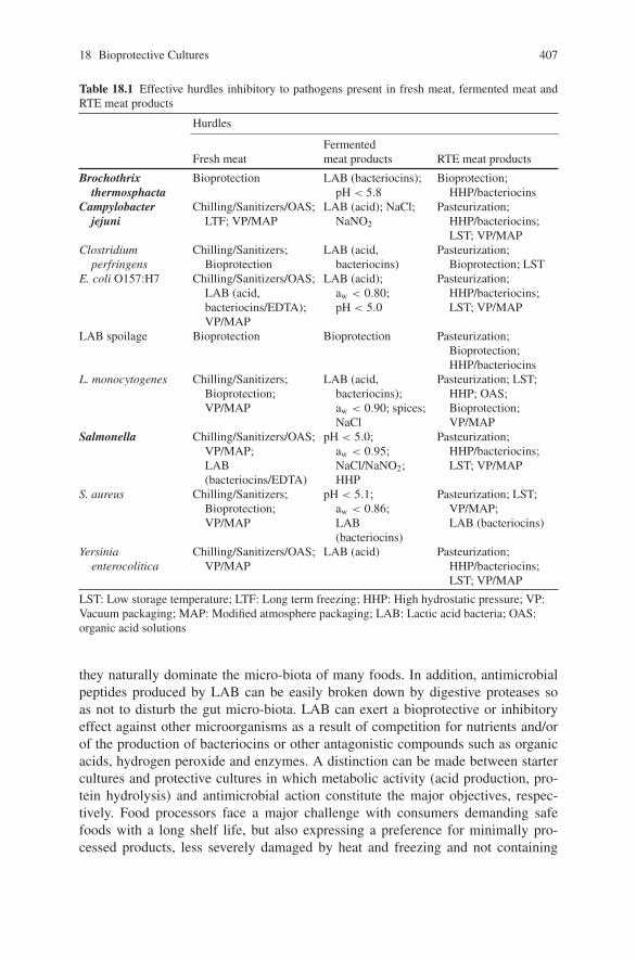
18 Bioprotective Cultures 407
Table 18.1 Effective hurdles inhibitory to pathogens present in fresh meat, fermented meat andRTE meat products
Hurdles
Fresh meatFermentedmeat products RTE meat products
Brochothrixthermosphacta
Bioprotection LAB (bacteriocins);pH < 5.8
Bioprotection;HHP/bacteriocins
Campylobacterjejuni
Chilling/Sanitizers/OAS;LTF; VP/MAP
LAB (acid); NaCl;NaNO2
Pasteurization;HHP/bacteriocins;LST; VP/MAP
Clostridiumperfringens
Chilling/Sanitizers;Bioprotection
LAB (acid,bacteriocins)
Pasteurization;Bioprotection; LST
E. coli O157:H7 Chilling/Sanitizers/OAS;LAB (acid,bacteriocins/EDTA);VP/MAP
LAB (acid);aw < 0.80;pH < 5.0
Pasteurization;HHP/bacteriocins;LST; VP/MAP
LAB spoilage Bioprotection Bioprotection Pasteurization;Bioprotection;HHP/bacteriocins
L. monocytogenes Chilling/Sanitizers;Bioprotection;VP/MAP
LAB (acid,bacteriocins);aw < 0.90; spices;NaCl
Pasteurization; LST;HHP; OAS;Bioprotection;VP/MAP
Salmonella Chilling/Sanitizers/OAS;VP/MAP;LAB(bacteriocins/EDTA)
pH < 5.0;aw < 0.95;NaCl/NaNO2;HHP
Pasteurization;HHP/bacteriocins;LST; VP/MAP
S. aureus Chilling/Sanitizers;Bioprotection;VP/MAP
pH < 5.1;aw < 0.86;LAB(bacteriocins)
Pasteurization; LST;VP/MAP;LAB (bacteriocins)
Yersiniaenterocolitica
Chilling/Sanitizers/OAS;VP/MAP
LAB (acid) Pasteurization;HHP/bacteriocins;LST; VP/MAP
LST: Low storage temperature; LTF: Long term freezing; HHP: High hydrostatic pressure; VP:Vacuum packaging; MAP: Modified atmosphere packaging; LAB: Lactic acid bacteria; OAS:organic acid solutions
they naturally dominate the micro-biota of many foods. In addition, antimicrobialpeptides produced by LAB can be easily broken down by digestive proteases soas not to disturb the gut micro-biota. LAB can exert a bioprotective or inhibitoryeffect against other microorganisms as a result of competition for nutrients and/orof the production of bacteriocins or other antagonistic compounds such as organicacids, hydrogen peroxide and enzymes. A distinction can be made between startercultures and protective cultures in which metabolic activity (acid production, pro-tein hydrolysis) and antimicrobial action constitute the major objectives, respec-tively. Food processors face a major challenge with consumers demanding safefoods with a long shelf life, but also expressing a preference for minimally pro-cessed products, less severely damaged by heat and freezing and not containing
408 G. Vignolo et al.
chemical preservatives. Bacteriocins constitute an attractive option that could pro-vide at least part of the solution; they also have implications for the develop-ment of desirable flora in fermented foods by conferring a rudimentary form ofinnate immunity to foodstuffs and helping processors extend their control over thefood flora long after manufacture (Cotter, Hill, & Ross, 2005). Bacteriocins canbe introduced into food in at least three different ways: (i) by in situ producedbacterial cultures; (ii) by direct addition of purified or semi-purified bacteriocinsto food or (iii) as an ingredient based on a fermentate of a bacteriocin-producingstrain.
Bacteriocins Produced by LAB
The preservative ability of LAB in foods is attributed to the production of antimi-crobial metabolites including organic acids and bacteriocins. Acid production isa common feature among LAB; however, not all LAB can produce antimicrobialpeptides during growth. The production of these peptides seems to be a commonphenotype among LAB since numerous bacteriocins have been isolated over thelast three decades, varying in size from small (<3 kDa), post-translationally heavilymodified peptides to large heat labile proteins. Bacteriocins produced by LAB area heterogeneous group of peptides and proteins. The last proposed revised classifi-cation scheme divides bacteriocins into two categories: the lanthionine-containinglantibiotics (class I) and the non-lanthionine-containing bacteriocins (class II), whilemoving the large, heat-labile murein hydrolases (formerly class III bacteriocins)to a separate designation called bacteriolysins (Cotter et al., 2005). These smallpeptides produced by LAB are secreted into the microbial environment where theymay cause the inhibition of spoilage bacteria and foodborne pathogens. Despitethe dramatic increase in the number of novel bacteriocins discovered in the pasttwo decades, biochemical and genetic characterization has only been carried out onsome of them. Most bacteriocins identified to date and produced by LAB belong toclasses I and II. Although they are generally heat resistant, they can be inactivatedby proteolytic enzymes in foods and due to their hydrophobic character, can bebound by fats and phospholipids. The bacteriocins of interest to the meat indus-try are class II bacteriocins, which involve class IIa pediocin-like or Listeria-activeand class IIb two-peptide bacteriocins. A bacteriocinogenic culture should be welladapted to grow in the product, survive throughout its storage, compete with therelatively high indigenous microbial loads of raw meat, actively inhibit pathogenicand spoilage bacteria, and not alter the sensory properties of meat except undertemperature-abuse conditions. Moreover, the use of bioprotective cultures in refrig-erated vacuum-packaged meat and meat products would be more feasible from aneconomic point of view, and without legal restrictions, compared to the additionof the bacteriocins produced. The use of bacteriocinogenic LAB may be a betterchoice for introducing bacteriocins into meats because they can provide a source ofbacteriocins over a longer period of time.
18 Bioprotective Cultures 409
Bioprotection Applied to Raw Meat
LAB growth on meat is considered as a “hidden” fermentation, since due to thelow carbohydrate content and the strong buffering capacity of the meat thesebacteria do not produce a dramatic change in sensory characteristics. Since meatcannot be pasteurized prior to the addition of LAB cultures, a biopreservativeculture in the meat must compete with the natural micro-biota present. LAB thatgrow at refrigeration temperatures are the natural psychrotrophic species that pre-vail in meat ecosystems. Decontamination of carcasses with organic acid solu-tions has been studied without apparent success (Castillo et al., 2001; Kanellos &Burriel, 2005); whereas decontamination of meat surfaces involving a mixtureof nisin (500 IU/ml) and lactic acid applied on beef carcasses at specific pointsduring slaughter resulted in a significant reduction in bacterial populations (totalcoliforms and E. coli) by 2 log units (Barboza de Martınez, Ferrer, & Salas,2002).
Systems for retail meat distribution are principally based on vacuum-packagingof meat cuts using low gas permeability films and refrigeration. Under theseconditions, CO2 concentration in the pack is critical for microbial inhibition(Phillips, 1996). For maximum antimicrobial effect, the storage temperatureof a modified atmosphere packaged (MAP) product should be kept as lowas possible, since a dramatic decrease in CO2 solubility is produced whentemperature increases. Under these conditions, the emergent psychrotrophicpathogens such as L. monocytogenes and the Gram-negative Y. enterocolitica andAeromonas hydroophila have been observed to grow (Church & Parsons, 1995).An additional hurdle to reduce the risk of L. monocytogenes would be theuse of competitive-bacteriocin producing cultures referred to as bioprotectivecultures.
Lactobacillus curvatus CRL705 in Vacuum-Packaged Fresh BeefStored at Refrigeration Temperatures
Lb. curvatus CRL705 isolated from artisanal Argentinean sausages produceslactocin 705 and lactocin AL705; a two-peptide (class IIb) and a pediocin-like(class IIa) bacteriocin, respectively. Lactocin 705 is a bacteriocin whose activitydepends on the complementary action of two peptides (lac705α and lac705β) of 33amino acid residues each. The genetic characterization as well as its mechanismof action against target microorganisms have been reported (Castellano, Raya, &Vignolo, 2003; Castellano, Vignolo, Farıas, Arrondo, & Chehın, 2007; Cuozzo,Castellano, Sesma, Vignolo, & Raya, 2003). Lactocin 705 was found to be activeagainst closely related LAB and B. thermosphacta while lactocin AL705 is ananti-Listeria bacteriocin (Castellano, Holzapfel, & Vignolo, 2004; Castellano &Vignolo, 2006). From an application point of view, the use of bacteriocinogenicLAB strains as bioprotective cultures for commercial meat preservation would be
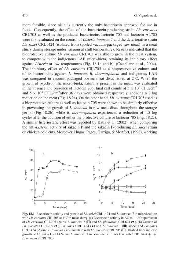
410 G. Vignolo et al.
more feasible, since nisin is currently the only bacteriocin approved for use infoods. Consequently, the effect of the bacteriocin-producing strain Lb. curvatusCRL705 as well as the produced bacteriocins lactocin 705 and lactocin AL705were first evaluated on the control of Listeria innocua 7 and the deteriorative strainLb. sakei CRL1424 (isolated from spoiled vacuum-packaged raw meat) in a meatslurry during storage under vacuum at chill temperatures. Results indicated that thebioprotective culture Lb. curvatus CRL705 was able to grow in the meat system,to compete with the indigenous LAB micro-biota, retaining its inhibitory effectagainst Listeria at low temperatures (Fig. 18.1a and b), (Castellano et al., 2004).The inhibitory effect of Lb. curvatus CRL705 as a biopreservative culture andof its bacteriocins against L. innocua, B. thermosphacta and indigenous LABwas compared in vacuum-packaged bovine meat discs stored at 2◦C. When thegrowth of psychrophilic micro-biota, naturally present in the meat, was evaluatedin the absence and presence of lactocin 705, final cell counts of 5 × 10⁶ CFU/cm2
and 5 × 10⁴ CFU/cm2after 36 days were obtained respectively, showing a 2 logreduction on the meat (Fig. 18.2a). On the other hand, Lb. curvatus CRL705 used asa bioprotective culture as well as lactocin 705 were shown to be similarly effectivein preventing the growth of L. innocua in raw meat discs throughout the storageperiod (Fig 18.2b), while B. thermosphacta experienced a reduction of 1.5 logcycles after the addition of either the protective culture or lactocin 705 (Fig. 18.2c).A similar listeriostatic effect was reported by Katla et al. (2002), when comparingthe anti-Listeria activity of sakacin P and the sakacin P-producing Lb. sakei strainon chicken cold cuts. Moreover, Hugas, Pages, Garriga, & Monfort, (1998), working
Time (days)
0 5 10 15 20 25
AU
ml–
1
Log
cfu
ml–
1
0
20
40
60
80
100
Time (days)0 5 10 15 20 25
0
2
4
6
8
10
12
14
a b
Fig. 18.1 Bacteriocin activity and growth of Lb. sakei CRL1424 and L . innocua 7 in mixed culturewith Lb. curvatus CRL705 at 4◦C in meat slurry. (a) Bacteriocin activity in AU ml−1 of supernatantof Lb. curvatus CRL705 against L. innocua 7 (�) and Lb. plantarum CRL691 ( �). (b) Growth ofLb. curvatus CRL705 ( �), Lb. sakei CRL1424 (�) and L. innocua 7 (�) alone, and Lb. sakeiCRL1424 (Δ) and L. innocua 7 co-inoculate with Lb. curvatus CRL705 (�). Dashed lines indicategrowth of Lb. sakei CRL1424 and L. innocua 7 in combined cultures (Lb. sakei CRL1424 + +L. innocua 7 CRL705)
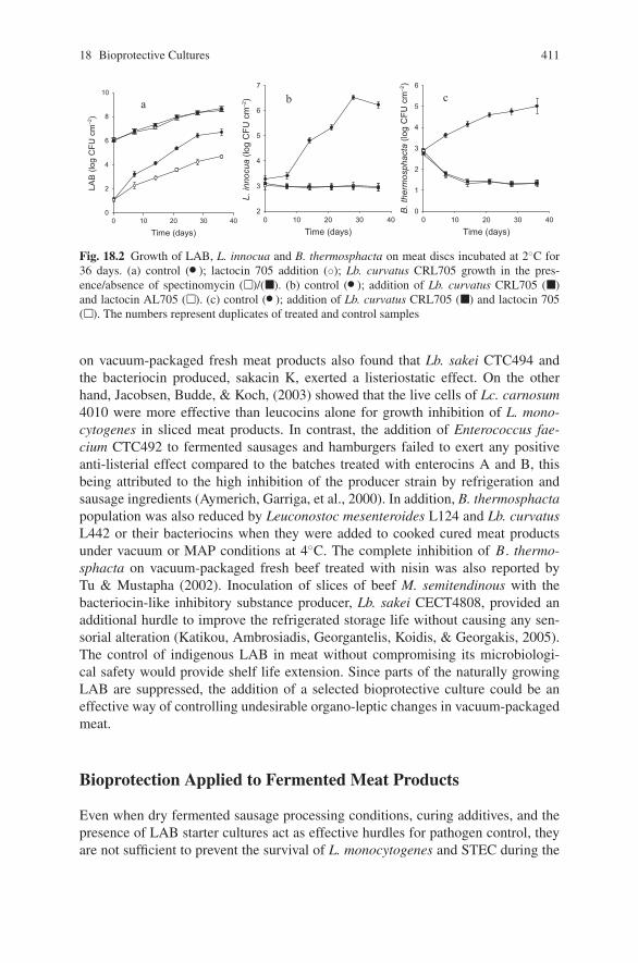
18 Bioprotective Cultures 411
Time (days)0 10 20 30 40
LAB
(log
CFU
cm
–2)
0
2
4
6
8
10
a
Time (days)0 10 20 30 40
L. in
nocu
a (lo
g C
FU c
m–2
)
2
3
4
5
6
7
b
Time (days)0 10 20 30 40
B. t
herm
osph
acta
(log
CFU
cm
–2)
0
1
2
3
4
5
6
c
Fig. 18.2 Growth of LAB, L. innocua and B. thermosphacta on meat discs incubated at 2◦C for36 days. (a) control ( �); lactocin 705 addition (◦); Lb. curvatus CRL705 growth in the pres-ence/absence of spectinomycin (�)/(�). (b) control ( �); addition of Lb. curvatus CRL705 (�)and lactocin AL705 (�). (c) control ( �); addition of Lb. curvatus CRL705 (�) and lactocin 705(�). The numbers represent duplicates of treated and control samples
on vacuum-packaged fresh meat products also found that Lb. sakei CTC494 andthe bacteriocin produced, sakacin K, exerted a listeriostatic effect. On the otherhand, Jacobsen, Budde, & Koch, (2003) showed that the live cells of Lc. carnosum4010 were more effective than leucocins alone for growth inhibition of L. mono-cytogenes in sliced meat products. In contrast, the addition of Enterococcus fae-cium CTC492 to fermented sausages and hamburgers failed to exert any positiveanti-listerial effect compared to the batches treated with enterocins A and B, thisbeing attributed to the high inhibition of the producer strain by refrigeration andsausage ingredients (Aymerich, Garriga, et al., 2000). In addition, B. thermosphactapopulation was also reduced by Leuconostoc mesenteroides L124 and Lb. curvatusL442 or their bacteriocins when they were added to cooked cured meat productsunder vacuum or MAP conditions at 4◦C. The complete inhibition of B. thermo-sphacta on vacuum-packaged fresh beef treated with nisin was also reported byTu & Mustapha (2002). Inoculation of slices of beef M. semitendinous with thebacteriocin-like inhibitory substance producer, Lb. sakei CECT4808, provided anadditional hurdle to improve the refrigerated storage life without causing any sen-sorial alteration (Katikou, Ambrosiadis, Georgantelis, Koidis, & Georgakis, 2005).The control of indigenous LAB in meat without compromising its microbiologi-cal safety would provide shelf life extension. Since parts of the naturally growingLAB are suppressed, the addition of a selected bioprotective culture could be aneffective way of controlling undesirable organo-leptic changes in vacuum-packagedmeat.
Bioprotection Applied to Fermented Meat Products
Even when dry fermented sausage processing conditions, curing additives, and thepresence of LAB starter cultures act as effective hurdles for pathogen control, theyare not sufficient to prevent the survival of L. monocytogenes and STEC during the
412 G. Vignolo et al.
manufacturing process. The use of bacteriocinogenic strains as starter cultures toreduce the risk of L. monocytogenes has been suggested as an extra hurdle to ensureproduct safety and quality. Besides, strategies to overcome the barrier presentedby the outer membrane in Gram-negative bacteria such as the addition of chelatingagents could improve bacteriocin efficiency (Belfiore, Castellano, & Vignolo, 2007).At present only nisin and pediocin PA-1/AcH are widely used in foods. Nisin isused mostly as Nisaplin (Danisco), a preparation containing 2.5% nisin. PediocinPA1 for food biopreservation has also been commercially exploited as ALTA 2431(Quest), based on a fermentate from a pediocin PA-1-producing strain of Pediococ-cus acidilactici; its use being covered by several US and European patents. Amongclass Ia bacteriocins, nisin has not been quite successful in meat products becauseof its low solubility, uneven distribution and lack of stability (Aymerich, Hugas, &Monfort, 1998).
Even when lactococcal bacteriocins seem to be not particularly adapted tosausage technology, the use of lacticin 3147-producing strains as starter culturesin the production of salami showed that the bacteriocin was produced throughoutmanufacture and that the final overall characteristics were similar to the control. Theprotective ability of this lantibiotic was demonstrated by the significant reduction inL. innocua and S. aureus in spiked sausages (Scannell, Ross, Hill, & Arendt, 2000).The effect of in situ production of a bacteriocin by Lactococcus lactis M on L.monocytogenes during fermentation and storage of “merguez” sausage showed agreater reduction of this pathogen in samples fermented with the bacteriocinogenicstrain (Benkerroum, Daoudi, & Kamal, 2003). Nevertheless, nisin producers iso-lated from fermented meat products seem to have some ecological advantages thatmay improve their effectiveness in these products. Among bacteriocinogenic strainsproducing class IIa bacteriocins, Lb. sakei, sakacin producers, have been assayedin in situ experiments, in which they demonstrated a strong activity against Liste-ria. Particularly, in fermented dry sausages, inoculation with the bacteriocinogenicstrains Lb. sakei CTC494 and Lb. curvatus LTH1174 producer of sakacin K andcurvacin A respectively, showed a reduction in L . ivanovii by more than 1-4 logcycles compared to a non-bacteriocinogenic control (Hugas, Garriga, Aymerich, &Monfort, 2002). In addition, the inhibition of L. monocytogenes in ostrich meatsalami by using bacteriocin-producing starter cultures of Lb. plantarum and Lb.curvatus strains was also reported (Dicks, Mellet, & Hoffman, 2004). On the con-trary, the inoculation in sausages as a starter culture of the sakacin K producer,Lb. sakei LTH673, isolated from meat failed to lead the fermentation, althoughit reduced the number of Listeria compared to the non-bacteriocinogenic starter(Hugas, Neumeyer, Pages, Garriga, & Hammes, 1997). Pediocin-producing strainsof P. acidilactici were used to reduce the counts of L. monocytogenes by 1–2.5logs more than a non-pediocin producing strain during summer sausage fermen-tation (Foegeding, Thomas, Pilkington, & Klaenhammer, 1992). Moreover, whenthe survival of L . monocytogenes was compared in German, American and Italian-style fermented sausages using pediococci as a starter culture, higher reductionsin Listeria numbers were observed in the American–style sausages after fermen-tation and drying. The higher fermentation temperature used in American-style
18 Bioprotective Cultures 413
sausages (25–35◦C) may have a positive influence on the production of bacteriocinsby P. acidilactici.
Although the use of bacteriocin-producing enterococci in cheese manufactureis well documented, information on their potential as biopreservatives in the meatindustry is scarce. Bacteriocin-producing E. faecium strains have been includedin the starter cultures in Spanish-style dry fermented sausage to effectively inhibitListeria (Callewaert, Hugas, & De Vuyst, 2000) The bacteriocin-producing Entero-coccus casseliflavus IM416K1, isolated from Italian “cacciatore” sausage, showeda high effectiveness in L. monocytogenes elimination while a listeriostatic effectwas obtained when enterocin CCM4231, produced by a strain of E. faecium,was added to the sausage batter during the manufacture of Hornad salami, adry fermented traditional Slovakian product (Laukova, Czikkova, Laczkova, &Turek, 1999; & Sabia, de Niederhausern, Messi, Manicardi, & Bondi, 2003). Incontrast, E. faecium CTC492, an enterocin A and B producer, was unable to exertany positive anti-listerial effect on fermented sausages compared to the batchestreated with enterocins A and B; this being attributed to a high inhibition ofthe producer strain by refrigeration and sausage ingredients (Aymerich, Artigas,Garriga, Monfort, & Hugas, 2000). Since enterococci are hardly competitivein the early stages of the fermentation in reducing listerial counts, bacteriocin-producing enterococcal strains may be used as adjunct cultures during sausagemanufacture.
The use of a bacteriocinogenic strain as a new functional starter culture mayoffer food safety advantages, without risks for human health due to toxicologicalside effects. It represents a safe way of natural preservation that has probablyoccurred in fermented foods for centuries. Moreover, bacteriocin production infermented sausages does not generally lead to organo-leptic imperfections, ashas been shown in sensorial studies with different strains (Coffey et al., 1998).However, some disadvantages have to be considered related to the lower in situbacteriocin activity compared to the one expected from in vitro experiments; thisbeing due to low production, genetic instability, inability for uniform distributionthroughout the product, low solubility, inactivation by meat proteases, resistanceof the target strain, and interference by meat components (Leroy, Verluyten, &De Vuyst, 2006). The use of strains well adapted to the sausage environment,preferably sausage isolates as well as strain compatibility (including GCC) must beconsidered for optimal performance and bacteriocin production. In this context, ithas been shown that the sausage isolates Lb. sakei CTC494, Lb. curvatus LTH1174and L442 optimally produce bacteriocins under conditions of pH and temperaturethat prevail during European sausage fermentation (Leroy et al., 2006). Moreover,a bioprotective culture must be selected according to the specific formulation andtechnology used in the fermentation. The performance of bacteriocinogenic LABas starters or co-cultures will depend on the processing technology (pediococciare more suitable than lactobacilli in fast fermentation at high temperatures), thepresence of nitrate or nitrate/nitrite, NaCl and spices (Hugas et al., 1998; Verluyten,Leroy, & De Vuyst, 2004; Vignolo, Fadda, Kairuz, Ruiz Holgado, & Oliver,1998).
414 G. Vignolo et al.
Bioprotection Applied to Ready-to-Eat Meat Products
The microbiological status of processed meat products is highly dependent on rawmaterials and hygiene of the handling procedures. Even when these products areheated to a temperature between 65–75◦C and most vegetative cells are killed, post-heat treatment re-contamination will determine their shelf life. Cooked meat andpoultry products are generally either pre-packaged in a permeable over-wrappingfilm or cut from a bulk block and may become contaminated during production orprocessing at home or food establishments. The low salt content, pH around 6.0and water activity higher that 0.95 constitute weak hurdles to prevent the growthof organisms associated with post-processing contamination. Even when processingconditions are adequate and the product has been correctly handled after processing,S. aureus, L. monocytogenes, Salmonellae and shiga toxin-producing E. coli may beimplicated in RTE contamination. Since products such as frankfurters and cookedham are massively consumed, effective hurdle technologies to inhibit the growth ofthese pathogens should be implemented.
During the last 20 years, a number of studies have focused on the biopreservationof RTE meat products with LAB, mainly to control the presence of L. monocyto-genes which has become a major concern for the meat processing industry. Cookedmeat products offer no protection against L. monocytogenes outgrowth if contami-nation occurs after post-processing during slicing and/or packaging. This pathogenwill grow exponentially even at refrigeration temperatures reaching high numberswithin days, the control of L. monocytogenes being difficult due to its ability togrow at a wide temperature range (3 to 45◦C), pH tolerance (4.8 to 9.6), high NaCllevels (8–9%) and sodium nitrite (< 1000 ppm). The use of meat borne LAB asprotective cultures for the biopreservation of cooked meat products has been exten-sively reported. Effective control of L. monocytogenes after refrigerated storage ofdifferent RTE meat products was obtained when selected strains of Lactobacil-lus and P. acidilactici, previously isolated from the same products, were directlyapplied on their surface (Amezquita & Brashears, 2002; Vermerien, Devlieghere, &Debevere, 2004). Similarly, the addition of Lb. sakei TH1 as a bioprotective cultureto cooked, sliced and vacuum-packaged ham and servelat sausage resulted in theinhibition of L. monocytogenes at 8 and 4◦C during 28 days of storage (Bredholt,Nesbakken, & Holck, 2001). In addition, when the live cells of the bacteriocin pro-ducing Lc. carnosum 4010 were applied by spray to cooked and sliced pork saveloy,it was also reported to be effective in the control of L . monocytogenes during 4weeks of refrigerated storage (Jacobsen et al., 2003). The anti-listerial effect of bio-protective cultures and/or their bacteriocins will depend on the product compositionand the presence of competitive microflora. The addition of enterocins A and Bproduced by E. faecium CTC492 (4,800 AU/g) to cooked ham and pork liver pateto inhibit L. innocua showed an effective reduction in its numbers; this being higherin pate than in cooked ham. Without competitive microflora and favorable physico-chemical conditions, Listeria growth will be promoted, but the higher efficiency ofenterocins in the fatter product (pate) is consistent with the hydrophobic nature ofbacteriocins (Aymerich, Garriga, et al., 2000).
18 Bioprotective Cultures 415
Even when studies on biopreservation in cooked meat products have mainlyfocused on the inhibition of food pathogens such as L. monocytogenes, the pres-ence of spoilage LAB as well as B. thermosphacta is of concern because of thehigh economic losses. Product handling after cooking plus slicing prior to pack-aging re-contaminates them with about 0.5–2 log CFU/g of total bacteria, mainlyLAB. When stored under vacuum-packaging or MAP and refrigeration conditions,LAB will dominate the spoilage process. The metabolic activity of LAB results inthe spoilage appearing as sour, off-flavors, off-odors, milky exudates, slime pro-duction, swelling of the package through gas production and discoloration such asgreening (Samelis & Georgiadou, 2000). The spoilage microflora of vacuum pack-aged cooked meat products as well as salty dry products mainly consists of Lacto-bacillus, Leuconostoc, Carnobacterium and Weissella species and B. thermosphacta(Koort et al., 2006; Samelis, Bjorkroth, Kakouri, & Rementzis, 2006; Samelis &Georgiadou, 2000). LAB that are homo-fermentative, salt tolerant, psychrotrophic,adapted to meat substrates and have a good potential to be used for bioprotection ofRTE products. The inoculation of vacuum-packaged sliced cooked ham and frank-furters with the bioprotective culture Lactobacillus alimentarius FloraCarn L2, atrefrigeration temperatures, showed 1 week and 19 days extension in their shelf liferespectively (Kotzekidou & Bloukas, 1998). When the bacteriocins producing Lc.mesenteroides L124 and Lb. curvatus L422 were used as bioprotective cultures insliced cooked cured pork shoulder and frankfurter-type sausage, a reduction in thenumbers of B. thermosphacta and enterococci was obtained thus extending theirshelf life (Metaxopoulos, Mataragas, & Drosinos, 2002). Specifically, the inhibitoryeffect of lactocin S producing Lb. sakei 148 on hetero-fermentative LAB present ina cooked model ham was demonstrated by Vermeiren et al. (2004). The use of E.faecium CTC492 and Lb. sakei CTC494 as bioprotective cultures prevented ropinessdue to Lb. sakei CTC476 until 21 days of storage at 8◦C in sliced cooked meatproducts (Hugas et al., 2002).
Application of bioprotective cultures to RTE products will depend on every par-ticular technology. Specifically, for cooked ham, inoculation with the bacteriocino-genic strain in the bulk mass during the tumbling procedure (5–6◦C, during 24 h)will ensure its growth and bacteriocin production. Since bacteriocins are thermo-resistant they would remain active in the cooked product mass after pasteurizationand during storage, preventing contaminant and/or pathogen growth. On the otherhand, during frankfurter processing bacteriocins may be added to meat batter beforecooking, included in the casing as a folding additive or sprayed on the surface duringpost-processing and packing. The anti-listerial effect of bacteriocins was found tobe higher when applied in the meat batter of the frankfurter-type sausage (Hugaset al., 2002).
Strategies to Improve Bioprotective Effect
Even when many bacteriocinogenic strains inhibited L. monocytogenes in vitro andin meat systems, most studies on the in situ effect were carried out on single strainsof the pathogen or used high levels of contamination. Significant variations in the
416 G. Vignolo et al.
sensitivity of the Listeria strains to the same bacteriocin as well as the emergence ofspontaneous resistant mutants to individual bacteriocins were reported (Castellano,Farıas, Holzapfel, & Vignolo, 2001). For improved control of target microorganismsand bacteriocin-resistant strains, a combined use of bacteriocins was proposed. Theuse of nisin (class Ia), lactocin 705 (class IIb) and enterocin CRL35 (class IIa)combination against different L . monocytogenes strains in meat slurry showed noviable counts after incubation (Vignolo et al., 2000). The simultaneous addition ofa nisin-curvaticin 13 combination also led to the absence of viable Listeria cells inthe broth, preventing the re-growth of bacteriocin resistant mutants (Bouttefroy &Milliere, 2000). However, when the direct addition of enterocin A, sakacin K (pro-duced by E. faecium CTC492 and Lb. sakei CTC494, respectively), Nisaplin (nisin)and ALTA 2351 (pediocin) to frankfurter sausages meat batter was carried out, onlythe batch with enterocin and sakacin showed significant Listeria reduction numbers(7.6 log cycles during 60 days of storage at 3.5◦C). No significant differences wereobserved when sakacin, enterocin or nisin were applied separately or with ALTA(Hugas et al., 2002). It is generally assumed that a mixture containing more thanone bacteriocin will be bactericidal to more cells in a sensitive population, sincecells resistant to one bacteriocin would be killed by another.
Combinations of selected and those that are generally recognized as safe preser-vative antimicrobial solutions applied to processed meat surface may also providea safeguard to contamination by L . monocytogenes. In this context, lactic, acetic,benzoic, propionic and sorbic acids or their salts in combination with bacteriocinswere reported to be highly inhibitory. Post-processing sequential treatment of dip-ping solutions of Nisaplin (5000 IU/ml) plus lactic acid, reduced L. monocytogenesto undetectable levels in vacuum-packaged bologna ham stored at 10◦C when com-pared to nisin applied alone (Geornaras et al., 2005). In order to find hurdles toavoid the risk of pathogen growth during storage and to reduce the levels of chem-icals applied, new food preservation technologies complementary and alternativeto traditional techniques have been assayed in combination with bacteriocins. Theeffectiveness of high hydrostatic pressure combined with nisin and lactate salts toinhibit the growth of L. monocytogenes and Salmonella in sliced cooked ham wasdemonstrated. It seems that the injured survivors after pressure became sensitive tonisin resulting in a viability loss (Aymerich, Jofre, Garriga, & Hugas, 2005). On theother hand, combining pediocin (ALTA2341) with post-packaging thermal pasteur-ization showed an effective treatment combination for improved control of L. mono-cytogenes on frankfurters (Chen, Sebranek, Dickson, & Mendonca, 2004). More-over, a combination of the non-bacteriocinogenic Lb. sakei subsp. carnosus 10Aand lactocin S producing Lb. sakei 148 with MAP containing 50% CO2, completelyinhibited the growth of L. monocytogenes on a model cooked ham (Vermeiren,Devlieghere, Vandekinderen, & Devebere, 2006). Food-grade chelators such asEDTA in combination with bacteriocins were also assayed as part of the hurdleconcept to overcome Gram-negative resistance to bacteriocins. Vacuum-packagedand refrigerated fresh beef treated with nisin combined with EDTA increased itsshelf life by inhibiting the growth E. coli as well as B. thermosphacta (Tu &Mustapha, 2002).
18 Bioprotective Cultures 417
Influence of the Bioprotective Culture on Meat SensorialChanges
The use of selected LAB strains as bioprotective cultures to extend the shelf life ofchill stored vacuum-packaged meat and meat products implies the growth and acidproduction of the bacteriocinogenic strain. As stated above, LAB induces typicalsensory changes such as souring, gas, SH2 and slime thus resulting in costly lossesto the industry. Organo-leptic defects related to lactic acid production would beperceived by consumers as sour or cheesy off odors and/or off tastes and even adiscoloration process may occur due to meat protein denaturation. When the pro-tective culture Lb. curvatus CRL705 applied on raw meat discs did not result insignificant pH changes after incubation for 36 days at 2◦C compared to the con-trol, this indicated that no organo-leptic defects related to pH decrease were pro-duced by the bioprotective culture (Castellano & Vignolo, 2006). This small pHdecline may be attributed to the buffering capacity of the meat and the poten-tial contribution of LAB to the breakdown of meat proteins and peptides, thisbeing responsible for the generation of basic compounds which partially neutral-ize the acid produced and prevent an excessive acidification of the meat. In addi-tion to a mild acidification, a bioprotective culture used to extend the shelf life offresh meat should neither be a gas-forming nor an exo-polysaccharide producerto avoid bulging in the packages due to gas accumulation and slime formationon meat surfaces. To fulfill industrial requirements, a protective culture should beeasy to culture, easy to apply to the meat and with a reliable and reproducibleresult.
Conclusions
Nowadays, consumers are greatly concerned about the relationship between foodand health. The demand for reduced use of additives and processing seems con-tradictory for a market demanding safer and tastier foods. These market demandshave placed the food industry under pressure to search for innovative solutions.Meat and meat products, in particular, face the challenge of overcoming thelack of consumer confidence that emerged during the last decade due to BSEand foodborne outbreaks caused by emerging pathogens. Novel insights intothe metabolism of LAB offer perspectives for the application of a new gen-eration of functional cultures. New starter LAB cultures with an industriallyimportant functionality are being developed, mainly contributing to microbialsafety or offering one or more sensorial, technological, nutritional or healthadvantages. The use of competitive bacteriocinogenic LAB as bioprotective cul-tures may well provide at least part of the solution. However, the applicationof biopreservation technology to meat and meat products should be consideredonly as an additional hurdle to good manufacturing processing and distributionpractices.
418 G. Vignolo et al.
References
Amezquita, A., & Brashears, M. (2002). Competitive inhibition of Listeria monocytogenes inready-to-eat products by lactic acid bacteria. Journal of Food Protection, 65, 316–325.
Autio, T., Hielm, S., Miettinen, M., Sjoberg, A., Aamisalo, K., Bjorkroth, J., et al. (1999). Sourcesof Listeria monocytogenes contamination in a cold-smoked rainbow trout processing plantdetected by pulsed-field gel electrophoresis typing. Applied Environmental Microbiology, 65,150–155.
Aymerich, M. T., Artigas, M. G., Garriga, M., Monfort, J. & Hugas, M. (2000). Effect of sausageingredients and additives on the production of enterocins A and B by Enterococcus faeciumCTC492. Optimization of in vitro production and anti-listerial effect in dry fermented sausages.Journal of Applied Microbiology, 88, 686–694.
Aymerich, M. T., Garriga, M., Ylla, J., Vallier, J., Monfort, J., & Hugas, M. (2000). Applicationsof enterocins as biopreservatives against Listeria innocua in meat products. Journal of FoodProtection, 63, 721–726.
Aymerich, M. T., Hugas, M., & Monfort, J. M. (1998). Review: Bacteriocinogenic lactic acidbacteria associated with meat products. Food Science and Technology International, 4,141–158.
Aymerich, M. T., Jofre, A., Garriga, M., & Hugas, M. (2005). Inhibition of Listeria monocytogenesand Salmonella by natural antimicrobials and high hydrostatic pressure in sliced cooked ham.Journal of Food Protection, 68, 173–177.
Balaban, N., & Rasooly, A. (2000). Staphylococcal enterotoxins. International Journal of FoodMicrobiology, 61, 1–10.
Barboza de Martınez, Y., Ferrer, K., & Salas, E. (2002). Combined effects of lactic acid and nisinsolution in reducing levels of microbiological contamination in red meat carcasses. Journal ofFood Protection, 65, 1780–1783.
Beach, J. C., Murano, E.-A., & Acuff, G. R. (2002). Prevalence of Salmonella and Campylobacterin beef from transport to slaughter. Journal of Food Protection, 65, 1687–1693.
Belfiore, C., Castellano, P., & Vignolo, G. (2007). Reduction of Escherichia coli population fol-lowing treatment with bacteriocins from lactic acid bacteria and chelators. Food Microbiology,24, 223–229.
Benkerroum, N., Daoudi, A., & Kamal, M. (2003). Behaviour of Listeria monocytogenes in rawsausages (merguez) in presence of a bacteriocins-producing lactococcal strain as a protectiveculture. Meat Science, 63, 479–484.
Bhaduri, S., Wesley, I., & Bush, E. (2005). Prevalence of pathogenic Yersinia enterocolitica strainsin pigs in the United States. Applied and Environmental Microbiology, 71, 7117–7121.
Bouttefroy, A., & Milliere, J. B. (2000). Nisin-curvaticin 13 combinations for avoiding theregrowth of bacteria resistant cell of Listeria monocytogenes ATCC 15313. International Jour-nal of Food Microbiology, 62, 65–75.
Bredholt, S., Nesbakken, T., & Holck, A. (2001). Industrial application of an antilisterial strain ofLactobacillus sakei as a protective culture and its effect on the sensory acceptability of cooked,sliced, vacuum-packaged meats. International Journal of Food Microbiology, 66, 191–196.
Bremer, V., Bocter, N., Rehmet, S., Klein, G., Breuer, T., & Ammon, A. (2005). Consumption,knowledge, and handling of raw meat: A representative cross-sectional survey in Germany,March 2001. Journal of Food Protection, 68, 785–789.
Cahill, S. M., & Jouve, J.-L. (2004). Microbiological risk assessment in developing countries.Journal of Food Protection, 67, 2016–2023.
Callewaert, R., Hugas, M., & De Vuyst, L. (2000). Competitiveness and bacteriocin production ofEnterococci in the production of Spanish-style dry fermented sausages. International Journalof Food Microbiology, 57, 33–42.
Castellano, P., Farıas, M. E., Holzapfel, W., & Vignolo, G. (2001). Sensitivity variations of Listeriastrains to the bacteriocins lactocin 705, enterocin CRL35 and nisin. Biotechnology Letters, 23,605–608.
18 Bioprotective Cultures 419
Castellano, P., Holzapfel, W., & Vignolo, G. (2004). The control of Listeria innocua and Lacto-bacillus sakei in broth and meat slurry with the bacteriocinogenic strain Lactobacillus caseiCRL705. Food Microbiology, 21, 291–298.
Castellano, P., Raya, R., & Vignolo, G. (2003). Mode of action of the two-component bacteriocinlactocin 705. International Journal of Food Microbiology, 85, 35–43.
Castellano, P., & Vignolo, G. (2006). Inhibition of Listeria innocua and Brochothrix thermosphactain vacuum-packaged meat by the addition of bacteriocinogenic Lactobacillus curvatus CRL705and its bacteriocins. Letters in Applied Microbiology, 43, 194–199.
Castellano, P., Vignolo, G., Farıas, R., Arrondo, J., & Chehın, R. (2007). Molecular view by Fouriertransform infrared spectroscopy of the relationship between lactocin 705 and membranes: Spec-ulations on antimicrobial mechanism. Applied of Environmental Microbiology, 73, 415–420.
Castillo, A., Lucia, L., Mercado, I., & Acuff, G. (2001). In-plant evaluation of a lactic acidtreatment for reduction of bacteria on chilled beef carcases. Journal of Food Protection, 64,738–740.
Cayre, M. E., Garro, O., & Vignolo, G. (2005). Effect of storage temperature and gas permeabil-ity of packaging film on the growth of lactic acid bacteria and Brochothrix thermosphacta incooked meat emulsions. Food Microbiology, 22, 505–512.
CDC, Centers for Disease Control and Prevention (1995). Escherichia coli O157:H7 outbreaklinked to commercially distributed dry cured salami-Washington and California. Morbidity andMortality Weekly Report, 44, 157–160.
CDC, Centers for Disease Control and Prevention (2002). Incidence of foodborne illness: Prelimi-nary FoodNet data on the incidence of foodborne illness-selected sites, United States. Morbidityand Mortality Weekly Report, 51, 325–329.
CDC, Centers for Disease Control and Prevention (2007). Preliminary FoodNet data on the inci-dence of infection with pathogens transmitted commonly through food – 10 States, 2006. Mor-bidity and Mortality Weekly Report, 56, 336–339.
Chang, V., Mills, E., & Cutter, C. (2003). Reduction of bacteria on pork carcasses associated withchilling method. Journal of Food Protection, 66, 1019–1024.
Chen, C., Sebranek, J., Dickson, J., & Mendonca, A. (2004). Combining pediocin (ALTA2341)with postpackaging pasteurization for control of Listeria monocytogenes frankfurters. Journalof Food Protection, 67, 1855–1865.
Chen, Y., Ross, W., Scott, V., & Gombas, D. (2003). Listeria monocytogenes: Low levels equal lowrisk. Journal of Food Protection, 66, 570–577.
Church, D. (2004). Major factors affecting the emergence and re-emergence of infectious diseases.Clinics in Laboratory Medicine, 24, 559–586.
Church, I., & Parsons, A. (1995). Modified atmosphere packaging technology: A review. Journalof Science and Food Agriculture, 67, 143–152.
Coffey, A., Ryan, M., Ross, R., Hill, C., Arendt, E., & Schwarz, G. (1998). Use of a broad-host-range bacteriocins-producing Lactococcus lactis transconjugant as an alternative starterfor salami manufacture. International Journal of Food Microbiology, 43, 231–235.
Cotter, P., Hill, C., & Ross, P. (2005). Bacteriocins: Developing innate immunity for foods. NatureReviews Microbiology, 3, 777–788.
Cuozzo, S., Castellano, P., Sesma, F., Vignolo. G., & Raya, R. (2003). Differential roles of twocomponent peptides of lactocin 705 in antimicrobial activity. Current Microbiology, 46, 180–183.
Demeyer, D. (2003). Meat fermentation: Principles and applications. In Y. Hui, L. Goddik, A.Hansen, J. Josephsen, W. Nip, P. Stanfield, et al. (Eds.), Handbook of Food and BeverageFermentation Technology (pp. 353–367). New York: Marcel Dekker.
Desmarchelier, P., Higgs, G., Mills, L., Sullivan, A., & Vanderlinde, P. (1999). Incidence of coag-ulase positive Staphylococcus on beef carcasses in three Australian abattoirs. InternationalJournal of Food Microbiology, 47, 221–229.
Dicks, L., Mellet, F., & Hoffman, L. (2004). Use of bacteriocin-producing starter culture of Lac-tobacillus plantarum and Lactobacillus curvatus in production of ostrich meat salami. MeatScience, 66, 703–708.
420 G. Vignolo et al.
Elmi, M. (2004). Food safety: Current situation, unaddressed issues and the emerging priorities.Eastern Mediterranean Health Journal, 10, 794–800.
Fegan, N., Vanderlinde, P., Higgs, G., & Desmarchelier, P. (2004). The prevalence and concentra-tion of Escherichia coli O157 in feces of cattle from different production systems at slaughter.Journal of Applied Microbiology, 97, 362–370.
Foegeding, P., Thomas, A., Pilkington, D., & Klaenhammer, T. (1992). Enhanced control of Lis-teria monocytogenes by in situ-produced pediocin during dry fermented sausage production.Applied & Environmental Microbiology, 58, 3053–3059.
Fontana, C., Cocconcelli, P., & Vignolo, G. (2005). Monitoring the bacterial population dynamicsduring fermentation of artisanal Argentinean sausages. International Journal of Food Microbi-ology, 103, 131–142.
Fontana, C., Cocconcelli, P., & Vignolo, G. (2006). Direct molecular approach to monitoringbacterial colonization on vacuum-packaged beef. Applied Environmental Microbiology, 72,5618–5622.
Geornaras, I., Belk, K., Scanga, J., Kendall, P., Smite, G., & Sofos, J. (2005). Postprocessingantimicrobial treatments to control L. monocytogenes in commercial vacuum-packaged bolognaham stored at 10◦C. Journal of Food Protection, 68, 991–998.
Gombas, D., Chen, Y., Clavero, R., & Scott, V. (2003). Survey of Listeria monocytogenes in ready-to-eat foods. Journal of Food Protection, 66, 559–560.
Griffin, P., & Tauxe, R. (1991). The epidemiology of infections caused by Escherichia coliO157:H7, other enterohemorrhagic E. coli and the associated haemolytic uremic syndrome.Epidemiology Reviews, 13, 60–98.
Gurtler, M., Alter, T., Kasimir, S., Linnebur, M., & Fehlhaber, K., (2005). Prevalence of Yersiniaenterocolıtica in fattening pigs. Journal of Food Protection, 68, 850–854.
Hald, T., Wingstrand, A., Swanenburg, M., von Altrock, A., & Thorberg, B. (2003). The occurrenceand epidemiology of Salmonella in European pig slaughterhouses. Epidemiology Infection,131, 1187–1203.
Hassan, Z., Purwati, E., Radu, S., Rahim, R., & Rusul, G. (2001). Prevalence of Listeria spp andListeria monocytogenes in meat and fermented fish in Malaysia. Southeast Asian Journal ofTropical Medicine Public Health, 32, 402–407.
Holeckova, B., Holoda, E., Fotta, M., Kalinacova, V., Gondol’, J., & Grolmus, J. (2002). Occur-rence of enterotoxigenic Staphylococcus aureus in food. Annals of Agricultural and Environ-mental Medicine, 9, 179–182.
Holley, R., Lammerding, A., & Tittiger, F. (1988). Microbiological safety of traditional and starter-mediated processes for the manufacture of Italian dry sausage. International Journal of FoodMicrobiology, 7, 49–62.
Holzapfel, W., Geisen, R., & Schillinger, U. (1995). Biological preservation of foods with refer-ence to protective cultures, bacteriocins and food-grade enzymes. International Journal of FoodMicrobiology, 24, 343–362.
Hugas, M., Garriga, M., Aymerich, M., & Monfort, J., (2002). Bacterial cultures and metabolitesfor the enhancement of safety and quality. In F. Toldra (Ed.), Research advances in the qualityof meat and meat products (pp. 225–247). India: Research Signpost.
Hugas, M., Neumeyer, B., Pages, F., Garriga, M., & Hammes, W. (1997). Comparisonof bacteriocins-producing lactobacilli on Listeria growth in fermented sausages. Fleis-chwirtschaft, 76, 649–652.
Hugas, M., Pages, F., Garriga, M., & Monfort, J. (1998). Application of the bacteriocinogenicLactobacillus sakei CTC494 to prevent growth of Listeria in fresh and cooked meat productspacked with different atmospheres. Food Microbiology, 15, 639–650.
Hussein, H., & Bollinger, L. (2005). Prevalence of Shiga toxin-producing Escherichia coli in beefcattle. Journal of Food Protection, 68, 2224–2241.
ICMSF International Commission on Mirobiological Specifications for Foods. (2002). Microor-ganisms in foods 7: Microbiological testing in food safety management (pp. 285–312). NewYork: Kluwer Academic/Plenum Publishers.
18 Bioprotective Cultures 421
ILSI Research Foundation; Risk Science Institute. (2005). Achieving continuous improvementin reductions in foodborne listeriosis-a risk-based approach. Journal of Food Protection, 68,1932–1994.
Ingham, S., Engel, R., Fanslau, M., Schoeller, E., Searls, G., Buege, D., et al. (2005). Fate ofStaphylococcus aureus on vacuum-packaged ready-to-eat meat products stored at 21 degreesC. Journal Food Protection, 68, 1911–1915.
Inglis, G., Kalischuk, L., Busz, H., & Kastelic, J. (2005). Colonization of cattle intestines byCampylobacter jejuni and Campylobacter lanienae. Applied and Environmental Microbiology,71, 5145–5153.
Jacobsen, T., Budde, B., & Koch, A. (2003). Application of Leuconostoc carnosum for biopreser-vation of cooked meat products. Journal of Applied Microbiology, 95, 242–249.
Jones, R. J. (2004). Observations on the succession dynamics of lactic acid bacteria populations inchill-stored vacuum-packaged beef. International Journal of Food Microbiology, 90, 273–282.
Kalinowski, R., Tompkin, R., Bodnaruk, P., & Pruett, W. (2003). Impact of cooking, and subse-quent refrigeration on the growth or survival of Clostridium perfringes in cooked meat andpoultry products. Journal of Food Protection, 66, 1227–1232.
Kanellos, T., & Burriel, A. (2005). The bactericidal effects of lactic acid and trisodium phosphateon Salmonella enteritidis serotype pt4, total viable counts and counts of Enterobacteriaceae.Food Protection Trends, 25, 346–350.
Katikou, P., Ambrosiadis, I., Georgantelis, D., Koidis, P., & Georgakis, S. (2005). Effect of Lac-tobacillus-protective cultures with bacteriocins-like inhibitory substances’ producing abilityon microbiological, chemical and sensory changes during storage of refrigerated vacuum-packaged sliced beef. Journal of Applied Microbiology, 99, 1303–1313.
Katla, T., Møretrø, T., Sveen, I., Aasen, I., Axelsson, L., & Rørvik, L. (2002). Inhibition of Lis-teria monocytogenes in chicken cold cuts by addition of sakacin P and sakacin P-producingLactobacillus sakei. Journal of Applied Microbiology, 93, 191–196.
Koort, J., Coenye, T., Santos, E., Molinero, C., Jaime, I., Rovira, J., et al. (2006). Diversity ofWeissella viridescens strains associated with “Morcilla de Burgos”. International Journal ofFood Microbiology, 109, 164–168.
Kotzekidou, P., & Bloukas, J. (1998). Microbial and sensory changes in vacuum-packedfrankfurter-type sausages by Lactobacillus alimentarius and fate of inoculated Salmonellaenteritidis. Food Microbiology 15, 101–111.
Laukova, A., Czikkova, S., Laczkova, S., & Turek, P., (1999). Use of enterocin CCM4231 tocontrol Listeria monocytogenes in experimentally contaminated dry fermented Hornad salami.International Journal of Food Microbiology, 52, 115–119.
Leistner, L. (1997). Microbial stability and safety of healthy meat, poultry and fish products. InA. M. Pearson, & T. R. Dutson (Eds.), Production and processing of healthy meat, poultry andfish products (pp. 347–360). London: Blackie Academic and Professional.
Leistner, L. (2000). Basic aspects of food preservation by hurdle technology. International Journalof Food Microbiology, 55, 181–186.
Leroy, F., Verluyten, J., & De Vuyst, L. (2006). Functional meat starter cultures for improvedsausage fermentation. International Journal of Food Microbiology, 106, 270–285.
Lin, C., Takeuchi, K., Zhang, L., Dohm, C., Meyer, J., Hall, P., et al. (2006). Cross-contaminationbetween processing equipment and deli meats by Listeria monocytogenes. Journal of FoodProtection, 69, 71–79.
Lucke, F. -K. (2000). Utilization of microbes to process and preserve meat. Meat Science, 56,105–115.
Marshall, K., Niebuhr, S., Acuff, G., Lucia, L., & Dickson, J. (2005). Identification of Escherichiacoli O157:H7 meat processing indicators for fresh meat through comparison of the effects ofselected antimicrobial interventions. Journal of Food Protection, 68, 2580–2586.
Marzoca, M., Marucci, P., Sica, M., & Alvarez, E. (2004). Listeria monocytogenes detectionin different food products and environmental samples of supermarkets of Bahia Blanca city(Argentina). Revista Argentina de Microbiologıa, 36, 179–181.
422 G. Vignolo et al.
McEvoy, J., Doherty, A., Sheridan, J., Blair, I., & McDowell, D. (2003). The prevalence ofSalmonella spp. in bovine faecal, rumen and carcass samples at a commercial abattoir. Journalof Applied Microbiology, 94, 693–700.
Meichtri, L., Miliwebsky, E., Gioffre, A., Chinen, I., Baschkier, A., Chillemi, G., et al. (2004).Shiga toxin-producing Escherichia coli in healthy young beef steers from Argentina: Preva-lence and virulence properties. International Journal of Food Microbiology, 96, 189–198.
Metaxopoulus, J., Mataragas, M., & Drosinos, E. (2002). Microbial interaction in cooked curedmeat products under vacuum or modified atmosphere at 4◦C. Journal of Applied Microbiology,93, 363–373.
Morse, S. S. (2004). Factors and determinants of disease emergence. Revue Scientifique et Tech-nique, 23, 443–451.
Morse, S. S. (2007).Global infectious disease surveillance and health intelligence. Health Affairs,26, 1069–1077.
Normano, G., Parisi, A., Dambrosio, A., Quaglia, N., Montagna, D., Chiocco, D., et al. (2004).Typing of Escherichia coli O157 strains isolated from fresh sausage. Food Microbiology, 21,79–82.
Okutani, A., Okada, Y., Yamamoto, S., & Igimi, S. (2004). Nationwide survey of human Listeriamonocytogenes infection in Japan. Epidemiology Infection, 132, 769–772.
Omisakin, F., MacRae, M., Ogden, I., & Strachan, N. (2003). Concentration and prevalence ofEscherichia coli O157 in cattle feces at slaughter. Applied and Environmental Microbiology,69, 2444–2447.
Pala, T., & Sevilla, A. (2004). Microbial contamination of carcasses, meat and equipment from anIberian pork cutting plant. Journal of Food Protection, 67, 1624–1629.
Peccio, A., Autio, T., Korkeala, H., Rosmini, R., & Trevisani, M. (2003). Listeria monocytogenesoccurrence and characterization in meat-producing plants. Letters in Applied Microbiology, 37,234–238.
Phillips, C. A. (1996). Modified atmosphere packaging and its effects on the microbiological qual-ity and safety of produce. International Journal of Food Science and Technology, 31, 463–479.
Phillips, D., Jordan, D., Morris, S., Jenson, I., & Sumner, J. (2006). A national survey of themicrobiological quality of beef carcasses and frozen boneless beef in Australia. Journal ofFood Protection, 69, 1113–1117.
Pitkala, A., Haveri, M., Pyorala, S., Myllys, V., & Honkanen-Buzalski, T. (2004). Bovine mastitisin Finland 2001-prevalence, distribution of bacteria, and antimicrobial resistance. Journal ofDairy Science, 87, 2433–2441.
Ransom, J., Belk, K., Sofos, J., Stopforth, J., Sacanga, J., & Smith, G. (2003). Comparison ofintervention technologies for reducing Escherichia coli O157:H7 on beef cuts and trimmings.Food Protection Trends, 23, 24–34.
Rantsiou, K., Drosinos, E., Gialitaki, M., Urso, R., Krommer, J., Gasparik-Reichardt, J., et al.(2005). Molecular characterization of Lactobacillus species isolated from naturally fermentedsausages produced in Greece, Hungary and Italy. Food Microbiology, 22, 19–28.
Rivera-Betancourt, M., Shackelford, S., Arthur, T., Westmoreland, K., Bellinger, G., Rossman, M.,et al. (2004). Prevalence of Escherichia coli O157:H7, Listeria monocytogenes, and Salmonellain two geographically distant commercial beef processing plants in the United States. Journalof Food Protection, 67, 295–302.
Rodriguez, A., Pangloli, P., Richards, H., Mount, J., & Draughton, F. (2006). Prevalence ofSalmonella in diverse environmental farm samples. Journal of Food Protection, 69, 2576–2580.
Sabia, C., de Niederhausern, S., Messi, P., Manicardi, G., & Bondi, M. (2003). Bacteriocins-producing Enterococcus casseliflavus IM416K1, a natural antagonist for control of Listeriamonocytogenes in Italian sausages (“cacciatore”). International Journal of Food Microbiology,87, 173–179.
Sagoo, S., Little, C., Allen, G., Williamson, K., & Grant, K. (2007). Microbiological safety of retailvacuum-packed and modified-atmosphere-packed cooked meats at end of shelf life. Journal ofFood Protection, 70, 943–951.
18 Bioprotective Cultures 423
Sakala, R., Hayashidani, H., Kato, Y., Hirata, T., Makino, Y., Fukushima, A., et al. (2002). Changein the composition of the microflora on vacuum-packaged beef during chiller storage. Interna-tional Journal of Food Microbiology, 74, 87–99.
Salvat, G., & Fravalo, G. (2004). Risk assessment strategies for Europe: Integratedsafety strategy or final product control: Example of Listeria monocytogenes in pro-cessed products from pork meat industry. Deutshe Tierarztliche Wochenschrift, 111,331–334.
Samelis, J., & Georgiadou, K. G. (2000). The microbial association of Greek taverna sausagestored at 4 and 10◦C in air, vacuum or 100% carbon dioxide, and its spoilage potential. Journalof Applied Microbiology, 88, 58–68.
Samelis, J., Bjorkroth, J., Kakouri, A., & Rementzis, J. (2006). Leuconostoc carnosum associatedwith spoilage of refrigerated whole cooked hams in Greece. Journal of Food Protection, 69,2268–2273.
Savell, J., Mueller, S., & Baird, B. (2005). The chilling of carcasses – a review. Meat Science, 70,449–459.
Scannell, A., Ross, R., Hill, C., & Arendt, E. (2000). An effective lacticin biopreservative in freshpork sausage. Journal of Food Protection, 63, 370–375.
Shale, K., Jacoby, A., & Plaatjies, Z. (2006). The impact of extrinsic sources on selected indi-cator organisms in a typical deboning room. International Journal of Environmental HealthResearch, 16, 263–272.
Shale, K., Lues, J., Venter, P., & Buys, E. (2006). The distribution of staphylococci in bioaerosolsfrom red-meat abattoirs. Journal of Environmental Health, 69, 25–32.
Smerdon, W., Adak, G., O’Brien, S., Gillespie, I., & Reacher, M. (2001). General outbreaks ofinfectious intestinal disease linked with red meat, England and Wales, 1992–1999. Communi-cable Disease and Public Health, 4, 259–267.
Spescha, C., Stephan, R., & Zweifel, C. (2006). Microbiological contamination of pig carcassesat different stages of slaughter in two European Union-approved abattoirs. Journal of FoodProtection, 69, 2568–2575.
Stiles, M. E. (1996). Biopreservation by lactic acid bacteria. Antoine van Leeuwenhoek, 70,331–345.
Talon, R., Leroy-Setrin, S., & Fadda, S. (2003). Dry fermented sausages. In Y. Hui, L. Goddik, A.Hansen, J. Josephsen, W. Nip, P. Stanfield, et al. (Eds.), Handbook of food and beverage fer-mentation technology (pp. 397–416). New York: Marcel Dekker.
Thevenot, D., Dernburg, A., & Vernozy-Rozand, C. (2006). An updated review of Listeria mono-cytogenes in the pork meat industry and its products. Journal of Applied Microbiology, 101,7–17.
Tilden, J., Young, W., McNamara, A., Custer, C., Boesel, B., Lambert-Fair, M., et al. (1996). A newroute of transmission for Escherichia coli: Infection from dry fermented salami. AmericanJournal Public Health, 86(8 Pt 1), 1142–1145.
Tjener, K., Stahnke, L., Andersen, L., & Martinussen, J. (2004). Growth and production of volatilesby Staphylococcus carnosus in dry sausages: Influence of inoculation level and ripening time.Meat Science, 67, 447–452.
Tu, L., & Mustapha, A. (2002). Reduction of Brochothrix thermosphacta and Salmonella serotypetyyphimurium on vacuum-packaged fresh beef treated with nisin and nisin combined withEDTA. Journal of Food Science, 67, 302–306.
Vaillant, V., de Valk, H., Baron, E., Ancelle, T., Colin, P., Delmas, M., et al. (2005). Foodborneinfections in France. Foodborne Pathogens and Disease, 2, 221–232.
Verluyten, J., Leroy, F., & De Vuyst, L. (2004). Effects of different spices used in the produc-tion of fermented sausages on growth of and curvacin A production by Lactobacillus curvatusLTH1174. Applied and Environmental Microbiology, 70, 4807–4813.
Vermerien, L., Devlieghere, F., & Debevere, J. (2004). Evaluation of meatborne lactic acid bacteriaas protective cultures for the biopreservation of cooked meat products. International Journal ofFood Microbiology, 96, 149–164.

424 G. Vignolo et al.
Vermeiren, L., Devlieghere, F., Vandekinderen, I., & Devebere, J. (2006). The interaction of thenon-bacteriocinogenic Lactobacillus sakei 10A and lactocina S producing Lactobacillus sakei148 towards Listeria monocytogenes on a model cooked ham. Food Microbiology, 23, 511–518.
Vignolo, G., Fadda, S., Kairuz, M., Ruiz Holgado, A., & Oliver, G. (1998). Effects of curingadditives on the control of Listeria monocytogenes by lactocin 705 in meat slurry. Food Micro-biology, 15, 259–264.
Vignolo, G., Palacios, J., Farıas, M., Sesma, F., Schillinger, U., Holzapfel, W. et al. (2000). Com-bined effect of bacteriocins on the survival of various Listeria especies in broth and meatsystem. Current Microbiology, 41, 410–416.
Wen, Q., & McClane, B. (2004). Detection of enterotoxigenic Clostridium perfringes type A iso-lates in American retail foods. Applied and Environmental Microbiology, 70, 2685–2691.
Chapter 19Smart Packaging Technologies and TheirApplication in Conventional Meat PackagingSystems
Michael N. O’Grady and Joseph P. Kerry
Introduction
Preservative packaging of meat and meat products should maintain acceptableappearance, odour and flavour and should delay the onset of microbial spoilage.Typically fresh red meats are placed on trays and over-wrapped with an oxygenpermeable film or alternatively, meats are stored in modified atmosphere packages(MAP) containing high levels of oxygen and carbon dioxide (80% O2:20% CO2)(Georgala & Davidson, 1970). Cooked meats are usually stored in 70% N2:30%CO2 (Smiddy, Papkovsky, & Kerry, 2002). The function of oxygen in MAP is tomaintain acceptable fresh meat colour and carbon dioxide inhibits the growth ofspoilage bacteria (Seideman & Durland, 1984). Nitrogen is used as an inert fillergas either to reduce the proportions of the other gases or to maintain the packshape (Bell & Bourke, 1996). High oxygen levels promote the oxidation of musclelipids over time with deleterious effects on fresh meat colour and quality (O’Gradyet al., 1998). In cooked meat products (e.g. cured ham) low residual levels of oxygenpromote pigment denaturation which imposes a dull greyness to the meat surface(Møller, Jensen, Olsen, Skibsted, & Bertelsen, 2000). Commercially, this problemis overcome with the use of an oxygen scavenger. Oxygen scavengers are examplesof entities described as ‘active packaging components’.
Smart packaging is a broad term encompassing a range of new packaging con-cepts, most of which can be placed in one of the two principle categories: activepackaging and intelligent packaging. Active packaging refers to the incorporationof certain additives into packaging systems (loose within the pack, attached to theinside of packaging material or incorporated into the packaging material) with theaim of maintaining or extending product quality and shelf life. Packaging may betermed active when it performs some desired role in food preservation other thanproviding an inert barrier to external conditions (Hutton, 2003). Active packaginghas been defined as packaging which ‘changes the condition of the packed food toextend shelf-life or to improve safety or sensory properties, while maintaining the
J.P. KerryDepartment of Food and Nutritional Sciences, University College Cork, National University ofIreland, Western Road, Cork, Ireland
F. Toldra (ed.), Meat Biotechnology, 425C© Springer Science+Business Media, LLC 2008
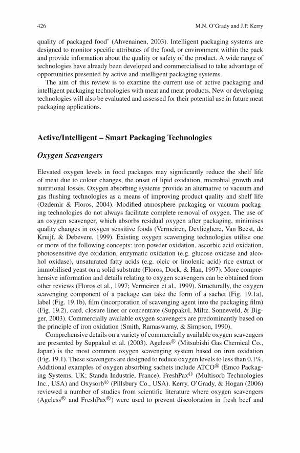
426 M.N. O’Grady and J.P. Kerry
quality of packaged food’ (Ahvenainen, 2003). Intelligent packaging systems aredesigned to monitor specific attributes of the food, or environment within the packand provide information about the quality or safety of the product. A wide range oftechnologies have already been developed and commercialised to take advantage ofopportunities presented by active and intelligent packaging systems.
The aim of this review is to examine the current use of active packaging andintelligent packaging technologies with meat and meat products. New or developingtechnologies will also be evaluated and assessed for their potential use in future meatpackaging applications.
Active/Intelligent – Smart Packaging Technologies
Oxygen Scavengers
Elevated oxygen levels in food packages may significantly reduce the shelf lifeof meat due to colour changes, the onset of lipid oxidation, microbial growth andnutritional losses. Oxygen absorbing systems provide an alternative to vacuum andgas flushing technologies as a means of improving product quality and shelf life(Ozdemir & Floros, 2004). Modified atmosphere packaging or vacuum packag-ing technologies do not always facilitate complete removal of oxygen. The use ofan oxygen scavenger, which absorbs residual oxygen after packaging, minimisesquality changes in oxygen sensitive foods (Vermeiren, Devlieghere, Van Beest, deKruijf, & Debevere, 1999). Existing oxygen scavenging technologies utilise oneor more of the following concepts: iron powder oxidation, ascorbic acid oxidation,photosensitive dye oxidation, enzymatic oxidation (e.g. glucose oxidase and alco-hol oxidase), unsaturated fatty acids (e.g. oleic or linolenic acid) rice extract orimmobilised yeast on a solid substrate (Floros, Dock, & Han, 1997). More compre-hensive information and details relating to oxygen scavengers can be obtained fromother reviews (Floros et al., 1997; Vermeiren et al., 1999). Structurally, the oxygenscavenging component of a package can take the form of a sachet (Fig. 19.1a),label (Fig. 19.1b), film (incorporation of scavenging agent into the packaging film)(Fig. 19.2), card, closure liner or concentrate (Suppakul, Miltz, Sonneveld, & Big-ger, 2003). Commercially available oxygen scavengers are predominantly based onthe principle of iron oxidation (Smith, Ramaswamy, & Simpson, 1990).
Comprehensive details on a variety of commercially available oxygen scavengersare presented by Suppakul et al. (2003). Ageless� (Mitsubishi Gas Chemical Co.,Japan) is the most common oxygen scavenging system based on iron oxidation(Fig. 19.1). These scavengers are designed to reduce oxygen levels to less than 0.1%.Additional examples of oxygen absorbing sachets include ATCO� (Emco Packag-ing Systems, UK; Standa Industrie, France), FreshPax� (Multisorb TechnologiesInc., USA) and Oxysorb� (Pillsbury Co., USA). Kerry, O’Grady, & Hogan (2006)reviewed a number of studies from scientific literature where oxygen scavengers(Ageless� and FreshPax�) were used to prevent discoloration in fresh beef and
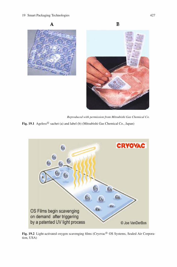
19 Smart Packaging Technologies 427
Reproduced with permission from Mitsubishi Gas Chemical Co.
Fig. 19.1 Ageless� sachet (a) and label (b) (Mitsubishi Gas Chemical Co., Japan)
Fig. 19.2 Light-activated oxygen scavenging films (Cryovac� OS Systems, Sealed Air Corpora-tion, USA)
428 M.N. O’Grady and J.P. Kerry
enhance pork and pork product quality. While many active packaging technolo-gies are still developmental, oxygen scavengers are widely used commercially inpre-packed cooked meat products. Emco Packaging Systems, specialists in activeand intelligent packaging, are a UK manufacturer and distributor for ATCO� DE10S self-adhesive oxygen absorbing labels. Emco supply ATCO� labels for usein pre-packed sliced cooked meats, especially hams, to meat processors in Ireland,throughout the UK and in Europe. While labels used in sliced cooked meat packagesscavenge between 10 and 20 cc’s of oxygen, Emco have recently launched largeroxygen scavenging labels onto the market (ATCO� 100 OS and 200 OS), whichscavenge between 100 and 200 cc’s oxygen, for use in larger capacity packagingapplications.
Cryovac� OS Systems (Cryovac Div., Sealed Air Corporation, USA) oxygenscavenging technology incorporates a polymer-based oxygen scavenger into the
packaging film. The UV light-activated Cryovac� OS2000TM
oxygen scavengingfilm (Fig. 19.2) is composed of an oxygen scavenger layer extruded into a multi-layer film and can reduce headspace oxygen levels from 1% to ppm levels in 4–10days, comparable with oxygen scavenging sachets. These films have applicationsin a wide variety of food products including dried or smoked meat products andprocessed meats (Butler, 2002). A similar UV light-activated oxygen scavenging
polymer ZEROTM2 , developed by CSIRO, Div. of Food Science Australia in collab-
oration with VisyPak Food Packaging, Visy Industries, Australia, forms a layer in amulti-layer package structure and has many applications including reduced discol-oration of sliced meats.
Carbon Dioxide Scavengers and Emitters
Within a packaging environment, carbon dioxide functions to suppress microbialgrowth. Therefore a carbon dioxide generating system can be viewed as a techniquecomplimentary to oxygen scavenging (Suppakul et al., 2003). The permeability ofcarbon dioxide is 3 to 5 times higher than that of oxygen in most plastic films;therefore, it must be continuously produced to maintain the desired concentrationwithin the package (Ozdemir & Floros, 2004). High carbon dioxide levels (10–80%)are desirable for foods such as meat and poultry in order to inhibit microbial growthand extend shelf life. Removal of oxygen from the package creates a partial vac-uum which may result in the collapse of flexible packaging. Also, when a packageis flushed with a mixture of gases including carbon dioxide, the carbon dioxidedissolves in the product creating a partial vacuum. In such cases, the simultaneousrelease of carbon dioxide from inserted sachets which consume oxygen is desir-able. Such systems are based on either ferrous carbonate or a mixture of ascorbicacid and sodium bicarbonate (Rooney, 1995). Examples of commercially availabledual action combined carbon dioxide generators/oxygen scavengers are Ageless�
G (Mitsubishi Gas Chemical Co, Japan) and FreshPax� M (Multisorb TechnologiesInc, USA). Carbon dioxide emitting sachets or labels can also be used alone. The
19 Smart Packaging Technologies 429
VerifraisTM package, manufactured by SARL Codimer (Paris, France) has been usedto extend the shelf life of fresh meats. This innovative package consists of a standardmodified atmosphere packaging tray, but has a perforated false bottom under whicha porous sachet containing sodium bicarbonate/ascorbate is positioned. When juiceexudates from the packaged meat drips onto the sachet, carbon dioxide is emitted,thus replacing any carbon dioxide absorbed by the meat and preventing packagecollapse.
The inhibition of spoilage bacteria utilizing active packaging technologymay reduce bacterial competition and thus permit growth and toxin produc-tion by non-proteolytic C. botulinum or the growth of other pathogenic bacteria(Sivertsvik, 2003). Lovenklev et al. (2004) reported that while a high concentrationof carbon dioxide decreased the growth rate of non-proteolytic C. botulinum typeB, the expression and production of the toxin was greatly increased which meansthe risk of botulism may also be increased, rather than being reduced, if used inmodified atmosphere packaging systems. Research into the safety risks associatedwith the use of carbon dioxide in packaging systems is necessary.
Carbon dioxide absorbers (sachets) consisting of either calcium hydroxide andsodium hydroxide, or potassium hydroxide, calcium oxide and silica gel, may beused to remove carbon dioxide during storage in order to prevent bursting of thepackage. Possible applications include their use in packs of dehydrated poultryproducts and beef jerkey (Ahvenainen, 2003).
Moisture Control
The main purpose of liquid water control is to lower the water activity of theproduct, thereby suppressing microbial growth (Vermeiren et al., 1999). Temper-ature cycling of high water activity foods has led to the use of plastics with ananti-fog additive that lowers the interfacial tension between the condensate andthe film. This contributes to the transparency of the film and enables the customerto clearly see the packaged food (Rooney, 1995), although it does not affect theamount of liquid water present inside the package. Several companies manufacturedrip absorbent sheets or pads such as Cryovac� Dri-Loc� (Sealed Air Corpora-tion, USA), Thermarite� or Peaksorb� (Australia), ToppanTM (Japan) and Fresh-R-PaxTM (Maxwell Chase Technologies, LLC, USA) for liquid control in high wateractivity foods such as meat and poultry. These systems consist of a super absorbentpolymer located between two layers of a micro porous or non-woven polymer. Suchsheets are used as drip-absorbing pads placed under whole chickens or chicken cuts(Suppakul et al., 2003).
Antimicrobial Packaging
Fresh meat is a highly perishable food product which, unless correctly stored,processed, packaged and distributed, spoils quickly and becomes hazardous dueto microbial growth and the subsequent risk of food borne illness. Antimicrobial

430 M.N. O’Grady and J.P. Kerry
packaging is a promising form of active packaging especially for meat products.Since microbial contamination of meat products occurs primarily at the surface, dueto post processing handling, attempts have been made to improve safety and to delayspoilage by the use of antibacterial sprays or dips. Limitations of such antibacterialsinclude neutralisation of compounds on contact with the meat surface or diffusionof compounds from the surface into the meat mass. Incorporation of bactericidalagents into meat formulations may result in partial inactivation of the active com-pounds by meat constituents and therefore exert a limited effect on the surfacemicroflora (Quintavalla & Vicini, 2002). Antimicrobial food packaging materialshave to extend the lag phase and reduce the growth phase of microorganisms, inorder to extend shelf life and to maintain product quality and safety (Han, 2000).Comprehensive reviews on antimicrobial food packaging have been published pre-viously (Appendini & Hotchkiss, 2002; Suppakul et al., 2003) and more recently byComa (2008). To confer antimicrobial activity, antimicrobial agents may be coated,incorporated, immobilised, or surface modified onto package materials (Suppakulet al., 2003). A comprehensive list of antimicrobial agents for use in antimicrobialfilms, containers and utensils is presented in a review by Suppakul et al. (2003). Theclasses of antimicrobials listed range from acid anhydride, alcohol, bacteriocins,chelators, enzymes, organic acids and polysaccharides. Examples of commercialantimicrobial materials in the form of concentrates (e.g. AgIONTM, AgION Tech-nologies LLC, USA), extracts (Nisaplin� (Nisin), Integrated Ingredients, USA)and films (MicrogardTM Rhone-Poulenc, USA) were also presented. Antimicro-bial packages have had relatively few commercial successes except in Japan whereAg-substituted zeolite is the most common antimicrobial agent incorporated intoplastics. Ag-ions inhibit a range of metabolic enzymes and have strong antimicro-bial activity (Vermeiren et al., 1999). Antimicrobial films can be classified into twotypes: those that contain an antimicrobial agent which migrates to the surface ofthe food and those which are effective against surface growth of microorganismswithout migration. The antibacterial activity of antimicrobial agents that are coated,directly incorporated or immobilised onto packaging films in meat and meat prod-ucts has been reviewed by Coma (2008) and Kerry et al. (2006).
Sensors
In modified atmosphere packaged meats, the headspace concentrations of oxygenand carbon dioxide are useful indicators of the meat product quality. The headspaceprofiles of oxygen and carbon dioxide change over time as a function of factors suchas product type, respiration, packaging material, pack size, volume ratios, storageconditions and package integrity. An optical sensor approach to monitor gas phasesin modified atmosphere packaged products offers an alternative to the conventionalmethods such as GC, GC/MS and portable gas analysers.
Many intelligent packaging concepts involve the use of sensors and indicators.A sensor is defined as a device used to detect, locate or quantify energy or matter,
19 Smart Packaging Technologies 431
giving a signal for the detection or measurement of a physical or chemical prop-erty to which the device responds (Kress-Rogers, 1998b). A sensor device mustprovide continuous output of a signal. The majority of sensors are composed ofa receptor and a transducer. In the receptor, physical or chemical information istransformed into a form of energy which may be measured by the transducer. Thetransducer is a device capable of transforming the energy carrying the physical orchemical information about the sample into a useful analytical signal. Transducerswith potential use in meat packaging systems include electrical, optical, thermal orchemical signal domains (Kerry et al., 2006). Development of a potential sensorfor rapid quantification of chemical or physical indicators of food quality is knownas the ‘marker approach’ (Kress-Rogers, 2001). Intelligent packaging incorporatinggas sensor technology provides a means by which the determination of indicatorheadspace gases gives information on meat quality and package integrity rapidlyand inexpensively.
The use of sensors in the meat industry is limited; however, significant practicalsteps towards more widespread use have been made (Kerry & Papkovsky, 2002).High development and production costs, strict industry specifications, safety con-siderations and relatively limited demand from the meat industry and consumersare considered as the main obstacles to commercial use. Greater pressure on foodmanufacturers to guarantee safety, quality and traceability is likely to promote futureestablishment of commercial sensor technology in food packaging.
Gas Sensors
Gas sensors are devices which respond reversibly and quantitatively to gaseous ana-lytes by changing the physical parameters of the sensor. Systems currently availablefor gas detection include amperometric oxygen sensors, potentiometric carbon diox-ide sensors, metal oxide semiconductor field effect transistors, organic conductingpolymers and piezo-electric crystal sensors (Kress-Rogers, 1998a). Conventionalsystems for oxygen sensors based on electrochemical methods have a number oflimitations (Trettnak, Gruber, Reiniger, & Klimant, 1995) including consumptionof analyte (oxygen), cross-sensitivity to carbon dioxide and hydrogen sulphide andfouling of sensor membranes. They also involve destructive analysis of packages.
In recent years, a number of instruments and materials for optical oxygen sensinghave been reported (Papkovsky, Ponomarev, Trettnak, & O’Leary, 1995; Trettnaket al., 1995). Such sensors are usually comprised of a solid-state material and operateon the principle of luminescence quenching or absorbance changes caused by directcontact with the analyte. They are chemically inert, do not consume analytes, andprovide a non-invasive technique for gas analysis through translucent materials.
Approaches to opto-chemical sensing include: a fluorescence-based system usinga pH sensitive indicator (Wolfbeis, Weis, Leiner, & Ziegler, 1988), absorption-basedcolorimetric sensing realised through a visual indicator (Mills, Qing Chang, &McMurray, 1992), and an energy transfer approach using phase fluorimetric detec-tion (Neurater, Klimant, & Wolfbeis, 1999). The latter allows for the possibilityof combining oxygen and carbon dioxide measurements in a single sensor through
432 M.N. O’Grady and J.P. Kerry
compatibility with previously developed oxygen sensing technology. Most carbondioxide sensors, however, have been developed for biomedical applications and theuse of existing carbon dioxide sensors in food packaging applications is currentlynot feasible (Kerry & Papkovsky, 2002).
Fluorescence-Based Oxygen Sensors
Fluorescence-based oxygen sensors represent the most promising systems to datefor remote measurement of headspace gases in packaged meat products. A numberof disposable oxygen sensing prototypes have been developed which may be pro-duced at low cost and provide rapid determination of oxygen concentration (Kerry &Papkovsky, 2002). The active component of a fluorescence-based oxygen sensorusually consists of a long-delay fluorescent or phosphorescent dye encapsulated ina solid polymer matrix. The dye-polymer coating is applied as a thin film coatingon a suitable solid support. Molecular oxygen, present in the packaging headspace,penetrates the sensitive coating through simple diffusion and quenches the lumines-cence by a dynamic, i.e. collisional, mechanism. Oxygen is quantified by measur-ing changes in luminescence parameters against a pre-determined calibration. Theprocess is reversible and clean; neither the dye nor the oxygen is consumed in thephotochemical reactions involved, no by-products are generated and the whole cyclecan be repeated.
Materials for oxygen sensors must meet strict sensitivity and working perfor-mance requirements in order to meet the suitability requirements for commercialintelligent packaging applications. They must also have fluorescence characteristicssuited to the construction of simple measuring devices. Fluorescence and phospho-rescence dyes with lifetimes in the micro-second range are best suited to oxygensensing in food packaging. Other necessary features include suitable intensity, wellresolved excitation and emission long wave bands and good photo-stability charac-teristics of the indicator dye. Such features allow sensor compatibility with simpleopto-electronic measuring devices (LEDs, photodiodes etc), minimise interferenceby scattering and sample fluorescence and allow long-term operation without recal-ibration (Papkovsky et al., 1995). Materials using fluorescent complexes of ruthe-nium, phosphorescent palladium(II)- and platinum(II)-porphyrin complexes andrelated structures have shown considerable promise as oxygen sensors (Papkovskyet al., 1991; Papkovsky et al., 1995).
The combination of indicator dye and encapsulating polymer medium determinesthe sensitivity and effective working range of such sensors. For the purpose of foodpackaging applications, dyes with relatively long emission lifetimes (∼ 40–500 μs)such as Pt-porphyrins combined with polystyrene as polymer matrix appear to offergreatest potential (Papkovsky, Papovskaia, Smyth, Kerry, & Ogurtsov, 2000). Otherpolymers with good gas-barrier properties such as polyamide, polyethylene teraph-thalate and PVC are not suitable for oxygen sensing as oxygen quenching is slow insuch media. The use of plasticized polymers is also unsuitable due to toxicity con-cerns associated with potential plasticizer migration. Information regarding sensor
19 Smart Packaging Technologies 433
fabrication and criteria to be considered for commercial sensor uptake, in food pack-aging, is reviewed by Kerry et al. (2006) and Kerry & Papkovsky (2002).
The ability of fluorescence-based oxygen sensors to accurately determine oxy-gen levels has been demonstrated in packaged beef, poultry and pork products(Fitzgerald et al., 2001; Papkovsky, Smiddy, Papkovskaia, & Kerry, 2002; Smiddy,Fitzgerald, et al., 2002; Smiddy, Papkovskaia, Papkovsky, & Kerry, 2002; Smiddy,Papkovskaia, et al., 2002;). The development of oxygen sensors is indicative of amove towards commercialisation of indicator-based intelligent meat packaging sys-tems. It has been estimated that each sensor should cost less than €0.01 to produce(Kerry & Papkovsky, 2002) and impact minimally on packaged meat productioncosts.
Biosensors
The recently developed biosensor technologies represent an additional area withpotential for application in intelligent meat packaging systems. Biosensors are com-pact analytical devices which detect, record and transmit information pertaining tobiological reactions (Yam, Takhistov, & Miltz, 2005). They consist of a bio-receptorspecific to a target analyte and a transducer to convert biological signals to a quan-tifiable electrical response. Bio-receptors are organic materials such as enzymes,antigens, microbes, hormones and nucleic acids. Transducers may be electrochemi-cal, optical, or calorimetric and are system dependent. Intelligent packaging systemsincorporating biosensors have the potential for extreme specificity and reliability.Market analysis of pathogen detection and safety systems for the food packag-ing industry suggests that biosensors offer considerable promise for future growth(Alocilja & Radke, 2003).
Toxin GuardTM
(Toxin Alert, Ontario, Canada), a commercially availablebiosensor, is a patented diagnostic system incorporating antibodies printed onpolyethylene-based plastic packaging capable of detecting target pathogens suchas Salmonella sp., Campylobacter sp., Escherichia coli 0517 and Listeria sp.(Bodenhammer, 2002; Bodenhammer, Jakowski, & Davies, 2004). When foodpackaging comes into contact with targeted bacteria, a positive test, indicated by a
dramatic visual signal, alerts the consumer or retailer. ToxinguardTM
can be targetedto detect freshness degradation, as well as the presence of specific food hazards suchas pesticides or indicators of genetic modification.
Bioett AB (Lund, Sweden) has developed a system based on a biosensor fortemperature monitoring. The Bioett System monitors the accumulated effect of tem-perature on products over time. The system consists of a chip-less RF circuit with abuilt-in biosensor which can be read with a handheld scanner at various points in thesupply chain. Information is stored in a database and can be used to analyze the coldchain and validate that the agreed temperature has been maintained. A Time Tem-perature Biosensor (TTB), attached to 5 kg cases of frozen meat balls, is activatedat source. The biosensor registers the accumulated temperatures that the producthas been exposed to and this information can be used to optimize and monitor the
434 M.N. O’Grady and J.P. Kerry
cold-chain distribution system. A scanner can read the biosensor via radio waves(radio frequency) and also uniquely identifies the goods using a barcode system.The scanner also incorporates a software defined radio subsystem which can also beused for reading of RFID tags. Such systems give an insight into products that arelikely to become mainstream in years to come.
Indicators
Indicators may be defined as substances which indicate the presence, absence orconcentration of another substance, or the degree of reaction between two or moresubstances by means of a characteristic change, especially in colour. By contrastwith sensors, indicators are not composed of receptor and transducer componentsand communicate information directly through a visual change (Kerry et al., 2006).A number of commercially available indicators are available for use with packagedmeats and meat products.
Integrity Indicators
Non-invasive indicator systems provide qualitative or semi-quantitative informationthrough visual colorimetric changes or through comparison with standard refer-ences. The majority of indicators have been developed to test package integrity. Themost common cause of integrity damage in flexible plastic packages is associatedwith leaking seals (Hurme, 2003). Attachment of a leak indicator or sensor to apackage ensures package integrity through the distribution chain.
A number of studies on package integrity in modified atmosphere packagedmeat products (Ahvenainen, Eilamo, & Hurme, 1997; Eilamo, Ahvenainen, Hurme,Heinio, & Mattila-Sandholm, 1995; Randell et al., 1995; Smolander, Hurme, &Ahvenainen, 1997) have established critical leak sizes and associated quality deteri-oration. Standard (destructive) manual methods for package integrity and leak test-ing are both laborious and can test only limited numbers of packs (Hurme, 2003).Currently available non-destructive detection systems have disadvantages such asrequirements for specialised equipment, slow sampling time and an inability todetect leakages that are penetrable by pathogens (Hurme & Ahvenainen, 1998;Stauffer, 1988). Much of the research into integrity detection has focused on visualoxygen indicators for modified atmosphere packaged products. Many visual oxy-gen indicators have been patented and are based mainly on the use of redox dyes(Davies & Gardner, 1996; Krumhar & Karel, 1992; Mattila-Sandholm, Ahvenainen,Hurme, & Jarvi-Kaarianen, 1995; Yoshikawa, Nawata, Goto, & Fujii, 1987). Test-ing and validation of such devices in modified atmosphere packaged minced steaksand minced meat pizzas have been reported (Ahvenainen et al., 1997; Eilamoet al., 1995). Disadvantages of such devices include high sensitivity (colour changeas a result of low oxygen concentrations (0.1%)) and reversibility (leak-inducedincreases in oxygen consumed through subsequent microbial growth).
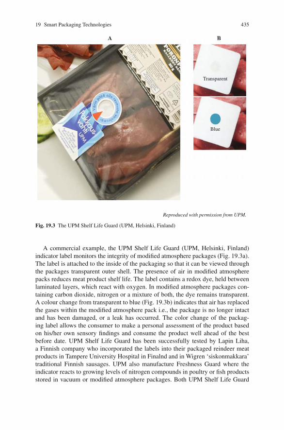
19 Smart Packaging Technologies 435
Reproduced with permission from UPM.
Transparent
Blue
Fig. 19.3 The UPM Shelf Life Guard (UPM, Helsinki, Finland)
A commercial example, the UPM Shelf Life Guard (UPM, Helsinki, Finland)indicator label monitors the integrity of modified atmosphere packages (Fig. 19.3a).The label is attached to the inside of the packaging so that it can be viewed throughthe packages transparent outer shell. The presence of air in modified atmospherepacks reduces meat product shelf life. The label contains a redox dye, held betweenlaminated layers, which react with oxygen. In modified atmosphere packages con-taining carbon dioxide, nitrogen or a mixture of both, the dye remains transparent.A colour change from transparent to blue (Fig. 19.3b) indicates that air has replacedthe gases within the modified atmosphere pack i.e., the package is no longer intactand has been damaged, or a leak has occurred. The color change of the packag-ing label allows the consumer to make a personal assessment of the product basedon his/her own sensory findings and consume the product well ahead of the bestbefore date. UPM Shelf Life Guard has been successfully tested by Lapin Liha,a Finnish company who incorporated the labels into their packaged reindeer meatproducts in Tampere University Hospital in Finalnd and in Wigren ‘siskonmakkara’traditional Finnish sausages. UPM also manufacture Freshness Guard where theindicator reacts to growing levels of nitrogen compounds in poultry or fish productsstored in vacuum or modified atmosphere packages. Both UPM Shelf Life Guard
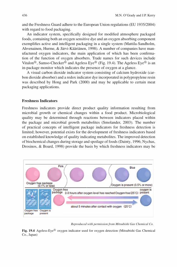
436 M.N. O’Grady and J.P. Kerry
and the Freshness Guard adhere to the European Union regulations (EU 1935/2004)with regard to food packaging.
An indicator system, specifically designed for modified atmosphere packagedfoods, containing both an oxygen sensitive dye and an oxygen absorbing componentexemplifies active and intelligent packaging in a single system (Mattila-Sandholm,Ahvenainen, Hurme, & Jarvi-Kaarianen, 1998). A number of companies have man-ufactured oxygen indicators, the main application of which has been confirma-tion of the function of oxygen absorbers. Trade names for such devices includeVitalon�, Samso-Checker� and Ageless Eye� (Fig. 19.4). The Ageless Eye� is anin-package monitor which indicates the presence of oxygen at a glance.
A visual carbon dioxide indicator system consisting of calcium hydroxide (car-bon dioxide absorber) and a redox indicator dye incorporated in polypropylene resinwas described by Hong and Park (2000) and may be applicable to certain meatpackaging applications.
Freshness Indicators
Freshness indicators provide direct product quality information resulting frommicrobial growth or chemical changes within a food product. Microbiologicalquality may be determined through reactions between indicators placed withinthe package and microbial growth metabolites (Smolander, 2003). The numberof practical concepts of intelligent package indicators for freshness detection islimited; however, potential exists for the development of freshness indicators basedon established knowledge of quality indicating metabolites. The improved detectionof biochemical changes during storage and spoilage of foods (Dainty, 1996; Nychas,Drosinos, & Board, 1998) provide the basis by which freshness indicators may be
Reproduced with permission from Mitsubishi Gas Chemical Co.
Fig. 19.4 Ageless-Eye� oxygen indicator used for oxygen detection (Mitsubishi Gas ChemicalCo., Japan)
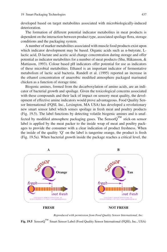
19 Smart Packaging Technologies 437
developed based on target metabolites associated with microbiologically-induceddeterioration.
The formation of different potential indicator metabolites in meat products isdependent on the interaction between product type, associated spoilage flora, storageconditions and the packaging system.
A number of marker metabolites associated with muscle food products exist uponwhich indicator development may be based. Organic acids such as n-butyrate, L-lactic acid, D-lactate and acetic acid change concentration during storage and offerpotential as indicator metabolites for a number of meat products (Shu, Hakanson, &Mattiason, 1993). Colour based pH indicators offer potential for use as indicatorsof these microbial metabolites. Ethanol is an important indicator of fermentativemetabolism of lactic acid bacteria. Randell et al. (1995) reported an increase inthe ethanol concentration of anaerobic modified atmosphere packaged marinatedchicken as a function of storage time.
Biogenic amines, formed from the decarboxylation of amino acids, are an indi-cator of bacterial growth and spoilage. Given the toxicological concerns associatedwith these compounds and their lack of impact on sensory meat quality, the devel-opment of effective amine indicators would prove advantageous. Food Quality Sen-sor International (FQSI, Inc., Lexington, MA USA) has developed a revolutionarynew smart sensor label which senses spoilage in fresh meat and poultry products(Fig. 19.5). The label functions by detecting volatile biogenic amines and is unaf-
fected by modified atmosphere packaging gases. The SensorQTM
stick-on sensorlabel is applied by the meat packer to the inside wrap of meat and poultry pack-ages to provide the consumer with a clear indication of product freshness. Whenthe inside of the quality ‘Q’ on the label is tangerine orange, the product is fresh(Fig. 19.5a). When bacterial growth inside the package reaches a critical level, the
A B
FRESH
Orange Tan
NOT FRESH
Reproduced with permission from Food Quality Sensor International, Inc.
Fig. 19.5 SensorQTM
Smart Sensor Label (Food Quality Sensor International (FQSI), Inc., USA)
438 M.N. O’Grady and J.P. Kerry
orange colour turns to tan which indicates spoilage (Fig. 19.5b). The SensorQ labelis made of food grade materials and is economical, costing less than 1% of the totalvalue of the average package of meat or poultry it labels. Market research indicatedthat over 95% of consumers would opt to purchase a product with a freshness indica-tor attached. FSQI are currently in the final stages of testing and validating SensorQand expect to launch the product in late 2008.
Hydrogen sulphide, a breakdown product of cysteine, with intense off-flavoursand low threshold levels, is produced during the spoilage of meat and poultry bya number of bacterial species. It forms a green pigment when bound to myoglobinand this pigment formed the basis for the development of an agarose-immobilised,myoglobin-based freshness indicator in un-marinated broiler pieces (Smolanderet al., 2002). The indicator was not affected by the presence of nitrogen or carbondioxide.
Freshness indicators based on broad spectrum colour changes have a numberof disadvantages which need to be resolved before widespread commercial uptakeis likely. A lack of specificity means that colour changes indicating contaminationcan occur in products free from any significant sensory or quality deterioration. Thepresence of certain target metabolites is not necessarily an indication of poor quality.More exact correlations need to be established between target metabolite, producttype and organo-leptic quality and safety. The possibilities of false-negatives arelikely to dissuade producers from adopting indicators unless specific indication ofactual spoilage can be guaranteed (Kerry et al., 2006).
Time-Temperature Indicators
A time-temperature indicator (or integrator) (TTI) is defined as a device (small tag orlabel) used to show a measurable, time-temperature dependent change that reflectsthe full or partial temperature history of a food product to which it is attached(Taoukis & Labuza, 1989). Operation of TTIs is based on mechanical, chemical,electrochemical, enzymatic or microbiological change, usually expressed as a visi-ble response in the form of a mechanical deformation, colour development or colourmovement (Taoukis & Labuza, 2003). Therefore, the visible response gives a cumu-lative indication of the storage temperature to which the TTI has been exposed. TTIsare classified as either partial history or full history indicators, depending on theirresponse mechanism.
Effective TTIs are required to indicate clear, continuous, irreversible reactionto changes in temperature. TTIs should also be small, reliable, low cost, easilyintegrated into food packaging, have a long pre- and post-activation shelf life andbe unaffected by ambient conditions other than temperature. TTIs should also beflexible to a range of temperatures, robust, pose no toxicological or safety hazardand convey information in a clear manner.
A large number of TTI types have been developed and patented, the principlesand applications of which have been reviewed previously (Taoukis & Labuza, 2003).TTIs currently commercially available include a number of diffusion, enzymatic andpolymer-based systems.
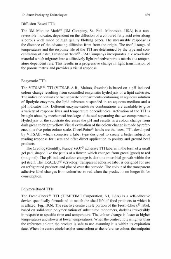
19 Smart Packaging Technologies 439
Diffusion-Based TTIs
The 3M Monitor Mark� (3M Company, St. Paul, Minnesota, USA) is a non-reversible indicator, dependent on the diffusion of a coloured fatty acid ester alonga porous wick made of high quality blotting paper. The measurable response isthe distance of the advancing diffusion front from the origin. The useful range oftemperatures and the response life of the TTI are determined by the type and con-centration of ester. FreshnessCheck� (3M Company) incorporates a visco-elasticmaterial which migrates into a diffusively light-reflective porous matrix at a temper-ature dependent rate. This results in a progressive change in light transmission ofthe porous matrix and provides a visual response.
Enzymatic TTIs
The VITSAB� TTI (VITSAB A.B., Malmo, Sweden) is based on a pH inducedcolour change resulting from controlled enzymatic hydrolysis of a lipid substrate.The indicator consists of two separate compartments containing an aqueous solutionof lipolytic enzymes, the lipid substrate suspended in an aqueous medium and apH indicator mix. Different enzyme–substrate combinations are available to givea variety of response lives and temperature dependencies. Activation of the TTI isbrought about by mechanical breakage of the seal separating the two compartments.Hydrolysis of the substrate decreases the pH and results in a colour change fromdark green to bright yellow. Visual evaluation of the colour change is made by refer-ence to a five-point colour scale. CheckPoint� labels are the latest TTIs developedby VITSAB, which comprise a label type designed to create a better subjectivereading response for users and offer direct application to poultry and ground beefproducts.
The Cryolog (Gentilly, France) (eO)� adhesive TTI label is in the form of a smallgel pad, shaped like the petals of a flower, which changes from green (good) to red(not good). The pH induced colour change is due to a microbial growth within thegel itself. The TRACEO� (Cryolog) transparent adhesive label is designed for useon refrigerated products and placed over the barcode. The colour of the transparentadhesive label changes from colourless to red when the product is no longer fit forconsumption.
Polymer-Based TTIs
The Fresh-Check� TTI (TEMPTIME Corporation, NJ, USA) is a self-adhesivedevice specifically formulated to match the shelf life of food products to which itis affixed (Fig. 19.6). The reactive centre circle portion of the Fresh-Check� label,based on solid-state polymerization of substituted monomers, darkens irreversiblyin response to specific time and temperature. The colour change is faster at highertemperatures and slower at lower temperatures. When the centre circle is lighter thanthe reference colour, the product is safe to use assuming it is within its expirationdate. When the centre circle has the same colour as the reference colour, the endpoint
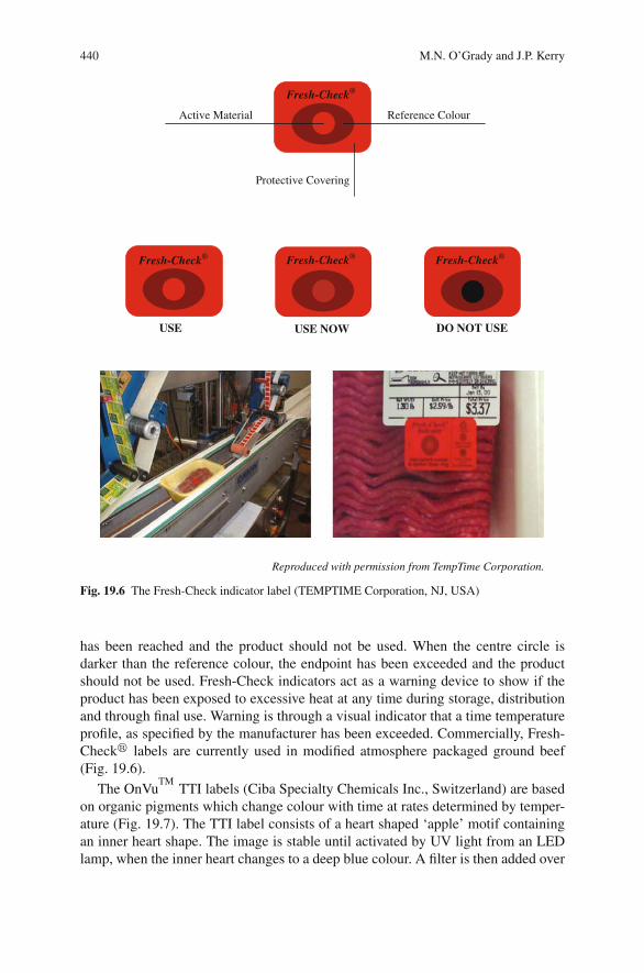
440 M.N. O’Grady and J.P. Kerry
Fresh-Check® Fresh-Check® Fresh-Check®
USE USE NOW DO NOT USE
Active Material Reference Colour
Protective Covering
Fresh-Check®
Reproduced with permission from TempTime Corporation.
Fig. 19.6 The Fresh-Check indicator label (TEMPTIME Corporation, NJ, USA)
has been reached and the product should not be used. When the centre circle isdarker than the reference colour, the endpoint has been exceeded and the productshould not be used. Fresh-Check indicators act as a warning device to show if theproduct has been exposed to excessive heat at any time during storage, distributionand through final use. Warning is through a visual indicator that a time temperatureprofile, as specified by the manufacturer has been exceeded. Commercially, Fresh-Check� labels are currently used in modified atmosphere packaged ground beef(Fig. 19.6).
The OnVuTM
TTI labels (Ciba Specialty Chemicals Inc., Switzerland) are basedon organic pigments which change colour with time at rates determined by temper-ature (Fig. 19.7). The TTI label consists of a heart shaped ‘apple’ motif containingan inner heart shape. The image is stable until activated by UV light from an LEDlamp, when the inner heart changes to a deep blue colour. A filter is then added over
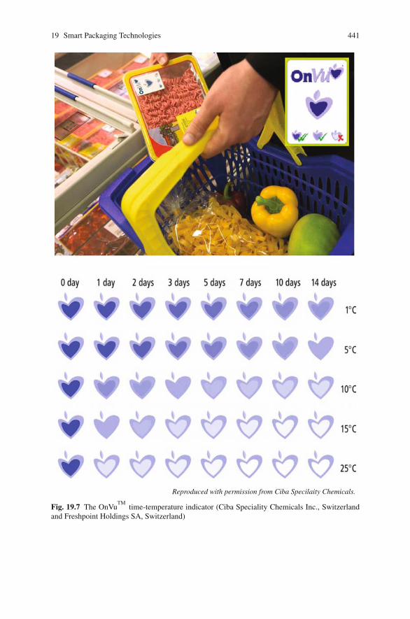
19 Smart Packaging Technologies 441
Reproduced with permission from Ciba Specilaity Chemicals.
Fig. 19.7 The OnVuTM
time-temperature indicator (Ciba Speciality Chemicals Inc., Switzerlandand Freshpoint Holdings SA, Switzerland)
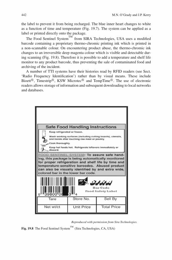
442 M.N. O’Grady and J.P. Kerry
the label to prevent it from being recharged. The blue inner heart changes to whiteas a function of time and temperature (Fig. 19.7). The system can be applied as alabel or printed directly onto the package.
The Food Sentinel SystemTM
from SIRA Technologies, USA uses a modifiedbarcode containing a proprietary thermo-chromic printing ink which is printed ina non-scannable colour. On encountering product abuse, the thermo-chromic inkchanges to an irreversible deep magenta colour which is visible and detectable dur-ing scanning (Fig. 19.8). Therefore it is possible to add a temperature and shelf lifemonitor to any product barcode, thus preventing the sale of contaminated food andarchiving of the incident.
A number of TTI systems have their histories read by RFID readers (see Sect.‘Radio Frequency Identification’) rather than by visual means. These includeBioett�, Timestrip�, KSW Microtec� and TempTime�. The use of electronicreaders allows storage of information and subsequent downloading to local networksand databases.
Reproduced with permission from Sira Technologies.
Fig. 19.8 The Food Sentinel SystemTM
(Sira Technologies, CA, USA)
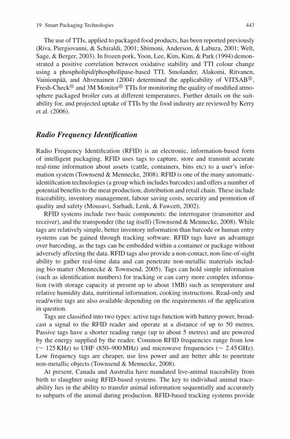
19 Smart Packaging Technologies 443
The use of TTIs, applied to packaged food products, has been reported previously(Riva, Piergiovanni, & Schiraldi, 2001; Shimoni, Anderson, & Labuza, 2001; Welt,Sage, & Berger, 2003). In frozen pork, Yoon, Lee, Kim, Kim, & Park (1994) demon-strated a positive correlation between oxidative stability and TTI colour changeusing a phospholipid/phospholipase-based TTI. Smolander, Alakomi, Ritvanen,Vainionpaa, and Ahvenainen (2004) determined the applicability of VITSAB�,Fresh-Check� and 3M Monitor� TTIs for monitoring the quality of modified atmo-sphere packaged broiler cuts at different temperatures. Further details on the suit-ability for, and projected uptake of TTIs by the food industry are reviewed by Kerryet al. (2006).
Radio Frequency Identification
Radio Frequency Identification (RFID) is an electronic, information-based formof intelligent packaging. RFID uses tags to capture, store and transmit accuratereal-time information about assets (cattle, containers, bins etc) to a user’s infor-mation system (Townsend & Mennecke, 2008). RFID is one of the many automatic-identification technologies (a group which includes barcodes) and offers a number ofpotential benefits to the meat production, distribution and retail chain. These includetraceability, inventory management, labour saving costs, security and promotion ofquality and safety (Mousavi, Sarhadi, Lenk, & Fawcett, 2002).
RFID systems include two basic components: the interrogator (transmitter andreceiver), and the transponder (the tag itself) (Townsend & Mennecke, 2008). Whiletags are relatively simple, better inventory information than barcode or human entrysystems can be gained through tracking software. RFID tags have an advantageover barcoding, as the tags can be embedded within a container or package withoutadversely affecting the data. RFID tags also provide a non-contact, non-line-of-sightability to gather real-time data and can penetrate non-metallic materials includ-ing bio-matter (Mennecke & Townsend, 2005). Tags can hold simple information(such as identification numbers) for tracking or can carry more complex informa-tion (with storage capacity at present up to about 1MB) such as temperature andrelative humidity data, nutritional information, cooking instructions. Read-only andread/write tags are also available depending on the requirements of the applicationin question.
Tags are classified into two types: active tags function with battery power, broad-cast a signal to the RFID reader and operate at a distance of up to 50 metres.Passive tags have a shorter reading range (up to about 5 metres) and are poweredby the energy supplied by the reader. Common RFID frequencies range from low(∼ 125 KHz) to UHF (850–900 MHz) and microwave frequencies (∼ 2.45 GHz).Low frequency tags are cheaper, use less power and are better able to penetratenon-metallic objects (Townsend & Mennecke, 2008).
At present, Canada and Australia have mandated live-animal traceability frombirth to slaughter using RFID-based systems. The key to individual animal trace-ability lies in the ability to transfer animal information sequentially and accuratelyto subparts of the animal during production. RFID-based tracking systems provide
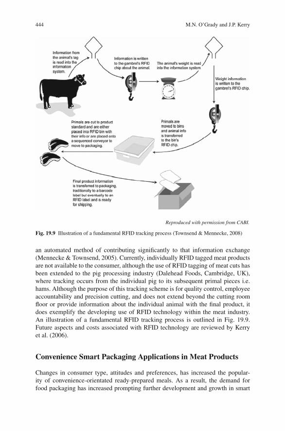
444 M.N. O’Grady and J.P. Kerry
Reproduced with permission from CABI.
Fig. 19.9 Illustration of a fundamental RFID tracking process (Townsend & Mennecke, 2008)
an automated method of contributing significantly to that information exchange(Mennecke & Townsend, 2005). Currently, individually RFID tagged meat productsare not available to the consumer, although the use of RFID tagging of meat cuts hasbeen extended to the pig processing industry (Dalehead Foods, Cambridge, UK),where tracking occurs from the individual pig to its subsequent primal pieces i.e.hams. Although the purpose of this tracking scheme is for quality control, employeeaccountability and precision cutting, and does not extend beyond the cutting roomfloor or provide information about the individual animal with the final product, itdoes exemplify the developing use of RFID technology within the meat industry.An illustration of a fundamental RFID tracking process is outlined in Fig. 19.9.Future aspects and costs associated with RFID technology are reviewed by Kerryet al. (2006).
Convenience Smart Packaging Applications in Meat Products
Changes in consumer type, attitudes and preferences, has increased the popular-ity of convenience-orientated ready-prepared meals. As a result, the demand forfood packaging has increased prompting further development and growth in smart
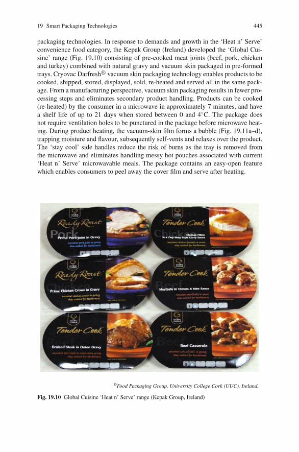
19 Smart Packaging Technologies 445
packaging technologies. In response to demands and growth in the ‘Heat n’ Serve’convenience food category, the Kepak Group (Ireland) developed the ‘Global Cui-sine’ range (Fig. 19.10) consisting of pre-cooked meat joints (beef, pork, chickenand turkey) combined with natural gravy and vacuum skin packaged in pre-formedtrays. Cryovac Darfresh� vacuum skin packaging technology enables products to becooked, shipped, stored, displayed, sold, re-heated and served all in the same pack-age. From a manufacturing perspective, vacuum skin packaging results in fewer pro-cessing steps and eliminates secondary product handling. Products can be cooked(re-heated) by the consumer in a microwave in approximately 7 minutes, and havea shelf life of up to 21 days when stored between 0 and 4◦C. The package doesnot require ventilation holes to be punctured in the package before microwave heat-ing. During product heating, the vacuum-skin film forms a bubble (Fig. 19.11a–d),trapping moisture and flavour, subsequently self-vents and relaxes over the product.The ‘stay cool’ side handles reduce the risk of burns as the tray is removed fromthe microwave and eliminates handling messy hot pouches associated with current‘Heat n’ Serve’ microwavable meals. The package contains an easy-open featurewhich enables consumers to peel away the cover film and serve after heating.
©Food Packaging Group, University College Cork (UUC), Ireland.
Fig. 19.10 Global Cuisine ‘Heat n’ Serve’ range (Kepak Group, Ireland)
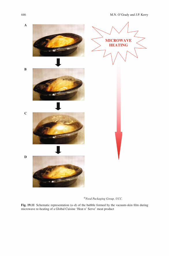
446 M.N. O’Grady and J.P. Kerry
A
MICROWAVEHEATING
B
C
D
©Food Packaging Group, UCC.
Fig. 19.11 Schematic representation (a–d) of the bubble formed by the vacuum-skin film duringmicrowave re-heating of a Global Cuisine ‘Heat n’ Serve’ meat product
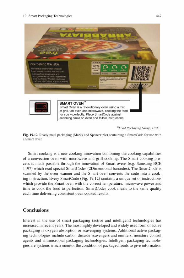
19 Smart Packaging Technologies 447
SMART OVEN™
Smart Oven is a revolutionary oven using a mix of grill, fan oven and microwave, cooking the food for you – perfectly. Place SmartCode against scanning circle on oven and follow instructions.
©Food Packaging Group, UCC.
Fig. 19.12 Ready meal packaging (Marks and Spencer plc) containing a SmartCode for use witha Smart Oven
Smart cooking is a new cooking innovation combining the cooking capabilitiesof a convection oven with microwave and grill cooking. The Smart cooking pro-cess is made possible through the innovation of Smart ovens (e.g. Samsung BCE1197) which read special SmartCodes (2Dimentional barcodes). The SmartCode isscanned by the oven scanner and the Smart oven converts the code into a cook-ing instruction. Every SmartCode (Fig. 19.12) contains a unique set of instructionswhich provide the Smart oven with the correct temperature, microwave power andtime to cook the food to perfection. SmartCodes cook meals to the same qualityeach time delivering consistent oven cooked results.
Conclusions
Interest in the use of smart packaging (active and intelligent) technologies hasincreased in recent years. The most highly developed and widely used form of activepackaging is oxygen absorption or scavenging systems. Additional active packag-ing technologies include carbon dioxide scavengers and emitters, moisture controlagents and antimicrobial packaging technologies. Intelligent packaging technolo-gies are systems which monitor the condition of packaged foods to give information
448 M.N. O’Grady and J.P. Kerry
regarding the quality of the packaged food during transport and storage. Sensortechnology, indicators (integrity, freshness and time-temperature (TTI) indicators)and radio frequency identification (RFID) all have potential applications in smartpackaging of meat and meat products; however, further research is necessary. Theincreased demand for ready prepared meals will serve to further advance smartpackaging research and technologies, for use in the convenience food sector. Therecognition of the benefits of smart packaging by the meat industry, developmentof economically viable packaging systems and increased consumer acceptance arenecessary for commercial realization of smart packaging technologies for use withmeat and meat products in the future.
References
Ahvenainen, R. (2003). Active and intelligent packaging: An introduction. In R. Ahvenainen (Ed.),Novel food packaging techniques (pp. 5–21). Cambridge, UK: Woodhead Publishing Ltd.
Ahvenainen, R., Eilamo, M., & Hurme, E. (1997). Detection of improper sealing and quality deteri-oration of modified-atmosphere-packed pizza by a colour indicator. Food Control, 8, 177–184.
Alocilja, E. C., & Radke, S. M. (2003). Market analysis of biosensors for food safety. Biosensorsand Bioelectronics, 18, 841–846.
Appendini, P., & Hotchkiss, J. H. (2002). Review of antimicrobial food packaging. Innovative FoodScience and Emerging Technologies, 3, 113–126.
Bell, R. G. & Bourke, B. J. (1996). Recent developments in packaging of meat and meat products.Proceedings of the international developments in process efficiency and quality in the meatindustry (pp. 99–119). Dublin Castle, Ireland.
Bodenhammer, W. T. (2002). Method and apparatus for selective biological material detection. USPatent 6376204.
Bodenhammer, W. T., Jakowski, G., & Davies, E. (2004). Surface binding of an immunoglobulinto a flexible polymer using a water soluble varnish matrix. US Patent 6692973.
Butler, B. L. (2002). Cryovac� OS2000TM
Polymeric oxygen scavenging systems. Presented atWorldpak 2002. From http://www.sealedair.com/library/articles/article-os2000.html.
Coma, V. (2008). Bioactive packaging technologies for extended shelf life of meat-based products.Meat Science, 78, 90–103.
Dainty, R. H. (1996). Chemical/biochemical detection of spoilage. International Journal of FoodMicrobiology, 33, 19–33.
Davies, E. S., & Gardner, C. D. (1996). Oxygen indicating composition. British Patent 2298273.Eilamo, M., Ahvenainen, R., Hurme, E., Heinio, R. L., & Mattila-Sandholm, T. (1995). The effect
of package leakage on the shelf life of modified atmosphere packed minced meat steaks and itsdetection. Lebensmittel-Wissenschaft und Technologie, 28, 62–71.
Fitzgerald, M., Papkovsky, D. B., Kerry, J. P., O’Sullivan, C. K., Buckley, D. J., & Guilbault, G. G.(2001). Nondestructive monitoring of oxygen profiles in packaged foods using phase-fluorimetric oxygen sensor. Journal of Food Science, 66, 105–110.
Floros, J. D., Dock, L. L., & Han, J. H. (1997). Active packaging technologies and applications.Food Cosmetics and Drug Packaging, 20, 10–17.
Georgala, D. L., & Davidson, C. L. (1970). Food package. British Patent 1199998.Han, J. H. (2000). Antimicrobial food packaging. Food Technology, 54, 56–65.Hong, S. I., & Park, W. S. (2000). Use of color indicators as an active packaging system for evalu-
ating kimchi fermentation. Journal of Food Engineering, 46, 67–72.Hurme, E. (2003). Detecting leaks in modified atmosphere packaging. In R. Ahvenainen (Ed.),
Novel food packaging techniques (pp. 276–286). Cambridge, UK: Woodhead Publishing Ltd.
19 Smart Packaging Technologies 449
Hurme, E., & Ahvenainen, R. (1998). A nondestructive leak detection method for flexible foodpackages using hydrogen as a tracer gas. Journal of Food Protection, 61, 1165–1169.
Hutton, T. (2003). Food packaging: An introduction. Key topics in food science and technology –number 7 (p. 108). Chipping Campden, Gloucestershire, UK: Campden and Chorleywood FoodResearch Association Group.
Kerry, J. P., O’Grady, M. N., & Hogan, S. A. (2006). Past, current and potential utilization ofactive and intelligent packaging systems for meat and muscle-based products: A review. MeatScience, 74, 113–130.
Kerry J. P., & Papkovsky, D. B. (2002). Development and use of non-destructive, continuousassessment, chemical oxygen sensors in packs containing oxygen sensitive foodstuffs. ResearchAdvances in Food Science, 3, 121–140.
Kress-Rogers, E. (1998a). Chemosensors, biosensors and immunosensors. In E. Kress-Rodgers(Ed.), Instrumentation and sensors for the food industry (pp. 581–669). Cambridge, UK: Wood-head Publishing Ltd.
Kress-Rogers, E. (1998b). Terms in instrumentation and sensors technology. In E. Kress-Rodgers(Ed.), Instrumentation and sensors for the food industry (pp. 673–691). Cambridge, UK: Wood-head Publishing Ltd.
Kress-Rogers, E. (2001). Instrumentation for food quality assurance. In E. Kress-Rodgers, &C. J. B. Brimelow (Eds.), Instrumentation and sensors for the food industry (2nd ed.,pp. 581–669). Cambridge, UK: Woodhead Publishing Ltd.
Krumhar, K. C., & Karel, M. (1992). Visual indicator system. US Patent 5096813.Lovenklev, M., Artin, I., Hagberg, O., Borch, E., Holst, E., & Radstrom, P. (2004). Quantitative
interaction effects of carbon dioxide, sodium chloride, and sodium nitrite on neurotoxin geneexpression in nonproteolytic Clostridium botulinum type B. Applied and Environmental Micro-biology, 70, 2928–2934.
Mattila-Sandholm, T., Ahvenainen, R., Hurme, E., & Jarvi-Kaarianen, T. (1995). Leakage indica-tor. Finnish Patent 94802.
Mattila-Sandholm, T., Ahvenainen, R., Hurme, E., & Jarvi-Kaarianen, T. (1998). Oxygen sensi-tive colour indicator for detecting leaks in gas-protected food packages. European Patent EP0666977.
Mennecke, B., & Townsend, A. (2005). Radio frequency identification tagging as a mechanismof creating a viable producer’s brand in the cattle industry. MATRIC (Midwest Agribusinessresearch and Information Center) Research Paper 05-MRP 8 (http://www.matric.iastate.edu).
Mills, A., Qing Chang, Q., & McMurray, N. (1992). Equilibrium studies on colorimetric plasticfilm sensors for carbon dioxide. Analytical Chemistry, 64, 1383–1389.
Møller, J. K. S., Jensen, J. S., Olsen, M. B., Skibsted, L. S., & Bertelsen, G. (2000). Effect ofresidual oxygen on colour stability during chill storage of sliced, pasteurised ham packaged inmodified atmosphere. Meat Science, 54, 399–405.
Mousavi, A., Sarhadi, M., Lenk, A., & Fawcett, S. (2002). Tracking and traceability in the meatprocessing industry: A solution. British Food Journal, 104, 7–19.
Neurater, G., Klimant, I., & Wolfbeis, O. S. (1999). Microsecond lifetime-based optical carbondioxide sensor using luminescence resonance energy transfer. Analytica Chimica Acta, 382,67–75.
Nychas, G. E., Drosinos, E. H., & Board, R. G. (1998). Chemical changes in stored meat. InA. Davies, & R. G. Board (Eds.), The microbiology of meat and poultry (pp. 288–326). London:Blackie Academic & Professional.
O’Grady, M. N., Monahan, F. J., Bailey, J., Allen, P., Buckley, D. J., & Keane, M. G. (1998).Colour-stabilising effect of muscle vitamin E in minced beef stored in high oxygen packs.Meat Science, 50, 73–80.
Ozdemir, M., & Floros, J. D. (2004). Active food packaging technologies. Critical Reviews in FoodScience and Nutrition, 44, 185–193.
Papkovsky, D. B., Olah, J., Troyanovsky, I. V., Sadovsky, N. A., Rumyantseva, V. D., Mironov,A. F., et al. (1991). Phosphorescent polymer films for optical oxygen sensors. Biosensors andBioelectronics, 7, 199–206.
450 M.N. O’Grady and J.P. Kerry
Papkovsky, D. B., Papovskaia, N., Smyth, A., Kerry, J. P., & Ogurtsov, V. I. (2000). Phosphorescentsensor approach for a non-destructive measurement of oxygen in packaged foods. AnalyticalLetters, 33, 1755–1777.
Papkovsky, D. B., Ponomarev, G. V., Trettnak, W., & O’Leary, P. (1995). Phosphorescent com-plexes of porphyrin-ketones: Optical properties and application to oxygen sensing. AnalyticalChemistry, 67, 4112–4117.
Papkovsky, D. B., Smiddy, M. A., Papkovskaia, N. Y., & Kerry, J. P. (2002). Nondestructive mea-surement of oxygen in modified atmosphere packaged hams using a phase-fluorimetric sensorsystem. Journal of Food Science, 67, 3164–3169.
Quintavalla, S., & Vicini, L. (2002). Antimicrobial food packaging in meat industry. Meat Science62, 373–380.
Randell, K., Ahvenainen, R., Latva-Kala, K., Hurme, E., Mattila-Sandholm, T., & Hyvonen, L.(1995). Modified atmosphere-packed marinated chicken breast and rainbow trout quality asaffected by package leakage. Journal of Food Science, 60, 667–672, 684.
Riva, M., Piergiovanni, L., & Schiraldi, A. (2001). Performance of time-temperature indicators inthe study of temperature exposure of packaged fresh foods. Packaging Technology and Science,14, 1–9.
Rooney, M. L. (1995). Active packaging in polymer films. In M. L. Rooney (Ed.), Active foodpackaging (pp. 74–110). Glasgow: Blackie Academic and Professional.
Seideman, S. C., & Durland, P. R. (1984). The utilization of modified atmosphere packaging frofresh meat: A review. Journal of Food Quality, 6, 239–252.
Shimoni, E., Anderson, E. M., & Labuza, T. P. (2001). Reliability of time temperature indicatorsunder temperature abuse. Journal of Food Science, 66, 1337–1340.
Shu, H. C., Hakanson, E. H., & Mattiason, B. (1993). D-lactic acid in pork as a freshness indi-cator monitored by immobilized D-lactate dehydrogenase using sequential injection analysis.Analytica Chimica Acta, 283, 727–737.
Sivertsvik, M. (2003). Active packaging in practice: Fish. In R. Ahvenainen (Ed.), Novel foodpackaging techniques (pp. 384–400). Cambridge, UK: Woodhead Publishing Ltd.
Smiddy, M., Fitzgerald, M., Kerry, J. P., Papkovsky, D. B., O’Sullivan, C. K., & Guilbault, G. G.(2002). Use of oxygen sensors to non-destructively measure the oxygen content in modifiedatmosphere and vacuum packed beef: Impact of oxygen content on lipid oxidation. Meat Sci-ence, 61, 285–290.
Smiddy, M., Papkovskaia, N., Papkovsky, D. B., & Kerry, J. P. (2002). Use of oxygen sensors forthe non-destructive measurement of the oxygen content in modified atmosphere and vacuumpacks of cooked chicken patties; impact of oxygen content on lipid oxidation. Food ResearchInternational, 35, 577–584.
Smiddy, M., Papkovsky, D. B., & Kerry, J. P. (2002). Evaluation of oxygen content in commercialmodified atmosphere packs (MAP) of processed cooked meats. Food Research International,35, 571–575.
Smith, J. P., Ramaswamy, H. S., & Simpson, B. K. (1990). Developments in food packaging tech-nology. Part II. Storage aspects. Trends in Food Science and Technology, 1, 111–118.
Smolander, M. (2003). The use of freshness indicators in packaging. In R. Ahvenainen (Ed.), Novelfood packaging techniques (pp. 128–143). Cambridge, UK: Woodhead Publishing Ltd.
Smolander, M., Alakomi, H.-L., Ritvanen, T., Vainionpaa, J., & Ahvenainen, R. (2004). Monitor-ing of the quality of modified atmosphere packaged broiler cuts stored in different tempera-ture conditions. A. Time-temperature indicators as quality-indicating tools. Food Control, 15,217–229.
Smolander, M., Hurme, E., & Ahvenainen, R. (1997). Leak indicators for modified-atmospherepackages. Trends in Food Science & Technology, 8, 101–106.
Smolander, M., Hurme, E., Latva-Kala, K., Luoma, T., Alakomi, H. L., & Ahvenainen, R. (2002).Myoglobin-based indicators for the evaluation of freshness of unmarinated broiler cuts. Inno-vative Food Science and Emerging Technologies, 3, 279–288.
19 Smart Packaging Technologies 451
Stauffer, T. (1988). Non-destructive in-line detection of leaks in food and beverage packages – ananalysis of methods. Journal of Packaging Technology, 2, 147–149.
Suppakul, P., Miltz, J., Sonneveld, K., & Bigger, S. W. (2003). Active packaging technologieswith an emphasis on antimicrobial packaging and its applications. Journal of Food Science, 68,408–420.
Taoukis, P. S., & Labuza, T. P. (1989). Applicability of time temperature indicators as shelf lifemonitors of food products. Journal of Food Science, 54, 783–788.
Taoukis, P. S., & Labuza, T. P. (2003). Time-temperature indicators (TTIs). In R. Ahvenainen (Ed.),Novel food packaging techniques (pp. 103–126). Cambridge, UK: Woodhead Publishing Ltd.
Townsend, A., & Mennecke, B. (2008). Application of radio frequency identification (RFID) inmeat production: Two case studies. CAB Reviews: Perspectives in Agriculture, Veterinary Sci-ence, Nutrition and Natural Resources, 3, 1–10.
Trettnak, W., Gruber, W., Reiniger, F., & Klimant, I. (1995). Recent progress in optical sensorinstrumentation. Sensors and Actuators B, 29, 219–225.
Vermeiren, L., Devlieghere, F., Van Beest, M., de Kruijf, N., & Debevere, J. (1999). Developmentsin the active packaging of foods. Trends in Food Science and Technology, 10, 77–86.
Welt, B. A., Sage, D. S., & Berger, K. L. (2003). Performance specification of time-temperatureintegrators designed to protect against botulism in refrigerated fresh foods. Journal of FoodScience, 68, 2–9.
Wolfbeis, O. S., Weis, L. J., Leiner, M. J. P., & Ziegler, W. E. (1988). Fibre-optic fluorosensor foroxygen and carbon dioxide. Analytical Chemistry, 60, 2028–2030.
Yam, K. L., Takhistov, P. T., & Miltz, J. (2005). Intelligent packaging: Concepts and applications.Journal of Food Science, 70, 1–10.
Yoon, S. H., Lee, C. H., Kim, D. Y., Kim, J. W., & Park, K. H. (1994). Time-temperature indicatorusing a phospholipids-phospholipase system and application to storage of frozen pork. Journalof Food Science, 59, 490–493.
Yoshikawa, Y., Nawata, T., Goto, M., & Fujii, Y. (1987). Oxygen indicator. US Patent 4169811.
Chapter 20Meat Safety and Regulatory Aspectsin the European Union
Ron H. Dwinger, Thomas E. Golden, Maija Hatakka, and Thierry Chalus
Introduction to Meat Safety
Meat safety is concerned with chemical, physical and biological aspects. Withregard to the chemical aspects, residues and contaminants should be kept at aslow a level as possible, but should certainly not exceed the maximum levels laiddown in Community legislation. To prevent residues and contaminants in meat, it isessential to follow good agricultural practice, which involves feeding and manage-ment requirements, observing the correct withdrawal period, following treatment ofanimals, with veterinary medicines, a strict selection of raw materials, correct use ofpesticides on grassland, preventing the access of animals to toxins or environmentalcontaminants, etc.
With regard to the physical aspects it is important that the slaughterhouse andprocessing industry implements a HACCP programme. By implementing such aprogramme physical hazards will have to be addressed. Consequently, a metal detec-tor is a regular piece of equipment in the meat processing industry.
With regard to the microbiological aspects meat inspection has focused tradition-ally on the detection of the major zoonotic diseases occurring in domestic animals,such as tuberculosis, cysticercosis, glanders, etc. In order to detect these diseasesit was necessary to palpate and incise various parts of each slaughtered animal.However, these diseases either have been largely eradicated from herds kept undermodern management conditions or do not occur in the majority of the very youngand generally healthy animals slaughtered nowadays. Moreover, it has been shownthat meat inspection is, in some specific cases, not the most sensitive way to detectinfestation. For example, it has been shown that in a sero-epidemiological studyof Taenia saginata cysticercosis in cattle presented for slaughter in Belgium, theprevalence of bovine cysticercosis was more than 10 times higher with the antigendetection ELISA than by classical meat inspection (Dorny et al., 2000). Further-more, micro-organisms that are of increasing zoonotic importance in modern ani-mal husbandry systems, like salmonella and campylobacter, are readily transmitted
R. H. DwingerVWA, Room 9A12, Postbus 19506, 2500 CM The Hague, The Netherlands
F. Toldra (ed.), Meat Biotechnology, 453C© Springer Science+Business Media, LLC 2008
454 R.H. Dwinger et al.
from one carcase to the next by various manipulations required to be performedduring the traditional meat inspection procedures. Modern meat inspection shouldbe based on risk assessment and should prevent cross-contamination in the slaughterhall (Berends, 1998). In addition, meat inspection can be improved by imposingstricter hygienic measures at the farm level and by requiring the farm operator tosend relevant management and health information to the slaughterhouse for thoseanimals that are to be slaughtered in the next 24 hours. These principles have beenintroduced in the new hygiene related legislation introduced in the EU in recentyears. The legislation will be explained in detail and related legislative aspects willbe mentioned.
Background to Regulatory Aspects in the EU
Following several disease outbreaks and food contamination scares in Europe inrecent years, the Commission adopted the White Paper on Food Safety in 2000. Thisambitious programme contains a number of recommendations aimed at increasingfood safety, improving the traceability of food products and regaining consumerconfidence in the food industry. To this end a package of proposals for new leg-islation on food and feed has been prepared with the following characteristics: tocomplete and update the legislation; to improve official controls and ensure theirefficient implementation.
General Food Law
The objective of the general food law (Regulation (EC) No. 178/20021 of the Euro-pean Parliament and of the Council) is to create a framework ensuring universalconsistency between all food end feed related legislation. It lays down guiding prin-ciples and establishes common definitions, such as for food business operator, retail,risk and hazard. It aims at bringing together various related aspects of Communitylegislation by including not only safety aspects but also protection of consumerinterests, by covering all foods and by including both national and Community lev-els. Furthermore, the Regulation puts the overall responsibility for producing safefood on the food business operator. It requires the food business operator to have asystem in place enabling them to identify the supplier(s) of the raw materials andthe immediate customer(s) of their products in order to ensure traceability. Otherissues that are covered within this Regulation are the principles of risk analysis,withdrawal of food from the market by the food business operator if safety is atstake and the precautionary principle, which enables the adoption of provisionalrisk management measures as long as scientific uncertainty persists. Finally, it laysdown the principles and requirements for the rapid alert system for food and feed
1 OJ L 31, 1.2.2002, p. 1
20 Meat Safety and Regulatory Aspects in the European Union 455
(RASFF) and for the establishment of the European Food Safety Authority (EFSA).The RASFF is a network involving Member States, the Commission and EFSA.Whenever a member of the network has any information relating to the existenceof a serious direct or indirect risk to human health, this information is immedi-ately notified to the Commission under the RASFF. The Commission immediatelytransmits the information to the other members of the network, which should takecorrective measures, if applicable. EFSA, based in Parma, Italy, is in charge of riskassessment and related risk communication. On the basis of the risk assessment,the legislators and decision makers within the Commission and the Member Stateswill develop and implement risk management measures and will communicate thesemeasures accordingly.
Legislation on Hygiene
The “hygiene package” consists of a total of five legislative parts, of which fourwere adopted in April 2004, and provided the Member States and the stakeholderswith a preparatory period of 18 months before becoming applicable with effect from1 January 2006. The hygiene package puts the responsibility for producing safefood on the food business operator, while the competent authority of the MemberState verifies correct implementation of the new rules. Production should be basedon good hygienic practice and the HACCP principles and products are subject tomicrobiological criteria and temperature limits. The legislative texts deal with avariety of food types and cover the entire food chain (“from stable to table”). Twoof the Regulations apply directly to food business operators:
� Regulation (EC) No 852/2004 of the European Parliament and of the Council onthe hygiene of foodstuffs2
� Regulation (EC) No 853/2004 of the European Parliament and of the Councillaying down specific hygiene rules for food of animal origin3
Regulation (EC) No 852/2004
The Regulation lays down general hygiene requirements to be respected by foodbusinesses at all stages of the food chain including primary production. The Regu-lation does not apply to small quantities of primary products supplied directly by theproducer to the final consumer or to local retail establishments that directly supplythe final consumer. Examples of such products are vegetables, fruits, eggs and rawmilk or products collected in the wild such as mushrooms and berries.
The Regulation requires all food business operators to put in place, implementand maintain a permanent procedure based on the Hazard Analysis and Critical
2 OJ L 139, 30.4.2004, p. 1, corrected by OJ L 226, 25.6.2004, p. 3.3 OJ L 139, 30.4.2004, p. 55, corrected by OJ L 226, 25.6.2004, p. 22.
456 R.H. Dwinger et al.
Control Point (HACCP) principles with the exception of those involved in primaryproduction. Food hygiene is the result of the implementation by food businessesof pre-requisite requirements (such as concerning infrastructure and equipment,pest control, water quality, personal hygiene, etc.) and procedures based on theHACCP principles. The pre-requisite requirements provide the foundation for effec-tive HACCP implementation and should be in place before a HACCP based proce-dure is established. The pre-requisite requirements to be respected are laid down inan Annex to the Regulation. The Regulation allows the HACCP based proceduresto be implemented with flexibility so as to ensure that they can be applied in all sit-uations. Guides to good practice for hygiene and for the application of the HACCPprinciples developed by the food business sectors themselves, either at national or atCommunity level, should help businesses to implement HACCP-based procedurestailored to the characteristics of their production.
In addition, the Regulation requires food businesses to be registered withthe competent authority, this being a simple procedure, whereby the competentauthority is informed about the address of the establishment and the activitycarried out.
Regulation (EC) No 853/2004
The Regulation is more specific than the previous one by laying down the hygienerequirements to be respected by food businesses handling food of animal origin suchas meat, live bivalve molluscs, fishery products, raw milk and dairy products, eggsand egg products, frogs’ legs and snails, collagen and gelatine at all stages of thefood chain. The Regulation does not apply to retail, which for food hygiene purposesmeans all activities involving direct sale or supply of food of animal origin to thefinal consumer. In such cases Regulation (EC) 852/2004 will apply. Establishmentshandling foods of animal origin for which the Regulation lays down requirementsin an Annex, must be approved. Approval procedures involve an on-site visit bythe competent authority to verify if the establishment fulfils all the requirementsconcerning infrastructure, equipment and hygiene. Those establishments carryingout only primary production, transport operations, storage of products not requiringtemperature controlled storage conditions or retail operations are exempted from theapproval procedure. However, some of the retailers, especially the larger ones, doneed approval.
Legislation on Official Controls
This legislation is directed at the competent authorities and lays down the generalprinciples to be respected for ensuring the official controls are objective and effi-cient. Furthermore, the legislation has been designed to promote a more risk-basedapproach to official controls.
20 Meat Safety and Regulatory Aspects in the European Union 457
Regulation (Ec) No 882/2004 (Official Feed and Food Controls)4
Regulation (EC) No 882/2004 of the European Parliament and of the Council onofficial controls performed to ensure the verification of compliance with feed andfood law, animal health and animal welfare rules is the result of a review of theexisting Community rules on the subject, which were adopted separately for theanimal feed sector, the food sector and the veterinary sector. The Regulation coversthe basic principles and the entire range of activities dealing with feed and food law,including animal health and animal welfare. It applies with effect from 1 January2006, except for the provision on financing of official controls which applies witheffect from 1 January 2007.
As a consequence of the new rules, the Member States have to reorganise theirofficial controls systems so as to integrate controls at all stages of production and inall the concerned sectors, using the ‘farm to fork” principles. They have to submitand annually update a general control plan for the implementation of feed and foodlegislation and to report annually on the implementation of that plan. National con-trol plans and reports shall take into account guidelines drawn up by the Commission(as mentioned under guidance documents).
At present, Community controls in the Member States and in third countries areorganised largely on a sectoral basis and are related to the mandates the Commissionhas, in different sectoral legislation. By means of this Regulation the Communityapproach to controls will evolve. The role of the Food and Veterinary Office as partof the EU Commission, will be essentially based on audit with the main purpose ofverifying the efficiency of the control systems in the Member States and auditingthe compliance or equivalence of third country legislation and control systems withEU rules. The requirement for all Member States to submit a multi-annual controlplan will facilitate the carrying out of these audits. Account will also be taken ofMember States’ own audits and of their annual reports.
The Regulation provides for a set of general rules applicable to the official con-trols of all feed and food at any stage of production, processing and distribution,whether produced within the EU, exported to or imported from third countries. Inaddition to these rules, there are other specific control measures which are importantin order to maintain a high level of protection and therefore must be kept in place.This is, for example, the case for the specific veterinary control rules on importsof animals and food of animal origin or for the specific control rules for organicproducts.
Regulation (EC) No 854/2004
Regulation (EC) No 854/2004 of the European Parliament and of the Council laysdown specific rules for the organisation of official controls on products of animal
4 OJ L 165, 30.4.2004, p. 1, corrected by OJ L 191, 28.5.2004, p. 1.
458 R.H. Dwinger et al.
origin intended for human consumption.5 The Regulation forms the third part ofthe “hygiene package” and deals, among other things, with the official controls ofanimals sent for slaughter, official controls with regard to fresh meat, fishery prod-ucts, raw milk and dairy products and with procedures concerning imports. Modernmeat inspection should be based on risk assessment and should prevent cross-contamination in the slaughter hall. In addition, meat inspection can be improvedby imposing stricter hygienic measures at the farm level and by requiring the farmoperator to send animals for slaughter in a clean state together with relevant man-agement and health information called the food chain information. These principleshave been introduced in the Regulation.
Other Legislation as Part of the Hygiene Package
Council Directive 2002/99/EC
Council Directive 2002/99/EC6 lays down the animal health rules governing the pro-duction, processing, distribution and introduction of products of animal origin forhuman consumption. In an Annex to the Council Directive the diseases are listed ofrelevance to trade in products of animal origin and for which control measures havebeen introduced under Community legislation. Another Annex lists the treatmentsthat are necessary to eliminate certain animal health risks linked to meat.
Directive 2004/41/EC
Directive 2004/41/EC7 of the European Parliament and of the Council repeals theold legislation, a total of 17 Directives; each Directive dealt with a specific fooditem (there was a Directive for meat, fish, milk, minced meat, etc.). Consequently,the legislation has been transformed from the so called “vertical” Directives into amore horizontal approach.
Implementing Measures of the Hygiene Package
Implementing Measures
A wide range of implementing measures has been adopted on the basis ofthe hygiene package as foreseen in Article 12 of Regulation (EC) 852/2004,
5 OJ L 139, 30.4.2004, p. 206, corrected by OJ L 226, 25.6.2004, p. 83.6 OJ L 18, 23.1.2003, p. 117 OJ L 195, 2.6.2004, p. 12
20 Meat Safety and Regulatory Aspects in the European Union 459
Articles 9 and 11 of Regulation (EC) 853/2004 and Articles 16 and 18 of Regulation(EC) 854/2004.
The measures laid down in Commission Regulation (EC) No 2074/20058 includeprovisions concerning food chain information, fishery products, recognised testingmethods for detecting marine biotoxins, calcium content of mechanically separatedmeat, lists of establishments, model health certificates for a number of products(frogs’ legs, snails, gelatine, collagen, fishing products and honey), a derogation forfoods with traditional characteristics and a number of amendments to Regulations(EC) No 853/2004 and (EC) No 854/2004. The amendments rectify some minordetails in the Regulations.
Transitional Arrangements
Transitional arrangements with respect to certain new provisions have been takento permit a smooth change-over from the old to the new regime. The principle ofgranting transitional arrangements was agreed by the European Parliament and theCouncil through Article 12 of Regulation (EC) No 852/2004, Article 9 of Regulation(EC) No 853/2004 and Article 16 of Regulation (EC) No 854/2004.
The measures laid down in Commission Regulation (EC) No 2076/20059 includeprovisions concerning stocks of food of animal origin, placing of food of animal ori-gin on national markets, materials bearing pre-printed health or identification marks,marking equipment, health import conditions, food chain information, compositioncriteria for minced meat, use of clean water, raw milk and dairy products, eggsand egg products, training of slaughterhouse staff, certification of establishments,accreditation of laboratories carrying out official controls and some amendments toRegulations (EC) No 853/2004 and (EC) No 854/2004.
Examination of Meat for Trichinella Parasites
The adoption of Directive 2004/41/EC on 21 April 2004 by the European Parliamentand theCouncil resulted in the repealofCouncilDirective77/96/EEC,whichspecifiedin detail the examination for Trichinella of carcases of swine, horses and other sus-ceptible species. Commission Regulation (EC) No 2075/200510 has retained manyelements from the previous legislation such as the sampling procedure, the variousexamination techniques in the laboratory and the derogations granted. However, atthe same time the Commission Regulation has introduced a number of new elementsto increase food safety for the consumer and facilitate the sampling procedure for
8 OJ L 338, 22.12.2005, p. 279 OJ L 338, 22.12.2005, p. 83.10 OJ L 338, 22.12.2005, p. 60.
460 R.H. Dwinger et al.
those establishments where the parasite has not been encountered for a long time. Thenew elements are as follows:
� A larger amount of sample has to be collected and examined from those animalspecies that pose the greatest risk for infecting humans, mainly horses and wildboar;
� Freezing is no longer allowed to replace the examination of horsemeat (becausein this host certain Trichinella species such as T. spiralis, T. pseudospiralis andT. britovi can survive freezing temperatures);
� The use of the trichinoscopic method for examining meat samples is no longerallowed, because it fails to detect T. pseudospiralis. A transitional arrangementfor four years will give the competent authority the possibility to switch to amore reliable examination method. A number of additional requirements have tobe applied whenever the trichinoscopic method is used;
� The most important regulatory change is the introduction of Trichinella-freeholdings or a category of holdings or regions having a negligible prevalence. Thecompetent authority can recognise a holding as free from Trichinella followingan on-site inspection. Animals coming from a Trichinella-free holding or froma region with a negligible Trichinella risk are exempted from examination forTrichinella. The derogation applies only to fattening pigs. Inspection procedurescan be very much simplified when the competent authority decides to recognisea category of holdings as free from Trichinella. Finally, the Regulation providesthe possibility for a Member State to declare a region as having a negligibleprevalence for Trichinella. Third countries will be able to apply the derogationof declaring a holding as free from Trichinella as well.
Regulation on Microbiological Criteria for Foodstuffs
Previously existing microbiological criteria were reviewed taking into accountrecent developments in food microbiology and scientific advice from the Euro-pean Food Safety Authority (EFSA). Commission Regulation (EC) No 2073/200511
revised these criteria and introduced additional ones. The main objectives of theCommission Regulation are to ensure a high level of consumer protection withregard to food safety and to harmonize the microbiological criteria in the MemberStates thus facilitating international trade. In particular, the target of the Commis-sion Regulation is to reduce the number of Salmonella, Listeria and Enterobactersakazakii cases in humans. A main component of the Regulation is to set two dif-ferent types of criteria for foodstuffs, which need to be complied with by the foodbusiness operator:
1. A food safety criterion defining safety of a product or a batch applicable to prod-ucts placed on the market
11 OJ L 338, 22.12.2005, p. 1, corrected by OJ L 278, 10.10.2006, p. 32 and by OJ L 283,14.10.2006, p. 62.
20 Meat Safety and Regulatory Aspects in the European Union 461
2. A process hygiene criterion indicating the correct functioning of the manufactur-ing process.
Food safety criteria have been laid down for certain microorganisms which arecommon causes of foodborne diseases in humans, such as Salmonella, Listeriamonocytogenes, Enterobacter sakazakii, staphylococcal enterotoxins and histamine.If food safety criteria are exceeded, the batch has to be withdrawn from the market.Food safety criteria have been set for the following combinations of food cate-gory/microorganism:
� A Listeria monocytogenes criterion for all ready-to-eat foods� A Salmonella criterion for certain ready-to-eat foods, minced meat, meat prepa-
rations and certain meat products� A criterion for staphylococcal enterotoxins in certain types of cheeses and milk
powder� An Enterobacter sakazakii criterion for dried infant formulae� An E. coli criterion in live bivalve molluscs� A histamine criterion for certain fishery products
In addition, the Commission Regulation includes process hygiene criteria, suchas Enterobacteriaceae and Salmonella in carcases of slaughtered animals, Staphy-lococci in certain types of cheese, E. coli in pre-cut fruits and vegetables.
The sampling frequency is stipulated for a few criteria, for example, forSalmonella in minced meat and carcases. In other cases the food business operatorshave to decide the sampling frequency on a case-by-case basis taking into accountthe risk related to their products. Although, the Commission Regulation is directedat food business operators, competent authorities may, for various reasons, takesamples to ensure that the criteria laid down are met. In the absence of Communitymicrobiological criteria the evaluation of the food can be done in accordance withArticle 14 of Regulation (EC) No 178/2002, which provides that unsafe food mustnot be placed on the market.
Related Food Safety Legislation
Legislation on Transmissible Spongiform Encephalopathies
The recognition of the first cases of bovine spongiform encephalopathy (BSE) inthe mid eighties and the first diagnoses of the Variant Creutzfeldt-Jacob Disease(vCJD) in humans in 1996 together with the causal link between BSE and vCJDled to one of the major crises which ever affected the feed and food sectors. Thekey piece of legislation to protect human and animal health from the risk of BSEand other transmissible spongiform encephalopathies (TSE’s) is Regulation (EC)No 999/200112 of the European Parliament and of the Council which lays down
12 OJ L 147, 31.5.2001, p. 1.
462 R.H. Dwinger et al.
rules for the prevention, control and eradication of certain TSE’s and is commonlyknown as the “TSE Regulation”. The TSE Regulation provides measures targetingall animal and public health risks resulting from all animal TSE, and governing theentire chain of production and placing on the market of live animals and productsof animal origin. It consolidates much of the existing legislation on BSE or TSE,including rules for the monitoring of TSE in bovine, ovine and caprine animals andprohibitions concerning animal feeding. It also introduces new legislation for areassuch as eradication of TSE as well as trade rules covering the domestic market, intra-community trade, import and export. Furthermore, it provides for the procedure, cri-teria and categories for the classification of countries according to BSE status. Thisvery comprehensive framework is constantly evaluated through scientific review.The removal of the so-called “specified risk material” is one of the most importantmeasures to protect the health of consumers against the risk of BSE. Specified riskmaterials are defined as the animal tissues being most at risk of harbouring the TSEagent. In order to prevent any recycling of possible BSE agent, these tissues arecollected and completely destroyed through incineration.
Legislation on Animal By-Products
Regulation (EC) No 1774/200213 of the European Parliament and of the Councillays down health rules for the collection, transport, storage, handling, processingand use or disposal of all animal by-products (ABPs) not intended for human con-sumption. It completes the rules laid down in Regulation (EC) No 852/2004 on foodwaste. Its purpose is to prevent by-products from presenting a risk to animal orpublic health. To that end, it distinguishes three different categories of ABPs, basedon risk. Category 1 material has the highest risk and is usually incinerated. Category2 is a less risky material and can not only be incinerated, but also be composted orused for biogas production. Category 3 material can be used for animal feed undercertain conditions. The last category includes parts of slaughtered animals that havebeen found fit for human consumption, but are not for one reason or another intendedfor human consumption.
Legislation on Residues
Residues are substances that can occur in foodstuffs as a side effect of using veteri-nary medicines or phyto-sanitary products. They are unwanted traces of medicinesor plant protection products or derivatives thereof which remain in the final prod-uct14. Member States need to adopt and implement a plan every year to monitor liveanimals and products thereof, including meat, for residues of prohibited substances
13 OJ L 273, 10.10.2002, p. 1.14 Questions and answers on residues and contaminants in foodstuffs (February 2003)
20 Meat Safety and Regulatory Aspects in the European Union 463
(for example hormonal substances for fattening purposes) or for substances permit-ted below a certain threshold, the so-called maximum residue limit (MRL). Thelatter group of substances includes veterinary medicinal products, pesticides andenvironmental contaminants. Details of the substances involved and of the residuemonitoring plan can be found in Council Directive 96/22/EC15 and Council Direc-tive 96/23/EC16 and their amendments. The aim of the national residue monitoringplan is to ensure that permitted levels are not exceeded and that forbidden substancesare not present in food products.
Legislation on Contaminants
Contaminants are substances that can unintentionally enter the food during thevarious stages of its production, packaging, transport or holding or as a result ofenvironmental contamination. Council Regulation (EC) No 315/9317 lays down thebasic principles to minimise contaminants in food, while Commission Regulation(EC) No 1881/200618 sets maximum levels for certain contaminants in foodstuffs.For fresh meat maximum levels have been laid down with regard to heavy metals,dioxins and polychlorinated biphenyls (PCB’s).
Other Legislation
As soon as meat is being processed additional legislation will start to apply, suchas rules on food additives (substances added intentionally to foodstuffs to performcertain technological functions), biocides, food contact materials, labelling require-ments etc.
Animal health requirements will apply to live animals that are going to be slaugh-tered, while welfare requirements will apply to the management of animals on thefarm, the transport of animals to the slaughterhouse and the killing of animals at theslaughterhouse.
International Aspects
Where international standards exist, they have been taken into consideration in thedevelopment or adaptation of the food safety legislation. This applies to standardsdeveloped by the Codex Alimentarius Commission, which has been created by theFood and Agriculture Organisation and the World Health Organisation to develop
15 OJ L 125, 23.5.1996, p. 3.16 OJ L 125, 23.5.1996, p. 10.17 OJ L 37, 13.2.1993, p. 1.18 OJ L 364, 20.12.2006, p. 5.
464 R.H. Dwinger et al.
food standards, guidelines and related texts such as codes of practice. It also appliesto standards related to animal health and animal welfare developed by the WorldOrganisation for Animal health (OIE). Similarly, ISO and CEN standards have beenincorporated in the legislation as analytical reference methods as far as possible (ascan be seen for example in Commission Regulation (EC) No 2073/2005).
Guidance Documents
A number of documents have been prepared to give guidance on the implementa-tion of the food hygiene requirements and related subjects. These documents aimto assist the food business operators and the competent authorities of the MemberStates. The Guidance documents are not formal acts of legislation, but the Commis-sion will defend where necessary the consensus laid down in these documents. Mostof the documents (with an∗) have been placed on the DG SANCO internet site19:
� Guidance document on Regulation (EC) No 178/2002∗� Guidance document on Regulation (EC) No 852/2004∗� Guidance document on Regulation (EC) No 853/2004∗� Guidance document on the implementation of HACCP and facilitation of the
implementation of the HACCP principles in certain food businesses∗� Guidance document on community guides to good practice∗� Practical guide on food contact materials∗� Guidance document on import requirements∗� Guidance document on the preparation of multi-annual control plans as laid
down in Regulation (EC) No 882/2004 (published as Commission Decision(2007/363/EC20)
� Guidance document laying down criteria for the conduct of audits (published asCommission Decision 2006/677/EC21)
� Guidance document on official controls, under Regulation (EC) No 882/2004,concerning microbiological sampling and testing of foodstuffs∗.
Future Legislative Work
Treatment to Remove Surface Contamination
Article 3(2) of Regulation (EC) No 853/2004 provides a legal basis to permit sub-stances other than potable water to remove surface contamination from products ofanimal origin. Such a legal basis did not exist in the previous legislation (Directive
19 http://ec.europa.eu/food/food/index en.htm20 OJ L 138, 30.5.2007, p. 24.21 OJ L 278, 10.10.2006, p. 15.
20 Meat Safety and Regulatory Aspects in the European Union 465
64/433 for red meat, Directive 71/118 for poultry meat, other Directives used tocross reference to the former Directive mentioned), but is available now that Regu-lation (EC) No 853/2004 is applicable.
With the adoption of the hygiene package and the introduction of the HACCPprinciples in the entire food chain, establishments are obliged to improve theirhygiene and processing procedures. Under such circumstances the use of substancesto remove surface contamination of food of animal origin can be reconsidered. It isessential that a fully integrated control programme is applied throughout the entirefood chain including reduction of pathogens in water and in feed, on farms, duringtransport and in the processing plant. Treatment to remove surface contaminationmight constitute an additional element in further reducing the number of pathogens,especially with regard to Salmonella and Campylobacter provided an integratedcontrol strategy is applied throughout the entire food chain. A draft implementingCommission Regulation is still under discussion.
Risk-Based Meat Inspection
Meat inspection has focused traditionally on the detection of the major zoonoticdiseases. As stated in the introduction these diseases have either been largely erad-icated from herds kept under modern management conditions or do not occur inthe majority of the very young and generally healthy animals slaughtered nowa-days. In order to prevent cross-contamination of subsequent carcases with microor-ganisms of importance in modern animal husbandry systems, like Salmonella andCampylobacter, inspection procedures without incision or palpation will have tobe introduced. A detailed visual inspection without any incision or palpation ofslaughter animals might be sufficient to ensure food safety. However, under thesecircumstances it will be necessary to take efficient preventive measures during therearing of the animals and to provide sufficient information to the slaughterhouse onthe life history of the animals. A draft Commission Regulation has been prepared,which lays down detailed requirements for risk-based meat inspection of fatteningpigs and young ruminants.
Acknowledgements We wish to acknowledge the technical advice from K. van Dyck and theencouraging supervision by W. Daelman.
References
Berends, B. R. (1998). A risk assessment approach to the modernization of meat safety assurance(Doctoral dissertation, University of Utrecht, 1998).
Dorny, P., Vercammen, F., Brandt, J., Vansteenkiste, W., Berkvens, D. & Geerts, S. (2000).Sero-epidemiological study of Taenia saginata cysticercosis in Belgian cattle. VeterinaryParasitology, 88, 43–49.