is there still room for novel viral pathogens in pediatric respiratory tract infections?
TRANSCRIPT

Is There Still Room for Novel Viral Pathogens in PediatricRespiratory Tract Infections?Blanca Taboada1, Marco A. Espinoza1, Pavel Isa1, Fernando E. Aponte1, Marıa A. Arias-Ortiz2,
Jesus Monge-Martınez2, Ruben Rodrıguez-Vazquez2, Fidel Dıaz-Hernandez2, Fernando Zarate-Vidal2,
Rosa Marıa Wong-Chew3, Veronica Firo-Reyes4, Carlos N. del Rıo-Almendarez5, Jesus Gaitan-Meza6,
Alberto Villasenor-Sierra7, Gerardo Martınez-Aguilar8, Ma. del Carmen Salas-Mier8, Daniel E. Noyola9,
Luis F. Perez-Gonzalez10, Susana Lopez1, Jose I. Santos-Preciado3, Carlos F. Arias1*
1 Instituto de Biotecnologıa, Universidad Nacional Autonoma de Mexico, Cuernavaca, Morelos, Mexico, 2 Colegio de Pediatrıa del Estado de Veracruz, Veracruz, Mexico,
3 Facultad de Medicina, Universidad Nacional Autonoma de Mexico, Mexico D.F., Mexico, 4 Hospital General de Mexico, Mexico D.F., Mexico, 5 Hospital Pediatirco de
Coyoacan, Mexico D.F., Mexico, 6 Nuevo Hospital Civil de Guadalajara "Dr. Juan I. Menchaca", Guadalajara, Jalisco, Mexico, 7 Centro de Investigacion Biomedica de
Occidente, IMSS, Guadalajara, Jalisco, Mexico, 8 Unidad de Investigacion Biomedica IMSS, Durango, Durango, Mexico, 9 Universidad Autonoma de San Luis Potosı, San Luis
Potosı, Mexico, 10 Hospital Central ‘‘Dr. Ignacio Morones Prieto’’, San Luis Potosı, Mexico
Abstract
Viruses are the most frequent cause of respiratory disease in children. However, despite the advanced diagnostic methodscurrently in use, in 20 to 50% of respiratory samples a specific pathogen cannot be detected. In this work, we used ametagenomic approach and deep sequencing to examine respiratory samples from children with lower and upperrespiratory tract infections that had been previously found negative for 6 bacteria and 15 respiratory viruses by PCR. Nasalwashings from 25 children (out of 250) hospitalized with a diagnosis of pneumonia and nasopharyngeal swabs from 46outpatient children (out of 526) were studied. DNA reads for at least one virus commonly associated to respiratory infectionswas found in 20 of 25 hospitalized patients, while reads for pathogenic respiratory bacteria were detected in the remaining5 children. For outpatients, all the samples were pooled into 25 DNA libraries for sequencing. In this case, in 22 of the 25sequenced libraries at least one respiratory virus was identified, while in all other, but one, pathogenic bacteria weredetected. In both patient groups reads for respiratory syncytial virus, coronavirus-OC43, and rhinovirus were identified. Inaddition, viruses less frequently associated to respiratory infections were also found. Saffold virus was detected inoutpatient but not in hospitalized children. Anellovirus, rotavirus, and astrovirus, as well as several animal and plant viruseswere detected in both groups. No novel viruses were identified. Adding up the deep sequencing results to the PCR data,79.2% of 250 hospitalized and 76.6% of 526 ambulatory patients were positive for viruses, and all other children, but one,had pathogenic respiratory bacteria identified. These results suggest that at least in the type of populations studied andwith the sampling methods used the odds of finding novel, clinically relevant viruses, in pediatric respiratory infections arelow.
Citation: Taboada B, Espinoza MA, Isa P, Aponte FE, Arias-Ortiz MA, et al. (2014) Is There Still Room for Novel Viral Pathogens in Pediatric Respiratory TractInfections? PLoS ONE 9(11): e113570. doi:10.1371/journal.pone.0113570
Editor: Amit Kapoor, Columbia University, United States of America
Received July 20, 2014; Accepted October 24, 2014; Published November 20, 2014
Copyright: � 2014 Taboada et al. This is an open-access article distributed under the terms of the Creative Commons Attribution License, which permitsunrestricted use, distribution, and reproduction in any medium, provided the original author and source are credited.
Data Availability: The authors confirm that all data underlying the findings are fully available without restriction. All relevant data are within the paper. The rawnext generation sequence reads were deposited in the European Nucleotide Archive, with study accession numbers PRJEB7390 and PRJEB7391 for URTI and LRTIsamples, respectively.
Funding: This work was supported by grants 153639 (to JIS-P) and "Influenza 2009" (to CFA) from the National Council for Science and Technology—Mexico.www.conacyt.mx. FEA was recipient of a scholarship from the National Council for Science and Technology—Mexico. The funders had no role in study design,data collection and analysis, decision to publish, or preparation of the manuscript.
Competing Interests: The authors have declared that no competing interests exist.
* Email: [email protected]
Introduction
Acute respiratory infections (ARIs) are the most common
illnesses in humans and are associated with significant morbidity
and mortality in young children in developing countries and
elderly people in developed countries. In children, 156 million
episodes of pneumonia are recorded annually worldwide, of which
more than 95% are reported in developing countries [1,2]. In
2008, 1.6 million children younger than 5 years died from
pneumonia [3]. To try to reduce child mortality due to ARIs, is
important to perform a more accurate diagnosis of the pathogens
associated with those deaths in children younger than 5 years of
age [1].
Introduction of PCR-based diagnostic methods has increased
the ability to detect respiratory viruses, which are responsible for
most ARIs in young children [4,5,6]. Several respiratory viruses,
such as influenza, parainfluenza virus, adenovirus, respiratory
syncytial virus (RSV) and coronavirus (HCoV) have been known
for some time as etiological agents of lower tract respiratory
infections (LRTI). More recently, with the improvement of
diagnostic methods, rhinovirus (RV), which had been thought to
be mostly associated with mild-to-moderate upper respiratory tract
PLOS ONE | www.plosone.org 1 November 2014 | Volume 9 | Issue 11 | e113570

infections (URTI) was also found to be associated with severe
respiratory infections [7,8] and, in the last decade, several new
respiratory viruses have been identified, such as human metap-
neumovirus (hMPV), HCoV-NL63 and -HKU1, human bocavirus
(HBoV), parechovirus (HPeV), polyomavirus KI and WU, and
enterovirus 104 and 109 [9,10,11,12,13,14,15]. In this regard, the
fact that even with state-of-the-art diagnostic tools in most studies
a virus is detected in only 50% to 80% of upper and lower ARIs
[4,5,6,16,17,18,19] a wonder is if there are more respiratory
viruses associated to ARIs than those currently known [20].
In this work, we analyzed by next generation sequencing (NGS)
nasopharyngeal samples from children with LRTI and URTI that
had been found negative for a panel of 21 respiratory pathogens
(15 viruses and 6 bacteria) using commercial multiplex PCR
methods. This study contributes to the description of the viral and
bacterial populations present in nasopharyngeal samples from
children with lower and upper ARIs using a metagenomic
approach, which so far has been employed in limited studies
[21,22,23], and suggests that the current diagnostic methods likely
miss known respiratory pathogens, which might explain the
relatively high proportion of undiagnosed cases.
Materials and Methods
Study populations and clinical samplesTwo pediatric populations with symptomatic respiratory tract
infections were included in this study. The first consisted of
children with LTRI that required hospital admission due to
clinical or radiological signs or symptoms of pneumonia in four
different states of Mexico. Nasal washings with 1.5 ml of saline
solution were collected from 250 children (male:female ratio, 1.43;
age range, 1–76 months) between March 2010 and April 2011.
The second population was composed of patients with symptom-
atic URTI that attended the private consult in five different cities
of the state of Veracruz, Mexico. Nasopharyngeal swabs (rayon-
tipped, BD BBL) were collected from 526 children (male:female
ratio, 1.27; age range, 0–191 months) from September 2011 to
April 2012. All samples were placed in vials containing viral
transport medium (1:1 in the case of nashal washings; Microtest
M4-RT, Remel) and sent frozen in blue ice either to the Institute
of Biotechnology in Cuernavaca (URTI samples) or to the School
of Medicine in Mexico City (LRTI samples) and stored at 270uCuntil analyzed. All children were previously healthy, not diagnosed
with tuberculosis or signs of malnutrition, and not immunocom-
promised. Administration of antibiotics before hospital admission
was not registered; in outpatients no antibiotics were administered
before sample collection. The children included in the study were
those that arrived consecutively at the collection places during the
study period, with no further selection. The study (project 186) was
approved by the institutional review boards of the School of
Medicine and the Institute of Biotechnology of the National
University of Mexico and from the institutional review board and
ethics committee of each participant hospital. Written informed
consent was obtained from each parent or guardian prior to
enrollment.
Pathogen detectionThe respiratory specimens from hospitalized and outpatient
children were previously screened for viruses using the xTAG
Bioplex respiratory Viral Panel (Abbott, Rungis, France) (JI Santos
et al., in preparation) and the Seeplex RV15 ACE detection kit
(Seegene, Seoul, Korea) (Wong-Chew et al., in preparation),
respectively. The virus-negative samples from both groups of
patients were screened in this work by a multiplex PCR (Seeplex
Pneumobacter ACE detection kit, Seegene, Seoul, Korea) for the
presence of six bacteria commonly associated to respiratory
infections: Streptococcus pneumoniae, Haemophilus influenzae,Chlamydophila pneumoniae, Legionella pneumophila, Bordetellapertussis, and Mycoplasma pneumoniae.
Nucleic acid extraction, amplification and barcodelabeling
Genetic material from clinical samples was extracted with the
PureLink Viral RNA/DNA kit according to the manufacturer’s
instructions (Invitrogen, Waltham, MA). Before extraction, sam-
ples (200 ml) were treated with Turbo DNAse (Ambion, Waltham,
MA) and RNAse (Sigma, St. Louis, MO) for 30 min at 37uC and
immediately chilled on ice. Nucleic acids were eluted in nuclease-
free water, aliquoted, quantified in NanoDrop ND-1000 (Nano-
Drop Technologies, Waltham, MA), and stored at 270uC until
further use. Sample random primer-amplification of nucleic acids
was performed essentially as described previously [24]. Briefly,
reverse transcription was done using SuperScript III Reverse
Transcriptase (Invitrogen, Waltham, MA) and primer-A
(5’-GTTTCCCAGTAGGTCTCN9-3’). Complementary DNA
(cDNA) strand was generated by two rounds of synthesis with
Sequenase 2.0 (USB, USA). The cDNA obtained was then
amplified with KlenTaq polymerase (Sigma, St. Louis, MO) using
the primer-B (5’-GTTTCCCAGTAGGTCTC-3’) and 20 cycles
of the following program: 30 sec at 94uC, 1 min at 50uC, 1 min at
72uC. After cleaning the PCR products with the DNA Clean &
Concentrator-5 kit (Zymo Research, Irvine, CA), DNA was
digested with the GsuI restriction enzyme (Fermentas Waltham,
MA) for 2 h at 30uC to remove sequences corresponding to PCR
primers. After digestion, samples were purified again and used as
starting material to prepare 300 bp-sized libraries using Illumina’s
Genomic DNA sample Prep Kit with multiplex primers as
suggested by the manufacturer (Illumina, San Diego, CA).
Libraries were loaded in a flow cell (4 or 5 libraries per lane)
and sequencing was performed by 72 cycles of nucleotide
extension followed by acquisition of multiplex code in a Genome
Analyzer IIx. The datasets generated by the GAIIx were deposited
in the European Nucleotide Archive, with study accession
numbers PRJEB7390 and PRJEB7391 for URTI and LRTI
samples respectively.
Deep sequencing and sequence analysisImage analysis and base calling were performed with the
Illumina GAPipeline program (version 1.3.0) using standard
parameters. To separate the samples, the pooled data from each
lane were binned by barcode. In-house scripts were developed for
the sequence analysis, including the following steps:
i) Preprocessing. For each read, the adapter and 5’ and 3’ bases
with no-call sites (N residues) and low-quality (Phred-like scores ,
20) were trimmed. Then, low complexity reads and less than 35
bases long were removed. Finally, identical reads were collapsed
into a single representative sequence to optimize analysis time.
Only reads passing the preprocessing step were considered valid.
ii) Removal of host sequences. The program SMALT (Wellcome
Trust Sanger Institute, 2012) was used to align the reads against
mitochondrial, human genome, and bacterial ribosomal RNA to
remove them, using 90% coverage and 90% identity.
iii) Taxonomic identification. To minimize CPU time, valid
reads were aligned to bacteria, fungi and viruses nt NCBI
databases, using SMALT with 70% coverage and identity. Then,
the reads that mapped were aligned with standalone BLASTn
against the databases described above, using an E-value of 1e–03.
Metagenomics of Respiratory Virus Infections
PLOS ONE | www.plosone.org 2 November 2014 | Volume 9 | Issue 11 | e113570

To avoid misclassification, the first 100 hits were obtained for each
sequence. Reads that did not map were considered as unidentified.
iv) Taxonomic classification. To assign reads to the most
appropriate taxonomic level the software MEGAN 4.70.4 was
used, which assigns a read to the lowest common taxonomic
ancestor of the organisms corresponding to the set of significant
hits.
v) Assembly. Reads assigned to the same virus family level were
subsequently used for de novo assembly with Velvet 1.1.04 to
increase the accuracy of classification. Each assembly contig was
aligned against BLASTn database.
vi) Detection of novel viruses. All unidentified sequences
unaligned using SMALT nucleotide alignment were assembled
de novo with Metavelvet modified by us to improve the assembly
efficiency. First, we conducted exploratory assemblies of the reads
using multiple hash lengths (k = 17–35). Then, additional assembly
of all unused reads from the exploratory assemblies was done
(k = 21). Finally, we assembled all contigs obtained from all
exploratory assemblies and the unused reads assembly by using the
program VelvetOptimiser. From this final assembly, contigs that
were greater than 180 nt were directly compared with NCBI nr
(non- redundant protein) database using BLASTx with an E-value
of 100 in an attempt to identify novel viruses.
Phylogenetic tree inferenceMetagenomic contigs from specific viruses that were at least 150
nt-long, were phylogenetically characterized. The analysis re-
quired a different approach compared to full-length genomes due
to the fact that metagenomics sequences are fragmentary and not
completely overlapping. Therefore, for each virus, a database of
complete genomes was first created using all sequences available in
GenBank until January 2014. Then, a reference alignment was
done with sequences of this database by using MUSCLE method.
Next, we combined metagenomics contigs into a single large
alignment by using the software MAFFT with the option align
fragment sequences to reference alignment. Finally, maximum
likelihood trees were generated with 1000 repetitions bootstrap
using the MEGA program.
Results
Pathogen detectionIn previous studies we screened by RT-PCR the presence of 15
respiratory viruses in nasal washings from 250 hospitalized
children with clinical diagnosis suggestive of viral pneumonia
and in 526 nasopharyngeal samples from pediatric children with
URTI (see Materials and Methods). Table 1 shows the frequency
of the different viruses found in both types of samples. Among the
viruses detected, considering both single and multiple infections,
RSV-A and rhinovirus showed the highest frequency in both
LRTI and URTI. At least one virus was detected in 71.2% (178/
250) of LRTI (Santos et al., manuscript in preparation) and 71.5%
(376/526) of URTI (Wong-Chew et al., manuscript in prepara-
tion). In 40 of the 250 LRTI samples (16%) a viral coinfection was
found. Thirty-four of these samples had a dual infection, with the
combination of RSV-A/RV and RSV-A/AdV being the more
frequent, while 6 children had triple virus infections. In the case of
URTI, 73 of the 526 samples (13.9%) showed a viral coinfection.
Sixty-three of these samples had a dual infection, with the
combination of AdV/EV and RV/CoV 229/N63 being the more
frequent. Eight children had triple virus infections, and two were
infected simultaneously with four viruses.
The virus-negative samples were screened by a multiplex PCR
for the presence of six bacteria commonly associated to respiratory
infections. In 64.7% (46/71, LRTI) and 68.7% (103/150, URTI)
of the virus-negative samples at least one bacterial pathogen was
found. The most frequent bacteria detected in children in both
types of populations were S. pneumoniae (36 LRTI, 88 URTI) and
H. influenzae (24 LRTI, 47 URTI); in a few cases C. pneumoniae(9 URTI) and M. pneumoniae (2 LRTI, 2 URTI) were also
detected. In 37 children with URTI two different bacteria were
found, and in 3 children 3 bacteria were detected. In the case of
LRTI, 8 children had a mixed infection. It is important to have in
mind that bacterial colonization, frequently at lower bacterial
colony counts, may be detected by very sensitive laboratory tests,
and even more frequently than viruses, these bacteria may not be
associated with acute disease.
After screening for common respiratory viruses and bacteria,
90% of children with LRTI and 91.3% with URTI had at least
one pathogen identified. The remaining 25 (10%) hospitalized and
46 (8.7%) outpatient children remained negative for all the tested
pathogens and were then characterized by next-generation
sequencing (NGS).
Next-generation sequencing of negative samplesTo search for either known or novel respiratory pathogens in
the double-negative (virus and bacteria) samples, the nucleic acids
in these samples were isolated, amplified by PCR, and sequenced
using the Illumina platform, as described in Materials and
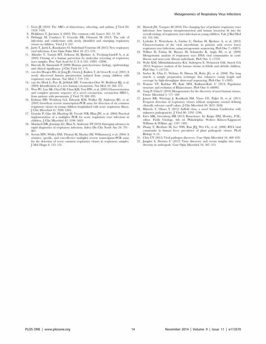
Methods. The 25 samples from children with LRTI were
sequenced individually (listed in Table 2). In the case of the
URTI samples, 9 were sequenced individually, while the amount
of DNA isolated from the other 37 samples was too low to be
analyzed independently, thus, they were used to prepare 16 pools
for sequencing: 13 pools of two samples, 1 pool of three samples,
and 2 pools of four samples (Table 3).
The total number of DNA reads and the valid unique reads
obtained from each sample after passing the quality controls are
shown in Tables 2 and 3. The valid reads were analyzed for the
presence of sequences from human, bacterial, fungal, or viral
origin. As expected, the most abundant reads were from human
origin, representing 70% and 80% of LRTI and URTI patients,
respectively (Fig. 1). Bacterial sequences made up the second
largest data set, representing 15.2% of the sequence reads in LRTI
and 8.5% in URTI. Viral sequences represented 0.56% and
0.57% of valid reads in LRTI and URTI, respectively, and only
0.05% of reads corresponded to fungi (Fig. 1). Finally, approxi-
mately 13% and 10% of the sequences in both LRTI and URTI
could not be classified since no homolog was found (E-value 1e–
03) or there were contradicting database hits. This category is
referred to as ‘undefined’ in Figure 1. Of interest, despite the fact
that the samples from LRTI and URTI were collected by different
methods (nasal washings vs. swabs), and from children with
different clinical syndromes and varying severity of respiratory
disease, the proportion of sequences from different origins was
very similar.
The undefined sequence reads from all samples were assembled,
and contigs $180 nt were compared with non-redundant protein
database of GenBank (E-value 100) to find sequences that could be
distantly related to known viral sequences and could thus represent
novel viruses. Indeed, short sequences are less likely than long
sequences to retrieve statistically significant similarities in Blast
searches, and sequence assembly into longer contigs is helpful to
overcome this difficulty. As result of this, all filtered contigs aligned
either to bacterial or human proteins during BLASTx runs. An
analysis revealed that the contigs that map to bacteria showed only
60–80% nucleotide identity to their best-matching reference,
indicating that they most likely represent novel species within their
Metagenomics of Respiratory Virus Infections
PLOS ONE | www.plosone.org 3 November 2014 | Volume 9 | Issue 11 | e113570
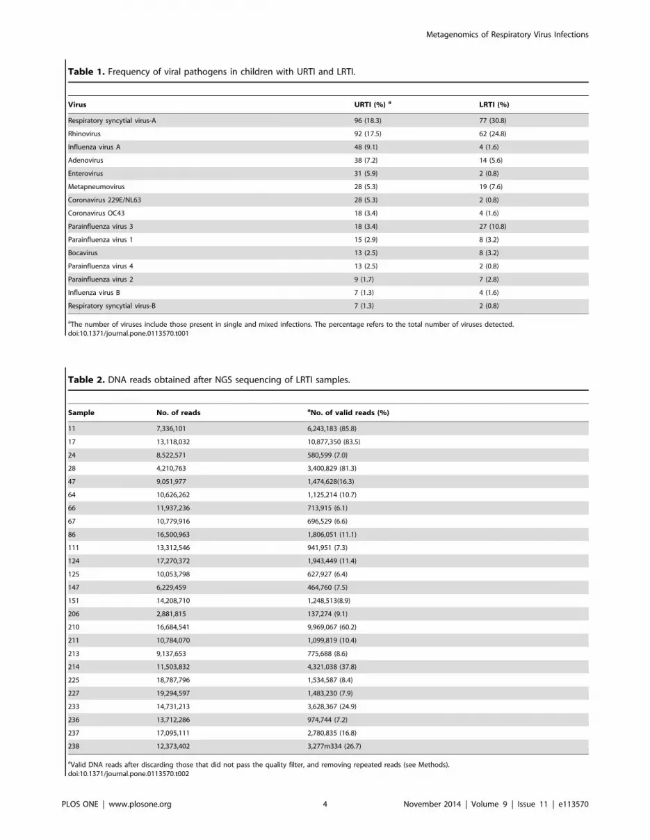
Table 1. Frequency of viral pathogens in children with URTI and LRTI.
Virus URTI (%) a LRTI (%)
Respiratory syncytial virus-A 96 (18.3) 77 (30.8)
Rhinovirus 92 (17.5) 62 (24.8)
Influenza virus A 48 (9.1) 4 (1.6)
Adenovirus 38 (7.2) 14 (5.6)
Enterovirus 31 (5.9) 2 (0.8)
Metapneumovirus 28 (5.3) 19 (7.6)
Coronavirus 229E/NL63 28 (5.3) 2 (0.8)
Coronavirus OC43 18 (3.4) 4 (1.6)
Parainfluenza virus 3 18 (3.4) 27 (10.8)
Parainfluenza virus 1 15 (2.9) 8 (3.2)
Bocavirus 13 (2.5) 8 (3.2)
Parainfluenza virus 4 13 (2.5) 2 (0.8)
Parainfluenza virus 2 9 (1.7) 7 (2.8)
Influenza virus B 7 (1.3) 4 (1.6)
Respiratory syncytial virus-B 7 (1.3) 2 (0.8)
aThe number of viruses include those present in single and mixed infections. The percentage refers to the total number of viruses detected.doi:10.1371/journal.pone.0113570.t001
Table 2. DNA reads obtained after NGS sequencing of LRTI samples.
Sample No. of reads aNo. of valid reads (%)
11 7,336,101 6,243,183 (85.8)
17 13,118,032 10,877,350 (83.5)
24 8,522,571 580,599 (7.0)
28 4,210,763 3,400,829 (81.3)
47 9,051,977 1,474,628(16.3)
64 10,626,262 1,125,214 (10.7)
66 11,937,236 713,915 (6.1)
67 10,779,916 696,529 (6.6)
86 16,500,963 1,806,051 (11.1)
111 13,312,546 941,951 (7.3)
124 17,270,372 1,943,449 (11.4)
125 10,053,798 627,927 (6.4)
147 6,229,459 464,760 (7.5)
151 14,208,710 1,248,513(8.9)
206 2,881,815 137,274 (9.1)
210 16,684,541 9,969,067 (60.2)
211 10,784,070 1,099,819 (10.4)
213 9,137,653 775,688 (8.6)
214 11,503,832 4,321,038 (37.8)
225 18,787,796 1,534,587 (8.4)
227 19,294,597 1,483,230 (7.9)
233 14,731,213 3,628,367 (24.9)
236 13,712,286 974,744 (7.2)
237 17,095,111 2,780,835 (16.8)
238 12,373,402 3,277m334 (26.7)
aValid DNA reads after discarding those that did not pass the quality filter, and removing repeated reads (see Methods).doi:10.1371/journal.pone.0113570.t002
Metagenomics of Respiratory Virus Infections
PLOS ONE | www.plosone.org 4 November 2014 | Volume 9 | Issue 11 | e113570

corresponding genera and thus could not be classified during
alignments with BLASTn. Nonetheless, the vast majority of reads
(50% to 90%) were not assembled into contigs. The unassembled
reads were low complexity sequences or library artifacts as adapter
chimeras, suggesting that it is unlikely that they correspond to
novel viruses. A remaining small amount of sequences could not be
assembled due to non-uniform read depth because of a non-
uniform species abundance distribution.
Viruses detected by NGS in double-negative samplesDNA sequence reads from at least one virus commonly
associated to respiratory infections was found in 20 out of the 25
double-negative samples of LRTI patients (Table 4): 5 samples
were positive for RSV reads, 11 samples for HCoV-OC-43, and 9
for RV. In addition, 5 samples contained HBoV and in 12 samples
anelloviruses (torque teno -TTV-, torque teno mini -TTMV-, or
torque teno midi viruses -TTMDV) were also detected; rotavirus,
papillomavirus, and herpesvirus sequences were identified once in
the samples, and reads from several viruses from both animal (bat
picornavirus, bovine viral diarrheal virus, bovine kobovirus) and
plant origin (potato virus Y, pepper mild mottle virus), as well as
various bacteriophages were also found (Table 4). Regarding
bacteria, DNA sequence reads from S. pneumoniae were the most
frequent, being present in all but one of the 25 samples sequenced,
and M. catarrhalis, L. pneumoniae, and H. influenzae were less
frequently found. DNA reads from other bacteria less commonly
associated with respiratory infections were also detected (Table 4).
Some of the samples had sequence reads corresponding to up to 8
different viruses or 15 different bacteria. Of interest, including the
NGS results, 79.2% (198/250) of the samples had a respiratory
virus detected, and in the remaining 52 samples at least one
bacteria was found, such that all 250 samples from children with
LRTI had a respiratory pathogen identified.
DNA reads from one to five typical respiratory viruses were
detected in 22 of the 25 sequenced double-negative individual
and/or pooled samples from children with URTI (Table 5): The
virus most frequently detected was RV, which was found in 19 of
the pooled and/or individual samples; some of the samples had
more than one type of virus, such that we found sequence reads
from 4 RV subtype A, 4 subtype B, and 19 subtype C. One sample
was positive for RSV, 3 for HCoV-OC43, 3 for human
enterovirus A71, and 3 samples had HBoV. Of interest, 5 of the
samples had DNA reads from Saffold virus, a virus recently
described to be associated to respiratory infections. Also, among
these samples we identified 3 containing herpesvirus, 5 papillo-
mavirus, 2 human astrovirus, 4 rotavirus, and 10 anelloviruses
(TTV, TTMV, TTMDV). Similar to what was found in LRTI, in
children with URTI DNA reads of viruses from animal (white spot
syndrome and bat picornavirus) and plant origin (Okra mosaic
virus, capsicum chlorosis virus, cucumber mosaic virus, pepper
Table 3. DNA reads obtained after NGS sequencing of URTI samples.
aIndividual and pooled samples No. of reads bNo. of valid reads (%)
C06, C55, T78, V24 4,323,309 1,707,160 (39.5)
C16, C61 2,113,065 1,134,387 (53.7)
C27, C01 2,951,784 1,474,233 (49.9)
C29, M40, T41, V39 3,325,315 1,235,867 (37.2)
C41, T50 3,765,099 1,708,531 (45.4)
C46, P54 4,289,979 2,278,068 (53.1)
M23, M44 3,607,011 1,982,487 (55.0)
M28 7,294,104 3,292,828(45.1)
P06, P150 5,010,364 2,387,550 (47.7)
P108 4,609,150 1,021,900 (22.2)
P147, P191 3,454,018 1,540,283 (44.6)
P149, P153 4,381,981 2,534,733 (57.8)
P151, P181 4,638,675 2,207,800 (47.6)
P173 2,974,252 613,487 (20.6)
P176, P186, P213 4,210,620 2,176,224 (51.7)
P183 1,654,081 272,720 (16.5)
P19, P88 8,727,411 5,067,242 (58.1)
P69 3,072,261 631,373 (20.6)
T33, T39 4,863,936 3,308,371 (68.0)
T36 1,228,687 446,932 (36.4)
T38 3,309,781 1,420,555 (42.9)
T43, T44 3,183,181 2,325,025 (73.0)
T65 78,467 30,443 (38.8)
V26 3,469,556 1,496,608 (43.1)
P131, V61 3,939,137 2,288,528 (58.1)
aThe samples that were pooled for sequencing are indicated.bValid DNA reads after discarding those that did not pass the quality filter, and removing repeated reads (see Methods).doi:10.1371/journal.pone.0113570.t003
Metagenomics of Respiratory Virus Infections
PLOS ONE | www.plosone.org 5 November 2014 | Volume 9 | Issue 11 | e113570

mild mottle virus, and tomato mosaic virus) were also detected. In
the majority of samples bacteriophages were found. After NGS, all
pooled samples had DNA sequences from at least one respiratory
virus, while three individual (not pooled) samples remained
negative for common respiratory viruses (Table 5). In two of
these samples (P183 and T38) M. catarrhalis was detected and S.pneumoniae was additionally present in one of them. The third
sample (T65) remained negative for both viruses (including phages)
and bacteria. Some of the samples from URTI had sequence reads
corresponding to up to 11 different viruses or 18 different bacteria.
Considering the NGS results, and assuming that in the pooled
samples the identified viruses were each present in a different
sample, 76.6% (403/526) of the children had a common
respiratory virus detected, while in all but one of the other 129
children a respiratory bacteria was identified.
Genome assembly and phylogenetic analysesTo estimate the sequence coverage of the NGS-identified
viruses, the sequence reads were assembled de novo, and all contigs
were used to estimate the extent of the virus genome coverage. A
significant coverage was obtained for several of the detected
viruses. In patients with LRTI, the genome of 14 viruses was
assembled with coverage higher than 20% (Table 6). As indication
of the sensitivity of NGS and of the relative abundance of some
viruses not detected by the conventional PCR, we could assemble
more than 95% of the genome of two RV strains, 98% of one
HBoV, and 99.8% of one HCoV-OC43 strain. In the case of
children with URTI, at least 50% of the genome was covered for
15 viruses, including RV, HEV, Saffold virus, and TTV (Table 7),
with 8 of them having a genome coverage of more than 90%. For
the DNA reads of the animal viruses identified in both types of
children populations the coverage ranged between 0.57 and 4.9%,
and for plant viruses between 0.13 and 7.15% in the case of
tomato mosaic virus (Tables 5A and B).
The assembled sequences from HCoV-OC43, RV, HBoV,
Saffold, and anelloviruses were used to construct phylogenetic
trees to determine the genetic similarity of the viruses character-
ized in this work with those in databases (see Materials and
Methods). All viruses from LRTI and URTI grouped with clades
formed by previously reported virus sequences (not shown).
Anelloviruses could be readily classified as TTV, TTMV, or
TTMDV, with some samples containing the three genera
(Tables 4 and 5). All HCoV detected belong to species HCoV-
OC43 and grouped with other known betacoronaviruses. HBoVs
were all genotype 1, while the Saffold viruses detected in this work
belonged to either genotype 2 or 3. Of interest, in the case of some
RV, HBoV, Saffold, and anelloviruses, different contigs mapped to
different clades, suggesting that recombination events are common
in this type of viruses, as has been reported for RV [25].
Figure 1. Taxonomic classification of the generated DNA sequencing reads. Valid DNA reads obtained by NGS of LRTI and URTI clinicalsamples were split into human, bacterial, fungal, and viral origin. Those reads not present in the four previous categories were classified as"undefined". Average values for all LRTI and URTI samples are shown.doi:10.1371/journal.pone.0113570.g001
Metagenomics of Respiratory Virus Infections
PLOS ONE | www.plosone.org 6 November 2014 | Volume 9 | Issue 11 | e113570

Ta
ble
4.
Pat
ho
ge
ns
ide
nti
fie
db
yN
GS
inch
ildre
nw
ith
low
er
resp
irat
ory
trac
tin
fect
ion
s.
Sa
mp
les
Pa
tho
ge
nd
ete
cte
d4
78
61
24
11
12
82
06
21
02
11
21
32
14
22
52
27
23
32
36
23
72
38
17
24
11
12
51
47
15
16
46
66
7
VIR
US
ES
Re
spir
ato
rysy
ncy
tia
lv
iru
s3
a3
72
--
--
--
--
--
--
--
12
--
--
7-
31
8-
Co
ron
av
iru
sO
C4
3-
--
-5
-4
54
31
22
2-
--
--
23
90
49
16
72
23
92
60
0-
--
-3
Rh
ino
vir
us
A5
-6
6-
2-
-1
60
09
--
-3
20
6-
16
--
--
--
3-
--
Rh
ino
vir
us
C-
-1
21
--
--
63
--
3-
38
47
5-
39
--
--
--
--
--
Hu
ma
nb
oca
vir
us
--
--
--
16
2-
-8
72
09
6-
--
-3
22
48
3-
46
--
--
--
To
rqu
ete
no
vir
us
(TT
V)
--
--
--
53
61
67
-1
00
71
25
--
15
73
2-
-2
-9
16
0-
To
rqu
ete
no
min
iv
iru
s(T
TM
V)
--
--
--
46
-6
--
--
--
3-
7-
--
-2
54
-
To
rqu
ete
no
mid
iv
iru
s(T
TM
DV
)-
--
--
--
--
--
-9
--
--
--
--
--
6-
Hu
ma
nh
erp
es
vir
us
--
--
--
--
--
--
--
--
--
2-
--
--
-
Hu
ma
np
ap
illo
ma
vir
us
--
--
--
--
--
-3
1-
--
--
--
--
--
--
Ro
tav
iru
s-
--
--
--
3-
--
--
--
--
--
--
--
--
Ba
tp
ico
rna
vir
us
--
--
--
-4
--
--
--
--
--
--
--
--
-
Bo
vin
ek
ob
uv
iru
s-
--
--
-6
13
0-
--
--
--
--
--
--
--
--
Bo
vin
ev
ira
ld
iarr
he
av
iru
s-
--
--
-5
20
--
--
--
--
--
--
--
--
-
Pe
pp
er
mo
ttle
vir
us
--
--
--
--
--
--
8-
--
--
--
--
--
-
Po
tato
vir
us
Y-
--
7-
--
--
--
--
--
--
--
--
--
--
Ph
ag
es
-2
36
29
49
55
--
33
10
98
39
82
39
43
21
17
25
17
-3
35
22
38
45
69
38
36
71
20
11
34
5
BA
CT
ER
IA
Str
ep
toco
ccu
sp
ne
um
on
iae
31
01
47
-5
74
34
25
37
32
22
56
94
41
84
25
23
27
42
04
81
86
10
43
,96
4
Ha
em
op
hil
us
infl
ue
nz
ae
41
30
41
21
0-
--
53
51
7-
13
--
--
-4
1-
--
-1
05
--
Mo
rax
ell
aca
tarr
ha
lis
11
01
1,8
45
38
0-
--
-2
32
,21
31
21
,23
73
1,9
09
57
0-
58
6-
10
-3
-6
13
Le
gio
ne
lla
pn
eu
mo
ph
ila
-5
5-
--
32
1-
-3
24
-4
--
--
--
-1
18
--
-
Kle
bsi
ell
ap
ne
um
on
iae
--
-3
7-
5-
56
-2
--
4-
--
-3
--
--
--
Sta
ph
ylo
cocc
us
au
reu
s-
-1
43
1-
-6
41
28
52
24
8-
17
32
25
--
52
-4
34
-3
1-
22
Ra
hn
ell
a-
14
-2
,23
9-
--
-5
-3
3-
--
--
90
6-
8-
--
15
4
Bu
rkh
old
eri
ace
pa
cia
32
28
24
21
,75
3-
13
32
94
75
23
62
24
20
1,1
35
98
14
81
6-
-3
25
-2
51
63
15
25
51
,16
81
06
Aci
ne
tob
act
er
ba
um
an
nii
67
1,4
19
1,0
92
4,6
22
-6
85
15
21
,49
11
,66
05
71
,25
21
,79
35
79
46
37
54
-2
,24
8-
2,2
10
18
03
50
34
77
83
2,4
25
Pse
ud
om
on
as
ae
rug
ino
sa1
43
98
22
21
,04
4-
53
16
12
82
43
09
71
91
,52
51
23
91
42
61
01
--
96
56
31
99
30
71
,17
53
69
My
cob
act
eri
um
tub
erc
ulo
sis
-7
-1
1-
--
-3
-7
--
--
--
--
--
--
--
Act
ino
my
ceta
les
15
,56
32
55
23
16
-3
8-
61
16
11
67
71
51
17
19
32
--
--
33
-1
01
5-
-
Bu
rkh
old
eri
ag
lad
ioli
22
3,7
16
3,8
97
36
,94
7-
17
43
20
7,7
56
2,2
64
43
91
0,1
57
13
,73
31
,66
12
,47
32
35
12
75
10
7-
64
53
55
35
53
,14
34
60
50
0
Ma
lass
ez
iag
lob
osa
35
15
42
70
-4
47
-1
,55
44
8-
78
52
08
14
68
48
-2
14
62
01
,91
5-
1,0
62
21
10
23
92
14
11
42
87
Metagenomics of Respiratory Virus Infections
PLOS ONE | www.plosone.org 7 November 2014 | Volume 9 | Issue 11 | e113570

Discussion
Improvements in diagnostic methods have increased the rate of
identification of viral pathogens in different clinical conditions,
such as gastrointestinal, respiratory, or neurologic infections.
However, despite these advances there are still a significant
number of cases (20–50%), in which the etiologic agents are
believed to be viruses, but the agent is not identified [26].
Previously, we reported the presence of a respiratory virus in about
71% of nasal samples obtained from children with LRTI and
URTI (Aponte et al., manuscript in preparation; see also the
Pathogen Detection section above), using a PCR method able to
detect 15 different respiratory viruses. After PCR screening the
virus-negative samples for the presence of respiratory bacteria,
89.6% of children with LRTI and 91.1% with URTI had at least
one potential pathogen identified. These percentages were raised
to levels close to 80% for viruses in both patient populations after
NGS analysis of the double-negative samples, and essentially
100% of the samples had either a common respiratory virus or
bacteria identified (in only one of 526 URTI samples DNA reads
from a potential pathogen was not identified). It is interesting that
6 of the 8 samples from both LRTI and URTI that were negative
for viruses after NGS had less than one million valid reads
(Tables 2 and 3). Since the number of sequence reads directly
correlates with the amount of nucleic acids present in the original
sample [21], the absence of virus detection in these samples could
represent false-negative results; it is likely that with a larger
amount of sample, or deeper sequencing, respiratory viruses could
have also been detected.
The samples that resulted negative for viruses by PCR, and
subsequently determined as PCR-positive for respiratory bacterial
pathogens, were not characterized by NGS, but it is reasonable to
assume that a high percentage of them could have also been
positive for viruses by deep sequencing. It is difficult, however, to
determine with confidence, which, if any, of the detected
pathogens could be responsible for the clinical respiratory
symptoms observed. The virus or bacteria detected by these
methods could be present in the patient as an asymptomatic
carrier state or as causal agents of asymptomatic infections. Studies
comparing the presence of respiratory pathogens in nasal
specimens from healthy children will help to resolve this issue.
In addition, PCR and NGS are such sensitive techniques that the
presence of small amounts of viral targets may not necessarily have
clinical relevance. An additional limitation of this study is the
limited number of samples analyzed. Exploring the possibility to
define cutoff levels represents the next necessary step for
diagnosing viral respiratory infections using molecular tests [27].
It is important to mention, however, that for several of the RV and
HCoV detected in this study high genome sequence coverages
were achieved. This observation indicates that a high number of
DNA reads, and probably also of virus particles, were present in
the samples. These viruses could have been undetected by PCR
due to mismatches in the diagnostic primers used.
Of interest, the classes of viruses found by NGS in patients with
LRTI and URTI were very similar, although their frequencies
were different in the two study populations. RV was more
frequently found in URTI (19 of 25 samples) vs. LRTI (9 of 25
samples), while coronavirus was more represented in LRTI (11/
25) than in URTI (3/25). Only one of the 46 samples of children
with URTI was positive for RSV, while in 5 of 25 samples from
children with LRTI RSV was detected. Saffold viruses, members
of the picornaviridae family and cardiovirus genus, were found
only in children with URTI. Since their initial description in 2007,
these viruses have been shown to circulate worldwide, occur early
Ta
ble
4.
Co
nt.
Sa
mp
les
Pa
tho
ge
nd
ete
cte
d4
78
61
24
11
12
82
06
21
02
11
21
32
14
22
52
27
23
32
36
23
72
38
17
24
11
12
51
47
15
16
46
66
7
Pse
ud
om
on
as
me
nd
oci
na
-9
86
73
59
-1
54
12
11
28
6-
29
14
30
27
90
10
--
63
-6
23
19
74
93
50
75
Sta
ph
ylo
cocc
us
ep
ide
rmid
is-
88
-2
68
1-
-5
,00
7-
1,3
41
--
--
-8
8-
--
-1
5-
21
0-
40
11
1
Le
ifso
nia
xy
li2
,49
4-
--
--
--
--
--
--
--
--
--
--
--
-
Aci
do
vo
rax
69
1,2
80
88
56
,70
4-
80
82
22
1,3
25
2,0
42
10
63
,81
45
,07
95
43
55
15
94
01
77
,07
4-
7,7
04
2,0
57
99
57
71
5,3
45
5,0
61
aN
um
be
ro
fva
lids
DN
Are
ads
inth
esa
mp
lep
or
the
corr
esp
on
din
gp
ath
og
en
.d
oi:1
0.1
37
1/j
ou
rnal
.po
ne
.01
13
57
0.t
00
4
Metagenomics of Respiratory Virus Infections
PLOS ONE | www.plosone.org 8 November 2014 | Volume 9 | Issue 11 | e113570

Ta
ble
5.
Pat
ho
ge
ns
ide
nti
fie
db
yN
GS
inch
ildre
nw
ith
up
pe
rre
spir
ato
rytr
act
infe
ctio
ns.
Sa
mp
lesa
Pa
tho
ge
nd
ete
cte
d
C0
6,
C5
5,
T7
8,
V2
4C
16
,C
61
C2
7,
C0
1
C2
9,
M4
0,
M4
1,
V3
9C
41
,T
50
C4
6,
P5
4M
23
,M
44
M2
8P
06
,P
15
0P
10
8P
13
1,
V6
1P
14
7,
P1
91
P1
4,9
P1
53
P1
51
,P
18
1P
17
3
P1
76
,P
18
6,
P2
13
P1
83
P1
9,
P8
8T
69
T3
3,
T3
9T
36
T3
8T
43
,T
44
T6
5V
26
VIR
US
ES
Re
spir
ato
rysy
ncy
tia
lv
iru
s-
--
--
--
--
--
--
--
--
--
3-
--
--
Co
ron
av
iru
sO
C4
3-
--
--
--
--
--
32
4-
38
--
--
--
--
--
Rh
ino
vir
us
A-
--
--
--
--
-4
95
9-
1,0
52
--
--
2-
--
--
-
Rh
ino
vir
us
B-
--
--
--
-1
4-
3,9
95
12
-8
--
--
--
--
--
-
Rh
ino
vir
us
C8
41
8b
15
59
3,6
67
11
5,6
17
44
65
,88
92
11
,23
3-
70
24
65
55
4-
66
-1
1,0
26
-1
,39
54
1-
14
,56
1-
20
7
Hu
ma
ne
nte
rov
iru
sA
--
--
15
--
-1
5,2
36
-7
8-
--
--
--
--
--
--
-
Hu
ma
nb
oca
vir
us
--
--
--
--
-1
28
--
--
--
--
--
--
-9
8
Sa
ffo
ldv
iru
s1
82
--
18
3-
--
--
-1
0-
2,6
72
--
9,8
91
--
--
--
--
-
To
rqu
ete
no
vir
us
(TT
V)
--
9-
71
87
--
12
-2
12
14
-7
--
--
-3
--
--
-
To
rqu
ete
no
min
iv
iru
s(T
TM
V)
--
-3
51
00
--
22
-3
,40
89
-1
7-
--
--
--
--
--
To
rqu
ete
no
mid
iv
iru
s(T
TM
DV
)3
--
81
21
1-
--
-2
,71
15
-7
--
-1
1-
--
--
--
Hu
ma
nh
erp
es
vir
us
--
--
-3
--
-3
6-
--
--
--
53
--
--
--
-
Hu
ma
np
ap
illo
ma
vir
us
--
3-
--
6-
--
3-
-1
18
--
-5
--
--
--
-
Ro
tav
iru
s-
--
3-
27
--
--
--
--
-3
--
-4
--
--
-
Hu
ma
na
stro
vir
us
6-
--
3-
--
--
--
--
--
--
--
--
--
-
Ba
tp
ico
rna
vir
us
--
--
--
--
--
4-
--
--
--
--
--
7-
-
Ca
psi
cum
chlo
rosi
sv
iru
s-
--
--
--
--
--
--
--
2-
--
--
--
--
Cu
cum
be
rm
osa
icv
iru
s1
21
8-
--
-6
--
--
--
--
--
--
--
--
--
Ok
ram
osa
icv
iru
s-
--
--
--
--
--
--
--
--
2-
--
--
--
Pe
pp
er
mo
ttle
vir
us
--
2-
--
--
--
--
--
--
--
--
--
--
-
To
ma
tom
osa
icv
iru
s-
16
12
--
4-
-3
--
4-
--
--
8-
--
--
--
Wh
ite
spo
tsy
nd
rom
ev
iru
s-
--
--
--
--
--
--
--
31
--
--
--
--
-
Ph
ag
es
10
28
31
54
46
10
26
06
12
11
16
32
73
61
39
10
56
86
21
84
34
-8
-6
BA
CT
ER
IA
Str
ep
toco
ccu
sp
ne
um
on
iae
13
80
10
31
43
18
01
25
14
23
15
21
51
83
-2
0-
27
49
-4
--
--
Hae
mo
ph
ilu
sin
flu
en
zae
15
-4
43
47
15
-4
--
17
31
04
1-
28
84
76
--
--
--
-
Mo
raxe
lla
cata
rrh
alis
23
58
15
43
41
72
22
6,1
92
26
10
44
6-
11
6-
6-
14
2-
76
11
8-
--
Leg
ion
ell
ap
ne
um
op
hil
a8
22
-1
4-
-2
24
--
15
--
--
--
--
--
--
--
Kle
bsi
ell
ap
ne
um
on
iae
-9
-3
--
--
--
--
--
--
-3
--
--
--
-
Sta
ph
ylo
cocc
us
aure
us
91
34
24
16
-3
6-
--
--
20
10
-1
2-
21
-5
--
28
8-
-
Metagenomics of Respiratory Virus Infections
PLOS ONE | www.plosone.org 9 November 2014 | Volume 9 | Issue 11 | e113570

Ta
ble
5.
Co
nt.
Sa
mp
lesa
Pa
tho
ge
nd
ete
cte
d
C0
6,
C5
5,
T7
8,
V2
4C
16
,C
61
C2
7,
C0
1
C2
9,
M4
0,
M4
1,
V3
9C
41
,T
50
C4
6,
P5
4M
23
,M
44
M2
8P
06
,P
15
0P
10
8P
13
1,
V6
1P
14
7,
P1
91
P1
4,9
P1
53
P1
51
,P
18
1P
17
3
P1
76
,P
18
6,
P2
13
P1
83
P1
9,
P8
8T
69
T3
3,
T3
9T
36
T3
8T
43
,T
44
T6
5V
26
Rah
ne
lla
5-
--
--
--
--
--
15
--
--
--
--
--
--
Bu
rkh
old
eri
ace
pac
ia6
54
61
69
7-
-1
4-
--
-4
--
--
--
--
--
--
Aci
ne
tob
acte
rb
aum
ann
ii1
,78
31
,75
48
25
1,3
01
77
82
,39
05
,20
44
02
20
2-
65
42
,10
72
08
1,5
44
-1
,34
71
75
56
-1
79
--
17
9-
50
Pse
ud
om
on
asae
rug
ino
sa8
13
73
21
34
16
11
59
72
91
31
-6
11
67
16
32
11
-1
96
-2
74
17
16
--
7-
-
Myc
ob
acte
riu
mtu
be
rcu
losi
s6
-4
--
--
--
--
--
--
--
--
--
--
--
Act
ino
my
ceta
les
1,0
17
41
02
,28
31
,14
17
,36
95
75
69
23
57
1,6
00
64
48
21
,04
91
,10
67
79
13
75
31
22
13
,67
92
01
2,1
59
13
-3
41
-1
17
Bu
rkh
old
eri
ag
lad
ioli
-4
-9
--
--
--
--
--
--
--
--
--
--
-
Mal
asse
zia
glo
bo
sa2
11
08
71
71
45
10
33
42
67
-4
35
78
12
11
14
81
23
10
2-
13
63
17
71
17
55
7-
42
Pse
ud
om
on
asm
en
do
cin
a1
24
10
65
32
42
56
10
91
60
46
14
6-
73
20
11
37
20
5-
21
4-
37
05
01
7-
-3
--
Rh
od
oto
rula
glu
tin
is2
65
39
67
42
02
13
46
95
10
11
--
--
10
--
8-
19
-7
--
--
-
Sta
ph
ylo
cocc
us
ep
ide
rmid
is2
71
10
62
19
14
92
75
79
52
-1
8-
50
20
5-
62
--
29
6-
-3
--
Lysi
nib
acil
lus
sph
aeri
cus
11
53
01
53
92
98
41
17
56
82
34
--
--
--
--
-1
8-
--
--
--
Aci
do
vora
x5
08
19
01
17
35
67
68
03
15
12
43
0-
16
31
36
-1
89
-2
07
-5
8-
31
--
9-
12
Ferv
ido
bac
teri
um
no
do
sum
27
86
77
51
93
45
32
09
99
1,1
52
16
43
49
-2
70
68
11
,48
71
,09
4-
59
0-
41
93
20
8-
-1
21
--
aW
he
nm
ore
than
on
esa
mp
lew
ere
po
ole
dfo
rse
qu
en
cin
g,
the
cod
efo
rth
eva
rio
us
sam
ple
sis
me
nti
on
ed
.b
Nu
mb
er
of
valid
sD
NA
read
sin
the
sam
ple
po
rth
eco
rre
spo
nd
ing
pat
ho
ge
n.
do
i:10
.13
71
/jo
urn
al.p
on
e.0
11
35
70
.t0
05
Metagenomics of Respiratory Virus Infections
PLOS ONE | www.plosone.org 10 November 2014 | Volume 9 | Issue 11 | e113570

in life, and involve the respiratory and gastrointestinal tracts. The
association of these viruses with clinical symptoms is under
investigation and requires additional epidemiological studies to
clarify their pathogenicity [28]. Anelloviruses (TTV, TTMV,
TTMDV) were found in both LRTIs and URTIs. Members of this
family ubiquitously infect humans and establish persistent infec-
tions, although causal disease associations are currently lacking
[10]. It is interesting to note that common gastrointestinal viruses
such as astrovirus and rotavirus were found in some of the samples.
It is not surprising though, since rotaviruses have long being
Table 6. Genome coverage for viruses present in LRTI samples.
Sample Virusa No. of Contigs No. of reads incorporated into contigs Genome size Genome Coverage (%)
86 RSV 22 213 15,191 6.75
124 RV-A 6 47 7,129 7.43
RV-C 10 96 7,107 24.34
111 PVY 1 7 9,704 1.75
210 HBoV 16 74 5,299 33.97
HCoV 3 10 30,578 5.89
TTMV 2 14 2,912 8.24
BKV 1 4 8,374 1.07
BVDV 1 3 12,230 0.57
211 RV-A 1 13,418 7,129 94.60
RV-C 11 47 7,107 17.03
BatPV 1 3 7,753 0.97
BKV 5 83 8,374 4.90
BVDV 1 9 12,230 0.61
RV 1 3 17,360 0.40
213 TTV 12 407 3,725 32.48
214 HBoV 4 53,548 5,299 97.62
HCoV 80 848 30,578 39.96
TTMV 1 3 2,912 2.40
225 HBoV 1 3 5,299 1.51
RV-C 1 2 3,725 2.01
227 TTV 8 777 3,725 34.90
HPV 1 12 7,466 0.94
233 RV-A 16 127 7,129 26.65
RV-C 2 30712 7,107 97.88
TTV 16 75 3,725 52.35
237 RV-A 1 3 7,129 0.98
RV-C 6 35 7,107 7.46
238 HBoV 22 202 5,299 38.69
HCoV 5 85766 30,578 99.81
TTV 1 4 3,725 2.01
17 HBoV 32 279 5,299 61.80
HCoV 112 1,021 30,578 20.52
RSV 1 3 15,191 0.49
24 TTV 12 1 3,725 1.88
11 HBoV 6 45 5,299 12.08
HCoV 15 291 30,578 5.72
125 HCoV 32 382 30,578 11.28
151 RSV 1 4 15,191 0.46
66 RSV 24 274 15,191 15.40
TTV 3 107 3,725 9.93
TTMV 2 23 2,912 4.46
aRSV, respiratory syncytial virus; RV-A, rhinovirus species A, RV-C; rhinovirus species C; HBoV, human bocavirus, HCoV, human coronavirus OC43; TTV, torque teno virus;TTMV, torque teno mini virus; BKV, bovine kobuvirus; BVDV, bovine viral diarrhea virus; BatPV, bat picornavirus; HPV, human papillomavirus; PVY, potato virus Y.doi:10.1371/journal.pone.0113570.t006
Metagenomics of Respiratory Virus Infections
PLOS ONE | www.plosone.org 11 November 2014 | Volume 9 | Issue 11 | e113570

Table 7. Genome coverage for viruses present in URTI samples.
Sample Virusa No. of Contigs No. of reads incorporated into contigs Genome size Genome Coverage (%)
C27, C01 ToMV 2 12 6,383 1.88
TTMDV 2 5 3256 4.45
HPV 1 2 7466 0.94
RV-C 9 45 7107 11.3
C41, T50 TTMV 2 5 2912 3.78
TTMDV 2 10 3256 3.22
TTV 1 3 3725 2.28
RV-C 1 92130 7107 95.40
C16, C61 CMV 4 10 3356 7.15
ToMV 3 15 6386 2.98
RV-C 2 10 7107 2.74
M23, M44 CMV 1 3 3356 5.36
RV-C 1 39840 7107 97.20
P108 HHV 5 12 116114 0.28
HBoV 2 5 5299 2.64
P06, P150 HEV-A 2 12352 7,413 97.88
TTMV 1 6 2912 4.12
RV-C 4 8798 7107 92.74
P149, P153 HCoV 3 11 30,578 0.78
SAFV 5 1909 8,115 83.03
RV-C 4 26 7107 4.50
P173 HCoV 3 9 30,578 0.69
P151, P181 RV-A 7 943 7,129 50.78
RV-B 1 4 7,215 0.97
HPV 9 45 7466 12.46
TTMV 2 7 2912 4.98
TTV 1 3 3725 0.87
RV-C 5 17 7107 6.33
P147, P191 RV-A 3 12 7,129 4.35
RV-B 1 4 7,215 1.25
ToMV 1 3 6,384 1.25
TTMV 1 3 2912 3.43
TTMDV 1 3 3256 2.15
TTV 1 5 3725 2.01
RV-C 5 15 7107 6.26
P176, P186, P213 WSSV 6 31 292,967 0.13
SAFV 5 8023 8,115 90.92
RV-C 8 37 7107 9.01
C46, P54 RV 3 12 17360 1.09
TTMV 6 73 2912 15.45
TTMDV 1 11 3256 2.15
TTV 13 150 3725 50.74
RV-C 3 9 7107 2.25
P19, P88 HHV 3 12 116114 0.18
HPV 0 0 7466 0.00
ToMV 1 7 6,384 1.25
TTMDV 1 6 3256 3.38
RV-C 10 8726 7107 62.77
T36 RV-C 5 21 7107 4.43
T33, T39 RV-C 12 1319 7107 68.74
Metagenomics of Respiratory Virus Infections
PLOS ONE | www.plosone.org 12 November 2014 | Volume 9 | Issue 11 | e113570

suspected to reach the gastrointestinal tract via mouth and nose. In
fact, some rotavirus infections have been associated with
respiratory symptoms [29]. Finally, we found low amounts of
DNA reads corresponding to animal and plant viruses. The
number of these types of viruses is larger than previously reported
in respiratory samples [21], although plant viruses have been
found more abundantly in human feces [30]. Both, plant and
animal viruses are thought to be derived either from consumed
food or acquired from the environment.
The search for new viruses using NGS technologies in
mammalian, avian, and in particular, human samples, has
contributed to the identification of new viruses in animal reservoirs
and in different conditions of disease [31]. However, the important
effort invested in mammal and avian virus detection has only
resulted in the discovery of variants of virus species, sister species to
known viruses, and rarely genera. These observations contrast
with the recent efforts to discover arthropod viruses, which have
yielded widely divergent taxa that sometimes have even defined
novel families [32]. Altogether, these observations and the
presence of DNA sequence reads from common respiratory
viruses or bacteria in essentially 100% of the samples collected
from children with LRTI and URTI, suggest there is limited
potential for the discovery of so far undescribed, clinically relevant,
viruses associated to pediatric respiratory disease at least in the
type of populations studied and with the sampling and diagnostic
methods employed.
Acknowledgments
We thank Miguel L. Garcıa-Leon for his help in handling and organizing
all pneumonia samples. We also thank the Instituto de Biotecnologia-
UNAM for giving us access to its computer cluster and Jerome Verleyen
for his computer support. This work was supported by grants 153639 (to JI
Santos) and "Influenza 2009" (to CF Arias) from the National Council for
Science and Technology-Mexico (CONACYT). F.E.A. is recipient of a
scholarship from CONACYT.
Author Contributions
Conceived and designed the experiments: BT PI CFA. Performed the
experiments: BT MAE FEA PI. Analyzed the data: BT CFA. Contributed
reagents/materials/analysis tools: MAA-O JM-M R-RV FD-H FZ-V
RMW-C VF-R CNR-A JG-M AV-S GM-A MCS-M DEN LFP-G JIS-P.
Wrote the paper: CFA BT SL JIS-P DEN GM-A RMW-C AV-S.
References
1. Rudan I, O’Brien KL, Nair H, Liu L, Theodoratou E, et al. (2013)
Epidemiology and etiology of childhood pneumonia in 2010: estimates of
incidence, severe morbidity, mortality, underlying risk factors and causative
pathogens for 192 countries. J Glob Health 3: 010401.
2. Ruuskanen O, Lahti E, Jennings LC, Murdoch DR (2011) Viral pneumonia.
Lancet 377: 1264–1275.
3. Black RE, Cousens S, Johnson HL, Lawn JE, Rudan I, et al. (2010) Global,
regional, and national causes of child mortality in 2008: a systematic analysis.
Lancet 375: 1969–1987.
4. Chiu CY, Urisman A, Greenhow TL, Rouskin S, Yagi S, et al. (2008) Utility of
DNA microarrays for detection of viruses in acute respiratory tract infections in
children. J Pediatr 153: 76–83.
5. Ruohola A, Waris M, Allander T, Ziegler T, Heikkinen T, et al. (2009) Viral
etiology of common cold in children, Finland. Emerg Infect Dis 15: 344–346.
6. van Gageldonk-Lafeber AB, Heijnen ML, Bartelds AI, Peters MF, van der Plas
SM, et al. (2005) A case-control study of acute respiratory tract infection in
general practice patients in The Netherlands. Clinical infectious diseases: an
official publication of the Infectious Diseases Society of America 41: 490–497.
Table 7. Cont.
Sample Virusa No. of Contigs No. of reads incorporated into contigs Genome size Genome Coverage (%)
T43, T44 RV-C 3 14165 7107 92.92
C06, C55, T78, V24 HASTV 2 5 6,759 2.52
CMV 1 6 3356 2.09
SAFV 2 126 8,115 2.42
RV-C 3 7989 7107 92.66
V26 HBoV 8 77 5299 15.30
RV-C 7 143 7107 15.06
C29, M40, M41, V39 SAFV 8 135 8,115 8.76
TTMV 1 5 2912 2.75
RV-C 11 3061 7107 63.89
P131, V61 BatPV 1 2 7,753 0.90
HBoV 1 4 5299 2.08
RV-A 3 14 7,129 3.23
RV-B 4 3145 7,215 90.33
HEV A 3 13 7,413 3.10
TTMV 9 1531 2912 47.12
TTMDV 4 1881 3256 29.67
TTV 13 126 3725 42.31
RV-C 4 618 7107 19.77
aRSV, respiratory syncytial virus; RV-A, rhinovirus species A; RV-B; rhinovirus species B; RV-C; rhinovirus species C; HBoV, human bocavirus, HCoV, human coronavirusOC43; TTV, torque teno virus; TTMV, torque teno mini virus; TTMDV, torque teno midi virus; BKV, bovine kobuvirus; BVDV, bovine viral diarrhea virus; BatPV, batpicornavirus; HPV, human papillomavirus; PVY, potato virus Y, ToMV, tomato mosaic virus; CMV, cucumber mosaic virus; HHV, human herpes virus; HVE-A, humanenterovirus A; SAFV, Saffold virus; WSSV, white spot syndrome virus; HASTV, human astrovirus.doi:10.1371/journal.pone.0113570.t007
Metagenomics of Respiratory Virus Infections
PLOS ONE | www.plosone.org 13 November 2014 | Volume 9 | Issue 11 | e113570
7. Gern JE (2010) The ABCs of rhinoviruses, wheezing, and asthma. J Virol 84:
7418–7426.8. Heikkinen T, Jarvinen A (2003) The common cold. Lancet 361: 51–59.
9. Debiaggi M, Canducci F, Ceresola ER, Clementi M (2012) The role of
infections and coinfections with newly identified and emerging respiratoryviruses in children. Virol J 9: 247.
10. Jartti T, Jartti L, Ruuskanen O, Soderlund-Venermo M (2012) New respiratoryviral infections. Curr Opin Pulm Med 18: 271–278.
11. Allander T, Tammi MT, Eriksson M, Bjerkner A, Tiveljung-Lindell A, et al.
(2005) Cloning of a human parvovirus by molecular screening of respiratorytract samples. Proc Natl Acad Sci U S A 102: 12891–12896.
12. Harvala H, Simmonds P (2009) Human parechoviruses: biology, epidemiologyand clinical significance. J Clin Virol 45: 1–9.
13. van den Hoogen BG, de Jong JC, Groen J, Kuiken T, de Groot R, et al. (2001) Anewly discovered human pneumovirus isolated from young children with
respiratory tract disease. Nat Med 7: 719–724.
14. van der Hoek L, Pyrc K, Jebbink MF, Vermeulen-Oost W, Berkhout RJ, et al.(2004) Identification of a new human coronavirus. Nat Med 10: 368–373.
15. Woo PC, Lau SK, Chu CM, Chan KH, Tsoi HW, et al. (2005) Characterizationand complete genome sequence of a novel coronavirus, coronavirus HKU1,
from patients with pneumonia. J Virol 79: 884–895.
16. Erdman DD, Weinberg GA, Edwards KM, Walker FJ, Anderson BC, et al.(2003) GeneScan reverse transcription-PCR assay for detection of six common
respiratory viruses in young children hospitalized with acute respiratory illness.J Clin Microbiol 41: 4298–4303.
17. Gruteke P, Glas AS, Dierdorp M, Vreede WB, Pilon JW, et al. (2004) Practicalimplementation of a multiplex PCR for acute respiratory tract infections in
children. J Clin Microbiol 42: 5596–5603.
18. Murdoch DR, Jennings LC, Bhat N, Anderson TP (2010) Emerging advances inrapid diagnostics of respiratory infections. Infect Dis Clin North Am 24: 791–
807.19. Syrmis MW, Whiley DM, Thomas M, Mackay IM, Williamson J, et al. (2004) A
sensitive, specific, and cost-effective multiplex reverse transcriptase-PCR assay
for the detection of seven common respiratory viruses in respiratory samples.J Mol Diagn 6: 125–131.
20. Hustedt JW, Vazquez M (2010) The changing face of pediatric respiratory tract
infections: how human metapneumovirus and human bocavirus fit into the
overall etiology of respiratory tract infections in young children. Yale J Biol Med
83: 193–200.
21. Lysholm F, Wetterbom A, Lindau C, Darban H, Bjerkner A, et al. (2012)
Characterization of the viral microbiome in patients with severe lower
respiratory tract infections, using metagenomic sequencing. PloS One 7: e30875.
22. Willner D, Furlan M, Haynes M, Schmieder R, Angly FE, et al. (2009)
Metagenomic analysis of respiratory tract DNA viral communities in cystic
fibrosis and non-cystic fibrosis individuals. PloS One 4: e7370.
23. Wylie KM, Mihindukulasuriya KA, Sodergren E, Weinstock GM, Storch GA
(2012) Sequence analysis of the human virome in febrile and afebrile children.
PloS One 7: e27735.
24. Sorber K, Chiu C, Webster D, Dimon M, Ruby JG, et al. (2008) The long
march: a sample preparation technique that enhances contig length and
coverage by high-throughput short-read sequencing. PloS One 3: e3495.
25. Waman VP, Kolekar PS, Kale MM, Kulkarni-Kale U (2014) Population
structure and evolution of Rhinoviruses. PloS One 9: e88981.
26. Tang P, Chiu C (2010) Metagenomics for the discovery of novel human viruses.
Future Microbiol 5: 177–189.
27. Jansen RR, Wieringa J, Koekkoek SM, Visser CE, Pajkrt D, et al. (2011)
Frequent detection of respiratory viruses without symptoms: toward defining
clinically relevant cutoff values. J Clin Microbiol 49: 2631–2636.
28. Himeda T, Ohara Y (2012) Saffold virus, a novel human Cardiovirus with
unknown pathogenicity. J Virol 86: 1292–1296.
29. Estes MK, Greenberg HB (2013) Rotaviruses. In: Knipe DM, Howley, P.M.,
editor. Fields Virology. 6th ed. Philadelphia: Wolters Kluwer/Lippincott
Williams & Wilkins. pp. 1347–1401.
30. Zhang T, Breitbart M, Lee WH, Run JQ, Wei CL, et al. (2006) RNA viral
community in human feces: prevalence of plant pathogenic viruses. PLoS
Biology 4: e3.
31. Chiu CY (2013) Viral pathogen discovery. Curr Opin Microbiol 16: 468–478.
32. Junglen S, Drosten C (2013) Virus discovery and recent insights into virus
diversity in arthropods. Curr Opin Microbiol 16: 507–513.
Metagenomics of Respiratory Virus Infections
PLOS ONE | www.plosone.org 14 November 2014 | Volume 9 | Issue 11 | e113570