dental implant systems - mdpi
TRANSCRIPT

Int. J. Mol. Sci. 2010, 11, 1580-1678; doi:10.3390/ijms11041580
International Journal of
Molecular Sciences ISSN 1422-0067
www.mdpi.com/journal/ijms
Review
Dental Implant Systems
Yoshiki Oshida 1,*, Elif B. Tuna 2,†, Oya Aktören 2 and Koray Gençay 2
1 Department of Mechanical and Aerospace Engineering, Syracuse University, Syracuse NY 13244,
USA 2 Department of Pedodontics, Faculty of Dentistry, Istanbul University 34093, Capa Istanbul, Turkey
† Current address: Department of Pediatric Dentistry, Nihon University School of Dentistry at
Matsudo, Chiba, Japan.
* Author to whom correspondence should be addressed; E-Mail: [email protected].
Received: 20 February 2010; in revised form: 28 March 2010 / Accepted: 30 March 2010 /
Published: 12 April 2010
Abstract: Among various dental materials and their successful applications, a dental
implant is a good example of the integrated system of science and technology involved in
multiple disciplines including surface chemistry and physics, biomechanics, from
macro-scale to nano-scale manufacturing technologies and surface engineering. As many
other dental materials and devices, there are crucial requirements taken upon on dental
implants systems, since surface of dental implants is directly in contact with vital hard/soft
tissue and is subjected to chemical as well as mechanical bio-environments. Such
requirements should, at least, include biological compatibility, mechanical compatibility,
and morphological compatibility to surrounding vital tissues. In this review, based on
carefully selected about 500 published articles, these requirements plus MRI compatibility
are firstly reviewed, followed by surface texturing methods in details. Normally dental
implants are placed to lost tooth/teeth location(s) in adult patients whose skeleton and bony
growth have already completed. However, there are some controversial issues for placing
dental implants in growing patients. This point has been, in most of dental articles,
overlooked. This review, therefore, throws a deliberate sight on this point. Concluding this
review, we are proposing a novel implant system that integrates materials science and
up-dated surface technology to improve dental implant systems exhibiting bio- and
mechano-functionalities.
OPEN ACCESS

Int. J. Mol. Sci. 2010, 11
1581
Keywords: titanium materials; dental implant system; compatibility; surface engineering;
gradual functional materials
1. Introduction
The goal of modern dentistry is to restore the patient to normal function, speech, health and
aesthetics, regardless of the atrophy, disease, or injury of the stomatognathic system. Responding to
this ultimate goal, dental implants are an ideal option for people in good general oral health who have
lost a tooth (or teeth) due to periodontal disease, an injury, or some other reasons. Dental implants
(considered as an artificial tooth root) are biocompatible metal anchors surgically positioned in the jaw
bone (in other words, surgically traumatized bone) underneath the gums to support an artificial crown
where natural teeth are missing. Using the root form implants (the closest in shape and size to the
natural tooth root), the non-union (due to traumatization) bone healing period usually varies from as
few as three months to six or more. During this period, osseointegration occurs. The bone grows in and
around the implant creating a strong structural support, to which a superstructure will be attached later
on by either cemetation or screw-tightening retaining technique. Today, titanium matrials (varied from
commercially pure titanium ASTM Grades 1 through 4 or Ti-based alloys) are considered to be the
most biologically compatible materials to vital tissue. Titanium materials are used preferentially in
many of the more recent applications in maxillofacial, oral, neuro and cardiovascular-surgery, as well
as gaining increasing preference in orthopedics. These facts indicate a superiority of titanium materials.
Moreover, they have been successfully used for orthopedic and dental implants. Direct bony interface
promised more longevity than previously used systems; hence, oral implantology gained significant
additional momentum.
Following the introduction of concept and practice of the osseintegration into restorative dentistry
in the early1960s [1], completely edendulous mandibular arches in elderly patients received primary
emphasis regarding the restoration of oral function. Following excellent long-term results in the
treatment of completely edentulous arches, implant-supported fixed partial dentures and overdentures
became common treatment modalities. The number of dental implants used in the United States ever
increased an estimated four-fold from 1983 to 1987 [2], and it further increased 75% between 1986
and 1990 [3]. At the beginning of this century, it was reported that there were 25 dental implants
manufacturers with marketing about 100 different dental implant systems with variety of diameters,
lengths, surfaces, platforms, interfaces, and body shapes [4]. Significant differentiation and distinctions
are based on (i) the implant/abutment interface, (ii) the body shape, and (iii) the implant-to-bone
surface. This remarkable increased need and use of implant-treatments may result from the combined
effect of a number of factors; including (1) aging population, (2) tooth loss related to age, (3) anatomic
consequences of edentulism, (4) poor performance of removable prostheses, (5) psychologic aspects of
tooth loss, (6) predictable long-term results of implant-supported prostheses, and (7) advantages of
implant-supported prostheses [5].
Many aspects of biocompatibility profiles established for dental implants have been shown to
depend on interrelated biomaterials, tissue, and host factors, being associated with either surface and

Int. J. Mol. Sci. 2010, 11
1582
bulk properties. In general, the biomaterial surface chemistry (purity and surface tension for wetting),
topography (roughness), and type of tissue integration (osseous, fibrous, or mixed) can be correlated
with shorter and longer term in vivo host responses. Additionally, the host environment has been
shown directly influence the biomaterial-to-tissue interface zone specific to the local biochemical and
biomechanical circumstances of healing and longer term clinical aspects of load-bearing function. The
interaction at interface between recipient tissues and implanted material are limited to the surface layer
of the implant and a few nanometers into the living tissues. The details of the interaction (hard or soft
tissue) and force transfer that results in static (stability) or dynamic (instability or motion) conditions
have also been shown to significantly alter the clinical longevities of intraoral device constructs [6].
In this review, several important requirements for successful dental implant systems will be firstly
reviewed, and followed by variety of surface modifications and technology to accommodate the
biological interaction at the interface between placed implant and receiving vital tissue. Within an
increased predictability of dental implants, the same treatment modalities have come under
consideration for growing patients [7]. However, there are special issues taken into account, due to
growing hard tissue. Although extensive reviews on implants have been previously published [6,8,9],
no integrated concerns can be found on dental implantology in growing patients. Accordingly, we
added some special and uniqueness to this review with implant practices in growing patients.
2. Requirements for Successful Implant Systems
2.1. Safety Concerns
Safety concerns should not be limited to dental implants, but also to all dental devices.
Specifications and standards have been developed to aid producers, users, and consumers in the
evaluation of the safety and effectiveness of dental products. However, the decision of products to test
their materials according to national and international standards is purely voluntary [10]. Until the
passage in 1976 of the Medical Device Amendments to the Food and Drug Act, medical and dental
materials and devices for use in the human were not regulated by any agency of the United States
government. The only exception was materials for which therapeutic claims were made, which
allowed the Food and Drug Administration (FDA) to consider them as a drug. The Medical Device
Amendments of 1976 gave the FDA jurisdiction over all materials, devices, and instruments used in
the diagnosis, cure, mitigation, treatment, or prevention of disease in man. This includes materials used
professionally and the over-the-counter products sold directly to the public. The Dental Panel places an
item one of three classes: Class I, materials posing minimum risk: these are subject only to good
manufacturing and recordkeeping procedures. Class II, materials for which safety and efficacy needs to
be demonstrated and for which performance standards are available: materials must be shown to meet
the performance standard. Class III, materials that pose significant risk and materials for which
performance standards have not been formulated: this class is subject to premarket approval by the
FDA for safety and efficacy, in much the same manner as a new drug [11].
According to ISO specifications [12], implant devices are required to evaluate several tests; for
Group I tests (cytotoxicity tests: ISO 7405, 6.1 and 6.2, and cytotoxicity tests: ISO 10993.5), for Group
II tests (subchronic systemic toxicity – Oral application: ISO 10993.11.6.7.1, skin irritation and

Int. J. Mol. Sci. 2010, 11
1583
intracutaneous reactivity: ISO 10993-11.5.2, and sensitization systemic toxicity–application by
inhalation: ISO 10993.11.6.7.3, genotoxicity: ISO 10993.3, and local effects after implantation: ISO
10993.6), and for Group III (pulp cappling and pulpotomy: ISO 7405.6.4, and endodontic usage test:
ISO 7405.6.5).
2.2. Compatibility
The implantation of devices for the maintenance or restoration of a body function imposes
extraordinary requirements on the materials of construction. Foremost among these is an issue of
biocompatibility. There are interactions between the foreign material and the surrounding host living
tissue, fluid and blood elements. Some of these are simply adaptive. Others constitute hazard, both
short and long term, to the survival of the living system [13,14]. There are mechanical and physical
properties which the material must provide, and structural nature which the system should exhibit.
Some of these govern the ability of the device to provide its intended function from an engineering
viewpoint. Others such as tribology (specifically friction and wear), corrosion and mechanical
compliance relate significantly to the biocompatibility concerns. Human implantation applications
impose more stringent requirements on reliability than any other engineering task. In most applications
an implanted device is expected to function for the life of the patient. As the medical profession
becomes more emboldened, the device lifetime must stretch to more than 30 years (if follow-up
maintenance is carefully and thoroughly performed and excellent coorperation from patients is
obtained), and there are very few engineering devices which have been designed to function for more
than 30 years. It is necessary to think in terms of performance reliability of thousands of devices for
the lifetime of a patient and a tolerable expectation of failure of perhaps no more than one in one
thousand [14,15]. One of many universal requirements of implants, wherever they are used in the body,
is the ability to form a suitably stable mechanical unit with the neighboring hard or soft tissues. A
loose (or unstable) implant may function less efficiently or cease functioning completely, or it may
induce an excessive tissue response. In either case, it may cause the patient discomfort and pain.
There are at three least major required compatibilities for placed implants to exhibit biointegration
to receiving hard tissue and biofucntionality thereafter. They include (1) biological compatibility, (2)
mechanical compatibility, and (3) morphological compatibility to receiving host tissues [16,17].
2.2.1. Biological Compatibility (Biocompatibility)
The service conditions in the mouth are hostile, both corrosively and mechanically. All intraorally
placed parts are continuously bathed in saliva, an aerated aqueous solution of about 0.1 N chlorides,
with varying amounts of Na, K, Ca, PO4, CO2, sulphur compounds and mucin [18]. The pH value is
normally in the range of 5.5 to 7.5, but under plaque deposits it can be as low as 2. Temperatures can
vary ±36.5 °C, and a variety of food and drink concentrations apply for short periods. Loads may be up
to 1,000 N (with normal masticatory force ranging from 150 N to 250 N) [18], sometimes at an
impact-load superimposed. Trapped food debris may decompose to create sulphur compounds, causing
placed devices discoloration [18]. With such hostile conditions, biocompatibility of metallic materials
essentially equates to corrosion resistance because it is thought that alloying elements can only enter

Int. J. Mol. Sci. 2010, 11
1584
the surrounding organic system and develop toxic effects by conversion to ions through chemical or
electrochemical process.
As mentioned before, the interface zone between placed implant and receiving hard/soft tissue is
strongly governing the success and longevity of placed implant. Hence, there are two major research
approaches; one is looking at the interface from foreign implant material side, the other is from vital
tissue side. Although this review’s principle scope is aiming at the material’s side, it would be worth to
take a brief look at what is happening right after the placing the implant surgically at vital traumatized
hard tissue.
After implant placement, initial healing of the bony compartment is characterized by formation of
blood clots at the traumatized wound site, protein adsorption and adherence of polymorphonuclear
leukocyte [19]. Then approximately two days after placement of the implant, fibroblasts proliferate
into the blood clot, organization begins, and an extra-cellular matrix is produced. Approximately one
week after the implant is placed, appearance of osteblast-like cells and new bone is seen. New bone
reaches the implant surface by osseoconduction (through growth of bone over the surface and
migration of bone cells over the implant surfaces) [19].
Why do titanium and its alloys show such good biocompatibility compared with other alloys? The
answer to this question is generally that titanium is passive in aqueous solutions, and the passive film
that forms on titanium is stable, even in a biological system including chemical and mechanical
environments. Such an interpretation is true in many cases. However, the presence of the passive film
is only part of the answer when we consider the complex interfacial phenomena to be found between
titanium and a biological system, in both biological and biomechanical environments [20,21]. There
are certain criteria for any potential metallic materials to be evaluate excellent corrosion resistant,
including (1) ease to be oxidized, (2) strong adherence of formed oxide to the substrate, (3) dense of
formed oxide, and (4) protectiveness of formed oxide. The Pilling –Bedworth (P-B) ratio is the very
simple indication to judge whether the formed oxide is protective or not [22]. If P-B ration is less than
1, since oxide occupies small volume than the metal, so that formed oxide is porous and non-protective.
If it is larger than 2, since oxide occupies a large volume and may flake from the surface, exposing
fresh substrate surface and again exhibits non-protectiveness. If P-B ration is between 1 and 2, the
volume of oxide is similar to that of metal, so that the formed oxide is adherent to substrate, nonporous,
and protective. It was calculated that P-B ratio for TiO2 formation is 1.76, indicating that the formed
TiO2 is protective. Titanium is a highly reactive metal and will react within microseconds to form an
oxide layer when exposed to the atmosphere [23]. Although the standard electrode potential was
reported in a range from −1.2 to −2.0 volts for the Ti Ti3+ electrode reaction [24], due to strong
chemical affinity to oxygen, it easily produces a compact oxide film, ensuring high corrosion
resistance of the metal. This oxide, which is primarily TiO2, forms readily because it has one of the
highest heats of reaction known (ΔH = −915 kJ/mole) (for 298.16°–2,000°K) [25]. It is also quite
impenetrable to oxygen (since the atomic diameter of Ti is 0.29 nm, the primary protecting layer is
only about 5 to 20 atoms thick) [26]. The formed oxide layer adheres strongly to the titanium substrate
surface. The average single-bond strength of the TiO2 to Ti substrate was reported to be about 300
kcal/mol, while it is 180 kcal/mol for Cr2O3/Cr, 320 kcal/mol for Al2O3/Al, and 420 kcal/mol for both
Ta2O5/Ta and Nb2O5/Nb [27].

Int. J. Mol. Sci. 2010, 11
1585
During implantation, titanium releases corrosion products (which is mainly titanium oxide or
titanium hydro-oxide) into the surrounding tissue and fluids even though it is covered by a
thermodynamically stable oxide film [28,29]. An increase in oxide thickness, as well as incorporation
of elements from the extra- cellular fluid (P, Ca, and S) into the oxide, has been observed as a function
of implantation time [30]. Moreover, changes in the oxide stoichiometry, composition, and thickness
have been associated with the release of titanium corrosion products in vitro [31]. Properties of the
oxide, such as stoichiometry, defect density, crystal structure and orientation, surface defects, and
impurities were suggested as factors determining biological performance [32,33].
The performance of titanium and its alloys in surgical implant applications can be evaluated with
respect to their biocompatibility and capability to withstand the corrosive species involved in fluids
within the human body [34]. This may be considered as an electrolyte in an electrochemical reaction. It
is well documented that the excellent corrosion resistance of titanium materials is due to the formation
of a dense, protective, and strongly-adhered film – which is called a passive film, as discussed before.
Such a surface situation is referred to passivity or a passivation state. The exact composition and
structure of the passive film covering titanium and its alloys is controversial. This is the case not only
for the "natural" air oxide, but also for films formed during exposure to various solutions, as well as
those formed anodically. The "natural" oxide film on titanium ranges in thickness from 2 to 7 nm,
depending on such parameters as the composition of the metal and surrounding medium, the maximum
temperature reached during the working of the metal, the surface finish, etc.
Oxides formed on Ti materials are varied with a general form; TiOX (1 < x < 2). Depending on x
values, there are five different crystalline oxides; i.e., (1) cubic TiO (ao = 4.24 Å), (2) hexagonal Ti2O3
(ao = 5.37 Å, = 56°48’), (3) tetragonal TiO2 (anatase) (ao = 3.78Å, co = 9.50 Å), (4) tetragonal TiO2
(rutile) (ao = 4.58 Å, co = 2.98 Å), and (5) orthorhombic TiO2 (brookite) (ao = 9.17 Å, bo = 5.43 Å, co =
5.13 Å). Besides these, there are (6) non-stoichiometric oxide (when x is not integral), and (7)
amorphous oxides. It is widely believed that, among these oxides, only rutile and anatase type oxides
are stable at normal conditions. Of interest, choice for rutile formation or anatase formation depends
on the acidity of used electrolyte [8]. The rutile and anatase type oxides exibit different physical
properties – interms of surface tension. Lim et al. [35] prepared various surface conditions on pure
titanim and measured surface contact angles, surface electrochemical potential and roughness. It was
found that the surface covered with only rutile type TiO2 was hydrophobic, whereas that covered with
a mixture of rutile and anatase type of oxides showed hydrophilicity [35].
The level of neutrophil priming and activation following implant placement may be linked to the
development and maintenance of long-term stability and osseointegration. Bisphosphonate effect on
neutrophil activation was examined on differently treated surfaces [36]. Neutrophils were isolated
from whole blood collected from healthy human donors, on a double dextran gradient. Treated
surfaces were incubated with 5 105 neutrophils per curette. Luminol-dependent CL
(chemiluminescence) was recorded for 60 min (priming or inflammatory phase), followed by
secondary stimulation with 10−7 M phorbol myrisitate acetate at 60 min (activation phase) for a
continuous CL measurement over 120 min. SEM evaluation was preformed. Results indicated that
titanium surfaces which were covered with a mixture of rutile and anatase type TiO2 oxide films are
capable of priming neutrophils, when compared to the acid-treated surface which was covered with
rutile oxide only [36].

Int. J. Mol. Sci. 2010, 11
1586
Using Auger Electron Spectroscopy (AES) to study the change in the composition of the titanium
surface during implantation in human bone, observed that the oxide formed on titanium implants
grows and takes up minerals during the implantation [30,37]. The growth and uptake occur even
though the adsorbed layer of protein is present on the oxide, indicating that mineral ions pass through
the adsorbed protein. It was shown that, using Fourier Transform Infrared Reflection Absorption
Spectroscopy (FTIR-RAS), phosphate ions are adsorbed by the titanium surface after the protein has
been adsorbed. Using x-ray photoelectron spectroscopy (XPS) [38], it was demonstrated that oxides on
commercially pure titanium and titanium alloy (Ti-6Al-4V) change into complex phosphates of
titanium and calcium containing hydroxyl groups which bind water on immersion in artificial saliva
(pH = 5.2) [39]. It was shown that titanium is in almost direct contact to bone tissue, separated only by
an extremely thin cell-free non-calcified tissue layer. Transmission electron microscopy revealed an
interfacial hierarchy that consisted of a 20–40 nm thick proteoglycan layer within 4 nm of the titanium
oxide, followed by collagen bundles as close as 100 nm and Ca deposits within 5 nm of the surface
[40]. To reach the steady-state interface described, both the oxide on titanium and the adjacent tissue
undergo various reactions. The physiochemical properties of titanium have been associated with the
unique tissue response to the materials: these include the biochemistry of released corrosion products,
kinetics of release and the oxide stoichiometry, crystal defect density, thickness and surface chemistry
[41]. All these studies indicate that the surface oxide on titanium materials reacts with mineral ions,
water, and other constituents of biofluids, and that these reactions, in turn, cause a remodeling of
the surface.
As seen in the above, in general, the titanium passivating layer not only produces good corrosion
resistance, but it seems also to allow physiological fluids, proteins, and hard and soft tissue to come
very close and/or deposit on it directly. Reasons for this are still largely unknown, but it may have
something to do with things such as the high dielectric constant for TiO2 (50 to 170 vs. 4–10 for
alumina and dental porcelain), which should result in considerably stronger van der Waal's bonds on
TiO2 than other oxides; TiO2 may be catalytically active for a number of organic and inorganic
chemical interactions influencing biological processes at the implant interface. The TiO2 oxide film
may permit a compatible layer of biomolecule to attach [42,43].
2.2.2. Mechanical Compatibility
Biomechanics involved in implantology should include at least (1) the nature of the biting forces on
the implants, (2) transferring of the biting forces to the interfacial tissues, and (3) the interfacial tissues
reaction, biologically, to stress transfer conditions. Interfacial stress transfer and interfacial biology
represent more difficult, interrelated problems. While many engineering studies have shown that
variables such as implant shape, elastic modulus, extent of bonding between implant and bone etc., can
affect the stress transfer conditions, the unresolved question is whether there is any biological
significance to such differences. The successful clinical results achieved with osseointegrated dental
implants underscore the fact that such implants easily withstand considerable masticatory loads. In fact,
it was reported that bite forces in patients with these implants were comparable to those in patients
with natural dentitions. A critical aspect affecting the success or failure of an implant is the manner in
which mechanical stresses are transferred from the implant to bone smoothly [44]. It is essential that

Int. J. Mol. Sci. 2010, 11
1587
neither implant nor bone be stressed beyond the long-term fatigue capacity. It is also necessary to
avoid any relative motion that can produce abrasion of the bone or progressive loosening of the
implants. An osseointegrated implant provides a direct and relatively rigid connection of the implant to
the bone. This is an advantage because it provides a durable interface without any substantial change
in form or duration. There is a mismatch of the mechanical properties at the interface of Ti and bone. It
is interesting to observe that from a mechanical standpoint, the shock-absorbing action would be the
same if the soft layer were between the metal implant and the bone. In the natural tooth, the
periodontum, which forms a shock-absorbing layer, is in this position between the tooth and jaw bone
[44]. Natural teeth and implants have different force transmission characteristics to bone. Compressive
strains were induced around natural teeth and implants as a result of static axial loading, whereas
combinations of compressive and tensile strains were observed during lateral dynamic loading [8,9].
Magnitude of strain around the natural tooth is significantly lower than the opposing implant and
occluding implants in the contra lateral side for most regions under all loading conditions. It was
repoted that there was a general tendency for increased strains around the implant opposing natural
tooth under higher loads and particularly under lateral dynamic loads [45].
By means of finite element (FEM) analysis, stress-distribution in bone around implants was
calculated with and without stress-absorbing element [46]. A freestanding implant and an implant
connected with a natural tooth were simulated. For the freestanding implant, it was reported that the
variation in the modulus of elasticity of the stress-absorbing element had no effect on the stresses in
bone. Changing the shape of the stress-absorbing element had little effect on the stresses in cortical
bone. For the implant connected with a natural tooth, it was concluded that (i) a more uniform stress
was obtained around the implant with a low modulus of elasticity of the stress-absorbing element, and
(ii) the bone surrounding the natural tooth showed a decrease in the height of the peak stresses.
The dental or orthopedic prostheses, particularly the surface zone thereof, should respond to the
loading transmitting function. The placed implant and receiving tissues establish a unique stress-strain
field. Between them, there should be an interfacial layer. During the loading with implant/bone couple,
the strain-field continuity should be held (if not, it should indicate that implant is not fused to vital
bone), although the stress-field is obviously in a discrete manner due to different values of modulus of
elasticity (E) between host tissue and foreign implant material. Namely, stress at bone B = EBB and
stress at implant I = EII. Under the continuous strain field, B = I. However EB EI due to dissimilar
material couple condition. If the magnitude of the difference in modulus of elasticity is large, then the
interfacial stress, accordingly, could become so large that the placed implant system will face a risky
failure or detachment situation. In other words, if interfacial stress due to stress difference = (I –
B) is larger than the osteointegrated fused implant retention strength, the placed impant will be failed.
Therefore, materials for implant or surface zone of implants should be mechanically compatible to
mechanical properties (especially, modulus of elasticity) of receiving tissues to minimize the
interfacial discrete stress. This is the second compatibility and is called as the mechanical
compatibility [8,16].
Figure 1 shows a relationship between yield strength and modulus of elasticity (in other words,
rigidity) of various types of biomaterials and bones which are receiving vital tissue for placed implants.
As seen clearly, strength and rigidity of all biomaterials concerned are related linearly on both-log
scale. From the point of strain-continuity viewpoint, it is ideal to choose any implantable materials that

Int. J. Mol. Sci. 2010, 11
1588
have both strength and rigidity values close to those of receiving bone. Hydroxyapatite coating onto
titanium implant has been widely adopted since the both hydroxyapatite (HA) and receiving vital bone
possess similar chemical compositions, hence early adaptation can be highly expected. At the same
time, EHA is positioned inbetween the values of EB and EI; as a result, HA coating will have a second
function for mechanical compatibility to make the stress a smooth transfer (or to minimize the
interfacial stress). This is one of the typical hindsight, because HA-coating is originally and still now
performing due to its similarity of its chemical composition to receiving bone.
Figure 1. Relationship between yield strength and modulus of elasticity of various
biomaterials.
P: polymeric materials, B: bone, D: dentin, HSP: high strength polymers (e.g., Kevlar), E: enamel, TCP: tricalcium phosphate, HAP: hydroxyapatite, TI: commercially pure titanium, TA: titanium alloys (e.g., Ti-6Al-4V), S: 304-series stainless steel, PSZ: partially stabilized zirconia, A: alumina, CF: carbon fiber.
2.2.3. Morphological Compatibility
Surface plays a crucial role in biological interactions for four reasons. First, the surface of a
biomaterial is the only part in contact with the bioenvironment. Second, the surface region of a
biomaterial is almost always different in morphology and composition from the bulk. Differences arise
from molecular rearrangement, surface reaction, and contamination. Third, for biomaterials that do not
release nor leak biologically active or toxic substances, the characteristics of the surface govern the

Int. J. Mol. Sci. 2010, 11
1589
biological response. And fourth, some surface properties, such as topography, affect the mechanical
stability of the implant/tissue interface [47,48].
In a scientific article [16], it was found that surface morphology of successful implants has an upper
and lower limitations in average roughness (1-50 m) and average particle size (10-500 m),
regardless of types of implant materials (either metallic, ceramics, or polymeric materials). If a particle
size is smaller than 10 m, the surface will be more toxic to fibroblastic cells and have an adverse
influence on cells due to their physical presence independent of any chemical toxic effects. If the pore
is larger than 500 m, the surface zone does not maintain sufficient structural integrity because it is too
coarse. This is the third compatibility – morphological compatibility [16,17].
It has been shown that preparation methods of implant surface can significantly affect the resultant
properties of the surface and subsequently the biological responses that occur at the surface [49-51].
Recent efforts have shown that the success or failure of dental implants can be related not only to the
chemical properties of the implant surface, but also its macromorphologic nature [52-55]. From an in
vitro standpoint, the response of cells and tissues at implant interfaces can be affected by surface
topography or geometry on a macroscopic basis [53,55], as well as by surface morphology or
roughness on a microscopic level [53,56]. These characteristics undoubtly affect how cells and tissues
respond to various types of biomaterials. Of all the cellular responses, it has been suggested that
cellular adhesion is considered the most important response necessary for developing a rigid structural
and functional integrity at the bone/implant interface [57]. Cellular adhesion alters the entire tissue
response to biomaterials [58].
The effect of surface roughness (Ra: 0.320, 0.490, and 0.874 μm) of the titanium alloy Ti-6Al-4V
on the short- and long-term response of human bone marrow cells in vitro and on protein adsorption
was investigated [59]. Cell attachment, cell proliferation, and differentiation (alkaline phosphatase
specific activity) were determined. The protein adsorption of bovine serum albumin and fibronectin,
from single protein solutions on rough and smooth Ti-6Al-4V surfaces was examined with XPS and
radio labeling. It was found that (i) cell attachment and proliferation were surface roughness sensitive,
and increased as the roughness of Ti-6Al-4V increased, (ii) human albumin was adsorbed
preferentially onto the smooth substratum, and (iii) the rough substratum bound a higher amount of
total protein (from culture medium supplied with 15% serum) and fibronectin (10-fold) than did the
smooth one [59], suggesting an importance of the rugophilicity.
Events leading to integration of an implant into bone, and hence determining the long-time
performance of the device, take place largely at the interface formed between the tissue and the
implant [60]. The development of this interface is complex and is influenced by numerous factors,
including surface chemistry and surface topography of the foreign material [61-65]. For example,
Oshida et al. treated NiTi by acid-pickling in HF-HNO3-H2O (1:1:5 by volume) at room temperature
for 30 seconds to control the surface topology and selectively dissolve Ni, resulting in a Ti-enriched
surface layer [66], demonstrating that surface topology can be easily controlled.
The role of surface roughness on the interaction of cells with titanium model surfaces of
well-defined topography was investigated using human bone-derived cells (MG63 cells). The early
phase of interactions was studied using a kinetic morphological analysis of adhesion, spreading, and
proliferation of the cells. SEM and double immuno-fluorescent labeling of vinculin and actin revealed
that the cells responded to nanoscale roughness with a higher cell thickness and a delayed apparition of

Int. J. Mol. Sci. 2010, 11
1590
the focal contacts. A singular behavior was observed on nanoporous oxide surfaces, where the cells
were more spread and displayed longer and more numerous filopods. On electrochemically
micro-structured surfaces, the MG63 cells were able to penetrate inside, adhere, and proliferate in
cavities of 30 or 100 μm in diameter, whereas they did not recognize the 10 μm diameter cavities.
Cells adopted a 3D shape when attaching inside the 30 μm diameter cavities. It was concluded that
nanotopography on surfaces with 30 μm diameter cavities had little effect on cell morphology
compared to flat surfaces with the same nanostructure, but cell proliferation exhibited a marked
synergistic effect of microscale and nanoscale topography [67].
On a macroscopic level (roughness > 10 m) roughness influences the mechanical properties of the
titanium/bone interface, the mechanical interlocking of the interface, and the biocompatibility of the
material [68,69]. Surface roughness in the range from 10 nm to 10 m may also influence the
interfacial biology, since it is the same order as the size of the cells and large biomolecules [23].
Microroughness at this level includes material defects, such as grain boundaries, dislocation steps and
kinks, and vacancies that are active sites for adsorption, and therefore influence the bonding of
biomolecules to the implant surface [70]. Microrough surfaces promote significantly better bone
apposition than smooth surfaces, resulting in a higher percentage of bone in contact with the implant.
Microrough surfaces may influence the mechanical properties of the interface, stress distribution, and
bone remodeling [71]. Increased contact area and mechanical inter-locking of bone to a microrough
surface can decrease stress concentrations resulting in decreased bone resorption. Bone resorption
takes place shortly after loading smooth surfaced implants [72], resulting in a fibrous connective tissue
layer, whereas remodeling occurs on rough surfaces [73].
Recently developed clinical oral implants have been focused on topographical changes of implant
surfaces, rather than alterations of chemical properties [55,74-77]. These attempts may have been
based on the concept that mechanical interlocking between tissue and implant materials relies on
surface irregularities in the nanometer to micron level. Recently published in vivo investigations have
shown significantly improved bone tissue reactions by modification of the surface oxide properties of
Ti implants [78-85]. It was found that in animal studies, bone tissue reactions were strongly reinforced
with oxidized titanium implants, characterized by a titanium oxide layer thicker than 600 nm, a porous
surface structure, and an anatase type of Ti oxide with large surface roughness compared with turned
implants [83,84]. This was later supported by work done by Lim et al. [35], Oshida and [86], and Elias
et al. [87] who found that the alkali-treated CpTi surface was covered mainly with anatase type TiO2,
and exhibited hydrophilicity, whereas the acid-treated CpTi was covered with rutile type TiO2 with
hydrophobicity. Besides this characteristic crystalline structure of TiO2, it was mentioned that good
osseointegration, bony apposition, and cell attachment of Ti implant systems [28,88,89] are partially
due to the fact that the oxide layer, with unusually high dielectric constant of 50-170, depending on the
TiO2 concentration, may be the responsible feature [23,42,43].
2.3. MRI Safety and Image Compatibility
Magnetic resonance imaging (MRI) is a technology developed in medical imaging that is probably
the most innovative and revolutionary other than computed tomography. MR is a three-dimensional
imaging technique used to image the protons of the body by employing magnetic fields, radio

Int. J. Mol. Sci. 2010, 11
1591
frequencies, electromagnetic detectors, and computers [90]. For millions of patients worldwide, MRI
examinations provide essential and potentially life-saving information. Some devices, such as
pacemakers and neurostimulators, have limitations related to MRI safety and may be contraindicated
for use with MRI. Even more devices, such as stents, vena cava filters, and some types of catheters and
guidewires, are safe for use with MRI but have limited MRI image compatibility. Some of these
devices are simply not well-imaged under MRI. Others have properties that interfere with the MRI
image by causing an image artifact (distortion) in the area in and around the device, limiting the
effectiveness of MRI for assisting placement or diagnostic follow up on these implants. It may be
contraindicated in certain situations because the magnetic field present in the MRI environment may,
under certain circumstances, result in movement or heating of a metallic orthopaedic implant device.
Metals that exhibit magnetic attraction in the MRI setting may be subject to movement (deflection)
during the procedure. Both magnetic and non-magnetic metallic devices of certain geometries may
also be subjected to heating caused by interactions with the magnetic field. Of secondary concern, is
the possibility of image artifacts that can compromise the procedure and image quality.
There are currently several researchers as well as an ASTM committee exploring methods for
accurately assessing the MRI compatibility of implant devices. The primary focus of the research has
been the measurement of implant movement in response to a magnetic field. Shellock and co-workers
[91-93] conducted several studies in which the movement/deflection of various orthopaedic implants
was measured in the high magnetic field (0.3-1.5 Tesla) region of MRI units. The results of these
studies show no measurable movement of implants fabricated from cobalt, titanium and stainless steel
alloys. The movemen/deflection of selected orthopaedic implants in a 3.0 Tesla MRI unit was also
examined and it was found that devices fabricated from cobalt, titanium and stainless steel exhibited
little or no movement/deflection [94].
Ferromagnetic metal will cause a magnetic field inhomogeneity, which, in turn, causes a local
signal void, often accompanied by an area of high signal intensity, as well as a distortion of the
image. They create their own magnetic field and dramatically alter precession frequencies of protons
in the adjacent tissues. Tissues adjacent to ferromagnetic components become influenced by the
induced magnetic field of the metal hardware rather than the parent field and, therefore, either fail to
precess or do so at a different frequency and hence do not generate useful signal. Two components
contribute to susceptibility artifact, induced magnetism in the ferromagnetic component itself and
induced magnetism in protons adjacent to the component. Artifacts from metal may have varied
appearances on MRI scans due to different type of metal or configuration of the piece of metal. In
relation to imaging titanium alloys are less ferro-magnetic than both cobalt and stainless steel, induce
less susceptibility artifact and result in less marked image degradation [94-96].
3. Surface Texturing
Surface modifications have been applied to metallic biomaterials in order to improve mechanical,
chemical, and physical properties such as wear resistance, corrosion resistance, biocompatibility and
surface energy, etc. For enhancing the mechanical retention between two surfaces, one or both surfaces
are normally modified to increase effective surface area either by sand-blasting, shot-peening, or
laser-peening method. Another distinct purpose of surface modification is found on implant surfaces

Int. J. Mol. Sci. 2010, 11
1592
for both dental and orthopedic applications to exhibit biological, mechanical and morphological
compatibilities to receiving vital hard/soft tissue, resulting in promoting osseointegration [97]. Such
modifications are, in general, divided into two categories: surface concave texturing and surface
convex texturing. Surface concave textures can be achieved by either material removal from its surface
layer by chemical or electrochemical action, or mechanical indentations (caused by sand-blasting,
shot-peening, or laser-peening) [97]. On the other hand, surface convex textured surfaces can be
formed by depositing certain types of particles by one of several physical or chemical depositing
techniques (like CVD, PVD, plasma-spraying, etc.) or diffusion bonding [97]. If density and porosity
of deposited particles can be appropriately controlled, a porous surface can be achieved, leading to
successful bone ingrowth. Surface roughness measurement is one of the most frequently and easily
employed methods to characterize the modified surfaces. Hence, alternation of surface roughness
should also be discussed in association with surface modifications.
Biological survival, particularly longevity of biological adhesive joints, is often dependent on thin
surface films. Surfaces and interfaces behave completely different from bulk properties, as previously
discussed. The characteristics of a biomaterial surface govern the processes involved in biological
response. Surface properties such as surface chemistry, surface energy, and surface morphology may
be studied in order to understand the surface region of biomaterials [97]. The surface plays a crucial
role in biological interactions for four reasons: (1) the surface of a biomaterials is the only part
contacting with the bio-environment, (2) the surface region of a biomaterial is almost always different
in morphology and composition from the bulk, (3) for biomaterials that do not release or leak
biologically active or toxic substance, the characteristics of the surface governs the biological response
(foreign material vs. host tissue), and (4) some surface properties such as topography affect the
mechanical stability of the implant-tissue interface [98-102]. Like the interface, the surface has a
certain characteristic thickness, (1) for the case when the interatomic reaction is dominant, such as
wetting or adhesion, atoms within a depth of 100 nm (1,000 Å) will be important, (2) for the case of
the mechanical interaction, such as tribology and surface hardening, since the elasticity due to the
surface contact and the plastically deformed layer will be a governing area, the thickness of about
0.1-10 μm will be important, and (3) for the case when mass transfer or corrosion is involved, the
effective layer for preventing the diffusion will be within 1-100 μm [98-102].
As described previously, controlled surface roughness (rugophilicity) plays an important role to
enhance the osseointegration of titanium implants [103-105]. Compared to smooth surfaces,
osteoblasts can be grown on rough surfaces, which were fabricated in various methods, as will be
discussed in details in the following sections [106,107]. One of the most important manufacturing
parameters of titanium implants is roughening of the surface for increasing the effective surface area of
implant body adjacent to the bone interface, thereby improving the cell attachment, bone apposition
and biomechanical stability of the implant [59,108-113].
Such important surfaces can be further modified or altered in a favorable fashion to accommodate,
facilitate, or promote more biofunctionality and bioactivity in mechanical, chemical, electrochemical,
thermal, or any combination of these methods.

Int. J. Mol. Sci. 2010, 11
1593
3.1. Sand-Blasting
Sand-blasting, as well as shot-peening (which will be discussed in the following section), has three
purposes: (1) cleaning surface contaminants prior to further operation, (2) roughening surfaces to
increase effective surface area (for example, under some circumstances, the effective surface area
could be double than the original surface area), and (3) producing beneficial surface compressive
residual stress [114]. As a result, such treated surfaces exhibit higher surface energy, indicating higher
surface chemical and physical activities, and enhancing fatigue strength as well as fatigue life due to
compressive residual stress [114].
In order to obtain satisfactory fixation and biofunctionality of biotolerated and bioinert materials,
some of the mechanical surface alternation such as threaded surface, grooved surface, pored surface,
and rough surface have been produced that promote tissue and bone ingrowth [115]. But so far, there is
no report on suitable roughness to specific metallic biomaterials. In general, on the macroscopic level
(>10 μm), roughness will influence the mechanical properties of the interface, the way stresses are
distributed and transmitted, the mechanical interlocking of the interface, and the biocompatibility of
biomaterials. On a smaller scale, surface roughness in the range from 10 nm to 10 m may influence
the interface biology, since it is of the same order in size as cells and large biomolecules [48].
Topographic variations of the order of 10 nm and less may become important because microroughness
on this scale length consists of material defects such as grain boundaries, dislocation steps, and
vacancies, which are known to be active sites for adsorption, and thus may influence the bonding of
biomolecules to the implant surface. There is evidence that surface roughness on a micron scale allows
cellular adhesion that alters the overall tissue response to biomaterials [48]. Microrough surfaces allow
early better adhesion of mineral ions or atoms, biomolecules, and cells, form stronger fixation of bone
or connective tissue, result in a thinner tissue-reaction layer with inflammatory cells decreased or
absent, and prevent microorganism adhesion and plaque accumulation, when compared with the
smooth surfaces [48].
Piattelli et al. [116] conducted a histological and histochemical evaluation in rabbits to study the
presence of multinucleated giant cells (MGCs) at the interface with machined, sand-blasted (with 150
μm alumina media), and plasma-sprayed titanium implants. It was reported that (i) MGCs were not
observed at any of the experimental times around machined and sand-blasted titanium surfaces;
whereas (ii) MGCs were present at the interface with titanium plasma-sprayed implants at two weeks
and two months, (iii) at four and eight weeks these cells tended to decrease in number, and (iv) an
inflammatory infiltrate was not present in connection with the MGCs [116].
Although alumina (Al2O3) or silica (SiO2) particles are most frequently used as a blasting media,
there are several different types of powder particles utilized as media [113]. Surface roughness
modulates the osseointegration of orthopedic and dental titanium implants [113]. This process may
cause the release of cytotoxic silicium or aluminium ions in the peri-implant tissue [113]. To generate
a biocompatible roughened titanium surface, an innovative grid-blasting process using biphasic
calcium phosphate (BCP) particles was developed by Citeau et al. [113]. Ti-6Al-4V discs were either
polished, BCP grid-blasted, or left as-machined. BCP grid-blasting created an average surface
roughness of 1.57 μm compared to the original machined surface of 0.58 μm. X-ray photoelectron
spectroscopy indicated traces of calcium and phosphorus and relatively less aluminum on the BCP

Int. J. Mol. Sci. 2010, 11
1594
grid-blasted surface than on the initial titanium specimen. It was reported that (i) scanning electronic
microscopy observations and measurement of mitochondrial activity (MTS assay) showed that
osteoblastic MC3T3-E1 cells were viable in contact with the BCP grid-blasted titanium surface, (ii)
MC3T3-E1 cells expressed alkaline phosphate (ALP) activity and conserved their responsiveness to
bone morphogenetic protein BMP-2, and (iii) the calcium phosphate grid-blasting technique increased
the roughness of titanium implants and provided a non-cytotoxic surface with regard to mouse
osteoblasts [113]. Tribo-chemical treatment has been proposed to enhance the bond strength between
titanium crown and resin base [117]. Using silica-coated alumina as a blasting media under relatively
low pressure, silica layer is expected to remain on the blasted surface so that retention force is
enhanced by silan-coupling treatment.
Although the recent development of investment materials and casting machines has enabled clinical
applications of titanium in dentistry, there remain several problems to be solved. First of all, efficient
finishing techniques are required. Titanium is known to be difficult to grind because of its plasticity,
stickiness, low heat conductivity, and chemical reactivity at high temperatures [118,119]. Although
blasting shows several advantages, there is evidence of adverse effects: (1) surface contamination,
depending on type of blasting media, and (2) distortion of blasted workpiece, depending on blasting
manner and intensity. Miyakawa et al. [120] studied the surface contamination of abraded titanium.
Despite low grinding speeds and water cooling, the abraded surfaces were found to be contaminated by
abrasive constituent elements. Element analysis and chemical bond state analysis of the contaminants
were performed using an electron probe microanalyzer. X-ray diffraction of the abraded surface was
performed to identify the contaminants. It was reported that (i) the contamination of titanium is related
to its reactivity as well as its hardness, (ii) in spite of water cooling and slow-speed abrading, titanium
surfaces were obviously contaminated, (iii) contaminant deposits with dimensions ranging from about
10 to 30 μm occurred throughout the surfaces, and (iv) the contaminant of titanium, although related to
the hardness, resulted primarily from a reaction with abrasive materials, and such contamination could
negatively influence titanium’s resistance to corrosion and its biocompatibility [120].
Normally, fine alumina particles (50 μm Al2O3) are recycled within the sand-blasting machine.
Ceramics such as alumina are brittle in nature, therefore some portions of recycled alumina might be
brittle-fractured. If fractured sand blasting particles are involved in the recycling media, it might result
in irregular surfaces, as well as potential contamination. Using fractal dimension analysis [121-123], a
sample plate surface was weekly analyzed in terms of topographic changes, as well as chemical
analysis of sampled recycled Al2O3 particles. It was found that after accumulated use time exceeded
30 mins, the fractal dimension (DF) remained a constant value of about 1.4, prior to that it continuously
increased from 1.25 to 1.4. By the electron probe microanalysis on collected blasting particles, unused
Al2O3 contains 100% Al, whereas used (accumulated usage time was about 2,400 sec) particles
contained Al (83.32 wt%), Ti (5.48), Ca (1.68), Ni (1.36), Mo (1.31), S (1.02), Si (0.65), P (0.55), Mn
(0.49), K (0.29), Cl (0.26), and V (0.08), strongly indicating that used alumina powder was heavily
contaminated, and a high risk for the next material surface to be contaminated. Such contaminants are
from previously blasted materials having various chemical compositions, and investing materials as
well [124].
There is evidence of surface contamination due to mechanical abrasive actions [125]. As a
metallographic preparation, the surface needs to be mechanically polished with a metallographic paper

Int. J. Mol. Sci. 2010, 11
1595
(which is normally SiC-adhered paper) under running water [125]. It is worth mentioning here that
polishing paper should be changed between different types of materials, and particularly when a
dissimilar metal-couple is used for galvanic corrosion tests, such couple should not be polished prior to
corrosion testing because both materials could become cross-contaminated. Hence, there are attempts
to use TiO2 powder for blasting onto titanium material surfaces. It was reported that titanium surfaces
were sand-blasted using TiO2 powder (particle size ranging from 45 μm, 45 μm-63 μm, and 63 μm-90
μm) to produce the different surface textures prior to fibroblast cell attachment [126].
In the rabbit tibia, CpTi implants, which were sand-blasted with 25 μm Al2O3 and TiO2 particles,
were inserted in the rabbit tibia for 12 weeks [127]. Even though the amount of Al on the implant
surface was higher than for the Al2O3-blasted implants compared to implants not blasted with Al2O3,
any negative effects of the Al element were not detected [128], which is in contrast to those reported
by Johansson et al. [127], who reported that Al release from Ti-6Al-4V implants was found to coincide
with a poorer bone-to-implant over a three month period. It is possible that the lack of differences
between TiO2-blasted and the Al2O3-blasted implants depends on lower surface concentrations of toxic
Al ions than those reported by Johansson et al. [127]. Wang [129] investigated the effects of various
surface modifications on porcelain bond strengths. Such modifications included Al2O3 blasting, TiO2
blasting, HNO3 + HF + H2O treatment, H2O2 treatment, and pre-oxidation in air at 600 °C for 10 min.
Ti-porcelain couples were subjected to 3-point bending tests. It was concluded that TiO2 air abrasion
showed the highest bond strength, which was significantly different from other surface treatments.
Recently, it was reported that sand-blasting using alumina particle caused a remarkable distortion
on a Co-Cr alloy and a noble alloy [130,131]. It was estimated that the stress causing the deflection
exceeded the yield strength of tested materials. It was also suggested that the sand-blasting should be
done using the lowest air pressure, duration of blasting period, and particle size alumina in order to
minimize distortion of crowns and frameworks. To measure distortion, Co-Cr alloy plates (25 mm long,
5 mm wide, 0.7 mm thick) were sand-blasted with Al2O3 of 125 μm. Distortion was determined as the
deflection of the plates as a distance of 20mm from the surface. It was reported that (i) the mean
deflections varied between 0.37 mm and 1.72 mm, and (ii) deflection increased by an increase in
duration of the blasting, pressure, particle size, and by a decrease in plate thickness [130].
3.2. Shot-Peening and Laser-Peening
Shot peening (which is a similar technique to sand-blasting, but has more controlled peening power,
intensity, and direction) is a cold working process in which the surface of a part is bombarded with
small spherical media called shot. Each piece of shot striking the material acts as a tiny hammer,
imparting to the surface small indentations or dimples. In order for the dimple to be created, the
surface fibers of the material must be yielded in tension. Below the surface, the fibers try to restore the
surface to its original shape, thereby producing below the dimple a hemisphere of cold-worked
material highly stressed in compression. Overlapping dimples (which are sometimes called forged
dimples) develop an even layer of metal in residual compressive stress. Both compressive stresses and
cold working effects are used in the application of shot peening in forming metal parts, called “shot
forming” [132].

Int. J. Mol. Sci. 2010, 11
1596
The laser peening technology is recently developed, claiming non-contact, no-media, and
contamination-free peening method [132]. Before treatment, the workpiece is covered with a
protective ablative layer (paint or tape) and a thin layer of water. High-intensity (5-15 GW/cm2)
nanosecond pulses (10-30 ns) of laser light beam (3-5 mm width) striking the ablative layer generate a
short-lived plasma which causes a shock wave to travel into the workpiece. The shock wave induces
compressive residual stress that penetrates beneath the surface and strengthens the workpiece
[133-136], resulting in improvements in fatigue life and retarding in stress corrosion cracking
occurrence. Cho et al. laser-treated CpTi screws and inserted in right tibia metaphysics of white rabbits
for 8 weeks [137]. It was reported that (i) SEM of laser-treated implants demonstrated a deep and
regular honeycomb pattern with small pores, and (ii) eight weeks implantation, the removal torque was
23.58 N-cm for control machined and 62.57 N-cm for laser-treated implants. Gaggl et al. reported that
(i) surfaces of laser-treated Ti implants showed a high purity with appropriate roughness for good
osseointegration, and (ii) the laser-treated Ti had regular patterns of micropore with interval of
10-12 μm, diameter of 25 μm, and depth of 20 μm [138].
At the end of this section, it is necessary to summarize various techniques to measure and
characterize the surface roughness. They include that (1) surface roughness can be measured using a
profilometer with sharp edge stylus, which is a contact method, (2) atomic force microscopy can
provide non-contact surface topography from which the surface roughness can be indirectly measured,
and (3) fractal dimension analysis can be used to present the surface roughness in non-Euclidian
dimension [121,124]. Recently, Hansson et al. [139] employed computer simulations to measure
surface roughness. The lateral resolution was defined as the pixel size of a profiling system. A surface
roughness was simulated by a trigonometric function with random periodicity and amplitude. The
function was divided into an array of pixels simulating the pixels of the profiling system. The mean
height value for each pixel was used to calculate the surface roughness parameters. It was found that
the accuracy of all the surface roughness parameters investigated decreased with increasing pixel size.
This tendency was most pronounced for mean slope and developed length ratio, amounting to about
80% of their true values for a pixel size of 20% of the true mean high-spot spacing. It was concluded
that the lateral resolution of an instrument/method severely compromises the precision of surface
roughness parameters which are measured for roughness features with a mean high-spot spacing less
than five times the lateral resolution [139].
3.3. Chemical, Electrochemical, and Thermal Modifications
There are several experimental results on chemical, electrochemical, thermal, and combinations of
these with regard to altering the Ti surface to facilitate better surface chemical, mechanical, and
biological reactions. Endo [140] treated NiTi in 30% NHO3, then heated at 400 °C for 0.75 h, and
NHO3 treatment, followed by boiling in water for 6-14 h. The variously treated NiTi surfaces were
tested for dissolution resistance in bovine serum. It was found that (i) those stems thermally treated
were found to have significantly lower metal ion release due to stable rutile oxide (TiO2) formation,
(ii) human plasma fibronection (an adhesive protein) was covalently immobilized onto an
alkylaminosilane derivate of NiTi substrate with glutaraldehyde, and (iii) the XPS spectra suggested
that gamma-aminopropyltriethoxysilane (γ-APS) was bonded to the surface through metallosiloxane

Int. J. Mol. Sci. 2010, 11
1597
bonds (Ti-O-Si) formed via a condensation reaction between the silanol end of γ-APS and the surface
of the hydroxyl group, with a highly cross-linked siloxane network formed after heat treatment of the
silanized surface at 100 °C. Based on these findings, it was concluded that human plasma fibronectin
was immobilized at the surface, and significantly promoted fibroblast spreading, suggesting that this
chemical modification offers an effective means of controlling metal/cell interactions [140]. In study
done by Browne et al. [141], hip replacement stems manufactured from the Ti-6Al-4V alloy were
surface-treated and tested for dissolution resistance in bovine serum. Specimens were degreased in
1,2-dichloroethane vapor and surface treated in one of four ways: (1) 35% nitric acid for 10
min-typical commercial treatment, (2) 35% nitric acid for 16 h and rinsed in distilled water,
(3) thermal heating in a furnace for 0.75 h at 400 °C, and (4) 35% nitric acid, then aged in boiling
distilled water in a silica beaker for various times, 6, 8, 10, and 14 h. It was found that thermal
treatment and aging of surface oxides promote the formation of dense rutile structure. This is effective
in reducing metal ion dissolution (up to 80%), particularly in the early stages of implantation where the
stem surface is equilibrating with its surroundings. This benefit is further enhanced on rough surfaces
with an increased surface area. It was, therefore, concluded that (i) the thermal treatment and aging of
the surface oxides are important with respect to cementless and porous implants, and (ii) such
treatments could be incorporated in commercial manufacturing procedures to reduce the risk of metal
dissolution being a contributory factor towards revision surgery [141].
Krozer et al. [142] investigated the possible influence of an amino-alcohol solution on machined Ti
surface properties. Screw-shaped CpTi implants and CpTi studs were used. They were rinsed (1) in
running deionized water for 2 min, (2) NaCl solution for 2 min followed by deionized water washing,
and (3) rinsed in 5% H2O2 for 2 min followed by deionizd water washing, and rinsed in deionized
water for 2 min. The amino-alcohol solution was supplied to the sample surfaces, and four methods
were used in order to remove the adsorbed alcohol molecules. It was shown that (i) rinsing in water,
saline solution, and 5% H2O2 did not remove the amino-alcohol from the surface; however
(ii) exposure to ozone produced by using a commercial mercury lamp in ambient air resulted in
complete removal of the adsorbed amino-alcohol, and (iii) the presence of such a film most likely
prevents re-integration to occur at the implant-tissue interface in vivo [141]. In study done by Rupp et
al. [142], CpTi was first blasted with 354-500 μm large grits, followed by (1) HCl/HF/HNO3 etching,
(2) HCl/H2SO4 etching, (3) HCl/H2SO4/HF/ oxalic acid + neutralized, and (4) HCl/H2SO4/HF/oxalic
acid + oxidized. It was reported that the Ti modifications which shift very suddenly from a
hydrophobic (high surface contact angle) to a hydrophilic (low surface contact angle) state adsorbed
the highest amount of immunologically assayed fibronectin [143]. This is suggesting that
microtexturing greatly influenced both the dynamic wettability of Ti implant surfaces during the initial
host contact and the initial biological response of plasma protein adsorption.
MacDonald et al. [144] investigated the microstructure, chemical composition, and wettability of
thermally, and chemically modified Ti-6Al-4V disks, and correlated the results with the degree of
adsorption between the radiolabeled fibronectin and Ti-6Al-4V alloy surface and subsequent adhesion
of osteoblast-like cells. It was found that (i) heating either in pure oxygen or atmosphere resulted in an
enrichment of Al and V within the surface oxide, (ii) heating (in pure oxygen or atmosphere) and
hydrogen peroxide treatment, both followed by butanol treatment, resulted in a reduction in content of
V, but not in Al, (iii) heating (oxygen/atm) or hydrogen peroxide treatment resulted in a thicker oxide

Int. J. Mol. Sci. 2010, 11
1598
layer and a more hydrophilic surface when compared with chemically-passivated controls (in 40%
NHO3); however, the post-treatment with butanol resulted in a less hydrophilic surface than heating or
hydrogen peroxide treatment alone, and (iv) the greatest increases in the adsorption of radiolabeled
fibronectin following treatment were observed with hydrogen peroxide/butanol-treated samples
followed by hydrogen peroxide/butanol and heat/butanol, although binding was only increased by
20-40% compared to untreated control. These experiments with radiolabeled fibronectin indicated that
enhanced adsorption to the glycoprotein was more highly correlated with changes in chemical
composition, reflected in V content and decrease in the V/Al ratio, than with changes in wettability. It
was, therefore, concluded that an increase in the absolute content of Al and/or V, or in the Al/V ratio is
correlated with an increase in the fibronectin-promoted adhesion of an osteoblast-like cell line [144].
Li et al. [145] modified the surface of CpTi (grade 2) implants by the micro-arc oxidation, operated
under voltage ranging from 190, 230, 270, 350, 450 and 600 V to form a porous layer. It was found
that (i) with increasing voltage, the roughness (from 0.3 to 2.5 μm) and thickness (from 1 μm to
15 μm) of the film increased, and (ii) the TiO2 phase changed from anatase to rutile. The micro-arc
oxidation was carried out in an aqueous electrolyte with calcium acetate monohydrate and calcium
glycerophosphate in deionized water. During the micro-arc oxidation, it was found that (i) Ca and P
ions were incorporated into the oxide layer, (ii) the in vitro cell responses were also dependent on the
oxidation condition, and (iii) with increasing voltage, the alkaline phosphatase activity increased,
while the cell proliferation rate decreased. Preliminary in vivo tests of the micro-arc oxidation-treated
specimens on rabbits showed a considerable improvement in their osseointegration capacity as
compared to the un-modified CpTi implant [145].
The surface bioactivity of titanium was investigated after water and hydrogen plasma immersion
ion implantation (PI3) by Xie et al. [146]. PI3 method excels in the surface treatment of components
possessing a complicated shape such as medical implants. In addition, water and hydrogen plasma
immersion ion implantation has been extensively studied as a method to fabricate silicon-on-insulator
substrates in the semiconductor industry, and so it is relatively straightforward to transfer the
technology to the biomedical field. Water and hydrogen were plasma-implanted into titanium
sequentially. It was found that (i) after incubation in simulated body fluids for cytocompatibililty
evaluation in vitro, bone-like hydroxyapatite was found to precipitate on the (H2O + H2) implanted
samples, while no apatite was found on titanium samples plasma implanted with water or hydrogen
alone, and (ii) human osteoblast cells were cultured on the (H2O+H2)-implanted titanium surface and
they exhibited good adhesion and growth. It was, accordingly, suggested that plasma immersion ion
implantation is a practical means to improve the surface bioactivity and cytocompatibility of medical
implants made of titanium [146].
Rohanizadeh et al. [147] investigated methods of preparing different types of titanium oxide (TiO2)
and their effects on apatite deposition and adhesion on titanium surfaces. CpTi discs were subjected to
the following treatments: (1) heat treatment at 750 °C; (2) oxidation in H2O2 solution followed by heat
treatment; (3) dipping in rutile/gelatin slurry; and (4) dipping in anatase/gelatin slurry. Surface-treated
Ti discs were immersed in a supersaturated calcium phosphate solution to allow apatite deposition. It
was shown that (i) the percentage of area covered by deposited apatite was highest in sample discs
which were dipped in an anatase/gelatin slurry, compared to the other groups, (ii) apatite deposited on
Ti discs pretreated in H2O2 solution demonstrated the highest adhesion to the titanium substrate, and
Int. J. Mol. Sci. 2010, 11
1599
(iii) the surface treatment method affects the type of TiO2 layer formed (anatase or rutile) and affects
apatite deposition and adhesion on the Ti surface [147].
3.4. Coating
The coating layer is not only required to exhibit an expected function, depending on its original
specific aims, but it is also important to notice that the coating layer is only functional if it adheres well
to the metal substrate and if it is strong enough to transfer all loads. Coated substrate possesses at least
two layers and one intermediate interface. If such coupled is subjected to stressing, although the strain
field should be assumed to be a continuum, the stress field of the couple exhibits a discrete one due to
differences in modulus of elasticity, as discussed in the previous section for mechanical compatibility.
This discrete stress field results in interfacial stress, and if the interfacial stress is higher than the
bonding strength, the couple can be debonded or delaminated, causing the structural integrity to no
longer be maintained.
3.4.1. Carbon, Glass, Ceramic Coating
The surface of Ti-6Al-4V has been modified by ion beam mixing a thin carbon film [148]. XPS
analysis showed that after mixing, the surface film consists essentially of a Ti compound containing
(Ti, O, and C), TiO2, Ti, and C. The composition of the surface modified film determined by
Rutherford backscattering spectrometry is approximately Ti0.5O0.3C0.2 and its thickness is about 200
m. It was also reported that after three months immersion in a simulated body fluid, the growth of
calcium phosphate species containing both HPO4- and H2PO4
- (probably CaHP4 and Ca(PO4)2) have
been observed [148]. The corrosion resistance and other surface and biological properties of NiTi were
enhanced using carbon plasma immersion ion implantation and deposition (PI3). Poon et al. [149]
mentioned that either an ion-mixed amorphous carbon coating fabricated by plasma immersion ion
implantation and deposition or direct carbon PI3 can drastically improve the corrosion resistance and
block the out-diffusion on Ni from the metal. The tribo-logical tests showed that the treated surfaces
are mechanically more superior and cytotoxicity tests revealed that both sets of plasma-treated samples
favored adhesion and proliferation of osteoblasts [149]. With regard to potential toxicity of Ni, this is
one of methods to prevent or shield the Ni element to diffuse out from NiTi surface. There is another
way to achieve the similar outcome by selectively leaching out Ni from the NiTi surface layer by
chemically etching the NiTi surface in mixed acid aqueous solution of HF + HNO3 + H2O (1:1:3 by
volume) [66].
Bioactive glass (BAG) is a bioactive material with a high potential as implant material. Reactive
plasma spraying produces a feasible BAG-coating for Ti-6Al-4V dental implants. It was shown that (i)
the coating withstands, without any damage, an externally generated tensile stress of 47 MPa, and (ii)
adhesion testing after two months of in vitro reaction in a simulated body fluid showed that coating
adhesion strength decreased by 10%, but the implant was still adequate for load-bearing application
[150].
Saiz et al. [151] evaluated the in vitro response in simulated body fluid of silicate glass coating on
Ti-6Al-4V. Glasses belonging to the SiO2-CaO-MgO-Na2O-K2O-P2O5 system were used to prepare
50-70 μm thick coatings by employing a simple enameling technique. It has been found that

Int. J. Mol. Sci. 2010, 11
1600
(i) coatings with silica content lower than 60 wt% are more susceptible to corrosion and precipitate
carbonated HA on their surface during in vitro tests; however (ii) these coatings have a higher thermal
expansion than the metal, (iii), after 2 month in simulated body fluid, crack grows in the coating,
reaches the glass/metal interface and initiates delamination, and (iv) glasses with silica content higher
than 60wt% are more resistant to corrosion and have lower thermal expansion, and these coatings do
not crack, but such glasses with silica do not precipitate apatite even after two months in simulated
body fluid [151]. Lee et al. [152] prepared calcium-phosphate, apatite-wollastonite (CaSiO3) (1:3 by
volume fraction) glass ceramic, apatite-wollastonite (1:1) glass ceramic, and bioactive CaO-SiO2-B2O3
glass ceramic coatings by the dipping method. Coated and uncoated Ti-6Al-4V screws were inserted
into the tibia of 18 adult mongrel male dogs for 2, 4 and 8 weeks. It was found that (i) at 2, 4, and 8
weeks, the extraction torque of these ceramic-coated screws was significantly higher than the
corresponding insertion torque, and (ii) strong fixation was observed even at two weeks in all three
coatings except CaO-SiO2-B2O3 glass ceramic coating [152].
3.4.2. Hydroxyapatite Coating
Enhancement of the osteoconductivity of Ti implants is potentially beneficial to patients since it
shortens the medical treatment time and increases the initial stability of the implant. To achieve better
osteoconductivity, apatite [Ca10-x(HPO4)x(PO4)6-x(OH)2-x] coating has been commonly employed on
the Ti implant surface [152].
Hydroxyapatite is a major mineral component in animal and human bodies [153]. It has been used
widely not only as a biomedical implant material but also as biological chromatography supports in
protein purification and DNA isolation. Spherical HA ceramic beads have recently been developed that
show improvements in mechanical properties and physical and chemical stability. These spherical
ceramic beads are typically 20-80 μm in size. There are advantages to reducing the granule size of the
spherical HA material: (1) the smaller the granule size, the higher the specific surface area and the
higher the bonding capacity, (2) theoretically, the specific surface area (i.e., surface area per volume) is
proportional to 6/d, where d is the diameter of the spherical granule, (3) in addition, the mechanical
properties of a packed column can be improved by reducing the granule size, resulting in more
contacting surface areas, and thereby greater frictional forces between granules, and (4) furthermore, a
uniform pack is expected to have a homogeneous pore distribution [154]. The porosity and the specific
surface areas of the HA material can be controlled by changing the morphology of the granules, for
example, the solid spheres and doughnuts. Hence, Luo et al. [155] introduced a new method to
produce spherical HA granules ranging in size from 1 to 8 μm with controlled morphology. This
method involves an initial precipitation followed by a spray-drying process, which is the controlling
step, to produce granules with various structures. It was reported that by adjusting the operating
parameters (e.g., atomization pressure) and starting slurry (e.g., concentration), the hollow or solid
spheres and doughnut-shaped HA were fabricated.
BSE (bovine spongiform encephalopathy) or ‘mad cow disease’ could have been caused by
animal-feed contaminated with human remains a controversial theory. Accordingly, although
bovine-derived hydroxyapatite was used as a semi-natural HA [154], the articlers reviewed here are
Int. J. Mol. Sci. 2010, 11
1601
limited to those published before the BSE issue became to be addressed and received a public
attention.
With the growing demands of bioactive materials for orthopedic as well as maxillofacial surgery,
the utilization of hydroxyapatite (HA, with Ca/P = 1.67) and tri-calcium phosphate (TCP, with Ca/P =
1.50) as fillers, spacers, and bone graft substitutes has received great attention during the past two to
three decades, primarily because of their biocompatibility, bioactivity, and osteoconduction
characteristics with respect to host tissue. Porous hydroxyapatite granules with controlled porosity,
pore size, pore size distribution, and granule size were fabricated using a drip-casting process.
Granules with a wide range of porosity from 24 to 76 vol.%, pore size from 95 to 400 μm, and granular
sizes from 0.7 to 4 mm can be obtained. This technique allows the fabrication of porous granules with
desirable porous characters simulating natural bone architecture, and is expected to provide advantages
for biomedical purposes [155].
Plasma-sprayed HA-coated devices demonstrated wide variability in the percentage of the HA
coating remaining on the stems. Porter et al. [156] reported that (i) the coating was missing from a
substantial portion of a stem after only about six months of implantation, and (ii) many ultrastructural
features of the bone bonded to the HA coatings on these implants from human subjects were
comparable to those on HA-coated devices implanted in a canine model. The geometric design and
chemical compositions of an implant surface may have an important part in affecting early implant
stabilization and influencing tissue healing.
The influence of different surface characteristics on bone integration of titanium implants was
investigated by Buser et al. [54]. Hollow-cylinder implants with different surfaces were placed in the
metaphyses of the tibia and femur in six miniature pigs. After three and six weeks, the implants with
surrounding bone were removed and analyzed in undecalcified transverse sections. The histologic
examination revealed direct bone-implant contact for all implants. However, the morphometric
analyses demonstrated significant differences in the percentage of bone-implant contact, when
measured in cancellous bone. It was reported that (i) electropolished, as well as the sand-blasted and
acid pickled (medium grit, HF/HNO3) implant surfaces, had the lowest percentage of bone contact
with mean values ranging between 20 and 25%, (ii) sand-blasted implants, with a large grit, and
titanium plasma-sprayed implants demonstrated 30-40% mean bone contact, and (iii) the highest
extent of bone-implant interface was observed in sand-blasted and acid attacked surfaces (large grit;
HCl/H2SO4) with mean values of 50-60%, and hydroxyapatite (HA)-coated implants with 60-70%. It
was therefore concluded that the extent of the bone-implant interface is positively correlated with an
increasing roughness of the implant surface. Moreover the morphometric results indicated that (i)
rough implant surfaces generally demonstrated an increase in bone apposition compared to polished or
fine structured surfaces, (ii) the acid treatment with HCl/H2SO4 used for sand-blasted with large grit
implants has an additional stimulating influence on bone apposition, (iii) the HA-coated implants
showed the highest extent of bone-implant interface, and (iv) the HA coating consistently revealed
signs of resorption. It was suggested that sand-blasting and chemical etching with HCl/H2SO4 as well
as HA coating, seemed to be the most promising alternatives to titanium implants with smooth or
titanium plasma-sprayed surfaces [54].
Souto et al. [157] investigated the corrosion behavior of four different preparations of
plasma-sprayed hydroxyapatite (HA) coatings (50 μm and 200 m) on Ti-6Al-4V substrates in static
Int. J. Mol. Sci. 2010, 11
1602
Hank's balanced salt solution through DC potentiodynamic and AC impedance EIS techniques.
Because the coatings are porous, ionic paths between the electrolytic medium and the base material
can eventually be produced, resulting in the corrosion of the coated metal. It was concluded that
significant differences were found in electrochemical behavior for similar nominal thicknesses of HA
coatings obtained under different spraying [157]. Filiaggi et al. [158] reported that evaluations of an
HA-coated Ti-6Al-4V implant system using a modified short bar technique for interfacial fracture
toughness determination revealed relatively low fracture toughness values. Using high resolution
electron spectroscopic imaging, evidence of chemical bonding was revealed at the plasma-sprayed
HA/Ti-6Al-4V interface, although bonding was primarily due to mechanical interlocking at the
interface [158]. The modulus of elasticity, residual stress and strain, bonding strength, and
microstructure of the plasma-sprayed hydroxyapatite coating were evaluated on Ti-6Al-4V substrate
with and without immersion in Hank's balanced salt solution. It was reported that (i) the residual stress
and strain, modulus of elasticity, and bonding strength of the plasma-sprayed HA coating after
immersion in Hank’s solution were substantially decreased, and (ii) the decayed modulus of elasticity
and mechanical properties of HA coatings were caused for by the degraded interlamellar or cohesive
bonding in the coating due to the increased porosity after immersion that weakens the bonding strength
of the coating and substrate system. It was also suggested that the controlled residual stress and strain
in the coating might promote the long-term stability of the plasma-sprayed HA-coated implant [159].
Yang et al. [160] investigated the effect of titanium plasma-sprayed (TPS) and zirconia (ZrO2)-coated
titanium substrates on the adhesive, compositional, and structural properties of plasma-sprayed HA
coatings. Apatite-type and α-tricalcium phosphate phases were observed for all HA coatings. The
coating surfaces appeared rough and melted, with surface roughness correlating to the size of the
starting powder. It was found that (i) no significant difference in the Ca/P ratio of HA on Ti and
TPS-coated Ti substrates was observed, (ii) however, the Ca/P ratio of HA on ZrO2-coated Ti substrate
was significantly increased, (iii) interfaces between all coatings and substrates were observed to be
dense and tightly bound, except for HA coatings on TPS-coated Ti substrate interface; however (iv) an
intermediate TPS or ZrO2 layer between the HA and Ti substrate resulted in a lower adhesive strength
as compared to HA on Ti substrate [160].
HA can be admixed with Ti powder. 80HA-20Ti powder and 90HA-10Ti powder (by weight) were
mechanically mixed and dry-pressed and heat treated at 1,100 °C for 30 min in vacuum (less than 6
10-3 Pa). Heat treatment of HA specimens in vacuum resulted in the loss of hydroxyl groups, as well as
the formation of a secondary β-tricalcium phosphate phase. It was concluded that the in vacuo heat
treatment process completely converted the metal-ceramic composites to ceramic composites [161].
Moreover, using air plasma spraying and vacuum plasma spraying methods, HA and a mixture of HA
+ Ti were deposited on Ti-6Al-4V. It was reported that a higher adhesion was obtained with vacuum
plasma spraying rather than with air plasma spraying [162].
Lee et al. [163] employed the electron-beam deposition method to obtain a series of fluoridated
apatite coatings. The fluoridation of apatite was aimed to improve the stability of the coating and elicit
the fluoride effect, which is useful in the dental restoration area. Apatite fluoridated at different levels
was used as initial evaporants for the coatings. It was observed that (i) the as-deposited coatings were
amorphous, but after heat-treated at 500 °C for 1 h, the coatings crystallized well to an apatite phase
without forming any cracks, (ii) the adhesion strengths of the as-deposited coatings were about 40 MPa,

Int. J. Mol. Sci. 2010, 11
1603
and after heat treatment at 500 °C, it decreased to about 20 MPa; however the partially fluoridated
coating maintained its initial strength. It was also reported that (i) the osteoblast-like cells responded to
the coatings in a similar manner to the dissolution behavior, (ii) the cells on the fluoridate coatings
showed a lower proliferation level compared to those on the pure HA coating, and (iii) the alkaline
phosphatase activity of the cells was slightly lower than of on the pure HA coating [163]. Kim et al.
[164] deposited HA and fluoridated HA films on CpTi (grade 2). HA sol was prepared by mixing
P(C2H5O)3 and distilled water, and fluoridated HA sol was prepared by NH4F in P containing solution
(like HA by replacing the OH group with F ions) and their films were deposited on CpTi (grade II).
The mixture was stirred at room temperature for 72 hours. Dipping CpTi into the solutions was
performed at 500 °C for 1 h. It was concluded that (i) the coatings layers were dense, uniform, and had
a thickness of approximately 5μm after heat treatment at 500 °C, (ii) the fluoridated HA layer showed
much lower dissolution rate (in 0.9% NaCl as physiological saline solution) than pure HA, suggesting
the tailoring of solubility with F-incorporation within the apatite structure, (iii) the osteoblast-like
MG63 and HOS cells grew and proliferated favorably on both coatings and pure Ti, and (iv) especially,
both coated Ti exhibited higher alkaline phosphatase expression levels as compared to non-coated Ti,
confirming the improved activity and functionality of cells on the substrate via the coatings [164].
There are several studies done on effects of post-spray coating heat-treatment on mechanical and
structural integrities of the coated film. A post-plasma-spray heat treatment (at 960 °C for 24 h
followed by slow furnace cooling) was performed to enhance chemical bonding at the metal/ceramic
interface, and hence improve the mechanical properties. It was found that any improvements realized
were lost due to the chemical instability of the coating in a moisture-laden environment, with a
concomitant loss in bonding properties, and this deterioration appeared to be related to
environmentally assisted crack growth as influenced by processing conditions [165]. HA coatings on
Ti-6Al-4V were annealed at 400 °C in air for 90 h, and were evaluated as post-coating heat treatment.
It was found that (i) the oxide species TiO2, Al2O3, V2O5, V2O3 and VO2 were present on both
as-coated and as heat-treated samples, and (ii) the fatigue resistance of the substrate was found to be
significantly reduced by the heat treatment, due to the stress relief [166]. Ti-6Al-4V substrate was
heated at 25°, 160°, and 250 °C, followed by cooling in air or air + CO2 gas during operating of the
HA plasma coating. It was reported that (i) when residual compressive stress was 17 MPa the bonding
strength was 9 MPa, while the bonding strength continuously reduced to 3 MPa for a residual
compressive stress of about 23 MPa, and (ii) the compressive residual stress weakened the bonding at
the interface of the HA and the Ti-substrate [167], due to remarkable mechanical mismatching.
As has been reviewed in the above, a plasma spray method has been widely accepted as the apatite
coating method because it gives tight adhesion between the apatite coating and Ti. However, the
plasma spray method employs extremely high temperatures (10,000-12,000 °C) during the coating
process. Unfortunately, it results in potentially serious problems including (1) an alteration of structure,
(2) formation of apatite with extremely high crystallinity, and (3) long term dissolution and the
accompanying debonding of the coating layer. To reduce these drawbacks associated with the
conventional plasma spray method, a high viscosity flame spray method was developed. Although the
high viscosity flame spray method employs lower temperature compared with the plasma spray
method, it is still 3,000 °C, which is adequate to alter the crystal structure and formation of apatite with
extremely high crystallinity. To avoid shortcomings in the apatite coating methods using high

Int. J. Mol. Sci. 2010, 11
1604
temperatures, many alternative room temperature coating methods were studied extensively including
ion beam sputtering, dipping, electrophoretic deposition, and electrochemical deposition [168]. Mano
et al. [168] developed a new coating method called blast coating, using common sand-blasting
equipment at room temperature. Blast-apatite coated CpTi implants were inserted in tibia of rats for 1,
3, and 6 weeks. It was found that (i) the apatite coating adhered tightly to the Ti surface even after the
6 week implantation, (ii) the implants cause no inflammatory response, showing strong bone response
and much better osteoconductivity compared with the uncoated CpTi implant, (iii) the new bone
formed on the surface of coated implants was thinner compared with that formed on the surface of Ti
implant. Therefore, the blast-apatite implant has a good potential as an osteoconductive implant
material [168].
Plasma-sprayed HA coatings on commercial orthopedic and dental implants consist of mixtures of
calcium phosphate phases, predominantly a crystalline calcium phosphate phase, hydroxyapatite, and
an amorphous calcium phosphate with varying ratios. Alternatives to the plasma-spray method are
being explored because of some of its disadvantages, as mentioned before. Moritz et al. [169]
developed a two-step (immersion and hydrolysis) method to deposit an adherent apatite coating on
titanium substrate. First, titanium substrates were immersed in acidic solution of calcium phosphate,
resulting in the deposition of a monetite (CaHPO4) coating. Second, the monetite crystals were
transformed to apatite by hydrolysis in NaOH solution. Energy dispersive spectroscopy had revealed
the presence of calcium and phosphorous elements on the titanium substrate after removing the coating
using tensile or scratching tests. It was reported that (i) the average tensile bond of the coating was 5.2
MPa and cohesion failures were observed more frequently than adhesion failures, (ii) this method has
the advantage of producing a coating with homogenous composition on even implants of complex
geometry or porosity, and (iii) this method involves low temperatures and, therefore, can allow the
incorporation of growth factors or biogenic molecules [169].
A novel method to rapidly deposit bone apatite-like coatings on titanium implants in simulated body
fluid (SBF) has been proposed by Han et al. [170]. The processing was composed of two steps:
micro-arc oxidation of titanium to form titania (TiO2) films, and UV-light illumination of the
titania-coated titanium in SBF. It was reported that (i) the micro-arc oxidation films were porous and
nanocrystalline, with pore sizes varying from 1 to 3 m and grain sizes varying from 10-20 to 70-80
nm; the predominant phase in titania films changed from anatase to rutile, and the bond strength of the
films decreased from 43.4 to 32.9 MPa as the applied voltage increased from 250 to 400 V, (ii) after
UV-light illumination of the films in SBF for 2 h, bone apatite-like coating was deposited on the
micro-arc oxidation film formed at 250 V, and (iii) the bond strength of the apatite/titania bilayer was
about 44.2 MPa [170].
The technique was further developed using the sol-gel method [164] to coat Ti surface with
hydroxyapatite (HA) films [171]. The coating properties, such as crystallinity and surface roughness,
were controlled and their effects on the osteoblast-like cell responses were investigated. The obtained
sol-gel films had a dense and homogeneous structure with a thickness of about 1m. It was found that
(i) the film heat-treated at higher temperature had enhanced crystallinity (600 °C > 500 °C > 400 °C),
while retaining similar surface roughness, (ii) when heat-treated rapidly (50 °C/min), the film became
quite rough, with roughness parameters being much higher (4-6 times) than that obtained at a low
heating rate (1 °C/min), and (iii) the dissolution rate of the film decreased with increasing crystallinity
Int. J. Mol. Sci. 2010, 11
1605
(400 °C > 500 °C > 600 °C), and the rougher film had slightly higher dissolution rate. The attachment,
proliferation, and differentiation behaviors of human osteosarcoma HOS TE85 cells were affected by
the properties of these films. It was further reported that (i) on the films with higher crystallinity (heat
treated over 500 °C), the cells attached and proliferated well, and expressed alkaline phosphatase and
osteocalcin to a higher degree as compared to the poorly crystallized film (heat treated at 400 °C), and
(ii) on the rough film, the cell attachment was enhanced, but the alkaline phosphatase and osteocalcin
expression levels were similar as compared to the smooth films [171].
The sol-gel method was favored due to the chemical homogeneity and fine grain size of the
resultant coating, and the low crystallization temperature and mass-producibility of the process itself.
The sol-gel-derived HA and TiO2 films, with thicknesses of about 800 and 200 nm, adhered tightly to
each other and to the CpTi (grade 2) substrate. It was reported that (i) the highest bond strength of the
double layer coating was 55 MPa after heat treatment at 500 °C due to enhanced chemical affinity of
TiO2 toward the HA layer, as well as toward the Ti substrate, (ii) human osteoblast-like cells, cultured
on the HA/TiO2 coating surface, proliferated in a similar manner to those on the TiO2 single coating
and on the CpTi surfaces; however (iii) alkaline phosphatase activiy of the cells on the HA/TiO2
double was expressed to a higher degree than on the TiO2 single coating and CpTi surfaces, and (iv)
the corrosion resistance of Ti was improved by the presence of the TiO2 coating, as confirmed by a
potensiodynamic polarization test [172]. Sol-gel-derived TiO2 coatings are known to promote
bone-like hydroxyapatite formation on their surfaces in vitro and in vivo. Hydroxyapatite integrates
into bone tissue. In some clinical applications, the surface of an implant is simultaneously interfaced
with soft and hard tissues, so it should match the properties of both. Ergun et al. [173] studied the
chemical reactions between hydroxylapatite (HA) and titanium in three different kinds of experiments
to increase understanding the bond mechanisms of HA to titanium for implant materials. HA powder
was bonded to a titanium rod with hot isostatic pressing. Interdiffusion of the HA elements and
titanium was found in concentration profiles measured in the electron microprobe. Titanium was
vapor-deposited on sintered HA discs and heated in air; perovskite (CaTiO3) was found on the HA
surface with Rutherford backscattering and X-ray diffraction measurements. Powder composites of
HA, titanium, and TiO2 were sintered at 1,100 °C; again, perovskite was a reaction product, as well as
β-Ca3(PO4)2, from a decomposition of the HA. Based on these findings, it was reported that
(i) chemical reactions and interdiffusion between HA and TiO2 during sintering resulted in chemical
bonding between HA and titanium; thus, cracks and weakness at HA-titanium interfaces probably
result from mismatch between the coefficients of thermal expansion of these materials, and (ii) HA
composites with other ceramics and different alloys should lead to better thermal matching and better
bonding at the interface [173].
The effects of HA coating and macrotexturing of Ti-6Al-4V was tested by Hayashi et al. [174,175].
Implants were inserted in dog’s femoral condyles. It was demonstrated that when grooved Ti implants
were used, the addition of HA coating significantly improved the biological fixation. In addition, a
grooved depth of 1 mm was found to give significantly better fixation than 2 mm. When compared to
implants with traditional diffusion-bonded bead-coated porous surfaces, HA-coated grooved Ti
implants were found to show better fixation at 4 weeks after implantation, but significantly inferior
fixation at 12 weeks. Hence, it was concluded that while a groove depth of 1 mm was optimal in
HA-coated and further grooved Ti implants, they are still inferior to bead-coated Ti implants with

Int. J. Mol. Sci. 2010, 11
1606
respect to longer-term fixation [174]. Two different groups of HA coated and uncoated porous Ti
implants, 250-350 μm and 500-700 μm diameter beads, were press-fitted into femoral canine
cancellous bone, for 12 weeks. It was reported that (i) the percentage of bone and bone index were
higher in the HA coated implants, when comparing HA coated vs. uncoated implant in the 250-350 μm
bead diameter group, (ii) comparing 500-700 μm, bone ingrowth and bone depth penetration were
higher in HA coasted samples, and (iii) the HA-coating was an effective method for improving bone
formation and ingrowth in the porous implants [176].
Several HA-coated and uncoated Ti-6Al-4V femoral endoprostheses were evaluated in adult dogs
for 52 weeks. It was found that (i) histologic sections from the uncoated grooved implants showed no
direct bone-implant apposition with no fibrous tissue interposition after up to 10 weeks, and (ii) the
HA-coated grooved implants demonstrated extensive direct bone-coating apposition after five weeks
[177]. HA-coated cylindrical plugs of CpTi, Cp-Ti screws, and partially HA-coated CpTi screws were
inserted in tibia of adult New Zealand white rabbits for three months. The histological results
demonstrated that although there were no marked differences in bony reaction at the cortical level to
the different implant materials, HA-coating appeared to induce more bone formation in the medullary
cavity. It was also noted that 3 months after insertion loss of coating thickness had occurred [178].
Threaded HA-coated CpTi implants were inserted in the rabbit tibial metaphysis. After six weeks and
1 year post-insertion, the semiloaded implants were histomorphometrically analyzed. It was reported
that (i) there was more direct bony contact with the HA-coated implants after 6 weeks, and (ii) one
year after insertion, there was significantly more direct bone-to-implant contact with the uncoated
CpTi controls, suggesting that the HA coating does not necessarily improve implant integration over a
long time period [179]. After six or 12 weeks, four goats were used for mechanical tests and three for
histology. To cope with the severe femoral bone stock loss encountered in revision surgery, impacted
trabecular bone grafts were used in combination with an HA-coated Ti stem. In this first experimental
study, the results indicated that this technique had a high complication rate. However, it was shown
that impacted grafts sustained the loaded stems and that incorporation of the graft occurred with a
biomechanically stable implant. The technique allows gradual graft incorporation and stability, but
more investigations are needed before its introduction into clinical practice [180]. Surface treatments,
such as sand-blasting or deposit or rough pure Ti obtained by vacuum plasma spraying, bring about an
increase of passive and corrosion current density values as a consequence of the increase of the surface
exposed to the aggressive environment. It should, however, be outlined that in the case of rough Ti,
only Ti ions are released instead of Al or V out of Ti-6Al-4V. The presence of deposits of HA by
vacuum plasma spraying causes an increase of about one order of magnitude in the passive and
corrosion current density of the metallic substrate [181].
There are several works in regard to mechanical properties of HA and bond strength of HA layer.
Cook et al. [182] conducted interface shear strength tests using a transcortical push-out model in dogs
after 3, 5, 6, 10 and 32 weeks on CpTi-coated Ti-6Al-4V and HA-coated Ti-6Al-4V. The mean values
for interface shear strength increased up to 7.27 MPa for HA-coated implants after 10 weeks of
implantation, while uncoated CpTi had 1.54MPa [182]. Yang et al. [183] reported that the direction of
principal stresses were in proximity to and perpendicular to the spraying direction. The measured
modulus of elasticity (MOE) of HA (16 GPa at maximum) was much lower than the theoretical value
(i.e., 112 GPa). The denser, well-melted HA exhibited a higher residual stress (compressive, 10-15
Int. J. Mol. Sci. 2010, 11
1607
MPa at maximum), as compared with the less dense, poor-melting HA. The denser coatings could be
affected by higher plasma power and lower powder feed rate. It was also reported that the thicker 200
μm HA exhibited higher residual stress than that of the thinner 50 μm HA [183]. Mimura et al. [184]
characterized morphologically and chemically the coating-substrate interface of a commercially
available dental implant coated with plasma-sprayed HA, when subjected to mechanical environment.
A thin Ti oxide film containing Ca and P was found at the interface on Ti-6Al-4V. When the implant
was subjected to mechanical stress, a mixed mode of cohesive and interfacial fractures occurred. The
cohesive fracture was due to separation of the oxide film from the substrate, while the interfacial
fracture was due to the exfoliation of the coating from the oxide film bonded to the substrate. It was
reported that (i) microanalytical results showed diffusion of Ca into the metal substrate, hence
indicating the presence of chemical bond at the interface; however, (ii) mechanical interlocking
seemed to play the major role in the interfacial bond [184]. Lynn et al. [166] conducted uniaxial
fatigue tests (σmax/σmin stress ratio, R = −1, stress amplitude of 620 MPa, frequency of 50Hz) on
blasted-Ti-6Al-4V with HA coating on Ti-6Al-4V with film thickness ranging from 0, 25, 50, 75, 100,
and 150 μm. It was found that (i) samples with 150 μm were shown to have significantly decreased
fatigue resistance, while coatings of 25-100 μm were found to have no effect on fatigue resistance, and
(ii) HA coatings with 25-50 μm show no observable delamination during fatigue tests, while coatings
with 75-150 μm thick were observed to spall following but not prior to the initiation of the first fatigue
crack in the substrate [166].
There are several studies done on apatite-like formation without HA coating. Ti can form a
bone-like apatite layer on its surface in simulated body fluid (SBF) when it is treated in NaOH. When
pre-treated Ti is exposed to SBF, the alkali ions are released from the surface into the surrounding
fluid. The sodium ions increase the degree of supersaturation of the soaking solution with respect to
apatite by increasing pH. On the other hand, the released Na+ causes an increase in external alkalinity
that triggers an inflammatory response and leads to cell death. Therefore, it would be beneficial to
decrease the release of Na+ into the surrounding tissue. It was found that (i) the rate of apatite
formation was not significantly influenced by a lower amount of Na+ ion in the surface layer, and (ii)
Ti with the lowest content of Na+ could be more suitable for implantation in the human body (4 at.%)
[185]. Jonášová et al. [186] pre-treated CpTi surfaces: (1) in 10 M NaOH at 60 °C for 24 h, and
(2) etched in HCl under inert atmosphere of CO2 for 2hr, followed by 10 M NaOH treatment. It was
found that Ti treated in NaOH can form hydroxycarbonated apatite after exposition in SBF. Generally,
Ti is covered with a passive oxide layer. In NaOH this passive film dissolves and an amorphous layer
containing alkali ions is formed. When exposed to SBF, the alkali ions are released from the
amorphous layer and hydronium ions center into the surface layer, resulting in the formation of Ti-OH
groups in the surface. The acid etching of Ti in HCl under inert atmosphere leads to the formation of a
micro-roughened surface, which remains after alkali treatment in NaOH. It was shown that the apatite
nucleation was uniform and the thickness or precipitated hydroxycarbonated apatite layer increased
continuously with time. The treatment of Ti by acid etching in HCl and subsequently in NaOH, is a
suitable method for providing the metal implant with bone-bonding ability [186]. Wang et al. [187]
reported that bone-like apatite was formed on Ti-6Al-4V surfaces pre-treated in NaOH solution
immersed in SBF, while no apatite was formed on untreated Ti-6Al-4V. The increase in electrical
resistance (by EIS) in the outermost surface of pre-treated Ti-6Al-4V indicated apatite nucleation.

Int. J. Mol. Sci. 2010, 11
1608
With an increase in immersion time in SBF, islands of apatite were seen to grow and coalesce on
pre-treated Ti-6Al-4V under SEM. It was also mentioned that the growth of apatite corresponded to
the increase in electrical resistance of the surface layer [187].
3.4.3. Ca-P Coating
As we have just reviewed the evidence showed a formation of an apatite-like film containing Ca
and P ions when Ti was pre-treated in NaOH, followed by immersion in SBF (simulated body fluid) or
PBL (phosphate buffered liquid). Clinically, it was found that for Ti an increase in oxide thickness and
an incorporation of Ca and P were found in placed Ti implants which were in the shape of screws, and
had been osseointegrated in patients’ jaws for times ranging from 0.5 to 8 years [30]. These provide
“hindsight” knowledge, where later on people will use this knowledge to modify the Ti surfaces.
Formation of calcium-phosphate on alkali- (10 M NaOH at 60 °C for 24 h) and heat-treated
(alkali-treated then at 600 °C for 1 hfollowed by furnace cooling) CpTi was tested. Samples were
immersed in the revised SBF with the same HCO3- concentration level as in human blood plasma. It
was reported that (i) electron diffraction for the precipitates revealed that octacalcium phosphate,
instead of HA, directly nucleates from amorphous calcium-phosphate, (ii) the octacalcium phosphate
(OCP) crystals continuously grew on the Ti surfaces rather than transforming to AP, and (iii) calcium
titanate (CaTiO3) was identified by electron diffraction [188]. Hanawa et al. [189] characterized
surface films formed on titanium specimens which were immersed in electrolyte solutions (pH: 4.5,
5.1, 7.4) at 37 °C for 1 h, 1 day, 30 days and 60 days by XPS, FTIR-RAS Fourier transform infrared
reflection absorption spectroscopy to understand the reaction between Ti and inorganic ions. For
comparison, the surfaces of Ti-6Al-4V and NiTi were also characterized. XPS data revealed that
calcium phosphates (CP) were naturally formed on these specimens. In particular, compared with the
CP formed on the Ti alloys, the CP formed on Ti immersed for 30 days in the solution with pH 7.4 was
more like HA. It was also reported that (i) the compositions of the CP formed on the specimens
changed with the immersion time and the pH value of the solution (the solution with pH 7.4 was
Hank’s balanced solution without organic species, solution with pH 4.5 was the artificial saliva
without sodium sulphide and urea), and (ii) a CP similar to AP is naturally formed on Ti in a neutral
electrolyte solution in 30 days [189].
Calcium phosphate (Ca-P) coatings have been applied onto titanium alloy prostheses to combine the
strength of the metals with the bioactivity of Ca-P. It has been clearly shown in many publications that
a Ca-P coating accelerates bone formation around the implant. However, longevity of the Ca-P coating
for an optimal bone apposition onto the prosthesis remains controversial. Barrère et al. [190] evaluated
biomimetic bone-like carbonate apatite (BCA) and octacalcium phosphate (OCP) coatings which were
deposited on Ti-6Al-4V samples to evaluate their in vitro and in vivo dissolution properties. The
coated plates were soaked in α-MEM for 1, 2, and 4 weeks, and they were analyzed by Back Scattering
Electron Microscopy (BSEM) and by Fourier Transform Infra Red spectroscopy (FTIR). Identical
coated plates were implanted subcutaneously in Wistar rats for similar periods. BSEM, FTIR, and
histomorphometry were performed on the explants. In vitro and in vivo, a carbonate apatite (CA)
formed onto OCP and BCA coatings via a dissolution-precipitation process. It was reported that (i) in
vitro, both coatings dissolved overtime, whereas in vivo BCA calcified and OCP partially dissolved

Int. J. Mol. Sci. 2010, 11
1609
after 1 week, and (ii) thereafter, OCP remained stable. This different in vivo behavior can be attributed
to (1) different organic compounds that might prevent or enhance Ca-P dissolution, (2) a greater
reactivity of OCP due to its large open structure, or (3) different thermodynamic stability between
OCP and BCA phases. It was, therefore, concluded that these structural and compositional differences
promote either the progressive loss or calcification of the Ca-P coating, and might lead to different
osseointegration of coated implants [190]. Barrère et al. [191] studied the nucleation and growth of a
calcium phosphate (Ca-P) coating deposited on titanium implants from simulated body fluid (SBF),
using atomic force microscopy (AFM) and environmental scanning electron microscopy (ESEM).
Forty titanium alloy plates were assigned into two groups: a smooth surface group having a maximum
roughness Rmax < 0.10 μm, and a rough surface group with an Rmax < 0.25 μm. Titanium samples were
immersed in SBF concentrated by five (SBF × 5) from 10 min to 5 hours and examined by AFM and
ESEM. It was observed that scattered Ca-P deposits of approximately 15 nm in diameter appeared
after only 10 min of immersion in SBF × 5, and (ii) these Ca-P deposits grew up to 60-100 nm after 4
h on both smooth and rough Ti-6Al-4V substrates. It was found that (i) a continuous Ca-P film formed
on titanium substrates, (ii) a direct contact between the Ca-P coating and the Ti-6Al-4V surface was
observed, and (iii) the Ca-P coating was composed of nanosized deposits and of an interfacial glassy
matrix, which might ensure the adhesion between the Ca-P coating and the Ti-6Al-4V substrate. The
Ca-P coating detached from the smooth substrate, whereas the Ca-P film extended onto the whole
rough titanium surface over time. In the case of rough Ti-6Al-4V, the Ca-P coating evenly covered the
substrate after immersion in SBF × 5 for 5 h. Accordingly, it was suggested that (i) the heterogeneous
nucleation of Ca-P on titanium was immediate and did not depend on the Ti-6Al-4V surface
topography, and (ii) the further growth and mechanical attachment of the final Ca-P coating strongly
depended on the surface, for which a rough topography was beneficial [191].
The bioconductivity of a new biomedical Ti-29Nb-13Ta-6Zr alloy was achieved by a combination
of surface oxidation and alkaline treatment [192]. It was reported that (i) immersion in a protein-free
SBF and fast calcification solution led to the growth of CP phase on the oxidized and alkali-treated (10
M NaOH at 40 °C for 24 h) alloy, and the new bioconductive surface was still harder than the substrate,
(ii) oxidation at 400 °C 24 h led to the formation of a hard layer, (iii) the oxides are mainly
composed of TiO2, with small amounts of Nb2O5 and ZrO2; an oxygen diffusion layer exists beneath
the surface oxide layer, and (iv) a titanate layer forms on the pre-oxidized surface after alkali treatment,
and growth of a layer of Ca-P has been successfully induced on the titanate layer by immersing in SBF
[192]. Bogdanski et al. [193] coated NiTi with CP by dipping in oversaturated CP solution. Since
polymorphonuclear neutrophil grannucytes (PMN) belong to the first cells which will adhere to
implant materials, the apoptosis of isolated human PMN after cell culture on non-coated and
CP-coated SME NiTi was analyzed by light and scanning electron microscopy and flow cytometry. It
was found that (i) in contrast to PMN adherent to non-coated TiNi, the apoptosis of PMN adherent
CP-coated samples was inhibited, and (ii) cell culture media obtained from cultured leukocytes with
CP-coated were able to transfer the apoptosis inhibiting activities to freshly isolated PMN [193]. The
compositions of the surface and the interface of calcium phosphate ceramic (CP) coatings
electrophoretically deposited and sintered on CpTi and Ti-6Al-4V were evaluated before and after four
weeks immersion in a simulated physiological solution. In the CP coating-metal interface, it was
mentioned that (i) the phosphorus diffused beyond the Ti oxide layer, resulting in the depleted

Int. J. Mol. Sci. 2010, 11
1610
phosphorus in the ceramic adjacent to the metal, and (ii) the surface of the ceramic, however, was
substantially unchanged [194].
The possible mechanisms of minimization of prosthesis-derived bone growth inhibitors by shielding
of the metal and reduction of the associated metal dissolution was investigated by Ducheyne et al.
[195]. Ti, Al, and V release rates were determined in vitro for Ti-6Al-4V alloy both with and without a
CP coating. Surfaces were passivated in 40% HNO3 at 55 °C for 20 min. Calcium phosphate ceramic
was electrophoretically deposited and subsequently vacuum sintered at 925 °C for 2 h. Immersion for 1,
2, 4, 8, and 16 weeks in Hank’s balanced salt solution, simulated physiological solution with 1.5 mM
DS-EDTA. It was found that (i) the CP-coated specimens contained no measurable amounts of Ti, (ii)
the Al ion solution around the CP-coated specimen was significantly greater than the concentration
around the non-coated specimens; however (iii) Al did not increase significantly with time, at least up
to 4 weeks immersion. The CP coating produced a significant increase of biological fixation, yet at the
same time a greater Al release into solution, calling into question the value of calcium phosphate
ceramic coating in shielding adverse metal passive dissolution to enhance bone growth [195].
Similarly to HA coating, there are various (chemical, electrochemical, and physical) processes for
Ca-P coating available. The ions were implanted in sequence, first Ca and then P, both at a dose of
1017 ions/cm2 at beam energy of 25 keV on CpTi (grade 2). The corrosion tests were done in SBF at
37 °C. It was concluded that (i) the CpTi surface subjected to two-step implantation of Ca+P at a dose
of 1017 ions/cm2 becomes amorphous, (ii) the implantation of Ca+P ions increases the corrosion
resistance in SBF exposure at 37 °C for up to 3,200 h, (iii) during exposures to SBF, CP precipitates
form the implanted as well as non-implanted samples, (iv) the CP precipitates have no effect on the
corrosion resistance, and (v) under the conditions of the applied examination, the biocompatibility of
Ti subjected to the two-step implantation of Ca+P ions was similar to that of untreated sample [196].
CpTi (grade 2) were surface-modified by anodic spark discharge anodization and a thin layer
(ca. 5 μm) of amorphous TiO2 containing Ca and P - Ti/AM. Some of the Ti/AM samples were further
modified by hydrothermal treatment to produce a thin outermost (ca. 1 μm) of HA (Ti/AM/HA). It was
reported that (i) non-anodized vacuum annealed and hydrofluoric acid etched samples, used as control
material, showed good bone adsorption producing Ti excellent surface properties, (ii) anodization at a
voltage of 275 V that produced a crack free amorphous TiO2 film containing Ca and P (Ti/AM)
provided good results for cytocompatibility, important morphological characteristics (micropores
without crack development) but presented the lowest bone apposition probably due to the degradation
of amorphous TiO2 film, and (iii) hydrothermal treatment at 300 °C for 2 h that produced a
sub-micrometer layer of HA crystals (ca. 1 μm) upon the amorphous TiO2 film (Ti/AM/HA) gave rise
to the highest bone apposition only at 8 weeks [81].
Coating by a RF magnetron sputter technique for the production of thin Ca-P coatings can be
produced that vary in Ca/P ratio as well as structural appearance. Jansen et al. [197] studied the effect
of non-coated titanium and three different Ca-P sputtered surfaces on the proliferation and
differentiation (morphology and matrix production) of osteoblast-like cells. It was found that
(i) proliferation of the osteoblast-like cells was significantly higher on non-coated than on Ca-P coated
samples, (ii) on the other hand, more mineralized extracellular matrix was formed on the coated
surfaces, and (iii) TEM confirmed that the cells on the coated substrates were surrounded by
extracellular matrix with collagen fibres embedded in crystallized needle shaped structures. On the

Int. J. Mol. Sci. 2010, 11
1611
basis of these findings, it was concluded that (i) the investigated Ca-P sputter coatings possess the
capacity to activate the differentiation and expression of osteogenic cells, and (ii) bone formation
proceeds faster on Ca-P surfaces than on Ti substrates. Further, it was noticed that this bone
stimulating effect appeared to be independent of the Ca-P ratio of the deposited coatings [197].
Hayakawa et al. [198] prepared four types of Ti implants: (1) as-blasted with titania (<150 μm),
(2) sintered with Ti beads with 50-150 μm diameter, (3) blasted with IBDM (ion beam dynamic
mixing) Ca-P coating, and (4) multilayered sintered implants with IBDM Ca-P coating. The Ca-P
coating was rapid heat-treated with infrared radiation at 700 °C. The implants were inserted into the
trabecular bone of the left and right femoral condyles of 16 rabbits, for 2, 3, 4, and 12 weeks. It was
reported that (i) histological evaluation revealed new bone formation around different implant
materials after already 3 weeks of implantation, (ii) after 12 weeks, mature trabecular bone surrounded
all implants, and (iii) the combination of surface geometry and Ca-P coating benefits the implant-bone
response during the healing phase [198].
Biomimetic deposition and electrochemical deposition in solution were used for CP deposition on
sintered CpTi bead-cylinders (1,300 °C for 2 h under vacuum). The supersaturated solution for CP
deposition was prepared by dissolving NaCl, CaCl2, Na2HPO4·2H2O in distilled water buffered to pH
7.4. Biomimetic deposition was done by immersing samples in solution at 37 °C for 1, 2, 4, and 6 days,
while electrochemical deposition was performed at −1.8 V (vs. Hg2SO4/Hg reference) for 2 h at 37 °C.
It was reported that (i) a pre-coating alkaline treatment (5 mol/L NaOH at 60 °C for 5 h) is necessary
to obtain a uniform coating layer on the inner pore surfaces when the biomimetric deposition is used,
(ii) electrochemical deposition is more efficient and less sensitive to the conditions of the Ti surfaces
compared to the biomimetric depositions; however (iii) the electrochemical deposition produces less
uniform and thinner coating layers on the inner pore surfaces compared with the biomimetric
deposition, and (iv) the crystal structure of the deposited Ca-P is octacalcium phosphate regardless of
the deposition methods [199]. Heimann et al. [200] deposited hydroxyapatite and duplex
hydroxyapatite + titania bond coat layers onto Ti-6Al-4V substrates by atmospheric plasma spraying at
moderate plasma enthalpies. From as-sprayed coatings and coatings incubated in simulated body fluid,
electron-transparent samples were generated by focused ion beam excavation. It was found that (i)
adjacent to the metal surface a thin layer of amorphous calcium phosphate (CP) was deposited, (ii)
after in vitro incubation of duplex coatings for 24 weeks, Ca-deficient defect apatite needles
precipitated from amorphous CP, and (iii) during incubation of hydroxyapatite without a bond coat for
1 week diffusion bands were formed within the amorphous CP of 1-2 μm width parallel to the interface
metal/coating, presumably by a dissolution-precipitation sequence [200]. Aluminoborosilicate glass +
HA powder were coated on CpTi at 800-900 °C. It was found that (i) the precipitates were Ca-deficient
carbonate with low crystallinity, (ii) both morphologies and composition of the precipitates in vivo
were similar to those in vitro, and (iii) the HA particles on the surface of the composite act as
nucleation sites for precipitation in physiologic environments, whereas the glass matrix is independent
of it [201].
For the same purpose as done for the HA-coating, a Ca-P coated layer is needed to post-coating heat
treatment. Lucas et al. [202] investigated the post-deposition heat treatments of ion beam as-sputtered
coatings by varying the time and temperature. Ca-P coatings, deposited using a
hydroxyapatite-fluorapatite target, received the post-deposition heat treatments as follows: 150, 300,

Int. J. Mol. Sci. 2010, 11
1612
400, 500, and 600 °C for either 30 min or 60 min, followed by furnace cooling to room temperature. It
was found that (i) the average bond strengths for the coatings heated to 500 °C (30 min), 600 °C
(30 min) and 600 °C (1 h) were 40.1 MPa, 13.8 MPa and 9.1 MPa, respectively. The 500 °C heat
treatment for 30 minute resulted in an HA-type coating without reducing the tensile bond strength of
the coating. Thus, temperature and time are critical parameters in optimizing coating properties [202],
suggesting that the diffusion process must be significantly involved.
Gan et al. [203] evaluated the interface shear strength of Ca-P thin films applied to Ti-6Al-4V
substrates using a substrate straining method - a shear lag model. The Ca-P films were synthesized
using sol-gel methods from either an inorganic or organic precursor solution. Strong interface bonding
was demonstrated for both film types. It was reported that (i) the films were identified as
non-stoichiometric hydroxyapatite, but with different Ca/P ratios, (ii) the Ca-P films were 1-1.5 μm
thick, and (iii) the shear strength was approximately 347 and 280 MPa for inorganic and organic
route-formed films, respectively [203]. NiTi was coated with CP by dipping in oversaturated CP
solution, with layer thickness (5-20 m). The porous nature of the layer makes it mechanically stable
enough to withstand both the shape memory transition upon cooling and heating and also strong
bending of materials (superelasticity). The adhence of human leukocytes and platelets to the AP layer
was analyzed in vivo. By comparison, in non-coated SME-NiTi, it was reported that leukocytes and
platelets showed a significantly increased adhesion to the coated NiTi [204]. Fend et al. [205]
pre-treated CpTi: by immersing in boiling Ca(OH)2 solution for 30-40 min to have only Ca-added
pre-calcification, immersing in 20% H3PO4 solution at 85-95 °C for 30 min to have only P-added
pre-phosphatization, and immersed these pre-calcified CpTi in supersaturated CP solution at 37 °C for
1 week. It was found that (i) the osteoblast amount and activity on the surfaces containing Ca are
higher than those on the surface containing sole P ions, (ii) Ca2+ ion sites on the material surfaces favor
protein adsorption, such as fibronectin or vitronectin as ligands of osteoblast, onto the surface due to
positive electricity, chemical, and biological function, and (iii) on the AP surfaces, Ca2+ ions are the
active sites of the osteoblast adhesion and also promote the cell adhesion on PO43- ion sites [205].
As we have seen above, there are many evidence indicating that, in vitro tests, calcium phosphate
is precipitated on such surfaces when they are immersed in a simulated physiological solution,
suggesting a main reason for excellent biocompatibility (e.g., [181]). On the other hand, there are
evidence suggesting that, in vivo, after even seven day implantation of such modified Ti in rat bone, it
was found that Ca/P ratio reduced less than 1 (i.e., Ca-depletion) [206].
3.4.4. Composite Coating
Bioactive calcium phosphate (CaP) coatings were produced on titanium by using phosphate-based
glass (P-glass) and hydroxyapatite (HA), and their feasibility for hard tissue applications was
addressed in vitro by Kim et al. [207]. P-glass and HA composite slurries were coated on Ti under
mild heat treatment conditions to form a porous thick layer, and then the micropores were filled in with
an HA sol-gel precursor to produce a dense layer. The resultant coating product was composed of HA
and calcium phosphate glass ceramics, such as tricalcium phosphate (TCP) and calcium pyrophosphate
(CPP). It was reported that the coating layer had a thickness of approximately 30-40 m and adhered
to the Ti substrate tightly, (ii) the adhesion strength of the coating layer on Ti was as high as about 30

Int. J. Mol. Sci. 2010, 11
1613
MPa, (iii) the human osteoblastic cells cultured on the coatings produced by the combined method
attached and proliferated favorably, and (iv) the cells on the coatings expressed significantly higher
alkaline phosphatase activity than those on pure Ti, suggesting the stimulation of the osteoblastic
activity on the coatings [207]. Maxian et al. [208] evaluated the effect of amorphous calcium
phosphate and poly-crystallized (60% crystalline) HA coatings on bone fixation of smooth and rough
(Ti-6Al-4V powder sprayed) Ti-6Al-4V implants after four and 12 weeks of implantation in a rabbit
trascortical femoral model. Histological evaluation of amorphous versus poorly crystallized HA
coatings showed significant differences in bone apposition and coating resorption that were increased
within cortical compared to cancellous bone. The poorly crystallized HA coatings showed the most
degradation and least bone apposition. Mechanical evaluation, however, showed no significant
differences in push-out shear strengths. Significant enhancement in interfacial shear strengths for
bioceremic coated, as compared to uncoated implants, was seen for smooth-surfaced implants (3.5-5
times greater) but not for rough-surfaced implants at four and 12 weeks. Based on these results, it was
suggested that once early osteointegration is achieved biodegradation of a bioactive coating should not
be detrimental to the bone/coating/implant fixation [208]. Plasma sprayed HA coatings on titanium
alloy substrates have been used extensively due to their excellent biocompatibility and
osteoconductivity. However, the erratic bond strength between HA and Ti alloy has raised concern
over the long-term reliability of the implant. Accordingly, Khor et al. developed
HA/yttria-stabilized-zirconia (YSZ)/Ti-6Al-4V composite coatings that possess superior mechanical
properties to conventional plasma sprayed HA coatings [209]. Ti-6Al-4V powders coated with fine
YSZ and HA particles were prepared through a unique ceramic slurry mixing method. The composite
powder was employed as feedstock for plasma spraying of the HA/YSZ/Ti-6Al-4V coatings. The
influence of net plasma energy, plasma spray standoff distance, post-spray heat treatment on
microstructure, phase composition, and mechanical properties were investigated. It was found that
(i) coatings prepared with the optimum plasma sprayed condition showed a well-defined splat structure,
(ii) HA/YSZ/Ti-6Al-4V solid solution was formed during plasma spraying, which was beneficial for
the improvement of mechanical properties, (iii) the microhardness, modulus of elasticity, fracture
toughness, and bond strength increased significantly with the addition of YSZ, and (iv) post-spray heat
treatment at 600 °C and 700 °C for up to 12 h was found to further improve the mechanical properties
of coatings [209]. Yttria stabilized zirconia (YSZ) is often used as reinforcement for many ceramics
because it has the merits of high strength and enhanced toughening characteristics during
crack-particle interactions [210-213].
Yamada et al. [214] utilized the Cullet method for which (1) the mixture of HA powder and glass
frits are sintered at 900-1000 °C for from 5 to 10 min to prepare well homogenized coating powder,
whereas the conventional method is just mixing and not sintering, and (2) the time of etching treatment,
through which the bioactive surface is formed using the mixed solution of HNO3 and HF, is relatively
short (within 1 min) compared to the conventional method. Through this method, functionally gradient
HA/Ti composite implants were successfully fabricated with higher quality compared with the
conventional method [214]. Suzuki et al. [215] coated titanium dioxide onto silicone substrates by
radio-frequency sputtering. It was reported that silicone coating with titanium dioxide enhanced the
breakdown of peroxynitrite by 79%. Titanium dioxide-coated silicone inhibited the nitration of
4-hydroxy-phenylacetic acid by 61% compared to aluminum oxide-coated silicone and 55% compared

Int. J. Mol. Sci. 2010, 11
1614
to uncoated silicone. Titanium dioxide-coated silicone exhibited a 55% decrease in superoxide
compared to uncoated silicone, and a 165% decrease in superoxide compared to uncoated polystyrene
[215]. Titanium dioxide-coated silicone inhibited IL-6 production by 77% compared to uncoated
silicone. Based on these findings, it was concluded that the anti-inflammatory properties of titanium
dioxide can be transferred to the surfaces of silicone substrates [215].
HA coatings with titania addition were produced by the high velocity oxy-fuel spray process by Li
et al. [216]. It was found that (i) the addition of TiO2 improves the MOE, fracture toughness, and shear
strength of high velocity oxy-fuel sprayed HA-based coatings, (ii) the incorporation of the secondary
titania phase is found to have a negative effects on the adhesive strength of high velocity oxy-fuel
sprayed HA coatings, (iii) the titania is found to inhibit the decomposition of HA at evelated
temperatures lower than 1,410 °C, at which point the mutual chemical reaction occurs, and (iv) a small
amount of TiO2 added into high velocity oxy-fuel sprayed HA coatings with less than 20 vol% is
therefore recommended for strengthening of HA-coatings [216]. Lu et al. [217] fabricated a two-layer
hydroxyapatite (HA)/HA + TiO2 bond coat composite coating (HTH coating) on titanium by the
plasma spraying technique. The HA + TiO2 bond coat (HTBC) consists of 50 vol% HA and 50 vol%
TiO2 (HT). The as-sprayed HT coating consists mainly of crystalline HA, rutile TiO2 and amorphous
Ca-P phase, but the post-spray heat treatment at 650 °C for 120 min effectively restores the structural
integrity of HA by transforming non-HA phases into HA [216]. It was found that there exists
interdiffusion of the elements within the HTBC, but no chemical product between HA and TiO2, such
as CaTiO3 was formed. The toughening and strengthening mechanism of HTBC is mainly due to TiO2
as obstacles resisting cracking, and the reduction of the near-tip stresses resulting from stress-induced
microcracking [217]. Ng et al. [218] mimicked bio-mineralization of bone by applying an initial TiO2
coating on Ti-6Al-4V by electrochemical anodization in two dissimilar electrolytes, followed by the
secondary calcium (CaP) coating, subsequently applied by immersing the substrates in a simulated
body fluid (SBF) with three times concentration (SBF × 3). Electrochemical impedance spectroscopy
(EIS) and DC potentiodynamic polarization assessments in SBF revealed that the anodic TiO2 layer is
compact, exhibiting up to a four-fold improvement in in vitro corrosion resistance over unanodised
Ti-6Al-4V. X-ray photoelectron spectroscopy analysis indicates that the anodic Ti oxide is thicker than
air-formed ones with a mixture of TiO2−x compound between the TiO2/Ti interfaces. The morphology
of the dense CaP film formed, when observed using scanning electron microscopy, is made up of
linked globules 0.1-0.5 μm in diameter without observable delamination. It was also found that (i) the
calculated Ca:P ratios of all samples (1.14-1.28) are lower than stoichiometric hydroxyapatite (1.67),
and (ii) a duplex coating consisting of a compact TiO2 with enhanced in vitro corrosion resistance and
bone-like apatite coating can be applied on Ti-6Al-4V by anodization and subsequent immersion in
SBF [218].
Knabe et al. [219] investigated the effects of novel calcium titanium, calcium, titanium zirconium
phosphates suitable for plasma spraying on CpTi substrate on the expression of bone-related genes and
proteins of human bone-derived cells, and compared the effects to that on native Ti and HA-coated Ti.
Test materials were acid etched and sand-blasted, plasma-sprayed HA, and sintered CaPO4 with Ti, Zr,
TiO2, and ZrO2. Human bone-derived cells were grown on these surfaces for 3, 7, 14, and 21 days,
counted and probed for various mRNAs and proteins. It was reported that (i) all surfaces significantly
affected cellular growth and the temporal expression of an array of bone-related genes and proteins,

Int. J. Mol. Sci. 2010, 11
1615
(ii) at 14 and 21 days, cells on sintered displayed significantly enhanced expression of all osteogenetic
mRNAss, and (iii) surfaces of 55CaO·20TiO2·31P2O5 and CaTi4(PO4)6 had the lost effect on
osteoblastic differentiation inducing a greater expression on an array of osteogenetic markers than
recorded for cells grown on HA, suggesting that these novel materials may possess a higher potency to
enhance osteogenesis [219]. Shtansky et al. [220] performed a comparative investigation of
multicomponent thin films based on the systems Ti-Ca-C-O-(N), Ti-Zr-C-O-(N), Ti-Si-Zr-O-(N) and
Ti-Nb-C-(N). TiC0.5 + 10%CaO, TiC0.5 + 20%CaO, TiC0.5 + 10%ZrO2, TiC0.5 + 20%ZrO2, Ti5Si3 +
10%ZrO2, TiC0.5 + 10%Nb2C and TiC0.5 + 30%Nb2C composite targets were manufactured by means
of self-propagating high-temperature synthesis, followed by DC magnetron sputtering in an
atmosphere of argon or in a gaseous mixture of argon and nitrogen. The biocompatibility of the films
was evaluated by both in vitro and in vivo experiments. The in vitro studies involved the investigation
of the proliferation of Rat-1 fibroblasts and IAR-2 epithelial cells on the tested films, morphometric
analysis and actin cytoskeleton staining of the cells cultivated on the films. In vivo studies were
fulfilled by subcutaneous implantation of Teflon plates coated with the tested films in mice and
analysis of the population of cells on the surfaces. It was reported that (i) the films showed high
hardness in the range of 30-37 GPa, significant reduced modulus of elasticity, low friction coefficient
down to 0.1-0.2, and low wear rate in comparison with conventional magnetron-sputtered TiC and TiN
films, (ii) no statistically significant differences in the attachment, spreading, and cell shape of cultured
IAR-2 and Rat-1 cells on the Ti-Ca-C-O-(N), Ti-Zr-C-O-(N) (TiC0.5+10%ZrO2 target), Ti-Si-Zr-O-(N)
films and the uncoated substrata was detected, and (iii) the adhesion and proliferation of cultured cells
in vitro was perfect at all investigated films. Based on these findings, it was concluded that the
combination of excellent mechanical properties with non-toxicity and biocompatibility makes
Ti-Ca-C-O-N, Ti-Zr-C-O-N, and Ti-Si-Zr-O-N films promising candidates as tribological coatings to
be used for various medical applications like total joint prostheses and dental implants [220]. Von
Walter et al. [221] introduced a porous composite material, named “Ecopore”, and described in vitro
investigation of the material and its modification with fibronectin. The material is a sintered compound
of rutile TiO2 and the volcanic silicate perlite with a macrostructure of interconnecting pores. In an in
vitro model, human primary osteoblasts were cultured directly on Ecopore. It was reported that human
osteoblasts grew on the composite as well as on samples of its single constituents, TiO2 and perlite
glass, and remained vital, but cellular spreading was less than on tissue culture plastic. To enhance
cellular attachment and growth, the surface of the composite was modified by etching,
functionalization with aminosilane and coupling of fibronectin, resulting in greatly enhanced spreading
of human osteoblasts. It was therefore concluded that (i) Ecopore is non-toxic and sustains human
osteoblasts growth, cellular spreading being improvable by coating with fibro-nectin, and (ii) the
composite may be usable in the field of bone substitution [221].
Different biomaterials have been used as scaffolds for bone tissue engineering. Rodrigues et al.
[222] characterized biomaterial composed of sintered (at 1,100 °C) and powdered hydroxyapatite and
type I collagen (both of bovine origin) designs for osteoconductive and osteoinductive scaffolds.
Collagen/HA proportions were 1/2.6 and 1/1 by weight, with particle sizes ranging from 200 to 400
μm. X-ray diffraction and infrared spectroscopy showed that the sintered (1,100 °C) bone was
composed essentially of HA with minimum additional groups as surface calcium hydroxide, surface
and crystal water, free carbon dioxide, and possibly brushite. It was reported that osteoblasts adhered

Int. J. Mol. Sci. 2010, 11
1616
and spread on both the HA particle surface and the collagen fibers, which seemed to guide cells
between adjacent particles [222], suggesting that this biocomposite can be considered as ideal for its
use as scaffold for osteoconduction and osteoinduction. Cheng et al. [223] prepared electrochemically
a bovine serum albumin (BSA) protein-containing AP coating on a HA coated Ti-6Al-4V. It was
reported that (i) the method resulted in a 70-fold increase in BSA inclusion compared to simple
adsorption, and was subsequently released by a slow mechanism (15% loss over 70 h), and (ii) thus,
this technique provides an efficient method of protein incorporation at physiological stem, with a
potential for sustained release of therapeutic agents, as may be required for metallic implant fixation
[223].
Redepenning et al. [224] prepared another type of biocomposite coatings containing brushite
(CaHPO4·2H2O) and chitosan by electrochemical deposition. The brushite/chitosan composites were
converted to hydroxyapatite/chitosan composites in aqueous solutions of sodium hydroxide. The
coatings ranged from about 1 to 15% chitosan by weight. It was mentioned that qualitative assessment
of the coatings showed adhesion significantly improved over that observed for electrodeposited
coatings of pure HA [224].
3.4.5. TiN Coating
In spite of their high strength, low density, and good corrosion resistance, the usefulness of Ti
alloys in general engineering components is frequently limited by their poor wear resistance. If the
alloy surface is subjected to conditions of sliding or fretting, adhesive wear can rapidly lead to
catastrophic failure unless appropriate surface engineering is carried out. In order to combat modest
contact loads, several surface treatments are commercially available, such as plasma nitriding or PVD
coating with TiN. Titanium nitride is known for its high surface hardness and mechanical strength. It
was also reported that the dissolution of Ti ions is very low [225]. As for dental implants, they are
comprised of various components. The implant abutment part (the mucosa penetration part) is exposed
in the oral cavity, and so plaque and dental calculus easily adhere on it. Removal of the plaque and
dental calculus is necessary to obtain a good prognosis throughout the long term maintenance of the
implant. Based on this background, Kokubo et al. [226] prepared CpTi (grade 1) samples by polishing
with #2000grit paper, or buff-polishing with 6 μm diamond emulsion paste, followed by a 0.1% HF
acid solution for 10 s to clean the surface, then treated in N2 atmosphere of 1 atm at 850 °C for 7 h (N2
flow rate: 50 L/min). It was reported that (i) the nitrided layer about 2 μm thick composed of TiN and
Ti2N was formed on Ti by a gas nitriding method, and the dissolved amount of Ti ion in SBF
(simulated body fluid) was as low as the detectable limit of ICP-MS (Inductively Coupled Plasma
Mass Spectroscopy), and that the 1% lactic acid showed no significant difference from Ti [226]. SBF,
in genral, consists of Na+ (142.0), K+ (5.0), Mg2+ (1.5), Ca2+ (2.5), Cl−(148.8), HCO3− (4.2), HPO4
−
(1.0). H.P. Na (142.0), K (5.0), Mg (1.5), Ca (2.5), Cl (103.0), HCO3 (13.5), and HPO4 (1.0).
Surface topography and chemistry have been shown to be extremely important in determining
cell-substrate interactions and influencing cellular properties such as cell adhesion, cell-cell reactions,
and cytoskeletal organization [227]. The cell-substrate interaction of primary hippocampal neurones
with thin films of TiN was studied in vitro. TiN films of different surface chemistries and topographies
were deposited by pulsed DC reactive magnetron sputtering and closed field unbalanced magnetron

Int. J. Mol. Sci. 2010, 11
1617
sputter ion plating to result in TiN thin films with similar surface chemistries, but different
topographical features. It was reported that (i) primary hippocampal neurones were found to attach and
spread to all of the TiN films, (ii) neuronal network morphology appeared to be more preferential on
the nitrogen rich TiN films, and also reduced nanotopographical features, (iii) at early time points of
one and four days in vitro primary hippocampal neurones respond to the presence of interstitial
nitrogen rather than differences in nanotopography; however (iv) at seven days more preferential
neutronal network morphology is formed on TiN thin films with lower roughness values and decreased
size of topographical features [227]. Bull et al. [228] studied the use of thin titanium interlayers to
promote the adhesion of TiN coatings on a range of substrates. For thin interlayers, an interstitially
hardened titanium layer is formed at the interface, resisting the interfacial crack propagation. However,
at a critical interlayer thickness, the surface contaminants are completely dissolved in the interfacial
layer, and depositing any further titanium leads to an overall softer interfacial layer which offers less
resistance to crack propagation, and delamination can easily take place. For this reason, failure is
observed within the interlayer for thick interlayers, whereas it occurs at the interlayer/substrate
interface for thinner interlayers. Another contribution to the enhanced adhesion comes from the
reduction in coating stresses in the interfacial region due to the presence of a soft compliant layer,
which was examined by changing the hardness of the interlayer deposited before coating deposition. It
was concluded that (i) softer interlayers do not lead to improved adhesion performance in most cases,
and (ii) it appears that the best adhesion results from a hard interlayer that leads to ductile failure at the
coating/substrate interface, rather than the brittle failure observed due to the presence of oxide films
[228].
Mechanical-electrochemical interactions accelerate corrosion in mixed-metal modular hip
prostheses. These interactions can be reduced by improving the modular component machining
tolerances or by improving the resistance of the components to scratch or fretting damage. Wrought
Co-alloy (Co-Cr-Mo) is known to have better tribological properties compared to the Ti-6Al-4V alloy.
Thus, improving the tribological properties of this mixed-metal interface should center on improving
the tribological properties of Ti-6Al-4V. It was mentioned that (i) the nitrogen-diffusion-hardened Ti
alloy samples had a more pronounced grain structure, more nodular surface, and significantly higher
mean roughness values than the control Ti-6Al-4V, (ii) the nitrided Ti-6Al-4V samples also exhibited
at least equivalent corrosion behavior and a definite increases in surface hardness compared to the
control Ti-6Al-4V samples, and (iii) fretting can be reduced by decreasing micromotion or by
improving the tribological properties (wear resistance and surface hardness) of the material
components at this interface [229]. The corrosion behavior of the titanium nitride-coated TiNi alloy
was examined in 0.9% NaCl solution by potentiodynamic polarization measurements and a
polarization resistance method [230]. XPS spectra showed that the titanium nitride film consisted of
three layers, a top layer of TiO2, a middle layer of TiNx (x > 1), and an inner layer of TiN, which
agreed very well with results obtained by Oshida et al. [231]. The passive current density for the
titanium nitride-coated alloy was approximately two orders of magnitude lower than that of the
polished alloy in the potential range from the free corrosion potential to +500 mV (vs. Ag/AgCl).
Pitting corrosion associated with breakdown of the coated film occurred above this potential. The
polarization resistance data also indicated that the corrosion rate of the titanium nitride-coated alloy at
the corrosion potential (+50 to +100mV) was more than one order of magnitude lower than that for the

Int. J. Mol. Sci. 2010, 11
1618
polished alloy. It was concluded that the corrosion rate of TiNi alloy can be reduced by more than one
order of magnitude by titanium nitride coating, unless the alloy is highly polarized anodically in vivo
[230]. Bordji et al. [232] prepared Ti alloys treated by: (1) glow discharge nitrogen implantation (1017
atoms cm-2), (2) plasma nitriding by plasma diffusion treatment, and (3) deposition of TiN layer by
plasma-assisted chemical vapour deposition additionally to plasma diffusion treatment. A considerable
improvement was noticed in surface hardness, especially after the two nitriding processes. A cell
culture model using human cells was chosen to study the effect of such treatments on the
cytocompatibility. The results showed that Ti-5Al-2.5Fe alloy was as cytocompatible as the Ti-6Al-4V
alloy, and the same surface treatment led to identical biological consequences on both alloys. It was
concluded that (i) after the two nitriding treatments, cell proliferation and viability appeared to be
significantly reduced and the SEM revealed somewhat irregular surface states; however (ii) osteoblast
phenotype expression and protein synthesis capability were not affected [232]. Goldberg et al. [233]
utilized the plasma vapor deposition technique, by which the samples were placed into a vacuum
chamber and sputtered to remove the oxide film, followed by depositing a 200 nm thick interlayer of
titanium to enhance the coating/substrate interface. Alternating layers of TiN and AlN were deposited
until a coating thickness of approximately 5 μm was produced. The mechanical and electro-chemical
behavior of the surface oxides of Co-Cr-Mo and Ti-6Al-4V alloys during fracture and repassivation
play an important role in the corrosion of the taper interfaces of modular hip implants. These corrosion
properties were investigated in one group of Co-Cr-Mo and Ti-6Al-4V alloy samples passivated with
nitric acid and another group coated with TiN/AlN coating. It was found that (i) Co-Cr-Mo had a
stronger surface oxide and higher interfacial adhesion strength, making it more resistant to fracture
than Ti-6Al-4V, (ii) if undistributed, the oxide on the surface of Ti-6Al-4V significantly reduced
dissolution currents at a wider range of potential than Co-Cr-Mo, making Ti-6Al-4V more resistant to
corrosion, (iii) the TiN/AlN coating had higher hardness and modulus of elasticity than Co-Cr-Mo and
Ti-6Al-4V. It was much less susceptible to fracture, had higher interfacial adhesion strength, and was a
better barrier to ionic diffusion than the surface oxides on Co-Cr-Mo and Ti-6Al-4V [233].
The most of surface treatments such as plasma nitriding or PVD coating with TiN are, however,
carried out in the solid state and the depth of coating or hardening is restricted by low diffusivity. The
diffusion coefficient of nitrogen in Ti is more than a thousand times slower than that in steels due to
different crystalline structures. The packing factor of Ti (HCP) is 74%, while that of steel (BCC) is
68%, so that steel has more spaces available for diffusing species. In order to achieve the depth of
hardening necessary to withstand the subsurface Hertzian stresses induced by heavy rolling contact, it
is necessary to alloy the Ti surface in the molten state. The necessary depth of surface hardening can
readily be achieved in this way by laser melting the surface in the presence of interstitial alloying
elements such as carbon, oxygen, and nitrogen. Of these, nitrogen has been found to provide the best
balance between increased hardness and decreased ductility, and can easily be added by laser gas
nitriding. The Hertzian compressive stress in the substrate was increased to 1.36 GPa [234]. Pure iron
has allotropic phase transformations: the first one is 910 °C between α-BCC and γ-FCC, and the
second one is 1,390 °C between γ-FCC and δ-BCC. While investigating the deformation mechanism of
transformation superplastivity, Oshida observed that the transformation front behaves as if sime-liquid
due to loosing a clear crystalline electorn diffraction pattern even both α-BCC and γ-FCC are still in
solid state. Based on this finding, the “semi-liquid trans-formation front” model was proposed. One of

Int. J. Mol. Sci. 2010, 11
1619
various applications using transformation superplasticity is a nitridation of metallic materials if they
have an allotropic phase transformation temperature. It was demonstrated that CpTi was succeesfully
nitrided when CpTi was heated and cooled repeatedly passing the β-transus temperature (between
800 °C and 930 °C) for several times in nitrogen gas filled chamber [231]. Ion implantation, diffusion
hardening, and coating are surface modification techniques for improving the wear resistance and
surface hardness of Ti alloy surfaces [235-239]. A Ti-6Al-4V sample was diffusion-hardened in a
nitrogen atmosphere for 8 h at 566 °C and argon or nitrogen quenched to room temperature. The
nitrogen-diffusion-hardened Ti-6Al-4V had TiN and TiNO complexes at the immediate surface and
sub-surface layers. The diffusion-hardened samples also had a deeper penetration of oxygen compared
to regular Ti-6Al-4V samples [240].
As briefly described in the above, Oshida et al. [231,241] applied a TiN coating onto CpTi substrate
prior to porcelain firing to develop a new method to control the excessive oxidation. The bonding
strength of porcelain to metals depends on the oxide layer between the porcelain and the metal
substrate. Oxidation of a metal surface increases the bonding strength, whereas excessive oxidation
decreases it. Titanium suffers from its violent reactivity with oxygen at high temperatures that yield an
excessively thick layer of TiO2, and this presents difficulties with porcelain bonding. The oxidation
kinetics of titanium simulated to porcelain firing was investigated, and the surface nitridation of CpTi
as a process of controlling the oxidation behavior was evaluated. Nitrided samples with the arc ion
plating PVD process and un-nitrided control CpTi were subjected to oxidation simulating of Procera
porcelain with 550, 700, and 800 °C firing temperatures for 10 min in both 1 and 0.1 atmospheric air.
The weight difference before and after oxidation was calculated, and the parabolic rate constant, Kp
(mg2/cm4/s), was plotted against inverse absolute temperature (i.e., in an Arrhenius plot). Surface
layers of the samples were subjected to x-ray and electron diffraction techniques for phase
identifications. Results revealed that both nitrided and un-nitrided samples obey a parabolic rate law
with activation energy of 50 kcal/mol. In addition, the study shows that nitrided CpTi had a Kp about 5
times lower than the un-nitrided CpTi, and hence the former needs 2.24 times longer oxidation time to
show the same degree of oxidation. Phase identification resulted in confirming the presence of TiO2 as
the oxide film in both groups, but with 1-2 μm thickness for the un-nitrided CpTi and 0.3-0.5 μm
thickness for nitrided samples. Therefore, it can be concluded that nitridation of titanium surfaces can
be effective in controlling the surface oxide thickness that might ensure satisfactory bonding with
porcelain [231]. Oshida et al. [241] evaluated CpTi substrates subjected to porcelain firing and bond
strengths under three-point bending mode (span length: 15mm; crosshead speed: 0.5 mm/min).
Experimental variables included surface treatments of CpTi and porcelain firing schedules. Variables
for the surface treatments were: (1) sandblasting, (2) mono- and triple-layered nitridation, and (3)
mono-layered chrome-doped nitridation. Variables for the porcelain firing schedule included
(4) bonding agent application, (5) bonding agent plus gold bonding agent application, and (6) Procera
porcelain application. Statistically, all of them exhibited no significant differences. Hence, we
employed two further criteria: (i) the minimum bond strength should exceed the maximum porcelain
strength per se, and (ii) the CpTi substrate should not be heated close to the beta-transus temperature.
After applying these criteria, it was concluded that mono-layered nitridation and mono-layered
application of chrome-doped nitridation on both (with and without) sand-blasted and non-sand-blasted
surfaces were the most promising conditions for a successful titanium-porcelain system [241]. It seems

Int. J. Mol. Sci. 2010, 11
1620
that an alloy which has the properties of titanium and is relatively inexpensive would be a very good
material for surgical purposes. These requirements could be met, for example, by stainless steel coated
with a firmly adhering non-porous titanium film. Głuszek et al. [242] coated 316L (18Cr-8Ni-2Mo
with low carbon content) stainless steel with Ti or TiN by ion plating. The galvanic effects for the
galvanic couples 316L/Ti, 316L/Ti-coated 316L, 316L/TiN-coated 316L were studied in Ringer’s
solution. It was concluded that (i) both Ti and TiN coatings were non-porous, (ii) Ti served as an
anode in the couple 316L/Ti, whereas for the other two couples, the coatings were the cathodes;
however (iii) the dissolution rate of 316L stainless steel in these couples was smaller than expected
owing to a strong polarization of the coatings [242].
For hip prostheses, the coupling between the metallic femoral head and the polymeric acetabular
cup is normally used. Biotribiological phenomena contribute principally to the clinical failure of the
prosthesis. In the metal-polymer coupling, the problems consist of biotribological wear, creep of the
UHMWPE (ultra high molecular weight polyethylene), and fretting corrosion of the metal femoral
head. Cyclic stress exceeding the fatigue resistance of UHMWPE produces surface microcracks and
particulates that can migrate into the tissue of the host implant. This fretting-wear debris causes local
irritation, proliferation of fibrous tissue, and necrosis of bone. Minimizing the wear is critically
important for maintaining the long life of the femoral prosthesis. On the other hand, titanium alloys are
susceptible to fretting corrosion; this susceptibility can be reduced via surface treatments. UHMWPE
was gamma-ray sterilized. This sterilization technique results in a cross-linking of the polymer, which
enhances its wear resistance. Tribio-logical behavior of N2-implanted and nonimplanted titanium
alloys coupled with UHMWPE were studied using pin-on-flat tests, according to ASTM F 732-82 in
bovine serum. The results show that while the non-implanted titanium alloy and the titanium with N2
on UHMWPE resulted in high final wear values, titanium implanted with O2 generates a wear value
less than that obtained for polyethylene against 316L stainless steel. Ti-6Al-4V implanted with
chromium exhibited the lowest wear. Hardness values of the implanted material corresponded to the
wear rates, which assist in determining optimal elements for implantation. Implantation of certain
elements may increase the surface activity, resulting in more adherent oxide layers that also increase
wettability [100].
3.4.6. Ti Coating
Lee et al. [243] conducted the in vivo study to evaluate the behavior and mechanical stability in
implants of three surface designs, which were smooth surface Ti, rough Ti surface by plasma spray
coating, and alkali and heat-treated. The implants were inserted transversely in a dog thighbone and
evaluated at 4 weeks of healing. At four weeks of healing after implantation in bone, it was found that
(i) the healing tissue was more extensively integrated with an alkali and heat-treated Ti implant than
with the implants of smooth surface and/or rough titanium surfaces, (ii) the bone bonding strength
(pull-out force) between living bone and implant was observed by a universal testing machine, (iii) the
pull-out forces of the smooth surface Ti, plasma spray coated Ti, and alkali and heat-treated Ti
implants were 235, 710, and 823 N, respectively, and (iv) histological and mechanical data
demonstrated that appropriate surface design selection can improve early bone growth and induce an
acceleration of the healing response, thereby improving the potential for implant osseointegration. In

Int. J. Mol. Sci. 2010, 11
1621
order to improve the biocompatibility of functional titanium-based alloys, Sonoda et al. [244]
investigated pure titanium coatings on Ti-6Al-4V alloy by sputtering. More high quality thin film and
higher growth rate were obtained by the sputtering with a DC source than with an RF source. After the
cleaning method was established, the effect of sputtering on the thickness of the film was investigated
with DC sputtering. It was concluded that the growth rate of sputtered titanium film was proportional
to the applied electric power, and the orientation of the film highly depended on the heating
temperature of the substrate [244]. Sonoda et al. [245] further applied this technique to the complete
denture base of the Ti-6Al-4V alloy fabricated by superplastic forming. The base was attached to the
substrate holder and cooled by water or heated at 417 °C. It was reported that the film deposited on the
heated base was superior to that on the cooled one in smoothness, glossiness, uniformity, and covering
of the fine cracks [245].
Histologically, Ti has been demonstrated to be a highly biocompatible material on account of its
good resistance to corrosion, absence of toxic effects on macrophages and fibroblasts, and lack of
inflammatory response in peri-implant tissues [245-249]. Ti endosseous dental screws with different
surfaces (smooth Ti, Ti plasma-sprayed, alumina oxide sand-blasted and acid-etched, zirconia (ZrO2)
sand-blasted and acid-etched) were implanted in femura and tibiae of sheep for 14 days to investigate
the biological evolution of the peri-implant tissues and detachment of Ti debris from the implant
surfaces in early healing. Implants were not loaded. It was reported that (i) all samples showed new
bone trabeculae and vascularized medullary spaces in those areas where gaps between the implants and
host bone were visible, (ii) in contrast, no osteogenesis was induced in the areas where the implants
were initially positioned in close contact with the host bone, (iii) the threads of some screws appeared
to be deformed where the host bone showed fractures, and (iv) Ti granules of 3-60 m were detectable
only in the peri-implant tissues of Ti plasma sprayed implants both immediately after surgery and after
14 days, thus suggesting that this phenomenon may be related to the friction of the Ti plasma spray
coating during surgical insertion [250].
The use of porous coated implants for long-term biological fixation has been receiving an
enthusiastic response, especially when the patients are younger and more active [251,252]. The
application of Ti plasma-sprayed coatings to Ti-6Al-4V orthopedic implants results in a dramatic
decrease in high-cycle fatigue performance. It was noted that the better bonding of the plasma sprayed
and heat-treated implants results in a lower high-cycle fatigue strength. As with conventional sintered
porous coatings, the application of a coating that contains defects serves as the crack initiator of the
high cycle fatigue. It was also mentioned that the addition of the post-coating heat treatment to
improve coating bonding strength resulted in a further reduction in the high cycle fatigue strength,
most likely due to a higher frequency of bonding sites between the coating and substrate, and a more
intimate metallurgical bond at those sites [253].
Recently, there was a growing interest in Ti plasma sprayed overcoats as a viable alternative to
sintered bead or diffusion-bonded fiber metal surfaces, since the inherent roughness of such coatings is
believed to favor the osteointegration of the bone [254,255]. Surface treatment plays an important role
in the corrosion resistance of Ti. The cement, in spite of having reduced electrical conductivity in
comparison to metal, is an ionic transporter, and therefore capable of participating in the corrosion
process. The crevice corrosion at the metal-cement interface was studied by Reclaru et al., who
Int. J. Mol. Sci. 2010, 11
1622
reported that in the case of plasma spray surfaces, a process of diffusion of Ti particles in the
electrolyte could accompany the crevice corrosion [256].
Xue et al. [257] modified plasma-sprayed titanium coatings by an alkali treatment. The changes in
chemical composition and structure of the coatings were examined by SEM and AES. The results
indicated that (i) a net-like microscopic texture feature, which was full of the interconnected fine
porosity, appeared on the surface of alkali-modified titanium coatings, (ii) the surface chemical
composition was also altered by alkali modification, and (iii) a sodium titanate compound was formed
on the surface of the titanium coating and replaced the native passivating oxide layer. The bone
bonding ability of titanium coatings were investigated using a canine model. The histological
examination and SEM observation demonstrated that more new bone was formed on the surface of
alkali-modified implants, and grew more rapidly into the porosity. It was therefore concluded that
(i) the alkali-modified implants appose directly to the surrounding bone, (ii) in contrast, a gap was
observed at the interface between the as-sprayed implants and bone, (iii) the push-out test showed that
alkali-modified implants had higher shear strength than as-sprayed implants after 1 month of
implantation, and (iv) an interfacial layer, containing Ti, Ca, and P, was found to form at the interface
between bone and the alkali-modified implant [257].
Borsari et al. [258] developed a new implant surface with the purpose of avoiding as much stress
shielding as possible, and thus prolong the prosthesis lifespan, and investigated the in vitro effect of
this new ultra-high roughness and dense Ti (Ra = 74 m) in comparison with medium (Ra = 18 m)
and high (Ra = 40 m) roughness and open porous coatings, which were obtained by vacuum plasma
spraying. MG63 osteoblast-like cells were seeded on the tested materials and polystyrene, as control,
for three and seven days. It was reported that (i) akaline phosphatase activity had lower values for high
roughness surfaces than medium and ultra-high rough surfaces, (ii) procollagen-I synthesis reduced
with increasing roughness, and the lowest data was found for the ultra-high rough surface, (iii) all
tested materials showed significantly higher Interleukin-6 levels than those of polystyrene at both
experimental times, and (iv) the new ultra-high roughness and dense coating provided a good
biological response, even though, at least in vitro, it behaved similarly to the coatings already used in
orthopedics [258]. The bone response to different titanium plasma-sprayed implants was evaluated in
the trabecular femoral condyles of 10 goats by Vercaigne et al. [259]. These implants were provided
with three different titanium plasma-sprayed coatings with a Ra of 16.5, 21.4, and 37.9 μm,
respectively. An Al2O3 grit-blasted implant with a Ra of 4.7 μm was used as a control. After an
implantation period of three months, the implants were evaluated histologically and
histomorphometrically. Only one implant was not recovered after the evaluation period. It was
reported that (i) most of the implants showed a different degree of fibrous tissue alternating with direct
bone contact, (ii) complete fibrous encapsulation of the implant was observed in some of the sections,
and no signs of delamination of the plasma-sprayed coating was visible, (iii) no significant differences
in bone contact were measured between the different types of implants, (iv) hismorphometrical
analysis revealed significantly higher bone mass close to the implants (0-500 μm) for treated implants
placed in medial femoral condyle and implants placed in the lateral condyle, and (v) at a distance of
500-1500 μm no difference in bone mass measurements between the different implants was observed
[259]. Ong et al. [260] in vivo evaluated the bone interfacial strength and bone contact length at the
plasma sprayed HA and Ti plasma sprayed implants. Non-coated Ti implants were used for control.

Int. J. Mol. Sci. 2010, 11
1623
Cylindrical coated or non-coated implants were implanted in the dogs’ mandibles. Loading of the
implants was performed at 12 weeks after implantation. At 12 weeks after implantation (prior to
loading) and one year after loading, implants were evaluated for interfacial bone-implant strength and
bone-implant contact length. It was found that (i) no significant differences in interfacial bone-implant
strength for all groups at 12 weeks after implantation and after one year loading in normal bone were
found; however (ii) bone contact length for HA implants was significantly higher than the Ti plasma
sprayed and Ti implants for both periods tested, and (iii) Ti plasma sprayed implants exhibited similar
pull-out strength compared to the HA implants [260].
3.4.7. Titania Film Coating
The high corrosion resistance and good biocompatibility of Ti and its alloys are due to a thin
passive film that consists essentially of TiO2. There is increasing evidence, however, that under certain
conditions, extensive Ti release may occur in vivo. An ion-beam-assisted sputtering deposition
technique has been used to deposit thick and dense TiO2 films on Ti and stainless steel surfaces. A
higher electrical film resistance, lower passive current density, and lower donor density (in order of
1015 cm-3) have been measured for sputter-deposited oxide film on Ti in contrast to the naturally
formed passive oxide film on Ti (donor density in the order of 1020 cm-3). It was found that (i) the
coated surface exhibited improved corrosion resistance in phosphate buffered saline, and (ii) the
improved corrosion protection of the sputter-deposited oxide film can be explained by a low defect
concentration and, consequently, by a slow mass transport process across the film [261]. The National
Industrial Research Institute of Nagoya has established technology for forming a functional gradient
Ti-O oxide film on Ti-6Al-4V by the reactive DC sputtering vapor deposition method. The overall film
thickness in the experiment was 3 μm, and the Vickers hardness of the surface was 1,500 (vs. CpTi:
200-300) [262].
The conditions for obtaining titanium dioxide from the substrates titanium tetra-chloride and
oxygen, and applying this to a surgical 316L stainless steel by PACVD (Plasma Assisted Chemical
Vapor Deposition) were determined. It was established that during the process, Ti dioxide anatase is
created. During exposure of the 316L stainless steel with the Ti dioxide coating, in Ringer’s solution, it
was found that (i) protective properties of this coating improved, (ii) Ti dioxide covering increased the
resistivity of 316L stainless steel to pitting corrosion and general corrosion, and (iii) any damage or
partial removal of the coating did not cause an increased galvanic corrosion of the substrate [263].
Wollastonite (CaSiO3) ceramic was studied as a medical material for artificial bones and dental
roots because of its good bioactivity and biocompatibility [264,265]. Lui et al. [266] prepared
wollastonite/TiO2 composite coating using plasma spraying technology onto Ti-Al6-4V substrate. The
composite coating revealed a lamellar structure with alternating wollastonite coatings and TiO2 coating.
In the case of composite coatings, the primarily crystalline phases of the coatings were wollastonite
and rutile, indicating wollastonite and TiO2 did not react during the plasma spraying process. It was
found that (i) the mean Vickers microhardness of the coatings increased with an increase in the content
of TiO2. Wollastonite/TiO2 composite coatings were soaked in SBF to examine their bioactivity, (ii) a
carbonate-containing HA layer was formed on the surface of the wollastonite and composite coatings
(wollastonite/TiO2: 7/3) soaked in SBF, while a carbonate-containing HA layer was not formed on the
Int. J. Mol. Sci. 2010, 11
1624
surface of the TiO2 and composite (with wollastonite/TiO2: 3/7) coatings after immersion, and (iii) a
silica-rich layer appeared at the interface of the carbonate-containing HA and wollastonite and
composite (7/3 with wollastonite/TiO2) coatings. The cytocompatibility study of osteoblasts seeded
onto the surface indicated that the addition of wollastonite promoted the proliferation of
osteoblasts [266].
Haddow et al. [267] investigated the effects of dip rate, sintering temperatures, and time on the
chemical composition of the titania films, their physical structure and thickness, and adherence to a
silica substrate. In order to produce films, the sol-gel method was employed. By this method, films
which can be mimiced as closely as possible the natural oxide layer that is found on titanium. CpTi
(grade IV) isopropoxide was dissolved in isopropanol to form the starting sol. Due to the ease of
hydrolysis of this sol, a chelating agent, diethanolamine, was added. A small amount of water was
added to the solution of alkoxide to partially polymerize the Ti species. Thin surface films of titania
have been deposited onto glass substrates. These films are to be used as substrates in an in vitro model
of osseointegration. If titania has been deposited onto glass substrates, the use of low dipping rates
prevents cracking in the films, irrespective of the subsequent firing time or temperature. Firing at
higher temperature (600 °C) produces predominantly glass films and mimic closely, in chemistry, the
natural oxide layer that is formed on Ti implants. Refinement of the dipping set-up, or the use of dilute
solutions, may result in the production of thinner films. Thinner films will almost certainly be
crack-free after firing, and may result in less organic products being trapped in the film during the
firing process. It was mentioned that (i) these data are important to use the titania films to develop an
in vitro model to study the phenomenon of osseointegration, and (ii) coatings may be deposited onto a
wide range of materials; this would be particularly beneficial, enabling the study of osseointegration
by TEM and photon-based spectroscopies [267].
Sato et al. [268] used the sol-gel processing to coat Ti substrates with HA, TiO2, and
poly(DL-lactic-glycolic acid). Coatings were evaluated by cytocompatibility testing with
osteoblast-like cells (or bone-forming cells). The cytocompatibility of the HA composite coatings was
compared to that of a traditional plasma-sprayed HA coating. Results showed that (i) osteoblast-like
cell adhesion was promoted on the novel HA sol-gel coating compared to the traditional
plasma-sprayed HA coating, and (ii) hydrothermal treatment of the sol-gel coating improved
osteoblast-like cell adhesion [268]. Wang et al. [269] treated an amorphous titania gel layer on the
CpTi surface after the Ti surface was treated with a H2O2/HCl solution at 80 °C. The thickness of the
gel layer increased almost linearly with the period of the treatment. It was found that (i) a subsequent
heat treatment above 300 °C transformed gradually the amorphous gels to the anatase crystal structure,
and the rutile started to appear after heat treatment at 600 °C, and (ii) similar to the sol-gel derived
titania gel coatings, titania gel layers exhibited in vitro apatite deposition ability after the gel layers
exceeded a minimum thickness (0.2 μm) and was subsequently heated in a proper temperature range
(400-600 °C) [269].
Titania (5-20 mol%) was mixed with pure HA or HA containing Ag2O (10-20 mol%) and was
heated at 900 °C for 12 h. The sintered samples were found to contain mainly tricalcium phosphate
(β-TCP). Enhanced TCP formation with impurity was observed with TiO2-Ag2O addition. An in vitro
solubility study in phosphate buffer at physiological conditions showed the resorbale nature of these

Int. J. Mol. Sci. 2010, 11
1625
materials. It was also mentioned that the gradually functional material structure was formed by
spreading a TiO2-Ag2O mixture on the surface of the HA green compact and heating at 900 °C [270].
3.5. Porosity Controlled Surface and Texturing
As a result of coating titanium surfaces, uncontrolled surface porosity is produced. As reviewed in
the previous section, not only coated material’s property but also porosity per se attributes the
favorable osseointegartion. There are several researches and methods proposed to control surface
porosity.
Void metal composite (VMC) is a porous metal developed to fix a prosthesis to bone by tissue
ingrowth. The material is made by techniques which produce structures with controlled porosity,
density, and physical properties. The ability to produce a range of structures creates porosities to study
the effects of pore size, shape, and density on bone/metal interface strength. Ti-6Al-4V is the metal of
choice for VMC. It was selected for its corrosion resistance, good mechanical properties, low density,
and good tolerance by body tissue. Structures with spherical pore size ranging from 275 to 650 μm,
and have been fabricated with up to 80% theoretical densities. The optimum structure for attachment
strength seems to be a pore size of 450 μm and 50% theoretical density [271]. The control porosity can
be achieved by blasting with alumina or titania [272].
The topography of titanium implants is of importance with respect to cellular attachment. Chung et
al. [273] examined the topographies of three as-received implant systems (Nobelpharma, Swede-Vent,
and Screw-Vent), followed by thermal (700 °C for 240 min) and anodic oxidation (70 V in 1M acetic
acid solution) of the fixtures. Fixtures were self tapped into freshly sacrificed swine rib bone. It was
found that (i) thermal and anodic oxidation, as well as implantation shear stress, had no effect on
topography, and (ii) the growth of oxides and implantation shear stress had no affect on
topography [272].
Petronis et al. [274] developed a model system for studying cell-surface interactions, based on
microfabricated cell culture substrates. Porous surfaces consisting of inter-connecting channels with
openings of subcellular dimensions are generated on flat, single crystal, silicon substrates. Channel
size (width, depth), distribution, and surface coating can be varied independently and used for
systematic investigation of how topographical, chemical, and elastic surface properties influence cell
or tissue biological responses. Model porous surfaces have been produced by using two different
microfabrication methods. Submicron-sized channels with very high depth-to-width aspect ratios (up
to 30) have been made by using electron beam lithography and anisotropic reactive ion etching into
single-crystal silicon. Another method uses thick-resist photolithography, which can be used to
produce channels wider than 1 m and with depth-to-width aspect ratios below 20 in an epoxy
polymer [274]. Xiaoxiong et al. [275] created pit with controlled pit density and geometry to exhibit
porosity controlled surfaces. The pit initiation process on CpTi in bromide solution was investigated
by means of surface analysis. The results showed that the titanium surface film formed by anodic
polarization in bromide solution was mainly TiO2. Prior to the pit initiation, Br ions were absorbed and
accumulated on localized spots of the TiO2 film, forming bromine nuclei containing mostly TiBr4. The
bromine nuclei grew into the critical nuclei when the film was in the critical state of breaking down.
The depth of the critical nuclei was equal to or less than 3 nm. The concentration of bromine in the

Int. J. Mol. Sci. 2010, 11
1626
critical nuclei was the critical surface concentration. It was the requisite condition for pit initiation that
the concentration of bromine in bromine nuclei reached critical surface concentration. It was
mentioned that, in the system of titanium/bromide solution, the critical surface concentration was
25-35 wt% and was independent of the temperature and concentration of the solution [275].
The in vitro mineralization of osteoblast-like cells on CpTi with different surface roughness was
examined. CpTi discs were polished through 600 grit SiC paper (grooved), polished through 1 μm
diamond paste (smooth), or sand-blasted (rough). The discs were cleaned, acid passivated and UV
sterilized (254 nm, 300 μW/cm2). Osteoblast-like cells were harvested from rat pups and were cultured.
The cultures were grown for 6 or 12 days in media supplemented with 5 mM β-glycerophosphate. It
was found that (i) in vitro mineralization responses were independent of surface roughness, and (ii)
Alizarin red staining indicated small zones of mineralization on all surfaces, indicating that surface
topography is known to affect osteoblast-like cell activities [272].
Ungersboeck et al. [276] investigated five types of limited contact dynamic compression plates of
CpTi with different surface treatments and electropolished stainless steel limited contact dynamic
compression plates. The surface roughness parameters and chemical surface conditions were
determined and checked for probable surface contamination. After an implantation period of 3 months
on long sheep bones, the soft tissue adjacent to the plates was evaluated histomorphometrically. The
difference in roughness parameters was statistically significant for most surface conditions. It was
reported that (i) a correlation was found between the surface roughness of the implants and the
thickness of the adjacent soft tissue layer, (ii) the thinnest soft tissue reaction layer with a good
adhesion to the implant surface was observed for the titanium anodized plates with coarse surface
(20% nitric acid at 60 °C for 30 min), and (iii) smooth implants, in particular the electropolished
stainless steel plates, induced statistically significant thicker soft tissue layers [276]. Larsson et al.
[277] investigated the bone formation around titanium implants with varied surface properties.
Machined and electropolished samples with and without thick anodically formed surface oxide were
prepared and inserted in the cortical bone of rabbits (1, 3, and 6 weeks). It was found that (i) light
microscopic morphology and morphometry showed that all implants were in contact with bone and
had a large proportion of bone within the threads at six weeks, (ii) the electro-polished implants,
irrespective of anodic oxidation, were surrounded by less bone than the machined implants after one
week, (iii) after six weeks the bone volume, as well as the bone-implant contact, were lower for the
merely electropolished implants than for the other three groups, and (iv) a high degree of bone contact
and bone formation are achieved with titanium implants which are modified with respect to oxide
thickness and surface topography; however, the result with the smooth (electropolished) implants
indicates that a reduction of surface roughness, in the initial phase, decreases the rate of bone
formation in rabbit cortical bone [277].
Thelen et al. [278] investigated mechanics issues related to potential use of a recently developed
porous titanium material for load-bearing implants. This material may have advantages over solid Ti
for enhancing the bone-implant interface strength by promoting bone and soft tissue ingrowth, and for
reducing the bone-implant modulus mismatch, which can lead to stress shielding. It was mentioned
that (i) simple analytic models provide good estimates of the elastic properties of the porous Ti, and
(ii) the moduli can be significantly reduced to decrease the mismatch between solid Ti and bone,
achieving the mechanical compatibility proposed by Oshida [279]. The finite element simulations
Int. J. Mol. Sci. 2010, 11
1627
show that bone ingrowth will dramatically reduce stress concentrations around the pores [278].
Takemoto et al. [280] prepared porous bioactive titanium implants (porosity of 40%) by a
plasma-spray method and subsequent chemical and thermal treatments of immersion in a 5 M aqueous
NaOH solution at 60 °C for 24 h, immersion in distilled water at 40 °C for 48 h, and heating to 600 °C
for 1 h. It was reported that compression strength and bending strength were 280 MPa (0.2% offset
yield strength 85.2 MPa) and 101 MPa, respectively. For in vivo analysis, bioactive and nontreated
porous titanium cylinders were implanted into 6 mm diameter holes in rabbit femoral condyles. It was
found that (i) the percentage of bone-implant contact (affinity index) of the bioactive implants was
significantly larger than for the nontreated implants at all post-implantation times (13.5 versus 10.5,
16.7 versus 12.7, 17.7 versus 10.2, 19.1 versus 7.8 at 2, 4, 8, and 16 weeks, respectively), and (ii) the
percentage of bone area ingrowth showed a significant increase with the bioactive implants, whereas
with the nontreated implants it appeared to decrease after four weeks (10.7 versus 9.9, 12.3 versus 13.1,
15.2 versus 9.8, 20.6 versus 8.7 at 2, 4, 8, and 16 weeks, respectively), suggesting that porous
bioactive titanium has sufficient mechanical properties and biocompatibility for clinical use under
load-bearing conditions [280].
3.6. FoaMed Metal
The foam-shaped materials exhibit a continuously connected ligamented, reticulated open-cell
geometry having a duodecahedronal cell shape. The foam-shaped material is in a density ranging from
1 to 20% theoretical, and a cell density of 10 to 50 cells per linear inch, with material density and cell
density independently variable. Foamed materials are presently produced in a wide range of plastics,
metals, and composites having either solid or tubular ligaments. The metals include aluminum, nickel,
copper, silver, zinc, leads, tin, magnesium, and stainless steel [281]. Unfortunately, although the
technology for fabricating foamed metal is available, there is no report found on foamed titanium yet.
4. Dental implants for Growing Patients
Considerable research supports the efficiency of rehabilitation of a completely or partially
edentulous mandible and maxilla using prosthesis supported by implants [282,283]. However, almost
all scientific investigations regarding implants have been performed in adults, when the dynamics of
growth and development (statural, skeletal, facial, dentoalveolar) are not an issue. Currently, it is seen
that severe hypodontia or even anodontia in children or adolescents, most often associated with
congenital syndromes such as ectodermal dysplasia (ED), are treated with implant-supported
prostheses for optimum functional and psychosocial development of the pediatric patient. The
insertion of dental implants in children or adolescents before the completion of craniofacial growth,
however, could cause problems since the maxilla and the mandible are dynamically changing during
childhood [284-287]. Therefore, several relevant aspects should be considered before inserting an
implant in growing patients.

Int. J. Mol. Sci. 2010, 11
1628
4.1. Special Considerations for Implant Therapy in Children
Implant timing and implant positioning are the important issues to be assessed carefully before the
implant therapy in young patients with dynamic growth. The finding of the ideal time for the implant
treatment in children has been reported as quite difficult since many different aspects have to be
considered while finding the best individual treatment strategy [288]. Additionally, lack of relevant
long term clinical implant studies in children and its effects on the development of the maxillofacial
structures also could create problems on implant timing in young patients. From the orthodontic view,
it is known that the safest time to place implants is the time during the lower portion of the declining
adolescent growth curve at or near adulthood [289]. A majority of studies have advocated delaying
implant placement until skeletal and dental growth has been completed [290-292] especially when
natural teeth are present [292,293]. However, in some cases, especially in children with completely
edentulous mandible and maxilla, insertion of an endosseous implant could be necessary before the
craniofacial growth is completed. Furthermore, in assessing young patients with implants caution must
be exercised in generalizing the results since there are difficulties in prediction of the growth process
which varies from individual to individual.
The potential problems associated with placing endosseous implants in growing patients have been
addressed by many authors [292,294-299]. The fact that the implants does not follow the normal
growth of the maxilla or mandible in the three planes of space and may interfere with the normal
growth of the alveolar process are important issues in implant dentistry of growing patients
[296,300,301]; and additionally, the end result of an osseointegrated implant placed during growth
could be difficult to predict.
There is no capacity for compensatory eruption or physiological movement of the implant fixture in
individuals where growth is incomplete since osseointegrated implants lack the compensatory
mechanisms of a periodontal ligament and are in direct apposition to bone [298,299]. An
osseointegrated implant behaves like an ankylosed tooth and become submerged because of growth
associated with the continued eruption of neighboring natural teeth [298]. In the nearly anadontic child,
however, these problems can be neglected. Placement of implants in the growing maxilla and/or
mandible with only a few missing permanent teeth has been studied, and it has been demonstrated both
clinically [292,302] and experimentally [298,299] that osseointegrated endosseous implants adjacent to
the natural teeth do not move in vertical, transverse, or sagittal direction and become submerged
because of lack of associated growth of the alveolar growth and continued eruption of neighboring
natural teeth [297-299,303]. The authors [298,299] stated, however, that it is difficult to directly
extrapolate the results from animals to growing children. Additionally, it was also emphasized that the
fact that implants placed adjacent to natural teeth in a growing child will become submerged should
not necessarily be considered a contraindication to the use of endosseous implants [288]. Kawanami et
al. [304] states that infraposition occurs even in patients more than 20 years old. The studies have also
reported that the changes in adults occur over decades and also result in teeth misalignment.
Additionally, it is known that the majority of skeletal growth in females is completed by 15 years, but
males grow up to 25 years [292,296].
The bony apposition and resorption patterns could alter the position of implants placed in maxilla
and mandible. Maxillary growth occurs as a result of both passive displacement and enlargement.

Int. J. Mol. Sci. 2010, 11
1629
Passive displacement occurs as the maxilla is carried downward and forward by growth and flexion of
the cranial base nada complicated system of sutures in the midface [305]. Because of the resorptive
aspects of maxillary growth at the nasal floor and the anterior surface of the maxilla, unpredictable
dislocations in vertical and anteroposteror direction can occur and even implant losses may be
expected. Cronin et al. [302] have stated delaying implant placement in the growing maxilla until early
adulthood. It is likely that in the absence of teeth, the alveolar bone apposition in a vertical dimension
is inhibited and that implant position is affected only by sutural growth in the maxilla. In mandibula, it
was noted [306] that mandibular growth pattern is generally characterized by upward and forward
curving growth in the condyles, with anteroposterior growth occuring mainly at the posterior mandible
[286]. It was stated that the rotational growth resulting particularly in vertical alterations [286,287]. It
was also demonstrated that the position of implants placed in the posterior mandible in growing pigs
could be altered as a result of bony apposition and resorption patterns, leading to a multidimensional
dislocation of the implants [299]. To our knowledge, there exist in the literature no reports on implant
insertions in the posterior mandible in pediatric patients. Additionally, one other potential problem of
implants in growing tissues is the fact that the implants could jeopardize the germs of the adjacent
permanent teeth or alter the path of eruption [299,303].
The fixed implant constructions crossing the midpalatal suture will result in a transversal growth
restriction of the maxilla since the transversal growth of the maxilla occurs mostly at the midpalatal
suture. In the mandible, the majority of transversal growth at the mandibular symphyseal suture occurs
quite early in childhood, usually ceases in the first six months, therefore, the transversal skeletal
growth or alveola-dental changes are less dramatic than the maxilla [306]. Kearns et al. [307] has
reported that no interference with transverse mandibular growth is to be expected when implants are
installed in the anterior mandible. Because maxillary transverse growth at the midpalatal suture has
been suggested to be adversely affected by rigid prosthetic devices [297], all maxillary prosthetic bar
attachments that cross the maxillary midline can be separated in the midline to maintain uninterrupted
growth at the midpalatal suture [292,307].
Implants placed in edentulous jaws of growing patients provided the most predictable outcomes in
the edentulous anterior mandible, fixtures moved in harmony with sagittal mandibular growth and
during the follow-up period no alteration in abutment or prosthesis was required. Several case reports
of implant insertions in the anterior mandible of children have been published [7,308-310]. However,
the use of implants to replace single teeth in the anterior mandible is not advisable due to the
compensatory anteroposterior and the vertical growth in this area. Consequently, implants would
remain in an infraocclusal position and would probably be displaced in the anteroposterior
direction [306].
A multidisciplinary approach to implant treatment is recommended for children [311,312]. A
pediatric dentist, an orthodontist, a prosthodontist, and an oral and maxillofacial surgeon are necessary
for the the best individual implant treatment strategy. All specialities have an impact on the process by
contributing their specific views and knowledge on rehabilitation [290]. The status of skelatal growth,
the individual status of the existing dentition (the degree of hypodontia, the functional status of
mastication and phonetics) should be evaluated, esthetic aspects and dental compliance of both the
pediatric patient and parent to implant treatment and implant hygiene should be taken into account in
determining the optimal individual time period of implant insertion [307].

Int. J. Mol. Sci. 2010, 11
1630
A risk/benefit assessment must be made for each individual to optimize dental rehabilitation [313].
It should be remembered also that there are significant shortcomings of removable prostheses. Poor
retention and instability of prostheses [314], dental hygiene problems, speech difficulties, and dietary
limitations, moreover, progressive resorption of basal bone when the edentulous ridge is loaded by
prosthesis at an early age should be weighed against the need to change abutments and the possibility
that implants will need to be removed at a later date. Furthermore, in young patients’ social
development, emotional/psychological well-being [315], extension of related psychosocial stress is
important issues. Congenitally missing teeth can create dental and facial disfigurement, which can lead
to social withdrawal, especially in the adolescent years [316]. Bergendal et al. [308] have stated that
functional, esthetic, and psychological rehabilitation should start early in the patient’s life. Högberg et
al. [317] reported that children with disabilities realize at the age of 9 years their specific conditions
when they compare themselves with other children. Nussbaum et al. [318] has focused the cases
probably resulting in a state of depression. Therefore, the dental team also has to support the child in
coping with issues of attractiveness during the formative years of childhood [317] and could apply
dental implants while they are still growing [316]; benefits of placing implants in young patients
should not be discouraged. Guckes [319] has stated that if implant –supported prostheses were shown
to have positive effects on craniofacial growth, social development, self image, and food choice, their
use in the anterior mandible might be routinely recommended in younger patients. Dhanrajani and
Jiffry [314] have reported that the patient’s skeletal and dental maturity, not chronological age should
be the determining factor for the time of implant placement, and the parents should be informed about
the possible complications.
4.2. Clinical Studies on Use of Endosseous Implants
The literature contains several anecdotal reports of the use of dental implants in growing patients,
many with anodontia or oligodontia, often associated with ED, or from trauma [295,296,308,320-324].
The lack of relevant long-term clinical studies has not prevented clinicians from using implant-assisted
prostheses in children; dental implants in children were described in dental literature as a successfull
adjunt to oral rehabilitation [308,314,320,325,326]; and implant success rates were reported as 87% in
preadolescents (ages 7-11), 90% in adolescents (ages 12-17), and 97% in adults (older than 17) [318].
The studies have demonstrated that implants may have high success rates in the edentulous child [307],
and implants placed in girls after 15 and in boys after 18 years of age have a better prognosis than in
younger children [297,302]. Stable implant conditions were reported after an observation period of 4 to
5.5 years in children with ED [308,320,325,327].
Alcan et al. [328] reported on 6-year follow-up of a child with ectodermal dysplasia who was
treated with implants surgery very early. In edentulous patients, the 10-year survival rates of such
implants were 82% and 94% for the maxilla and mandible, respectively [307]. The mandibular
endosseous implant was placed in a 4-year-old patient with hypohidrotic ectodermal dysplasia and
oligodontia. It was reported that (i) the congenital anomaly does not appear to retard healing and the
osseointegration remains after six years and three months of loading, (ii) mandibular and maxillary
skeletal growth and development was normal, (iii), however, because of lack of alveolar growth, in
time, patient's vertical growth pattern changed to low angle. This could be corrected by changing the

Int. J. Mol. Sci. 2010, 11
1631
vertical heights of the abutment and prosthesis. As a result, in ectodermal dysplasias cases with
anadontia, early implant placement and fixed prosthesis could be a good multidisciplinary treatment
option for poor cooperative child. Balshi et al. [329] claimed the advantages of implants (Ti
Brånemark implants, and zygomatic implants as well) due to biomechanical and esthetic uniqueness
associated with implant-related prosthodontic orofacial rehabilitation for ED patients. A similar
clinical opinion can be found from Becktor et al. [330], who used the endosseous implants in the oral
rehabilitation of adolescent patients with HED and reported it should be considered a viable treatment
option. The clinical report of a 18-year-old patient with hypohidrotic ED treated with a maxillary
implant overdenture and a mandibular hybrid prosthesis supported by osseointegrated implants have
presented significant improvements in oral function and psychosocial activities at the one-year
follow-up [331]. Patients with oligodontia may benefit from the use of dental implants in the
mandibualr anterior region, with restoration of function and improvement in psychosocial development,
without waiting for the completion of growth to initiate treatment [7]. In older ED patients, for whom
growth has stabilized, osseointegrated implants can be used as an alternative treatment to support,
stabilize, and retain the prosthesis [308,314].
Failures in dental implant treatment can be classified as early or late depending on certain
complications. Late failures are usually attributed to peri-implantitis and/or occlusal overload.
However, the hypothesis that occlusal overload causes peri-implant bone loss is still being debated
[332-335], and scientific evidence for such a relationship has not been fully established [336]. In
several studies [337-339], marginal bone defects similar to periodontal lesions found around teeth were
created around the peri-implant tissues experimentally, through plaque accumulation promoted by
various methods [7]. Although an increasing number of reports have presented the successful
regenerative treatment of peri-implantitis defects [7,340-342], histologic evidence of
re-osseointegration in humans is lacking. Persson et al. [343] demonstrated only a dense connective
tissue capsule formation in the peri-implantitis defects next to commercially pure titanium surfacesin a
dog study. However, the same authors recently demonstrated substantial re-osseointegration next to a
sandblasted/acid-etched surface in another dos study [343,344]. Also, rapid biologic host recovery of
the sandblasted/acid-etched surface was shown, with early radiographic signs of loss of
osseointegration [316].
Becktor et al. [330] pointed out that opinions vary as to whether it is advisable to place endosseous
implants in growing patients, since there is a lack of scientific knowledge concerning the fate of these
implants and associated prosthetic rehabilitation. Bone volume in young patients may not be sufficient
for placing the implants in ideal positions to support the prosthesis. Also unknown is what happens
psychologically to these patients when no treatment or various temporary solutions are provided. It
was postulated that implants in the alveolus of a young, growing maxilla may become significantly
buried in bone and their apical portions exposed as the nasal floor and maxillary sinuses remodel. The
effect of remodeling in the presented subject can be seen on the maxillary superimposition. Björk et al.
[284] showed an average nasal floor remodeling of 4.6 mm in boys aged four to 20 years. The inferior
repositioning related to surface remodeling in the present patient was 3.8 mm. Because the continuous
lengthening of the maxilla occurred posterior to the implants, the implants moved in harmony with the
sagittal displacement and growth of the maxilla. No transverse enlargement could be registered in the

Int. J. Mol. Sci. 2010, 11
1632
tuberosity region of models from age nine to 20 years. By not rigidly connecting the right and left
implants, interference at the midline growth suture could possibly be avoided [307].
Bergendal et al. [345] reported that dental implant placement has been a rarely used treatment
modality in Swedish children less than 16 years old in the last 20 years. The failure rate in children
treated because of tooth agenesis was only slightly higher than that reported for adult individuals,
where in young children with ED and anadontia in the mandible, implants seemed to present special
challenges, and the failure rate was very high. The small jaw size and preoperative conditions were
thought to be the main risk factors.
The fact that the imp does not follow the normal growth of the maxilla or mandible in the three
planes of space and behaves like an ankylosed tooth has been demonstrated in some clinical studies
[296,300,301]. Additionally, Rossi and Andreasen [346], analyzed the unfavorable clinical and
radiographic findings of a single-tooth replacement in a 10 year child and reported 9 mm discrepancy
between the implant collar and the CEJ of the adjacent teeth in 25 years of age; additionally they have
stated that there was no significant bone loss at the implant site during a 10 year observation.
4.3. Alternative Treatments
In childhood, a removable partial denture (RPD) or complete overdenture is often the treatment of
choice because of the need to easily modify the intraoral prosthesis during rapid growth periods. These
treatment options afford the patient and his or her family an easy, affordable, and reversible method of
oral rehabilitation. Cooperation of the patient, as well as the support of the family, are necessary of
removable prostheses are to be successful in young patients. The functional, esthetic, and psychologic
benefits of successful prosthetic restoration for these children should be weighed against the need to
change abutments and the possibility that implants will need to be removed at a later date. Rockman et
al. [347] reported a technique using magnets to enhance the retention of maxillary and mandibular
prostheses in a 9-year-old boy, and suggested that the case report introduces an alternative prosthetic
design for children.
It was demonstrated that anadontia has many adverse effects on the psychological and physiological
conditions of patients during childhood. Therefore, complete dentures must be applied. As a rule, the
younger the child, the easier will be the adaptation to the denture. However, treatment is completely
dependent on patient-parent cooperation [348]. Based on oral examination showing total anadontia in
both maxillary and mandibular arches, a 5 year old male patient who had ED with anadontia was
treated with upper and lower complete dentures. It was reported that retention and stabilization of the
dentures were clinically acceptable. Prosthodontic treatment in patients with ectodermal dysplasia is
difficult to manage because of the oral deficiencies typical in this disorder and because afflicted
individuals are quite young when they are evaluated for treatment. Furthermore, pediatric patients,
with oligodontia or anadontia, using prostheses to restore form and function can be a challenge.
In conclusion, the use of endosseous implants is a viable option for dental rehabilitation of children
with anadontia or oligodontia. However, the published reports about implant application in young
patients are as yet very limited. Well controlled, randomized, prospective longitudinal trials that
include a sufficient number of patients are needed; and for a successful outcome, a multidisciplinary
approach for oral and maxillofacial rehabilitation of these patients is strongly recommended.
Int. J. Mol. Sci. 2010, 11
1633
5. Future Perspectives
Research and development in science and technology should not be discrete. If we evaluate
correctly and examine carefully what have been done in the past, we will be able to foresee what
would be available for us in future. Titanium materials science is a typical interdisciplinary science and
engineering. In order for engineered titanium materials to serve as titanium biomaterials, we have been
discussing and reviewing numerous articles to prove that appropriate surface modifications and
characterizations should be properly preformed and reflected to appropriate fabrication technologies
and methods.
In the past, when tissues became diseased or damaged, a physician had little recourse but to remove
the offending part, with obvious limitations. Removal of joints, vertebrae, teeth, or organs led to only a
marginally improved quality of life. However, human survivability seldom exceeded the progressive
decrease in quality of tissues, so the need for replacement parts was small. During the last century the
situation changed greatly. The discovery of antiseptics, penicillin and other antibiotics, chemical
treatment of water supplies, improved hygiene, and vaccination all contributed to a major increase in
human survivability in developed countries. Life expectancy is now in the range of 80+ years. In the
past, it was the major practice to remove the diseased tissues, whereas at the present, either transplants
(using autografts, heterografts, or homografts) or implants (using biological fixation, bioactive fixation,
or cement fixation) are commonly utilized, and in the future, regeneration of tissues, based on
engineered tissues and regenerative bioactive materials will become a major clinical impact.
Regeneration of tissues implies restoration of structure, restoration of function, restoration of
metabolic and biochemical behavior, and restoration of biomechanical performance. Our challenge for
the future is to extend these findings to studies in compromised bones with osteopenia and
osteoporosis, to apply the findings to larger animals, and especially humans, with aging bones, and to
use the findings to design the 3-D architectures required for engineering of tissues [349].
Based on what we have seen so far, we can see further research and development in materials,
technology, engineering and clinical applications to provide better health services coming years,
particularly characterized by an ever-aging society. In this section, among many future perspectives
associated with medical and dental, as well as industry, Ti biomaterials development and related
technologies will be revealed.
5.1. Titanium Industry and New Materials R&D
The titanium industry is rebounding with projections that the worldwide mill product shipments by
2008 will be about 56,000 tones per year – up significantly from the 43,000 tones per year of
2002/2003. This is a result of the increase in commercial airplane production along with increased
sales to the military, industrial, and consumer markets. As always, cost remains the major barrier to ore
titanium use, especially in the industrial and consumer markets [350]. Numerous attempts have been
undertaken in the last 60 years to reduce the cost of producing titanium [351-353]. With the advent of
high-quality, lower-cost titanium powders, the emphasis in titanium powder metallurgy P/M
technology has centered on production of near-net shapes with acceptable levels of mechanical

Int. J. Mol. Sci. 2010, 11
1634
properties. The pre-alloyed, blended elemental, and metal injection molding (MIM) approaches are all
looking attractive, and should post significant growth in the next few years [354].
Among many developed titanium materials, these are just few to list of newly developed titanium
materials; V-free Ti alloys [355,356], Ti-Pd-Co alloy [357], Ti-V-Fe-Al alloy [358], Ti-Cr alloy [359],
Ti-Cu-Ni-Sn-M (M: Nb, Ta, Mo) alloy [360], Ti-Cu-Pd [361], Ti-Cu-Si [362], Ti-Zr [363],
Ti-Hf [364,365].
Amorphous materials are non-crystalline solids. For the last three decades, amorphous alloys have
attracted great interest because of the results from their new alloy compositions and new atomic
configurations [366]. TiAl amorphous alloy provides high strength, linear elastic behavior, and the
infinite fatigue life necessary for high device reliability. This alloy was originally developed for
material for the digital micromirror device (DMD) chip, and actually it is a TiAl3 phase [367]. Bulk
amorphous Ti-based alloys were found to be formed in the diameter range up to 5 mm for the
Ti-Ni-Cu-Sn and Ti-Ni-Cu-Si-B systems, which possess a high glass-forming ability [366,368]. There
are newly reported Ti45-Ni20-Cu25-Sn5-Zr5 [369] and Ti50-Cu20-Ni24-Si4-B2 [370] that can be
amorphatized, too.
5.2. Gradient Functional Material System
Material is composed of multilayer, with each layer having unique characteristics, yet adjacent
layers having some similarity is called gradient functional material (GFM). Although such functions
can include various properties, it is limited to mechanical, physical, or thermal properties since other
properties, such as chemical or electrochemical, are more likely important to the surface layer, and not
related to bulky or semi-bulky behavior. For example, if hydroxyapatite is needed to spray-coat onto
CpTi, this GFM concept can be applied. Instead of applying HA powder directly onto the CpTi surface,
a multilayer of the following sequence: HA/HA + Al2O3/Al2O3 + TiO2/TiO2/CpTi can be prepared to
enhance the bonding strength. From the HA side to CpTi side, the mechanical properties (particularly,
modulus of elasticity) and thermal properties (such as linear coefficient of thermal expansion) are
gradually changing, so that when this HA-coated CpTi is subjected to stressing, interfacial stress
between each constituent layer can be minimized, resulting in that the degree of discreteness in the
stress field can also be minimized.
A novel technology for forming a gradient functional titanium-oxide film on a titanium alloy
(Ti-6Al-4V) was developed by the reactive DC sputtering vapor deposition method [371]. The method
was developed for fabricating denture bases and implants, and the oxygen concentration was changed
continuously during sputtering to provide a gradient in the film composition, by which adhesivity to
the alloy, surface hardness, and biocompatibility was improved. Denture bases produced by
superplastic forming (SPF) are cleaned with an organic solvent, the oxygen concentration is changed
continuously during sputtering, and pure titanium is vapor-deposited by the reactive DC sputtering. In
the initial stage, intermetallic bonding is achieved by oxygen-free vapor deposition, so the adhesion is
excellent and there is no fear of exfoliation. But farther away from the metal surface, the oxygen
concentration is raised gradually to form a gradient film. At the surface some titanium oxides are
formed. Titanium oxide features excellent biocompatibility, and since it is a hard material, resists

Int. J. Mol. Sci. 2010, 11
1635
damage. The overall film thickness in the experimental was 3 μm, and the Vickers hardness of the
surface was 1,500 (200-300 for pure titanium) [371].
Bogdanski et al. [372] fabricated the functionally graded material, which was prepared through
powder metallurgical processing with thoroughly mixed powders of the elements. Ten mixtures were
prepared ranging from Ni:Ti of 90:10 (by atomic %) through 80:20, to Ni:Ti of 10:90, and pure Ti
(0:100). The compaction was done by hot isostatic pressing (HIP) at 1,050 °C and 195 MPa for 5 h. It
was reported that, using cells (comprised of osteoblast-like osteosarcoma cells, primary human
osteoblasts, and murine fibroblasts), good biocompatibility of Ni-Ti alloys has shown up to about Ni
50% [372].
5.3. Coating
Surface modifications have been applied to metallic biomaterials in order to improve their wear
properties, corrosion resistance, and biocompatibility. Methods of applying calcium phosphate-based
materials are being actively investigated with the aim of enhancing osteoinduction on titanium
materials [373,374]. This work is necessary because a plasma sprayed calcium phosphate coating has
disadvantages, such as the need for a critical thickness to ensure complete coverage of the implant
surface [375]. Another approach to enhancing osteoinduction is to promote the formation of
hydroxyapatite on titanium in the human body. Calcium ion implantation [376] and a CaTiO3 coating
[377] for titanium materials have been examined, and the improvement of biocompatibility with bone
was confirmed.
The development of a post-operative infection following the implantation, such as a Ti-6Al-4V
alloy total joint prosthesis, is a severe complication in many orthopedic surgeries. Preventing these
bacterial infections could theoretically be accomplished by administering therapeutic doses of
antibiotics as close to the implant site as possible. Mixing antibiotics with PMMA
(polymethylmetaacrylate) bone cements has been shown to provide adequate local antibiotic
concentrations for extended periods of time [378-380]. Because metallic materials dominate
orthopedic bioprosthetic devices, there exists a definite need for developing methods to attach
antibiotics to metallic surfaces. Since the naturally forming passive surface oxide layer of Ti-6Al-4V is
thought to be responsible for the excellent biocompatibility and corrosion resistance of this alloy, this
oxide layer would be a natural choice for facilitating antibiotic attachment and retainment. By carefully
controlling the surface chemistry of the oxide and utilizing the pH dependence of surface charge
characteristics of the oxide, the attachment of charged antibiotics may be facilitated at suitable pH
values. Such a concept has already been successfully tested with macroporous oxides (1-10 μm pores)
formed in sulfuric acid solutions [381]. Dunn et al. [378] also studied the microporous (about 1.5 μm)
anodized oxides formed on Ti-6Al-4V alloy to facilitate the attachment and sustained release of
antibiotics for longer times. The degree of entamicin sulfate attachment and retainment to microporous
oxide layers created on the surface of Ti-6Al-4V materials was determined to be a function of the
oxide morphology and surface chemistry. Sulfuric (5-10%) anodized samples were observed to retain
the electrostatically attached antibiotic for a period of 13 days when washed in saline at a pH of 7.4. It
was found that a longer retention of gentamicin by potentiostatically anodized surfaces in phosphoric
acid may be attributed to the lower isoelectric point and more negative zeta potential of these surfaces

Int. J. Mol. Sci. 2010, 11
1636
[378]. Similar studies were conducted by Kato et al. [382,383] to evaluate the applicability of the
titanium material as a carrier or a substratum. Spongy titanium adsorbed bone morphogenetic protein
(BMP) was implanted in muscle pouches in the thighs of mice. It was found that the quantity of new
bone induced was somewhat less than that of the control.
The adsorption of bovine serum albumin (BSA) on titanium powder has been studied as a function
of protein concentration and pH, and in the presence of calcium and phosphate ions. Isotherm data
have shown that the adsorption process does not follow the Langmuir model (inflection points). For
the pH dependence of adsorption, it was found that (i) the amount adsorbed increased with decreasing
pH, indicating that hydration effects are important, and (ii) adsorption increases and decreases in the
presence of calcium and phosphate ions, indicating that electrostatic effects are important. The time
dependence, isotherm, and desorption data provide indirect evidence of possible conformational
changes in the BSA molecule [384]. Hence, protein adsorption is a dynamic event with proteins
adsorbing and desorbing as a function of time. McAlarney et al. [385] investigated the role of
complement C3 in the competitive adsorption of proteins from diluted human plasma (the Vroman
effect) onto TiO2 surfaces. Ti oxide surfaces were made: (1) four anatase surfaces (70 nm, 140 nm, 70
nm aged and solid anatase), (2) three rutile surfaces (70 nm, 140 nm, and solid rutile), and (3) one
electropolished Ti. It was found that (i) in both rutile and anatase surfaces, there was an increase in
adsorption with increasing oxide film thickness and/or crystallinity, and (ii) anatase surfaces had
greater C3 concentration than the equivalent rutile surfaces [385].
Titanium dental implants are widely used with success, but their rejection is not rare. One of the
causes for implant failure may be due to biofilms created by interactions between the implant material
and the surrounding tissues and fluids. The study described the selective adsorption of a specific
salivary protein to Ti-oxide and the mechanism of adsorption. Klinger et al. [386] treated enamel
powder, CpTi powder, as well as Ti powder by Ca, Mg, or K, which were suspended in vitro in human
clarified whole saliva, or in various concentrations of purified salivary constituents, at pH 3.0 and 7.0.
The powders were then suspended in EDTA solution in order to release proteins that may have
adsorbed to their surfaces. It was found that (i) Ti powders adsorbed considerably less salivary proteins
as compared with the enamel powder, (ii) human salivary albumin was identified by
Western-immunoblot as the main protein that adsorbed to Ca-treated Ti powder, (iii) the Ca effect was
not evident at pH 3.0 due to a neutral-basic shift of the protein at a pH level lower than its isoelectric
point, and (iv) the in vivo investigation of salivary proteins adsorbing to Ti parts confirmed these
results. Based on these findings, it was concluded that albumin was shown to be the main salivary
protein adsorbing to Ti via a selective calcium and pH-dependent mechanism, and these findings are
important for the understanding of Ti biocompatibility properties, as well as patterns of bacterial dental
plaque accumulation on Ti implants, and the consequent implant success [386].
Hayakawa et al. [387] investigated to attach fibronectin directly to a titanium surface treated with
tresyl chloride (2,2,2-trifluoroethanesulfonyl chloride) for the development of a strong connection of a
dental implant to subepithelial connective tissues and/or peri-implant epithelia. Basic terminal OH
groups of mirror polished titanium were allowed to react with tresyl chloride at 37 °C for 2 days. After
the reaction of fibronectin with titanium, the X-ray photoelectron spectroscopy revealed the
remarkable effect of the activation of terminal OH groups with the tresyl chloride treatment. It was
mentioned that fibronectin, a well-known cell-adhesive protein, could easily be attached to the

Int. J. Mol. Sci. 2010, 11
1637
titanium surface by use of the tresyl chloride activation technique [387]. Studies in developmental and
cell biology have established the fact that responses of cells are influenced to a large degree by
morphology and composition of the extracellular matrix. In order to use this basic principle for
improving the biological acceptance of implants by modifying the surfaces with components of the
extracellular matrix (ECM), Bierbaum et al. [388,389] modified titanium surfaces with the collagen
types I and III in combination with fibronectin. It was reported that (i) increasing the collagen type III
amount resulted in a decrease of fibril diameter, while no significant changes in adsorption could be
detected, (ii) the amount of fibronectin bound to the heterotypic fibrils depended on fibrillogenesis
parameters, such as ionic strength or concentration of phosphate, and varied with the percentage of
integrated type III collagen, and (iii) the initial adhesion mechanism of the cells depended on the
substrate (titanium, collagen, fibronectin) [388,389].
Collagen, as a major constituent of human connective tissues, has been regarded as one of the most
important biomaterials. Kim et al. [390] investigated the fibrillar self-assembly of collagen by
incubating acid-dissolved collagen in an ionic-buffered medium at 37 °C. It was reported that (i) the
degree of assembly was varied with the incubation time and monitored by the turbidity change, (ii) the
partially assembled collagen contained fibrils with varying diameters, as well as nonfibrillar
aggregates, while the fully assembled collagen showed the complete formation of fibrils with uniform
diameters of approximately 100-200 nm with periodic stain patterns within the fibrils, which are
typical of native collagen fibers, and (iii) without the assembly, the collagen layer on Ti adversely
affected the cell attachment and proliferation [390].
A unique surface treatment on Ti was developed by Wang et al. [391]. Titanium screws and
titanium flat sheets were implanted into the epithelial mantle pearl sacs of a fresh water bivalve by
replacing the pearls. After 45 days of cultivation, the implant surfaces were deposited with a nacre
coating with iridescent luster. The coating could conform, to some extent, to the thread topography of
the screw implant, and was about 200-600 μm in thickness. It was found that (i) the coating was
composed of a laminated nacreous layer and a transitional non-laminated layer that consisted mainly of
vaterite and calcite polymorphs of calcium carbonate, and (ii) the transitional layer was around 2-10
μm thick in the convex and flat region of the implant surface, and could form close contact with
titanium surface while the transitional layer was much thicker in the steep concave regions, and could
not form close contact with the titanium surface. It was hence concluded that it was possible to
fabricate a biologically active and degradable, and mechanically tough and strong nacre coating on
titanium dental implants [391].
Frosch et al. [392] evaluated the partial surface replacement of a knee with stem cell-coated
titanium implants for a successful treatment of large osteochondral defects. Mesenchymal stem cells
(MSCs) were isolated from bone marrow aspirates of adult sheep. Round titanium implants were
seeded with autologous MSC and inserted into an osteochondral defect in the medial femoral condyle.
As controls, defects received either an uncoated implant or were left untreated. Nine animals with 18
defects were sacrificed after six months. It was reported that (i) the quality of regenerated cartilage was
assessed by in situ hybridization of collagen type II and immunohistochemistry of collagen types I and
II, (ii) in 50% of the cases, defects treated with MSC-coated implants showed a complete regeneration
of the subchondral bone layer, (iii) a total of 50% of MSC-coated and uncoated implants failed to
osseointegrate, and formation of fibrocartilage was observed, (iv) untreated defects, as well as defects
Int. J. Mol. Sci. 2010, 11
1638
treated with uncoated implants, demonstrated incomplete healing of subchondral bone and formation
of fibrous cartilage. It was, therefore, concluded that in a significant number of cases, a partial joint
resurfacing of the knee with stem cell-coated titanium implants occurs, and a slow bone and cartilage
regeneration and an incomplete healing in half of the MSC-coated implants are limitations of the
method [392]. The osseointegration of four different kinds of bioactive ceramic-coated Ti screws were
compared with uncoated Ti screws by biomechanical and histomorphometric analysis by Lee et al.
[152]. Calcium pyrophosphate, 1:3 patite-wollastonite glass ceramic, 1:1 apatite-wollastonite glass
ceramic, and bioactive CaO-SiO2-B2O3 glass ceramic coatings were prepared and coated by the
dipping method. Coated and uncoated titanium screws were inserted into the tibia of 18 adult mongrel
male dogs for 2, 4, and 8 weeks. It was reported that (i) at 2 and 4 weeks after implantation, the
extraction torque of ceramic-coated screws was significantly higher than that of uncoated screws, (ii)
at 8 weeks, the extraction torques of calcium pyrophosphate coated and both apatite-wollastonite glass
ceramics-coated screws were significantly higher than those of CaO-SiO2-B2O3 glass-coated and
uncoated screws, and (iii) the fixation strength was increased by the presence of osteoconductive
coating materials, such as calcium pyrophosphate, and apatite-wollastonite glass ceramic, which
enabled the achievement of higher fixation strength even as early as 2-8 weeks after the
insertion [152].
Bigi et al. [393] performed a fast biomimetic deposition of hydroxyapatite (HA) coatings on
Ti-6Al-4V substrates using a slightly supersaturated Ca/P solution, with an ionic composition simpler
than that of simulated body fluid (SBF) to fabricate nanocrystalline HA. It was found that (i) soaking
in supersaturated Ca/P solution results in the deposition of a uniform coating in a few hours, whereas
SBF, or even 1.5 SBF, requires 14 days to deposit a homogeneous coating on the same substrates,
(ii) the coating consists of HA globular aggregates, which exhibit a finer lamellar structure than those
deposited from SBF, and (iii) the extent of deposition increases on increasing the immersion
time [393].
5.4. Fluoride Treatment
It is known in the literature that fluoride ions have osteopromoting capacity leading to increased
calcification of the bone. Titanium fluoride is reported to form a stable layer on enamel surfaces
consisting of titanium which share the oxygen atoms of phosphate on the surface of hydroxyapatite.
Ellingsen et al. [394] investigated as to whether a similar, or rather reverse, reaction would take place
on fluoride pre-treated titanium after implantation in bone. Threaded TiO2-blasted titanium implants
were pre-conditioned with fluoride. The implants were operated into the tibia of Chinchilla rabbits and
let to heal for two months before sacrificing the animals. The strength of the bonding between the
implants and bones was tested by removing the implants from the bones by the use of an electronic
removal torque gauge. It was reported that (i) the fluoride conditioned titanium implants had a
significantly increased retention in bone (69.5 N-cm) compared to non-treated blasted implants
(56.0 N-cm) and smooth surface implants (17.2 N-cm), and (ii) the histological evaluation revealed
that new bones formed on the surface of the test implants, as well as in the marrow or cancellous
regions, which was not observed in the control groups, suggesting that fluoride conditioning of
titanium has an osteopromoting effect after implantation [394]. Furthermore, push-out tests of

Int. J. Mol. Sci. 2010, 11
1639
fluoridated and control Ti implants placed in rabbits for up to 8 weeks were conducted [395]. It was
found that the fluoridated implants sustained greater push-out forces than the controls, and substantial
bone adhesion was observed in fluoridated implants, whereas the controls always failed at the interface
between the bone and foreign materials. In other rabbit test conducted by Ellingsen et al. [396], it was
reported that the fluoridated, blasted implants showed a significantly higher removal torque than the
blasted test implant, again indicative of a bioactive reaction of fluoridated Ti implants.
5.5. Laser Applications
As mentioned before, laser technology and laser application have advanced remarkably. Lasers can
be made to heat, melt, or vaporize materials, depending on laser power density [397,398]. Materials
absorb power more readily from Nd:Yag laser beams (λ = 1.06 μm) than they do from CO2 laser
beams (λ = 10.6μm). By heating materials, materials can be annealed, or solid state
phase-transformation hardened. Using melting, materials can be alloyed, cladded, grain refined,
amorphatized, and welded. Using vaporization, materials can be thin film deposited, cleaned, textured,
and etched. Using shocking, materials can be shock hardened/peened [399]. Recent advances in the
Nd:Yag and CO2 lasers have made possible a wide range of applications and emerging technologies in
the computer, microelectronics, and materials fields. In the realm of materials processing, surface
treatments and surface processing, surface treatments and surface modification for metals and
semiconductors are of particular interest. With appropriate manipulation of the processing conditions
(e.g., laser power density or interaction time), a single laser can be used to perform several
processes [400,401].
Laser alloying is a material-processing method that utilizes the high power density available from
focused laser sources to melt metal coatings and a portion of the underlying substrate [402]. Since the
melting occurs in a very short time and only at the surface, the bulk of the material remains cool, thus
serving as an infinite heat sink. Large temperature gradients exist across the boundary between the
melted surface region and the underlying solid substrate. This results in rapid self-quenching
(1011 ks-1) and resolidification (velocities of 20 m/s). What makes laser surface alloying both attractive
and interesting is the wide variety of chemical and microstructural states that can be retained because
of the repaid quench from the liquid phase. The types of observed microstructures include extended
solid solutions, metastable crystalline phases and metallic glasses as an amorphous metal [402,403].
Alloy production with a wide variety of elements, as well as a wide range of compositional content,
can also be accomplished by a mechanical alloying or powder metallurgy, both of which do not
involve liquids (in other words, in solid state fabrication).
Direct laser forming (DLF) is a rapid prototyping technique that enables prompt modeling of metal
parts with high bulk density on the base of individual three-dimensional data, including computer
tomography models of anatomical structures. Hollander et al. [404] investigated DLFed Ti-6Al-4V for
its applicability as hard tissue biomaterial. It was reported that rotating bending tests revealed that the
fatigue profile of post-DLF annealed Ti-6Al-4 V was comparable to cast/hot isostatic pressed alloy. In
an in vitro investigation, human osteoblasts were cultured on non-porous and porous blasted DLFed
Ti-6Al-4V specimens to study morphology, vitality, proliferation and differentiation of the cells. It was
reported that (i) the cells spread and proliferated on DLFed Ti-6Al-4V over a culture time of 14 days,

Int. J. Mol. Sci. 2010, 11
1640
(ii) on porous specimens, osteoblasts grew along the rims of the pores and formed circle-shaped
structures, as visualized by live/dead staining, as well as scanning electron microscopy, and (iii)
overall, the DLFed Ti-6Al-4V approach proved to be efficient, and could be further advanced in the
field of hard tissue biomaterials [404].
Recently, the femtosecond-laser-based tooth preparation technique has been developed [405,406].
Any one of the existing laser technologies using a CO2 laser, Er:Yag laser, Ho:Yag laser, excimer laser,
frequency-doubled Alexandrite laser, superpulsed CO2 laser, or picosecond Nd:Yag laser induce
severe thermal adverse effects or do not supply sufficient ablation rates for completion of the
mechanical drill [407]. Using the femtosecond laser technology for micromachining was successfully
developed, for example in machining tools for repairing photolithographic masks or fuel injector
nozzles [408].
5.6. Near-Net Shape (NNS) Forming
In order to achieve the better condition for fit, for example superstructure for implants or denture
base for prostheses, the accuracy of the final products are very crucial and need to be improved.
Among various manufacturing technologies, near-net shape forming and nanotechnology are most
promising and supportive for these specific aims. New classes of fabrication processes, such as
direct-write (DW) technique [409] and solid freedom fabrication (SFF) [410], have established the
capability to produce 3-dimensional parts faster, cheaper, and with added functionality. The choice of
starting materials and the specific processing technique will produce unique microstructures that
impact the final performance, especially of macroscopic structural and electronic parts. Furthermore,
the ability to do point-wise deposition of one or more materials provides the opportunity for
fabricating structures with novel microstructural and macrostructural features, such as
micro-engineered porosity, graded interfaces, and complex multi-material constructions. Near-net
shape (NNS) processing offers cost reduction by minimizing machining, reducing part count, and
avoiding part distortion from welding. NSS technologies such as flow-forming (FF), superplastic
forming (SPF), casting, forging, powder metallurgy (P/M) methods, three-dimensional laser deposition,
and plasma arc deposition (PAD) have been explored for potential use in tubular geometries and other
shapes of varying complexity. It appears that the reduction in cost of a given titanium product will be
maximized by achieving improvements in all of the manufacturing steps, from extraction to
finishin [411].
5.7. Tissue Engineering and Scaffold Structure and Materials
Tissue engineering can perhaps be best defined as the use of a combination of cells, engineering
materials, and suitable biochemical factors to improve or replace biological functions [412]. The
advanced medicine indicates an interdisciplinary field that applies the principles of engineering and
life sciences toward the development of biological substitutes that restore, maintain, or improve tissue
function for understanding the principles of tissue growth, and applying this to produce functional
replacement tissue for clinical use [412]. The term “regenerative medicine” is often used
synonymously with “tissue engineering”, although those involved in regenerative medicine place more
emphasis on the use of stem cells to produce tissues [412]. Tissue engineering in vitro and in vivo

Int. J. Mol. Sci. 2010, 11
1641
involves the interaction of cells with a material surface. The nature of the surface can directly influence
cellular response, ultimately affecting the rate and quality of new tissue formation. Initial events at the
surface include the orientated adsorption of molecules from the surrounding fluid, creating a
conditioned interface to which the cell responds. The gross morphology, as well as the
micro-topography and chemistry of the surface, determine which molecules can adsorb and how cells
will attach and align themselves. The local attachments made of the cells with their substrate determine
cell shape, which, when transduced via the cytoskeleton to the nucleus, result in expression of specific
phenotypes. Osteoblasts and chondrocytes are sensitive to subtle differences in surface roughness and
surface chemistry. Boyan et al. [413] investigated the chondrocyte response to TiO2 of differing
crystallinities, and showed that cells can discriminate between surfaces at this level as well. Cellular
response also depends on the local environmental and state of maturation of the responding cells. It
was mentioned that optimizing surface structure for site-specific tissue engineering is one option;
modifying surfaces with biological means is another biological engineering [413].
One major determination of the suitability of various engineering materials for use in biological
settings is the relative strength of adhesion obtained between those materials and their contacting
viable phases [414]. Maximal adhesive strength and immobility are desired for orthopedic and dental
implants. For example, while minimal bio-adhesion is critical to preventing unwanted thrombus
formation in cardiovascular devices, plaque buildup on dental prostheses, and bacterial fouling [414].
Attention should be directed to adhesive phenomena in the oral environment, examining new surface
conditioning methods for the prevention of micro-organism deposits, as well as the promotion of
excellent tissue bonding to implanted prosthetic devices. Other bio-adhesive phenomena considered
included those important to the safe and effective function of new cardiovascular devices [414].
Scaffold material has a two-fold function: artificial extracellular matrices (ECM) and as a spacer
keeping a certain open space [415]. Furthermore, scaffold material has to be dissolved completely into
the living body after auto-cell is regenerated with artificial extracellular matrices [415]. There are
several important biodegradable and/or biofunctional scaffold architectures, structures and materials.
They include blended-polymer scaffolds, collagen-based scaffolds, and composite scaffolds of
polyhydroxybutyrate-polyhydroxyvalerate with bioactive wollastonite (CaSiO3) [416]. Using an
ink-injection technique [417], a thin film (with thickness of about 0.1mm) of calcium phosphate and
binding agent is injected onto the substrate to build 3-D bony-like structures [418]. Lee et al. [419]
employed three-dimensional printing (3DP) technology to fabricate porous scaffolds by inkjet printing
liquid binder droplets. Direct 3DP, where the final scaffold materials are utilized during the actual 3DP
process, imposes several limitations on the final scaffold structure. An indirect 3DP protocol was
developed, where molds are printed and the final materials are cast into the mold cavity to overcome
the limitations of the direct technique. Results of SEM observations revealed highly open, well
interconnected, uniform pore architecture (about 100-150 μm) [419]. Scaffold materials for bone tissue
engineering often are supplemented with bone morphogenetic proteins (BMPs). Walboomers et al.
[420] investigated a bovine extracellular matrix product containing native BMPs. Hollow cylindrical
implants were made from titanium fiber mesh, and were implanted subcutaneously into the back of
Wistar rats. It was reported that (i) after eight weeks, in two out of six loaded specimens,
newly-formed bone and bone marrow-like tissues could be observed, and (ii) after 12 weeks, this had

Int. J. Mol. Sci. 2010, 11
1642
increased to five out of six loaded samples. It was, therefore, concluded that the bovine extracellular
matrix product loaded in a titanium fiber mesh tube showed bone-inducing properties [420].
Electrospinning [421] has recently emerged as a leading technique for generating biomimetic
scaffolds made of synthetic and natural polymers for tissue engineering applications. Li et al. [422]
compared collagen, gelatin (denatured collagen), solubilized alpha-elastin, and recombinant human
tropoelastin as biopolymeric materials for fabricating tissue engineered scaffolds by electrospinning. It
was reported that (i) the average diameter of gelatin and collagen fibers could be scaled down to
200-500 nm without any beads, while the alpha-elastin and tropoelastin fibers were several microns in
width, and (ii) cell culture studies confirmed that the electrospun engineered protein scaffolds support
attachment and growth of human embryonic palatal mesenchymal cells [422]. For fabricating meshes
of collagen and/or elastin by means of electrospinning from aqueous solutions, Buttafoco et al. [423]
added polyethylene oxide and NaCl to spin continuous and homogeneous fibers. It was reported that
(i) upon crosslinking, polyethylene oxide and NaCl were fully leached out, and (ii) smooth muscle
cells grew as a confluent layer on top of the crosslinked meshes after 14 days of culture [423].
Surface properties of scaffolds play an important role in cell adhesion and growth. Biodegradable
poly(α-hydroxy acids) have been widely used as scaffolding materials for tissue engineering; however,
the lack of functional groups is a limitation. Liu et al. [424] mentioned in their studies that gelatin was
successfully immobilized onto the surface of poly(α-hydroxy acids) films and porous scaffolds by an
entrapment process. It was found that (i) the amount of entrapped gelatin increased with the ratio of
dioxane/water in the solvent mixture used, (ii) chemical crosslinking after physical entrapment
considerably increased the amount of retained gelatin on the surface of poly(α-hydroxy acids),
(iii) osteoblasts were cultured on these films and scaffolds, (iv) cell numbers on the surface-modified
films and scaffolds were significantly higher than those on controls 4 h and 1 day after cell seeding, (v)
the osteoblasts showed higher proliferation on surface-modified scaffolds than on the control during 4
weeks of in vitro cultivation, and (vi) more collagen fibers and other cell secretions were deposited on
the surface-modified scaffolds than on the control scaffolds [424].
There are still unique scaffold systems developed, such as the collagen-carbon nanotubes composite
matrices [425], chitosan-based hyaluronan hybrid polymer fibers system [426], bioactive porous
CaSiO3 scaffold structure [427], or a three-dimensional porous scaffold composed of biodegradable
polyesters [428].
5.8. Application of Nanotechnology to Surface Modification
Several studies have suggested that materials with nanopatterned surfaces produced from various
chemistries, such as metals, polymers, composites and ceramics, exhibit better osseointegration when
compared to conventional materials [429-432]. Nano-patterned surfaces provide a higher effective
surface area and nanocavities when compared to the conventional microrough surfaces. These
properties are crucial for the initial protein adsorption that is very important in regulating the cellular
interactions on the implant surface. Taylor [433] pointed out that nanotechnology offers the key to
faster and remote diagnostic techniques – including new high throughput diagnostics, multi-parameter,
tunable diagnostic techniques, and biochips for a variety of assays. It also enables the development of
tissue-engineered medical products and artificial organs, such as heart valves, veins and arteries, liver,

Int. J. Mol. Sci. 2010, 11
1643
and skin. These can be grown from the individual’s own tissues as stem cells on a 3-D scaffold, 3-D
tissue engineering extracellular matrix, or the expansion of other cell types on a suitable substrate. The
applications which seem likely to be most immediately in place are external tissue grafts; dental and
bone replacements; protein and gene analysis; internal tissue implants; and nanotechnology
applications within in vivo testing devices and various other medical devices. Nanotechnology is
applied in a variety of ways across this wide range of products. Artificial organs will demand
nanoengineering to affect the chemical functionality presented at a membrane or artificial surface, and
thus avoid rejection by the host. There has been much speculation and publicity about more futuristic
developments such as nanorobot therapeutics, but these do not seem likely within our time
horizon [433].
There are studies done on nanotubes. For example, Frosch et al. [434] investigated the effect of
different diameters of cylindrical titanium channels on human osteoblasts. Titanium samples with
continuous drill channels with various diameters (300, 400, 500, 600, and 1,000 microns) were put into
osteoblast cell cultures that were isolated from 12 adult human trauma patients. It was reported that
(i) within 20 days, cells grew an average of 838 m into the drill channels with a diameter of 600 m,
and were significantly faster than in all other channels, (ii) cells produced significantly more
osteocalcin messenger RNA (mRNA) in 600 m channels than they did in 1,000 m channels, and
demonstrated the highest osteogenic differentiation, (iii) the channel diameter did not influence
collagen type I production, and (iv) the highest cell density was found in 300 m channels, suggesting
that the diameter of cylindrical titanium channels has a significant effect on migration, gene expression,
and mineralization of human osteoblasts [434]. Macak et al. [435] reported on the fabrication of
self-organized porous oxide-nanotube layers on the biomedical titanium alloys Ti-6Al-7Nb and
Ti-6Al-4V by an anodizing treatment in 1M (NH4)2SO4 electrolytes containing 0.5 wt % of NH4F. It
was shown that (i) under specific anodization conditions, self-organized porous oxide structures can be
grown on the alloy surface, (ii) SEM images revealed that the porous layers consist of arrays of single
nanotubes with a diameter of 100 nm and a spacing of 150 nm, (iii) for the V-containing alloy,
enhanced etching of the β-phase is observed, leading to selective dissolution and an inhomogeneous
pore formation, and (iv) for the Nb-containing alloy an almost ideal coverage of both phases is
obtained. According to XPS measurements, the tubes are a mixed oxide with an almost stoichiometric
oxide composition, and can be grown to thicknesses of several hundreds of nanometers, suggesting
that a simple surface treatment for Ti alloys has high potential for biomedical applications [435]. A
vertically aligned nanotube array of titanium oxide was fabricated on the surface of titanium substrate
by anodization. The nanotubes were then treated with NaOH solution to make them bioactive, and to
induce growth of hydroxyapatite (bone-like calcium phosphate) in a simulated body fluid. It is found
that (i) the presence of TiO2 nanotubes induces the growth of a “nano-inspired nanostructure”, i.e.,
extremely fine-scale (~8 nm feature) nanofibers of bioactive sodium titanate structure on the top edge
of the ~15 nm thick nanotube wall, (ii) during the subsequent in vitro immersion in a simulated body
fluid, the nano-scale sodium titanate, in turn, induced the nucleation and growth nano-dimensioned
hydroxyapatite phase, and (iii) such TiO2 nanotube arrays and associated nanostructures can be useful
as a well-adhered bioactive surface layer on Ti implant metals for orthopedic and dental implants, as
well as for photocatalysts and other sensor applications [436].
Int. J. Mol. Sci. 2010, 11
1644
Webster et al. have suggested that enhanced vitronectin adsorption, conformation and bioactivity
are the major reason for increased osteoblast adhesion on nanophase alumina [437]. In the last years, a
new method has been described to fabricate nanotubular structures on titanium surfaces. These titania
nanotubes can be produced by a variety of methods including electrochemical deposition, sol-gel
method, hydrothermal processes and anodic oxidation [438-440]. Using this novel approach, several
studies showed that the presence of the nanotube structure on a titanium surface induced a significant
increase in the action of osteoblastic cells compared to those grown on flat titanium surfaces [441,442].
Nanotubular TiO2 layer produced using anodization has an amorphous crystal structure and it has been
shown that using heat-treatment it can be transformed into anatase to improve cellular interactions
[441]. In this study, a sintering protocol at 450 C for 2 h was used to perform a crystal phase
transformation of nanotubes. Significantly higher cell proliferation rates and better cellular
morphologies were observed on anatase nanotubular surfaces after 7 days of culture, as shown in the
literature [442]. Park et al. [443] produced nanotubular surfaces having pore diameter of 15, 20, 30, 50,
70 and 100 nm without heat treatment and documented that on nanotubular surfaces above 50 nm, the
cell attachment and spreading was significantly decreased, thereby causing an increased programmed
cell death. They only showed better cell proliferation and matrix mineralization results on nanotubes
having 15 nm pore diameter. Whereas, in another study, Oh et al. [444] performed heat treatment
following anodization and showed that anatase nanotubes having larger pore diameter (70 to 100 nm)
MSCs elongate better and undergo selective differentiation into osteoblast-like cells compared to small
nanotubes. In the present study, nanotubes having pore diameter of 70-100 nm were produced and
impaired cellular functions on non heat-treated amorphous nanotubular surfaces were observed.
However, on anatase nanotubular surfaces, significantly increased cell proliferation values were
recorded after 7 days of culture, as shown in the literature [441,442,444]. Residual fluorine within the
amorphous nanotubes following anodization might be the factor for this decreased cellular density, as
stated in the literature [441]. Therefore, this study was able to show that heat treatment is essential for
the production of nanotubular implant surfaces since it provides a more ideal oxide crystal structure for
the spreading and proliferation of the cells.
Beside microtopographical features, surface wettability and surface free energy are also important
parameters influencing cell attachment, proliferation and differentiation [445]. Bauer et al. [446]
cultured rat mesenchymal stem cells on nanotubular titanium surfaces having different wettability
characteristics and found an increased cell attachment on super-hydrophobic surfaces compared with
super-hydrophilic ones. In the present study, samples having different wettability profiles were
obtained following roughening, anodization and heat treatment procedures. After anodization and heat
treatment, water contact angles decreased gradually. However, due to the changes in surface
chemistries following treatments, no correlation was found between surface wettability and cellular
functions. Further studies are needed to evaluate the effect of hydrophilicity and surface chemistry of
titania nanotubes on protein adsorption and cell responses.
Three types of bioactive polymethylmethacrylate (PMMA)-based bone cement containing
nano-sized titania (TiO2) particles were prepared, and their mechanical properties and
osteoconductivity are evaluated by Goto et al. [447]. The three types of bioactive bone cement were
un-silanized TiO2, 50 wt%, silanized TiO2 50 wt%, and 60 wt% mixed to PMMA. Commercially
available PMMA cement was used as a control. The cements were inserted into rat tibiae and allowed

Int. J. Mol. Sci. 2010, 11
1645
to solidify in situ. After 6 and 12 weeks, tibiae were removed for evaluation of osteoconductivity. It
was reported that (i) bone cements using silanized TiO2 were directly apposed to bone, while
un-silanized TiO2 cement and PMMA control were not, (ii) the osteoconduction of cement with 60
wt% of silanized TiO2 was significantly better than that of the other cements at each time interval, and
(iii) the compressive strength of cement with 60 wt% of silanized TiO2 was equivalent to that of
PMMA, indicating that cement with 60 wt% of TiO2 was a promising material for use as a bone
substitute [447]. Since it is essential for the gap between the hydroxyapatite coated titanium and
juxtaposed bone to be filled out with regenerated bone, promoting the functions of bone-forming cells
is desired. In order to improve orthopedic implant performance, Sato et al. [448] synthesized
nanocrystalline hydroxyapatite (HA) powders to coat titanium through a wet chemical process. The
precipitated powders were either sintered at 1,100 °C for 1 h in order to produce microcrystalline size
HA, or were treated hydrothermally at 200 °C for 20 h to produce nanocrystalline HA. These powders
were then deposited onto titanium by a room temperature process. It was reported that (i) the chemical
and physical properties of the original HA powders were retained when coated on titanium by the
room temperature process, (ii) osteoblast adhesion increased on the nanocrystalline HA coatings
compared to traditionally used plasma-sprayed HA coatings, (iii) greater amounts of calcium
deposition by osteoblasts cultured on Y-doped nanocrystalline HA coatings were observed [448].
With a wide variety of applications, nanotechnology has attracted the attention of researchers as well
as regulators and industrialists, including nanodrugs and drug delivery, prostheses and implants, and
diagnostics and screening technologies. We can take advantages of availability of ultra-fine
nanomicrostructures of solid metals, alloys, powder, fibers, or ceramics to fabricate superplastically
formed products.
It is indispensable here to mention the minimally invasive dentistry (MID) and minimally invasive
surgery (MIS). The MIS concept has been created to allow new thinking and a new approach to
dentistry where restoration of a tooth becomes the last treatment decision rather than first consideration
as at present. It provides a practical approach to caries preventive measures based on the notion of
demineralization and remineralization in a micro-phase in order to retain healthy teeth. The medical
model of MID is characterized by (1) reduction in cariogenic bacteria, (2) preventive measures,
(3) remineralization of early enamel lesions, (4) minimum surgical intervention of cavitated lesions,
and (5) repair of defective restorations [449]. At the same time, it is mentioned that MIS has several
advantages: (1) since the surgical area is narrower, damage on surrounding soft tissue can be
minimized, (2) post-operation pain can be minimized, (3) hospital time can be shortened, and (4) early
rehabilitation can be initiated [450]. These MIs in both dentistry and medicine inevitably require
precisely manufactured prostheses in micro-scale or even nano-scale. It is anticipated that the
MI-based technologies, as well as MI-oriented technologies, will be advanced in the near future.
5.9. Bioengineering and Biomaterial-Integrated Implant System
With the aforementioned supportive technologies, surfaces of dental and orthopedic implants have
been remarkably advanced. These applications can include not only ordinal implant system but also
miniaturized implants, as well as customized implants. Dental implant therapy has been one of the
most significant advances in dentistry in the past 25 years. The computer and medical worlds are both

Int. J. Mol. Sci. 2010, 11
1646
working hard to develop smaller and smaller components. Using a precise, controlled, minimally
invasive surgical (MIS) technique, the mini dental implants (MDI) are placed into the jawbone. The
heads of the implants protrude from the gum tissue and provide a strong, solid foundation for securing
the dentures. It is a one-step procedure that involves minimally invasive surgery, no sutures, and none
of the typical months of healing. Advantages associated with the MDI are (1) it can provide immediate
stabilization of a dental prosthetic appliance after a minimally invasive procedure. (2) It can be used in
cases where traditional implants are impractical, or when a different type of anchorage system is
needed. (3) Healing time required for mini-implant placement is typically shorter than that associated
with conventional 2-stage implant placement and the accompanying aggressive surgical procedure.
According the clinical reports, a biometric analysis of 1,029 MDI min-implants, five months to eight
years in vivo showed that the MDI mini-implant system can be implemented for long-term prosthesis
stabilization, and delivers a consistent level of implant success [451].
In addition to the aforementioned miniature implants, an immediate loading, as well as customized
implants, have been receiving attention recently. Conventionally, a dental implant patient is required to
have two-stages of treatment consisting of two dental appointments five to six months apart. Recently,
a single stage treatment has received attention. Placing an implant immediately or shortly after tooth
extraction offers several advantages for the patient as well as for the clinician. These advantages
include shorter treatment time, less bone sorption, fewer surgical sessions, easier definition of the
implant position, and better opportunities for osseointegration because of the healing potential of the
fresh extraction site [452-455]. Titanium bar (particularly the portions in direct contact to connective
tissue and bony tissue) is machined to have the exact shape of the root portion of the extracted tooth of
the patient. The expected outcome of this method is a perfect mechanical retention, and therefore an
ideal osseointegration can be achieved. This is called a custom (or customized) implant, which is
fabricated by the electro-discharge machining (EDM) technique.
Prefabricated dental implant can be further machined to replicate the extracted tooth and machined
implant (whose shape is exactly same as the patient’s extracted tooth) can be prepared within a
relatively short operation time and can be placed within one hour to the patient. Since the root shape of
the placed implant is exactly same as the extracted tooth’s root form, the follow-up reaction including
osseointegration is expected shorter and placed implant’s retention force can be achieved within a
relatively short time.
Electron-beam machining (EBM) is a machining process where high-velocity electrons (in the
range of 50 to 200 kV to accelerate electrons to 200,000 km/s) are directed toward a work piece,
creating heat and vaporizing the material. Electromagnetic lenses are used to direct the electron beam,
by means of deflection, into a vacuum. The electrons strike the top layer of the work piece, removing
material, and then become trapped in some layer beneath the surface. Applications of this process are
annealing, metal removal, and welding. EBM can be used for very accurate cutting of a wide variety of
metals. Surface finish is better and kerf width is narrower than those for other thermal cutting
processes. The process is similar to laser-beam machining, but because EBM requires a vacuum, it is
not used as frequently as laser-beam machining [456]. In addition to the immediate placement of
dental implants, another concept has been introduced. Techniques such as stereoscopic lithography and
computer-assisted design and manufacture (CAD/CAM) have been successfully used with
computer-numerized control milling to manufacture customized titanium implants for single-stage

Int. J. Mol. Sci. 2010, 11
1647
reconstruction of the maxilla, hemimandible, and dentition without the use of composite flap over after
the removal of tumors [457]. Nishimura et al. [458] applied this concept to dental implants to fabricate
the individual and splinted customized abutments for all restoration of implants in partially edentulous
patients. It was claimed that complicated clinical problems such as angulation, alignment, and position
can be solved. However, with this technique, the peri-implant soft tissues are allowed to heal 2 to
3 weeks, so that at least two dental appointments are required.
Many oral implant companies (about 25 companies are currently marketing 100 different dental
implant systems) have recently launched new products with claimed unique, and sometimes bioactive
surfaces [459,460]. The focus has shifted from surface roughness to surface chemistry and a
combination of chemical manipulations on the porous structure. To properly explain the claims for
new surfaces, it is essential to summarize current opinions on bone anchorage, with emphasis on the
potentials for biochemical bonding. There were two ways of implant anchorage or retention:
mechanical and bioactive [459,460].
Recent research has further redefined the retention means of dental implants into the terminology of
osseointegration versus biointegration. When examining the interface at a higher magnification level,
Sundgren et al. [30] showed that unimplanted Ti surfaces have a surface oxide (TiO2) with a thickness
of about 35 nm. During an implantation period of eight years, the thickness of this layer was reported
to increase by a factor of 10. Furthermore, calcium, phosphorous, and carbon were identified as
components of the oxide layer, with the phosphorous strongly bound to oxygen, indicating the
presence of phosphorous groups in the metal oxide layer. Many retrospective studies on retrieved
implants, as well as clinical reports, confirm the aforementioned important evidence (1) surface
titanium oxide film grows during the implantation period, and (2) calcium, phosphorous, carbon,
hydroxyl ions, proteins, etc. are incorporated in an ever-growing surface oxide even inside the human
biological environments [460,461]. Numerous in vitro studies, (e.g., [461]) on treated or untreated
titanium surfaces were covered and to some extent were incorporated with Ca and P ions when such
surfaces were immersed in SBF (simulated body fluid). Additionally, we know that bone and blood
cells are rugophilia, hence in order not only to accommodate for the bone growth, but also to facilitate
such cells adhesion and spreading, titanium surfaces need to be textured to accomplish and show
appropriate roughness [462]. Furthermore, gradient functional concept (GFC) on materials and
structures has been receiving special attention not only in industrial applications, but in dental as well
as medical fields [462]. Particularly, when such structures and concepts are about to be applied to
implants, its importance becomes more clinically crucial. For example, the majority of implant mass
should be strong and tough, so that occlusal force can be smoothly transferred from the placed implant
to the receiving hard tissue [462]. However, the surface needs to be engineered to exhibit some extent
of roughness. From such macro-structural changes from bulk core to the porous case, again the
structural integrity should be maintained. The GFC can also be applied for the purpose of having a
chemical (compositional) gradient. Ca-, P-enrichment is not needed in the interior materials of the
implants. Some other modifications related to chemical dressing or conditioning can also be utilized
for achieving gradient functionality on chemical alternations on surfaces as well as near-surface
zones [462].

Int. J. Mol. Sci. 2010, 11
1648
5.10. Technology-Integrated Implant Systems
Any new type of implant (not only dental but also orthopedic applications) should possess a gradual
function of mechanical and biological behaviors, so that mechanical compatibility and biological
compatibility can be realized with s single implant system [462]. Since microtextured Ti surfaces
[67,463,464] and/or porous Ti surfaces [465-467] promote fibroblast apposition and bone ingrowth,
the extreme left side representing the solid Ti implant body should have gradually increased internal
porosities toward to the case side which is in contact with vital hard/soft tissue. Accordingly,
mechanical strength of this implant system decreases gradually from core to case, whereas biological
activity increases from core to case side. Therefore, the mechanical compatibility can be completely
achieved. Porosity-controlled surface zones can be fabricated by an electrochemical technique [468],
polymeric sponge replication method [65], powder metallurgy technique, superplastic diffusion
bonding method [469], or foamed metal structure technique [470].
Once the Ti implant is placed in hard tissue, TiO2 grows and increases its thickness [30,41,471-478],
due to more oxygen availability inside the body fluid, as well as co-existence of superoxidant. It is
very important to mention here that Ti is not in contact with the biological environment, but rather
there is a gradual transition from the bulk Ti material, stoichiometric oxide (i.e., TiO2), hydrated
polarized oxide, adsorbed lipoproteins and glycolipids, portroglycnas, collagen filaments and bundles
to cells [40]. Such gradient functional structure can be also fabricated in CpTi and microtextured
polyethylene terephthalate (PET) system [479]. In addition, a gradient structural system of Ti and TiN
was developed [480]. During HA coating, a gradient functional layer was successfully fabricated [214].
To promote these gradient functional (GF) and gradient structural (GS) transitions, there are many in
vivo, as well as in vitro, evidences indicting that surface titanium oxide is incorporated with mineral
ions, water and other constituents of biofluids [30,38,39,481]. Since a surface layer of TiO2 is
negatively charged, the calcium ion attachment can be easily achieved [33,482]. Retrieved Ti implants
showed that surface TiO2 was incorporated with Ca and P ions [483], while in vitro treatment of TiO2
in extracellular fluids or simulated body fluid (SBF) for prolonged periods of incubation time resulted
in the incorporation of Ca, P, and S ions into TiO2 [30,38-41,218,471,481,484]. Without prolonged
treatment, there are several methods proposed to relatively short-time incubation for incorporation of
Ca and P ions. For example, TiO2 can be electrochemically treated in an electrolyte of a mixture of
calcium acetate monohydrate and calcium glycerophosphate [145]. As a result of incorporation of Ca
and P ions, bone-like hydroxyapatite can be formed in macro-scale [485] or nano-dimension [448].
Again for reducing the incubation time, bone-like hydroxyapatite crystals can be formed by treating
the TiO2 surface with water and hydrogen plasma immersion ion implantation, followed by immersion
in SBF [146], or by treating in hydrogen peroxide followed by SBF immersion [147], or immersion in
SBF while treating the TiO2 surface with micro-arc oxidation and irradiation with UV light [171]. It is
also known that P ions can be incorporated into TiO2 while it is immersed in the human serum [40].
Bony growth extreme surface zones should have a same roughness as the roughness of receiving
hard tissue through micro-porous texturing techniques. This area can be structured using nanotube
concepts [434-436]. Because this zone responds strongly to osseointegration, the structure, as well as
the chemistry, should accommodate favorable osteoinductive reactions. Bone morphogenetic protein
[385,486,487], and nano-apatite can be coated [488]. The zone may be treated by femtosecond laser
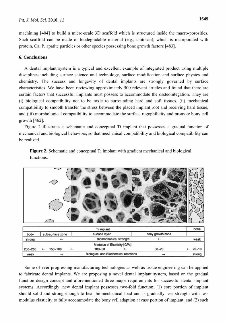
Int. J. Mol. Sci. 2010, 11
1649
machining [404] to build a micro-scale 3D scaffold which is structured inside the macro-porosities.
Such scaffold can be made of biodegradable material (e.g., chitosan), which is incorporated with
protein, Ca, P, apatite particles or other species possessing bone growth factors [483].
6. Conclusions
A dental implant system is a typical and excellent example of integrated product using multiple
disciplines including surface science and technology, surface modification and surface physics and
chemistry. The success and longevity of dental implants are strongly governed by surface
characteristics. We have been reviewing approximately 500 relevant articles and found that there are
certain factors that successful implants must possess to accommodate the ossteointegation. They are
(i) biological compatibility not to be toxic to surrounding hard and soft tissues, (ii) mechanical
compatibility to smooth transfer the stress between the placed implant root and receiving hard tissue,
and (iii) morphological compatibility to accommodate the surface rugophilicity and promote bony cell
growth [462].
Figure 2 illustrates a schematic and conceptual Ti implant that possesses a gradual function of
mechanical and biological behaviors, so that mechanical compatibility and biological compatibility can
be realized.
Figure 2. Schematic and conceptual Ti implant with gradient mechanical and biological
functions.
Some of ever-progressing manufacturing technologies as well as tissue engineering can be applied
to fabricate dental implants. We are proposing a novel dental implant system, based on the gradual
function design concept and aforementioned three major requirements for successful dental implant
systems. Accordingly, new dental implant possesses two-fold function; (1) core portion of implant
should solid and strong enough to bear biomechanical load and is gradually less strength with less
modulus elasticity to fully accommodate the bony cell adaption at case portion of implant, and (2) such

Int. J. Mol. Sci. 2010, 11
1650
case portion’s characteristics could be suitable to growing hard tissue while core protion has still same
function as those for adult patients.
References
1. Brånemark, P-I.; Zarb, G.A.; Albrektsson, T. Tissue-Integrated Prostheses; Osseointegration in
Clinical Dentistry; Quintessence: Chicago, IL, USA, 1985; pp.175-186.
2. National Institutes of Health consensus development conference statement on dental implants. J.
Dent. Educ, 1988, 52, 686-691.
3. Stillman, N.; Douglass, C.W. Developing market for dental implants. J. Am. Dent. Assoc. 1993,
124, 51-56.
4. Binon, P.P. Implants and Components. Int. J. Oral Max. Implants 2000, 15, 76-94.
5. Misch, C.E. In Contemporary Implant Dentistry; Misch, C.E., Ed.; Mosby, Inc.: St. Louis, MO,
USA, 1999; Chapter 1, p. 3.
6. Lemons, J.E. In Contemporary Implant Dentistry; Misch, C.E., Ed.; Mosby, Inc.: St. Louis, MO,
USA, 1999; Chapter 20, pp. 286-287.
7. Giray, B.; Akça, K.; İplikçioğlu, H.; Akça, E. Two-year follow-up of a patient with oligodontia
treated with implant- and tooth-supported fixed partial dentures: A case report. Int. J. Oral
Maxillofac. Implants 2003, 18, 905-911.
8. Oshida, Y. Bioscience and Bioengineering of Titanium Materials; Elsevier: Amsterdam, Holland,
2007; pp. 217-253.
9. Oshida, Y.; Tuna, E.B. In Advanced Biomaterials; Basu, B., Ed.; Wiley: Hoboken, NJ, USA,
2009; Chapter 5, pp. 143-177.
10. Anusavice, A.J. In Phillip’s Science of Dental Materials, 11th ed.; Anusavice, A.J., Ed.;
Saunders: St. Louis MO, USA, 2003; Chapter 1, pp. 3-19.
11. Moore, B.K.; Oshida, Y. Materials science and technology in dentistry, In Encyclopedic
Handbook of Biomaterilas and Bioengineering; Wise, D.L., Ed.; Marcel Dekker: Boston, MA,
USA, 1995; Chapter 48, pp. 1325-1430.
12. ISO/FDIS: 7405:2008, Dentistry – Evaluation of Biocompatibility of Medical Devices Used in
Dentistry; BSI: London, UK, 2008.
13. Long M.; Rack, H.J. Review: Titanium alloys in total joint replacement – a materials science
perspective. Biomaterials 1998, 19, 1621-1639.
14. Brunski, J.B.; Puleo, D.A.; Nanci, A. Biomaterials and biomechanics of oral and maxillofacial
implants: Current status and future developments. Int. J. Oral Maxillofac. Implants 2000, 15,
15-46.
15. Bannon, B.P.; Mild, E.E. In Titanium Alloys in Surgical Implants; Luckey, H.A.; Kubli, F., Ed.;
ASTM STP 796, American Society for Testing and Materials: West Conshohocken, PA, USA,
1983; pp. 7-15.
16. Oshida, Y.; Hashem, A.; Nishihara, T.; Yapchulay, M.V. Fractal dimension analysis of
mandibular bones: Toward a morphological compatibility of implants. J. BioMed. Mater Eng.
1994, 4, 397-407.

Int. J. Mol. Sci. 2010, 11
1651
17. Oshida, Y. Requirements for successful biofunctional implants. In The 2nd Symposium
International of Advanced Bio-Materials; Montreal, Canada, 2000; pp. 5-10.
18. Brockhurst, P.J. Dental Materials: New teritories for materials science. Au. Inst. Metals. 1980, 3,
200-210.
19. Steinmann, S.G. In Evaluation of Biomaterials; Winter, G.D., Leray, J.L., de Groot, K., Eds.;
John Wiley & Sons: New York, NY, USA, 1980; pp.1-34.
20. Kruger, J. In Corrosion and Degradation of Implant Materials; Syrett, B.C., Acharya, A., Ed.;
ASTM STP 684, American Society for Testing and Materials: West Conshohocken, PA, USA,
1979; pp. 107-127.
21. Greene, N.D. In Corrosion and Degradation of Implant Materials: Second Symposium; Farker,
A.C., Griffin, C.D., Ed.; ASTM STP 859, American Society for Testing and Materials: West
Conshohocken, PA, USA, 1983; pp. 5-10.
22. Kubaschewski, O.; Hopkins, B.E. In Oxidation of Metals and Alloys; Butterworths: London, UK,
1962; pp. 38-45.
23. Kasemo, B. Biocompatibility of titanium implants: Surface science aspects. J. Pros Dent. 1983,
49, 832-837.
24. Tomashov, N.D. Theory of Corrosion and Protection of Metals: The Science of Corrosion; The
MacMillan: New York, NY, USA, 1966.
25. Ashby, M.F.; Jones, D.R.H. Engineering Materials. An Introduction to Their Properties and
Applications; Pergamon Press: New York, NY, USA, 1980; pp. 194-200.
26. Lautenschlager, E.P.; Monaghan, P. Titanium and titanium alloys as dental materials. Int. Dent. J.
1993, 43, 245-253.
27. Coddet, C.; Chaze, A.M.; Beranger, G. Measurements of the adhesion of thermal oxide films:
Application to the oxidation of titanium. J. Mater. Sci. 1987, 22, 2969-2974.
28. Meachim, G.; Williams, D.F. Changes in nonosseous tissue adjacent to titanium implants. J.
BioMed. Mater. Res. 1973, 7, 555-572.
29. Ducheyne, P.; Williams, G.; Martens, M.; Helsen, J. In vivo metal-ion release from porous
titanium-silver material. J. BioMed. Mater. Res. 1984, 18, 293-308.
30. Sundgren, J.-E.; Bodö, P.; Lundström, I. Auger electron spectroscopic studies of the interface
between human tissue and implants of titanium and stainless steel. J. Colloid Interf. Sci. 1986,
110, 9-20.
31. Ducheyne, P.; Healy, K.E. In Surface Characterization of Biomaterials; Ratner, B.D., Ed.:
Elsevier Science: Amsterdam, Holland, 1988; pp. 175-192.
32. Fraker, A.C.; Ruff, A.W. In Titanium Science and Technology; Plenum Press: New York, NY,
USA, 1973; Volume 4, pp. 2447-2457.
33. Albreksson, T.; Hansson, H.A. An ultrastructural characterization of the interface between bone
and sputtered titanium or stainless steel surfaces. Biomaterials 1986, 7, 201-205.
34. Solar, R.J. In Corrosion and Degradation of Implant Materials; Syrett, B.D., Ed.; ASTM STP
684, American Society for Testing and Materials: West Conshohocken, PA, USA, 1979;
pp. 259-273.
35. Lim, Y.J.; Oshida, Y.; Andres, C.J.; Barco, M.T. Surface characterizations of variously treated
titanium materials. Int. J. Oral Maxillofac. Implants 2001, 16, 333-342.

Int. J. Mol. Sci. 2010, 11
1652
36. Gaydos, J.M.; Moore, M.A.; Garetto, L.P.; Oshida, Y.; Kowolik, M.J. Bisphosphonate effect on
neutrophil activation by titanium and hydroxyapatite implants. J. Dent. Res. 2000, 79, 225
(Abstract No. 890).
37. McQueen, D.; Sundgren, J.E.; Ivarsson, B.; Lundstrom, I.; Ekenstam, C.B.; Svensson, A.;
Brånemark, P.I.; Albrektsson, T. In Clinical Applications of Biomaterials; Lee, A.J.C., Ed.;
Wiley: New York, NY, USA, 1982; pp. 179-185.
38. Liedberg, B.; Ivarsson, B.; Lundstrom, I. Fourier transform infrared reflection absorption
spectroscopy (FTIR-RAS) of fibrinogen adsorbed on metal and metal oxide surfaces. J. Biochem.
Biophys. Method. 1984, 9, 233-243.
39. Hanawa, T. In The Bone-Biomaterial Interface; Davies, J.E., Ed.; Univ. of Toronto Press:
Toronto, ON, Canada, 1991; pp. 49-61.
40. Healy, K.E.; Ducheyne, P. Hydration and preferential molecular adsorption on titanium in vitro.
Biomaterials 1992, 13, 553-561.
41. Healy, K.E.; Ducheyne, P. The mechanisms of passive dissolution of titanium in a model
physiological environment. J. BioMed. Mater. Res. 1992, 26, 319-338.
42. Brånemark, P.I. In Tissue-Integrated Protheses; Brånemark, P.I., Ed.; Quintessence Pub.:
Chicago, IL, USA, 1985; pp. 63-70.
43. Kasemo, B.; Lausmaa, J. In Tissue-Integrated Protheses; Brånemark, P.I., Ed. Quintessence,
Chicago, IL, USA, 1985; pp. 108-115.
44. Skalak. R. Biomechanical considerations in osseointegrated prostheses. J. Prosthet Dent. 1983,
49, 843-848.
45. Hekimoglu, C.; Anil, N.; Cehreli, M.C. Analysis of strain around endosseous implants opposing
natural teeth or implants. J. Prosthet. Dent. 2004, 92, 441-446.
46. van Rossen, I.P.; Braak, L.H.; de Putter, C.; de Groot, K. Stress-absorbing elements in dental
implants. J. Prosthet. Dent. 1990, 64, 198-205.
47. Eriksson, C.; Lausmaa, J.; Nygren, H. Intercations between human whole blood and modified
TiO2-surfaces: Influence of surface topography and oxide thickness on leukocyte adhesion and
activation. Biomaterials 2001, 22, 1987-1996.
48. Wen, X.; Wang, X.; Zhang, N. Microsurface of metallic biomaterials: A literature review. J.
BioMed. Mater. Eng. 1996, 6, 173-189.
49. Keller, J.C.; Dougherty, W.J.; Grotendorst, G.R.; Wrightman, J.P. In vitro cell attachment to
characterized cpTi surfaces. Adhesion 1989, 28, 115-133.
50. Keller, J.C.; Wrightman, J.P.; Dougherty, W.J. Characterization of acid passivated cpTi surfaces.
J. Dent. Res. 1989, 68, 872 (Abstract).
51. Keller, J.C.; Draughn, R.A.; Wrightman, J.P.; Dougherty, W.J. Characterization of sterilized CP
titanium implant surfaces. Int. J. Oral Maxillofac. Implants 1990, 5, 360-369.
52. Schroeder, A.; van der Zypen, E.; Stich, H.; Sutter, F. The reactions of bone, connective tissue
and epithelium to endosteal implants with titanium sprayed surfaces. J. Maxillofac. Surg. 1981, 9,
15-25.
53. Rich, A.; Harris, A.K. Anomalous preferences of cultured macrophages for hydrophobic and
roughended substrata. J. Cell Sci. 1981, 50, 1-7.

Int. J. Mol. Sci. 2010, 11
1653
54. Buser, D.; Schenk, R.K.; Steinemann, S.; Fiorellinni, J.P.; Fox, C.H.; Stich, H. Influence of
surface characteristics on bone integration of titanium implants. A histomorphometric study in
miniature pigs. J. BioMed. Mater. Res. 1991, 25, 889-902.
55. Buser, D.; Nydegger, T.; Oxland, T.; Cochran, D.L.; Schenk, R.K.; Hirt, H.P.; Sneitivy, D.; Nolte,
L.P. Interface shear strength of titanium implants with a sandblasted and acid-etched surface: A
biomechanically study in the maxilla of miniature pigs. J. BioMed. Mater. Res. 1999, 45, 75-83.
56. Murray, D.W.; Rae, T.; Rushton, N. The influence of the surface energy and roughness of
implants on bone resorption. J. Bone JoInt. Surg. 1989, 71B, 632-637.
57. Cherhoudi, B.; Gould, T.R.; Brunette, D.M. Effects of a grooved epoxy substratum on epithelial
behavior in vivo and in vitro. J. BioMed. Mater. Res. 1988, 22, 459-477.
58. von Recum, A.F. In Clinical Implant Materials; Heimke, G., Soltesz, U., Lee, A.J.C., EdS.;
Elseveier Science Pub.: Amstrdam, The Netherlands, 1990; pp. 297-302.
59. Deligianni, D.D.; Katsala, N.; Ladas, S.; Sotiropoulou, D.; Amedee, J.; Missirlis, Y.F. Effect of
surface roughness of the titanium alloy Ti-6Al-4V on human bone marrow cell response and on
protein adsorption. Biomaterials 2001, 22, 1241-1251.
60. Yang, Y.; Cavin, R.; Ong, L.J. Protein adsorption on titanium surfaces and their effect on
osteoblast attachment. J. BioMed. Mater. Res. A 2003, 67, 344-349.
61. Albrektsson, T. Direct bone anchorage of dental implants. J. Prosth. Dent. 1983, 50, 255-261.
62. Kasemo, B.; Lausmaa, J. Surfaces science aspects on inorganic Biomaterials CRC Crit Rev.
Biocomp. 1986; 2, 335-380.
63. Schenk, R.K.; Buser, D. Osseointegration: A reality. Periodontology 1998, 17, 22-35.
64. Masuda, T.; Yliheikkaila, P.K.; Fleton, D.A.; Cooper, L.F. Generalizations regarding the process
and phenomenon of osseointegration. Part I. In vivo studies. Int. J. Oral Maxillofac. Implants
1998, 13, 17-29.
65. Larsson, C.; Esposito, M.; Liao, H.; Thomsen, P. In Titanium in Medicine: Materials Science,
Surface Science, Engineering, Biological Responsese, and Medical Applications; Brunette, D.M.,
Tengvall, P., Textor, M., Thomsen, P., Eds.; Springer: New York, NY, USA, 2001; pp. 587-648.
66. Oshida, Y.; Sachdeva, R.; Miyazaki, S. Microanalytical characterization and surface
modification of NiTi orthodontic archwires. J. BioMed. Mater. Eng. 1991, 2, 51-69.
67. Zinger, O.; Anselme, K.; Denzer, A.; Habersetzer, P.; Wieland, M.; Jeanfils, J.; Hardouin, P.;
Landolt, D. Time-dependent morphology and adhesion of osteoblastic cells on titanium model
surfaces featuring scale-resolved topography. Biomaterials 2004, 25, 2695-2711.
68. Ratner, B.D. Surface characterization of biomaterials by electron spectroscopy for chemical
analysis. Ann. BioMed. Eng. 1983, 11, 313-336.
69. Baro, A.M.; Garcia, N.; Miranda, R.; Vázquez, L.; Aparicio, C.; Olivé, J.;Lausmaa, J.
Characterization of surface roughness in titanium dental implants measured with scanning
tunneling microscopy at atmospheric pressure. Biomaterials 1986, 7, 463-466.
70. Moroni, A.; Caja, V.L.; Egger, E.L.; Trinchese, L.; Chao, E.Y. Histomorphometry of
hydroxyapatite coated and uncoated porous titanium bone implants. Biomaterials 1994, 15,
926-930.
71. Keller, J.C.; Young, F.A.; Natiella, J.R. Quantitative bone remolding resulting from the use of
porous dental implants. J. BioMed. Mater. Res. 1987, 21, 305-319.

Int. J. Mol. Sci. 2010, 11
1654
72. Pilliar, R.M.; Deporter, D.A.; Watson, P.A.; Valiquette, N. Dental implant design-effect on bone
remodeling. J. BioMed. Mater. Res. 1991, 25, 467-483.
73. Gilbert, J.L.; Berkery, C.A. Electrochemical reaction to mechanical disruption of titanium oxide
films. J. Dent. Res. 1995, 74, 92-96.
74. Deporter, D.; Watson, P.; Pharoah, M.; Levy, D.; Todescan, R. Five-to six-year results of a
prospective clinical trial using the ENDOPORE dental implant and a mandibular overdenture.
Clin Oral Implants Res. 1999, 10, 95-102.
75. Palmer, R.M.; Plamer, P.J.; Smith, B.J. A 5-year prospective study of astra single tooth implants.
Clin Oral Implants Res. 2000, 11, 179-182.
76. Testori, T.; Wiseman, L.; Woolfe, S.; Porter, S.S. A prospective multicenter clinical study of the
osseitite implant: Four-years interim report. Int. J. Oral Maxillofac. Implants 2001, 16, 193-200.
77. Sul, Y.-T. The significance of the surface properties of oxidized titanium to the bone response:
Special emphasis on potential biochemical bonding of oxidized titanium implant. Biomaterials
2003, 24, 3893-3907.
78. Ishizawa, H.; Fujiino, M.; Ogino, M. Mechanical and histological investigation of
hydrothermally treated and untreated anodic titanium oxide films containing Ca and P. J. BioMed.
Mater. Res. 1995, 29, 1459-1468.
79. Larsson, C.; Emanuelsson, L.; Thomsen, P.; Ericson, L.; Aronsson, B.; Rodahl. M.; Kasemo, B.;
Lausmaa, J. Bone response to surface-modified titanium implants: Studies on the tissue response
after one year to machined and electropolished implants with different oxide thicknesses. J.
Mater Sci.: Mater Med. 1997, 8, 721-729.
80. Skripitz, R.; Aspenberg, P. Tensile bond between bone and titanium. Acta Orthop Scand. 1998, 6,
2-6.
81. Fini, M.; Cigada, A.; Rondelli, G.; Chiesa, R.; Giardino, R.; Giavaresi, G.; Aldini, N.; Torricelli,
P.; Vicentini, B. In vitro and vivo behaviour of Ca and P-enriched anodized titanium.
Biomaterials 1999, 20, 1587-1594.
82. Henry, P.; Tan, A.E.; Allan, B.P. Removal torque comparison of TiUnite and turned implants in
the Greyhound dog mandible. Appl. Osseointegr. Res. 2000, 1, 15-17.
83. Sul, Y.-T.; Johansson, C.B.; Jeong, Y.; Röser, K.; Wennerberg, A.; Albreksson. T. Oxidized
implants and their influence on the bone response. J. Mater Sci.: Mater Med. 2001, 12,
1025-1031.
84. Sul, Y.-T.; Johansson, C.B.; Jeong, Y.; Wennerberg, A.; Albrektson, T. Resonance frequency
and removal torque analysis of implants with turned and anodized surface oxide. Clin Oral
Implants Res. 2002, 13, 252-259.
85. Sul, Y.-T.; Johansson, C.B.; Albreksson, T. Oxidized titanium screws coated with calcium ions
and their performance in rabbit bone. Int. J. Oral Maxillofac. Implants 2002, 17, 625-634.
86. Oshida, Y. Surface science and technology – Titanium dental implant systems. J. Soc. Titanium
Alloys Dent. 2007, 5, 52-53.
87. Elias, C.N.; Oshida, Y.; Lima, J.H.C.; Muller, C.A. Relationship between surface properties
(roughness, wettability and morphology) of titanium and dental implant torque. J. Mech. Behav.
Biomed. Mater. 2008, 1, 234-242.

Int. J. Mol. Sci. 2010, 11
1655
88. Lausmaa, G.J. Chemical composition and morphology of titanium surface oxides. Mat. Res. Soc.
Symp. Proc. 1986, 55, 351-359.
89. Albrektsson, T.; Jacobsson, M. Bone-metal interface in osseointegration. J. Prosthet. Dent. 1987,
57, 5-10.
90. Lauterbur, P.C. Image formation by induced local interactions: Example employing nuclear
magnetic resonance. Nature 1973, 242, 190.
91. Shellock, F.G.; Crues, J.V. High field strength MR imaging and metallic biomedical implants:
An ex vivo evaluation of deflection forces. Am. J. Roentgenol. 1988, 151, 389-392.
92. Shellock, F.G.; Morisoli, S.; Kanal, E. MR Procedures and Biomedical Implants, Materials, and
Devices: Update. Radiology 1993, 189, 587-599.
93. Shellock, F.G.; Mink, J.H.; Curtin, S.; Friesman, M.J. MR imaging and metallic implants for
anterior cruciate ligament reconstruction: Assessment of ferromagnetism and artifact. J. Magn.
Reson. Imaging 1992, 2, 225-228.
94. Shellock, F.G. Biomedical implants and devices: Assessment of magnetic field interactions with
a 3.0 tesla MR system. J. Magn. Reson. Imaging 2002, 16, 721-732.
95. Shellock, F.G.; Fieno, D.S.; Thompson, L.J.; Talavage, T.M.; Berman, D.S. Cardiac pacemaker:
In vitro assessment at 1.5T. Am. Heart J. 2006, 151, 436-443.
96. Shellock, F.G.; Crues, J. High field strength MR imaging and metallic biomedical implants: An
in vitro evaluation of deflection forces and temperature changes induced in large prostheses.
Radiology 1987, 165, 150-152.
97. Oshida, Y. Bioscience and Bioengineering of Titanium Materials; Elsevier: Amstredam, The
Netherlands, 2007; pp. 313-379.
98. Kasemo, B.; Lausmaa J. Biomaterials and implant surfaces: A surface science approach. Int. J.
Oral Maxillofac. Implants 1988, 3, 247-259.
99. Baier, R.E.; Meyer AE. Implant surface preparation. Int. J. Oral Maxillofac. Implants 1988, 3,
9-20.
100. Reitz, W.E. The eighth international conference on surface modification technology. J. Metal.
1995, 47, 14-16.
101. Smith, D.C.; Pilliar, R.M.; Chernecky, R. Dental implant materials. I. Some effects of preparative
procesdures on surface topography. J. BioMed. Mater. Res. 1991, 25, 1045-1068.
102. Buddy, D.; Ratner, B.; Thomas, J.L. Biomaterial surfaces. J. BioMed. Mater. Res. 1987, 21,
59-89.
103. Cooper, L.F. A role for surface topography in creating and maintaining bone at titanium
endosseous implants. J. Prosthet. Dent. 2000, 84, 522-534.
104. Bachle, M.; Kohal, R.J. A systematic review of the influence of different titanium surfaces on
proliferation, differentiation and protein synthesis of osteoblast-like MG63 cells. Clin Oral
Implants Res. 2004, 15, 683-692.
105. Rompen, E.; Domken, O.; Degidi, M.; Farias Pontes, A.E.; Piattelli, A. The effect of material
characteristics, of surface topography and of implant components and connections on soft tissue
integration: A literature review. Clin Oral Implants Res. 2006, 17, S55-S67.

Int. J. Mol. Sci. 2010, 11
1656
106. Boyan, B.D.; Lossdorfer, S.; Wang, L.; Zhao, G.; Lohmann, C.H.; Cochran, D.L.; Schwartz, Z.
Osteoblasts generate an osteogenic microenvironment when grown on surfaces with rough
microtopographies. Eur. Cell Mater. 2003, 6, 22-27.
107. Lossdorfer, S.; Schwartz, Z.; Wang, L.; Lohmann, C.H.; Turner, J.D.; Wieland, M.; Cochran,
D.L.; Boyan, B.D. Microrough implant surface topographies increase osteogenesis by reducing
osteoclast formation and activity. J. BioMed. Mater. Res. A 2004, 70, 361-369.
108. Mustafa, K.; Wennerberg, A.; Wroblewski, J.; Hultenby, K.; Lopez, B.S.; Arvidson, K.
Determining optimal surface roughness of TiO2 blasted titanium implant Material for attachment,
proliferation and differentiation of cells derived from human mandibular alveolar bone. Clin Oral
Implants Res. 2001, 12, 515-525.
109. Castellani, R.; de Ruijter, A.; Renggli, H.; Jansen, J. Response of rat bone marrow cells to
differently roughened titanium discs. Clin Oral Implants Res. 1999, 10, 369-378.
110. Diniz, M.G.; Soares, G.A.; Coelho, M.J.; Fernandes, M.H. Surface topography modulates the
osteogenesis in human bone marrow cell cultures grown on titanium samples prepared by a
combination of mechanical and acid treatments. J. Mater. Sci. Mater. Med. 2002, 13, 421-432.
111. Anselme, K.; Bigerelle, M.; Noel, B.; Dufresne, E.; Judas, D.; Iost, A.; Hardouin, P. Qualitative
and quantitative study of human osteoblast adhesion on materials with various surface
roughnesses. J. Biomed. Mater. Res. 2000, 49, 155-166.
112. Sader, M.S.; Balduino, A.; Soares, G.A.; Borojevic, R. Effect of three distinct treatments of
titanium surface on osteoblast attachment, proliferation, and differentiation. Clin Oral Implants
Res. 2005, 16, 667-675.
113. Citeau, A.; Guicheux, J.; Vinatier, C.; Layrolle, P.; Nguyen, T.P.; Pilet, P.; Daculsi, G. In vitro
biological effects of titanium rough surface obtained by calcium phosphate grid blasting.
Biomaterials 2005, 26, 157-165.
114. Oshida, Y.; Daly, J. Fatigue damage evaluation of shot peened high strength aluminum alloy. In
Surface Engineering; Meguid, S.A., Ed.; Elsevier Applied: New York, NY, USA, 1990;
pp. 404-416.
115. Oshida, Y. Bioscience and Bioengineering of Titanium Materials; Elsevier: Amstredam, The
Netherlands, 2007; pp. 179-182.
116. Piattelli, A.; Scarano, A.; Coriglano, M.; Piattelli, M. Presence of multinucleated giant cells
around machined, sandblasted and plasma-sprayed titanium implants: A histological and
histochemical time-course study in rabbit. Biomaterials 1996, 17, 2053-2058.
117. Kubo, M.; Fijishima, A.; Hotta, Y.; Kunii, J.; Shimakura, Y.; Shimidzu, T.; Kawawa, T.;
Miyazaki, T. Production method of resin-bonded titanium crown by CAD/CAM. Proc. Soc.
Symp. Res. 2006, 929, 42
118. Miyakawa, O.; Watababe, K.; Okawa, S.; Shiokawa, N.; Kobayashi, M.; Tamura, H. Grinding of
titanium, Part 1: Commercial and experimental wheels made of silicon carbide abrasives. Dent.
Mater. J. 1990, 9, 30-41.
119. Miyakawa, O.; Watanabe, K.; Okawa, S.; Nakano, S.; Shiokawa, N.; Kobayashi, M.; Tamura, H.
Grinding of titanium, Part 2: Commercial vitrified wheels made of alumina abrasives. Dent.
Mater. J. 1990, 9, 42-52.

Int. J. Mol. Sci. 2010, 11
1657
120. Miyakawa, O.; Watanabe, K.; Okawa, S.; Kanatani, M.; Nakano, S.; Kobayashi, M. Surface
contamination of titanium by abrading treatment. Dent. Mater. J. 1996, 15, 11-21.
121. Mandelbrot, B.B. The Fractal Geometry of Nature; Freeman: New York, NY, USA, 1983; p. 34.
122. Chesters, S.; Wen, H.Y.; Lundin, M.; Kasper, G. Fractal-based characterization of surface texture.
Appl. Surf. Sci. 1989, 40, 185-192.
123. Sayles, R.S.; Thomas, T.R. Surface topography as a non-stationary random process. Nature 1978,
271, 431-434.
124. Oshida, Y.; Munoz, C.A.; Winkler, M.M.; Hashem, A.; Ito, M. Fractal dimension analysis of
aluminum oxide particle for sandblasting dental use. J. Biomed. Mater. Eng. 1993, 3, 117-126.
125. Miyakawa, O.; Okawa, S.; Kobayashi, M.; Uematsu, K. Surface contamination of titanium by
abrading treatment. Dent. Jpn. 1998, 34, 90-96.
126. Mustafa, K.; Lopez, B.S.; Hultenby, K.; Arvison, K. Attachment of HGF of titanium surfaces
blasted with TiO2 particles. J. Dent. Res. 1997, 76, 85 (Abstract No.573).
127. Johansson, C.B.; Albrektsson, T.; Thomsen, P.; Sonnerby, I.; Lodding, A.; Odelius, H. Tissue
reactions to titanium-6aluminum-4vanadium alloy. Eur. J. Exp. Musculoskel. Res. 1992, 1,
161-169.
128. Wennerberg, A.; Albrektsson, T.; Johnsson, C.; Andersson, B. Experimental study of turned and
grit-blasted screw-shaped implants with special emphasis on effects of blasting material and
surface topography. Biomaterials 1996, 17, 15-22.
129. Wang, C-S. Surface modification of titanium for enhancing titabium-pocelain bond strength.
Master Degree Thesis, Indiana University: Bloomington, IN, USA, 2004; pp. 97-115.
130. Peutzfeld, A.; Asmussen, E. Distortion of alloy by sandblasting. Am. J. Dent. 1996, 9, 65-66.
131. Peutzfeld, A.; Asmussen, E. Distortion of alloys caused by sandblasting. J. Dent. Res. 1996, 75,
259 (Abstract No. 1162).
132. Shot Peening Applications, 7th ed.; Metal Improvement Company, Inc.: Vernon, CA, USA,
1990.
133. DeWald, A.T.; Rankin, J.E.; Hill, M.R.; Lee, M.J.; Chen, H-L. Assessment of tensile residual
stress mitigation in Alloy 22 welds due to laser peening. J. Eng. Mater. Technol. Trans. ASME.
2004, 126, 81-89.
134. Fairland, B.P.; Wilcox, B.A.; Gallagher, W.J.; Williams, D. Laser shock-induced microstructural
and mechanical property changes in 7075 aluminum. J. Appl. Phys. 1972, 43, 3893-3895.
135. Fairland, B.P.; Clauer, A.H. Laser generation of high amplitude stress waves in materials. J. Appl.
Phys. 1979, 50, 1497-1502.
136. Dane, C.B.; Hackel, L.A.; Daly, J.; Harrison, J. High power laser for peening of metals enabling
production technology. Mater. Manuf. Proc. 2000, 15, 81-96.
137. Cho, S.-A.; Jung, S.-K. A removal torque of the laser-treated titanium implants in rabbit tibia.
Biomaterials 2003, 24, 4859-4863.
138. Gaggl, A.; Schultes, G.; Muller, W.D.; Karcher, H. Scanning electron microscopical analysis of
laser-treated titanium implants surfaces–a comparative study. Biomaterials 2000, 21, 1067-1073.
139. Hansson, S.; Hansson, K.N. The effect of limited lateral resolution in the measurement of
implant surface roughness: A computer simulation. J. Biomed. Mater. Res. A 2005, 75, 472-477.
Int. J. Mol. Sci. 2010, 11
1658
140. Endo, K. Chemical modification of metallic implant surfaces with biofunctional proteins (Part 1)
Molecular structure and biological activity of a modified NiTi alloy surface. Dent. Mater. J. 1995,
14, 185-198.
141. Browne, M.; Gregson, P.J. Surface modification of titanium alloy implants. Biomaterials 1994,
15, 894-898.
142. Krozer, A.; Hall, J.; Ericsson, I. Chemical treatment of machined titanium surfaces. Clin Oral
Impl. Res. 1999, 10, 204-211.
143. Rupp, F.; Scheideler, L.; Rehbein, D.; Axmann, D.; Geis-Gerstorfer, J. Roughness induced
dynamic changes of wettability of aid etched titanium implant modifications. Biomaterials 2004,
25, 1429-1438.
144. MacDonald, D.E.; Rapuano, B.E.; Deo, N.; Stranick, M.; Somasundaran, P.; Boskey, A.L.
Thermal and chemical modification of titanium-aluminum-vanadium implant materials: Effects
of surface properties, glycoprotein adsorption, and MG63 cell attachment. Biomaterials 2004, 25,
3135-3146.
145. Li, L.-H.; Kong, Y.-M.; Kim, H-W.; Kim, Y.-W.; Kim, H.-E.; Heo, S.-J.; Koak, J.-Y. Improved
biological performance of Ti implants due to surface modification by micro-arc oxidation.
Biomaterials 2004, 15, 1867-1875.
146. Xie, Y.; Liu, X.; Huang, A.; Ding, C.; Chu, P.K. Improvement of surface bioactivity on titanium
by water and hydrogen plasma immersion ion implantation. Biomaterials 2005, 26, 6129-6135.
147. Rohanizadeh, R.; Al-Sadeq, M.; LeGeros, R.Z. Preparation of different forms of titanium oxide
on titanium surface: Effects on apatite deposition. J. BioMed. Mater. Res. A 2004, 71, 343-352.
148. Demri, B.; Hage-Ali, M.; Moritz, M.; Muster, D. Surface characterization of C/Ti-6Al-4V
coating treated with ion beam. Biomaterials 1997, 18, 305-310.
149. Poon, R.W.Y.; Yeung, K.W.K.; Liu, X.Y.; Chu, P.K.; Chung, C.Y.; Lu, W.W.; Cheung, K.M.C.;
Chan, D. Carbon plasma immersion ion implantation of nickel- titanium shape memory alloys.
Biomaterials 2005, 26, 2265-2272.
150. Schrooten, J.; Helsen, J.A. Adhesion of bioactive glass coating to Ti6Al4V oral implant.
Biomaterials 2000, 21, 1461-1419.
151. Saiz, E.; Goldman, M.; Gomez-Vega, J.M.; Tomsia, A.P.; Marshall, G.W.; Marshall, S.J. In vitro
behavior of silicate glass coatings on Ti6Al4V. Biomaterials 2002, 23, 3749-3756.
152. Lee, J.H.; Ryu, H.-S.; Lee, D.-S.; Hong, K.S.; Chang, B.-S.; Lee, C.-K. Bio-mechanical and
histomorphometric study on the bone-screw interface of bioactive ceramic-coated titanium
screws. Biomaterials 2005, 26, 3249-3257.
153. Chu, K.T.; Oshida, Y.; Hancock, E.B.; Kowolik, M.J.; Barco, T.M.; Zunt, S.L.
Hydroxyapatite/PMMA composites as bone cements. J. BioMed. Mat. Eng. 2004, 14, 87-105.
154. Liu, D.M. Fabrication and characterization of porous hydroxyapatite granules. Biomatrials 1996,
17, 1955-1957.
155. Luo, P.; Nieh, T.G. Preparing hydroxyapatite powders with controlled morphology. Biomaterials
1996, 17, 1959-1964.
156. Porter, A.E.; Taakm, P.; Hobbs, L.W.; Coathup, M.J.; Blunn, G.W.; Spector, M. Bone bonding to
hydroxyapatite and titanium surfaces on femoral stems retrieved from human subjects at autopsy.
Biomaterials 2004, 25, 5199-5208.
Int. J. Mol. Sci. 2010, 11
1659
157. Souto, R.M.; Lemus, M.M.; Reis, R.L. Electrochemical behavior of different preparations of
plasma-sprayed hydroxyapatite coatings on Ti6Al4V substrate. J. BioMed. Mater. Res. A 2004,
70, 59-65.
158. Filiaggi, M.J.; Coombs, N.A.; Pilliar, R.M. Characterization of the interface in the
plasma-sprayed HA coating/Ti-6Al-4V implant system. J. BioMed. Mater. Res. 1991, 25,
1211-1229.
159. Yang, Y.C.; Chang, E.; Lee, S.T. Mechanical properties and Young's modulus of plasma-sprayed
hydroxyapatite coating on Ti substrate in simulated body fluid. J. BioMed. Mater. Res. A 2003,
67, 886-899.
160. Yang, Y.C.; Ong, J.L. Bond strength, compositional, and structural properties of hydroxyapatite
coating on Ti, ZrO2-coated Ti, and TPS-coated Ti substrate. J. BioMed. Mater. Res. A 2003, 4,
509-516.
161. Yang, Y.C.; Kim, K-H.; Agrawal, C.M.; Ong, J.L. Interaction of hydroxyapatite-titanium at
elevated temperature in vacuum environment. Biomaterials 2004, 25, 2927-2932.
162. Brossa, F.; Cigada, A.; Chiesa, R.; Paracchini, L.; Consonni, C. Adhesion properties of plasma
sprayed hydroxylapatite coating for orthopaedic prostheses. J. BioMed. Mater. Eng. 1993, 3,
127-136.
163. Lee, E.-J.; Lee, S.-H.; Kim, H.-W.; Kong, Y.-M.; Kim, H-E. Fluoridated apatite coatings on
titanium obtained by electron-beam deposition. Biomaterials 2005, 26, 3843-3851.
164. Kim, H.-W.; Kim, H.-E.; Knowles, L.C. Fluor-hydroxyapatite sol–gel coating on titanium
substrate for hard tissue implants. Biomaterials 2004, 25, 3351-3358.
165. Filiaggi, M.J.; Pilliar, R.M.; Coombs, N.A. Post-plasma-spraying heat treatment of the HA
coating/Ti-6Al-4V implant system. J. BioMed. Mater. Res. 1993, 27, 191-198.
166. Lynn, A.K.; DuQuesnay, D.L. Hydroxyapatite-coated Ti-6Al-4V Part 2: The effect of
post-deposition heat treatment at low temperatures. Biomaterials 2002, 23, 1947-1953.
167. Yang, Y.C.; Chang, E. Influence of residual stress on bonding strength and fracture of
plasma-sprayed hydroxyapatite coatings on Ti-6Al-4V substrate. Biomaterials 2001, 22,
1827-1836.
168. Mano, T.; Ueyama, Y.; Ishikawa, K.; Matsumura, T.; Suzuki, K. Initial tissue response to a
titanium implant coated with apatite at room temperature using a blast coating method.
Biomaterials 2002, 23, 1931-1936.
169. Moritz, N.; Jokinen, M.; Peltola, T.; Areva, S.; Yli-Urpo, A. Local induction of calcium
phosphate formation on TiO2 coatings on titanium via surface treatment with a CO2 laser. J.
BioMed. Mater. Res. A 2003, 65, 9-16.
170. Han, Y.; Xu, K. Photoexcited formation of bone apatite-like coatings on micro-arc oxidized
titanium. J. BioMed. Mater. Res. A 2004, 71, 608-614.
171. Kim, H.-W.; Kim, H.-E.; Salih, V.; Knowles, J.C. Sol-gel-modified titanium with
hydroxyl-apatite thin films and effect on osteoblast-like cell responses. J. BioMed. Mater. Res. A
2005, 74, 294-305.
172. Kim, H.-W.; Koh, Y.-H.; Li, L.-H.; Lee, S.; Kim, H.-E. Hydroxyapatite coating on titanium
substrate with titania buffer layer processed by sol-gel method. Biomaterials 2004, 25,
2533-2538.
Int. J. Mol. Sci. 2010, 11
1660
173. Ergun, C.; Doremus, R.; Lanford, W. Hydroxylapatite and titanium: Interfacial reactions. J.
BioMed. Mater. Res. A 2003, 65, 336-343.
174. Hayashi, K.; Mashima, T.; Uenoyama, K. The effect of hydroxyapatite coating on bony ingrowth
into grooved titanium implants. Biomaterials 1999, 20, 111-119.
175. Hayashi, K.; Inadome, T.; Tsumura, H.; Nakashima, Y.; Sugioka, Y. Effect of surface roughness
of hydroxyapatite-coated titanium on the bone-implant interface shear strength. Biomaterials
1994, 15, 1187-1191.
176. Moroni, A.; Caja, V.L.; Egger, E.L.; Trinchese, L.; Chao, E.Y.S. Histomorphometry of
hydroxyapatite coated and uncoated porous titanium bone implant. Biomaterials 1994, 15,
926-930.
177. Thomas, K.A.; Cook, S.D.; Haddad, R.J.; Kay, J.F.; Jarcho. M. Biologicla response to
hydroxyapatite-coated titanium hips. J. Arthrop. 1989, 4, 43-53.
178. Jansen, J.A.; van de Waerden, J.P.C.M.; Wolke, J.G.C.; de Groot, K. Histologic evaluation of the
osseo adaptation to titanium and hydroxyapatite-coated titanium implants. J. BioMed. Mater. Res.
1991, 25, 973-989.
179. Gottlander, M.; Albrektsson, T. Histomorphometric studies of hydroxylapatite-coated and
uncoated cp titanium threaded implants in bone. Int. J. Oral Maxillofac. Implants 1991, 6,
399-404.
180. Schreurs, B.W.; Huiskes, R.; Buma, P.; Slooff, T.J.J.H. Biomechanicla and histological
evaluation of a hydroxyapatite-coated titanium femoral stem fixed with an intramedullary
morsellized bone grafting technique: An animal experiment on goats. Biomaterials 1996, 17,
1177-1186.
181. Cabrini, M.; Cigada, A.; Rondelli, G.; Vicentini, B. Effect of different surface finishing and of
hydroxyapatite coatings on passive and corrosion current of Ti6Al4V alloy in simulated
physiological solution. Biomaterials 1997, 18, 783-787.
182. Cook, S.D.; Thomas, K.A.; Kay, J.; Jarcho, M. Hydroxyapatite-coated titanium for orthopedic
implant applications. Clin Orthop. 1988, 186, 225-243.
183. Yang, Y.C.; Chang, E.; Hwang, B.H.; Lee, S.Y. Biaxial residual stress states of plasma-sprayed
hydroxyapatite coatings on titanium alloy substrate. Biomaterials 2000, 12, 1327-1337.
184. Mimura, K.; Watanabe, K.; Okawa, S.; Kobayashi, M.; Miyakawa, O. Morpho- logical and
chemical characterizations of the interface of a hydroxyapatite-coated implant. Dent. Mater. J.
2004, 23, 353-360.
185. Jonášová, L.; Müller, F.A.; Helebrant, A.; Strnad, J.; Greil, P. Hydroxyapatite formation on
alkali-treated titanium with different content of Na+ in the surface layer. Biomaterials 2002, 23,
3095-3101.
186. Jonášová, L.; Müller, F.A.; Helebrant, A.; Strnad, J.; Greil, P. Biomimetic apatite formation on
chemically treated titanium. Biomaterials 2004, 25, 1187-1194.
187. Wang, C.X.; Wang, M.; Zhou, X. Nucleation and growth of apatite on chemically treated
titanium alloy: An electrochemical impedance spectroscopy study. Biomaterials 2003, 24,
3069-3077.
188. Lu, X.; Leng, Y. TEM study of calcium phosphate precipiotation on bioactive titanium surfaces.
Biomaterials 2004, 25, 1779-1786.

Int. J. Mol. Sci. 2010, 11
1661
189. Hanawa, T.; Ota, M. Calcium phosphate naturally formed on titanium in electrolyte solution.
Biomaterials 1991, 12, 767-774.
190. Barrère, F.; van der Valk, C.M.; Dalmeijer, R.A.J.; van Blitterswijk, C.A.; de Groot, K.; Layrolle,
P. In vitro and in vivo degradation of biomimetic octacalcium phosphate and carbonate apatite
coatings on titanium implants. J. BioMed. Mater. Res. A 2003, 64,378-387.
191. Barrère, F.; Snel, M.M.E.; van Blitterswijk, C.A.; de Groot, K.; Layrolle, P. Nano- scale study of
the nucleation and growth of calcium phosphate coating on titanium implants. Biomaterials 2004,
25, 2901-2910.
192. Li, S.J.; Yang, R.; Niinomi, M.; Hao, Y.L.; Cui, Y.Y. Formation and growth ofcalcium
phosphate on the surface of oxidized Ti-29Nb-13Ta-4.6Zr alloy. Biomaterials 2004, 25,
2525-2532.
193. Bogdanski, D.; Esenwien, S.A.; Prymak, O.; Epple, M.; Muhr, G.; Köller, M. Inhibition of PMN
apotosis after adherence to dip-coated calcium phosphate surfaces on a NiTi shape memory alloy.
Biomaterials 2004, 25, 4627-4623.
194. Kim, C.S.; Ducheyne, P. Compositional variations in the suface and interface of calcium
phosphate ceramic coatings on Ti and Ti-6Al-4V due to sintering and immersion. Biomaterials
1991, 12, 461-469.
195. Ducheyne, P.; Bianco, P.D.; Kim, C. Bone tissue growth enhancement of calcium phosphate
coatings on porous titanium alloys: Effect of shielding metal dissolution product. Biomaterials
1992, 13, 617-624.
196. Krupa, D.; Baszkiewicz, J.; Kozubowski, J.A.; Barcz, A.; Sobczak, J.W.; Biliński, A.;
Lewandowska-Szumieł, M.; Rajchel, B. Effect of dual ion implantation of calcium and
phosphorus on the properties of titanium. Biomaterials 2005, 26, 2847-2856.
197. Jansen, J.A.; Hulshoff, J.E.; van Dijk, K. Biological evaluation of thin calcium- phosphate (Ca-P)
coatings. J. Dent. Res. 1997, 76, 282 (Abstract No. 2150).
198. Hayakawa, T.; Yoshinari, M.; Kiba, H.; Yamamoto, H.; Nemoto, K.; Jansen, J.A. Trabeular bone
response to surface roughened and calcium phosphate (Ca-P) coated titanium implants.
Biomaterials 2002, 23, 1025-1031.
199. Zhang, Q.; Leng, Y.; Xin, R. A comparative study of electrochemical deposition and biomimetic
deposition of calcium phosphate on porous titanium. Biomaterials 2005, 26, 2857-2865.
200. Heimann, R.B.; Wirth. R. Formation and transformation of amorphous calcium phosphates on
titanium alloy surfaces during atmospheric plasma spraying and their subsequent in vitro
performance. Biomaterials 2006, 27, 823-831.
201. Ban, S.; Maruno, S.; Iwata, H.; Itoh, H. Calcium phosphate precipitation on the surface HA-G-Ti
composite under physiologic conditions. J. BioMed. Mater. Res. 1994, 28, 65-71.
202. Lucas, L.C.; Ong, J.L. Post deposition heat treatments of Ca-P coating. J. Dent. Res. 1993, 71,
228 (Abstract No. 998).
203. Gan, L.; Wang, J.; Pilliar, R.M. Evaluating interface strength of calcium phosphate
sol-gel-derived thin films to Ti6Al4 V substrate. Biomaterials 2005, 26, 189-196.
204. Choi, J.; Bogdanski, D.; Köller, M.; Esenwein, S.A.; Müller, D.; Epple, M. Calcium phosphate
coating of nickel-titanium shape-memory alloys, coating procedure and adherence of leukocytes
and platelets. Biomaterials 2003, 24, 3689-3696.
Int. J. Mol. Sci. 2010, 11
1662
205. Fend, B.; Weng, J.; Yangm, B.C.; Qu, S.X.; Zhang, X.D. Characterization of titanim surfaces
with calcium and phosphate and osteoblast adhesion. Biomaterials 2004, 25, 3421-3428.
206. Watanabe, K.; Hashimoto, A.; Nomura, S.; Endo, M.M.; Okawa, S.; Kanatani, M.; Nakano, S.;
Kobayashi, M.; Miyakawa, O. Surface properties of dental implants (Part 2) Surface analysis of
the titanium implant extracted from a rat bone. J. Jpn Dent. Mater. 2004, 23, 329-334.
207. Kim, H.-W.; Lee, E.-J.; Jun, I.-K.; Kim, H.-E. On the feasibility of phosphate glass and
hydroxyapatite engineered coating on titanium. J. BioMed. Mater. Res. A 2005, 75, 656-667.
208. Maxian, S.H.; Zawadsky, J.P.; Dunn, M.G. Mechanical and histological evaluation of amorphous
calcium phosphate and poorly crystallized hydroxyapatite coatings on titaniuim implants. J.
BioMed. Mater. Res. 1993, 27, 717-728.
209. Khor, K.A.; Gu, Y.W.; Pan, D.; Cheang, P. Microstructure and mechanical properties of plasma
sprayed HA/YSZ/Ti–6Al–4V composite coatings. Biomaterials 2004, 25, 4009-4017.
210. Gu, Y.W.; Khor, K.A.; Pan, D.; Cheang, P. Activity of plasma sprayued yttria stabilized zirconia
reinforced hydroxyapatite/Ti-6Al-4V composite coatings in simulated body fluid. Biomaterials
2004, 25, 3177-3185.
211. Chou, B.Y.; Chang, E.; Yao, S.Y.; Chen, J.M. Phase transformation during plasma spraying of
hydroxyapatite-19wt%-zirconia composite coating. J. Am. CerAm. Soc. 2002. 85, 661-669.
212. Kasuga, T.; Yoshida, M.; Ikushima, A.J.; Tuchiya, M.; Kusakari, H. Stability of
zirconia-toughended bioactive glass-ceramics - invivo study using dogs. J. Mater Sci.: Mater.
Med.1993, 4, 36-39.
213. Takagi, M.; Mochida, M.; Uchida, N.; Saito, K.; Uematsu, K.; Filter cake forming and hot
isostatic pressing for TZP-dispressed hydroxyapatite composite. J. Mater Sci.: Mater. Med. 1992,
3, 199-203.
214. Yamada, K.; Imamura, K.; Itoh, H.; Iwata, H.; Maruno, S. Bone bonding behavior of the
hydroxyapatite containing glass-titanium composite prepared by the Cullet method. Biomaterials
2001, 22, 2207-2214.
215. Suzuki, R.; Muyco, J.; McKittrick, J.; Frangos, J.A. Reactive oxygen species inhibited by
titanium oxide coatings. J. BioMed. Mater. Res. A 2003, 66, 396-402.
216. Li, H.; Khor, K.A.; Cheang, P. Titanium dioxide reinforced bydroxyapatite coatings deposited by
high velocity oxy-fuel (HVOF) spray. Biomaterials 2002, 23, 85-91.
217. Lu, Y.-P.; Li, M.-S.; Li, S.-T.; Wang, Z.-G.; Zhu, R-F. Plasma-sprayed hydroxyapatite + titania
composite bond coat for hydroxyapatite coating on titanium substrate. Biomaterials 2004, 25,
4393-4403.
218. Ng, B.S.; Annergren, I.; Soutar, A.M.; Khor, K.A.; Jarfors, A.E.W. Characterization of a douplex
TiO2/CaP coatings on Ti6Al4V for hard tissue replacement. Biomaterials 2005, 26, 1087-1095.
219. Knabe, C.; Berger, G.; Gildenhaar, R.; Klar, F.; Zreiqat, H. The modulation of osteogenesis in
vitro by calcium titanium phosphate coatings. Biomaterials 2004, 25, 4911-4919.
220. Shtansky, D.V.; Gloushankova, N.A.; Sheveiko, A.N.; Kharitonova, M.A.; Moizhess, T.G.;
Levashov, E.A.; Rossi, F. Design, characterization and testing of Ti-based multi-component
coatings for load-bearing medical applications. Biomaterials 2005, 26, 2909-2924.

Int. J. Mol. Sci. 2010, 11
1663
221. von Walter, M.; Rüger, M.; Ragoß, C.; Steffens, G.C.M.; Hollander, D.A.; Paar, O.; Maier. H.R.;
Jahnen-Dechent, W.; Bosserhoff, A.K.; Erli, H.-J. In vitro behavior of a porous TiO2/perlite
composite and its surface modification with fibronectin. Biomaterials 2005, 26, 2813-2826.
222. Rodrigues, C.V.M.; Serricella, P.; Linhares, A.B.R.; Guerdes, R.M.; Borojevic, R.; Rossi, M.A.;
Duarte, M.E.L.; Farina, M. Characteriation of a bovine collagen-hydroxyapatite composite
scaffold for bone tissue engineering. Biomaterials 2003, 24, 4987-4997.
223. Cheng, X.; Filiaggi, M.; Roscoe, S.G. Electrochemically assisted co-precipitation of protein with
calcium phosphate coatings on titanium alloy. Biomaterials 2004, 25, 5395-5403.
224. Redepenning, J.; Venkataraman, G.; Chen, J.; Stafford, N. Electrochemical preparation of
chitosan/hydroxyapatite composite coatings on titanium substrates. J. BioMed. Mater. Res. 2003,
66A, 411-416.
225. Tamura, Y.; Yokoyama, A.; Watari, F.; Kawasaki, T. Surface properties and biocompatibility of
nitrided titanium for abrasion resistant implant materials. Dent. Mater. J. 2002, 21, 355-372.
226. Kokubo, T.; Ito, S.; Shigematsu, M.; Sakka, S. Fatigue and life-time of bioactive glass-ceramic
A-W containing apatite and wollastonite. J. Mater. Sci. 1987, 22, 4067-4070.
227. Cyster, L.A.; Parkerm K.G.; Parker, T.L.; Grant, D.M. The effect of surface chemistry and
nanotopography of titanium nitride (TiN) filmson primary hippocampal neurons. Biomaterials
2004, 25, 97-107.
228. Bull, S.J.; Chalker, P.R. The role of titanium interlayers in the adhesion of titanium nitride thin
films. In Surface Modification Technologies; Sudarsahn, T.S., Ed.; Cambridge University Press:
Cambrodge, UK, 1992; pp. 205-215.
229. Venugopalan, R.; George, M.A.; Weimer, J.J.; Lucas, L.C. Surface topography, corrosion and
microhardness of nitrogen-diffusion-hardened titanium alloy. Biomaterials 1999, 20, 1709-1716.
230. Dion, I.; Rouais, F.; Trut, L.; Baquency, C.H.; Monties, J.R.; Havlik, P. TiN coating: Surface
characterization and haemocompatibility. Biomaterials 1993, 14, 1230-1235.
231. Oshida, Y.; Hashem, A. Titanium-procelain system. Part I: Oxidation kinetics of nitrided pure
titanium, simulated to porcelain firing process. J. BioMed. Mater. Eng. 1993, 3, 185-198.
232. Bordji, K.; Jouzeau, J.M.; Mainaird, D.; Payan, E.; Netter, P.; Rie, K.T.; Stucky, T.; Hage-Ali, M.
Cytocompatibility of Ti-6Al-4V and Ti-5Al-2.5Fe alloys according to three surface treatments,
using human fibroblasts and osteoblasts. Biomaterials 1996, 17, 929-940.
233. Goldberg, J.R.; Gilbert, J.L. The electrochemical and mechanical behavior of passivated and
TiN/AlN-coated CoCrMo and Ti6Al4V alloys. Biomaterials 2004, 25, 851-864
234. Morton, P.H.; Bell, T.; Weisheit, A.; Kroll, J.; Mordike, B.L.; Sagoo, K. Laser gas nitriding of
titanium and titanium alloys. In Surface Modification Technologies V; Sudarshan, T.S., Braza,
J.F., Eds.; Cambridge University Press: Cambridge, UK, 1992; pp. 593-609.
235. Eberhardt, A.W.; Pandey, R.; Williams, J.M.; Weimer, J.J.; Ila, D.; Zimmerrmann, R.L. The role
of residual stress and surface topography on hardness of ion implanted Ti-6Al-4V. Mater. Sci.
Eng. 1997, A229, 147-155.
236. Shetty, R.H. Mechanicl and corrosion properties of nitrogen diffusion hardened Ti-6Al-4V alloy.
In Medical Applications of Titanium and Its Alloys: The Material and Biological Issues; Brown,
S.A., Lemons, J.E., Eds.; ASTM STP 1272. American Society for Testing and Materials: West
Conshohocken, PA, USA, 1996; pp. 240-251.

Int. J. Mol. Sci. 2010, 11
1664
237. Semlitsch, M.F.; Weber, H.; Streicher, R.M.; Schon, R. Joint replacement components made of
hot-forged ad surface-treated Ti-6Al-7Nb alloy. Biomaterials 1992, 13, 781-788.
238. Thull, R. Semiconductive properties of passived titanium and titanium based hard coatings on
metals for implants – an experimental approach. Med. Progr Technol. 1990, 16, 225-234.
239. Wisbey, A.; Gregson, P.J.; Tuke, M. Application of PVD TiN coating to Co-Cr-Mo-based
surgical implants. Biomaterials 1987, 8, 477-480.
240. Venugopalan, R.; Weimer, J.J.; George, M.A.; Lucas, L.C. The effect of nitrogen diffusion
hardening on the surface chemistry and scratch resistance of Ti-6Al-4V alloy. Biomaterials 2000,
21, 1669-1677.
241. Oshida, Y.; Fung, L.W.; Ishikbay, S.C. Titanium-porcelain system. Part II: Bond strength of fired
porcelain on nitrided pure titanium. J. BioMed. Mater. Eng. 1997, 7, 13-34.
242. Głuszek, J.; Jedrokowiak, J.; Markowski, J.; Masalski, J. Galvanic couples of 316L steel with Ti
and ion plated Ti and TiN coatings in Ringer’s solutions. Biomaterials 1990, 11, 330-335.
243. Lee, B.-H.; Kim, J.K.; Kim, Y.D.; Choi, K.; Lee, K.H. In vivo behavior and mechanical stability
of surface-modified titanium implants by plasma spray coating and chemical treatments. J.
BioMed. Mater. Res. A 2004, 69, 279-285.
244. Sonoda, T.; Kotake, S.; Kakimi, H.; Yamada, M.; Naganuma, K.; Kato, M.; Saka, T.; Shimizu,
T.; Katoh, K. On the dental application of titanium-base alloy. Part 7: Titanium coating by
sputtering. News Govon. Ind. Res. Inst. (Osaka) 1991, 40, 291-299.
245. Sonoda, T.; Kotake, S.; Kakimi, H.; Yamada, M.; Naganuma, M.; Kato, M.; Saka, T.; Shimizu,
T.; Katoh, K. On the dental application of titanium-base alloy. Part 8: Pure titanium coating on
the denture base of Ti-6Al-4V alloy by sputtering. Gov. Indust. Res. Inst. 1991, 40, 300-307.
246. Breme, J.; Steinhäuser, E.; Paulus, G. Commercially pure titanium Steinäuser plate-screw system
for maxillofacial surgery. Biomaterials 1988, 9, 310-313.
247. Rae, T. A study on the effects of particulate metals of orthopaedic interest on murine
macrophages in vitro. J. Bone Jt Surg. Br. 1975, 57, 444-450.
248. Rae, T. The toxicity of metals used in orthopaedic prostheses. An experimental study using
cultured human synovial fibroblasts. J. Bone Jt Surg Br. 1981, 63-B, 435-440.
249. Brune, D.; Evje, D.; Melson, S. Corrosion of gold alloys and titanium in artificial saliva. Scand. J.
Dent. Res. 1982, 90, 168-171.
250. Franchi, M.; Bacchelli, B.; Martini, D.; DePasquale, V.; Orsini, E.; Ottani, V.; Fini, M.;
Giavaresi, G.; Giardino, R.; Ruggeri, A. Early detachment of titanium particles from various
different surfaces of endosseous dental implants. Biomaterials 2004, 25, 2239-2246.
251. Smith, T.S. Rationale for biological fixation of prosthetic devices. SAMPE J. 1985, 21, 14-16.
252. Engh, C.A.; Bobyn, J.D. Biologic fixation of hip prosthesis: A review of the clinical status and
current concepts. Adv. Orthop. Surg. 1984, 18, 136-149.
253. Smith, T. The effect of plasma-sprayed coatings on the fatigue of titanium alloy implants. J.
Metal. 1994, 46, 54-56.
254. Head, W.C.; Mallory, T.H.; Emerson, R.H. The promixal porous coating alternative for primary
total hip arthroplasty. Orthopedics 1999, 22, 813-815.
255. Bourne, R.B.; Rorabeck, C.H.; Burkart, B.C.; Kirk, P.G. Ingrowth surfaces-plasma sprayed
coating to titanium alloy hip replacements. Clin Orthop Relat. Res. 1994, 298, 37-46.
Int. J. Mol. Sci. 2010, 11
1665
256. Reclaru, L.; Lerf, R.; Eschler, P-Y.; Blatter, A.; Meyer, J-M. Evaluation of corrosion on plasma
sprayed and anodized titanium implants, both with and without bone cement. Biomaterials 2003,
24, 3027-3038.
257. Xue, W.; Liu, X.; Zheng, X.B.; Ding, C. In vivo evaluation of plasma-sprayed titanium coating
after alkali modification. Biomaterials 2005, 26, 3029-3037.
258. Borsari, V.; Giavaresi, G.; Fini, M.; Torricelli, P.; Tschon, M.; Chiesa, R.; Chiusoli, L.; Salito,
A.; Volpert, A.; Giardino, R. Comparative in vitro study on a ultra-high roughness and dense
titanium coating. Biomaterials 2005, 26, 4948-4955.
259. Vercaigne, S.; Wolke, J.B.C.; Naert, I.; Jansen, J.A. The effect of titanium plasma-sprayed
implants on trabecular bone healing in the goat. Biomaterials 1998, 19, 1093-1099.
260. Ong, J.L.; Carnes, D.L.; Bessho, K. Evaluation of titanium plasma-sprayed and plasma-sprayed
hydroxyapatite implants in vivo. Biomaterials 2004, 25, 4601-4606.
261. Pan, J.; Leygraf, C.; Thierry, D.; Ektessabi, A.M. Corrosion resistance for biomaterial
applications of TiO2 films deposited on titanium and stainless steel by ion-beam-assisted
sputtering. J. BioMed. Mater. Res. 1997, 35, 309-318.
262. New Technology Japan. Ti-O oating in Ti-6Al-4V alloy by DC reactive sputtering method. JETR.
1994, 22, 18.
263. Głuszek, J.; Masalski, J.; Furman, P.; Nitsch. K. Structural and electrochemical examinations of
PACVD TiO2 films in Ringer solution. Biomaterials 1997, 18, 789-794.
264. De Aza, P.N.; Guitian, F.; De Aza, S. Reactivity of wollastonite-tricalcium phosphate bioeutectic
ceramic in human parotid saliva. Biomaterials 2000, 21, 1735-1741.
265. Siriphannon, P.; Hayashi, S.; Yasumori, A.; Okada, K. Preparation and sintering of CaSiO3 from
coprecipitated powder using NaOH as precipitant and its apatite formation in simulated body
fluid solution. J. Mater. Res. 1999, 4, 529-536.
266. Lui X, Ding C. Plasma sprayed wollastonite/TiO2 composite coatings on titanium alloys.
Biomaterials 2002, 23, 4065-4077.
267. Haddow, D.B.; Kothari, S.; James, P.F.; Short, R.D.; Hatton, P.V.; van Noort, R. Synthetic
implant surfaces 1. The formation and characterization of sol-gel titania films. Biomaterials 1996,
17, 501-507.
268. Sato, M.; Slamovich, E.B.; Webster, T.J. Enhanced osteoblast adhesion on hydrothermally
treated hydroxyapatite/titania/poly(lactide-co-glycolide) sol-gel titanium coatings. Biomaterials
2005, 26, 1349-1347.
269. Wang, X.-X.; Hayakawa, S.; Tsuru, K.; Osaka, A. Bioactive titania gel layers formed by
chemical treatment of Ti substrate with a H2O2/HCl solution. Biomaterials 2002, 23, 1353-1357.
270. Manjubala, I.; Kumar, T.S.S. Effect of TiO2-Ag2O additives on the formation of calcium
phosphate based functionally graded bioceramics. Biomaterials 2000, 21, 1995-2002.
271. Wheeler, K.R.; Karagianes, M.T.; Sump, K.R. Porous titanium alloy for prosthesis attachment. In
Titanium Alloys in Surgical Implants; Luckey, H.A., Kubli, F., Eds.; ASTM STP 796, ASTM
Internationla: West Conshohoden, PA, USA, 1983; pp. 241-54.
272. Engelhard, G.; Zaharias, R.; Keller, J.C. Effects of CP Ti surface roughness on osteoblast
mineralization. J. Dent. Res. 1995, 74, 189 (Abstract No. 1424).

Int. J. Mol. Sci. 2010, 11
1666
273. Chung, F.H.; McAlarney, M.E. Effects of variations in surface treatment on titanium topography.
J. Dent. Res. 1995, 74, 112 (Abstract No. 802).
274. Petronis, S.; Gretzer, C.; Kasemo, B.; Gold, J. Model porous surfaces for systematic studies of
material-cell interactions. J. BioMed. Mater. Res. A 2003, 66, 707-721.
275. Xiaoxiong, M.; Shizhong, H. The states of bromide on titanium surface prior to pit initiation. J.
Chin. Soc. Corr. Protec. 1989, 9, 29-35.
276. Ungersboeck, A.; Pohler, O.E.M.; Perren, S.M. Evaluation of soft tissue reactions at the interface
of titanium limited contact dynamic compression plate implants with different surface treatments:
An experimental sheep study. Biomaterials 1996, 17, 797-806.
277. Larsson, C.; Thomsen, P.; Aronsson, B.O.; Rodahl, M.; Lausmaa, J.; Kasemo, B.; Ericson, L.E.
Bone response to surface-modified titanium implants: Studies on the early tissue response to
machined and electropolished implants with different oxide thickness. Biomaterials 1996, 17,
605-616.
278. Thelen, S.; Barthelat, F.; Brinson, L.C. Mechanics considerations for microporous titanium as an
orthopedic implant material. J. BioMed. Mater. Res. A 2004, 69, 601-610.
279. Oshida, Y. Requirements for successful, biofunctional implants. In International Advanced
Biomaterials; Yahia, L.H., Ed.; Montréal, Canada, 2000; p. 5.
280. Takemoto, M.; Fujibayashi, S.; Neo, M.; Suzuki, J.; Kokubo, T.; Nakamura, T. Mechanical
properties and osteoconductivity of porous bioactive titanium. Biomaterials 2005, 26,
6014-6023.
281. Duocel Foam Metal: A New Basic Design Material; Energy Research & Generation, Inc.:
Oakland, CA, USA, 1998.
282. Adell, R.; Lekholm, U.; Rockler, B.; Brånemark, P.-I. A 15-year study of osseointegrated
implants in the treatment of the edentulous jaw. Int. J. Oral Surg. 1981, 10, 387-416.
283. Adell, R. Clinical results of osseointegrated implants supporting fixed prostheses in edentulous
jaws. J. Prosthet. Dent. 1983, 50, 251-254.
284. Björk, A.; Skieller, V. Growth of the maxilla in three dimensions as revealed radiographically by
the implant method. Br. J. Orthod. 1977, 4, 53-64.
285. Björk, A. Variations of the growth pattern of the human mandible: A longitudinal radiographic
study by the implant method. J. Dent. Res. 1963, 42, 400-411.
286. Skieller, V. Björk, A.; Linde-Hansen, T. Prediction of mandibular growth rotation evaluated
from a longitudinal implant sample. Am. J. Orthod. 1984, 86, 359-370.
287. Enlow, D.H. Facial Growth, 3rd ed.; Saunders Publishers: Philedelphia, PA, USA, 1990;
pp. 282-284.
288. Kramer, F.; Baethge, C.; Tschernitschek, H. Implants in children ith ectodermal dysplasia: A
case report and literature review. Clin Oral Impl. Res. 2007, 18, 140-146.
289. Oesterle, L.J. Implant considerations in the growing child. In Orthodontic Application sof
Osseointegrated Implants; Huguchi, K.W., Ed., Quintessence Publishing Co.: Chicago, IL, USA,
2000; pp. 133-159.
290. Bergendal, B.; Bergendal, T.; Hallonsten, A.-L.; Koch, G.; Kurol, J.; Kvint, S. A
multidisciplinary approach to oral rehabilitation with osseointegrated implants in children and
adolescents with multiple aplasia. Eur. J. Orthod. 1996, 18, 119-129.

Int. J. Mol. Sci. 2010, 11
1667
291. Dietschi, D.; Schatz, J.P. Current restorative modalities for young patients with missing anterior
teeth. Quintessence Int. 1997, 28, 231-240.
292. Thilander, B.; Odman, J.; Gröndahl, K.; Friberg, B. Osseointagrated implants in adolescents. An
alterative in replacing missing teeth? Eur. J. Orthod. 1994, 16, 84-95.
293. Iseri, H.; Solow, B. Continued eruption of maxillary incisors and first molars in girls from 9 to 25
years, studied by the implant method. Eur. J. Orthod. 1996, 18, 245-256.
294. Perrott, D.H. Restoration and maintenance of maxillary position. Atlas Oral Maxillofac. Surg
Clin North Am. 1993, 1, 31-55.
295. Guckes, A.D.; Brahim, J.S.; McCarthy, G.R.; Rudy, S.F.; Cooper, L.F. Using endosseous
implants for patients with ectodermal dysplasia. J. Am. Dent. Assoc. 1991, 122, 59-62.
296. Cronin, R.; Osterle, L.; Ranly, D. Mandibular implants and the growing patient. Int. J. Oral
Maxillofac. Implants 1994, 9, 55-62.
297. Oesterle, L.J.; Cronin, R.J., Jr.; Ranly, D.M. Maxillary implants and the growing patient. Int. J.
Oral Maxillofac. Implants 1993, 8, 377-387.
298. Odman, J.; Gröndahl, K.; Lekholm, U.; Thilander, B. The effect of osseointegrated implants on
the dento-alveolar development. A clinical and radiographic study in growing pigs. Eur. J.
Orthod. 1991, 13, 279-286.
299. Thilander, B.; Odman, J.; Gröndahl, K.; Lekholm, U. Aspects on osseointegrated implants on the
dento-alveolar development. A clinical and radiographic study in growing opigs. Eur. J. Orthod.
1992, 14, 99-109.
300. Brugnolo, E.; Mazzocco, C.; Cordioll, G.; Majzoub, Z. Clinical and radiographic findings
following placement of single-tooth implants in young patinets – case reports. Int. J. Periodontics
Restorative Dent. 1996, 16, 421-433.
301. Westwood, R.M.; Duncan, J.M. Implants in adolescents: A literature review and case reports. Int.
J. Oral Maxillofac. Implants 1996, 11, 750-755.
302. Cronin, R.J.; Oesterle, L.J. Implant use in growing patients. Treatment planning concerns. Dent.
Clin North Am. 1998, 42, 1-34.
303. Sennerby. L.; Odman, J.; Lekholm, U.; Thilander, B. Tissue reactions towards titanium implants
inserted in growing jaws. A histological study in the pig. Clin Oral Implants Res. 1993, 4, 65-75.
304. Kawanami, M.; Andreasen, J.O.; Borum, M.K.; Schou, S.; Hjørting-Hansen, E.; Kato, H.
Infraposition of ankylosed permanent maxillary incisors after replantation related to age and sex.
Endod Dent. Traumatol. 1999, 15, 50-56.
305. Björk, A. Prediction of mandibular growth rotation. Am. J. Orthod. 1969, 55, 585-599.
306. Oesterle. L.J.; Cronin, R.J. Adult growth, aging, and the single-tooth implant. Int. J. Oral
Maxillofac. Implants 2000, 15, 252-260.
307. Kearns, G.; Sharma, A.; Perrott, D.; Schmidt, B.; Kaban, L.; Vargervik, K. Placement of
endosseous implants in children and adolescents with hereditary ectodermal dysplasia. Oral Surg.
Oral Med. Oral Pathol. Oral Radiol. Endod. 1999, 88, 5-10.
308. Bergendal, B.; Eckerdal, O.; Hallonsten, A-L.; Koch, G.; Kurol, J.; Kvint, S. Osseointegrated
implants in the oral rehabilitation of a boy with ectodermal dysplasia: A case report. Int. Dent. J.
1991, 41, 149-156.
Int. J. Mol. Sci. 2010, 11
1668
309. Bergendal, B. Prosthetic rehabilitation of a young patient with hypodidrotic ectodermal dysplasia
and oligodontia: A case report of 20-year of treatment. Int. J. Prosthodont. 2001, 14, 471-479.
310. Kargul, B.; Alcan, T.; Kabalay, U.; Atasu, M. Hypohidrotic ectodermal dysplasia: Dental,
clinical, genetic and dermatoglyphic findings of three cases. J. Clin Pediatr. Dent. 2001, 26,
5-12.
311. Hickey, A.J.; Vergo, T.J., Jr. Prosthetic treatments for patients with ectodermal dysplasia. J.
Prosthet. Dent. 2001, 86, 364-368.
312. Behnoush, R.B. Prosthodontic treatment with implant fixed prosthesis for a patient with
ectodermal dysplasia: A clinical report. J. Prosthodont. 2003, 12, 198-201.
313. Kraut, R.A. Dental implants for children: Creating smiles for children without teeth. Pract
Periodontics Aesthet. Dent. 1996, 8, 909-913.
314. Dhanrajani, P.J.; Jiffry, A.O. Management of ectodermal dysplasia: A literatute review. Dent.
Update 1998, 25, 73-75.
315. Nunn, J.H.; Carter, N.E.; Gillgrass, T.J.; Hobson, R.S.; Jepson, N.J.; Meechan, J.G.; Nohl, F.S.
The interdisciplinary management of hypodontia: Background and role of paediatric dentistry. Br.
Dent. J. 2002, 194, 245-251.
316. Akça, K.; Iplikcioglu, H. One-year follow-up of an implant with early radiographic signs of loss
of osseointegration: Case report. Clin Impl. Dent. Relat. Res. 2002, 4, 43-46.
317. Högberg, G.; Lagerheim, B.; Sennerstam, R. The 9-year crisis reflected at a rehabilitation center,
at a child health care center and at a child and adolescent psychiatric center. Lakartidningen 1986,
83, 2038-2042.
318. Nussbaum, B.; Carrel, R. The behavior modification of a dentally disabled child. ASDC J. Dent.
Child. 1976, 43, 255-261.
319. Guckes, A.D.; Roberts, M.W.; McCarthy, G.R. Pattern of permanent teeth present in individuals
with ectodermal dysplasia and severe hypodontia suggests treatment with dental implants.
Pediatr. Dent. 1998, 20, 278-280.
320. Lederman, D.Oral medicine: It's basic dentistry. NY State Dent. J. 1993, 59, 35-37.
321. Smith, R.A.; Vargervik, K.; Kearns, G.; Bosch, C.; Koumjian, J. Placement of an endoesseous
implant in a growing child with ectodermal dysplasia. Oral Surg. Oral Med. Oral Pathol. 1993,
75, 669-673.
322. Oesterle, L.J.; Wood, L.W. Raising the root: A look at orthodontic extrusion. J. Am. Dent. Assoc.
1991, 122, 193-198.
323. Mackie, I.C.; Quayle, A.A. Alternative management of a crown root fractured tooth in a child. Br.
Dent. J. 1992, 173, 60-62.
324. Mehrali, M.C.; Baraoidan, M.; Cranin, A.N. Use of endosseous implants in treatment of
adolescent trauma patients. NY State Dent. J. 1994, 60, 25-29.
325. Guckes, A.D.; McCarthy, G.R.; Brahim, J. Use of endosseous implants in a 3-year old child with
ectodermal dysplasia: Case report and 5 year follow-up. Pediatr Dent. 1997; 19, 282-285.
326. Ekstrand, K.; Thomsson, M. Ectodermal dysplasia with partial anodontia: Prosthetic treatment
with imaplnt fixed prosthesis. ASDC J. Dent. Child. 1988, 55, 282-284.
327. Escobar, V.; Epker, B.N. Alveolar bone growth in response to endosteal implants in two patients
with ectodermal dysplasia. Int. J. Oral Maxillofac. Surg. 1998, 27, 445-447.

Int. J. Mol. Sci. 2010, 11
1669
328. Alcan, T.; Basa, S.; Kargül, B. Grwoth analysis of a patient with ectodermal dysplasia treated
with endosseous implants: 6-year follow-up. J. Oral Rehabil. 2006, 33, 175-182.
329. Balshi, T.J.; Wolfinger, G.J. Treatment of congenital ectodermal dysplasia with zygomatic
implants: A case report. Int. J. Oral Maxillofac. Implants 2002, 17, 277-281.
330. Becktor, K.B.; Becktor, J.P.; Keller, E.E. Growth analysis of a patient with ectodermal dysplasia
treated with edosseous implants: A case report. Int. J. Oral Maxillofac. Implants 2001, 16,
864-874.
331. Rad, A.S.; Siadat, H.; Monzavi, A.; Mangoli, A.A. Full mouth rehabilitation of a hypohidrotic
ectodermal dysplasia patient with dental implants: A clinical report. J. Prosthodont. 2007, 16,
209-213.
332. Isidof, F. Loss of osseointegration casued by occlusal load of oral implants. A clinical and
radiographic study in monkeys. Clin Oral Implants Res. 1996, 7, 143-152.
333. Miyata, T.; Kobayashi, Y.; Araki, H.; Motomura, Y.; Shin, K. The influence of controlled
occlucsal overload on peri-implant tissue: A histologoic study in monkeys. Int. J. Oral
Maxillofac. Implants 1998, 13, 667-683.
334. Quirynen, M.; Naert, L.; van Steenberghe, D. Fixture design and overload influence marginal
bone loss and fixture success in the Brånemark system. Clin Oral Implants Res. 1992, 3,
104-111.
335. Celleti, R.; Pamaeijer, C.H.; Brachetti, G.; Donath, K.; Persichetti, Z.; Visani, I. Histologic
evaluation of osseointagrated implants restored in nonaxial functional occlusion with pre-angled
abutments. Int. J. Periodontics Restorative Dent. 1995, 15, 563-573.
336. Tonetti, M.; Schmid, J. Pathogenesis of implant failures. Periodontology 1994, 4, 127-138.
337. Loe, H.; Theilade, E.; Jensen, S.B. Experiemntal gingivitis in man. J. Periodontol. 1965, 36,
177-187.
338. Quirynen, M.; Listgarten, M.A. The distribution of bacterial morphotypes around natural teeth
and titanium implants ad modum Branemark. Clin Oral Implants Res. 1990, 1, 8-12.
339. Berglundh, T.; Lindhe, J.; Marinello, C.; Ericsson, I.; Liljenberg, B. Soft tissue reactions to de
novo plaque formation at implants and teeth. Clin Oral Implants Res. 1992, 3, 1-8.
340. Bragger, U. Use of radiographs in evaluating success, stability and failure in implant dentistry.
Periodontology. 1998, 17, 77-88.
341. Hammerie, C.H.F.; Fourmousis, I.; Winkler, J.R.; Weigel, C.; Bragger, U.; Lang, N.P. Successful
bone fill in late peri-implant defects using guided tissue regeneration. A short communication. J.
Periodontol. 1995, 66, 303-308.
342. Jowanovic, S.A. The management of peri-implant breakdown around functioning osseointegarted
dental implants. J. Periodontol. 1993, 64, 1176-1183.
343. Persson, L.G., Berglundh, T.; Sennerby, L.; Lindhe, J. Reoeesointagratio after treatment of
peri-implantitis at different implant surfaces. An experimental study in dog. Clin Oral Implants
Res. 2001, 12, 595-603.
344. Persson, L.G.; Ericsson, I.; Berglundh, T.; Lindhe, J. Guided bone regeneration in the treatment
of peri-implantitis. Clin Oral Implants Res. 1996, 7, 366-372.
Int. J. Mol. Sci. 2010, 11
1670
345. Bergendal, B.; Ekman, A.; Nilsson, P. Implant failure in young children with ectodermal
dysplasia: A retrospective evaluation of use and outcome of dental implants treatment in children
in Sweden. Int. J. Oral Maxillofac. Implants 2008, 23, 520-524.
346. Rossi, E.; Andreasen, J.O. Maxillary bone growth and implant positioning in a young patient. A
case report. Int. J. Periodontics Restorative Dent. 2003, 23, 113-119.
347. Rockman, R.A.; Hall, K.B.; Fiebiger, M. Magnetic retention of dental prostheses in a child with
ectodermal dysplasia. J. Am. Dent. Assoc. 2007; 138, 610-615.
348. Dalkız, M.; Beydemirm, B. Pedodontic complete dentures. Turk. J. Med. Sci. 2002, 32, 277-281.
349. Hench, L.L. Biomaterials: A forecast for the future. Biomaterials 1998, 19, 1419-1423.
350. Froes, F.H. The better characterization of titanium alloys. J. Metal. 2005, 57, 41.
351. Hartman, A.D.; Gerdeman, S.J.; Hansen, J.S. Producing low-cost titanium for automotive
applications. J. Metal. 1998, 50, 16-19.
352. van Vuuren, D.S.; Engelbrecht, A.D.; Hadley, T.D. Opportunities in the electro-winning of
molten titanium from titanium dioxide. J. Metal. 2005, 57, 53-55.
353. Fuwa, A.; Takaya, S. Producing titanium by reducing TiCl4-MgCl2 mixed salt with magnesium
in the molten state. J. Metal. 2005, 57, 56-60.
354. Froes, F.H.; Mashl, S.J.; Moxson, V.S.; Hebeisen, J.C.; Duz, V.A. The tech-nologies of titanium
powder metallurgy. J. Metal. 2004, 56, 46-48.
355. Lopez, M.F.; Gutierrez, A.; Jimémez, J.A. In vitro corrosion behavior of titanium alloys without
vanadium. Electrochimia Acta. 2002, 47, 1359-1364.
356. Khan, M.A.; Williams, R.L.; Williams, D.F. The corrosion behavior of Ti-6Al-4V, Ti-6Al-7Nb
and Ti-13Nb-13Zr in protein solutions. Biomaterials 1999, 20, 631-637.
357. Katakura, N.; Takada, Y.; Iijima, K.; Hosotani, M.; Honma, H. Studies in the shape memory
alloys for biomaterials (Part II) - Electrochemical behavior of Ti, Pd, Co and TiPd alloys. J. Jpn.
Dent. Mater. 1991, 10, 809-813.
358. Sohmura, T.; Kimura, H.T. Shape recovery in Ti-V-Fe-Al alloy and its application to dental
implant. In Proceedings of the International Conference on Martensitic Transformation
ICOMAT-86, Nara, Japan, 25-30 August, 1986; pp. 1065-1070.
359. Takemoto, S.; Hattori, M.; Yoshinari, M.; Kawada, E.; Asami, K.; Oda, Y. Corrosion behavior
and surface characterization of Ti-20Cr alloy in a solution containing fluoride. Dent. Mater. J.
2004, 23, 379-386.
360. He, G.; Eckert, J.; Dai, Q.L.; Sui, M.L.; Löser, W.; Hagiwara, M.; Ma, E. Nanostructured
Ti-based multi-component alloys with potential for biomedical applications. Biomaterials 2003,
24, 5115-5120.
361. Hattori, M.; Takemoto, M.; Yoshinari, M.; Kawada, E.; Oda, U. Alloying effect of Pd to Ti-Cu
alloy. In Proc. 19th Meeting, Soc. Titanium Alloys in Dentistry, Tokyo, Japan, June, 2005; p. 45.
362. Koike, M.; Okabe, T. Mechanical properties of Ti-Cu-Si alloys. In Proc. 19th Meeting, Soc.
Titanium Alloys in Dentistry, Tokyo, Japan, June, 2005; p. 44.
363. Takahashi, S.; Kikuchi, S.; Okuno, O. Machinabilty of Ti-Zr alloy. In Proc. 19th Meeting, Soc.
Titanium Alloys in Dentistry, Tokyo, Japan, June, 2005; p. 58.
Int. J. Mol. Sci. 2010, 11
1671
364. Kikuchi, S.; Takahashi, M.; Satoh, H.; Komatsu, S.; Okabe, T.; Okuno, O. Machinability of
Ti-Hf alloy. In Proc. 19th Meeting, Soc. Titanium Alloys in Dentistry, Tokyo, Japan, June, 2005;
p. 46.
365. Satoh, H.; Kikuchi, S.; Komatsu, S.; Okuno, O.; Okabe, T. Mechanical properties of Ti-Hf casts.
In Proc. 19th Meeting, Soc. Titanium Alloys in Dentistry, Tokyo, June, 2005; p. 57.
366. Masumoto, T. Amorphous Ti-Cu system alloys. Japan Patent Application Laid-Open
No.7-54086, 1995.
367. Tregilgas, J. Amorphous hinge material. Adv. Mater. Proc. 2005, 163, 46-49.
368. Inoue, A. Synthesis and properties of Ti-based bulk amorphous alloys with a large supercooled
liquid region. Mater. Sci. Forum. 1999, 313/314, 307-314.
369. Louzguine, D.V.; Inoue, A. Multicomponent metastable phase formed by crystallization of
Ti-Ni-Cu-Sn-Zr amorphous alloy. J. Mater. Res. 1999, 14, 4426-4430.
370. Zhang, T.; Inoue, K. Preparation of Ti-Cu-Ni-Si-B amorphous alloy with a large supercooled
liquid region. Mater. Trans. JIM. 1999, 40, 301-306.
371. JETRO. Ti-O coating on Ti-6Al-4V alloy by DC reactive sputtering method, New Technology
Japan. No. 94-06-001-01. 1994:22:18, 1994.
372. Bogdanski, D.; Köller, M.; Müller, D.; Muhr, G.; Bram, M.; Buchkremer, H.P.; Stöver, D.; Choi,
J.; Epple, M. Easy assessment of the biocompatibility of Ni-Ti alloy by in vitro cell culture
experiments on a functionally graded Ni-NiTi-Ti material. Biomaterials 2002, 23, 4549-4555.
373. Kasuga, T.; Mizuno, T.; Watanabe, M.; Nogami, M.; Niinomi, M. Calcium Phosphate invert
glass-ceramic coating joined by self-development of compositionally gradient layers on a
titanium alloy. Biomaterials 2001, 22, 577-582.
374. Okido, M.; Kuroda, K.; Ishikawa, M.; Ichino, R.; Takai, O. Hydroxyapatite coating on titanium
solutions. Solid State Ionics 2002, 151, 47-52.
375. van Dijk, K.; Gupta, V.; Yu, A.K.; Jansen, J.A. Measurement and control of interface strength of
RF mahnetron-sputtered Ca-PO coatings on Ti-6Al-4V substrates using a laser spallation
technique. J. BioMed. Mater. Res. 1998, 41, 624-632.
376. Takamura, R. The bone response of titanium implant with calcium ion implantation J. Dent. Res.
1997, 76, 1177. (Abstract No. 1123).
377. Ohtsu, N. Evaluation of degradability of CaTiO3 thin films in simulated body fluids. Mater Trans.
2004, 45, 1778-1781.
378. Dunn, D.S.; Raghavan, S.; Volz, R.G. Gentamicin sulfate attachment and release from anodized
Ti-6Al-4V orthopedic materials. J. BioMed. Mater. Res. 1993, 27, 895-900.
379. Hill, J.; Klenerman, L.; Trustey, S.; Blowers, B. Diffusion of antibiotics from acrylic
bone-cement in vitro. J. Bone JoInt. Surg. 1977, 59B, 197-199.
380. Petty, W.; Spanier, S.; Shuster, J.J. Prevention of infection after total joint replacement. J. Bone
JoInt. Surg. 1988, 70A, 536-539.
381. Dunn, D.S.; Raghavan, S.; Volz, R.G. Anodized layers on titanium and titanium alloy orthopedic
materials for antimicrobial activity applications. Mater. Manuf. Proc. 1992, 7, 123-137.
382. Kato, M.; Naganuma, K.; Yamada, M.; Sonoda, T.; Kakimi, H.; Kotake, S.; Kawai, T. On the
dental application of titanium-base alloy, Part 5: Anodic oxidation aiming at the clinical use of

Int. J. Mol. Sci. 2010, 11
1672
the material as a carrier for bone morphogenetic protein (BMP). Gov. Ind. Res. Inst. 1991, 40,
269-280.
383. Kato, M.; Naganuma, K.; Yamada, M.; Sonoda, T.; Kotake, S.; Kakimi, H. On the dental
application of titanium-base alloy, Part 6: Effect of the composition of electrolytic solutions in
anodizing the material. Gov. Ind. Res. Inst. 1991, 40, 282-290.
384. Wassell, D.T.H.; Embery, G. Adsorption of bovine serum albumin on to titanium powder.
Biomaterials 1996, 17, 859-864.
385. McAlarney, M.E.; Oshiro, M.A. Possible role of C3 in competitive protein adsorption onto TiO2.
J. Dent. Res. 1994, 73, 401 (Abstract No. 2397).
386. Klinger, A.; Steinberg, D.; Kohavi, D.; Sela, M.N. Adsorption of human salivary albumin to
titanium-oxide. J. Dent. Res. 1996, 75, 273 (Abstract No. 2043).
387. Hayakawa, T.; Yoshinari, M.; Nemoto, K. Direct attachment of fibronectin to tresyl
chloride-activated titanium. J. BioMed. Mater. Res. A 2003, 67, 684-688.
388. Bierbaum, S.; Beutner, R.; Hanke, T.; Scharnweber, D.; Hempel, U.; Worch, H. Modification of
Ti6Al4V surfaces using collagen I, III, and fibronectin. I. Biochemical and morphological
characteristics of the adsorbed matrix. J. BioMed. Mater. Res. A 2003, 67, 421-430.
389. Bierbaum, S.; Hempel, U.; Geißler, U.; Hanke, T.; Scharnweber, D.; Wenzel, K-W.; Worch, H.
Modification of Ti6AL4V surfaces using collagen I, III, and fibronectin. II. Influence on
osteoblast responses. J. BioMed. Mater. Res. A 2003, 67, 431-438.
390. Kim, H.-W.; Li, L-H.; Lee, E.-J.; Lee, S.-H.; Kim, H.-E. Fibrillar assembly and stability of
collagen coating on titanium for improved osteoblast responses. J. BioMed. Mater. Res. A 2005,
75, 629-638.
391. Wang, X.-X.; Xie, L.; Wang, R. Biological fabrication of nacreous coating on titanium dental
implant. Biomaterials 2005, 26, 6229-6232.
392. Frosch, K-H.; Drengk, A.; Krause, P.; Viereck, V.; Miosge, N.; Werner, C.; Schild, D.; Stürmer,
E.K.; Stürmer, K.M. Stem cell-coated titanium implants for the partial joint resurfacing of the
knee. Biomaterials 2006, 27, 2542-2549.
393. Bigi, A.; Boanini, E.; Bracci, B.; Facchini, A.; Panzavolta, S.; Segatti, F.; Sturba, L.
Nanocrystalline hydroxyapatite coatings on titanium: A new fast biomimetic method.
Biomaterials 2005, 26, 4085-4089.
394. Ellingsen, J.E.; Birkeland, G. The effect on bone healing of fluoride conditioned titanium
implants. J. Dent. Res. 1996, 75, 400 (Abstract No. 3060)
395. Ellingsen, J.E. Pre-treatment of titanium implants with fluoride improves their retention in bone.
J. Mater. Sci.: Mater. Med. 1995, 6, 749-758.
396. Ellingsen, J.E. Increasing biocompatibility by chemical modification of titanium surfaces. In
Bioimplant interface; Ellingsen, J.E., Lyndstadaas, S.P., Eds.; CRC: Boa Raton, FL, USA, 2003;
pp. 323-340.
397. Mazumder, J. Laser welding. In Laser Materials Processing; Bass, M., Ed.; North-Holland Pub.:
Amsterdam, Holland, 1983; pp. 115-200.
398. Bass, M.; Jau, B.; Wallace, R. Shaping materials with lasers. In Laser Materials Processing;
Bass, M., Ed.; North-Holland Pub.: Amsterdam, Holland, 1983; pp. 290-336.
399. Dahotre, N.B. Laser surface engineering. Adv. Mater. Proc. 2002, 160, 35-39.
Int. J. Mol. Sci. 2010, 11
1673
400. Kopel, A.; Reitza, W. Laser surface treatment. Adv. Mater. Proc. 1999, 157, 39-41.
401. Singh, J. The constitution and microstructure of laser- surface-modified metals. J. Metal. 1992,
44, 8-14.
402. Draper, C.W.; Poate, J.M. Laser surface alloying. Int. Metal. Rev. 1985, 30, 85-108.
403. Galerie, A.; Fasasi, A.; Pons, M.; Caillet, M. Improved high temperature oxidation resistance of
Ti-6Al-4V by superficial laser alloying. In Surface Modification Technologies V; Sudarshan, T.S.,
Braza, J.F., Eds.; Cambridge University Press: Cambrodge, UK, 1992; pp. 401-411.
404. Hollander, D.A.; von Walter, M.; Wirtz, T.; Sellei, R.; Schmidt-Rohlfing, B.; Paar, O.; Erli, H-J.
Structural, mechanical and in vitro characterization of individually structured Ti–6Al–4V
produced by direct laser forming. Biomaterials 2006, 7, 955-963.
405. Weigl, P.; Kasenbacher, A.; Werelius, K. Dental applications. In Femtosecond Technology for
Technical and Medical Applications; Dausinger, F., Lichtner, F., Lubatschowski, H., Eds.;
Springer: New York, NY, USA, 2004; Volume 96, pp. 167-187.
406. Gamaly, E.G.; Rode, A.V.; Luther-Davis, B.; Tikhonchuk, V.T. Ablation of solids by
femtosecond lasers: Ablation mechanism and ablation thresholds for metals and dielectrics. Phys.
Plasmas. 2002, 9, 949-953.
407. Frentzen, M.; Winkelstraeter, C.; van Benthem, H.; Koort, H.J. Bearbeitung der
Schmelzorberflachen mit gepulster Laserstrahlung. Dtsch. Zahnaertzl. Z. 1994, 49, 166-168.
408. Femtosecond Lasers. Technology Application Programme, Missile Defense Agency: Dexter MI,
USA, 1999. Available at: www.mdatechnology.net/techsearch.asp?articleid=191 (Accessed on
26 March 2010).
409. Ringeisen, B.R.; Chrisey, D.B.; Piqué, A.; Krizman, D.; Bronks, M.; Spargo, B. Direct write
technology and a tool to rapidly prototype patterns of biological and electronic systems.
Nanotechnology 2001, 1, 414-417.
410. Finke, S.; Feenstra, K. Solid freedom fabrication by extrusion and deposition of semi-solid alloys.
J. Mater Sci. 2002, 37, 3101-3106.
411. Klug, K.L.; Ucok, I.; Gungor, M.N.; Gulu, M.; Kramer, L.S.; Tack, W.T.; Nastac, L.; Martin,
N.R.; Dong, H. The near-net-shape manufacturing of affordable titanium components for the
M777 lightweight howitzer. J. Metal 2004, 56, 35-42.
412. MacArthur, B.D.; Oreffo, R.O.C. Bridging the gap. Nature 2005, 433, 19
413. Boyan, B.D.; Hummert, T.W.; Dean, D.D.; Schwartz, Z. Role of materials surfaces in regulating
bone and cartilage cell response. Biomaterials 1996, 17, 137-146.
414. Baier, R.E. Conditioning surfaces to suit the biomedical environment: Recent Progress. J.
Biomech. Eng. 1982, 104, 257-271.
415. Taira, M.; Araki, Y. Scaffolds for tissue engineering – Kinds and clinical application. J. Dental
Eng. 2005, 152, 27-30.
416. Li, H.; Chang, J. Fabrication and characterization of bioactive wollastonite/PHBV composite
scaffolds. Biomaterials 2004, 25, 5473-5480.
417. Kawase, T.; Sirringhaus, H.; Friend, R.H.; Shimoda, T. Inkjet printed via-hole interconnection
and resistors for all-polymer transistor circuits. Adv. Mater. 2001, 13, 1601-1605.
418. Burgess, D.S. InkJet Technique precludes microvessels and mirolenses. Technology World 2005,
5, 213.

Int. J. Mol. Sci. 2010, 11
1674
419. Lee, M.; Dunn, J.C.Y.; Wu, B.M. Scaffold fabrication by indirect three-dimensional printing.
Biomaterials 2005, 26, 4281-4289.
420. Walboomers, X.F.; Jansen, J.A. Bone tissue induction, using a COLLOSS®-filled titanium fibre
mesh-scaffolding material. Biomaterials 2005, 26, 4779-4785.
421. Hohman, M.M.; Shin, M.; Rutledge, G.; Brenner, M.P. Electrospinning and electrorically fored
jets. Phys. Fluids. 2001, 13, 2201-2220.
422. Li, M.; Mondrinos, M.J.; Gandhi, M.R.; Ko, F.K.; Weiss, A.S.; Lelkes, P.I. Electropun protein
fibers as matrices for tissue engineering. Biomaterials 2005, 26, 5999-6008.
423. Buttafoco, L.; Kolkman, N.G.; Engbers-Buijtenhuijs, P.; Poot, A.A.; Dijkstra, P.J.; Vermes, I.;
Feijen, J. Electrospinning of collagen and elastin for tissue engineering applications.
Biomaterials 2006, 27, 724-734.
424. Liu, X.; Won, Y.; Ma, P.X. Surface modification of interconnected porous scaffolds. J. BioMed.
Mater. Res. A 2005, 74, 84-91.
425. MacDonald, R.A.; Laurenzi, B.F.; Viswanathan, G.; Ajayan, P.M.; Stegemann, J.P.
Collagen-carbon nanotube composite materials as scaffolds in tissue engineering. J. BioMed.
Mater. Res. A 2005, 74, 489-496.
426. Funakoshi, T.; Majima, T.; Iwasaki, N.; Yamane, S.; Masuko, T.; Minami, A.; Harada, K.;
Tamura, H.; Tokura, S.; Nishimura, S-I. Novel chitosan-based hyaluronan hybrid polymer fibers
as a scaffold in ligament tissue engineering. J. BioMed. Mater. Res. A 2005, 7, 338-346.
427. Ni, S.; Chang, J.; Chou, L. A novel bioactive porous CaSiO3 scaffold for bone tissue engineering.
J. BioMed. Mater. Res. A 2006, 76, 196-205.
428. Wu, L.; Jing, D.; Ding, J. A “room-temperature” injection molding/particulate leaching approach
for fabrication of biodegradable three-dimensional porous scaffolds. Biomaterials 2006, 27,
185-191.
429. Gutwein, L.G.; Webster, T.J. Increased viable osteoblast density in the presence of nanophase
compared to conventional alumina and titania particles. Biomaterials 2004, 25, 4175-4183.
430. Webster, T.J.; Ejiofor, J.U. Increased osteoblast adhesion on nanophase metals: Ti, Ti6Al4V, and
CoCrMo. Biomaterials 2004, 25, 4731-4739.
431. Price, R.L.; Ellison, K.; Haberstroh, K.M.; Webster, T.J. Nanometer surface roughness increases
select osteoblast adhesion on carbon nanofiber compacts. J. BioMed. Mater. Res. A 2004, 70,
129-138
432. Khang, D.; Lu, J.; Yao, C.; Haberstroh, K.M.; Webster, T.J. The role of nanometer and
sub-micron surface features on vascular and bone cell adhesion on titanium. Biomaterials 2008,
29, 970-983.
433. Taylor, J. Nanobiotechnology. Available at: http://www.dti.gov.uk (Accessed on 26 March
2010).
434. Frosch, K.-H.; Barvencik, F.; Viereck, V.; Lohmann, C.H.; Dresing, K.; Breme, J.; Brunner, E.;
Stürmer, K.M. Growth behavior, matrix production, and gene expression of human osteoblasts in
defined cylindrical titanium channels. J. BioMed. Mater. Res. A 2004, 68, 325-334.
435. Macak, J.M.; Tsuchiya, H.; Taveira, L.; Ghicov, A.; Schmuki, P. Self-organized nanotubular
oxide layers on Ti-6Al-7Nb and Ti-6Al-4V formed by anodization in NH4F solutions. J. BioMed.
Mater. Res. A 2005, 75, 928-933.

Int. J. Mol. Sci. 2010, 11
1675
436. Oh, S.; Finõnes, R.R.; Daraio, C.; Chen, L.-H.; Jin, S. Growth of nano-scale hydroxyapatite
using chemically treated titanium oxide nanotubes. Biomaterials 2005, 26, 4938-4943.
437. Webster, T.J.; Schadler, L.S.; Siegel, R.W.; Bizios, R. Mechanisms of enhanced osteoblast
adhesion on nanophase alumina involve vitronectin. Tissue Eng. 2001, 7, 91-301.
438. Kobayashi, S.; Hamasaki, N.; Suzuki, M.; Kimura, M.; Shirai, H.; Hanabusa, K. Preparation of
helical transition-metal oxide tubes using organogelators as structure-directing agents. J. Am.
Chem. Soc. 2002, 124, 6550-6551.
439. Michailowski, A.; AlMawlawi, D.; Cheng, G.; Moskovits, M. Highly regular anatase nanotubule
arrays fabricated in porous anodic templates. Chem. Phys. Lett. 2001, 349, 1-5.
440. Gong, D.; Grimes, C.A.; Varghese, O.K.; Chen, Z.; Dickey, E.C. Titanium oxide nanotube arrays
prepared by anodic oxidation. J. Mater. Res. 2001, 16, 3331-3334.
441. Oh, S.; Daraio, C.; Chen, L.H.; Pisanic, T.R.; Fiñones, R.R.; Jin, S. Significantly accelerated
osteoblast cell growth on aligned TiO2 nanotubes. J. BioMed. Mater. Res. A 2006, 78, 97-103.
442. Popat, K.C.; Leoni, L.; Grimes, C.A.; Desai, T.A. Influence of engineered titania nanotubular
surfaces on bone cells. Biomaterials 2007, 28, 3188-3197.
443. Park, J.; Bauer, S.; von der Mark, K.; Schmuki, P. Nanosize and vitality: TiO2 nanotube diameter
directs cell fate. Nano Lett. 2007, 7, 1686-1691.
444. Oh, S.; Brammer, K.S.; Li, Y.S.; Teng, D.; Engler, A.J.; Chien, S.; Jin, S. Stem cell fate dictated
solely by altered nanotube dimension. Proc. Natl. Acad. Sci. USA 2009, 106, 2130-2135.
445. Köse, G.T.; Ber, S.; Korkusuz, F.; Hasirci, V. Poly(3-hydroxybutyric acid-co-3- hydroxyvaleric
acid) based tissue engineering matrices. J. Mater. Sci.: Mater. Med. 2003, 14, 121-126.
446. Bauer, S.; Park, J.; Mark, K.V.; Schmuki, P. Improved attachment of mesenchymal stem cells on
super-hydrophobic TiO2 nanotubes. Acta Biomater. 2008, 4, 1576-1582.
447. Goto, K.; Tamura, J.; Shinzato, S.; Fujibayashi, S.; Hashimoto, M.; Kawashita, M.; Kokubo, T.;
Nakamura, T. Bioactive bone cements containing nano-sized titania particles for use as bone
substitutes. Biomaterials 2005, 26, 6496-6505.
448. Sato, M.; Sambito, M.A.; Aslani, A.; Kalkhoran, N.M.; Slamovich, E.B.; Webster, T.J. Increased
osteoblast functions on undoped and yttrium-doped nanocrystalline hydroxyapatite coatings on
titanium. Biomaterials 2006, 27, 2358-2369.
449. Tyas, M.J. Minimum intervention dentistry – the cutting edge? Available at:
http://www.mdhs.unimelb.edue.au (Accessed on 26 March 2010).
450. Minimally invasive surgery. Available at: http://www.wpahs.org/agh/mis (Accessed on 26 March
2010).
451. Bulard, R.A.; Vance, J.B. Multi-clinic evaluation using mini-dental implants for long-term
denture stabilization: A preliminary biometric evaluation. Compend. Contin. Educ. Dent. 2005,
26, 892-897.
452. Lazzarra, R.J. Immediate implant placement into extraction sites: Surgical and restorative
advantages. Int. J. Periodontics Restorative Dent. 1989, 9, 333-343.
453. Parel, S.K.; Triplett, R.G. Immediate fixture placement: A treatment planning alternative. Int. J.
Oral Maxillofac. Implants 1990, 5, 337-345.
454. Werbitt, M.J.; Goldberg, P.V. The immediate implant. Bone preservation and bone regeneration.
Int. J. Periodontics Restorative Dent. 1992, 12, 207-217.
Int. J. Mol. Sci. 2010, 11
1676
455. Gruder, U.; Polizzi, G.; Goebe, R.; Hatano, N.; Henry, P.; Jackson, W.J.; Kawamura, K.; Koehler,
S.; Renouard, F.; Rosenberg, R.; Triplett, G.; Werbitt, M.J.; Lithner, B. A 3-year prospective
multicenter follow-up report on the immediate and delayed-immediate placement of implants. Int.
J. Oral Maxillofac. Implants 1999, 14, 210-216.
456. Kalpakjian, S.; Schmid, S. Manufacturing Engineering and Technology; Pearson Prentice Hall:
Upper Saddle River, NJ, USA, 2006; pp. 854-855.
457. Peckitt, N.S. Stereoscopic lithography: Customized titanium implants on orofacial reconstruction.
Br. J. Oral Maxillofac. Surg. 1999, 37, 353-369.
458. Nishimura, R.D.; Chang, T.L.; Perri, G.R.; Beumer, J. Restoration of partially edentulous
patients using customized implant abutmens. Pract. Period Aesthet. Dent. 1999, 11, 669-676.
459. De Putter, M.; de Lange, G.L.; de Groot, K. Permucosla dental implants of dense hydroxyapatite:
Fixation in alveolar bone. In Proc. Int. Congress on Tissue Integration in Oral and Maxillofacial
Reconstruction, Brussels, Belgium, May 1985, Current Practice Series #29, Excerta Media:
Amsterdam, Holland, 1986; pp. 389-394.
460. Albrektsson, T.; Wennerberg, A. Oral implant surfaces: Part 1 –review focusing on topographic
and chemical properties of different surfaces and in vitro responses to them. Int. J. Prosthodont.
2004, 17, 536-543.
461. Ellingsen, J.E. A study on the mechanism of protein adsorption to TiO2. Biomaterials 1991, 12,
593-596.
462. Oshida, Y. Bioscience and Bioengineering of Titanium Materials; Elsevier, Amsterdam, Holland,
2007; pp. 406-411.
463. Masuda, T.; Yliheikkaila, P.K.; Fleton, D.A.; Cooper, L.F. Generalizations regarding the process
and phenomenon of osseointegration. Part I. In vivo studies. Int. J. Oral Maxillofac. Implants
1998, 13, 17-29.
464. Leven, R.M.; Virdi, A.S.; Sumner, D.R. Patterns of gene expression in rat bone marrow stromal
cells cultured on titanium alloy discs of different roughness. J. BioMed. Mater. Res. A 2004, 70,
391-401.
465. Jasty, M.; Bragdon, C.R.; Haire, T.;Mulroy, R.O.; Harris, W.H. Comparison of bone ingrowth
into cobalt chrome sphere and titanium fiber mesh porous coated cementless canine acetabular
components. J. BioMed. Mater. Res.1993, 27, 639-644.
466. Simske, S.J.; Sachdeva, R. Cranial bone apposition and ingrowth in a porous nickel-titanium
implant. J. BioMed. Mater. Res. 1995, 29, 527-533.
467. Chang, Y-S.; Oka, M.; Kobayashi, M.; Gu, H-O.; Li, Z-L.; Nakamura, T.; Ikada, Y. Significance
of interstitial bone ingrowth under load-bearing conditions: A comparison between solid and
porous implant materials. Biomaterials 1996, 17, 1141-1148.
468. Aziz-Kerrzo, M.; Conroy, K.G.; Fenelon, A.M.; Farrell, A.T.; Breslin, C.B. Electrochemical
studies on the stability and corrosion resistance of titanium-based implant materials. Biomaterials
2001, 22, 1531-1539.
469. Cook, S.D.; Thongpreda, N.; Anderson, R.C.; Haddad, R.J. The effect of post-sintering heat
treatments on the fatigue properties of porous coated Ti-6Al-4V alloy. J. BioMed. Mater. Res.
1988, 22, 287-302.

Int. J. Mol. Sci. 2010, 11
1677
470. Kohn, D.H.; Ducheyne, P. A parametric study of the factors affecting the fatigue strength of
porous coated Ti-6Al-4V implant alloy. J. BioMed. Mater. Res. 1990, 24, 1483-1501.
471. Wisbey, A.; Gregson, P.J.; Peter, L.M.; Tuke, M. Effect of surface treatment on the dissolution of
titanium-based implant materials. Biomaterials 1991, 12, 470-473.
472. Sutherland, D.S.; Forshaw, P.D.; Allen, G.C.; Brown, I.T.; Williams, K.R. Surface analsyis of
titanium implants. Biomaterials 1993, 14, 893-899.
473. Lausmaa, G.J.; Kasemo, B. Surface spectroscopic characterization of titanium implant materials.
Appl. Surf. Sci. 1990, 44, 133-146.
474. Kubaschewski, O.; Hopkins, B.E. In Oxidation of Metals and Alloys; Geissman, T.A., Ed.;
Butterworths: London, UK, 1962; p. 24.
475. Tummler, H.; Thull, R. Model of the metal/tissue connection of implants made of titanium or
tantalum. In Biological and Biochemical Performance of Biomaterials; Elsevier: Amsterdam,
Holland, 1986; pp. 403-408.
476. Eliades, T. Passive film growth on titanium alloys: Physiochemical and biologic considerations.
Int. J. Oral Maxillofac. Implants 1997, 12, 621-627.
477. Drath, D.B.; Karnovsky, M.L. Superoxide production by phagocytic leukocytes. J. Exp Med.
1975, 144, 257-261.
478. Root, B.K.; Metcalf, J.A. H2O2 release from human granulocytes during phagocytosis:
Relationship to superoxide anion formation and cellular catabolism of H2O2. Studies with normal
and cytochalasin B-treated cells. J. Clin Invest. 1977, 60, 1266-1279.
479. Jain, R.; von Recum, A.F. Fibroblast attachment to smooth and microtextured PET and thin cp-Ti
films. J. BioMed. Mater. Res. A 2004, 68, 296-304.
480. Clem, W.C.; Konovalov, V.K.; Chowdhury, S.; Vohra, Y.K.; Catledge, S.A.; Bellis, S.L.
Mesenchymal stem cell adhesion and spreading on microwave plasma-nitrided titanium alloy. J.
BioMed. Mater. Res. A 2006, 76, 279-287.
481. McQueen, D.; Sundgren, J.E.; Ivarsson, B.; Lundstrom, I.; Af Ekenstam, C.B.; Svensson, A.;
Brånemark, P.I.; Albrektsson, T. Auger electron spectroscopic studies of titanium implants. In
Clinical Applications of Biomaterials; Lee, A.J.C., Ed.; Wiley: New York, NY, USA, 1982;
pp. 179-185.
482. Parsegian, V.A. Molecular forces governing tight contact between cellular surfaces and
substrates. J. Prosthet. Dent. 1983, 49, 838-842.
483. Uo, M.; Watari, F.; Yokoyama, A.; Matsuno, H.; Kawasaki, T. Tissue reaction around metal
implant observed by X-ray scanning analytical microscopy. Biomaterials 2001, 22, 677-685.
484. Lu, Y.-P.; Li, M.-S.; Li, S.-T.; Wang, Z.-G.; Zhu, R.-F. Plasma-sprayed hydroxyapatite + titania
composite bond coat for hydroxyapatite coating on titanium substrate. Biomaterials 2004, 25,
4393-4403.
485. Spriano, S.; Bosetti, M.; Bronzoni, M.; Vernè, E.; Maina, G.; Bergo, V.; Cannas, M. Surface
properties and cell response of low metal ion release Ti-6Al-7Nb alloy after multi-step chemical
and thermal treatments. Biomaterials 2005, 26, 1219-1229.
486. JETRO. Titanium surface reforming technology. New Technology Japan No. 92-02-100-08.
1992:19:18, 1992.

Int. J. Mol. Sci. 2010, 11
1678
487. Klinger, A.; Steinberg, D.; Kohavi, D.; Sela, M.N. Adsorption of human salivary Albumin to
titanium-oxide. J. Dent. Res. 1996, 75, 273 (Abstract No. 2043).
488. Pham, M.T.; Reuther, H.; Maitz, M.F. Native extracellular matrix coating on Ti surfaces. J.
Biomed. Mater. Res. A 2003, 66, 310-316.
© 2010 by the authors; licensee Molecular Diversity Preservation International, Basel, Switzerland. This
article is an open-access article distributed under the terms and conditions of the Creative Commons
Attribution license (http://creativecommons.org/licenses/by/3.0/).