current concepts in stereotactic radiosurgery - a neurosurgical and radiooncological point of view
TRANSCRIPT
EUROPEAN JOURNAL OF MEDICAL RESEARCH 93
AbstractStereotactic radiosurgery is related to the history of“radiotherapy” and “stereotactic neurosurgery”. Theconcepts for neurosurgeons and radiooncologists havebeen changed during the last decade and have alsotransformed neurosurgery. The gamma knife and thestereotactically modified linear accelerator (LINAC)are radiosurgical equipments to treat predeterminedintracranial targets through the intact skull withoutdamaging the surrounding normal brain tissue. Thesetechnical developments allow a more precise intracra-nial lesion control and offer even more conformaldose plans for irregularly shaped lesions. Histologicaldetermination by stereotactic biopsy remains the basisfor any otherwise undefined intracranial lesion. As aminimal approach, it allows functional preservation,low risk and high sensitivity. Long-term results havebeen published for various indications. The impact ofradiosurgery is presented for the management ofgliomas, metastases, brain stem lesions, benign tu-mours and vascular malformations and selected func-tional disorders such as trigeminal neuralgia. In AVM’sit can be performed as part of a multimodality strate-gy including resection or endovascular embolisation.Finally, the technological advances in radiation oncolo-gy as well as stereotactic neurosurgery have led to sig-nificant improvements in radiosurgical treatment op-portunities. Novel indications are currently under in-vestigation. The combination of both, the neurosurgi-cal and the radiooncological expertise, will help tominimize the risk for the patient while achieving agreater treatment success.
INTRODUCTION / HISTORY
The term “radiosurgery” refers to a combination ofprinciples and methods derived from “radiotherapy”and “stereotactic neurosurgery”. Stereotaxy is definedas “operating in a 3-dimensional space with precalcu-lated directions (trajectories)”. The history of stereo-taxy is closely connected to the history of neuro-surgery itself.Already in 1908 Horsley and Clarke developed the
first stereotactic apparatus in order to precisely locate
the cerebellum of the rat. They included coordinatesfrom countless brain sections for orientation withinthe skull. The next milestone was the development ofa stereotactic system in humans by Spigel and Wycis inthe late 1940’s, designed to treat movement disordersin humans for the first time. Herein, help-structureslike the foramina of Monroi, the pineal gland andboth the anterior and posterior commissure were de-fined as targets in the basal ganglia by means of pneu-matocephalograms [60]. Finally, Lars Leksell and Trau-gott Riechert, and also Robert and Wells establishedframe based stereotactic methods on the basis of co-ordinates of linear computertomography data. Thistechnique remains the gold standard for stereotacticplanning up until now [18; 94; 112].However, with the introduction of new imaging
modalities new frame materials, i.e. titanium, carbon orceramics, became necessary [112]. Importantly, the in-troduction of image fusion software has enabled theuse of combined imaging techniques, i.e. CT, PET,SPECT, MRI, which further improved the quality andprecision of stereotactic techniques [15; 79; 146; 153].However, despite the significant progress in the dia-
gnostic accuracy of modern imaging modalities, thehistological determination of brain pathologies re-mains necessary in most cases, especially if a radiosur-gical treatment is planned. Reasons for the failure ofstereotactic radiosurgery in achieving an adaequate tu-mor control include an inadequate visualization of thetumor, a lack of intraoperative 3-D (volumetric) imag-ing, or an insufficient or limited dose (e.g. due to prox-imity to the brainstem) [21; 29; 33; 47; 51; 58; 68; 145;154].The principles of radiosurgery were developed in
1951 by Leksell. This technical realization led to thedevelopment of the gamma knife and the stereotacti-cally modified linear accelerator (LINAC). The gammaknife and the LINAC are radiosurgical equipmentsused to treat predetermined intracranial targets throughthe intact skull without damaging the surroundingnormal brain tissue. Gamma knife radiosurgery in-volves the stereotactic target localization with the Lek-sell frame and subsequent closed-skull single-treat-ment session irradiation of a lesion with multiplehighly focused gamma ray beams produced from 60Cosources. The hemispherical array of sources, the largenumber of small-diameter beams, and the steep dose
March 17, 2009
Eur J Med Res (2009) 14: 93-101 © I. Holzapfel Publishers 2009
Review
CURRENT CONCEPTS IN STEREOTACTIC RADIOSURGERY –A NEUROSURGICAL AND RADIOONCOLOGICAL POINT OF VIEW
J. Vesper1* , E. Bölke2* , C. Wille1, P. A. Gerber2, C. Matuschek1, M. Peiper3, H. J. Steiger1,W. Budach2, G. Lammering4
1Department of Neurosurgery, 2Department of Radiation Oncology, 3Department of Surgery, University of Düsseldorf, Germany,4Maastro Clinic (Radiation Oncology), Maastricht, The Netherlands
* J. Vesper and E. Bölke: equal contribution
1. Vesper/Bo?lke:Umbruchvorlage 23.02.2009 15:58 Uhr Seite 93

gradients surrounding a targeted lesion bear the com-plexicity of the physical characterization of the radia-tion field. LINAC systems appear to be advantageousin terms of cost, the variety of collimator sizes avail-able and the sophistication of computerized doseplanning. Currently, further improvements in confor-mal LINAC treatment techniques are being developedand implemented, which will further boost the entirefield of radiosurgery by offering even more conformaldose plans for irregularly shaped lesions. In addition,LINAC systems are also being adapted for stereotacti-cally focused fractionated radiotherapy and for stereo-tactic radiation treatments in other parts of the body[13; 14; 44; 137].
STEREOTACTIC BIOPSY
There is no doubt that the histological determinationof a brain pathology remains to be a basic necessityprior to any therapeutic intervention. Knowledge ofthe exact histology allows better predictions of theprognosis of intracranial lesions, to name only one ad-vantage. Stereotactic biopsy is indicated in the vastmajority of detected intracranial lesions, if not other-wise defined. Notably, novel, more sophisticated imag-ing techniques enable the detection of intracranialpathologies at earlier stages. Consequently, microsurgi-cal approaches in order to reduce intracranial massesare required less [5; 7; 12; 27; 32; 36; 55; 83; 108; 110;111; 147].A retrospective analysis reviewing 5000 stereotactic
interventions between 1990 and 2005 demonstrated adiagnostic sensitivity of more than 95% and an overallcomplication rate of < 3% [138]. This stresses thegrowing importance of accurate stereotactic tech-niques, which allow a safe and secure proof of patho-logical features.Moreover, stereotactic principles were the basis for
the development of modern neuronavigational proce-dures, providing less invasive approaches. Today radio-surgical techniques, representing minimally invasivetreatment options, are of specific interest to operativeneurosurgeons. Taken together, it is to be expectedthat navigation and stereotaxy will become “reunified”in the near future [24; 48; 71; 96; 133].
METASTASES
Brain metastases occur in one third of all cancer pa-tients. Without any intervention, the prognosis is quitepoor with a median survival of only one month [39].Notably, there is an increasing incidence of brainmetastasis as a late complication of extracerebral tu-mors. Due to the recent improvement in the efficacyof radiotherapy and chemotherapy for primary tu-mors, today those metastases commonly determine theindividual prognosis [1; 34; 43; 77; 103; 115; 134; 135].The constraints of the blood-brain barrier limit the in-tracranial efficacy of most chemotherapeutic agentslimiting treatment options to surgery, whole brain irra-diation, or stereotactic irradiation [8].As previously mentioned,, in recent years the accu-
racy of imaging techniques has been steadily improv-ing, enabling the detection of metastases at an earlier
stage and at a smaller size. With less morbidity andmortality as compared to open microsurgical proce-dures, the non-invasive concepts of radiosurgery pro-vide an important therapy option for patients with fewlesions. (Fig. 1) [2; 26; 52; 57; 109; 148].Alternatively, the interstitial brachytherapy with
temporary I125 seeds represents an additional optionfor patients suffering from a single metastasis. Theimplantation of the seeds can be performed immedi-ately after confirmation of the diagnosis in the oper-ating theatre in a single session procedure. Usuallyseeds are left in place approximately 25 days and areremoved under local anaesthesia [6; 31; 33; 92; 101;111; 128].
MALIGNANT GLIOMAS
For patients with malignant glioma clear survival ad-vantages have been demonstrated with postresectionexternal beam radiotherapy. However, there is Level I-III evidence that the use of a radiosurgery boost fol-lowed by external beam radiotherapy does not conferbenefit in terms of overall survival, local brain control,or quality of life as compared with external beam ra-diotherapy alone. Notably, radiotherapeutic doses es-calating 60 Gy have been shown to solely increase tox-icity [9; 11; 20; 30; 35; 46; 107; 113; 140; 140]. Never-thless, for these patients the total resection of >90%of the “visible” tumor masses, which is defined bycontrast enhanced T1 weighted MRI, is a prerequisite.Any further “cytoreduction” in terms of incompleteresection remains out of evidence for outcome andsurvival. The inefficiency of current treatment modali-ties is derived from multiple factors, including the dif-fusely infiltrative nature of the disease, which limits acomplete surgical resection, the difficulty in overcom-ing the blood-brain barrier with systemic therapies, andfinally the extreme radioresistant biological nature ofmalignant glioma cells. Once more the histologicalproof of a malignant glioma is mandatory. The currentstandard treatment consists of external beam radio-therapy combined with concomitant and adjuvanttemozolomide chemotherapy with respect to clinicaland social conditions. The combined and adjuvant ad-ministration of temozolomide has been proven to bebeneficial in terms of survival in newly diagnosed aswell as recurrent malignant brain tumors [10; 17; 19;25; 39; 41; 56; 61; 72; 89; 91; 95; 117; 143; 152].Patients with large tumors causing brainstem com-
pression should be initially managed by a surgical de-compression of the tumor. Finally, several newpromising targeted agents are being explored as poten-tial radiosensitizers, which are currently entering earlyclinical trials [22].
BRAIN STEM LESIONS
Due to the poor risk-benefit ratio, many lesions of thebrainstem are not being considered for microsurgicalresection. Stereotactic biopsies are considered thesafest and most reliable method for the histological di-agnosis of intraaxial brain stem lesions. Keeping inmind the broad variety of possible neoplasias, the de-finitive pathological diagnosis permits the choice of
EUROPEAN JOURNAL OF MEDICAL RESEARCH94 March 17, 2009
1. Vesper/Bo?lke:Umbruchvorlage 23.02.2009 15:58 Uhr Seite 94

EUROPEAN JOURNAL OF MEDICAL RESEARCHMarch 17, 2009 95
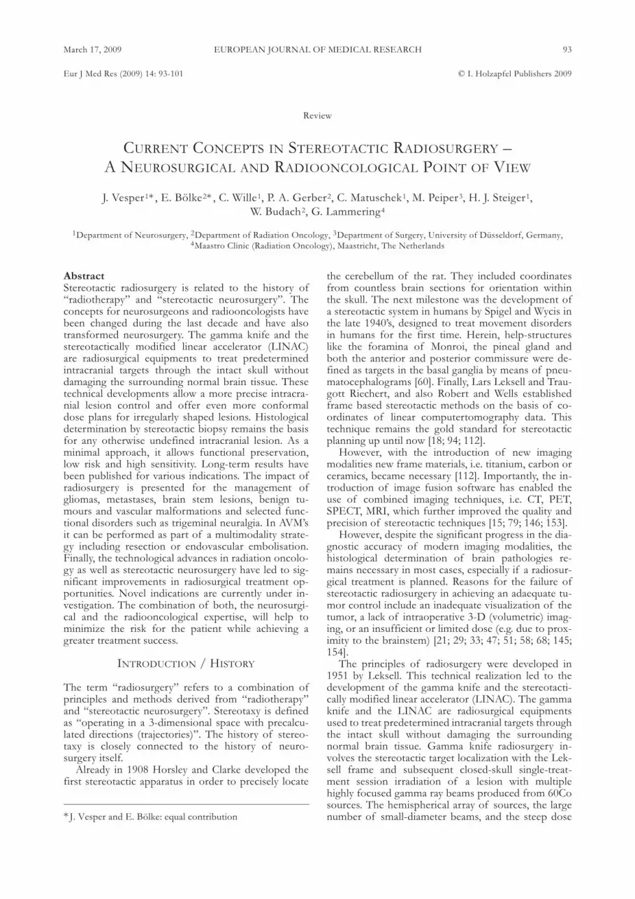
Fig. 1. Stereotactic radiotherapy of a single brain metastasis. A: Visualization of thetarget volumes for the treatment of a single intracerebral metastasis of a carcinomaof the lung (58 y old female, Adeno-Ca, pT2, pN1 (2/8), cM1). A 3D-reconstructiondisplaying the metastasis (violett) and proximate sensitive structures (bulbus andtractus opticus = green and pink; brown = brain stem). B: Visualization of photonbeams for the target volume based on planning CT applying stereotactic frames.Surrounding isodoses account for 90 percent at 20 Gy. Margins can be reduced to aminimum with regard to the possibility of exact patient positioning.
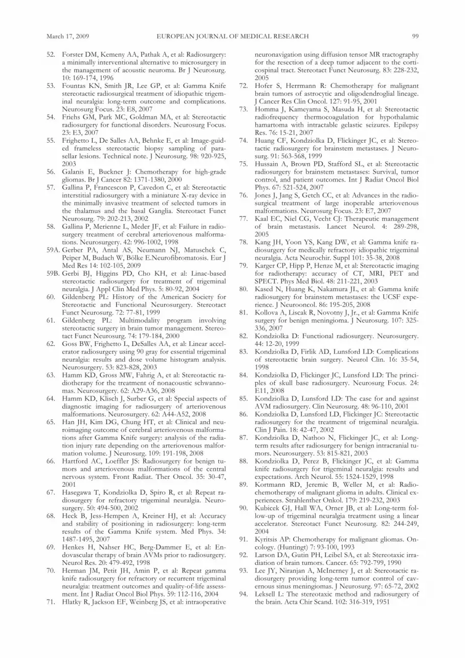
Fig. 2. Patient with mask and stereotactic lo-calizer.
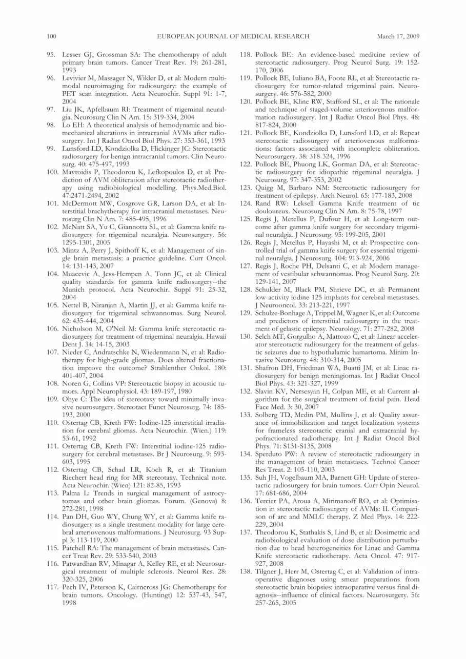
Fig. 3. Patient undergoing stereotactic radio-surgery.
1. Vesper/Bo?lke:Umbruchvorlage 23.02.2009 15:58 Uhr Seite 95

the most adequate therapy. In a series of 50 patientswith infiltrating tumors of the brainstem, 30 caseswere histologically diagnosed as low-grade astrocy-tomas, 13 cases as high-grade astrocytomas, 2 cases asprimitive neuroectodermic tumors, 2 cases as rhabdoidtumors and 1 case as an ependymoma, and 2 patientswith non-specified tumors. No mortality due tostereotactic biopsies were reported. [27]. In the major-ity of the patients the histological diagnosis led to atherapeutic intervention. Thus, due to the low risk ofthe procedure, a stereotactic biopsy should be per-formed in all cases. The radiosurgical treatment of abrainstem lesion might offer a promising non-invasivetreatment which is not associated with severe sur-rounding oedema [27; 74; 75; 80; 149].
BENIGN TUMORS
Historically, external beam radiotherapy has been andis still being extensively applied in the treatment ofmalignant and aggressive intracranial tumors and itsimportant role has been repeatedly verified by im-proved patient survival and increased tumor controlrates. As more modern therapies are being employedin surgery and radiotherapy, attention is now also be-ing directed towards the utility of radiotherapy as ei-ther primary or secondary treatment of benign prima-ry brain tumors and meningeomas. Primary tumortreatment encompasses the irradiation of small benigntumors without bioptic confirmation of the histologi-cal tumor type. Secondary treatment involves postop-erative radiotherapy, with the possibility that less ag-gressive tumor resections may be performed in areaswith a higher probability of resultant neurologicaldeficit. Recent studies suggest that this is not only apossible treatment strategy, but that it may be even su-perior to a more radical resection strategy in selectedcases [37; 45; 49; 87; 99].Stereotactic radiosurgery is typically employed as
first line treatment in patients with small to mediumsize tumors (without symptomatic brainstem compres-sions). Furthermore, it is also applied to control thegrowth of recurrent or residual tumor after surgicalresection. However, stereotactic radiotherapy, whichrepresents a non-invasive, hypo-fractionated treatmentstrategy, may also be especially suitable for patientswho desire preservation of neurological function(cochlear, facial nerve) and a high rate of tumorgrowth control. Notably, a local tumor control rate ofup to 95% (5y FU) can be achieved [37; 81; 131].Meningioma control rates range from 90 to 95%, andthe risk of morbidity is low [38; 40; 84; 93; 104]. TheMarseille SRS experience included 1,500 patients, with1,000 patients having follow-up of more than 3 years.A long-term tumor control rate of 97%, a transient fa-cial palsy of less than 1%, and a probability of func-tional hearing preservation between 50 and 95% couldbe achieved in this large series of patients treated withstereotactic radiosurgery [28; 63; 127].In another large series of a total of 285 patients, a
local tumor control of 95% was reported (63% regres-sion, and 32% no further tumor growth). After 15years the tumor control rate still remained above 90%at 93.7%. In 5% of the patients a delayed tumor
growth could be identified. A surgical resection wasperformed after radiosurgery in 13 patients (5%).None of the patients developed a radiation-inducedtumor. Eighty-one percent of the patients were stillalive at the time of this analysis with a mean follow-uptime of 10 years [59A]. In patients undergoing treat-ment for acoustic neuromas, a normal facial nervefunction was maintained in 95% of patients who hadnormal function before. Other authors also reportedon comparable results [49; 66; 87].A further indication for radiosurgical therapy in be-
nign tumors is the interstitial treatment of hypothala-mic hamartomas with temporarly implanted iodineseeds (also called brachytherapy). These tumors oftenbecome symptomatic with gelastic seizures. Schulze-Bohnhage et al. found that 11 out of 24 patients wereseizure free or experienced a seizure reduction of atleast 90% after a mean follow-up period of 24-monthsfollowing the last interstitial radiosurgical treatment.Notably, the duration of epilepsy prior to radiosurgerynegatively influenced this outcome. Moreover, alsoseizure-patients who present at younger ages (<15 y)can be successfully treated with brachytherapy. I125seed implantation as a radiosurgical technique is pre-dominantly applied for this indication due the advan-tage of continuous dose application and the possibilityof immediate interruption of therapy in cases of sideeffects, e.g. alteration of the optical tract [4; 42; 73;123; 129; 130; 141].
AVM
Radiosurgery has been proven to be succesful in thetreatment of small arteriovenous malformations(AVMs) of the brain [16; 85; 139; 142; 144; 150]. Untilnow, digital subtraction angiography (DSA) has been amandatory tool for the planning of these interven-tions. By integrating different imaging modalities inthe planning and follow-up procedure, e.g. MRI, manyside effects can be avoided [64]. However, due to theoften significant volume of healthy tissue being irradi-ated in cases of larger AVM lesions, reduced radiationdoses would be preferable in order to minimize therate of irreversible radiation injuries. On the otherhand, lower radiation doses lead to lower obliterationrates. Thus, several strategies have been developed inthe past decade to overcome these dose-volume prob-lems with larger AVMs, including reduced prescriptiondoses, volume fractionation and fractionated stereo-tactic radiotherapy treatments. AVMs with a volume of~ >3 ml can be completely obliterated (obliterationrate 72-96%) [76], whereas in larger AVMs complica-tion rate and obliteration rate still remain unsatisfacto-ry, especially in AVM’s >10ml. [114]. However, recentoptimistic reports suggest a benefit of conventionalsingle-dose stereotactic radiosurgery (SRS). Radio-surgery with marginal dose or peripheral dose around12 Gy rarely obliterates AVMs and yet most lesionsdiminish in size after SRS. Higher doses may be reap-plied to any residual nidi after an adequate follow-upperiod [64, 76]. However, long-term data show thatsome authors retreat the patients with lower doseswith lesions that failed to completely obliterate in thefirst place [100; 121].
EUROPEAN JOURNAL OF MEDICAL RESEARCH96 March 17, 2009
1. Vesper/Bo?lke:Umbruchvorlage 23.02.2009 15:58 Uhr Seite 96

Volume segmentation divides AVMs into smallersegments in order to irradiate them separately. Targetvolumes of only 5-15 ml irradiated with doses ofmore than 15 Gy can reduce the irradiated volume de-livered to the surrounding brain tissue [50; 76; 120].Furthermore, fewer radiation injuries have been re-ported with fractionated stereotactic radiotherapycompared to standard radiosurgery [16; 69; 85; 98;136; 144]. Advances in AVM localization, dose homo-geneity and dosimetry and fractionated radiotherapyregimen have refocused the interest in stereotactic ra-diotherapy. A recently published study of Han et al.on 218 patients with a follow-up of >2years providesa focus on the analysis of the radiation injury rate de-pending on the AVM volume. Investigators dispensed25 Gy for small (<4 cm3) and medium size (4-14 cm3)AVMs, and 10 Gy for larger AVMs (>14cm3). Theoverall obliteration rate was 66.4 %, 81.7% for small,53.1% for medium and 12.5% for large AVMs. Theauthors reported an acceptable complication rate of1.7%-17.4%, depending on the size of the AVM [65].The extended latency period between treatment andocclusion, about 5 years for emerging techniques (suchas salvage, staged volume, and hypofractionated radio-therapy), exposes the patient to the risk of haemor-rhage during that period. Nevertheless, improvementsin dose planning and target delineation will continueto improve the prognosis in patients suffering from in-operable AVM’s [16; 69; 85; 98; 136; 144].
OTHER INDICATIONS
Especially in the field of functional neurosurgerymore indications for radiosurgery are emerging. Suc-cessful treatments of trigeminal neuralgia have beenreported with radiosurgery of the ganglion gasseri inpatients with typical trigeminal neuralgia but also withfacial pain due to multiple sclerosis and petroclivalmeningeomas with infiltration of the trigeminal nerve.Facial pain has become a common indication for ra-diosurgery with an acceptable rate for hypaesthesiaand a meaningful relief of pain in the vast majority ofthe treated patients [88; 105; 118; 119; 125; 132]. Theoverall failure rate is about 15%, which is approxi-mately in the same as for decompression. Chen et al.identified preoperative factors which can determinethe outcome for pain control: The response to anti-convulsant medication has been regarded as the singlemost important prognostic indicator for treatmentsuccess [3; 23; 53; 54; 59B; 62; 67; 70; 78; 82; 86; 90;97; 102; 106; 116; 118; 122; 124; 126; 151].
SUMMARY
Radiosurgery is enjoying an increasing popularity sincethe last decade in terms of neurosurgical treatmentopportunities but also in terms of treatment optionsfor brain metastases. External beam and interstitial ra-diosurgery have been implemented as commonly ap-plied treatment techniques in radiation oncology aswell as neurosurgery due to significant improvementsin therapy efficacy, technological safety (smaller multi-leaf collimators), as well as dose homogeneity provid-ed by the newer LINAC generations and newer gener-
ation radioactive seeds. Technological advances pro-vide larger treatment flexibility. Apart from the treat-ment of oncologic processes newer indications alsoinclude the management of AVMs and pain syn-dromes within the functional neurosurgical field.Technological advances in stereotactic neurosurgery
not only lead to higher accuracy and safety in planningof both the target coordinates and trajectories (way tothe target) but also provide superior and sophisticatedmethods for defining any intracranial target volume.Correspondingly, current developments in radio-surgery are in part a result of the long tradition ofstereotaxy, which today could be considered as an in-ert component of stereotactic neurosurgery.In conclusion, the technological advances in radia-
tion oncology as well as stereotactic neurosurgery haveled to significant improvements in radiosurgical treat-ment opportunities, which will certainly lead to furtherexpansions in treatment opportunities for radio-surgery. Combining both, the expertise of the longtradition of sterotaxy in the field of neurosurgery andthe expertise of highly conformal irradiation in thefield of radiation oncology, will certainly yield to fur-ther improvements in the treatment success for ourpatients, while minimizing the risk for irreversible ra-diation injuries.
Acknowlegement: We would like to thank Ethelyn RusnakClinical Assistant Professor, Department of AnesthesiologyState University of New York at Buffalo for reading and cor-recting the manuscript.
REFERENCES1. Stereotactic radiosurgery for multiple or recurrent brain
metastases. Tecnologica. 9-10, 19952. Towards Minimally Invasive Neurosurgery. 1st Asian
Congress of Stereotactic, Functional and Computer-As-sisted Neurosurgery. Singapore, December 11-14, 1994.Abstracts. Stereotact Funct Neurosurg. 64: 57-100,1995
3. Abram S, Rosenblatt P, Holcomb S: Stereotactic radia-tion techniques in the treatment of acoustic schwan-nomas. Otolaryngol Clin North Am. 40:571-88, ix,2007
4. Addas B, Sherman EM, Hader WJ: Surgical manage-ment of hypothalamic hamartomas in patients withgelastic epilepsy. Neurosurg Focus. 25:E8, 2008
5. Adler JR, Cox RS, Kaplan I, et al: Stereotactic radiosur-gical treatment of brain metastases. J Neurosurg. 76:444-449, 1992
6. Alesch F, Hawliczek R, Koos WT: Interstitial irradiationof brain metastases. ActaNeurochir. Suppl 63: 29-34, 1995
7. Alesch F, Pappaterra J, Trattnig S, et al: The role ofstereotactic biopsy in radiosurgery. Acta Neurochir.Suppl 63: 20-24, 1995
8. Andrews DW: Current neurosurgical management ofbrain metastases. Semin Oncol. 35: 100-107, 2008
9. Ashamalla H, Zaki B, Mokhtar B, et al: Fractionatedstereotactic radiotherapy boost and weekly paclitaxel inmalignant gliomas clinical and pharmacokinetics results.Technol Cancer Res Treat. 6: 169-176, 2007
10. Bauman GS, Cairncross JG: Multidisciplinary manage-ment of adult anaplastic oligodendrogliomas and ana-plastic mixed oligo-astrocytomas. Semin Radiat Oncol.11: 170-180, 2001
EUROPEAN JOURNAL OF MEDICAL RESEARCHMarch 17, 2009 97
1. Vesper/Bo?lke:Umbruchvorlage 23.02.2009 15:58 Uhr Seite 97

11. Black P: Management of malignant glioma: role ofsurgery in relation to multimodality therapy. J Neurovi-rol. 4: 227-236, 1998
12. Blond S, Lejeune JP, Dupard T, et al: The stereotacticapproach to brain stem lesions: a follow-up of 29 cases.Acta Neurochir. Suppl (Wien) 52: 75-77, 1991
13. Bova FJ, Goetsch SJ: Modern linac stereotactic radio-surgery systems have rendered the Gamma Knife obso-lete. Med Phys. 28: 1839-1841, 2001
14. Branch CL, Jr., Coric D, Olds W, et al: Stereotactic ra-diosurgery. A review of "gamma knife" and "linac knife"technology and the unit at the Wake Forest UniversityMedical Center. N C Med J. 53: 395-399, 1992
15. Brucke T, Djamshidian S, Bencsits G, et al: SPECT andPET imaging of the dopaminergic system in Parkinson'sdisease. J Neurol. 247 Suppl 4: IV/2-IV/7, 2000
16. Buis DR, Lagerwaard FJ, Barkhof F, et al: Stereotacticradiosurgery for brain AVMs: role of interobserver vari-ation in target definition on digital subtraction angiogra-phy. Int J Radiat Oncol Biol Phys. 62: 246-252, 2005
17. Burton E, Prados M: New chemotherapy options forthe treatment of malignant gliomas. Curr Opin Oncol.11: 157-161, 1999
18. Carini S, Scielzo G, Grillo RF, et al: Halo ring support-ing the Brown-Roberts-Wells stereotactic frame forfractionated radiotherapy. Acta Neurochir. (Wien) 129:92-96, 1994
19. Carpentier AF: Neuro-oncology: the growing role ofchemotherapy in glioma. Lancet Neurol. 4:4-5, 2005
20. Carpentier AF: Neuro-oncology: the growing role ofchemotherapy in glioma. Lancet Neurol. 4:4-5, 2005
21. Chang EL, Shiu AS, Mendel E, et al: Phase I/II study ofstereotactic body radiotherapy for spinal metastasis andits pattern of failure. J Neurosurg Spine 7: 151-160, 2007
22. Chang JE, Khuntia D, Robins HI, et al: Radiotherapyand radiosensitizers in the treatment of glioblastomamultiforme. Clin Adv Hematol Oncol. 5: 894-15, 2007
23. Chang JW, Kim SH, Huh R, et al: The effects of stereo-tactic radiosurgery on secondary facial pain. StereotactFunct Neurosurg. 72 Suppl 1: 29-37, 1999
24. Chang SD, Main W, Martin DP, et al: An analysis of theaccuracy of the CyberKnife: a robotic frameless stereo-tactic radiosurgical system. Neurosurgery. 52: 140-146,2003
25. Chatel M, Lebrun C, Frenay M: Chemotherapy and im-munotherapy in adult malignant gliomas. Curr OpinOncol. 5: 464-473, 1993
26. Chernov M, Kamikawa S, Toledo R, et al: Minimally in-vasive management of the third ventricle glioma in a pa-tient without hydrocephalus: neurofiberscopic biopsyfollowed by gamma knife radiosurgery. Minim InvasiveNeurosurg. 47: 238-241, 2004
27. Chico-Ponce dL, Perezpena-Diazconti M, Castro-SierraE, et al: Stereotactically-guided biopsies of brainstem tu-mors. Childs Nerv Syst. 19: 305-310, 2003
28. Chin LS, Szerlip NJ, Regine WF: Stereotactic radio-surgery for meningiomas. Neurosurg Focus. 14: e6, 2003
29. Chitapanarux I, Goss B, Vongtama R, et al: Prospectivestudy of stereotactic radiosurgery without whole brainradiotherapy in patients with four or less brain metas-tases: incidence of intracranial progression and salvageradiotherapy. J Neurooncol. 61: 143-149, 2003
30. Cho KH, Hall WA, Gerbi BJ, et al: Single dose versusfractionated stereotactic radiotherapy for recurrenthigh-grade gliomas. Int J Radiat Oncol Biol Phys. 45:1133-1141, 1999
31. Choudhury AR: Interstitial iodine-125 radiosurgery forcerebral metastases. Br J Neurosurg. 10: 229, 1996
32. Coffey RJ, Lunsford LD: The role of stereotactic tech-niques in the management of craniopharyngiomas. Neu-rosurg Clin N Am. 1: 161-172, 1990
33. Dagnew E, Kanski J, McDermott MW, et al: Manage-ment of newly diagnosed single brain metastasis usingresection and permanent iodine-125 seeds without ini-tial whole-brain radiotherapy: a two institution experi-ence. Neurosurg Focus. 22: E3, 2007
34. Datta R, Jawahar A, Ampil FL, et al: Survival in relationto radiotherapeutic modality for brain metastasis: wholebrain irradiation vs. gamma knife radiosurgery. Am JClin Oncol. 27: 420-424, 2004
35. DeAngelis LM, Burger PC, Green SB, et al: Malignantglioma: who benefits from adjuvant chemotherapy? AnnNeurol. 44: 691-695, 1998
36. Dempsey PK, Kondziolka D, Lunsford LD: Stereotacticdiagnosis and treatment of pineal region tumours andvascular malformations. Acta Neurochir. (Wien) 116:14-22, 1992
37. DiBiase SJ, Chin LS: Stereotactic radiosurgery for benignneoplasms. Technol Cancer Res Treat. 2: 127-134,2003
38. DiBiase SJ, Kwok Y, Yovino S, et al: Factors predictinglocal tumor control after gamma knife stereotactic ra-diosurgery for benign intracranial meningiomas. Int JRadiat Oncol Biol Phys. 60: 1515-1519, 2004
39. Dropcho EJ: Novel chemotherapeutic approaches tobrain tumors. Hematol Oncol Clin North Am. 15: 1027-1052, 2001
40. Dufour H, Muracciole X, Metellus P, et al: Long-termtumor control and functional outcome in patients withcavernous sinus meningiomas treated by radiotherapywith or without previous surgery: is there an alternativeto aggressive tumor removal? Neurosurgery. 48: 285-294, 2001
41. Dunkel IJ, Finlay JL: High-dose chemotherapy with au-tologous stem cell rescue for brain tumors. Crit RevOncol Hematol. 41: 197-204, 2002
42. Dunoyer C, Ragheb J, Resnick T, et al: The useof stereotactic radiosurgery to treat intractable child-hood partial epilepsy. Epilepsia. 43: 292-300,2002
43. Eichler AF, Loeffler JS: Multidisciplinary managementof brain metastases. Oncologist. 12: 884-898, 2007
44. Ekstrand KE, Hinson WH, Bourland JD, et al: The useof a Leksell-BRW adapter for linac radiosurgery as anadjunct to Gamma Knife treatment. Phys Med Biol. 48:4105-4110, 2003
45. Elia AE, Shih HA, Loeffler JS: Stereotactic radiationtreatment for benign meningiomas. Neurosurg Focus.23: E5, 2007
46. Ernst-Stecken A, Ganslandt O, Lambrecht U, et al: Sur-vival and quality of life after hypofractionated stereotac-tic radiotherapy for recurrent malignant glioma. J Neu-rooncol. 81: 287-294, 2007
47. Ertl A, Saringer W, Heimberger K, et al: Quality assur-ance for the Leksell gamma unit: considering magneticresonance image-distortion and delineation failure in thetargeting of the internal auditory canal. Med Phys. 26:166-170, 1999
48. Ewend MG, Morris DE, Carey LA, et al: Guidelines forthe initial management of metastatic brain tumors: roleof surgery, radiosurgery, and radiation therapy. J NatlCompr Canc Netw. 6: 505-513, 2008
49. Flickinger JC, Kondziolka D, Lunsford LD: Radio-surgery of Benign Lesions. Semin.Radiat Oncol. 5: 220-224, 1995
50. Flickinger JC, Kondziolka D, Maitz AH, et al: An analy-sis of the dose-response for arteriovenous malformationradiosurgery and other factors affecting obliteration. Ra-diother Oncol. 63: 347-354, 2002
51. Foote RL, Pollock BE, Link MJ, et al: Leksell GammaKnife coordinate setting slippage: how often, howmuch? J Neurosurg. 101: 590-593, 2004
EUROPEAN JOURNAL OF MEDICAL RESEARCH98 March 17, 2009
1. Vesper/Bo?lke:Umbruchvorlage 23.02.2009 15:58 Uhr Seite 98
52. Forster DM, Kemeny AA, Pathak A, et al: Radiosurgery:a minimally interventional alternative to microsurgery inthe management of acoustic neuroma. Br J Neurosurg.10: 169-174, 1996
53. Fountas KN, Smith JR, Lee GP, et al: Gamma Knifestereotactic radiosurgical treatment of idiopathic trigem-inal neuralgia: long-term outcome and complications.Neurosurg Focus. 23: E8, 2007
54. Friehs GM, Park MC, Goldman MA, et al: Stereotacticradiosurgery for functional disorders. Neurosurg Focus.23: E3, 2007
55. Frighetto L, De Salles AA, Behnke E, et al: Image-guid-ed frameless stereotactic biopsy sampling of para-sellar lesions. Technical note. J Neurosurg. 98: 920-925,2003
56. Galanis E, Buckner J: Chemotherapy for high-gradegliomas. Br J Cancer 82: 1371-1380, 2000
57. Gallina P, Francescon P, Cavedon C, et al: Stereotacticinterstitial radiosurgery with a miniature X-ray device inthe minimally invasive treatment of selected tumors inthe thalamus and the basal Ganglia. Stereotact FunctNeurosurg. 79: 202-213, 2002
58. Gallina P, Merienne L, Meder JF, et al: Failure in radio-surgery treatment of cerebral arteriovenous malforma-tions. Neurosurgery. 42: 996-1002, 1998
59A. Gerber PA, Antal AS, Neumann NJ, Matuschek C,Peiper M, Budach W, Bölke E.Neurofibromatosis. Eur JMed Res 14: 102-105, 2009
59B. Gerbi BJ, Higgins PD, Cho KH, et al: Linac-basedstereotactic radiosurgery for treatment of trigeminalneuralgia. J Appl Clin Med Phys. 5: 80-92, 2004
60. Gildenberg PL: History of the American Society forStereotactic and Functional Neurosurgery. StereotactFunct Neurosurg. 72: 77-81, 1999
61. Gildenberg PL: Multimodality program involvingstereotactic surgery in brain tumor management. Stereo-tact Funct Neurosurg. 74: 179-184, 2000
62. Goss BW, Frighetto L, DeSalles AA, et al: Linear accel-erator radiosurgery using 90 gray for essential trigeminalneuralgia: results and dose volume histogram analysis.Neurosurgery. 53: 823-828, 2003
63. Hamm KD, Gross MW, Fahrig A, et al: Stereotactic ra-diotherapy for the treatment of nonacoustic schwanno-mas. Neurosurgery. 62: A29-A36, 2008
64. Hamm KD, Klisch J, Surber G, et al: Special aspects ofdiagnostic imaging for radiosurgery of arteriovenousmalformations. Neurosurgery. 62: A44-A52, 2008
65. Han JH, Kim DG, Chung HT, et al: Clinical and neu-roimaging outcome of cerebral arteriovenous malforma-tions after Gamma Knife surgery: analysis of the radia-tion injury rate depending on the arteriovenous malfor-mation volume. J Neurosurg. 109: 191-198, 2008
66. Hartford AC, Loeffler JS: Radiosurgery for benign tu-mors and arteriovenous malformations of the centralnervous system. Front Radiat. Ther Oncol. 35: 30-47,2001
67. Hasegawa T, Kondziolka D, Spiro R, et al: Repeat ra-diosurgery for refractory trigeminal neuralgia. Neuro-surgery. 50: 494-500, 2002
68. Heck B, Jess-Hempen A, Kreiner HJ, et al: Accuracyand stability of positioning in radiosurgery: long-termresults of the Gamma Knife system. Med Phys. 34:1487-1495, 2007
69. Henkes H, Nahser HC, Berg-Dammer E, et al: En-dovascular therapy of brain AVMs prior to radiosurgery.Neurol Res. 20: 479-492, 1998
70. Herman JM, Petit JH, Amin P, et al: Repeat gammaknife radiosurgery for refractory or recurrent trigeminalneuralgia: treatment outcomes and quality-of-life assess-ment. Int J Radiat Oncol Biol Phys. 59: 112-116, 2004
71. Hlatky R, Jackson EF, Weinberg JS, et al: intraoperative
neuronavigation using diffusion tensor MR tractographyfor the resection of a deep tumor adjacent to the corti-cospinal tract. Stereotact Funct Neurosurg. 83: 228-232,2005
72. Hofer S, Herrmann R: Chemotherapy for malignantbrain tumors of astrocytic and oligodendroglial lineage.J Cancer Res Clin Oncol. 127: 91-95, 2001
73. Homma J, Kameyama S, Masuda H, et al: Stereotacticradiofrequency thermocoagulation for hypothalamichamartoma with intractable gelastic seizures. EpilepsyRes. 76: 15-21, 2007
74. Huang CF, Kondziolka D, Flickinger JC, et al: Stereo-tactic radiosurgery for brainstem metastases. J Neuro-surg. 91: 563-568, 1999
75. Hussain A, Brown PD, Stafford SL, et al: Stereotacticradiosurgery for brainstem metastases: Survival, tumorcontrol, and patient outcomes. Int J Radiat Oncol BiolPhys. 67: 521-524, 2007
76. Jones J, Jang S, Getch CC, et al: Advances in the radio-surgical treatment of large inoperable arteriovenousmalformations. Neurosurg Focus. 23: E7, 2007
77. Kaal EC, Niel CG, Vecht CJ: Therapeutic managementof brain metastasis. Lancet Neurol. 4: 289-298,2005
78. Kang JH, Yoon YS, Kang DW, et al: Gamma knife ra-diosurgery for medically refractory idiopathic trigeminalneuralgia. Acta Neurochir. Suppl 101: 35-38, 2008
79. Karger CP, Hipp P, Henze M, et al: Stereotactic imagingfor radiotherapy: accuracy of CT, MRI, PET andSPECT. Phys Med Biol. 48: 211-221, 2003
80. Kased N, Huang K, Nakamura JL, et al: Gamma kniferadiosurgery for brainstem metastases: the UCSF expe-rience. J Neurooncol. 86: 195-205, 2008
81. Kollova A, Liscak R, Novotny J, Jr., et al: Gamma Knifesurgery for benign meningioma. J Neurosurg. 107: 325-336, 2007
82. Kondziolka D: Functional radiosurgery. Neurosurgery.44: 12-20, 1999
83. Kondziolka D, Firlik AD, Lunsford LD: Complicationsof stereotactic brain surgery. Neurol Clin. 16: 35-54,1998
84. Kondziolka D, Flickinger JC, Lunsford LD: The princi-ples of skull base radiosurgery. Neurosurg Focus. 24:E11, 2008
85. Kondziolka D, Lunsford LD: The case for and againstAVM radiosurgery. Clin Neurosurg. 48: 96-110, 2001
86. Kondziolka D, Lunsford LD, Flickinger JC: Stereotacticradiosurgery for the treatment of trigeminal neuralgia.Clin J Pain. 18: 42-47, 2002
87. Kondziolka D, Nathoo N, Flickinger JC, et al: Long-term results after radiosurgery for benign intracranial tu-mors. Neurosurgery. 53: 815-821, 2003
88. Kondziolka D, Perez B, Flickinger JC, et al: Gammaknife radiosurgery for trigeminal neuralgia: results andexpectations. Arch Neurol. 55: 1524-1529, 1998
89. Kortmann RD, Jeremic B, Weller M, et al: Radio-chemotherapy of malignant glioma in adults. Clinical ex-periences. Strahlenther Onkol. 179: 219-232, 2003
90. Kubicek GJ, Hall WA, Orner JB, et al: Long-term fol-low-up of trigeminal neuralgia treatment using a linearaccelerator. Stereotact Funct Neurosurg. 82: 244-249,2004
91. Kyritsis AP: Chemotherapy for malignant gliomas. On-cology. (Huntingt) 7: 93-100, 1993
92. Larson DA, Gutin PH, Leibel SA, et al: Stereotaxic irra-diation of brain tumors. Cancer. 65: 792-799, 1990
93. Lee JY, Niranjan A, McInerney J, et al: Stereotactic ra-diosurgery providing long-term tumor control of cav-ernous sinus meningiomas. J Neurosurg. 97: 65-72, 2002
94. Leksell L: The stereotaxic method and radiosurgery ofthe brain. Acta Chir Scand. 102: 316-319, 1951
EUROPEAN JOURNAL OF MEDICAL RESEARCHMarch 17, 2009 99
1. Vesper/Bo?lke:Umbruchvorlage 23.02.2009 15:58 Uhr Seite 99
95. Lesser GJ, Grossman SA: The chemotherapy of adultprimary brain tumors. Cancer Treat Rev. 19: 261-281,1993
96. Levivier M, Massager N, Wikler D, et al: Modern multi-modal neuroimaging for radiosurgery: the example ofPET scan integration. Acta Neurochir. Suppl 91: 1-7,2004
97. Liu JK, Apfelbaum RI: Treatment of trigeminal neural-gia. Neurosurg Clin N Am. 15: 319-334, 2004
98. Lo EH: A theoretical analysis of hemodynamic and bio-mechanical alterations in intracranial AVMs after radio-surgery. Int J Radiat Oncol Biol Phys. 27: 353-361, 1993
99. Lunsford LD, Kondziolka D, Flickinger JC: Stereotacticradiosurgery for benign intracranial tumors. Clin Neuro-surg. 40: 475-497, 1993
100. Mavroidis P, Theodorou K, Lefkopoulos D, et al: Pre-diction of AVM obliteration after stereotactic radiother-apy using radiobiological modelling. Phys.Med.Biol.47:2471-2494, 2002
101. McDermott MW, Cosgrove GR, Larson DA, et al: In-terstitial brachytherapy for intracranial metastases. Neu-rosurg Clin N Am. 7: 485-495, 1996
102. McNatt SA, Yu C, Giannotta SL, et al: Gamma knife ra-diosurgery for trigeminal neuralgia. Neurosurgery. 56:1295-1301, 2005
103. Mintz A, Perry J, Spithoff K, et al: Management of sin-gle brain metastasis: a practice guideline. Curr Oncol.14: 131-143, 2007
104. Muacevic A, Jess-Hempen A, Tonn JC, et al: Clinicalquality standards for gamma knife radiosurgery--theMunich protocol. Acta Neurochir. Suppl 91: 25-32,2004
105. Nettel B, Niranjan A, Martin JJ, et al: Gamma knife ra-diosurgery for trigeminal schwannomas. Surg Neurol.62: 435-444, 2004
106. Nicholson M, O'Neil M: Gamma knife stereotactic ra-diosurgery for treatment of trigeminal neuralgia. HawaiiDent J. 34: 14-15, 2003
107. Nieder C, Andratschke N, Wiedenmann N, et al: Radio-therapy for high-grade gliomas. Does altered fractiona-tion improve the outcome? Strahlenther Onkol. 180:401-407, 2004
108. Noren G, Collins VP: Stereotactic biopsy in acoustic tu-mors. Appl Neurophysiol. 43: 189-197, 1980
109. Ohye C: The idea of stereotaxy toward minimally inva-sive neurosurgery. Stereotact Funct Neurosurg. 74: 185-193, 2000
110. Ostertag CB, Kreth FW: Iodine-125 interstitial irradia-tion for cerebral gliomas. Acta Neurochir. (Wien.) 119:53-61, 1992
111. Ostertag CB, Kreth FW: Interstitial iodine-125 radio-surgery for cerebral metastases. Br J Neurosurg. 9: 593-603, 1995
112. Ostertag CB, Schad LR, Koch R, et al: TitaniumRiechert head ring for MR stereotaxy. Technical note.Acta Neurochir. (Wien) 121: 82-85, 1993
113. Palma L: Trends in surgical management of astrocy-tomas and other brain gliomas. Forum. (Genova) 8:272-281, 1998
114. Pan DH, Guo WY, Chung WY, et al: Gamma knife ra-diosurgery as a single treatment modality for large cere-bral arteriovenous malformations. J Neurosurg. 93 Sup-pl 3: 113-119, 2000
115. Patchell RA: The management of brain metastases. Can-cer Treat Rev. 29: 533-540, 2003
116. Patwardhan RV, Minagar A, Kelley RE, et al: Neurosur-gical treatment of multiple sclerosis. Neurol Res. 28:320-325, 2006
117. Pech IV, Peterson K, Cairncross JG: Chemotherapy forbrain tumors. Oncology. (Huntingt) 12: 537-43, 547,1998
118. Pollock BE: An evidence-based medicine review ofstereotactic radiosurgery. Prog Neurol Surg. 19: 152-170, 2006
119. Pollock BE, Iuliano BA, Foote RL, et al: Stereotactic ra-diosurgery for tumor-related trigeminal pain. Neuro-surgery. 46: 576-582, 2000
120. Pollock BE, Kline RW, Stafford SL, et al: The rationaleand technique of staged-volume arteriovenous malfor-mation radiosurgery. Int J Radiat Oncol Biol Phys. 48:817-824, 2000
121. Pollock BE, Kondziolka D, Lunsford LD, et al: Repeatstereotactic radiosurgery of arteriovenous malforma-tions: factors associated with incomplete obliteration.Neurosurgery. 38: 318-324, 1996
122. Pollock BE, Phuong LK, Gorman DA, et al: Stereotac-tic radiosurgery for idiopathic trigeminal neuralgia. JNeurosurg. 97: 347-353, 2002
123. Quigg M, Barbaro NM: Stereotactic radiosurgery fortreatment of epilepsy. Arch Neurol. 65: 177-183, 2008
124. Rand RW: Leksell Gamma Knife treatment of ticdouloureux. Neurosurg Clin N Am. 8: 75-78, 1997
125. Regis J, Metellus P, Dufour H, et al: Long-term out-come after gamma knife surgery for secondary trigemi-nal neuralgia. J Neurosurg. 95: 199-205, 2001
126. Regis J, Metellus P, Hayashi M, et al: Prospective con-trolled trial of gamma knife surgery for essential trigemi-nal neuralgia. J Neurosurg. 104: 913-924, 2006
127. Regis J, Roche PH, Delsanti C, et al: Modern manage-ment of vestibular schwannomas. Prog Neurol Surg. 20:129-141, 2007
128. Schulder M, Black PM, Shrieve DC, et al: Permanentlow-activity iodine-125 implants for cerebral metastases.J Neurooncol. 33: 213-221, 1997
129. Schulze-BonhageA, TrippelM, Wagner K, et al: Outcomeand predictors of interstitial radiosurgery in the treat-ment of gelastic epilepsy. Neurology. 71: 277-282, 2008
130. Selch MT, Gorgulho A, Mattozo C, et al: Linear acceler-ator stereotactic radiosurgery for the treatment of gelas-tic seizures due to hypothalamic hamartoma. Minim In-vasive Neurosurg. 48: 310-314, 2005
131. Shafron DH, Friedman WA, Buatti JM, et al: Linac ra-diosurgery for benign meningiomas. Int J Radiat OncolBiol Phys. 43: 321-327, 1999
132. Slavin KV, Nersesyan H, Colpan ME, et al: Current al-gorithm for the surgical treatment of facial pain. HeadFace Med. 3: 30, 2007
133. Solberg TD, Medin PM, Mullins J, et al: Quality assur-ance of immobilization and target localization systemsfor frameless stereotactic cranial and extracranial hy-pofractionated radiotherapy. Int J Radiat Oncol BiolPhys. 71: S131-S135, 2008
134. Sperduto PW: A review of stereotactic radiosurgery inthe management of brain metastases. Technol CancerRes Treat. 2: 105-110, 2003
135. Suh JH, Vogelbaum MA, Barnett GH: Update of stereo-tactic radiosurgery for brain tumors. Curr Opin Neurol.17: 681-686, 2004
136. Tercier PA, Aroua A, Mirimanoff RO, et al: Optimisa-tion in stereotactic radiosurgery of AVMs: II. Compari-son of arc and MMLC therapy. Z Med Phys. 14: 222-229, 2004
137. Theodorou K, Stathakis S, Lind B, et al: Dosimetric andradiobiological evaluation of dose distribution perturba-tion due to head heterogeneities for Linac and GammaKnife stereotactic radiotherapy. Acta Oncol. 47: 917-927, 2008
138. Tilgner J, Herr M, Ostertag C, et al: Validation of intra-operative diagnoses using smear preparations fromstereotactic brain biopsies: intraoperative versus final di-agnosis--influence of clinical factors. Neurosurgery. 56:257-265, 2005
EUROPEAN JOURNAL OF MEDICAL RESEARCH100 March 17, 2009
1. Vesper/Bo?lke:Umbruchvorlage 23.02.2009 15:58 Uhr Seite 100

139. Treuer H, Kocher M, Hoevels M, et al: Impact of targetpoint deviations on control and complication probabili-ties in stereotactic radiosurgery of AVMs and metas-tases. Radiother Oncol. 81: 25-32, 2006
140. Tsao MN, Mehta MP, Whelan TJ, et al: The AmericanSociety for Therapeutic Radiology and Oncology (AS-TRO) evidence-based review of the role of radiosurgeryfor malignant glioma. Int J Radiat Oncol Biol Phys. 63:47-55, 2005
141. Unger F, Schrottner O, Feichtinger M, et al: Stereotacticradiosurgery for hypothalamic hamartomas. Acta Neu-rochir. Suppl 84: 57-63, 2002
142. Valentino V: Radiosurgery in cerebral tumours andAVM. Acta Neurochir. Suppl (Wien) 42: 193-197, 1988
143. van den Bent MJ, Taphoorn MJ, Brandes AA, et al:Phase II study of first-line chemotherapy with temo-zolomide in recurrent oligodendroglial tumors: the Eu-ropean Organization for Research and Treatment ofCancer Brain Tumor Group Study 26971. J Clin Oncol.21: 2525-2528, 2003
144. Voges J, Treuer H, Lehrke R, et al: Risk analysis ofLINAC radiosurgery in patients with arteriovenous mal-formation (AVM). Acta Neurochir. Suppl 68: 118-123,1997
145. Wang L, Jacob R, Chen L, et al: Stereotactic IMRT forprostate cancer: setup accuracy of a new stereotacticbody localization system. J Appl Clin Med Phys. 5: 18-28, 2004
146. Wara W, Bauman G, Gutin P, et al: Stereotactic radio-surgery in children. Stereotact Funct Neurosurg. 64Suppl 1: 118-125, 1995
147. Warnke PC, Kopitzki K, Ostertag CB: Interstitialstereotactic radiosurgery. Acta Neurochir. Suppl 88: 45-50, 2003
148. Wowra B, Czempiel H, Cibis R, et al: [Profile of ambu-latory radiosurgery with the gamma knife system. 1:Method and multicenter irradiation concept]. Radiologe.37: 995-1002, 1997
149. Yen CP, Sheehan J, Steiner M, et al: Gamma knifesurgery for focal brainstem gliomas. J Neurosurg. 106:8-17, 2007
150. Young CS, Schwartz ML, O'Brien P, et al: Stereotacticradiotherapy for AVMs: the University of Toronto ex-perience. Acta Neurochir. Suppl 63: 57-59, 1995
151. Young RF, Vermulen S, Posewitz A: Gamma knife ra-diosurgery for the treatment of trigeminal neuralgia.Stereotact Funct Neurosurg. 70 Suppl 1: 192-199, 1998
152. Yung WK: Chemotherapy for malignant brain tumors.Curr Opin Oncol. 2: 673-678, 1990
153. Zeck OF, Fang B, Mullani N, et al: PET and SPECTimaging for stereotactic localization. Stereotact FunctNeurosurg. 64 Suppl 1: 147-154, 1995
154. Zheng LG, Xu DS, Kang CS, et al: Stereotactic radio-surgery for primary trigeminal neuralgia using the Lek-sell Gamma unit. Stereotact Funct Neurosurg. 76: 29-35, 2001
Received: January 2, 2009 / Accepted: February 2, 2009
Address for correspondence:Prof. Dr. med. Jan VesperFunktionelle Neurochirurgie und StereotaxieUniversitätsklinikum DüsseldorfMoorenstr. 540225 DüsseldorfTel.: +49 (0)211/81-18408Fax: +49 (0)211/81 015 18408E-mail: [email protected]
EUROPEAN JOURNAL OF MEDICAL RESEARCHMarch 17, 2009 101
1. Vesper/Bo?lke:Umbruchvorlage 23.02.2009 15:58 Uhr Seite 101