crystal structures of the nitrite and nitric oxide complexes of horse heart myoglobin
TRANSCRIPT

JOURNAL OF
www.elsevier.com/locate/jinorgbio
Journal of Inorganic Biochemistry 100 (2006) 1413–1425
InorganicBiochemistry
Crystal structures of the nitrite and nitric oxide complexesof horse heart myoglobin
Daniel M. Copeland a, Alexei S. Soares b, Ann H. West a, George B. Richter-Addo a,*
a Department of Chemistry and Biochemistry, University of Oklahoma, 620 Parrington Oval, Norman, OK 73019, USAb Macromolecular Crystallography Research Resource, National Synchrotron Light Source, Brookhaven National Laboratory, Upton, NY 11973, USA
Received 21 January 2006; received in revised form 29 March 2006; accepted 14 April 2006Available online 5 May 2006
Abstract
Nitrite is an important species in the global nitrogen cycle, and the nitrite reductase enzymes convert nitrite to nitric oxide (NO).Recently, it has been shown that hemoglobin and myoglobin catalyze the reduction of nitrite to NO under hypoxic conditions. We havedetermined the 1.20 A resolution crystal structure of the nitrite adduct of ferric horse heart myoglobin (hh Mb). The ligand is bound toiron in the nitrito form, and the complex is formulated as MbIII(ONO�). The Fe–ONO bond length is 1.94 A, and the O–N–O angle is113�. In addition, the nitrite ligand is stabilized by hydrogen bonding with the distal His64 residue. We have also determined the 1.30 Aresolution crystal structures of hh MbIINO. When hh MbIINO is prepared from the reaction of metMbIII with nitrite/dithionite, theFeNO angle is 144� with a Fe–NO bond length of 1.87 A. However, when prepared from the reaction of NO with reduced MbII, theFeNO angle is 120� with a Fe–NO bond length of 2.13 A. This difference in FeNO conformations as a function of preparative methodis reproducible, and suggests a role of the distal pocket in hh MbIINO in stabilizing local FeNO conformational minima.� 2006 Elsevier Inc. All rights reserved.
Keywords: Iron; Nitrite; X-ray; Nitric oxide; Nitrosyl; Heme; Myoglobin
1. Introduction
The nitrite anion ðNO�2 Þ is a simple oxyanion of nitro-gen with a pKa of 3.2 at 20 �C [1]. The nitrite anion haslong been considered, in mammalian systems, a ‘‘deadend’’ species that results from oxidation of nitric oxide pro-duced by the nitric oxide synthase (NOS) enzymes. As aconsequence, its role in human physiology has beenneglected until very recently [2,3]. Nitrite is a key speciesin the global nitrogen cycle. In dissimilatory denitrification(Eq. (1)) [4,5], nitrate is converted to nitrite by the nitratereductase (NaR) enzymes. Nitrite is then converted tonitric oxide by nitrite reductase (NiR) enzymes. Follow-up conversions by nitric oxide reductases (NOR) andnitrous oxide reductases (N2OR) result in the generationof dinitrogen.
0162-0134/$ - see front matter � 2006 Elsevier Inc. All rights reserved.
doi:10.1016/j.jinorgbio.2006.04.011
* Corresponding author. Tel.: +1 405 325 6401; fax: +1 405 325 6111.E-mail address: [email protected] (G.B. Richter-Addo).
NO�3 �!NaR
NO�2 �!NiR
NO�!NORN2O �!N2OR
N2 ð1ÞTwo types of dissimilatory NiR enzymes are known, andthey both carry out the one-electron reduction of nitriteto NO (Eq. (2)).
NO�2 þ 2Hþ þ 1e� ! NOþH2O ð2Þ
One is the periplasmic heme-containing cytochrome cd1
NiR which is present in about two-thirds of denitrifyingbacteria [4,6]. The crystal structure of product obtainedwhen nitrite was added to the reduced cytochrome cd1
NiR from Paraccocus pantotrophus reveals N-binding ofthe nitrite ligand to the metal center (structure I).
Fe
NOO
Cu
NOO
(I) (II)

1414 D.M. Copeland et al. / Journal of Inorganic Biochemistry 100 (2006) 1413–1425
A second class of dissimilatory NiR enzymes contain
copper in the active site [7]. Recent high-resolution crystalstructures of the nitrite adducts of the copper-containingNiR [8] from Alcaligenes xylosoxidans (the His313Glymutant) [9] and the soil bacterium Achromobacter cyclocl-astes [10] reveal an asymmetric O,O 0-bidentate binding ofthe nitrite ligand (structure II).
In addition, nitrite can be reduced by the assimilatory(pentaheme) cytochrome c NiR (ccNiR) directly to ammo-nia without the detection of intermediates [11]. The activesite heme contains lysine as a proximal ligand to iron,and the crystal structure of the nitrite-bound complex fromWolinella succinogenes reveals an N-binding of this groupto iron (i.e., structure I) [12].
Recently, in September 2005, a two-day symposium atthe US National Institutes of Health was held to highlightthe increased recognition of nitrite as a bioactive smallmolecule that plays important roles in physiology and ther-apeutics [2]. The results of several studies support theseproposed roles. For example, it has been known for sometime that deoxyHb will reduce nitrite to NO [13].
NO�2 þ deoxyHbðFeIIÞ þHþ
! NOþ metHbðFeIIIÞ þOH� ð3Þ
Ferrous myoglobin [14,15] and xanthine oxidoreductase[16–18] are also known to reduce nitrite to NO [2]. NiRactivity by deoxyHb has now been shown to be physiolog-ically relevant under hypoxic conditions, generating an O2-independent pool of NO (NOS enzymes require O2)[14,19,20]. A related role for the NiR activity of deoxyMb(in the myocardium and skeletal muscles) and mitochon-drial cytochrome oxidase and microsomal cytochromeP450 in tissues has been proposed [21]. Indeed, recent stud-ies suggest a key role for myoglobin in the conversion ofnitrite to NO in tissue, where endogenous nitrite levelscan reach concentrations of 20 lM (cf. 121 nM in plasmaand 288 nM in erythrocytes [22]), and that this NiR activitymay minimize ischemia-reperfusion injury during exercise[14,23]. In addition, nitrite has been recognized as a biolog-ical signaling molecule that influences cGMP production,cytochrome P450 activities, and heat shock protein 70and heme oxygenase-1 expression in various tissues [24].
Nitrite has long been utilized as an additive in meatproducts due to its antimicrobial and antioxidative activity[25,26]. Addition of nitrite to meats during the curing pro-cess also restores the pink color due to the formation of theheme-NO pigment [27]. The kinetics of reversible bindingof nitrite to metMb has been studied by van Eldik andcoworkers; at pH 7.4 and 20 �C, kon = 156 ± 3 M�1 s�1,with Keq (kon/koff) of 60 M�1 which is two orders of magni-tude smaller than that for NO binding to metMb [28]. Fordand coworkers have shown that nitrite catalyzes the reduc-tive nitrosylation of synthetic iron porphyrins and metMbto produce the FeIINO derivatives; it was suggested thatthe proposed ferric-nitrite species may be inhibitory inthese reactions [29]. The electrocatalytic reduction of nitrite
to NO by myoglobin in surfactant films [30] and syntheticiron porphyrins [31–35] has been demonstrated.
As mentioned above, nitrite reductase activity has beenascribed to Hb and Mb. Despite the importance of theheme–nitrite interaction in these proteins, no structuralinformation exists to date on how nitrite interacts withtheir heme centers. We were thus interested in determiningthe companion structures of the nitrite and nitric oxidederivatives of a representative globin, namely myoglobin.Three MbIINO structures have been reported previously;the 1.7 A resolution structure of sperm whale (sw) MbIINO[36], the 1.9 A resolution structure of the L29F/D122Ndouble mutant of sw MbIINO [36], and the 1.9 A resolutionstructure for horse heart (hh) MbIINO [37]. In this paper,we report the 1.20 A resolution crystal structure of thenitrite complex of horse heart myoglobin, and we showthat the nitrite ligand interacts with the ferric center viaan O-binding mode. We also describe the 1.30 A resolutionstructures of nitrosyl horse heart myoglobin prepared fromtwo different synthetic routes.
2. Experimental
Horse heart myoglobin (hh Mb) was purchased fromSigma, and crystals of aqua-metMb were obtained as pre-viously described [37]. Sodium nitrite was obtained fromBaker and Adamson (98.0%) or Fluka (P99.5%). Tris–HCl (>99.0%) and ammonium sulfate (P99.5%) werepurchased from Fluka, and sodium dithionite (sodiumhydrosulfite, 85%) was purchased from Aldrich.
2.1. Complex formation
2.1.1. Nitrite-bound myoglobin
Method 1. Crystals of aqua-metMb (in �10 lL of100 mM Tris–HCl, pH 7.4, 3.10–3.30 M ammonium sul-fate containing 15% glycerol as cryoprotectant) were sub-merged in mineral oil under an atmosphere of nitrogen.A solution of sodium nitrite (10 lL, 250 mM) was addedto the aqueous phase and allowed to soak into the crystalsfor �10 min. The crystals were harvested with cryoloopsand flash frozen in liquid nitrogen.
Method 2. Solid sodium nitrite (�9 mg) was added to1 mL of a 30 mg/mL metMb solution (100 mM Tris–HCl,pH 7.4; [nitrite] = 130 mM) and incubated on ice for10 min. The protein solution was then centrifuged for1 min (at 10,000g). Crystal trays were then set up as previ-ously described for MbII(EtNO) [37]. Suitable crystalsformed in three days, and they were harvested with cryol-oops and passed through the cryosolution and flash frozenin liquid nitrogen.
2.1.2. NO-bound myoglobin using the nitrite/dithionite
methodCrystals of aqua-metMb (in 100 mM Tris–HCl, pH 7.4,
3.10–3.30 M ammonium sulfate containing 15% glycerol ascryoprotectant) were submerged in mineral oil under an

D.M. Copeland et al. / Journal of Inorganic Biochemistry 100 (2006) 1413–1425 1415
atmosphere of nitrogen. The addition of the solution ofsodium nitrite (10 lL, 250 mM; 10 min soaking time) wasfollowed by addition of 20 lL of the cryosolution contain-ing 500 mM freshly dissolved sodium dithionite. The colorof the crystals slowly changed from brown to red over a10 min period. The red crystals were harvested and flashfrozen in liquid nitrogen.
2.1.3. NO-bound myoglobin using nitric oxide
Crystals of aqua-metMb were submerged in mineral oilas described above. Addition of a solution of sodium dithi-onite (10 lL, 50 mM; 10 min soak) was followed by addi-tion of 20 lL of an NO-saturated cryosolution. After5 min, the red crystals were harvested and flash frozen inliquid nitrogen.
2.2. X-ray data collection
The frozen crystals were initially screened for diffractionquality at our home source. X-ray data sets were collectedat 100 K on a RigakuMSC RU-H3R X-ray generator oper-ated at 50 kV/100 mA to produce Cu Ka radiation(k = 1.5418 A). Diffracted X-rays were detected using anR-AXIS 4++ dual image plate detector system. For theMbIII(ONO�) crystal data collected in-house, 1� oscillationimages were collected over a range of 216� with the crystal-to-detector distance of 70 mm and an exposure time of 300 s.
High quality crystals were shipped to the National Syn-chrotron Light Source (NSLS) at Brookhaven NationalLaboratory (BNL) via the Mail-in Data Collection Programoperated by the Protein Crystallography Research Resource(BNL-PXRR; www.px.nsls.bnl.gov/fedex.html) for highresolution X-ray data collection. X-ray data were collectedat 100 K on beamlines X-12B (using an ADSC Quantum4CCD detector) or X-29 (using an ADSC Quantum-315CCD detector). For the MbIII(ONO�) crystal, 1� oscillationimages were collected over a range of 180� with an exposuretime of 120 s per image and a crystal-to-detector distance of100 mm. For the MbIINO crystal obtained from the nitrite/dithionite soak method, 1� oscillation images were collectedover a range of 360� with an exposure time of 10 s per imageand a crystal-to-detector distance of 150 mm. For theMbIINO crystal obtained from the dithionite/NO method,1� oscillation images were collected over a range of 180� withan exposure time of 10 s per image and a crystal-to-detectordistance of 150 mm.
2.3. Data processing, structure solution and refinement
2.3.1. Data processing
All data sets collected at our home source were pro-cessed using the stand-alone d*TREK program (Macin-tosh v.2D) [38] from Molecular Structure Corporation.The synchrotron-derived data sets were initially processedat BNL using Denzo and Scalepack as contained in theHKL2000 suite [39], and then reprocessed with d*TREKas with the data from the home source.
2.3.2. Structure solution and refinement
Phase information was obtained using molecularreplacement, as implemented in the CNS suite of programs[40], or as implemented in CCP4 (MOLREP [41]) [42]. Thesearch model was the 1.9 A resolution structure of hhMbIINO (PDB access code 1NPF) [37] with the NO andsolvent molecules removed from the structure. In all cases,ARP/wARP was used to add waters to the structure duringrefinement. The C-terminal residue Gly153 was poorlydefined in all four structures and was therefore omittedfrom the model. After completion of the refinement ofthe individual structures reported here, the interactive mac-romolecular structure validation tool MolPROBITY
(available online from the Richardson Lab at Duke Uni-versity at http://kinemage.biochem.duke.edu/molprobity/)[43,44], was utilized to assign the final rotamer orientationsof Asn, Gln, and His sidechains, and to test for any unu-sual sidechain contacts.
2.3.2.1. MbIII(ONO�). Synchrotron data (crystals from
metMb soaked with nitrite). After molecular replacement,the R-factor was 27.7%. After 10 cycles of restrainedrefinement, nitrite and sulfate molecules were modeledinto the structure based on Fo � Fc difference electrondensity maps. After 30 cycles of refinement the R-factordropped to 24.9%, and the model was checked for resi-dues with low occupancy or multiple conformations. Sev-eral cycles of model manipulation with Xfit [45] andrefinement were carried out to refine the multiple posi-tions/conformations of several sidechains (Lys47, Lys50,Lys77, Gln91) where electron density was clearly observedfor these multiple conformations. In addition, severalsidechains (of Glu21, Glu27, Asp44, Glu54, Glu59,Lys78, His81, Glu83, Glu85, Glu148) were modeled withlow occupancy due to disorder of the sidechains. Watermolecules were added, followed by an additional 100cycles of refinement with B-factors refined anisotropically,and the R-factor dropped to 17.0%. Coordinates havebeen deposited at the Protein Data Bank with the accesscode 2FRF.
Home source (pre-formed adduct crystallized). The ini-tial R-factor was 31.4% after molecular replacement withMOLREP as implemented in the CCP4 suite. Nitrite andsulfate molecules were modeled into well-defined electrondensity after 10 cycles of restrained refinement with Ref-mac5 based on an Fo � Fc difference electron densitymap, and 40 additional cycles of refinement reduced theR-factor to 23.3%. Waters were added and the R-factordropped to 19.2%. At this point, the Fo � Fc electron den-sity map with the nitrite ligand modeled in at full occu-pancy showed slight negative density around the N andO2 atoms of the ligand. The ligand was then modeled inat a final lower occupancy of 65%, to remove the occur-rence of the negative density. This resulted in positive den-sity around the O1 atom of the ligand. A water moleculewas modeled into the density as an alternate ligand at anoccupancy of 35% at which point no more positive or

1416 D.M. Copeland et al. / Journal of Inorganic Biochemistry 100 (2006) 1413–1425
negative density was seen in the vicinity of the ligand. Thefinal cycles of refinement were carried out using anisotropicrefinement of B-factors, and the R-factor dropped to14.6%. Coordinates have been deposited at the ProteinData Bank with the access code 2FRI.
2.3.2.2. MbIINO from the nitrite/dithionite method. Afterthe rotation and translation search, the initial model hadan R-factor of 33.9%. Restrained refinement was carriedout for 20 cycles in Refmac5 prior to the addition of any sol-vent, which lowered the R-factor to 24.9%. At this point, theNO ligand and sulfate ions were added to the model basedon density seen in an Fo � Fc electron density map, and thestructure was checked for sidechains exhibiting low occu-pancy or multiple conformations. Lysine 47 was modeledin with low occupancy; however, no sidechains were seenwith clear alternate positions in an Fo � Fc difference elec-tron density map. After adding water molecules the R-fac-tor dropped to 21.5%. Additional cycles of restrainedrefinement and model manipulation resulted in an R-factorof 20.1%. After this point, B-factors were refined anisotrop-ically, and the R-factor dropped to 17.7%. Coordinates
Table 1X-ray data collection and refinement statistics
MbIII(ONO�) MbIII
Method of preparation Nitrite soak Addu
Data collectiona
Space group P21 P21
Source BNL, X-12B In-hok (A) 0.90 1.541Cell dimensions
a, b, c (A) 35.24,28.61,62.94 35.22b (�) 105.75 106.0
Resolution(A) 1.20 1.60Mean I/r(I) 7.8 (3.0) 15.7 (Number of reflections
Observed 153,540 (17,881) 85,98Unique 45,327 (4948) 21705
Completeness (%) 98.3 (99.8) 98.5 (Rmerge (%)b 7.3 (31.8) 4.7 (2
Refinement statisticsa
Resolution range (A) 9.94–1.20 19.96R-factor (%)c 17.0 (30.0) 14.6 (Rfree (%)d 21.1 (39.1) 20.6 (r.m.s.d. bond distances (A) 0.007 0.010r.m.s.d. angles (�) 1.076 1.135B factor (A2)
Mean 16.68 16.34r.m.s.d. mainchain 0.839 0.523r.m.s.d. sidechain 2.472 1.829
Ramachandran plote
% residues in most favored91.7 94.0
Allowed 8.3 6.0
a Values in parentheses correspond to the highest resolution shells for MbII
MbIINO (nitrite/dithionite) (1.333–1.30 A), and MbIINO (dithionite/NO(g)) (1b Rmerge = RjI � ÆIæj/R(I) where I is the individual intensity observation andc R-factor =
PiFoj � jFci/
PjFoj where Fo and Fc are the observed and calc
d Rfree is calculated using randomly selected reflections comprising 5% of the As calculated using PROCHECK (Ref. [46]).
have been deposited at the Protein Data Bank with theaccess code 2FRJ.
2.3.2.3. MbIINO from the dithionite/NO method. Aftermolecular replacement the initial R-factor was 34.2%. After30 cycles of restrained refinement with Refmac5 the R-fac-tor dropped to 24.1% prior to the addition of ligand or sol-vent molecules. Nitric oxide and sulfate molecules weremodeled into density based on an Fo � Fc difference elec-tron density map. At this point the structure was checkedfor sidechains that had low occupancy or multiple positionsand Lysine 47 was again modeled in with low occupancy,however no sidechains in multiple positions were observed.Water molecules were added and the R-factor dropped to18.74%. Once all of the major structural refinement wascomplete, anisotropic B-factors were refined using databetween 10 A and 1.30 A to complete refinement, and thefinal R-factor was 16.8%. Coordinates have been depositedat the Protein Data Bank with the access code 2FRK.
X-ray data collection and structure refinement statisticsare summarized in Table 1. Figures were drawn usingPyMOL (DeLano Scientific, 2002; http://www.pymol.org).
(ONO�) MbIINO MbIINO
ct crystallized Nitrite/dithionite Dithionite/NO(g)
P21 P21
use BNL, X-29 BNL, X-298 1.0 0.9611
,28.59,62.84 35.49,28.79,63.25 35.49,28.58,63.283 105.97 105.59
1.30 1.303.6) 17.8 (1.5) 9.9 (2.2)
5 (7421) 129,648 (12,087) 105,934 (10,253)(2179) 35,401 (2670) 30,078 (3012)
95.8) 91.4 (70.1) 98.7 (98.1)4.7) 5.2 (44.2) 5.5 (42.5)
–1.60 9.78–1.30 9.89–1.3025.4) 17.7 (35.9) 16.8 (26.8)38.6) 22.0 (40.6) 20.5 (32.5)
0.009 0.0081.189 1.138
20.71 20.350.912 0.9353.348 3.72093.2 92.5
6.8 7.5
I(ONO�) (BNL) (1.231–1.20 A), MbIII(ONO�) (in-house) (1.641–1.60 A),.333–1.30 A).ÆIæ is the mean of all measurements of I.
ulated structure factors, respectively.e data not used throughout refinement.

D.M. Copeland et al. / Journal of Inorganic Biochemistry 100 (2006) 1413–1425 1417
3. Results
3.1. Nitrite-bound myoglobin
3.1.1. The heme environment of MbIII(ONO�) in the crystal
The interaction of nitrite with metMb in solution shiftsthe kmax of metMb from 409 to 412 nm, consistent withcomplex formation [47]. Soaking of pre-formed aqua-metMb crystals with sodium nitrite also produced thedesired nitrite-bound Mb complex. Several X-ray data setsof this compound, from different crystals and from differ-ent preparations, were collected on our home source instru-ment (at 1.7–1.9 A resolution). A higher-resolution data set(to 1.20 A resolution) was collected for one of these crystalsusing synchrotron radiation. In all cases, O-binding of thenitrite ligand was observed in the crystal structure of thenitrite-bound complex. We will thus refer to this complexas MbIII(ONO�). We will focus on the heme environmentof MbIII(ONO�), as this is the most relevant to our studiesreported here. The protein backbone of MbIII(ONO�)retains the typical myoglobin fold, and will not be dis-cussed further.
The heme environment of the 1.20 A resolution crystalstructure of MbIII(ONO�) is shown in Fig. 1(top). Theposition of the nitrite ligand was modeled into clearlydefined electron density in the initial Fo � Fc difference
Fig. 1. Top: Stereoview of the final model of the heme site in hhMbIII(ONO�). The electron density shown is from the Fo � Fc omitelectron density map contoured at 3r. Carbon, nitrogen, oxygen, and ironatoms are colored grey, blue, red, and orange, respectively. The bonds toFe are not shown for clarity. Bottom: Stereoview of the heme site from thedistal side (with the nitrite ligand on the side of the porphyrin facing theviewer), showing the orientation of the nitrite ligand. The His93 residue onthe proximal side is shown in yellow for clarity.
electron density map, and the ONO ligand did not movesignificantly during the remainder of the refinement. Theprimary interaction of the nitrite ligand with this heme pro-tein is through O-binding to the iron center, with an Fe–O1distance of 1.94 A. The O1–N and N–O2 distances are 1.32and 1.31 A, respectively. The angle made by the Fe–O1–Nmoiety is 116�, and the O1–N–O2 angle is 113�. The(His93)N–Fe bond length is 2.07 A. The top view of theheme site is shown in Fig. 1 (bottom). The iron–nitrite tor-sion angle, as defined by the Fe–O1–N–O2 group, is 11�.The proximal imidazole plane lies almost directly along a(por)N–Fe–N(por) bond, with a deviation of only 4.5�,and the nitrite ligand plane is oriented �30� to the proxi-mal imidazole (of His93) plane.
The nitrite ligand is oriented away from the distal His64residue. The distance between the nitrite O1 atom (boundto Fe) and the Ne-atom of His64 is 2.83 A, whereas therelated distance between the nitrite O2 atom (not boundto Fe) and the Ne-atom is 3.20 A. Thus, the nitrite ligandis likely stabilized in the distal pocket by hydrogen bondingprimarily between the O1 atom and the distal His64 resi-due. No other close contacts between the nitrite and thedistal residues are observed. The next closest contacts ofthe nitrite ligand with the distal residues (other than withHis64, and ignoring hydrogen atoms) are between theCc2 atom from Val68 and the nitrite O2 atom (3.12 A),the nitrite N atom (3.24 A) and the nitrite O1 atom(3.25 A). All other distal residues are located P3.5 A awayfrom the nitrite ligand.
The iron atom in MbIII(ONO�) is located essentially inthe mean plane of the four porphyrin N-atoms (i.e., the4N-plane). The axial (His93)-Fe–O1 angle is 179�, exhibit-ing linearity, and the O1 atom of the nitrite is tilted 3� fromthe normal to the heme 4N-plane.
3.1.2. Does soaking a pre-formed metMb crystal with nitrite
bias the ligand towards the O-binding mode?
We considered the possibility that soaking the nitriteligand into the distal cavity of pre-formed metMb crystalsinfluenced the preference for the O-binding mode. Conse-quently, we prepared a solution of ferric Mb(nitrite) andwere able to obtain suitable crystals of the compound pre-pared by this method. The structure reveals the O-bindingmode of the nitrite ligand as described above. The occu-pancy of the ONO ligand, however, was only �65%, withthe remaining �35% being an aqua ligand; the Fe–O(aqua)distance is 2.25 A. Fig. 2 shows the ONO and aqua ligandsin the heme active site. We conclude that, in our hands andunder the experimental conditions used here, soaking apre-formed metMb crystal with nitrite does not bias theONO ligand towards the O-binding mode.
3.1.3. Attempts at obtaining an N-bound form of MbII(NO2)
We considered the possibility that N-binding might befavored for the reduced (i.e, ferrous) form of Mb. Twomethods to obtain a ferrous MbII(NO2) derivative wereattempted: (i) addition of dithionite to the ferric

Fig. 2. Stereoview of the final refined model of the heme environment ofMbIII(ONO�) prepared from crystallization of the pre-formed adduct,overlaid with the initial Fo � Fc electron density map (contoured at 3r)after molecular replacement. The bulge in the electron density on the leftof the modeled nitrite (and not present in the Fo � Fc in the nitrite-soakedstructure; Fig. 1) was modeled as a low occupancy water ligand. Thebonds to Fe are not shown for clarity. The view is from the propionates ofthe porphyrin, and the color scheme is as in Fig. 1.
Fig. 3. Fo � Fc omit electron density map contoured at 3r and final modelshowing a side view of the heme site in MbIINO formed using the nitrite/dithionite method. The bonds to Fe are not shown for clarity, and thecolor scheme is as in Fig. 1.
1418 D.M. Copeland et al. / Journal of Inorganic Biochemistry 100 (2006) 1413–1425
MbIII(ONO�) complex, and (ii) addition of nitrite to fer-rous Mb.
We exposed ferric MbIII(ONO�) crystals to dithionitefor 15 s, and subjected a suitable crystal to crystallographicanalysis. The O-bound nitrite form was retained in thestructure, but the occupancy of the ligand was reduced to�85%. In separate experiments, exposure of the ferricMbIII(ONO�) crystals to dithionite for 35 and 45 s resultedin structures with O-bound nitrite occupancies of�60% and�30%, respectively. No secondary ligand was observed/present bound to the iron center. Addition of excess dithio-nite to the ferric MbIII(ONO�) crystals in the presence ofexcess nitrite produced the ferrous MbIINO derivative withfull occupancy of the NO ligand.
We also added nitrite to crystals of reduced Mb in anattempt to obtain a ferrous MbII(NO2/ONO) structure.The crystal structure of the product was identical to thatof the ferric MbIII(ONO�) described above, displaying anO-binding mode of the nitrite ligand. Nitrite is known tooxidize reduced Mb, hence it is conceivable that we gener-ated the ferric nitrite complex.
In summary, we have not been successful to date at gen-erating the N-bound nitrite form.
3.2. The MbIINO crystal structures
A product of nitrite reduction is NO, and we have beeninterested in the conformations of the FeNO group in nit-rosylated heme proteins. We recently reported the 1.9 Aresolution crystal structure of hh MbIINO, and we deter-mined an FeNO angle of 147� for this compound [37].We noted that this angle was different from the relatedangle of 112� determined from the 1.7 A resolution crystalstructure of sw MbIINO [36]. Importantly, both the hhMbIINO and sw MbIINO derivatives were obtained fromnitrosylation of metMb crystals; however, we used thenitrite/dithionite method to generate hh MbIINO, whereasBruker et al. [36] used NO gas in their preparation of sw
MbIINO. We were intrigued by the possibility that themethods of preparation of the MbIINO derivatives wereresponsible for the different conformations of the FeNOmoiety observed in these species. Indeed, from our 1.7–1.9 A diffraction data obtained in-house for hh MbIINOcrystals, the FeNO bond angles appeared to cluster aroundtwo geometries depending on how the compound was pre-pared; the FeNO group exhibited a reproducible angle of�143� when prepared from the nitrite/dithionite method,whereas it exhibited a reproducible but more acute angleof �118� when prepared from NO gas. Intrigued by theresults from our in-house diffraction data, we obtainedhigher-resolution diffraction data for the hh MbIINO crys-tals using synchrotron radiation.
3.2.1. MbIINO from the nitrite/dithionite method
Reaction of metMb with nitrite and dithionite results inMbIINO formation as demonstrated previously. X-ray dif-fraction data was obtained for this hh MbIINO complex at1.30 A resolution. The heme environment is shown inFig. 3. The Fe–N bond length is 1.87 A, and the Fe–N–Oangle is 144�. The NO ligand, oriented away from the distalHis64 residue, is stabilized by electrostatic interactions withthe Ne-atom of His64; the nitrosyl N-atom is located at adistance of 3.05 A from the Ne-atom, whereas the nitrosylO-atom is located 3.35 A away. As with the nitrite struc-ture discussed above, no other significant interactions areapparent between the NO ligand and other distal amino

D.M. Copeland et al. / Journal of Inorganic Biochemistry 100 (2006) 1413–1425 1419
acid residues; the closest contacts (ignoring hydrogenatoms) are with the Cc2 of Val68, at a distance of 3.25 A(nitrosyl O-atom) and 3.40 A (nitrosyl N-atom). All othercontacts are P3.6 A from the NO ligand.
The Fe–N(His93) distance is 2.08 A, and the (His93)N–Fe–N(O) axial bond angle is 170�. The imidazole plane ofthe proximal His93 ligand essentially eclipses a pair ofdiagonal (por)N–Fe–N(por) bonds, displaying only a 10�deviation with this diagonal. The angle between the planeof the imidazole and the FeNO plane is 42�, and the nitro-syl N-atom is titled 9� from the normal to the heme 4N-plane.
3.2.2. MbIINO from the dithionite/NO methodCrystals of hh MbIINO were also obtained by exposure
of reduced myoglobin to NO gas. Diffraction data for thisMbIINO crystal was collected to 1.1 A, but the crystal suf-fered radiation decay, and the structure was solved to1.30 A resolution. The bulk features of the FeNO groupare similar to that described for the MbIINO structure ofthe complex obtained from the nitrite/dithionite method.Notable differences lay in the FeNO conformation. Thus,the Fe–N(O) distance is 2.13 A, and the Fe–N–O angle is120�. The nitrosyl N-atom is located 2.72 A away fromthe Ne-atom of His64, indicating a stronger hydrogenbonding interaction with this residue than that observedin the MbIINO crystal structure described in the previoussection (i.e., MbIINO from nitrite/dithionite). The distancebetween the nitrosyl O-atom and the Ne atom of His64 is3.33 A. As with the MbIINO structure described in the pre-vious section, the nitrosyl N-atom is tilted 9� off-axis fromthe normal to the heme 4-N plane of this derivative. Impor-tantly, the Fe atom is displaced 0.13 A below the mean por-phyrin 4N-plane towards the proximal His93 residue. The(His93)N–Fe–N(O) axial angle is 175�, and the anglebetween the proximal imidazole plane and the FeNO planeis 43�.
The superposition of the two hh MbIINO structures justdescribed is shown in Fig. 4 with the electron density dis-
Fig. 4. Stereoview of the two MbIINO heme sites highlighting thedifferences in FeNO conformations. The final models and associatedFo � Fc omit electron density maps (contoured at 3r) are overlaid forthese two structures. The MbIINO formed from the nitrite/dithionitemethod is shown in green, and the MbIINO formed from the dithionite/NO method is shown in red.
played at 3r, and clearly indicates that the two FeNO con-formations are different.
3.2.3. Are the differences in the FeNO conformations
reproducible?
We examined the issue of reproducibility regarding theobservation of the two FeNO bond conformationsdescribed above. Multiple data sets collected on these sam-ples gave confirmatory results regarding our observed clus-tering of the FeNO angles at �143� and at �118�. Forexample, five separate determinations of the structure ofMbIINO from nitrite/dithionite, from separate prepara-tions, in the 1.9–2.6 A resolution range (with three at1.9 A resolution) yielded FeNO angles in the 139–147�range (avg. 143�). In contrast, the related five independentstructural determinations of MbIINO from NO gas, in the1.7–2.35 A resolution range (with three at 1.7 A) yieldedFeNO bond angles in the 117–121� range (avg. 118�).
We next explored the possibility that temperature differ-ences during sample preparation were responsible for theobserved difference in the FeNO angles produced by thetwo synthetic routes. Specifically, dissolution of solidnitrite in water is endothermic, resulting in a lowering ofthe temperature of the droplet containing the crystals dur-ing the crystal soak with solid nitrite. Three methods ofMbIINO formation from nitrite were thus carried out, aswere two methods of formation of MbIINO from the useof NO gas: (i) addition of solid nitrite to the soak solutionat room temperature (�21 �C) followed by dithionite addi-tion, (ii) addition of pre-dissolved nitrite (in buffer; equili-brated at room temperature) to the soak solution atroom temperature followed by dithionite addition, (iii)addition of pre-dissolved nitrite (in buffer; equilibrated at4 �C) to the soak solution equilibrated at 4 �C followedby dithionite addition at 4 �C, (iv) addition of NO-satu-rated buffer to the soak solution containing reduced Mbat room temperature, and (v) addition of NO-saturatedbuffer (equilibrated at 4 �C) to the soak solution containingreduced Mb equilibrated at 4 �C. In-house X-ray data werecollected on the MbIINO crystals from duplicate prepara-tions for each of the five methods above (data not shown).All the nitrite-derived MbIINO structures exhibited FeNOangles in the 144–147� range, whereas those derived fromNO exhibited FeNO angles in the 118–120� range.
4. Discussion
4.1. MbIII(ONO�) structures
Several binding modes of nitrite to metals are known[48], however, the structures reported for heme proteinnitrite compounds have been limited to the N-bindingmode (structure I in Section 1). The three structures arethose of the nitrite adducts of cyt cd1 NiR from P. pantot-rophus [49], SiR from Escherichia coli [50], and cyt c NiRfrom W. succinogenes [12]. It is interesting to note that inall synthetic iron porphyrin nitrite compounds whose
1420 D.M. Copeland et al. / Journal of Inorganic Biochemistry 100 (2006) 1413–1425
X-ray structures have been reported, the nitrite anion has
been shown to bind to both ferric and ferrous centers via
the N-binding mode [51,52]. The only exception is for adisordered component of nitrite in a crystal formof [(TpivPP)Fe(NO2)(NO)]� where both N-binding andO-binding were observed in the same crystal due to disor-der of the nitrite group [53]. O-binding of nitrite has beenobserved to result from photoirradiation of the ground-state nitrosyl-nitro compound (TPP)Fe(NO)(NO2) to giveits mestable nitrosyl-nitrito linkage isomer (TPP)Fe(NO)-(ONO) [54,55]. Such nitrito binding has been observed inthe crystal structures of other metalloporphyrin nitritecomplexes of Mn [56], Ru [57–59], and Os [60].
To date, only the nitrite N-binding mode has beenobserved for heme protein systems or for synthetic iron(ferrous or ferric) porphyrins containing nitrite ligands.Thus, the nitrite O-binding mode determined for ferric hhMbIII(ONO�) reported here represents the first such exam-ple for a structurally characterized heme protein.
It is important to recognize that the previously reportedN-binding modes in nitrite heme proteins have laid a foun-dation for understanding the mechanism of NiR activityfor the classical NiR enzymes [6,11]. For example, the crys-tal structure of the cyt cd1 NiR enzyme complexed withnitrite reveals a conformation of the N-bound nitrite thatplaces one of the nitrite O-atoms within hydrogen-bondingdistance of two distal His residues that could play the roleof proton donors [49]; presumably setting the stage for con-version of the bound NO2 ligand to NO (structure III).
Fe
NOO
(III)
HHis
HHis
Similarly, the crystal structure of the ferric cytochrome c
NiR enzyme complexed with nitrite reveals hydrogenbonding interactions between the N-bound nitrite O-atomswith distal residues [12].
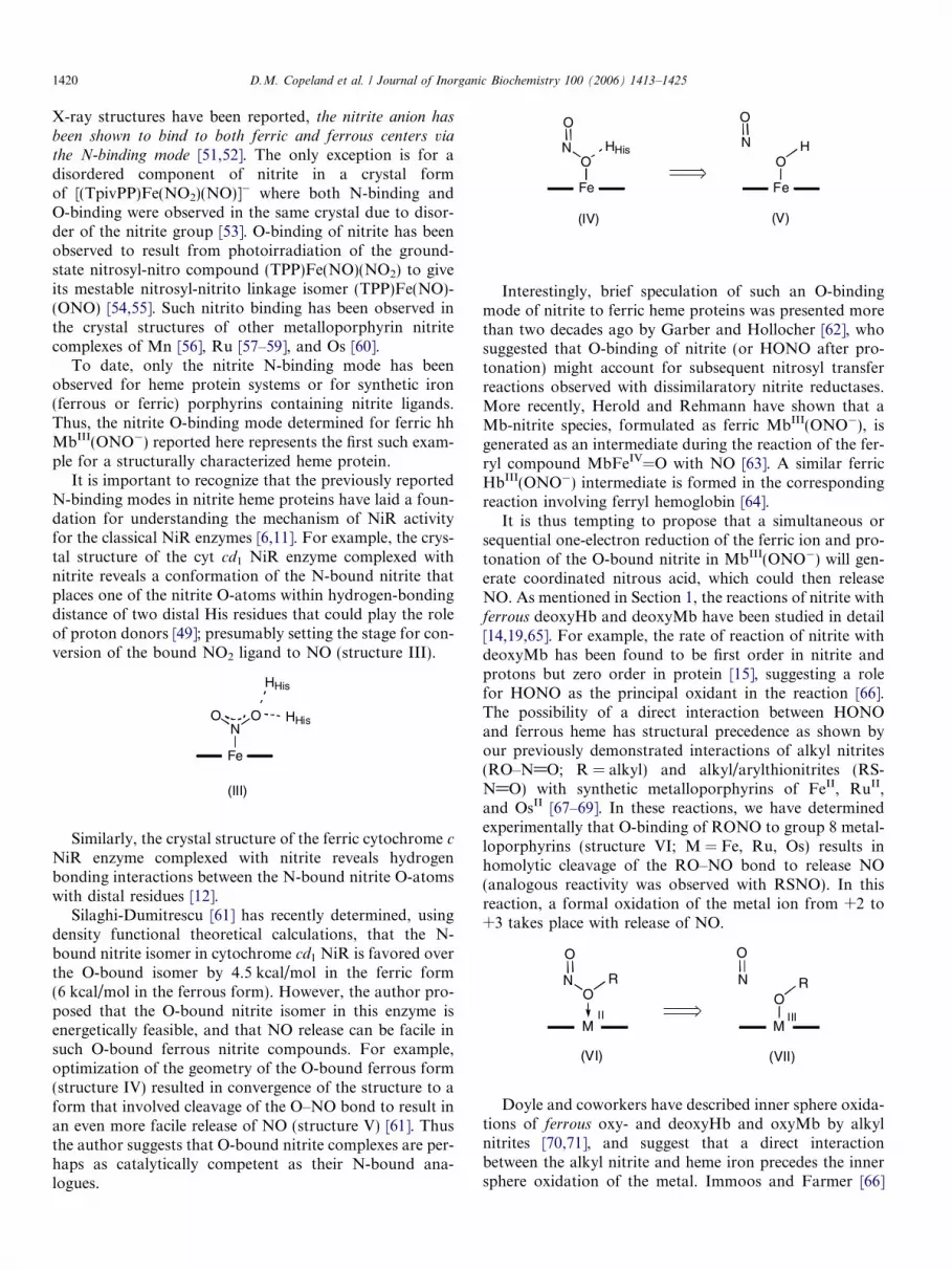
Silaghi-Dumitrescu [61] has recently determined, usingdensity functional theoretical calculations, that the N-bound nitrite isomer in cytochrome cd1 NiR is favored overthe O-bound isomer by 4.5 kcal/mol in the ferric form(6 kcal/mol in the ferrous form). However, the author pro-posed that the O-bound nitrite isomer in this enzyme isenergetically feasible, and that NO release can be facile insuch O-bound ferrous nitrite compounds. For example,optimization of the geometry of the O-bound ferrous form(structure IV) resulted in convergence of the structure to aform that involved cleavage of the O–NO bond to result inan even more facile release of NO (structure V) [61]. Thusthe author suggests that O-bound nitrite complexes are per-haps as catalytically competent as their N-bound ana-logues.
Fe
O
O
N HHis
Fe
O
O
N H
(IV) (V)
Interestingly, brief speculation of such an O-bindingmode of nitrite to ferric heme proteins was presented morethan two decades ago by Garber and Hollocher [62], whosuggested that O-binding of nitrite (or HONO after pro-tonation) might account for subsequent nitrosyl transferreactions observed with dissimilaratory nitrite reductases.More recently, Herold and Rehmann have shown that aMb-nitrite species, formulated as ferric MbIII(ONO�), isgenerated as an intermediate during the reaction of the fer-ryl compound MbFeIV=O with NO [63]. A similar ferricHbIII(ONO�) intermediate is formed in the correspondingreaction involving ferryl hemoglobin [64].
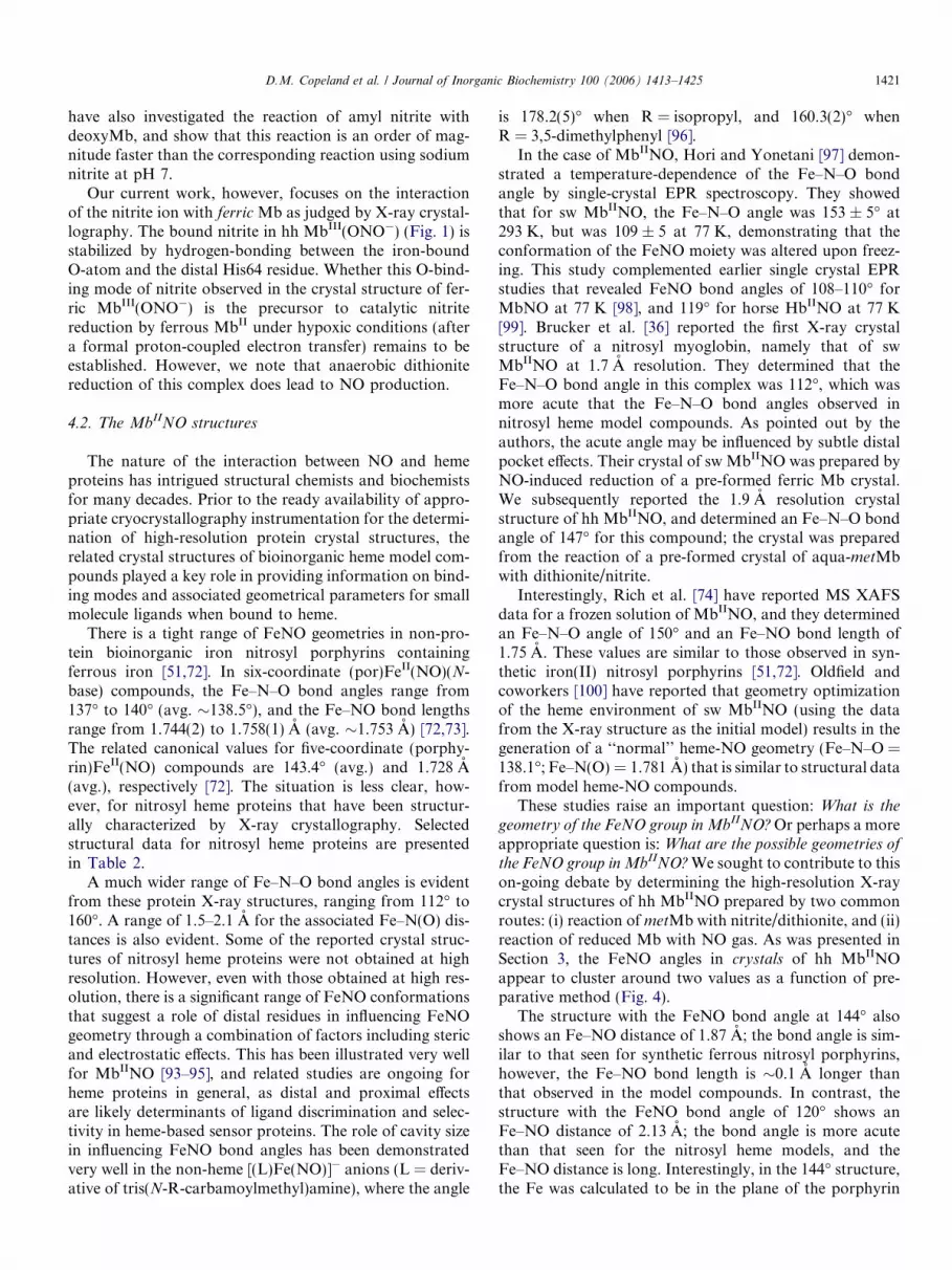
It is thus tempting to propose that a simultaneous orsequential one-electron reduction of the ferric ion and pro-tonation of the O-bound nitrite in MbIII(ONO�) will gen-erate coordinated nitrous acid, which could then releaseNO. As mentioned in Section 1, the reactions of nitrite withferrous deoxyHb and deoxyMb have been studied in detail[14,19,65]. For example, the rate of reaction of nitrite withdeoxyMb has been found to be first order in nitrite andprotons but zero order in protein [15], suggesting a rolefor HONO as the principal oxidant in the reaction [66].The possibility of a direct interaction between HONOand ferrous heme has structural precedence as shown byour previously demonstrated interactions of alkyl nitrites(RO–N@O; R = alkyl) and alkyl/arylthionitrites (RS-N@O) with synthetic metalloporphyrins of FeII, RuII,and OsII [67–69]. In these reactions, we have determinedexperimentally that O-binding of RONO to group 8 metal-loporphyrins (structure VI; M = Fe, Ru, Os) results inhomolytic cleavage of the RO–NO bond to release NO(analogous reactivity was observed with RSNO). In thisreaction, a formal oxidation of the metal ion from +2 to+3 takes place with release of NO.
M
O
O
N
(VI)
R
M
O
O
N R
(VII)
II III
Doyle and coworkers have described inner sphere oxida-tions of ferrous oxy- and deoxyHb and oxyMb by alkylnitrites [70,71], and suggest that a direct interactionbetween the alkyl nitrite and heme iron precedes the innersphere oxidation of the metal. Immoos and Farmer [66]
D.M. Copeland et al. / Journal of Inorganic Biochemistry 100 (2006) 1413–1425 1421
have also investigated the reaction of amyl nitrite withdeoxyMb, and show that this reaction is an order of mag-nitude faster than the corresponding reaction using sodiumnitrite at pH 7.
Our current work, however, focuses on the interactionof the nitrite ion with ferric Mb as judged by X-ray crystal-lography. The bound nitrite in hh MbIII(ONO�) (Fig. 1) isstabilized by hydrogen-bonding between the iron-boundO-atom and the distal His64 residue. Whether this O-bind-ing mode of nitrite observed in the crystal structure of fer-ric MbIII(ONO�) is the precursor to catalytic nitritereduction by ferrous MbII under hypoxic conditions (aftera formal proton-coupled electron transfer) remains to beestablished. However, we note that anaerobic dithionitereduction of this complex does lead to NO production.
4.2. The MbIINO structures
The nature of the interaction between NO and hemeproteins has intrigued structural chemists and biochemistsfor many decades. Prior to the ready availability of appro-priate cryocrystallography instrumentation for the determi-nation of high-resolution protein crystal structures, therelated crystal structures of bioinorganic heme model com-pounds played a key role in providing information on bind-ing modes and associated geometrical parameters for smallmolecule ligands when bound to heme.
There is a tight range of FeNO geometries in non-pro-tein bioinorganic iron nitrosyl porphyrins containingferrous iron [51,72]. In six-coordinate (por)FeII(NO)(N-base) compounds, the Fe–N–O bond angles range from137� to 140� (avg. �138.5�), and the Fe–NO bond lengthsrange from 1.744(2) to 1.758(1) A (avg. �1.753 A) [72,73].The related canonical values for five-coordinate (porphy-rin)FeII(NO) compounds are 143.4� (avg.) and 1.728 A(avg.), respectively [72]. The situation is less clear, how-ever, for nitrosyl heme proteins that have been structur-ally characterized by X-ray crystallography. Selectedstructural data for nitrosyl heme proteins are presentedin Table 2.
A much wider range of Fe–N–O bond angles is evidentfrom these protein X-ray structures, ranging from 112� to160�. A range of 1.5–2.1 A for the associated Fe–N(O) dis-tances is also evident. Some of the reported crystal struc-tures of nitrosyl heme proteins were not obtained at highresolution. However, even with those obtained at high res-olution, there is a significant range of FeNO conformationsthat suggest a role of distal residues in influencing FeNOgeometry through a combination of factors including stericand electrostatic effects. This has been illustrated very wellfor MbIINO [93–95], and related studies are ongoing forheme proteins in general, as distal and proximal effectsare likely determinants of ligand discrimination and selec-tivity in heme-based sensor proteins. The role of cavity sizein influencing FeNO bond angles has been demonstratedvery well in the non-heme [(L)Fe(NO)]� anions (L = deriv-ative of tris(N-R-carbamoylmethyl)amine), where the angle
is 178.2(5)� when R = isopropyl, and 160.3(2)� whenR = 3,5-dimethylphenyl [96].
In the case of MbIINO, Hori and Yonetani [97] demon-strated a temperature-dependence of the Fe–N–O bondangle by single-crystal EPR spectroscopy. They showedthat for sw MbIINO, the Fe–N–O angle was 153 ± 5� at293 K, but was 109 ± 5 at 77 K, demonstrating that theconformation of the FeNO moiety was altered upon freez-ing. This study complemented earlier single crystal EPRstudies that revealed FeNO bond angles of 108–110� forMbNO at 77 K [98], and 119� for horse HbIINO at 77 K[99]. Brucker et al. [36] reported the first X-ray crystalstructure of a nitrosyl myoglobin, namely that of swMbIINO at 1.7 A resolution. They determined that theFe–N–O bond angle in this complex was 112�, which wasmore acute that the Fe–N–O bond angles observed innitrosyl heme model compounds. As pointed out by theauthors, the acute angle may be influenced by subtle distalpocket effects. Their crystal of sw MbIINO was prepared byNO-induced reduction of a pre-formed ferric Mb crystal.We subsequently reported the 1.9 A resolution crystalstructure of hh MbIINO, and determined an Fe–N–O bondangle of 147� for this compound; the crystal was preparedfrom the reaction of a pre-formed crystal of aqua-metMbwith dithionite/nitrite.
Interestingly, Rich et al. [74] have reported MS XAFSdata for a frozen solution of MbIINO, and they determinedan Fe–N–O angle of 150� and an Fe–NO bond length of1.75 A. These values are similar to those observed in syn-thetic iron(II) nitrosyl porphyrins [51,72]. Oldfield andcoworkers [100] have reported that geometry optimizationof the heme environment of sw MbIINO (using the datafrom the X-ray structure as the initial model) results in thegeneration of a ‘‘normal’’ heme-NO geometry (Fe–N–O =138.1�; Fe–N(O) = 1.781 A) that is similar to structural datafrom model heme-NO compounds.
These studies raise an important question: What is the
geometry of the FeNO group in MbIINO? Or perhaps a moreappropriate question is: What are the possible geometries of
the FeNO group in MbIINO? We sought to contribute to thison-going debate by determining the high-resolution X-raycrystal structures of hh MbIINO prepared by two commonroutes: (i) reaction of metMb with nitrite/dithionite, and (ii)reaction of reduced Mb with NO gas. As was presented inSection 3, the FeNO angles in crystals of hh MbIINOappear to cluster around two values as a function of pre-parative method (Fig. 4).
The structure with the FeNO bond angle at 144� alsoshows an Fe–NO distance of 1.87 A; the bond angle is sim-ilar to that seen for synthetic ferrous nitrosyl porphyrins,however, the Fe–NO bond length is �0.1 A longer thanthat observed in the model compounds. In contrast, thestructure with the FeNO bond angle of 120� shows anFe–NO distance of 2.13 A; the bond angle is more acutethan that seen for the nitrosyl heme models, and theFe–NO distance is long. Interestingly, in the 144� structure,the Fe was calculated to be in the plane of the porphyrin

Table 2X-ray crystal structural data for nitrosylated His-liganded heme proteinsa,b
Nitrosylated protein res (A)c pdbd O.S.e Fe–N(O) (A) N–O (A) \FeNO (�) Fe-(Lax) (A) Reference
sw Mb 1.7 1HJT II 1.89 1.15 112 2.18 [36]L29F/D122N sw Mb 1.9f 1JDO II 1.86f 1.14f 127f 2.31f [36]hh Mb 1.30 II 1.87 1.20 144 2.08 g
hh Mb 1.30 II 2.13 1.17 120 2.15 g
hh Mb (MS XAFS)h II 1.75 1.12(2) 150(2) 2.05 [74]III 1.68(2) 1.13(2) 180(4) 2.04 [74]
NP1 from Rhodnius prolixus 2.3 4NP1 II 2.06f 1.34f 119.5f 2.10 f [75]III 2.02f 1.32f 145f 2.0f
NP2 1.45 1T68 1.93f 1.38f 134f 2.10f [76]NP4 from Rhodnium proxilus
WT (pH 7.4) 1.08 1X8Q II 1.74(2) 1.20(2) 143.8(1.6) 2.06(1) [77]WT (pH 5.6) 1.01 1X8O III 1.69(1) 1.09(1) 159.1(1.1) 1.994(7) [77]D129A/L130A mutant 1.0 1SXX III 1.60(2) 1.35f 155(2) 2.05f [78]T121V mutant 1.0 1SY1 III 1.62(2) 1.29f 158(2) 2.03f [78]D30N mutant 1.0 1SY3 II 1.78(2) 1.38f 132(2) 2.06f [78]D30A mutant 1.05 1SXW II 1.71(3) 1.35f 139(2) 2.09f [78]
HO from Neisseriae meningitides 1.75 1P3U II 1.58f 1.17f 147f 2.13f [79]HO-1 from rat 1.7 1JO2 II 2.10f 1.14f 125f 2.17f [80]Human HO-1
WT 1.55 1OZW II 1.64f 1.14f 138f 2.12f [81]D140A mutant 2.59 1OZL II 1.49f 1.16f 148f 2.12f [81]Verdoheme 2.10 1TWR 1.83f 1.15f 150.9f 2.54f [82]
1.98f 1.16f 150.4f 2.37Lupin legHb 1.8 1GDL II 1.97f 1.35f 145f 2.19f [83]Soybean legHb (MS XAFS)h II 1.77 1.12 147 1.98 [84]
III 1.68 1.12 173 1.89 [84]FixL from 2.5 1DP8 II 1.76f 1.14f 154f 2.10f [85]
Bradyrhizobium japonicum
cyt c 0 from 1.35 1E85 II 2.03f 1.16f 125f 5-Coord. [86]Alcaligenes xylosoxidans 1.92f 132f
cyt cd1 NiR from 1.8 1AOM II 2.0 1.37f 128f 1.98f [49]Paracoccus pantotropha
cyt cd1 NiR from 2.65 1NNO II 1.8 1.15 140f 1.98f [87]Pseudomonas aeruginosa
cyt c peroxidase from yeast 1.85 1.82 125, 135 2.04 [88]cyt c from 2.20 1DW2 II 1.75f 1.42f 113f 2.23f [89]
Rhodobacter sphaeroides II 1.82f 1.37f 112f 2.16f
Horse Hb 2.8 1.74 1.11 �145 (His) [90]Hb (bcysSNOH) 1.8 1BUW
a heme II 1.75 1.13 131 2.28 [91]b heme II 1.74 1.11 123 2.28 [91]
T-state human Hb 2.15 1RPSa heme II 1.72 1.13 138 5-coord. [92]b heme II 1.75 1.15 128 2.25 [92]
T-state human Hb (bcysSOH) 2.11 1RQ4a heme 1.72 1.15 138 5-coord. [92]b heme 1.76 1.17 138 2.19 [92]
T-state human Hb bW37E mutant 2.11 1RQAa heme 1.71 1.16 135 2.24 [92]b heme 1.76 1.18 126 2.19 [92]
a All nitrosylated sites are hexacoordinate of the form (por)Fe(NO)(His), except those of (i) cyt c’, and (ii) the a-hemes of T-state Hb, which arepentacoordinate. These are indicated in the Fe-(Lax) column by the term ‘‘5-coord’’.
b Abbreviations: sw Mb = recombinant sperm whale myoglobin; hh Mb = horse heart myoglobin; NP = nitrophorin; NP1 = nitrophorin-1;NP2 = nitrophorin-2; NP4 = nitrophorin-4; HO = heme oxygenase; legHb = leghemoglobin; Hb = hemoglobin; NiR = nitrite reductase.
c Resolution.d pdb access code.e Formal oxidation state of iron if known.f Metric data were obtained from the Protein Data Bank.g This work.h Data obtained from multiple-scattering X-ray absorption fine structure spectroscopic analyses.
1422 D.M. Copeland et al. / Journal of Inorganic Biochemistry 100 (2006) 1413–1425

D.M. Copeland et al. / Journal of Inorganic Biochemistry 100 (2006) 1413–1425 1423
ring (with less than 0.01 A deviation from the 4-N plane),whereas in the 120� structure, the Fe atom was calculatedto be 0.12 A below the 4-N porphyrin plane. This latter fea-ture suggests or reinforces the notion of a weaker bondbetween the NO ligand and the Fe center in this structure.Consistent with this view is the occurrence of a slightlylonger Fe–NO bond and more acute Fe–N–O angle.Indeed, the N-atom of the nitrosyl group is thus situatedcloser to the distal His64 ligand suggesting a strongerhydrogen bonding interaction between the NO and His64residue.
Our current thinking, therefore, is that the pre-formeddistal pocket in the precursor aqua-metMb crystals of hhMb imposes some constraints in the geometry of theheme distal pocket during and after formation of theMbIINO derivative. Nitrosylation of the aqua-metMbcrystals by the two methods employed here involve differ-ent intermediates along the reaction pathways, and theobserved MbIINO product geometries probably reflectdistal pocket stabilizations of local minima on the groundstate potential energy surface. In this scenario, the distalHis64 residue must play an important role. Such a possi-bility is reinforced by the MS XAFS work on frozensolutions of hh MbIINO that reveal more ‘‘normal’’FeNO geometries reflective of perhaps the global mini-mum structure [74]. The geometry optimization work byOldfield and coworkers [100] appears to support thishypothesis.
It is interesting to note that all the heme NO crystal
structures reported to date have been obtained from nitrosy-lation of pre-formed crystals of the protein. Put anotherway, the ‘‘NO’’ equivalent is added to a pre-formed distalpocket in the crystal. It is thus tempting to speculate thatthe wide range of FeNO geometries observed in the crystalstructures of heme-NO proteins to date may be reflective ofthe distal pocket stabilizations of various local minima inthese nitrosylated derivatives, something that is not avail-able to the synthetic iron nitrosyl porphyrins. In otherwords, the observed FeNO geometries in crystals ofheme-NO compounds prepared from nitrosylation of pre-cursor crystals may not necessarily represent the globalminimum conformations of the FeNO moiety.
5. Conclusion
Nitrite is now recognized as an oxyanion of nitrogenthat has important physiological roles in addition to its rolein denitrification. In this article, we report the first struc-tural study of the nitrite adduct of myoglobin. The high-resolution crystal structure reveals that the nitrite ligandis bound to the ferric heme center via the O-binding mode.The three heme protein nitrite crystal structures reported todate revealed the N-binding mode of nitrite to the hemecenters. The observation of a O-binding mode of nitriteto a heme protein provides support for this binding modeas a viable intermediate in nitrite reductase activity byheme proteins.
In addition, we have redetermined the crystal structureof hh MbIINO to higher resolution, and we have shownthat the FeNO bond angles cluster around two valuesdepending on the mode of preparation of the complex. Nit-rosylation of pre-formed Mb crystals may therefore imposelimitations on the attainment of global FeNO conforma-tion minima, and may enable variable FeNO conforma-tions that are stabilized by subtle electrostatic and stericeffects imposed by the pre-formed distal pockets.
6. Abbreviations
EPR electron paramagnetic resonance
Hb hem oglobin hh Mb hor se heart myoglobin Mb myo globin MS XAFS multiple-scattering X-ray absorptionfine structure
NiR nitr ite reductase por por phyrinato dianion SiR sulfi te reductase hemoprotein sw Mb sper m whale myoglobin TpivPP tetr akis(a,a,a,a-pivalamidophenyl)-porphyrinato dianion
TPP mes o-tetraphenylporphyrinato dianionAcknowledgements
We are grateful to the National Institutes of Health (GM64476; GBR-A) for funding for this work. X-ray diffractiondata for this study were measured at beamlines X-12B andX-29 of the National Synchrotron Light Source; financialsupport comes principally from the Offices of Biologicaland Environmental Research and of Basic Energy Sciencesof the US Department of Energy, and from the NationalCenter for Research Resources of the National Institutesof Health. We also thank Professor Patrick Farmer (Uni-versity of California, Irvine) for a copy of reference 66.
References
[1] W. Braida, S.K. Ong, Water, Air, Soil Pollut. 118 (2000) 13–26.[2] M.T. Gladwin, A.N. Schechter, D.B. Kim-Shapiro, R.P. Patel, N.
Hogg, S. Shiva, R.O. Cannon III, M. Kelm, D.A. Wink, M.G.Espey, E.H. Oldfield, R.M. Pluta, B.A. Freeman, J.R. Lancaster Jr.,M. Feelisch, J.O. Lundberg, Nat. Chem. Biol. 1 (2005) 308–314, andreferences therein.
[3] A. Dejam, C.J. Hunter, A.N. Schechter, M.T. Gladwin, Blood Cells,Mol., Diseases 32 (2004) 423–429.
[4] R.R. Eady, S.S. Hasnain, Denitrification, in: L. Que Jr., W.B.Tolman (Eds.), Comprehensive Coordination Chemistry II, Elsevier,San Diego, CA, 2004, pp. 759–786.
[5] I.M. Wasser, S. de Vries, P. Moenne-Loccoz, I. Schroder, K.D.Karlin, Chem. Rev. 102 (2002) 1201–1234.
[6] J.W.A. Allen, S.J. Ferguson, V. Fulop, Cytochrome cd1 nitritereductase, in: A. Messerschmidt, R. Huber, T.L. Poulos, K.

1424 D.M. Copeland et al. / Journal of Inorganic Biochemistry 100 (2006) 1413–1425
Wieghardt (Eds.), Handbook of Metalloproteins, Wiley, Chichester,2001, pp. 424–439.
[7] S. Suzuki, K. Kataoka, Y. Yamaguchi, T. Inoue, Y. Kai, Coord.Chem. Rev. 190–192 (1999) 245–265.
[8] E.I. Tocheva, F.I. Rosell, A.G. Mauk, M.E.P. Murphy, Science 304(2004) 867–870.
[9] M.L. Barrett, R.L. Harris, S. Antonyuk, M.A. Hough, M.J. Ellis, G.Sawers, R.R. Eady, S.S. Hasnain, Biochemistry 43 (2004) 16311–16319.
[10] S.V. Antonyuk, R.W. Strange, G. Sawers, R.R. Eady, S.S. Hasnain,Proc. Natl. Acad. Sci. USA 102 (2005) 12041–12046.
[11] O. Einsle, Cytochrome c nitrite reductase, in: A. Messerschmidt, R.Huber, T.L. Poulos, K. Wieghardt (Eds.), Handbook of Metallo-proteins, Wiley, Chichester, 2001, pp. 440–453.
[12] O. Einsle, A. Messerschmidt, R. Huber, P.M.H. Kroneck, F. Neese,J. Am. Chem. Soc. 124 (2002) 11737–11745.
[13] M.P. Doyle, R.A. Pickering, T.M. DeWeert, J.W. Hoekstra, D.Pater, J. Biol. Chem. 256 (1981) 12393–12398.
[14] Z. Huang, S. Shiva, D.B. Kim-Shapiro, R.P. Patel, L.A. Ringwood,C.E. Irby, K.T. Huang, C. Ho, N. Hogg, A.N. Schechter, M.T.Gladwin, J. Clin. Invest. 115 (2005) 2099–2107.
[15] F. Sulc, C.E. Immoos, D. Pervitsky, P.J. Farmer, J. Am. Chem. Soc.126 (2004) 1096–1101.
[16] A. Webb, R. Bond, P. McLean, R. Uppal, N. Benjamin, A.Ahluwalia, Proc. Natl. Acad. Sci. USA 101 (2004) 13683–13688, andreferences therein.
[17] H. Li, A. Samouilov, X. Liu, J.L. Zweier, J. Biol. Chem. 279 (2004)16939–16946.
[18] B.L.J. Godber, J.J. Doel, G.P. Sapkota, D.R. Blake, C.R.Stevens, R. Eisenthal, R. Harrison, J. Biol. Chem. 275 (2000)7757–7763.
[19] K. Cosby, K.S. Partovi, J.H. Crawford, R.P. Patel, C.D. Reiter, S.Martyr, B.K. Yang, M.A. Waclawiw, G. Zalos, X. Xu, K.T. Huang,M. Shields, D.B. Kim-Shapiro, A.N. Schechter, R.O. Cannon III,M.T. Gladwin, Nat. Med. 9 (2003) 1498–1505.
[20] E. Nagababu, S. Ramasamy, D.R. Abernethy, J.M. Rifkind, J. Biol.Chem. 278 (2003) 46349–46356.
[21] V.P. Reutov, Biochemistry (Moscow) 67 (2002) 293–311.[22] A. Dejam, C.J. Hunter, M.M. Pelletier, L.L. Hsu, R.F. Machado, S.
Shiva, G.G. Power, M. Kelm, M.T. Gladwin, A.N. Schechter, Blood106 (2005) 734–739.
[23] M.R. Duranski, J.J.M. Greer, A. Dejam, S. Jaganmohan, N. Hogg,W. Langston, R.P. Patel, S.-F. Yet, X. Wang, C.G. Kevil, M.T.Gladwin, D.J. Lefer, J. Clin. Invest. 115 (2005) 1232–1240.
[24] N.S. Bryan, B.O. Fernandez, S.M. Bauer, M.F. Garcia-Suara, A.B.Milson, T. Rassaf, R.E. Maloney, A. Bharti, J. Rodriguez, M.Feelisch, Nat. Chem. Biol. 1 (2005) 290–297.
[25] B. Arendt, L.H. Skibsted, H. Andersen, Z. Lebensm. Unters.Forsch. A 204 (1997) 7–12, and references therein.
[26] C.U. Carlsen, J.K.S. Møller, L.H. Skibsted, Coord. Chem. Rev. 249(2005) 485–498.
[27] J.K.S. Møller, L.H. Skibsted, Chem. Rev. 102 (2002) 1167–1178.[28] A. Wanat, J. Gdula-Argasinska, D. Rutkowska-Zbik, M. Witko, G.
Stochel, R. van Eldik, J. Biol. Inorg. Chem. 7 (2002) 165–176, andreferences therein.
[29] B.O. Fernandez, I.M. Lorkovic, P.C. Ford, Inorg. Chem. 43 (2004)5393–5402.
[30] R. Lin, M. Bayachou, J. Greaves, P.J. Farmer, J. Am. Chem. Soc.119 (1997) 12689–12690.
[31] W.R. Murphy Jr., K.J. Takeuchi, T.J. Meyer, J. Am. Chem. Soc.104 (1982) 5817–5819.
[32] M.H. Barley, K.J. Takeuchi, T.J. Meyer, J. Am. Chem. Soc. 108(1986) 5876–5885.
[33] M.H. Barley, M.R. Rhodes, T.J. Meyer, Inorg. Chem. 26 (1987)1746–1750.
[34] J.N. Younathan, K.S. Wood, T.J. Meyer, Inorg. Chem. 31 (1992)3280–3285.
[35] Y. Chi, J. Chen, K. Aoki, Inorg. Chem. 43 (2004) 8437–8446.
[36] E.A. Brucker, J.S. Olson, M. Ikeda-Saito, G.N. Philips Jr., Proteins:Struct. Funct. Genet. 30 (1998) 352–356.
[37] D.M. Copeland, A.H. West, G.B. Richter-Addo, Proteins: Struct.Func. Genet. 53 (2003) 182–192.
[38] J. Pflugrath, Acta Crystallogr. D 55 (1999) 1718–1725.[39] Z. Otwinowski, W. Minor, Meth. Enzymol. 276 (1997) 307–326.[40] A.T. Brunger, P.D. Adams, G.M. Clore, W.L. DeLano, P. Gros,
R.W. Grosse-Kunstleve, J.-S. Jiang, J. Kuszewski, M. Nilges, N.S.Pannu, R.J. Reed, L.M. Rice, T. Simonson, G.L. Warren, ActaCrystallogr. D 54 (1998) 905–921.
[41] A. Vagin, A. Teplyakov, J. Appl. Crystallogr. 30 (1997) 1022–1025.[42] CCP4 Project, Acta Crystallogr. D 50 (1994) 760–763.[43] I.W. Davis, L.W. Murray, J.S. Richardson, D.C. Richardson, Nucl.
Acids Res. 32 (2004) W615–W619, Web Server.[44] W.B. Arendall III, W. Tempel, J.S. Richardson, W. Zhou, S. Wang,
I.W. Davis, Z.-J. Liu, J.P. Rose, W.M. Carson, M. Luo, D.C.Richardson, B.-C. Wang, J. Struct. Funct. Genomics 6 (2005) 1–11.
[45] D.E. McRee, J. Struct. Biol. 125 (1999) 156–165.[46] R.A. Laskowski, M.W. MacArthur, D.S. Moss, J.M. Thornton, J.
Appl. Cryst. 26 (1993) 283–291.[47] M. Sono, J.H. Dawson, J. Biol. Chem. 257 (1982) 5496–5502, and
references therein.[48] M.A. Hitchman, G.L. Rowbottom, Coord. Chem. Rev. 42 (1982)
55–132.[49] P.A. Williams, V. Fulop, E.F. Garman, N.F.W. Saunders, S.J.
Ferguson, J. Hajdu, Nature 389 (1997) 406–412.[50] B.R. Crane, L.M. Siegel, E.D. Getzoff, Biochemistry 36 (1997)
12120–12137.[51] G.R.A. Wyllie, W.R. Scheidt, Chem. Rev. 102 (2002) 1067–1089.[52] H. Nasri, M.K. Ellison, M. Shang, C.E. Schultz, W.R. Scheidt,
Inorg. Chem. 43 (2004) 2932–2942.[53] H. Nasri, M.K. Ellison, S. Chen, B.H. Huynh, W.R. Scheidt, J. Am.
Chem. Soc. 119 (1997) 6274–6283.[54] J. Lee, A.Y. Kovalevsky, I.V. Novozhilova, K.A. Bagley, P.
Coppens, G.B. Richter-Addo, J. Am. Chem. Soc. 126 (2004) 7180–7181.
[55] I.V. Novozhilova, P. Coppens, J. Lee, G.B. Richter-Addo, K.A.Bagley, J. Am. Chem. Soc. 128 (2006) 2093–2104.
[56] K.S. Suslick, R.A. Watson, Inorg. Chem. 30 (1991) 912–919.[57] K.M. Kadish, V.A. Adamian, E.V. Caemelbecke, Z. Tan, P.
Tagliatesta, P. Bianco, T. Boschi, G.-B. Yi, M.A. Khan, G.B.Richter-Addo, Inorg. Chem. 35 (1996) 1343–1348.
[58] D.S. Bohle, C.-H. Hung, B.D. Smith, Inorg. Chem. 37 (1998) 5798–5806.
[59] K.M. Miranda, X. Bu, I. Lorkovic, P.C. Ford, Inorg. Chem. 36(1997) 4838–4848.
[60] F.A. Leal, I.M. Lorkovic, P.C. Ford, J. Lee, L. Chen, L. Torres,M.A. Khan, G.B. Richter-Addo, Can. J. Chem. 81 (2003) 872–881.
[61] R. Silaghi-Dumitrescu, Inorg. Chem. 43 (2004) 3715–3718.[62] E.A.E. Garber, T.C. Hollocher, J. Biol. Chem. 257 (1982) 8091–
8097.[63] S. Herold, F.-J.K. Rehmann, J. Biol. Inorg. Chem. 6 (2001) 543–555.[64] S. Herold, F.-J.K. Rehmann, Free Radical Biol. Med. 34 (2003)
531–545.[65] K.T. Huang, A. Keszler, N. Patel, R.P. Patel, M.T. Gladwin, D.B.
Kim-Shapiro, N. Hogg, J. Biol. Chem. 280 (2005) 31126–31131.[66] C. Immoos, Ph.D. thesis, University of California at Irvine, 2001,
pp. 138–144 (Chapter 4).[67] G.-B. Yi, L. Chen, M.A. Khan, G.B. Richter-Addo, Inorg. Chem.
36 (1997) 3876–3885.[68] G.B. Richter-Addo, Acc. Chem. Res. 32 (1999) 529–536.[69] J. Lee, L. Chen, A.H. West, G.B. Richter-Addo, Chem. Rev. 102
(2002) 1019–1065, and references therein.[70] M.P. Doyle, D.M. LePoire, R.A. Pickering, J. Biol. Chem. 256
(1981) 12399–12404.[71] M.P. Doyle, R.A. Pickering, J. da Conceicao, J. Biol. Chem. 259
(1984) 80–87.

D.M. Copeland et al. / Journal of Inorganic Biochemistry 100 (2006) 1413–1425 1425
[72] G.R.A. Wyllie, C.E. Schultz, W.R. Scheidt, Inorg. Chem. 42 (2003)5722–5734.
[73] S.P. Rath, R. Koerner, M.M. Olmstead, A.L. Balch, J. Am. Chem.Soc. 125 (2003) 11798–11799.
[74] A.M. Rich, R.S. Armstrong, P.J. Ellis, P.A. Lay, J. Am. Chem. Soc.120 (1998) 10827–10836.
[75] X.D. Ding, A. Weichsel, J.F. Andersen, T.K. Shokhireva, C.Balfour, A.J. Pierik, B.A. Averill, W.R. Montfort, F.A. Walker, J.Am. Chem. Soc. 121 (1999) 128–138.
[76] A. Weichsel,W.R. Montfort, deposited with the PDB (PDB accesscode 1T68).
[77] D.A. Kondrashov, S.A. Roberts, A. Weichsel, W.R. Montfort,Biochemistry 43 (2004) 13637–13647.
[78] E.M. Maes, A. Weichsel, J.F. Andersen, D. Shepley, W.R. Mont-fort, Biochemistry 43 (2004) 6679–6690.
[79] J. Friedman, L. Lad, R. Deshmukh, H. Li, A. Wilks, T.L. Poulos, J.Biol. Chem. 278 (2003) 34654–34659.
[80] M. Sugishima, H. Sakamoto, M. Noguchi, K. Fukuyama, Bio-chemistry 42 (2003) 9898–9905.
[81] L. Lad, J. Wang, H. Li, J. Friedman, B. Bhaskar, P.R. Ortiz deMontellano, T.L. Poulos, J. Mol. Biol. 330 (2003) 527–538.
[82] L. Lad, P.R. Ortiz de Montellano, T.L. Poulos, J. Inorg. Biochem.98 (2004) 1686–1695.
[83] E.H. Harutyunyan, T.N. Safonova, I.P. Kuranova, A.N. Popov,A.V. Teplyakov, G.V. Obmolova, B.K. Vainshtein, G.G. Dodson,J.C. Wilson, J. Mol. Biol. 264 (1996) 152–161.
[84] A.M. Rich, P.J. Ellis, L. Tennant, P.E. Wright, R.S. Armstrong,P.A. Lay, Biochemistry 38 (1999) 16491–16499.
[85] W. Gong, B. Hao, M.K. Chan, Biochemistry 39 (2000) 3955–3962.
[86] D.M. Lawson, C.E.M. Stevenson, C.R. Andrew, R.R. Eady, EMBOJ. 19 (2000) 5661–5671.
[87] D. Nurizzo, F. Cutruzzola, M. Arese, D. Bourgeois, M. Brunori, C.Cambillau, M. Tegoni, Biochemistry 37 (1998) 13987–13996.
[88] S.L. Edwards, J. Kraut, T.L. Poulos, Biochemistry 27 (1988) 8074–8081.
[89] D. Leys, K. Backers, T.E. Meyer, W.R. Hagen, M.A. Cusanovich,J.J. Van Beeumen, J. Biol. Chem. 275 (2000) 16050–16056.
[90] J.F. Deatherage, K. Moffat, J. Mol. Biol. 134 (1979) 401–417.[91] N.-L. Chan, P.H. Rogers, A. Arnone, Biochemistry 37 (1998)
16459–16464.[92] N.L. Chan, J.S. Kavanaugh, P.H. Rogers, A. Arnone, Biochemistry
43 (2004) 118–132.[93] B.A. Springer, S.G. Sligar, J.S. Olson, G.N. Phillips Jr., Chem. Rev.
94 (1994) 699–714.[94] T. Tomita, H. Hirota, T. Ogura, J.S. Olson, T. Kitagawa, J. Phys.
Chem. B 103 (1999) 7044–7054.[95] C.M. Coyle, K.M. Vogel, T.S. Rush III, P.M. Kozlowski, R.
Williams, T.G. Spiro, Y. Dou, M. Ikeda-Saito, J.S. Olson, M.Z.Zgierski, Biochemistry 42 (2003) 4896–4903.
[96] M. Ray, A.P. Golombek, M.P. Hendrich, G.P.A. Yap, L.M. Liable-Sands, A.L. Rheingold, A.S. Borovik, Inorg. Chem. 38 (1999) 3110–3115.
[97] H. Hori, M. Ikeda-Saito, T. Yonetani, J. Biol. Chem. 256 (1981)7849–7855.
[98] L.C. Dickinson, J.C.W. Chien, J. Am. Chem. Soc. 93 (1971) 5036–5040.
[99] J.C.W. Chien, J. Chem. Phys. 51 (1969) 4220–4227.[100] Y. Zhang, W. Gossman, E. Oldfield, J. Am. Chem. Soc. 125 (2003)
16387–16396.