crystal structure and functional dissection of the cytostatic cytokine oncostatin m
TRANSCRIPT

Crystal structure and functional dissection of the cytostaticcytokine oncostatin MMarc C Deller1,2†#, Keith R Hudson2‡#, Shinji Ikemizu1, Jerónimo Bravo1§,E Yvonne Jones1* and John K Heath2
Background: The cytokine oncostatin M (OSM) inhibits growth ofcertain tumour-derived cells, induces proliferation in other cell types(e.g. haemangioblasts) and is a mediator of inflammatory responses. Itsmechanism of action is via specific binding to gp130 and either the leukaemiainhibitory factor receptor (LIFR) or oncostatin M receptor (OSMR) systemsat the cell surface to form an active signalling complex.
Results: We report here the crystal structure of human oncostatin M (hOSM)along with mutagenesis data which map the receptor-binding epitopes of themolecule. The structure was determined to a resolution of 2.2 Å and conformsto the haematopoietin cytokine up-up-down-down four-helix bundle topology.The site 2 epitope, responsible for gp130 binding, is centred around Gly120which forms a ‘dimple’ on the surface of the molecule located on helices A andC. The site 3 motif, responsible for LIFR and OSMR binding, consists of aprotruding Phe160/Lys163 pair located at the start of helix D.
Conclusions: The data presented allow functional dissection of thereceptor-binding interfaces to atomic resolution. Modelling suggests that thegp130 residue Phe169 packs into the site 2 dimple in an analogous fashionto structurally equivalent residues at the growth hormone–growth hormonereceptor interface, implying that certain key features may underlie recognitionacross the whole cytokine/receptor superfamily. Conversely, detailedcomparison of the available structures suggests that variations on a commontheme dictate the specificity of receptor–ligand interactions within the gp130family of cytokines.
IntroductionHuman oncostatin M (hOSM) is a secreted 252 amino acidpolypeptide cytokine that was originally isolated by virtueof its ability to suppress the proliferation of humanmelanoma cells in vitro [1]. Subsequent biological investiga-tions have revealed that hOSM and mouse OSM (mOSM)exhibit a diversity of effects on different target cell types.These include the ability to inhibit the multiplication of avariety of different tumour-derived cell lines (reviewed in[2]) as well as inducing the proliferation of certain cellsincluding Kaposi sarcoma-derived spindle cells [3] and hae-mangioblasts derived from the aorta–gonad–mesonephrosregion of the developing embryo [4]. hOSM, released frommacrophages and neutrophils [5], has also been implicatedas an important mediator of pro-inflammatory responses injoint destruction [6] and central nervous system inflamma-tion and neurodegeneration [7,8] via, inter alia, the induc-tion of acute- phase protein gene expression [9,10].
hOSM is a member of the family of gp130 cytokines thatare characterised by the use of the shared transmembranetransducing receptor gp130 in their mechanism of action[11]. hOSM is therefore functionally related to leukemiainhibitory factor (LIF), interleukin (IL)-6 and IL-11, car-diotrophin-1 (CT-1) and ciliary neurotrophic factor(CNTF). A key feature of the gp130 cytokines is thatintracellular signalling results from the ligand-mediatedformation of oligomeric receptor complexes which,depending upon the identity of the ligand, can differ inboth composition and stoichiometry [12]. In all cases,however, receptor complexes include one or more mol-ecules of the shared transducer gp130 [13]. gp130cytokines can therefore exhibit both unique and sharedbiological activities in vivo and in vitro depending upon thecomposition of the receptor signalling complex that isformed [11]. hOSM signals via formation of an activereceptor signalling complex that comprises gp130 and one
Addresses: 1Cancer Research Campaign ReceptorStructure Group, Division of Structural Biology, TheWellcome Trust Centre for Human Genetics,University of Oxford, Roosevelt Drive, Oxford OX37BN, UK and 2Cancer Research Campaign GrowthFactors Group, School of Biochemistry, University ofBirmingham, Edgbaston, Birmingham B15 2TT, UK.
Present addresses: †Yale University School ofMedicine, Department of Pharmacology, SterlingHall of Medicine, PO Box 208066, New Haven,CT 06520-8066, USA, ‡Genesis Research andDevelopment, 1 Fox Street, Parnell, Auckland, NewZealand and §Laboratory of Molecular Biology,Medical Research Council Centre, Hills Road,Cambridge CB2 2QH, UK.
*Corresponding author.E-mail: [email protected]
#These authors contributed equally to this work.
Key words: four-helix bundle cytokine, gp130binding, oncostatin M, structure–function
Received: 26 April 2000Revisions requested: 7 June 2000Revisions received: 22 June 2000Accepted: 28 June 2000
Published: 20 July 2000
Structure 2000, 8:863–874
0969-2126/00/$ – see front matter © 2000 Elsevier Science Ltd. All rights reserved.
Research Article 863

of two partner receptors: either the LIF receptor (LIFR)[14] or the structurally related OSM-specific receptor,OSMR [15]. mOSM [16] differs from hOSM in that it onlysignals via association with gp130 and the murine OSMR[17,18]; this means that there is a significant divergence inaction between mOSM and hOSM and that the LIFRrecognition elements of OSM are not conserved. Thisfinding also indicates that the unique biological activitiesof hOSM, such as cytostatic effects on tumour cells, aremediated via association with OSMR. Those activities ofhOSM that are shared with other cytokines, such as LIF orCNTF, may reflect a common association with the LIFR.
Knowledge of the structural basis of receptor recognitionby gp130 cytokines is a central issue in understanding boththe specificity of their biological functions and developingmethods for therapeutic intervention in gp130-mediatedsignalling. The crystal structures of three gp130 cytokines,LIF [19], CNTF [20] and IL-6 [21], have been previouslydescribed. These reveal that all three cytokines share acommon three-dimensional fold, the four-helix bundle[22], which comprises four α helices ranging from 15 to 22amino acids in length (termed A, B, C and D) and linkedby polypeptide loops. Detailed structure–function studiesof human LIF, which is most closely related to hOSM insequence and receptor repertoire, have defined two recep-tor recognition epitopes: sites 2 and 3. Site 2 (so-termed byanalogy to the receptor-binding sites in growth hormone[23]), is required for recognition of gp130 and has beennarrowed down to a cluster of candidate amino acidresidues located in helices A and C [24]. Site 3 is requiredfor recognition of LIFR and is dominated by a pair of con-served amino acid residues located at the N-terminal tip ofhelix D [24]. The combined activity of these two recogni-tion epitopes leads to the formation of a trimeric, LIF-mediated, high-affinity signalling complex [24].
Structural comparisons of ligands with differing recognitionproperties present a valuable opportunity to analyse themolecular determinants that dictate receptor specificity.Here, we report the determination of the three-dimensionalstructure of hOSM by crystallographic techniques and con-comitant functional dissection by mutagenesis. BecausehOSM binds to gp130 with a higher affinity than LIF [24],this allows structure–function analysis of the site 2 gp130recognition epitope at the resolution of individual aminoacid residues. Combining these findings with the high-reso-lution crystal structure of the complementary ligand-recog-nition epitope located in the cytokine receptor homologydomain (CHD) of gp130 [25] results, for the first time, in adefinition of the structure of the interface between hOSMand gp130. In addition a detailed comparison of the three-dimensional structure of the site 3 recognition epitope ofhOSM and hLIF also offers insights into the molecularbasis by which related ligands exhibit different receptorrecognition properties. These findings reveal that, in both
cases, receptor specificity is controlled by subtle structuralchanges to a common recognition framework.
ResultsExpression and structure determinationA soluble 21.4 kDa fragment of hOSM was expressed inEscherichia coli as a glutathione-S-transferase (GST) fusionprotein. The resulting purified protein consists of residues1–187 of hOSM and a further four residues at the N termi-nus corresponding to the 3C protease site linker (GPGS).Ba/F3 cell-survival assays and receptor-binding assays con-firmed that this recombinant form binds gp130 with anaffinity equivalent to wild-type (WT) hOSM.
Crystallisation trials produced Bragg diffraction qualitycrystals belonging to the orthogonal space group P212121,with unit-cell dimensions a = 35.8 Å, b = 53.1 Å, c = 106.8 Å.The structure was determined from a single ethylmer-curyphosphate (EMP) derivative using the multipleanomalous dispersion (MAD) phasing technique withX-ray diffraction data collected on beam line BM14 of theEuropean Synchrotron Radiation Facility (ESRF). Thestructure has been refined to a crystallographic R factor of20.5% (Rfree 26.1%) for all data between 20.0 and 2.2 Å,with stereochemistry typified by root mean squared devia-tions (rmsd) in bond lengths of 0.015 Å and in bond anglesof 1.8°. Crystallographic statistics are reported in Tables 1and 2. Residues 1–3 and 135–155 appear to be disorderedand are not included in the final model. With the excep-tion of these regions, and a flexible loop between residuesGlu90 and Lys110, the electron density is good through-out the course of the mainchain (a representative portionof electron density is shown in Figure 1a). Residue num-bering is that of the human native mature protein(GenBank accession number M27286).
Structure description hOSM is a compact barrel-shaped molecule with dimen-sions of approximately 20 Å × 27 Å × 56 Å that conformsto the up-up-down-down four-helix bundle topology(Figure 1b,c). The basic fold consists of the four mainhelical regions (helix A, residues 10–37; helix B, residues67–90; helix C, residues 105–131; helix D, residues159–185) connected by two long overhand loops (AB loop,residues 38–66; CD loop, residues 130–158) and one shortloop (BC loop, residues 91–104). The overall arrangementmay be considered as two pairs of antiparallel helices thatpack together, with A–D forming one pair and B–Cforming the other.
Both helices A and C exhibit breaks in the classical hydro-gen-bonding pattern, with substitute hydrogen bondsformed to tightly bound water molecules. In helix C thisresults in a slight kink (between residues Gln112 andPro116). The kink in helix A is more pronounced and iscaused by a break in the helical conformation, as residues
864 Structure 2000, Vol 8 No 8

Gln25 and Leu30 adopt a hydrogen-bonded turn confor-mation, with four tightly bound water molecules substitut-ing for the classical hydrogen-bonding pattern (Figure 1a).Residues Thr27–Ile37 of helix A adopt a 310 helix confor-mation. The curved nature of helices A and C facilitatesclose packing of the A–D and B–C helix pairs, so promot-ing an extensive solvent-inaccessible core.
The core of the protein is composed of two groups of aro-matic stacking interactions in which Phe56, Tyr173,Phe169 and Phe176 form one group and Phe70, Phe185and Trp187 form another. This distribution emphasizesthe hydrophobic nature of helix D that donates all of thearomatics to these groupings, with the exception of Phe56(AB loop) and Phe70 (B helix).
The N-terminal loop preceding helix A (Gly4–Glu9) istethered to the C terminus of helix C by one of the twodisulphide bridges (Cys6 and Cys127). The second disul-phide bridge, between Cys49 and Cys167, anchors thestart of the AB loop to the N-terminal region of helix D.The AB loop itself consists of two separate α-helicalregions running from Pro43 to Arg46 and Glu59 to Gly64.Residues between these two helical regions adopt anextended conformation and pack closely against helix D.By contrast, the second two loops (BC and CD) are lessclosely associated with the core structure. The BC loopprotrudes above the four-helix bundle and exhibits higherthan average B factors. Despite this relative mobility itdoes, however, contain a high percentage of regular sec-ondary structure with a 310 helix running between residuesAla95 and Asp97 followed by an α helix up to residueSer101. The long CD loop appears to be highly mobileand could not be modelled as a single conformation.
Structural comparison with other helical cytokineshOSM is the fourth member of the gp130 cytokine familyto be analysed structurally; crystal and/or solution struc-tures have been reported for IL-6 [21,26], CNTF [20] and
LIF [19,27]. Structural superimpositions with these andother four-helix bundle cytokines show that the gp130cytokines form a distinct family both structurally andfunctionally. Within the gp130 cytokine family hOSM hasthe greatest structural similarity to LIF (rmsd of 2.1 Åover 145 pairs of structurally equivalent Cα atoms). Themore distant structural similarity to the four-helix bundlesof the growth hormone family is at its best for superimpo-sition of hOSM and erythropoietin (EPO; [28]) (rmsd of2.8 Å for 120 pairs of structurally equivalent Cα atoms).
The addition of hOSM to the gp130 cytokine structuraldatabase highlights several features of potential functional
Research Article Crystal structure of oncostatin M Deller et al. 865
Table 1
Data collection statistics.
Native EMPλ1 EMPλ2 EMPλ3
Wavelength (Å) 0.979273 1.00914 0.826516 1.00669Unit cell
a,b,c (Å) 35.8, 53.1, 106.8 35.9, 53.3, 106.7α = β = γ (°) 90 90
Resolution range (Å) 25–2.2 25–2.6Total reflections 43,901 24,040 26,223 24,159Unique reflections 10,513 11,650 12,069 11,691Redundancy 4.2 2.1 2.2 2.1Completeness (%) 96.5 (98.2) 96.4 (63.1) 99.9 (100) 96.8 (66.7)<I/σ(I)> 13.2 (4.3) 12.1 (2.9) 12.5 (4.7) 12.3 (3.3)Rmerge (%) 8.3 (37.6) 4.6 (18.7) 4.6 (16.4) 4.3 (17.3)
EMPλ denotes MAD data collected from EMP-derivatised hOSM. Rmerge = Σ|I–<I>|/Σ<I>. Values in parentheses correspond to the highestresolution shell (2.66–2.60 Å for EMP, 2.25–2.20 Å for native).
Table 2
Refinement statistics.
Resolution range (Å) 20–2.2 (2.28–2.20)Rcryst (%) 20.5 (27.1)Rfree (%) 26.1 (33.6)Completeness
working set (%) 86.1 (81.0)test set (%) 9.9 (8.6)
No. of reflections (F > 0)working set 9410 (870)test set 1082 (92)
No. of non-hydrogen atomsprotein 1319water 116
Rmsd from idealitybond lengths (Å) 0.0147bond angles (Å) 1.7997dihedrals (°) 19.8882improper (°) 1.0381
Average B factorsmainchain (Å2) 27.1sidechain (Å2) 32.4water (Å2) 38.4all atoms (range) (Å2) 30.5 (6.3–84.9)
Rcryst = Σ||Fobs|–|Fcalc||/Σ|Fobs|. Rfree is as for Rcryst but calculated using a10% test set of reflections excluded from the refinement. Values inparentheses correspond to the highest resolution shell (2.28–2.20 Å).

importance (Figure 2). First, the kinked helix A isrevealed as a distinctive feature not just of the gp130cytokines but also specifically of the members of thefamily of cytokines that bind to LIFR; IL-6 is the solegp130 cytokine lacking this feature (Figures 1a,2a). Thekink results in a repositioning of the C-terminal end ofhelix A relative to the ‘top’ of the four-helix bundle, aregion carrying the major epitope for LIFR, and OSMR,binding (at the start of helix D). This feature is thereforesuggestive of the positioning of the C-terminal end ofhelix A impacting on LIFR and OSMR binding. Con-versely, the positioning of the N-terminal portion ofhelix A, which is implicated in gp130 binding, is relativelyconserved across all members of the family.
In general, functional studies on the gp130 cytokines haveimplicated the helices, rather than the connecting loops,
as having the dominant role in ligand binding. Comparisonof loop structures between family members appears tobear this out as there are few clearly conserved characteris-tics. Each crystal structure, except LIF, indicates highlyflexible regions in the long interhelix loops; however,these vary in location between the AB loop in CNTF andIL-6, and the CD loop in CNTF and hOSM. Similarly thepositioning of disulphides tethering loop regions to thecore four-helix bundle is not conserved. It has been noted,however, that the positioning, relative to helix D, of theN-terminal portion of the AB loop in LIF differs from thatin the distantly related four-helix bundle cytokines [19].The current structural comparisons may point to somefunctional correlation of this feature with LIFR binding asboth LIF and hOSM have a disulphide bridge anchoringan α-helical N-terminal portion of the AB loop to the topof helix D (Figure 2a).
866 Structure 2000, Vol 8 No 8
Figure 1
The structure of hOSM. (a) Refinedcoordinates and 2|Fo|–|Fc|αcalc electron-densitymap of hOSM contoured at 1σ showingdensity for the kinked region of helix A centredon Thr27. CNTF and LIF also have kinks owingto disruption of the mainchain hydrogenbonding around residues Thr32 and Ser36,respectively [19,20]. Both these residues bindwater molecules through the Oγ1 atom toprovide a substitute hydrogen-bond networksimilar to the pattern observed here for hOSMinvolving residue Thr27. (b) Ribbon diagram ofhOSM coloured from blue at the N terminusthrough to red at the C terminus. Disulphidebonds are represented as ball-and-stickmodels with the sulphur atoms highlighted asyellow spheres. The transparent dotted sectionrepresents the CD loop as observed in LIF.(c) Stereodiagram of the Cα trace for hOSM.
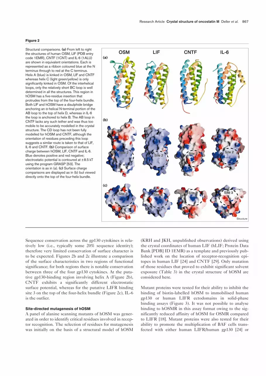
Sequence conservation across the gp130 cytokines is rela-tively low (i.e., typically some 20% sequence identity);therefore very limited conservation of surface character isto be expected. Figures 2b and 2c illustrate a comparisonof the surface characteristics in two regions of functionalsignificance; for both regions there is notable conservationbetween three of the four gp130 cytokines. At the puta-tive gp130-binding region involving helix A (Figure 2b),CNTF exhibits a significantly different electrostaticsurface potential, whereas for the putative LIFR bindingsite 3 on the top of the four-helix bundle (Figure 2c), IL-6is the outlier.
Site-directed mutagenesis of hOSMA panel of alanine scanning mutants of hOSM was gener-ated in order to identify critical residues involved in recep-tor recognition. The selection of residues for mutagenesiswas initially on the basis of a structural model of hOSM
(KRH and JKH, unpublished observations) derived usingthe crystal coordinates of human LIF (hLIF; Protein DataBank [PDB] ID 1EMR) as a template and previously pub-lished work on the location of receptor-recognition epi-topes in human LIF [24] and CNTF [29]. Only mutationof those residues that proved to exhibit significant solventexposure (Table 3) in the crystal structure of hOSM areconsidered here.
Mutant proteins were tested for their ability to inhibit thebinding of biotin-labelled hOSM to immobilised humangp130 or human LIFR ectodomains in solid-phasebinding assays (Figure 3). It was not possible to analysebinding to hOSMR in this assay format owing to the sig-nificantly reduced affinity of hOSM for OSMR comparedto LIFR [18]. Mutant proteins were also tested for theirability to promote the multiplication of BAF cells trans-fected with either human LIFR/human gp130 [24] or
Research Article Crystal structure of oncostatin M Deller et al. 867
Figure 2
Structural comparisons. (a) From left to rightthe structures of human OSM, LIF (PDB entrycode 1EMR), CNTF (1CNT) and IL-6 (1ALU)are shown in equivalent orientations. Each isrepresented as a ribbon coloured blue at the Nterminus through to red at the C terminus.Helix A (blue) is kinked in OSM, LIF and CNTFwhereas helix C (light green/yellow) is onlysignificantly kinked in OSM. Of the interhelicalloops, only the relatively short BC loop is welldetermined in all the structures. This region inhOSM has a five-residue insertion thatprotrudes from the top of the four-helix bundle.Both LIF and hOSM have a disulphide bridgeanchoring an α-helical N-terminal portion of theAB loop to the top of helix D, whereas in IL-6the loop is anchored to helix B. The AB loop inCNTF lacks any such tether and was thus toomobile to be accurately modelled in the crystalstructure. The CD loop has not been fullymodelled for hOSM and CNTF, although theorientation of residues preceding this loopsuggests a similar route is taken to that of LIF,IL-6 and CNTF. (b) Comparison of surfacecharge between hOSM, LIF, CNTF and IL-6.Blue denotes positive and red negative;electrostatic potential is contoured at ±8.5 kTusing the program GRASP [53]. Theorientation is as in (a). (c) Surface chargecomparisons are displayed as in (b) but vieweddirectly onto the top of the four-helix bundle.
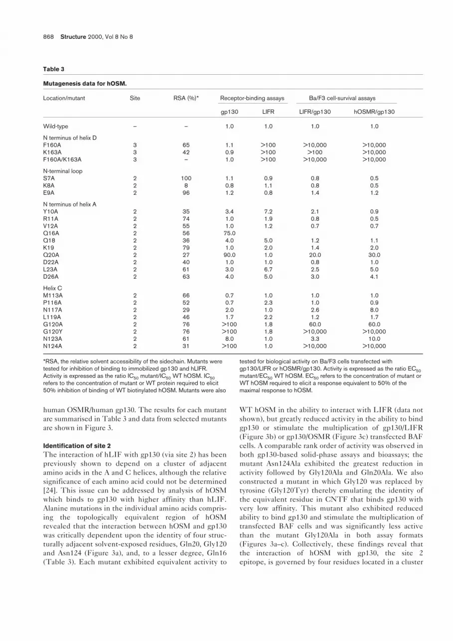
human OSMR/human gp130. The results for each mutantare summarised in Table 3 and data from selected mutantsare shown in Figure 3.
Identification of site 2The interaction of hLIF with gp130 (via site 2) has beenpreviously shown to depend on a cluster of adjacentamino acids in the A and C helices, although the relativesignificance of each amino acid could not be determined[24]. This issue can be addressed by analysis of hOSMwhich binds to gp130 with higher affinity than hLIF.Alanine mutations in the individual amino acids compris-ing the topologically equivalent region of hOSMrevealed that the interaction between hOSM and gp130was critically dependent upon the identity of four struc-turally adjacent solvent-exposed residues, Gln20, Gly120and Asn124 (Figure 3a), and, to a lesser degree, Gln16(Table 3). Each mutant exhibited equivalent activity to
WT hOSM in the ability to interact with LIFR (data notshown), but greatly reduced activity in the ability to bindgp130 or stimulate the multiplication of gp130/LIFR(Figure 3b) or gp130/OSMR (Figure 3c) transfected BAFcells. A comparable rank order of activity was observed inboth gp130-based solid-phase assays and bioassays; themutant Asn124Ala exhibited the greatest reduction inactivity followed by Gly120Ala and Gln20Ala. We alsoconstructed a mutant in which Gly120 was replaced bytyrosine (Gly120Tyr) thereby emulating the identity ofthe equivalent residue in CNTF that binds gp130 withvery low affinity. This mutant also exhibited reducedability to bind gp130 and stimulate the multiplication oftransfected BAF cells and was significantly less activethan the mutant Gly120Ala in both assay formats(Figures 3a–c). Collectively, these findings reveal thatthe interaction of hOSM with gp130, the site 2epitope, is governed by four residues located in a cluster
868 Structure 2000, Vol 8 No 8
Table 3
Mutagenesis data for hOSM.
Location/mutant Site RSA (%)* Receptor-binding assays Ba/F3 cell-survival assays
gp130 LIFR LIFR/gp130 hOSMR/gp130
Wild-type – – 1.0 1.0 1.0 1.0
N terminus of helix DF160A 3 65 1.1 >100 >10,000 >10,000K163A 3 42 0.9 >100 >100 >10,000F160A/K163A 3 – 1.0 >100 >10,000 >10,000
N-terminal loopS7A 2 100 1.1 0.9 0.8 0.5K8A 2 8 0.8 1.1 0.8 0.5E9A 2 96 1.2 0.8 1.4 1.2
N terminus of helix AY10A 2 35 3.4 7.2 2.1 0.9R11A 2 74 1.0 1.9 0.8 0.5V12A 2 55 1.0 1.2 0.7 0.7Q16A 2 56 75.0Q18 2 36 4.0 5.0 1.2 1.1K19 2 79 1.0 2.0 1.4 2.0Q20A 2 27 90.0 1.0 20.0 30.0D22A 2 40 1.0 1.0 0.8 1.0L23A 2 61 3.0 6.7 2.5 5.0D26A 2 63 4.0 5.0 3.0 4.1
Helix CM113A 2 66 0.7 1.0 1.0 1.0P116A 2 52 0.7 2.3 1.0 0.9N117A 2 29 2.0 1.0 2.6 8.0L119A 2 46 1.7 2.2 1.2 1.7G120A 2 76 >100 1.8 60.0 60.0G120Y 2 76 >100 1.8 >10,000 >10,000N123A 2 61 8.0 1.0 3.3 10.0N124A 2 31 >100 1.0 >10,000 >10,000
*RSA, the relative solvent accessibility of the sidechain. Mutants weretested for inhibition of binding to immobilized gp130 and hLIFR.Activity is expressed as the ratio IC50 mutant/IC50 WT hOSM. IC50refers to the concentration of mutant or WT protein required to elicit50% inhibition of binding of WT biotinylated hOSM. Mutants were also
tested for biological activity on Ba/F3 cells transfected withgp130/LIFR or hOSMR/gp130. Activity is expressed as the ratio EC50mutant/EC50 WT hOSM. EC50 refers to the concentration of mutant orWT hOSM required to elicit a response equivalent to 50% of themaximal response to hOSM.

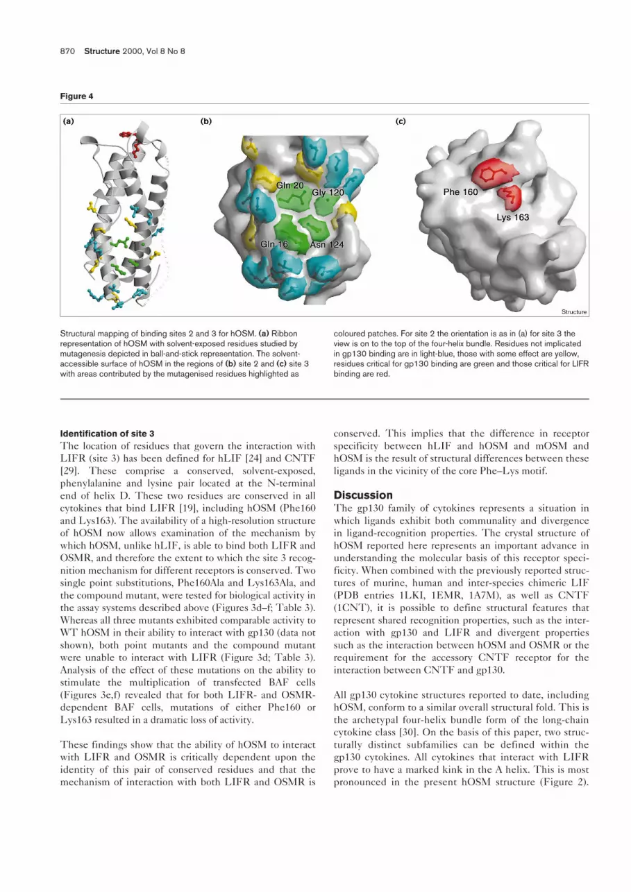
on the A and C helices (Figure 4). Of these four residuesthe most significant, in terms of the consequences ofalanine substitution, is Asn124 (in helix C) followed byGly120 and Gln20. This study also reveals that Gly120
plays a critical role in receptor recognition, in that asingle substitution to the bulky tyrosine residue (foundin CNTF) almost completely ablates the ability ofhOSM to interact with gp130.
Research Article Crystal structure of oncostatin M Deller et al. 869
Figure 3
10 0 10 1 10 2 10 3 10 4 10 5 10 60.0
0.1
0.2
0.3
0.4
0.5
0.6
0.7
0.8
0.9
1.0
OSM
OSM Q20A
OSM G120A
OSM G120Y
OSM N124A
Mutein concentration (ng/ml)10 0 10 1 10 2 10 3 10 4 10 5 10 6
Mutein concentration (ng/ml)
Mutein concentration (ng/ml)
Mutein concentration (ng/ml)
Mutein concentration (ng/ml)
Mutein concentration (ng/ml)
0.0
0.2
0.4
0.6
0.8
1.0
1.2
1.4
1.6
1.8
2.0OSM
OSM Q20A
OSM G120A
OSM G120Y
OSM N124A
0.0
0.2
0.4
0.6
0.8
1.0
1.2
1.4
1.6
1.8
2.0
OSM
OSM Q20A
OSM G120A
OSM G120Y
OSM N124A
0.00
0.25
0.50
0.75
1.00
1.25
OSM
OSM F160A
OSM K163A
0.00
0.25
0.50
0.75
1.00
1.25OSM
OSM F160A
OSM K163A
0.0
0.2
0.4
0.6
0.8
1.0
1.2
1.4
1.6
1.8
2.0OSM
OSM F160A
OSM K163A
10 10 10 10 10 10 10 10 10 10 10 10 10 10 10 10 10 10 10 10Ð3 Ð2 Ð1 0 1 2 3 4 5 Ð5 Ð4 Ð3 Ð2 Ð1 0 1 2 3 4 5
10 10 10 10 10 10 10 10 10 10 10 10 10 10 10 10 10 10 10Ð4 Ð3 Ð2 Ð1 0 1 2 3 4 5 Ð3 Ð2 Ð1 0 1 2 3 4 5
OD470
OD570
OD570
OD490
OD570
OD570
Structure
(a) (d)
(b) (e)
(c) (f)
Functional binding data for hOSM muteins. (a) Competitive inhibitionof biotinylated OSM binding to gp130–Fc by site 2 OSM muteins.(b) Biological activity of OSM muteins in the Ba/F3–LIFR/gp130assay. (c) Biological activity of OSM muteins in the
Ba/F3–OSMR/gp130 assay. (d) Competitive inhibition of biotinylatedOSM binding to LIFR–Fc by site 3 OSM muteins. (e) Biological activityof OSM muteins in the Ba/F3–LIFR/gp130 assay. (f) Biological activityof OSM muteins in the Ba/F3–OSMR/gp130 assay.
Identification of site 3The location of residues that govern the interaction withLIFR (site 3) has been defined for hLIF [24] and CNTF[29]. These comprise a conserved, solvent-exposed,phenylalanine and lysine pair located at the N-terminalend of helix D. These two residues are conserved in allcytokines that bind LIFR [19], including hOSM (Phe160and Lys163). The availability of a high-resolution structureof hOSM now allows examination of the mechanism bywhich hOSM, unlike hLIF, is able to bind both LIFR andOSMR, and therefore the extent to which the site 3 recog-nition mechanism for different receptors is conserved. Twosingle point substitutions, Phe160Ala and Lys163Ala, andthe compound mutant, were tested for biological activity inthe assay systems described above (Figures 3d–f; Table 3).Whereas all three mutants exhibited comparable activity toWT hOSM in their ability to interact with gp130 (data notshown), both point mutants and the compound mutantwere unable to interact with LIFR (Figure 3d; Table 3).Analysis of the effect of these mutations on the ability tostimulate the multiplication of transfected BAF cells(Figures 3e,f) revealed that for both LIFR- and OSMR-dependent BAF cells, mutations of either Phe160 orLys163 resulted in a dramatic loss of activity.
These findings show that the ability of hOSM to interactwith LIFR and OSMR is critically dependent upon theidentity of this pair of conserved residues and that themechanism of interaction with both LIFR and OSMR is
conserved. This implies that the difference in receptorspecificity between hLIF and hOSM and mOSM andhOSM is the result of structural differences between theseligands in the vicinity of the core Phe–Lys motif.
DiscussionThe gp130 family of cytokines represents a situation inwhich ligands exhibit both communality and divergencein ligand-recognition properties. The crystal structure ofhOSM reported here represents an important advance inunderstanding the molecular basis of this receptor speci-ficity. When combined with the previously reported struc-tures of murine, human and inter-species chimeric LIF(PDB entries 1LKI, 1EMR, 1A7M), as well as CNTF(1CNT), it is possible to define structural features thatrepresent shared recognition properties, such as the inter-action with gp130 and LIFR and divergent propertiessuch as the interaction between hOSM and OSMR or therequirement for the accessory CNTF receptor for theinteraction between CNTF and gp130.
All gp130 cytokine structures reported to date, includinghOSM, conform to a similar overall structural fold. This isthe archetypal four-helix bundle form of the long-chaincytokine class [30]. On the basis of this paper, two struc-turally distinct subfamilies can be defined within thegp130 cytokines. All cytokines that interact with LIFRprove to have a marked kink in the A helix. This is mostpronounced in the present hOSM structure (Figure 2).
870 Structure 2000, Vol 8 No 8
Figure 4
Structural mapping of binding sites 2 and 3 for hOSM. (a) Ribbonrepresentation of hOSM with solvent-exposed residues studied bymutagenesis depicted in ball-and-stick representation. The solvent-accessible surface of hOSM in the regions of (b) site 2 and (c) site 3with areas contributed by the mutagenised residues highlighted as
coloured patches. For site 2 the orientation is as in (a) for site 3 theview is on to the top of the four-helix bundle. Residues not implicatedin gp130 binding are in light-blue, those with some effect are yellow,residues critical for gp130 binding are green and those critical for LIFRbinding are red.

By contrast, in IL-6 [21], which does not interact withLIFR, the A helix conforms to the standard hydrogen-bonding pattern throughout its length. This indicatesthat the A helix kink may represent a structural feature ofLIFR recognition. In this respect it is notable that theLIFR contains two copies of the CHD in the extracellu-lar domain and that the A helix kink may represent astructural feature which permits a low-affinity interactionwith the N-terminal CHD of LIFR. The existence ofsuch an interaction has been inferred from homologyscanning studies of the interaction between LIF andLIFR [31,32].
The interaction with gp130The detailed mutagenesis study of hOSM reported here,combined with earlier studies of hLIF [24], has identifiedthose amino acid residues that contribute the majority ofbinding energy to the interaction with gp130, as definedby the consequences of alanine scanning analysis. Thisreveals a quartet of residues in the A and C helices thatform a distinct patch in the crystal structure (Figure 4). Interms of the interaction between hOSM and gp130, themost important residues are Asn124 and Gly120 in helix C,with lesser contributions from Gln16 and Gln20 in helix A.The crystal structure of the CHD of gp130 [25] combinedwith mutagenesis data of gp130 ([33]; KRH, E Raulo, OMcKenzie and JKH unpublished observations) provides astructural explanation for this interaction. The most promi-nent feature of gp130 is a solvent-exposed hydrophobicresidue (Phe169) in an interstrand loop of the N-terminalhalf of the bipartite CHD (Figure 5). The second signifi-cant determinant of gp130 recognition is a prominentsolvent-exposed tyrosine residue (Tyr196), located close tothe junction between the two elements of the CHD, thehydroxyl group of which faces away from the main body ofthe molecule into solution (Figure 5). Inspection of thetwo functional epitopes in open book format (Figure 5)
immediately suggests an explanation for the molecularmechanism of ligand recognition. This indicates that theexposed phenylalanine residue on gp130 may be dockedinto the hydrophobic ‘dimple’ formed on the hOSMsurface from the Cα backbone of Gly120. In this configu-ration the hydroxyl group of gp130 Tyr196 would be ableto hydrogen bond with the exposed amide group ofAsn124 from hOSM. This explanation is consistent withmutagenesis of hOSM Gly120 reported here in which sub-stitution to alanine, which has a relatively small non-polarsidechain, has less effect on binding than substitution tothe bulkier tyrosine residue (Table 3). The substitution ofa bulky sidechain would be expected to occlude the non-polar dimple and thereby inhibit docking of the Phe169 ofgp130 and formation of the hydrogen bond between theTyr196 of gp130 and the Asn124 of hOSM.
This proposed mode of docking between hOSM site 2 andgp130 has strong similarities to the interaction betweengrowth hormone and the growth hormone receptor [23]and EPO/EPO receptor [28]. In both these cases the site 2interaction also involves burying a bulky, solvent exposed,aromatic residue in a hydrophobic patch formed from theCα backbone of helix C. This suggests that the basicsite 2 recognition mechanism may exhibit strong structuralsimilarities across the whole cytokine/receptor family. Thepoint of divergence between gp130 and these othersystems appears to arise from the use of polar interactions,for example the putative Tyr100–Asn124 hydrogen bond,that contribute additional specificity to complex forma-tion. Thus a major contribution to binding affinity from adistinctive hydrophobic interaction is conserved whilstspecificity is controlled through polar interactions. Asimilar pattern of conservation has recently been noted fora second set of cytokine–receptor interactions, those of thetumour necrosis factor (TNF) like cytokines with theircognate receptors [34].
Research Article Crystal structure of oncostatin M Deller et al. 871
Figure 5
Complimentarity between the interactionsurfaces of hOSM and gp130. The solvent-accessible surfaces of site 2 on hOSM (left)and the cognate binding site on gp130(right) are displayed with areas contributedby residues implicated in binding highlightedas coloured patches. The orientation forhOSM site 2 is rotated 90° from that inFigure 4 whereas gp130 is rotated by 180°from its orientation relative to hOSM in theputative interaction complex, as in theopening of a book.

This hypothesis also suggests why CNTF interacts withgp130 at a very much lower affinity than either LIF orhOSM. In CNTF the equivalent residue to Gly120 is atyrosine which, as argued above, would be predicted todisrupt the core interaction. The employment of the acces-sory receptor CNTF-R in this scheme is required to com-pensate for this loss of affinity by introducing additionalsites of receptor–receptor interaction into the complex.
The interaction with LIFR and OSMRA distinctive feature of the gp130 cytokine family is theuse of a novel receptor recognition site 3 located at the Nterminus of helix D. This epitope engages the second sig-nalling receptor in the complex: either LIFR/OSMR in thecase of OSM, LIF and CNTF, or gp130 in the case of IL-6and IL-11. Previous studies [24,29] have revealed that, forthe interaction with LIFR, this site 3 epitope involves asolvent-exposed pair of residues which are conserved in allLIFR-binding cytokines described to date. The availabil-ity of the crystal structure of hOSM has enabled us todetermine both the extent to which this recognitionscheme holds for additional members of the family and toexamine the involvement of this epitope in the interactionbetween hOSM and OSMR. Mutagenesis of the two keysite 3 residues in hOSM, Phe160 and Lys163, reveals thateach residue is required for the interaction with both LIFRand OSMR. This epitope (Figure 4) therefore represents acore structural element in site 3 recognition, although theprecise significance of each residue in the epitope differsbetween LIFR and OSMR. It follows that the ability ofOSM to interact with OSMR must result from either theinvolvement of additional residues in the vicinity of thisepitope or the presence of residues in LIF which blocksome feature of the interaction with OSMR.
It is intriguing that the structure of the site 3 recognitionepitope is similar to the site 2 recognition site of gp130:both employ a prominent aromatic sidechain as a ‘core’with additional affinity contributed by the sidechain of aresidue in close physical proximity. This suggests thatsite 3 receptor recognition may involve a similar overallscheme to that of site 2 except that the complementaryepitopes on receptor and ligand are reversed. This suggeststhat the site 3 recognition site in LIFR, which is located inthe immunoglobulin-like domain [31,32,35], may involvean analogous hydrophobic dimple or patch formed fromresidues with small sidechains. In this respect it is notablethat alanine scanning studies of the LIFR site 3 hOSMrecognition site imply a short sequence motif with a con-served glycine residue at its core [32].
Biological implicationsThe interaction of cytokine ligands with transmembranereceptors to form signalling complexes is a major mecha-nism for intercellular signal transduction and cell regula-tion. The molecular specificity of the extracellular
cytokine–receptor interaction is of fundamental biologi-cal significance as it dictates the resulting intracellularsignal-transduction processes and, accordingly, the asso-ciated biological responses. The family of cytokineswhich mediate their effects through binding to the gp130receptor — namely oncostatin M (OSM), leukaemiainhibitory factor, interleukins 6 and 11, cardiotrophin-1and ciliary neurotrophic factor (CNTF) — are of partic-ularly broad biomedical importance. Of these gp130-binding cytokines, human OSM (hOSM) is of potentialinterest for novel cancer therapeutics as well as provid-ing a target for the treatment of inflammatory responsesin the joints and nervous system.
The combined crystal structure and mutagenesis analy-sis of hOSM reported here has provided importantinsights into the molecular basis of biological specificityin the gp130 cytokine system. Particular value accruesfrom the structural comparisons these data now allowwith a panel of gp130 cytokines that exhibit bothcommon and divergent receptor-recognition properties.The study confirms the concept [36] that receptor recog-nition involves a conserved non-polar ‘core’ inter-action with specificity contributed by the identity ofresidues surrounding the core. This study also suggestsa second aspect to recognition specificity: the require-ment for soluble receptors in some gp130 signalling com-plexes (e.g. CNTF receptor in the CNTF-mediatedrecognition complex) acting to compensate for low-affin-ity core interactions by the introduction of additionalreceptor–receptor interactions. Finally, this study pro-vides the structural basis for the development of small-molecule compounds to intervene in hOSM-mediatedsignalling events.
Materials and methodsProduction and expression of hOSM proteinsA 21.4 kDa fragment of mature hOSM (residues 1–187) wasexpressed as a GST fusion protein using the pGex-2T expressionvector (Pharmacia). The hOSM and GST proteins were joined by arecognition site for rhinovirus 3C protease. All OSM mutants were gen-erated by overlap polymerase chain reaction from the pGEX–hOSMtemplate GST fusion using specific oligonucleotides for each mutant;the sequences of primers used are available on request. hOSM(1–187) and mutant proteins were expressed as GST fusions in E. colistrain JM109. Soluble GST fusion proteins were bound to glu-tathione–sepharose resin and the OSM protein released by overnightcleavage with 3C protease at 37°C and purified by high-resolution gel-filtration on a Sephadex-75 column (Pharmacia). All proteins had apurity of greater than 80% as determined by SDS PAGE and silverstaining. Protein concentrations were determined by the CoomassieProtein Plus assay (Bio-rad).
The identity of the purified hOSM (1–187) protein employed for crys-tallisation was confirmed using electrospray ionisation on a MicromassBioQ II triple, quadrapole, atmospheric pressure mass spectropho-tometer equipped with an electrospray interface operating in the posi-tive ion mode (Robin Aplin, Oxford Centre for Molecular Science,Oxford). N-terminal sequencing was also carried out for 10 cyclesusing an Applied Biosystems 494A/473A Sequencer (Tony Willis,MRC Immunochemistry Unit, Oxford).
872 Structure 2000, Vol 8 No 8
Bioassays of hOSM mutantsMutant forms of hOSM were tested for biological activity on the Ba/F3-hLIFR/hgp130 cell line as described previously [24]. The BA/F3-hOSMR/hgp130 cell line was generated by co-transfecting Ba/F3 cellswith a full-length hgp130 cDNA expressed under the control of thecytomegalovirus (CMV) promoter in plasmid pCDNA3 and the full-lengthhOSMR cDNA [15] expressed under the control of the CMV promoter inplasmid pCDNA3. Transfected cells were cultured overnight in 50 ng/mlIL-3 and then selected for co-expression of hgp130 and hOSMR in thepresence of 50 ng/ml hOSM. Co-expression of both receptors in co-transfectants was confirmed by FACS analysis after staining with anti-bodies directed against hgp130 [37] and hOSMR (KRH and JKH,unpublished observations). Bioassays were performed as described pre-viously [19]. All assays were performed in triplicate in at least three inde-pendent experiments. The data presented are the results from a singleexperiment, as it was noted that absolute response to defined concentra-tions of ligand varied between different batches of cells, although the rel-ative potencies of the WT and mutant proteins were preserved. The SEMof triplicate determinations was always less than 15% of the mean.
Ligand-binding assaysHuman gp130 and hLIFR were expressed as fusion proteins withhuman Fc as described previously [24]. The fusions were purified byprotein A sepharose affinity chromatography and employed in solid-phase ligand-binding assays using biotin-conjugated hOSM as thetracer [24]. The ability of mutant forms of hOSM to compete withbiotin–hOSM tracer was analysed using the PRISM software package(Graphpad Software) using a single-site binding model.
Crystallization and derivatisationFor crystallisation trials, hOSM(1–187) was concentrated to 8 mg/ml in50 mM Tris-HCl containing 150 mM sodium chloride, pH 7.5. Crystalsgrew by vapour diffusion within 1–3 days at 22°C from hanging dropstypically comprising 2 µl of protein solution plus 2 µl of reservoir solu-tion (30% w/v PEG 35,000, 0.25 M sodium acetate, pH 7.5). Crystalsmeasured approximately 0.3 × 0.2 × 0.2 mm3 and showed orderedBragg diffraction to 2.2 Å when exposed to synchrotron radiation. Pre-liminary characterisation indicated that the crystals belong to theorthogonal spacegroup P212121 with unit cell dimensions a = 35.8 Å,b = 53.1 Å, c = 106.8 Å and α = β = γ = 90°. There is one moleculeper asymmetric unit and the crystal solvent content is ~48.0%(Matthews coefficient, Vm = 2.3 Å/Da). Tests using a number of heavyatom compounds highlighted EMP as a putative heavy atom derivative.Derivatisation was carried out by soaking crystals in pre-equilibrateddrops of mother liquor containing 14 mM EMP for 16–24 h.
Data collectionMAD data were collected to a resolution of 2.6 Å from a single EMP deriv-ative crystal on beam line BM14 at the ESRF, Grenoble, France. Crystalswere briefly passed through a cryoprotectant solution consisting of a 2 µldrop of 30% (w/v) PEG 35,000, 0.25 M sodium acetate, pH 7.5 over-layed with 2 µl of 30% (w/v) PEG 8000, 0.25 M sodium acetate, pH 7.5and then flash-cooled directly in liquid propane. The crystal was main-tained at 100K in a dry air stream and data recorded using a MAR 345image plate detector (MAR Research, Hamburg, Germany). The mercuryLΙΙΙ edge of the derivative crystal was characterised by a fluorescencescan and X-ray data sets collected at three wavelengths (λ = 1.00669 Å,1.00914 Å and 0.826516 Å). A 2.2 Å resolution native data set was alsocollected at the same beamline from a single cryo-cooled crystal.
Individual data sets were auto-indexed, integrated and scaled using theprograms DENZO and SCALEPACK [38]. Structure factor amplitudeswere calculated using the program TRUNCATE [39], the data setsscaled using the program SCALEIT [40], and normalised anomalousscattering magnitudes estimated using the program REVISE [41]. Datacollection statistics are reported in Table 1.
Structure determination and analysisScaling of MAD data sets yielded maximal dispersive differences of10.6% and maximal anomalous differences of 9.7%. The mercury sites
for the EMP derivative were determined by manual inspection of differ-ence and anomalous Patterson syntheses and confirmed using the pro-grams RSPS and SOLVE [42]. The sites were refined using theprogram SHARP [43], and MAD phases were calculated to 2.6 Å reso-lution (figure of merit = 0.46, phasing power = 1.84). Initial maps basedon these phases and Fobs from the EMPλ2 data set proved uninter-pretable. Hendrickson-Lattman coefficients for SIRAS phases derivedusing the EMP derivative and native data were combined with the MADphases. Maps based on these combined phases and the EMPλ2 Fobs(figure of merit 0.51, phasing power = 1.90) showed improved mainchain density. Phase improvement was carried out using solvent flat-tening, as implemented by the program SOLOMON [44], and thestructure was traced using the interactive graphics program O [45].
All refinement steps (positional, torsional dynamics, grouped B factorand individual B factor) were carried out using the program CNS-SOLVE [46]. A bulk-solvent correction was employed (solventdensity = 0.29 e/Å3, B-factor = 31.1 Å2), so allowing all data in the res-olution range 20.0–2.2 Å to be included. In the final stages of refine-ment 116 waters were located. The current model consists of residues4–134 and 156–187 of the mature protein with no residues falling indisallowed regions of the Ramachandran plot. Refinement and modelstatistics are reported in Table 2.
The quality of the final model was assessed using the programPROCHECK [47], secondary structure assignments were calculatedusing the program DSSP [48] and solvent accessibilities calculatedusing NACCESS [49]. Structural superimpositions were performedusing the program SHP [50] and structural figures were producedusing BOBSCRIPT [51], RASTER3D [52], VOLUMES (R Esnouf, per-sonal communication) and GRASP [53].
Accession numbersAtomic coordinates for hOSM have been deposited with the PDB(accession number 1EVS).
AcknowledgementsThis work was supported by the Cancer Research Campaign. We thank K.Harlos for help with ‘in house’ data collection; the EMBL and ESRF staff, inparticular V. Stroganoff, for assistance on BM14 at the ESRF, R. Esnouf forprovision of computing facilities, V. Barton and L. McGovern for discussion.We are grateful to T. Willis and R. Aplin for performing the protein sequenc-ing and mass spectrometry, respectively. EYJ is a Royal Society UniversityResearch Fellow. SI is supported by the Wellcome Trust and MCD wassupported by the MRC and CRC.
References1. Malik, N., et al., & Wei, C.M. (1989). Molecular cloning, sequence
analysis, and functional expression of a novel growth regulator,oncostatin M. Mol. Cell Biol. 9, 2847-2853.
2. Grant, S.L. & Begley, C.G. (1999). The oncostatin M signalling pathway:reversing the neoplastic phenotype? Mol. Medicine Today 5, 406-412.
3. Miles, S.A., et al., & Linsley, P.S. (1992). Oncostatin M as a potentmitogen for AIDS-Kaposi’s sarcoma-derived cells. Science255, 1432-1434.
4. Mukouyama, Y., et al., & Miyajima, A. (1998). In vitro expansion ofmurine multipotential hematopoietic progenitors from the embryonicaorta–gonad–mesonephros region. Immunity 8, 105-114.
5. Grenier, A., et al., & Chollet Martin, S. (1999). Oncostatin Mproduction and regulation by human polymorphonuclear neutrophils.Blood 93, 1413-1421.
6. Kerr, C., Langdon, C., Graham, F., Gauldie, J., Hara, T. & Richards,C.D. (1999). Adenovirus vector expressing mouse oncostatin Minduces acute-phase proteins and TIMP-1 expression in vivo in mice.J. Interferon Cytokine Res. 19, 1195-1205.
7. Ensoli, F., et al., & Aiuti, F. (1999). Inflammatory cytokines and HIV-1-associated neurodegeneration: Oncostatin-M produced bymononuclear cells from HIV-1-infected individuals induces apoptosisof primary neurons. J. Immunol. 162, 6268-6277.
8. Kordula, T., Rydel, R.E., Brigham, E.F., Horn, F., Heinrich, P.C. & Travis,J. (1998). Oncostatin M and the interleukin-6 and soluble interleukin-6receptor complex regulate α1-antichymotrypsin expression in humancortical astrocytes. J. Biol. Chem. 273, 4112-4118.
Research Article Crystal structure of oncostatin M Deller et al. 873

9. Heinrich, P.C., et al., & Wollmer, A. (1998). Interleukin-6 and relatedcytokines: effect on the acute phase reaction. ZeitschriftErnährungswissenschaft 37, 43-49.
10. Kuropatwinski, K.K., De Imus, C., Gearing, D., Baumann, H. & Mosley,B. (1997). Influence of subunit combinations on signaling by receptorsfor oncostatin M, leukemia inhibitory factor, and interleukin-6. J. Biol.Chem. 272, 15135-15144.
11. Taga, T. & Kishimoto, T. (1997). Gp130 and the interleukin-6 family ofcytokines. Annu. Rev. Immunol. 15, 797-819.
12. Heinrich, P.C., Behrmann, I., Muller-Newen, G., Schaper, F. & Graeve,L. (1998). Interleukin-6-type cytokine signalling through thegp130/Jak/STAT pathway. Biochem. J. 334, 297-314.
13. Hibi, M., Murakami, M., Saito, M., Hirano, T., Taga, T. & Kishimoto, T.(1990). Molecular cloning and expression of an IL-6 signal transducer,gp130. Cell 63, 1149-1157.
14. Gearing, D.P. & Bruce, A.G. (1992). Oncostatin M binds the high-affinity leukemia inhibitory factor receptor. New Biol. 4, 61-65.
15. Mosley, B., et al., & Cosman, D. (1996). Dual oncostatin M (OSM)receptors. Cloning and characterization of an alternative signalingsubunit conferring OSM-specific receptor activation. J. Biol. Chem.271, 32635-32643.
16. Yoshimura, A., et al., & Miyajima, A. (1996). Mouse oncostatin M: animmediate early gene induced by multiple cytokines through the JAK-STAT5 pathway. EMBO J. 15, 1055-1063.
17. Ichihara, M., Hara, T., Kim, H., Murate, T. and Miyajima, A. (1997).Oncostatin M and leukemia inhibitory factor do not use the samefunctional receptor in mice. Blood 90, 165-173.
18. Lindberg, R.A et al., & Fletcher, F.A. (1998). Cloning andcharacterization of a specific receptor for mouse oncostatin M. Mol.Cell Biol. 18, 3357-3367.
19. Robinson, R.C., et al., & Jones, E.Y. (1994). The crystal structure andbiological function of leukemia inhibitory factor: implications forreceptor binding. Cell 77, 1101-1116.
20. McDonald, N.Q., Panayotatos, N. & Hendrickson, W.A. (1995).Crystal structure of dimeric human ciliary neurotrophic factordetermined by MAD phasing. EMBO J. 14, 2689-2699.
21. Somers, W., Stahl, M. & Seehra, J.S. (1997). 1.9 Å crystal structure ofinterleukin 6: implications for a novel mode of receptor dimerizationand signaling. EMBO J. 16, 989-997.
22. Bazan, J.F. (1991). Neuropoietic cytokines in the hematopoietic fold.Neuron 7, 197-208.
23. de Vos, A.M., Ultsch, M. & Kossiakoff, A.A. (1992). Human growthhormone and extracellular domain of its receptor: crystal structure ofthe complex. Science 255, 306-312.
24. Hudson, K.R., Vernallis, A.B. & Heath, J.K. (1996). Characterization ofthe receptor binding sites of human leukemia inhibitory factor andcreation of antagonists. J. Biol. Chem. 271, 11971-11978.
25. Bravo, J., Staunton, D., Heath, J.K. & Jones, E.Y. (1998). Crystal structureof a cytokine-binding region of gp130. EMBO J. 17, 1665-1674.
26. Xu, G.Y., et al., & Cumming, D.A. (1997). Solution structure ofrecombinant human interleukin-6. J. Mol. Biol. 268, 468-481.
27. Hinds, M.G., Maurer, T., Zhang, J.G., Nicola, N.A. & Norton, R.S.(1998). Solution structure of leukemia inhibitory factor. J. Biol. Chem.273, 13738-13745.
28. Syed, R.S., et al., & Stroud, R.M. (1998). Efficiency of signallingthrough cytokine receptors depends critically on receptor orientation.Nature 395, 511-516.
29. Di Marco, A., et al., & Laufer, R. (1996). Identification of ciliaryneurotrophic factor (CNTF) residues essential for leukemia inhibitoryfactor receptor binding and generation of CNTF receptor antagonists.Proc. Natl Acad. Sci. USA 93, 9247-9252.
30. Boulay, J. & Paul, W.E. (1993). Hemapoietin sub-family classificationbased on size, gene organization and sequence homology. Curr. Biol.3, 573-575
31. Owczarek, C.M., Zhang, Y., Layton, M.J., Metcalf, D., Roberts, B. &Nicola, N.A. (1997). The unusual species cross-reactivity of theleukemia inhibitory factor receptor α-chain is determined primarily bythe immunoglobulin-like domain. J. Biol. Chem. 272, 23976-23985
32. Chobotova, K., Hudson, K.R. and Heath, J.K. (2000). The LIGHT motif:a ligand recognition site in the immunoglobulin-like domain of leukemiainhibitory factor receptor, submitted.
33. Horsten, U., et al., & Grotzinger, J. (1997). Molecular modeling-guidedmutagenesis of the extracellular part of gp130 leads to theidentification of contact sites in the interleukin-6 (IL-6).IL-6receptor.gp130 complex. J. Biol. Chem. 272, 23748-23757.
34. Mongkolsapaya, J., et al., & Screaton, G.R. (1999). Structure of theTRAIL-DR5 complex reveals mechanisms conferring specificity inapoptotic initiation. Nat. Struct. Biol. 6, 1048-1053.
35. Taupin, J.L., et al., & Moreau J.F. (1999). Binding of leukemia inhibitoryfactor (LIF) to mutants of its low affinity receptor, gp 190, reveals a LIFbinding site outside and interactions between the two cytokinebinding domains. J. Biol. Chem. 274, 14482-14489.
36. Clackson, T. & Wells, J.A. (1995). A hot spot of binding energy in ahormone-receptor interface. Science 267, 383-386.
37. Vernallis, A.B., Hudson, K.R. and Heath, J.K. (1997). An antagonist forthe leukemia inhibitory factor receptor inhibits leukemia inhibitoryfactor, cardiotrophin-1, ciliary neurotrophic factor, and oncostatin M.J. Biol. Chem. 272, 26947-26952.
38. Otwinowski, Z. & Minor, W. (1997). Processing of X-ray diffractiondata collected in oscillation mode. Meth. Enzymol. 276, 307-326.
39. French, S. & Wilson, K. (1978). On the treatment of negative intensityobservations. Acta Crystallogr. A 34, 517-525.
40. Howell, P.L. & Smith, G.D. (1992). Identification of heavy-atomderivatives by normal probability methods. J. Appl. Crystallogr. 25, 81-86.
41. Woolfson, M.M., Yao, J.X. & Fan, H.F. (1997). New techniques forapplying anomalous-scattering and isomorphous-replacement dataincorporated in ANOMIR - a general application package. ActaCrystallgr. 53, 673-681.
42. Terwilliger, T.C. & Berendzen, J. (1999). Automated MAD and MIRstructure solution. Acta Crystallgr. 55, 849-861.
43. de La Fortelle, E. & Bricogne, G. (1997). Maximum-likelihood heavy-atom parameter refinement for multiple isomorphous replacement andmultiwavelength anomalous diffraction methods. Meth. Enzymol.276, 472-494.
44. Abrahams, J.P. & Leslie, A.G.W. (1996). Methods used in thestructure determination of bovine mitochondrial F1 ATPase. ActaCrystallogr. D 52, 30-42.
45. Jones, T.A., Zou, J.-Y., Cowan, S.W. & Kjeldgaard, M. (1991). Improvedmethods for building protein models in electron density maps and thelocation of errors in these models. Acta Crystallgr. A 47, 110-119.
46. Brünger, A.T., et al., & Warren, G.L. (1998). Crystallography & NMRsystem: a new software suite for macromolecular structuredetermination. Acta Crystallogr., 54, 905-921.
47. Laskowski, R.A., MacArthur, M.W., Moss, D.S. & Thornton, J.M.(1993). PROCHECK: A program to check the stereochemical qualityof protein structures. J. Appl. Crystallogr. 26, 283-291.
48. Kabsch, W. & Sander, C. (1983). Dictionary of protein secondarystructure: pattern recognition of hydrogen-bonded and geometricalfeatures. Biopolymers 22, 2577-2637.
49. Hubbard, S.J. & Thornton, J.M. (1993). ‘NACCESS’. Computerprogram. Department of Biochemistry and Molecular Biology,University College, London.
50. Stuart, D.I., Levine, M., Muirhead, H. & Stammers, D.K. (1979). Crystalstructure of cat muscle pyruvate kinase at a resolution of 2.6 A. J. Mol.Biol. 134, 109-142.
51. Esnouf, R.M. (1997). An extensively modified version of Molscript thatincludes greatly enhanced colouring capabilities. J. Mol. Graphics15, 133-138.
52. Merritt, E.A. & Murphy, M.E.P. (1994). Raster3D version 2.0. A programfor photorealistic molecular graphics. Acta Crystallogr. D 50, 869-873.
53. Nicholls, A., Sharp, K.A. and Honig, B. (1991). Protein folding andassociation: insights from the interfacial and thermodynamicproperties of hydrocarbons. Proteins 11, 281-296.
874 Structure 2000, Vol 8 No 8
Because Structure with Folding & Design operates a‘Continuous Publication System’ for Research Papers, thispaper has been published on the internet before being printed(accessed from http://biomednet.com/cbiology/str). Forfurther information, see the explanation on the contents page.