controlled interconversion of semiconducting and metallic forms of polyaniline nanofibers
TRANSCRIPT
Synthetic Metals 148 (2005) 237–243
Controlled interconversion of semiconducting and metallicforms of polyaniline nanofibers
David M. Sarnoa,1, Sanjeev K. Manoharb, Alan G. MacDiarmida,b,∗a Department of Chemistry, University of Pennsylvania, 231 South 34th Street, Philadelphia, PA 19104–6323, USA
b Alan G. MacDiarmid Laboratory for Technical Innovation, Department of Chemistry, The University of Texas at Dallas, Richardson, TX 75083, USA
Received 6 August 2004; received in revised form 26 August 2004; accepted 16 September 2004
Abstract
Self-assembled polyaniline nanofibers doped with 2-acrylamido-2-methyl-1-propanesulfonic acid were prepared by oxidative polymer-ization of aniline in the presence of a nonionic surfactant. These nanofibers were dedoped to the semiconducting emeraldine base and thenr l synthesisw of samplep anofiberst –vis–NIRa ine base ob-t rochloriden©
K
1
iprirdbd(o
C
e.g.,the
at-tteryfield
sub-on ofhisratedntlly asl
rtic-s fordue
and
0d
edoped to the metallic emeraldine hydrochloride. It was possible to introduce a different dopant anion from that used in the initiaith no significant changes in fiber morphology or diameter, as observed by scanning electron microscopy (SEM). The methodreparation for SEM significantly affected the observed morphology. Deposition from aqueous dispersions resulted primarily in n
hat ranged in diameter from 28 to 82 nm (average: 56 nm), whereas drying to solid powder resulted in a less fibrous material. UVbsorbance spectroscopy indicated that the electronic structure of the emeraldine base nanofibers was identical to bulk emerald
ained by conventional synthesis. Estimates from X-ray diffraction data suggested that the fractional crystallinity of emeraldine hydanofibers did not differ significantly from the bulk powder.2004 Published by Elsevier B.V.
eywords:Polyaniline; Nanofibers; Doping; Scanning electron microscopy
. Introduction
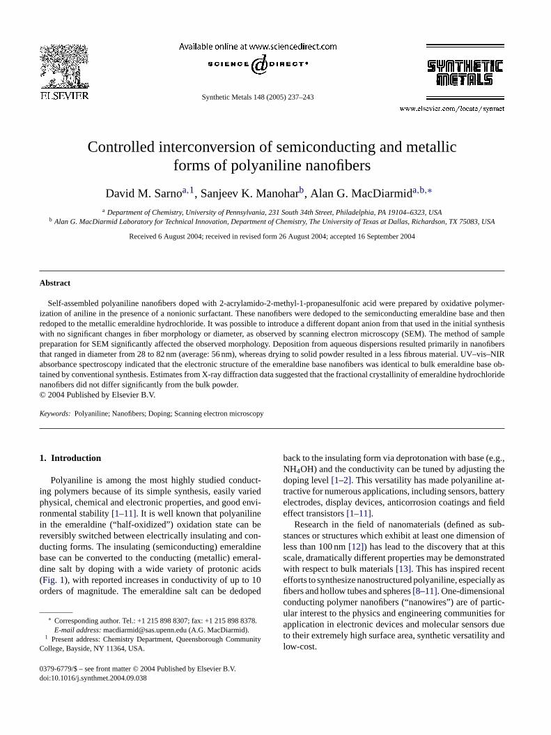
Polyaniline is among the most highly studied conduct-ng polymers because of its simple synthesis, easily variedhysical, chemical and electronic properties, and good envi-onmental stability[1–11]. It is well known that polyanilinen the emeraldine (“half-oxidized”) oxidation state can beeversibly switched between electrically insulating and con-ucting forms. The insulating (semiconducting) emeraldinease can be converted to the conducting (metallic) emeral-ine salt by doping with a wide variety of protonic acidsFig. 1), with reported increases in conductivity of up to 10rders of magnitude. The emeraldine salt can be dedoped
∗ Corresponding author. Tel.: +1 215 898 8307; fax: +1 215 898 8378.E-mail address:[email protected] (A.G. MacDiarmid).
1 Present address: Chemistry Department, Queensborough Communityollege, Bayside, NY 11364, USA.
back to the insulating form via deprotonation with base (NH4OH) and the conductivity can be tuned by adjustingdoping level[1–2]. This versatility has made polyanilinetractive for numerous applications, including sensors, baelectrodes, display devices, anticorrosion coatings andeffect transistors[1–11].
Research in the field of nanomaterials (defined asstances or structures which exhibit at least one dimensiless than 100 nm[12]) has lead to the discovery that at tscale, dramatically different properties may be demonstwith respect to bulk materials[13]. This has inspired receefforts to synthesize nanostructured polyaniline, especiafibers and hollow tubes and spheres[8–11]. One-dimensionaconducting polymer nanofibers (“nanowires”) are of paular interest to the physics and engineering communitieapplication in electronic devices and molecular sensorsto their extremely high surface area, synthetic versatilitylow-cost.
379-6779/$ – see front matter © 2004 Published by Elsevier B.V.oi:10.1016/j.synthmet.2004.09.038
238 D.M. Sarno et al. / Synthetic Metals 148 (2005) 237–243
Fig. 1. Reversible doping from (a) semiconducting emeraldine base to (b)metallic emeraldine salt form of polyaniline (HA = HCl, AMPSA, etc.)[1].
Conventional synthesis of polyaniline, based on the ox-idative polymerization of aniline in the presence of a strongacid dopant[2], typically results in an irregular granularmorphology that is accompanied by a very small percent-age of nanoscale fibers[9]. In recent years, however, sev-eral approaches have been developed by which nanofiberscan be obtained as the dominant nanostructure. For exam-ple, the cylindrical pores of nanoporous membranes havebeen used for the generalized template synthesis of conduct-ing polymers, metals and other materials. Highly monodis-perse nanofibers and/or tubules are subsequently obtainedby dissolving the membrane template[8]. Electrospinning, anon-mechanical, electrostatic method, has yielded compos-ite polyaniline/polyethylene oxide nanofibers with diametersbelow 30 nm and lengths of up to hundreds of microme-ters [11]. In the absence of an external physical or chemi-cal template, pure polyaniline nanofibers/tubes and spherescan be obtained by making use of large organic acids thatserve as both dopants and in situ templates by forming mi-celles upon which aniline is polymerized[10]. Fiber diam-eters are observed to be as low as 30–60 nm and are highlyinfluenced by reagent ratios[10]. Uniform nanofibers of puremetallic polyaniline (30–120 nm diameter, depending on thedopant) have also been prepared by polymerization at anaqueous–organic interface. Migration of the product into thea polymI rtains con-tv ect toi oly-m ndm ap-p ina-t singw s, es-p
bersd cid( oft was
carried out in order to ascertain (1) whether these nanofiberscould be interconverted between the semiconducting andmetallic forms with retention of morphology; (2) if nanofibersof the metallic form of polyaniline could be obtained by dop-ing the emeraldine base form with an acid different from thatused in the initial synthesis; (3) whether the method of samplepreparation for SEM studies (as distinct from the method ofsynthesis) had an effect on the observed morphology. In addi-tion to characterization by SEM, the electronic and molecu-lar structure of the polyaniline nanofibers has been probed byUV–vis–NIR absorbance spectroscopy and X-ray diffraction.All results have been compared to data previously obtainedfrom conventionally prepared polyaniline.
2. Experimental
2.1. Synthesis of polyaniline emeraldine base nanofibers
All materials were used as received. Aniline (Aldrich) wasstored in a sealed container in a refrigerator. The dopant2-acrylamido-2-methyl-l-propanesulfonic acid (AMPSA;Aldrich; 4.15 g, 0.0200 mol) was weighed into a 150 mLbeaker and dissolved in 20 mL of deionized water at roomtemperature by magnetic stirring for∼5 min. Approxi-m y-l the1 cht ands m-p so-l lida ol)w callys hes ar re-m n byd beo tion.W mix-t epgd
ppedb turei to-n ameb d,i per,9 lyd -u dry.T e fil-t thatel the
queous phase is hypothesized to suppress uncontrolleder growth by isolating the fibers from excess reagent[9].
t has recently been demonstrated that the addition of ceurfactants to such an interfacial system grants furtherrol over the diameter of the nanofibers[14]. “Seeding” witharious nanomaterials has also been used with great effnitiate polyaniline nanofiber formation in single-phase p
erizations[15]. The template-free interfacial, seeding aicellar methods each employ a different “bottom up”roach to obtain pure polyaniline nanofibers. The comb
ion of self-assembly with minimal post-synthesis procesarrants further study and application of these nanofiberecially in the field of electronic nanomaterials.
We have prepared self-assembled polyaniline nanofioped with 2-acrylamido-2-methyl-1-propanesulfonic aAMPSA) by polymerization of aniline in the presencehe nonionic surfactant Triton X-100. The present study
-
ately 5 mL of the surfactant Triton X-100 (polyoxyethene(10) isooctylphenyl ether; Acros) was dissolved inM AMPSA solution by sonication for 1 h, during whi
ime the solution became warm. The clear, colorlesslightly viscous solution was allowed to return to room teerature. Approximately 1 mL of aniline was added to the
ution, which was then magnetically stirred for 20 min. Sommonium peroxydisulfate (APS; Fisher; 0.58 g, 2.5 mmas added to the solution, which was then magnetitirred for∼5 min. The APS was not fully soluble and tystem became cloudy. Stirring was stopped, the stir boved and the beaker covered to prevent contaminatioust, etc. After∼3 min, small blue–green particles couldbserved suspended in the slightly turbid, colorless soluithin ∼5 min, the color deepened and the blue–green
ure became opaque. Within∼10 min, it had become dereen in color, typical of metallic polyaniline[2], in this caseoped with AMPSA.
After 2.5 h at room temperature, the reaction was stoy pouring the thick, opaque green precipitate mix
nto ∼1 L of 0.1 M ammonium hydroxide, which deproated the doped polymer. The mixture immediately beclue–purple in color (pH∼9.5, pH paper) and it was adde
n portions, to a Buchner funnel (Whatman 41 filter pacm). The volume of liquid in the funnel was very slowecreased and∼4 L of 0.1 M NH4OH was added to continously wash the precipitate, which was not allowed tohe product was washed with the aqueous base until th
rate became colorless and was free of foam, indicatingxcess surfactant had been removed. When∼20 mL of so-
ution remained in the funnel, the upper portion only of
D.M. Sarno et al. / Synthetic Metals 148 (2005) 237–243 239
precipitate/NH4OH mixture was decanted to a glass vial toprevent contamination by filter paper fibers. The product wascharacterized by UV–vis–NIR spectroscopy, confirming thatit was the emeraldine base form of polyaniline. Portions wereset aside for study by SEM and X-ray diffraction (see below).
2.2. Interconversion of emeraldine base nanofibers toemeraldine hydrochloride
Approximately 10 mL of the above dispersion of emeral-dine base was added to∼250 mL 1 M HC1 and magneticallystirred overnight. The resulting green suspension was slowlyfiltered in a Buchner funnel as described above and was con-tinuously washed with∼2.5 L hydrochloric acid until the fil-trate was colorless. When the volume had been reduced to∼20 mL, the upper portion only of the green dispersion (pH∼0) in the funnel was decanted to a glass vial to prevent con-tamination by filter paper fibers. Conversion to emeraldinehydrochloride was confirmed by UV–vis–NIR spectroscopyand portions of the product were set aside for SEM and X-raydiffraction studies (see below). Nanofibers of the emeraldinehydrochloride could be readily dedoped to the emeraldinebase in an identical manner to the deprotonation of AMPSA-doped polyaniline, as described above.
2
af-f de-s hy-d me-c thep ntedf pec-t tionw thea
2
inedo z cu-v n ofe olvedi o-l fewd ddedt trumo ences
2
basei of0 ns-
parent, pale blue dispersion. Similarly, one to three dropsof the dispersion of redoped nanofibers in hydrochloric acidwere added to 3 mL of 1 M HCl and then shaken to pro-duce a transparent, pale green dispersion. The nanofiberdispersions could also be diluted by addition to 3 mL ofethanol instead of NH4OH or HCl. The dispersions weredeposited drop-wise on small segments of microscope slideglass that had been rinsed with methanol and air-dried. Thesolvent evaporated in air and small particles became visi-ble on the glass surface. After vacuum-drying in a desicca-tor for 1.5 h, the samples were then attached to aluminumsample stubs using double-sided carbon tape. Electrical con-tact was maintained between the sample surface and thealuminum stub with a short strip of copper tape. All sam-ples were sputter coated with a gold–palladium alloy priorto imaging on a JEOL 6300 FV field emission scanningelectron microscope, operating at an accelerating voltageof 5 kV. Average nanofiber diameters were measured on atleast 25 randomly chosen fibers at several regions on eachsample. All measurements were made on images obtainedat 30,000× magnification (using standard imaging software,1 pixel = 3.9 nm).
2.6. X-ray diffraction of polyaniline nanofibers
f ap-p end sper-sd . Thes sub-s s re-p s of thes s fromt plesw nd in-s rflexXa
3
3n
asg withc l-i beend thisa thaty 634a akh s forc se in
.3. Sample preparation
The morphology of the nanofibers was significantlyected by the method of sample preparation. As will becribed below, nanofibers of the emeraldine base androchloride could be destroyed by filtration, drying andhanical handling (e.g., scraping). It was found thatolyaniline nanofibers remained intact if they were preve
rom drying. Therefore, characterization by absorbance sroscopy, scanning electron microscopy and X-ray diffracas performed using only samples prepared directly fromqueous dispersions obtained as described above.
.4. UV–vis–NIR absorbance spectroscopy
Absorbance spectra from 1300 to 200 nm were obtan a Perkin-EImer Lambda 9 spectrometer using quartettes (1 cm path length). A few drops of the dispersiomeraldine base nanofibers prepared above were diss
n N-methyl-2-pyrrolidone (NMP) to obtain a light blue sution. NMP was used as a reference solvent. Similarly, arops of the dispersion of HCl-doped nanofibers were a
o 1 M HCl to produce a pale green dispersion. The specf the dispersion was obtained using 1 M HCl as a referolvent.
.5. Scanning electron microscopy
One to three drops of the dispersion of emeraldinen ammonium hydroxide were diluted by addition to 3 mL.1 M NH4OH and then briefly shaken to produce a tra
Glass microscope slides were cut into segments oroximately 2 cm× 1.6 cm, rinsed with methanol and thried in air. Enough of the opaque, deep blue–purple diion of polyaniline emeraldine base in 0.1 M NH4OH waseposited on the substrate to cover the entire surfaceample dried in air at room temperature, coating thetrate with a layer of dark blue powder. This process waeated several times as needed to increase the thicknesample. The same steps were taken to prepare samplehe emeraldine hydrochloride dispersed in 1 M HCl. Samere then mounted in a stainless steel sample holder aerted into the sample compartment of a Rikagu Geige-ray diffractometer using Cu K� radiation (λ= 1.54A) andgraphite monochromater.
. Results and discussion
.1. Oxidation and protonation states of polyanilineanofibers
The initially prepared AMPSA-doped polyaniline wreen in color and became blue–purple upon washingopious amounts of 0.1 M NH4OH. This color change qua
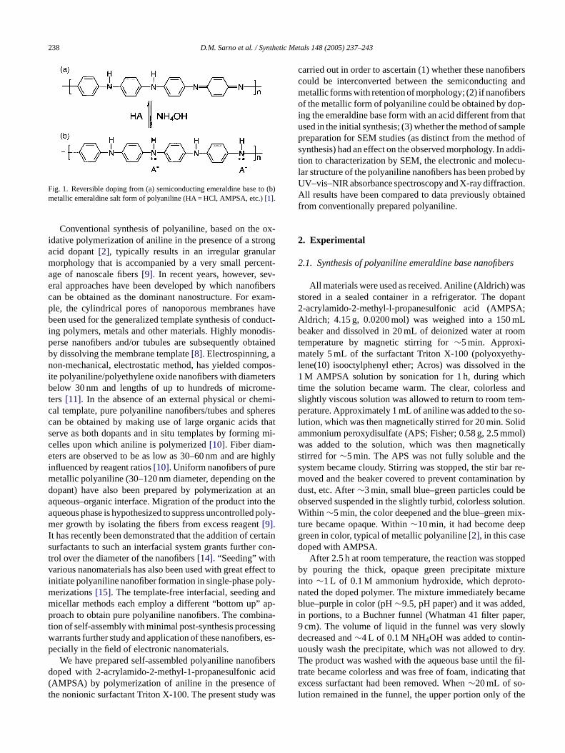
tatively indicated that the doped emeraldine salt hadeprotonated to the emeraldine base. A few drops ofqueous dispersion formed a clear blue solution in NMPielded UV–vis–NIR absorbance spectra with maxima atnd 328 nm (Fig. 2). The peak positions and relative peeights were in excellent agreement with literature resultonventionally prepared semiconducting emeraldine ba
240 D.M. Sarno et al. / Synthetic Metals 148 (2005) 237–243
Fig. 2. UV–vis–NIR absorbance spectra of dedoped emeraldine base dissolved in NMP (dotted line) and redoped emeraldine hydrochloride dispersed in 1MHCl (solid line). Noise observed at wavelengths >860 nm is attributed to an instrumental artifact.
NMP solution[16,17]. Washing with 1 M HCl resulted in areturn to the characteristic green color of metallic emeraldinehydrochloride. UV–vis–NIR spectra of the redoped aqueousdispersion diluted with 1 M HCl showed absorbance maximaat 820 and 398 nm (Fig. 2), in close agreement with previ-ously reported emeraldine hydrochloride thin films depositedin situ on glass slides[5,18], as well as nanofibers dispersedin water[9]. From the above spectra, it was concluded thatpolyaniline in these aqueous dispersions could be completelyand controllably interconverted between the semiconductingand metallic regimes, in a manner similar to the convention-ally prepared material[2].
3.2. Morphology and structure of polyaniline nanofibers
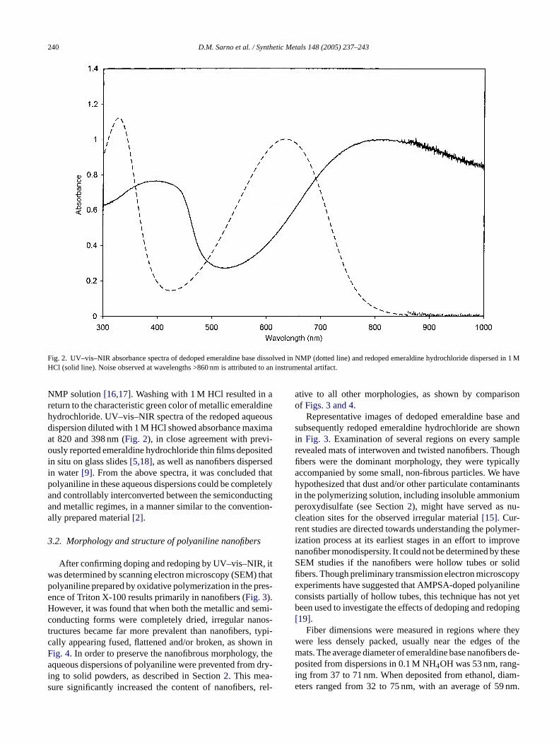
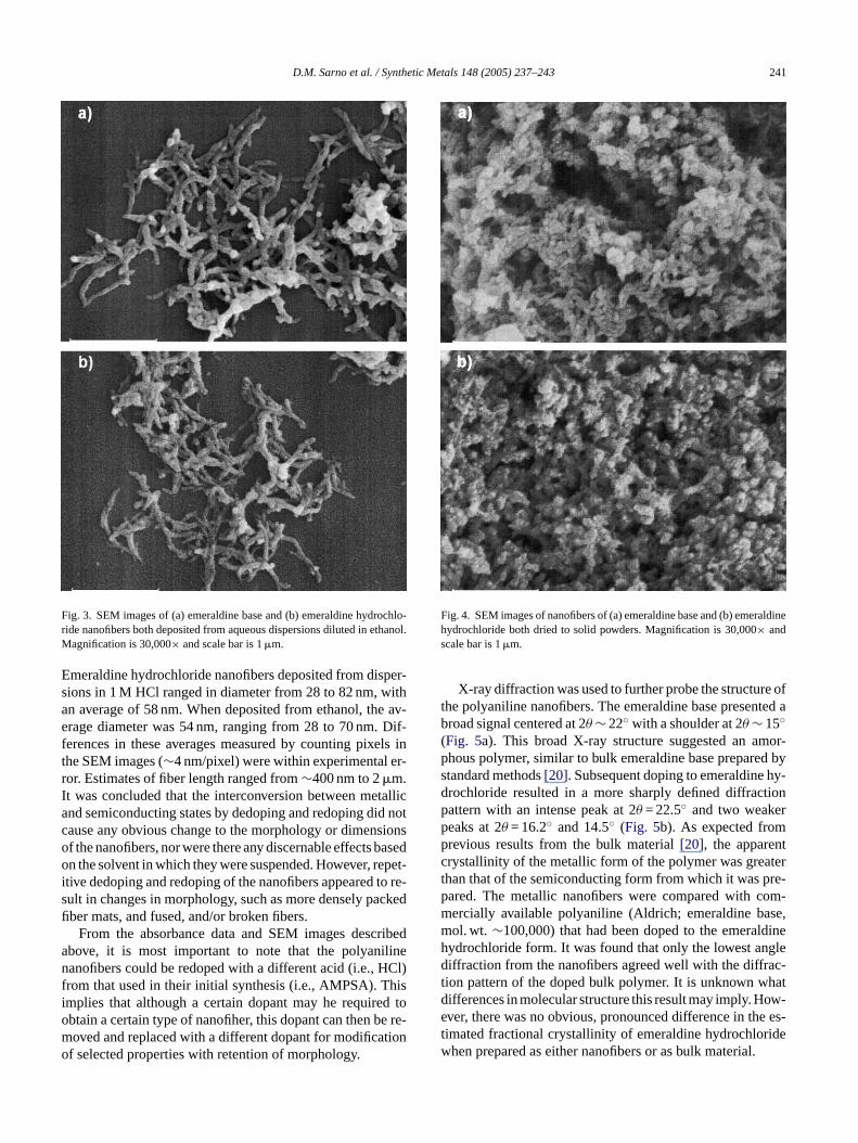
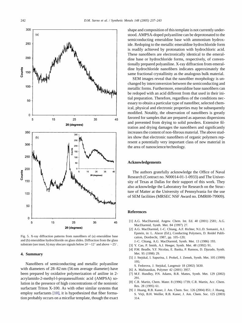
After confirming doping and redoping by UV–vis–NIR, itwas determined by scanning electron microscopy (SEM) thatpolyaniline prepared by oxidative polymerization in the pres-ence of Triton X-100 results primarily in nanofibers (Fig. 3).However, it was found that when both the metallic and semi-conducting forms were completely dried, irregular nanos-tructures became far more prevalent than nanofibers, typi-cally appearing fused, flattened and/or broken, as shown inFig. 4. In order to preserve the nanofibrous morphology, theaqueous dispersions of polyaniline were prevented from dry-i -s , rel-
ative to all other morphologies, as shown by comparisonof Figs. 3 and 4.
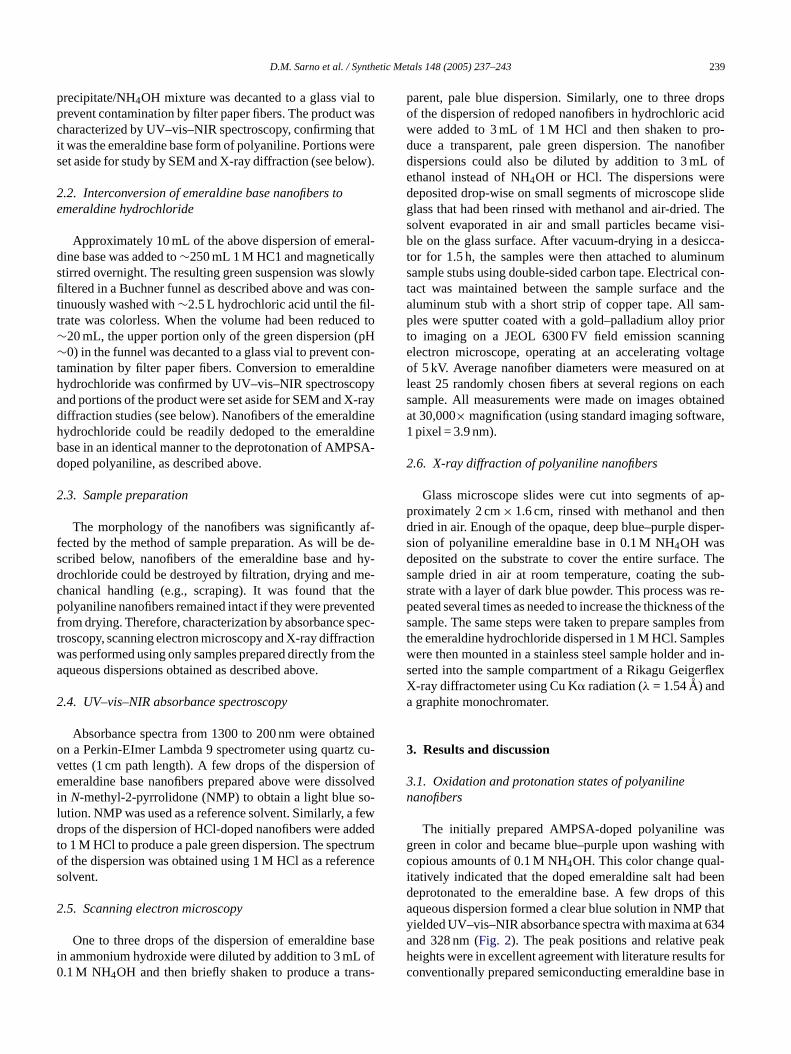
Representative images of dedoped emeraldine base andsubsequently redoped emeraldine hydrochloride are shownin Fig. 3. Examination of several regions on every samplerevealed mats of interwoven and twisted nanofibers. Thoughfibers were the dominant morphology, they were typicallyaccompanied by some small, non-fibrous particles. We havehypothesized that dust and/or other particulate contaminantsin the polymerizing solution, including insoluble ammoniumperoxydisulfate (see Section2), might have served as nu-cleation sites for the observed irregular material[15]. Cur-rent studies are directed towards understanding the polymer-ization process at its earliest stages in an effort to improvenanofiber monodispersity. It could not be determined by theseSEM studies if the nanofibers were hollow tubes or solidfibers. Though preliminary transmission electron microscopyexperiments have suggested that AMPSA-doped polyanilineconsists partially of hollow tubes, this technique has not yetbeen used to investigate the effects of dedoping and redoping[19].
Fiber dimensions were measured in regions where theywere less densely packed, usually near the edges of themats. The average diameter of emeraldine base nanofibers de-posited from dispersions in 0.1 M NH4OH was 53 nm, rang-i am-e nm.
ng to solid powders, as described in Section2. This meaure significantly increased the content of nanofibers
ng from 37 to 71 nm. When deposited from ethanol, diters ranged from 32 to 75 nm, with an average of 59
D.M. Sarno et al. / Synthetic Metals 148 (2005) 237–243 241
Fig. 3. SEM images of (a) emeraldine base and (b) emeraldine hydrochlo-ride nanofibers both deposited from aqueous dispersions diluted in ethanol.Magnification is 30,000× and scale bar is 1�m.
Emeraldine hydrochloride nanofibers deposited from disper-sions in 1 M HCl ranged in diameter from 28 to 82 nm, withan average of 58 nm. When deposited from ethanol, the av-erage diameter was 54 nm, ranging from 28 to 70 nm. Dif-ferences in these averages measured by counting pixels inthe SEM images (∼4 nm/pixel) were within experimental er-ror. Estimates of fiber length ranged from∼400 nm to 2�m.It was concluded that the interconversion between metallicand semiconducting states by dedoping and redoping did notcause any obvious change to the morphology or dimensionsof the nanofibers, nor were there any discernable effects basedon the solvent in which they were suspended. However, repet-itive dedoping and redoping of the nanofibers appeared to re-sult in changes in morphology, such as more densely packedfiber mats, and fused, and/or broken fibers.
From the absorbance data and SEM images describedabove, it is most important to note that the polyanilinenanofibers could be redoped with a different acid (i.e., HCl)from that used in their initial synthesis (i.e., AMPSA). Thisimplies that although a certain dopant may he required toobtain a certain type of nanofiher, this dopant can then be re-moved and replaced with a different dopant for modificationof selected properties with retention of morphology.
Fig. 4. SEM images of nanofibers of (a) emeraldine base and (b) emeraldinehydrochloride both dried to solid powders. Magnification is 30,000× andscale bar is 1�m.
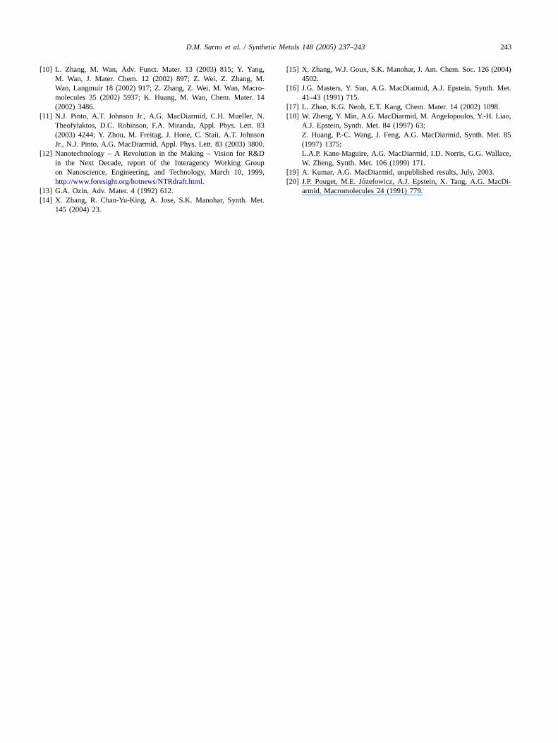
X-ray diffraction was used to further probe the structure ofthe polyaniline nanofibers. The emeraldine base presented abroad signal centered at 2θ ∼ 22◦ with a shoulder at 2θ ∼ 15◦(Fig. 5a). This broad X-ray structure suggested an amor-phous polymer, similar to bulk emeraldine base prepared bystandard methods[20]. Subsequent doping to emeraldine hy-drochloride resulted in a more sharply defined diffractionpattern with an intense peak at 2θ = 22.5◦ and two weakerpeaks at 2θ = 16.2◦ and 14.5◦ (Fig. 5b). As expected fromprevious results from the bulk material[20], the apparentcrystallinity of the metallic form of the polymer was greaterthan that of the semiconducting form from which it was pre-pared. The metallic nanofibers were compared with com-mercially available polyaniline (Aldrich; emeraldine base,mol. wt. ∼100,000) that had been doped to the emeraldinehydrochloride form. It was found that only the lowest anglediffraction from the nanofibers agreed well with the diffrac-tion pattern of the doped bulk polymer. It is unknown whatdifferences in molecular structure this result may imply. How-ever, there was no obvious, pronounced difference in the es-timated fractional crystallinity of emeraldine hydrochloridewhen prepared as either nanofibers or as bulk material.
242 D.M. Sarno et al. / Synthetic Metals 148 (2005) 237–243
Fig. 5. X-ray diffraction patterns from nanofibers of (a) emeraldine baseand (b) emeraldine hydrochloride on glass slides. Diffraction from the glasssubstrate (see inset, b) may obscure signals below 2θ ∼12◦ and above∼25◦.
4. Summary
Nanofibers of semiconducting and metallic polyanilinewith diameters of 28–82 nm (56 nm average diameter) havebeen prepared by oxidative polymerization of aniline in 2-acrylamido-2-methyl-l-propanesulfonic acid (AMPSA) so-lution in the presence of high concentrations of the nonionicsurfactant Triton X-100. As with other similar systems thatemploy surfactants[10], it is hypothesized that fiber forma-tion probably occurs on a micellar template, though the exact
shape and composition of this template is not currently under-stood. AMPSA-doped polyaniline can be deprotonated to thesemiconducting emeraldine base with ammonium hydrox-ide. Redoping to the metallic emeraldine hydrochloride formis readily achieved by protonation with hydrochloric acid.These nanofibers are electronically identical to the emeral-dine base or hydrochloride forms, respectively, of conven-tionally prepared polyaniline. X-ray diffraction from emeral-dine hydrochloride nanofibers indicates approximately thesame fractional crystallinity as the analogous bulk material.
SEM images reveal that the nanofiber morphology is un-changed by interconversion between the semiconducting andmetallic forms. Furthermore, emeraldine base nanofibers canbe redoped with an acid different from that used in their ini-tial preparation. Therefore, regardless of the conditions nec-essary to obtain a particular type of nanofiber, selected chem-ical, physical and electronic properties may be subsequentlymodified. Notably, the observation of nanofibers is greatlyfavored for samples that are prepared as aqueous dispersionsand prevented from drying to solid powders. Extensive fil-tration and drying damages the nanofibers and significantlyincreases the content of non-fibrous material. The above stud-ies show that electronic nanofibers of organic polymers rep-resent a potentially very important class of new material inthe area of nanoscience/technology.
A
valR iver-s eya truc-t useo 9).
R
.G.
.J.bli-
nth.
9)
02)
em.
ang,03)
cknowledgements
The authors gratefully acknowledge the Office of Naesearch (Contract no. N00014-01-1-0933) and The Unity of Texas at Dallas for their support of this work. Thlso acknowledge the Laboratory for Research on the S
ure of Matter at the University of Pennsylvania for thef SEM facilities (MRSEC NSF Award no. DMR00-7990
eferences
[1] A.G. MacDiarmid, Angew. Chem. Int. Ed. 40 (2001) 2581; AMacDiarmid, Synth. Met. 84 (1997) 27.
[2] A.G. MacDiarmid, J.-C. Chiang, A.F. Richter, N.L.D. Somasiri, AEpstein, in: L. Alacer (Ed.), Conducting Polymers, D. Reidel Pucation, Dordrecht, 1987, pp. 105–120;J.-C. Chiang, A.G. MacDiarmid, Synth. Met. 13 (1986) 193.
[3] Y. Cao, P. Smith, A.J. Heeger, Synth. Met. 48 (1992) 91.[4] P.M. Beadle, Y.F. Nicolau, E. Banka, P. Rannou, D. Djurado, Sy
Met. 95 (1998) 29.[5] J. Stejskal, I. Sapurina, J. Prokes, J. Zemek, Synth. Met. 105 (199
195;S. Fedorova, J. Stejskal, Langmuir 18 (2002) 5630.
[6] A. Malinauskas, Polymer 42 (2001) 3957.[7] M.F. Hundley, P.N. Adams, B.R. Mattes, Synth. Met. 129 (20
291.[8] C.R. Martin, Chem. Mater. 8 (1996) 1739; C.R. Martin, Acc. Ch
Res. 28 (1995) 61.[9] J. Huang, R.B. Kaner, J. Am. Chem. Soc. 126 (2004) 851; J. Hu
S. Virji, B.H. Weiller, R.B. Kaner, J. Am. Chem. Soc. 125 (20314.
D.M. Sarno et al. / Synthetic Metals 148 (2005) 237–243 243
[10] L. Zhang, M. Wan, Adv. Funct. Mater. 13 (2003) 815; Y. Yang,M. Wan, J. Mater. Chem. 12 (2002) 897; Z. Wei, Z. Zhang, M.Wan, Langmuir 18 (2002) 917; Z. Zhang, Z. Wei, M. Wan, Macro-molecules 35 (2002) 5937; K. Huang, M. Wan, Chem. Mater. 14(2002) 3486.
[11] N.J. Pinto, A.T. Johnson Jr., A.G. MacDiarmid, C.H. Mueller, N.Theofylaktos, D.C. Robinson, F.A. Miranda, Appl. Phys. Lett. 83(2003) 4244; Y. Zhou, M. Freitag, J. Hone, C. Staii, A.T. JohnsonJr., N.J. Pinto, A.G. MacDiarmid, Appl. Phys. Lett. 83 (2003) 3800.
[12] Nanotechnology – A Revolution in the Making – Vision for R&Din the Next Decade, report of the Interagency Working Groupon Nanoscience, Engineering, and Technology, March 10, 1999,http://www.foresight.org/hotnews/NTRdraft.html.
[13] G.A. Ozin, Adv. Mater. 4 (1992) 612.[14] X. Zhang, R. Chan-Yu-King, A. Jose, S.K. Manohar, Synth. Met.
145 (2004) 23.
[15] X. Zhang, W.J. Goux, S.K. Manohar, J. Am. Chem. Soc. 126 (2004)4502.
[16] J.G. Masters, Y. Sun, A.G. MacDiarmid, A.J. Epstein, Synth. Met.41–43 (1991) 715.
[17] L. Zhao, K.G. Neoh, E.T. Kang, Chem. Mater. 14 (2002) 1098.[18] W. Zheng, Y. Min, A.G. MacDiarmid, M. Angelopoulos, Y.-H. Liao,
A.J. Epstein, Synth. Met. 84 (1997) 63;Z. Huang, P.-C. Wang, J. Feng, A.G. MacDiarmid, Synth. Met. 85(1997) 1375;L.A.P. Kane-Maguire, A.G. MacDiarmid, I.D. Norris, G.G. Wallace,W. Zheng, Synth. Met. 106 (1999) 171.
[19] A. Kumar, A.G. MacDiarmid, unpublished results, July, 2003.[20] J.P. Pouget, M.E. Jozefowicz, A.J. Epstein, X. Tang, A.G. MacDi-
armid, Macromolecules 24 (1991) 779.