control of postnatal apoptosis in the neocortex byrhoa-subfamily gtpases determines neuronal density
TRANSCRIPT

Development/Plasticity/Repair
Control of Postnatal Apoptosis in the Neocortex byRhoA-Subfamily GTPases Determines Neuronal Density
Hitomi Sanno,1,2 Xiao Shen,1,2 Nilgun Kuru,3,4 Ingo Bormuth,5,6 Kristin Bobsin,1,2 Humphrey A. R. Gardner,7
Dorde Komljenovic,2 Victor Tarabykin,5,6 Reha S. Erzurumlu,4 and Kerry L. Tucker1,2
1Interdisciplinary Center for Neurosciences and 2Institute of Anatomy, University of Heidelberg, D-69120 Heidelberg, Germany, 3Department of Biology,Faculty of Education, Cumhuriyet University, TR-58140 Sivas, Turkey, 4Department of Anatomy and Neurobiology, University of Maryland School ofMedicine, Baltimore, Maryland 21201, 5Max Planck Institute for Experimental Medicine, D-37075 Gottingen, Germany, 6Institute of Cell Biology andNeurobiology, Charite-Universitatsmedizin Berlin, Campus Mitte, D-10098 Berlin, Germany, and 7Novartis Institutes for BioMedical Research, Cambridge,Massachusetts 02139
Apoptosis of neurons in the maturing neocortex has been recorded in a wide variety of mammals, but very little is known about its effectson cortical differentiation. Recent research has implicated the RhoA GTPase subfamily in the control of apoptosis in the developingnervous system and in other tissue types. Rho GTPases are important components of the signaling pathways linking extracellular signalsto the cytoskeleton. To investigate the role of the RhoA GTPase subfamily in neocortical apoptosis and differentiation, we have engineereda mouse line in which a dominant-negative RhoA mutant (N19 –RhoA) is expressed from the Mapt locus, such that all neurons of thedeveloping nervous system are expressing the N19 –RhoA inhibitor. Postnatal expression of N19 –RhoA led to no major changes inneocortical anatomy. Six layers of the neocortex developed and barrels (whisker-related neural modules) formed in layer IV. However, thedensity and absolute number of neurons in the somatosensory cortex increased by 12–26% compared with wild-type littermates. This was notexplained by a change in the migration of neurons during the formation of cortical layers but rather by a large decrease in the amount of neuronalapoptosis at postnatal day 5, the developmental maximum of cortical apoptosis. In addition, overexpression of RhoA in cortical neurons wasseen to cause high levels of apoptosis. These results demonstrate that RhoA-subfamily members play a major role in developmental apoptosis inpostnatal neocortex of the mouse but that decreased apoptosis does not alter cortical cytoarchitecture and patterning.
IntroductionProgrammed neuronal cell death plays a central role in the dy-namic organization of developing neuronal networks (Purves,1990). The classical neurotrophic model describes populations ofperipheral neurons that compete for limiting amounts of survivalfactors (e.g., NGF, BDNF, and neurotrophins 3 and 4/5) (Bibel andBarde, 2000). Failure to bind neurotrophins results in an apoptoticresponse and neuronal death. The activation of apoptotic ma-
chinery in the periphery is well investigated, but correspondingmechanisms in developing brain are poorly understood. Apopto-sis plays a crucial role in embryonic development of the cerebralcortex, cerebellum, and brainstem. The key apoptosis proteinscaspase-3, caspase-9, Apaf1, Bax, Bcl-XL, and survivin have beendemonstrated to regulate and execute apoptosis through mouseknock-out studies (for review, see Kuan et al., 2000). However,the proximal causes of apoptosis within the embryonic brainremain unknown.
Neuronal loss is known to occur in postnatal mammalianneocortex (Heumann et al., 1978; Finlay and Slattery, 1983;Heumann and Leuba, 1983; Price and Blakemore, 1985), markedby a wave of apoptosis that peaks at postnatal day 5 (P5) to P7 inrodents (Pearlman, 1985; Ferrer et al., 1990, 1992; Spreafico et al.,1995; Verney et al., 2000). Aside from IGF-1 (Chrysis et al., 2001;Hodge et al., 2007), the molecular players in postnatal corticalapoptosis are not known.
The Rho GTPases RhoA, RhoB, and RhoC belong to and de-fine the RhoA-subfamily within the Ras superfamily of smallGTP-binding proteins (Hall, 1998). They cycle between a GDP-bound, inactive and a GTP-bound, active state. These two statesare primarily regulated by the guanine nucleotide exchange fac-tors (GEFs) and the GTPase-activating proteins families (Etienne-Manneville and Hall, 2002), which promote the activation andinactivation, respectively, of GDP- and GTP-bound Rho. One ofthe most important targets of active RhoA is Rho-associated ki-
Received July 12, 2009; revised Jan. 25, 2010; accepted Jan. 30, 2010.This work was supported by the German Research Foundation [Deutsche Forschungsgemeinschaft: Sonderfors-
chungsbereich 488, Teilprojekt B7/B9 (K.L.T.) and Exzellenzcluster 257 (V.T.)], the University of Heidelberg [Excel-lence Cluster Cellular Networks (K.L.T.)], National Institutes of Health/National Institute of Neurological Disordersand Stroke Grant NS039050 (R.S.E.), the Max Planck Society (V.T.), and the Heisenberg Program (V.T.). We thankJoachim Kirsch for generous scientific support, Karin Gorgas for enormous help with anatomical analysis, Yves-AlainBarde for thoughtful commentary on this manuscript, Silvia Arber for the floxed stop and Mapt targeting constructs,Frank Zimmermann for blastocyst injections, Stefan Offermanns for EIIa::CRE mice and rhotekin-expressing bacteria,Julia Hoffmann for preparation of the rhotekin beads, Robert Grosse for Rac1 and cdc42 expression plasmids,Antonio Caputi for the anti-parvalbumin antibody, Ulrich Muller for the N19 –RhoA cDNA, Gunter Giese and Anne-marie Scherbarth for assistance with confocal microscopy, and Dmitry Rusanov and Jana Hechler for superb technicalassistance.
Correspondence should be addressed to Dr. Kerry L. Tucker, Institute of Anatomy, University of Heidelberg, ImNeuenheimer Feld 307, 69120 Heidelberg, Germany. E-mail: [email protected].
H. Sanno’s present address: Department of Physiology and Center for Integrative Genomics, University of Lau-sanne, CH-1015 Lausanne, Switzerland.
D. Komljenovic’s present address: Department of Medical Physics, German Cancer Research Center, INF 280,D-69120 Heidelberg, Germany.
DOI:10.1523/JNEUROSCI.3318-09.2010Copyright © 2010 the authors 0270-6474/10/304221-11$15.00/0
The Journal of Neuroscience, March 24, 2010 • 30(12):4221– 4231 • 4221
nase (ROCK), a serine/threonine kinasewith multiple substrates (Katoh et al.,1998). Although much is known about themodeling of axons and dendrites by RhoGTPases (Luo, 2000), the role that theyplay in the control of apoptosis is contro-versial. In vitro studies have indicated thatRho can induce apoptosis in hippocampal(Donovan et al., 1997) and cortical (Zhanget al., 2007) neurons. In vivo studies havedelivered conflicting evidence, with one in-vestigation showing that apoptosis of spinalcord motor neurons increased during inhi-bition of Rho activity (Kobayashi et al.,2004), whereas a rat model of spinal cordinjury showed that Rho inhibition reducedinjury-related apoptotic levels (Dubreuil etal., 2003).
To investigate Rho GTPases in thecontrol of postnatal cortical apoptosis, wedeveloped a mouse line in which a domi-nant-negative inhibitor of Rho GTPasesis expressed specifically in neurons. Wefound that inhibition of Rho GTPases inpostnatal cortical neurons in vivo resultedin a significant reduction in apoptosis ofexcitatory neurons and a correspondingincrease in the absolute number and den-sity of neurons in the cortex. Despite theincrease in neuronal numbers, corticallamination and pattern formation wereunaltered. These findings suggest thatpostnatal apoptosis does not contribute tocytoarchitectonic differentiation and cel-lular patterning of the neocortex.
Materials and MethodsGeneration of the N19 –RhoA mouse line. All an-imal experiments were in compliance with theregulations of Baden–Wurttemberg. To con-struct the targeting vector, a human RhoAcDNA containing the N19 mutation and anN-terminal hemagglutinin (HA) tag was insertedinto a vector (pLSL; courtesy of Dr. Silvia Arber,University of Basel, Basel, Switzerland) down-stream of a transcriptional stop cassetteflanked by loxP sites. The resultant cassette wasinserted into a targeting vector (courtesy of Dr.Silvia Arber) (Hippenmeyer et al., 2005) con-taining genomic Mapt sequence and a neomycin-selectable marker. Thelinearized targeting vector was electroporated into J1 embryonic stem(ES) cells as described previously (Tucker et al., 2001), and 28 neomycin-resistant colonies were analyzed by Southern blot, using externalgenomic probes as described (Tucker et al., 2001).
Targeted, euploid ES cells were injected into C57BL/6 blastocysts. Twohigh-contribution male chimeras derived from two different ES cell lineswere bred with C57BL/6 wild-type mice to generate two independentN19 –RhoA mouse lines. Germ-line transmission of the targeted Maptallele was confirmed by Southern blots of mouse tail DNA. Subsequentgenerations were maintained on a C57BL/6 background. For genotypingthe N19 –RhoA mice, the primers 5�-TACGACGTGCCCGACTAC-3�and 5�-GCTGTGTCCCACAAAGCC-3� delivered a 220 bp amplicon.EIIa::CRE mice were genotyped using the primers 5�-GCCGAAATT-GCCAGGATCAG-3� and 5�-AGCCACCAGCTTGCATGATC-3�, givinga 486 bp amplicon. For the Southern blot analysis of the efficiency of the
Cre-based excision of the stop cassette, a 600 bp N19 –RhoA cDNA frag-ment was used as a probe.
Rhotekin beads. Rhotekin-expressing bacteria (Ren et al., 1999) werecultured in 20 ml of Luria broth (LB) media with ampicillin (100 �g/ml)and chloramphenicol (34 �g/ml) on a shaker at 37°C overnight. Thefollowing day, 2– 4 ml of overnight culture were added to 4� 500 ml LBmedia with ampicillin and chloramphenicol on a shaker at 37°C andinduced with 500 �l of 0.5 M isopropyl-�-D-thiogalactopyranoside untilthe OD600 became between 0.7 and 0.8, followed by 3 h incubation at30°C on a shaker. The bacterial cultures were placed in 500 ml tubes andcentrifuged for 10 min at 4000 rpm, and the pellets were washed twicewith 200 ml of cold 1� PBS. The pellets were resuspended and centri-fuged for 10 min at 4000 rpm at 4°C. These pellets were resuspended in4� 50 ml lysis buffer, the cells were sonicated for 15 s six times at 130 W,and 500 �l of 10% Triton X-100 was added per tube to make the finalconcentration of 0.1% Triton X-100 and incubated for 15 min on ice,rocking. Lysates were decanted in eight ultracentrifuge tubes on ice and
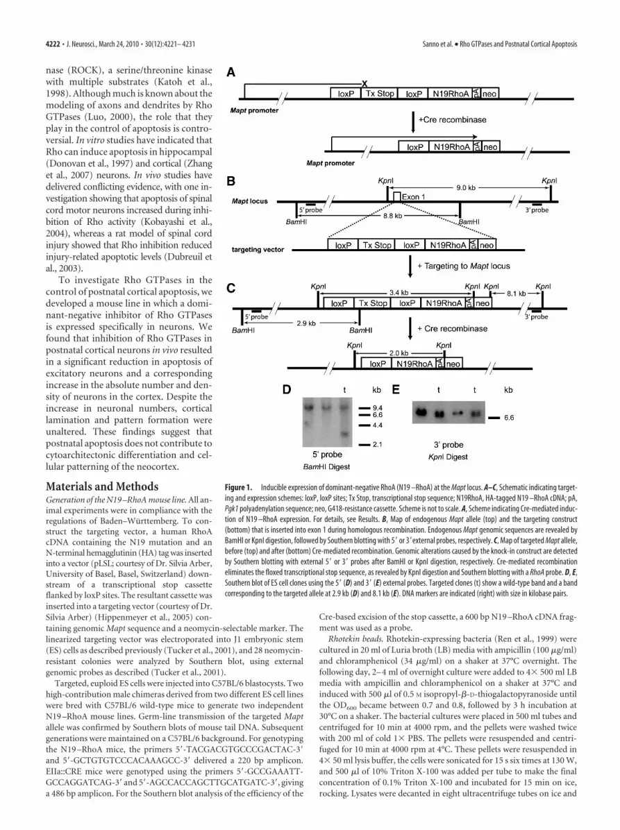
Figure 1. Inducible expression of dominant-negative RhoA (N19 –RhoA) at the Mapt locus. A–C, Schematic indicating target-ing and expression schemes: loxP, loxP sites; Tx Stop, transcriptional stop sequence; N19RhoA, HA-tagged N19 –RhoA cDNA; pA,Pgk1 polyadenylation sequence; neo, G418-resistance cassette. Scheme is not to scale. A, Scheme indicating Cre-mediated induc-tion of N19 –RhoA expression. For details, see Results. B, Map of endogenous Mapt allele (top) and the targeting construct(bottom) that is inserted into exon 1 during homologous recombination. Endogenous Mapt genomic sequences are revealed byBamHI or KpnI digestion, followed by Southern blotting with 5� or 3�external probes, respectively. C, Map of targeted Mapt allele,before (top) and after (bottom) Cre-mediated recombination. Genomic alterations caused by the knock-in construct are detectedby Southern blotting with external 5� or 3� probes after BamHI or KpnI digestion, respectively. Cre-mediated recombinationeliminates the floxed transcriptional stop sequence, as revealed by KpnI digestion and Southern blotting with a RhoA probe. D, E,Southern blot of ES cell clones using the 5� (D) and 3� (E) external probes. Targeted clones (t) show a wild-type band and a bandcorresponding to the targeted allele at 2.9 kb (D) and 8.1 kb (E). DNA markers are indicated (right) with size in kilobase pairs.
4222 • J. Neurosci., March 24, 2010 • 30(12):4221– 4231 Sanno et al. • Rho GTPases and Postnatal Cortical Apoptosis

centrifuged for 12 min at 27,000 rpm (Hitachi HimacCS 100fx). Thebeads (1.3 ml) (Glutathione Sepharose 4B; GE Healthcare) for 2 L ofmedium was washed in a 50 ml tube twice with 15 ml of 1� PBS at 4°C.Beads were centrifuged at 1000 rpm at 2°C for 1 min. Rhotekin lysateswere added to the beads and incubated for 45 min, shaking horizontallyto avoid making bubbles. The beads were washed with 20 ml of cold washbuffer [50 mM Tris-HCl, pH 7.5, 0.5% Triton X-100, 150 mM NaCl, 5 mM
MgCl2, 10 �g/ml aprotinin (Roche), 10 �g/ml leupeptin (Roche), and 10�g/ml PMSF] for 1 min at 1000 rpm at 2°C, and then the supernatant wasremoved. Beads were resolved in 20 ml of wash buffer/10% glycerol, andaliquoted beads were kept at �80°C.
Rhotekin pulldown assay. Cerebral cortex from postnatal animals wascarefully microdissected and flash frozen in liquid nitrogen. Tissue waslysed (50 mM Tris-HCl, pH 7.4, 150 mM NaCl, and 5 mM MgCl2) with 10�g/ml aprotinin (Roche), 10 �g/ml leupeptin (Roche), and 10 �g/mlPMSF (Carl Roth GmbH) by sonication three times for 15 s. Brain lysate(450 �g) was incubated with 200 �l Rhotekin beads for 19 h at 4°C.Lysates were centrifuged at 4°C for 15 min at 2900 rpm, and the pellet wasresuspended and washed three times in 50 mM Tris-HCl, pH 7.5, 0.5%Triton X-100, 150 mM NaCl, 5 mM MgCl2, 10 �g/ml aprotinin, 10 �g/mlleupeptin, and 10 �g/ml PMSF for 5 min with rotation. The pellet wasresuspended in 15 �l of sample buffer and electrophoresed in acrylamide
gels, and RhoA levels were analyzed by West-ern blot. The antibodies (Abs) used includedmouse anti-RhoA (sc-418, 1:500; Santa CruzBiotechnology) and rabbit anti-actin (A-2066, 1:1000; Sigma-Aldrich).
Western blotting. Western blotting was per-formed on homogenized brain as describedpreviously (Willaredt et al., 2008), with either amouse anti-RhoA Ab (sc-418, 1:1000; SantaCruz Biotechnology) or a rabbit anti-HA Ab(sc-805, 1:1000; Santa Cruz Biotechnology).For a positive control, HN10 cells were tran-siently transfected with the HA-tagged N19 –RhoA construct used above, and protein lysateswere prepared 48 h after transfection.
In situ hybridization. In situ hybridizationon paraffin sections was performed (Shakedet al., 2008) with a human RhoA cDNA(Deutsches Ressourcenzentrum fur Genom-forschung GmbH).
Barrel cortex analyses. Four P9 and fourP65 mice (two control and two transgeniceach age) were used to analyze the barrel cor-tex. In these experiments, serial sectionsthrough flattened cortices and coronal brainsections were processed with routine histo-logical methods for visualization of barrelpatterns. The brains were split in half along thesagittal plane. The left cortex from each halfwas removed and flattened between two glassslides. Thirty-micrometer-thick coronal andtangential sections were kept in PBS, pH 7.4.Alternate sections were stained for either Nissl,cytochrome oxidase (CO) histochemistry orserotonin transporter (5-HTT) immunohisto-chemistry for P9 brains. For CO histochemis-try, free-floating sections were incubated withphosphate buffer containing 0.5 mg/ml cyto-chrome C (type III; Sigma), 0.5 mg/ml diami-nobenzidine (DAB) (Sigma), and 40 mg/mlsucrose for 2– 4 h at 37°C in a shaker incubator.Alternate sections were stained with 2% cresylviolet for Nissl staining. For 5-HTT immuno-histochemistry, sections were treated as below(immunohistochemical analysis) for peroxi-dase detection using an anti-5-HTT rabbitpolyclonal Ab (1:10,000; Diasorin). Sectionswere mounted on slides, dehydrated, cleared in
xylene, and coverslipped with Permount.Immunohistochemical analysis and cell counting. Free-floating 20 �m
brain sections were rinsed in 2% H2O2 in 10% methanol for 20 min toblock endogenous peroxidase activity, and biotinylated secondary anti-body and peroxidase-labeled avidin– biotin complex were applied ac-cording to the instructions of the manufacturer (Vectastain Elite ABC;Vector Laboratories). Sections were washed in PBS for 30 min, developedin 0.05% DAB (Sigma) with the addition of 0.006% H2O2, washed in PBSfor 10 min, and mounted (Aquatex medium). The following primaryantibodies were used, with clone name, source, and dilution indicated:mouse anti-neuronal-specific nuclear protein (NeuN) (MAB377,1:1000; Millipore Corporation), mouse anti-parvalbumin (clone PARV-19, 1:2000; Sigma), rabbit anti-calretinin (catalog #7699/4, 1:3000;Swant), mouse anti-calbindin (catalog #300, 1:3000; Swant), rabbit anti-cleaved-caspase-3 (clone 5A1, 1:200; Cell Signaling Technology), rabbitanti-phospho-histone H3 (Ser10, rabbit 06-570, 1:200; Millipore Corpo-ration), and rabbit anti-ER81 (1:200; kind gift from Dr. Silvia Arber). Forcaspase-3, phospho-histone H3, and ER81 stains, secondary Abs wereused as described previously (Brachmann et al., 2007).
Sections were photographed with a Leica DMLB microscope equippedwith a Leica DFC320 CCD camera and two different air objectives: a 40�
Figure 2. Postnatal expression of N19 –RhoA inhibits endogenous Rho activity. A, Complete recombination of the targetedMapt allele during in vivo exposure to EIIa::Cre recombinase was detected in acute cultures of cerebellar granule neurons, indicatedby Southern blots on KpnI-digested genomic DNA with a RhoA cDNA probe. Band sizes are explained in Figure 1 C. DNA markers areindicated (right) with size in kilobase pairs. B, C, Western blot analysis indicates HA-tagged N19 –RhoA expression (arrow) in thebrain of E13.5 (B) and newborn (C) animals after exposure to Cre recombinase, using an anti-HA antibody. B, HN10, Positive controlin which HA-tagged N19 –RhoA expression construct was transfected into HN10 cells. D, N19 –RhoA acts as a dominant-negativeinhibitor of RhoA function in vivo. Brain lysates from N19 –RhoA or control mice were lysed, incubated with Sepharose beadscoupled to the Rho-binding domain of Rhotekin, and protein bound to Rho-binding domain subsequently analyzed by Westernblotting, using an anti-RhoA antibody to detect wild-type RhoA (22 kDa). Loading levels were analyzed using an anti-actinantibody (42 kDa). B–D, Protein markers (in kilodaltons) are indicated (right). E, F, In situ hybridization analysis of N19 –RhoAexpression in the telencephalon of P5 (E) and P9 (F ) N19 –RhoA mice. Dorsal is to the top. Scale bars: 200 �m.
Sanno et al. • Rho GTPases and Postnatal Cortical Apoptosis J. Neurosci., March 24, 2010 • 30(12):4221– 4231 • 4223

HCX PLAN APO objective (numerical aperture, 0.85) and a 5� HC PLFLUOTAR objective (numerical aperture, 0.15), using the program Fire-cam 3.1 (Leica). Somatosensory cortex and the anterior extent of thevisual cortex was used for analyses. Ten slides were chosen at equidistantlocations within this area, photographed on both the right and left sides,and subsequently quantitated. In NeuN-stained preparations, the darklystained nuclei were easy to identify at both magnifications. In the photostaken with a 5� objective, the entire width of the cortex could be iden-tified, nuclei in specific layers were counted by hand, and these data arepresented in Figure 4. Photographs taken at 40� were assigned to specificlayers, and a correction for the thickness of the section and the size of thenuclei was made according to Abercrombie (1946). Because the size ofthe nuclei did not vary significantly between wild-type (layer IV, 10.0 �0.9 �m; layer V, 11.8 � 1.0 �m; layer VI, 10.8 � 0.8 �m; mean � SEM;n � 150 for all layers) and N19 –RhoA (layer IV, 10.2 � 0.9 �m; layer V,12.8 � 0.9 �m; layer VI, 10.8 � 0.7 �m; mean � SEM; n � 150 for alllayers) mice, this correction did not affect the relative increase in neuronnumber when comparing wild-type with N19 –RhoA mice, and the re-sults agreed with the counts made using sections photographed with the5� objective. With the 40� objective, we recorded an increase in layer IVof 23.4 � 1.9%, in layer V of 18.0 � 2.7%, and in layer VI of 21.0 � 5.8%(n � 4; mean � SEM, Student’s t test). All counting was performed in ablind manner.
Developmental analysis of cortical layer formation. Brains from new-born (P1) or P3 N19 –RhoA and control littermate pups were perfusedtranscardially and removed for processing to paraffin blocks. For paraffinsectioning, tissue was dehydrated in an ascending ethanol row, isopro-panol, and xylene. Brains from N19 –RhoA and control littermates wereembedded in one common paraffin block and were sectioned at 10 �mon a Leica sliding microtome. Sections were deparaffinated using xyleneand isopropanol and were rehydrated in a descending ethanol row. An-tigen unmasking was performed by boiling for 10 min in either citricbuffer (pH 6) or Tris EDTA (pH 9) in a microwave oven at 600 W.Sections were washed with Tris buffer containing 2% milk powder(Tris�), blocked in 20% goat serum in PBS for 1 h, and incubated withprimary antibodies in 5% goat serum in PBS for 4 h. Sections werewashed three times with Tris�, incubated for 20 min with appropriatebiotinylated secondary antibodies (Vector Laboratories), washed threetimes with Tris�, incubated for 10 min with streptavidin/biotin complex(Vector Laboratories), washed three times in Tris (without milk pow-der), incubated for 10 min with DAB substrate (Vector Laboratories),washed, and counterstained with hematoxylin. Sections were dehydratedin an ascending ethanol row, followed by isopropanol and xylene. Cov-erslips were applied using Eukitt. Primary antibodies were directedagainst Reelin (mouse IgG, 1:500; Calbiochem), Satb2 (mouse IgG,1:1000; from V. Tarabykin), Ctip2 (rat IgG, 1:500; Abcam), Brn2 (poly-clonal goat, 1:200; Santa Cruz Biotechnology), Tbr2 (polyclonal rabbit,1:500; Abcam), and Pax6 (polyclonal rabbit, 1:500; Millipore BioscienceResearch Reagents).
In vitro apoptosis assays. Five to seven embryonic day 16.5 (E16.5)embryos were removed from timed pregnant CD-1 mice, the cerebralhemispheres cut apart with a scalpel, the meninges were peeled away, and�3 mm of the cortex lying above the hippocampus was cut out anddigested in 0.25% trypsin (Invitrogen) at 37°C for 15 min. After threewashes in HBSS/10 mM HEPES, pH 7.3, cells were triturated with a fire-polished Pasteur pipette, and 700,000 cells were centrifuged 7 min at100 � g and electroporated in 100 �l nucleofection solution (mouseneuron nucleofector kit, Program O5; Amaxa Biosystems), contain-ing either 5 �g of an expression vector expressing enhanced greenfluorescent protein (pEGFP–N1), a RhoA expression vector [wild-typehuman RhoA cDNA with a 5�-localized HA tag cloned into the EcoRI/NotI site of the pcDNA3.1(�) vector], or myc-tagged wild-type humanRac1 or cdc42 expression plasmids (kind gift from Dr. Robert Grosse,Philipps University of Marburg, Marburg, Germany). Transfected cellswere plated in 24-well plates at a concentration of 30,000 cells per 13 mmcoverslip (pretreated with 1 mg/ml poly-L-lysine and 1 �g/ml laminin) inMEF medium (DMEM/10% fetal calf serum/2 mM glutamine), incu-bated at 37°C/5% CO2, with an exchange of MEF medium to Neurobasalmedium/1� B27 supplement/2 mM glutamine (Invitrogen) after 24 h,
and fixed after 48 h in culture for 10 min with 4% paraformaldehyde, pH7.4, at room temperature. Immunocytofluorescence was performed asdescribed previously (Shaked et al., 2008), with the following changes:cells were permeabilized for 5 min with 0.2% Triton X-100 only at thebeginning of the staining, followed by a quenching of the cells for 5 minwith 0.1% sodium borohydride. The following primary antibodies wereused: FITC-coupled goat anti-GFP (G8965�12A, 1:2000; US Biologicals),mouse anti-HA tag (clone 6E2, 1:200; Cell Signaling Technology), mouseanti-c-Myc tag (clone 9E10, sc-40, 1:100; Santa Cruz Biotechnology),rabbit anti-cleaved-caspase-3 (Asp175, clone 5A1, 1:200; Cell SignalingTechnology). Secondary Abs were used as described previously (Brach-
Figure 3. Normal barrel patterns in N19 –RhoA mice. A, B, At P9, thalamocortical afferentterminal patterns, as assessed with 5-HTT immunohistochemistry (A), are similar between thecontrol and N19 –RhoA mice as are the barrels as cytoarchitectonic entities, revealed by Nisslstain (B). Whisker-related barrel rows (a– e) are indicated. C, High-magnification views ofNissl-stained barrels in rows d and e. Asterisks mark barrel centers. D, Cytochrome oxidasestaining in P65 mice also shows clear barrel patterns in both the control and N19 –RhoA mice.These micrographs were montaged from serial sections. E, High-magnification views of Nissl-stained barrels in P65 mice also revealed clear barrels. Barrel centers are marked with asterisks.Scale bars: A, B, 500 �m; C, E, 200 �m; D, 700 �m.
4224 • J. Neurosci., March 24, 2010 • 30(12):4221– 4231 Sanno et al. • Rho GTPases and Postnatal Cortical Apoptosis

mann et al., 2007). 4�,6�-Diamidino-2-phenylindole (DAPI) (2 �g/ml) wasused to stain nuclei. The stained cortical neurons were analyzed with epif-luorescent microscopy (Microscope BX61WI; Olympus) using identical ex-posure conditions and a digital CCD camera (F-View II; Soft ImagingGmbH) in combination with imaging software (analysis, Soft ImagingGmbH). One hundred cells positive for either pEGFP–N1 or Rho–HA wereassayed for their cellular and nuclear morphology and for cleaved caspase-3activity. Statistical analysis was performed using the Mann–Whitney U test.
ResultsA mouse line expressing a dominant-negative inhibitor ofRho specifically in newborn neurons of the nervous systemTo inhibit the activity of proteins in the RhoA GTPase subfamily,we used the N19 –RhoA construct, a dominant-negative inhib-itor in which the threonine at position 19 has been mutated toasparagine (N) (Qiu et al., 1995). N19 –RhoA is predicted toblock the GTPase activity of endogenous RhoA-subfamilyGTPases through competitive binding to GEFs (Feig, 1994), andit has been suggested that this substitution also disturbs RhoGTPase activity by interfering with an essential Mg 2� ion re-quired for guanine nucleotide binding in all Ras superfamilyGTP-binding proteins (Farnsworth and Feig, 1991). It has beenshown to specifically inhibit members of the RhoA-subfamilyGTPases and to demonstrate no direct effect on other RhoGTPase family members, such as cdc42 or Rac, either biochemically(Ren et al., 1999) or in cell-based assays (Hall, 1998; Wojciak-Stothard et al., 1999; Bouzahzah et al., 2001). However, it cannot bedefinitively excluded that N19–RhoA may also affect cdc42 or Rac-based pathways by binding to promiscuous GEFs that interact withboth RhoA- and cdc42/Rac-subfamily members (Rossman et al.,2005). To distinguish the mutant RhoA protein from the endoge-nous murine RhoA, a human N19–RhoA cDNA was modified toexpress an HA tag at its N terminus.
The N19 –RhoA cDNA was engineered to allow for its induc-ible expression after exposure to the Cre recombinase. A cassettewas constructed in which the N19 –RhoA cDNA lies downstreamof a transcriptional stop cassette flanked by two loxP sites (Fig.
1B). This entire cassette was placed intothe first exon of the gene (Mapt) encodingtau, which had been used previously toexpress EGFP specifically in newbornneurons (Tucker et al., 2001). We chosean inducible approach because we wereconcerned that the long-term expressionof N19 –RhoA in all neurons of the mousecould cause potentially lethal side effects ifN19- RhoA were constitutively expressedfrom the Mapt locus. Because of the floxedstop cassette, transcription from the endog-enous tau promoter at the altered locusshould produce a truncated, non-protein-coding transcript, and the introducedcDNA should not be transcribed (Fig.1A). Exposure to the Cre recombinaseshould cause recombination between thetwo loxP sites, excision of the stop cas-sette, and subsequent expression of theN19 –RhoA protein (Fig. 1A). The target-ing vector was electroporated into J1 EScells, and 49 G418-resistant colonies werepicked, of which 28 were analyzed bySouthern blot. Using 5� (Fig. 1B,D) and 3�(Fig. 1B,E) external probes, five cloneswere found to be targeted using both
probes, indicating a targeting efficiency of 18%. Two indepen-dent euploid clones were used to generate chimeras, both ofwhich transmitted the targeted allele through the germ line. Withrespect to all data presented in this paper, similar phenotypeswere observed for both lines.
Inducible expression of N19 –RhoA in postnatal brainTo remove the floxed stop cassette from the Mapt locus, the N19–RhoA mice were crossed with the EIIa::Cre mouse line, whichexpresses Cre recombinase under the control of the adenovi-rus EIIa promoter (Lakso et al., 1996). In this line, Cre recom-binase is expressed already in the zygote, and all cells of theresulting embryo should show recombination at the loxP sitesand removal of the floxed stop cassette. After Cre-based recombina-tion, the N19–RhoA cassette can then be expressed specifically inneurons. Compound heterozygotes for both the N19–RhoA and theEIIa::Cre alleles (Cre�/N19–RhoA�) were generated, and culturesof cerebellar granule neurons (CGNs) were prepared from P5Cre�/N19 –RhoA� and Cre�/N19 –RhoA� mice. The ability ofthe loxP sites to undergo recombination by Cre recombinase wasexamined using Southern blot analysis. KpnI-digested genomicDNA from CGN cultures was probed with a human RhoA cDNA.The cassette before recombination was seen to be 3.4 kb in theCre�/N19 –RhoA� cultures (Fig. 1D, 2A). Cre-mediated re-combination would remove the 1.4 kb floxed transcriptional stopsequence, with a corresponding decrease in the size of the RhoA-hybridizing band on Southern blotting (Fig. 1D). Indeed, in theCre�/N19 –RhoA� cultures, no signal could be detected at 3.4kb, whereas a strong band was seen at 2.0 kb, indicating a 100%efficiency in the removal of the floxed stop cassette in CGN dur-ing exposure to Cre recombinase (Fig. 2A). Protein lysates wereprepared from embryonic and postnatal brain of Cre�/N19 –RhoA� mice to examine expression of the N19 –RhoA constructand were examined with Western blot analysis using an antibodyrecognizing the HA-tagged N19 –RhoA. Surprisingly, no embry-onic expression could be detected (Fig. 2B), despite the fact that
Figure 4. N19 –RhoA mice show an increased density of neurons in somatosensory cortex. A, Immunohistochemical staining ofsomatosensory cortex in P65 wild-type (�/�) and N19 –RhoA/� mice, using an antibody recognizing NeuN. For each coronalsection, dorsal is to the top, and lateral is to the right. Layers I–VI are indicated to the left. B, Quantification of the dorsoventralwidth of layers II/III, IV, V, and VI. C, Quantification of NeuN-positive neurons in layers II/III, IV, V, and VI. *p 0.5, **p 0.01,***p 0.001, Student’s t test (n � 4). Scale bar: A, 200 �m.
Sanno et al. • Rho GTPases and Postnatal Cortical Apoptosis J. Neurosci., March 24, 2010 • 30(12):4221– 4231 • 4225

EGFP is very strongly expressed from the tau locus as early as9.5 d postcoitum (Tucker et al., 2001). Expression could first bedetected at P3 in the brain, with expression continuing at P20(Fig. 2C). The expression of the N19 –RhoA protein was shown toact as an inhibitor of endogenous Rho function by performing aRhotekin assay on lysates from the cerebral cortex of N19 –RhoA-expressing or control mice. A large reduction in active GTP-bound Rho was observed (Fig. 2D). To examine the distributionof RhoA expression, in situ hybridization was performed on brainsections from postnatal N19 –RhoA transgenic mice. At P5 andP9, expression could be seen throughout the neocortex and hip-pocampus, with particularly high levels in layers IV/V of the cor-tex and the pyramidal layers of CA1, CA3, and the dentate gyrusof the hippocampus (Fig. 2E,F).
Cortical anatomy is not disrupted in N19 –RhoAtransgenic miceWe examined the barrel cortex of P9 and P65 N19 –RhoA trans-genic mice to determine whether laminar differentiation andwhisker-related barrel formation were affected. Six laminae of theparietal cortex were visible with Nissl (Fig. 3B,C,E) stains intransgenic mice, and there were no noticeable differences fromthe controls at both ages. CO histochemistry, a routine methodfor visualization of whisker-related patterns (i.e., cortical barrels)also revealed distinct patterning in the transgenic mice at bothages (Fig. 3D). In addition, we examined thalamocortical pat-terning with 5-HTT immunohistochemistry in P9 mice. Mono-amine transporters are transiently expressed in the primarysensory thalamic nuclei in mice (Lebrand et al., 1998), and5-HTT immunohistochemistry has been established as a reliablemarker for thalamocortical afferent terminal patterns in the bar-rel cortex (Iwasato et al., 2000; Rebsam et al., 2002). The thalamo-cortical afferent patterning in the barrel cortex was similarbetween the N19 –RhoA transgenic and control mice, in both thetangential and coronal planes (Fig. 3A).
The absolute number and density of neurons is increased inthe somatosensory cortex in N19 –RhoA transgenic miceTo assess the number of neurons within the barrel cortex, anantibody recognizing NeuN, which is expressed by all corticalneurons except for Cajal-Retzius cells (Mullen et al., 1992; Lyck etal., 2007), was used. Careful counting of the neurons in specificlayers revealed a large and significant increase in the density ofNeuN-positive neurons in layers II/III, IV, V, and VI of the so-matosensory cortex of the N19 –RhoA transgenic mice whencounted at maturity (P65) such that, in layer II/III, neuronaldensity increased by 11.6 � 1.3%, in layer IV by 26.4 � 1.9%, inlayer V by 17.2 � 1.5%, and in layer VI by 18.7 � 1.2% (n � 4;mean � SEM, Student’s t test) (Fig. 4A,C). A similar increase wasalso observed in juvenile mice at P24 (supplemental Fig. 1A,available at www.jneurosci.org as supplemental material), indi-cating that the increase in neuronal number is already establishedat an early age. Because the dorsoventral width of the layers didnot change significantly in the N19 –RhoA transgenic mice (Fig.4B), it can be concluded that the absolute number of neurons hadincreased by 12–26% in these cortical layers.
The density of interneuron populations is not affected insomatosensory cortex of N19 –RhoA miceNeuN is expressed by both excitatory projection neurons, whichconstitute the vast majority of the neurons in the cortex, andinhibitory interneurons, which make up 20 –30% of the neuronalpopulation (Markram et al., 2004). To distinguish which popu-
lation was demonstrating an increase in the number in the cortexof the N19 –RhoA mice, we performed stainings for interneuronsubpopulations characterized by the expression of parvalbumin(Fig. 5A,B), calbindin (Fig. 5C), and calretinin (Fig. 5D). In allthree cases, quantification of positive neurons revealed no differ-ence in number between wild-type and N19 –RhoA cortex (Fig.5E). Because these interneurons do not show any change in num-ber in the N19 –RhoA cortex, we conclude that the change inneuronal density in the N19 –RhoA cortex is attributable to anincrease in the number of excitatory projection neurons and notin that of the interneuron population.
Neuronal migration and cell type specification of corticalneurons is not affected in N19 –RhoA miceRhoA activity has been implicated in the control of radial migra-tion of cortical neurons (Hand et al., 2005), and it has been shownto be involved in a large number of migratory processes through-
Figure 5. The density of interneurons is not affected in somatosensory cortex of N19 –RhoAmice. A, B, Immunohistochemical staining of somatosensory cortex in P65 wild-type (�/�)and N19 –RhoA/� mice, using antibodies recognizing parvalbumin (A, B), calbindin (C), andcalretinin (D). For each coronal section, one telencephalic half is shown, with dorsal to the topand lateral to the right. Arrows (white) indicate positive neurons. B, Magnification of A. E,Quantification of interneurons. Scale bars: A, 500 �m; B–D, 200 �m.
4226 • J. Neurosci., March 24, 2010 • 30(12):4221– 4231 Sanno et al. • Rho GTPases and Postnatal Cortical Apoptosis

out the developing nervous system (Liu and Jessell, 1998; Ruppand Kulesa, 2007; Carmona-Fontaine et al., 2008; Groysman etal., 2008). To investigate the possibility that the increased numberof neurons in deeper cortex layers of N19 –RhoA mice resultsfrom abnormal radial migration of upper layer neurons, we per-formed immunohistochemistry for typical layer-specific proteinsin brain sections of newborn (P1) (Fig. 6) and P3 mice. We foundthe expression patterns of Reelin, Satb2, Ctip2, Brn2, Tbr2, andPax6 (Fig. 6A–F) to be unchanged in N19 –RhoA mice. In addi-tion, in E16.5 cbs/cbs mutants, the number of mitotic cells at thecortical ventricular zone was not significantly altered in the cor-tex (n � 4; p � 0.69, Student’s t test) (supplemental Fig. 1B,available at www.jneurosci.org as supplemental material). Finally,
we examined the expression of the layerV-specific transcription factor ER81 at P24in juvenile mice, when layer formation iscomplete. Comparison of the distributionof ER81-positive neurons between wild-type and N19–RhoA/� cortex revealedthat, in both cases, expression of ER81 wasrestricted to layer V (supplemental Fig.1C,D, available at www.jneurosci.org assupplemental material), which confir-med that migration of cortical neurons isnormal. Together, these results demonstratethat the specification and migration of cor-tical neurons was normal in the N19–RhoA-expressing mice.
Apoptosis is severely reduced insomatosensory cortex ofN19 –RhoA miceTo test whether reduced apoptosis plays arole in the increase of neurons in corticallayers IV–VI of the N19 –RhoA transgenicmice, the numbers of apoptotic cells wereexamined at P5. Postnatal days 5– 8 havebeen reported to be the developmentalpeak of apoptosis in both the mouse andrat cortex (Pearlman, 1985; Ferrer et al.,1990, 1992; Spreafico et al., 1995; Verneyet al., 2000). Apoptotic cells were detectedusing an antibody recognizing cleaved, ac-tivated caspase-3, a processed protein thatis not only produced early during the ap-optotic process but is also responsible forexecuting the proteolytic cascade commonto both intrinsic and extrinsic apoptoticpathways (Porter and Janicke, 1999) (Fig.7A–C). A dramatic reduction of 67.5% wasobserved in the cortex of the N19–RhoAtransgenic mice at this time point (wild-type cortex, 94.7 � 6.4 caspase-3-positivecells per section; N19 –RhoA/� cortex,28.0 � 4.7 caspase-3-positive cells per sec-tion; n � 4; p 0.001, Student’s t test).Caspase-3-positive cells in both wild-typeand N19 –RhoA transgenic mice showedlayer-appropriate morphology of neuronsfrom upper (Fig. 7B) and lower (Fig. 7C)layers, and morphological analysis indi-cated that �90% of the caspase-3-positivecells were neurons (n � 314) (Fig. 7C).
Because inhibition of Rho activity resulted in a decrease in thelevels of apoptosis in vivo in postnatal cortex, we tested whetherapoptosis could be promoted by the overexpression of wild-typeRhoA in cortical neurons. Acutely isolated E16.5 cortical neuronswere electroporated with expression constructs expressing eitherEGFP alone or HA-tagged wild-type RhoA and cultivated onpoly-lysine-coated coverslips. RhoA-expressing neurons demon-strated high levels of apoptosis 48 h after transfection, as assayedby either expression of activated caspase-3 (Fig. 7D–G) or thepresence of pyknotic nuclei (Fig. 7D–G). Indeed, the levels ofapoptosis seen in both the control (EGFP-expressing) and RhoA-expressing neurons (Fig. 7G) were very similar to those reportedfor cortical cultures prepared from wild-type mice and mice
Figure 6. Migration and specification of cortical neurons is not affected in N19 –RhoA mice. Characterization of radial migrationand specification in coronal sections of somatosensory cortex from newborn (P1, day of birth) N19 –RhoA-transgene-positive micewith or without EIIa::Cre recombinase. A–F, Immunohistochemistry using primary antibodies raised against the characteristiclayer-specific proteins: A, Reelin; B, Satb2; C, Ctip2; D, Brn2; E, Tbr2; F, Pax6. For each coronal section, dorsal is to the top and lateralis to the right. II, III, IV, V, VI, Layers II–VI, respectively; MZ, IZ, VZ, SVZ, marginal, intermediate, ventricular, and subventricularzones, respectively. Scale bars, 50 �m.
Sanno et al. • Rho GTPases and Postnatal Cortical Apoptosis J. Neurosci., March 24, 2010 • 30(12):4221– 4231 • 4227

lacking the antiapoptotic protein Bcl-XL
(Shindler et al., 1997). To test whetherother members of the Rho GTPase sub-family could also induce apoptosis, con-structs expressing myc-tagged wild-typeRac1 or cdc42 cDNAs were transfectedinto acutely isolated E16.5 cortical neu-rons. In distinct contrast to RhoA, neitherof these constructs showed any influenceon apoptotic levels (Fig. 7H).
DiscussionIn this paper, we demonstrate that theRhoA-subfamily signaling pathway regu-lates the postnatal apoptosis of corticalneurons. Inhibition of Rho activity in vivogreatly reduced the amount of apoptosisoccurring in postnatal cortex and resultedin a concomitant increase in the densityand absolute number of neurons in theadult cortex. Overexpression of wild-typeRhoA in vitro led to the apoptosis of cor-tical neurons, but this was not the case forother members of the Rho GTPase family,such as cdc42 or Rac1. A significant in-crease in neuronal numbers in the so-matosensory cortex did not interfere withlamination or barrel formation in layerIV. In layer IV of the rodent somatosensorycortex, thalamocortical afferent terminalsfrom the ventroposteromedial thalamicnucleus form distinct patches reflectingthe distribution of whiskers on the con-tralateral snout, and cortical neurons or-ganize around these patches formingbarrels (Woolsey and Van der Loos, 1970;Rebsam et al., 2002). The mechanisms un-derlying the formation of cell-dense barrelwalls and cell-sparse barrel hollows in thecytoarchitectonic organization in the mousesomatosensory cortex are not clear. At thetime of arrival and terminal arborization ofthalamocortical axons, the developing layerIV is uniformly populated with neurons.Thalamocortical afferents are the first ele-ments to show whisker-specific pattern-ing, followed by layer IV neuronalpatterning (Erzurumlu and Jhaveri, 1990;Senft and Woolsey, 1991). A potentialmechanism contributing to barrel forma-tion could be apoptotic events in postna-tal barrel cortex. Our results, however,indicate that a significant reduction inlayer IV neuronal apoptosis, caused by aninhibition of Rho activity, and a concom-itant increase in neuronal numbers do not interfere with barrelformation. Barrel patterning most likely involves active distribu-tion of layer IV neuronal somata and dendritic arbors with re-spect to thalamocortical axon arbor patches.
Apoptosis has been demonstrated to be a critical process dur-ing embryonic development in which to control the neuronalpopulations in both the CNS and PNS. This is the first report toshow that Rho signaling is important for the survival of neurons
in vivo in cerebral cortex during postnatal development. Manyinvestigations have outlined the critical roles that components ofthe apoptotic machinery play in developmentally regulated celldeath. For example, gene targeting of the proapoptotic proteinBax (Shindler et al., 1997), the downstream effector caspasescaspase-3 (Kuida et al., 1996; Roth et al., 2000) and caspase-9(Hakem et al., 1998; Kuida et al., 1998), and the tumor suppressorgene Pten (Groszer et al., 2001) have all shown reductions in
Figure 7. Apoptosis is severely reduced in somatosensory cortex of N19 –RhoA mice. A–C, Immunofluorescence analysis ofapoptosis in cerebral cortex of P5 wild-type (�/�) and N19 –RhoA/� mice, using an antibody recognizing cleaved, activatedcaspase-3. For each coronal section, one telencephalic half is shown, with dorsal to the top and lateral to the right. Arrows (white)indicate positive neurons. C, Caspase-3-positive cells (green) in layers IV–VI display the typical morphology of cortical projectionneurons in both wild-type (�/�) and N19 –RhoA/� mice. D–H, Expression of wild-type RhoA (D–G), Rac1 (H ), or cdc42 (H ) inisolated cortical neurons leads to apoptosis only during expression of RhoA. Cortical neurons isolated from E16.5 embryonic cortex48 h after electroporation with a GFP-tagged (D, green stain), an HA-tagged wild-type RhoA (E, F, green stain), or a myc-taggedRac1 (H ) or cdc42 (H ) expression construct. D–F, Red indicates staining for activated caspase-3. G, H, Quantitation of GFP-, HA-,or myc-tagged positive cells for their fraction of pyknotic nuclei (G) or the fraction positive for an antibody recognizing activated-caspase-3 (G, H ). In each case, �100 neurons were counted. *p 0.05, **p 0.01, Mann–Whitney U test; n � 3. C–F, Blueindicates DAPI-labeled nuclei. Scale bars: A, 300 �m; B, 50 �m; C, F, 10 �m; D, E, 20 �m.
4228 • J. Neurosci., March 24, 2010 • 30(12):4221– 4231 Sanno et al. • Rho GTPases and Postnatal Cortical Apoptosis
levels of apoptosis and a subsequent expansion of neuronal pop-ulations. In contrast, knock-out mice for the antiapoptotic pro-tein Bcl-XL die at E13 with very high levels of apoptosis in thespinal cord, brainstem, and dorsal root ganglia (Motoyama et al.,1995), and deficiency in Bax has been shown to prevent the celldeath in Bcl-XL knock-out mice (Shindler et al., 1997). An epi-static pathway has been thereby delineated in which caspase-3and caspase-9 activation lie downstream of the apoptosis-activating and -inhibiting activities of Bax and Bcl-XL, respec-tively (Shindler et al., 1997; Roth et al., 2000; Zaidi et al., 2001),although several other apoptosis proteins have also been shownto play a role in this process, such as Apaf1 (Cecconi et al., 1998;Yoshida et al., 1998), survivin (Jiang et al., 2005), and Pten(Groszer et al., 2001). Our results showed that inhibition of RhoAdecreased the number of cleaved caspase-3-expressing neuronsin cortex and that overexpression of wild-type RhoA can activatecaspase-3, indicating that RhoA is involved upstream of this keyexecutor of the apoptotic pathway. However, our results show arole of RhoA in the postnatal apoptosis in the cortex, not theembryonic apoptosis examined in the aforementioned studies.Intriguingly, RhoA has been reported to be necessary for motorneuron survival in embryonic spinal cord, using an approachvery similar to the one used here (Kobayashi et al., 2004). Thissuggests that the role of RhoA-subfamily proteins in develop-mental apoptosis is very complex, because it can produce oppo-site effects in various neural tissues and at various developmentalstages. We could not address the role of RhoA in apoptosis inembryonic cortex, because the transgene was not expressed untilpostnatal stages.
The exact molecular mechanism by which RhoA activity pro-motes apoptosis is not clear. RhoA activation was found to benecessary for both thrombin-induced (Donovan et al., 1997) andphenylalanine-induced (Zhang et al., 2007) neuronal apoptosisin vitro. The latter study identified a downstream target of acti-vated RhoA, the Rho-activated serine/threonine kinase ROCK, astransducing the apoptotic signal from Rho, whereas p38 kinasehas been shown to be activated by RhoA in excitotoxic neuronaldeath in the adult brain (Semenova et al., 2007). ROCK activityhas clearly been shown to control the actin–myosin-based cellcontractility, membrane blebbing (Coleman et al., 2001; Sebbaghet al., 2001), and nuclear disintegration (Croft et al., 2005) char-acteristic of apoptosis, but whether these processes are even RhoAdependent is unclear (Coleman and Olson, 2002). In any case, thein vitro studies in neurons (Donovan et al., 1997; Zhang et al.,2007) and non-neuronal cells (Jimenez et al., 1995; Lai et al.,2003; Minambres et al., 2006), together with our in vivo results,clearly place RhoA high in the pathway controlling the decision toundergo apoptosis. With respect to postnatal cortical apoptosis,very little is known of the mechanism. The overexpression ofIGF-I has been shown to promote the survival of neurons inpostnatal cerebral cortex (Hodge et al., 2007). It would be of greatinterest to see whether IGF-1 exerts its effect through modulationof Rho activity. However, knock-out studies to demonstrate anecessary role of IGF-I in this process are notably lacking. More-over, it is entirely unclear whether the postnatal apoptotic processis ligand mediated, as in the classical case of neurotrophins andthe PNS, or ligand independent. Additional studies are needed toanswer this intriguing question.
Our studies show that Rho signaling is involved in apoptosismechanism during early postnatal development. Inhibition ofRho signaling led to a cortex with a 12–26% increase in the num-ber of neurons. Because cell death occurs over an extended pre-natal and postnatal period in the neocortex, it is difficult to
estimate from our counts at P5 in the cortex of the N19 –RhoAexpressing mice whether a 67.5% decrease in activated caspase-3-positive neurons would lead to the observed increase in excita-tory neuron numbers. A previous report (Verney et al., 2000) hasused terminal deoxynucleotidyl transferase-mediated biotinyl-ated UTP nick end labeling assays to quantitatively estimatelayer-specific loss of neurons during the first 2 postnatal weeks inthe parietal cortex of the mouse, the same area under investiga-tion in this study. The authors reported losses of 10 –23% in thistimeframe, which is in agreement with the increases in neuro-nal numbers that we see during inhibition of Rho-subfamilyGTPases using the N19 –RhoA transgene. Simple counting oftotal neuronal number has revealed slightly higher levels of celldeath in entire postnatal mouse cortex (24 –30%) (Heumann etal., 1978). However, without detailed quantitative analyses ofcaspase-positive cells across different developmental ages, we cannotdefinitively conclude that Rho-subfamily GTPases are the sole con-trollers of neuronal numbers in the neocortex.
The aberrant increase in neuronal numbers occurs at the earlypostnatal stage in cortex, but the additional neurons persist intoadulthood, which suggests that they may functionally integrateinto the cortex. The supernumerary neurons are presumed to beexcitatory, because a quantitation of parvalbumin-, calbindin-,and calretinin-positive interneurons indicated no change in theseinhibitory populations in the N19 –RhoA brain. It would be use-ful to see whether this increase in neuronal density results in anyeffects on behavior or on learning and memory. Neuronal cellloss is seen in a host of pathological conditions, including epi-lepsy (Henshall and Murphy, 2008), ischemia (Rami et al.,2008), microcephaly (Chen et al., 2009), alcohol abuse (Younget al., 2003), and neurodegenerative diseases such as Alzhei-mer’s (Jellinger, 2006) and Parkinson’s (Burke, 2008) disease.The protection against apoptosis seen here in early postnatalbrain may be used to study neuronal loss in other contexts andpotentially offers a new target for neuroprotective therapy.
ReferencesAbercrombie M (1946) Estimation of nuclear population from microtome
sections. Anat Rec 7:382–389.Bibel M, Barde YA (2000) Neurotrophins: key regulators of cell fate and cell
shape in the vertebrate nervous system. Genes Dev 14:2919 –2937.Bouzahzah B, Albanese C, Ahmed F, Pixley F, Lisanti MP, Segall JD, Condeelis
J, Joyce D, Minden A, Der CJ, Chan A, Symons M, Pestell RG (2001)Rho family GTPases regulate mammary epithelium cell growth and me-tastasis through distinguishable pathways. Mol Med 7:816 – 830.
Brachmann I, Jakubick VC, Shaked M, Unsicker K, Tucker KL (2007) Asimple slice culture system for the imaging of nerve development in em-bryonic mouse. Dev Dyn 236:3514 –3523.
Burke RE (2008) Programmed cell death and new discoveries in the geneticsof parkinsonism. J Neurochem 104:875– 890.
Carmona-Fontaine C, Matthews HK, Kuriyama S, Moreno M, Dunn GA,Parsons M, Stern CD, Mayor R (2008) Contact inhibition of locomo-tion in vivo controls neural crest directional migration. Nature456:957–961.
Cecconi F, Alvarez-Bolado G, Meyer BI, Roth KA, Gruss P (1998) Apaf1(CED-4 homolog) regulates programmed cell death in mammalian de-velopment. Cell 94:727–737.
Chen L, Melendez J, Campbell K, Kuan CY, Zheng Y (2009) Rac1 deficiencyin the forebrain results in neural progenitor reduction and microcephaly.Dev Biol 325:162–170.
Chrysis D, Calikoglu AS, Ye P, D’Ercole AJ (2001) Insulin-like growthfactor-I overexpression attenuates cerebellar apoptosis by altering theexpression of Bcl family proteins in a developmentally specific manner.J Neurosci 21:1481–1489.
Coleman ML, Olson MF (2002) Rho GTPase signalling pathways in themorphological changes associated with apoptosis. Cell Death Differ9:493–504.
Sanno et al. • Rho GTPases and Postnatal Cortical Apoptosis J. Neurosci., March 24, 2010 • 30(12):4221– 4231 • 4229

Coleman ML, Sahai EA, Yeo M, Bosch M, Dewar A, Olson MF (2001) Mem-brane blebbing during apoptosis results from caspase-mediated activa-tion of ROCK I. Nat Cell Biol 3:339 –345.
Croft DR, Coleman ML, Li S, Robertson D, Sullivan T, Stewart CL, Olson MF(2005) Actin-myosin-based contraction is responsible for apoptotic nu-clear disintegration. J Cell Biol 168:245–255.
Donovan FM, Pike CJ, Cotman CW, Cunningham DD (1997) Thrombininduces apoptosis in cultured neurons and astrocytes via a pathway re-quiring tyrosine kinase and RhoA activities. J Neurosci 17:5316 –5326.
Dubreuil CI, Winton MJ, McKerracher L (2003) Rho activation patternsafter spinal cord injury and the role of activated Rho in apoptosis in thecentral nervous system. J Cell Biol 162:233–243.
Erzurumlu RS, Jhaveri S (1990) Thalamic axons confer a blueprint of thesensory periphery onto the developing rat somatosensory cortex. BrainRes Dev Brain Res 56:229 –234.
Etienne-Manneville S, Hall A (2002) Rho GTPases in cell biology. Nature420:629 – 635.
Farnsworth CL, Feig LA (1991) Dominant inhibitory mutations in theMg 2�-binding site of RasH prevent its activation by GTP. Mol Cell Biol11:4822– 4829.
Feig LA (1994) Guanine-nucleotide exchange factors: a family of positiveregulators of Ras and related GTPases. Curr Opin Cell Biol 6:204 –211.
Ferrer I, Bernet E, Soriano E, del Rio T, Fonseca M (1990) Naturally occur-ring cell death in the cerebral cortex of the rat and removal of dead cells bytransitory phagocytes. Neuroscience 39:451– 458.
Ferrer I, Soriano E, del Rio JA, Alcantara S, Auladell C (1992) Cell death andremoval in the cerebral cortex during development. Prog Neurobiol39:1– 43.
Finlay BL, Slattery M (1983) Local differences in the amount of early celldeath in neocortex predict adult local specializations. Science219:1349 –1351.
Groszer M, Erickson R, Scripture-Adams DD, Lesche R, Trumpp A, Zack JA,Kornblum HI, Liu X, Wu H (2001) Negative regulation of neural stem/progenitor cell proliferation by the Pten tumor suppressor gene in vivo.Science 294:2186 –2189.
Groysman M, Shoval I, Kalcheim C (2008) A negative modulatory role forrho and rho-associated kinase signaling in delamination of neural crestcells. Neural Dev 3:27.
Hakem R, Hakem A, Duncan GS, Henderson JT, Woo M, Soengas MS, Elia A,de la Pompa JL, Kagi D, Khoo W, Potter J, Yoshida R, Kaufman SA, LoweSW, Penninger JM, Mak TW (1998) Differential requirement forcaspase 9 in apoptotic pathways in vivo. Cell 94:339 –352.
Hall A (1998) Rho GTPases and the actin cytoskeleton. Science279:509 –514.
Hand R, Bortone D, Mattar P, Nguyen L, Heng JI, Guerrier S, Boutt E, PetersE, Barnes AP, Parras C, Schuurmans C, Guillemot F, Polleux F (2005)Phosphorylation of Neurogenin2 specifies the migration properties andthe dendritic morphology of pyramidal neurons in the neocortex. Neuron48:45– 62.
Henshall DC, Murphy BM (2008) Modulators of neuronal cell death in ep-ilepsy. Curr Opin Pharmacol 8:75– 81.
Heumann D, Leuba G (1983) Neuronal death in the development and agingof the cerebral cortex of the mouse. Neuropathol Appl Neurobiol9:297–311.
Heumann D, Leuba G, Rabinowicz T (1978) Postnatal development of themouse cerebral neocortex. IV. Evolution of the total cortical volume, ofthe population of neurons and glial cells. J Hirnforsch 19:385–393.
Hippenmeyer S, Vrieseling E, Sigrist M, Portmann T, Laengle C, Ladle DR,Arber S (2005) A developmental switch in the response of DRG neuronsto ETS transcription factor signaling. PLoS Biol 3:e159.
Hodge RD, D’Ercole AJ, O’Kusky JR (2007) Insulin-like growth factor-I(IGF-I) inhibits neuronal apoptosis in the developing cerebral cortex invivo. Int J Dev Neurosci 25:233–241.
Iwasato T, Datwani A, Wolf AM, Nishiyama H, Taguchi Y, Tonegawa S,Knopfel T, Erzurumlu RS, Itohara S (2000) Cortex-restricted disruptionof NMDAR1 impairs neuronal patterns in the barrel cortex. Nature406:726 –731.
Jellinger KA (2006) Challenges in neuronal apoptosis. Curr Alzheimer Res3:377–391.
Jiang Y, de Bruin A, Caldas H, Fangusaro J, Hayes J, Conway EM, RobinsonML, Altura RA (2005) Essential role for survivin in early brain develop-ment. J Neurosci 25:6962– 6970.
Jimenez B, Arends M, Esteve P, Perona R, Sanchez R, Ramon y Cajal S, WyllieA, Lacal JC (1995) Induction of apoptosis in NIH3T3 cells after serumdeprivation by overexpression of rho-p21, a GTPase protein of the rassuperfamily. Oncogene 10:811– 816.
Katoh H, Aoki J, Ichikawa A, Negishi M (1998) p160 RhoA-binding kinaseROKalpha induces neurite retraction. J Biol Chem 273:2489 –2492.
Kobayashi K, Takahashi M, Matsushita N, Miyazaki J, Koike M, Yaginuma H,Osumi N, Kaibuchi K, Kobayashi K (2004) Survival of developing mo-tor neurons mediated by Rho GTPase signaling pathway through Rho-kinase. J Neurosci 24:3480 –3488.
Kuan CY, Roth KA, Flavell RA, Rakic P (2000) Mechanisms of programmedcell death in the developing brain. Trends Neurosci 23:291–297.
Kuida K, Zheng TS, Na S, Kuan C, Yang D, Karasuyama H, Rakic P, Flavell RA(1996) Decreased apoptosis in the brain and premature lethality inCPP32-deficient mice. Nature 384:368 –372.
Kuida K, Haydar TF, Kuan CY, Gu Y, Taya C, Karasuyama H, Su MS, Rakic P,Flavell RA (1998) Reduced apoptosis and cytochrome c-mediatedcaspase activation in mice lacking caspase 9. Cell 94:325–337.
Lai JM, Hsieh CL, Chang ZF (2003) Caspase activation during phorbolester-induced apoptosis requires ROCK-dependent myosin-mediatedcontraction. J Cell Sci 116:3491–3501.
Lakso M, Pichel JG, Gorman JR, Sauer B, Okamoto Y, Lee E, Alt FW, Westphal H(1996) Efficient in vivo manipulation of mouse genomic sequences at thezygote stage. Proc Natl Acad Sci U S A 93:5860–5865.
Lebrand C, Cases O, Wehrle R, Blakely RD, Edwards RH, Gaspar P (1998)Transient developmental expression of monoamine transporters in therodent forebrain. J Comp Neurol 401:506 –524.
Liu JP, Jessell TM (1998) A role for rhoB in the delamination of neural crestcells from the dorsal neural tube. Development 125:5055–5067.
Luo L (2000) Rho GTPases in neuronal morphogenesis. Nat Rev Neurosci1:173–180.
Lyck L, Krøigård T, Finsen B (2007) Unbiased cell quantification reveals acontinued increase in the number of neocortical neurones during earlypost-natal development in mice. Eur J Neurosci 26:1749 –1764.
Markram H, Toledo-Rodriguez M, Wang Y, Gupta A, Silberberg G, Wu C(2004) Interneurons of the neocortical inhibitory system. Nat Rev Neu-rosci 5:793– 807.
Minambres R, Guasch RM, Perez-Arago A, Guerri C (2006) The RhoA/ROCK-I/MLC pathway is involved in the ethanol-induced apoptosis byanoikis in astrocytes. J Cell Sci 119:271–282.
Motoyama N, Wang F, Roth KA, Sawa H, Nakayama K, Nakayama K, NegishiI, Senju S, Zhang Q, Fujii S, et al (1995) Massive cell death of immaturehematopoietic cells and neurons in Bcl-x-deficient mice. Science267:1506 –1510.
Mullen RJ, Buck CR, Smith AM (1992) NeuN, a neuronal specific nuclearprotein in vertebrates. Development 116:201–211.
Pearlman AL (1985) The visual cortex of the normal mouse and the reelermutant. In: Cerebral cortex, Vol 3, Visual cortex (Peters A, Jones EG, eds),pp 1–18. New York: Plenum.
Porter AG, Janicke RU (1999) Emerging roles of caspase-3 in apoptosis. CellDeath Differ 6:99 –104.
Price DJ, Blakemore C (1985) Regressive events in the postnatal develop-ment of association projections in the visual cortex. Nature 316:721–724.
Purves D (1990) Body and brain: a trophic theory of neural connections.Cambridge, MA: Harvard UP.
Qiu RG, Chen J, McCormick F, Symons M (1995) A role for Rho in Rastransformation. Proc Natl Acad Sci U S A 92:11781–11785.
Rami A, Bechmann I, Stehle JH (2008) Exploiting endogenous anti-apoptotic proteins for novel therapeutic strategies in cerebral ischemia.Prog Neurobiol 85:273–296.
Rebsam A, Seif I, Gaspar P (2002) Refinement of thalamocortical arbors andemergence of barrel domains in the primary somatosensory cortex: astudy of normal and monoamine oxidase a knock-out mice. J Neurosci22:8541– 8552.
Ren XD, Kiosses WB, Schwartz MA (1999) Regulation of the small GTP-binding protein Rho by cell adhesion and the cytoskeleton. EMBO J18:578 –585.
Rossman KL, Der CJ, Sondek J (2005) GEF means go: turning on RHOGTPases with guanine nucleotide-exchange factors. Nat Rev Mol Cell Biol6:167–180.
Roth KA, Kuan C, Haydar TF, D’Sa-Eipper C, Shindler KS, Zheng TS, KuidaK, Flavell RA, Rakic P (2000) Epistatic and independent functions of
4230 • J. Neurosci., March 24, 2010 • 30(12):4221– 4231 Sanno et al. • Rho GTPases and Postnatal Cortical Apoptosis

caspase-3 and Bcl-X(L) in developmental programmed cell death. ProcNatl Acad Sci U S A 97:466 – 471.
Rupp PA, Kulesa PM (2007) A role for RhoA in the two-phase migratorypattern of post-otic neural crest cells. Dev Biol 311:159 –171.
Sebbagh M, Renvoize C, Hamelin J, Riche N, Bertoglio J, Breard J (2001)Caspase-3-mediated cleavage of ROCK I induces MLC phosphorylationand apoptotic membrane blebbing. Nat Cell Biol 3:346 –352.
Semenova MM, Maki-Hokkonen AM, Cao J, Komarovski V, Forsberg KM,Koistinaho M, Coffey ET, Courtney MJ (2007) Rho mediates calcium-dependent activation of p38alpha and subsequent excitotoxic cell death.Nat Neurosci 10:436 – 443.
Senft SL, Woolsey TA (1991) Growth of thalamic afferents into mouse bar-rel cortex. Cereb Cortex 1:308 –335.
Shaked M, Weissmuller K, Svoboda H, Hortschansky P, Nishino N, Wolfl S,Tucker KL (2008) Histone deacetylases control neurogenesis in embry-onic brain by inhibition of BMP2/4 signaling. PLoS ONE 3:e2668.
Shindler KS, Latham CB, Roth KA (1997) Bax deficiency prevents the in-creased cell death of immature neurons in bcl-x-deficient mice. J Neuro-sci 17:3112–3119.
Spreafico R, Frassoni C, Arcelli P, Selvaggio M, De Biasi S (1995) In situlabeling of apoptotic cell death in the cerebral cortex and thalamus of ratsduring development. J Comp Neurol 363:281–295.
Tucker KL, Meyer M, Barde YA (2001) Neurotrophins are required fornerve growth during development. Nat Neurosci 4:29 –37.
Verney C, Takahashi T, Bhide PG, Nowakowski RS, Caviness VS Jr (2000)
Independent controls for neocortical neuron production and histoge-netic cell death. Dev Neurosci 22:125–138.
Willaredt MA, Hasenpusch-Theil K, Gardner HA, Kitanovic I, Hirschfeld-Warneken VC, Gojak CP, Gorgas K, Bradford CL, Spatz J, Wolfl S, TheilT, Tucker KL (2008) A crucial role for primary cilia in cortical morpho-genesis. J Neurosci 28:12887–12900.
Wojciak-Stothard B, Williams L, Ridley AJ (1999) Monocyte adhesion andspreading on human endothelial cells is dependent on Rho-regulatedreceptor clustering. J Cell Biol 145:1293–1307.
Woolsey TA, Van der Loos H (1970) The structural organization of layer IVin the somatosensory region (SI) of mouse cerebral cortex. The descrip-tion of a cortical field composed of discrete cytoarchitectonic units. BrainRes 17:205–242.
Yoshida H, Kong YY, Yoshida R, Elia AJ, Hakem A, Hakem R, Penninger JM,Mak TW (1998) Apaf1 is required for mitochondrial pathways of apo-ptosis and brain development. Cell 94:739 –750.
Young C, Klocke BJ, Tenkova T, Choi J, Labruyere J, Qin YQ, Holtzman DM,Roth KA, Olney JW (2003) Ethanol-induced neuronal apoptosis invivo requires BAX in the developing mouse brain. Cell Death Differ10:1148 –1155.
Zaidi AU, D’Sa-Eipper C, Brenner J, Kuida K, Zheng TS, Flavell RA, Rakic P, RothKA (2001) Bcl-X(L)-caspase-9 interactions in the developing nervous sys-tem: evidence for multiple death pathways. J Neurosci 21:169–175.
Zhang Y, Gu X, Yuan X (2007) Phenylalanine activates the mitochondria-mediated apoptosis through the RhoA/Rho-associated kinase pathway incortical neurons. Eur J Neurosci 25:1341–1348.
Sanno et al. • Rho GTPases and Postnatal Cortical Apoptosis J. Neurosci., March 24, 2010 • 30(12):4221– 4231 • 4231