the lzt proteins; the liv-1 subfamily of zinc transporters
TRANSCRIPT

www.bba-direct.com
Biochimica et Biophysica Acta 1611 (2003) 16–30
Review
The LZT proteins; the LIV-1 subfamily of zinc transporters
Kathryn M. Taylor*, Robert I. Nicholson
Tenovus Centre for Cancer Research, Welsh School of Pharmacy, Cardiff University, Redwood Building, King Edward VIIth Avenue, Cardiff CF10 3XF, UK
Received 7 October 2002; received in revised form 17 January 2003; accepted 28 January 2003
Abstract
Zinc is an essential ion for cells with a vital role to play in controlling the cellular processes of the cell, such as growth, development and
differentiation. Specialist proteins called zinc transporters control the level of intracellular zinc in cells. In mammals, the ZIP family of zinc
transporters has a pivotal role in maintaining the correct level of intracellular zinc by their ability to transport zinc into cells from outside,
although they may also transport metal ions other than zinc. There are now recognised to be four subfamilies of the ZIP transporters,
including the recently discovered LIV-1 subfamily which has similarity to the oestrogen-regulated gene LIV-1, previously implicated in
metastatic breast cancer. We call this new subfamily LZT, for LIV-1 subfamily of ZIP zinc Transporters. Here we document current
knowledge of this previously uncharacterised group of proteins, which includes the KE4 proteins. LZT proteins are similar to ZIP
transporters in secondary structure and ability to transport metal ions across the plasma membrane or intracellular membranes. However, LZT
proteins have a unique motif (HEXPHEXGD) with conserved proline and glutamic acid residues, unprecedented in other zinc transporters.
The localisation of LZT proteins to lamellipodiae mirrors cellular location of the membrane-type matrix metalloproteases. These differences
to other zinc transporters may be consistent with an alternative role for LZT proteins in cells, particularly in diseases such as cancer.
D 2003 Elsevier Science B.V. All rights reserved.
Keywords: LIV-1; ZIP transporter; Zinc; HEXXH; Metalloprotease; KE4; Molecular phylogeny
1. Introduction
Zinc is essential to cell growth and is a cofactor for
more than 300 enzymes, representing over 50 different
enzyme classes [1]. Zinc is involved in protein, nucleic
acid, carbohydrate and lipid metabolism, as well as in the
control of gene transcription and the coordination of other
biological processes controlled by proteins containing
DNA-binding zinc finger motifs [2], RING fingers and
LIM domains (for review, see Ref. [3]). Many molecules
associated with DNA and RNA synthesis are also zinc
metalloenzymes such as RNA polymerase [4], reverse
transcriptases and transcription factors [5]. Zinc is a non-
redox active ion, like calcium, and is therefore targeted to
transcription factors and other enzymes involved in DNA
metabolism as the use of redox active metal ions for these
tasks could lead to radical reactions and nucleic acid
damage. However, these processes must be tightly regu-
0005-2736/03/$ - see front matter D 2003 Elsevier Science B.V. All rights reserv
doi:10.1016/S0005-2736(03)00048-8
* Corresponding author. Tel.: +44-29-20-875292/5226; fax: +44-29-
20-875152.
E-mail address: [email protected] (K.M. Taylor).
lated to ensure that the exact amount of zinc ions is present
at all times. Zinc also associates with many macromole-
cules in cells that act to control growth, apoptosis, devel-
opment and differentiation. Zinc is a component of
hormones and part of the active site of numerous metal-
loenzymes, especially metallothionen, which acts as an
intracellular pool of zinc [6]. Excess zinc can also be
toxic to cells [7] and aberrant levels of zinc have been
linked to various disease states thereby making it vital that
the level of intracellular zinc is tightly controlled. For
example, increases in zinc have been linked to neuro-
degeneration [8], whereas decreases have been associated
with immunological impairment [9]. In order for zinc to
have such a varied role in cells, and because it cannot
passively diffuse cell membranes, it has to be transported
into the intracellular compartments of a cell where it is
required for these zinc-dependent processes. A group of
proteins called zinc transporters is dedicated to this trans-
port of zinc across biological membranes.
There are many zinc transporters in eukaryotes, which
have now been implicated in this transport process, but not
all of these have been fully characterised. The most well
researched of these transporters belong to either the ZIP (for
ed.

K.M. Taylor, R.I. Nicholson / Biochimica et Biophysica Acta 1611 (2003) 16–30 17
Zrt-, Irt-like Proteins) [10–12] or the CDF (for Cation
Diffusion Family) [13–18] transporter families. Both CDF
and ZIP transporters, with members from both prokaryotes
and eukaryotes, have a similar secondary structure and
function, as clearly demonstrated by Gaither and Eide
[19]. There are three subfamilies of the CDF transporters
(I, II and III) which transport zinc either from the cytoplasm
to intracellular organelles or from the cytoplasm to the
extracellular space. In contrast, ZIP transporters are impor-
tant for zinc uptake from the extracellular space into the
cytoplasm as well as mobilising stored zinc from intra-
cellular compartments. The ZIP family consists of at least
86 members and can be divided into four separate subfami-
lies [19]: subfamily I is mainly fungal and plant sequences,
subfamily II consists of mammalian, nematode and insect
genes [20], the gufA subfamily is related to the gufA gene of
Myxococcus xanthus which has unknown function, and the
LIV-1 subfamily is related to the oestrogen-regulated gene,
LIV-1 (Fig. 1). Clearly, it is important to determine the role
of each of these subgroups in cellular zinc transport and how
they co-operate in the management of cellular zinc homeo-
stasis.
Here we document much that is currently known about
the previously uncharacterised LIV-1 subfamily of ZIP
transporters that we have termed LZT (for LIV-1 subfamily
of ZIP zinc Transporters). We intend to clarify not only the
similarities of the LZT subfamily to ZIP transporters but
also their unique differences and suggest a potential role for
them in diseases such as cancer.
2. The discovery of the LIV-1 subfamily of ZIP
transporters (LZT)
LIV-1 is an oestrogen-regulated gene that has been
implicated in metastatic breast cancer [21]. Its detection
was associated with oestrogen receptor-positive breast
cancer [22] and with the metastatic spread of these cancers
to the regional lymph nodes [23]. Subsequent computer
searches of secondary structure predicted LIV-1 to contain
six to eight transmembrane domains, a long extracellular
N terminus, a short extracellular C terminus and a con-
sensus sequence for the catalytic zinc-binding site of
metalloproteases (HEXXH, where H = histidine, E = gluta-
mic acid and X= any amino acid). This latter motif was
unusual in LZT sequences in that it also contained two
novel residues (HEXPHE), proline (P) and glutamic acid
(E), previously unprecedented in these positions in any
other metalloprotease motifs [24]. Originally, the non-
redundant NCBI database was searched using BLAST
[25] and the unique HEXPHEXGD motif of LIV-1
(U41060, residues 629–637) which was a restricted set
within the consensus sequence HEXXHXXG present in
zincin and PDF metalloproteases [26–28]. This procedure
identified a number of sequences not only containing this
unique potential metalloprotease motif but also containing
six or eight transmembrane domains and numerous histi-
dine-rich repeats, relating them to the ZIP family of zinc
transporters. Initial searches found 11 sequences from five
different species that all contained this unique sequence,
suggesting that they belonged to a new family involved in
zinc transport [24]. However, with recent progress in the
sequencing of the various genomes, searches of the
GenBank have to date uncovered 39 LZT sequences from
12 species, including human, mouse, Caenorhabditis ele-
gans, Drosophila, yeast and bacteria, that contain this
unique and highly conserved motif. Fig. 1 shows a
phylogenetic tree of the ZIP family of transporters, which
is similar to that in Gaither and Eide [19] but with
additional LIV-1 subfamily members. It is noteworthy that
although the KE4 proteins share the LIV-1 conserved
HEXPHEXGD motif, they cluster together as a separate
group. Clearly, all these sequences have been well con-
served during evolution, implying an important role in
zinc homeostasis.
We have aligned this exclusive LZT motif with the
corresponding region of ZIP transporters, encompassing
transmembrane (TM) domains IV and V (Fig. 2). It is clear
that there is a high degree of identical (black) and comple-
mentary (grey) residues shared by both the LZT subfamily
and other ZIP transporters, especially in TM IV. However, it
is equally clear that the ZIP transporters do not contain the
same degree of similarity in TM V, where the LZT specific
motif (HEXPHEXGDFAXLLXXG) is situated. We suggest
that the following consensus sequence in single amino acid
code, where residues within brackets are allowed and X
refers to any amino acid, GX9–10(H,E)E(L,F)P(H,Q,A)E(L,
I,V,M)(G,S)D(F,L,V)(M,A,V,G)XL(L,I,V)X2–4G, is a good
marker to identify those sequences belonging to the LZT
subfamily of ZIP transporters.
Previously, computer software for prediction of secon-
dary structure forecast that the HEXPHEXGD motif was
situated adjacent to rather than within TM V [24]. However,
this prediction may have been erroneous due to the presence
of the proline residue in this sequence causing a premature
termination of the TM prediction. When LIV-1 and other
LZT sequences are aligned with other ZIP transporters (Fig.
2), it is apparent that the first H of the HEXPHEXGD motif
lines up with the conserved H in TM V of the ZIP trans-
porters. We suggest that, by similarity to conventional ZIP
transporters, this motif is situated within TM V (Fig. 3), and
therefore placed within the pore, able to form a metal
binding site in a similar manner to conventional ZIP trans-
porters [29].
3. Similarity of LZT subfamily to other ZIP transporters
The LZT subfamily of ZIP transporters exhibit consid-
erable similarity to conventional ZIP transporters particu-
larly in their secondary structure prediction, plasma
membrane location and ability to transport zinc into cells.

K.M. Taylor, R.I. Nicholson / Biochimica et Biophysica Acta 1611 (2003) 16–3018

K.M. Taylor, R.I. Nicholson / Biochimica et Biophysica Acta 1611 (2003) 16–30 19
3.1. Secondary structure prediction
ZIP transporters [19,29] are predicted to contain eight
TM domains with extracellular N and C termini and a long
variable region in the cytoplasmic loop between TM III and
IV (Fig. 3). This variable loop contains a histidine-rich
repeat with the general formula (HX)n, where H = histidine,
X = any amino acid and n = 3–6 [20,21,30], which is
considered to be a metal-binding domain with a role in
metal ion transport [19,20]. Interestingly, the LZT subfamily
sequences found by searching for the HEXPHEXGD motif
also (i) contain this histidine-rich repeat seen in ZIP trans-
porters and (ii) are predicted to contain between six to eight
transmembrane domains. Some of the protein sequences of
the LIV-1 subfamily are either identical or vary by a few
amino acids, which may be due to inconsistencies in the
sequencing, and therefore to prevent bias, only one repre-
sentative protein for each type was included in the full
alignment used for Figs 1 and 2. The additional accession
numbers of the omitted sequences are included in Table 1.
The exact number of TM domains predicted for each protein
generally varies from six to eight and according to the
software package used for the prediction. It appears that the
discrepancy of TM number arises concerning the potential
pore region across TM IVand V. These TM domains are not
as hydrophobic as the other TM domains which results in
some software packages failing to predict a TM domain in
this region. Here we have used the prediction from six
different software packages (SOSUI [31], Tmpred [32],
SOPMA [33], HMMTOP, [34], PSORT [35–37] and DAS
[38]), and allowing the prediction of TM IV and V as
obtained by Tmpred and DAS only, we have obtained a
general prediction of eight TM domains, similar to other ZIP
transporters (Fig. 3). Ermelin, the mouse equivalent of LIV-
1, has been predicted to contain six TM domains [39], yet it
has been aligned with other members of the LZT family,
especially the KE4 family members which have been
predicted by us and others to contain eight TM domains
[24,40,41].
LZT sequences also exhibit other similarities to ZIP trans-
porters, especially in the region of TM IVand V. For example,
they fit the single amino acid code consensus sequence for
ZIP transporters across TM IV [29], where any residue in
square brackets is allowed in that position, [LIVFA][GAS]
[LIVMD][LIVSCG][LIVFA]H[SAN] [LIVFA][LIVFMAT]
[LIVDE]G[LIVF][SAN][LIVF][GS]. Zip transporters have
Fig. 1. Phylogenetic tree of the ZIP family sequences. The association of different Z
perform an alignment. This dendrogram shows how the KE4 proteins form a sepa
accession number or gene name (see Table 1 for further details) and the two-letter
As =Anabaena sp., At =Arabidopsis thaliana, Bb =Borrelia burgdorferi, Br =
Cj =Campylobacter jejuni, Dm=Drosophila melanogaster, Dr =Deinococcus rad
innocua, Lm= Listeria monocytogenes, Mf =Macaca fascicularis, Mj =Metha
Nm=Neisseria meningitides, Os =Oryza sativa, Pa =Pseudomonas aeruginosa,
Rs =Ralstonia solanacearum, Sc = Saccharomyces cerevisae, Sj = Schistosoma ja
caerulescens, Tm=Thermotoga maritime, Xl =Xenopus laevii, Zr = Zygosaccharo
two adjacent conserved residues, HS (at positions 6 and 7 in
this motif) which lie in TM IV and conserved residues
(HX3E) which lie in TM V (Figs. 2, asterisks, and 3). These
residues have been shown to be crucial for function [29] as
their removal from Irt1 prevents zinc transport [42]. This
HS motif is thought to be part of the intramembrane heavy
metal binding site, which forms the pore region of the ZIP
transporters [29]. The LZT sequences contain a highly
conserved HNF motif in place of the HS motif in TM IV
of ZIP transporters (Figs. 2 and 3). The HXXXE motif in
TM V of ZIP transporters (Fig. 1) is replaced in LZT
transporters by a similar motif (HEXPHE) which is almost
completely conserved (Fig. 2). This similarity of residues
suggests a comparable mechanism and pore structure for
both LZT and ZIP transporters. Another example of sim-
ilarities in the protein sequences of ZIP transporters to the
LZT family of proteins is the extremely short C termini that
they share.
3.2. Cellular location of LZT family proteins
It is noteworthy that LZT sequences are predicted to be
complex transmembrane proteins located in either endoplas-
mic reticulum or plasma membranes (Table 1). This endo-
plasmic reticulum prediction may be flawed as most of the
LZT sequences contain endoplasmic reticulum retention
signals in the first few residues of their signal peptides,
for example, residues 2ARKL5 for LIV-1 which would be
expected to be removed after synthesis were completed and
before entering the secretory pathway. LIV-1 itself is pre-
dicted to be endoplasmic reticulum located (57%) but we
have evidence of plasma membrane location (Fig. 4) in non-
permeabilised cells.
To determine the cellular location of LZT family mem-
bers, we have expressed four of the human sequences as
recombinant V5-fusion proteins in CHO cells. This V5 tag
enabled their detection by Western blot (Fig. 5) and immu-
nofluorescent microscopy (Fig. 4). HKE4 is predicted to be
50 kDa with an additional N-linked carbohydrate side chain
and LZT-Hs4 is predicted to be 53 kDa with up to three
additional N-linked carbohydrate side chains. The bands
obtained (Fig. 5) agree with this prediction in reducing
conditions, but in non-reducing conditions, both proteins
appeared as high molecular weight bands with an additional
band in between that approximated to a trimer band. This
ability to form complexes is consistent with the formation of
IP family sequences was determined using the Clustal V [66] programme to
rate section of the LIV-1 subfamily. The LZT family code is given after the
species abbreviations are Ag =Anopheles gambiae, Ap =Aeropyrum pernix,
Brachydanio rerio, Ce =Caenorhabditis elegans, Ci =Ciona intestinalis,
iodurans, Hs =Homo sapiens, Le = Lycopersicon esculentum, Li = Listeria
nococcus jannaschii, Mm=Mus musculus, Mx =Myxococcus xanthus,
Pax =Pyrococcus abyssi, Ph =Pyrococcus horikoshii, Ps =Pisum sativum,
ponicum, Sp = Schizosaccharomyces pombe, Ss = Sus scrofa, Tc = Thlaspi
myces rouxxii.
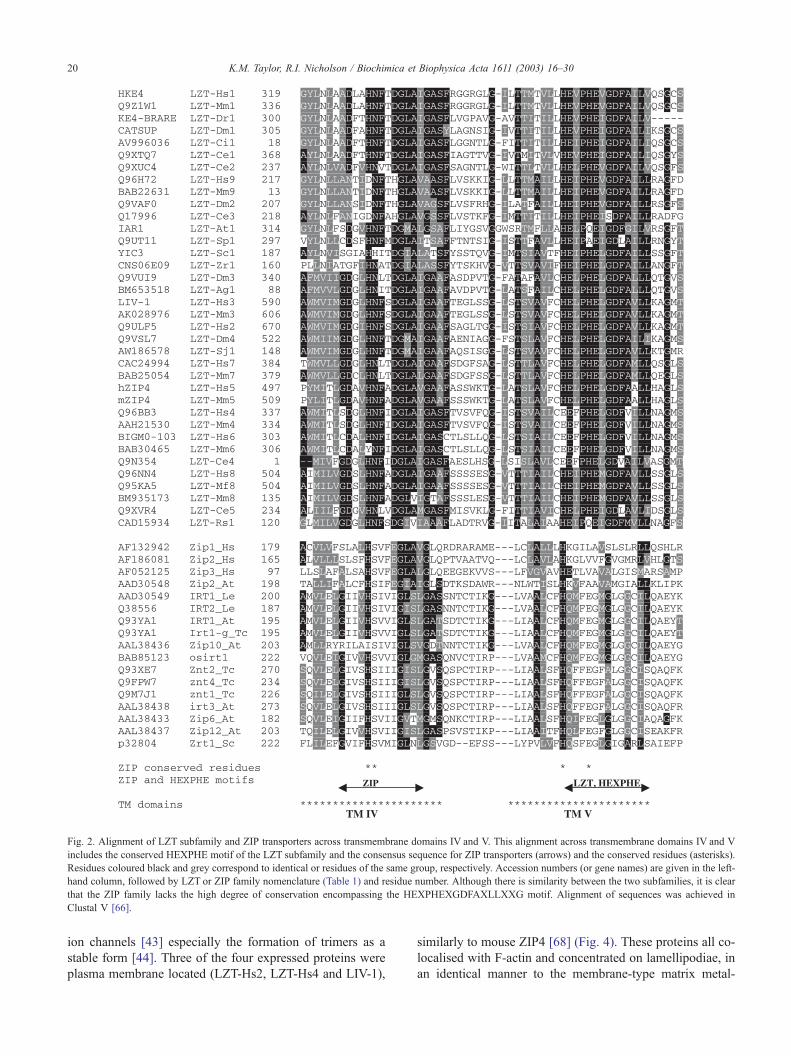
Fig. 2. Alignment of LZT subfamily and ZIP transporters across transmembrane domains IV and V. This alignment across transmembrane domains IV and V
includes the conserved HEXPHE motif of the LZT subfamily and the consensus sequence for ZIP transporters (arrows) and the conserved residues (asterisks).
Residues coloured black and grey correspond to identical or residues of the same group, respectively. Accession numbers (or gene names) are given in the left-
hand column, followed by LZT or ZIP family nomenclature (Table 1) and residue number. Although there is similarity between the two subfamilies, it is clear
that the ZIP family lacks the high degree of conservation encompassing the HEXPHEXGDFAXLLXXG motif. Alignment of sequences was achieved in
Clustal V [66].
K.M. Taylor, R.I. Nicholson / Biochimica et Biophysica Acta 1611 (2003) 16–3020
ion channels [43] especially the formation of trimers as a
stable form [44]. Three of the four expressed proteins were
plasma membrane located (LZT-Hs2, LZT-Hs4 and LIV-1),
similarly to mouse ZIP4 [68] (Fig. 4). These proteins all co-
localised with F-actin and concentrated on lamellipodiae, in
an identical manner to the membrane-type matrix metal-

Fig. 3. Schematic comparing the predicted secondary structure of LZT and
ZIP transporters. The predicted secondary structure of the LZT family is
compared with that previously suggested for the ZIP family by Gaither and
Eide [19]. His =Histidine-rich repeats (numbers are total histidines),
Star =mixed charge region, Barrels = transmembrane domains, Pale bar-
rel = conserved HEXPHE motif, letters in barrels represent the highly
conserved residues known to be important for zinc transport in ZIP
transporters and the corresponding residues in LZT sequences.
K.M. Taylor, R.I. Nicholson / Biochimica et Biophysica Acta 1611 (2003) 16–30 21
loproteases [45], which have a HEXXH motif in their active
site. To our knowledge, no other mammalian zinc trans-
porters contain such a potential metalloprotease motif,
which could impart an alternative function for LZT trans-
porters. It is noteworthy that LIV-1 has been implicated in
metastatic breast cancer [22], while the relationship of
membrane-type matrix metalloproteases with cancer inva-
sion has been well documented [46–49].
3.3. Zinc transport of LZT family of proteins
The similarity of the LZT subfamily sequences to ZIP
transporters suggests an ability to transport zinc into cells.
To test this, we transiently expressed recombinant LZT
proteins in CHO cells that had been loaded with Newport
Green Diacetate, a membrane-permeable fluorescent zinc-
specific indicator that increases in fluorescence when in
contact with free zinc. We tested that the indicator did in fact
respond to changes in intracellular zinc, for example,
addition of 50 AM zinc or the membrane-permeable zinc-
specific chelator TPEN to the medium produced a rise and
fall, respectively, in the percentage of cells with high
fluorescence. When LIV-1-expressing cells were tested,
instead of the usual 10% basal level of highly fluorescent
cells, 40% of the cells had high fluorescence. This indicated
that those cells transfected with LIV-1 (usually 25–40%)
were able to transport zinc into cells across the plasma
membrane. Interestingly, the LZT protein lacking the initial
H of the HEXXH motif (LZT-Hs4), which has been shown
to be crucial for both zinc transport in ZIP transporters [29]
and zinc-binding in metalloproteases [28], was not able to
transport zinc, never varying from control cells. Expectedly,
HKE4, which is present on intracellular membranes, did not
transport zinc into cells.
Two recent reports support the involvement of one LZT
sequence, the hZIP4 gene, in acrodermatitis enteropathica.
This disease is caused by a defective uptake of zinc in the
intestine which is believed to be due to a defect in hZIP4
[68,69]. These reports provide compelling evidence that
mutations in the hZIP4 gene are responsible for this defective
zinc uptake. Most of the mutations documented are either in
transmembrane domains, cause premature termination of the
protein sequence or are predicted to alter the tertiary structure
of the protein. This has been clearly demonstrated [69] by
observing the position of the mutations within the conserved
regions when a number of other ZIP sequences were aligned
together. These results, coupledwith our observations of LIV-
1, provide good evidence that LZT family members situated
on the plasma membrane of cells can function as zinc influx
transporters, with the possible exception of the KE4 sub-
family, situated on intracellular membranes, and those
sequences with a CEXXH motif.
3.4. Ubiquitination of LIV-1
Zrt1 is processed in cells by ubiquitin degradation via
interaction with a lysine residue (K195) on the cytoplasmic
loop between TM III and IV [30]. We have successfully
immunoprecipitated recombinant LIV-1 with an ubiquitin
antibody (unpublished results), indicating that it too binds
ubiquitin in cells. Whether one of the 15 lysine residues on
the cytoplasmic loop of LIV-1 is involved has not been
determined.
3.5. Tissue distribution of LZT family of proteins
The tissue distribution of LIV-1 and two other human
LZT family members, LZT-Hs4 and HKE4, has been
investigated by probing a multi-tissue Northern array
(unpublished results). The occurrence of LIV-1 is restricted
to hormonal tissues such as breast, prostate, pituitary gland
and brain. HKE4, however, has a different distribution to
LIV-1 in that it is not expressed in the brain, but is expressed
in liver, kidney and most hormonal tissues similarly to LIV-
1. These arrays suggest that LZT family proteins are widely
expressed in many different normal tissues as are ZIP zinc
transporters. However, it is noteworthy that LIV-1 expres-
sion was increased further in any cancer samples included
on the array, for example, HeLa cells and lung carcinoma.
4. Differences of LZT subfamily to ZIP zinc transporters
Although the LZT subfamily of proteins exhibit similar-
ities to the conventional ZIP transporters, there are also a
number of differences that suggest they may have a different
role in cells.

Table 1
Current information about the LZT family sequences and their predicted locations
Table 1A
Accession
number
Family
name
Species Other sequences Tissue Given name Length Predicted
location
Special features Database
description
Q9UIQ0 LZT-Hs1 Homo sapiens
chromosome 6
CAA20238, AL031228,
E12645 (fragment)
Kidney HKE4 469 ER 56/PM 22 KE4 group HLA class II
region
expressed
gene KE4
Q92504 (Hs1) Homo sapiens D82060, BAA11528 Breast, embryo,
embryonic
carcinoma, heart,
kidney, liver,
lung, muscle,
pancreas,
placenta, thymus
429 ER HKE4 protein with
premature stop
membrane
protein with
histidine-
rich repeats
Q9ULF5 LZT-Hs2 Homo sapiens
chromosome 2
BAA86579, AB033091,
KIAA1265, Q8NC35
(fragment)
Brain 835 PM 39/ER 35 N-term zinc finger,
cytochrome c
heme-binding site
Q9Y3Z1 (Hs2) Homo sapiens AL050294, CAB43393 529 Fragment of Q9ULF5
Q13433 LZT-Hs3 Homo sapiens
chromosome 18
U41060, AX017261
(fragment), AU120027,
Q96HP5,
AAH39498 (fragment)
Numerous LIV-1 749 ER 57/PM 30 Oestrogen
regulated and
implicated in
metastatic
breast cancer
Q96BB3 LZT-Hs4 Homo sapiens
chromosome 8
Fragments D31887, Q15043,
BC015770, AAH15770
537 PM 61/ER 35 CEEXXH, ABC
transporter
Cell cycle and
proliferation
Q9H6T8 LZT-Hs5 Homo sapiens
chromosome 8
BAB15164, AK025537 hZIP4 647 ER 56/PM 22 Zinc uptake gene
involved in
acrodermatitis
enteropathica [68,69]
Q9NXC4 (Hs5) Homo sapiens BAA91200, BAA91091,
AK000695, AK000334,
Q9NX22, AAH01688,
AX083511, AK025537
Fragments
BAB21559 LZT-Hs6 Homo sapiens
chromosome 4
AAG22480, AFL93052,
BAA96442 (fragment),
Q9C0K1, CAC38522
patent wo0129221
Bcg-induced,
neuronal
precursor cells,
retinoic acid
induction
BIGMO-103 460 ER 67/PM 22 Inhibit cancer
cell growth
Bcg induced
integral membrane
protein
CAC24994 LZT-Hs7 Homo sapiens AX061633, AAH27884,
Q8N6Y3
540 ER 36/PM 28
Q96NN4 LZT-Hs8 Homo sapiens AK055061, BAB70848 Brain, retina 654 ER 56/PM 44 Similar to LIV-1
Q96H72 LZT-Hs9 Homo sapiens AAH19016, BC019016,
Q8WV10, Q8N7C9
Fragment 364 KE4 group ZIP MOTIF
K.M
.Taylo
r,R.I.
Nich
olso
n/Biochimica
etBiophysica
Acta
1611
(2003)16–30
22

Table 1B
Accession
number
Family
name
Species Other sequences Given name Length Predicted
location
Special features
Q9GKV2 LZT-Mf2 Macaca fascicularis AB051127; BAB18153 528 PM 39 Hllp motif
Q95KA5 LZT-Mf8 Macaca fascicularis 654 ER 44/PM 44
Q9Z1W1 LZT-Mm1 Mus musculus AF100956, AAC69903,
BM054306, BM054306
KE4 476 ER 44 KE4 group
Q31125 (Mm1) Mus musculus M32010, AAA37747 KE4 436 ER Fragment of Q9Z1W1,
development, MHC
section of genome
BM721841 LZT-Mm3 Mus musculus Q8R518, BAB86300,
ABO71697
Ermelin >733 PM 47 ER located but possibly
missing >228aa of
N terminus (see text)
AAH21530 LZT-Mm4 Mus musculus 489 ER 35/PM 61
BAB24106 LZT-Mm5 Mus musculus Q9DAT9 mZIP4 660 ER 44/PM 33 Zinc uptake
gene involved
in acrodermatitis
enteropathica
BAB30465 LZT-Mm6 Mus musculus BAB29610, Q91W10,
Q9D5V4, Q9D426
462 ER 67/PM 22 CEEXXH, ABC
transporter
BAB25054 LZT-Mm7 Mus musculus BAB25675, Q9D856,
Q9D909
535 ER 48/PM 26
BM935173 LZT-Mm8 Mus musculus Fragment 200
BAB22631 LZT-Mm9 Mus musculus Possible fragment, Q9D1R4 160 KE4 group
Table 1C
Accession
number
Family
name
Species Other sequences Given name Length Predicted
location
Special features
Q9XTQ7 LZT-Ce1 C. elegans
chromosome X
(Z99942), CAB17070,
E1322656
KE4L 515 ER 44/PM 44 KE4/CATSUP
FAM
Q9XUC4 LZT-Ce2 C. elegans
chromosome IV
Z82285, CAB05297 KE4 404 ER 56/PM 33 KE4/CATSUP
FAM
Q17996 LZT-Ce3 C. elegans
chromosome X
Z50863, CAA90736,
T19285
338 ER 48/PM 30 KE4 group
Q9N354 LZT-Ce4 C. elegans
Y55F3BL.2
AC024828; AAF60809 157 Fragment
Q9XVR4 LZT-Ce5 C. elegans
chromosome IV
Z81044, CAB02806,
E275651
360 ER 22/PM 57
Q9V3A4 LZT-Dm1 Drosophila
melanogaster
AAF37226, AF216584,
AAF53744, AE003661,
AAL13757
CATSUP 449 ER 44/PM 33 CATSUP,
kininogen
pr00334,
KE4 group
Q9VAF0 LZT-Dm2 Drosophila
melanogaster
AE003771, AAF56969 355 ER 35/PM 52 KE4 group
Q9VUI9 LZT-Dm3 Drosophila
melanogaster
AAF49687, AE003532 519 ER 44/PM 44
(continued on next page)
K.M
.Taylo
r,R.I.
Nich
olso
n/Biochimica
etBiophysica
Acta
1611
(2003)16–30
23

Table 1 (continued)
Table 1C
Accession
number
Family
name
Species Other sequences Given name Length Predicted
location
Special features
Q9VSL7 LZT-Dm4 Drosophila
melanogaster
AAF50401, AE003555 684 ER 44/PM 33
Q9M647 LZT-At1 Arabidopsis
thaliana
AF216524, AAF32299,
AAF16552, AC012563
Q9SFR8, AAG52008,
Q9C9X4
IAR1 569 ER 44/PM 22 IAA-alanine
resistance
protein,
KE4 group
Q29175 LZT-Ss1 Sus scrofa Q9XT01, F14787;
CAA23256, AF146397,
AAD44801
KE4 PIG Fragment
Q9PUB8 LZT-Br1 Brachydanio
rerio
AAF05821, AF196345 KE4-BRARE 352 ER 33/PM 22
Table 1D
Accession
number
Family
name
Species Other sequences Given name Length Predicted
location
Special features
P40544 LZT-Sc1 Saccharomyces
cerevisiae
chromosome IX
PIR: S49959,
CAA86969, Z46881
YIC3 YEAST 346 ER 48/PM 30 KE4 group
Q9UT11 LZT-Sp1 Schizosaccharomyces
pombe
CAB55170, AL117210,
PIR: T39240
453 ER 33/PM 33 KE4 group
AW186578 LZT-Sj1 S. japonicum Fragment 233
BJ044228 LZT-Xl1 Xenopus laevii BJ081638, BJ047405 155
CAD15934 LZT-Rs1 Ralstonia
solanaceaerum
268
CNS06E09 LZT-Zr1 Zygosaccharomyces
rouxii
317 ER 44/PM 26 KE4 group
CAD15934 LZT-Rs1 Ralstonia
solanacearum
268 ER 22/PM 57
AV996036 LZT-Ci1 Ciona intestinalis 162 HNIP MOTIF,
KE4 group
BM653518 LZT-Ag1 Anopheles gambiae BH380314 224
The suggested LZT family name is given in the family name column, where brackets indicate identical, similar or fragments of sequences that have not been included in the overall family alignment. Human
sequences are shown in Table 1A, mouse and monkey sequences are shown in Table 1B, other mammals and plant sequences are shown in Table 1C, and remaining fungal and bacteria sequences are shown in
Table 1D.
K.M
.Taylo
r,R.I.
Nich
olso
n/Biochimica
etBiophysica
Acta
1611
(2003)16–30
24

Fig. 4. Fluorescent microscopy of three human LZT family members. Recombinant V5-fusion proteins fluorescently labelled with Alexa-Fluor 488 (green)
(middle row) and F-actin filaments stained with Texas-Red phalloidin (bottom row). The overlay on the top row shows plasma membrane staining for LIV-1
and LZT-Hs4 in CHO cells fixed with 4% formaldehyde, whereas HKE4 shows an intracellular location in CHO cells fixed in 4% formaldehyde and 0.4%
saponin.
K.M. Taylor, R.I. Nicholson / Biochimica et Biophysica Acta 1611 (2003) 16–30 25
4.1. Histidine-rich repeats
The LZT subfamily sequences contain the same histi-
dine-rich repeat, (HX)n, of the ZIP transporters, also on the
Fig. 5. Western Blot of two human LZT family proteins. CHO cells
transiently transfected with HKE4 and LZT-Hs4, with C-terminal V5 tags,
were lysed and samples applied to a 10% SDS-PAGE gel. Bands of protein
were transferred to nitrocellulose and probed with V5 antibody. Results in
reducing (R) and non-reducing (NR) conditions are shown. Both proteins
have high molecular weight bands in non-reducing conditions as well as a
band that approximates to a trimer.
long cytoplasmic loop, yet the incidence of these repeats is
as much as sevenfold more than other ZIP transporters [24]
over the whole sequence. In a previous alignment of ZIP
sequences [29], two KE4 sequences (human gbD82060 and
mouse spQ31125) were included and a region constituting
40 histidine residues in total was observed in the N-terminal
region of both sequences. However, these sequences were
the human and mouse KE4 sequences (LZT-Hs1 and LZT-
Mm1, respectively), which are recognised here as members
of the LZT subfamily. Therefore, one characteristic differ-
ence of the LZT subfamily from other ZIP transporters is
still the increased incidence of histidine-rich repeats [24]
throughout the sequence. These repeats are present in the
cytosolic loop between TM III and IV, as in ZIP trans-
porters, but also on the extracellular loop between TM II and
III and the extracellular N terminus [24]. The presence of
these repeats on both sides of the membrane is unprece-
dented in zinc transporters and although the function of
these extra repeats is not known, they would be predicted to
have a role in the transport of metal ions by similarity with
other such motifs [42].
However, as more family members emerge, it is evident
that some sequences are not as histidine-rich. In fact, there is
one or more family member from most of the higher species
that has few or no histidine residues at all. For example,
human LZT-Hs4, mouse LZT-Mm4, and C. elegans LZT-
Ce3, contain 4, 6 and 0 histidines, in (HX)n repeats,
respectively. Interestingly, these former two sequences and
two others (human LZT-Hs6 and mouse LZT-Mm6) have
the initial histidine of the HEXXH motif replaced by a

K.M. Taylor, R.I. Nicholson / Biochimica et Biophysica Acta 1611 (2003) 16–3026
glutamic acid residue (EEXXH, Fig. 2). We have already
mentioned that cells expressing recombinant LZT-Hs4 did
not transport extracellular zinc into cells, in contrast to LIV-
1. However, this replacement of the conserved histidine with
glutamic acid may indicate a preference for transporting
ions other than zinc as a QEXXH motif has preference for
copper transport [67].
4.2. Zinc-binding site motif of metalloproteases
One feature that is unique amongst the LZT subfamily is
the presence of a motif (HEXPHEXG) that fits the con-
sensus sequence for the catalytic zinc-binding site of matrix
metalloproteases [50,51] and snake venom metalloprotei-
nases [52] (HEXXHXXGXXH) lacking only the final
coordinating histidine. The zinc ligands in this latter motif
(marked in bold) consist of three histidine residues and one
water molecule, linked to the glutamic acid, E [53,54]. The
catalytic zinc ion is essential for the proteolytic activity of
matrix metalloproteases [46] and the three histidine residues
that coordinate the zinc ion in these molecules are com-
pletely conserved. Different subgroups of metalloproteases
have been characterised by the distance and identity of the
fourth zinc coordinating residue in this motif [55]. For
example, the catalytic zinc-binding site of the zincin and
PDF groups of metalloproteases (HEXXHXXG), similar to
that in LZT sequences, lacks the terminal histidine residue
of the matrix metalloprotease motif and, therefore in these
proteins, the conserved glycine residue, three residues
downstream of the second histidine, generates the proper
coordination geometry of the ligands [26,27].
The completely conserved second glutamic acid down-
stream of the second histidine (marked in bold) in the LZT
family motif (HEXPHEXGD) is also a good candidate
residue to coordinate the zinc ion via a second water molecule
[24]. Thermolysin, a zinc-bindingmetalloprotease, belongs to
a group of metalloproteases that use a glutamic acid residue
for the fourth ligand to coordinate the zinc ion. In thermolysin,
this residue is 20 amino acids downstream from the second
histidine in the first motif [54] and present in a small
conservedmotif (NEXXSD). LIV-1 possesses a similar motif,
NDXSD, at residues 706–710. However, there is no evidence
for such a motif in any of the other LZT family proteins.
It is also probable, due to the integral TM location of the
motif, that the fourth zinc coordinating residue is provided
by one of the other TM domains. The prime candidate for
this would be TM domain II as it contains a conserved
histidine and other residues spaced to be consistent with a
position on one side of a helix [24]. Interestingly, two
human LZT family members, LZT-Hs4 and LZT-Hs6, both
of which have the initial H of the HEXXH motif replaced by
a glutamic acid (E) residue (Fig. 2) have the conserved
histidine in TM II replaced by a glutamine (Q) residue (Fig.
6). We have already shown that LZT-Hs4 does not transport
zinc [50] even though it is located on the plasma membrane,
reinforcing the potential importance of these two residues.
The three-dimensional structure of this HEXXHmotif has
been determined to be a helical tertiary structure [52]. This
motif in the LZT family, which is well conserved across all
36 sequences, contains a unique proline residue (Fig. 2).
Proline residues can be both inconsistent with the formation
of a helical structure [56,57] and also provide increased
stability [58–60]. The presence of this motif in a TM domain
may therefore be predicted to improve stability [61].
It is noteworthy that TM IV and V of LIV-1 have
similarity to the B, D and E chains of haemagglutinin-
esterase-fusion glycoprotein of influenza C virus (Hef2)
which exists as a helical structure up to the equivalent of
the start of the HEXPHEXGD motif (PDB:1flc, residues
96–134). Interestingly, Hef2 helices appear as parallel pairs
of helices and TM II of LIV-1 are similar to the second helix
of Hef2 which runs parallel to it in the tertiary structure.
This suggests that the tertiary structure of TM II and V of
LIV-1 may indeed act synergistically. These observations
are consistent with the ability of LZT proteins to transport
zinc across the plasma membrane through a pore region
maintained by interaction of conserved residues in TM II,
IV, and V.
4.3. Long N terminus
The LZT subfamily sequences have long N termini,
especially when compared to ZIP transporters [29]. The N
terminus of LIV-1 itself consists of 317 residues (42%) of
the total 749 residues in the full-length sequence. This
region in LIV-1 contains 27 histidine residues including
the following cluster of 19 histidines, HIHHDHDHHSD-
HEHHSDHERHSDHEHHSDHEHHSDHNH, which can be
divided into six repeats of the general formula H(H,S)DH
(E,D,N)(H,R).
4.4. Conserved TM domains
TM domains I, II, III and VII of the LZT subfamily also
contain unique signatures (Fig. 6) not present in any other
sequences in the GenBank. The role of TM domain II and its
probable interaction with TM IV and V has already been
mentioned above. TM IV and V contain the highly con-
served consensus sequence for the LZT subfamily with
conservation of every third or fourth residue, consistent
with their presence on one side of a helix. These adjacent
TM domains are well conserved, including three charged
residues, with no apparent loop and a conserved mixed
charge region directly upstream (Fig. 3). This suggests a
possible pore structure involved in the binding and/or
transporting of zinc or other ions.
5. The KE4 subfamily of LZT proteins
A number of the LZT subfamily sequences are also KE4
proteins, some of which have been previously aligned with

Fig. 6. Alignment of the whole sequence of nine human LZT family members. Residues coloured black and grey correspond to at least 50% identical or complementary residues, respectively. Solid arrows with
roman numerals indicate transmembrane domains, dotted arrows indicate conserved motifs, solid square =CPALLY motif, solid circle =HELPHE motif, solid arrows = ZIP consensus. Alignment of sequences was
achieved in Clustal V [66].
K.M
.Taylo
r,R.I.
Nich
olso
n/Biochimica
etBiophysica
Acta
1611
(2003)16–30
27

K.M. Taylor, R.I. Nicholson / Biochimica et Biophysica Acta 1611 (2003) 16–3028
other ZIP transporters and suggested as both zinc trans-
porters [29,39,40] and members of the LIV-1 subfamily
[24]. At present, there are apparently 17 KE4 proteins,
which appear from the phylogenetic tree (Fig. 1) to form a
separate sub family of LZT proteins. This KE4 subgroup
includes CATSUP from Drosophila (AAF37226) [41],
IAR1 from Arabidopsis (AAF32299) [40], mouse KE4,
pig KE4 and zebrafish KE4. We have expressed the LZT
protein, HKE4 (LZT-Hs1), in CHO cells where it located to
intracellular membranes (Fig. 4) and not the plasma mem-
brane as was observed with other LZT proteins. Another
KE4 protein, IAR1, a putative transporter with alanine-
resistance properties, has been localised to the endoplasmic
reticulum and proposed to transport manganese out of this
organelle [40]. It has been suggested to be involved in auxin
metabolism in plants by inhibiting an auxin conjugate
hydroxylase. Interestingly, mouse KE4, was shown to func-
tionally substitute for IAR1 [40]. CATSUP has been sug-
gested to down-regulate tyrosine hydroxylase [41], a rate-
limiting enzyme for dopamine production in the brain, in
Drosophila embryos. This was achieved by performing
knockouts in Drosophila, which were lethal, and testing
mutated partial loss of function alleles.
One interesting feature of the KE4 sequences is the lack
of another motif (CPALLY) which is uniquely present in the
LZT sequences of human, mouse and monkey origin (Fig.
7), upstream of TM I. This motif, not present in any other
sequences in the GenBank, includes cysteine and proline
residues, suggesting a conserved tertiary structure. Interest-
ingly, the final conserved cysteine in this motif is mutated to
a tyrosine residue (C309Y) in acrodermatitis enteropathica
[68]. This may suggest a crucial functional role for this
motif but it could also represent disruption of the tertiary
structure by a missing cysteine which is usually present in a
disulfide bond, as has been observed with other protein
mutations [70].
The mouse sequence ermelin has virtually the identical
sequence to LIV-1, rather than the endoplasmic reticulum-
located KE4 proteins (Table 1, LZT-Mm3), and yet has been
localised to the endoplasmic reticulum [39] instead of the
plasma membrane as LIV-1 has been. However, the recent
appearance of AK028976 (LZT-Mm3) with 765 amino acids
suggests that the ermelin sequence is missing most of the N-
terminal residues which may account for its endoplasmic
reticulum location. In fact, on close inspection of the ermelin
DNA sequence (BM721841), another 228 residues that
Fig. 7. Alignment of seven LZT human sequences that contain a CPALLY m
complementary residues, respectively. This motif is not present in the KE4 prote
match AK028976 can be obtained from the next reading
frame, suggesting a possible sequencing error.
6. Predicted function of LZT subfamily proteins
The exact function of LZT family proteins is unknown,
but some function has been suggested in the individual
GenBank files. Human LIV-1 (LZT-Hs3) has been impli-
cated in metastatic breast cancer [23], whereas the other
proteins in the family group are as yet of unknown
function. The oestrogen-regulation of LIV-1 in breast
cancer is not without precedent as hZIP1, a human ZIP
transporter, is expressed in the malignant prostate cancer
cell lines, LN-CaP and PC-3, in a hormone-dependant
manner [62]. A role of LIV-1 in breast cancer may be
explained by the fact that zinc can induce EGF receptor
phosphorylation and MapKinase activation by activation of
both the IGF-1 and EGF receptors [63]. This is achieved
by stimulation of the tyrosine phosphorylation of these
receptors by zinc, possibly by interference with tyrosine
dephosphorylation as zinc can inhibit various protein
tyrosine phosphatases [64]. These pathways are all of
critical importance in the development of a breast cancer
phenotype.
One human family member, LZT-Hs6, has been sug-
gested to inhibit cancer cell growth being induced by Bcg
and retinoic acid, whereas another, LZT-Hs4, has been
implicated in the cell cycle and cell proliferation. The
KE4 subfamily has been implicated in ion transport [40]
and possible down-regulation of specific hydrolases [41], as
mentioned earlier.
Several human LZT family members are expressed in the
brain (e.g. LZT-Hs2), where alteration of tyrosine hydrox-
ylase activity may be important for the control of neuro-
degeneration and growth. Control of zinc is important for
the normal functioning of the brain and can be altered in
diseases such as epilepsy [65]. Auxin metabolism in plants
is also important for the control of growth.
7. Conclusion
The suggestion that the LIV-1 family is intimately
involved in zinc homeostasis implies that its function may
be critical for cell growth. This is supported by the small
otif. Residues coloured black and grey correspond to 100% identical or
ins.

K.M. Taylor, R.I. Nicholson / Biochimica et Biophysica Acta 1611 (2003) 16–30 29
amount of suggested function available in the GenBank
files. We are therefore encouraged to investigate this excit-
ing new group of proteins, both in normal cells and diseases
such as cancer. As zinc is essential for cell growth, mem-
brane proteins capable of transporting zinc into cells will
have a crucial role in maintaining the cellular balance
between apoptosis and cell growth. Such a function would
probably require a family of proteins under tight regulation
of expression because faults in the system could easily lead
to premature death or cancer.
Acknowledgements
We gratefully acknowledge the gift of LIV-1 cDNA in
bluescript plasmid from Chris Green, Liverpool, the gift of
Clone 1033B10 of library RPC15 from the Sanger Centre,
Cambridge, and the gift of gene KIAA0062 (Clone
HA1020) from the Kazusa DNA Research Institute, Japan.
We thank Helen Morgan for ubiquitin assays, Emma Joyce
for Western blotting and Andrea Johnson for excellent
technical help. We thank the Tenovus Cancer Charity for
supporting this work.
References
[1] B.L. Vallee, D.S. Auld, Biochemistry 29 (24) (1990) 5647–5659.
[2] D. Rhodes, A. Klug, Sci. Am. 268 (1993) 56–65.
[3] B.L. Vallee, K.H. Falchuk, Physiol. Rev. 73 (1993) 79–118.
[4] F.Y. Wu, W.J. Huang, R.B. Sinclair, L. Powers, J. Biol. Chem. 267
(35) (1992) 25560–25567.
[5] F.Y. Wu, C.W. Wu, Annu. Rev. Nutr. 7 (1987) 251–272.
[6] D.H. Hamer, Annu. Rev. Biochem. 55 (1986) 913–951.
[7] J.Y. Koh, S.W. Suh, B.J. Gwag, Y.Y. He, C.Y. Hsu, D.W. Choi,
Science 272 (5264) (1996) 1013–1016.
[8] C.J. Frederickson, A.I. Bush, BioMetals 14 (3–4) (2001) 353–366.
[9] A.Q. Truong-Tran, J. Carter, R.E. Ruffin, P.D. Zalewski, BioMetals
14 (3–4) (2001) 315–330.
[10] M. Lioumi, C.A. Ferguson, P.T. Sharpe, T. Freeman, I. Marenholz,
D. Mischke, C. Heizmann, J. Ragoussis, Genomics 62 (2) (1999)
272–280.
[11] L.A. Gaither, D.J. Eide, J. Biol. Chem. 275 (8) (2000) 5560–5564.
[12] L.A. Gaither, D.J. Eide, J. Biol. Chem. 276 (2001) 22258–22264.
[13] R.D. Palmiter, S.D. Findley, EMBO J. 14 (1995) 639–649.
[14] R.D.Palmiter, T.B.Cole, S.D.Findley,EMBOJ. 15 (1996) 1784–1791.
[15] R.D. Palmiter, T.B. Cole, C.J. Quaife, S.D. Findley, Proc. Natl. Acad.
Sci. U. S. A. 93 (1996) 1493–14939.
[16] L. Huang, J. Gitschier, Nat. Genet. 17 (1997) 292–295.
[17] T.B. Cole, H.J. Wenzel, K.E. Kafer, P.A. Schwartzkroin, R.D. Pal-
miter, Proc. Natl. Acad. Sci. U. S. A. 96 (4) (1999) 1716–1721.
[18] L.T. Paulsen, M.H. Saier, J. Membr. Biol. 156 (1997) 99–103.
[19] L.A. Gaither, D.J. Eide, BioMetals 14 (2001) 251–270.
[20] M.L. Guerinot, Biochim. Biophys. Acta 1465 (2000) 190–198.
[21] D.L. Manning, R.A. McClelland, J.M. Knowlden, S. Bryant, J.M.W.
Gee, C.D. Green, J.F. Robertson, R.W. Blamey, R.L. Sutherland, C.J.
Ormandy, R.I. Nicholson, Acta Oncol. 34 (1995) 641–646.
[22] R.A. McClelland, D.L. Manning, J.M. Gee, P. Willsher, J.F. Robert-
son, I.O. Ellis, R.W. Blamey, R.I. Nicholson, Br. J. Cancer 77 (10)
(1998) 1653–1656.
[23] D.L. Manning, J.F.R. Robertson, I.O. Ellis, C.W. Elston, R.A.
McClelland, J.M.W. Gee, R.J. Jones, C.D. Green, P. Cannon, R.W.
Blamey, R.I. Nicholson, Eur. J. Cancer. 30A (1994) 675–678.
[24] K.M. Taylor, IUBMB Life 49 (4) (2000) 249–253.
[25] S.F. Altschul, T.L. Madden, A.A. Schaffer, J. Zhang, Z. Zhang, W.
Miller, D.J. Lipman, Nucleic Acids Res. 25 (1997) 3389–3402.
[26] N.M. Hooper, FEBS Lett. 354 (1994) 1–6.
[27] F. Dardel, S. Ragusa, C. Lazennec, S. Blanquet, T. Mainel, J. Mol.
Biol. 280 (1998) 501–513.
[28] W. Jiang, J.S. Bond, FEBS Lett. 312 (1992) 112–114.
[29] B.H. Eng, M.L. Guerinot, D. Eide, M.H. Saier Jr., J. Membr. Biol.
166 (1) (1998) 1–7.
[30] R.S. Gitan, D.J. Eide, Biochem. J. 346 (2000) 329–336.
[31] T. Hirokawa, S. Boon-Chieng, S. Mitaku, Bioinformatics 14 (1998)
378–379.
[32] K. Hofmann, W. Stoffel, Biol. Chem. Hoppe-Seyler 347 (1993) 166.
[33] C. Geourjon, G. Deleage, Comput. Appl. Biosci. 11 (1995) 681–684.
[34] G.E. Tusnady, I. Simon, J. Mol. Biol. 283 (1998) 489–506.
[35] K. Nakai, M. Kanehisa, Genomics 14 (1992) 897–911.
[36] K. Nakai, Bull. Inst. Chem. Res., Kyoto Univ. 69 (1991) 269–291.
[37] P. Horton, K. Nakai, Proc. Int. Conf. Intellig. Syst. Mol. Biol. 4
(1996) 109–115.
[38] M. Cserzo, E. Wallin, I. Simon, G. von Heijne, A. Elofsson, Prot.
Eng. 10 (6) (1997) 673–676.
[39] A. Suzuki, T. Endo, Gene 284 (1–2) (2002) 31–40.
[40] J. Lasswell, L.E. Rogg, D.C. Nelson, C. Rongey, B. Bartel, Plant Cell
12 (12) (2000) 2395–2408.
[41] D.G. Stathakis, D.Y. Burton, W.E. McIvor, S. Krishnakumar, T.R.
Wright, J.M. O’Donnell, Genetics 153 (1) (1999) 361–382.
[42] E.E. Rogers, D.J. Eide, M.L. Guerinot, Proc. Natl. Acad. Sci. U. S. A.
97 (22) (2000) 12356–12360.
[43] W.N. Green, N.S. Millar, Trends Neurosci. 18 (1995) 280–287.
[44] W.N. Green, C.P. Wanamaker, J. Neurosci. 18 (1998) 5555–5564.
[45] H. Nakahara, L. Howard, E.W. Thompson, H. Sato, M. Seiki, Y. Yeh,
W. Chen, Proc. Natl. Acad. Sci. U. S. A. 94 (1997) 7959–7964.
[46] I. Massova, L.P. Kotra, S. Mobashery, Bioorg. Med. Chem. Lett. 8
(1998) 853–858.
[47] A.F. Chambers, L.M. Matrisian, J. Natl. Cancer Inst. 89 (1997)
1260–1270.
[48] M. Polette, P. Birembaut, Int. J. Biochem. Cell Biol. 30 (1998)
1195–1202.
[49] C. Gilles, M. Polette, M. Seiki, P. Birembaut, E.W. Thompson, Lab.
Invest. 76 (5) (1997) 651–660.
[50] I. Massova, L.P. Kotra, R. Fridman, S. Mobashery, FASEB J. 12
(1998) 1075–1095.
[51] T. Takino, H. Sato, E. Yamamoto,M. Seiki, Gene 155 (1995) 293–298.
[52] F. Gomis-Ruth, L.F. Kress, W. Bode, EMBO J. 12 (11) (1993)
4151–4157.
[53] W. Bode, F. Grams, P. Reinemer, F.X. Gomis-Ruth, U. Bauman, D.B.
McKay, W. Stocker, Adv. Exp. Med. Biol. 389 (1996) 1–11.
[54] W. Stocker, W. Bode, Curr. Opin. Struct. Biol. 5 (1995) 383–390.
[55] J.E. Coleman, Curr. Opin. Chem. Biol. 2 (1998) 222–234.
[56] P.Y. Chou, G.D. Fasman, Adv. Enzymol. 47 (1978) 45–148.
[57] K. Peters, H.J. Hinz, G. Cesareni, Biol. Chem. 378 (1997) 1141–1152.
[58] D.N. Woolfson, D.H. Williams, FEBS Lett. 17 (1990) 185–188.
[59] S. Nakamura, T. Tanaka, R.Y. Yada, S. Nakai, Protein Eng. 10 (1997)
1263–1269.
[60] F. De Lamotte-Guery, C. Pruvost, P. Minard, M.A. Delsuc, M. Migi-
niac-Maslow, J.M. Schmitter, M. Stein, P. Decottignies, Protein Eng.
10 (1997) 1425–1432.
[61] S. Hong, K.S. Ryu, M.S. Oh, I. Ji, T.H. Ji, J. Biol. Chem. 272 (1997)
4166–4171.
[62] L.C. Costello, Y. Liu, J. Zou, R.B. Franklin, J. Biol. Chem. 274 (25)
(1999) 17499–17504.
[63] W. Wu, L.M. Graves, I. Jaspers, R.B. Devlin, W. Reed, J.M. Samet,
Am. J. Physiol. 277 (1999) L924–L931.
[64] J.M. Samet, R. Silbajoris, W. Wu, L.M. Graves, Am. J. Respir. Cell
Mol. Biol. 21 (3) (1999) 357–364.

K.M. Taylor, R.I. Nicholson / Biochimica et Biophysica Acta 1611 (2003) 16–3030
[65] A. Takeda, BioMetals 14 (2001) 343–351.
[66] J.D. Thompson, D.G. Higgins, T.J. Gibson, Nucleic Acids Res. 22
(1994) 4673–4680.
[67] M. Heitzer, A. Hallmann, J. Biol. Chem. 277 (31) (2002)
28280–28286.
[68] K. Wang, B. Zhou, Y.M. Kuo, J. Zemansky, J. Gitschier, Am. J. Hum.
Genet. 71 (2002) 66–73.
[69] S. Kury, B. Dreno, S. Bezieau, S. Giraudet, M. Kharfi, R. Kamoun,
J.P. Moisan, Nat. Genet. 31 (3) (2002) 239–240.
[70] K.M. Taylor, A.R. Trimby, A.K. Campbell, Immunology 91 (1997)
20–27.