concurrent radiochemotherapy in locally-regionally advanced oropharyngeal squamous cell carcinoma:...
TRANSCRIPT
Krstevska et al. Radiation Oncology 2012, 7:78http://www.ro-journal.com/content/7/1/78
RESEARCH Open Access
Concurrent radiochemotherapy in locally-regionally advanced oropharyngeal squamouscell carcinoma: analysis of treatment results andprognostic factorsValentina Krstevska1†, Igor Stojkovski1*† and Beti Zafirova-Ivanovska2†
Abstract
Background: Concurrent radiochemotherapy is a recommended treatment option for patients with locallyadvanced squamous cell head and neck carcinomas with recent data showing the most significant absolute overalland event-free survival benefit achieved in patients with oropharyngeal tumours. The aim of this study was toanalyse the results of three-dimensional conformal radiotherapy given with concomitant weekly cisplatin in patientswith advanced oropharyngeal carcinoma and to identify prognostic factors influencing outcomes of this patientscategory.
Methods: Sixty-five patients with stage III or IV squamous cell carcinoma of the oropharynx who underwentconcurrent radiochemotherapy between January 2005 and December 2010 were retrospectively analyzed. Allpatients received radiotherapy to 70 Gy/35 fractions/2 Gy per fraction/5 fractions per week. Concurrentchemotherapy consisted of weekly cisplatin (30 mg/m2) started at the first day of radiotherapy.
Results: Median age was 57 years (range, 36 to 69 years) and 59 (90.8%) patients were male. Complete compositeresponse was achieved in 47 patients (72.3%). Local and/or regional recurrence was the most frequent treatmentfailure present in 19 out of 25 patients (76.0%). At a median follow-up of 14 months (range, 5 to 72 months), 2-yearlocal relapse-free, regional relapse-free, locoregional relapse-free, disease-free, and overall survival rates were 48.8%,57.8%, 41.7%, 33.2% and 49.7%, respectively.On multivariate analysis the only significant factor for inferior regional relapse-free survival was the advanced Nstage (p = 0.048). Higher overall stage was independent prognostic factor for poorer local relapse-free survival,locoregional relapse-free survival and disease-free survival (p = 0.022, p = 0.003 and p= 0.003, respectively). Pre-treatment haemoglobin concentration was an independent prognostic factor for local relapse-free survival, regionalrelapse-free survival, locoregional relapse-free survival, disease-free survival, and overall survival (p = 0.002, p = 0.021,p = 0.001, p = 0.002 and p= 0.002, respectively).
Conclusions: Poor treatments results of this study suggested that introduction of intensity-modulated radiotherapy,use of induction chemotherapy followed by concurrent radiochemotherapy, accelerated radiotherapy regimens,and molecular targeted therapies could positively influence treatment outcomes. The incorporation of reversal ofanaemia should be also expected to provide further improvement in locoregional control and survival in patientswith advanced squamous cell carcinoma of the oropharynx.
Keywords: Concurrent radiochemotherapy, Oropharyngeal carcinoma, Prognostic factor
* Correspondence: [email protected]†Equal contributors1Department of Head and Neck Cancer, University Clinic of Radiotherapy andOncology, Skopje, MacedoniaFull list of author information is available at the end of the article
© 2012 Krstevska et al.; licensee BioMed CentrCommons Attribution License (http://creativecreproduction in any medium, provided the or
al Ltd This is an Open Access article distributed under the terms of the Creativeommons.org/licenses/by/2.0), which permits unrestricted use, distribution, andiginal work is properly cited.
Krstevska et al. Radiation Oncology 2012, 7:78 Page 2 of 14http://www.ro-journal.com/content/7/1/78
BackgroundSquamous cell carcinoma of the oropharynx is theeleventh most common cancer worldwide [1] with anannual incidence of 0.8 per 100000 [2]. Besides thestrong association of oropharyngeal carcinoma withtobacco and alcohol abuse observed in epidemiologicaland clinical studies [3], the specific association of thiscancer with human papillomavirus (HPV) infection hasbeen now well-known [4,5]. The tonsil is the most fre-quently represented subsite of the primary oropharyn-geal carcinoma followed by the base of tongue [6,7].These two subsites account for between 80–90% of cases[2]. Oropharyngeal carcinomas are usually diagnosed aslocoregionally advanced disease [8,9]. Thus, most of theprimary tumours presents at an advanced stage (T2 orgreater) [10], and the incidence of nodal metastasesranges between 60-70% [11,12] which is probably relatedto the rich lymphatic supply of the dominant subsites ofthe oropharyngeal cancers.Treatment decision making process for oropharyngeal
carcinomas arising in this functionally important ana-tomic region must take into consideration not only themost optimal treatment strategy for local/regionaltumour control achievement but also the associatedmorbidity to this critical site in the upper aerodigestivetract. The treatment for advanced but resectable oropha-ryngeal carcinoma has traditionally been radical surgeryand postoperative radiotherapy often resulting in sub-optimal rates of locoregional control (LRC) and signifi-cant long-term functional deficits, or radiotherapy alonefor advanced unresectable lesions inducing long-termtoxicities accompanied with initial affection of speechand swallowing as a consequence of the primary tumourgrowth. However, in the last two decades, several rando-mized studies and meta-analyses indicated that concur-rent radiochemotherapy (CRCT) has been shown toprovide an improvement in LRC and survival as well asa significant increase in the rate of organ preservationwhen compared with radiation alone in patients withadvanced head and neck cancer including those pre-sented with advanced oropharyngeal carcinoma [13-22].Apart from the trials including multiple sites in the headand neck cancers, the French Head and Neck Oncologyand Radiotherapy Group (GORTEC 94-01) phase IIIrandomized trial analyzing separately squamous cell car-cinomas arising from the oropharynx established CRCTas the standard treatment for locoregionally advancedoropharyngeal cancer over conventional radiotherapyalone [23]. CRCT has been shown superior as comparedwith radiotherapy alone with regard to LRC, disease-freesurvival (DFS) and overall survival (OS) [23]. The finalresults of this trial reported in 2004 also confirmed thatCRCT improved OS and LRC rates in patients withadvanced oropharyngeal carcinoma [24]. Consequently,
CRCT has been adopted as a preferred treatment ap-proach that enables an achievement of increased rate ofdisease control with anatomic and physiologic functionpreservation [25]. Recently, in order to reveal the magni-tude of the benefit of addition of chemotherapy to radio-therapy in terms of OS in head and neck squamous cellcarcinoma according to tumour site, a comprehensiveanalysis was performed using individual data of 16,192patients included in the meta-analysis of chemotherapyin head and neck cancer (MACH-NC) [26]. The inter-action test showed the most significant effect of chemo-therapy timing (p< 0.0001) in the oropharynx cancersgroup which was the largest group analysed consistingof 5878 patients, suggesting a significantly better effectof platinum-based concurrent chemotherapy [26]. Al-though the superiority of platinum-based chemotherapyhas been confirmed in the meta-analysis of the MACH-NC Collaborative Group [19] and in the meta-analysis ofBrowman et al. [21], regarding the question about thenumber of chemotherapeutic agents and schedules ofdrug delivery, the optimum regimen seems to remainunclear. The most common method used worldwide wasdelivery of cisplatin every 3 weeks [17,27]. However,based on the assumption that more frequent drug ad-ministration could provide greater radiosensitizing bene-fit and taking into account the less induced morbiditywith smaller individual doses of drug without comprom-ising treatment efficacy [28], weekly administration ofsingle-platinum agent has been also studied [29,30] al-though this schedules utilizing smaller doses more fre-quently has been not compared directly with the cyclicalapproach to delivery of concurrent cisplatin [31].Regarding the radiotherapy techniques employed, it
should be mentioned that the introduction of defini-tive three-dimensional conformal radiotherapy(3DCRT) with or without chemotherapy as the stand-ard of practice in the treatment of oropharyngeal can-cer in clinics around the world with tight targetdefinitions of the primary tumour, metastatic nodesin the neck and neck nodal levels, enabled improve-ment of tumour coverage while sparing the surround-ing critical tissues [32]. Recently, intensity-modulatedradiotherapy (IMRT) achieving higher total doses intumours by delivering larger doses per fraction to thetumour only, has also been shown as an effectivetreatment technique for locally advanced oropharyn-geal carcinoma [33] offering tumour control rateskept at least at the level of 3DCRT while limitingdose to nearby normal tissues, e.g., the parotid glands[34,35]. Additionally, the Memorial Sloan-KetteringCancer Centre experience and the University of Califor-nia-San Francisco experience has revealed encouraginglocal control with acceptable treatment toxicity achievedusing IMRT chemoradiation for treatment of stage III and

Krstevska et al. Radiation Oncology 2012, 7:78 Page 3 of 14http://www.ro-journal.com/content/7/1/78
IV oropharyngeal carcinoma [36,37]. The assessment ofresults of radiochemotherapy utilizing IMRT for advancedstage oropharyngeal carcinoma in the prospective studyconducted by Feng et al. [38] demonstrated the possibilityof this treatment approach to reduce post-therapy func-tional impairment obtaining at the same time high rates oflocoregional tumour control.The objective of this retrospective study was to
summarize the results of treatment following 3DCRTand concomitant chemotherapy in patients withadvanced oropharyngeal squamous cell carcinoma aswell as to examine and identify the influence of variouspatient and tumour-related prognostic factors on localcontrol, regional control, and survival in this patientspopulation. We also considered that this retrospectiveanalysis will give us an opportunity to compare resultsobtained with CRCT with those that would be achievedwith IMRT whose implementation in the treatment ofadvanced oropharyngeal carcinomas at our institutionhas been started recently.
MethodsThis study is based on a retrospective analysis of 65 con-secutive patients with previously untreated, stage III orIV primary squamous cell carcinoma of the oropharynxwithout distant metastases, at the age of at least 18 yearsand not more than 70 years and performance status 0 to1, that underwent CRCT between January 2005 and De-cember 2010 at the University Clinic of Radiotherapyand Oncology in Skopje.Detailed patients evaluation prior to treatment
included complete medical history with attention paid todisease-related signs and symptoms, and tobacco or al-cohol abuse, clinical examination and fiberoptic endos-copy with biopsy, fine-needle aspiration biopsy in caseswith detectable neck adenopathy, computed tomography(CT) scanning and/or magnetic resonance imaging(MRI) of head and neck region, chest x-ray, liver ultra-sound, complete blood count, basic blood chemistry,and liver and renal function tests. Patients were stagedaccording to the 2002 classification of the AmericanJoint Committee on Cancer Staging (AJCC) [39].
Treatment3DCRT was performed on a linear accelerator usingphotons with beam qualities of 6 MV and 15 MV andelectrons with energies 9-16 MeV. In patients withclinically negative neck the gross tumour volume (GTV)was represented by the gross tumour volume of the pri-mary tumour (GTVt70) only and defined as any visibletumour revealed on imaging studies and/or physicalexamination. In patients with clinically positive neck theGTV was an union of GTVt70 and GTVn70. TheGTVn70 was defined as the gross nodal disease revealed
on imaging studies and/or physical examination. Necklymph nodes were considered metastatic when theirsmallest axis diameter was greater than 1.0 cm. Theclinical target volume (CTVt50) encompassed theGTVt70 plus a margin of 1.0-2.0 cm for the potentialmicroscopic extension of the disease according toanatomical barriers. The CTVn50 encompassed themetastatic lymph node(s) if present plus at least 0.5-1.0 cm margins. This volume also included nodelevels in the neck according to the nodal status (bilat-eral level II, III and IV in patients with clinicallynegative neck, and bilateral level Ib, II, III, IV, V andretropharyngeal lymph nodes in patients with nodaldisease). Retrostyloid space was also included in caseswith positive lymph node(s) in level II. Delineation ofthe neck lymph node levels was realized according toDanish Head and Neck Cancer Group (DAHANCA),European Organization for Research and Treatmentof Cancer (EORTC), Groupe d'Oncologie Radiothéra-pie Tête et Cou (GORTEC), National Cancer Institu-teof Canada (NCIC), Radiation Therapy OncologyGroup (RTOG) consensus guidelines [40] and propo-sals for the delineation of the nodal clinical targetvolume in the node positive and the postoperativeneck [41]. CTV50 was created by integration ofCTVt50 and CTVn50. The planning target volumeswere PTV50 and PTV70. The PTV50 provided a mar-gin of 0.5 cm around CTV50. The PTV70 encom-passed the GTV plus a 0.5 cm margin. Conventionalfractionation was used with a daily dose of 2.0 Gy, 5 timesper week.Chemotherapy was administered with radiation in
concomitant setting. The regimen used consisted ofweekly cisplatin (30 mg/m2) started at the first day ofradiotherapy. Cisplatin was given before irradiation andthe time gap between cisplatin administration and radio-therapy was no longer than three hours. Hydration andantiemetics were delivered according to standards ofcare. Complete blood count and biochemical analysis ofserum urea and creatinine were done every week.
Response assessment and follow-upEvaluation of tumour response was performed 3 monthsafter the completion of CRCT by physical examination,fiberoptic endoscopy, and CT or MRI of the primary siteand the neck. Endoscopy under anaesthesia and biopsyof any clinical, endoscopic or radiological abnormalityfound was performed to reveal and confirm the suspi-cious residual lesion. Response to treatment was docu-mented by the World Health Organization (WHO)response grading system [42]. Complete response of theprimary tumour was defined as complete disappearanceof all detectable disease at the primary site to visual in-spection and imaging studies. Complete response of the
Krstevska et al. Radiation Oncology 2012, 7:78 Page 4 of 14http://www.ro-journal.com/content/7/1/78
nodal disease was defined as complete disappearance ofall nodal disease on clinical examination and imagingstudies. Complete composite response was defined ascomplete disappearance of locoregional disease. Partialresponse was defined as tumour reduction by at least50% of the sum of the product of perpendicular dia-meters of all measurable lesions on endoscopy and im-aging studies without any appearance of new lesions.Patients were followed up every month over the first
year after treatment, every 2 months in the second yearafter treatment, every 3 to 6 months in the third throughthe fifth years after treatment, and every 12 monthsthereafter. Each follow-up examination included history,physical examination, and fiberoptic endoscopy, or indir-ect mirror exam. Diagnostic imaging of the head andneck region was performed in any patient with signs andsymptoms suggesting recurrence development with bi-opsy performed in order to obtain histological proof ofclinically suspicious recurrent disease.
Statistical analysisAll patients were included in the survival analysis. Statis-tical end points of this study were local relapse-free sur-vival (LRFS), regional relapse-free survival (RRFS),locoregional relapse-free survival (LRRFS), DFS, and OS.LRFS for patients with complete response of the primarytumour was measured from the day of treatment start tothe date when reappearance of primary disease was firstrecorded, or to the date of the last follow-up. Forpatients with persistent primary disease LRFS was mea-sured from the first day of treatment to the date of thefirst follow-up visit. RRFS for patients with clinicallynegative neck and for those who achieved complete re-sponse of the nodal disease following treatment wasmeasured from the day of treatment start to the datewhen appearance of metastatic lymph nodes in the neckor recurrence of the neck disease was first recorded, orto the date of the last follow-up. For patients with per-sistent nodal disease RRFS was measured from the firstday of treatment to the date of the first follow-up visit.LRRFS for patients who achieved complete compositeresponse to CRCT was measured from the first day oftreatment to the date of reappearance of disease eitherat the primary site and/or regional lymph nodes, or untilthe day of the last follow-up. For patients initially stagedas N0 who manifested complete primary response totreatment, LRRFS was calculated from the date of treat-ment beginning until the date when appearance of meta-static lymph node(s) in the neck and/or reappearance ofdisease at the primary site were first reported, or to thelast follow-up date. DFS was calculated from the date ofcommencement of treatment to the date when local, re-gional, locoregional or distant failure was first recordedor, in the case of local and/or regional persistent disease,
to the date of first follow-up visit. OS was measuredfrom the start date of treatment to the date of the lastfollow-up or to the date of death from any cause. LRFS,RRFS, LRRFS, PFS and OS were calculated using themethod of Kaplan-Meier [43].Gender, age at diagnosis (≤ 50 years vs. > 50 years ),
Eastern Cooperative Oncology Group (ECOG) perform-ance status (0 vs. 1), cigarette smoking (non-smokers vs.current smokers), alcohol consumption (non-drinkersvs. current drinkers), subsite at the primary site (tonsilvs. base of tongue vs. soft palate vs. posterior pharyngealwall), T stage (T2-3 vs. T4), N stage (N0-1 vs. N2-N3),overall stage (III vs. IVA-B), histological differentiation(well vs. moderate vs. poor), and haemoglobin concen-tration before treatment (≤ 12.5 g/dL vs. > 12.5 g/dL)were also assessed as potential prognostic factors investi-gating their impact on LRFS, RRFS, LRRFS, DFS, andOS using the log-rank test and p index. Cox's regressionmodel was used for multivariate analysis. Statistical sig-nificance was defined as p-value less than 0.05. Multi-variate analysis included those prognostic factors thathad displayed p-value< 0.05 in the univariate analysis.
ResultsPatient and tumour characteristicsThere were 59 (90.8%) males and 6 (9.2%) females studied.Median age was 57 years (range, 36 to 69 years). Meanage was 56.4 years ± 8.33 SD. Forty four patients (67.7%)were presented with ECOG performance status 0 and 21patients (32.3%) had ECOG performance status 1. Regard-ing the cigarette smoking status, more than four fifths ofpatients (83.1%) were current smokers while only 11patients (16.9%) were considered non-smokers (patientswho never smoked or those who quitted smoking morethan 3 years ago). Likewise, regarding the drinking status,almost two thirds of patients (25/65 [61.5%]) were currentalcohol drinkers while 25 patients (38.5%) were considerednon-drinkers (patients who never drunk and those whoquitted alcohol consumption more than 3 years ago). Thelevel of haemoglobin> 12.5 g/dL before treatment com-mencement was measured in 45 patients (69.2%). Haemo-globin concentration≤ 12.5 g/dL was present in 20patients (30.8%). The subsites of the primary tumour trea-ted were: tonsil 36 (55.4%), base of tongue 21 (32.3%), softpalate 6 (9.2%), and posterior pharyngeal wall 2 (3.1%).The distribution according to AJCC of overall stages wasas follows: stage III 23 (35.4%), stage IVA 36 (55.4%), andstage IVB 6 (9.2%). The distribution of T and N stages aswell as the distribution of degrees of histological differen-tiation is provided in Table 1.
Compliance of treatmentAll patients received the prescribed total radiotherapydose (70 Gy). In vast majority of patients (55 patients,
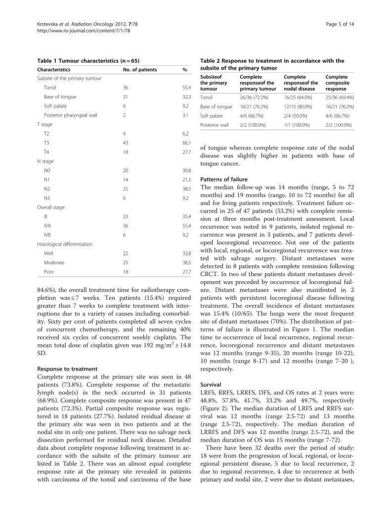
Table 1 Tumour characteristics (n = 65)
Characteristics No. of patients %
Subsite of the primary tumour
Tonsil 36 55,4
Base of tongue 21 32,3
Soft palate 6 9,2
Posterior pharyngeal wall 2 3,1
T stage
T2 4 6,2
T3 43 66,1
T4 18 27,7
N stage
N0 20 30,8
N1 14 21,5
N2 25 38,5
N3 6 9,2
Overall stage
III 23 35,4
IVA 36 55,4
IVB 6 9,2
Histological differentiation
Well 22 33,8
Moderate 25 38,5
Poor 18 27,7
Table 2 Response to treatment in accordance with thesubsite of the primary tumor
Subsiteofthe primarytumour
Completeresponseof theprimary tumour
Completeresponseof thenodal disease
Completecompositeresponse
Tonsil 26/36 (72.2%) 16/25 (64.0%) 25/36 (69.4%)
Base of tongue 16/21 (76.2%) 12/15 (80.0%) 16/21 (76.2%)
Soft palate 4/6 (66.7%) 2/4 (50.0%) 4/6 (66.7%)
Posterior wall 2/2 (100.0%) 1/1 (100.0%) 2/2 (100.0%)
Krstevska et al. Radiation Oncology 2012, 7:78 Page 5 of 14http://www.ro-journal.com/content/7/1/78
84.6%), the overall treatment time for radiotherapy com-pletion was ≤ 7 weeks. Ten patients (15.4%) requiredgreater than 7 weeks to complete treatment with inter-ruptions due to a variety of causes including comorbid-ity. Sixty per cent of patients completed all seven cyclesof concurrent chemotherapy, and the remaining 40%received six cycles of concurrent weekly cisplatin. Themean total dose of cisplatin given was 192 mg/m2± 14.8SD.
Response to treatmentComplete response at the primary site was seen in 48patients (73.8%). Complete response of the metastaticlymph node(s) in the neck occurred in 31 patients(68.9%). Complete composite response was present in 47patients (72.3%). Partial composite response was regis-tered in 18 patients (27.7%). Isolated residual disease atthe primary site was seen in two patients and at thenodal site in only one patient. There was no salvage neckdissection performed for residual neck disease. Detaileddata about complete response following treatment in ac-cordance with the subsite of the primary tumour arelisted in Table 2. There was an almost equal completeresponse rate at the primary site revealed in patientswith carcinoma of the tonsil and carcinoma of the base
of tongue whereas complete response rate of the nodaldisease was slightly higher in patients with base oftongue cancer.
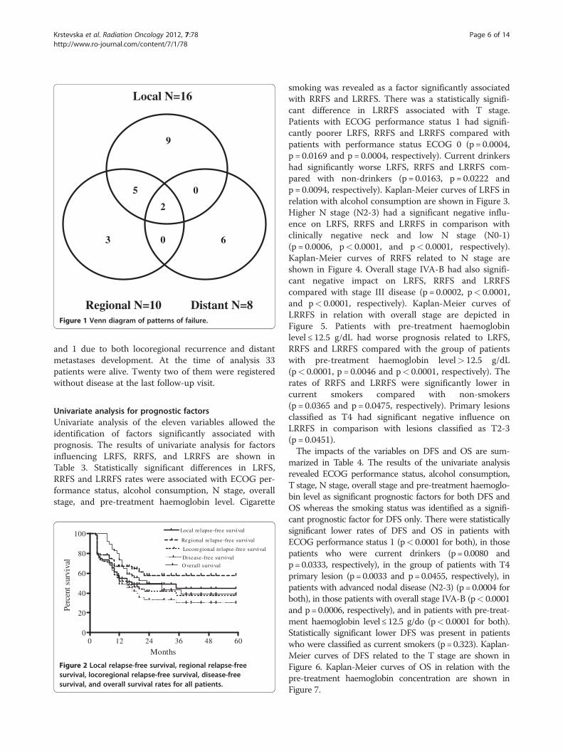
Patterns of failureThe median follow-up was 14 months (range, 5 to 72months) and 19 months (range, 10 to 72 months) for alland for living patients respectively. Treatment failure oc-curred in 25 of 47 patients (53.2%) with complete remis-sion at three months post-treatment assessment. Localrecurrence was noted in 9 patients, isolated regional re-currence was present in 3 patients, and 7 patients devel-oped locoregional recurrence. Not one of the patientswith local, regional, or locoregional recurrence was trea-ted with salvage surgery. Distant metastases weredetected in 8 patients with complete remission followingCRCT. In two of these patients distant metastases devel-opment was preceded by occurrence of locoregional fail-ure. Distant metastases were also manifested in 2patients with persistent locoregional disease followingtreatment. The overall incidence of distant metastaseswas 15.4% (10/65). The lungs were the most frequentsite of distant metastases (70%). The distribution of pat-terns of failure is illustrated in Figure 1. The mediantime to occurrence of local recurrence, regional recur-rence, locoregional recurrence and distant metastaseswas 12 months (range 9-35), 20 months (range 10-22),10 months (range 8-17) and 12 months (range 7-20 ),respectively.
SurvivalLRFS, RRFS, LRRFS, DFS, and OS rates at 2 years were:48.8%, 57.8%, 41.7%, 33.2% and 49.7%, respectively(Figure 2). The median duration of LRFS and RRFS sur-vival was 12 months (range 2.5-72) and 13 months(range 2.5-72), respectively. The median duration ofLRRFS and DFS was 12 months (range 2.5-72), and themedian duration of OS was 15 months (range 7-72).There have been 32 deaths over the period of study:
18 were from the progression of local, regional, or locor-egional persistent disease, 5 due to local recurrence, 2due to regional recurrence, 4 due to recurrence at bothprimary and nodal site, 2 were due to distant metastases,
9
3
5
6
2
0
0
Local N=16
Regional N=10 Distant N=8 Figure 1 Venn diagram of patterns of failure.
Krstevska et al. Radiation Oncology 2012, 7:78 Page 6 of 14http://www.ro-journal.com/content/7/1/78
and 1 due to both locoregional recurrence and distantmetastases development. At the time of analysis 33patients were alive. Twenty two of them were registeredwithout disease at the last follow-up visit.
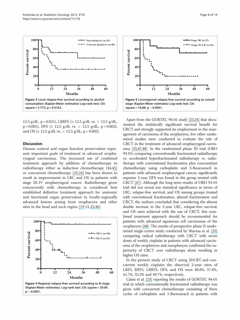
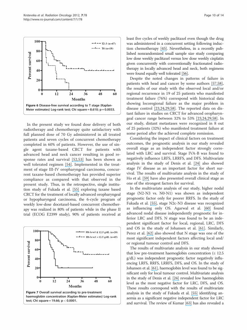
Univariate analysis for prognostic factorsUnivariate analysis of the eleven variables allowed theidentification of factors significantly associated withprognosis. The results of univariate analysis for factorsinfluencing LRFS, RRFS, and LRRFS are shown inTable 3. Statistically significant differences in LRFS,RRFS and LRRFS rates were associated with ECOG per-formance status, alcohol consumption, N stage, overallstage, and pre-treatment haemoglobin level. Cigarette
0 12 24 36 48 600
20
40
60
80
100 Local relapse-free survival
Regional re lapse-free survival
Locoregional relapse-free survival
Disease-free survival
O verall survival
Months
Perc
ent s
urvi
val
Figure 2 Local relapse-free survival, regional relapse-freesurvival, locoregional relapse-free survival, disease-freesurvival, and overall survival rates for all patients.
smoking was revealed as a factor significantly associatedwith RRFS and LRRFS. There was a statistically signifi-cant difference in LRRFS associated with T stage.Patients with ECOG performance status 1 had signifi-cantly poorer LRFS, RRFS and LRRFS compared withpatients with performance status ECOG 0 (p = 0.0004,p = 0.0169 and p = 0.0004, respectively). Current drinkershad significantly worse LRFS, RRFS and LRRFS com-pared with non-drinkers (p = 0.0163, p = 0.0222 andp = 0.0094, respectively). Kaplan-Meier curves of LRFS inrelation with alcohol consumption are shown in Figure 3.Higher N stage (N2-3) had a significant negative influ-ence on LRFS, RRFS and LRRFS in comparison withclinically negative neck and low N stage (N0-1)(p = 0.0006, p< 0.0001, and p< 0.0001, respectively).Kaplan-Meier curves of RRFS related to N stage areshown in Figure 4. Overall stage IVA-B had also signifi-cant negative impact on LRFS, RRFS and LRRFScompared with stage III disease (p = 0.0002, p< 0.0001,and p< 0.0001, respectively). Kaplan-Meier curves ofLRRFS in relation with overall stage are depicted inFigure 5. Patients with pre-treatment haemoglobinlevel ≤ 12.5 g/dL had worse prognosis related to LRFS,RRFS and LRRFS compared with the group of patientswith pre-treatment haemoglobin level> 12.5 g/dL(p< 0.0001, p = 0.0046 and p< 0.0001, respectively). Therates of RRFS and LRRFS were significantly lower incurrent smokers compared with non-smokers(p = 0.0365 and p = 0.0475, respectively). Primary lesionsclassified as T4 had significant negative influence onLRRFS in comparison with lesions classified as T2-3(p = 0.0451).The impacts of the variables on DFS and OS are sum-
marized in Table 4. The results of the univariate analysisrevealed ECOG performance status, alcohol consumption,T stage, N stage, overall stage and pre-treatment haemoglo-bin level as significant prognostic factors for both DFS andOS whereas the smoking status was identified as a signifi-cant prognostic factor for DFS only. There were statisticallysignificant lower rates of DFS and OS in patients withECOG performance status 1 (p< 0.0001 for both), in thosepatients who were current drinkers (p=0.0080 andp=0.0333, respectively), in the group of patients with T4primary lesion (p=0.0033 and p=0.0455, respectively), inpatients with advanced nodal disease (N2-3) (p=0.0004 forboth), in those patients with overall stage IVA-B (p< 0.0001and p=0.0006, respectively), and in patients with pre-treat-ment haemoglobin level≤12.5 g/do (p< 0.0001 for both).Statistically significant lower DFS was present in patientswho were classified as current smokers (p=0.323). Kaplan-Meier curves of DFS related to the T stage are shown inFigure 6. Kaplan-Meier curves of OS in relation with thepre-treatment haemoglobin concentration are shown inFigure 7.

Table 3 Univariate analysis correlating prognostic factors with disease control above clavicles
Factor LRFS RRFS LRRFS
n 2 years % p-value 2 years % p-value 2 years % p-value
Gender
Male 59 45.2 0,2713 52.3 0,0527 36.2 0,1562
Female 6 83.0 100.0 83.3
Age (years)
≤ 50 20 49.3 0,7046 42.3 0,3524 30.7 0,6735
> 50 45 48.2 63.2 44.8
Performance status (ECOG)
0 44 62.8 0,0004 68.3 0,0169 54.7 0,0004
1 21 19.2 20.8 9.8
Cigarette smoking
Non-smokers 11 77.7 0,1651 89.8 0,0365 79.9 0,0475
Current smokers 54 43.2 49.8 33.8
Alcohol consumption
Non-drinkers 25 71.8 0,0163 75.3 0,0222 64.7 0,0094
Current drinkers 40 34.8 47.2 27.2
Subsite
Tonsil 36 42.8 0,3656 58.7 0,7991 37.8 0.4509
Base of tongue 21 66.7 54.7 45.9
Soft palate 6 49.2 50.2 50.9
Posterior pharyngeal wall 2 0 0 0
T stage
T2-3 47 54.0 0,1094 58.8 0,9960 49.1 0,0451
T4 18 35.4 48.1 21.6
N stage
N0-1 34 68.8 0.0006 93.8 <0.0001 66.6 <0.0001
N2-3 31 28.3 22.2 16.4
Overall stage
III 23 81.7 0.0002 95.3 <0.0001 81.6 <0.0001
IVA-B 42 30.8 33.2 19.1
Histological differentiation
Well 22 57.2 0,1931 59.2 0,1385 44.1 0,2296
Moderate 25 54.3 76.3 54.7
Poor 18 31.8 36.3 23.4
Haemoglobin level
≤ 12.5 g/dL 20 21.3 <0.0001 44.2 0,0046 19.0 <0.0001
> 12.5 g/dL 45 60.1 65.8 51.5
n, number of patients; LRFS, local relapse-free survival; RRFS, regional relapse-free survival; LRRFS, locoregional relapse-free survival; ECOG, Eastern CooperativeOncology Group.
Krstevska et al. Radiation Oncology 2012, 7:78 Page 7 of 14http://www.ro-journal.com/content/7/1/78
Multivariate analysis for prognostic factorsThe results of multivariate analysis performed using theprognostic factors confirmed as significant in the univari-ate analysis are depicted in Table 5 and Table 6. Theadvanced N stage was revealed as an independent prog-nostic factor for inferior RRFS (N2-3 vs. N0-1, p = 0.048).
The advanced overall stage was found to be an independ-ent factor negatively influencing LRFS (IVA-B vs. III,p = 0.022), LRRFS (IVA-B vs. III, p = 0.003), and DFS(IVA-B vs. III, p = 0.003). Haemoglobin level was also in-dependently prognostic for lower rates of LRFS (≤ 12.5 g/dL vs. > 12.5 g/dL, p = 0.002), RRFS (≤ 12.5 g/dL vs. >
0 12 24 36 48 600
20
40
60
80
100 Non-drinkers (n=25)
Current drinkers (n=40)
Months
Per
cent
sur
viva
l
Figure 3 Local relapse-free survival according to alcoholconsumption (Kaplan-Meier estimates) Log-rank test; Chisquare = 5.773; p = 0.0163.
0 12 24 36 48 600
20
40
60
80
100 Stage III (n=23)
Stage IVA-B (n=42)
Months
Per
cent
sur
viva
l
Figure 5 Locoregional relapse-free survival according to overallstage (Kaplan-Meier estimates) Log-rank test; Chisquare = 18.88; p< 0.0001.
Krstevska et al. Radiation Oncology 2012, 7:78 Page 8 of 14http://www.ro-journal.com/content/7/1/78
12.5 g/dL, p = 0.021), LRRFS (≤ 12.5 g/dL vs. > 12.5 g/dL,p = 0.001), DFS (≤ 12.5 g/dL vs. > 12.5 g/dL, p = 0.002),and OS (≤ 12.5 g/dL vs. > 12.5 g/dL, p = 0.002).
DiscussionDisease control and organ function preservation repre-sent important goals of treatment in advanced oropha-ryngeal carcinomas. The increased use of combinedtreatment approach by addition of chemotherapy toradiotherapy either as induction chemotherapy [44,45]or concurrent chemotherapy [23,24] has been shown toresult in improvement in LRC and OS in patients withstage III-IV oropharyngeal cancer. Radiotherapy givenconcurrently with chemotherapy is considered bestestablished definitive treatment approach for anatomicand functional organ preservation in locally-regionallyadvanced lesions arising from oropharynx and othersites in the head and neck region [19-21,23,46].
0 12 24 36 48 600
20
40
60
80
100
N0-1 (n=34)
N2-3 (n=31)
Months
Per
cent
sur
viva
l
Figure 4 Regional relapse-free survival according to N stage(Kaplan-Meier estimates). Log-rank test; Chi square = 29.04;p< 0.0001.
Apart from the GORTEC 94-01 study [23,24] that docu-mented the statistically significant survival benefit forCRCT and strongly supported its employment in the man-agement of carcinoma of the oropharynx, few other rando-mized studies were conducted to evaluate the role ofCRCT in the treatment of advanced oropharyngeal carcin-oma [25,47,48]. In the randomized phase III trial (ORO93-01) comparing conventionally fractionated radiotherapyvs. accelerated hyperfractionated radiotherapy vs. radio-therapy with conventional fractionation plus concomitantchemotherapy using carboplatin and 5-fluorouracil inpatients with advanced oropharyngeal cancer, significantlysuperior 2-year DFS was found in the group treated withCRCT [47]. Although the long-term results of ORO 93-01trial did not reveal any statistical significance in terms ofLRC, relapse-free survival, and OS among groups treatedwith conventional fractionation, altered fractionation andCRCT, the authors concluded that considering the almostdouble increase in the 5-year LRC, relapse-free survival,and OS rates achieved with the use of CRCT, this com-bined treatment approach should be recommended forpatients with advanced squamous cell carcinomas of theoropharynx [48]. The results of prospective phase II rando-mized single-centre study conducted by Sharma et al. [25]comparing radical radiotherapy with CRCT with sevendoses of weekly cisplatin in patients with advanced carcin-oma of the oropharynx and nasopharynx confirmed the su-periority of CRCT over radiotherapy alone resulting inhigher OS rates.In the present study of CRCT using 3DCRT and con-
current weekly cisplatin the observed 2-year rates ofLRFS, RRFS, LRRFS, DFS, and OS were 48.8%, 57.8%,41.7%, 33.2% and 49.7%, respectively.Calais et al. [23] reporting the results of GORTEC 94-01
trial in which conventionally fractionated radiotherapy wasgiven with concurrent chemotherapy consisting of threecycles of carboplatin and 5-fluorouracil in patients with
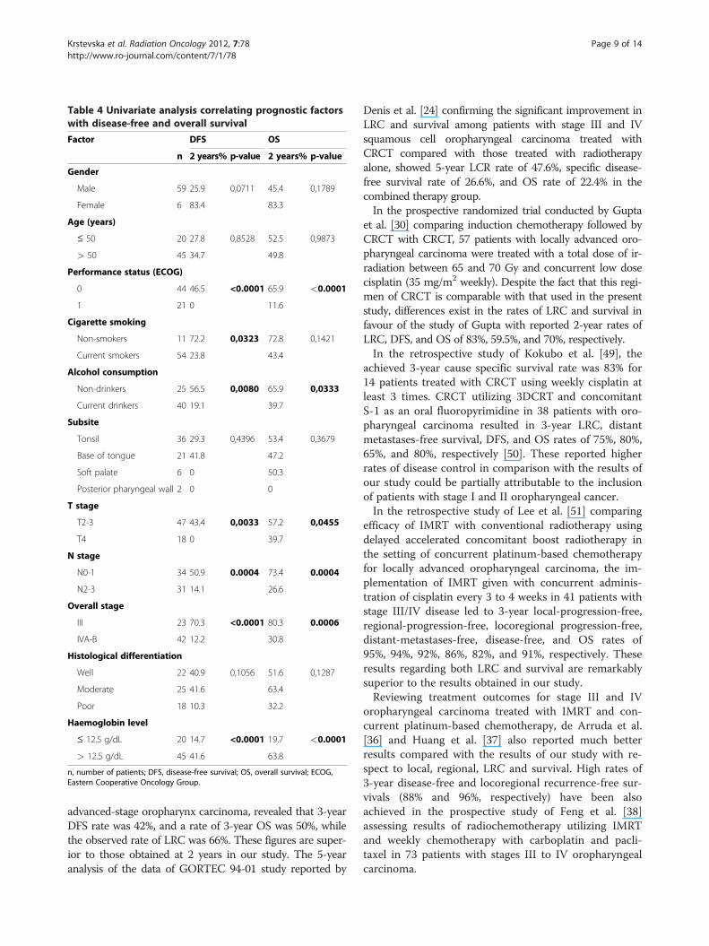
Table 4 Univariate analysis correlating prognostic factorswith disease-free and overall survival
Factor DFS OS
n 2 years% p-value 2 years% p-value
Gender
Male 59 25.9 0,0711 45.4 0,1789
Female 6 83.4 83.3
Age (years)
≤ 50 20 27.8 0,8528 52.5 0,9873
> 50 45 34.7 49.8
Performance status (ECOG)
0 44 46.5 <0.0001 65.9 <0.0001
1 21 0 11.6
Cigarette smoking
Non-smokers 11 72.2 0,0323 72.8 0,1421
Current smokers 54 23.8 43.4
Alcohol consumption
Non-drinkers 25 56.5 0,0080 65.9 0,0333
Current drinkers 40 19.1 39.7
Subsite
Tonsil 36 29.3 0,4396 53.4 0,3679
Base of tongue 21 41.8 47.2
Soft palate 6 0 50.3
Posterior pharyngeal wall 2 0 0
T stage
T2-3 47 43.4 0,0033 57.2 0,0455
T4 18 0 39.7
N stage
N0-1 34 50.9 0.0004 73.4 0.0004
N2-3 31 14.1 26.6
Overall stage
III 23 70.3 <0.0001 80.3 0.0006
IVA-B 42 12.2 30.8
Histological differentiation
Well 22 40.9 0,1056 51.6 0,1287
Moderate 25 41.6 63.4
Poor 18 10.3 32.2
Haemoglobin level
≤ 12.5 g/dL 20 14.7 <0.0001 19.7 <0.0001
> 12.5 g/dL 45 41.6 63.8
n, number of patients; DFS, disease-free survival; OS, overall survival; ECOG,Eastern Cooperative Oncology Group.
Krstevska et al. Radiation Oncology 2012, 7:78 Page 9 of 14http://www.ro-journal.com/content/7/1/78
advanced-stage oropharynx carcinoma, revealed that 3-yearDFS rate was 42%, and a rate of 3-year OS was 50%, whilethe observed rate of LRC was 66%. These figures are super-ior to those obtained at 2 years in our study. The 5-yearanalysis of the data of GORTEC 94-01 study reported by
Denis et al. [24] confirming the significant improvement inLRC and survival among patients with stage III and IVsquamous cell oropharyngeal carcinoma treated withCRCT compared with those treated with radiotherapyalone, showed 5-year LCR rate of 47.6%, specific disease-free survival rate of 26.6%, and OS rate of 22.4% in thecombined therapy group.In the prospective randomized trial conducted by Gupta
et al. [30] comparing induction chemotherapy followed byCRCT with CRCT, 57 patients with locally advanced oro-pharyngeal carcinoma were treated with a total dose of ir-radiation between 65 and 70 Gy and concurrent low dosecisplatin (35 mg/m2 weekly). Despite the fact that this regi-men of CRCT is comparable with that used in the presentstudy, differences exist in the rates of LRC and survival infavour of the study of Gupta with reported 2-year rates ofLRC, DFS, and OS of 83%, 59.5%, and 70%, respectively.In the retrospective study of Kokubo et al. [49], the
achieved 3-year cause specific survival rate was 83% for14 patients treated with CRCT using weekly cisplatin atleast 3 times. CRCT utilizing 3DCRT and concomitantS-1 as an oral fluoropyrimidine in 38 patients with oro-pharyngeal carcinoma resulted in 3-year LRC, distantmetastases-free survival, DFS, and OS rates of 75%, 80%,65%, and 80%, respectively [50]. These reported higherrates of disease control in comparison with the results ofour study could be partially attributable to the inclusionof patients with stage I and II oropharyngeal cancer.In the retrospective study of Lee et al. [51] comparing
efficacy of IMRT with conventional radiotherapy usingdelayed accelerated concomitant boost radiotherapy inthe setting of concurrent platinum-based chemotherapyfor locally advanced oropharyngeal carcinoma, the im-plementation of IMRT given with concurrent adminis-tration of cisplatin every 3 to 4 weeks in 41 patients withstage III/IV disease led to 3-year local-progression-free,regional-progression-free, locoregional progression-free,distant-metastases-free, disease-free, and OS rates of95%, 94%, 92%, 86%, 82%, and 91%, respectively. Theseresults regarding both LRC and survival are remarkablysuperior to the results obtained in our study.Reviewing treatment outcomes for stage III and IV
oropharyngeal carcinoma treated with IMRT and con-current platinum-based chemotherapy, de Arruda et al.[36] and Huang et al. [37] also reported much betterresults compared with the results of our study with re-spect to local, regional, LRC and survival. High rates of3-year disease-free and locoregional recurrence-free sur-vivals (88% and 96%, respectively) have been alsoachieved in the prospective study of Feng et al. [38]assessing results of radiochemotherapy utilizing IMRTand weekly chemotherapy with carboplatin and pacli-taxel in 73 patients with stages III to IV oropharyngealcarcinoma.
0 12 24 36 48 600
20
40
60
80
100T2-3 (n=47)
T4 (n=18)
Months
Per
cent
sur
viva
l
Figure 6 Disease-free survival according to T stage (Kaplan-Meier estimates) Log-rank test; Chi square = 8.610; p = 0.0033.
Krstevska et al. Radiation Oncology 2012, 7:78 Page 10 of 14http://www.ro-journal.com/content/7/1/78
In the present study we found dose delivery of bothradiotherapy and chemotherapy quite satisfactory withfull planned dose of 70 Gy administered in all treatedpatients and seven cycles of concurrent chemotherapycompleted in 60% of patients. However, the use of sin-gle agent taxane-based CRCT for patients withadvanced head and neck cancer resulting in good re-sponse rates and survival [52,53] has been shown aswell tolerated regimen [54]. Implemented in the treat-ment of stage III-IV oropharyngeal carcinoma, concur-rent taxane-based chemotherapy has provided superiorcompliance as compared with that observed in thepresent study. Thus, in the retrospective, single institu-tion study of Fukada et al. [55] exploring taxane basedCRCT for the treatment of locally advanced oropharyngealor hypopharyngeal carcinoma, the 6-cycle program ofweekly low-dose docetaxel-based concurrent chemother-apy was realized in 80% of patients while in the phase IItrial (ECOG E2399 study), 90% of patients received at
0 12 24 36 48 600
20
40
60
80
100 Hb level ≤≤
Hb level > 12.5 g/dL (n=45)
Months
Per
cent
sur
viva
l
12.5 g/dL (n=20)
Figure 7 Overall survival according to pre-treatmenthaemoglobin concentration (Kaplan-Meier estimates) Log-ranktest; Chi square = 19.66; p< 0.0001.
least five cycles of weekly paclitaxel even though the drugwas administered in a concurrent setting following induc-tion chemotherapy [45]. Nevertheless, in a recently pub-lished nonrandomized small sample size study comparinglow dose weekly paclitaxel versus low dose weekly cisplatingiven concurrently with conventionally fractionated radio-therapy in locally advanced head and neck, both regimenswere found equally well tolerated [56].Despite the noted changes in patterns of failure in
patients with head and cancer by some authors [57,58],the results of our study with the observed local and/orregional recurrence in 19 of 25 patients who manifestedtreatment failure (76%) correspond with historical datashowing locoregional failure as the major problem indisease control [23,24,29,58]. The reported data on dis-tant failure in studies on CRCT for advanced oropharyn-geal cancer range between 32% to 53% [23,24,29,58]. Inour study, distant metastases were recognized in 8 outof 25 patients (32%) who manifested treatment failure atsome period after the achieved complete remission.Considering the impact of clinical factors on treatment
outcomes, the prognostic analysis in our study revealedoverall stage as an independent factor strongly corre-lated with LRC and survival. Stage IVA-B was found tonegatively influence LRFS, LRRFS, and DFS. Multivariateanalysis in the study of Denis et al. [24] also showedstage IV disease as an important factor for short sur-vival. The results of multivariate analysis in the study ofHo et al. [59] have also presented overall clinical stage asone of the strongest factors for survival.In the multivariate analysis of our study, higher nodal
stage (N2-N3 vs. N0-N1) was shown as independentprognostic factor only for poorer RRFS. In the study ofFukada et al. [55], stage N2c-N3 disease was recognizedas influencing only OS. Agarwal et al. [60] foundadvanced nodal disease independently prognostic for in-ferior LRC and DFS. N stage was found to be an inde-pendent significant factor for local, regional, LRC, DFSand OS in the study of Johansen et al. [61]. Similarly,Perez et al. [62] also showed that N stage was one of themost significant independent factors affecting local and/or regional tumour control and DFS.The results of multivariate analysis in our study showed
that low pre-treatment haemoglobin concentration (≤ 12.5g/dL) was independent prognostic factor negatively influ-encing LRFS, RRFS, LRRFS, DFS, and OS. In the study ofJohansen et al. [61], haemoglobin level was found to be sig-nificant only for local tumour control. Multivariate analysisin the study of Denis et al. [24] revealed low haemoglobinlevel as the most negative factor for LRC, DFS, and OS.These results correspond with the results of multivariateanalysis in the study of Fukada et al. [55] identifying an-aemia as a significant negative independent factor for LRCand survival. The review of Kumar [63] has also revealed a

Table 5 Multivariate analysis for local relapse-free survival, regional relapse-free survival and locoregional relapse-freesurvival
Factors Hazard ratio (95% CI)
LRFS RRFS LRRFS
Performance status (ECOG)(1 vs. 0) 1.11 (0.48-2.55) p = 0.806 0.67 (0.25-1.82) p = 0.431 1.12 (0.50-2.54) p = 0.779
Cigarette smoking(current smokers vs. non-smokers) / 3.98 (0.42-37.89) p = 0.229 0.61 (0.15-2.45) p = 0.485
Alcohol consumption(current drinkers vs. non-drinkers) 1.35 (0.57-3.20) p = 0.503 0.81 (0.25-2.63) p = 0.723 1.71 (0.61-4.84) p = 0.308
T stage(T4 vs. T2-3) / / 0.58 (0.22-1.52) p = 0.265
N stage(N2-3 vs. N0-1) 1.13 (0.44-2.89) p = 0.803 7.68 (1.02-58.05) p=0.048 0.73 (0.23-2.25) p = 0.577
Overall stage(IVA-B vs. III) 4.49 (1.24-16.27) p=0.022 3.57 (0.21-60.76) p = 0.379 9.91 (2.16-45.42) p=0.003
Haemoglobin level≤ 12.5 g/dL vs. > 12.5 g/dL 0.26 (0.11-0.62) p=0.002 0.27 (0.09-0.82) p=0.021 0.24 (0.10-0.57) p=0.001
95% CI, 95% confidence interval; LRFS, local relapse-free survival; RRFS, regional relapse-free survival; LRRFS, locoregional relapse-free survival; ECOG, EasternCooperative Oncology Group.
Krstevska et al. Radiation Oncology 2012, 7:78 Page 11 of 14http://www.ro-journal.com/content/7/1/78
strong evidence that low pre-treatment haemoglobin con-centration as powerful, statistically significant prognosticfactor had negative impact on local control and survival inhead and neck cancer patients treated with definitive radio-therapy. The impact of anaemia on locoregional tumourcontrol and survival in patients with squamous cell carcin-oma of the larynx and pharynx was confirmed in the largeDAHANCA study in which the use of the radiosensitizernimorazole in association with radiotherapy was shown tosignificantly improve LRC and disease-specific survival[64]. According to Becker et al. [65], low haemoglobin con-centration as a factor with a negative impact on treatmentoutcomes was associated with reduced tumour oxygen-ation resulting in radioresistance in head and neck cancer.The analysis of the relationship between pre-treatmentmeasurements of tumour oxygen tension (pO2) and LRCand survival provided evidence that tumour hypoxia wasassociated with a poor prognosis in patients with advancedhead and neck squamous cell carcinoma treated withradiotherapy [66,67]. The investigation of Nordsmark andOvergaard [68] confirmed pre-treatment haemoglobinlevel and tumour hypoxia prognostic for locoregionaltumour control emphasizing that low level of tumour oxy-genation was shown the strongest independent prognostic
Table 6 Multivariate analysis for disease-free survival and ove
Factors
Performance status (ECOG)(1 vs. 0)
Cigarette smoking(current smokers vs. non-smokers)
Alcohol consumption(current drinkers vs. non-drinkers)
T stage(T4 vs. T2-3)
N stage(N2-3 vs. N0-1)
Overall stage(IVA-B vs. III)
Haemoglobin level≤ 12.5 g/dL vs. > 12.5 g/dL
95% CI, 95% confidence interval; DFS, disease-free survival; OS, overall survival; ECO
indicator for locoregional tumour control after definitiveradiotherapy in advanced head and neck cancers.Although the recent re-analysis of a large randomized
trial conducted by DAHANCA [69] suggested that HPVpositive oropharyngeal cancers most probably would notbenefit from any modification of tumour oxygenation, itshould be pointed out that in conditions without availabledata regarding HPV status, the reversal of anaemia shouldbe strongly considered in all patients with advanced oro-pharyngeal carcinoma in order to eliminate the negative re-flection of low pre-treatment haemoglobin level on thepatient’s general condition and tumour progression [63,70].Considering the established importance of HPV,
expressed by p16, as a strong independent prognosticfactor for survival among patients with oropharyngealcancer [71], and taking into account published data indi-cating HPV-positivity as consistent determinant of su-perior survival irrespectively of the treatment approachused [69,70,72,73] it could be admitted that specific test-ing for HPV in oropharyngeal cancers is highly recom-mendable since the combination of HPV status andoverall disease stage could be useful in further classifica-tion of patients providing in that way treatment deci-sions for individual patient moreover that the optimal
rall survival
Hazard ratio (95% CI)
DFS OS
1.20 (0.56-2.60) p = 0.634 1.51 (0.59-3.86) p = 0.395
0.93 (0.25-3.41) p = 0.909 /
1.47 (0.59-3.63) p = 0.399 1.17 (0.49-2.77) p = 0.722
0.70 (0.28-1.72) p = 0.431 0.76 (0.29-2.02) p = 0.582
0.62 (0.22-1.74) p = 0.362 1.10 (0.30-4.11) p = 0.887
8.40 (2.11-33.47) p=0.003 3.82 (0.73-19.97)p = 0.112
0.28 (0.13-0.61) p=0.002 0.24 (0.10-0.60) p=0.002
G, Eastern Cooperative Oncology Group.
Krstevska et al. Radiation Oncology 2012, 7:78 Page 12 of 14http://www.ro-journal.com/content/7/1/78
treatment regimen for HPV/p16 positive oropharyngealcancer has not been yet clarified.
ConclusionsOur study, presenting single centre treatment experience,demonstrated inferior results in patient outcomes followingsingle-agent weekly cisplatin radiochemotherapy regimencompared with the results of studies evaluating CRCTusing the same or different scheduling of concomitant cis-platin regardless of the radiation technique used. Results ofour study, especially when taking into consideration thehighest proportion of local/regional relapse in the wholenumber of recurrences, do suggest that further progress inthe management of this disease in our institution could beachieved by introduction of advanced radiotherapy techni-ques (IMRT) as an attempt to reduce incidence of locore-gional failures and to influence improvement of DFS andOS rates. However, the high percentage of patients withdistant metastatic development with or without synchron-ous manifestation of locoregional recurrence recognized instudies of CRCT should not be neglected and should pointout the importance of further investigation of combinedtreatment approach represented with induction chemo-therapy followed by CRCT. Despite all uncertainties ofradiotherapy, the use of accelerated radiotherapy regimenswhich can lead to redefinition of treatment protocols andreorganization of patient and staff flow in the department,can lead to improvement of treatment outcome. Also,introduction of other cytotoxic agents (taxanes) in concur-rent setting with conventional fractionated radiotherapyand the inclusion of molecular targeted therapies areexpected to provide further improvement in treatment out-comes in patients with advanced squamous cell carcinomaof the oropharynx.
AbbreviationsHPV: Human papillomavirus; LRC: Locoregional control; CRCT: Concurrentradiochemotherapy; DFS: Disease-free survival; OS: Overall survival;MACH-NC: Meta-Analysis of Chemotherapy in Head and Neck Cancer;3DCRT: Three-dimensional conformal radiotherapy; IMRT: Intensity-modulatedradiotherapy; CT: Computed tomography; MRI: Magnetic resonance imaging;AJCC: American Joint Committee on Cancer; GTV: Gross Tumour Volume;CTV: Clinical Target Volume; DAHANCA: Danish Head and Neck CancerGroup; EORTC: European Organization for Research and Treatment of Cancer;GORTEC: Groupe d'Oncologie Radiothérapie Tête et Cou; NCIC: NationalCancer Institute of Canada; RTOG: Radiation Therapy Oncology Group;PTVs: Planning Target Volumes; WHO: World Health Organization; LRFS: Localrelapse-free survival; RRFS: Regional relapse-free survival; LRRFS: Locoregionalrelapse-free survival; ECOG: Eastern Cooperative Oncology Group.
Competing interestsThe authors declare that they have no competing interests.
Author details1Department of Head and Neck Cancer, University Clinic of Radiotherapy andOncology, Skopje, Macedonia. 2Institute of Epidemiology, Statistics andInformatics, Faculty of Medicine, Skopje, Macedonia.
Authors’ contributionsVK and IS have made substantial contributions to design of the study andanalysed the data. VK collected the data and created the data base. VKperformed much of the work and drafted the manuscript. VK and BI-Zperformed the statistical analysis. VK and IS interpreted the data. All authorsread and approved the final manuscript.
Received: 22 March 2012 Accepted: 28 May 2012Published: 28 May 2012
References1. Global data on incidence of oral cancer, Oral Health Programme: World
Health Organization; [http://www.who.int/oral_health/publications/oral_cancer_brochure.pdf].
2. Evans PHR, Patel SG, Henk JM: Tumours of the oropharynx. In In Principlesand Practice in Head and Neck Oncology. 2nd edition. Edited by Evans PHR,Montgomery PQ, Gullane PJ. London: Taylor & Francis e-Library;2006:376–437.
3. Brugere J, Guenel P, Leclerc A, Rodriguez J: Differential effects of tobaccoand alcohol in cancer of the larynx, pharynx, and mouth. Cancer 1986,57:391–395.
4. Ringstrom E, Peters E, Hasegawa M, Posner M, Liu M, Kelsey KT: Humanpapillomavirus type 16 and squamous cell carcinoma of the head andneck. Clin Cancer Res 2002, 8:3187–3192.
5. Gillison ML, D’Souza G, Westra W, Sugar E, Xiao W, Begum S, Viscidi R:Distinct risk factor profiles for human papillomavirus type 16-positiveand human papillomavirus 16-negative head and neck cancers. J NatlCancer Inst 2008, 100:407–420.
6. Forastiere A, Koch W, Trotti A, Sidransky D: Head and neck cancer. N Engl JMed 2001, 345:1890–1900.
7. Ruff T, Lenis A, Skinner OD: Carcinoma of the oral cavity and oropharynx.Surg Clin North Am 1986, 66:659–671.
8. Machtay M, Perch S, Markiewicz D, Thaler E, Chalian A, Goldberg A,Kligerman M, Weinstein G: Combined surgery and postoperataiveradiotherapy for carcinoma of the base of tongue. Head Neck 1997,19:494–499.
9. Carvalho AL, Magrin J, Kowalski LP: Sites of recurrence in oral andoropharyngeal cancers according to the treatment approach.Oral Dis 2003, 9:112–118.
10. Guggenheimer J, Verbin RS, Johnson JT, Horkowitz CA, Myers EN: Factorsdelaying the diagnosis of oral and oropharyngeal carcinomas.Cancer 1989, 64:932–935.
11. Henk JM: Results of radiotherapy for carcinoma of the oropharynx.Clin Otolaryngol & Allied Sci 1978, 3:137–143.
12. Lindberg R: Distribution of cervical lymph node metastases fromsquamous cell carcinoma of the upper respiratory and digestive tracts.Cancer 1972, 29:1446–1450.
13. Browman GP, Cripps C, Hodson DI, Eapen L, Sathya J, Levine MN:Placebo-controlled randomized trial of infusional fluorouracil duringstandard radiotherapy in locally advanced head and neck cancer. J ClinOncol 1994, 12:2648–2653.
14. Haffty BG, Son YH, Papac R, Sasaki CT, Weissberg JB, Fischer D, Rockwell S,Sartorelli AC, Fischer JJ: Chemotherapy as an adjunct to radiation in thetreatment of squamous cell carcinoma of the head and neck: Results ofthe Yale Mitomycin Randomized Trials. J Clin Oncol 1997, 15:268–276.
15. Wendt TG, Grabenbauer GG, Rodel CM, Thiel HJ, Aydin H, Rohloff R,Wustrow TP, Iro H, Popella C, Schalhorn A: Simultaneousradiochemotherapy versus radiotherapy alone in advanced head andneck cancer: a randomized multicenter study. J Clin Oncol 1998,16:1318–1324.
16. Brizel DM, Albers ME, Fisher SR, Scher RL, Richtsmeier WJ, Hars V, George SL,Huang AT, Prosnitz LR: Hyperfractionated irradiation with or withoutconcurrent chemotherapy for locally advanced head and neck cancer.N Engl J Med 1998, 338:1798–1804.
17. Adelstein DJ, Lavertu P, Saxton JP, Secic M, Wood BG, Wanamaker JR,Eliachar I, Strome M, Larto MA: Mature results of a phase III randomizedtrial comparing chemoradiotherapy with radiation therapy alone inpatients with stage III and IV squamous cell carcinoma of the head andneck. Cancer 2000, 88:876–883.
18. Jeremic B, Shibamoto Y, Milicic B, Nikolic N, Dagovic A, Aleksandrovic J,Vaskovic Z, Tadic L: Hyperfractionated radiation therapy with or without

Krstevska et al. Radiation Oncology 2012, 7:78 Page 13 of 14http://www.ro-journal.com/content/7/1/78
concurrent low-dose daily cisplatin in locally advanced squamous cellcarcinoma of the head and neck: a prospective randomized trial. J ClinOncol 2000, 18:1458–1464.
19. Pignon JP, Bourhis J, Domenge C, Designe L: Chemotherapy added tolocoregional treatment for head and neck squamous-cell carcinoma:three meta analyses of updated individual data. MACH-NC CollaborativeGroup. Meta-analysis of chemotherapy on head and neck cancer.Lancet 2000, 355:949–955.
20. Pignon JP, le Maitre A, Maillard E, Bourhis J: Meta-analysis ofchemotherapy in head and neck cancer (MACH-NC): an update on 93randomized trials and 17,346 patients. Radiother Oncol 2009, 92:4–14.
21. Browman GP, Hodson DI, Mackenzie RJ, Bestic N, Zuraw L, Cancer CareOntario Practice Guideline Initiative Head and Neck Cancer Disease SiteGroup: Choosing a concomitant chemotherapy and radiotherapyregimen for squamous cell head and neck cancer: a systematic review ofthe published literature with subgroup analysis. Head Neck 2001,23:579–589.
22. Budach W, Hehr T, Budach V, Belka C, Dietz K: A Meta-Analysis ofhyperfractionated and accelerated radiotherapy and combinedchemotherapy and radiotherapy regimens in unresected locallyadvanced squamous cell carcinoma of the head and neck. BMC Cancer2006, 6:28–39.
23. Calais G, Alfonsi M, Bardet E, Sire C, Germain T, Bergerot P, Rhein B,Tortochaux J, Oudinot P, Bertrand P: Randomized trial of radiation therapyversus concomitant chemotherapy and radiation therapy for advanced-stage oropharynx carcinoma. J Natl Cancer Inst 1999, 91:2081–2086.
24. Denis F, Garaud P, Bardet E, Alfonsi M, Sire C, Germain T, Bergerot P, RheinB, Tortochaux J, Calais G: Final results of the 94-01 French Head and NeckOncology and Radiotherapy group randomized trial comparingradiotherapy alone with concomitant radiochemotherapy in advanced-stage oropharynx carcinoma. J Clin Oncol 2004, 22:69–76.
25. Sharma A, Mohanti BK, Thakar A, Bahadur S, Bhasker S: Concomitantchemoradiation versus radical radiotherapy in advanced squamous cellcarcinoma of oropharynx and nasopharynx using weekly cisplatin: aphase II randomized trial. Ann Oncol 2010, 21:2272–2277.
26. Blanchard P, Baujat B, Holostenco V, Bourredjem A, Baey C, Bourhis J,Pignon J-P, on behalf of the MACH-CH Collaborative group: Meta-analysisof chemotherapy in head and neck cancer (MACH-HC: A comprehensiveanalysis by tumour site. Radiother Oncol 2011, 100:33–40.
27. Adelstein DJ, Li Y, Adams GL, Wagner H Jr, Kish JA, Ensley JF, Schuller DE,Forastiere AA: An intergroup phase III comparison of standard radiation andtwo schedules of concurrent chemoradiotherapy in patients withunresectable squamous cell head and neck cancer. J Clin Oncol 2003, 21:92–98.
28. Kurihara N, Kubota T, Hoshiya Y, Otani Y, Ando N, Kumai K, Kitajima M:Pharmacokinetics of cis-diamminedichloroplatinum (II) given as low-dose and high-dose infusions. J Surg Oncol 1996, 62:135–138.
29. Gupta T, Agarwal JP, Ghosh-Laskar S, Parikh PM, D’Cruz AK, Dinshaw KA:Radical radiotherapy with concurrent weekly cisplatin in locoregionallyadvanced squamous cell carcinoma of the head and neck: a single-institution experience. Head Neck Oncol 2009, 1:17.
30. Gupta D, Shukla P, Bisht SS, Dhawan A, Pant MC, Bhatt ML, Gupta S, GuptaR, Negi MPS: A prospective comparision of sequential chemoradiation vsconcurrent chemoradiation in locally advanced oropharyngealcarcinomas. Cancer Biol Ther 2009, 8:213–217.
31. Brizel DM, Esclamado R: Concurrent chemoradiotherapy for locallyadvanced, nonmetastatic, squamous carcinoma of the head and neck:consensus, controversy, and conundrum. J Clin Oncol 2006, 24:2612–2617.
32. Purdy A: Dose to normal tissues outside the radiation therapy patient'streated volume: a review of different radiation therapy techniques.Health Phys 2008, 5:666–676.
33. Chao KS, Ozyigit G, Blanco AI, Thorstad WL, Deasy JO, Haughey BH, Spector GJ,Sessions DG: Intensity-modulated radiation therapy for oropharyngealcarcinoma: impact of tumor volume. Int J Radiat Oncol Biol Phys 2004, 59:43–50.
34. Eisbruch A, Ship JA, Dawson LA, Kim HM, Bradford CR, Terrell JE, ChepehaDB, Teknos TN, Hogikyan ND, Anzai Y, Marsh LH, Ten Haken RK, Wolf GT:Salivary gland sparing and improved target irradiation by conformal andintensity modulated irradiation of head and neck cancer. World J Surg2003, 27:832–837.
35. Studer G, Huguenin PU, Davis JB, Kunz G, Lutolf UM, Glanzmann C: IMRTusing simultaneously integrated boost (SIB) in head and neck patients.Radiat Oncol 2006, 1:7–21.
36. de Arruda FF, Puri DR, Zhung J, Narayana A, Wolden S, Hunt M, Stambuk H,Pfister D, Kraus D, Shaha A, Shah J, Lee NY: Intensity modulated radiationtherapy for the treatment of oropharyngeal carcinoma: the MemorialSloan-Kettering Cancer Center experience. Int J Radiat Oncol Biol Phys2005, 64:363–373.
37. Huang K, Xia P, Chuang C, Weinberg V, Glastonbury CM, Eisele DW, Lee NY,Yom SS, Phillips TL, Quivey JM: Intensity-modulated chemoradiation fortreatment of stage III and IV oropharyngeal carcinoma: the University ofCalifornia-San Francisco experience. Cancer 2008, 113:497–507.
38. Feng FY, Kim HM, Lyden TH, Haxer MJ, Worden FP, Feng M, Moyer JS,Prince ME, Carey TE, Wolf GT, Bradford CR, Chepeha DB, Eisbruch A:Intensity-modulated chemoradiotherapy aiming to reduce dysphagia inpatients with oropharyngeal cancer: clinical and functional results. J ClinOncol 2010, 28:2732–2738.
39. Greene FL, Page DL, Fleming ID, Fritz A, Balch CM, Haller DG, Morrow M:AJCC Cancer Staging Manual. New York: Springer-Verlag 2002, 6:33–46.
40. Gregoire V, Levendag P, Ang KK, Bernier J, Braaksma M, Budach V, Chao C,Coche E, Cooper JS, Cosnard G, Eisbruch A, El-Sayed S, Emami B, Grau C,Hamoir M, Lee N, Maingon P, Muller K, Reychler H: CT-based delineation oflymph node levels related CTVs in the node negative neck: DAHANCA,EORTC, GORTEC, NCIC, RTOG consensus guidelines. Radiother Oncol 2003,69:227–236.
41. Gregoire V, Eisbruch A, Hamoir M, Levendag P: Proposal for thedelineation of the nodal CTV in the node-positive and post-operativeneck. Radiother Oncol 2006, 79:15–20.
42. Miller AB, Hoogstraten B, Staquet M, Winkler A: Reporting results of cancertreatment. Cancer 1981, 47:207–214.
43. Kaplan EL, Meier P: Nonparametric estimation for incomplete observation.J Am Stat Assoc 1958, 53:457–481.
44. Domenge C, Hill C, Lefebre JL, De Raucourt D, Rhein B, Wibault P, MarandasP, Coche-Dequeant B, Stromboni-Luboinski M, Sancho-Garnier H, LuboinskiB: Randomized trial of neoadjuvant chemotherapy in oropharyngealcarcinoma. French Groupe d’Etude des Tumeurs de la Tête et du Cou(GETTEC). Br J Cancer 2000, 83:1594–1598.
45. Cmelak AJ, Li S, Goldwasser MA, Murphy B, Cannon M, Pinto H, RosenthalDI, Gillison M, Forastiere AA: Phase II trial of chemoradiation for organpreservation in resectable stage III or IV squamous cell carcinomas ofthe larynx or oropharynx: results of Eastern Cooperative OncologyGroup Study E2399. J Clin Oncol 2007, 25:3971–3977.
46. Forastiere AA, Trotti A: Radiotherapy and concurrent chemotherapy: astrategy that improves locoregional control and survival inoropharyngeal cancer. J Natl Cancer Inst 1999, 91:2065–2066.
47. Olmi P, Crispino S, Fallai C, Torri V, Rossi F, Bolner A, Amichetti M, Signor M,Taino R, Sguadrelli M, Colombo A, Ardizzoia A, Ponticelli P, Franchin G,Minatel E, Gobitti C, Atzeni G, Gava A, Flann M, Marsoni S: Locoregionallyadvanced carcinoma of the oropharynx: conventional radiotherapy vs.accelerated hyperfractionated radiotherapy vs. concomitantradiotherapy and chemotherapy-a multicenter randomized trial. Int JRadiat Oncol Biol Phys 2003, 55:78–92.
48. Fallai C, Bolner A, Signor M, Gava A, Franchin G, Ponticelli P, Taino R, Rossi F,Ardizzoia A, Oggionni M, Crispino S, Olmi P: Long-term results ofconventional radiotherapy versus accelerated hyperfractionatedradiotherapy versus concomitant radiotherapy and chemotherapy inlocoregionally advanced carcinoma of the oropharynx. Tumori 2006,92:41–54.
49. Kokubo M, Nagata Y, Nishimura Y, Kimura H, Shoji K, Asato R, Sasai K,Hiraoka M: Concurrent chemoradiotherapy for oropharyngeal carcinoma.Am J Clin Oncol 2001, 24:71–76.
50. Ohnishi K, Shioyama Y, Nakamura T, Ohga S, Nonoshita T, Yoshitake T,Terashima K, Komune S, Honda H: Concurrent chemoradiotherapy withS-1 as first-line treatment for patients with oropharyngeal cancer.J Radiat Res 2011, 52:47–53.
51. Lee NY, de Arruda FF, Puri DR, Wolden SL, Narayana A, Mechalakos J,Venkatraman ES, Kraus D, Shaha A, Shah JP, Pfister DG, Zelefsky MJ: Acomparison of intensity-modulated radiation therapy and concomitantboost radiotherapy in the setting of concurrent chemotherapy for locallyadvanced oropharyngeal carcinoma. Int J Radiat Oncol Biol Phys 2006,66:966–974.
52. Lovey J, Koronczay K, Remenar E, Csuka O, Nemeth G: Radiotherapy andconcurrent low-dose paclitaxel in locally advanced head and neckcancer. Radiother Oncol 2003, 68:171–174.
Krstevska et al. Radiation Oncology 2012, 7:78 Page 14 of 14http://www.ro-journal.com/content/7/1/78
53. Suzuki M, Nishimura Y, Nakamatsu K, Kanamori S, Koike R, Kawamoto M,Mori K: Phase I study of weekly docetaxel infusion and concurrentradiation therapy for head and neck cancer. Jpn J Clin Oncol 2003,33:297–301.
54. Pergolizzi S, Adamo V, Ferraro G, Sergi C, Santacaterina A, Romeo A, DeRenzis C, Zanghi M, Rossello R, Settineri N: Induction chemotherapy toweekly paclitaxel concurrent with curative radiotherapy in stage IV (M0)unresectable head and neck squamous cell carcinoma: a dose escalationstudy. J Chemother 2004, 16:201–205.
55. Fukada J, Shigematsu N, Takeda A, Ohashi T, Tomita T, Shiotani A, KuneidaE, Kawaguchi O, Fujii M, Kubo A: Weekly low-dose docetaxel-basedchemoradiotherapy for locally advanced oropharyngeal orhypopharyngeal carcinoma: a retrospective, single institution study. Int JRadiat Oncol Biol Phys 2010, 76:417–424.
56. Jain RK, Kirar P, Gupta G, Dubey S, Gupta SK, Goyal J: A comparative studyof low dose weekly paclitaxel versus cisplatin with concurrent radiationin the treatment of locally advanced head and neck cancer. Indian JCancer 2009, 46:50–53.
57. Vokes EE, Kies MS, Haraf DJ, Stenson K, List M, Humerickhouse R, Dolan ME,Pelzer H, Sulzen L, Witt ME, Hsieh YC, Mittal BB, Weichselbaum RR:Concomitant chemoradiotherapy as primary treatment for locoregionallyadvanced head and neck cancer. J Clin Oncol 2000, 18:1652–1661.
58. Machtay M, Rosenthal DI, Hershock D, Jones H, Williamson S, Greenberg MJ,Weinstein GS, Aviles VM, Chalian AA, Weber RS: Organ preservationtherapy using induction plus concurrent chemoradiation for advancedresectable oropharyngeal carcinoma: A University of Pennsylvania phaseII trial. J Clin Oncol 2002, 20:3964–3971.
59. Ho T, Zahurak M, Koch WM: Prognostic significance of presentation-to-diagnosis interval in patients with oropharyngeal carcinoma. ArchOtolaryngol Head Neck Surg 2004, 130:45–51.
60. Agarwal JP, Mallick I, Bhutani R, Ghosh-Laskar S, Gupta T, Budrukkar A,Murthy V, Sengar M, Dinshaw KA: Prognostic factors in oropharyngealcancer-analysis of 627 cases receiving definitive radiotherapy. Acta Oncol2009, 48:1026–1033.
61. Johansen LV, Grau C, Overgaard J: Squamous cell carcinoma of theoropharynx-an analysis of treatment results in 289 consecutive patients.Acta Oncol 2000, 39:985–994.
62. Perez CA, Patel MM, Chao KS, Simpson JR, Sessions D, Spector GJ, HaugheyB, Lockett MA: Carcinoma of the tonsillar fossa: prognostic factors andlong-term therapy outcome. Int J Radiat Oncol Biol Phys 1998,42:1077–1084.
63. Kumar P: Impact of anemia in patients with head and neck cancer.Oncologist 2000, 5(Suppl 2):13–18.
64. Overgaard J, Hansen HS, Overgaard M, Bastholt L, Berthelsen A, Specht L,Lindelov B, Jorgensen K: A randomized double-blind phase III study ofnimorazole as hypoxic radiosensitizer of primary radiotherapy insupraglottic larynx and pharynx carcinoma. Results form the DanishHead and Neck Cancer Study (DAHANCA). Protocol 5-85. Radiother Oncol1998, 46:135–146.
65. Becker A, Stadler P, Lavey RS, Hansgen G, Kuhnt T, Lautenschlager C,Feldmann HJ, Molls M, Dunst J: Severe anemia is associated with poortumor oxygenation in head and neck squamous cell carcinomas. Int JRadiat Oncol Biol Phys 2000, 46:459–466.
66. Nordsmark M, Overgaard J: A confirmatory prognostic study onoxygenation status and loco-regional control in advanced head andneck squamous cell carcinoma treated by radiation therapy. RadiotherOncol 2000, 57:39–43.
67. Nordsmark M, Bentzen SM, Rudat V, Brizel D, Lartigau E, Stadler P, Becker A,Adam M, Molls M, Dunst J, Terris DJ, Overgaard J: Prognostic value oftumor oxygenation in 397 head and neck tumors after primary radiationtherapy: An international multi-center study. Radiother Oncol 2005,77:18–24.
68. Nordsmark M, Overgaard J: Tumor hypoxia is independent of hemoglobinand prognostic for loco-regional tumor control after primaryradiotherapy in advanced head and neck cancer. Acta Oncol 2004,43:396–403.
69. Lassen P, Eriksen JG, Krogdahl A, Therkildsen MH, Ulhoi BP, Overgaard M,Specht L, Andersen E, Johansen J, Andersen LJ, Grau C, Overgaard J: Theinfluence of HPV-associated p16-expression on accelerated fractionatedradiotherapy in head and neck cancer: Evaluation of the randomizedDAHANCA 6&7 trial. Radiother Oncol 2011, 100:49–55.
70. Glaser CM, Millesi W, Kornek GV, Lang S, Schull B, Watzinger F, Selzer E,Lavey RS: Impact of hemoglobin level and use of recombinanterythropoietin on efficacy of preoperative chemoradiation therapy forsquamous cell carcinoma of the oral cavity and oropharynx. Int J RadiatOncol Biol Phys 2001, 50:705–715.
71. Ang KK, Harris J, Wheeler R, Weber R, Rosenthal DI, Nguyen-Tan PF, WestraWH, Chung CH, Jordan RC, Lu C, Kim H, Axelrod R, Silverman CC, RedmondKP, Gillison ML: Human papillomavirus and survival of patients withoropharyngeal cancer. N Engl J Med 2010, 363:24–35.
72. Licitra L, Perrone F, Bossi P, Suardi S, Mariani L, Artusi R, Oggionni M, RossiniC, Cantu G, Sguadrelli M, Quattrone P, Locati LD, Bergamini C, Olmi P,Pierotti MA, Pilotti S: High-risk human papillomavirus affects prognosis inpatients with surgically treated oropharyngeal squamous cell carcinoma.J Clin Oncol 2006, 24:5630–5636.
73. Lassen P, Eriksen JG, Hamilton-Dutoit S, Tramm T, Alsner J, Overgaard J:Effect of HPV-associated p16INK4A expression on response toradiotherapy and survival in squamous cell carcinoma of the head andneck. J Clin Oncol 2009, 27:1992–1998.
doi:10.1186/1748-717X-7-78Cite this article as: Krstevska et al.: Concurrent radiochemotherapy inlocally-regionally advanced oropharyngeal squamous cell carcinoma:analysis of treatment results and prognostic factors. Radiation Oncology2012 7:78.
Submit your next manuscript to BioMed Centraland take full advantage of:
• Convenient online submission
• Thorough peer review
• No space constraints or color figure charges
• Immediate publication on acceptance
• Inclusion in PubMed, CAS, Scopus and Google Scholar
• Research which is freely available for redistribution
Submit your manuscript at www.biomedcentral.com/submit