exposure to cigarette smoke causes dna damage in oropharyngeal tissue in dogs
TRANSCRIPT
Et
NR
a
ARRAA
KDBDOP
1
s[eomcwgtrsota
o
T
h1
Mutation Research 769 (2014) 13–19
Contents lists available at ScienceDirect
Mutation Research/Genetic Toxicology andEnvironmental Mutagenesis
jo ur nal home page: www.elsev ier .com/ locate /gentoxComm uni t y ad dress : www.elsev ier .com/ locate /mutres
xposure to cigarette smoke causes DNA damage in oropharyngealissue in dogs
atalia Pérez, Alina Berrío, Jairo Enrique Jaramillo, Rodrigo Urrego, María Patricia Arias ∗
esearch Group in Animal Sciences – INCA-CES, School of Veterinary Medicine and Animal Production, CES University, Medellin, Colombia
r t i c l e i n f o
rticle history:eceived 23 April 2013eceived in revised form 7 April 2014ccepted 12 April 2014vailable online 9 May 2014
eywords:ogiomarkerNA damages
a b s t r a c t
More than 40 mutagenic and carcinogenic agents present in cigarette smoke have been identified ascausative factors of human cancer, but no relation has been clearly documented in companion animals.In dogs, in addition to smoke inhalation and transdermic absorption, exposure to smoke includes oralingestion of particles adhered to the animal’s fur. This study evaluates the presence and type of histologicalalterations and DNA integrity in oropharyngeal tissue in dogs exposed and non-exposed to householdcigarette smoke by means of histopathology and comet assay studies on biopsy and swab samples. Anon-probabilistic convenience sample of 12 dogs were selected and classified in two groups: exposedand non-exposed to cigarette smoke. Non-parametric Kruskal–Wallis test was carried out on biopsy andswab data and a Chi2 test was performed on the information obtained by histopathology. A significance
ropharynxassive smoking
level was set at P < 0.05. Statistically significant differences were found between groups in comet assayscarried out on biopsy samples. No differences (P > 0.05) were found between groups based on cometassays swab samples and histopathology assessment. In conclusion, exposure to cigarette smoke causesDNA damage in dog oropharyngeal tissue. The use of dogs as sentinels for early DNA damage caused byexposure to environmental genotoxic agents like cigarette smoke is reported for the first time.
© 2014 Elsevier B.V. All rights reserved.
. Introduction
Dogs are the sentinels of their master’s diseases [1–6] as theyhare the same environment and are exposed to similar pollutants1,2]. In humans, smoking is the leading cause of mouth, pharynx,sophagus and lung cancer, which is explained by the direct contactf cigarette smoke with these structures [7]. Studies have identifiedore than 40 mutagens and carcinogens in cigarette smoke asso-
iated to cancer in humans [4–6,8], including nicotine, a substanceith high mutagenic and apoptotic potential, able to induce angio-
enesis and development of malignant growths in pets [9]. Evenhough some authors claim that exposure to cigarette smoke is aisk to the health of the animal [2,9–11], few reports indicate thepecific consequences it could generate [9,10]. In dogs, exposureccurs by inhalation of particles generated by cigarette combus-ion, transdermal absorption and ingestion of residues through the
nimal’s fur [12–14].Studies in humans and laboratory animals have found numer-us histological changes in the oropharyngeal mucosa caused by
∗ Corresponding author at: Calle 10 A 22-04, Medellin, Colombia.el.: +57 4 444 0 555.
E-mail addresses: [email protected], [email protected] (M.P. Arias).
ttp://dx.doi.org/10.1016/j.mrgentox.2014.04.013383-5718/© 2014 Elsevier B.V. All rights reserved.
cigarette smoke and some of its derivatives. Several changes havebeen described, with hyperkeratosis as the most common find-ings, specifically, being this one the most common injury causedby almost all tobacco products [13,15]; however, it is not knownwhether the cigarette smoke causes similar histological changes inexposed dogs.
DNA damage is known to be a mechanism that leads to thedevelopment of cancer. In humans, tobacco smoke is the mostimportant and well documented cause of cancer currently knownand its relationship with mouth, pharynx, esophagus and lungscancer is easily explained by their direct contact with smoke. Eval-uation of DNA damage in buccal epithelial cells may thus provide agood biomarker of early damage in target tissues [7]. This relationhas not been documented in companion animals.
Exposed tissue to cigarette smoke causes chronic inflamma-tion, which generates a series of biological processes in responseto the aggression [16]. It has been shown that exposed to cigarettesmoke (ECS) induces genetic material damage by oxidative stressat the cellular level which is generated by reactive oxygen species,and which leads to lipid peroxidation and breaks of DNA strands
[17–21]. Recent studies have found that cigarette smoke con-densates in the oropharyngeal region and generates significantfragmentation of DNA [22]. It is not known whether the effect ofcigarette smoke is similar in companion pets.
1 n Rese
htamHmc[
hsad
2
C6
2
nabaino
(Tf
citt
2
eofiaca
2
gto
2
prwf
2
2
ifnllctF
4 N. Pérez et al. / Mutatio
Several diagnostic techniques are used in order to determineistopathological changes and DNA damage in oropharyngealissue. The most common techniques include histopathologynd comet assay test from biopsies and swabs which deter-ine the effects on the area exposed to cigarette smoke.istopathology identifies tissue changes while the comet assayeasures and analyzes DNA breaks produced by chemi-
al and physical agents in any eukaryotic cell population23].
Therefore, this study was conducted in order to perform aistopathological evaluation and comet assay of biopsies and swabpecimens, in order to determine the presence and type of cell alter-tions in cellularity and DNA integrity in oropharyngeal tissue ofogs with ECS.
. Materials and methods
A study of cases and controls was conducted with the approval of the Ethicsommittee for Animal Experiments of the University of Antioquia, Colombia (Act9 of 2011).
.1. Sample selection
This study employed dogs from Medellin city, Colombia (South America). Aon-probabilistic convenience sample made of 12 dogs was selected based on
written survey carried out to the owners [14]. Clinically healthy dogs of anyreed, older than one year of age, with no prior history of respiratory disease orny other disease during the last 12 months and that were kept indoors werencluded. Dogs exposed to smoke sources such as wood stoves, furnaces, chim-eys or exhaust pipes were excluded from the study, as well as dogs that livedutdoors.
Individuals were classified into two groups: non-exposed to cigarette smokeNECS) and ECS. For the former, only dogs that had never been ECS were selected.he latter consisted of dogs exposed to the smoke of five or more cigarettes per dayor more than one year [24].
The sample size was estimated based on the data obtained through the pilotomet assay test. With regards to the number of cells required per each individual,t was determined based on the weighted average of cellular counts conducted inhe studies of Kimura et al. (2010), Harreus et al. (2000) and Tice et al. (2000), for aotal of 60 cells per each sample obtained [25–27].
.2. Sampling
The procedure was performed under aseptic conditions with patient under gen-ral anesthesia. Induction was performed with 5 mg/kg of ketamine, 0.025 mg/kgf acepromazine and 0.5 mg/kg of diazepam intramuscular. Maintenance was per-ormed with 8 mg/kg intravenous of ketamine. All patients received 0.2 mg/kgntravenous meloxicam for pain management. Animal’s mouth was opened with
tongue depressor in order to expose the oropharynx, exactly in the tonsilar fosaraneal to palatine tonsile [28]. Swab and biopsy samples were obtained from thisrea.
.2.1. SwabbingFirst, samples were obtained with a sterile swab from the described oropharyn-
eal area. It was preserved in 350 �l PBS and transported to the laboratory at 37 ◦Co be processed in the shortest possible time. Samples were protected from any typef natural or artificial light to prevent DNA damage.
.2.2. BiopsyThen, two biopsy samples were obtained with a 5 mm diameter retractable
unch. Samples intended for histopathological evaluation were obtained from theight side of the described area and fixed in 10% formalin. Fragments for comet assayere obtained from the left side and preserved using the same protocol described
or swab samples.
.3. Sample processing
.3.1. HistopathologySamples were processed in paraffin and a routine hematoxylin – eosin stain-
ng was performed. A NikonTM E100 Biological Optical Microscope was usedor examination of the samples. Ten fields were observed per sample, and theumber of inflammatory cells, (neutrophils, macrophages, eosinophil, mast cells,
ymphocytes, plasmocytes), tissue characteristics (number of epithelial layers, gob-et cells, epithelial and submucosa thickness), type of lesions present (acute orhronic inflammation, vascular congestion, edema, fibrosis, hypertrophy, acan-hosis, metaplasia, dysplasia, orthokeratosis, hyperkeratosis) were determined.indings were graded on a scale from zero to three, with zero being the absence
arch 769 (2014) 13–19
of damage; one, minor damage; two, moderate damage; and three, severe dam-age.
2.3.2. Evaluation of DNA integrity by comet assayBiopsies were subjected to enzymatic digestion. Each fragment was suspended
in 350 �l PBS pre-warmed at 37 ◦C, 10 mg/ml type II collagenase, 50 �l of 2.5%trypsin-EDTA and 50 �l of 0.5% pronase. They were incubated at 37 ◦C for 1 h andthen subjected to vortex for 3 min [25]. Afterwards, the remaining sample waswithdrawn, then, 50 �l of fetal bovine serum (FBS) at 10% was added to inac-tivate the action of the trypsin and finally centrifuged at 700 g for 10 min [26].Samples obtained by swabbing underwent the same treatment, except enzymaticdigestion.
To determine the viability of the cells, 2 �l of 4% trypan blue solution were addedto 10 �l of the cell suspension. The concentration was adjusted to 1 × 10⁵ cells/ml[26]. Then, cells were suspended in 0.5% low melting point agarose in pretreatedslides with 100 �l at 0.5% normal melting point agarose during 6 min at 4 ◦C. A newlayer of normal melting point at 0.5% agarose was added and suspended during 6 minat 4 ◦C again. Samples were then put in lysis solution (2.5 M NaCl, 0.1 M EDTA, 10 mMTris–HCl, 10% DMSO, 1% Triton X-100, pH 10.5, during 1 h at 4 ◦C). In a dark room,samples were incubated in electrophoresis buffer (0.3 M NaOH, 200 mM EDTA, pH13, during 15 min at 4 ◦C) and subsequent electrophoresis was carried out (30 min,25 V, 300 mA) [29]. Slides were washed with neutralizing buffer (0.4 M Tris–HCl,pH 7.5), stained with 20 �g/ml ethidium bromide, visualized on a Nikon Optiphot-2microscope equipped with a fluorescence system and a Nikon digital sight DS-L1camera with 20× magnification. Experiments were performed in duplicate. Cellsused as positive control were suspended in 200 �l of 4% H2O2 during 10 min at37 ◦C. From the images obtained, at least 60 cells per sample were tested randomly.Comet Score software (Tritek CorpTM) was used in this case. The variable used toquantify DNA damage was Olive Tail Moment (OTM), tail length and percentage ofDNA in tail [29].
2.4. Reagents
Sigma Chemical Co., St. Louis, MO, USA; LLC Amresco, OH, USA; Merck & Co, NJ,USA enzymes and reagents were used in the laboratory tests.
2.5. Processing techniques and data analysis
Data gathered from primary sources was stored in an Excel spreadsheet(Microsoft Office©). Non-parametric Kruskal–Wallis test was carried out on cometassay biopsy and swab data and a Chi2 test was performed on the informationobtained by histopathology. Data analysis was executed in STATA 10 software©
(State Corp. LP, TX, USA), using a 95% level of confidence (P < 0.05).
3. Results
The ECS group consisted of two males (33%) and four females(67%), 4.08 ± 2.06 years old and 13.75 ± 6.21 kg of weight. NECSgroup consisted of two males (33%) and four females (67%),4.67 ± 2.58 years old and 23.53 ± 18.21 kg of weight. It was alsodetermined that exposure to cigarette smoke had been present fora period of 3.92 ± 2.20 years for the ECS group.
3.1. Histopathology
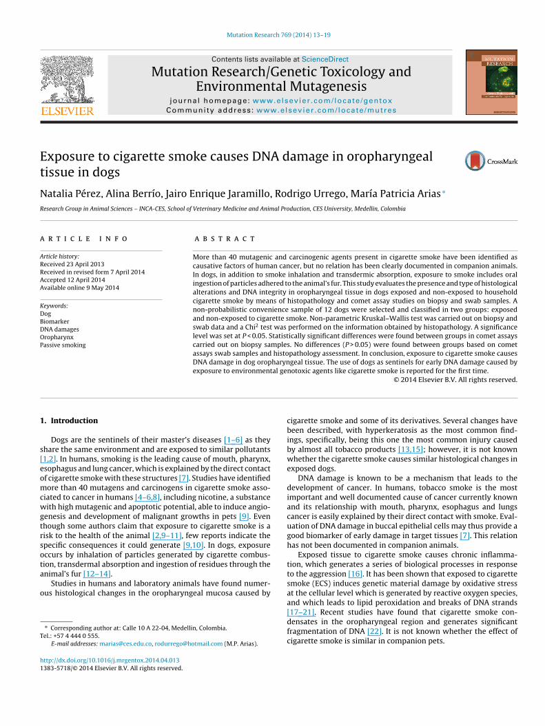
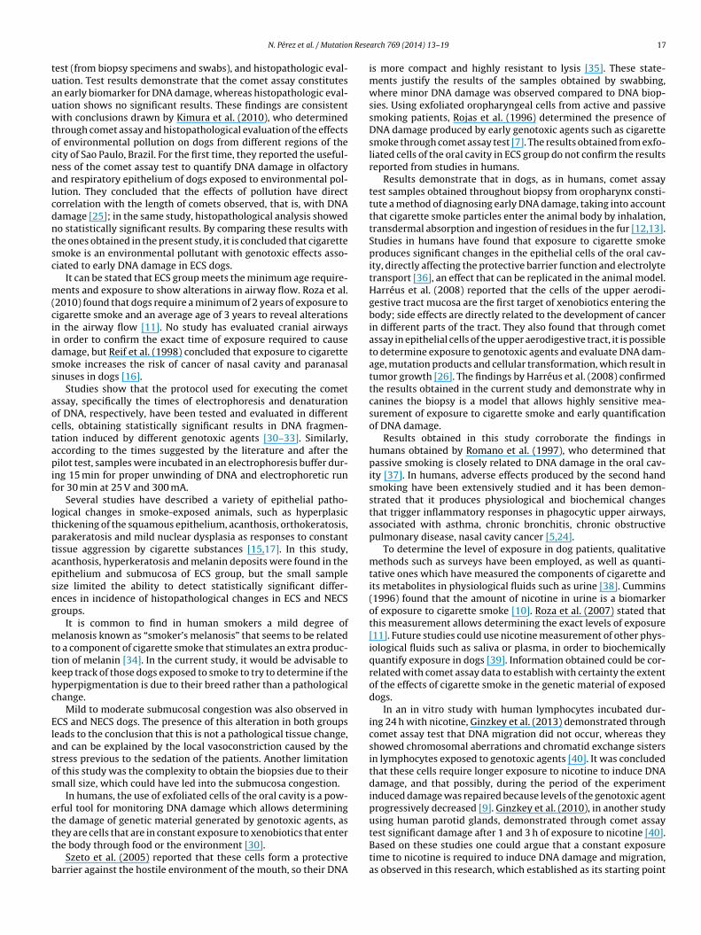
Histopathological evaluation showed that both ECS and NECSgroups showed alterations in the architecture and oropharyngealtissue cellularity, although, they were more common on the ECSgroup. Alterations in the ECS group were acanthosis, hyperkera-tosis, increased thickness of the epithelium and the presence ofmucosa melanin deposits. Histophatological changes used to definethe grade of tissue alteration on both groups are shown in Table 1.Fig. 1 shows some of the alterations found in the oropharyngealtissue of non-exposed and exposed dogs to cigarette smoke.
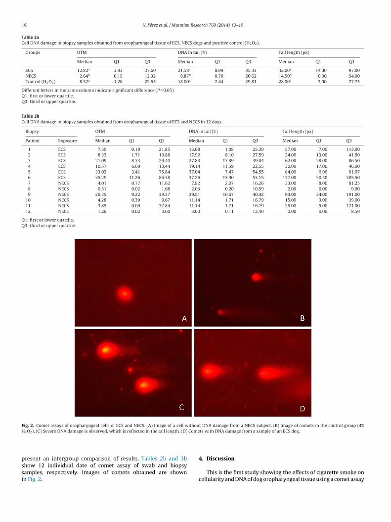
3.2. Comet assay
Data analysis allowed quantification of DNA damage in swaband biopsy samples from Olive Tail Moment (OTM), tail length and
percentage of DNA in tail variables. The results obtained from theexfoliated cells did not show significant difference among groups(P < 0.05), while data obtained from the biopsies did show signifi-cant difference between ECS and NECS (P < 0.01). Tables 2a and 3a
N. Pérez et al. / Mutation Research 769 (2014) 13–19 15
Table 1Incidence table shows the most common alterations found in the oropharyngeal tissue of ECS and NECS dogs.
Group Acanthosis Hyperkeratosis Increased thicknessof the epithelium
Melanin deposits inepithelium andsubmucosa
Submucosacongestion
ECS, n (%) 2 (33) 4 (67) 4 (67) 5 (83) 4 (67)NECS, n (%) 4 (67) 2 (33) 2 (33) 1 (17) 2 (33)
Fig. 1. Histopathological findings in oropharyngeal tissue in ECS and NECS; hematoxylin-eosin staining. (A) Normal oropharyngeal mucosa from a NECS (10×). (B) Orop-haryngeal mucosa with acanthosis from an ECS group (10×). (C) Melanosis in an ECS dog (10×). (D) Oral submucosal congestion from a NECS dog (10×).
Table 2aCell DNA damage in swab samples obtained from oropharyngeal tissue of ECS, NECS dogs and positive control (H2O2).
Groups OTM DNA in tail (%) Tail length (px)
Median Q1 Q3 Median Q1 Q3 Median Q1 Q3
ECS 0.34 0.00 2.62 1.61 0.00 9.05 5.50 0.00 20.00NECS 0.79 0.01 4.04 3.14 0.11 12.48 7.00 0.00 24.30Control (H2O2) 0.64 0.00 4.92 2.36 0.02 11.01 5.00 0.00 21.00
Q1: first or lower quartile.Q3: third or upper quartile.
Table 2bCell DNA damage in swab samples obtained from oropharyngeal tissue of ECS and NECS in 12 dogs.
SWAB OTM DNA in tail (%) Tail length (px)
Patient Exposure Median Q1 Q3 Median Q1 Q3 Median Q1 Q3
1 ECS 0.03 0.00 0.72 0.14 0.00 1.92 1.00 0.00 4.002 ECS 0.96 0.02 6.84 2.33 0.08 12.15 8.00 0.00 29.003 ECS 0.20 0.00 2.48 1.02 0.00 6.20 9.00 0.00 22.004 ECS 1.02 0.00 8.20 2.25 0.00 17.17 15.00 2.00 31.005 ECS 2.73 0.01 17.53 5.31 0.07 12.95 9.00 0.00 57.506 ECS 1.16 0.64 5.50 4.12 0.85 10.32 5.00 0.00 28.007 NECS 9.74 0.46 70.18 21.96 1.07 45.48 59.50 2.50 303.508 NECS 4.23 0.36 77.57 13.28 1.21 45.99 44.00 4.00 227.009 NECS 1.65 0.07 8.50 3.16 0.12 18.25 7.00 1.00 43.00
10 NECS 3.24 1.30 9.32 9.72 3.29 15.98 17.00 5.50 45.2511 NECS 0.89 0.01 5.18 1.70 0.06 13.35 6.00 0.00 29.0012 NECS 0.68 0.00 5.83 1.68 0.01 11.08 7.00 0.00 20.25
Q1: first or lower quartile.Q3: third or upper quartile.
16 N. Pérez et al. / Mutation Research 769 (2014) 13–19
Table 3aCell DNA damage in biopsy samples obtained from oropharyngeal tissue of ECS, NECS dogs and positive control (H2O2).
Groups OTM DNA in tail (%) Tail length (px)
Median Q1 Q3 Median Q1 Q3 Median Q1 Q3
ECS 12.82a 3.83 27.60 21.58a 8.99 35.33 42.00a 14.00 97.00NECS 2.64b 0.15 12.32 8.87b 0.70 20.62 14.50b 0.00 54.00Control (H2O2) 8.32a 1.28 22.55 18.00a 7.44 29.81 28.00a 3.00 77.75
Different letters in the same column indicate significant difference (P < 0.05).Q1: first or lower quartile.Q3: third or upper quartile.
Table 3bCell DNA damage in biopsy samples obtained from oropharyngeal tissue of ECS and NECS in 12 dogs.
Biopsy OTM DNA in tail (%) Tail length (px)
Patient Exposure Median Q1 Q3 Median Q1 Q3 Median Q1 Q3
1 ECS 7.59 0.19 21.85 13.68 1.08 25.39 57.00 7.00 113.002 ECS 8.33 1.71 19.88 17.92 8.10 27.59 24.00 13.00 41.503 ECS 21.09 8.73 29.40 27.83 17.89 39.04 62.00 28.00 86.504 ECS 10.57 6.60 13.44 19.14 11.59 22.55 30.00 17.00 46.005 ECS 33.02 3.41 75.84 37.04 7.47 54.55 84.00 6.96 91.076 ECS 35.29 11.26 86.38 37.26 13.90 53.15 177.00 30.50 305.507 NECS 4.01 0.77 11.62 7.92 2.07 16.26 33.00 8.00 81.258 NECS 0.51 0.02 1.68 2.03 0.20 10.59 2.00 0.00 9.009 NECS 20.35 9.22 39.37 29.51 16.67 40.42 93.00 34.00 191.00
10 NECS 4.28 0.39 9.67 11.14 1.71 16.79 15.00 3.00 39.0011 NECS 3.85 0.00 37.84 11.14 1.71 16.79 28.00 5.00 171.0012 NECS 1.29 0.02 3.60 3.00 0.11 12.40 0.00 0.00 8.50
Q1: first or lower quartile.Q3: third or upper quartile.
Fig. 2. Comet assays of oropharyngeal cells of ECS and NECS. (A) Image of a cell without DNA damage from a NECS subject. (B) Image of comets in the control group (4%H Come
pssi
2O2). (C) Severe DNA damage is observed, which is reflected in the tail length. (D)
resent an intergroup comparison of results, Tables 2b and 3b
how 12 individual date of comet assay of swab and biopsyamples, respectively. Images of comets obtained are shownn Fig. 2.ts with DNA damage from a sample of an ECS dog.
4. Discussion
This is the first study showing the effects of cigarette smoke oncellularity and DNA of dog oropharyngeal tissue using a comet assay
n Rese
tuauwtocnalcdntsc
m(ciidss
aoctapif
ltptaeseg
mttkhc
Elasos
ettt
b
N. Pérez et al. / Mutatio
est (from biopsy specimens and swabs), and histopathologic eval-ation. Test results demonstrate that the comet assay constitutesn early biomarker for DNA damage, whereas histopathologic eval-ation shows no significant results. These findings are consistentith conclusions drawn by Kimura et al. (2010), who determined
hrough comet assay and histopathological evaluation of the effectsf environmental pollution on dogs from different regions of theity of Sao Paulo, Brazil. For the first time, they reported the useful-ess of the comet assay test to quantify DNA damage in olfactorynd respiratory epithelium of dogs exposed to environmental pol-ution. They concluded that the effects of pollution have directorrelation with the length of comets observed, that is, with DNAamage [25]; in the same study, histopathological analysis showedo statistically significant results. By comparing these results withhe ones obtained in the present study, it is concluded that cigarettemoke is an environmental pollutant with genotoxic effects asso-iated to early DNA damage in ECS dogs.
It can be stated that ECS group meets the minimum age require-ents and exposure to show alterations in airway flow. Roza et al.
2010) found that dogs require a minimum of 2 years of exposure toigarette smoke and an average age of 3 years to reveal alterationsn the airway flow [11]. No study has evaluated cranial airwaysn order to confirm the exact time of exposure required to causeamage, but Reif et al. (1998) concluded that exposure to cigarettemoke increases the risk of cancer of nasal cavity and paranasalinuses in dogs [16].
Studies show that the protocol used for executing the cometssay, specifically the times of electrophoresis and denaturationf DNA, respectively, have been tested and evaluated in differentells, obtaining statistically significant results in DNA fragmen-ation induced by different genotoxic agents [30–33]. Similarly,ccording to the times suggested by the literature and after theilot test, samples were incubated in an electrophoresis buffer dur-
ng 15 min for proper unwinding of DNA and electrophoretic runor 30 min at 25 V and 300 mA.
Several studies have described a variety of epithelial patho-ogical changes in smoke-exposed animals, such as hyperplasichickening of the squamous epithelium, acanthosis, orthokeratosis,arakeratosis and mild nuclear dysplasia as responses to constantissue aggression by cigarette substances [15,17]. In this study,canthosis, hyperkeratosis and melanin deposits were found in thepithelium and submucosa of ECS group, but the small sampleize limited the ability to detect statistically significant differ-nces in incidence of histopathological changes in ECS and NECSroups.
It is common to find in human smokers a mild degree ofelanosis known as “smoker’s melanosis” that seems to be related
o a component of cigarette smoke that stimulates an extra produc-ion of melanin [34]. In the current study, it would be advisable toeep track of those dogs exposed to smoke to try to determine if theyperpigmentation is due to their breed rather than a pathologicalhange.
Mild to moderate submucosal congestion was also observed inCS and NECS dogs. The presence of this alteration in both groupseads to the conclusion that this is not a pathological tissue change,nd can be explained by the local vasoconstriction caused by thetress previous to the sedation of the patients. Another limitationf this study was the complexity to obtain the biopsies due to theirmall size, which could have led into the submucosa congestion.
In humans, the use of exfoliated cells of the oral cavity is a pow-rful tool for monitoring DNA damage which allows determininghe damage of genetic material generated by genotoxic agents, as
hey are cells that are in constant exposure to xenobiotics that enterhe body through food or the environment [30].Szeto et al. (2005) reported that these cells form a protectivearrier against the hostile environment of the mouth, so their DNA
arch 769 (2014) 13–19 17
is more compact and highly resistant to lysis [35]. These state-ments justify the results of the samples obtained by swabbing,where minor DNA damage was observed compared to DNA biop-sies. Using exfoliated oropharyngeal cells from active and passivesmoking patients, Rojas et al. (1996) determined the presence ofDNA damage produced by early genotoxic agents such as cigarettesmoke through comet assay test [7]. The results obtained from exfo-liated cells of the oral cavity in ECS group do not confirm the resultsreported from studies in humans.
Results demonstrate that in dogs, as in humans, comet assaytest samples obtained throughout biopsy from oropharynx consti-tute a method of diagnosing early DNA damage, taking into accountthat cigarette smoke particles enter the animal body by inhalation,transdermal absorption and ingestion of residues in the fur [12,13].Studies in humans have found that exposure to cigarette smokeproduces significant changes in the epithelial cells of the oral cav-ity, directly affecting the protective barrier function and electrolytetransport [36], an effect that can be replicated in the animal model.Harréus et al. (2008) reported that the cells of the upper aerodi-gestive tract mucosa are the first target of xenobiotics entering thebody; side effects are directly related to the development of cancerin different parts of the tract. They also found that through cometassay in epithelial cells of the upper aerodigestive tract, it is possibleto determine exposure to genotoxic agents and evaluate DNA dam-age, mutation products and cellular transformation, which result intumor growth [26]. The findings by Harréus et al. (2008) confirmedthe results obtained in the current study and demonstrate why incanines the biopsy is a model that allows highly sensitive mea-surement of exposure to cigarette smoke and early quantificationof DNA damage.
Results obtained in this study corroborate the findings inhumans obtained by Romano et al. (1997), who determined thatpassive smoking is closely related to DNA damage in the oral cav-ity [37]. In humans, adverse effects produced by the second handsmoking have been extensively studied and it has been demon-strated that it produces physiological and biochemical changesthat trigger inflammatory responses in phagocytic upper airways,associated with asthma, chronic bronchitis, chronic obstructivepulmonary disease, nasal cavity cancer [5,24].
To determine the level of exposure in dog patients, qualitativemethods such as surveys have been employed, as well as quanti-tative ones which have measured the components of cigarette andits metabolites in physiological fluids such as urine [38]. Cummins(1996) found that the amount of nicotine in urine is a biomarkerof exposure to cigarette smoke [10]. Roza et al. (2007) stated thatthis measurement allows determining the exact levels of exposure[11]. Future studies could use nicotine measurement of other phys-iological fluids such as saliva or plasma, in order to biochemicallyquantify exposure in dogs [39]. Information obtained could be cor-related with comet assay data to establish with certainty the extentof the effects of cigarette smoke in the genetic material of exposeddogs.
In an in vitro study with human lymphocytes incubated dur-ing 24 h with nicotine, Ginzkey et al. (2013) demonstrated throughcomet assay test that DNA migration did not occur, whereas theyshowed chromosomal aberrations and chromatid exchange sistersin lymphocytes exposed to genotoxic agents [40]. It was concludedthat these cells require longer exposure to nicotine to induce DNAdamage, and that possibly, during the period of the experimentinduced damage was repaired because levels of the genotoxic agentprogressively decreased [9]. Ginzkey et al. (2010), in another studyusing human parotid glands, demonstrated through comet assay
test significant damage after 1 and 3 h of exposure to nicotine [40].Based on these studies one could argue that a constant exposuretime to nicotine is required to induce DNA damage and migration,as observed in this research, which established as its starting point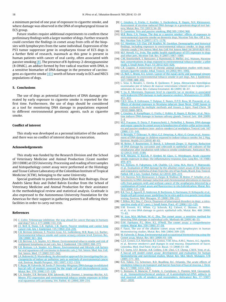
1 n Rese
awE
pcsPahp(agp
5
efiats
C
a
A
o2aaM
AViaAf
R
[[
[
[
[
[
[
[
[
[
[
[
[
[
[
[
[
[
[
[
[
[
[
[
[
[
[
8 N. Pérez et al. / Mutatio
minimum period of one year of exposure to cigarette smoke, andhere damage was observed in the DNA of oropharyngeal tissue in
CS group.Future studies require additional experiments to confirm these
reliminary findings with a larger number of dogs. Further researchould correlate the findings in comet assay from cells of the biop-ies with lymphocytes from the same individual. Expression of the53 tumor suppressor gene in oropharynx tissue of ECS dogs is
further field of research, inasmuch as this gene is present inuman patients with cancer of oral cavity, often associated withassive smoking [8]. The presence of 8-hydroxy-2-deoxyguanosine8-OHdG), an adduct formed by free radical reaction with DNA, is
sensitive biomarker of DNA damage in the presence of carcino-ens as cigarette smoke [41] worth of future study in ECS and NECSopulations of dogs.
. Conclusions
The use of dogs as potential biomarkers of DNA damage gen-rated by early exposure to cigarette smoke is reported for therst time. Furthermore, the use of dogs should be considereds a tool for monitoring DNA damage in populations exposedo different environmental genotoxic agents, such as cigarettemoke.
onflict of interest
This study was developed as a personal initiative of the authorsnd there was no conflict of interest during its execution.
cknowledgements
This study was funded by the Research Division and the Schoolf Veterinary Medicine and Animal Production (Grant number011DI08) at CES University. Processing and reading of test samplesnd histopathology comet assay were performed at the Histologynd Tissue Culture Laboratory of the Colombian Institute of Tropicaledicine (ICTM), belonging to the same University.Special gratitude to professors Jhon Didier Ruiz Buitrago, Oscar
ndrés Sáenz Ruiz and Dubel Balvin Escobar from the School ofeterinary Medicine and Animal Production for their assistance
n the methodological review and statistical analysis. Gratitude islso expressed to the Autonomous University Foundation for themericas for their support in gathering patients and offering their
acilities in order to carry out tests.
eferences
[1] C. Colitz, Telomerase inhibition: the way ahead for cancer therapy in humansand dogs? Vet. J. 177 (2008) 153–154.
[2] J.S. Reif, K. Dunn, G.K. Ogilvie, C.K. Harris, Passive smoking and canine lungcancer risk, Am. J. Epidemiol. 135 (1992) 234–239.
[3] E.R. Bertone-Johnson, E. Procter-Gray, A.L. Gollenberg, M.B. Ryan, L.G. Barber,Environmental tobacco smoke and canine urinary cotinine level, Environ. Res.106 (2008) 361–364.
[4] E.R. Bertone, L.A. Snyder, A.S. Moore, Environmental tobacco smoke and risk ofmalignant lymphoma in pet cats, Am. J. Epidemiol. 156 (2002) 268–273.
[5] E.R. Bertone, L.A. Snyder, A.S. Moore, Environmental and lifestyle risk factorsfor oral squamous cell carcinoma in domestic cats, J. Vet. Intern. Med. Am. Coll.Vet. Intern. Med. 17 (2003) 557–562.
[6] J.A. Bukowski, D. Wartenberg, An alternative approach for investigating the car-cinogenicity of indoor air pollution: pets as sentinels of environmental cancerrisk, Environ. Health Perspect. 105 (1997) 1312–1319.
[7] E. Rojas, M. Valverde, M. Sordo, P. Ostrosky-Wegman, DNA damage in exfoliated
buccal cells of smokers assessed by the single cell gel electrophoresis assay,Mutat. Res. 370 (1996) 115–120.[8] L.A. Snyder, E.R. Bertone, R.M. Jakowski, M.S. Dooner, J. Jennings-Ritchie, A.S.Moore, P53 expression and environmental tobacco smoke exposure in felineoral squamous cell carcinoma, Vet. Pathol. 41 (2004) 209–214.
[
arch 769 (2014) 13–19
[9] C. Ginzkey, G. Friehs, C. Koehler, S. Hackenberg, R. Hagen, N.H. Kleinsasser,Assessment of nicotine-induced DNA damage in a genotoxicological test bat-tery, Mutat. Res. 751 (2013) 34–39.
10] D. Cummins, Pets and passive smoking, BMJ 309 (1994) 960.11] M.R. Roza, C.A. Viegas, The dog as a passive smoker: effects of exposure to
environmental cigarette smoke on domestic dogs, Nicotine Tob. Res. Off. J. Soc.Res. Nicotine Tob. 9 (2007) 1171–1176.
12] E.C. Hawkins, L.D. Clay, J.M. Bradley, M. Davidian, Demographic and historicalfindings, including exposure to environmental tobacco smoke, in dogs withchronic cough, J. Vet. Intern. Med. Am. Coll. Vet. Intern. Med. 24 (2010) 825–831.
13] M.F. Hovell, V.L. Irvin, The public health significance of ETS exposure to dogsand other pets, Nicotine Tob. Res. 9 (2007) 1067–1069.
14] C.M. Knottenbelt, S. Bawazeer, J. Hammond, D. Mellor, D.G. Watson, Nicotinehair concentrations in dogs exposed to environmental tobacco smoke: a pilotstudy, J. Small Anim. Pract. 53 (2012) 623–626.
15] C.R. Coggins, A minireview of chronic animal inhalation studies with main-stream cigarette smoke, Inhal. Toxicol. 14 (2002) 991–1002.
16] J.S. Reif, C. Bruns, K.S. Lower, Cancer of the nasal cavity and paranasal sinusesand exposure to environmental tobacco smoke in pet dogs, Am. J. Epidemiol.147 (1998) 488–492.
17] L. Sosa, A. Rosales, L. Dávila, B. Quinonez, P. Jarpa, Alteraciones histológicasocasionadas por el tabaco de mascar venezolano (chimó) en los tejidos peri-odontales de ratas, Rev. Cubana Estomatol. 46 (2009) 38–47.
18] Y. Lu, K. Morimoto, Exposure level to cigarette tar or nicotine is associatedwith leukocyte DNA damage in male Japanese smokers, Mutagenesis 23 (2008)451–455.
19] F.R.D. Silva, B. Erdtmann, T. Dalpiaz, E. Nunes, D.P.D. Rosa, M. Porawski, et al.,Effects of dermal exposure to Nicotiana tabacum (Jean Nicot, 1560) leaves inmouse evaluated by multiple methods and tissues, J. Agric. Food Chem. 58(2010) 9868–9874.
20] C. Ginzkey, K. Kampfinger, G. Friehs, C. Kohler, R. Hagen, E. Richter, et al., Nico-tine induces DNA damage in human salivary glands, Toxicol. Lett. 184 (2009)1–4.
21] M.E. Fracasso, D. Doria, P. Franceschetti, L. Perbellini, L. Romeo, DNA damageand repair capacity by comet assay in lymphocytes of white-collar active smok-ers and passive smokers (non- and ex-smokers) at workplace, Toxicol. Lett. 167(2006) 131–141.
22] D. Beyoglu, T. Ozkozaci, N. Akici, G.Z. Omurtag, A. Akici, O. Ceran, et al., Assess-ment of DNA damage in children exposed to indoor tobacco smoke, Int. J. Hyg.Environ. Health 213 (2010) 40–43.
23] M. Reiter, P. Baumeister, D. Boeck, S. Schwenk-Zieger, U. Harréus, Reductionof DNA damage by curcumin and celecoxib in epithelial cell cultures of theoropharynx after incubation with tobacco smoke condensate, Anticancer Res.32 (2012) 3185–3189.
24] W.R. Abrams, U. Kucich, P. Kimbel, M. Glass, G. Weinbaum, Acute cigarettesmoke exposure in dogs: the inflammatory response, Exp. Lung Res. 14 (1988)459–475.
25] K.C. Kimura, H. Fukumasu, L.M. Chaible, C.E. Lima, M.A. Horst, P. Matsuzaki,et al., Evaluation of DNA damage by the alkaline comet assay of the olfactoryand respiratory epithelia of dogs from the city of Sao Paulo, Brazil, Exp. Toxicol.Pathol. Off. J. Ges. Toxikol. Pathol. 62 (2010) 209–219.
26] U.A. Harreus, N.H. Kleinsasser, S. Zieger, B. Wallner, M. Reiter, P. Schuller, et al.,Sensitivity to DNA-damage induction and chromosomal alterations in mucosacells from patients with and without cancer of the oropharynx detected by acombination of Comet assay and fluorescence in situ hybridization, Mutat. Res.563 (2004) 131–138.
27] R.R. Tice, E. Agurell, D. Anderson, B. Burlinson, A. Hartmann, H. Kobayashi, et al.,Single cell gel/comet assay: guidelines for in vitro and in vivo genetic toxicologytesting, Environ. Mol. Mutagen. 35 (2000) 206–221.
28] F. Billen, M.J. Day, C. Clercx, Diagnosis of pharyngeal disorders in dogs: a retro-spective study of 67 cases, J. Small Anim. Pract. 47 (2006) 122–129.
29] S.M. Everett, K.L. White, C.J. Schorah, R.J. Calvert, C. Skinner, D. Miller,et al., In vivo DNA damage in gastric epithelial cells, Mutat. Res. 468 (2000)73–85.
30] W. Liao, M.A. McNutt, W.-G. Zhu, The comet assay: a sensitive method fordetecting DNA damage in individual cells, Methods 48 (2009) 46–53.
31] D.W. Fairbairn, P.L. Olive, K.L. O’Neill, The comet assay: a comprehensivereview, Mutat. Res. 339 (1995) 37–59.
32] F. Faust, The use of the alkaline comet assay with lymphocytes in humanbiomonitoring studies, Mutat. Res. 566 (2004) 209–229.
33] M. Valverde, E. Rojas, Environmental and occupational biomonitoring using theComet assay, Mutat. Res. 681 (2009) 93–109.
34] G.J.A. Gomez, E.A. Martinez, R.J. Gomez, Y.M. Silva, A.M.G. Nunez, A.G. Agudelo,et al., Reverse smokers’s and changes in oral mucosa. Department of Sucre,Colombia, Med. Oral Patol. Oral Cir. Bucal. 13 (2008) E1–E8.
35] Y.T. Szeto, I.F.F. Benzie, A.R. Collins, S.W. Choi, C.Y. Cheng, C.M.N. Yow, et al.,A buccal cell model comet assay: development and evaluation for humanbiomonitoring and nutritional studies, Mutat. Res. Mol. Mech. Mutagen. 578(2005) 371–381.
36] N.A. Tobey, V.J. Schreiner, R.D. Readling, R.C. Orlando, The acute effects ofsmokeless tobacco on transport and barrier function of buccal mucosa, J. Dent.
Res. 67 (1988) 1414–1421.37] G. Romano, R. Mancini, P. Fedele, G. Curigliano, G. Flamini, M.R. Giovagnoli,et al., Immunohistochemical analysis of 4-aminobiphenyl-DNA adducts inoral mucosal cells of smokers and nonsmokers, Anticancer Res. 17 (1997)2827–2830.

n Rese
[
[
[
N. Pérez et al. / Mutatio
38] E.A. McNiel, S.G. Carmella, L.A. Heath, R.L. Bliss, K.A. Le, S.S. Hecht, Urinary
biomarkers to assess exposure of cats to environmental tobacco smoke, Am.J. Vet. Res. 68 (2007) 349–353.39] R.S. Brazell, A.C. Stiff, G.M. Henderson, R.A. Jenkins, P.L. Romig, O. Auerbach,Plasma nicotine and cotinine in tobacco smoke exposed beagle dogs, Toxicol.Appl. Pharmacol. 73 (1984) 152–158.
[
arch 769 (2014) 13–19 19
40] C. Ginzkey, G. Friehs, C. Koehler, S. Hackenberg, H.-U. Voelker, E. Richter,
et al., Nicotine and methyl methane sulfonate in mini organ cultures of humanparotid gland tissue, Toxicol. Lett. 197 (2010) 69–74.41] G. Borthakur, C. Butryee, M. Stacewicz-Sapuntzakis, P.E. Bowen, Exfoliated buc-cal mucosa cells as a source of DNA to study oxidative stress, Cancer Epidemiol.Biomarkers Prev. 17 (2008) 212–219.